Tumor vesicle-associated CD147 modulates the angiogenic capability of endothelial cells
9
Tumor Vesicle–Associated CD147 Modulates the Angiogenic Capability of Endothelial Cells 1 Danilo Millimaggi *, Marianna Mari *, Sandra D’Ascenzo *, Eleonora Carosa *, Emmanuele Angelo Jannini *, Stanley Zucker y , Gaspare Carta z , Antonio Pavan * ,§ and Vincenza Dolo * *Department of Experimental Medicine, L’Aquila University, L’Aquila, Italy; y Department of Medicine, Stony Brook University, Stony Brook, NY, USA; z Department of Surgical Science, L’Aquila University, L’Aquila, Italy; § Department of Experimental Medicine, University of Rome, ‘‘La Sapienza’’ Abstract Matrix metalloproteinase (MMP) degradation of extra- cellular matrix is thought to play an important role in invasion, angiogenesis, tumor growth, and metasta- sis. Several studies have demonstrated that CD147/ extracellular MMP inducer, a membrane-spanning mole- cule highly expressed in tumor cells, may be involved in the progression of malignancies by regulating ex- pression of MMP in peritumoral stromal cells. In the present study we show that CD147 is expressed in microvesicles derived from epithelial ovarian cancer cells and that CD147-positive vesicles may promote an angiogenic phenotype in endothelial cells in vitro. Vesicles shed by human ovarian carcinoma cell lines OVCAR3, SKOV3, and A2780 expressed different levels of CD147 and stimulated proangiogenic activities of human umbilical vein endothelial cells (HUVECs) in a CD147-dependent fashion (OVCAR3 > SKOV3 > A2780). Moreover, vesicles shed by ovarian carcinoma cell line CABA I with low CD147 expression had no significant effect on the development of angiogenic phenotype in HUVECs. The treatment of OVCAR3 cells with small interfering RNA against CD147 suppressed the angio- genic potential of OVCAR3-derived microvesicles. How- ever, transfection of CD147 cDNA into the CABA I cell line enabled CABA I–derived vesicles to induce angio- genesis and to promote MMP genes expression in HUVECs. We therefore conclude that vesicles shed by ovarian cancer cells may induce proangiogenic ac- tivities of HUVECs by a CD147-mediated mechanism. Neoplasia (2007) 9, 349–357 Keywords: Shed membrane microvesicles, CD147, ovarian cancer, angio- genesis, matrix metalloproteinases. Introduction CD147, also named extracellular matrix metalloproteinase (MMP) inducer or basigin, is a plasma membrane glyco- protein enriched on the surface of many malignant tumor cells [1–3]. Thus far, several studies have convincingly demonstrated that CD147 could play a crucial role in the progression of malignancies by regulating expression of vascular endothelial growth factor (VEGF) and MMPs in stro- mal cells [1,4]. Accordingly, it has been previously shown that CD147 interacts with fibroblasts and stimulates their production of MMP-1, MMP-2, MMP-3, and membrane type 1 MMP (MT1- MMP) [5,6]. CD147 is also believed to be involved in the interaction of the tumor with its stromal microenvironment by acting in a paracrine fashion on stroma cells to regulate the production of MMPs [4]. Interestingly, recent studies have provided evidence that microvesicular release of CD147 from tumor cells could play a role in tumor – stromal interactions through upregulation of MMPs production [7,8]. In addition, CD147 is also known to stimulate the production of MMPs in human umbilical vein endothelial cells (HUVECs) [9]. Matrix metalloproteinase degradation of extracellular matrix is thought to play an important role in invasion, angiogenesis, tumor growth, and metastasis [10]. In this regard, increased tumor aggressiveness has been associated with enhanced MMP expression both in vitro and in vivo [11]. Because stromal cells are the major source of MMPs in most of the human cancers analyzed so far, CD147 is deemed to be a critical determinant in cancer growth and dissemination [9,10,12]. In line with this possibility, CD147 expression in human breast and ovarian cancer has been related to a more invasive phenotype [3,13 – 15]. In addition, human breast cancer cells transfected with CD147 cDNA showed a significantly higher rate of tumor growth in a xenograft model [11]. Shedding of membrane vesicles is a vital phenomenon frequently observed in eukaryotic cells and suggested to be involved in several pathophysiologic processes such as an- giogenesis, thrombosis, inflammation, and immunity [16,17]. In previous investigations, two types of membrane vesicles have been described, referred to as microvesicles and exo- somes [17 – 19]. It is worth noting that microvesicles and exo- somes are released by different cellular mechanisms, namely, Address all correspondence to: Vincenza Dolo, Dipartimento di Medicina Sperimentale, Universita ` di L’Aquila, Via Vetoio-Coppito 2, I-67100 L’Aquila, Italy. E-mail: [email protected] 1 This work was supported by grants from the Italian Ministry of University and Scientific and Technological Research (MURST) to A.P. and V.D. Received 17 January 2007; Revised 23 February 2007; Accepted 26 February 2007. Copyright D 2007 Neoplasia Press, Inc. All rights reserved 1522-8002/07/$25.00 DOI 10.1593/neo.07133 Neoplasia . Vol. 9, No. 4, April 2007, pp. 349 – 357 349 www.neoplasia.com RESEARCH ARTICLE
-
Upload
independent -
Category
Documents
-
view
0 -
download
0
Transcript of Tumor vesicle-associated CD147 modulates the angiogenic capability of endothelial cells

Tumor Vesicle–Associated CD147 Modulates the AngiogenicCapability of Endothelial Cells1
Danilo Millimaggi*, Marianna Mari*, Sandra D’Ascenzo*, Eleonora Carosa*, Emmanuele Angelo Jannini*,Stanley Zucker y, Gaspare Carta z, Antonio Pavan*,§ and Vincenza Dolo*
*Department of Experimental Medicine, L’Aquila University, L’Aquila, Italy; yDepartment of Medicine, StonyBrook University, Stony Brook, NY, USA; zDepartment of Surgical Science, L’Aquila University, L’Aquila, Italy;§Department of Experimental Medicine, University of Rome, ‘‘La Sapienza’’
Abstract
Matrix metalloproteinase (MMP) degradation of extra-
cellular matrix is thought to play an important role in
invasion, angiogenesis, tumor growth, and metasta-
sis. Several studies have demonstrated that CD147/
extracellular MMP inducer, a membrane-spanning mole-
cule highly expressed in tumor cells, may be involved in
the progression of malignancies by regulating ex-
pression of MMP in peritumoral stromal cells. In the
present study we show that CD147 is expressed in
microvesicles derived from epithelial ovarian cancer
cells and that CD147-positive vesicles may promote
an angiogenic phenotype in endothelial cells in vitro.
Vesicles shed by human ovarian carcinoma cell lines
OVCAR3, SKOV3, and A2780 expressed different levels
of CD147 and stimulated proangiogenic activities of
human umbilical vein endothelial cells (HUVECs) in a
CD147-dependent fashion (OVCAR3 > SKOV3 > A2780).
Moreover, vesicles shed by ovarian carcinoma cell line
CABA I with low CD147 expression had no significant
effect on the development of angiogenic phenotype in
HUVECs. The treatment of OVCAR3 cells with small
interfering RNA against CD147 suppressed the angio-
genic potential of OVCAR3-derived microvesicles. How-
ever, transfection of CD147 cDNA into the CABA I cell
line enabled CABA I–derived vesicles to induce angio-
genesis and to promote MMP genes expression in
HUVECs. We therefore conclude that vesicles shed
by ovarian cancer cells may induce proangiogenic ac-
tivities of HUVECs by a CD147-mediated mechanism.
Neoplasia (2007) 9, 349–357
Keywords: Shed membrane microvesicles, CD147, ovarian cancer, angio-genesis, matrix metalloproteinases.
Introduction
CD147, also named extracellular matrix metalloproteinase
(MMP) inducer or basigin, is a plasma membrane glyco-
protein enriched on the surface of many malignant tumor
cells [1–3]. Thus far, several studies have convincingly
demonstrated that CD147 could play a crucial role in the
progression of malignancies by regulating expression of
vascular endothelial growth factor (VEGF) and MMPs in stro-
mal cells [1,4]. Accordingly, it has been previously shown that
CD147 interacts with fibroblasts and stimulates their production
of MMP-1, MMP-2, MMP-3, and membrane type 1 MMP (MT1-
MMP) [5,6]. CD147 is also believed to be involved in the
interaction of the tumor with its stromal microenvironment by
acting in a paracrine fashion on stroma cells to regulate the
production of MMPs [4]. Interestingly, recent studies have
provided evidence that microvesicular release of CD147 from
tumor cells could play a role in tumor–stromal interactions
through upregulation of MMPs production [7,8]. In addition,
CD147 is also known to stimulate the production of MMPs in
human umbilical vein endothelial cells (HUVECs) [9].
Matrix metalloproteinase degradation of extracellular matrix
is thought to play an important role in invasion, angiogenesis,
tumor growth, and metastasis [10]. In this regard, increased
tumor aggressiveness has been associated with enhanced
MMP expression both in vitro and in vivo [11]. Because stromal
cells are the major source of MMPs in most of the human
cancers analyzed so far, CD147 is deemed to be a critical
determinant in cancer growth and dissemination [9,10,12]. In
line with this possibility, CD147 expression in human breast and
ovarian cancer has been related to a more invasive phenotype
[3,13–15]. In addition, human breast cancer cells transfected
with CD147 cDNA showed a significantly higher rate of tumor
growth in a xenograft model [11].
Shedding of membrane vesicles is a vital phenomenon
frequently observed in eukaryotic cells and suggested to be
involved in several pathophysiologic processes such as an-
giogenesis, thrombosis, inflammation, and immunity [16,17].
In previous investigations, two types of membrane vesicles
have been described, referred to as microvesicles and exo-
somes [17–19]. It is worth noting that microvesicles and exo-
somes are released by different cellular mechanisms, namely,
Address all correspondence to: Vincenza Dolo, Dipartimento di Medicina Sperimentale,
Universita di L’Aquila, Via Vetoio-Coppito 2, I-67100 L’Aquila, Italy. E-mail: [email protected] work was supported by grants from the Italian Ministry of University and Scientific and
Technological Research (MURST) to A.P. and V.D.
Received 17 January 2007; Revised 23 February 2007; Accepted 26 February 2007.
Copyright D 2007 Neoplasia Press, Inc. All rights reserved 1522-8002/07/$25.00
DOI 10.1593/neo.07133
Neoplasia . Vol. 9, No. 4, April 2007, pp. 349–357 349
www.neoplasia.com
RESEARCH ARTICLE

microvesicles by surface shedding and exosomes from
exocytosis of multivesicular bodies [18–20].
We have previously shown that tumor microvesicles may
carry tumor-associated surface antigens and may be de-
tected both in the serum and in ascites from patients with
ovarian cancer [21]. Furthermore, a significant positive corre-
lation was seen between tumor malignancy and both vesi-
cle amount and vesicle-associated MMP-2 activity. Hence,
human tumors constitutively release microvesicles, trans-
porting a broad array of biologically active molecules, includ-
ing cell surface receptors, adhesion molecules, and MMPs
[22–26]. Growing interest has been focused on tumor-
released microvesicles because they have the ability to
modulate invasive capabilities [27–29] and to promote angio-
genesis [30–32]. Previous studies have shown that human
cancer cells (including lung carcinoma, colon carcinoma,
and pancreatic cancers) produce large amounts of CD147-
positive microvesicles [7,8]. However, the pathophysiologic
role of CD147-positive vesicles has not been completely
understood. Working from these assumptions, the aim of
our study was to investigate the role of microvesicles released
by human ovarian cancer cells in the angiogenic process by
evaluating their effect on HUVEC phenotype.
Materials and Methods
Cell Culture
HUVECs were isolated from umbilical cord veins and
grown on 1% gelatin–coated flasks in DMEM supplemented
with 10% fetal calf serum (FCS), 10% newborn calf serum,
2 mmol/l glutamine, 50 mg/ml endothelial cell growth fac-
tor (crude extract from bovine brain), penicillin, and strep-
tomycin. Cells from the third to fifth passages of culture
were used. The CABA I cell line was established from the
ascitic fluid of an ovarian carcinoma patient not under-
going drug treatment [33]. CD147-transfected CABA I cells
were maintained as monolayers in RPMI 1640 (Euroclone,
Devon, UK) containing 5% FCS and 400 mg/ml G418.
Human ovarian carcinoma cell lines A2780, OVCAR 3 and
SKOV 3 were cultured in RPMI medium supplemented with
10% FCS.
Small Interfering RNA and Transfection
Transfection of CD147/green fluorescent protein cDNA
into human CABA I ovarian cancer cells with low CD147
expression was performed by the method of Zucker et al.
[11]. Cells were stably transfected using FuGene (Roche,
Basel, Switzerland) as per manufacturer’s protocol. G418-
resistant clones were microscopically screened for green
fluorescent protein fluorescence and positive clones were
pooled to minimize clonal variations. Control transfectants
carrying the pcDNA3 vector alone were generated. Identical
results were obtained with nontransfected CABA I cells or
CABA I cells transfected with pcDNA3 (data not shown). We
silenced CD147 in OVACAR3 cells by using CD147 small
interfering RNA (siRNA) (h) sc-35298, scrambled oligos
sc-37007 as control, and sc-29528 siRNA transfection re-
agents (Santa Cruz Biotechnology, Santa Cruz, CA) ac-
cording to the manufacturer’s protocol. The same results
were obtained with nonsilenced OVCAR3 cells or OVCAR3
cells transfected with scrambled oligos.
Flow Cytometric Analysis
Flow cytometry analysis was performed as previously
described [33], with slight modifications. Briefly, cells were
incubated on ice for 60 minutes with mouse anti–human
CD147 primary antibody (8D6 mouse monoclonal IgG1,
Santa Cruz Biotechnology; 1 mg/106 cells) in PBS containing
0.03% BSA. After washing with PBS, cells were incubated
with fluorescein-labeled goat anti–mouse IgG (Kirkegaard
& Perry Laboratories Inc., Gaithersburg, MD) on ice for
30 minutes. The cells were analyzed for cell-associated fluo-
rescence in a flow cytometer (FACScan, Becton Dickinson,
Collaborative Research, Bedford, MA).
Isolation of Membrane Microvesicles from
Cell-Conditioned Medium
Microvesicles were prepared as previously described
[19]. Conditioned medium obtained as above was centri-
fuged at 600g for 15 minutes and then at 1500g for 15 min-
utes to remove cells and large debris. Supernatants were
centrifuged at 100,000g for 1 hour at 4jC. Pelleted micro-
vesicles were resuspended in PBS (pH 7.4). Vesicles were
quantified based on measurements of vesicle-associated
protein levels, using the method of Bradford (Bio-Rad, Milan,
Italy), with BSA (Sigma, St. Louis, MO) as standard.
Cord Formation Assay
HUVECs (70,000/well) were seeded onto Matrigel-
coated 24-well plates in endothelial growth medium con-
taining 5% serum. Cells were stimulated by microvesicles
added as indicated. After 2 and 24 hours, the formation of
cords was photographed and independently scored by two
blinded observers.
Invasion Assay
HUVEC invasion was assayed using modified Boyden
chambers with polycarbonate PVP-free Nucleopore filters
(pore size, 8 mm) [34]. Filters were coated with a thick layer of
reconstituted basement membrane (0.05 mg/ml Matrigel,
Becton Dickinson), which cells must degrade to migrate
through the filter. Microvesicles were added as indicated.
HUVECs were detached, washed in DMEM–0.1% BSA,
resuspended in the same medium at a concentration of
5 � 105 cells/ml, and added to the upper compartment of
the chamber. After 6 hours, filters were stained with 1%
crystal violet in methanol, and migrated cells in 10 high-
power fields were counted.
Zymography
Zymography was performed using SDS–polyacrylamide
gels copolymerized with 1 mg/ml gelatin type B (Sigma).
Microvesicles (2.5 mg) and fivefold concentrated HUVEC-
350 Vesicle-Associated CD147 in Tumor Angiogenesis Millimaggi et al.
Neoplasia . Vol. 9, No. 4, 2007

conditioned medium were diluted in SDS-PAGE sample
buffer under nonreducing conditions without heating. After
electrophoresis, gels were washed twice for 30 minutes in
2.5% Triton X-100 at room temperature and incubated over-
night in collagenase buffer (50 mmol/l Tris–HCl, pH 7.6,
10 mmol/l NaCl, 0.02% Brij 35, and 5 mmol/l CaCl2) at 37jC.Gels were stained with Coomassie Blue R 250 (Bio-Rad) in
30%methanol and 10% acetic acid for 2 hours and destained
in the same solution without dye. Gelatinase activity was
visualized as clear bands on a dark background, indicating
proteolysis of the substrate. The supernatant of WM983A
melanoma cells was used as a reference standard for
MMP-2 and MMP-9.
Western Blot Analysis
Cells were lysed with lysis buffer containing 50 mmol/l Tris
(pH 7.8), 150 mmol/l NaCl, and 1% NP40. Protein concen-
trations of cellular lysates and microvesicles were determined
as described above. Cell lysates (40 mg) and microvesicle
proteins (10 mg) were resolved by SDS 10% PAGE under
reducing or nonreducing conditions and then transferred
to nitrocellulose membranes (Schleicher & Schuell, Dassel,
Germany). Nonspecific binding sites were blocked by in-
cubation with 10% nonfat dry milk in TBST at room tem-
perature for at least 1 hour. The blots were incubated
with a monoclonal antibody raised against human CD147
(8D6 mouse monoclonal IgG1, 1:500 dilution; Santa Cruz
Biotechnology) for 1 hour, or with antibody raised against
actin (SC-1616, Santa Cruz Biotechnology), followed by
peroxidase-conjugated secondary antibody in blocking
buffer. After washing, reactive bands were visualized by
using a chemiluminescence detection kit (ECL, Amersham-
Pharmacia; Biotech, Piscataway, NJ).
Real-Time Polymerase Chain Reaction
Total RNA was isolated by using SV Total RNA Isolation
System (Promega, Madison, WI) and cDNA was synthe-
sized from 5 mg RNA by using the ImProm-II Reverse Trans-
cription System (Promega) according to the manufacturer’s
protocol. Each real-time polymerase chain reaction (PCR)
reaction was prepared in triplicate and contained 2.0 ml ofcDNA. PCR was carried out using SYBR-green detection
of PCR products in real time (Roche). The sequences of
the primers used for PCRwere as follows: MT1-MMP forward,
5V-GAGCTCAGGGCAGTGGATAG-3V, MT1-MMP reverse
5V-GGTAGCCCGGTTCTACCTTC (215 bp); MMP-1 forward,
5V-AGGTCTCTGAGGGTCAAGCA-3V, MMP-1 reverse, 5V-
CTGGTTGAAAAGCATGAGCA (111 bp); MMP-2 forward,
5V-CACTTTCCTGGGCAACAAAT-3V, MMP-2 reverse, 5V-
TGATGTCATCCTGGGACAGA-3V (257 bp); MMP-9 for-
ward, 5V-TTGACAGCGACAAGAAGTGG, MMP-9 reverse,
5V-GCCATTCACGTCGTCCTTAT-3V (179 bp); GADPH for-
ward 5V-GGCCTCCAAGGAGTAAGACC-3V, GADPH reverse
5V-AGGGGTCTACATGGCAACTG-3V (147 bp). A compara-
tive DCt method was used to determine gene expression.
Expression levels were normalized to the expression levels
of the housekeeping gene GADPH.
Results
CD147 Expression in Ovarian Cancer Cell Lines
and Shed Microvesicles
We examined four ovarian cancer cell lines with different
invasion capability (CABA I, A2780, SKOV3, and OVCAR3 in
order of increasing invasion activity; Figure 1A). All tested
cell lines were positive for CD147 byWestern blot analysis of
cell lysates (Figure 1B), except CABA I, which show not
detectable CD147, with increasing levels in the following
order: CABA I, A2780, SKOV3, and OVCAR3 (Figure 1C). A
representative image, obtained using scanning electron mi-
croscopy, of the vesicle shedding phenomenon from CABA I
cells is shown in Figure 2A. The size of microvesicles ranged
between 200 and 900 nm. To investigate whether CD147 is
released from ovarian cancer cell lines through the shedding
Figure 1. (A) Comparison of the invasive potential of three ovarian cancer
cell lines. Migration activity (representative of three experiments) was cal-
culated as the mean number of migrated cells observed in ten high-power
fields (mean ± SD of triplicates). (B) Western blot analysis of CD147. (C) Flow
cytometric analysis of CD147. Right profile, white area, OVACR3; gray pro-
file, SKOV3; central profile, white area, A2780; black profile, CABA I.
Vesicle-Associated CD147 in Tumor Angiogenesis Millimaggi et al. 351
Neoplasia . Vol. 9, No. 4, 2007

of microvesicles, membrane microvesicles were isolated
by centrifugation and analyzed by Western blotting (Fig-
ure 2B). Vesicles shed by ovarian cancer cells were positive
for CD147, with increasing levels in the following order:
CABA I, A2780, SKOV3, and OVCAR3.
Effect of Tumor CD147-Positive Microvesicles on HUVECs
In vitro experiments using a Matrigel invasion assay
showed that CD147-positive microvesicles shed by ovarian
cancer cell lines enhanced HUVEC invasiveness. There was
a direct correlation between CD147 expression in micro-
vesicles and endothelial cell invasiveness (OVCAR3 >
SKOV3 > A2780 > CABA I; Figure 2C). Microvesicles were
also able to induce HUVEC proliferation (data not shown).
Vesicles shed by ovarian carcinoma cell line CABA I with
nondetectable CD147 expression had no significant effect on
the development of angiogenic phenotype in HUVECs. We
thus hypothesized that vesicles shed by ovarian cancer cells
may induce proangiogenic activities of HUVEC by a CD147-
mediated mechanism. To shed more light on this issue, we
transfected CD147 cDNA into noninvasive CABA I human
ovarian cell line to generate CABA I cells with high CD147
expression (Figure 3A). Expression of the CD147-GPF fusion
protein was confirmed by flow cytometry (Figure 3B) and con-
focal microscopy (data not shown). We used real-time PCR
analysis to examine MMP induction in CD147-transfected
CABA I cells. CD147-transfected cells showed enhanced
MMP-1, MT1-MMP, MMP-2, and MMP-9 mRNA compared
with vector-transfected CABA I cells. The most prominent
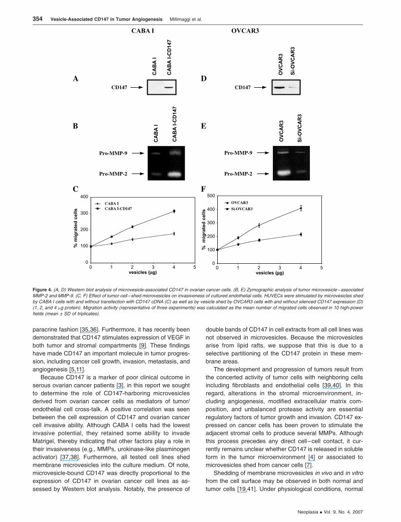
gene induced was MMP-2 (Figure 3C). CABA I–shed micro-
vesicles, after the CD147 transfection, became CD147 posi-
tive (Figure 4A). Zymographic analysis of vesicle-associated
MMP confirmed higher expression of pro–MMP-2 and pro–
MMP-9 in microvesicles shed by CD147-transfected CABA I
compared with vector-transfected cells (Figure 4B). More-
over, CD147-positive microvesicles exhibited a greater ca-
pacity to induce migration of HUVECs compared with those
generated from vector-transfected cells (Figure 4C). To
further investigate the potential regulatory role of vesicle-
associated CD147 on HUVEC phenotype, CD147-positive
OVCAR3 cells were transfected with a specific siRNA
against CD147 to silence its expression. Western blot analy-
sis of cell extracts from cells treated with siRNA showed a
lower CD147 expression (fsixfold) compared with the pa-
rental OVCAR3 cells (Figure 3D). The result was confirmed
by FACS (Figure 3E ) and confocal microscopy analyses
(data not shown). Real-time PCR was performed to quan-
tify MMP gene expression in cells transfected with siRNA
relative to the parental OVCAR3 population. Cells with si-
lenced CD147 expression showed a marked inhibition in the
MMP-1, MT1-MMP, MMP-2, and MMP-9 gene expression
compared with vector-transfected CABA I cells. A weak
effect on MT1-MMP mRNA was also noticed (Figure 3F ).
Microvesicles shed by OVCAR3 cells with silenced CD147
showed a reduced CD147 expression compared with paren-
tal cell–derived microvesicles (Figure 4D). Similarly, micro-
vesicles isolated from OVCAR3 cells with silenced CD147
expression were less effective inducers of pro–MMP-2 and
pro–MMP-9 synthesis in HUVECs compared with parental
cell–derived microvesicles (Figure 4E ). Moreover, micro-
vesicles derived from OVCAR3 cells with silenced CD147
expression were less effective at inducing the migration of
HUVECs (Figure 4F ).
Effects of Tumor-Shed Microvesicles on MMP Production
by HUVECs
To further explore the mechanisms whereby shed micro-
vesicles can mediate tumor–endothelium interactions, re-
verse transcription-PCR (RT-PCR) analysis was used to
monitor MMP synthesis in HUVECs treated with tumor-
released microvesicles. CABA I cell-derived microvesicles
did not induce MMP expression in HUVECs. In contrast,
microvesicles shed by CD147-transfected CABA I cells were
able to increase MT1-MMP (f1.5-fold), MMP-1 (f2-fold),
and MMP-2 (f1.9-fold) mRNA transcription. However,
Figure 2. (A) Western blot analysis of microvesicle-associated CD147. (B)
Effect of microvesicles derived from tumor cells on endothelial cell
invasiveness. HUVECs were stimulated by microvesicles shed by ovarian
cancer cells (1, 2, and 4 �g protein). Migration activity (representative of three
experiments) was calculated as the mean number of migrated cells observed
in 10 high-power fields (mean ± SD of triplicates). Supernatant of NIH-3T3,
used as a reference attractant, stimulated invasion with a value of 480%.
352 Vesicle-Associated CD147 in Tumor Angiogenesis Millimaggi et al.
Neoplasia . Vol. 9, No. 4, 2007

vesicles shed by OVCAR3 cells induced upregulation of
MT1-MMP (f1.5-fold), of MMP-1 (f2.2-fold), and MMP-2
(f2.8-fold) mRNA. MMP-9 gene expression was not affected
by microvesicles derived from OVCAR3 cells (Figure 5A). No
induction of MMP was observed in HUVECs after treatment
with microvesicles shed by OVCAR3 cells with silenced
CD147 expression. Each conditioned medium was collected
and examined by gelatin zymography. The conditioned media
from HUVECs treated with OVCAR3-derived microvesicles
showed increased MMP-2 activity (Figure 5B), whereas no
evidence of MMP-9 induction was found (data not shown).
Effects of Tumor Microvesicles on HUVEC Cord Formation
We have already shown that HUVEC microvesicles may
stimulate formation of capillary-like structures in an autocrine
manner [16]. To investigate whether microvesicles shed by
ovarian cancer cells can exert similar effects, we used a
three-dimensional matrix (Matrigel) to analyze the outgrowth
of human microvascular endothelial cells in capillary-like
tubular structures. After 24 hours in complete medium con-
taining 10% serum, more than 95% of the HUVECs were
organized into capillary-like structures (Figure 6A). In con-
trast, cells maintained in low-serum (5%) medium were
unable to form a tube network (Figure 6D). Treatment of
HUVECmaintained in low-serummedium with microvesicles
shed by OVCAR3 resulted in the formation of cords as
observed with complete medium (Figure 6B). However,
microvesicles shed from CABA I cells were not able to in-
duce capillary-like tube formation (Figure 6C). No cord
formation was observed after treatment of HUVECs with
microvesicles shed by OVCAR3 cells with silenced CD147
expression (Figure 6E ), whereas microvesicles shed by
CD147-transfected CABA I cells were able to induce the
formation of capillary-like tubular structures (Figure 6F ).
These results suggest that tube formation by HUVEC main-
tained in low-serum medium is crucially dependent on
CD147 expression in tumor-shed microvesicles.
Discussion
CD147 is highly expressed on the surface of tumor cells and
stimulates surrounding fibroblasts to produce MMPs in a
Figure 3. (A, D) Western blot analysis of CD147 in CABA I and OVAR3 ovarian cancer cell lines. (B) Flow cytometry analysis of CD147 in CABA I: gray profile
indicates control cells, whereas black profile indicates CD147-transfected cells. (E) Flow cytometry analysis of CD147 in OVCAR3 cells. Gray profile indicates
control cells, whereas black profile indicates cells with silenced CD147 expression. (C, F) RT-PCR analysis of ovarian cancer cell lines for MMP-1, -2, and -9 and
MT1-MMP mRNA expression. Control cells were arbitrarily set at 1.
Vesicle-Associated CD147 in Tumor Angiogenesis Millimaggi et al. 353
Neoplasia . Vol. 9, No. 4, 2007
paracrine fashion [35,36]. Furthermore, it has recently been
demonstrated that CD147 stimulates expression of VEGF in
both tumor and stromal compartments [9]. These findings
have made CD147 an important molecule in tumor progres-
sion, including cancer cell growth, invasion, metastasis, and
angiogenesis [5,11].
Because CD147 is a marker of poor clinical outcome in
serous ovarian cancer patients [3], in this report we sought
to determine the role of CD147-harboring microvesicles
derived from ovarian cancer cells as mediators of tumor/
endothelial cell cross-talk. A positive correlation was seen
between the cell expression of CD147 and ovarian cancer
cell invasive ability. Although CABA I cells had the lowest
invasive potential, they retained some ability to invade
Matrigel, thereby indicating that other factors play a role in
their invasiveness (e.g., MMPs, urokinase-like plasminogen
activator) [37,38]. Furthermore, all tested cell lines shed
membrane microvesicles into the culture medium. Of note,
microvesicle-bound CD147 was directly proportional to the
expression of CD147 in ovarian cancer cell lines as as-
sessed by Western blot analysis. Notably, the presence of
double bands of CD147 in cell extracts from all cell lines was
not observed in microvesicles. Because the microvesicles
arise from lipid rafts, we suppose that this is due to a
selective partitioning of the CD147 protein in these mem-
brane areas.
The development and progression of tumors result from
the concerted activity of tumor cells with neighboring cells
including fibroblasts and endothelial cells [39,40]. In this
regard, alterations in the stromal microenvironment, in-
cluding angiogenesis, modified extracellular matrix com-
position, and unbalanced protease activity are essential
regulatory factors of tumor growth and invasion. CD147 ex-
pressed on cancer cells has been proven to stimulate the
adjacent stromal cells to produce several MMPs. Although
this process precedes any direct cell–cell contact, it cur-
rently remains unclear whether CD147 is released in soluble
form in the tumor microenvironment [4] or associated to
microvesicles shed from cancer cells [7].
Shedding of membrane microvesicles in vivo and in vitro
from the cell surface may be observed in both normal and
tumor cells [19,41]. Under physiological conditions, normal
Figure 4. (A, D) Western blot analysis of microvesicle-associated CD147 in ovarian cancer cells. (B, E) Zymographic analysis of tumor microvesicle–associated
MMP-2 and MMP-9. (C, F) Effect of tumor cell – shed microvesicles on invasiveness of cultured endothelial cells. HUVECs were stimulated by microvesicles shed
by CABA I cells with and without transfection with CD147 cDNA (C) as well as by vesicle shed by OVCAR3 cells with and without silenced CD147 expression (D)
(1, 2, and 4 �g protein). Migration activity (representative of three experiments) was calculated as the mean number of migrated cells observed in 10 high-power
fields (mean ± SD of triplicates).
354 Vesicle-Associated CD147 in Tumor Angiogenesis Millimaggi et al.
Neoplasia . Vol. 9, No. 4, 2007

cells release only a limited amount of microvesicles in
response to specific stimuli [16]. In contrast, vesicle shed-
ding by tumor cells is largely an uncontrolled process where-
by numerous vesicles are constitutively shed from the entire
cell surface [19,22]. Moreover, tumor-derived microvesicles
carry angiogenic factors [30,42] that may enhance angio-
genesis by promoting endothelial cell migration and tubulo-
genesis [31,32].
We have previously shown that microvesicles shed by
HUVECs may induce cord formation of arterial endothelial
cells in an autocrine manner [16]. In this study, we provide
evidence that vesicles shed by human ovarian carcinoma
cell lines OVCAR3, SKOV3, and A2780 expressed different
levels of CD147 and stimulated proangiogenic activities
of HUVECs in a CD147-dependent fashion (OVCAR3 >
SKOV3 > A2780). Moreover, vesicles shed by ovarian carci-
noma cell line CABA I with low CD147 expression had no
significant effect on the development of angiogenic pheno-
type in HUVECs. The treatment of OVCAR3 cells with
siRNA against CD147 suppressed the angiogenic potential
of OVCAR3-derived microvesicles. However, transfection
of CD147 cDNA into the CABA I cell line enabled CABA
I–derived vesicles to induce an angiogenic phenotype in cul-
tured HUVECs. These effects of tumor-derived microvesicles
on in vitro angiogenesis were likely to occur, at least in part,
through transcriptional upregulation of endothelial cell MMPs.
Recently, we have demonstrated that tumor-shed vesicles
transport VEGF and that the bioavailability of angiogenic
factor depends on vesicle rupture induced by acidic pH in
the microenvironment [42,43]. However, it remains unclear
whether vesicle-associated CD147 may directly interact with
the plasma membrane of the target cells. Another possibility
Figure 5. (A) RT-PCR analysis of MMP expression in HUVECs after
exposure to tumor-derived microvesicles. MMP mRNA expression of vesicle-
untreated HUVECs was arbitrarily set at 1. (B) Gelatin zymography of
HUVEC-conditioned medium after addition of microvesicles.
Figure 6. Effect of microvesicles on the formation of capillary-like structures by endothelial cells. HUVECs were plated on Matrigel in complete medium containing
10% serum (A), medium with 5% serum (D), medium with 5% serum containing OVCAR3-derived microvesicles (B), medium with 5% serum containing micro-
vesicles derived from OVCAR3 with silenced CD147 expression (E), medium with 5% serum containing microvesicles shed by CABA I cells (C), and medium with
5% serum containing microvesicles shed by CABA I cells transfected with CD147 cDNA (F). The photographs were taken after 24 hours of incubation (original
magnification, �100).
Vesicle-Associated CD147 in Tumor Angiogenesis Millimaggi et al. 355
Neoplasia . Vol. 9, No. 4, 2007
is that CD147 availability could involve release of this mole-
cule from bursting vesicles. Further studies are thus war-
ranted to shed more light on the molecular mechanisms
underlying this phenomenon.
In conclusion, we have shown that vesicles shed by
ovarian cancer cells may induce proangiogenic activities of
HUVECs by a CD147-mediated mechanism. Taken together,
our findings point to CD147 as an important player in tumor-
induced angiogenesis and, therefore, a potential target for
novel therapeutic approaches.
References[1] Biswas C, Zhang Y, DeCastro R, Guo H, Nakamura T, Kataoka H, and
Nabeshima K (1995). The human tumor cell – derived collagenase
stimulatory factor (renamed EMMPRIN) is a member of the immunoglo-
bulin superfamily. Cancer Res 55, 434–439.
[2] Toole BP (2003). Emmprin (CD147), a cell surface regulator of ma-
trix metalloproteinase production and function. Curr Top Dev Biol 54,
371–389.
[3] Davidson B, Goldberg I, Berner A, Kristensen GB, and Reich R (2003).
EMMPRIN (extracellular matrix metalloproteinase inducer) is a novel
marker of poor outcome in serous ovarian carcinoma. Clin Exp Metas-
tasis 20, 161–169.
[4] Tang Y, Kesavan P, Nakada MT, and Yan L (2004). Tumor– stroma
interaction: positive feedback regulation of extracellular matrix metallopro-
teinase inducer (EMMPRIN) expression and matrix metalloproteinase-
dependent generation of soluble EMMPRIN. Mol Cancer Res 2, 73–80.
[5] Sun J and Hemler ME (2001). Regulation of MMP-1 and MMP-2 pro-
duction through CD147/extracellular matrix metalloproteinase inducer
interactions. Cancer Res 61, 2276–2281.
[6] Guo H, Zucker S, Gordon MK, Toole BP, and Biswas C (1997). Stimu-
lation of matrix metalloproteinase production by recombinant extra-
cellular matrix metalloproteinase inducer from transfected Chinese
hamster ovary cells. J Biol Chem 272, 24–27.
[7] Sidhu SS, Mengistab AT, Tauscher AN, LaVail J, and Basbaum C
(2004). The microvesicle as a vehicle for EMMPRIN in tumor–stromal
interactions. Oncogene 23, 956–963.
[8] Baj-Krzyworzeka M, Szatanek R, Weglarczyk K, Baran J, Urbanowicz
B, Branski P, Ratajczak MZ, and Zembala M (2006). Tumour-derived
microvesicles carry several surface determinants and mRNA of tumour
cells and transfer some of these determinants to monocytes. Cancer
Immunol Immunother 55, 808–818.
[9] Caudroy S, Polette M, Nawrocki-Raby B, Cao J, Toole BP, Zucker S,
and Birembaut P (2002). EMMPRIN-mediated MMP regulation in tumor
and endothelial cells. Clin Exp Metastasis 19, 697–702.
[10] Stetler-Stevenson WG, Aznavoorian S, and Liotta LA (1993). Tumor cell
interactions with the extracellular matrix during invasion and metastasis.
Annu Rev Cell Biol 9, 541–573.
[11] Zucker S, Hymowitz M, Rollo EE, Mann R, Conner CE, Cao J, Foda HD,
Tompkins DC, and Toole BP (2001). Tumorigenic potential of extracel-
lular matrix metalloproteinase inducer. Am J Pathol 158, 1921–1928.
[12] Tang Y, Nakada MT, Kesavan P, McCabe F, Millar H, Rafferty P, Bugelski
P, and Yan L (2005). Extracellular matrix metalloproteinase inducer stimu-
lates tumor angiogenesis by elevating vascular endothelial cell growth
factor and matrix metalloproteinases. Cancer Res 65, 3193–3199.
[13] Reiland J, Kempf D, Roy M, Denkins Y, and Marchetti D (2006). FGF2
binding, signaling and angiogenesis are modulated by heparanase in
metastatic melanoma cancer. Neoplasia 8, 596–606.
[14] Morimoto-Tomita M, Uchimura K, Bistrup A, Lum DH, Egeblad M,
Boudreau N, Werb Z, and Rosen SD (2005). SULF-2, a proangiogenic
heparan sulfate endosulfatase, is upregulated in breast cancer. Neo-
plasia 7, 1001–1010.
[15] Reimers N, Zafrakas K, Assmann V, Egen C, Riethdorf L, Riethdorf S,
Berger J, Ebel S, Janicke F, Sauter G, et al. (2004). Expression of
extracellular matrix metalloproteases inducer on micrometastatic and
primary mammary carcinoma cells. Clin Cancer Res 10, 3422–3428.
[16] Taraboletti G, D’Ascenzo S, Borsotti P, Giavazzi R, Pavan A, and Dolo V
(2002). Shedding of the matrix metalloproteinases MMP-2, MMP-9 and
MT1-MMP as membrane vesicles–associated components by endo-
thelial cells. Am J Pathol 160, 673–680.
[17] Hugel B, Martinez MC, Kunzelmann C, and Freyssinet JM (2005).
Membrane microparticles: two sides of the coin. Physiology (Bethesda)
20, 22–27.
[18] Heijnen HF, Schiel AE, Fijnheer R, Geuze HJ, and Sixma JJ (1999).
Activated platelets release two types of membrane vesicles: micro-
vesicles by surface shedding and exosomes derived from exocytosis
of multivesicular bodies and alpha-granules. Blood 94, 3791–3799.
[19] Dolo V, Ginestra A, Ghersi G, Nagase H, and Vittorelli ML (1994).
Human breast carcinoma cells cultured in the presence of serum shed
membrane vesicles rich in gelatinolytic activities. J Submicrosc Cytol
Pathol 26, 173–180.
[20] Stoorvogel W, Kleijmeer MJ, Geuze HJ, and Raposo G (2002). The
biogenesis and functions of exosomes. Traffic 3, 321–330.
[21] Ginestra A, Miceli D, Dolo V, Romano FM, and Vittorelli ML (1999).
Membrane vesicles in ovarian cancer fluids: a new potential marker.
Anticancer Res 19, 3439–3445.
[22] Dolo V, Adobati E, Canevari S, Picone MA, and Vittorelli ML (1995).
Membrane vesicles shed into the extracellular medium by human breast
carcinoma cells carry tumor-associated surface antigens. Clin Exp
Metastasis 13, 277–286.
[23] Dolo V, Ginestra A, Cassara D, Violini S, Lucania G, Torrisi MR, Nagase
H, Canevari S, Pavan A, and Vittorelli ML (1998). Selective localization
of matrix metalloproteinase 9, beta1 integrins, and human lymphocyte
antigen class I molecules on membrane vesicles shed by 8701-BC
breast carcinoma cells. Cancer Res 58, 4468–4474.
[24] Dolo V, D’Ascenzo S, Violini S, Pompucci L, Festuccia C, Ginestra A,
Vittorelli ML, Canevari S, and Pavan A (1999). Matrix-degrading pro-
teinases are shed in membrane vesicles by ovarian cancer cells in vivo
and in vitro. Clin Exp Metastasis 17, 131–140.
[25] Albanese J, Meterissian S, Kontogiannea M, Dubreuil C, Hand A, Sorba
S, and Dainiak N (1998). Biologically active Fas antigen and its cognate
ligand are expressed on plasma membrane –derived extracellular
vesicles. Blood 91, 3862–3874.
[26] Zitvogel L, Fernandez N, Lozier A, Wolfers J, Regnault A, Raposo G,
and Amigorena S (1999). Dendritic cells or their exosomes are effective
biotherapies of cancer. Eur J Cancer 35 (Suppl 3), S36–S38.
[27] Ginestra A, Monea S, Seghezzi G, Dolo V, Nagase H, Mignatti P, and
Vittorelli ML (1997). Urokinase plasminogen activator and gelatinases
are associated with membrane vesicles shed by human HT1080 fibro-
sarcoma cells. J Biol Chem 272, 17216–17222.
[28] Angelucci A, D’Ascenzo S, Festuccia C, Gravina GL, Bologna M, Dolo
V, and Pavan A (2000). Vesicle-associated urokinase plasminogen
activator promotes invasion in prostate cancer cell lines. Clin Exp Metas-
tasis 18, 163–170.
[29] Poste G and Nicolson GL (1980). Arrest and metastasis of blood-borne
tumor cells are modified by fusion of plasma membrane vesicles from
highly metastatic cells. Proc Natl Acad Sci USA 77, 399–403.
[30] Taverna S, Ghersi G, Ginestra A, Rigogliuso S, Pecorella S, Alaimo G,
Saladino F, Dolo V, Dell’Era P, Pavan A, et al. (2003). Shedding of
membrane vesicles mediates fibroblast growth factor-2 release from
cells. J Biol Chem 278, 51911–51919.
[31] Kim CW, Lee HM, Lee TH, Kang C, Kleinman HK, and Gho YS (2002).
Extracellular membrane vesicles from tumor cells promote angiogene-
sis via sphingomyelin. Cancer Res 62, 6312–6317.
[32] Janowska-Wieczorek A, Wysoczynski M, Kijowski J, Marquez-Curtis L,
Machalinski B, Ratajczak J, and Ratajczak MZ (2005). Microvesicles
derived from activated platelets induce metastasis and angiogenesis in
lung cancer. Int J Cancer 113, 752–760.
[33] Dolo V, Ginestra A, Violini S, Miotti S, Festuccia C, Miceli D, Migliavacca
M, Rinaudo C, Romano FM, Brisdelli F, et al. (1997). Ultrastructural and
phenotypic characterization of CABA I, a new human ovarian cancer
cell line. Oncol Res 9, 129–138.
[34] Taraboletti G, Roberts D, Liotta LA, and Giavazzi R (1990). Platelet
thrombospondin modulates endothelial cell adhesion, motility, and
growth: a potential angiogenesis regulatory factor. J Cell Biol 111,
765–772.
[35] Bordador LC, Li X, Toole B, Chen B, Regezi J, Zardi L, Hu Y, and
Ramos DM (2000). Expression of emmprin by oral squamous cell car-
cinoma. Int J Cancer 85, 347–352.
[36] Kanekura T, Chen X, and Kanzaki T (2002). Basigin (CD147) is ex-
pressed on melanoma cells and induces tumor cell invasion by stimu-
lating production of matrix metalloproteinases by fibroblasts. Int J
Cancer 99, 520–528.
[37] Millimaggi D, Festuccia C, Angelucci A, D’Ascenzo S, Rucci N, Flati S,
Bologna M, Teti A, Pavan A, and Dolo V (2006). Osteoblast-conditioned
media stimulate membrane vesicle shedding in prostate cancer cells.
Int J Oncol 28, 909–914.
[38] Reichardt W, Hu-Lowe D, Torres D, Weissleder R, and Bogdanov A Jr
356 Vesicle-Associated CD147 in Tumor Angiogenesis Millimaggi et al.
Neoplasia . Vol. 9, No. 4, 2007

(2005). Imaging of VEGF receptor kinase inhibitor – induced antiangio-
genic effects in drug-resistant human adenocarcinoma. Neoplasia 7,
847–853.
[39] Said NA, Najwer I, Socha MJ, Fulton DJ, Mok S, and Motamed K
(2007). SPARC inhibits LPA-mediated mesothelial –ovarian cancer cell
crosstalk. Neoplasia 9, 23–35.
[40] Zechmann CM, Woenne EC, Brix G, Radzwill N, Ilg M, Bachert P,
Peschke P, Kirsch S, Kauczor HU, Delorme S, et al. (2007). Impact
of stroma on growth, microcirculation and metabolism of experimental
prostate tumors. Neoplasia 9, 57–67.
[41] Taylor DD and Black PH (1986). Shedding of plasma membrane frag-
ments. In Developmental Biology. Steinberg, M (Ed.). Plenum Press,
New York, pp. 33–57.
[42] Taraboletti G, D’Ascenzo S, Giusti I, Marchetti D, Borsotti P, Millimaggi
D, Giavazzi R, Pavan A, and Dolo V (2006). Bioavailability of VEGF in
tumor-shed vesicles depends on vesicle burst induced by acidic pH.
Neoplasia 8, 96–103.
[43] Schiffelers RM, Metselaar JM, Fens MH, Janssen AP, Molema G, and
Storm G (2005). Liposome-encapsulated prednisolone phosphate in-
hibits growth of established tumors in mice. Neoplasia 7, 118–127.
Vesicle-Associated CD147 in Tumor Angiogenesis Millimaggi et al. 357
Neoplasia . Vol. 9, No. 4, 2007