Interactions of Bartonella henselae with Myeloid Angiogenic ...
224
-
Upload
khangminh22 -
Category
Documents
-
view
1 -
download
0
Transcript of Interactions of Bartonella henselae with Myeloid Angiogenic ...


Interactions of Bartonella henselae with Myeloid Angiogenic Cells and Consequences
for Pathological Angiogenesis
Dissertation
zur Erlangung des Doktorgrades
der Naturwissenschaften
vorgelegt beim Fachbereich Biowissenschaften
der Johann Wolfgang Goethe-Universität
in Frankfurt am Main
von
Fiona OʼRourke
aus Calgary (Kanada)
Frankfurt am Main
2015


vom Fachbereich Biowissenschaften der
Johann Wolfgang Goethe-Universität als Dissertation angenommen.
Dekanin: Prof. Dr. Meike Piepenbring
1. Gutachter: Prof. Dr. Volker Müller
2. Gutachter: Prof. Dr. Volkhard A. J. Kempf
Datum der Disputation: 02.12.2015


I
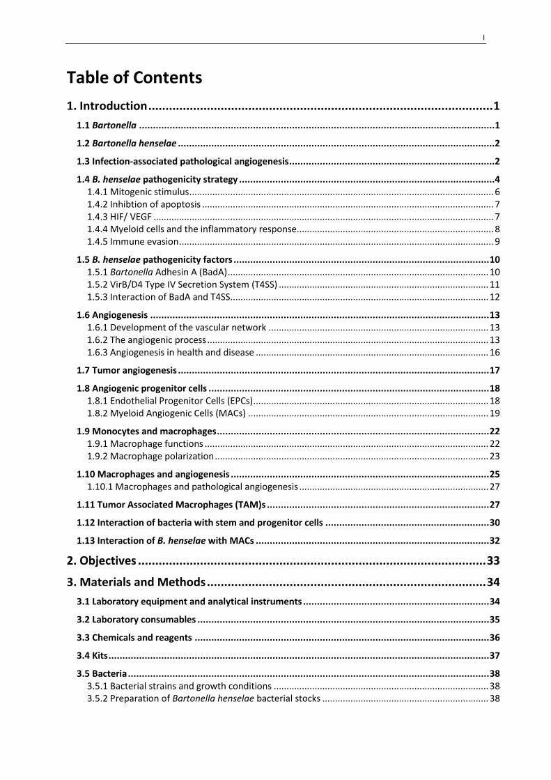
Table of Contents
1. Introduction .................................................................................................... 1
1.1 Bartonella ................................................................................................................................1
1.2 Bartonella henselae ..................................................................................................................2
1.3 Infection-associated pathological angiogenesis ..........................................................................2
1.4 B. henselae pathogenicity strategy ............................................................................................4 1.4.1 Mitogenic stimulus ....................................................................................................................... 6 1.4.2 Inhibtion of apoptosis .................................................................................................................. 7 1.4.3 HIF/ VEGF ..................................................................................................................................... 7 1.4.4 Myeloid cells and the inflammatory response............................................................................. 8 1.4.5 Immune evasion ........................................................................................................................... 9
1.5 B. henselae pathogenicity factors ............................................................................................ 10 1.5.1 Bartonella Adhesin A (BadA) ...................................................................................................... 10 1.5.2 VirB/D4 Type IV Secretion System (T4SS) .................................................................................. 11 1.5.3 Interaction of BadA and T4SS ..................................................................................................... 12
1.6 Angiogenesis .......................................................................................................................... 13 1.6.1 Development of the vascular network ...................................................................................... 13 1.6.2 The angiogenic process .............................................................................................................. 13 1.6.3 Angiogenesis in health and disease ........................................................................................... 16
1.7 Tumor angiogenesis ................................................................................................................ 17
1.8 Angiogenic progenitor cells ..................................................................................................... 18 1.8.1 Endothelial Progenitor Cells (EPCs) ............................................................................................ 18 1.8.2 Myeloid Angiogenic Cells (MACs) .............................................................................................. 19
1.9 Monocytes and macrophages .................................................................................................. 22 1.9.1 Macrophage functions ............................................................................................................... 22 1.9.2 Macrophage polarization ........................................................................................................... 23
1.10 Macrophages and angiogenesis ............................................................................................. 25 1.10.1 Macrophages and pathological angiogenesis .......................................................................... 27
1.11 Tumor Associated Macrophages (TAM)s ................................................................................ 27
1.12 Interaction of bacteria with stem and progenitor cells ........................................................... 30
1.13 Interaction of B. henselae with MACs .................................................................................... 32
2. Objectives ..................................................................................................... 33
3. Materials and Methods ................................................................................. 34
3.1 Laboratory equipment and analytical instruments ................................................................... 34
3.2 Laboratory consumables ......................................................................................................... 35
3.3 Chemicals and reagents .......................................................................................................... 36
3.4 Kits ......................................................................................................................................... 37
3.5 Bacteria .................................................................................................................................. 38 3.5.1 Bacterial strains and growth conditions .................................................................................... 38 3.5.2 Preparation of Bartonella henselae bacterial stocks ................................................................. 38

II
3.6 Cell culture ............................................................................................................................. 39 3.6.1 Ethics statement ........................................................................................................................ 39 3.6.2 Human umbilical vein endothelial cells (HUVECs) ..................................................................... 40 3.6.3 Isolation and cultivation of Myeloid Angiogenic Cells (MACs) .................................................. 40
3.7 Infection experiments ............................................................................................................. 42
3.8 Electron microscopy ................................................................................................................ 42
3.9 Nicoletti apoptosis assay ......................................................................................................... 43
3.10 HIF-1 Western Blot ................................................................................................................ 43 3.10.1 Cell stimulation and preparation of protein extracts .............................................................. 44 3.10.2 Sodium dodecyl sulfate- polyacrylamice gel electrophoresis (SDS-PAGE) .............................. 44 3.10.3 Western Blot ............................................................................................................................ 44
3.11 Spheroid assay of sprouting angiogenesis .............................................................................. 47
3.12 Matrigel capillary formation assay ......................................................................................... 48
3.13 16S rDNA PCR ....................................................................................................................... 49
3.14 Flow cytometry ..................................................................................................................... 51
3.15 Immunohistochemistry ......................................................................................................... 53
3.16 Quantitative real-time PCR (qRT-PCR) .................................................................................... 56 3.16.1 Isolation of total RNA ............................................................................................................... 56 3.16.2 Reverse Transcription PCR (RT-PCR) ........................................................................................ 56 3.16.3 Quantitative RT-PCR ................................................................................................................. 57
3.17 Microarray gene expression profiling ..................................................................................... 58
3.18 Secretome analysis ............................................................................................................... 60
3.19 Enzyme linked immunosorbent assays (ELISAs) ...................................................................... 60
3.20 Statistical analysis ................................................................................................................. 61
4. Results .......................................................................................................... 62
4.1 B. henselae invades MACs and resides in intracellular vacuoles .......................................... 62
4.2 B. henselae infected MACs maintain high viability ............................................................. 62
4.3 B. henselae infection of MACs induces BadA dependent HIF-1 activation ........................... 64
4.4 B. henselae infected MACs incorporate into sprouting endothelium and increase the rate of angiogenic growth .................................................................................................................. 65
4.5 Conditioned medium from B. henselae infected MACs is sufficient to increase sprouting angiogenesis in endothelial cells ............................................................................................. 66
4.6 B. henselae infected MACs display a vascular mimicry phenotype over long term culture ... 68
4.7 The development of the B. henselae-induced vascular mimicry phenotype is BadA dependent .............................................................................................................................................. 70
4.8 MACs develop and maintain a macrophage phenotype after infection with B. henselae ..... 73
4.9 The infection of MACs with B. henselae is associated with broad phenotypic re-programming .............................................................................................................................................. 76
4.10 The gene expression profiles of B. henselae infected MACs are strongly divergent from endothelial cells and show most similarity to cells of the myeloid lineage ................................ 78
4.11 B. henselae infection of MACs induces regulation of genes involved in angiogenic and immune regulatory pathways. ................................................................................................ 81

III
4.12 B. henselae infection of MACs induces a predominantly M2-alternativly activated macrophage phenotype .......................................................................................................... 98
4.13 The secretome of B. henselae infected MACs is dominated by angiogenic-inflammatory cytokines and matrix remodeling compounds ......................................................................... 98
4.14 B. henselae induced cytokine secretion in MACs is distinct from general LPS mediated inflammation ......................................................................................................................... 99
5. Discussion ................................................................................................... 103
5.1 Initial response of MACs to B. henselae infection ................................................................... 103
5.2 Functional effects of B. henselae infection on MAC pro-angiogenic activity ............................ 105 5.2.1 Spheroid assay of sprouting angiogenesis ............................................................................... 105 5.2.2 Matrigel capillary formation assay ........................................................................................... 108
5.3 Differentiation phenotype of B. henselae infected MACs ....................................................... 109
5.4 Vascular mimicry .................................................................................................................. 111
5.5 Phenotypic re-programming in B. henselae infected MACs ..................................................... 113 5.5.1 Angiogenesis ............................................................................................................................ 114 5.5.2 Structural organization ............................................................................................................ 114 5.5.3 Apoptosis ................................................................................................................................. 115 5.5.4 Sterol metabolism .................................................................................................................... 116
5.6 Immune activation phenotypes of B. henselae infected MACs ................................................ 118 5.6.1 Inflammatory activation........................................................................................................... 118 5.6.2 Immune regulation .................................................................................................................. 120 5.6.3 Alternative macrophage activation.......................................................................................... 121
5.7 The Role of BadA in induction of a pro-angiogenic phenotype in B. henselae infected MACs ... 123
5.8 The role of the paracrine microenvironment in the angiogenic activity of B. henselae infected MACs ......................................................................................................................................... 126
5.8.1 Inflammatory-angiogenic chemokines .................................................................................... 127 5.8.2 Immune regulatory cytokines .................................................................................................. 128 5.8.3 Matrix remodeling compounds ............................................................................................... 129 5.8.4 Angiogenin ............................................................................................................................... 130 5.8.5 Angiogenic growth factors ....................................................................................................... 130
5.9 Parallels between B. henselae infected MACs and TAMs ........................................................ 133
5.10 Possible mechanisms of B henselae induced TAM-like phenotypic differentiation in MACs ... 136 5.10.1 Hypoxia and HIF-1 .................................................................................................................. 136 5.10.2 Smoldering Inflammation ...................................................................................................... 137
5.11 Bacterial infection and pathological tissue growth ............................................................... 137

IV
6. Summary .................................................................................................... 141
7. Zusammenfassung ...................................................................................... 143
8. References .................................................................................................. 147
9. Abbreviations.............................................................................................. 186
10. Supplementary Data ................................................................................. 190
11. Acknowledgements (removed for digital publication) ............................... 204
12. Peer Reviewed Publications ...................................................................... 206
12.1 Scientific publications ......................................................................................................... 206
12.2 Oral presentations .............................................................................................................. 206
12.3 Poster presentations ........................................................................................................... 206
13. Curriculum Vitae (removed for dgital publication) .................................... 208

V
List of Figures
Figure 1.1 B. henselae associated pathological angiogenesis: Bacillary angiomatosis. ......................... 3
Figure 1.2 Several pathogenic mechanisms work synergistically to promote B. henselae associated
pathological angiogenesis in vivo............................................................................................................ 6
Figure 1.4 Tumor angiogenesis is characterized by dysfunctional vessels and a chaotic vascular
architecture. .......................................................................................................................................... 18
Figure 1.5 Myeloid Angiogenic Cells (MACs) are a subset of circulating myeloid progenitors that play
an important role in pathological and regenerative angiogenesis and tumor vascularization. ........... 20
Figure 1.6 Macrophages play key roles in angiogenic growth. ............................................................ 26
Figure 1.8 The infiltration of myeloid cells is a decisive factor in malignant tumor vascularization and
progression. .......................................................................................................................................... 29
Figure 4.1 B. henselae invades MACs and resides in intracellular vacuoles. ........................................ 63
Figure 4.2 Apoptosis in MACs in response to B. henselae, S. aureus and E. coli infection. .................. 64
Figure 4.3 HIF-1 activation in MACs upon B. henselae infection. ......................................................... 65
Figure 4.4 Incorporation of uninfected and B. henselae infected MACs into growing vascular sprouts.
.............................................................................................................................................................. 66
Figure 4.5 Infection of MACs with B. henselae results in increased angiogenic growth via paracrine
mechanisms. ......................................................................................................................................... 67
Figure 4.6 Detection of cell integrity and morphological changes of B. henselae-infected MACs in
long-term Matrigel cultures. ................................................................................................................. 68
Figure 4.7 B. henselae infected MACs develop a vascular mimicry phenotype over long term culture.
.............................................................................................................................................................. 69
Figure 4.8 Viability of B. henselae infected MACs in long term Matrigel culture. ................................ 71
Figure 4.9 Presence of B. henselae DNA in fully formed vascular mimicry structures. ........................ 71
Figure 4.11 MACs develop and maintain a macrophage phenotype after infection with B. henselae.
.............................................................................................................................................................. 74
Figure 4.12 MACs maintain a non-endothelial phenotype after infection with B. henselae and
formation of vascular mimicry structures. ........................................................................................... 75
Figure 4.13 MAC angiogenic re-programming upon B. henselae infection does not involve
upregulation of endothelial marker genes. .......................................................................................... 76
Figure 4.14 B. henselae infection of MACs leads to broad phenotypic re-programming. ................... 77
Figure 4.15 Comparison of gene expression profiles between MACs and mature endothelial cells. .. 79
Figure 4.16 B. henselae infection of MACs leads to a broad phenotypic transformation characterized
by upregulation of angiogenic and immune regulatory cellular programs. ......................................... 95

VI
Figure 4.17 B. henselae infected MACs exhibit a predominantly M2 macrophage activation
phenotype. .......................................................................................................................................... 100
Figure 4.18 B. henselae infected MACs create a pro-angiogenic paracrine microenvironment over
long term culture, dominated by angiogenic-inflammatory cytokines and matrix remodeling
compounds. ........................................................................................................................................ 101
Figure 4.19 Inflammatory and anti-inflammatory cytokine secretion of MACs upon B. henselae
infection or LPS treatment. ................................................................................................................. 102
Figure 5.1 Infection of MACs with B. henselae results in a phenotypic transformation towards a
macrophage with increased pro-angiogenic capacity. ....................................................................... 131
Figure 5.2 B. henselae infected MACs show many functional and phenotypic parallels to tumor
associated macrophages. .................................................................................................................... 139
List of Tables
Table 3.1 Laboratory Equipment and Analytical Instruments .............................................................. 34
Table 3.2 Laboratory Consumables ...................................................................................................... 35
Table 3.3 Chemicals and Reagents ....................................................................................................... 36
Table 3.4 Kits ......................................................................................................................................... 37
Table 3.5 Bacterial Strains .................................................................................................................... 39
Table 3.6 Bacterial Culture Media ........................................................................................................ 39
Table 3.7 Cells ....................................................................................................................................... 41
Table 3.8 Cell Culture Medium and Additives ...................................................................................... 41
Table 3.9 Western Blot Antibodies and Substrates .............................................................................. 45
Table 3.10 Buffer and Solution Compositions for Western Blot. ......................................................... 46
Table 3.11 Staining Reagents and Buffers for Florescent Labeling of Vascular Mimicry Structures. ... 49
Table 3.12 Bartonella spp. 16S rDNA Primers ...................................................................................... 50
Table 3.13 Master Mix Composition for 16S rDNA PCR ....................................................................... 51
Table 3.14 16S rDNA PCR Cycling Program, First Amplification Phase ................................................ 51
Table 3.15 16S rDNA PCR Cycling Program Second Amplification Phase ............................................. 51
Table 3.16 Antibodies and Reagents for FACS Analysis ........................................................................ 52
Table 3.17 Buffer Composition for FACS Analysis ................................................................................ 53
Table 3.18 Antibodies and Staining Reagents for Immunohistochemistry. ......................................... 54
Table 3.19 Buffers and Solutions for Immunohistochemistry. ............................................................. 55
Table 3.20 Cycling Program for RNA-cDNA Reverse Transcription. ..................................................... 57
Table 3.21 SYBR Green (Fast) qPCR Cycling Program. .......................................................................... 58

VII
Table 3.22 Taqman (Fast) qPCR Cycling Program. ................................................................................ 58
Table 3.23 Primers ................................................................................................................................ 58
Table 4.1 Comparison of gene expression signals from B. henselae infected MACs to gene expression
profiles of known cell differentiation. .................................................................................................. 80
Table 4.2 Comparison of B. henselae induced phenotypic re-programming in MACs to changes
induced during in vitro monocyte to macrophage differentiation. ...................................................... 81
Table 4.3 Detailed functional annotation of upregulated genes in B. henselae-infected MACs at d53
(FC ≥ 4 and FDR < 0.05). ........................................................................................................................ 83
Table 4.4 Selected functional annotation catagories enriched amoung genes upregulated (FC ≥ 2.0,
FDR < 0.05) in MACs (d53) upon B. henselae infection. ....................................................................... 96
Table 4.5 Selected functional annotation catagories enriched amoung genes downregulated (FC ≥
2.0, FDR < 0.05) in MACs (d53) upon B. henselae infection. ................................................................ 97
S Table 10.1 DAVID analysis of upregulated transcripts in B. henselae-infected MACs at d53. List of
enriched functional annotations (P<0.01). ......................................................................................... 190
S Table 10.2 DAVID analysis of downregulated transcripts in B. henselae-infected MACs at d53. List
of enriched functional annotations (P<0.01). ..................................................................................... 195
S Table 10.3 DAVID analysis of upregulated transcripts in B. henselae-infected MACs at d53.
Clustering of enriched, functional annotations to related functional groups. Minimum similarity
overlap > 3 (P < 0.01). ......................................................................................................................... 197
S Table 10.4 DAVID analysis of downregulated transcripts in B. henselae-infected MACs at d53.
Clustering of enriched, functional annotations to related functional groups. Minimum similarity
overlap > 3 (P < 0.01). ......................................................................................................................... 201


Introduction 1
1. Introduction
1.1 Bartonella
The bacterial genus Bartonella is comprised of slow-growing, facultative intracellular pathogens that
infect mammalian hosts and are transferred by hematophagous arthropod vectors. Phylogenetically,
these microaerophilic, pleomorphic, rod shaped, bacteria belong to the class of Gram-negative
alphaproteobacteria and are most closely related to the bacterial genera Brucella, Agrobacterium
und Rhizobium (Anderson and Neuman, 1997; Maurin et al., 1997).
A common feature of Bartonella infection is the transition between an often asymptomatic
intraerythrocytic phase and a secondary tissue niche which facilitates chronic bacterial persistence
and a periodic reseeding of the bloodstream (Dehio, 2004).
The advent of polymerase chain reaction (PCR) technology has greatly expanded the diversity of the
Bartonella genus. Samples from both wild and domestic animals have revealed a wide variety of
Bartonella species specialized to various mammalian hosts and arthropod vectors (Kaiser et al.,
2011).
To date at least thirteen human pathogenic species of Bartonella have been identified (Kaiser et al.,
2011). Most clinically relevant Bartonella infections in humans are caused by three species:
Bartonella bacilliformis, Bartonella quintana and Bartonella henselae.
Bartonella bacilliformis is believed to be the ancestral strain of human pathogenic Bartonella and
causes the most severe form of disease (Engel et al., 2011). B. bacilliformis is endemic to the South
American Andes and is transmitted between human hosts via sand flies of the genus Luzomyia.
Archeological evidence originating from pre-Inca ceramics and mummified remains indicate that B.
bacilliformis has existed as a human pathogen for at least 2,000 years (Allison et al., 1974). Infection
first manifests as an intense fever (Oroya fever) which results from an anemia due to the massive
colonization of erythrocytes and carries a high mortality rate. Individuals who survive Oroya fever
may develop a secondary chronic tissue phase which can arise one to two months after the initial
fever and involves the chronic growth of blood-filled, wart-like structures consisting of proliferating
vessels on the skin of infected patients (Peruvian warts/ verruga peruana; Minnick et al., 2014).
Bartonella quintana, emerged first as a significant human pathogen during the first and second
world wars as it spread among soldiers in trenches and prisoner of war camps. More recently, cases
of B. quintana infection have also been identified in urban homeless populations (Raoult et al.,
2001). Infection is transmitted by the human body louse, Pediculus humanus, and causes a recurring

2 Introduction
dehabilitating fever (Trench fever) believed to be caused by the cyclic release of bacteria from a
secondary niche into the blood stream and subsequent colonization of erythrocytes (Rolain et al.,
2002).
1.2 Bartonella henselae
Bartonella henselae emerged as a relevant human pathogen in the late 20th century. In contrast to
the other major human pathogenic Bartonella species, B. henselae is a zoonotic pathogen specialized
to the domestic cat as a reservoir host. Here it exists as an asymptomatic intraerythrocytic
bacteremia and is transmitted between cats via the cat flea (Ctenocephalides felis). Humans are
infected as incidental hosts through the contamination of cat bites or scratches with flea feces
(Rolain et al., 2001; Rolain et al., 2003).
Depending on the immune status of the individual, B. henselae infection in humans can result in
differing pathological outcomes.
In immune-competent individuals infection develops into a generally self limiting condition referred
to as “cat scratch disease” which involves swelling of local lymph nodes and occasional granuloma
formation (Anderson and Neuman, 1997).
In immune-compromised patients however, chronic infection leads to the development of a
vaculoproliferative condition known as bacillary angiomatosis or peliosis hepatis which is
characterized by the appearance of tumorous blood filled lesions on the skin and inner organs
(Manders, 1996).
Before the advent of effective antiretroviral therapy, bacillary angiomatosis was most commonly
observed in immunosuppressed patients suffering from AIDS, which lead to the first description of
the condition in 1983 (Stoler et al., 1983; Koehler and Tappero, 1993) and subsequent identification
of the infectious agent by the newly developed 16S ribosomal DNA (rDNA) analysis in 1990 (Relman
et al., 1990).
1.3 Infection-associated pathological angiogenesis
A unique hallmark of Bartonella infections is the ability to induce angiogenic growth in human hosts.
This pathogenic process is evident in the development of vascular lesions associated with chronic
Bartonella infections: verruga peruana (B. bacilliformis) and bacillary angiomatosis (B. henselae;
Dehio, 2005). Of the two, B. henselae induced pathological angiogenesis has been the most widely
studied.

Introduction 3
Ultrastructural examinations of B. henselae associated vascular lesions reveal a disorganized
proliferation of immature, capillary-sized vessels, misshapen endothelial cells and myeloid cell
infiltrate. Aggregates of rod shaped bacteria can be identified in and around the proliferating
endothelial cells and complete regression of the angioproliferative structures can be achieved
through antibiotic treatment (LeBoit et al., 1989; Kostianovsky and Greco, 1994; Manders, 1996;
Schwartz et al., 1997).
The vascular architecture, myeloid cell infiltrate and proliferative growth of B. henselae induced
vascular lesions bear a strong resemblance to the growth of malignant tumor vasculature suggesting
that these neoplastic structures may develop via similar mechanisms (Cockerell and LeBoit, 1990;
Koehler and Tappero, 1993; Manders, 1996).
Figure 1.1 B. henselae associated pathological angiogenesis: Bacillary angiomatosis. (A) Bacillary angiomatosis is characterized by the occurrence of vascular tumors on the skin and inner organs of immune-suppressed patients (Beatty and Lukusa, October 24. 2014). (B) Histological section of bacillary angiomatosis lesion stained with Meyers hematoxlyin and anti-VEGF antibodies (red). Arrows indicate the presence of VEGF in the endothelium of pathologically formed vasculature (Kempf et al., 2001). (C) Aggregates of rod shaped bacteria (black) can be identified within the vascular lesions via Warthin-Starry staining (Koehler, 1997).
Infection associated pathological angiogenesis has also been described in the context of several viral
infections. Viruses such as human papilloma virus (HPV) and human herpes virus 8 (HHV8) have been
associated with the development of neoplasia and increased angiogenic growth. Most notably,
infection with HHV8 can lead to the development of Kaposi’s sarcoma; a condition identified by the
growth of vascular tumors on the skin of immunosuppressed patients (Flore et al., 1998). These
vascular lesions are characterized by the proliferation of endothelial cells, the formation of spindle
shaped tumor cells and the dense vascularization of a localized area with chaotic and leaky blood

4 Introduction
vessels. This appearance, combined with its particular occurrence in AIDS patients, makes it a
common differential diagnosis to bacillary angiomatosis.
Increased angiogenesis in viral infections is often associated with cancer formation and therefore
linked to the angiogenic qualities of viral oncogenic proteins such as the HHPV8 papillomavirus
proteins E6 and E7 (López-Ocejo et al., 2000).
In viral conditions specifically characterized by angiogenic pathologies, angiogenic growth is
stimulated by the transcription of virally encoded proteins which mimic host regulators of
angiogenesis. The Orf parapox virus, for example, is the agent of a vasculoproliferative condition in
sheep and goats and encodes a viral homologue of vascular endothelial growth factor (VEGF), viral
(v)VEGF-E which activates endothelial cell proliferation and vascular permeability at levels similar to
human VEGF-A (Meyer et al., 1999; Cébe-Suarez et al., 2008). The HHV8 genome also encodes for
several open reading frames (ORFs) with known homology to cellular proteins. The transcription of
these virally encoded proteins such as viral C-C motif ligand v(CCL)-2, vCCL3 and vIL-6 modulates
host immune responses, promotes angiogenesis, and dysregulates cell growth in Kaposi’s sarcoma
(Ensoli et al., 2001; Gramolelli and Schulz, 2015).
In some cases, chronic bacterial infections have also been associated with increased angiogenic
growth. Biopsies of gastric tissue from patients with H. pylori associated gastritis or gastric
carcinoma demonstrate an increased capillary density (Pousa and Gisbert, 2006; Yeo, 2006).
However, these effects are thought to be connected to the prolonged states of chronic inflammation
associated with long-term bacterial infections rather than specific pathogenic activities.
Bartonella species, in contrast, are the only known bacteria with the ability to cause the concerted,
proliferative growth of angiogenic vessels and the creation of localized vascular tumors in humans.
The process by which B. henselae induces the development of vascular tumors presents a valuable
topic for study due to the importance of angiogenesis in many pathological and regenerative
processes and as an example of complex host-pathogen interactions.
1.4 B. henselae pathogenicity strategy
Evidence suggest that endothelial cells may represent an important cellular niche for Bartonella
species in vivo (Harms and Dehio, 2012). It has been proposed that the induction of pathological
angiogenesis by B. henselae infection may represent a pathogenicity strategy by which B. henselae
induces the proliferation of its own nutrient rich, immune-privileged cellular niche (Kempf et al.,
2002).

Introduction 5
In vitro experiments demonstrate that B. henselae readily invades endothelial cells and can persist
intracellularlly (Kyme et al., 2005). B. henselae, co-cultured with endothelial cells, show a rapid
increase in rRNA production and a replication rate that is 100-fold higher than bacteria cultured in
isolation (Kempf et al., 2000). In another study, the proliferation of B. henselae infected endothelial
cells in response to VEGF or conditioned medium was associated with a 75- and 150-fold increase in
B. henselae growth rate respectively (Kempf et al., 2001). The increased replication of intracellular
B. henselae is inhibited when the synthesis of host cell proteins is blocked with cycloheximide,
indicating that endothelial cells may provide intracellular bacteria with proteins to support growth
(Kempf et al., 2000).
Research into the mechanisms of B. henselae induced angiogenesis over the years has been limited
by the lack of in vivo animal models. Although several models of Bartonella infection have been
described in the literature, infected animals do not develop the pathological angiogenic growth
characteristics of B. henselae induced bacillary angiomatosis (Regnath et al., 1998; Arvand et al.,
2001; Koesling et al., 2001; Schülein et al., 2001; Velho, Paulo Eduardo Neves Ferreira et al., 2002;
Kabeya et al., 2006; Chiaraviglio et al., 2010). As a result, most studies examining B. henselae
induced pathological angiogenesis have focused on in vitro primary-cell culture experiments,
angiogenesis models and ex vivo immunohistochemistry
So far, B. henselae is believed to induce pathological angiogenesis through the synergistic
combination of several mechanisms including i) direct stimulation of endothelial proliferation ii)
inhibition of apoptosis iii) activation of the hypoxia-inducible factor (HIF)-1 dependent pro-
angiogenic programs iv) activation of nuclear factor 'kappa-light-chain-enhancer' of activated B-cells
(NFκB) and release of angiogenic-inflammatory cytokines and e) immune evasion via a stealth-
pathogen strategy.

6 Introduction
Figure 1.2 Several pathogenic mechanisms work synergistically to promote B. henselae associated pathological angiogenesis in vivo. B. henselae has been shown to release a mitogenic compound which can induce endothelial cell proliferation without host-cell contact. When B. henselae encounter endothelial cells they adhere and are taken up into intracellular vacuoles. Infection of endothelial cells results in inhibition of apoptosis and activation of the inflammatory and angiogenic transcription factors NFκB and HIF-1, respectively. Activation of HIF-1 leads to angiogenic re-programming including the secretion of the pro-angiogenic cytokine VEGF which stimulates angiogenesis in infected cells as well as the surrounding endothelium. Activation of NFκB results of inflammatory gene transcription. Infected endothelial cells show upregulation of cellular adhesion molecules ICAM-1 and E-selectin as well as secretion of the inflammatory angiogenic chemokines CCL2 and CXCL8 which induces infiltration of myeloid cells into the infectious microenvironment. Infection of accessory myeloid and epithelial cells with B. henselae also leads to secretion of VEGF, further promoting angiogenesis in the surrounding endothelium.
1.4.1 Mitogenic stimulus
Several early studies into B. henselae induced angiogenic growth identified a B. henselae secreted
compound with the ability to increase endothelial cell proliferation in a manner that was
independent of bacterial cell contact or invasion. Co-culture of B. henselae with endothelial cells
separated by a filter membrane or the use of B. henselae conditioned cell culture medium was
sufficient to induce endothelial cell proliferation (Maeno et al., 1999). The nature of this bacterial
factor or factors remains elusive, however, GroEL an endothelial mitogenic factor released by B.
bacilliformis has been detected in B. henselae conditioned medium and was discussed as one of
these mitogenic compounds (Minnick et al., 2003).
Introduction 7
1.4.2 Inhibtion of apoptosis
Apoptosis represents one of the most basic immune responses to intracellular bacterial infection.
The programmed death of host cells acts to deprive bacteria of their immune privileged habitat and
releases strong inflammatory alarm signals which trigger the immune response.
B. henselae has been demonstrated to inhibit apoptosis in both endothelial cells and
monocyte-macrophage 6 (MM6) cells (Kirby, 2002; Kempf et al., 2005b; Schmid et al., 2006). The
process by which B. henselae inhibits apoptosis is not well understood, however, studies have shown
that B. henselae infection suppresses both early and late events in the apoptotic cascade. In MMP6
cells B. henselae infection was associated with inhibition of caspase three activity and induction of
the anti-apoptotic molecules: cellular inhibitor of apoptosis proteins -1 and 2 (cIAP-1,-2; Kempf et
al., 2005b). In another study, inhibition of apoptosis in endothelial cells correlated with increased
cytoplasmic concentrations of cyclic adenosine monophosphate (cAMP; Schmid et al., 2006).
The ability of B. henselae to stimulate proliferation of endothelial cells has been shown to depend
considerably on its ability to inhibit apoptosis (Kirby, 2002). Through the inhibition of programmed
cell death, B. henselae circumvents a fundamental immune response mechanism and maintains the
viability of its cellular niche. The inhibition of apoptosis is also an essential pre-requisite for further
cellular reprogramming and the induction of angiogenic growth.
1.4.3 HIF/ VEGF
In addition to promoting angiogenic growth through direct stimulation of endothelial cell
proliferation and inhibition of apoptosis, B. henselae infection activates autologous angiogenic
response programs in infected cells. Central to this is the activation of HIF-1; a heterodimeric
transcription factor that regulates responses to oxygen supply, metabolic demands and is the key
regulator of the angiogenic response.
B. henselae infection has been shown to induce HIF-1 activation in several cell types including
epithelial and endothelial cells (Kempf et al., 2005a). The activation of HIF-1 in B. henselae infected
cells leads to activation of pro-angiogenic response programs. In B. henselae infected epithelial and
endothelial cells, infection is associated with up-regulation and release of angiogenic cytokines such
as VEGF, adrenomedullin (ADM), and insulin-like growth factor-binding protein -1 (Kempf et al.,
2001; Kempf et al., 2005a).
Among these cytokines, VEGF is probably the most important and has been found to play a
significant role in B. henselae induced endothelial proliferation. This pro-angiogenic stimulation is

8 Introduction
not only relevant to infected cells but is transmitted in a paracrine manner to the surrounding
endothelium. Conditioned medium from B. henselae infected EA.hy 926 cells promoted the
proliferation of endothelial cells and this pro-angiogenic effect was found to decrease 50% after
treatment with VEGF neutralizing antibodies (Kempf et al., 2001).
A role for HIF-1 activation and VEGF in B. henselae induced pathological angiogenesis is also
supported by evidence from ex vivo bacillary angiomatosis samples showing strong HIF-1 and VEGF
expression in and around vessels of infected vascular lesions (Kempf et al., 2001; Kempf et al.,
2005a).
To date, the exact cellular mechanisms of B. henselae induced HIF-1 activation has not been
elucidated. Although mitogen-activated protien kinases (MAPKs) are phosphorylated during
B. henselae infection of epithelial cells, these signal transduction pathways appear to have little
influence over B. henselae induced activation of HIF-1 dependent pro-angiogenic programs. Instead,
B. henselae infection may activate HIF-1 by mimicking a cellular hypoxic state as infected cells display
signs of cellular hypoxia, increased cellular oxygen consumption, decreased adenosintriphosphat
(ATP) levels and a gene expression profile characteristic of the hypoxic response (Kempf et al.,
2005a).
It is known that HIF-1 activation in B. henselae infected cells is dependent on direct bacterial-host
cell interaction, specifically the activity of the B. henselae outer membrane protein Bartonella
adhesin A (BadA). HIF-1 activation decreases by 30% when cells are cultured with heat killed bacteria
and a decrease of 90% was observed when cells were infected with B. henselae mutant strains
lacking BadA protein expression (Riess et al., 2004).
1.4.4 Myeloid cells and the inflammatory response
The interaction of B. henselae with components of the innate immune system is also believed to be
an essential component of B. henselae induced pathological angiogenesis.
B. henselae infection clearly activates the inflammatory transcription factor NFκB in several cell
types and is associated with the up-regulation and release of a range of NFκB dependent
inflammatory cytokines such as C-X-C motif ligand (CXCL)8, CCL2, and granulocyte-macrophage
colony-stimulating factor (GM-CSF; Resto-Ruiz et al., 2002; Schmid et al., 2004a; Kempf et al., 2005a;
Kempf et al., 2005b; McCord et al., 2005).
The induction of NFκB dependent inflammatory gene regulation and cytokine release is believed to
play a role in activating the angiogenic response in infected cells.

Introduction 9
Infection of endothelial cells with B. henselae was shown to result in secretion of the inflammatory-
angiogenic cytokine CXCL8 and upregulation of the angiogenic chemokine receptor CXCR2. The
secretion of CXCL8 from infected endothelial cells was found to be required for B. henselae induced
cell proliferation, tube formation and apoptosis inhibition in vitro (McCord et al., 2006).
NFκB regulated cytokines also act as chemokines to attract myeloid cells to the site of infection.
NFκB dependent upregulation of cellular adhesion molecules intercellular adhesion molecule
(ICAM)-1 and E-selectin in B. henselae infected endothelial cells resulted in increased
polymorphonuclear leukocyte rolling and adhesion to infected endothelial monolayers. NFκB
dependent release of CCL2 from infected human micro-vascular cells induced migration of THP-1
mononuclear cells in vitro (McCord et al., 2005).
The attraction of myeloid cells to the site of B. henselae infection is hypothesized to contribute to
pathological angiogenesis through the release of pro-angiogenic cytokines and the initiation of a pro-
angiogenic paracrine loop of cell activation and cytokine secretion (Kempf et al., 2002; Resto-Ruiz et
al., 2002). Infection of the J774 murine macrophage cell line with B. henselae induced increased
secretion of tumor necrosis factor (TNF)α, IL-1β and IL-6 (Musso et al., 2001). B. henselae infected
THP-1 macrophages were found to secrete angiogenic cytokines VEGF and CXCL8 and conditioned
medium from infected THP-1 cells induced increased proliferation of HMEC-1 cells in vitro (Resto-
Ruiz et al., 2002).
1.4.5 Immune evasion
Although B. henselae clearly activates important aspects of the inflammatory response in infected
cells, in vivo infection is not associated with high levels of systematic inflammation. B. henselae
employs what has been described as a “stealth pathogen” strategy of intercellular lifestyle and slow
growth which requires simultaneous activation and suppression of the inflammatory responses to
insure bacterial persistence and evasion of immunity (Pulliainen and Dehio, 2012).
To prevent recognition by host pattern recognition receptors, the B. henselae lipopolysaccaride (LPS)
molecule contains structural modifications which prevent binding to toll-like receptor (TLR)4 and
activation of the inflammatory response cascade. Purified LPS from B. henselae has been shown to
be 1000-10,000 fold less potent at activating TLR4 signaling than Salmonella enterica LPS (Zähringer
et al., 2004).
Following infection of host cells, B. henselae can control the fate of its membrane-bound
compartment. B. henselae containing vacuoles lack typical endocytic marker proteins and fails to
acidify. In J774A.1 macrophages, fusion with the lysosome is delayed and in endothelial cells, fusion

10 Introduction
can be inhibited completely. Viable bacteria are necessary to prevent lysosome fusion indicating that
B. henselae actively subverts the normal endosomal trafficking pathways in the creation of its
intracellular habitat (Kyme et al., 2005).
Some evidence exists that B. henselae may also actively suppresses inflammatory responses as part
of its pathogenicity strategy.
Within its natural host, the cat, B. henselae infection induces a Th2 immune response including a
transient decrease in cluster of differentiation (CD)4+ T cells (Kabeya et al., 2006). In B. henselae
infected mice and humans, infection is associated with an increased concentration of the anti-
inflammatory cytokine IL-10 in blood plasma (Papadopoulos et al., 2001; Kabeya et al., 2007). In vitro
examination of the dendritic cell response to B. henselae infection also revealed increased IL-10
secretion in B. henselae infected cells (Vermi et al., 2006). Lastly, an early study found that
B. henselae infection impairs the production of reactive oxygen species in polymorphonuclear
leukocytes and thereby avoids the induction of oxidative burst (Fumarola et al., 1996).
1.5 B. henselae pathogenicity factors
Two main B. henselae pathogenicity factors have been identified as being involved in the process of
B. henselae induced pathological angiogenesis: the trimetric autotransporter adhesin BadA and the
VirB/D4 type IV secretion system.
1.5.1 Bartonella Adhesin A (BadA)
Bartonella adhesin A (BadA) is a extremely long (ca. 240nm ) fibril adhesion protein expressed on the
outer membrane of B. henselae. BadA is a member of the trimeric autotranporter (TAA) family of
proteins commonly found in animal and plant pathogenic proteobacteria and conserved within the
Bartonella genus. The BadA structure displays the typical TAA modular construction of membrane
anchor, multiple repeating neck and stalk components and a terminal head domain (O'Rourke et al.,
2011).
BadA is important for several pathogenicity functions. The head and stalk domains mediate bacterial
autoagglutination and adhesion to several extracellular matrix proteins including fibronectin, laminin
and various collagens (Kaiser et al., 2008; Kaiser et al., 2012). BadA also plays an essential role in
effective bacterial adhesion and uptake into Bartonella-containing-vacuoles (BCV)s in various host
cells including endothelial and epithelial cells as well as preventing phagocytic uptake by
macrophages (Riess et al., 2004; Kyme et al., 2005). The role of BadA in adhesion to the extra cellular
matrix and endothelial monolayers has been shown to be of particular significance under dynamic

Introduction 11
flow conditions suggesting that this pathogenicity factor may be involved in colonization of the
endothelial niche from the bloodstream (Müller et al., 2011).
In addition to its roles in bacterial adhesion, BadA is essential in activation of the B. henselae
dependent angiogenic response in infected cells. The expression of BadA on the surface of
B. henselae is necessary to activate the angiogenic transcription factor HIF-1 and for the
upregulation and release of the HIF-1 regulated angiogenic cytokine VEGF (Riess et al., 2004). The
mechanisms of this action are not yet clear but it has been hypothesized that the interactions of
BadA with fibronectin and other extracellular matrix proteins may subvert intracellular signaling via
interaction with β1-integrins (Riess et al., 2004).
Molecular studies employing various BadA mutant strains have found most BadA pathogenicity
functions to be concentrated in the head domain including autoagglutination, binding to
extracellular matrix and host cells as well as activation of the angiogenic response (Kaiser et al.,
2008). The significance of BadA’s extreme length is not yet known. The length is determined by the
number of neck-stalk sub-domain repetitions present in the stalk domain of the expressed protein
and has been demonstrated to vary among isolates. Some regions of the BadA stalk domain have
been shown to play a role in binding extracellular matrix proteins and are essential for binding to
fibronectin (Kaiser et al., 2012). The repetition of these domains along the length of the BadA stalk
may allow for more efficient binding under the dynamic environment of the blood stream. It has also
been suggested that the length of the BadA protein may have some physical function such as in
extending the head domain at a defined distance from the bacterial cell surface (Kaiser et al., 2012).
1.5.2 VirB/D4 Type IV Secretion System (T4SS)
Along with BadA, the VirB/D4 type IV secretion system (T4SS) represents an important pathogenicity
factor in B. henselae induced pathological angiogenesis. T4SS are common structures among
pathogenic bacteria and are constructed as needle-like macromolecular machines that translocate
bacterial effector proteins into host cells.
The B. henselae T4SS is encoded by the VirB operon which encodes the individual components of a
translocation channel spanning both inner and outer membranes and a longer external component
that interacts with the host cell (Schröder and Dehio, 2005). Seven Bartonella effector proteins (Beps
A-G) have been identified so far. Beps are modularly constructed, each containing an C-terminal
secretion signal including a Bartonella intercellular delivery domain (BID) along with effector
domains which are activated within the host cell (Schulein, 2005; Pulliainen and Dehio, 2009; Engel
et al., 2011).
12 Introduction
T4SS activity has been shown to play an important role in several aspects of B. henselae
pathogenicity. The most striking effect of T4SS on host cells is the formation of a specific invasion
structure deemed the “invasome” in which stress fibers form an F-actin ring and create membrane
protrusions which engulf and internalize large bacterial aggregates (Rhomberg et al., 2009). The
formation of the invasome has been shown to be dependent on the translocation of BepG and in
part BepC and BepF. The interaction of Beps with host cell Rho guanosine-5'-triphosphate (GTP)ases
induces massive F-actin reorganization and polymerization to form the invasome structures
(Truttmann et al., 2011a).
T4SS activity may also activate B. henselae specific NFκB response programs. In B. henselae infected
endothelial cells the secretion of CXCL8 and expression of ICAM-1 was dependent on T4SS activity.
NFκB transcriptional activity was shown to be significantly reduced in mutants lacking functional
T4SS (Schmid et al., 2004a).
The T4SS has also been shown to inhibit B. henselae mediated apoptosis. In particular the
translocation of BepA to the cellular cytoplasm is associated with increased cAMP concentration in
infected cells and inhibition of endothelial cell death (Schmid et al., 2006). BepA translocation also
appears to have a pro-angiogenic effect on vascular sprouting in a spheroid angiogenesis model; an
effect which is believed to be connected to its anti-apoptotic qualities (Scheidegger et al., 2009).
Interestingly, B. henselae T4SS has also been demonstrated to produce bacterial factors that inhibit
angiogenic activity. Activities of the T4SS have been shown to interfere with VEGF-VEGFR2
downstream signaling in endothelial cells (Scheidegger et al., 2011). The effector protein BepG, has
been shown to be a potent anti-angiogenic factor inhibiting endothelial sprout formation in the
spheroid model of sprouting angiogenesis (Scheidegger et al., 2009). It has been speculated that
these anti-angiogenic effects may coordinate with other pro-angiogenic factors to regulate
B. henselae induced pathological angiogenesis or may be employed during a phase of infection in
which angiogenic growth is unneeded or undesirable (Truttmann et al., 2011b).
1.5.3 Interaction of BadA and T4SS
Although both BadA and T4SS clearly have significant functional roles in B. henselae infection, the
interaction between these pathogenicity factors could not be examined until recently. Most studies
describing B. henselae infections have been performed using B. henselae strains possessing either a
functional T4SS but lacking expression of full length BadA proteins (strain B. henselae Houston-1) or
strains with a full length BadA but only a weak ability to translocate bacterial effector proteins
(strain B. henselae Marseille). It had been previously hypothesized that the two pathogenicity factors

Introduction 13
could work synergistically when combined. However, a recent investigation revealed that strains
carrying both pathogenicity factors were rare among available isolates. Further investigation utilizing
a genetically manipulated B. henselae stain artificially expressing both pathogenicity factors revealed
that BadA interferes with T4SS effector protein translocation into host cells. Bep translocation into
host cells was found to be inversely correlated with the length of the BadA protein. The capacity of
BadA to adhere to extra cellular matrix proteins, endothelial cells and to activate HIF-1 were not
affected by the presence of the functional T4SS (Lu et al., 2013). These results have led to the
conclusion that BadA and T4SS have separate functions, possibly being expressed at differing time
points over the course of B. henselae infection.
1.6 Angiogenesis
Angiogenesis; the system of blood vessel growth and expansion is a fundamental process in the
development, growth and adaptive capacity of higher organisms. The regulation and dysregulation
of vascular growth can have wide reaching consequences for many biological processes.
1.6.1 Development of the vascular network
Within the developing embryo, vasculature first develops from mesodermal hemangioblasts which
act as a common progenitor for both endothelial and hematopoietic cell lineages. Hemangioblasts
differentiate to angioblasts and finally endothelial cells which create a primitive vascular network in
a process known as vasculogenesis (Coultas et al., 2005).
Following initial vascular expansion newly formed vessels mature via the process of arteriogenesis in
which vascular channels are stabilized by the secretion of a basement membrane and the
establishment of intercellular junctions. A layer of pericytes is deposited and in larger vasculature, a
concentric band of smooth muscle surrounds vessels to provide structural support and regulate
vascular function (Jain, 2003).
In adult organisms new vessels are predominantly formed through the process of angiogenesis
which is defined as the sprouting, branching and expansion of the vascular system from existing
vasculature.
1.6.2 The angiogenic process
Once formed, adult blood vessels are maintained in quiescence though a dynamic balance of
angiogenic and anti-angiogenic factors. An excess of angiogenic stimulus activates the so called
“angiogenic switch” initiating the process of sprouting angiogenesis (Carmeliet, 2005; Herbert and
Stainier, 2011).

14 Introduction
Hypoxia represents one of the most important activators of angiogenesis in both healthy and
pathogenic tissue growth. As tissue expands or when existing vasculature is damaged, diffusion can
no longer adequately supply cells with oxygen creating a local hypoxic microenvironment. This
triggers the activation of HIF-1, a transcription factor which acts upon hypoxic response elements in
the promoters of angiogenic genes, activating angiogenic response programs and the release of pro-
angiogenic signaling factors (Pugh and Ratcliffe, 2003).
The list of compounds with pro-angiogenic qualities includes specialized growth factors, cytokines,
chemokines, lipid mediators, hormones and neuropeptides (Carmeliet and Jain, 2011). Among these,
the most intensely studied is vascular endothelial growth factor (VEGF), an angiogenic growth factor
which acts via binding to the vascular endothelial growth factor receptor 2 (VEGFR2) and activation
of an angiogenic signaling cascade (Herbert and Stainier, 2011).
The activation of angiogenic signaling in endothelial cells initiates a multi-step angiogenic process
which includes dissociation of the existing vascular barrier, vascular sprout extension, vessel
stabilization and resolution.
In the first stage of angiogenic sprouting, activated endothelial cells initiate the dissolution of the
existing vascular barrier via degradation of the extracellular matrix basement membrane,
detachment of pericytes and dissociation of endothelial cellular junctions (Potente et al., 2011).
Next, the coordination of sprout formation is mediated by the differentiation of activated
endothelial cells into migrating tip- and proliferating stalk-cells. Endothelial cells compete for tip-cell
position through the secretion of inhibitory compounds which prevent the tip-cell differentiation in
surrounding cells and induce a stalk cell differentiation by default. The selected tip cells then extend
filopodia into the extracellular space and migrate towards the angiogenic stimulus (e.g. hypoxia)
guided by a gradient of pro-angiogenic signaling molecules (e.g. VEGF). Stalk cells are guided by the
migration of the tip cell and proliferate to extend sprout length in the direction of angiogenic
stimulus (Carmeliet et al., 2009).
Proteases such as matrix metalloproteinases (MMPs) and plasminogen activators (PAs) facilitate the
invasion of vascular sprouts into surrounding tissue via directional degradation of the extracellular
matrix (ECM). A support scaffold for the extending sprout is also provided by the secretion of
provisional matrix proteins such as fibronectin and fibrin (Sottile, 2004).
As sprouts extend, stalk cells form lumens though the coordinated fusion of intracellular vacuoles
and perfused branches are formed when neighboring tips cells encounter one another via filopodian
interactions and fuse in a process of anastomosis.

Introduction 15
Figure 1.3 The angiogenic process. (A) In the absence of pro-angiogenic signalling, endothelial cells are maintained in a quiescent state. (B) In response to pro-angiogenic signals (e.g. VEGF) tip cells (blue) are selected for sprouting. Tip cell fate is inhibited in neighboring endothelial cells via Notch signaling. Vascular sprouting is facilitated via detachment of the pericytes layer (green), loosening of endothelia cell junctions and degradation of the extracellular matrix. (C) Invasive tip cell sprouting is guided by pro-angiogenic cytokine gradient and vascular sprouts elongated via the proliferation of stalk cells (yellow) followed by vascular lumen morphogenesis. Upon contact with other vessels, tip cell phenotype is repressed and vessels fuse via the process of anastomosis which is guided by accessory macrophages. (D) Successful perfusion of new vessels leads to a resolution phase of angiogenic growth in which pericytes are recruited, endothelial cell junctions are strengthen and the extracellular matrix re-established. Upon vessel maturation endothelial cells return to a quiescent phalanx cell state. ECM: extracellular matrix. Adapted from (Herbert and Stainier, 2011).
16 Introduction
In healthy angiogenic growth, vascular perfusion also initiates a resolution stage. Endothelial cells
take on a quiescent, phalanx cell phenotype increasing intercellular junctions, depositing a basement
membrane, inducing recruitment and maturation of pericytes and reestablishing a smooth muscle
layer.
Following successful angiogenesis, increased oxygen and tissue vascularization restores tissue
homeostasis and reestablishes a balance of pro- and anti- angiogenic factors that maintain vessel
quiescence. With the cessation of vascular stimulus, redundant or unperfused vessels regress in a
process of vascular pruning (Potente et al., 2011).
1.6.3 Angiogenesis in health and disease
Following embryonic development, angiogenesis occurs as a regular process during normal
developmental tissue growth as well as in adulthood during the menstrual cycle and growth of the
placenta. Expansion of the vascular network is also a component of normal adaptive tissue
modification such as increased angiogenesis in muscle tissue in response to exercise and the
vascularization of adipose tissue during weight gain (Wagner, 2001; Carmeliet, 2005; Ye, 2011).
The provision of damaged tissue with functional blood vessels is also an important factor in
regenerative growth and is crucial to wound healing and tissue repair (Tonnesen et al., 2000). The
revascularization of damaged tissue after ischemic injury (myocardial infarction, thrombosis, stroke)
has been shown to play a major role in the body’s ability to recover (Liman and Endres, 2012;
Silvestre, 2012; Ouma et al., 2013).
Due to its comprehensive role in so many biological processes, dysregulation of angiogenesis can
also precipitate or exacerbate a wide range of pathological conditions.
Excessive angiogenic growth is involved in the progression of cancer, psoriasis, arthritis and several
retinopathies while a lack of sufficient angiogenic growth or vascular degradation are characteristic
of cardiovascular conditions, diabetes, osteoporosis and neurodegenerative disorders such as
Alzheimer’s disease (Carmeliet, 2003).
The importance of angiogenesis in various fundamental biological processes has made the
development of therapies to increase or prevent angiogenic growth a major focus of several fields of
medical research. In particular, research into tumor angiogenesis has contributed much
understanding to the process of pathological angiogenesis in general.

Introduction 17
1.7 Tumor angiogenesis
The initiation of angiogenesis and the in-growth of new vessels to the tumor microenvironment is a
decisive event in the progression of solid cancers.
As tumors develop, their metabolic requirements outstrip the existing supply of oxygen and
metabolites, creating localized regions of hypoxia. The resulting release of proangiogenic cytokines
from tumor cells and surrounding tissue activates the angiogenic switch and initiates angiogenic
sprouting from neighboring vessels (Bergers et al., 2000).
Reflecting the dysregulatory nature of tumor microenvironments, the architecture, phenotype and
function of tumor vasculature differs greatly from that of healthy vascular networks. Tumor
angiogenesis is associated with an uncoordinated barrage of pro-angiogenic factors (dominated in
particular by a few growth factors e.g. VEGF) that continues indefinitely, resulting in the formation of
a dense vascular network of constantly remodeling vessels (Nagy et al., 2009). The vessels that form
are irregularly shaped and vascular architecture is chaotic, tortuous and irregularly branched.
Vessels are also often immature, lacking an effective pericyte layer. The overabundance of
permeability cytokines, such as VEGF, makes vessels leaky and hemorrhagic. Tumor vessels also lack
the defined hierarchy of arterious and venouls displaying mixed phenotypic characteristic of both. As
a result, the circulation of blood through this network is inefficient. Blood flow is slow, and irregular,
alternating in direction and pooling in the various “dead ends” created when disorganized vessel
formation results in incomplete vascular connections (Goel et al., 2011).
The access of tumors to the circulatory system facilitates the transport of nutrients to the growing
tumor and increases opportunities for metastasis throughout the body. Microvessel density has
been shown to correlate with disease prognosis and the rate of tumor progression in many cancers
(Uzzan et al., 2004; Yao et al., 2005; Bremnes et al., 2006; Des Guetz et al., 2006; Rubatt et al.,
2009).
Elucidation of the distinct vascular phenotype associated with tumor angiogenesis is not only of
relevance for understanding the mechanisms of tumor progression but also plays a role in effective
cancer therapy and acts as a model for pathological angiogenesis in general (Jain, 2005).

18 Introduction
Figure 1.4 Tumor angiogenesis is characterized by dysfunctional vessels and a chaotic vascular architecture. Tumor angiogenesis is induced through an uncoordinated barrage of pro-angiogenic factors and thus results in immature vessels and a densely packed and disorganized vascular growth. Vasculature is dilated and leaky, lacking directional flow and ateriol/venouls differentiation characteristics. (A) Schematic representation of healthy vs. tumor vasculature (B) two-photon microscopy images showing vasculature in healthy skeletal muscle compared to human colon carcinoma in mice. (C) Diagram depicting the changes in pericytes (green) and basement membrane coverage between healthy and tumor vasculature. Modified from (Goel et al., 2011).
1.8 Angiogenic progenitor cells
1.8.1 Endothelial Progenitor Cells (EPCs)
In the adult organism self renewing, multipotent populations of adult stem cells are distributed at
specific sites throughout the body where they act as a source for new cells to replenish tissue and
contribute to growth and repair. Derived from these are various sets of progenitor cells, with pre-
defined differentiation spectrums and limited self renewal potential.
In 1997 a subset of bone marrow derived circulating cells were identified by Ashara et. al. as having
the ability to take on endothelial characteristics in culture and to improve vascularization when
applied in a mouse hind limb ischemia model (Asahara, 1997).
The discovery of these “endothelial progenitor cells” (EPCs) was welcomed as a prospective tool to
improve therapeutic vascular regeneration and as a target in preventing pathological angiogenesis
(Kocher et al., 2001).

Introduction 19
EPCs were hypothesized to be mobilized from the bone marrow niche to the circulation in response
to vascular injury or hypoxia. The circulating progenitor cells were then believed to migrate towards
sites of angiogenic growth and aid in neovascularization though differentiation into endothelial cells
and formation of new vessels in a process of “post-natal vasculogenesis”.
In the following years several groups expanded the range of functional qualities attributed to
endothelial progenitor cells. EPCs were found to improve angiogenesis and revascularization in
several animal models including mouse models of hind limb ischaemia, myocardial infarction and
wound healing (Kalka et al., 2000; Kawamoto et al., 2002; Bauer et al., 2006).
In human subjects the importance of EPCs in the maintenance of vascular biology was underlined by
studies demonstrating low levels and functional deficits of EPCs recovered from individuals with
cardiovascular disorders. The level of EPCs in the blood was found to be a biomarker for
atherosclerotic status and cardiovascular risk (Eizawa, 2004; Schmidt-Lucke et al., 2005) and EPCs
were identified as important players in cardiovascular disorders, tumor angiogenesis, and diabetes
(Werner et al., 2005; Gao and Mittal, 2009; Calzi et al., 2010; Fadini et al., 2012; La Puente et al.,
2013).
The regenerative qualities of EPCs in animal models encouraged the development of human
therapeutic techniques. The isolation, culture and re-injection of patient autologous EPCs was
developed as a technique to improve revascularization after myocardial infarction and thrombosis
(Losordo and Dimmeler, 2004). These techniques were expanded and developed as far as
randomized, double-blinded placebo controlled multicenter clinical trials (Lunde et al., 2006; Mills
and Rao, 2007). The results of these clinical studies however were mixed. While some were able to
demonstrate improvement in revascularization, most advance stage clinical trials could not replicate
the success seen in animal models (Fadini et al., 2012).
1.8.2 Myeloid Angiogenic Cells (MACs)
Although the in vivo regenerative qualities of EPCs and their participation in angiogenic growth is
well established, over the years debate has arisen about their identity and exact roles in
angiogenesis (Richardson and Yoder, 2011).
To a large extent, this debate stems from the wide variety of techniques used to isolate and study
EPCs. Due to the variation in isolation and cultivation techniques phenotypically and functionally
distinct subsets of circulating progenitors cells have been studied under the umbrella term of
“endothelial progenitor cells” (Seeger et al., 2007).

20 Introduction
Figure 1.5 Myeloid Angiogenic Cells (MACs) are a subset of circulating myeloid progenitors that play an important role in pathological and regenerative angiogenesis and tumor vascularization. MACs originate in the bone marrow where they mature and are released into the blood stream. In response to angiogenic signals (e.g. hypoxia, inflammation) MACs migrate to the sites of angiogenic growth and contribute to increased angiogenesis in a paracrine manner as well as through physical interaction with the growing vessels.
Furthermore, the various combinations of endothelial markers used to isolate EPCs, identify EPCs in
vivo and confirm the endothelial characteristic of isolated EPCs have since been shown to be
unspecific, and to overlap with several other cell differentiation phenotypes including monocytes
and other hematopoietic cells (Rohde et al., 2007; Yoder, 2012).
Several studies examining the origin and phenotype of various EPC subtypes have revealed the
majority of cells previously identified as EPCs to be of hematopoietic origin with myeloid, monocytic
features. This includes the majority of EPC subtypes used in in vivo animal models, for enumeration

Introduction 21
from human peripheral blood and in clinical trials (Rehman et al., 2003; Sieveking et al., 2008;
Medina et al., 2010b; Yoder, 2013b).
While some techniques used to culture isolated EPCs have been demonstrated to produce cells with
true endothelial characteristics in vitro, controversy exists as to whether these cells represent a
biologically relevant cellular subset or the product of progenitor phenotypic plasticity and
differentiation pressure from culture conditions (Hur et al., 2004; Rohde et al., 2006; Rohde et al.,
2007).
Originally, EPCs were believed to simulate angiogenesis through the differentiation to mature
endothelial cells and participation in the development of new vessels. Several groups reported EPCs
as incorporating into the vascular wall, aiding in vascular regeneration via differentiation into
endothelial cells and the formation of new vasculature (Lyden et al., 2001; Aicher et al., 2003). In
one study utilizing an inducible “suicide gene” in infused cells, it was demonstrated that the
incorporation of EPCs into newly formed vasculature was essential to maintaining vascular integrity
in the weeks following myocardial infarction (Ziebart et al., 2008). However, many studies have also
disputed this mechanism as the source of EPC pro-angiogenic activity (Göthert et al., 2004;
Wickersheim et al., 2009; Hagensen et al., 2011; Hagensen et al., 2012). More recently the majority
of reports focus predominantly on paracrine effects (Rehman et al., 2003; Kinnaird et al., 2004;
Urbich et al., 2005; Yoon et al., 2005; Purhonen et al., 2008). Di Santo et. al. (2009) demonstrated
that paracrine effects alone could account for the angiogenic regenerative qualities of EPCs in a rat
model of chronic hind limb ischemia. Recent advances in three dimensional (3D) confocal
microscopy have also localized “integrating” EPCs to the perivascular area of the vasculature but not
to the endothelium itself (Göthert et al., 2004; Rajantie et al., 2004; Purhonen et al., 2008;
Wickersheim et al., 2009) leading to the hypothesis that these cells may contribute to vascular
stability by providing structural support and though the release of paracrine factors rather than
through true endothelial differentiation (O'Neill et al., 2005; Fadini et al., 2009; Ieronimakis et al.,
2012).
To better reflect the new functional and phenotypic characterizations of these angiogenic progenitor
cells, several authors have adopted a new name for this subset of circulating cells: “myeloid
angiogenic cells” (MACs) to represent a circulating myeloid progenitor subset which responds to
angiogenic signals and plays a role in stimulating regenerative and pathological angiogenesis
(Medina et al., 2010a).
The identification of many EPC subsets as being of myeloid differentiation is in line with the growing
evidence from other fields demonstrating the importance of cells of the myeloid linage in angiogenic

22 Introduction
processes. The monocyte- macrophage linage in particular has been attributed important roles in
several aspects of pathological and regenerative angiogenesis.
1.9 Monocytes and macrophages
Cells of the myeloid lineage arise from hematopoietic stem cells in the adult bone marrow and
include monocytes, neutrophils, basophils, eosinophils, mast cells and dendritic cells.
As they mature, monocytes are released into the circulation where they comprise approximately 5-
10% of the peripheral blood mononuclear fraction. From there they migrate into tissues in response
to physiological signals, differentiate into macrophages and develop distinct functional phenotypes
depending on microenvironmental stimuli and their localization within the body (van Furth and
Cohn, 1968; Gordon and Taylor, 2005; Geissmann et al., 2010; Wynn et al., 2013).
Phenotypic plasticity and motility are defining characteristics of the monocyte/macrophage linage.
The ability of macrophages to transform local microenvironments through the secretion of diverse
cytokines and growth factors as well as the ability to physically re-model surrounding tissue
architecture through phagocytosis, the secretion of proteases and extracellular matrix proteins make
them important functional players in many physiological processes (Wynn et al., 2013).
1.9.1 Macrophage functions
Macrophages have diverse functional roles which range from innate immunity, to tissue
regeneration, developmental growth, and regulation of metabolism.
In their most basic function both resident macrophages and circulating monocytes act as a rapid
response mechanism to disruption of homeostasis such as infection, tissue damage, and other
physiological disturbances including foreign materials, hypoxia and metabolic imbalances.
Inflammatory signals from the affected tissue stimulate the infiltration of resident tissue
macrophages as well as extravasation and macrophage-differentiation of circulating monocytes.
After migration to the site of disturbance, macrophages are activated by microenvironmental
signals, identify and engulf microbial pathogens, infected and apoptotic cells, release antimicrobial
compounds, inflammatory cytokines and play a role in antigen presentation to the adaptive immune
system (Twigg, 2004).
Following the initial inflammatory response, macrophages are also involved in mediating a transition
to the resolution phase of inflammation through the release of anti-inflammatory cytokines and
participation in tissue regeneration processes (Porcheray et al., 2005; Serhan and Savill, 2005).

Introduction 23
For example, in wound healing, macrophages not only mediate the initial inflammatory response,
but in later stages secrete growth factors and anti-inflammatory cytokines, clear debris and
apoptotic cells and participate in fibrosis though matrix deposition and remodeling (Lucas et al.,
2010). The role of macrophages in regeneration of both peripheral and central nervous systems have
been highlighted in recent research (Barrette et al., 2008; Gensel et al., 2012; Niemi et al., 2013).
Macrophages have also been attributed similar roles in regeneration of other tissues such as skeletal
muscle, liver and kidney (Chazaud, 2014) and have recently been shown to be required for successful
limb regeneration in adult salamanders (Godwin et al., 2013).
In addition to their roles as rapid response mediators of inflammation and tissue regeneration
macrophages also play important roles in several trophic processes and in maintenance of
homeostasis. In developmental growth, macrophages have been shown to be particularly important
in tissue patterning; guiding the development of branching morphologies in several tissue types
including the nervous system, mammary glands, kidneys and pancreas (Pollard, 2009; Stefater et al.,
2011). Macrophages also facilitate maintenance and adaptive remodeling in tissues such as bone
(Charles and Aliprantis, 2014), skeletal muscle (Bosurgi et al., 2011) and adipose tissue (Sun et al.,
2011) as well as participating in the regulation of metabolic processes (Biswas and Mantovani, 2012).
Some organs contain highly specialized populations of resident tissue macrophages that are
responsible for maintaining tissue homeostasis in these microenvironments including Kupffer cells of
the liver, alveolar macrophages in the lung, Langerhans cells in the skin, osteoclasts in bone, and
microglia of the central nervous system (Pollard, 2009).
1.9.2 Macrophage polarization
The functional diversity of the monocyte/macrophage lineages is largely dependent on their
phenotypic plasticity and ability to respond dynamically to microenvironmental signals (Stout and
Suttles, 2004). Depending on the polarizing stimulus, macrophages can develop phenotypes that
range from highly inflammatory cells that drive immune responses and eliminate infection to anti-
inflammatory phenotypes that actively inhibit inflammation and facilitate regenerative tissue growth
and immune suppression. The activation state of resident or infiltrating macrophages can have
significant effects on their functional qualities and the terms M1 and M2 or classically and
alternatively activated macrophages have been adopted to describe these inflammatory and anti-
inflammatory phenotypes respectively.
M1 macrophages are attributed with activating and perpetuating the expansion of the inflammatory
response and are classically found at sites of acute infection, tissue damage and hypoxia (Murray
and Wynn, 2011). M1 macrophage polarization is induced in vitro through stimulation with

24 Introduction
inflammatory cytokines such as IFNγ or treatment with LPS. Activated macrophages secrete large
amounts of inflammatory cytokines including TNFα, IL-1β, IL-6, IL-12 as well as reactive oxygen
species and other antimicrobial compounds. M1 macrophages also express higher levels of co-
stimulatory molecules and enzymes including inducible nitric oxide sythase (iNOS; Mantovani et al.,
2004; Mosser and Edwards, 2008).
M2 macrophages have more multifaceted functions. They may be involved in inhibition or resolution
of inflammatory responses, participate in tissue development, remodeling and repair such as
angiogenic growth, and also play a role in the Th2 immune response to parasitic infections (Murray
and Wynn, 2011). In vitro M2 macrophage polarization is stimulated by incubation with cytokines
such as IL-10, IL-4 or IL-13. M2 activated macrophages in turn release anti-inflammatory cytokines
such as IL-10, transforming growth factor (TGF)-β and IL-1 receptor antagonist (IL-1ra) and have
been shown to express higher levels of molecules such as CD163 (scavenger receptor), CD206
(mannose receptor) and the enzyme arginase 1 (Arg1; Mantovani et al., 2004; Mosser and Edwards,
2008).
Over the years a wide range of markers have been used to characterize M1 and M2 macrophages
phenotypes. No set of markers, however, has been demonstrated to be reliably specific for one
activation phenotype. Furthermore, several markers of macrophage polarization developed in mice
do not apply to human macrophage phenotypes (e.g. Arg1 vs. iNOS expression and molecules F4/80
or Frizzels; (Murray and Wynn, 2011) making in vivo identification of macrophage activiation
phenotype complicated. In reflection of these overlapping phenotypic identifiers, it has since been
widely recognized that in a biological context the range of factors influencing macrophage
polarization can be complex and often results in a spectrum of macrophage activation profiles,
expressing elements of both M1 and M2 phenotypes simultaneously. In vivo these polarization
phenotypes exist in a dynamic equilibrium that shifts in response to environmental changes (Stout et
al., 2005; Gratchev et al., 2006).
Nevertheless, the overall temporal and spatial coordination of macrophage polarization in vivo has
important functional implications and its dysregulation can determine the outcome of several
conditions. Failure to transition to the M2 phenotype after an inflammatory response can lead to
conditions of chronic inflammation and tissue damage whereas insufficient M1 activation or
sustained M2 activation may suppress immunity through the inhibition of an effective inflammatory
response (Kraakman et al., 2014; van Overmeire et al., 2014).

Introduction 25
1.10 Macrophages and angiogenesis
During homeostasis macrophages maintain an intimate relationship with the vascular endothelium.
Signals from the endothelium cause activation of circulating monocytes, macrophage differentiation
and extraversion into the adjacent tissue. Similarly, activated macrophages also transmit signals to
the endothelium regulating factors such as vascular tension and permeability (Sprague and Khalil,
2009). Macrophages have been shown to play important roles in multiple stages of angiogenic
growth and exert their pro-angiogenic effects through several mechanisms including paracrine
singles, matrix remodeling and direct cell-cell contacts.
In the developing organism, macrophages are essential to the formation of vascular architecture and
are responsible for the vascular patterning of complex structures such as the hind brain and retina
(Lobov et al., 2005; Fantin et al., 2010). In adult organisms, macrophages are involved in vascular
development during tissue and organ re-modeling such as at the maternal-placenta interface during
pregnancy (Hazan et al., 2010), in adipose tissue (Pang et al., 2008; Xu et al., 2012), skeletal muscle
(Rowe et al., 2014) and remodeling of the vascular wall to accommodate increased blood flow under
hemodynamic stress (Nuki et al., 2008).
The importance of macrophages in regeneration has also been attributed in large part to their role in
promoting the revascularization of newly formed or damaged tissue. In wound healing,
macrophages initiate vascular sprouting at the early stages of granulomatous tissue formation and
ensure stabilization and maturation of vasculature in the regenerated tissue (Lucas et al., 2010;
Okuno et al., 2011). In several ischemic conditions such as myocardial infarction (Lambert et al.,
2008), limb ischemia (Hirose et al., 2008; Jetten et al., 2014; Yoo et al., 2014) and stroke
(Manoonkitiwongsa et al., 2001) the infiltration of macrophages improves revascularization and
plays a role in compensatory collateral artery remodeling (Fung and Helisch, 2012).
Several mechanisms have been proposed for the involvement of macrophages in angiogenic growth.
One major mechanism by which macrophages influence angiogenesis is through the release of
diverse pro- angiogenic cytokines, chemokines and growth factors such as VEGF, CXCL8, CCL2,
fibroblast growth factor (FGF)-2, platelet derived growth factor and TGF-β which activate and sustain
the angiogenic program in surrounding tissue (Crowther et al., 2001; Owen and Mohamadzadeh,
2013). Recently, macrophages have been shown to regulate angiogenesis in development and
wound healing through the secretion of Wnt ligands and activation of the non-canonical Wnt-Flt
signal transduction pathways (Stefater III et al., 2011; Stefater et al., 2013).

26 Introduction
Figure 1.6 Macrophages play key roles in angiogenic growth. Macrophages have been shown to play several roles in promoting pathological and regenerative angiogenesis, most significantly via (A) paracrine mechanisms i.e. secretion of pro-angiogenic cytokines, (B) participation in matrix remodeling processes and secretion of proteolytic enzymes and (C) guidance of vessel anastomosis through physical interaction with neighboring tip cells. ECM: extracellular matrix.

Introduction 27
Macrophages also secrete several proteases that degrade the extracellular matrix such as MMPs and
urokinase-like plasminogen activator (uPA). These not only facilitate the invasion of vascular sprouts
but also release matrix bound growth factors from the basement membrane. This, combined with
the deposition of extracellular matrix proteins to form a provisionary matrix, stimulates and guides
the growth of new vascular sprouts (Pepper, 2001; Arroyo and Iruela-Arispe, 2010).
Direct cell-cell contact between macrophages and endothelial cells also play a role in macrophage
mediated vascular growth and maturation. In the developing mouse hindbrain macrophages have
been shown to associate with sites of active vascular branching and assist vascular fusion by
physically guiding vessel anastomosis in a VEGF independent manner (Fantin et al., 2010). This
phenomenon has also been observed in the developing mouse retina where macrophages were
frequently found interacting with anastomosing tip cells. The presence of macrophages correlated
with the number of filopodian-bearing vascular sprouts formed and the level of complexity in the
mature retinal vascular network (Rymo et al., 2011).
Some studies have reported monocyte/macrophages incorporating into newly formed vasculature
and even displaying an extent of endothelial transdifferentiation (Fujiyama et al., 2003; Li et al.,
2009; Al-Roubaie et al., 2012). However, further studies have shown that while monocytes and
macrophages associate very closely with the endothelium (i.e. within the perivascular space) and
may incorporate into damaged vessels, they maintain their myeloid identity, suggesting therefore,
that their contribution to vascular integrity may be to provide structural support or to maintain the
physical barrier directly after vascular injury (Glod et al., 2006).
1.10.1 Macrophages and pathological angiogenesis
In addition to their role in developmental and regenerative processes, macrophages can also be
agents of pathological angiogenic growth. This is particularly true in conditions of chronic
inflammation. In patients suffering from rheumatoid arthritis, macrophages are attracted to the
inflamed synovial tissue where they release angiogenic inflammatory cytokines and stimulate the
growth of pathological vasculature (Maruotti et al., 2013). Similarly, in obesity, the inflammatory
stimulus created by the death of engorged adipocytes also creates a chronic inflammatory
environment that attracts macrophages and induces pathological angiogenesis of the adipose tissue
(Pang et al., 2008; Sun et al., 2011; Xu et al., 2012; Kraakman et al., 2014).
1.11 Tumor Associated Macrophages (TAM)s
The best studied setting in which macrophages drive pathological angiogenic growth is that of the
malignant tumor. As a reflection of macrophage phenotypic plasticity and involvement in diverse

28 Introduction
physiological processes, the interaction of macropages with the developing tumor plays a role in
multiple stages of cancer progression.
In recent years the connection between inflammation and the initiation of carcinogenesis has been
firmly established. Conditions of chronic inflammation such as inflammatory bowel disease
(colorectal cancer), persistent infections with bacteria or viruses such as H. pylori (gastric carcinoma)
and hepatitis A and B (hepatocellular carcinoma) or chronic irritants such as smoking (lung
carcinoma) and asbestos (mesothelioma lung carcinoma) all strongly pre-dispose individuals to
carcinogenesis (Shacter and Weitzman, 2002). The role of macrophages as mediators of chronic
inflammation makes them important players in this process. Through the release of inflammatory
cytokines, macrophages participate in the perpetuation of the inflammatory response. The release
of reactive oxygen and nitrogen species causes tissue damage and mutagenic stress which can inflict
genetic instability and activate the oncogenic process.
Regardless of whether carcinogenesis is initiated by chronic inflammatory conditions or other
factors, the early stages of tumor development are characterized by a state of chronic localized
inflammation. Hypoxia, apoptosis, and nutrient starvation at the core of the developing tumor mass
as well as oncogenic gene dysregulation result in the release of inflammatory signals such as colony
stimulating factor 1 (CSF-1) and CCL2 and induce massive myeloid cell infiltration into the tumor
microenvironment. A large proportion of this myeloid cell infiltrate is made up of macrophages.
Ideally, the function of infiltrating macrophages would be to recognize and eliminate malignant cells
through phagocytosis and cellular cytotoxicity. Under the right conditions, activated macrophages
can act as effective cytotoxic eliminators of cancer cells and form an important part of anti-tumor
immune responses in cancer therapy (Chao et al., 2010; Weiss et al., 2010).
However, contrary to this, macrophage infiltration to the tumor microenvironment is generally
associated with increased tumor angiogenesis, metastasis and overall poor prognosis in the majority
of cancers (Hanada et al., 2000; Leek et al., 2000; Lissbrant et al., 2000; Bingle et al., 2002; Zhang et
al., 2012c). In malignant tumors, the nature of the tumor microenvironment acts to suppress
macrophage anti-tumor immunity and instead reprograms infiltrating myeloid cells toward a pro-
tumor macrophage phenotype (van Overmeire et al., 2014).
Mouse studies investigating the depletion of macrophages in tumors via CSF-1 null mutations or
liposome–encapsulated clodronate have identified tumor angiogenesis at the malignant transition
and in late carcinogenic tumor progression as one of the most significant functional contributions of
TAMs to tumor progression (Zeisberger et al., 2006; Lin et al., 2007). These studies are corroborated

Introduction 29
by observations in human cancers demonstrating that macrophage density collates with increased
tumor angiogenesis (Takanami et al., 1999; Knowles et al., 2004). It has been shown that the
accumulation of TAM derived VEGF is the essential determining factor for the transition to the
“angiogenic switch” in a murine breast cancer model (Lin et al., 2006; Stockmann and Johnson,
2012). TAMs also secrete various other angiogenic factors such as TGFβ1, bFGF, ADM and CXCL8 into
the tumor microenvironment and as well as proteases such as uPA and MMP9 which release matrix
bound growth factors and degrade the extracellular matrix (Lewis et al., 2000; Leek and Harris,
2002).
Figure 1.7 The infiltration of myeloid cells is a decisive factor in malignant tumor vascularization and progression. The microenvironment of hypoxia and chronic inflammation in the developing tumor cause the infiltration of myeloid cells which differentiate into tumor associated macrophages (TAMs). The tumor microenvironment re-programs TAMs to take on a pro-angiogenic, immune-suppressive and invasive phenotype. Through secretion of paracrine factors and participation in matrix re-modeling processes, TAMs transform the tumor microenvironment, drive tumor invasion, and vascularization, and suppress effective immune responses.
30 Introduction
In addition to their angiogenic and immune suppressive properties, TAMs also play a key role in
tumor cell invasion and migration and are found in high density at the leading edge of expanding
tumors (Condeelis and Pollard, 2006). The invasive migration of TAMs and tumor cells may occur via
a co-migratory CSF-1-EGF paracrine loop that leads to the infiltration of tumor cells into surrounding
tissue (Wyckoff et al., 2004). Other pathways, such as the non-canonical Wnt5a pathway normally
employed by macrophages during developmental growth has also been associated with macrophage
dependent tumor cell migration (Pukrop et al., 2006; Yamamoto et al., 2010). Macrophage
participation in proteolytic degradation and matrix remodeling also establishes a migration pathway
of least resistance for tumor cells into surrounding tissue and enables intravasion to blood vessel via
the interaction with perivascular macrophages (Wyckoff et al., 2004; Joyce and Pollard, 2008).
The combination of these pro-tumorogenic qualities makes the presence of TAMs in the tumor
microenvironment a significant force in tumor progression. In some tumors TAMs can constitute up
to 50% of the tumor mass (van Overmeire et al., 2014). As a demonstration of this importance, the
contribution of TAMs to tumor progression through the activation of angiogenesis, participation in
invasion and immune suppression is so significant, that their activities can compensate for the
malignant properties of the original tumor cells after irradiation or anti-angiogenic chemotherapy
(DeNardo et al., 2011; De Palma and Lewis, 2013).
1.12 Interaction of bacteria with stem and progenitor cells
Stem and progenitor cells have unique properties which make them important in many biological
processes. However, the diverse differentiation potential and ability for self renewal, also make
these cells susceptible targets for dysregulation. Despite the importance of stem and progenitor cells
as mediators of homeostatic tissue renewal, regenerative tissue repair and immune function, little is
known about the interactions of these cells with bacterial pathogens.
A major role of stem cells in the adult organism is in mediating wound healing and tissue
regeneration. Mesenchymal stem cells (MSC) are important in these settings and participate in
regeneration in part through their differentiation to fibroblasts. Examination of the MSCs in the
cystic fibrosis lung found that quorum sensing molecules released by Pseudomonas aeruginosa
interfere with MSC apoptotic signaling and cytokine secretion, hindering effective tissue
regeneration (Holban et al., 2014). Another study investigating the interaction of MSCs with bacteria
found in wound infections (Escherichia coli, Staphylococcus aureus, and Streptococcus pyogenes)
revealed that bacteria can adhere to and invade cells. Bacterial antigens were also shown to increase
MSC proliferation and influence their differentiation (Fiedler et al., 2013).

Introduction 31
Gut microbiota have also been found to have important effects on the functions of intestinal stem
cells. Dysregulation of the gut microbiota brought on by high fat diets may impair the regenerative
powers of intestinal stem cells and play a role in the development of metabolic syndrome (Serino et
al., 2014).
The differentiation potential of stem cells and their ability for self renewal also makes them
susceptible to oncogenic dysregulation. The aberrant activation of stem cell activity can play an
important role in cancer development. In H. pylori infection, the interaction of MSCs with H. pylori
has been shown to induce their differentiation towards cancer-associated-fibroblast-like cells
displaying an altered cytokine secretion profile. Secretion of inflammatory factors from transformed
MSCs induces epithelial-mesenchymal transition in gastric epithelial cells which plays a key role in
the development of adenocarcinoma and the progression of gastric cancer (Qian, 2013; Bessède et
al., 2014).
Hematopoietic stem and progenitor cells (HSCs; HPCs) in particular are intimately connected with
bacterial infection as their expansion, differentiation and mobilization from the bone marrow forms
the basis of the leukocyte based immune response.
HSCs have been shown to be resistant to infection with Listeria monocytogenes, Salmonella enterica
and Yersinia enterocolitica, a condition that is unique to their undifferentiated state (Kolb-Mäurer et
al., 2002). The infection of the more specialized HPCs with Listeria monocytogenes und
Yersinia enterocolitica, on the other hand, was found to stimulate an accelerated differentiation
toward myeloid cells and may lead to secretion of cytokines that can influence the hematopoietic
stem cell differentiation capacity and immune response (Kolb-Mäurer et al., 2004). In wound
infections, the interaction of S. aureus with infiltrating hematopoietic progenitor cells induces a
preferred differentiation towards granulocytes via interaction with the TLR2 signal transduction
pathway and prostaglandin E2 signaling. (Granick et al., 2013).
B. henselae has also been shown to interact with hematopoietic progenitor cells in vitro. B. henselae
was found to invade, persist and proliferate within HSCs through their differentiation towards
mature erythrocytes. The invasion and occupation of HSCs during the process of differentiation has
been proposed as a mode of latent infection and dissemination via the blood stream to other sites
throughout the body (Mändle et al., 2005).

32 Introduction
1.13 Interaction of B. henselae with MACs
Following the finding that B. henselae invades and persists in HSCs, the question arose as to whether
other progenitor cell subsets may also play a role in B. henselae pathogenicity. The accumulation of
MACs in sites of angiogenic growth and their roles in promoting pathological angiogenesis identified
these cells as an important potential targets for B. henselae infection in vivo. Furthermore, as MACs
were previously believed to differentiate into endothelial cells at sites of angiogenic growth the
infection and persistence of B. henselae in circulating MACs was proposed as an immune privileged
dispersion mechanism of B. henselae to sites of angiogenic growth (Mändle, 2005).
Initial investigations into the interaction of B. henselae with MACs were performed by Mändle
(2005) [also published in (O'Rourke et al., 2015) ]. MACs were shown to be susceptible to B. henselae
infection and as with endothelial cells, B. henselae infection inhibited apoptosis and induced the
activation of the angiogenic transcription factor HIF-1 and inflammatory transcription factor NFκB.
This was accompanied by the upregulation and release of angiogenic and inflammatory cytokines
VEGF, ADM, CCL2, CXCL8, GM-CSF and ICAM-1. B. henselae infection was also shown to influence the
rate of β-1 integrin expression on the MAC cell surface and increased migration along a stromal cell
derived factor-1 (SDF-1) cytokine gradient.
Altogether, investigations by Mändle (2005) indicated that MACs represent a feasible infection
target for B. henselae in vivo and that infection of MACs with B. henselae results in significant
phenotypic changes. These initial results lay out a basic framework for the interaction between
MACs and B. henselae and established a starting point for further investigations in this study.

Objectives 33
2. Objectives
A unique quality of B. henselae infections is the ability to cause the development of vascular tumors
in humans. The composition and development of B. henselae induced vascular tumors is in many
ways similar to malignant tumor formation. In human cancers, the infiltration of myeloid cells to the
tumor microenvironment contributes significantly to tumor angiogenesis and invasive phenotypes.
Myeloid angiogenic cells (MACs) are a subset of circulating myeloid progenitor cells associated with
increased angiogenic growth and tumor vascularization. Their phenotypic plasticity and importance
in pathological angiogenic processes makes MACs a particularly potent potential target for the
induction of pathological angiogenesis in B. henselae infections.
To explore the possible role of MACs in the development of B. henselae associated vascular tumors,
the main objective of this study was to investigate the interaction of B. henselae with MACs in vitro
and to determine how this may affect their role in angiogenic processes. Specifically, the effect of
B. henselae infection on MAC cell differentiation, angiogenic phenotype and functional angiogenic
capacity were to be investigated. For this, in vitro angiogenesis assays, phenotypic functional assays,
genomic and proteomic profiling of infected cells should be performed.
Furthermore, to examine parallels between infection associated pathological angiogenesis and
cancer, a comparison is made between the possible role of MACs in B. henselae associated vascular
tumor formation and the activity of myeloid cells in malignant tumors.

34 Materials and Methods
3. Materials and Methods
3.1 Laboratory equipment and analytical instruments
Table 3.1 Laboratory Equipment and Analytical Instruments
Instrument Manufacturer
Analytical Scale, Kern 822-36 Kern & Sohn, Albstadt
Autoradiography Film Developer, Curix 60 AGFA, Köln
Bioanalyser 2100 Agilent, Santa Clara, CA, USA
Cell counter, CasyTT OLS, OMNI Life Science, Bremen
Centrifuges: MiniSpin Plus
Eppendorf 5417R
Multifuge 3 S-R
Biofuge stratus
Eppendorf, Hamburg
Eppendorf
Thermo Scientific, Waltham, MA, USA
Thermo Scientific
Confocal Microscope: LSM 510 Axiovert 200M Software: Zen
Zeiss, Jena
Cytospin Centrifuge, Shandon Thermo Scientific
ELISA-Reader, Tecan sunrise
Software: Magellan 6
Tecan, Mӓnnedorf, CH
Eppendorf pipettes
Multistepper
Multicanalpipettes
Eppendorf
Flow Cytometer: FACS Canto
Software: FACS Diva
Beckton Dickinson (BD), Franklin Lakes, NJ,
USA
Fine-Balance, BP 110 S Sartorius, Gӧttingen
Fluorescence Microscope for Digital Imaging: Axio
Observer Z1
Software: Axiovision
Zeiss
GeneAmp PCR System 9700 Perkin Elmer Applied Biosystems-life technologies,
Darmstadt
Image Editing Software: Photoshop CS6 Adobe Systems, Mountain View, CA, USA
Incubator, BBD 6220 Thermo Scientific
Light Microscope for Cell Culture: Axiovert 4D CFL Zeiss
Light Microscope for Digital Imaging: Z1 Cell Observer
Software: AxioVision
Zeiss
Magnetic Stirrer, MR Hei Standard Heidolph, Schwabach
Nanopdrop UV-Absorbance Spectrophotomerter Thermo Scientific
Neubauer Cell-Counting Chamber Labor Optik, Lancing, UK
pH-Meter 654 Methrom, Filderstadt-Plattenhardt
Photometer, Ultrospec 3100 pro Amersham Biosciences, Freiburg
Pipetus Hirschmann, Eberstadt
Power Supply for Gel Electrophoresis and Western
Blotting, Power Pac Universal Power Supply
BioRad, München

Materials and Methods 35
Programmable Microwave, Jet-Chef Crisp Wirlpool, Benton Harbour, MI, USA
Rocking Platform Shaker, Rocky LTF Labortechnik, Wasserburg
Rotating Platform Shaker, Unimax 2010 Heidolph, Schwabach
SDS Gel Casting Chamber and Gel Cassettes BioRad
SDS Gel Electrophoresis Running Cell, Mini Protean
Tetra Cell
BioRad
Shaking Incubator, Novotron Infors HT, Bottmingen, CH
Step One Plus, Real Time PCR System Applied Biosystems
Sterile Workbench, Herasafe KS Thermo Scientific
Thermomixer Comfort Eppendorf
Ultra-Turrax T 10 Basic Ika-Werke, Staufen
Vaccum System, VacuSafe Comfort IBS, München
Vortexer Heidolph
Waterbath Memmert, Schwabach
Western-Blot Running Chamber and Blotting Cassettes BioRad
3.2 Laboratory consumables
Table 3.2 Laboratory Consumables
Product Manufacturer
Angiogenesis Micro-Slide Ibidi, Martinsried
Autoradiography-Film (Kodak X-OMAT) Sigma-Aldrich, St. Louis, MIS, USA
Poly-l-lysine Coated Glass Coverslips (ø12mm) Marienfeld-Superior, Lauda-Kӧnigshofen
Cotton Swabs WS Laborservice, Neu-Isenberg
Disposable Cuvettes Sarstedt, Berlin
Eppendorf Tubes (0.5, 1.5 and 2ml) Eppendorf
Eppendorf-pipette Tips Star Lab, Hamburg
Falcon Polypropylene Test Tubes (15, 50ml) Greiner Bio-One, Kremsmünster, AU
Filtered -Pipette Tips Star Lab
Glass Coverslips Menzel Gläser- Thermo Scientific
Glassware and Flasks Schott Duran, Mainz, Germany
Hypodermic Needle, Sterican (0.40 x 20mm,
20 gauge)
Braun, Melsungen
Inoculation Loops Greiner Bio-One
Parafilm Pechiney Plastic Packaging, Menasha, WI,
USA
PCR-Tubes Fisher Scientific, Schwerte
Plastic Laboratory Spatula, Cell Lifter, Costar Corning, Corning, USA
Polyvinylidene Difluoride (PVDF) Transfer Membrane Millipore, Schwalbach
Serological Pipettes (5, 10, 25, 50 ml) Greiner Bio-One
Sterile Filters, Minisart Single use Filter Units (0.2,
0.45µm)
Sartorius
Sterile, Square Petri Dish 595 x 395 x 370 mm Greiner Bio-One

36 Materials and Methods
Super Frost Plus, Adhesive Immunohistochemistry
Slides
Menzel Gläser- Thermo Scientific
Syringe, Luer-LokTM Tip, (1ml) BD
TaqMan MicroAmp Adhesive Film Applied Biosystems
TaqMan MicroAmp Optical 96 well Reaction Plate Applied Biosystems
Tissue Culture Flasks (75cm2) Cell Star, Greiner Bio-One
Tissue Culture Plates ( 6, 12, 48, 96-well) Cell Star, Greiner Bio-One
Whatman Paper Schleicher & Schüll, Dassel
3.3 Chemicals and reagents
Table 3.3 Chemicals and Reagents
Chemicals/ Reagents Manufacturer
123-base pair (bp)-Marker Invitrogen, Karlsruhe
7-amino-actinomycin D (7-AAD) staining solution eBiosciences
Acrylamide/Bis-acrylamide (37,5:1) Roth, Karlsruhe
Ammonium persulfate (APS) Applichem, Darmstadt
Biocoll-Seperating Solution (Density 1.077) Biochrom, Berlin
Bovine Serum Albumin (BSA) Sigma-Aldrich
Bromphenol blue Sigma-Aldrich
Cell Tracker (Red and Green) Molecular Probes –life technologies
Citrate Buffer, 10mM pH 6.0, Target Retrieval Solution Dako-Aglient Technologies, Santa Clara, CA
USA
Complete Protease Inhibitor Cocktail Roche
Dimethyl sulfoxid (DMSO) Sigma-Aldrich
dNTPs Roche
Dispase BD
Dithiothreitol (DTT) Invitrogen
DNAse Qiagen, Venlo, NL
E. coli LPS (TLR grade Serotype R515) Enzo Life Sciences, Farmingdale, NY, USA
Ethanol AppliChem, Darmstadt
Ethylenediaminetetraacetic acid (EDTA) Sigma-Aldrich
Fetal Calf Serum (FCS) Sigma-Aldrich
Fibronectin (from human plasma) Sigma-Aldrich
Formalin , Roti-Histofix 4% Roth
Glutaraldhyde Electron Microscopy Science, Hatfield, PA,
USA
Glycerin /Glycerol Roth
Glycin Sigma-Aldrich
Hydrogen peroxide (H2O2) Roth
Hydrogen chloride (HCl) Sigma-Aldrich
Isopropanol AppliChem
M199 (10x) Sigma-Aldrich

Materials and Methods 37
Matrigel BD
Methanol Merck, Darmstadt
Methylcellulose Sigma-Aldrich
Mounting Medium, Aquatex Merck
Paraformaldehyde (PFA) Sigma-Aldrich
Phenylmethylsulfonyl fluoride (PMSF) Sigma-Aldrich
Phosphate Buffered Saline (PBS; +CaCl2/MgCl2) Gibco-life technologies, Karlsruhe
Phosphate Buffered Saline (without CaCl2/MgCl2) Gibco
Ponceau S AppliChem
Prestained Protein Ladder, Page Ruler Plus Thermo Scientific-life sciences
Propidium Iodide Soultion (1mg/ml) Sigma-Aldrich
Skim Milk Powder Sucofin, Düsseldorf
Sodium Chloride (NaCl) Merck
Sodium Hydroxide (NaOH ) AppliChem
Sodium dodecyl sulfate (SDS) Roth
Sulphosalicylic Acid AppliChem
Tetramethylenediamine (TEMED) Sigma-Aldrich
Trichloroacetic Acid Sigma-Aldrich
Tris(hydroxymethyl)-aminomethane (Tris) AppliChem
Triton-X-100 Sigma-Aldrich
Trypsin-EDTA Gibco-life technologies
Turk‘s Solution Merck
Tween 20 Merck
Type-1 Collagen BD
Urea Roth
3.4 Kits
Table 3.4 Kits
Kit Manufacturer
High Pure PCR Template Preparation Kit Roche
Taq DNA Polymerase (with W -1) Kit,
including: Taq DNA Polymerase
10x PCR Buffer
50mM MgCl2
Invitrogen
Nested Bartonella spp. 16S rDNA PCR Primer Sets TIBMOLBIOL, Berlin
Cytofix/CytopermTM BD
CompBeads BD
RNeasy Mini and Micro Kits Qiagen
DNase (RNase free DNase Set) Qiagen
High Capacity RNA-to-cDNA Master Mix Applied Biosystems
Fast SYBR Green Master Mix Applied Biosystems
TaqMan Fast Universal PCR Master Mix, No AmpErase Applied Biosystems
38 Materials and Methods
TaqMan Gene Expression Assays
PolR2A
VEGFR2 (KDR)
VE-Cadherin
Applied Biosystems
Primer Set for RPLP0 Sigma-Aldrich
Primer Set for eNOS Sigma-Aldrich
GeneChip Human Gene 1.0 ST Array Affymetrix, Santa Clara, CA, USA
Ambion WT Expression Kit Affymetrix
Human Angiogenesis Proteome Profiler R&D Systems, Minneapolis, MN, USA
Human Cytokine Proteome Profiler R&D Systems
Human CXCL10 Quantikine ELISA Kit R&D Systems
Human TNFα Quantikine ELISA Kit R&D Systems
Human IL-1ra Quantikine ELISA Kit R&D Systems
3.5 Bacteria
3.5.1 Bacterial strains and growth conditions
For in vitro experiments the following bacterial strains were used : B. henselae strain Marseille BadA+
(wild type) and BadA- [transposon mutant, deficient for BadA expression (Riess et al., 2004)].
Bartonella were cultivated on Columbia blood agar plates, supplemented with 5% defibrinated
sheep blood (CBA; BD) at 37°C, 5% CO2, 95% humidity. For infection experiments frozen bacterial
stocks were used. For control experiments, Escherichia coli (E. coli; ATCC 25922) and Staphylococcus
aureus (S. aureus; ATCC 35556) cultivated on LB-agar at 37°C humidity were used.
3.5.2 Preparation of Bartonella henselae bacterial stocks
To minimize the occurrence of phase variation events, stocks were produced from original stocks of
minimal passage. A small sample was taken from existing frozen stocks, streaked on CBA and
incubated for 3-5 days at 37°C (5% CO2, 95% humidity). Bacteria were harvested with cotton swabs
and re-suspended in PBS. From this mixture bacteria were expanded onto 7 CBA plates. These were
then harvested and expanded to 700 CBA plates 3-5 days later. Plates were incubated for a further 2
weeks after which they were harvested and re-suspended in PBS. Bacteria were centrifuged (10 min,
6000 rpm, 4°C) and uniformly resuspended in LB containing 20% glycerol. Bacterial stock-suspension
were aliquoted in volumes of 0.5-1ml and frozen at -80°C. To determine bacterial concentration, a
control sample of four aliquots was thawed after 24h. Aliquots were washed once in PBS and a ten-
fold dilution series was plated on CBA. Within two weeks single colonies could be counted and an
average bacterial concentration calculated.

Materials and Methods 39
Table 3.5 Bacterial Strains
Bacterial Strain Reference
Bartonella henselae Marseille (BadA+) (Drancourt et al., 1996)
Bartonella henselae Marseille BadA- (Riess et al., 2004)
Escherichia coli ATCC 25922
Staphylococcus aureus ATCC 35556
Table 3.6 Bacterial Culture Media
Columbia Blood Agar Plates (5% defibrinated
sheeps blood)
BD
Luria-Bertani (LB)-Medium
Roth, prepared mixture:
10g Tryptone
5g Yeast Extract
10g NaCl
1L H2O, pH 7.0±0.2
LB-Agar
Roth, prepared mixture:
10g Tryptone
5g Yeast Extract
10g NaCl
15g Agar agar
1L H2O, pH 7.0±0.2
Bacteria Freezing Medium 20% Glycerol in LB-Medium
3.6 Cell culture
Cell culture was performed under sterile conditions. Sterile materials and reagents were purchased
from the manufacturer, autoclaved or sterile-filtered before use.
3.6.1 Ethics statement
Buffy coat samples from healthy donors were received from the German Red Cross blood donor
bank (Deutsches Rotes Kreuz/Blutspendedienst) as residues from whole blood donation. Samples
were pseudonymized to allow for tracking/tracing as required by law. Authors had no access to
databases, nor was knowledge of any donor-related information (age, gender, etc.) relevant for the
studies. Experiments were performed in accordance with ethical approval from the University
Hospital Frankfurt Institutional Review Board (permit #329-10). All blood donors provided written
consent to pseudonymized use of collected material for scientific purposes.

40 Materials and Methods
3.6.2 Human umbilical vein endothelial cells (HUVECs)
Single lot, pooled donor, HUVECs were purchased (Lonza) and cultured in 75cm2 cell culture flasks
coated with 50µg/ml fibronectin. Cells were maintained at 37°C, 5% CO2, 95% humidity in
Endothelial Growth Medium-2 (EGM, Lonza) with supplements, 10% fetal calf serum, penicillin (50
µg/ml) and streptomycin (50 µg/ml). Cells were detached with 0.05% trypsin-EDTA (Gibco, life
technologies) and expanded to new flasks as needed. First to third passage HUVECs were used
throughout. Cells were cultured in antibiotic-free medium for at least one passage before infection.
The number of viable cells was determined using a CasyTT automatic cell counter (OLS OMNI Life
Science).
3.6.3 Isolation and cultivation of Myeloid Angiogenic Cells (MACs)
Myeloid Angiogenic Cells (MACs) were cultivated from peripheral blood mononuclear cells (PBMCs)
and isolated from buffy coats via gradient centrifugation as follows.
Buffy coats were diluted 1:3 in PBS without calcium and magnesium. 15ml Biocoll Seperating
Solution (density 1.077) was added to prepared 50ml Falcon-polypropylene test tubes. 35ml of the
diluted buffy coat was carefully pipetted onto the Biocoll Seperating Solution in each Falcon-tube to
create a layered bi-phase composition. Falcon-tubes were centrifuged at 800g for 20 min without
brake. After centrifugation a layered composition of erythrocyte pellet followed by Biocoll
Seperating Solution, a thin layer of PBMCs and a top layer of plasma was formed. The PBMC layer
was transferred to a fresh 50ml Falcon-tube using a 10ml pipette and remaining layers discarded.
Falcon tubes containing PBMCs were filled to 50ml with PBS and centrifuged at 800g for 20 min with
break. The supernatant was discarded and pellet resuspended in 50ml of PBS. The cell suspension
was centrifuged for 10 min at 1800rpm and supernatant discarded as described. PBMCs were
washed in this manner two following times and resuspended in 10ml of PBS. A small sample of cell
suspension was used to determine viable cell number via 1:10 dilution in Turk’s solution and a
Neubauer cell-counting chamber.
Immediately after isolation (d0), cells were plated in 6 well cell culture dishes (2.0x10⁷ cells/well),
coated with 10µg/ml human fibronectin in PBS. Cells were maintained in EGM with supplements,
20% fetal calf serum and incubated at 37°C, 5% CO2, 95% humidity. After three days of culture (d3),
MACs are adherent to the fibronectin coated plates. Nonadherent cells were removed by washing
with PBS (without calcium and magnesium). Cells were detached as needed via incubation with 1mM
EDTA for 12min.

Materials and Methods 41
To determine viable cell number, a sample of 3 wells were detached and counted as described above
to assess the average number of adherent MACs per well for each donor. This value varied from
donor to donor but typically ranged between 0.3-1x 10⁶ cells/well.
Antibiotic free medium was used throughout.
Table 3.7 Cells
Cell Origin Source
Human Umbilical Vein Endothelial
Cells (HUVECs)
Purchased (CloneticsTM), single lot,
pooled donor, human primary cells
Lonza
Myeloid Angiogenic Cells
(MACs)
human, Peripheral Blood
Mononuclear Cell (PBMC) fraction,
isolated from buffy coats
German Red Cross,
blood donor bank,
University Hospital
Frankfurt
Table 3.8 Cell Culture Medium and Additives
Fetal Calf Serum (FCS) was complement-inactivated for 30min at 56°C before use. Media for cell
culture was stored at 4°C.
Endothelial Cell Growth Medium- 2 (EGM),
Lonza , Basal, CH
500 ml Endothelial Basal Medium- 2 (EBM)
10 or 20 % FCS
included supplements:
hEGF ( 0.5ml)
VEGF (0.5ml)
R3-IGF-1 (0.5ml)
Ascorbic Acid (0.5 ml)
Hydrocortisone (0.2ml)
hFGF-β (2.0ml)
Herapin (0.5ml)
Penicillin/Streptomycin Gibco
Fibronectin (from human plasma) Sigma Aldrich
10 or 50 µg/ml in PBS

42 Materials and Methods
3.7 Infection experiments
Infection experiments were performed four days after isolation (d4). Infections were done at a
multiplicity of infection (MOI) of 100-300 (see below) according to the number of remaining
adherent cells.
For infection with B. henselae, appropriate quantities of bacterial stocks were thawed and
centrifuged at (1500rpm, 4°C for 5min). Bacteria were washed once in PBS and resuspended in the
appropriate volume of cell culture medium according to the desired multiplicity of infection. Heat-
killed B. henselae were generated by incubation of bacterial stock resuspended in EGM medium at
60°C for 30min in a water bath. Effective killing was confirmed by parallel long-term cultivation of
heat killed sample stock on CBA.
For infection, with S. aureus and E. coli, bacteria were cultured in 15ml LB overnight on a shaking
incubator at 37°C. To ensure exponential growth phase at the time of infection a secondary culture
was inoculated 2h before infection and cultured under the same conditions. Bacterial concentration
was measured via optical density (E. coli OD 1= 5x10⁸, S. aureus OD 1= 3x10⁸). The appropriate
volume of bacterial culture was washed once in PBS (1500rpm, 4°C for 5min) and resuspended in cell
culture medium according to the desired MOI.
MACs were washed once in PBS and culture medium replaced with fresh medium containing the
appropriate bacterial suspension. Alternately, cell culture media was replaced with fresh cell culture
media without bacteria as a negative control. Cell culture plates were centrifuged at 1000rpm for
5min to synchronize infection and incubated in a cell culture incubator at 37°C, 95% humidity and
5% CO2.
3.8 Electron microscopy
For transmission electron microscopy (TEM) cells were grown in fibronectin coated cell culture
plates as described above. For scanning electron microscopy (SEM) MACs were grown on ø12mm
poly-l-lysine coated glass coverslips inserted in cell culture wells before fibronectin coating and cell
seeding. For both processes, MACs were infected (MOI 300) for 30 and 60min, washed 3 times with
PBS and fixated with a mixture of 4% formaldehyde and 2.5% glutaraldehyde in PBS. Samples were
stored at 4°C until further processing.
For SEM, samples were post-fixed for 1h with 1% osmium tetroxide on ice, washed three times,
dehydrated through a graded series of ethanol, and finally critical point dried with CO2 as transitional
fluid (E3000, Polaron, Quorum Technologies). Samples were mounted on a SEM sample holder and

Materials and Methods 43
sputter-coated with a 7nm layer of gold/palladium (MED010, Bal-Tec-Leica Microsystems). Samples
were examined under vacuum with an S-800 field-emission SEM (Hitachi, Tokyo, Japan; Secondary
electron detector and SEM Control Software DISS-5, Point Electronic GmbH) operated at 20kV
acceleration voltage.
For TEM, samples were post-fixed for 1h with 1% osmium tetroxide on ice, washed three times,
stained with 1% aqueous uranyl acetate for 1h. After washing three times, samples were dehydrated
through a graded series of ethanol, infiltrated with increasing concentrations of epon/araldite epoxy
resin and polymerized at 60°C for 48h. From the sample blocks, 70nm ultrathin sections were
prepared and mounted on formvar-coated 400 mesh copper grids, post-stained with 1% aqueous
uranyl acetate and lead citrate. Finally, the samples were examined with a Tecnai G2 TEM equipped
with a 40µm aperture BioTwin objective (FEI) and operated at 120kV. Images were acquired with an
Ultrascan 4000 digital camera and Digital Micrograph software (Gatan) at full resolution.
Electron microscopy was performed in co-operation with Matthias Flötenmeyer (Max-Planck-
Institute for Developmental Biology, Department of Electronmicroscopy, Tübingen).
3.9 Nicoletti apoptosis assay
For determination of apoptosis, MACs were infected with B. henselae (MOI 100), E. coli and S. aureus
(MOI 10). 6 and 24h after infection cell supernatant and adherent cells were harvested and pooled.
Cells were fixed in Cytofix CytopermTM fixation and permabilization solution (BD) for 20min at 4°C
and washed twice in Cytofix CytopermTM buffer. DNA content was stained with a 40µg/ml Propidium
Iodid Solution (PI) and analyzed via flow cytometry according to the Nicoletti method (Riccardi and
Nicoletti, 2006). The Nicoletti method is based on the principle that apoptotic cells are characterized
by DNA fragmentation and a loss of nuclear DNA content. Under flow cytometry apoptotic
hypodiploid nuclei are measured as a lower fluorescence signal and appear as a broad (sub G0) peak
which is distinguishable from the narrow peak of normal diploid G0/G1 cells or the G2 peak of
tetraploid cells shortly before division.
3.10 HIF-1 Western Blot
The activation of HIF-1 in vivo is dependent on the accumulation of the post-transcriptionally
regulated HIF-1α protein subunit which is constitutively expressed but continually degraded under
non-stimulatory (i.e. normoxic) conditions. To investigate the activation of HIF-1 in MACs in response
to infection, the presence of HIF-1α in cell lysates was detected via Western Blot.

44 Materials and Methods
3.10.1 Cell stimulation and preparation of protein extracts
MACs were infected with B. henselae wild type, B. henselae BadA- (MOI 300), E. coli and S. aureus
(MOI 10). Following 6h of incubation, media was removed and 250μl of extraction buffer was added
to each well. Due to the susceptibility of HIF-1α to degradation, a specially formulated extraction
buffer was used (Table 3.10). Adherent cells were scraped from the bottom of the cell culture well,
transferred to an Eppendorf tube and homogenized using an Ultra-Turrax for 10s at full speed. Cell
lysate was incubated at 70°C for 10min. Samples were stored at -80°C until further use. To prevent
protein degradation reagents were incubated on ice throughout.
3.10.2 Sodium dodecyl sulfate- polyacrylamice gel electrophoresis (SDS-PAGE)
SDS-PAGE was used to separate cellular proteins according to size. 8% SDS Resolving Gel was
prepared and overlayed with a 5% Stacking Gel. Gel cassettes were placed in a stand, loaded into an
electrophoresis running tank and overlayed with running buffer. 10μl of each sample was mixed with
2μl of 5x concentrated sample buffer and added the sample lanes of the prepared gels. For each gel,
one lane was prepared with 5μl of Page Ruler, unstained protein ladder. Protein separation was
performed at 30mA/gel for approximately 35min.
3.10.3 Western Blot
Following separation, the proteins were transferred to a PVDF-membrane via Western Blot. PVDF-
membranes were briefly incubated in methanol (30s), washed for 2min in double distilled water and
equilibrated in Blotting Buffer for several minutes. SDS gels were separated from their glass plates
and the stacking gel removed. The equilibrated PVDF-membrane was layered over the gel and then
sandwiched between two sheets of Whatman paper followed by two blotting sponges. Protein
transfer was performed at 300mA for 1h. The blotted PVDF-membranes were removed from the
blotting cassettes and briefly incubated in methanol (30s). Blots were air dried for approximately
15min.
To confirm successful transfer PVDF-membranes were incubated in a Ponceau Rouge solution for
10min and then washed with distilled water. Blots were then washed 3x in 20ml of Western Wash
Buffer on a rotating platform shaker (10min, 50rpm).
To prevent unspecific antibody binding, membranes were incubated with 3% Blocking Buffer [1h,
room temperature (RT)] on a rotating platform shaker. Blots were washed once as described above
and incubated with primary mouse anti-HIF-1α antibodies (Abs; BD; 1:1000 in 3% Blocking Buffer,
4°C, over night) on a rocking platform shaker.

Materials and Methods 45
Following incubation with primary Abs, membranes were washed three times as described above
and incubated with secondary horseradish peroxidase-conjugated, goat anti-mouse
immunoglobulins (Dako; 1:1000 in 3% Blocking Buffer, 1h, RT) on a rotating platform shaker.
Membranes were washed three times as described above.
Specific antibody binding to HIF-1α proteins was visualized with enhanced chemiluminescent
reagent (ECL Plus, Amersham) and autoradiography films (X-OMAT, Kodak).
As a loading control, membranes were stripped and stained again for the presence of the reference
protein β-Actin as follows. Membranes were rinsed briefly and washed for 5min in distilled water,
then washed 3x in Western Wash as described above to remove remaining ECL solution and excess
bound Abs. Membranes were incubated with monoclonal mouse anti-β-Actin primary antibody
(Sigma-Aldrich; 1:10,000 in 3% Blocking Buffer, 1h, RT) on a rotating platform shaker. Secondary
antibody staining and protein signal detection was performed as described above, using 5% Blocking
Buffer and non-enhanced chemiluminescent reagent (ECL).
Table 3.9 Western Blot Antibodies and Substrates
Manufacturer Dilution
HIF-1α Primary mouse anti-human HIF-1α
BD 1:1,000
β-Actin Monoclonal mouse anti-human β-Actin
Sigma-Aldrich 1:10,000
Secondary antibody
Horseradish peroxidase (HRP)-conjugated goat anti-mouse
Dako 1:1,000
Developing Reagent
Enhanced and non-enhanced chemiluminescent reagent (ECL Plus/ECL)
Amersham- GE Healthcare Life Science, Chalfont St. Giles, UK
NA

46 Materials and Methods
Table 3.10 Buffer and Solution Compositions for Western Blot.
Cell Extraction Buffer 7M Urea
1% SDS
10% Glycerol
10mM Tris/HCl (pH 6.8)
1 Tablet Complete Protease Inhibitor Cocktail (Roche)
0.5mM PMSF
5mM DTT
50ml H2O
SDS-Sample Buffer (5x)
(Lӓmmli Buffer)
312.5mM Tris/ HCl (pH 6.8)
50% Glycerin
10% (w/v) SDS
250mM DTT
0.5% (w/v) Bromphenol Blue
SDS-Running Buffer
50mM Tris
384mM Glycine
0.1% SDS
1L H2O
SDS-8 % Resolving Gel
(2 Gels)
2.7ml 30 % Acrylamide-Stock solution
2.5ml 1M Tris (pH 8.8)
100µl 10% SDS
100µl 10% APS
6µl TEMED
4.6ml H2O
SDS-Stacking Gel
(2 Gels)
500µl 30% Acrylamide-Stock solution
380µl 1M Tris (pH 6.8)
30µl 10% SDS
30µl 10% APS
3µl TEMED
2.1ml H2O
Blotting-Buffer (10x) 25mM Tris
192mM Glycine
1Lml H2O
Western Wash (10x) 10mM Tris/HCl (pH7.4)
0.15mM NaCl
0.2% Tween 20
ad 2L H2O

Materials and Methods 47
Blocking Buffer 3% -5% Skim Milk Powder in Western Wash
Ponceau-Rouge-Solution (10x) 2% Ponceau S
30% Trichloroacetic Acid
30% Sulphosalicylic Acid
3.11 Spheroid assay of sprouting angiogenesis
To examine the interaction of B. henselae infected MACs with growing endothelium and their
functional effects on angiogenic growth, a co-culture spheroid assay of sprouting angiogenesis was
developed.
For preparation of hanging drop spheroids a methylcellulose base solution was prepared as follows:
6g methylcellulose (Sigma) was measured in a 500ml glass bottle and autoclaved. 250ml of sterile,
warmed (60°C) EBM was added and mixed with a magnetic stirrer until crystals were dissolved. A
further 250ml of room temperature EBM was added and the mixture was allowed to cool at 4°C.
When cool, the ethylcellulose base solution was aliquoted into 50ml Falcons and centrifuged at
(200min, 4000rpm, 4°C) to sediment any remaining methylcellulose crystals. A 20% methylcellulose
mixture was prepared by combining 10ml of methylcellulose basal solution with 40ml of warmed
EBM.
To create endothelial spheroids, cells were detached from cell culture wells, resuspended in 1.5ml
20% methylcellulose and counted. A defined number of cells were resuspended in 20%
methylcellulose according to the following calculation: Each 25µl hanging drop contains one
spheroid of 400 cells. Each plate contains 125 drops. One plate is prepared for each experimental
condition.
A 12 canal multi-pipette was used to pipette 125, 25µl drops onto the inner surface of a sterile,
square plastic petridish. The petridish was flipped and hanging drops incubated overnight at 37°C. To
observe for proper spheroid formation, cell culture plates were flipped again and droplets examined
through the underside of the petri dish using an inverted light microscope. The spheroids were then
washed from the plates using a cell culture pipette and 10ml PBS. Spheroids from each plate were
pooled, centrifuged (3min at 1000rpm) and resuspend in 0.3ml warmed methylcellulose basal
solution. For each plate 0.3ml collagen gel mixture (type 1 collagen; BD) was prepared on ice as
follows: 0.3ml collagen was mixed with 42µl of 10x M199. 0.5M NaOH was added in small µl
intervals until a colour change (red/fuchsia) was observed indicating a change in pH which catalyzes
polymerization of the collagen gel (Phenol Red, red/fuchsia = pH ≥ 8) .Working quickly, collagen gel

48 Materials and Methods
and the methylcellulose-spheroid suspension were mixed and distributed between two wells of a 48-
well cell culture plate. Spheroids were incubated briefly at 37°C to polymerize collagen gel and
200µl/well of EGM medium was added. Spheroids were incubated at 37°C for at least 12h and then
fixated with 4% PFA.
Unless otherwise stated, spheroids were composed solely of HUVECs. For HUVEC-MAC co-culture
spheroids, MACs were infected with B. henselae at increasing MOIs (200, 250, 300) or left uninfected
for 24h prior to spheroid formation. MACs and HUVECs were mixed at a ratio of 1:3 before spheroid
formation. Alternatively, MACs (1.75x10⁵ cells/well infected or uninfected) were mixed with the
collagen gel or seeded onto the spheroid gels with growth medium. Lastly, spheroid gels were
cultured with 400µl conditioned EGM medium collected from MACs infected for 24h prior to
spheroid formation. The rate of endothelial sprouting was assessed after 12h by computer assisted
inverted light microscopy (Z1 Cell Observer, Zeiss).
For red-green co-culture spheroids, HUVECs and MACs were first stained with red and green Cell
Tracker (Molecular Probes, life technologies) respectively according to manufacturer instructions.
30µl spheroid gel suspension and 30µl EGM was added to each well of an angiogenesis micro-slide
(ibidi). The localization of MACs and HUVECs within the spheroids was analyzed using confocal laser
scanning microscopy (LSM 510 Axiovert 200M, Zeiss). At least ten spheroids were analyzed for each
condition.
3.12 Matrigel capillary formation assay
To further examine the angiogenic functional capacities of the B. henselae infected MACs, Matrigel
capillary formation assays were performed. Cells were seeded on a 3D matrix gel which mimics the
extracellular matrix and enables capillary-like vessel formation in vitro.
To prepare Matrigel coated plates, aliquots of Matrigel basement membrane matrix were thawed on
ice. Using pre-chilled pipette tips, 12-well cell culture plates were coated with 200µl of Matrigel
matrix/well. Cell culture plates were incubated at 37°C for 30min to allow polymerization of the
matrix and 1ml of EGM medium without supplements or FCS was added to each well. Plates were
further incubated at 37°C (5% CO2, 95% humidity) overnight. At least three hours before seeding,
medium on Matrigel coated plates was replaced with 1ml/well of fresh EGM without supplements or
FCS.
MACs were infected with B. henselae wild type, BadA- or heat killed (MOI 300) for 24h. Cells were
harvested and 2.0x10⁵ cells/well in 1.5 ml EGM medium without FCS were seeded onto Matrigel
Materials and Methods 49
coated plates. Matrigels with infected or uninfected MACs were incubated at 37°C (5% CO2, 95%
humidity). A partial medium change (500µl, EGM without FCS) was performed twice per week over a
course of 41-77 days.
For fluorescence microscopy, chord structures were fixed with 3.75% PFA (15min at 4°C), washed
with PBS, and permeabilized with 0.2% TritonX-100 (0.5ml, 30min, RT). To prevent unspecific binding
of Abs, cells were blocked with 0.2% BSA blocking solution (0.5ml, 30min, RT). Structures were
washed again and stained with tetramethylrhodamine (TRITC)-labeled phalloidin (1:500, 30min, RT,
protected from light) and 4′,6-Diamidin-2-phenylindol (DAPI; 1:500, 10min, 4°C, protected from
light). Samples were analyzed using an Axio Observer Z1 (Zeiss). Images of complete capillary-like
networks were produced by digitally stitching multiple individual images with 10% overlap
(Axiovison software, Zeiss). The extent of capillary-like structure formation was quantified as relative
pixel area of actin staining structures in full well images (ImageJ software).
To recover cells for fluorescence activated cell sorting (FACS) analysis, immunohistochemistry, RNA
or DNA isolation, cultures were incubated with dispase (BD) for 2h at 37°C to dissolve the Matrigel
matrix. Dispase digestion was stopped with 7mM EDTA and cells were washed and resuspended in
PBS for further processing.
For analysis of intracellular B. henselae in Matrigel cultures, cells were isolated via dispase digestion,
washed 3x in PBS and incubated in distilled H2O for 4min to lyse cells. Lysis was stopped with 10x
concentrated PBS and cell lysate cultured on blood-agar at 37°C and 5%CO2 to observe the growth of
colony forming units.
Table 3.11 Staining Reagents and Buffers for Florescent Labeling of Vascular Mimicry Structures.
Staining Agent Manufacturer
Blocking Buffer 0.2% BSA in PBS
Tetramethylrhodamine (TRITC) -labeled
Phalloidin
Sigma-Aldrich
DAPI (4′,6-Diamidin-2-phenylindol) Merck
3.13 16S rDNA PCR
To detect the presence of Bartonella-DNA in long term Matrigel cultures a 16S rDNA PCR was
performed.
Cells were isolated from endpoint vascular mimicry structure via dispase digestion as described
above. DNA isolation and a nested Bartonella spp. 16S rDNA PCR was performed in co-operation

50 Materials and Methods
with the PCR diagnostics department of the Institute for Medical Microbiology and Infection Control,
University Hospital of the Goethe-University, Frankfurt am Main according to the accredited
standard operating procedures of the institute and as described (Dauga et al., 1996). In a nested
PCR, two PCR reactions are performed in sequence with the PCR-product from the first amplification
phase acting as the template for the second amplification phase. Primers for the second PCR
amplification bind to sequences within the first PCR transcript (nested primers) providing increased
amplification specificity and yield.
Briefly, DNA was isolated using the High Pure PCR Template Preparation Kit (Roche) according to
manufacturer instructions. For the first amplification 5µl DNA was combined with 45µl master mix
(containing forward and reverse primers, dNTPs, Taq-Polymerase, MgCl2, buffer and H2O) and
amplified on a T3000 Thermocycler (Biometra) according to the cycling program given below. Two
negative controls were included in the amplification reactions: an amplification control consisting of
5µl pure H2O and an extraction control consisting of 5µl H2O processed with samples through DNA
extraction. A combined positive and inhibition control consisting of 2.5µl sample DNA and 2.5µl
positive control was also used. For the second amplification phase 0.5µl of PCR product from the
first reaction was combined with 49.5µl master mix and amplified according to the cycling program
given below. The size of the final expected amplification product is 990bp.
15µl of each PCR amplification product along with two lanes containing 123-base pair ladder were
loaded into a 1.5% agarose gel and separated via gel electrophoresis (35min, 170V). Gels were
stained with ethidium bromide, visualized under UV light and documented using a gel
documentation system (Biometra) and CU-5 UV-camera (Polaroid).
Table 3.12 Bartonella spp. 16S rDNA Primers
First Amplification Phase (forward and reverse)
5’-AGA GTT TGA TC(AC) TGG CTC AGA-3’ and 5’-GTA GCA CGT GTG TAG CCC A-3’
1210bp
Second Amplification Phase (forward and reverse)
5´-CAC TCT TTT AGA GTG AGC GGC AA-3´ and 5´-CCC CCT AGA GTG CCC AAC CA-3´
990bp

Materials and Methods 51
Table 3.13 Master Mix Composition for 16S rDNA PCR
First Amplification Phase (45.0µl) 5µl 10x PCR Buffer
1.5µl MgCl2
8µl dNTPs
2µl forward primer
2µl reverse primer
26.25µl H2O
0.25µl Taq-Polymerase
Second Amplification Phase (49.5µl) 5µl 10x PCR Buffer
1.5µl MgCl2
8µl dNTPs
2µl forward primer
2µl reverse primer
30.75µl H2O
0.25µl Taq-Polymerase
Table 3.14 16S rDNA PCR Cycling Program, First Amplification Phase
2s at 94°C (1x)
Cycling Stage (35x) Step 1: 1s at 94°C Step 2: 1s at 62°C Step 3: 1s at 72°C 5s at 72°C (1x)
Hold at 4°C
Table 3.15 16S rDNA PCR Cycling Program Second Amplification Phase
2s at 94°C (1x)
Cycling Stage (35x) Step 1: 1s at 94°C Step 2: 1s at 65°C Step 3: 1s at 72°C 5s at 72°C (1x)
Hold at 4°C
3.14 Flow cytometry
Differentiation of MACs in response to B. henselae infection (MOI 300; d5-24h p.i. and d62 from
Matrigel structures) was assessed via flow cytometry. To prepare for staining, detached cells were
fixed in 1% PFA (15min, 4°C) washed 3 times with PBS and blocked with 10% FCS (30min, 4°C). Cells
were incubated in staining buffer (PBS with 0.1% BSA) with the following monoclonal murine human-
specific Abs for surface antigens: anti-CD14, anti-CD45, anti-human leukocyte antigen (HLA)-DR, anti-
CD31 (BD) and anti-VEGF-R2 (R&D Systems; 30min, RT, protected from light). Cells were washed 3x
in staining buffer. For detection of intracellular antigens cells were permeabilized with Cytofix
Cytoperm (BD; 20min 4°C protected from light) washed 3x with Cytofix Cytoperm buffer and
incubated with murine anti-CD68 (BD) in Cytofix Cytoperm buffer (30min, RT, protected from light).

52 Materials and Methods
Cells were washed 3x in Cytofix Cytoperm staining buffer and re-suspended in 1ml Cytofix Cytoperm
staining buffer for analysis. In each experiment, control groups were stained with immunoglobulin-
isotype control Abs [mouse IgG2aκ, IgG1κ, IgG2bκ (BD) and mouse IgG1 (R&D Systems)].
For assessment of cell viability, MACs isolated from Matrigel structures were incubated with 7-AAD
staining solution (eBiosciences) and fixated with 1% PFA (15min, 4°C). Unstained cells were used as
negative controls.
Cells were analyzed on a fluorescence-activated cell sorter (FACS Canto, BD) and results were
analyzed using Flowing Software (Perttu Terho, 2012). Compensation settings were defined for
polychromatic FACS analysis using individually labeled compensation beads (CompBeads; BD). Dead
cells and artifacts were excluded according to their size and granularity.
Table 3.16 Antibodies and Reagents for FACS Analysis
Phycoerythrin (PE), Fluorescein isothiocyanate (FITC), Allophycocyanin (APC)
Target Antigen Antibody Conjugated
Fluorophore Manufacturer Volume/ Test
NA 7-AAD staining solution
NA eBiosciences- Affymetrix
NA
CD14 monoclonal, mouse IgG2aκ, anti-human
PE BD 20µl
CD45 monoclonal, mouse IgG1κ, anti-human
FITC BD 20µl
CD31 monoclonal, mouse IgG2aκ, anti-human
Alexa Fluor 488 BD 5µl
VEGFR-2 monoclonal, mouse IgG1, anti-human
PE R&D Systems 10µl
CD68 monoclonal, mouse IgG2bκ, anti-human
Alexa Fluor 647 BD 5µl
CD11c monoclonal, mouse IgG1κ, anti-human
FITC BD 5µl
HLA-DR monoclonal, mouse IgG2aκ, anti-human
APC BD 20µl
Isotype Controls
IgG2aκ mouse, monoclonal PE BD 20µl
IgG2aκ mouse, monoclonal APC BD 20µl
Materials and Methods 53
IgG2aκ mouse, monoclonal Alex Fluor 488 BD 5µl
Mouse IgG2bκ mouse, monoclonal Alexa Fluor 647 BD 5µl
Mouse IgG1κ mouse, monoclonal FITC BD 20µl
Mouse IgG1κ mouse polyclonal PE R&D Systems 10µl
CompBeads Anti-mouse Ig, κ Negative Control Compensation Particles Set
NA BD NA
Table 3.17 Buffer Composition for FACS Analysis
Buffers
Blocking Buffer 10% FCS in PBS
Staining Buffer 0.1% BSA in PBS
3.15 Immunohistochemistry
For further analysis of B. henselae infected MAC (d46, d51) cell differentiation phenotype, cells from
Matrigel vascular mimicry structures were analyzed via immunohistochemistry. MACs were isolated
from the Matrigel matrix via dispase digestion as described above. Samples in single cell suspensions
(200µl, EGM) were centrifuged onto adhesive immunohistochemistry slides (Super Frost plus,
Menzel Gläser-Thermo Scientific) using a Cytospin centrifuge (500rpm, 5min; Shandon-Thermo
Scientific) and allowed to air-dry overnight.
The next day, specimens were fixed in 4% formalin for 5min and then rinsed under flowing tap water
(1min) followed by a single washing step with distilled water. Antigen de-masking was performed via
immersion in 10mM pH6.0 citrate buffer heated using a programmable microwave for 10min (jet-
chef crisp, Wirlpool). Samples were cooled for 15min on ice and rinsed in distilled water.
When peroxide staining was used, blocking was performed with 3% H2O2 in methanol (10min)
followed by rinsing under flowing tap water (1min) and distilled water as described above. Slides
were briefly rinsed in Tris-buffered saline solution (TBS) and immersed in TBS for 5min. 100μl of
blocking buffer (3% BSA in TBS) was pipetted onto each slide and incubated for 10min.
Slides were blotted dry with paper towel and 100μl/slide of the following primary Abs diluted in
blocking buffer pipetted onto the area of cell deposition: anti-Vimentin, anti-CD34, anti-CD31, anti-

54 Materials and Methods
vWF (Dako) and anti-VEGFR2 (anti-Flk-1; Santa Cruz Biotechnology). Samples were covered with
21x26mm coverslips and incubated overnight (4°C, humid environment).
The next day, slides were rinsed and washed 3x via immersion in a TBS bath (5min) and blotted with
paper towel. 100μl/slide of secondary antibody; rabbit anti-mouse biotinylated F(ab')2 fragment
(Dako) was pipetted onto the area of cell deposition and incubated for 30min (RT, humid
environment) then washed 3x in TBS as described above.
For slides to be developed with Permanent Red, 100μl/slide of the enzyme conjugate streptavidin-
alkaline phosphatase (AP) diluted in blocking buffer was pipetted onto the area of cell deposition.
For slides to be developed with peroxidase, streptavidin-HRP was used (30min, RT, humid
environment).
Substrate chromogen solutions diaminobenzidine (DAB, Sigma-Aldrich) and Liquid Permanent RedTM
(Dako) were prepared according to package instruction. Cells were washed 3x in TBS and incubated
in 100μl/slide of DAB substrate solution or Liquid Permanent RedTM. substrate solution. Enzyme
reactions were allowed developed for 20min and then stopped by immersion in a TBS bath and
rinsing in distilled water.
Counterstaining was performed via immersion in Mayer’s hematoxylin (AppliChem; 1min) followed
by rinsing under flowing tap water and then brief rinsing in distilled water. Specimens were sealed
with glass coverslips and mounting medium (aquatex, Merck) for further analysis.
Control stains were performed omitting primary Abs.
Table 3.18 Antibodies and Staining Reagents for Immunohistochemistry.
Antibodies
Target Antigen Antibody Manufacturer Dilution
Vimentin mouse anti-human monoclonal IgG1 κ
Dako 1:500
CD34 mouse anti-human monoclonal IgG1 κ
Dako 1:60
FactorVIII mouse anti-human monoclonal IgG1 κ
Dako 1:100
CD31 mouse anti-human monoclonal IgG1 κ
Dako 1:100

Materials and Methods 55
Flk-1 mouse anti-human monoclonal IgG1 κ
Santa Cruz Biotechnology, Dallas, TX, USA
1:100
Mouse IgG- Biotinylated Secondary Ab
anti-mouse immunoglobins /biotin rabbit F (ab1)2
Dako
1:200
Staining Reagents
Strep-HRP- biotin binding enzyme conjugate
LSAB 2 Streptavidin-HRP
Dako 1:100
Strep-AP- biotin binding enzyme conjugate
Streptavidin -alkalische phosphotase
Dako 1:100
DAB- chromogenic substrate
Fast 3,3 Diaminobenzidine
Sigma- Aldrich NA
Perm-Red- chromogenic substrate
Liquid Permanent Red
Dako NA
Mayer’s hematoxylin- nuclear stain
AppliChem NA
Table 3.19 Buffers and Solutions for Immunohistochemistry.
Tris Buffered Saline Solution (TBS)
84g NaCl 12g Tris 10L H2O set pH to 7.4 with 2M HCl
NA
Citrate Buffer Target Retrieval Solution, 10mM pH 6.0
Dako
Peroxidase Blocking Buffer
3% H2O2 diluted in Methanol
NA
Blocking buffer 3% BSA in TBS NA
Mounting Medium
aquatex Merck

56 Materials and Methods
3.16 Quantitative real-time PCR (qRT-PCR)
3.16.1 Isolation of total RNA
For analysis of endothelial gene expression in freshly isolated MACs (d3) and infected MACs from
vascular mimicry structures (d60), total RNA was extracted using the RNeasy Mini and Micro Kit
(Qiagen), respectively according to manufacturer instructions.
Briefly, to harvest RNA from d3 MACs, medium was aspirated from cell cultures and 350µl of cell
lysis buffer (RTL Buffer, RNeasy Kit) added to each well. Cells were scraped from the bottom of the
cell culture plate with a plastic laboratory spatula. For isolation of RNA from vascular mimicry
structures (d60), cells were first isolated from Matrigel culture via dispase digestion as described
above. After centrifugation, the resulting cell pellet was resuspended in 350µl of cell lysis buffer (RTL
Buffer, RNeasy kit). Cell lysates from both time points were passed through a 20 gauge hypodermic
needle fitted to a sterile syringe at least 10x to insure adequate membrane disruption and
homogenization.
RNA isolation from d3 MACs was performed using the RNeasy Mini Kit (Qiagen). To accommodate
the expected low yield from the Matrigel capillary-like structures, RNA isolation was performed
using the RNeasy Micro Kit. Both kits are based on a principle of specific RNA binding to silica based
membranes, rinsing of accessory material by way of centrifugation and elution of the purified RNA
into highly concentrated solution. To avoid contamination with genomic DNA an additional DNase
treatment step (Qiagen) was performed during purification according to manufacturer instructions.
Concentration and purity of the isolated RNA was measured using the Nanodrop microvolume UV
absorption spectrophotometer (Thermo Scientific).
3.16.2 Reverse Transcription PCR (RT-PCR)
To accommodate low RNA yield from the Matrigel structures, RT reactions were performed using
the High Capacity RNA-to-cDNA Master Mix (Applied Biosystems) which is designed to handle
samples with a high variance in RNA abundance and specifically suited to transcribing samples with
low total RNA.
For each sample 0.3μg of RNA was mixed with 4μl of High Capacity Mastermix (containing
recombinant RNase inhibitor protein, reverse transcriptase, MgCl2, Oligo(dT) primer, random
primers, and stabilizers) and diluted to a total volume of 20μl with H2O. The reverse transcription
reaction was performed on a GeneAmp PCR System 9700, Perkin Elmer (Applied Biosystems)
according to the following program.

Materials and Methods 57
Table 3.20 Cycling Program for RNA-cDNA Reverse Transcription.
Step 1: 5min at 25°C
Step 2: 30min at 42°C
Step 3: 5min at 85°C
Step 4: Hold at 4°C
3.16.3 Quantitative RT-PCR
To analyze the expression of endothelial marker genes in infected and uninfected MACs,
quantitative RT-PCRs were performed using two fluorescent reporter systems, SYBR Green and
Taqman.
The SYBR Green reporter system is based on the asymmetrical cyanine dye SYBR Green which
intercalates with double stranded DNA. The resulting DNA-SYBR Green complex absorbs blue light
(λmax = 497nm) and emits green light (λmax = 520 nm) a property used to dynamically measure the
course of DNA amplification in qRT-PCR.
The TaqMan method of qRT-PCR makes use of oligonucleotide probes that are specific to target
sequences. The probes bind to the single stranded DNA downstream of the PCR primer site and are
equipped with a fluorophore covalently bound to the 5’ end and a quencher molecule bound to the
3’ end. Under normal conditions the fluorescence of the fluorophore is hindered through
fluorescence resonance energy transfer by the close proximity of the quencher. However, as the Taq
polymerase extends the primer and synthesizes the nascent strand, it also degrades the probe
through its natural 5’-3’ endonuclease activity. Through the degradation of the probe the
fluorophore is released from the quencher molecule and a detectable fluorescence signal produced.
To analyze the expression of endothelial nitric oxide synthase (eNOS) in relation to the control gene
ribosomal phosphoprotein P0 (RPLP0), qRT-PCRs were performed using SYBR Green as a fluorescent
PCR reporter complex. 10μl of Fast SYBR Green Mastermix (2x) was combined with 8μl of
complementary DNA (cDNA) from each sample and 2µl of specific primers.
For analysis of VE-Cadherin, VEGFR2, and the control gene DNA-directed RNA polymerase II subunit
RPB1 (PolR2A) expression, TaqMan Gene Expression Assays were performed. 9μl of cDNA from each
sample was combined with 10μl of Taqman Fast Universal PCR Master Mix (No AmpErase; 2x) and
1μl of specific TaqMan Gene Expression Assay (pre-formulated assay consisting of a pair of unlabeled
PCR primers and a Taqman probe).
Samples where cycled and measured on a StepOne Plus, Real Time PCR System (Applied Biosystems)
according to the programs below.

58 Materials and Methods
Table 3.21 SYBR Green (Fast) qPCR Cycling Program.
Holding Stage:
20s at 95°C
Cycling Stage (40x):
Step 1: 3s at 95°C
Step 2: 30s at 60°C
Melt Curve stage:
Step 1: 15s at 95°C
Step 2: 1s at 60°C
Step 3: +0.5°C, 15s at 95°C
Table 3.22 Taqman (Fast) qPCR Cycling Program.
Holding stage:
20s at 95°C
Cycling stage (40x):
Step 1: 1s at 95°C
Step 2: 20s at 60°C
Table 3.23 Primers
RPLP0 (forward and reverse) 5’-TCGACAATGGCAGCATCTAC-3’and 5’-ATCCGTCTCCACAGACAAGG-3'
eNOS (forward and reverse) 5´-CTGCACTATGGAGTCTGCTC-3´and 5´-AGCCCTTTGCTCTCAAG-3´
To prevent degradation, RNA samples were processed either chilled to 4°C or on ice at all times and
only sterile, RNase free laboratory materials including filtered pipette tips were used throughout.
Unless otherwise stated, all products from Applied Biosystems.
qRT-PCRs were performed in co-operation with Andrea Knau (Institute for Cardiovascular
Regeneration, University of the Goethe University Frankfurt am Main).
3.17 Microarray gene expression profiling
To examine changes in overall gene expression profiles between freshly isolated MACs (d3) and
infected MACs from vascular mimicry structures (d60) gene microarray transcriptional profiling was
performed using GeneChip Human Gene 1.0 ST arrays (Affymetrix).
Cells were processed for RNA isolation using an RNeasy Mini and Micro Kit (Qiagen) and RNA
quantity was measured using the Nanodrop UV-absorbance spectrophotometer (Thermo Scientific)
as described above. To confirm RNA quantity and integrity, samples were also analyzed using the
2100 Bioanalyzer (Agilent) which utilizes a microfluidics chip system to measure the quantity and
size distribution of fluorescently labeled RNA samples via gel electrophoresis. As RNA passes a
detector, fluorescence signals can be measured and compared to standard RNA ladder probes to
determine RNA concentration and degradation status. A RIN (RNA Integrity Number) ranging from 1

Materials and Methods 59
(most degraded) to 10 (most intact) is calculated for each sample. For this study, a RIN number of
more that 7 was deemed acceptable for microarray analysis.
cDNA synthesis from RNA samples was performed using Ambion WT Expression Kit (Ambion)
according to standardized protocols. Microarray hybridization, washing steps, and scanning of the
microarrays were performed according to the Affymetrix protocol.
Statistical analysis was done with the statistical computing environment R version 2.12 (R
Development Core Team, 2005). Additional software packages were taken from the Bioconductor
project (Gentleman et al., 2004).
Probe level normalization was conducted using the variance stabilization method by Huber et al.,
(2002). Probeset summarization was calculated using the median polish method (Tukey, 1977) on
the normalized data. For each probe set a robust additive model was fitted across the arrays,
considering the different sensitivity of the probe sets via the probe effect.
Data was filtered with an intensity and variance filter. After the expression intensity filtering, p-
values were calculated with the two sample t-test (variance=equal) to identify genes that are
differentially expressed between infected and uninfected cells. For the multiple testing problems a
False Discovery Rate (FDR) was used (Hochberg and Benjamini, 1990). Also, Fold Changes (FC)
between the two groups were calculated for each gene. The lists of differentially expressed genes
were filtered with FDR and FC criteria.
To examine the variation in gene expression between samples and compared to an endothelial
control sample (HUVECs grown on Matrigel for 12h), unsupervised hierarchical clustering was
performed for genes with a standard deviation ≥ 1.0 and ≥ 1.5 respectively, across all samples using
the Manhattan distance and the average linkage method.
Detailed functional analysis of individual genes up-regulated 4.0 fold or higher and with a FDR <0.05
was performed via GeneCards (http://www.genecards.org/) and literature searches.
Functional annotation enrichment analysis was performed on a list of gene upregulated with a FC ≥
2.0 and a FDR < 0.05 using the DAVID database functional annotation tool
[http://david.abcc.ncifcrf.gov; (Huang et al., 2008; Huang et al., 2009a)]. Included in the search for
enriched functional annotations were level 5 “molecular function” and “biological process” gene
ontology terms as well as KEGG and BIOCARTA biological pathways. Terms with a minimum of 2 hits
and a significant EASE score of < 0.01 were considered. Clustering of functional annotations was
performed for terms with a similarity overlap of 3, similarity threshold of 0.05, initial group

60 Materials and Methods
membership of 3, final group membership of 3, and multiple linkage threshold of 0.05. The
classification stringency was set at medium and the EASE significance score at < 0.01. A list of down-
regulated genes with a FC ≤-2.0 and a FDR < 0.05 was similarly analyzed.
RNA processing, microarray chip analysis, data pre-processing, normalization, probe set
summarization, calculation of differential expression values (FC and FDR) and hierarchical clustering
analysis was performed in co-operation with Claudia Dӧring (Dr. Senckenberg Institute for
Pathology, University Hospital Goethe University, Frankfurt am Main). Comparison of Microarray
gene expression profiles with data set from the NCBI GEO Database was performed in co-operation
with Mario Looso (Max Plank Institute for Heart and Lung Research, Bad Nauheim, Germany).
Data have been deposited in NCBI's Gene Expression Omnibus and are accessible through GEO
Series accession number GSE55170
(http://www.ncbi.nlm.nih.gov/geo/query/acc.cgi?acc=GSE55170).
3.18 Secretome analysis
To determine what secreted compounds may be responsible for the increased angiogenesis
associated with B. henselae infected MACs, secretome analysis was performed. Cell culture
supernatants were collected from infected (MOI 200) and control MACs at d5-24h p.i. and at the
midpoint (d25) and endpoint (d77) of long term Matrigel culture assays. Supernatants were
centrifuged (5min, 4°C, 20,000g) to sediment debris, transferred to a new sample tube and stored at
-20°C until further processing. Human Angiogenesis and Human Cytokine Proteome Profiler antibody
arrays (R&D Systems) were incubated with supernatants from each condition (1ml) according to
manufacturer instructions. Luminescence signals were developed using autoradiography (X-OMAT,
Kodak).
3.19 Enzyme linked immunosorbent assays (ELISAs)
To compare the B. henselae induced cellular response to a general inflammatory response, cells
were infected with B. henselae (MOI300) or treated with 1µg/ml E. coli LPS (TLR grade Serotype
R515; Enzo Life Sciences). Supernatants were collected after 6h and 24h, centrifuged (5min, 4°C,
20,000g) to sediment debris, transferred to a new sample tube and stored at -20°C until further
processing . TNFα, CXCL10 and IL-1ra concentrations were determined via ELISA (R&D systems)
according to manufacturer instructions. Colorimetric signals were measured and cytokine
concentrations calculated using the ELISA-Reader (Tecan) and Magellan 6 Software (Tecan). Samples
were measured in triplicate.

Materials and Methods 61
3.20 Statistical analysis
All experiments were performed with cells from different donors and reproductions revealed
comparable results. Differences between mean values of experimental and control groups were
analyzed by the Student's t test. A value of p < 0.05 was considered statistically significant. Images
were digitally processed as needed using Adobe Photoshop CS6.

62 Results
4. Results
4.1 B. henselae invades MACs and resides in intracellular vacuoles
Preliminary investigations by Mändle (2005) found that B. henselae readily invades MACs and
persists intercellularlly for up to seven days post infection (p.i.).
To further visualize the B. henselae adhesion and invasion process, MACs were fixated at different
time points post exposure to B. henselae. When examined via scanning electron microscopy (SEM),
uninfected cells appeared spherical or slightly flatten in shape with uneven surface protrusions often
displaying many thin philopodian anchoring them to the surface or extending into space. In infected
cultures large aggregates of bacteria were visible adhering to the surface of MACs after 30 min (Fig.
4.1A, enlargement). Transmission electron microscopy (TEM) of MACs revealed intracellular
structures including mitochondria, endoplasmic reticulum and a large nucleus. In infected cells
aggregates or single bacteria were observed interacting with MAC protrusions at 30 min (Fig. 4.1B,
enlargement). After one hour, most bacteria appear internalized within intracellular vacuoles and
can be identified as dense aggregates in intracellular compartments and by the double membrane
structures of the individual bacteria (Fig. 4.1C, enlargement).
4.2 B. henselae infected MACs maintain high viability
B. henselae has been shown to inhibit apoptosis in infected endothelial and monocytic cells (Kempf
et al., 2005b; Schmid et al., 2006). The ability of B. henselae to prevent apoptosis is considered an
essential element of its pro-angiogenic re-programming of infected cells. In MACs, B. henselae
infection has been shown to result in low basal cell death and significantly lower rates of cell death
after stimulation with the cytostatic agent Mitomycin C. (Mändle et al., 2005; O'Rourke et al., 2015).
To compare the apoptotic response in B. henselae infected MACs with other bacterial infections,
MACs were infected with B. henselae, E. coli and S. aureus and the rate of apoptosis measured 6h
and 24h p.i.. At both time points B. henselae infection caused very low rates of apoptosis,
comparable to those measured in uninfected cells (~0%; Fig. 4.2). In contrast, S. aureus infection
lead to apoptosis rates of over 70% after 6h which rose to over 80% after 24h. E. coli showed
apoptosis rates of almost 20% after 6h rising to over 60% after 24h of infection. These findings
confirm previous data indicating that B. henselae efficiently inhibits the apoptotic response in MACs
and suggests this to be an important feature distinguishing B. henselae infection from a general
inflammatory response.

Results 63
Figure 4.1 B. henselae invades MACs and resides in intracellular vacuoles. MACs were incubated with B. henselae (MOI 300). Aggregates of rod shaped bacteria are adherent to MACs after 30min as visible via (A) scanning electron microscopy (scale bar: 5µm) and (B) transmission electron microscopy (scale bar: 2µm). Enlargements show interaction of B. henselae with MAC membrane protrusions. Bacteria are internalized after 60min as visible via transmission electron microscopy (C). Vacuolic compartment harbouring B. henselae visible in enlargement.

64 Results
Figure 4.2 Apoptosis in MACs in response to B. henselae, S. aureus and E. coli infection. MACs were infected with B. henselae (MOI 300), E. coli (MOI 10) and S. aureus (MOI 10). Uninfected cells were used as a control. Rates of apoptosis were measured after 6h and 24h via Nicoletti assay. Figure shows representative results (mean and standard deviation, asterisk denotes p<0.01).
4.3 B. henselae infection of MACs induces BadA dependent HIF-1 activation
A major consequence of B. henselae infection in endothelial and epithelial cells is the activation of
the angiogenic transcription factor HIF-1 (Riess et al., 2004; Kempf et al., 2005a). The activation of
HIF-1 and the subsequent HIF-1 dependent pro-angiogenic response is assumed to be the dominant
mechanism driving B. henselae induced angiogenic reprogramming in endothelial cells (Dehio, 2005).
Mändle (2005) demonstrated that B. henselae infection of MACs also resulted in activation of HIF-1.
HIF-1 activation has been shown to be a general phenomenon in infections (Werth et al., 2010). To
assess the specificity of the B. henselae induced HIF-1 response in MACs, cells were infected with
B. henselae or standard laboratory strains of E. coli and S. aureus. Results demonstrated that
infection with not only B. henselae, but other pathogenic bacteria can induce HIF-1 activation in
MACs (Fig. 4.3A).
In endothelial cells, the activation of HIF-1 in response to B. henselae infection has been shown to
require the expression of the B. henselae pathogenicity factor BadA (Riess et al., 2004). The HIF-1
response in B. henselae infected MACs was also found to be BadA dependent, as infection with the
BadA- transposon mutant strain of B. henselae did not result in activation of HIF-1 (Fig. 4.3B). These
results indicate that B. henselae infection induces HIF-1 activation in MACs not as part of a general
inflammatory response but through the specific activity of its pathogenicity factor BadA.

Results 65
Figure 4.3 HIF-1 activation in MACs upon B. henselae infection. (A) HIF-1α protein (120 kDa) was analyzed via Western Blot (loading control: β-actin, 41.7 kDa) from B. henselae (MOI 250), E. coli (MOI 10) and S. aureus (MOI 10) infected MACs in duplicate, 6h p.i. Uninfected cells were used as control. (B) BadA dependent HIF-1 activation upon B. henselae infection of MACs. HIF-1α protein was analyzed via Western Blot of B. henselae wild type and B. henselae BadA- infected MACs (MOI 250), 6h p.i.
4.4 B. henselae infected MACs incorporate into sprouting endothelium and increase the rate of angiogenic growth
To investigate the functional interaction of B. henselae infected MACs with mature endothelial cells
a co-culture spheroid assay of sprouting angiogenesis was performed. Prior to spheroid formation,
MACs were infected with B. henselae at increasing MOIs for 24h. HUVECs and MACs were stained
with green and red cell tracker respectively and co-culture spheroids were composed of HUVECs and
MACs at a ratio of 3:1. Both uninfected and infected MACs were found incorporated into the
endothelial spheroids and associated with the sprouting vasculature (Fig 4.4). MACs were localized
to both tip and stalk areas of the vascular sprouts. A significant increase in angiogenic sprouting was
observed when MACs were infected with B. henselae indicating that the infection of MACs with
B. henselae increases their functional angiogenic activity and does not interfere with their ability to
home to and interact with angiogenic endothelium (Fig 4.5A).

66 Results
4.5 Conditioned medium from B. henselae infected MACs is sufficient to increase sprouting angiogenesis in endothelial cells
To determine whether B. henselae infected MACs acted upon endothelial cells in a paracrine manner
or through physical interactions, several variations on the spheroid co-culture assay were
performed, placing the infected MACs at varying degrees of separation from the growing
endothelium. Spheroids were created exclusively from HUVECs and suspended in collagen gel. MACs
were infected for 24h and either mixed with the collagen gel or seeded onto the surface. In both
conditions the rate of sprouting angiogenesis increased in a dose dependent manner when MACs
were infected with B. henselae indicating that no physical contact was necessary to transmit the pro-
angiogenic effects of infected MACs to the growing endothelium (Fig. 4.5B, C).
Finally, conditioned, filter-sterilized medium was collected from MACs infected with B. henselae for
24h at varying MOIs and cultured with spheroids composed solely of HUVECs. Here, B. henselae
infection had a strong, dose dependent effect on the rate of sprouting angiogenesis induced by
conditioned medium, indicating that paracrine effects are predominantly responsible for the pro-
angiogenic activity of B. henselae infected MACs (Fig. 4.5D).
Figure 4.4 Incorporation of uninfected and B. henselae infected MACs into growing vascular sprouts. HUVECs and MACs were labeled green or red respectively with Cell-Tracker. MACs were infected with B. henselae for 24h. Co-culture spheroids were composed of HUVECs and MACs at a ratio of 3:1 and suspended in collagen gel. Localization of MACs within vascular sprouts was analyzed via confocal microscopy. Representative spheroids from 3 independent experiments are presented. Maximum intensity projections of 21-160µm/slice z-stacks are displayed. Scale bars: 50μm. Arrows indicate examples of individual MACs incorporating into the spheroid body and vascular sprouts.

Results 67
Figure 4.5 Infection of MACs with B. henselae results in increased angiogenic growth via paracrine mechanisms. MACs were infected with B. henselae for 24h. (A) Co-culture spheroids were composed of HUVECs and MACs at a ratio of 3:1 and suspended in collagen gel. (B-D) Spheroids were composed exclusively of HUVECs, suspended in collagen gel and grown in the presence of 1.75x10⁵ MACs (B) mixed with collagen gel, (C) seeded onto the gel with cell culture medium or (D) with conditioned medium from MACs infected with B. henselae for 24h. The rate of angiogenic sprouting was determined via light microscopy. Representative spheroids are presented, followed by means and standard deviations from representative experiments. A minimum of 10 spheroids was analyzed per condition in at least 3 independent experiments. Scale bars: 50μm, asterisk denotes p<0.01.

68 Results
4.6 B. henselae infected MACs display a vascular mimicry phenotype over long term culture
To further examine the potential angiogenic capacity of B. henselae infected MACs, a modified
Martrigel capillary forming assay was employed. Culture plates were coated with a thick layer of
Matrigel basement membrane gel. MACs were infected for 24h with B. henselae and re-seeded onto
Matrigel coated plates. Cells were maintained in long term culture (41-77 days). Initially, both
infected and uninfected cells adhered to the matrix and maintained a flattened spherical
morphology. Over time however, uninfected cells died and degraded completely in culture. Infected
cells on the other hand, developed spindle shaped or spreading morphologies and extended long
branched fillopodia (Fig. 4.6).
Figure 4.6 Detection of cell integrity and morphological changes of B. henselae-infected MACs in long-term Matrigel cultures. Phase contrast light-microscopy images of B. henselae infected (MOI 100) and uninfected Matrigel cultures at different time points. Infected cells extend long branching filopodia, develop spindle shaped or spreading morphologies and gradually assembled into complex two-dimensional chord structures of linearly aligned cells. Uninfected cells show no morphological changes and die over time. Note, that at d51 only cellular debris remains in uninfected Matrigel cultures whereas B. henselae-infected cells appear intact and display vascular mimicry structures. Scale bars: 200µm. Figures show representative images from two independent experiments.
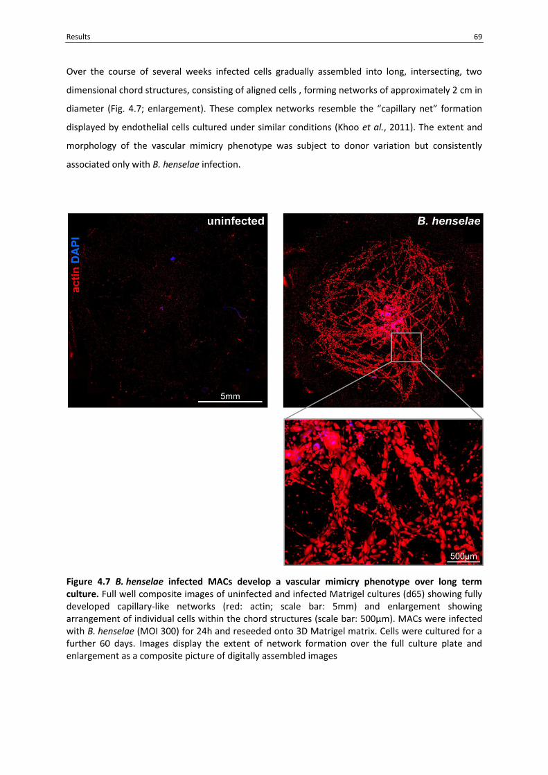
Results 69
Over the course of several weeks infected cells gradually assembled into long, intersecting, two
dimensional chord structures, consisting of aligned cells , forming networks of approximately 2 cm in
diameter (Fig. 4.7; enlargement). These complex networks resemble the “capillary net” formation
displayed by endothelial cells cultured under similar conditions (Khoo et al., 2011). The extent and
morphology of the vascular mimicry phenotype was subject to donor variation but consistently
associated only with B. henselae infection.
Figure 4.7 B. henselae infected MACs develop a vascular mimicry phenotype over long term culture. Full well composite images of uninfected and infected Matrigel cultures (d65) showing fully developed capillary-like networks (red: actin; scale bar: 5mm) and enlargement showing arrangement of individual cells within the chord structures (scale bar: 500µm). MACs were infected with B. henselae (MOI 300) for 24h and reseeded onto 3D Matrigel matrix. Cells were cultured for a further 60 days. Images display the extent of network formation over the full culture plate and enlargement as a composite picture of digitally assembled images

70 Results
Almost 100% (96.72±4.43%) of cells isolated from fully developed vascular mimicry structures (d51)
stained negative for the apoptosis marker 7-AAD in FACS analysis demonstrating sustained viability
of B. henselae infected MACs in Matrigel culture (n=4, Fig. 4.8). B. henselae DNA was detectable via
16S rDNA PCR at the end of long term cultures, however, no viable bacteria could be cultured from
vascular mimicry structures (n=3; Fig. 4.9 and data not shown) indicating that the sustained presence
of viable B. henselae is not required for the full development of vascular mimicry structures..
4.7 The development of the B. henselae-induced vascular mimicry phenotype is BadA
dependent
BadA is crucially involved in the HIF-1-dependent angiogenic reprogramming of B. henselae-infected
cells (Riess et al., 2004). Indeed, HIF-1 activation upon infection was also shown to be BadA
dependent in MACs (see above; Fig. 4.3B). To further dissect the role of BadA in the B. henselae-
induced vascular mimicry phenotype, MACs were infected with B. henselae wildtype (BadA+),
B. henselae BadA- and heat killed B. henselae and subsequently cultured on Matrigel as described
above. Although some structure formation was observed in cultures infected with BadA- and heat
killed B. henselae, results demonstrate that only BadA-expressing viable B. henselae were able to
induces strong capillary-like structure formation in MACs (Fig. 4.10 A-E).

Results 71
Figure 4.8 Viability of B. henselae infected MACs in long term Matrigel culture. FACS analysis of apoptosis marker 7-AAD [(red) compared to unstained negative controls (black)] on uninfected d3 MACs and B. henselae infected MACs (MOI300) from Matrigel vascular mimicry structures (day 51). Histograms display representative results [events vs. fluorescence intensity (log10)] as well as means and standard deviations of percent positive cells from at least three independent experiments.
Figure 4.9 Presence of B. henselae DNA in fully formed vascular mimicry structures. MACs were isolated from Matrigel vascular mimicry structures at end point of culture (day 60) and analyzed via nested-16S rDNA PCR for the presence of B. henselae DNA. Expected amplification product: 990bp. Figure shows results in duplicate from one experiment.

72 Results
Figure 4.10 Development of the vascular mimicry structures is dependent on infection with viable BadA expressing B. henselae. MACs were infected with B. henselae (MOI 300) for 24h and reseeded onto 3D Matrigel matrix. Cells were cultured for a further 60 days. Representative images of capillary-like growth networks formed by (A) uninfected MACs, MACs infected with (B) B. henselae-wildtype, (C) B. henselae-BadA- or (D) heat-killed B. henselae (scale bar: 5mm). (E) Quantification of capillary-like growth in the presence of B. henselae wild type, B. henselae BadA- and heat-killed B. henselae as a measure of the relative actin-stained pixel area. Results show mean and standard deviation from three independent experiments. Asterisk denotes p<0.01.

Results 73
4.8 MACs develop and maintain a macrophage phenotype after infection with B. henselae
To determine the differentiation phenotype of B. henselae-infected MACs, flow cytometric analysis
was performed to test for the expression of cell surface markers at different time points [day 5 (24h
p.i.) and day 62 from fully developed vascular mimicry structures; Fig. 4.11]. Endothelial markers
antigens VEGFR2 and CD31 were absent or weakly expressed at all time points examined. Under all
conditions, MACs showed moderate expression of the pan-leukocyte marker CD45 and no
expression of the monocytic marker CD14. The marker of antigen presenting cells human leukocyte
antigen (HLA)-DR and the macrophage marker CD68 were expressed on all cells. A strong increase in
CD68 and HLA-DR occurred after infection with B. henselae and lasted over long term culture. In
contrast, CD11c a marker of monocyte derived dendritic cells, was only moderately expressed on
MACs and was completely absent on cells derived from the vascular mimicry structures.
Additionally, MACs were removed from Matrigel vascular mimicry structures by dispase-treatment
and analyzed via cytospin-immunocytochemistry. Cells of the vascular mimicry structures stained
positive for vimentin; a protein of the cytoskeleton expressed in cells of the mesenchymal lineage
but also by hematopoietic cells such as macrophages. Staining for CD34, a stem cell marker also
found on endothelial cells was found to be negative along with the endothelial markers CD31, von-
Willebrand Factor (vWF) and vascular endothelial growth factor receptor-2 (VEGFR2; Fig. 4.12).
Finally, qRT-PCR analysis was preformed comparing the rate of endothelial marker gene
transcription between uninfected MACs and infected cells of the vascular mimicry structures.
Analysis revealed only a small and statistically insignificant fold increase in the transcription of key
endothelial marker genes endothelial nitric oxide synthase (eNOS), vascular endothelial cadherin (VE
Cadherin) and VEGFR-2 (Fig. 4.13).
Taken together, these results suggest that the phenotype induced in MACs in response to
B. henselae infection and present in the vascular mimicry structures is of a myeloid cell phenotype
with typical macrophage characteristics. Despite the pro-angiogenic phenotype of these cells and
the formation of vessel-like structures, results did not demonstrate any evidence of the
development of an endothelial cell phenotype in either gene transcription or protein expression of
B. henselae infected MACs.

74 Results
Figure 4.11 MACs develop and maintain a macrophage phenotype after infection with B. henselae. FACS analysis of endothelial cell markers VEGFR2, CD31, myeloid marker CD14, CD45, macrophage markers CD68, HLA-DR and dendritic cell marker CD11c expression [(red) compared to isotype-stained negative controls (black)] on uninfected and B. henselae infected MACs (MOI 300) d5-24h p.i. and d62 from vascular mimicry structures. Histograms display representative results [events vs. fluorescence intensity (log10)] as well as means and standard deviations of percent positive cells from three independent experiments.

Results 75
Figure 4.12 MACs maintain a non-endothelial phenotype after infection with B. henselae and formation of vascular mimicry structures. Infected MACs (MOI 300) were isolated from Matrigel vascular mimicry structures via dispase digestion and centrifuged onto glass slides for cytospin-immunohistochemistry. Cells were analyzed for the presence of (A) mesenchymal marker Vimentin, stem cell marker CD34 (d46) and (B) endothelial markers CD31, vWF and VEGFR2 (d51). For endothelial markers, HUVECs were used as internal positive control. Control stains were performed omitting primary antibodies.

76 Results
Figure 4.13 MAC angiogenic re-programming upon B. henselae infection does not involve upregulation of endothelial marker genes. Induction of endothelial marker genes eNOS, VE-Cadherin and VEGFR2 in B. henselae infected MACs (MOI 300) isolated at d60 from vascular mimicry structures was measured in relation to uninfected freshly isolated MACs at d3 via qRT-PCR. mRNA transcripts were quantified [ratio: eNOS/ RPLP0, VE-cadherin/ PolR2A and VEGFR2/ PolR2A]. Results show mean and standard deviation from three independent experiments.
Transcriptional profiling
To examine phenotypic changes induced in MACs in response to B. henselae infection, gene
expression profiles were compared between uninfected MACs directly after isolation (d3) and
B. henselae infected MACs cultured on Matrigel (d53) via gene expression microarray.
4.9 The infection of MACs with B. henselae is associated with broad phenotypic re-programming
To evaluate the general variation in overall gene expression between samples and between
experimental conditions, unsupervised hierarchical clustering was performed for genes with a
standard deviation ≥ 1.5 across all samples (Fig. 4.14 A-B).
All three donor samples isolated from d3 MACs clustered closely together [average person
correlation coefficient (r)= 0.992; (max correlation r =1)] and were separate from the corresponding
infected samples (d53; average r =0.989). In contrast, a high level of variance was displayed between
the two experimental conditions (average r =0.963) indicating that variations in gene expression
between donors is negligible in comparison to the broad phenotypic reprogramming induced in
MACs in response to infection.

Results 77
Figure 4.14 B. henselae infection of MACs leads to broad phenotypic re-programming. RNA was isolated from freshly isolated MACs (d3) and B. henselae infected, Matrigel cultured MACs (MOI 300; d53). mRNA gene expression profiles were analyzed via Affymetrix microarrays. Variance in gene expression profiles were compared between three biological replicates of uninfected MACs (d3) and B. henselae infected MACs (d53). (A) Unsupervised hierarchical clustering was performed for genes with a standard deviation ≥ 1.0 across all samples using the Manhattan distance and the average linkage method. Clustering arrangement of 399 genes of highest variance is shown. Absolute gene expression signals are represented in red (high) and green (low). Linkage between samples is represented by the length of the connecting branches displayed above the image. Connecting branches to the left display the linkage for expression variance of individual genes. (B) The average person correlation coefficient (r) is displayed as a measure of overall variance in gene expression among biological replicates and experimental conditions.

78 Results
4.10 The gene expression profiles of B. henselae infected MACs are strongly divergent from endothelial cells and show most similarity to cells of the myeloid lineage
To compare gene expression profiles of infected and uninfected MACs to endothelial cells, an
endothelial control sample (HUVECs grown on Matrigel for 12h) was included in the analysis as a
control. A distinct clustering occurred, separating the HUVEC control sample from all other samples
by a long linkage distance (Fig. 4.14B and Fig. 4.15; average r = 0.871). The pronounced disparity in
gene expression between endothelial cells and both infected and uninfected MACs provides further
evidence that the pro-angiogenic phenotype of B. henselae infected MACs is not associated with an
endothelial phenotype.
As an additional measure to examine the cellular differentiation of B. henselae infected MACs, gene
expression profiles were compared to datasets of know cell differentiation posted to the online
database NCBI Gene Expression Omnibus (GEO).
In order to compare gene expression profiles of the B. henselae infected MACs to datasets found in
the GEO databank, it was necessary to convert the relative gene expression values to an earlier
version of the Affymetrix gene microarray platform (Affymetrix Human Genome U133 Plus 2.0 Array;
GPL570). As conversion resulted in considerable statistical noise, direct comparison between
datasets from the two platforms was not possible. Nevertheless, some general observations could
be made.
Gene expression profiles form infected MACs (d53) showed the highest correlation with cells of the
monocytic myeloid lineage; monocytes, macrophages and dendritic cells with calculated coefficients
of determination (R2) ranging from 0.530 to 0.380 (Table 4.1). These were followed by gene
expression profiles from smooth muscle, fibroblasts, endothelial cells, other immune cells and finally
embryonic stem cells for which the calculated R2 was only 0.223.
The variation between infected and uninfected MACs (R2=0.8742) was found to exceed the variation
observed between monocytes and in vitro differentiated macrophages (GSE11430 R2=0.909) as
measured in one study documented in the GEO database. This suggests that B. henselae infection
induces significant phenotypic changes which exceed the phenotypic transformation taking place
during typical macrophage differentiation process and further supports the finding that B. henselae
infection results in a comprehensive phenotypic re-programming in infected MACs (Table 4.2).
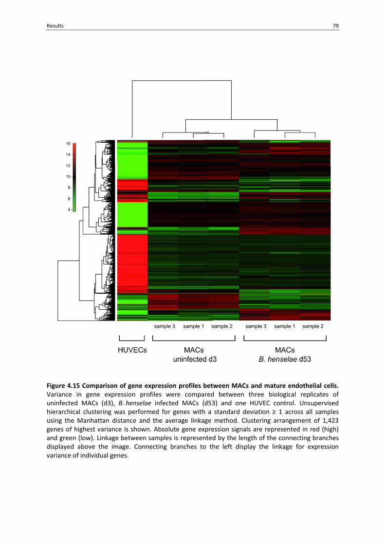
Results 79
Figure 4.15 Comparison of gene expression profiles between MACs and mature endothelial cells. Variance in gene expression profiles were compared between three biological replicates of uninfected MACs (d3), B. henselae infected MACs (d53) and one HUVEC control. Unsupervised hierarchical clustering was performed for genes with a standard deviation ≥ 1 across all samples using the Manhattan distance and the average linkage method. Clustering arrangement of 1,423 genes of highest variance is shown. Absolute gene expression signals are represented in red (high) and green (low). Linkage between samples is represented by the length of the connecting branches displayed above the image. Connecting branches to the left display the linkage for expression variance of individual genes.

80 Results
Table 4.1 Comparison of gene expression signals from B. henselae infected MACs to gene expression profiles of known cell differentiation. Gene expression profiles of B. henselae infected MACs (d53) were compared to selected reference gene expression profiles from cells of known differentiation. Reference gene expression profiles were retrieved from NCBI Gene Expression Omnibus (GEO) and expression signals converted between platforms for comparison. Similarity in gene expression patterns calculated as coefficient of determination (R2).
Platform Reference
Series Cell type Description Sample R2=
GPL6244 GSE55170 MAC MACs, uninfected, d3
0.874
GPL570 GSE4984. dendritic monocyte derive dendritic cells
control 0.530
GPL570 GSE13670 macrophage human monocyte-derived macrophages
control 0.513
GPL570 GSE16385. macrophage alternatively and classically activated macrophages
macrophage IL-4, 24h (alternatively activated)
0.473
GPL570 GSE20484 macrophage monocyte derived macrophages stimulated with MCSF and CXCL4
MCSF 0.478
GPL570 GSE16385. macrophage alternatively and classically activated macrophages
control, 24h 0.458
GPL570 GSE6054 monocyte monocytes control 0.449
GPL570 GSE16385. macrophage alternatively and classically activated macrophages
macrophage
INF+TNFα, 24h (classically activated)
0.424
GPL570 GSE12261 smooth muscle
human aortic smooth muscle cells
control 0.340
GPL570 GSE28491+ GSE28492
B cell human immune cell subsets B cells 0.333
GPL570 GSE14801 endothelial human umbilical vein endothelial cells
control 0.328
GPL570 GSE11919 fibroblast GM5659 skin fibroblasts control 0.321
GPL570 GSE28491+ GSE28492
eosinophils human immune cell subsets eosinophils 0.315
GPL570 GSE28491+ GSE28492
neutrophils human immune cell subsets neutrophils 0.315
GPL570 GSE28491+ GSE28492
CD8 T cells human immune cell subsets CD8 T cells 0.310
GPL570 GSE28491+ GSE28492
CD4 T cells human immune cell subsets CD4 T cells 0.309
GPL570 GSE7896 stem cell embryonic stem cells control 0.226

Results 81
Table 4.2 Comparison of B. henselae induced phenotypic re-programming in MACs to changes induced during in vitro monocyte to macrophage differentiation. The variance in gene expression profiles between uninfected MACs (d3) and B. henselae infected MACs (d53) was compared to changes in gene expression occurring between monocytes and in vitro cytokine differentiated monocyte-derived-macrophages. Reference expression patterns retrieved from NCBI Gene expression Omnibus (GEO). Samples evaluated within reference series and platforms. Similarity in gene expression patterns calculated as coefficient of determination (R2).
Platform Reference Series Cell type Description R2=
GPL6244 GSE55170 MACs, uninfected, d3 0.874
MACs, B. henselae, d53
GPL570 GSE11430 monocyte monocytes and macrophages 0.909
macrophage
4.11 B. henselae infection of MACs induces regulation of genes involved in angiogenic and immune regulatory pathways.
To examine the nature of cellular re-programming in B. henselae infected MACs in more detail,
absolute normalized expression values from the gene expression microarrays of infected and
uninfected MACs [fold change (FC)] were compared. Overall, 1,400 genes or ca. 36% of the 3,872
messenger RNA (mRNA) transcripts evaluated, showed a change in expression in excess of 2.0 fold.
620 genes were upregulated and 780 genes downregulated at a rate of 2.0 fold or more. For genes
upregulated 4.0 fold or higher, and with a false discovery rate (FDR) of less than 0.05, a detailed
functional analysis was performed via GeneBank database and PubMed literature searches (Table
4.3). A functional annotation analysis using the DAVID database tool was performed on genes
upregulated or downregulated with a fold change greater than or equal to 2.0 and a false discovery
rate of less than 0.05.
Functional annotations in the upregulated gene sets enriched to statistical significance (p<0.01) were
revealed in 157 annotation categories (S Table 10.1 and 10.2). These annotation categories fell into
20 broadly related functional groups (S Table 10.4 and 10.5). For downregulated genes, 52 enriched
functional annotation categories could be identified in 6 broadly related functional groups. A
selection of these highly enriched functional annotation categories from up- and downregulated
transcripts are presented in (Fig. 4.16) and (Table 4.4 and 4.5).
Functional annotations relating to angiogenesis and vascular development were most highly
enriched among the upregulated genes. Highly upregulated genes included some of the most
important positive inducers of angiogenesis (VEGFA, CXCL8, HIF1A, MMP9, TGFB). These were
followed by annotations relating to positive and negative regulation of apoptosis including
upregulation of genes such as IER3, CKS2, PPP1R15A, PHLDA1 CDKN1A and TNFAIP3.
82 Results
Enriched functional annotations were also found in categories for inter- and intracellular signaling
pathways including cytokine production, receptor signaling, intracellular signaling cascades, MAPK
pathways and transcriptional regulation. Genes for chemokines (CCL2, CXCL8, CXCL1, CXCL2, CCL4L1,
CC3L1/CCL3, CSF) were particularly highly upregulated. Highly enriched functional categories also
included annotations involved in structural organization such as those for cell migration and
adhesion. Many of the highly upregulated genes also had functions related to interaction with the
extracellular matrix such as matrix degrading metalloproteinases MMP9, MMP2, MMP7, MMP12,
ADAMDEC1, MME, and other compounds involved in matrix remodeling SERPINE1, CHI3L1, CHIT1
CTSK, HTRA1 TNFAIP6. Furthermore, several extracellular matrix components such FN1, SPARC,
FBLN5, SPP1, THBS1 as well as several integrins ITGB3, ITGA3, ITGB5, ITGA5, and ITGAV were
upregulated.
Other enriched functional groups were identified as being linked to the regulation of immune
responses including those for myeloid cell differentiation, leukocyte activation, proliferation, and
hematopoietic cell differentiation. This was reflected in strong upregulation of several genes
involved in innate immune responses (NFKB, IL1B , IL8, CSF, TLR2). Along with the upregulation of
pro-inflammatory gene clusters, upregulation of negative regulators of inflammation and NFκB
mediated gene transcription was also observed (NR4A2, PTGS2, AREG, IER3, IFRD1, NR4A1, ARG2,
TGFB2, TNFAIP3, ZC3H12A, MMP12, IL1RA, SOCS3).
Unexpectedly, functional groups representing sterol metabolism were also highly enriched. Several
genes for cholesterol processing and metabolism OLR1, LPL, LDLR, DHCR24, INSIG1, DHCR7, general
sterol biosynthesis, CYP19A1 , SQLE, and several members of the prostaglandin biosynthesis
pathway FADS1, PTGS2, ALOX5AP were highly upregulated.
The functional annotation analysis of downregulated genes was less diverse and allocated functional
annotations to three general categories. The majority of the down regulated genes were classified to
regulation of gene transcription and RNA metabolic processes including numerous zinc finger
proteins, 93 of which were downregulated 2.0 fold or more. Following this was a strong
downregulation of genes involved in T-cell activation, differentiation and T-cell receptor signaling
pathways such as the CD3-TCR complex components CD3E, CD3D, CD3G, CD247, ZAP70, and
downstream pathway elements LCK, FYN, STAT4, CD2, CD8A. Annotations relating to positive
regulation of innate immune responses were also enriched among the downregulated gene
transcripts.
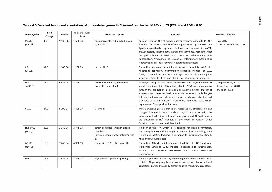
Table 4.3 Detailed functional annotation of upregulated genes in B. henselae-infected MACs at d53 (FC ≥ 4 and FDR < 0.05).
Gene Symbol Fold
Change p value
False Discovery
Rate Gene Description Function Relevant Citations
NR4A2
(Nurr1)
80.5 9.13E-06 1.60E-03 nuclear receptor subfamily 4, group
A, member 2
Nuclear receptor (NR) of orphan nuclear receptor subfamily 4A. NRs
interact directly with DNA to influence gene transcription. NR4s are
ligand-independently regulated. Induced in response to cAMP,
growth factors, inflammatory signals and hormones. Associates with
the p65 subunit of NFκB and attenuates inflammatory gene
transcription. Attenuates the release of inflammatory cytokines in
macrophages. Essential for VEGF mediated angiogenesis.
(Han, 2012)
(Zhao and Bruemmer, 2010)
IL8
(CXCL8)
44.5 1.18E-06 1.29E-03 interleukin 8 Chemokine. Chemoattractant for neutrophils, basophils and T-cells.
Neutrophil activation, inflammatory response, member of CXCL
family of chemokines with ELR motif (glutamic acid-leucine-arginine
sequence). Binds to CXCR1 and CXCR2. Potent angiogenic properties.
OLR1
(LOX-1)
33.2 6.58E-04 4.72E-03 oxidized low density lipoprotein
(lectin-like) receptor 1
Scavenger receptor that binds, internalizes and degrades oxidized
low-density lipoprotein. This action activates NFκB and inflammation
through the production of intracellular reactive oxygen. Marker of
atherosclerosis. Also involved in immune response as a leukocyte-
adhesion molecule and acts as a receptor for advanced glycation end
products, activated platelets, monocytes, apoptotic cells, Gram-
negative and Gram-positive bacteria.
(Campbell et al., 2012)
(Shimaoka et al., 2001)
(Zhu et al., 2013)
GLDN 24.8 5.79E-04 4.48E-03 Gliomedin Transmembrane protein that is characterized by olfactomedin and
collagen domains in its extracellular region. Interaction with the
axonodal cell adhesion molecules neurofascin and NrCAM induces
the clustering of Na+ channels at the nodes of Ranvier. Other
functions have not been well described.
SERPINE1
(PAI-1)
20.8 3.04E-04 3.77E-03 serpin peptidase inhibitor, clade E
member 1,
( plasminogen activator inhibitor type
1)
Inhibitor of the uPA which is responsible for plasmin formation,
matrix degradation and proteolytic activation of extracellular growth
factors and MMPs. Induced in response to inflammatory stimuli.
NFκB and MAPK regulated.
CCL20
(MIP-3β)
18.8 7.44E-04 4.95E-03 chemokine (C-C motif) ligand 20 Chemokine. Attracts mainly immature dendritic cells (iDCs) and some
leukocytes. Binds to CCR6. Induced in response to inflammatory
stimulus and hypoxia. Associated with tumor associated
macrophages.
RGS1 16.6 1.82E-04 3.24E-03 regulator of G-protein signaling 1 Inhibits signal transduction by interacting with alpha subunits of G-
proteins. Negatively regulates cytokine and growth factor induced
signal transduction through G-protein coupled membrane receptors.
Resu
lts
8
3
83
R
esults
83
Resu
lts 8
3
Resu
lts

PTGS2
(Cox-2)
16.0 1.96E-06 1.29E-03 prostaglandin-endoperoxide
syntheses 2
(cyclooxygenase -2)
Enzyme responsible for conversion of arachidonic acid (AA) to
prostaglandin H2. Induced under inflammatory conditions.
Dysregulated in many cancers, e.g., gastric carcinoma. Involved in
pro-inflammatory response, evasion of apoptosis,
immunosuppression, sustained proliferation, angiogenesis, invasion
and metastasis.
(Greenhough et al., 2009)
(Chen and Smyth, 2011)
(Tjiu et al., 2008)
(Nakanishi et al., 2011;
Obermajer et al., 2012)
LPL 15.8 1.13E-04 3.00E-03 lipoprotein lipase Triglyceride hydrolase that converts very low density lipoprotien
(vLDL) to low density lipoprotein (LDL) in blood plasma. Ligand
bridging factor for receptor-mediated lipoprotein uptake.
CD83 14.1 2.63E-06 1.29E-03 CD83 molecule Common surface protein on mature dendritic cells. Preformed
intracellularly in monocytes and macrophages. Rapidly exported to
the surface in response to inflammatory stimuli. Function not well
characterized. Immune modulation.
CHI3L1
(YK-L40)
14.0 7.59E-04 5.03E-03 chitinase 3-like 1
(cartilage glycoprotein-39)
Secreted glycoprotein. Member of the glycosyl hydrolase 18 family
but has lost chitanase activity. Involved in macrophage maturation,
immune response, tissue remodeling, angiogenesis.
(Shao et al., 2009)
(Mizoguchi, 2006)
SLC16A10 12.7 1.46E-03 6.22E-03 solute carrier family 16, member 10
(aromatic amino acid transporter)
Plasma membrane amino acid transporter. Na(+)-independent
transport of aromatic amino acids.
(Mariotta et al., 2012)
(Ramadan et al., 2007)
CHIT1 12.6 4.18E-04 4.19E-03 chitinase 1 (chitotriosidase) Secreted glycoprotein. Member of the glycosyl hydrolase 18 family.
Chitanase activity. Host defense against chitinous pathogens, also
involved in inflammation, infection and matrix remodeling.
(Cozzarini et al., 2009)
(Kanneganti et al., 2012)
GPX3 12.4 1.04E-04 2.95E-03 glutathione peroxidase 3 (plasma) Secreted protein. Catalyses the reduction of peroxides. Protection
from oxidative stress.
(Falck et al., 2010)
(Peng et al., 2012)
CYGB 12.4 2.30E-05 2.01E-03 cytoglobin Member of the globin family of oxygen binding proteins. Protection
from hypoxia. Reaction to oxidative stress.
(Emara et al., 2010)
AREG
11.2 1.50E-03 6.31E-03 amphiregulin Growth factor of the endothelial growth factor (EGF) family. Control
of inflammation. Tissue growth and remodeling.
(Zaiss et al., 2013)
CCL4L1 11.1 6.80E-04 4.79E-03 chemokine (C-C motif) ligand 4-like 1 Chemokine. Paralogous gene to CCL4. Encoded protein differs in
three amino acid positions. Function appears to be redundant, see
CCL4.
(Modi et al., 2001)
(Pedrosa et al., 2011)
(Howard, O M Zack et al.,
2004)
AQP9 10.1 1.94E-04 3.31E-03 aquaporin 9 Membrane transport channel with broad substrate selectivity. (Calamita et al., 2012)
(Jelen et al., 2011)
IL1B 10.0 1.68E-03 6.51E-03 interleukin 1, beta Cytokine. Important mediator of the inflammatory response.
Produced as a pro-protein in activated macrophages, which must be
proteolytically processed to its active form by caspase 1
84
Resu
lts

R4A3 9.9 3.02E-05 2.06E-03 nuclear receptor subfamily 4, group
A, member 3
Nuclear receptor of orphan nuclear receptor subfamily 4A. NRs
interact directly with DNA to influence gene transcription. NR4s are
ligand-independently regulated. Regulators of gene expression and
metabolism. Induced by inflammatory stimulus. May be regulated by
NFB. Acts to negatively regulate inflammation (conflicting results).
Induces M2 macrophage polarization.
C3 9.9 1.74E-04 3.21E-03 complement component 3 Component of the complement cascade. Innate immune response.
DHCR24 9.2 9.90E-04 5.37E-03 24-dehydrocholesterol reductase Oxidoreductase enzyme involved in cholesterol biosynthesis.
MMP9 9.2 1.14E-04 3.00E-03 matrix metallopeptidase 9 Matrix metallopeptidase. Matrix remodeling. Cytokine activation
through cleavage. Angiogenesis.
(Jadhav et al., 2004)
(Morancho et al., 2013)
(van Hinsbergh and Koolwijk,
2008)
(Zajac et al., 2013)
CXCL1
(Gro-α)
9.1 2.69E-04 3.65E-03 chemokine (C-X-C motif) ligand 1
(melanoma growth stimulating
activity, alpha)
Chemokine. member of CXCL family of chemokines. Contains the ELR
motif. Binds CXCR2. Chemoattractant for neutrophils. Inflammatory
response. Pro-angiogenic. Oncogene.
CXCL2
(Gro-β)
9.0 1.26E-04 3.00E-03 chemokine (C-X-C motif) ligand 2 Chemokine. Member of CXCL family of chemokines. Contains the ELR
motif. Binds CXCR2. Chemoattractant for neutrophils. Inflammatory
response. Pro-angiogenesis.
CCL3 9.0 3.47E-04 3.95E-03 chemokine (C-C motif) ligand 3 Cytokine. Binds receptors CCR1, CCR4, CCR5. Acute inflammatory
response. Immunomodulation. Chemoattractant. Recruitment and
activation of monocytes/macrophages, neutrophils, lymphocytes.
Shares 68% amino acid similarity with CCL4.
CCL4 9.0 6.39E-04 4.66E-03 chemokine (C-C motif) ligand 4 Chemokine. Binds receptors CCR5 and CCR1. Chemoattractant
forimmune cells such as natural killer cells and monocytes.
Immunomodulation and inflammation.
KAL1
(anosmin-1)
8.8 2.00E-04 3.34E-03 Kallmann syndrome 1 sequence Extracellular matrix-associated glycoprotein. Can form part of active
β1 integrin complex inducing downstream signaling pathways. Has
been shown to facilitate tumor cell proliferation, migration, invasion,
and survival.
SLC7A11 8.7 4.63E-04 4.29E-03 solute carrier family 7, (cationic
amino acid transporter, y+ system)
member 11
Transmembrane transporter of amino acids. Highly specific for
cysteine and glutamate.
ITGB3 8.6 3.99E-03 9.35E-03 integrin, beta 3 (platelet glycoprotein
IIIa, antigen CD61)
Beta integrin subunit (heterodimeric transmembrane extracellular
matrix receptor).
(Feng et al., 2013)
(Steri et al., 2014)
Resu
lts
8
5

IER3 8.5 3.88E-05 2.25E-03 immediate early response 3 Anti-apoptotic. Interference with signaling pathways e.g.
NFκB/MAPK.
(Arlt and Schäfer, 2011)
LDLR 8.5 4.18E-04 4.19E-03 low density lipoprotein receptor Extracellular receptor that recognizes apoprotein B100, which is
embedded in the outer phospholipid layer of LDL particles. Mediates
the endocytosis of cholesterol-rich low density lipoprotein.
CRIM1 8.4 3.00E-06 1.30E-03 cysteine rich transmembrane BMP
regulator 1 (chordin-like)
Transmembrane protein with growth factor receptor domains.
Control of cellular development including tube formation in the
capillary development of VEGF induced angiogenesis.
(Fan et al., 2013)
(Glienke et al., 2002)
FN1 8.4 2.61E-04 3.60E-03 fibronectin 1 Extracellular matrix glycoprotein. Important in cellular adhesion,
migration and differentiation and involved in processes such as
angiogenesis
DUSP1 8.3 1.23E-04 3.00E-03 dual specificity phosphatase 1 Non-receptor-type protein-tyrosine phosphatase that interacts with
members of signaling cascades e.g. MAPKs in response to cellular
stress or growth factors.
(Echavarria and Hussain,
2013)
(Patterson et al., 2009)
IRAK2 8.3 1.56E-04 3.10E-03 interleukin-1 receptor-associated
kinase 2
Associates with IL-1 receptor upon cytokine binding and transmits the
signaling cascade. Involved in IL-1 associated NFκB activation.
SPARC
(osteonectin)
7.8 3.59E-05 2.24E-03 secreted protein, acidic, cysteine-rich
(osteonectin)
Extracellular matrix glycoprotein with high affinity for calcium and
collagen. Cell extracellular matrix protein interaction and growth
factor response. Increases the production and activity of matrix
metalloproteinases and angiogenesis.
(Rivera et al., 2011)
INSIG1 7.7 2.28E-03 7.31E-03 insulin induced gene 1 Endoplasmic reticulum membrane protein involved in cholesterol
metabolism.
TM7SF4
(DCSTAMP)
7.3 4.72E-04 4.29E-03 transmembrane 7 superfamily
member 4
Transmembrane protein highly expressed in dendritic cells. Involved
in multinucleated osteoclast formation, myeloid cell differentiation
and immune regulation.
(Chiu et al., 2012)
(Eleveld-Trancikova et al.,
2007)
(Sawatani et al., 2008)
CTSK 6.9 1.78E-03 6.66E-03 cathepsin K Cysteine protease with potent endoprotease activity against
fibrinogen at acid pH. Involved in bone re-absorption and matrix
remodeling.
HBEGF 6.8 4.67E-06 1.33E-03 heparin-binding EGF-like growth
factor
Epidermal growth factor family member. Produced predominantly by
monocytes and macrophages. Chemotactic and mitogenic effects.
Cancer and wound healing.
GK 6.8 4.01E-04 4.19E-03 glycerol kinase Key enzyme in the regulation of glycerol uptake and metabolism.
RGS2 6.6 7.24E-04 4.86E-03 regulator of G-protein signaling 2,
24kDa
Negative regulator of G-protein mediated signaling pathways.
Involved in hypertension and other cardiovascular conditions
MAMLD1 6.4 3.21E-03 8.42E-03 mastermind-like domain containing 1 Transcriptional regulator. Co-activator of Notch signaling.
86
Resu
lts

NAMPT
(PBEF1,
vistafin)
6.3 4.21E-05 2.25E-03 nicotinamide
phosphoribosyltransferase
Inflammatory cytokine. Rate limiting enzyme in nicotinamide adenine
dinucleotide (NAD)+ biosynthesis. Involved in macrophage
differentiation and arteriosclerosis.
(Yun et al., 2013)
SIGLEC8 6.1 3.03E-04 3.77E-03 sialic acid binding Ig-like lectin 8 Transmembrane receptor. Expressed predominantly in eosinophils.
Involved in apoptosis when activated.
(Bochner, 2009)
(Crocker et al., 2012)
(Kano et al., 2013)
CCL3L1 6.1 1.13E-03 5.65E-03 chemokine (C-C motif) ligand 3-like 1 Paralogous gene to CCL3. 96% nucleotide and protein similarity.
Functions appear to be redundant except that the encoded CCL3
isoform is 2-fold more efficient at chemoattracting human
lymphocytes (see CCL3).
GNG12 5.9 4.60E-05 2.32E-03 guanine nucleotide binding protein (G
protein), gamma 12
G-protein modulator and transducer of transmembrane signaling
pathways. May be involved in negative regulation of LPS induced
inflammation.
(Larson et al., 2010)
HTRA1 5.9 4.23E-04 4.19E-03 HtrA serine peptidase 1 Secreted serine peptidase. Degrades extracellular matrix proteins and
cleaves growth factors notably insulin-like growth factors (IGFs) and
TGF-β. May regulate cellular growth and matrix deposition.
CKS2 5.9 2.00E-04 3.34E-03 CDC28 protein kinase regulatory
subunit 2
Regulator of cyclin dependent kinases. Regulator of cell cycle and
division.
FOSL2 5.9 2.87E-05 2.06E-03 FOS-like antigen 2 FOS family protein. Can dimerize with members of the JUN family
proteins to form the leucin zipper transcription factor activator
protien (AP)-1 which binds to genes with 2-O-tetradecanoylphorbol-
13-acetate (TPA) DNA response elements and regulate various
processes such as cellular differentiation, proliferation and apoptosis.
OTUD1 5.8 6.90E-05 2.71E-03 OTU domain containing 1 Deubiquitinating enzyme. Unknown function
TNFAIP6 5.8 2.29E-02 2.96E-02 tumor necrosis factor, alpha-induced
protein 6
Protein secreted in response to inflammatory stimulus. Contains a
hyaluronan-binding domain that is involved in cell migration,
extracellular matrix stability and protease activity.
SEP3 5.8 3.46E-04 3.95E-03 septin 3 Filament forming GTP-binding protein. Expressed in neurons and may
be involved in synaptic vesicle function.
(Macedo et al., 2013)
IFRD1
(PC4, Tis7)
5.8 5.63E-05 2.58E-03 interferon-related developmental
regulator 1
Negative regulator of NFκB. Involved in cell differentiation
development and muscle regeneration.
GPR183
(EB12)
5.7 1.27E-04 3.00E-03 G protein-coupled receptor 183 G-protein coupled membrane receptor for oxysterols. Directs
immune cell migration. Particularly well characterized for B cell
maturation and splenic DC positioning. Also a negative regulator of
type I interferon responses in plasmacytoid DCs and CD11b+ myeloid
cells through trans-regulation of the interferon regulatory factor 7.
(Chiang et al., 2013)
(Hannedouche et al., 2011)
(Yi and Cyster, 2013)
Resu
lts
8
7

ATP1B1 5.7 6.68E-04 4.74E-03 ATPase, Na+/K+ transporting, beta 1
polypeptide
Regulatory β subunit of an integral membrane ion pump, responsible
for establishing and maintaining the electrochemical gradients of Na
and K ions across the plasma membrane. These gradients are
essential for osmoregulation, for sodium-coupled transport of a
variety of organic and inorganic molecules, and for electrical
excitability of nerve and muscle. Also acts as a cell adhesion molecule
and signal transducer by interacting with small GTPases and MAPK
cascades.
(Tokhtaeva et al., 2012)
FOS
(c-FOS)
5.6 2.76E-05 2.06E-03 FBJ murine osteosarcoma viral
oncogene homolog
Dimerizes with members of the JUN family proteins to form
transcription factor activator protien-1 (AP-1) which binds to genes
with TPA DNA response elements and regulate various processes
such as cellular differentiation, proliferation and apoptosis.
NR4A1
(Nurr-77)
5.5 8.27E-05 2.77E-03 nuclear receptor subfamily 4, group
A, member 1
Nuclear receptor of orphan nuclear receptor subfamily 4A. Involved
in negative regulation of inflammation and macrophage polarization.
Mediates VEGF induced angiogenesis
(Hanna et al., 2012)
(Lefebvre et al., 2012)
(Zeng et al., 2006)
THBD 5.4 1.39E-03 6.09E-03 thrombomodulin Integral membrane protein. Acts as a co-factor to thrombin. Anti-
coagulative, anti-fibrolytic and anti-inflammatory.
(Conway, 2012)
(Martin et al., 2013)
HTR2B 5.4 3.29E-03 8.52E-03 5-hydroxytryptamine (serotonin)
receptor 2B
G-protein coupled receptor. Serotonin receptor. Signal transduction
through extracellular-signal regulated kinase (ERK)1/2. Preferentially
expressed on M2 macrophages and tumor associated macrophages.
Outside of nervous system serotonin also acts as a growth factor and
anti-inflammatory agent through modulation of cytokine release by
myeloid cells.
(las Casas-Engel et al., 2013)
--- 5.3 7.24E-04 4.86E-03 ---
SIGLEC10 5.3 2.46E-02 3.13E-02 sialic acid binding Ig-like lectin 10 Sialic-acid-binding transmembrane protein. SIGLEC-10 has been
shown to associate with CD24 and negatively regulate inflammatory
cascades via NFκB in distinguishing danger associated molecular
patterns from pathogen associated molecular patterns. Has also been
shown to be the leucocyte co-receptor for vascular adhesion protien -
1 and is involved in leukocyte adhesion to endothelial cells and the
inflammatory response.
(Chen et al., 2009)
(Crocker et al., 2012)
(Kivi et al., 2009)
88
Resu
lts

EGR2 5.3 6.82E-05 2.71E-03 early growth response 2 Transcription factor. Immediate early response gene that is important
for the induction of cellular programs of differentiation, proliferation,
and cell death in response to environmental stimuli, control of
inflammation and antigen receptor-induced proliferation in a cell-
intrinsic manner. Control of inflammatory autoimmunity, regulates
AP-1, suppressor of cytokine signaling (SOCS) and signal transducer
and activator of transcription (STAT).
(Li et al., 2012)
(Liu et al., 2003)
(Sumitomo et al., 2013)
DUSP4 5.3 1.72E-03 6.55E-03 dual specificity phosphatase 4 Regulators of MAPK signaling. DUSP4 preferentially dephosphorylates
ERK1/2, p38, and stress-activated protein kinase / c-Jun N-terminale
Kinasen proteins.
(Balko et al., 2013)
(Caunt and Keyse, 2013)
(Echavarria and Hussain,
2013)
(Kao et al., 2013)
NAMPT 5.3 3.55E-06 1.30E-03 nicotinamide
phosphoribosyltransferase
Catalyses the rate limiting step in the NAD biosynthesis pathway.
Also behaves as a cytokine with immunomodulating properties in its
secreted form.
EREG 5.3 2.19E-02 2.88E-02 epiregulin Member of the epidermal growth factor family. Ligand to EGF
receptor and avian erythroblastosis oncogene B 2 receptors. Involved
in angiogenesis and cytokine production.
LGI2 5.2 7.22E-04 4.86E-03 leucine-rich repeat LGI family,
member 2
Not well characterized. Involved in synaptic development. (Kegel et al., 2013)
SOD2 5.0 2.65E-06 1.29E-03 superoxide dismutase 2,
mitochondrial
Iron/manganese superoxide dismutase. Binds to the superoxide by
products of oxidative phosphorylation and converts them to
hydrogen peroxide and diatomic oxygen. Active in the mitochondria
where it is the primary antioxidant for cellular respiration.
CYP19A1
(aromatase)
4.9 4.09E-04 4.19E-03 cytochrome P450, family 19,
subfamily A, polypeptide 1
Monooxygenase localized to the endoplasmic reticulum. Catalyses a
key step in biosynthesis of estrogens.
SDC2 4.9 1.38E-03 6.08E-03 syndecan 2 Transmembrane protein. Heparin sulfate proteoglycan. Regulates cell
proliferation, cell migration and cell-matrix interactions via its
receptor for extracellular matrix proteins. May interact with integrins.
Expressed highly in fibroblast and endothelial cells, involved in
angiogenesis.
(Noguer et al., 2009)
PDPN 4.9 4.32E-03 9.77E-03 podoplanin Mucin-type transmembrane glycoprotein. Involved in actin
cytoskeleton organization, filopodia formation and cell motility.
Highly expressed in cancer where it mediates invasion, and
metastasis. Expressed in lymphatic endothelial cells.
(Krishnan et al., 2013)
(Mei et al., 2013)
Resu
lts
8
9

BCL6 4.9 5.01E-06 1.35E-03 B-cell CLL/lymphoma 6 Zinc finger transcription factor. Transcriptional repressor.
Suppression of cytokine production in macrophages. Involved in the
development of B and T cells and germinal centers.
(Wagner et al., 2011)
PDE3A 4.8 5.11E-04 4.38E-03 phosphodiesterase 3A, cGMP-
inhibited
Enzyme that hydrolyzes both cAMP and cyclic guanosine
monophosphate. Regulates amplitude and duration of intracellular
cyclic nucleotide signals. Important in cell cycle, platelet aggregation
and vascular smooth muscle contraction.
RG2 4.8 1.86E-03 6.76E-03 arginase, type II Enzyme that catalyses the hydrolysis of arginine. Anti-inflammatory
as it competes with iNOS for the production of nitric oxide. Type II
isoform expressed in the mitochondria.
(Lewis et al., 2010)
(Matsuo et al., 2009)
(Lee et al., 2013)
(Elms et al., 2013)
SQLE 4.8 9.68E-05 2.95E-03 squalene epoxidase Rate limiting enzyme in sterol biosynthesis. Catalyzes the first
oxygenation step.
GABARAPL1 4.8 1.37E-05 1.81E-03 GABA(A) receptor-associated protein
like 1
Interacts with tubulin for intracellular transport of proteins and
vesicles. Involved in late stages of autophagosome formation.
(Le Grand et al., 2011)
(Seguin-Py et al., 2012)
TGFB2 4.7 9.33E-04 5.37E-03 transforming growth factor, beta 2 Cytokine. Signals through combinations of transmembrane type I and
type II receptors (TGFBR1 and TGFBR2) and SMAD, ERK, JUN, p38,
phosphoinositide 3-kinase pathways. Involved in many cellular
processes including, proliferation, differentiation, migration,
apoptosis and angiogenesis. Promotes M2 macrophage
differentiation.
(Gong et al., 2012)
(Zhou et al., 2012)
(Worthington et al., 2012)
FBLN5 4.7 5.99E-04 4.52E-03 fibulin 5 Extracellular matrix protein containing an Arginyl- glycyl- aspartic acid
(RGD) motif. Promotes adhesion of endothelial cells through
interaction with integrins. Anti-angiogenic.
(Albig and Schiemann, 2004)
SASH1 4.7 5.62E-04 4.46E-03 SAM and SH3 domain containing 1 Expressed in microvascular endothelial cells. Positively regulates TLR4
signaling and downstream activation and NFκB and MAPKs.
(Dauphinee et al., 2013)
HSPA1B
(HSP70-2)
4.7 1.33E-04 3.01E-03 heat shock 70kDa protein 1B Molecular chaperones that accompany protein folding and prevent
misfolding under conditions of stress such as hypoxia, heat, and
oxidative stress. Upregulated in cancer.
(Huang et al., 2009b)
TNFAIP3
(A20)
4.7 2.13E-05 1.95E-03 tumor necrosis factor, alpha-induced
protein 3
Ubiquitin-editing enzyme. Induced in response to inflammatory
stimulus. Terminates NFκB activation. Anti-apoptotic. Regulates Wnt
signalling, interferon regulatory factor and autophagy.
(Catrysse et al., 2014)
(Vereecke et al., 2011)
SCIN 4.6 3.05E-02 3.72E-02 scinderin Ca(2+)-dependent actin-severing and -capping protein. Plays a role in
actin network dynamics in exocytosis.
(Trifaró et al., 2000)
90
Resu
lts
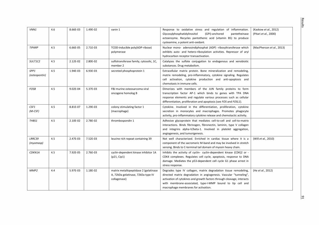
VNN1 4.6 8.66E-03 1.49E-02 vanin 1 Response to oxidative stress and regulation of inflammation.
Glycosylphosphatidylinositol (GPI)-anchored pantetheinase
ectoenzyme. Recycles pantothenic acid (vitamin B5) to produce
cysteamine, a potent anti-oxidant.
(Kaskow et al., 2012)
(Pitari et al., 2000)
TIPARP 4.5 6.66E-05 2.71E-03 TCDD-inducible poly(ADP-ribose)
polymerase
Nuclear mono- adenosindiphosphat (ADP) -ribosyltransferase which
exhibits auto- and hetero-ribosylation activities. Repressor of aryl
hydrocarbon receptor transactivation.
(MacPherson et al., 2013)
SULT1C2 4.5 2.12E-02 2.80E-02 sulfotransferase family, cytosolic, 1C,
member 2
Catalyzes the sulfate conjugation to endogenous and xenobiotic
substances. Drug metabolism.
SPP1
(osteopontin)
4.5 1.94E-03 6.93E-03 secreted phosphoprotein 1 Extracellular matrix protein. Bone mineralization and remodeling,
matrix remodeling, pro-inflammatory, cytokine signaling. Regulates
cell activation, cytokine production and anti-apoptosis and
chemotaxis in immune cells.
FOSB 4.5 9.02E-04 5.37E-03 FBJ murine osteosarcoma viral
oncogene homolog B
Dimerizes with members of the JUN family proteins to form
transcription factor AP-1 which binds to genes with TPA DNA
response elements and regulate various processes such as cellular
differentiation, proliferation and apoptosis (see FOS and FOSL1).
CSF1
(M-CSF)
4.5 8.81E-07 1.29E-03 colony stimulating factor 1
(macrophage)
Cytokine. Involved in the differentiation, proliferation, cytokine
secretion in monocytes and macrophages. Promotes phagocyte
activity, pro-inflammatory cytokine release and chemotactic activity.
THBS1 4.5 2.10E-02 2.78E-02 thrombospondin 1 Adhesive glycoprotein that mediates cell-to-cell and cell-to-matrix
interactions. Binds fibrinogen, fibronectin, laminin, type V collagen
and integrins alpha-V/beta-1. Involved in platelet aggregation,
angiogenesis, and tumorigenesis.
LRRC39
(myomasp)
4.5 2.47E-03 7.52E-03 leucine rich repeat containing 39 Not well characterized. Enriched in cardiac tissue where it is a
component of the sacromeric M-band and may be involved in stretch
sensing. Binds to C-terminal tail domain of myosin heavy chain.
(Will et al., 2010)
CDKN1A 4.5 7.92E-05 2.76E-03 cyclin-dependent kinase inhibitor 1A
(p21, Cip1)
Inhibits the activity of cyclin- cyclin-dependent kinase (CDK)2 or -
CDK4 complexes. Regulates cell cycle, apoptosis, response to DNA
damage. Mediates the p53-dependent cell cycle G1 phase arrest in
stress response.
MMP2 4.4 5.97E-03 1.18E-02 matrix metallopeptidase 2 (gelatinase
A, 72kDa gelatinase, 72kDa type IV
collagenase)
Degrades type IV collagen, matrix degradation tissue remodeling,
directed matrix degradation in angiogenesis. Vascular "tunneling",
activation of cytokines and growth factors through cleavage, interacts
with membrane-associated, type-I-MMP bound to tip cell and
macrophage membranes for activation.
(He et al., 2012)
Resu
lts
9
1

PSD3 4.4 3.78E-03 9.07E-03 pleckstrin and Sec7 domain
containing 3
Guanine nucleotide exchange factor. Regulates signal transduction by
activating ADP-ribosylation factor 6.
CES1 4.4 3.97E-03 9.34E-03 carboxylesterase 1
(monocyte/macrophage serine
esterase 1)
Member of the α, β-hydrolase family. Hydrolysis or transesterification
of foreign and endogenous substrates. Drug and lipid metabolism.
(Zhang et al., 2012a)
PHLDA1 4.4 2.85E-04 3.71E-03 pleckstrin homology-like domain,
family A, member 1
Regulation of apoptosis.
FADS1 4.4 1.08E-03 5.56E-03 fatty acid denaturize 1 Lipid metabolism. Biosynthesis of highly unsaturated fatty acids.
Catalyzes the desaturation of dihomo-gamma-linoleic acid (DHGLA)
to generate AA. AA is the preferred substrate of cyclooxygenase-2
COX-2 in the production of PGE2.
(Fan et al., 2012)
(Wang and DuBois, 2010)
MMP7
(matrilysin)
4.4 4.44E-02 5.11E-02 matrix metallopeptidase 7
(matrilysin, uterine)
Extra cellular matrix degradation. Degrades a broad range of
extracellular matrix substrates, such as type IV collagen, laminin,
gelatin, fibronectin, and entactin 1. Also cleaves additional
substrates, such as osteopontin and cell-associated Fas ligands.
Promotes the release of TNF-α, mediates E-cadherin ectodomain
shedding; and activates other proteinases, such as urokinase
plasminogen activator and pro–MMP-1, -2 and -9. Involved in cancer
invasion and metastasis, cell proliferation and apoptosis,
inflammation. Regulated by Wnt/β-catenin signaling.
(He et al., 2012)
PPP1R15A
(GADD34)
4.4 2.25E-04 3.43E-03 protein phosphatase 1, regulatory
(inhibitor) subunit 15A
Regulation of mRNA translation and cell growth in response to stress
or DNA damage. Apoptosis.
(Zhou et al., 2013)
ALOX5AP 4.3 1.17E-02 1.81E-02 arachidonate 5-lipoxygenase-
activating protein
Non-heme iron dioxygenase required for leukotriene synthesis from
AA. Leukotrienes are eicosanoid inflammatory mediators which
regulate immune responses through lipid signaling.
(Wang and DuBois, 2010)
GEM 4.2 1.90E-05 1.93E-03 GTP binding protein over expressed in
skeletal muscle
Member of the RAD/GEM family of GTP-binding proteins. Is
associated with the inner face of the plasma membrane and is
speculated to play a role as a regulatory protein in receptor-mediated
signal transduction.
TM4SF19 4.2 5.33E-03 1.10E-02 transmembrane 4 L six family
member 19
Member of the transmembrane 4 superfamily. Cell surface
glycoprotein. Mediates signal transduction events that play a role in
the regulation of cell development, activation, growth motility and
proliferation.
GPC4 4.2 3.38E-02 4.04E-02 glypican 4 Cell surface heparan sulfate proteoglycan. Involved in Wnt-β-catenin
signaling.
(Sakane et al., 2012)
92
Resu
lts

ADAMDEC1
(decysin 1)
4.1 2.76E-04 3.66E-03 ADAM-like, decysin 1 Secreted disitegrin metalloproteinase. Selectively expressed in
mature dendritic cells and macrophages.
(Lund et al., 2013)
TBC1D30 4.1 6.12E-04 4.53E-03 TBC1 domain family, member 30 Membrane protein. Function not well understood. Putative small Rab
GTPase.
SLC5A3 4.1 1.92E-03 6.89E-03 solute carrier family 5 (sodium/myo-
inositol co-transporter), member 3
Transmembrane co-transporter that maintains concentrations of
myo-inositol (MI; an osmolyte) in cells via co-transport with two Na+
molecules. MI derivatives include MI polyphosphates,
phosphatidylinositols, and phosphoinositides. These are involved in
cell physiology processes e.g. cell signaling, cell adhesion, vesicular
trafficking.
(Dai et al., 2011)
HIVEP1 4.1 5.63E-04 4.46E-03 human immunodeficiency virus type I
enhancer binding protein 1
ZAS family zinc finger transcription factor. Not well characterized.
Binds specifically to the NFκB motifs and related sequences.
(Wu, 2002)
MME
(neprilysin,
CD10)
4.1 2.67E-02 3.34E-02 membrane metallo-endopeptidase Integral membrane metalloproteinase. Catalyses peptide hydrolysis
at the extracellular face. Involved in processing signaling peptides
such as neuropeptides and angiotensin-1.
VEGFA 4.0 3.27E-03 8.49E-03 vascular endothelial growth factor A Growth factor. Key growth factor in angiogenesis. Vascular
permeability.
TNFSF9 4.0 5.10E-04 4.38E-03 tumor necrosis factor (ligand) super
family, member 9
Cytokine. Positive regulation of T-cell activation and survival. (Wang et al., 2009)
ZC3H12A 4.0 1.60E-04 3.10E-03 zinc finger CCCH-type containing 12A Down regulation of inflammatory responses. Degradation of cytokine
mRNAs e.g. IL-6, inhibition of LPS- and IL-1-induced NFκB signaling
pathway. Responsible for CCL2 induced angiogenesis.
(Jura et al., 2012)
(Matsushita et al., 2009)
(Niu et al., 2008)
(Uehata and Akira, 2013)
MMP12 4.0 7.62E-05 2.76E-03 matrix metallopeptidase 12
(macrophage elastase)
Matrix metalloproteinase with elastase activity. Predominantly
secreted by macrophages. Matrix remodeling, inflammatory cytokine
deactivation. Anti-inflammatory.
(Dean et al., 2008)
CLN8 4.0 3.00E-05 2.06E-03 ceroid-lipofuscinosis, neuronal 8
(epilepsy, progressive with mental
retardation)
Transmembrane protein predominantly located to the endoplasmic
reticulum. Function not well characterized. May be involved in
synthesis and transport of lipids, vesicular/membrane trafficking,
autophagy/mitophagy and apoptosis.
(Passantino et al., 2013)
DHCR7 4.0 5.53E-06 1.41E-03 7-dehydrocholesterol reducates Lipid metabolism, membrane-bound enzyme that catalyzes the final
step of cholesterol biosynthesis.
WNT5A 4.0 9.32E-04 5.37E-03 wingless-type MMTV integration site
family, member 5A
Member of the Wnt family of secreted signaling proteins. Ligand for
frizzeled-5 receptor and tyrosine kinase orphan receptor 2. Stem cell
differentiation and inflammatory response.
SLC19A2 4.0 8.52E-05 2.77E-03 solute carrier family 19 (thiamine
transporter), member 2
Transmembrane thiamine transporter.
Resu
lts
9
3

CHMP1B 4.0 7.66E-05 2.76E-03 chromatin modifying protein 1B Component of endosomal sorting complex required for transport III.
A complex responsible for membrane deformations involved in the
formation of endocytic multivesicular bodies which are responsible
for recycling of receptor proteins out of the endocytic pathway.
MXD1 4.0 2.71E-03 7.79E-03 MAX dimerization protein 1 Member of the MYC/MAX/MAD network of transcription factors.
Antagonizes MYC-mediated transcriptional activation of target genes
which are responsible for cellular processes such as proliferation,
differentiation and apoptosis.
MCOLN3
(TRPML3)
4.0 1.51E-04 3.10E-03 mucolipin 3 Organelle transmembrane Ca2+ permeable cation channel,
membrane trafficking and autophagy.
(Choi and Kim, 2014)
--- 4.0 2.12E-03 7.17E-03 ---
94
Resu
lts

Results 95
Figure 4.16 B. henselae infection of MACs leads to a broad phenotypic transformation characterized by upregulation of angiogenic and immune regulatory cellular programs. Gene expression profiles from uninfected MACs (d3) and B. henselae infected MACs (d53) were evaluated via gene microarray. Genes regulated with positive or negative fold change ≥ 2.0 (red and green respectively) and false discovery rate <0.05 were analyzed for enrichment of functional gene ontology annotations via the DAVID database analysis tool. Selected enriched functional categories are displayed (P-values represent a Bonferroni corrected EASE score).
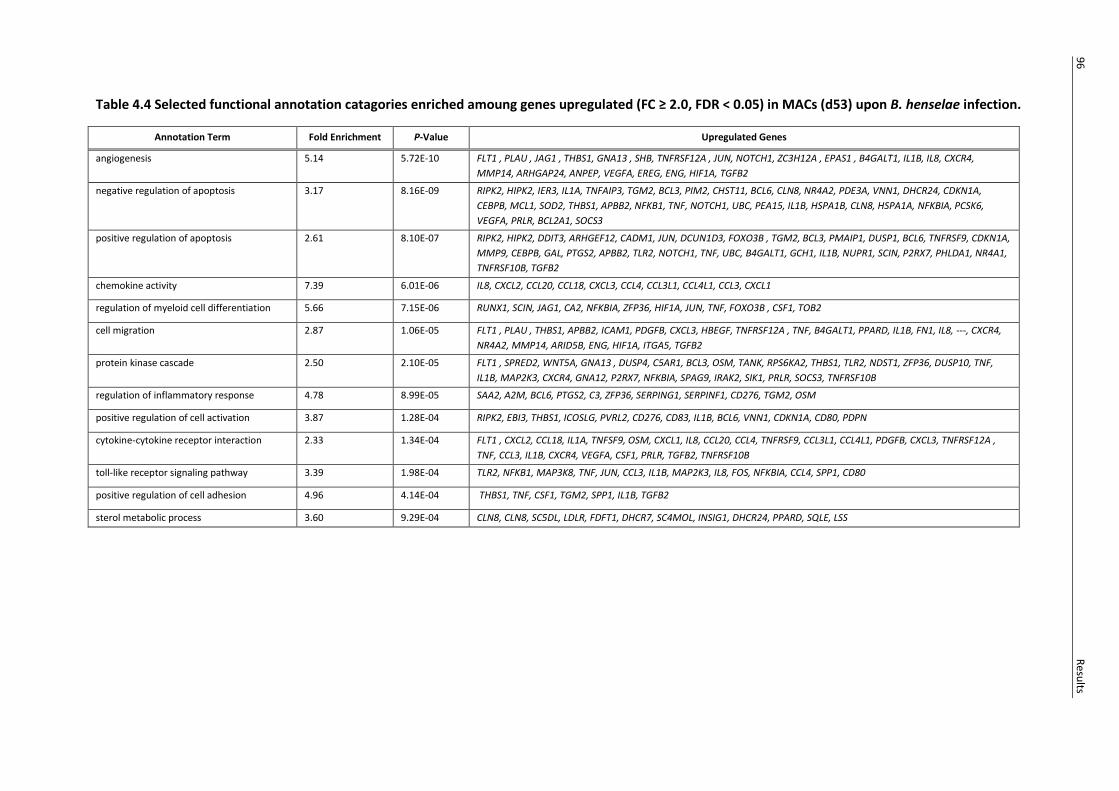
Table 4.4 Selected functional annotation catagories enriched amoung genes upregulated (FC ≥ 2.0, FDR < 0.05) in MACs (d53) upon B. henselae infection.
Annotation Term Fold Enrichment P-Value Upregulated Genes
angiogenesis 5.14 5.72E-10 FLT1 , PLAU , JAG1 , THBS1, GNA13 , SHB, TNFRSF12A , JUN, NOTCH1, ZC3H12A , EPAS1 , B4GALT1, IL1B, IL8, CXCR4,
MMP14, ARHGAP24, ANPEP, VEGFA, EREG, ENG, HIF1A, TGFB2
negative regulation of apoptosis 3.17 8.16E-09 RIPK2, HIPK2, IER3, IL1A, TNFAIP3, TGM2, BCL3, PIM2, CHST11, BCL6, CLN8, NR4A2, PDE3A, VNN1, DHCR24, CDKN1A,
CEBPB, MCL1, SOD2, THBS1, APBB2, NFKB1, TNF, NOTCH1, UBC, PEA15, IL1B, HSPA1B, CLN8, HSPA1A, NFKBIA, PCSK6,
VEGFA, PRLR, BCL2A1, SOCS3
positive regulation of apoptosis 2.61 8.10E-07 RIPK2, HIPK2, DDIT3, ARHGEF12, CADM1, JUN, DCUN1D3, FOXO3B , TGM2, BCL3, PMAIP1, DUSP1, BCL6, TNFRSF9, CDKN1A,
MMP9, CEBPB, GAL, PTGS2, APBB2, TLR2, NOTCH1, TNF, UBC, B4GALT1, GCH1, IL1B, NUPR1, SCIN, P2RX7, PHLDA1, NR4A1,
TNFRSF10B, TGFB2
chemokine activity 7.39 6.01E-06 IL8, CXCL2, CCL20, CCL18, CXCL3, CCL4, CCL3L1, CCL4L1, CCL3, CXCL1
regulation of myeloid cell differentiation 5.66 7.15E-06 RUNX1, SCIN, JAG1, CA2, NFKBIA, ZFP36, HIF1A, JUN, TNF, FOXO3B , CSF1, TOB2
cell migration 2.87 1.06E-05 FLT1 , PLAU , THBS1, APBB2, ICAM1, PDGFB, CXCL3, HBEGF, TNFRSF12A , TNF, B4GALT1, PPARD, IL1B, FN1, IL8, ---, CXCR4,
NR4A2, MMP14, ARID5B, ENG, HIF1A, ITGA5, TGFB2
protein kinase cascade 2.50 2.10E-05 FLT1 , SPRED2, WNT5A, GNA13 , DUSP4, C5AR1, BCL3, OSM, TANK, RPS6KA2, THBS1, TLR2, NDST1, ZFP36, DUSP10, TNF,
IL1B, MAP2K3, CXCR4, GNA12, P2RX7, NFKBIA, SPAG9, IRAK2, SIK1, PRLR, SOCS3, TNFRSF10B
regulation of inflammatory response 4.78 8.99E-05 SAA2, A2M, BCL6, PTGS2, C3, ZFP36, SERPING1, SERPINF1, CD276, TGM2, OSM
positive regulation of cell activation 3.87 1.28E-04 RIPK2, EBI3, THBS1, ICOSLG, PVRL2, CD276, CD83, IL1B, BCL6, VNN1, CDKN1A, CD80, PDPN
cytokine-cytokine receptor interaction 2.33 1.34E-04 FLT1 , CXCL2, CCL18, IL1A, TNFSF9, OSM, CXCL1, IL8, CCL20, CCL4, TNFRSF9, CCL3L1, CCL4L1, PDGFB, CXCL3, TNFRSF12A ,
TNF, CCL3, IL1B, CXCR4, VEGFA, CSF1, PRLR, TGFB2, TNFRSF10B
toll-like receptor signaling pathway 3.39 1.98E-04 TLR2, NFKB1, MAP3K8, TNF, JUN, CCL3, IL1B, MAP2K3, IL8, FOS, NFKBIA, CCL4, SPP1, CD80
positive regulation of cell adhesion 4.96 4.14E-04 THBS1, TNF, CSF1, TGM2, SPP1, IL1B, TGFB2
sterol metabolic process 3.60 9.29E-04 CLN8, CLN8, SC5DL, LDLR, FDFT1, DHCR7, SC4MOL, INSIG1, DHCR24, PPARD, SQLE, LSS
96
Resu
lts

Table 4.5 Selected functional annotation catagories enriched amoung genes downregulated (FC ≥ 2.0, FDR < 0.05) in MACs (d53) upon B. henselae infection.
Annotation Term Fold Enrichment P-Value Downregulated Genes
T cell activation 3.59 7.17E-05 FYN, CD2, FLT3, CD3E, GIMAP5, CD48, CD8A, GIMAP1, CD3D, LCK, RHOH, THEMIS, ZAP70, CD3G, IL7R
regulation of transcription 1.70 2.75E-12 IKZF3 , LBH, ZNF701, F2R, GABPA, STAT4 , ZNF382, ZNF551, ZNF507, GTF2E1, ZNF766, ZNF449, ZNF84, ZNF543, ZNF320,
MDM4, ZNF564, ZNF43, ZNF808, ATF7IP2, ZNF671, ZEB1 , ZNF542, CASP8AP2, NCOA5, ZNF107, ZBTB3, ZNF765, ZNF577,
ZNF304, SKAP1 , ZNF321, ZNF680, ZNF816A, PRKCQ, ZNF75A, ZNF619, ZNF101, 8180265, ZNF552, ZNF561, ZNF587, ZNF512,
TFCP2, EZH1, RBL1, PPP1R3E, VPS25, ZNF429, ZNF548, ZNF93, ZNF75A, MYBL1, SUV420H1, ZNF253, ZNF28, TBP, RORA,
ZNF525, ZNF776, MED6, TCF7, CHURC1, ZNF562, 8180200, STAT1, ZNF419, ZNF484, ZNF187, MSL3, ZNF426, ZNF595,
SETDB1, GATA3, ZNF383, ING3, TCEANC, LEF1, ZNF845, ZNF90, MSL3L2, ZNF626, TRERF1, ZNF624, ZNF260, ZNF506, NRAS,
ETS1, GTF2H2D, MED7, ZNF322A, ZNF302, MNDA, ZNF780A, ZNF772, FAM120B, ZNF66, ASF1A, ZNF518A, ZNF14, NME1,
THAP1, ZNF559, ZNF235, ZNF226, MAP3K1, MTERF, ZNF813, ZNF594, ZNF234, MYC, ZNF600, ZNF486, ZNF347, ZNF780B,
ZNF268, ZNF550, ZNF841, TAF7, JRKL, ZNF24, TIGD1, ZNF626, GTF2H2, ZNF721, ZFP14, ZNF518B, ZNF274, ZNF211, ZNF223,
ZNF416, ZNF585B, ZNF616, CGGBP1, ZFP62, ZNF266, DMTF1, ZNF717, ZNF256, ZNF146, TRIM22, ZNF181, TADA2A, KLF12,
ZNF154, RHOH, ZNF134, CDCA7L, ZNF583, ZNF763, ZNF83, ZFP3
regulation of RNA metabolic process 1.83 1.06E-10 IKZF3 , ZNF701, F2R, GABPA, STAT4 , ZNF382, ZNF551, ZNF766, ZNF449, ZNF543, ZNF84, ZNF320, MDM4, ZNF43, ZNF564,
ZNF808, ZNF671, ZNF542, ZEB1 , ZNF765, ZNF577, SKAP1 , ZNF304, ZNF321, ZNF680, ZNF816A, ZNF619, ZNF75A, ZNF101,
ZNF718, ZNF552, ZNF561, ZNF587, TFCP2, RBL1, PPP1R3E, ZNF429, ZNF548, ZNF93, MYBL1, ZNF75A, ZNF253, ZNF28, TBP,
RORA, ZNF776, MED6, TCF7, ZNF562, ZNF493, ZNF419, STAT1, ZNF484, ZNF187, ZNF426, ZNF595, GATA3, ZNF383, TCEANC,
LEF1, ZNF845, ZNF90, 8180217, TRERF1, ZNF624, ZNF506, ETS1, NRAS, 8057441, MED7, ZNF302, ZNF780A, ZNF772, ZNF66,
ASF1A, ZNF14, THAP1, ZNF559, NME1, ZNF226, ZNF235, MAP3K1, ZNF813, MYC, ZNF234, ZNF486, ZNF347, ZNF780B,
ZNF268, ZNF550, ZNF841, TAF7, ZNF24, ZNF626, ZFP14, ZNF274, ZNF223, ZNF211, ZNF416, ZNF585B, ZNF616, ZNF266,
DMTF1, ZNF717 , ZNF256, ZNF146, TRIM22, ZNF181, ZNF154, KLF12, ZNF134, ZNF583, ZNF763, ZNF83
T cell activation 3.59 7.17E-05 FYN, CD2, FLT3, CD3E, GIMAP5, CD48, CD8A, GIMAP1, CD3D, LCK, RHOH, THEMIS, ZAP70, CD3G, IL7R
positive regulation of immune response 3.12 3.21E-04 FYN, KLRC4, CD3E, GIMAP5, CR1, GIMAP1, CD247, TNFSF13B, C5, SH2D1A , THEMIS, SKAP1, ZAP70, CD79A, CD226
regulation of lymphocyte activation 3.05 3.96E-04 CD2, KLRK1, ZEB1 , FLT3, CD3E, GIMAP5, CORO1A, GIMAP1, LCK, TNFSF13B, CD5, ZAP70, PRKCQ, CD27, IL7R
immune response-activating signal
transduction
4.63 0.001531 FYN, KLRK1, CD3E, CD247, ZAP70, SKAP1 , THEMIS, CD79A
positive regulation of natural killer cell
mediated cytotoxicity
9.41 0.00157 KLRK1, GIMAP5, SH2D1A , GIMAP1, CD226
Resu
lts
97
98 Results
4.12 B. henselae infection of MACs induces a predominantly M2-alternativly activated macrophage phenotype
Depending on microenvironmental signals macrophages can take on activation profiles that range
from classicaly activated M1-inflammatory to alternatively activated M2-anti-inflammatory
phenotypes which have a strong influence on macrophage functional capacity and angiogenic
activity.
To determine the activation profile induced in MACs by B. henselae infection, microarray data was
used to examine absolute expression signals for typical M1 and M2 macrophage marker genes (Fig.
4.17). Typically, bacterial infections induce a highly polarized, highly inflammatory M1 macrophage
activation state (Benoit et al., 2008b). Contrary to this, the gene expression profiles of B. henselae
infected MACs displayed a predominantly M2 activation phenotype. B. henselae infected MACs
displayed particularly high expression of key M2 marker genes such as MSR1, MRC1, CD163 and
TGFB1. However, the M2 polarization of infected cells was not absolute. High expression of M1
inflammatory marker genes such as IL-1B, NFκB, TNF and CCL3 was also observed.
4.13 The secretome of B. henselae infected MACs is dominated by angiogenic-inflammatory cytokines and matrix remodeling compounds
Macrophages are important accessory cells in many biological processes, secreting a diverse array of
growth factors, cytokines and enzymes that transform local microenvironments and direct the
actions of proximate cells (Wynn et al., 2013). Results indicated that the pro-angiogenic effects of
B. henselae infected MACs are paracrine in manner. To determine what compounds secreted by
B. henselae infected MACs may be responsible for increased angiogenic activity, MAC-conditioned
medium was analyzed via proteome profiler arrays at different time points day 5-24h p.i., from
intermediate Matrigel cultures (day 25), and from fully formed Matrigel chord networks (d77; Fig.
4.18).
Infected MACs displayed a diverse cytokine secretion profile that was maintained over long term
culture. Due to donor variation and the heterogeneity that developed in cell number and medium
volume over long term culture, there were differences in the composition and intensity of the
cytokine secretion profiles among samples. However, a consistent “core” of cytokines was found to
be characteristic. In contrast, the cytokine signal from uninfected cells diminished over time and is
almost completely absent at d77 reflecting the fact that no viable cells remained at this time point.
The paracrine microenvironment created by B. henselae infected MACs was dominated by cytokines
with inflammatory and angiogenic properties including CXCL1, CXCL8, CXCL16, CCL2 and
macrophage migration inhibitory factor (MIF; Salcedo et al., 2000; Strieter et al., 2005; Bernhagen et
Results 99
al., 2007). Angiogenin, a direct angiogenic activator and enhancer of angiogenic signals from other
cytokines, appeared in the cytokine secretion profile late in culture (day 25; Kishimoto et al., 2005).
Immune modulatory cytokines like IL-6, GM-CSF were detected along with CCL3, CCL4, CCL5, the
anti-inflammatory cytokine interleukin-1 receptor antagonist (IL-1ra), soluble ICAM-1 and
thrombospondin-1 (TSP-1). Anti-angiogenic CXCL4 and CXCL10 were also secreted in the early stages
of infection (Strieter et al., 2005).
Over time, many of the early inflammatory cytokines disappeared from the secretion profile and
matrix remodeling elements became more prominent. B. henselae-infected MACs secreted MMP8,
MMP9, urokinase-type plasminogen activator (uPA) as well as their respective inhibitors, tissue
inhibitor of metalloproteinases-1 (TIMP-1) and plasminogen activator inhibitor -1 (PAI-1). The
absence of the key inflammatory cytokines IL-1β, TNFα, interferon (INF)γ which were not secreted to
detectable levels at any time point tested was also notable.
4.14 B. henselae induced cytokine secretion in MACs is distinct from general LPS mediated inflammation
To further characterize the activation profile of B. henselae infected MACs, the secretion of pro-
inflammatory cytokine TNFα, angiogenesis-regulating chemokine CXCL10 and the anti-inflammatory
cytokine IL-1ra by B. henselae-infected MACs was compared to MACs treated with 1µg/ml E. coli LPS.
B. henselae infections resulted in significantly diminished TNFα and strongly increased CXCL10 and
IL-1ra (Fig. 4.19) levels, demonstrating a B. henselae associated secretion profile distinct from a
typical LPS-triggered inflammatory response.

100 Results
Figure 4.17 B. henselae infected MACs exhibit a predominantly M2 macrophage activation phenotype. Gene expression profiles of B. henselae infected MACs (d53) were evaluated via gene microarray. Absolute gene expression signals for typical human M1-classically activated and M2-alternatively activated macrophage marker genes were compared. Gene expression signals are displayed as ln2 logarithmic values. Heat map colour denominations range from red (high expression) to green (low expression) for individual genes. Results are displayed for three independent biological replicates.

Results 101
Figure 4.18 B. henselae infected MACs create a pro-angiogenic paracrine microenvironment over long term culture, dominated by angiogenic-inflammatory cytokines and matrix remodeling compounds. Conditioned medium was isolated from B. henselae infected (B. h.; MOI 200) and uninfected (ø) MACs d5-24h p.i. and from Matrigel cultures (d25, d77). The secretome was analyzed via human cytokine and angiogenesis proteome profiler arrays. Data show representative results for selected cytokines from three independent experiments.
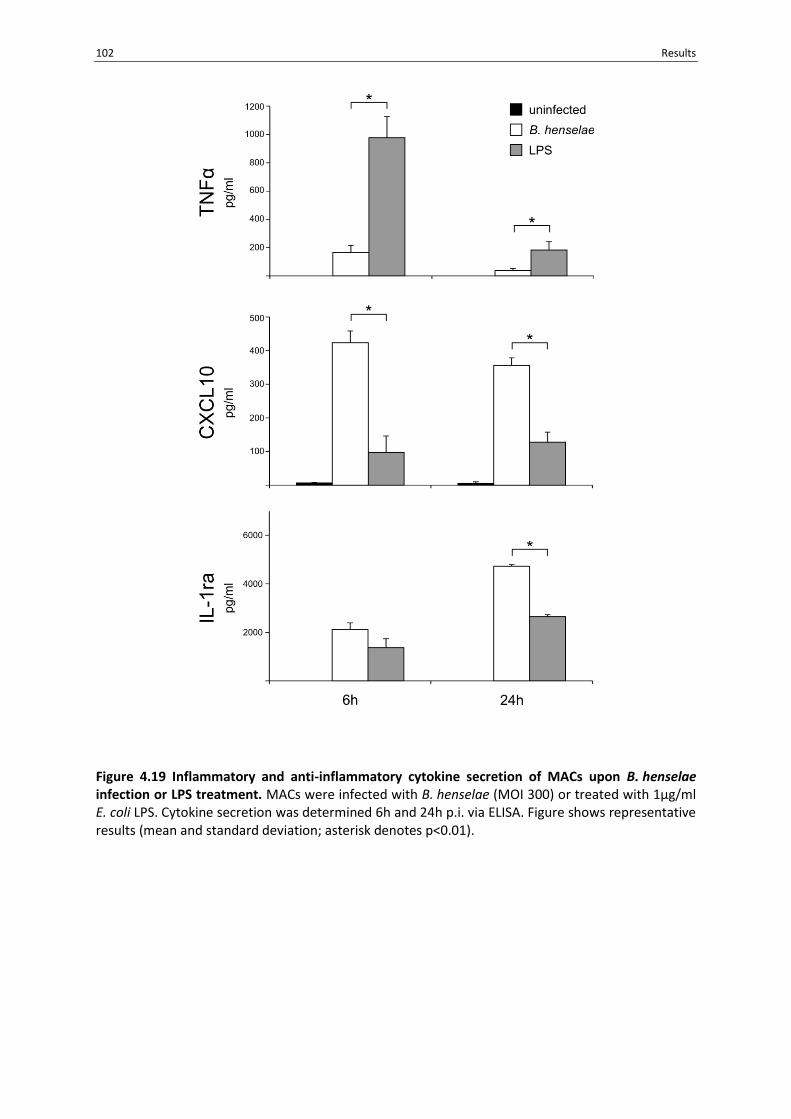
102 Results
Figure 4.19 Inflammatory and anti-inflammatory cytokine secretion of MACs upon B. henselae infection or LPS treatment. MACs were infected with B. henselae (MOI 300) or treated with 1µg/ml E. coli LPS. Cytokine secretion was determined 6h and 24h p.i. via ELISA. Figure shows representative results (mean and standard deviation; asterisk denotes p<0.01).

Discussion 103
5. Discussion
The ability to cause pathological angiogenesis in humans is a unique quality of the genus Bartonella.
Angiogenesis plays a central role in a range of pathological and regenerative settings and the
manipulation of this key biological process reflects a complex host pathogen interaction. In the case
of B. henselae, the vascular architecture and cellular composition of the infection induced vascular
lesions shows many similarities to malignant tumors suggesting that these two processes may share
similar mechanisms (Koehler and Tappero, 1993). Further elucidation of the factors involved in
B. henselae induced pathological angiogenesis and how these may relate to the formation of
cancerous tumors may provide further insights into other pathological angiogenic conditions, cancer
biology and bacterial pathogenicity.
Endothelial cells are suspected to be a key niche for chronic B. henselae infection and it has been
suggested that the development of vascular tumors may represent a pathologically induced
proliferation of the bacteria’s own cellular habitat (Kempf et al., 2002). Therefore, in the context of
B. henselae associated pathological angiogenesis most research has focused on the interaction of
B. henselae with endothelial cells directly.
In vivo angiogenesis however, is a multi-step process which can involve multiple cell types. In recent
years there has been a growing interest in the contribution of progenitor cells to angiogenic growth.
Myeloid angiogenic cells (MACs) are a heterogeneous group of circulating progenitor cells with key
roles in regenerative and pathological angiogenic processes (Fadini et al., 2012). MACs are known to
accumulate in areas of angiogenic growth such as the tumor microenvironment and promote
angiogenesis through the interaction with the endothelium and release of paracrine factors (Kalka et
al., 2000; La Puente et al., 2013). Due to their key role as promoters of pathological angiogenesis,
the interaction of B. henselae with MACs has the potential to significantly influence the development
of B. henselae associated vascular tumors.
To gain further insight into what role progenitor cells play in the development of B. henselae
associated pathological angiogenesis, this study aims to examine what effects the interaction of
MACs with B. henselae has on the MAC phenotype and role in angiogenic processes.
5.1 Initial response of MACs to B. henselae infection
So far, in vitro cell culture studies have identified several characteristic aspects of B. henselae
pathogenicity essential to the induction of pathological angiogenesis. Intracellular invasion,
inhibition of host cell apoptosis and activation of angiogenic response programs are representative

104 Discussion
features of B. henselae pathogenicity which lead to angiogenic re-programming of infected
endothelial cells. To determine whether B. henselae infection of MACs results in similar cellular re-
programming, the initial response of MACs to B. henselae infection was examined in detail.
Results from this study confirmed and expanded on preliminary findings of Mӓndle (2005) which
establish a basic framework for the interaction of B. henselae with MACs. Initial investigations into
the interaction between MACs and B. henselae by Mӓndle (2005) revealed that MACs are readily
susceptible to B. henselae infection and that bacteria persist within cells for several days. In this
study, scanning and transmission electron microscopy confirmed the adhesion of B. henselae and
their invasion of MACs after 30min and 60min respectively. Transmission electron microscopy
identified bacteria localized to intracellular vacuoles (Fig. 4.1).
B. henselae has been shown to inhibit apoptosis in several cell types including endothelial cells and
MM6 monocytes/macrophages (Kempf et al., 2005b; Schmid et al., 2006). Mӓndle (2005) also found
that B. henselae infection resulted in decreased caspase 3-like activity and inhibition of the apoptotic
response in infected MACs. Results generated in the present study confirm the ability of B. henselae
to prevent apoptosis in infected MACs. Infection of MACs with other pathogenic bacteria; E. coli and
S. aureus, was shown to induce massive cell death within 24h as compared to the high viability of
B. henselae infected cells (Fig. 4.2).
In examining the initial host-cell response of MACs to B. henselae Mӓndle (2005) found that infection
with B. henselae resulted in the activation of angiogenic and inflammatory cellular activation
programs similar to those associated with B. henselae infection of endothelial and epithelial cells.
Activation of the key transcription factors regulating angiogenic and inflammatory responses; HIF-1
and NFκB were detected as well as the up regulation and release of HIF-1 regulated angiogenic
cytokines VEGF, ADM and NFκB regulated cytokines CXCL8, CCL2 and ICAM-1.
In this study the activation of HIF-1 in B. henselae infected MACs was confirmed via Western Blot.
HIF-1 activation is known to be a general phenomenon in infections (Werth et al., 2010).
Accordingly, infection of MACs with other pathogenic bacterial species E. coli and S. aureus was also
found to result in HIF-1 activation (Fig. 4.3A). In contrast to B. henselae, these infections resulted in
massive cell death (see above). Furthermore, whereas, the HIF-1 activation associated with E. coli
and S. aureus may be associated with a general inflammatory response, B. henselae infection of
MACs was found to be dependent on the expression of BadA (Fig. 4.3B).
Taken together, these results demonstrate that, as in endothelial cells, B. henselae infection of MACs
results in an increase in angiogenic potential. Through the inhibition of apoptosis and activation of

Discussion 105
basic angiogenic response programs, B. henselae infection establishes essential conditions for the
induction of pathological angiogenic growth.
5.2 Functional effects of B. henselae infection on MAC pro-angiogenic
activity
5.2.1 Spheroid assay of sprouting angiogenesis
As an intermediary between monolayer cell culture and in vivo assays, the endothelial spheroid
assay of sprouting angiogenesis provides a reproducible and easily manipulated culture environment
that more closely mimics the complexity of tissue in vivo (Pampaloni et al., 2007). In the absence of
suitable animal models, the spheroid assay is a reasonable setting to examine the effects of
B. henselae infection on multiple molecular events and cellular interactions that make up in vivo
angiogenic process.
In the spheroid assay cells form spherical aggregates and under angiogenic conditions develop radial
sprouts which mimic the process of tip cell activation, tip-versus-stalk cell selection, matrix
degradation, and chemotactic sprout extension. (Korff and Augustin, 1999). Importantly, the 3D
spheroid culture environment provides realistic cell-cell and cell-matrix interactions which have
strong effects on cellular phenotype and functional capacity (Fennema et al., 2013).
The spheroid assay has previously been used to model B. henselae induced pathological
angiogenesis in endothelial cells. Scheidegger et. al. (2009) demonstrated that infection of HUVECs
with B. henselae prior to spheroid formation increased the rate of angiogenic sprouting significantly.
Activities of the VirB/VirD T4SS translocated effector proteins were found to modulate the rate of
infection associated angiogenic sprouting. BepA, which is strongly associated with B. henselae
induced inhibition of apoptosis, was shown to increase angiogenic sprouting whereas BepG, which is
associated with cytoskeletal rearrangement and invasome formation, was shown to decrease the
rate of vascular sprouting.
To investigate how B. henselae infection may influence the interaction of MACs with the vascular
endothelium, a co-culture spheroid model of HUVECs and MACs was developed for the first time.
The pro-angiogenic activities of MAC in vivo are dependent on their ability to home to sites of
angiogenic activity and interact with the vascular endothelium. MACs are recruited to the site of
neo-vascularization or repair in a multistep process involving maturation and release form the bone
marrow, chemoattraction via cytokines (such as SDF-1 and CCL2), adhesion to the vascular wall (via

106 Discussion
interaction with cellular adhesion molecules) and incorporation into the growing vessel or
perivascular space (Urbich et al.; Shen et al., 2011).
The ability of MACs to home to and integrate at the site of angiogenic growth would also be an
essential prerequisite for their participation in the process of B. henselae induced pathological
angiogenesis in vivo. To investigate the ability of B. henselae infected MACs to associate with
growing endothelium, MACs and HUVECs were stained red and green respectively and their
interaction observed in a co-culture spheroid assay.
When MACs and HUVECs were combined at a ratio of 1:3, MACs incorporated successfully into the
endothelial spheroid. Both infected and uninfected MACs were found to associate with growing
vascular sprouts. MACs were found localized to both, tip and stalk areas of sprouts, indicating that
B. henselae infection does not affect their ability to integrate into and interact with the growing
endothelium.
Some indications exist that B. henselae infection may in fact increase the affinity of host-cells to the
endothelium. In endothelial and epithelial cells, B. henselae infection has been shown to increase
the expression of cellular adhesion molecules E-selectin, ICAM-1 and myeloid chemokine CCL2
(Fuhrmann et al., 2001; McCord et al., 2005). Furthermore, Mӓndle (Mändle, 2005) demonstrated
that the infection of MACs with B. henselae also increases their chemoatracttant migration along an
SDF-1 cytokine gradient.
The retention of infected MACs to sites of angiogenic growth and their intimate interaction with the
active endothelium is also important in facilitating the transfer of pro-angiogenic effects to adjacent
endothelial cells and the surrounding microenvironment.
Initial experiments indicated that infection of MACs with B. henselae resulted in activation of
endogenous angiogenic response programs, increasing their angiogenic potential. To determine
whether B. henselae infection also resulted in increased functional angiogenic effects, the rate of
angiogenic sprouting was compared between co-culture spheroids composed of HUVECs mixed with
infected or uninfected MACs. Indeed, in spheroids seeded with B. henselae infected MACs, the rate
of endothelial sprouting increased significantly. Infected MACs induced an average of up to 1.61 fold
more angiogenic sprouting in endothelial spheroids than uninfected cells indicating that the
infection of MACs with B. henselae increases their pro-angiogenic activity and has significant
functional effects on angiogenic growth of adjacent endothelium (Fig. 4.5A).
In previous studies the infection of THP-1 macrophages, HeLas and EA hy. 926 cells with B. henselae
was found to induce the secretion of vasculoproliferative cytokines such as VEGF and CXCL8 and

Discussion 107
conditioned medium from B. henselae infected THP-1 macrophages increased endothelial cell
proliferation in vitro (Kempf et al., 2001; Resto-Ruiz et al., 2002; McCord et al., 2006). This has lead
to the hypothesis that the infection of accessory cells such as myeloid and epithelial cells with
B. henselae in vivo may contribute to pathological angiogenesis via the creation of a pro-angiogenic
paracrine loop in which the release of pro-angiogenic cytokines stimulates proliferation of adjacent
endothelium, attracts further myeloid cells and activates resident epithelial cells thus promoteing
the progression of the B. henselae induced vascular tumor (Kempf et al., 2001; Kempf et al., 2002;
Resto-Ruiz et al., 2002).
A similar phenomenon is observed in malignant tumors where infiltrating myeloid cells (most
prominently TAMs) are induced to secrete pro-angiogenic and immune regulatory cytokines,
creating a pro-angiogenic tumor-microenvironment which stimulates the adjacent endothelium and
contributes to tumor vascularization and invasion (Schmid and Varner, 2010).
To determine whether, the increased pro-angiogenic activity in B. henselae infected MACs was due
to paracrine effects or physical interaction, further spheroid experiments were performed, placing
infected MACs at varying degrees of separation from the endothelial spheroids. Spheroids were
created purely from HUVECs and suspended in collagen gel. B. henselae infected MACs were either
mixed with (Fig. 4.5B) or seeded onto the surface of collagen gel (Fig. 4.5C). In both conditions, the
rate of sprouting angiogenesis increased dose-dependently when MACs were infected with
B. henselae, indicating that no physical contact was necessary to transmit the pro-angiogenic effects
of infected MACs.
Finally, conditioned medium from B. henselae infected MACs was used to culture HUVEC spheroids
(Fig 4.5D). In this case, conditioned medium from B. henselae infected MACs showed a dose
dependent effect and induced up to 6.71 fold more average sprouting than conditioned medium
from uninfected MACs. These experiments indicate that paracrine effects are predominantly
responsible for the pro-angiogenic activity of B. henselae infected MACs.
Overall, these results demonstrate that the infection of MACs with B. henselae not only results in the
activation of angiogenic programs on a molecular level but increases their functional pro-angiogenic
activity. Infection of cells did not inhibit their ability to home to and incorporate in growing vessels
and had significant angiogenic effects on the surrounding endothelium.
In addition to demonstrating functional effects of B. henselae infection, results of the spheroid assay
provides important clues about how B. henselae infection may affect the behavior of MACs in vivo.
The co-culture spheroid assay reproduces the interaction of MACs with a 3D sprouting endothelium

108 Discussion
in vito and provides a realistic representation of the role of MACs as accessory cells in the process of
angiogenic growth.
When exposed to angiogenic endothelium, B. henselae infected MACs incorporate into growing
vascular sprouts and significantly increase angiogenic growth of the adjacent endothelium via
predominantly paracrine factors. In the context of B. henselae associated vascular tumor formation,
the interaction of infected MACs with the activated endothelium and parallel contribution to
angiogenic growth through the release of paracrine factors would make a significant contribution to
pathological angiogenesis and vascular tumor progression.
5.2.2 Matrigel capillary formation assay
To further elucidate the effects of B. henselae infection on MAC pro-angiogenic activity a second in
vitro assay of angiogenic growth, the Matrigel capillary formation assay, was employed.
In the Matrigel assay, cells are seeded onto a 3D proteinacious matrix composed of mostly laminin
and collagen IV. Mature endothelial cells such as HUVECs adhere to the matrix and over the course
of 2-4h arrange into evenly spaced cell cluster connected by a web of “tubules” consisting of
elongated endothelial cell branches 2-3 cells in length (Khoo et al., 2011).
In previous studies the infection of cultured endothelial cells with B. henselae has been shown to
increases capillary structure formation in the Matrigel assay (McCord et al., 2006; Berrich et al.,
2011).
In contrast to endothelial cells, MACs are not known to form spontaneous “capillary networks” in
the Matrigel assay (Hur et al., 2004; Yoder, 2013a). Accordingly, when uninfected MACs were seeded
in Matrigel culture they displayed no morphological differentiation, maintained spherical
phenotypes, died and degraded over time (Fig. 4.6). B. henselae infected MACs, on the other hand,
adhered strongly to the Matrigel matrix, developed long branching philopodia and gradually
assembled into a complex network of chord structures that at times extend over the surface of the
entire well plate (Fig. 4.6 and 4.7).
Although they display a similar basic composition and morphology to endothelial capillary networks,
the chord networks developed by infected MACs in Matrigel culture differ from endothelial
structures in two major ways.
In contrast to endothelial capillary networks, which consist of a dense web of thin chord-like
connections 2-3 cells long, the chords networks formed by the B. henselae infected MACs were
thicker, usually consisting of many directionally elongated and aligned cells. The intersecting chords

Discussion 109
of heterogeneous length formed a sparser network and left larger interim areas mostly void of other
cells.
In addition to differences in morphology, the capillary structures formed by B. henselae infected
MACs differed from endothelial cells in the rate of structure assembly. While endothelial cell
networks form in Matrigel cultures within 2-12h and degrade again shortly after, the capillary-like
networks formed by the B. henselae infected MACs assembled gradually over the course of several
weeks and were viable over long periods of time (41-77days). It is unclear why B. henselae infected
MACs require such lengthy incubation times to fully develop the capillary-like network structures,
however, it is likely that chord formation by B. henselae infected MACs is a more complex process
that requires different cellular events. B. henselae infected MACs may first have to undergo some
differentiation process, cellular activation or angiogenic re-programming before functional effects
such as complex structure formation become apparent. The extended development time and
long-term stability of the capillary structures formed by B. henselae infected MACs also highlights
the anti-apoptotic effect of B. henselae infection. Uninfected MACs generally died out and degraded
in culture after approximately 25 days (a typical lifespan of untreated primary human myeloid cells
in culture) wheras B. henselae infected cells in the capillary-like structures were shown to be still
viable at the end point of Matrigel cultures (d51) (Fig. 4.6 and Fig. 4.8).
Despite the difference in capillary-like structure formation between B. henselae infected MACs and
endothelial cells, the ability of infected MACs to self-organize into such complex structures requires
coordination of complex inter- and intracellular signaling networks, cellular migration, changes in
morphology, and possibly remodeling of the extracellular matrix as well as the ability to resist
apoptosis over long culture periods. This activity further underscores previous results indicating a
significant angiogenic re-programming in B. henselae MACs leading to increased functional pro-
angiogenic activity in infected cells.
5.3 Differentiation phenotype of B. henselae infected MACs
MACs, previously known as endothelial progenitor cells (EPCs), were originally identified as
circulating progenitors that participated in new vascular growth though differentiation into mature
endothelial cells and formation of new vasculature (Asahara, 1997; Shi et al., 1998). Many studies
report endothelial phenotypic characteristics in MACs cultured in vitro and several in vivo studies
describe the vascular incorporation and endothelial differentiation of MACs as the predominant
mechanism of their pro-angiogenic effects (Lyden et al., 2001; Aicher et al., 2003).

110 Discussion
This paradigm is conflicted by a growing body of evidence which indicates that circulating bone
marrow derived angiogenic progenitor cells are of myeloid origin and contribute to angiogenic
growth as accessory cells, either through paracrine effects, or by providing other structural and
physiological support to growing vessels (Rehman et al., 2003; Kinnaird et al., 2004; O'Neill et al.,
2005; Medina et al., 2010a).
Several of the pro-angiogenic qualities observed in B. henselae infected MACs could suggest that
angiogenic re-programming induced by B. henselae infection may culminate in endothelial
differentiation. Infection resulted in activation of angiogenic molecular programs including activation
of HIF-1 and secretion of the pro-angiogenic cytokine VEGF. Infected cells incorporate into growing
vascular sprouts and increase the rate of sprouting angiogenesis. Most strikingly, infected MACs
were found to assemble into capillary-like chord networks similar to those formed by endothelial
cells when cultured in a Matrigel capillary assay.
To investigate the differentiation phenotype of B. henselae infected MACs, several tests for
endothelial gene expression and antigen expression were performed.
Results revealed that B. henselae does not induce upregulation of endothelial marker genes in
infected MACs. (Fig. 4.12). Gene expression profiles from both infected and uninfected MACs
showed little similarity to endothelial control samples (Fig. 4.14) and immunohistochemistry
preformed on MACs derived from capillary-like structures proved negative for typical endothelial
marker proteins (Fig. 4.11).
Instead, comparison of gene expression profiles from B. henselae infected MACs with samples of
known cell differentiation revealed the most similarity with macrophages and other cells of the
myeloid linage ( Table 4.1).
Finally, a FACS analysis for various differentiation markers was performed on infected and
uninfected MACs at different time points (d5, d62). Neither infected nor uninfected MACs displayed
endothelial characteristics at any time-point tested. In contrast, markers for myeloid cells were
displayed on both infected and uninfected cells. In particular, macrophage-specific differentiation
markers were strongly expressed on all MACs and displayed upregulation after infection with
B. henselae. This macrophage phenotype was maintained over long term culture and in the cells of
the capillary-like structures (Fig. 4.10).
These results indicate that endothelial differentiation does not play a role in the development of the
pro-angiogenic phenotype in infected MACs and confirms findings by other investigators
demonstrating that MACs represent cells of a myeloid phenotype. Specifically, it can be concluded

Discussion 111
that the pro-angiogenic cell phenotype induced in MACs by B. henselae infection and present in the
capillary-like structures, is of macrophage differentiation.
Although macrophages cannot themselves form true vasculature, this cell type is well equipped to
have a significant impact on the process of pathological angiogenesis. As accessory cells,
macrophages are attracted by hypoxia, angiogenic and inflammatory signals to the site of
angiogenesis and participate in a paracrine manner through the release of pro-angiogenic cytokines
and growth factors into the cellular microenvironment (Chambers et al., 2013). In addition to their
paracrine effects macrophages are also known to participate structurally in the angiogenic process
for example through matrix degradation and remodeling, aiding the process of tip cell fusion,
capillary pruning in vascular patterning and facilitating vascular repair by adhering to the vessel wall
at sites of vascular injury (Moldovan et al., 2000; Fujiyama et al., 2003; Fantin et al., 2010; Stefater et
al., 2013).
5.4 Vascular mimicry
Among the endothelial-like behaviors of B. henselae infected MACs is the ability to form capillary-
like chord networks over long term culture. Investigation into the differentiation phenotype
expressed by cells of the capillary-like structures (discussed above) clearly demonstrates them to be
of macrophage differentiation. This indicates that the phenomenon of capillary structure formation
observed in B. henselae infected MACs reflects a vascular mimicry behavior rather than true capillary
formation activity.
The formation of cellular networks and chord structures in Matrigel culture is not exclusive to
endothelial cells. Fibroblasts and several cancer cell lines (breast cancer, prostate cancer and
glioblastoma) have been described to form web-like networks when cultured on Matrigel (Donovan
et al., 2001).
Macrophages are known to associate closely with endothelial structures in vivo and play diverse
roles in angiogenic processes (Baer et al., 2013). Several examples of vascular mimicry behavior have
been described in macrophages in vivo in conjunction with situations of intense angiogenic stimulus.
In some cases macrophages have been reported to participate in the formation of a provisory
vascular barrier after vascular injury. Glod et al. (2006) found that circulating populations of CD14+
monocytes associated with repairing vasculatureafter injury of the skin and brain, forming a vascular
barrier and participating in vessel repair. CD14+ peripheral blood monocytes were also found by

112 Discussion
Fujiyama et al. (2003) to adhere to injured endothelium in a mouse carotid balloon injury model and
inhibit the development of neointimal hyperplasia.
In several in vitro and in vivo studies a macrophage “tunneling” behavior has been described in
which stimulated macrophages create tubes or chords via directed proteolytic degradation of the
extracellular matrix (Anghelina et al., 2002; Anghelina et al., 2004; Anghelina et al., 2006b; Anghelina
et al., 2006a). In an in vivo transgenic model of ischemic cardiomyopathy (Moldovan et al., 2000)
macrophages were observed to proteolytically degrade metalloproteinase-lined tunnels throughout
the myocardial tissue. Macrophages are known to play a crucial role in the matrix degradation and
remodeling that takes place during vessel sprouting (Pepper, 2001; Arroyo and Iruela-Arispe, 2010;
Nucera et al., 2011). Such macrophage “tunneling” behavior is believed to reflect this directed
matrix remodeling processes which facilitate the invasion and extension of vascular sprouts into the
surrounding tissue.
Vascular mimicry behavior is also a common feature of aggressive tumor phenotypes and has been
observed in many cancers in vivo. Tumor cells assemble into vascular structures that are in some
cases perfused and may even establish connections to endothelial vessels. This additional
vascularization of tumor tissue contributes to cancer growth, progression and can even compensate
for normal angiogenic growth in response to anti-angiogenic chemotherapy (Döme et al., 2007). In
cancerous tumors the development of vascular mimicry appears to be connected to the de-
differentiation process that occurs in tumor cells when genetic deterioration leads to the breakdown
of regulatory checkpoints and the aberrant activation of embryonic signaling pathways.
Dysregulation of angiogenic pathways ,in particular the activation of VE-Cadherin, VEGFR-1, COX-2,
metalloproteinases and hypoxic signaling via HIF transcription factors have also been shown to be
important activating factors (Paulis et al., 2010).
Recently, macrophages in some aggressive tumors have also been reported to participate in vascular
mimicry. Macrophages from patients with aggressive multiple myeloma were found to participate in
mosaic vessel formation within the bone marrow in vivo. Macrophages isolated from these patients
form vascular mimicry structures and develop endothelial characteristics when stimulated with
angiogenic growth factors in vitro whereas macrophages from patients with less aggressive
melanoma and from control patients did not exhibit these qualities (Scavelli et al., 2007). In
anaplastic thyroid carcinoma, tumor associated macrophages have also been found to form a
branching network radiating from endothelial vessels and may play a role in transporting nutrients
to these poorly vascularized tumors (Caillou et al., 2011).

Discussion 113
It is unclear how exactly the B. henselae induced vascular mimicry phenotype in the Matrigel assay
might translate to cellular behavior in vivo.
The capillary-like structure formation may reflect one of several structure building behaviors
observed in pro-angiogenic macrophages, all of which would have functional angiogenic effects on
the growth of B. henselae induced vascular tumors.
Vascular mimicry behaviors in malignant tumors can dramatically affect tumor growth and
progression (Liang, 2013). The ability of B. henselae infected MACs to participate in the formation of
such functional vascular structures would have meaningful implications for B. henselae induced
pathological angiogenesis. Similarly, the coordinated “tunneling” degradation of extracellular matrix
or the lining of growing vessels to provide physical support would also be advantageous for the
invasion of new vessels and progression of the B. henselae associated vascular tumors in vivo.
In general, the vascular mimicry phenotype displayed by B. henselae infected MACs indicates a
considerable phenotypic re-programming and angiogenic activity in infected cells. Cells demonstrate
dramatic morphological and structural changes. The formation of complex cellular networks in the
extracellular matrix also indicates an activated cell phenotype, as well as sophisticated proteolytic
and cellular communication systems which would be significant to the role of MACs as accessory
cells in the induction of pathological angiogenesis.
5.5 Phenotypic re-programming in B. henselae infected MACs
The myeloid cell linage represents one of the most functionally and phenotypically diverse cell types
within the body. Myeloid cell phenotypic plasticity and ability to react rapidly to local
microenvironmental conditions make them influential players in numerous biological processes
(Pollard, 2009; Wynn et al., 2013).
In this study it has been demonstrated that B. henselae infection leads to significant changes in MAC
functional angiogenic capacity including the activation of angiogenic response programs, the ability
to induce significantly increased angiogenic growth and the formation of vascular mimicry
structures.
In order to examine further the phenotypic changes underlying these functional angiogenic effects,
microarray transcriptional profiling was performed to compare the gene expression profiles of
uninfected MACs (d3) and infected MACs (d53).

114 Discussion
Infection with B. henselae was found to result in the regulation of 1400 genes in excess of 2.0 fold.
This represents 36% of gene transcripts evaluated with 620 genes being upregulated and 780 down
regulated in response to infection. Hierarchical clustering of evaluated samples revealed a high level
of variance between sample groups (Fig. 4.13).The variance in gene expression induced in MACs in
response to B. henselae infection was found to be considerably higher than changes induced in the
course of a standard monocyte-to-macrophage differentiation process (Table 4.2). These results
describe extensive changes in overall gene expression associated with B. henselae infection and
make evident that the functional angiogenic effects observed in infected MACs are accompanied by
a broad-scale phenotypic re-programming in infected cells.
Further analysis of individual genes via functional annotation and statistical analysis (DAVID analysis)
revealed that changes in gene expression induced by B. henselae infection are associated with a
broad range of cellular processes (Table 4.3-4.5, Fig. 15, S Table 9.1-9.4). B. henselae infection of
MACs was found to alter gene expression patterns in four major areas of cellular function:
angiogenesis, structural organization, apoptosis, sterol-metabolism and immune response.
5.5.1 Angiogenesis
Corresponding to the increase in angiogenic activity in B. henselae infected MACs, functional
annotations relating directly to angiogenesis and vascular growth were found to be the most highly
enriched among upregulated gene transcripts.
The gene HIF1A (encoding the HIF-1α protein subunit) was found to be highly upregulated in
infected cells confirming that this angiogenic transcription factor is upregulated on both
transcriptional and post-transcriptional levels in response to B. henselae infection.
Genes encoding angiogenic chemokines (CCL2, CXCL8, CXCL1, CXCL2, CC3L1/CCL3, CSF) and
angiogenic growth factors (VEGFA, TGFB) were also particularly highly upregulated. Enriched
functional annotations were also found in categories related to angiogenic growth including inter-
and intracellular signaling (cytokine production, receptor signaling, intracellular signaling cascades,
MAPK pathways and transcriptional regulation).
5.5.2 Structural organization
Highly enriched functional categories also included annotations involved in structural organization
such as those for cell migration and adhesion. Many of the highly upregulated genes also had
functions related to interaction with the extracellular matrix such as matrix degrading
metalloproteinases (MMP9, MMP2, MMP7, MMP12, ADAMDEC1, MME), and other compounds

Discussion 115
involved in matrix remodeling (SERPINE1, CHI3L1, CHIT1, CTSK, HTRA1, TNFAIP6). Furthermore,
genes for several extracellular matrix components such as FN1, SPARC, FBLN5, SPP1, THBS1 as well
as several integrins (ITGA1, ITGB5, ITGB3), were highly upregulated.
The upregulation of genetic programs involved in structural organization, cellular adhesion and
matrix interaction may be important to structurally coordinate the formation of capillary-like
networks and the interaction with vascular sprouts observed in Matrigel capillary formation and
spheroid assays respectively.
These genetic activation patterns also indicate a pro-angiogenic re-programming in infected MACs as
matrix remodeling represents an important function of macrophages in regulating angiogenic
growth. In vivo, macrophage-mediated directional degradation of the extracellular matrix enables
vascular sprouting into surrounding tissue. Deposition of matrix scaffolds supports vascular
expansion and proteolytic processing of extracellular matrix proteins activates matrix bound pro-
angiogenic growth factors (Pepper et al., 1996; Arroyo and Iruela-Arispe, 2010).
5.5.3 Apoptosis
Following angiogenesis, functional annotations for negative, and to a lesser extent, positive
regulation of apoptosis were most highly enriched among upregulated genes.
This reflects results generated in this study and by Mӓndle (2005) which demonstrated that
B. henselae infection inhibits the apoptotic response in infected MACs (Fig. 4.2).
Although the mechanism by which B. henselae inhibits apoptosis is not yet clear, other authors have
found this effect to be associated with upregulation of anti-apoptotic proteins, BCL2, BAX, BRIC-2
and -3 (cIAP-1,-2) as well as inhibition of pro-caspase preprocessing and -activity of the apoptotic
executioner caspase-3 (Kempf et al., 2005b; McCord et al., 2006). Results of the transcriptome
analysis did not show a significant increase in mRNA transcription for these anti-apoptotic proteins.
However, BCL protein family members BCL2A1, BCL3 and BCL6, also known to be important anti-
apoptotic proteins, were highly upregulated. Furthermore, genes encoding caspase-3 and caspase 6
proteins (CASP3, CASP6) were downregulated indicating that caspase activity may be downregulated
both transcriptionally and post-transcriptionally is response to B. henselae infection.
Given the anti-apoptotic nature of B. henselae infection, the overrepresentation of functional
annotations for both negative and positive regulation of apoptosis to high statistical significance
appears contradictory. However, this expression pattern may also reflect a state of dynamic
equilibrium. Several apoptosis regulation pathways operate on a system of dynamic equilibrium with

116 Discussion
the ratio of pro- versus. anti-apoptotic factors determining survival outcomes. The significance of
over representation for “negative regulation of apoptosis” was considerably higher (P= 8.16 x 10⁻⁹)
than for “positive regulation of apoptosis” (P= 8.10x10⁻⁷). Thus, although both pro- and anti-
apoptotic factors are highly expressed, the activities of B. henselae and its pathogenicity factors may
work to tip the balance of apoptotic factors toward anti-apoptosis, overriding the apoptotic
response program and preventing cell death.
B. henselae inhibition of apoptosis represents a fundamental component of B. henselae
pathogenicity and is an essential component of infection associated pathological angiogenesis (Kirby
2002; Schmid et al. 2006). Inhibiting cell death in B. henselae infected cells not only preserves the
intracellular habitat but is also necessary to prolong the interaction of B. henselae with host-cells
and allows for induced changes in phenotype to be translated into functional effects. The survival of
B. henselae infected MACs also enables the development of long-term pathogenic consequences
such as the creation of sustained paracrine microenvironments, the development of complex
vascular mimicry structures and facilitates the overall chronic infection strategy which is
characteristic of Bartonella pathogenicity.
5.5.4 Sterol metabolism
Along with functional annotations relating to cellular processes known to be associated with
B. henselae infection such as angiogenesis, apoptosis inhibition and cytokine production, functional
annotations for sterol metabolism were found to be highly enriched among upregulated gene
transcripts.
11 genes with the functional annotations for “sterol metabolic process” were upregulated greater
than or equal to 2.0 fold and overrepresentation in this category was attributed a high statistical
significance (P = 9.29 x 10⁻⁴). Several genes for low density lipoprotein (LDL) uptake, ORL1, LPL and
LDLR, were very highly upregulated. Other genes for cholesterol biosynthetic processes such as
DHCR24, INSIG, SQLE, and DHCR7 were also highly upregulated along with two members of the
eicosanoid pathway PTGS2 and ALOX5AP.
No connection between B. henselae infection and sterol-metabolism has been described to date. It is
possible that increased cholesterol-metabolism in B. henselae infected MACs represents a non-
specific metabolic host-cell response to chronic infection. However, in several intracellular bacteria,
host-cell sterol-metabolism has been shown to be essential for intracellular colonization and overall
pathogenicity. Manipulation of sterol-metabolic pathways can be utilized by pathogenic bacteria for
invasion, as an intracellular energy source or as a means of immune suppression.

Discussion 117
Cholesterol is obtained by eukaryotic cells either through import of LDL or through endogenous
cholesterol production in the endoplasmic reticulum (Ikonen, 2008). Cholesterol makes up a
significant proportion of the eukaryotic plasma membrane lending structural stability and enriching
lipid rafts (Simons and Sampaio, 2011). Cholesterol is also involved in regulating endosomal
trafficking and serves as a precursor for steroids and vitamins (Ikonen, 2008).
In some cases the importance of host-cell cholesterol is limited to its role as a component of
membrane lipid rafts. In several species, the organization of docking partners for bacterial secretion
systems and outer membrane pathogenicity factors in lipid rafts has proved necessary for effective
invasion and translocation of bacterial toxins and effector proteins (Orlandi and Fishman, 1998;
Seveau et al., 2004; Hayward et al., 2005; Lai et al., 2008).
There are also several examples of intracellular bacterial pathogens that actively hijack host-cell
cholesterol-metabolism as part of their intracellular pathogenicity strategy. B. henselae infected
MACs displayed an upregulation of several members of the LDL uptake pathway. This pathway in
particular appears to be co-opted as part of the intracellular bacterial lifestyle.
Both Chlamydia trachomatis and Anaplasma phagocytophilum, obtain cholesterol from host-cell
pathways to compensate for their inability to produce it endogenously. A. phagocytophilum
upregulates host-cell LDL receptor expression leading to increased LDL uptake. LDL trafficking to the
ER is intercepted and the transport of endocytic vesicles is redirected to the Anaplasma intracellular
inclusion (Xiong and Rikihisa, 2012). Chlamydia trachomatis similarly redirects endocytic transport of
cholesterol and sphingomyelin obtained from either LDL uptake or endogenous synthesis to the
Chlamydia inclusion (Carabeo et al., 2003; Moore et al., 2008).
Within atherosclerotic lesions infiltrating macrophages engulf large amounts of LDL and oxidized LDL
(oLDL) which induces a “foam-cell” phenotype (Shashkin et al., 2005). Mycobacterium tuberculosis
and Chlamydia pneumoniae infection has been shown to induce such foam-cell macrophage
phenotypes independent of lipoprotein engulfment. As observed in B. henselae infected MACs, both
M. tuberculosis and C. pneumoniae, induce the expression of host-cell cholesterol uptake proteins
such as LDLR, ORL1 and LPL which leads to the accumulation of lipid bodies in infected cells
(Azenabor et al., 2004; Kim et al., 2010; Lin et al., 2011; Palanisamy et al., 2012; Campbell et al.,
2013). In M. tuberculosis the fusion of these lipid bodies with bacteria-containing-vacuoles is
believed to act as a carbon source for resident bacteria (Peyron et al., 2008). Foam-cell
differentiation of infected macrophages is also associated with an immune suppressed phenotype
creating an immune privileged intracellular niche for bacterial persistence (Mahajan et al., 2012). C.

118 Discussion
pneumoniae dissemination from the lung via peripheral blood mononuclear cells and involvement in
foam cell formation is now accepted as a potential, independent risk factor in the development of
artherogenic disease (Sessa et al., 2009).
Theoretically, interference in sterol-metabolism pathways to facilitate invasion, effector protein
translocation, intracellular energy acquisition or immune evasion would be advantageous for
B. henselae pathogenicity. However, it is still unclear whether the upregulation of sterol metabolic
pathways observed in B. henselae infected MACs translates into a specific host response or any
related functional effects.
5.6 Immune activation phenotypes of B. henselae infected MACs
In addition to gene expression patterns associated with angiogenesis, apoptosis, matrix-remodeling
and sterol-metabolism, the gene expression profile of B. henselae infected MACs also displayed an
overrepresentation of upregulated genes with roles in the regulation of immune responses.
Interaction with the innate immune system is a defining event in the infection process. The ability of
pathogenic bacteria to alter regulation of immune responses can determine outcomes of bacterial
infections and translate to important functional effects in infected host cells.
Interestingly, assessment of the MAC immune response to B. henselae infection revealed elements
of both positive and negative regulation of inflammation and a distinct activation phenotype in
infected cells. Although this activation profile appears contradictory, the combination of
inflammatory and anti-inflammatory immune activation phenotypes may translate into an overall
immune status that is particularly advantageous for the induction of pathological angiogenesis and
overall B. henselae pathogenicity.
5.6.1 Inflammatory activation
Results of the gene microarray revealed functional annotations such as “immune response”,
“myeloid cell differentiation” “leukocyte activation”, “leukocyte proliferation”, and “hematopoietic
cell differentiation” to be highly enriched among upregulated transcripts induced in MACs in
response to B. henselae infection. Several key genes involved in activation of the pro-inflammatory
response (NFκB, IL1B, IL8, CSF, TLR2) also showed strong upregulation upon infection.
In past studies B. henselae has also been shown to actively stimulate the activation of inflammatory
transcription factor NFκB in several cell types including MM6 monocytes/macrophages and
endothelial cells (Fuhrmann et al., 2001; Kempf et al., 2005a; Kaiser et al., 2008). The activation of
NFκB signaling in B. henselae infected MACs and the upregulation and release of NFκB regulated
Discussion 119
cytokines CXCL8, CCL2, GM-CSF, ICAM-1 was also demonstrated by Mӓndle (Mändle, 2005) via
Electrophoretic Mobility Shift Assay (EMSA) and ELISA respectively.
Inflammation and NFκB activation can be powerful factors in the promotion of both pathological and
physiological angiogenic growth. As a consequence, pathological angiogenesis is an inherent
characteristic of chronically inflamed tissues and makes up a significant pathological component of
chronic inflammatory conditions such as rheumatoid arthritis, psoriasis, diabetes, inflammatory
bowel diseases, obesity and cancer (Walsh and Pearson, 2001; Szekanecz and Koch, 2007; Ye, 2011).
There is evidence that elements of the NFκB dependent inflammatory response also make up an
important component of B. henselae induced angiogenic effects.
NFκB regulates the expression of key inflammatory-angiogenic compounds which contribute to the
creation of a pro-angiogenic microenvironment at sites of chronic inflammation. In endothelial cells
the NFκB dependent release of CXCL8 after B. henselae infection was found to be required to induce
in vitro endothelial proliferation and capillary tube formation (Schmid et al., 2004a; Kempf et al.,
2005a; McCord et al., 2006). NFκB activation was also found to lead to the expression of cellular
adhesion molecules on the surface of infected endothelial cells and the release of chemokine CCL2
which probably plays a role in the attracting myeloid cells to growing vascular lesions (Fuhrmann et
al., 2001; McCord et al., 2005).
In this study, the upregulation of many NFκB-regulated pro-angiogenic factors was also observed in
B. henselae infected MACs. Several NFκB regulated matrix remodeling proteases including MMP9
MMP2, SERPINE1 and cellular adhesion molecule ICAM1 were highly upregulated along with several
important inflammatory angiogenic cytokines such as CCL2, CXCL1, and CXCL8. The secretion of such
NFκB-regulated angiogenic compounds is likely to constitute a significant component of the
B. henselae induced pro-angiogenic paracrine effects in MACs.
The interaction between inflammatory and angiogenic regulatory pathways may also play a role in
B. henselae induced pro-angiogenic re-programming. Hypoxia represents a common stimulus for
both inflammatory and angiogenic response programs and recent studies have revealed a high
degree of interdependence between NFκB and the hypoxia-sensitive, pro-angiogenic transcription
factor HIF-1. NFκB has been shown to transcriptionally regulates HIF-1α activity and basal NFκB
activity was found to be essential for HIF-1α accumulation and induction of HIF-1 target genes (e.g.
VEGF) (BelAiba et al., 2007; Rius et al., 2008; van Uden et al., 2008). HIF-1 and NFκB also share
overlapping downstream regulatory pathways with binding sites for both transcription factors
located on a set of common genes (Fitzpatrick et al., 2011; Bruning et al., 2012). In this study

120 Discussion
increased HIF-1 activity was observed on both transcriptional and post-transcriptional levels along
with upregulation of several pro-angiogenic HIF-1 target genes. The activation of HIF-1 and related
pro-angiogenic response programs is a key element of B. henselae angiogenic re-programming
(Kempf et al., 2005a). Thus, in B. henselae infected MACs the interaction of NFκB with the HIF-1
regulatory system may also play a role in increasing the angiogenic activity via its contribution to
increased HIF-1α transcription and support of synergetic angiogenic gene expression.
5.6.2 Immune regulation
Despite its activation of fundamental elements of the inflammatory response, B. henselae remains a
slow growing chronic infection in vivo and is not associated with rapid inflammatory reaction or
septic shock (Pulliainen and Dehio, 2012). The importance of the immune response in B. henselae
induced pathological angiogenesis is further highlighted by the fact that, in humans, the
development of bacillary angiogenesis is seen almost exclusively in patients suffering from immune
suppression such as acquired immune deficiency syndrome (AIDS) (Koehler and Tappero, 1993;
Chian et al., 2002).
Further analysis of gene expression profiles from B. henselae infected MACs reveal that along with
several pro-inflammatory genes, many negative regulators of inflammation and NFκB mediated gene
transcription were highly upregulated upon B. henselae infection (NR4A2, PTGS2, AREG, IER3, IFRD1,
NR4A1, ARG2, TGFB2, TNFAIP3, ZC3H12A, MMP12, IL1RA, SOCS3). Furthermore, annotations such as
“positive regulation of innate immune responses”, “T-cell activation”,”T-cell differentiation” and “T-
cell receptor signaling” were highly enriched among down-regulated transcripts indicating that
infection of MACs with B. henselae induces aspects of both positive and negative regulation of
inflammation.
These results correspond to what previous authors have described as the B. henselae “stealth
pathogen” strategy in which intracellular lifestyle and slow growth requires the simultaneous
repression and activation of inflammatory responses at different points during the infection process
(Pulliainen and Dehio, 2012).
Details of how B. henselae moderates inflammation in infected hosts as part of this “stealth”
strategy is limited. The LPS of Bartonella is known to contain structural modifications hindering TLR
activation (Zähringer et al., 2004). This disguises bacteria from the immune system, preventing TLR4
dependent activation of NFκB, and subsequent pro-inflammatory cascade. NFκB activation observed
in B. henselae infected cells must therefore be triggered by some alternate activation pathway.
Evidence exists that the B. henselae VirB/VirD T4SS may, in part, be responsible for specific, TLR

Discussion 121
independent, activation of NFκB in infected cells however, no further data exist in this area and
there are no indications as to how NFκB signaling is activated in B. henselae strains lacking fully
functional VirB/VirD T4SS (e.g. the strain Marseille) (Schmid et al., 2004a).
Results of the microarray transcriptome analysis in this study indicate that not only NFκB but several
of its inflammatory target genes are transcriptionally active in B. henselae infected MACs suggesting
that any regulation of the inflammatory response induced by B. henselae infection must occur
downstream of NFκB activation and selectively inhibit only some elements of the NFκB inflammatory
response.
In Shigella the bacterial effector phosphates OspF acts downstream of NFκB activation to
epigenetically restrict access of the transcription factor to a subset of inflammatory genes (Arbibe et
al., 2006). Several bacterial pathogens such as Mycobacterium tuberculosis also inhibit
inflammasome activation via interaction with caspase 1, preventing the release of IL-1β (Master et
al., 2008). It is possible that B. henselae mediated attenuation of inflammation may function via
similar mechanisms.
The gene expression profile of B. henselae infected MACs indicates upregulation of several genes
with roles in regulation of NFκB mediated inflammatory signaling such as NR4A2, PTGS2, AREG, IER3,
IFRD1, NR4A1, ARG2, TGFB2, TNFAIP3, ZC3H12A, MMP12, IL1RA, and SOCS3. The increased
expression of these proteins may also be involved in regulating specific aspects of NFκB mediated
gene translation and downstream inflammatory cytokine secretion.
The ability of B. henselae to prevent full activation of the inflammatory response in MACs is
advantageous for B. henselae pathogenicity at multiple levels. Firstly, the attenuation of
inflammation prevents the activation of immune signaling cascades and protects bacteria from
effective elimination by the immune effectors. Secondly, the incomplete inhibition of inflammation
allows for elements of low level inflammatory activation but prevents escalation of the immune
response to a systematic scale. This maintains inflammatory stimulus to a chronic, localized level,
permitting the prolonged secretion of angiogenic-inflammatory cytokines and matrix remodeling
elements but avoiding the explosive release of cytotoxic molecules and indiscriminate tissue
destructive forces associated with a short-lived acute immune reaction.
5.6.3 Alternative macrophage activation
Analysis of the cellular differentiation phenotype in this study revealed the pro-angiogenic cell
phenotype induced in MAC in response to B. henselae infection to be of macrophage differentiation.

122 Discussion
In the contrasting roles macrophages play as mediators of innate immunity but also regenerative
and homeostatic processes, immune activation is one of the most important factors in determining
macrophage phenotype and functional activities. In response to microenvironmental conditions
macrophages can develop immune activation states ranging from highly inflammatory (M1) to highly
anti-inflammatory (M2) phenotypes which have a significant impact on the nature of macrophage
functional capacity and pro-angiogenic activity (Lawrence and Natoli, 2011).
Typically, bacterial infections induce a highly polarized, highly inflammatory M1 macrophage
activation state that acts to eliminate infection (Nau et al., 2002; Jenner and Young, 2005; Benoit et
al., 2008b). The manipulation of macrophage activation can thus be an advantageous pathogenicity
strategy for immune evasion. Some Yersinia species, induce a complete reversal of macrophage
polarization, not only inhibiting inflammatory macrophage activation but also triggering the release
of anti-inflammatory cytokines that actively inhibit inflammatory signaling (Bliska et al., 2013). In
other bacterial species (e.g. Coxiella burnetti, Salmonella enterocolitica serovar Typhymurium and
Streptococcus pyogenes) interference with selected aspects of macrophage activation results in an
attenuated macrophage activation phenotype displaying elements of both M1 and M2 macrophage
polarization (Goldmann et al., 2007; Benoit et al., 2008a; Kyrova et al., 2012).
To examine the macrophage activation phenotype of B. henselae infected MACs, gene expression
data from the transcriptome analysis was examined and absolute gene expression signals for typical
M1 and M2 marker genes in B. henselae infected MACs were compared (Fig. 4.16).
Infected cells showed high absolute expression of some key inflammatory M1 marker genes such as
NFκB, IL1B and TNF and inflammatory cytokines (e.g. CCL2, CCL3, CXCL8). However, infected cells
also showed high absolute expression of key M2 macrophage markers such as MRC1, CD163 and
TGFB1. Overall gene expression profiles of B. henselae infected MACs displayed a predominantly M2
macrophage phenotype indicating that B. henselae infection can inhibit the development of
inflammatory macrophage polarization.
Alternative macrophage activation in infected MACs may have a twofold effect on B. henselae
pathogenicity. M2 macrophage polarization prevents expansion of a systematic inflammatory
response ensuring bacterial persistence and enabling the B. henselae “stealth” pathogenicity
strategy. In addition, alternatively activated macrophages play an important role in promoting
angiogenic growth (Kodelja et al., 1997; Medina and O'Neill, 2011; Jetten et al., 2014). M2
macrophages have been shown to induce significantly greater rates of angiogenic growth and are
found more frequently at sites of neoangiogenesis in vivo (Jetten et al., 2013; Jetten et al., 2014).

Discussion 123
These effects have been associated with the secretion of pro-angiogenic cytokines such as, FGF-1,
IGF-1, CCL2, PGF, CXCL8 and MMP9 into the pro-angiogenic cellular microenvironment and
participation in regenerative processes.
Altogether, the distinct immune activation status of B. henselae infected MACs may contribute to
the pro-angiogenic phenotype of infected cells via the interaction of both inflammatory and anti-
inflammatory elements in the cellular response. Activation of inflammatory signaling in infected cells
promotes angiogenesis through the co-stimulatory interaction of NFκB and HIF-1 regulatory
pathways. The up-regulation of NFκB dependent response programs including the release of
angiogenic-inflammatory cytokines and matrix degrading proteases also stimulates angiogenesis by
contributing to the pro-angiogenic paracrine microenvironment. The attenuation of the
inflammatory response, on the other hand, protects bacteria from immune elimination and
maintains inflammation at a chronic inflammatory level conducive to angiogenic growth. The
characterization of a predominantly M2-anti-inflammatory macrophage phenotype in B. henselae
infected MACs further highlights the subversion of immune response associated with B. henselae
infection and the pro-angiogenic phenotype induced in B. henselae infected MACs.
5.7 The Role of BadA in induction of a pro-angiogenic phenotype in
B. henselae infected MACs
Of the pathogenicity factors possessed by B. henselae, the long outer membrane adhesin BadA is
believed to be the most significant in inducing pro-angiogenic activity in infected cells (Riess et al.,
2004). BadA is an outer membrane adhesin of the trimeric autotransporter adhesin (TAA) family; a
class of proteins found on many Gram-negative bacteria and often associated with pathogenicity
(O'Rourke et al., 2011). As with other TAAs the primary function of BadA is adherence to host-cells. It
has been demonstrated to bind extracellular matrix proteins including fibronectin, laminin and
collagen. BadA also mediates adhesion and invasion of host cells and participate in bacterial
autoagglutination (Kaiser et al., 2008; Kaiser et al., 2012). In addition to its role in adherence, the
expression of BadA is essential for B. henselae angiogenic re-programming. BadA expression was
found to be required for induction of B. henselae associated HIF-1 activation and the release of VEGF
from infected endothelial cells (Riess et al., 2004).
To determine whether BadA expression also plays a role in the pro-angiogenic re-programming of
MACs upon B. henselae infection, MACs were infected with the BadA- transposon mutant lacking the
expression of the BadA protein.

124 Discussion
Investigations revealed that B. henselae induce HIF-1 activation in MACs was also BadA dependent
(Fig 4.3B). Furthermore, in the Matrigel capillary formation assay BadA expression was required to
induce the full vascular mimicry phenotype in B. henselae infected MACs. MACs infected with
transposon mutants lacking the expression of the BadA protein (BadA-) and heat killed wild type
B. henselae did not develop the extensive capillary like structures in Matrigel culture as observed in
wild type infected cells (Fig. 4.10).
These results indicate that angiogenic re-programming in B. henselae infected MACs is a specific
process dependent on a unique B. henselae pathogenicity factor and suggest a key function for BadA
in the development of the B. henselae associated pro-angiogenic cell phenotypes.
Outer membrane proteins represent the first point of contact between host-cells and bacterial
invaders. In several bacterial species the interaction of bacterial adhesins with cellular surface
receptors not only function to mediate adhesion and bacterial invasion, but are also involved in
subverting host-cell signaling and intracellular molecular pathways (Kline et al., 2009). Recently, the
Fusobacterium nucleatum adhesin FadA was found to be responsible for increased inflammation and
carcinogenic growth in colorectal cancer cells through its interaction with E-cadherin and activation
of beta-catinin signaling (Rubinstein et al., 2013).
A common interaction partner for adhesins in such interactions are integrins; a class of
heterodimeric bidirectional signaling molecules responsible for cell-cell and cell-matrix interactions
(Hauck et al., 2006). Each heterodimer consists of an alpha and beta subunit and the specific
combination of subunits in an integrin pair determines the binding affinity with specific extracellular
matrix molecules. Binding and clustering of integrin receptors initiates signal transduction cascades.
Several bacterial adhesins have been shown to manipulate host-cell signaling through direct or
indirect interaction with integrins (Al-Okla et al., 1999; Agerer et al., 2003; Banerjee et al., 2011;
Hillman et al., 2013). Two TAA homologues of BadA; YadA of Yersinia spp. and NadA of Nesserium
meningitidis interact indirectly with β1 integrins via binding to the extracellular matrix protein
fibronectin (Eitel and Dersch, 2002; Unkmeir et al., 2002; Nagele et al., 2011). This interaction has
been shown to result in cytoskeletal rearrangement, bacterial invasion and the release of
inflammatory cytokine CXCL8 (Schmid et al., 2004b).
Although the details of BadA molecular interaction with host-cells have not yet been elucidated,
there is evidence to suggest that BadA may also interact indirectly with β1 integrins through its
binding to extracellular fibronectin. Riess et. al. (Riess et al., 2004) found that adherence of
Discussion 125
B. henselae was significantly decreased to cells lacking expression of β1 integrins or when cells and
bacteria were treated with anti-fibronectin antibodies.
Mӓndle (Mändle, 2005) demonstrated that infection of MACs with B. henselae resulted in increased
surface β1 integrin expression and that treatment of MACs with and a β1 integrin inhibitory antibody
reduced B. henselae adherence by approximately one third. In this study, microarray analysis
revealed that genes encoding for various integrins (ITGB3, ITGA3, ITGB5, ITGA5, ITGAV) as well as
many genes involved in downstream cellular signaling pathways, such as MAPK pathways were
upregulated in response to B. henselae infection.
Integrin signaling can play an important role in regulating angiogenic growth and it is conceivable
that interaction of BadA with integrins may be involved in transmitting angiogenic effects
(Avraamides et al., 2008). The interaction of integrin αvβ3 with extracellular matrix proteins plays an
important role in regulating VEGF angiogenic signaling, endothelial cell survival and migration
(Hodivala-Dilke, 2008). Due to its vital role in tumor angiogenesis and common over-expression in
cancer cells αvβ3 is also an important target of anti-angiogenic cancer drugs (Kumar, 2003). The β1
integrin family also plays multiple roles in tumor angiogenesis and is associated with resistance to
anti-VEGF cancer therapies (Jahangiri et al., 2014). The interaction of integrin α5β1 with fibronectin,
for example, was shown to be essential in mediating VEGF-independent angiogenic growth in
tumors (Kim et al., 2000). Integrins are also important in the function of myeloid cells. β3 integrin
activation has been implicated in macrophage polarization (Zhang et al., 2012b). Integrin α4β1
selectively promotes the homing of monocytes to tumors and is essential for the participation of
myeloid cells in angiogenesis and tumor growth (Jin et al., 2006). α5β1 –mediated interaction with
fibronectin has been shown to contribute to pro-angiogenic phenotypes and secretion of pro-
angiogenic cytokines in myeloid cells (White et al., 2001).
It is also possible that BadA binding itself, may not be involved in angiogenic re-programming
directly but only mediates the necessary events that enable downstream pro-angiogenic activities. In
this study, infection of MACs with heat killed wild type B. henselae failed to induce significant
vascular mimicry behavior, revealing that only viable BadA expressing bacteria can induce the
vascular mimicry phenotype in infected cells. The fact that living bacteria are required suggests that
the process of B. henselae induced pro-angiogenic re-programming in MACs may be more complex,
requiring complementary downstream events for which bacterial viability is necessary. For example
the activities of the VirB/VirD T4SS, includeing the inhibition of apoptosis in infected cells are known
to play a key role in the B. henselae induced angiogenic response. The translocation of effector

126 Discussion
proteins via this molecular structure requires ATP and would be inactivated in heat killed bacteria
(Dehio, 2008).
In Yersinia, YadA, in addition to mediating adhesion, also acts as a docking system for T3SS injection
of effector proteins (Yops) into host-cells. Yops are important manipulators of cellular behaviors and
play a key role in Yersinia mediated immune suppression. Effective YadA, T3SS coordination is
required for the delivery of Yops to the host-cell cytoplasm (Eitel and Dersch, 2002). Although such a
synergistic interaction between BadA and the B. henselae effector protein transport system
(VirB/VirD T4SS) have been recently ruled out, other such yet to be identified secondary aspects of
B. henselae pathogenicity may be dependent on successful BadA activity to carry out their pro-
angiogenic effects (Lu et al., 2013).
5.8 The role of the paracrine microenvironment in the angiogenic
activity of B. henselae infected MACs
It has been demonstrated in this study that B. henselae infected MACs cause a significant increase in
the rate of angiogenic sprouting when co-cultured with endothelial cells. No-physical contact was
found to be required to transmit these pro-angiogenic effects and conditioned medium from
B. henselae infected MACs was sufficient to induce increased angiogenic sprouting. This indicates
that the release of angiogenic compounds to the paracrine microenvironment must play a important
role in mediating the pro-angiogenic effects of B. henselae infected MACs.
Myeloid cells are known to have important paracrine roles in angiogenic growth and secrete a large
range of cytokines, chemokines, growth factors and proteases that contribute to the transformation
of local microenvironments and the regulation of angiogenesis (Chambers et al., 2013).
It has been suggested in the past that the infiltration of myeloid cells to B. henselae induced vascular
lesions could contribute to the development of pathological angiogenesis. McCord et. al. (McCord et
al., 2005) demonstrated that the release of CCL2 from B. henselae infected endothelial cells causes
migration of THP-1 monocytes and suggested that this may play a role in attracting myeloid cells to
the developing vascular tumor in vivo. THP-1 cells have also been found to secrete angiogenic
cytokines VEGF and CXCL8 when infected with B. henselae and conditioned medium from infected
THP-1 macrophages promoted the proliferation of endothelial cells in culture (Resto-Ruiz et al.,
2002; McCord et al., 2006).

Discussion 127
To investigate the nature of the paracrine microenvironment created by B. henselae infected MACs,
conditioned medium taken from infected and uninfected MACs was analyzed at three time points
over long term culture (Fig. 4.18).
Secretome analysis revealed that infection of MACs with B. henselae induces a diverse cytokine
secretion profile that includes growth factors, chemokines, immune modulatory cytokines and
matrix remodeling compounds. Overall, B. henselae infected MACs create a pro-angiogenic
microenvironment with the potential to promote pathological angiogenesis at multiple levels.
5.8.1 Inflammatory-angiogenic chemokines
In particular, the paracrine microenvironment created by B. henselae infected MACs was
characterized by cytokines known to be dual mediators of both inflammatory and angiogenic
processes. The secretome included several examples of inflammatory chemokines with potent pro-
angiogenic properties including members of the CXCL and CCL chemokine families and MIF. These
inflammatory angiogenic cytokines likely contribute to pathological angiogenesis through their direct
stimulation of endothelial cells but also through the attraction of additional myeloid cells to sites of
angiogenic proliferation.
The CXCL family of chemoattractant cytokines are well characterized as dual mediators of both
inflammatory and angiogenic processes (Strieter et al., 2005). The paracrine microenvironment
created by B. henselae infected MACs included CXCL subfamily members CXCL1, CXCL4, CXCL8,
CXCL10 and CXLC16. In response to inflammatory stimulus CXCLs act as chemokines, attracting
mainly neutrophils in the early stages of the inflammatory response. CXCL chemokines containing
the ELR motif (a glutamate-leucin-arginine amino acid sequence at the protein’s N-terminus) such as
CXCL1, CXCL8 and CXCL16 induce angiogenic growth in the absence of preceding inflammation
through interaction with the endothelial receptor CXCR2 and CXCR1. In contrast CXCL chemokines
lacking the ELR motif such as CXCL10 and CXCL4 act as anti-angiogenic agents through their
interaction with the CXCR3 (Strieter et al., 1995).
Along with the CXCL family, B. henselae infection of MACs also induced secretion of CCL chemokine
family members CCL2, CCL3, CCL4 and low levels of CCL5. Whereas CCL3, CCL4 and CCL5 levels
diminished after initial infection, CCL2 was found present in the conditioned medium of B. henselae
infected MACs for an extend period of time and was still detectable at the midpoint of Matrigel
culture (day 25). CCL chemokines function primarily as chemoattractants for monocytes,
macrophages, polymorphonuclear leucocytes and natural killer cells. Like CXCL cytokines, they also
play a role in promoting angiogenesis. CCL2 has been shown to have direct angiogenic properties

128 Discussion
independent of its effect on leukocyte recruitment and acts via the endothelial CCR2 (Salcedo et al.,
2000; Stamatovic et al., 2006).
MIF, a multi functional chemokine-like cytokine, was also detected in conditioned medium at high
levels throughout long term culture. MIF acts as an inflammatory cytokine responsible for immune
cell activation and recruitment but also displays direct angiogenic properties as a non-cognate ligand
for endothelial surface receptors CXCR2 and CXCR4 (Calandra and Roger, 2003; Bernhagen et al.,
2007).
5.8.2 Immune regulatory cytokines
Along with inflammatory-angiogenic chemokines, B. henselae infected MACs also secreted GM-CSF,
the key cytokine responsible for macrophage differentiation and mobilization, as part of the early
paracrine microenvironment. GM-CSF may act to induce macrophage differentiation and maturation
in MACs in response to B. henselae infection. In vivo, the presence of GM-CSF in the
microenvironment could also promote the migration and macrophage differentiation of newly
infiltrating myeloid cells. Additionally, GM-CSF has been attributed angiogenic properties and
stimulates activation, proliferation and migration of endothelial cells (Bussolino et al., 1989;
Bussolino et al., 1991).
Despite the presence of many inflammatory cytokines in the microenvironment of B. henselae
infected MACs, some of the most important cytokines involved in amplification of the acute
inflammatory response such as TNFα, IL-1β and INFγ were missing from the cytokine secretion
profile. Although results from the gene microarray indicate up-regulation of NFκB regulated genes
encoding the inflammatory mediators IL-1β (IL1B) and TNFα (TNF), these compounds are not
represented in the paracrine microenvironment suggesting that their release is inhibited via some
form of post-transcriptional regulation.
The early microenvironment of B. henselae infected MACs shows secretion of immunomodulatory
cytokines IL-6 and IL-1ra. IL-1ra, a receptor antagonist, inhibits inflammatory signaling via binding to
ILR1 and ILR2 and is typically elevated during the resolution phase of acute inflammation (Arend et
al., 1998). IL-6 similarly plays an important role in the transition from acute to chronic inflammation
and has also been shown to act as an anti-inflammatory agent by inhibiting the activity of other
inflammatory cytokines such as TNFα and IL-1β (Aderka et al., 1989; Schindler et al., 1990; Tilg et al.,
1994; Xing et al., 1998; Gabay, 2006; Scheller et al., 2011).
In particular, when compared to LPS stimulation, infection with B. henselae induced significantly
reduced levels of TNFα and increased levels of IL-1ra and CXCL10 indicating that the activation
Discussion 129
profile and paracrine microenvironment created by B. henselae infected MACs is distinct from a
general inflammatory response (Fig. 4.19).
These results correlate with previous findings in this study examining the immune response of MACs
to B. henselae infection (discussed above in section 5.6). Microarray data from this study as well as
additional gene expression and protein secretion data from Mӓndle (Mändle, 2005) indicate that
B. henselae infection of MACs activates elements of inflammatory signaling (activation of NFκB,
upregulation and release of inflammatory cytokines) but demonstrates an attenuated inflammatory
response overall (upregulation of immune regulatory gene expression, M2 macrophage activation).
This activation pattern is reflected in the B. henselae infected MAC paracrine microenvironment
which is characterized by inflammatory-angiogenic cytokines but also contains immune regulatory
cytokines and lacks the secretion of key inflammatory mediators responsible for acute expansion of
the inflammatory response. Thus, the B. henselae manipulation of the inflammatory response allows
for the creation of a localized microenvironment of chronic pro-angiogenic stimulus but prevents
elimination via effective immune responses. This distinctly balanced microenvironment of
attenuated inflammation likely makes up an important component of B. henselae pathogenicity in
MACs.
5.8.3 Matrix remodeling compounds
Over time, several of the early inflammatory cytokines disappear from the secretion profile of
B. henselae infected MACs and matrix remodeling elements, including the metalloproteinases
MMP8, MMP9, uPA and respective inhibitors TIMP-1 and PAI-1, became more prominent.
Matrix proteases systems play important roles in angiogenic processes by degrading the basement
membrane, promoting cell migration, extra cellular matrix degradation and capillary lumen
formation . As in many biological processes balance between proteolytic compounds and their
inhibitors determines the net proteolytic activity. The balance of matrix remodeling factors drives
endothelial cell invasion but preserves matrix scaffold stability and allows for matrix deposition and
vessel stabilization (Pepper et al., 1996).
The metalloproteinases MMP8 and MMP9 (zinc dependent collagenase and gelatinase respectively)
are responsible for degrading various components of the extracellular matrix (Pepper, 2001). Of the
secreted metalloproteinases, MMP9 in particular has been implicated as a key player in activation
and regulation of angiogenic growth (Vu et al., 1998; Jadhav et al., 2004). MMP9 not only enables
angiogenesis via matrix degradation and invasion, but also through proteolytic activation of matrix
bound growth factors most notably VEGF and TGF-β (Bergers et al., 2000; Yu and Stamenkovic, 2000;

130 Discussion
van Hinsbergh and Koolwijk, 2008). TIMP-1 is considered an anti-angiogenic factor due to its
inhibitory function on MMPs but upregulation has also been associated with increased angiogenic
growth in some cases. This may be connected to the independent functions of TIMP-1 in preventing
cell death and triggering cellular signaling (Hornebeck et al., 2005; van Hinsbergh and Koolwijk,
2008).
The protease uPA cleaves plasminogen to create plasmin, a serine protease with multiple targets.
uPA activity mediates matrix degradation and, as with MMPs, promotes angiogenesis through
release and activation of matrix bound growth factors (Lyons et al., 1990; Houck et al., 1992).
Additionally, through its interaction with the membrane receptor uPAR, uPA regulates cell migration,
endothelial cell motility, invasion, proliferation and survival (Estreicher et al., 1990; Smith and
Marshall, 2010). The inhibitor of uPA, PAI-1, participates in angiogenesis through its regulation of
uPA activity but has also been shown to have independent roles in pathological angiogenesis
depending on its level of expression (Nykjaer et al., 1997; Isogai et al., 2001; Devy et al., 2002; Bajou
et al., 2004).
5.8.4 Angiogenin
Angiogenin, an angiogenic ribonuclease also appeared in the B. henselae induced paracrine
microenvironment at the mid-point of culture and was maintained over long term culture.
Angiogenin is known to promote angiogenesis by several mechanisms. Following activation of
endothelial cells, angiogenin is translocated to the nucleus where it facilitates and amplifies
angiogenic signals through its regulation of ribosome biogenesis (Xu et al., 2002; Kishimoto et al.,
2005). Angiogenin also binds endothelial cell receptors activating angiogenic signaling pathways
directly and is involved in matrix degradation via it’s interaction with actin and protease systems at
the endothelial cell surface (Liu et al., 2001; Kim et al., 2007; Dutta et al., 2014).
5.8.5 Angiogenic growth factors
The secretion of the angiogenic growth factor VEGF in response to B. henselae infection has been
described in multiple cell types including monocytes, endothelial and epithelial cells and was
understood to be one of the most important mechanism of B. henselae induced angiogenic effects
to date (Kempf et al., 2001). In this study we have found the gene encoding VEGF (VEGFA) to be
strongly upregulated in B. henselae infected MACs. Increased levels of VEGF secretion in MACs due
to B. henselae infection was also described by Mӓndle (2005) via ELISA. Unexpectedly, secretome
analysis returned inconsistent results for the presence of VEGF across samples, time points and
conditions. These contradictory results may be explained by the interference of supplementary

Discussion 131
Figure 5.1 Infection of MACs with B. henselae results in a phenotypic transformation towards a macrophage with increased pro-angiogenic capacity. The infection of MACs with B. henselae induces broad phenotypic changes which combine to promote an increased pro-angiogenic phenotype. Infection of MACs with B. henselae inhibits apoptosis and results in activation of inflammatory and angiogenic transcription factors NFκB and HIF-1, respectively. The interaction of HIF-1 and NFκB regulatory pathways may also have co-stimulatory effects which amplify their combined transcriptional activity. Activation of HIF-1 in infected MACs results in the upregulation and secretion of angiogenic cytokines such as VEGF which stimulate angiogenesis in endothelial cells directly. The activation of NFκB results in the upregulation of various inflammatory regulated genetic programs which also play a role in the pro-angiogenic phenotype of B. henselae infected MACs. The upregulation and release of inflammatory-angiogenic cytokines such CXCL8, CXCL1 and CXCL16 may contribute to angiogenesis via their interaction with endothelial cells directly and via their role as chemoattractants for further myeloid cell infiltration. The upregulation and release of NFκB regulated matrix remodeling compounds such as MMP9, TIMP-1 and uPA may also play a pro-angiogenic role by facilitating the invasion of proliferating endothelial cells and via the activation of matrix bound angiogenic growth factors including VEGF. Despite the upregulation of inflammatory gene programs, B. henselae infected MACs also show upregulation of genes involved in regulation of the immune responses. These may be involved in mediating aspects of inflammatory activation. Accordingly, some of the most important inflammatory cytokines involved in expansion of the inflammatory response to the systematic level are absent from the B. henselae induced paracrine microenvironment although upregulated at the mRNA level. This regulation of the inflammatory response may further contribute to pathological angiogenesis by preventing an effective immune response and by establishing a microenvironment of chronic low level inflammation which is conducive to angiogenic growth.

132 Discussion
growth factors with the accurate measurement of MAC generated cytokines. Recombinant
angiogenic cytokines VEGF, FGF-B and EGF are present at unspecified concentrations as growth
factor supplements in the endothelial cell culture medium (EBM, Lonza). The detection signals
observed for FGF-B were similarly inconsistent across samples. EGF was observed at consistently
high concentrations in the conditioned medium of both infected and uninfected MACs but
oversaturation of the detection signal in all profiles prevented a relative comparison. As a result of
these complications, no reliable conclusions could be made about the level of VEGF secretion
induced by B. henselae infection via this particular assay.
Taken together, the examination of the cytokine secretion profile of B. henselae infected MACs has
revealed the creation of a paracrine microenvironment characterized by a diverse range of secreted
factors with the potential to promote pathological angiogenesis on multiple levels.
The release of inflammatory angiogenic cytokines such as CXCL8 and CCL2 promote angiogenesis
through direct interaction with endothelial cells and by attracting myeloid cells to sites of angiogenic
growth. Matrix remodeling compounds such as MMP9 allow invasion of angiogenic sprouts and
activate matrix bound growth factors from the extracellular matrix. Angiogenin promotes
angiogenesis by multiple mechanisms including the amplification of pro-angiogenic signals. The lack
of acute inflammatory cytokines such as IL-1β and TNFα and the presence of immunomodulatory
cytokines such as IL-1ra and IL-6 also reflect an overall attenuated inflammatory status which is
conducive to angiogenic growth.
The results of the secretome array provides a broader picture of the diverse pro-angiogenic
microenvironment created by B. henselae infected myeloid cells and expands the scope of cytokines
known to be associated with B. henselae infection.
Angiogenesis is a multistep process requiring the coordinated expression of various cytokines,
growth factors and proteases with the interaction of various factors resulting in additive and even
synergistic effects (Ahlers, 2001; Gouwy et al., 2005; Bartee and McFadden, 2013).
Although this assay does not provide evidence about the extent to which individual cytokines
contribute to angiogenic effects, the delineation of the overall spectrum of factors secreted by
B. henselae infected MACs and their dynamics over time provides a more detailed representation of
the complex process of interactions involved in the development of B. henselae induced pathological
angiogenesis.
It is likely that the diverse and distinctly balanced microenvironment created by B. henselae infected
MACs, acting at multiple levels of the angiogenic process, contributes most significantly to the

Discussion 133
development of B. henselae induced pathological angiogenesis as a whole. Further elucidation of the
actions and interactions of individual cytokines in the complex paracrine microenvironment created
by B. henselae infected MACs would however be a valuable topic for further study.
5.9 Parallels between B. henselae infected MACs and TAMs
The development of B. henselae associated vascular lesions has been shown to mirror malignant
tumor progression in several aspects. Both Bartonella induced vascular proliferations and malignant
tumors are dominated by prominent myeloid cell infiltrates (Cockerell and LeBoit, 1990; Manders,
1996), chronic inflammatory microenvironments and the chaotic growth of abnormal vasculature,
(Koehler and Tappero, 1993) suggesting that these two processes may develop according to similar
mechanisms.
As with chronic B. henselae infections, pathological angiogenesis is a defining characteristic of
malignant tumors. Myeloid cells have been shown to be intimately involved in this process and the
infiltration of myeloid cells to the tumor microenvironment has been linked to increased tumor
angiogenesis, aggressive phenotype and poor prognosis (Murdoch et al., 2008; Zhang et al., 2012c).
In response to microenvironmental cues, infiltrating myeloid cells take on a specific tumor
associated macrophage (TAM) phenotype characterized by a distinct pro- angiogenic, invasive and
immune suppressive qualities (Allavena and Mantovani, 2012). Through the secretion of pro-
angiogenic cytokines and participation in matrix remodeling processes TAMs transform the tumor
microenvironment, activate the angiogenic switch and drive tumor vascularization and metastasis
(Qian and Pollard, 2010; Quail and Joyce, 2013).
Examination of the interaction between B. henselae and MACs presented in this study revealed that
infection induces a phenotypic transformation towards a distinct pro-angiogenic macrophage
phenotype. Further examination of this phenotype revealed many functional and phenotypic
parallels to tumor associated macrophages including increased pro-angiogenic, invasive and immune
modulatory properties.
Similar to TAMs, B. henselae infected MACs display increased pro-angiogenic activity. Infected cells
incorporate into growing vasculature and increase the rate of sprouting angiogenesis in a paracrine
manner. B. henselae infection induced activation of the angiogenic transcription factor HIF-1 and
transcriptome analysis revealed a strong up-regulation of genes involved in angiogenesis and related
pathways such as signal transduction and cell migration.

134 Discussion
The most significant contribution of TAMs to overall tumor growth and angiogenesis is the creation
of a pro-angiogenic and immune-modulatory paracrine microenvironment. The cytokine secretion
profile of B. henselae infected MACs also shows many similarities to the TAM-created paracrine
microenvironment of malignant tumors including the release of inflammatory-angiogenic cytokines
CXCL1, CXCL8, CXCL16, CCL2, CCL3, CCL4, MIF, IL-6 and the angiogenic ribonuclease angiogenin.
Pro-angiogenic cytokines of the CXCL family (e.g. CXCL1, CXCL8 and CXCL16) contribute significantly
to tumor vascularization in several cancers and are common components of the tumor
microenvironment (Chen, Jeremy J W et al., 2003; Li et al., 2004; Wang et al., 2006; Han et al., 2014).
CXCL8 in particular is most commonly associated with tumor associated macrophages and promotes
carcinogenesis through the activation and regulation of various cellular signaling pathways (Chen,
Jeremy J W et al., 2005; Waugh and Wilson, 2008). CCL chemokines in the tumor microenvironment
primarily increase tumor angiogenesis by promoting the infiltration of myeloid cells to the tumor site
but have also been attributed direct angiogenic effects (Salcedo et al., 2000; Keeley et al., 2011).
CCL2 is a defining component of the tumor microenvironment and associated with increased tumor
associated macrophage infiltration, pathological angiogenesis and progression in several cancers
(Ueno et al., 2000; Saji et al., 2001; Bailey et al., 2007). The presence of angiogenin in the tumor
microenvironment is associated with the pro-angiogenic activity of tumor associated macrophages;
promoting the invasion, survival and proliferation of cancer cells (Etoh et al., 2000; Miyake et al.,
2014). Finally, the secretion of immune regulatory cytokines MIF and IL-6 to the tumor
microenvironment have also been identified as drivers of tumor angiogenesis (Ren et al., 2003;
Huang et al., 2004; Hira et al., 2005; Nilsson et al., 2005).
In accordance with the TAM-like qualities induced in MACs by B. henselae, the development of a
vascular mimicry phenotype was also observed. The formation of non-endothelial vascular
structures in aggressive tumors is associated with intense angiogenic pressure (Döme et al., 2007;
Kirschmann et al., 2012). Tumor associated macrophages have also been reported to participate in
mosaic pseudo-vessel formation in cooperation with other cell types, and also form independent
vascular mimicry structures (Scavelli et al., 2007; Caillou et al., 2011).
The B. henselae infected MACs also mirror tumor associated macrophages in their tissue invasive
qualities. Microarray analysis revealed an over-representation of upregulated genes involved in cell
adhesion and migration as well as strong upregulation of genes for matrix components and matrix
degrading proteases. In the paracrine microenvironment created by B. henselae infected MACs the
secretion of compounds involved in the metalloproteinase and plasmin matrix remodeling systems
(MMP9, MMP8, TIMP-1, uPA, PAI-1) was observed consistently over long culture periods.
Discussion 135
Matrix remodeling compounds are a common component of TAM-created tumor
microenvironments. In particular, members of the metalloproteinase and plasmin-plasminogen
proteolytic systems such as MMP9, uPA and their inhibitors TIMP-1 and PAI-1 have been shown to
be decisive factors in the angiogenic activity of tumor associated macrophages (Bajou et al., 1998;
Sidenius and Blasi, 2003; Hornebeck et al., 2005; Zajac et al., 2013). The activity of these proteolytic
systems not only enables tumor angiogenesis via matrix degradation and invasion but also through
interaction with cellular receptors and the proteolytic activation of matrix bound growth factors
(Sidenius and Blasi, 2003; Kessenbrock et al., 2010). The MMP9-dependent release of VEGF from the
extracellular matrix, for example, was found to be the decisive factor leading to activation of the
tumor angiogenic switch (Bergers et al., 2000).
Tumor associated macrophages associated with the angiogenic and invasive stages of tumor
progression are characterized by a distinct immune activation profile that is skewed towards an M2
anti-inflammatory phenotype (Movahedi et al., 2010; Qian and Pollard, 2010). This characteristic
anti-inflammatory macrophage activation maintains a chronic-inflammatory tumor
microenvironments and prevents effective tumor immunity (Sica et al., 2006; Movahedi et al., 2010).
B. henselae infected MACs also exhibited a predominantly M2 macrophage activation profile and the
paracrine microenvironment created by infected cells lacks several key inflammatory cytokines
central to the acute inflammatory response (IL-1β, TNFα and IFNγ). Furthermore, the paracrine
microenvironment of B. henselae infected MACs contained the characteristic tumor
microenvironmental components CCL2, IL-6 and MIF which are responsible for TAM anti-
inflammatory immune-conditioning (Gu et al., 2000; Yaddanapudi et al., 2013).
TAMs are highly effective drivers of pathological angiogenesis and tumor progression. In vivo the
infiltration of TAMS to the tumor microenvironment correlates with increased tumor angiogenesis,
progression and overall prognostic outcome (Takanami et al., 1999; Knowles et al., 2004; Tsutsui et
al., 2005; Lin et al., 2006; Zhang et al., 2012c). This activity is associated with transformation of
tumor paracrine microenvironments, matrix degradation and immune modulation (Noy and Pollard,
2014).
B. henselae associated vascular lesion are also characterized by a prominent myeloid cell infiltrate
and the induction of a parallel TAM-like phenotype in infiltrating MACs via B. henselae infection
could conceivably have similarly dramatic effects on the development of B. henselae associated
vascular tumors.

136 Discussion
Mirroring the behavior of TAMs in the malignant tumor environment, B. henselae infected MACs
were found to interact intimately with activated endothelium and formed vascular mimicry
structures over long term culture. Infected cells create a tumor-like pro-angiogenic
microenvironment of “smoldering” inflammation, pro-angiogenic cytokines and matrix remodeling
compounds which extend significant angiogenic pressure to the surrounding endothelium.
Therefore, via its interaction with MACs and manipulation of myeloid cell phenotype, B. henselae
can amplify its pathogenic activity. Through the powerful functional qualities of the TAM phenotype,
B. henselae infected MACs extend pro-angiogenic pressure to surround microenvironment and
induces pathological angiogenesis in a paracrine manner.
5.10 Possible mechanisms of B henselae induced TAM-like phenotypic
differentiation in MACs
Within the tumor context, macrophages take on the specific TAM phenotype in response to
microenvironmental signals from the developing malignancy (van Overmeire et al., 2014). In the
context of B. henselae infection, elements of B. henselae specific pathogenicity such as the BadA
dependent activation of HIF-1 and the distinct immune activation phenotype may create similar
conditions for the development of an analogous TAM-like phenotype in MACs.
5.10.1 Hypoxia and HIF-1
Hypoxia is a hallmark of the tumor microenvironment and the migration of macrophages into the
hypoxic compartment is associated with the development of many aspects of the specific TAM
phenotype (Movahedi et al., 2010). Macrophages localized to hypoxic tumor areas develop an M2
macrophage activation, increased pro-angiogenic capacity, immune suppressive activity and are
associated with a general increase in tumor progression and vascularization (Lewis and Murdoch,
2005; Movahedi et al., 2010; Casazza et al., 2013).
HIF-1, the most important regulator of the macrophage response to hypoxia is strongly activated in
tumor associated macrophages. HIF-1α knockout studies have demonstrated that HIF-1 is required
for the immune-suppressive activities and some of the angiogenic qualities of hypoxic TAMs
(Doedens et al., 2010; Werno et al., 2010). Hypoxia is also an important activator of vascular mimicry
phenotypes in aggressive tumor cells (Paulis et al., 2010; Kirschmann et al., 2012).
Results from this study indicate that B. henselae infection of MACs results in the up-regulation of
HIF-1α on both transcriptional and post-transcriptional levels. Additionally, experiments revealed
that HIF-1 activation in MACs and the development of the B. henselae induced vascular mimicry

Discussion 137
phenotype was dependent on the expression of the B. henselae pathogenicity factor BadA. It is
conceivable that BadA mediated HIF-1 activation may also play a role in inducing a TAM–like
phenotype in B. henselae infected MACs.
5.10.2 Smoldering Inflammation
The ability of B. henselae to manipulate the inflammatory response may also be an important factor
inducing the TAM-like phenotype in infected MACs.
Tumors represent microenvironments of “smoldering” inflammation that contain a diverse array of
angiogenic-inflammatory cytokines and matrix remodeling compounds. Simultaneously however,
tumor-educated macrophages participate actively in immune suppression preventing the expansion
of an effective immune response, thus creating a “perfect storm” of paracrine factors for the
promotion of pathological tissue growth (Qian and Pollard, 2010).
Results of this study demonstrate that B. henselae infection of MACs also results in a duality of
inflammatory and anti-inflammatory gene regulation. Infected cells show activation of the immune
response but prevent escalation to an effective and systematic inflammatory reaction. Instead the
B. henselae induced paracrine microenvironment is chronically dominated by secondary
inflammatory cytokines with angiogenic properties. This ability to balance inflammation and immune
suppression in B. henselae infection may contribute to the development of a TAM-like phenotype
and tumor-like paracrine microenvironment in infected MACs.
5.11 Bacterial infection and pathological tissue growth
The intimate interaction of pathogenic bacteria with host-cells over the course of chronic infection
requires the use of sophisticated cellular subversion strategies to ensure the survival and
proliferation within the host-specific niche. The high evolutionary advantages attributed to
manipulation of processes such as innate immunity, apoptosis and cellular differentiation has
favored the ability of bacterial pathogens to intervene in host-cell pathways normally protected by
layers of regulatory mechanisms. Inevitably, however, interference in such essential cellular systems
can lead to fundamental dysregulation of overall cellular phenotypes.
Over the years, several bacterial species as well as interactions with the microbiome have been
linked to pathological tissue growth and in particular the development of carcinogenesis (Martel et
al., 2012). Chronic H. pylori infection has been well established as a mediator of adenocarcinoma
(Polk and Peek, 2010).

138 Discussion

Discussion 139
Figure 5.2 B. henselae infected MACs show many functional and phenotypic parallels to tumor associated macrophages. (A) The inflammatory and hypoxic microenvironment in early malignant tumors promotes myeloid cell infiltration and macrophage differentiation. (B) The tumour microenvironment reprograms tumour associated macrophages (TAMs) towards a pro-angiogenic, invasive and immune suppressive phenotype. TAMs secrete angiogenic factors, degrade the extracellular matrix and create an
environment of “smouldering” inflammation which attracts further myeloid cell infiltrate but prevents an effective immune response.(C) The activities of TAMs transform the tumour microenvironment promoting tumour vascularization, invasive phenotypes and cancer progression. Similarly, in B. henselae associated vascular tumors, (D) the creation of a hypoxic microenvironment and secretion of chemoattractant cytokines by B. henselae infected endothelial cells causes the infiltration of myeloid cells such as MACs into the infectious microenvironment. (E) The infection of MACs with B. henselae results in phenotypic reprogramming towards a TAM-like cell with increased angiogenic, invasive and immune modulatory properties. Infected MACs secrete a range of paracrine factors which create a tumor-like microenvironment dominated by inflammatory–angiogenic cytokines and matrix remodeling factors. As in TAMs, B. henselae infected MACs show upregulation and secretion of inflammatory elements but also display a predominantly M2-anti-inflammatory activation resulting in a state of chronic low level inflammation. (F) Due to the important roles of myeloid cells in pathological angiogenesis and tissue growth, the ability of B. henselae to induce a TAM-like phenotype in MACs and the creation of a tumor-like microenvironment has the potential to contribute significantly to infection associated pathological angiogenesis and the development of B. henselae associate vascular tumors in vivo.
Other bacterial species Salmonella enterica serovar Typhi and Chlamydia pneumonia are associated
with gallbladder cancer and lung cancer respectively (Dutta et al., 2000; Zhan et al., 2011) and
dysregulation of gut microbial homeostasis has also been increasingly connected to colon carcinoma
and may even have remote effects on cancer development in distant areas of the body (Iida et al.,
2013; Schwabe and Jobin, 2013).
As yet, mechanisms of bacterial associated carcinogenesis have focused on the acquisition of genetic
instability resulting from the general genotoxic effects of chronic inflammation combined with the
continuous dysregulation of oncogenic pathways via bacterial effectors [e.g., cytotoxin-associated
antigen A (CagA) of H. pylori (Polk and Peek, 2010; Guidi et al., 2013; Schwabe and Jobin, 2013)].
In the context of B. henselae induced re-programming of MACs, the continuous presence of viable
bacteria did not appear to be necessary to induce the long term cellular effects such as the
development of the vascular mimicry phenotype as no viable bacteria were isolated from the fully

140 Discussion
formed structures at the end point of culture. This observation suggests a “hit and run” mechanism
triggering a pro-angiogenic phenotype by initial host-cell re-programming. Similar phenomena have
been observed in H. pylori CagA-initiated gastric carcinogenesis (Hatakeyama, 2014) or long term
exposure to the cylomodulin family of bacterial toxins (Guerra et al., 2011).
In the case of B. henselae infections in vivo, however, pathological tissue growth is transient and can
be reversed by the elimination of infection suggesting an alternate mechanism for infection
associated pathological tissue growth (Dehio, 2005). Through the manipulation of myeloid cell
responses and the induction of a tumor associated macrophage-like phenotype; B. henselae
infection induces a sustained stimulatory microenvironment that promotes pathological tissue
growth via paracrine mechanisms.
For B. henselae, manipulation of myeloid cells and innate immunity in general may be an optimal
strategy to ensure survival and persistence within its intracellular niche. The induction of a TAM-like
macrophage phenotype in MACs may be due to the convergence of several evolutionarily
advantageous B. henselae specific pathogenicity mechanisms (a) inhibition of cell death (Schmid et
al., 2006), (b) HIF-1 mediated angiogenic gene programming triggered by BadA (Riess et al., 2004),
(c) activation of NFκB and the release of inflammatory-angiogenic compounds (Schmid et al., 2004a)
and (d) the “stealth pathogen” strategy of B. henselae which prevent the expansion of a strong
inflammatory response (Pulliainen and Dehio, 2012).
Myeloid cells, however, have a diverse functional spectrum and manipulation of their phenotypic
qualities can translate into wide ranging effects to surrounding microenvironments. Due to the
functional significance of MACs in conditions of pathological tissue growth, the re-programming of
these cells and the resulting creation of a pro-angiogenic paracrine microenvironment has the
potential to contribute disproportionately to the tumor-like growth of the B. henselae induced
vascular lesions.

Summary 141
6. Summary
Vascular tumors associated with chronic B. henselae infections are unique examples of infection-
associated pathological angiogenesis. The chaotic vascular architecture and prominent myeloid
infiltrate of B. henselae induced vascular lesions show many similarities with malignant tumors.
In human cancers infiltrating myeloid cells play a decisive role in tumor progression and
vascularization. In particular, tumor associated macrophages (TAMs) transform the tumor
microenvironment, drive tumor invasion and vascularization through secretion of pro-angiogenic
and immune modulatory cytokines and participation in matrix remodeling processes.
Myeloid angiogenic cells (MACs) are a subset of circulating myeloid progenitors with important roles
in regenerative and pathological angiogenesis and a critical involvement in tumor vascularization.
The phenotypic plasticity and importance of MACs in pathological angiogenic processes, position
these cells as key potential players in B. henselae associated vascular tumor formation.
To investigate the possible role of MACs in B henselae induced pathological angiogenesis, the
objective of this study was to examine the interaction of B. henselae with MACs and determine how
this may affect their angiogenic capacity.
Building on previous work by Mӓndle (2005) this study has demonstrated that MACs are susceptible
to infection with B. henselae and reside in intracellular vacuoles. As in endothelial cells, infection of
MACs with B. henselae was associated with inhibition of apoptosis and activation of endogenous
angiogenic programs including activation of the angiogenic transcription factor HIF-1.
In addition to angiogenic re-programming on a molecular level B. henselae infection increases MAC
functional angiogenic capacity. B. henselae infected MACs were found to integrate into growing
endothelium and increase the rate of angiogenic sprouting in a paracrine manner.
When cultured in a Matrigel capillary formation assay, infected MACs were also found to form
networks of capillary-like structures that were stable over long periods of time. The B. henselae
pathogenicity factor BadA was essential for the induction of this vascular mimicry phenotype as well
as the activation of HIF-1 in infected MACs indicating that this factor may play an important role in
MAC angiogenic re-programming.
Examination of infected MACs via FACS analysis, cytospin immunohistochemistry and qRT-PCR
revealed that endothelial differentiation does not play a role in the B. henselae induced pro-
angiogenic phenotype. Instead, MACs were shown to be myeloid in phenotype displaying typical

142 Summary
macrophage markers which were upregulated upon B. henselae infection and maintained over long-
term culture.
The increased angiogenic activity of B. henselae infected MACs was found to be associated with a
broad phenotypic reprogramming in infected cells. In particular, gene expression programs related
to angiogenesis, structural organization, apoptosis, sterol metabolism and immune regulation, were
upregulated. Further examination of microarray gene expression profiles revealed that B. henselae
infected MACs display a predominantly M2 anti-inflammatory macrophage activation status.
Finally, examination of the paracrine microenvironment created by B. henselae infected MACs
revealed a diverse cytokine secretion profile dominated by inflammatory-angiogenic cytokines and
matrix remodeling elements and lacking expression of some of the most important cytokines
involved in the expansion of the inflammatory response. This B. henselae induced activation status
was demonstrated to be distinct from the general inflammatory response induced by E. coli LPS
treatment.
Comparison of B. henselae infected MACs to TAMs revealed many parallels in functional and
phenotypic characteristics. Both TAMs and B. henselae infected MACs demonstrate increased
angiogenic capacity, invasive, and immune modulatory phenotypes and the ability to participate in
the formation of vascular mimicry phenotypes under angiogenic pressure. Furthermore, the pro-
angiogenic paracrine microenvironment created by B. henselae infected MACs shows many
similarities to the TAM-created tumor-microenvironment.
In conclusion, these investigations have demonstrated that the infection of MACs with B. henselae
results in the phenotypic re-programming towards TAM-like cells with increased pro-angiogenic,
invasive and immune-modulatory qualities. The results of this study elucidate new aspects of
B. henselae pathogenicity in myeloid cells and highlight the role of these cells as paracrine mediators
of B. henselae induced vascular tumor formation. In addition, these findings demonstrate that
manipulation of myeloid cells by pathogenic bacteria can contribute to microenvironmental
regulation of pathological tissue growth and suggest parallels underlying bacterial infections and
cancer.

Zusammenfassung 143
7. Zusammenfassung
Bartonella ist die einzig bekannte Bakteriengattung, die beim Menschen im Zusammenhang mit
pathologischer Angiogenese steht. Bei immunsupprimierten Patienten führt die chronische Infektion
mit den Gram-negativen, fakultativ-intrazellulären B. henselae Bakterien zu der Entstehung
vaskulärer Tumore in der Haut und den inneren Organen (bazilläre Angiomatose, Peliosis hepatis).
Die Entstehung B. henselae assoziierter vaskuloproliferativer Läsionen ist ein einzigartiges Beispiel
für eine bakterielle Angiogenese und ist auf komplexe Pathogen-Wirtszelle-Interaktionen
zurückzuführen.
Die durch B. henselae verursachten vaskuloproliferativen Läsionen sind durch die Bildung
unorganisierter, vaskulärer Strukturen und die Infiltration myeloide Zellen gekennzeichnet und
weisen eine starke Ähnlichkeit zu der malignen Tumorvaskularisierung auf, was darauf hindeuten
könnte, dass beide Prozesse einem ähnlichen Mechanismus unterliegen.
Bei der Tumorvaskularisierung und -progression spielt die Infiltration von myeloide Zellen eine
entscheidende Rolle. Durch die Beteiligung an Matrix-modulierenden Prozessen und die Sekretion pro-
angiogenetischer und immunregulatorischer Zytokine verändern Tumor-assoziierte Makrophagen
(TAMs) das Mikromilieu des Tumors und treiben die Vaskularisierung und Metastasierung voran.
Bisher wurde der durch B. henselae verursachte Mechanismus der pathologischen Angiogenese
vorwiegend in vitro an Endothelzellen untersucht. Tatsächlich sind aber in vivo immer mehrere
Zellarten an diesem Prozess beteiligt, wobei hier sich die Rolle von Stammzellen zunehmend als
entscheidend herausstellt.
Myeloide angiogenetische Zellen (MACs) sind eine Unterklasse zirkulierender Vorläuferzellen, die
eine entscheidende Funktion bei regenerativen und pathologisch-angiogenetischen Prozessen sowie
bei der Tumorvaskularisierung übernehmen. MACs entstehen im Knochenmark und werden in den
Blutkreislauf abgegeben, wo sie molekularen Signalen zum Ort des angiogenetischen Geschehens
folgen. Dort begünstigen sie durch die Interaktion mit dem Gefäßendothel und die parakrine
Sekretion pro-angiogenetischer Faktoren die Neoangiogenese.
Aufgrund ihrer phänotypischen Plastizität und Bedeutung bei pathologisch-angiogenetischen
Prozessen könnten MACs auch eine wichtige Rolle bei der Entstehung und Progression der durch
B. henselae verursachten vaskularen Läsionen spielen. Das Ziel dieser Arbeit war die Analyse der
Interaktion von B. henselae und MACs und die Untersuchung einer möglichen Rolle dieser Zellen bei
der Entwicklung B. henselae-assoziierter vaskulärer Tumore.

144 Zusammenfassung
Vorangegangene Arbeiten an Endothelzellen haben die durch B. henselae verursachte pathologische
Angiogenese auf ein Zusammenspiel mehrerer Pathogenitätsmechanismen zurückgeführt: Die
Inhibierung der Apoptose in der infizierten Wirtszelle, die angiogenetische Reprogrammierung durch
Aktivierung des Transkriptionsfaktors HIF-1, die Sekretion pro-angiogenetischer Zytokine (z.B. VEGF)
und die selektive Aktivierung von NFκB regulierten (z.B. anti-apoptotisch) Gene. Bartonella Adhäsin
A (BadA) gilt als wichtigster Pathogenitätsfaktor für die pathologische Angiogenese bei B. henselae
Infektionen. BadA vermittelt die Adhäsion an die endotheliale Wirtszelle und ist für die Aktivierung
von HIF-1 und der dadurch initiierten Sekretion von VEGF verantwortlich.
Initiale Untersuchungen dieser Arbeit hatten gezeigt, dass MACs auch für die Infektion mit B. henselae
empfänglich sind, die dann intrazellulär in Vakuolen persistieren. Wie bei Endothelzellen induziert die
Infektion von MACs mit B. henselae eine Inhibition der Apoptose sowie die Aktivierung von
zellulären, angiogenetischen Programmen, wie die Aktivierung von HIF-1.
Neben der angiogenetischen Aktivierung von MACs auf molekularer Ebene verstärkt die Infektion
mit B. henselae auch die angiogenetische Kapazität von MACs auf funktioneller Ebene. Um die
Interaktion von MACs mit Endothelzellen zu untersuchen, wurde ein 3-dimensionales Ko-Kultur
Sphäroid Angiogenese Model einwickelt. Hier wurde gezeigt, dass sich B. henselae infizierte MACs in
wachsende Endothelzellsprosse integrieren und durch parakrine Faktoren das angiogenetische
Wachstum (Sprossung) signifikant erhöhen.
Weiterhin führte die Kultivierung von B. henselae infizierten MACs in einem in vitro „capillary-
formation assay“ über längere Zeiträume zu der Bildung stabiler, komplexer, 2-dimensionaler,
kapillarer Netzwerke. Diese Formation kapillarer Netzwerke bei Zellen nicht endothelialen Ursprungs
(„vascular mimicry“) kann auch in vivo bei Makrophagen und Tumorzellen, die unter starkem
angiogenetischen Druck stehen, beobachtet werden.
Der Pathogenitätsfaktor BadA wurde als essentieller Faktor für die angiogenetische Wirkung von
B. henselae auf MACs und für das „vascular mimicry“ Verhalten identifiziert. Nur die Infektion mit
dem BadA exprimierenden B. henselae Wildtyp-Stamm konnte HIF-1 in MACs aktivieren und die
Bildung kapillarer Netzwerke induzieren, während die Infektion von MACs mit BadA defizienten B. henselae
Mutanten weder zur HIF-1 Aktivierung noch zur Bildung kapillarer Netzwerke führte.
Bei der phänotypischen Charakterisierung von B. henselae infizierten MACs mittels FACS-Analyse,
Immunzytochemie und qRT-PCR wurde die Expression endothelialer Merkmale ausgeschlossen.
Stattdessen, wurden MACs als myeloide Zellen mit charakteristischen Eigenschaften von
Makrophagen identifiziert.

Zusammenfassung 145
Um die durch B. henselae erzeugten pro-angiogenetischen Veränderungen in MACs besser zu
verstehen, wurden Gen-Expressionsprofile erstellt. Untersuchungen infizierter und uninfizierter MACs
mittels Transkriptom- Analyse haben ergeben, dass die erhöhte pro-angiogenetische Aktivität von
infizierten Zellen mit einer massiven phänotypischen Reprogrammierung zusammenhängt. Es zeigte
sich, dass insbesondere Gene, die an der Regulation der Angiogenese und des Sterol-Metabolismus
beteiligt sind, sowie immun- und Matrix-modulierende Gene durch B. henselae beeinflusst werden.
Des Weiteren zeigte sich, dass B. henselae infizierte MACs trotz Expression Elemente eines
inflammatorischen Genmusters, vorwiegend einen anti-inflammatorischen M2-Makrophagen
Phänotyp aufweisen.
Um die parakrine, pro-angiogenetische Wirkung B. henselae infizierter MACs weiter zu untersuchen,
wurde eine Sekretomanalyse durchgeführt. Dazu wurden Zellkulturüberstände an mehreren
Infektionszeitpunkten einer Proteom-Profiler-Mikroarray-Analyse unterzogen. Die Ergebnisse zeigten,
dass durch die Infektion mit B. henselae ein Mikromilieu erzeugt wird, in dem inflammatorisch-
angiogenetische, sowie matrix-modulierende Zytokine dominieren, wobei die Anwesenheit von Zytokinen, die
eine Ausbreitung der inflammatorischen Antwort bewirken, fehlen. Zudem sezernieren B. henselae
infizierte MACs im Vergleich zu E. coli- LPS behandelten MACs signifikant geringere Konzentrationen
des inflammatorischen Zytokins TNFα und höhere Konzentrationen des anti-inflammatorischen
Zytokins IL-1ra sowie des Angiogenese-modulierenden Zytokins CXCL10. Diese Ergebnisse zeigen,
dass die Infektion mit B. henselae einen Aktivierungzustand hervorruft, der sich deutlich von dem
einer unspezifischen Entzündungsreaktion unterscheidet und sowohl inflammatorische als auch anti-
inflammatorische Elemente besitzt.
Weitere Untersuchungen des pro-angiogenetischen Phänotyps B. henselae infizierter MACs im
Vergleich zu tumor-assoziierten Makrophagen (TAMs) haben viele phänotypische und funktionelle
Ähnlichkeiten gezeigt. Sowohl TAMs als auch B. henselae infizierte MACs weisen eine erhöhte
angiogenetische Kapazität sowie einen invasiven, immunsuppremierenden Phänotyp auf und haben die
Fähigkeit unter angiogenetischem Druck blutgefäßartige Strukturen zu bilden. Weiterhin weist das
durch infizierte MACs erzeugte pro-angiogenetische, parakrine Mikromilieu viele Ähnlichkeiten mit
dem des durch TAMs erzeugten Mikromilieus im Tumor auf. Beide Zelltypen tragen durch die
Sekretion von angiogenetischen, sowie Matrix- und immun-modulatorischen Zytokinen zur
Förderung der pathologischen Angiogenese und der allgemeinen Tumorprogression bei.
Zusammengenommen zeigen diese Untersuchungen, dass die Infektion von MACs mit B. henselae zu
einer phänotypischen Reprogrammierung zu TAM-artigen Zellen mit erhöhten pro-
angiogenetischen, invasiven und immun-modulatorischen Eigenschaften und zu der Bildung eines

146 Zusammenfassung
tumor-ähnlichen Mikromilieus, das aus inflammatorisch-angiogenetischen und matrix-modulierende
Substanzen besteht, führt. Diese Eigenschaften sind sowohl für die Entstehung maligner Tumore,
sowie der B. henselae assoziierter vaskulärer Tumore von großer funktioneller Bedeutung und
unterstreichen eine mögliche Rolle von MACs in der B. henselae-assoziierten pathologischen
Angiogenese in vivo.
In der vorliegenden Arbeit konnten neue Erkenntnisse über die Rolle myeloider Zellen bei der
Entstehung B. henselae induzierter, vaskulärer Tumore gewonnen werden. Darüber hinaus zeigen die
Ergebnisse Parallelen zwischen Tumor assoziierten myeloiden Zellen und den durch B. henselae
manipulierte myeloiden Zellen. Beide beeinflussen das Mikromilieus dahingehend, dass die
Tumorprogression und -metastasierung vorangetrieben wird. Damit erschließen sich bisher
unbekannte Parallelen zwischen bakteriellen Infektionen und Tumorgenese.

References 147
8. References
Aderka, D., Le, J.M., and Vilcek, J. (1989) IL-6 inhibits lipopolysaccharide-induced tumor necrosis
factor production in cultured human monocytes, U937 cells, and in mice. J. Immunol. 143 (11):
3517–3523.
Agerer, F., Michel, A., Ohlsen, K., and Hauck, C.R. (2003) Integrin-mediated Invasion of
Staphylococcus aureus into Human Cells Requires Src Family Protein-tyrosine Kinases. J. Biol.
Chem. 278 (43): 42524–42531.
Ahlers, J.D. (2001) Mechanisms of cytokine synergy essential for vaccine protection against viral
challenge. Int. Immunol. 13 (7): 897–908.
Aicher, A., Brenner, W., Zuhayra, M., Badorff, C., Massoudi, S., Assmus, B., et al. (2003) Assessment
of the tissue distribution of transplanted human endothelial progenitor cells by radioactive
labeling. Circulation 107 (16): 2134–2139.
Albig, A.R., and Schiemann, W.P. (2004) Fibulin-5 antagonizes vascular endothelial growth factor
(VEGF) signaling and angiogenic sprouting by endothelial cells. DNA Cell Biol. 23 (6): 367–379.
Allavena, P., and Mantovani, A. (2012) Immunology in the clinic review series; focus on cancer:
tumour-associated macrophages: undisputed stars of the inflammatory tumour
microenvironment. Clin. Exp. Immunol. 167 (2): 195–205.
Allison, M.J., Pezzia, A., Gerszten, E., and Mendoza, D. (1974) A case of Carrion's disease associated
with human sacrifice from the Huari culture of Southern Peru. Am. J. Phys. Anthropol. 41 (2): 295–
300.
Al-Okla, S., Chatenay-Rivauday, C., Klein, J.P., and Wachsmann, D. (1999) Involvement of alpha5beta1
integrins in interleukin 8 production induced by oral viridans streptococcal protein I/IIf in cultured
endothelial cells. Cell. Microbiol. 1 (2): 157–168.
Al-Roubaie, S., Hughes, J.H., Filla, M.B., Lansford, R., Lehoux, S., and Jones, E.A. (2012) Time-lapse
microscopy of macrophages during embryonic vascular development. Dev. Dyn. 241 (9): 1423–
1431.
Anderson, B.E., and Neuman, M.A. (1997) Bartonella spp. as emerging human pathogens. Clinical
microbiology reviews 10 (2): 203–219.
Anghelina, M., Krishnan, P., Moldovan, L., and Moldovan, N.I. (2004) Monocytes and Macrophages
Form Branched Cell Columns in Matrigel: Implications for a Role in Neovascularization. Stem Cells
Dev. 13 (6): 665–676.
Anghelina, M., Krishnan, P., Moldovan, L., and Moldovan, N.I. (2006a) Monocytes/macrophages
cooperate with progenitor cells during neovascularization and tissue repair: conversion of cell
columns into fibrovascular bundles. Am. J. Pathol. 168 (2): 529–541.

148 References
Anghelina, M., Moldovan, L., Zabuawala, T., Ostrowski, M.C., and Moldovan, N.I. (2006b) A
subpopulation of peritoneal macrophages form capillarylike lumens and branching patterns in
vitro. J. Cell. Mol. Med. 10 (3): 708–715.
Anghelina, M., Schmeisser, A., Krishnan, P., Moldovan, L., Strasser, R.H., and Moldovan, N.I. (2002)
Migration of monocytes/macrophages in vitro and in vivo is accompanied by MMP12-dependent
tunnel formation and by neovascularization. Cold Spring Harb. Symp. Quant. Biol. 67: 209–215.
Arbibe, L., Kim, D.W., Batsche, E., Pedron, T., Mateescu, B., Muchardt, C., et al. (2006) An injected
bacterial effector targets chromatin access for transcription factor NF-κB to alter transcription of
host genes involved in immune responses. Nat Immunol 8 (1): 47–56.
Arend, W.P., Malyak, M., Guthridge, C.J., and Gabay, C. (1998) Interleukin-1 receptor antagonist: role
in biology. Annu. Rev. Immunol. 16: 27–55.
Arlt, A., and Schäfer, H. (2011) Role of the immediate early response 3 (IER3) gene in cellular stress
response, inflammation and tumorigenesis. Eur. J. Cell Biol. 90 (6-7): 545–552.
Arroyo, A.G., and Iruela-Arispe, M.L. (2010) Extracellular matrix, inflammation, and the angiogenic
response. Cardiovasc. Res. 86 (2): 226–235.
Arvand, M., Ignatius, R., Regnath, T., Hahn, H., and Mielke, M.E. (2001) Bartonella henselae-specific
cell-mediated immune responses display a predominantly Th1 phenotype in experimentally
infected C57BL/6 mice. Infect. Immun. 69 (10): 6427–6433.
Asahara, T. (1997) Isolation of Putative Progenitor Endothelial Cells for Angiogenesis. Science 275
(5302): 964–966.
Avraamides, C.J., Garmy-Susini, B., and Varner, J.A. (2008) Integrins in angiogenesis and
lymphangiogenesis. Nat Rev Cancer 8 (8): 604–617.
Azenabor, A.A., Job, G., and Yang, S. (2004) Induction of lipoprotein lipase gene expression in
Chlamydia pneumoniae-infected macrophages is dependent on Ca2+ signaling events. Biol. Chem.
385 (1): 67–74.
Baer, C., Squadrito, M.L., Iruela-Arispe, M.L., and Palma, M. de (2013) Reciprocal interactions
between endothelial cells and macrophages in angiogenic vascular niches. Exp. Cell. Res. 319 (11):
1626–1634.
Bailey, C., Negus, R., Morris, A., Ziprin, P., Goldin, R., Allavena, P., et al. (2007) Chemokine expression
is associated with the accumulation of tumour associated macrophages (TAMs) and progression in
human colorectal cancer. Clin. Exp. Metastasis 24 (2): 121–130.
Bajou, K., Maillard, C., Jost, M., Lijnen, R.H., Gils, A., Declerck, P., et al. (2004) Host-derived
plasminogen activator inhibitor-1 (PAI-1) concentration is critical for in vivo tumoral angiogenesis
and growth. Oncogene 23 (41): 6986–6990.

References 149
Bajou, K., Noël, A., Gerard, R.D., Masson, V., Brunner, N., Holst-Hansen, C., et al. (1998) Absence of
host plasminogen activator inhibitor 1 prevents cancer invasion and vascularization. Nat. Med. 4
(8): 923–928.
Balko, J.M., Schwarz, L.J., Bhola, N.E., Kurupi, R., Owens, P., Miller, T.W., et al. (2013) Activation of
MAPK Pathways due to DUSP4 Loss Promotes Cancer Stem Cell-like Phenotypes in Basal-like
Breast Cancer. Cancer Res. 73 (20): 6346–6358.
Banerjee, A., Kim, B.J., Carmona, E.M., Cutting, A.S., Gurney, M.A., Carlos, C., et al. (2011) Bacterial
Pili exploit integrin machinery to promote immune activation and efficient blood-brain barrier
penetration. Nat Comms 2: 462.
Barrette, B., Hebert, M.-A., Filali, M., Lafortune, K., Vallieres, N., Gowing, G., et al. (2008)
Requirement of Myeloid Cells for Axon Regeneration. J. Neurosci. 28 (38): 9363–9376.
Bartee, E., and McFadden, G. (2013) Cytokine synergy: an underappreciated contributor to innate
anti-viral immunity. Cytokine 63 (3): 237–240.
Bauer, S.M., Goldstein, L.J., Bauer, R.J., Chen, H., Putt, M., and Velazquez, O.C. (2006) The bone
marrow-derived endothelial progenitor cell response is impaired in delayed wound healing from
ischemia. J. Vasc. Surg. 43 (1): 134–141.
Beatty, G., and Lukusa, A. (October 24. 2014) Bartonella infection (presumptive): nodules of various
sizes on the face: Bartonella henselae/Bacillary Angiomatosis. [WWW document]. URL
http://www.hiv.va.gov/provider/image-library/bartonella.asp?post=1&slide=352.
BelAiba, R.S., Bonello, S., Zähringer, C., Schmidt, S., Hess, J., Kietzmann, T., and Görlach, A. (2007)
Hypoxia up-regulates hypoxia-inducible factor-1alpha transcription by involving
phosphatidylinositol 3-kinase and nuclear factor kappaB in pulmonary artery smooth muscle cells.
Molecular biology of the cell 18 (12): 4691–4697.
Benoit, M., Barbarat, B., Bernard, A., Olive, D., and Mege, J.-L. (2008a) Coxiella burnetii, the agent of
Q fever, stimulates an atypical M2 activation program in human macrophages. Eur. J. Immunol. 38
(4): 1065–1070.
Benoit, M., Desnues, B., and Mege, J.-L. (2008b) Macrophage polarization in bacterial infections. J.
Immunol. 181 (6): 3733–3739.
Bergers, G., Brekken, R., McMahon, G., Vu, T.H., Itoh, T., Tamaki, K., et al. (2000) Matrix
metalloproteinase-9 triggers the angiogenic switch during carcinogenesis. Nat. Cell Biol. 2 (10):
737–744.
Bernhagen, J., Krohn, R., Lue, H., Gregory, J.L., Zernecke, A., Koenen, R.R., et al. (2007) MIF is a
noncognate ligand of CXC chemokine receptors in inflammatory and atherogenic cell recruitment.
Nat. Med. 13 (5): 587–596.

150 References
Berrich, M., Kieda, C., Grillon, C., Monteil, M., Lamerant, N., Gavard, J., et al. (2011) Differential
Effects of Bartonella henselae on Human and Feline Macro- and Micro-Vascular Endothelial Cells.
PLoS ONE 6 (5): e20204.
Bessède, E., Dubus, P., Mégraud, F., and Varon, C. (2014) Helicobacter pylori infection and stem cells
at the origin of gastric cancer. Oncogene.
Bingle, L., Brown, N.J., and Lewis, C.E. (2002) The role of tumour-associated macrophages in tumour
progression: implications for new anticancer therapies. J. Pathol. 196 (3): 254–265.
Biswas, S.K., Gangi, L., Paul, S., Schioppa, T., Saccani, A., Sironi, M., et al. (2006) A distinct and unique
transcriptional program expressed by tumor-associated macrophages (defective NF-kappaB and
enhanced IRF-3/STAT1 activation). Blood 107 (5): 2112–2122.
Biswas, S.K., and Mantovani, A. (2010) Macrophage plasticity and interaction with lymphocyte
subsets: cancer as a paradigm. Nat Immunol 11 (10): 889–896.
Biswas, S.K., and Mantovani, A. (2012) Orchestration of Metabolism by Macrophages. Cell Metab. 15
(4): 432–437.
Bliska, J.B., Wang, X., Viboud, G.I., and Brodsky, I.E. (2013) Modulation of innate immune responses
by Yersinia type III secretion system translocators and effectors. Cell Microbiol: n/a.
Bochner, B.S. (2009) Siglec-8 on human eosinophils and mast cells, and Siglec-F on murine
eosinophils, are functionally related inhibitory receptors. Clin. Exp. Allergy 39 (3): 317–324.
Bosurgi, L., Manfredi, A.A., and Rovere-Querini, P. (2011) Macrophages in Injured Skeletal Muscle: A
Perpetuum Mobile Causing and Limiting Fibrosis, Prompting or Restricting Resolution and
Regeneration. Front. Immun. 2.
Bremnes, R.M., Camps, C., and Sirera, R. (2006) Angiogenesis in non-small cell lung cancer: the
prognostic impact of neoangiogenesis and the cytokines VEGF and bFGF in tumours and blood.
Lung cancer (Amsterdam, Netherlands) 51 (2): 143–158.
Bruning, U., Fitzpatrick, S.F., Frank, T., Birtwistle, M., Taylor, C.T., and Cheong, A. (2012) NFκB and HIF
display synergistic behaviour during hypoxic inflammation. Cell. Mol. Life Sci. 69 (8): 1319–1329.
Bussolino, F., Wang, J.M., Defilippi, P., Turrini, F., Sanavio, F., Edgell, C.J., et al. (1989) Granulocyte-
and granulocyte-macrophage-colony stimulating factors induce human endothelial cells to
migrate and proliferate. Nature 337 (6206): 471–473.
Bussolino, F., Ziche, M., Wang, J.M., Alessi, D., Morbidelli, L., Cremona, O., et al. (1991) In vitro and in
vivo activation of endothelial cells by colony-stimulating factors. J. Clin. Invest. 87 (3): 986–995.
Caillou, B., Talbot, M., Weyemi, U., Pioche-Durieu, C., Al Ghuzlan, A., Bidart, J.M., et al. (2011) Tumor-
Associated Macrophages (TAMs) Form an Interconnected Cellular Supportive Network in
Anaplastic Thyroid Carcinoma. PLoS ONE 6 (7): e22567.

References 151
Calamita, G., Gena, P., Ferri, D., Rosito, A., Rojek, A., Nielsen, S., et al. (2012) Biophysical assessment
of aquaporin-9 as principal facilitative pathway in mouse liver import of glucogenetic glycerol. Biol
Cell. 104 (6): 342–351.
Calandra, T., and Roger, T. (2003) Macrophage migration inhibitory factor: a regulator of innate
immunity. Nat Rev Immunol 3 (10): 791–800.
Calzi, S.L., Neu, M.B., Shaw, L.C., Kielczewski, J.L., Moldovan, N.I., and Grant, M.B. (2010) EPCs and
pathological angiogenesis: When good cells go bad. Microvasc. Res. 79 (3): 207–216.
Campbell, L.A., Lee, A.W., Rosenfeld, M.E., and Kuo, C.-C. (2013) Chlamydia pneumoniae induces
expression of pro-atherogenic factors through activation of the lectin-like oxidized LDL receptor-1.
Pathogens Disease 69 (1): n/a.
Campbell, L.A., Puolakkainen, M., Lee, A., Rosenfeld, M.E., Garrigues, H.J., and Kuo, C.-C. (2012)
Chlamydia pneumoniae binds to the lectin-like oxidized LDL receptor for infection of endothelial
cells. Microbes Infect. 14 (1): 43–49.
Carabeo, R.A., Mead, D.J., and Hackstadt, T. (2003) Golgi-dependent transport of cholesterol to the
Chlamydia trachomatis inclusion. Proc. Natl. Acad. Sci. U.S.A. 100 (11): 6771–6776.
Carmeliet, P. (2003) Angiogenesis in health and disease. Nat Med 9 (6): 653–660.
Carmeliet, P. (2005) Angiogenesis in life, disease and medicine. Nature 438 (7070): 932–936.
Carmeliet, P., and Jain, R.K. (2011) Molecular mechanisms and clinical applications of angiogenesis.
Nature 473 (7347): 298–307.
Carmeliet, P., Smet, F. de, Loges, S., and Mazzone, M. (2009) Branching morphogenesis and
antiangiogenesis candidates: tip cells lead the way. Nat Rev Clin Oncol 6 (6): 315–326.
Casazza, A., Laoui, D., Wenes, M., Rizzolio, S., Bassani, N., Mambretti, M., et al. (2013) Impeding
Macrophage Entry into Hypoxic Tumor Areas by Sema3A/Nrp1 Signaling Blockade Inhibits
Angiogenesis and Restores Antitumor Immunity. Cancer Cell 24 (6): 695–709.
Catrysse, L., Vereecke, L., Beyaert, R., and van Loo, G. (2014) A20 in inflammation and autoimmunity.
Trends in Immunol. 35 (1): 22–31.
Caunt, C.J., and Keyse, S.M. (2013) Dual-specificity MAP kinase phosphatases (MKPs). FEBS Journal
280 (2): 489–504.
Cébe-Suarez, S., Grünewald, F.S., Jaussi, R., Li, X., Claesson-Welsh, L., Spillmann, D., et al. (2008) Orf
virus VEGF-E NZ2 promotes paracellular NRP-1/VEGFR-2 coreceptor assembly via the peptide
RPPR. FASEB journal : official publication of the Federation of American Societies for Experimental
Biology 22 (8): 3078–3086.
Chambers, S.E., O’Neill, C.L., O’Doherty, T.M., Medina, R.J., and Stitt, A.W. (2013) The role of
immune-related myeloid cells in angiogenesis. Immunobiology 218 (11): 1370–1375.

152 References
Chao, M.P., Alizadeh, A.A., Tang, C., Myklebust, J.H., Varghese, B., Gill, S., et al. (2010) Anti-CD47
Antibody Synergizes with Rituximab to Promote Phagocytosis and Eradicate Non-Hodgkin
Lymphoma. Cell 142 (5): 699–713.
Charles, J.F., and Aliprantis, A.O. (2014) Osteoclasts: more than ‘bone eaters’. Trends Mol Med 20 (8):
449–459.
Chazaud, B. (2014) Macrophages: Supportive cells for tissue repair and regeneration. Immunobiology
219 (3): 172–178.
Chen, E.P., and Smyth, E.M. (2011) COX-2 and PGE2-dependent immunomodulation in breast cancer.
Prostaglandins Other Lipid Mediat. 96 (1-4): 14–20.
Chen, G.-Y., Tang, J., Zheng, P., and Liu, Y. (2009) CD24 and Siglec-10 Selectively Repress Tissue
Damage-Induced Immune Responses. Science 323 (5922): 1722–1725.
Chen, Jeremy J W, Lin, Y.-C., Yao, P.-L., Yuan, A., Chen, H.-Y., Shun, C.-T., et al. (2005) Tumor-
associated macrophages: the double-edged sword in cancer progression. J. Clin. Oncol. 23 (5):
953–964.
Chen, Jeremy J W, Yao, P.-L., Yuan, A., Hong, T.-M., Shun, C.-T., Kuo, M.-L., et al. (2003) Up-regulation
of tumor interleukin-8 expression by infiltrating macrophages: its correlation with tumor
angiogenesis and patient survival in non-small cell lung cancer. Clin. Cancer Res. 9 (2): 729–737.
Chian, C.A., Arrese, J.E., and Piérard, G.E. (2002) Skin manifestations of Bartonella infections. Int. J.
Dermatol. 41 (8): 461–466.
Chiang, E.Y., Johnston, R.J., Grogan, J.L., and Thiel, V. (2013) EBI2 Is a Negative Regulator of Type I
Interferons in Plasmacytoid and Myeloid Dendritic Cells. PLoS ONE 8 (12): e83457.
Chiaraviglio, L., Duong, S., Brown, D.A., Birtles, R.J., and Kirby, J.E. (2010) An Immunocompromised
Murine Model of Chronic Bartonella Infection. Am J Pathol 176 (6): 2753–2763.
Chiu, Y.-H., Mensah, K.A., Schwarz, E.M., Ju, Y., Takahata, M., Feng, C., et al. (2012) Regulation of
human osteoclast development by dendritic cell-specific transmembrane protein (DC-STAMP). J
Bone Miner Res 27 (1): 79–92.
Choi, S., and Kim, H.J. (2014) The Ca2+ channel TRPML3 specifically interacts with the mammalian
ATG8 homologue GATE16 to regulate autophagy. Biochem. Biophys. Res. Commun. 443 (1): 56–61.
Cockerell, C.J., and LeBoit, P.E. (1990) Bacillary angiomatosis: a newly characterized,
pseudoneoplastic, infectious, cutaneous vascular disorder. J. Am. Acad. Dermatol. 22 (3): 501–
512.
Condeelis, J., and Pollard, J.W. (2006) Macrophages: obligate partners for tumor cell migration,
invasion, and metastasis. Cell 124 (2): 263–266.
Conway, E.M. (2012) Thrombomodulin and its role in inflammation. Semin Immunopathol 34 (1):
107–125.

References 153
Coultas, L., Chawengsaksophak, K., and Rossant, J. (2005) Endothelial cells and VEGF in vascular
development. Nature 438 (7070): 937–945.
Cozzarini, E., Bellin, M., Norberto, L., Polese, L., Musumeci, S., Lanfranchi, G., and Paoletti, M.G.
(2009) CHIT1 and AMCase expression in human gastric mucosa: correlation with inflammation and
Helicobacter pylori infection. Eur J Gastroenterol Hepatol 21 (10): 1119–1126.
Crocker, P.R., McMillan, S.J., and Richards, H.E. (2012) CD33-related siglecs as potential modulators
of inflammatory responses. Ann N Y Acad Sci 1253 (1): 102–111.
Crowther, M., Brown, N.J., Bishop, E.T., and Lewis, C.E. (2001) Microenvironmental influence on
macrophage regulation of angiogenesis in wounds and malignant tumors. J. Leukoc. Biol. 70 (4):
478–490.
Dai, Z., Chung, S.K., Miao, D., Lau, K.S., Chan, A.W.H., and Kung, A.W.C. (2011) Sodium/myo-inositol
cotransporter 1 and myo-inositol are essential for osteogenesis and bone formation. J Bone Miner
Res 26 (3): 582–590.
Dauga, C., Miras, I., and Grimont, P.A. (1996) Identification of Bartonella henselae and B. quintana
16s rDNA sequences by branch-, genus- and species-specific amplification. J. Med. Microbiol. 45
(3): 192–199.
Dauphinee, S.M., Clayton, A., Hussainkhel, A., Yang, C., Park, Y.-J., Fuller, M.E., et al. (2013) SASH1 Is a
Scaffold Molecule in Endothelial TLR4 Signaling. J. Immunol. 191 (2): 892–901.
De Palma, M., and Lewis, C.E. (2013) Macrophage Regulation of Tumor Responses to Anticancer
Therapies. Cancer Cell 23 (3): 277–286.
Dean, R.A., Cox, J.H., Bellac, C.L., Doucet, A., Starr, A.E., and Overall, C.M. (2008) Macrophage-specific
metalloelastase (MMP-12) truncates and inactivates ELR+ CXC chemokines and generates CCL2, -
7, -8, and -13 antagonists: potential role of the macrophage in terminating polymorphonuclear
leukocyte influx. Blood 112 (8): 3455–3464.
Dehio, C. (2004) Molecular and cellular basis of Bartonella pathogenesis. Annu. Rev. Microbiol. 58:
365–390.
Dehio, C. (2005) Bartonella–host-cell interactions and vascular tumour formation. Nat Rev Micro 3
(8): 621–631.
Dehio, C. (2008) Infection-associated type IV secretion systems of Bartonella and their diverse roles
in host cell interaction. Cell. Microbiol. 10 (8): 1591–1598.
DeNardo, D.G., Brennan, D.J., Rexhepaj, E., Ruffell, B., Shiao, S.L., Madden, S.F., et al. (2011)
Leukocyte Complexity Predicts Breast Cancer Survival and Functionally Regulates Response to
Chemotherapy. Cancer Discov. 1 (1): 54–67.

154 References
Des Guetz, G., Uzzan, B., Nicolas, P., Cucherat, M., Morere, J.-F., Benamouzig, R., et al. (2006)
Microvessel density and VEGF expression are prognostic factors in colorectal cancer. Meta-
analysis of the literature. British journal of cancer 94 (12): 1823–1832.
Devy, L., Blacher, S., Grignet-Debrus, C., Bajou, K., Masson, V., Gerard, R.D., et al. (2002) The pro- or
antiangiogenic effect of plasminogen activator inhibitor 1 is dose dependent. FASEB J. 16 (2): 147–
154.
Di Santo, S., Yang, Z., Wyler von Ballmoos, M., Voelzmann, J., Diehm, N., Baumgartner, I., et al. (2009)
Novel Cell-Free Strategy for Therapeutic Angiogenesis: In Vitro Generated Conditioned Medium
Can Replace Progenitor Cell Transplantation. PLoS ONE 4 (5): e5643.
Doedens, A.L., Stockmann, C., Rubinstein, M.P., Liao, D., Zhang, N., DeNardo, D.G., et al. (2010)
Macrophage Expression of Hypoxia-Inducible Factor-1 Suppresses T-Cell Function and Promotes
Tumor Progression. Cancer Res. 70 (19): 7465–7475.
Döme, B., Hendrix, M.J., Paku, S., Tóvári, J., and Tímár, J. (2007) Alternative Vascularization
Mechanisms in Cancer. Am J Pathol 170 (1): 1–15.
Donovan, D., Brown, N.J., Bishop, E.T., and Lewis, C.E. (2001) Comparison of three in vitro human
'angiogenesis' assays with capillaries formed in vivo. Angiogenesis 4 (2): 113–121.
Drancourt, M., Birtles, R., Chaumentin, G., Vandenesch, F., Etienne, J., and Raoult, D. (1996) New
serotype of Bartonella henselae in endocarditis and cat-scratch disease. Lancet 347 (8999): 441–
443.
Dutta, S., Bandyopadhyay, C., Bottero, V., Veettil, M.V., Wilson, L., Pins, M.R., et al. (2014)
Angiogenin interacts with the plasminogen activation system at the cell surface of breast cancer
cells to regulate plasmin formation and cell migration. Mol. Oncol. 8 (3): 483–507.
Dutta, U., Garg, P.K., Kumar, R., and Tandon, R.K. (2000) Typhoid carriers among patients with
gallstones are at increased risk for carcinoma of the gallbladder. Am. J. Gastroenterol. 95 (3): 784–
787.
Echavarria, R., and Hussain, S.N.A. (2013) Regulation of Angiopoietin-1/Tie-2 Receptor Signaling in
Endothelial Cells by Dual-Specificity Phosphatases 1, 4, and 5. J Am Heart Assoc. 2 (6): e000571.
Eitel, J., and Dersch, P. (2002) The YadA Protein of Yersinia pseudotuberculosis Mediates High-
Efficiency Uptake into Human Cells under Environmental Conditions in Which Invasin Is
Repressed. Infect. Immun. 70 (9): 4880–4891.
Eizawa, T. (2004) Decrease in circulating endothelial progenitor cells in patients with stable coronary
artery disease. Heart 90 (6): 685–686.
Eleveld-Trancikova, D., Janssen, R.A.J., Hendriks, I.A.M., Looman, M.W.G., Moulin, V., Jansen, B.J.H.,
et al. (2007) The DC-derived protein DC-STAMP influences differentiation of myeloid cells.
Leukemia 22 (2): 455–459.
References 155
Elms, S., Chen, F., Wang, Y., Qian, J., Askari, B., Yu, Y., et al. (2013) Insights into the arginine paradox:
evidence against the importance of subcellular location of arginase and eNOS. Am J Physiol Heart
Circ Physiol 305 (5): H651.
Emara, M., Turner, A.R., and Allalunis-Turner, J. (2010) Hypoxic regulation of cytoglobin and
neuroglobin expression in human normal and tumor tissues. Cancer Cell Int 10 (1): 33.
Engel, P., Salzburger, W., Liesch, M., Chang, C.-C., Maruyama, S., Lanz, C., et al. (2011) Parallel
Evolution of a Type IV Secretion System in Radiating Lineages of the Host-Restricted Bacterial
Pathogen Bartonella. PLoS Genet 7 (2): e1001296.
Ensoli, B., Sgadari, C., Barillari, G., Sirianni, M.C., Stürzl, M., and Monini, P. (2001) Biology of Kaposi's
sarcoma. European journal of cancer (Oxford, England : 1990) 37 (10): 1251–1269.
Estreicher, A., Mühlhauser, J., Carpentier, J.L., Orci, L., and Vassalli, J.D. (1990) The receptor for
urokinase type plasminogen activator polarizes expression of the protease to the leading edge of
migrating monocytes and promotes degradation of enzyme inhibitor complexes. J. Cell Biol. 111
(2): 783–792.
Etoh, T., Shibuta, K., Barnard, G.F., Kitano, S., and Mori, M. (2000) Angiogenin expression in human
colorectal cancer: the role of focal macrophage infiltration. Clin. Cancer Res. 6 (9): 3545–3551.
Fadini, G.P., Albiero, M., Boscaro, E., Agostini, C., and Avogaro, A. (2009) Endothelial progenitor cells
as resident accessory cells for post-ischemic angiogenesis. Atherosclerosis 204 (1): 20–22.
Fadini, G.P., Losordo, D., and Dimmeler, S. (2012) Critical Reevaluation of Endothelial Progenitor Cell
Phenotypes for Therapeutic and Diagnostic Use. Circ. Res. 110 (4): 624–637.
Falck, E., Karlsson, S., Carlsson, J., Helenius, G., Karlsson, M., and Klinga-Levan, K. (2010) Loss of
glutathione peroxidase 3 expression is correlated with epigenetic mechanisms in endometrial
adenocarcinoma. Cancer Cell Int. 10: 46.
Fan, J., Ponferrada, V.G., Sato, T., Vemaraju, S., Fruttiger, M., Gerhardt, H., et al. (2013) Crim1
maintains retinal vascular stability during development by regulating endothelial cell Vegfa
autocrine signaling. Development 141 (2): 448–459.
Fan, Y.-Y., Monk, J.M., Hou, T.Y., Callway, E., Vincent, L., Weeks, B., et al. (2012) Characterization of
an arachidonic acid-deficient (Fads1 knockout) mouse model. J. Lipid Res. 53 (7): 1287–1295.
Fantin, A., Vieira, J.M., Gestri, G., Denti, L., Schwarz, Q., Prykhozhij, S., et al. (2010) Tissue
macrophages act as cellular chaperones for vascular anastomosis downstream of VEGF-mediated
endothelial tip cell induction. Blood 116 (5): 829–840.
Feng, X.-X., Liu, M., Yan, W., Zhou, Z.-Z., Xia, Y.-J., Tu, W., et al. (2013) β3 Integrin Promotes TGF-
β1/H2O2/HOCl-Mediated Induction of Metastatic Phenotype of Hepatocellular Carcinoma Cells by
Enhancing TGF-β1 Signaling. PLoS ONE 8 (11): e79857.

156 References
Fennema, E., Rivron, N., Rouwkema, J., van Blitterswijk, C., and Boer, J. de (2013) Spheroid culture as
a tool for creating 3D complex tissues. Trends Biotechnol 31 (2): 108–115.
Fiedler, T., Salamon, A., Adam, S., Herzmann, N., Taubenheim, J., and Peters, K. (2013) Impact of
bacteria and bacterial components on osteogenic and adipogenic differentiation of adipose-
derived mesenchymal stem cells. Exp. Cell. Res. 319 (18): 2883–2892.
Fitzpatrick, S.F., Tambuwala, M.M., Bruning, U., Schaible, B., Scholz, C.C., Byrne, A., et al. (2011) An
Intact Canonical NFκB Pathway Is Required for Inflammatory Gene Expression in Response to
Hypoxia. J. Immunol. 186 (2): 1091–1096.
Flore, O., Rafii, S., Ely, S., O'Leary, J.J., Hyjek, E.M., and Cesarman, E. (1998) Transformation of
primary human endothelial cells by Kaposi's sarcoma-associated herpesvirus. Nature 394 (6693):
588–592.
Fuhrmann, O., Arvand, M., Gohler, A., Schmid, M., Krull, M., Hippenstiel, S., et al. (2001) Bartonella
henselae Induces NF-κB-Dependent Upregulation of Adhesion Molecules in Cultured Human
Endothelial Cells: Possible Role of Outer Membrane Proteins as Pathogenic Factors. Infect. Immun.
69 (8): 5088–5097.
Fujiyama, S., Amano, K., Uehira, K., Yoshida, M., Nishiwaki, Y., Nozawa, Y., et al. (2003) Bone marrow
monocyte lineage cells adhere on injured endothelium in a monocyte chemoattractant protein-1-
dependent manner and accelerate reendothelialization as endothelial progenitor cells. Circ. Res.
93 (10): 980–989.
Fumarola, D., Giuliani, G., and Pece, S. (1996) Pathogenic mechanisms of Bartonella henselae
infections. Pediatr. Infect. Dis. J. 15 (4): 385–386.
Fung, E., and Helisch, A. (2012) Macrophages in Collateral Arteriogenesis. Front. Physio. 3.
Gabay, C. (2006) Interleukin-6 and chronic inflammation. Arthritis Res. Ther. 8 Suppl 2: S3.
Gao, D., and Mittal, V. (2009) The role of bone-marrow-derived cells in tumor growth, metastasis
initiation and progression. Trends Mol Med 15 (8): 333–343.
Geissmann, F., Manz, M.G., Jung, S., Sieweke, M.H., Merad, M., and Ley, K. (2010) Development of
Monocytes, Macrophages, and Dendritic Cells. Science 327 (5966): 656–661.
Gensel, J.C., Kigerl, K.A., Mandrekar-Colucci, S.S., Gaudet, A.D., and Popovich, P.G. (2012) Achieving
CNS axon regeneration by manipulating convergent neuro-immune signaling. Cell Tissue Res 349
(1): 201–213.
Gentleman, R.C., Carey, V.J., Bates, D.M., Bolstad, B., Dettling, M., Dudoit, S., et al. (2004)
Bioconductor: open software development for computational biology and bioinformatics.
Genome Biol. 5 (10): R80.
Glienke, J., Sturz, A., Menrad, A., and Thierauch, K.H. (2002) CRIM1 is involved in endothelial cell
capillary formation in vitro and is expressed in blood vessels in vivo. Mech. Dev. 119 (2): 165–175.

References 157
Glod, J., Kobiler, D., Noel, M., Koneru, R., Lehrer, S., Medina, D., et al. (2006) Monocytes form a
vascular barrier and participate in vessel repair after brain injury. Blood 107 (3): 940–946.
Godwin, J.W., Pinto, A.R., and Rosenthal, N.A. (2013) Macrophages are required for adult salamander
limb regeneration. PNAS 110 (23): 9415–9420.
Goel, S., Duda, D.G., Xu, L., Munn, L.L., Boucher, Y., Fukumura, D., and Jain, R.K. (2011) Normalization
of the Vasculature for Treatment of Cancer and Other Diseases. Physiol Rev 91 (3): 1071–1121.
Goldmann, O., Kockritz-Blickwede, M. von, Holtje, C., Chhatwal, G.S., Geffers, R., and Medina, E.
(2007) Transcriptome Analysis of Murine Macrophages in Response to Infection with
Streptococcus pyogenes Reveals an Unusual Activation Program. Infect. Immun. 75 (8): 4148–
4157.
Gong, D., Shi, W., Yi, S.-j., Chen, H., Groffen, J., and Heisterkamp, N. (2012) TGFβ signaling plays a
critical role in promoting alternative macrophage activation. BMC Immunol 13 (1): 31.
Gordon, S., and Taylor, P.R. (2005) Monocyte and macrophage heterogeneity. Nature reviews.
Immunology 5 (12): 953–964.
Göthert, J.R., Gustin, S.E., van Eekelen, J Anke M, Schmidt, U., Hall, M.A., Jane, S.M., et al. (2004)
Genetically tagging endothelial cells in vivo: bone marrow-derived cells do not contribute to
tumor endothelium. Blood 104 (6): 1769–1777.
Gouwy, M., Struyf, S., Proost, P., and van Damme, J. (2005) Synergy in cytokine and chemokine
networks amplifies the inflammatory response. Cytokine & growth factor reviews 16 (6): 561–580.
Gramolelli, S., and Schulz, T.F. (2015) The role of Kaposi sarcoma-associated herpesvirus in the
pathogenesis of Kaposi sarcoma. J. Pathol. 235 (2): 368–380.
Granick, J.L., Falahee, P.C., Dahmubed, D., Borjesson, D.L., Miller, L.S., and Simon, S.I. (2013)
Staphylococcus aureus recognition by hematopoietic stem and progenitor cells via
TLR2/MyD88/PGE2 stimulates granulopoiesis in wounds. Blood 122 (10): 1770–1778.
Gratchev, A., Kzhyshkowska, J., Köthe, K., Muller-Molinet, I., Kannookadan, S., Utikal, J., and Goerdt,
S. (2006) Mphi1 and Mphi2 can be re-polarized by Th2 or Th1 cytokines, respectively, and respond
to exogenous danger signals. Immunobiology 211 (6-8): 473–486.
Greenhough, A., Smartt, H.J., Moore, A.E., Roberts, H.R., Williams, A.C., Paraskeva, C., and Kaidi, A.
(2009) The COX-2/PGE2 pathway: key roles in the hallmarks of cancer and adaptation to the
tumour microenvironment. Carcinogenesis 30 (3): 377–386.
Gu, L., Tseng, S., Horner, R.M., Tam, C., Loda, M., and Rollins, B.J. (2000) Control of TH2 polarization
by the chemokine monocyte chemoattractant protein-1. Nature 404 (6776): 407–411.
Guerra, L., Guidi, R., and Frisan, T. (2011) Do bacterial genotoxins contribute to chronic inflammation,
genomic instability and tumor progression? FEBS Journal 278 (23): 4577–4588.
158 References
Guidi, R., Guerra, L., Levi, L., Stenerlöw, B., Fox, J.G., Josenhans, C., et al. (2013) Chronic exposure to
the cytolethal distending toxins of Gram-negative bacteria promotes genomic instability and
altered DNA damage response. Cell Microbiol 15 (1): 98–113.
Hagensen, M.K., Raarup, M.K., Mortensen, M.B., Thim, T., Nyengaard, J.R., Falk, E., and Bentzon, J.F.
(2012) Circulating endothelial progenitor cells do not contribute to regeneration of endothelium
after murine arterial injury. Cardiovasc. Res. 93 (2): 223–231.
Hagensen, M.K., Shim, J., Falk, E., and Bentzon, J.F. (2011) Flanking Recipient Vasculature, Not
Circulating Progenitor Cells, Contributes to Endothelium and Smooth Muscle in Murine Allograft
Vasculopathy. Arterioscler Thromb Vasc Biol 31 (4): 808–813.
Han, E.C., Lee, J., Ryu, S.-W., and Choi, C. (2014) Tumor-conditioned Gr-1+CD11b+ myeloid cells
induce angiogenesis through the synergistic action of CCL2 and CXCL16 in vitro. Biochem. Biophys.
Res. Commun. 443 (4): 1218–1225.
Han, Y.-F. (2012) Role of nuclear receptor NR4A2 in gastrointestinal inflammation and cancers. WJG
18 (47): 6865.
Hanada, T., Nakagawa, M., Emoto, A., Nomura, T., Nasu, N., and Nomura, Y. (2000) Prognostic value
of tumor-associated macrophage count in human bladder cancer. Int. J. Urol. 7 (7): 263–269.
Hanna, R.N., Shaked, I., Hubbeling, H.G., Punt, J.A., Wu, R., Herrley, E., et al. (2012) NR4A1 (Nur77)
Deletion Polarizes Macrophages Toward an Inflammatory Phenotype and Increases
Atherosclerosis. Circ. Res. 110 (3): 416–427.
Hannedouche, S., Zhang, J., Yi, T., Shen, W., Nguyen, D., Pereira, J.P., et al. (2011) Oxysterols direct
immune cell migration via EBI2. Nature 475 (7357): 524–527.
Harms, A., and Dehio, C. (2012) Intruders below the radar: molecular pathogenesis of Bartonella spp.
Clin. Microbiol. Rev. 25 (1): 42–78.
Hatakeyama, M. (2014) Helicobacter pylori CagA and Gastric Cancer: A Paradigm for Hit-and-Run
Carcinogenesis. Cell Host Microbe 15 (3): 306–316.
Hauck, C.R., Agerer, F., Muenzner, P., and Schmitter, T. (2006) Cellular adhesion molecules as targets
for bacterial infection. Eur. J. Cell Biol. 85 (3-4): 235–242.
Hayward, R.D., Cain, R.J., McGhie, E.J., Phillips, N., Garner, M.J., and Koronakis, V. (2005) Cholesterol
binding by the bacterial type III translocon is essential for virulence effector delivery into
mammalian cells. Mol. Microbiol. 56 (3): 590–603.
Hazan, A.D., Smith, S.D., Jones, R.L., Whittle, W., Lye, S.J., and Dunk, C.E. (2010) Vascular-Leukocyte
Interactions. Am J Pathol 177 (2): 1017–1030.
He, W., Tan, R.J., Li, Y., Wang, D., Nie, J., Hou, F.F., and Liu, Y. (2012) Matrix Metalloproteinase-7 as a
Surrogate Marker Predicts Renal Wnt/ -Catenin Activity in CKD. J Am Soc Nephrol 23 (2): 294–304.
References 159
Herbert, S.P., and Stainier, D.Y.R. (2011) Molecular control of endothelial cell behaviour during blood
vessel morphogenesis. Nat Rev Mol Cell Biol 12 (9): 551–564.
Hillman, R.D., Baktash, Y.M., and Martinez, J.J. (2013) OmpA-mediated rickettsial adherence to and
invasion of human endothelial cells is dependent upon interaction with α2β1 integrin. Cell
Microbiol 15 (5): 727–741.
Hira, E., Ono, T., Dhar, D.K., El-Assal, O.N., Hishikawa, Y., Yamanoi, A., and Nagasue, N. (2005)
Overexpression of macrophage migration inhibitory factor induces angiogenesis and deteriorates
prognosis after radical resection for hepatocellular carcinoma. Cancer 103 (3): 588–598.
Hirose, N., Maeda, H., Yamamoto, M., Hayashi, Y., Lee, G.-H., Chen, L., et al. (2008) The local injection
of peritoneal macrophages induces neovascularization in rat ischemic hind limb muscles. Cell
Transplant 17 (1-2): 211–222.
Hochberg, Y., and Benjamini, Y. (1990) More powerful procedures for multiple significance testing.
Stat Med 9 (7): 811–818.
Hodivala-Dilke, K. (2008) αvβ3 integrin and angiogenesis: a moody integrin in a changing
environment. Curr Opin Cell Biol 20 (5): 514–519.
Holban, A.-M., Bleotu, C., Chifiriuc, M.C., Bezirtzoglou, E., and Lazar, V. (2014) Role of Pseudomonas
aeruginosa quorum sensing (QS) molecules on the viability and cytokine profile of human
mesenchymal stem cells. Virulence 5 (2).
Hornebeck, W., Lambert, E., Petitfrère, E., and Bernard, P. (2005) Beneficial and detrimental
influences of tissue inhibitor of metalloproteinase-1 (TIMP-1) in tumor progression. Biochimie 87
(3-4): 377–383.
Houck, K.A., Leung, D.W., Rowland, A.M., Winer, J., and Ferrara, N. (1992) Dual regulation of vascular
endothelial growth factor bioavailability by genetic and proteolytic mechanisms. J. Biol. Chem. 267
(36): 26031–26037.
Howard, O M Zack, Turpin, J.A., Goldman, R., and Modi, W.S. (2004) Functional redundancy of the
human CCL4 and CCL4L1 chemokine genes. Biochem. Biophys. Res. Commun. 320 (3): 927–931.
Huang, D.W., Sherman, B.T., and Lempicki, R.A. (2008) Systematic and integrative analysis of large
gene lists using DAVID bioinformatics resources. Nat Protoc 4 (1): 44–57.
Huang, D.W., Sherman, B.T., and Lempicki, R.A. (2009a) Bioinformatics enrichment tools: paths
toward the comprehensive functional analysis of large gene lists. Nucleic Acids Res. 37 (1): 1–13.
Huang, S.-P., Wu, M.-S., Shun, C.-T., Wang, H.-P., Lin, M.-T., Kuo, M.-L., and Lin, J.-T. (2004)
Interleukin-6 increases vascular endothelial growth factor and angiogenesis in gastric carcinoma.
J. Biomed. Sci. 11 (4): 517–527.

160 References
Huang, W.-J., Xia, L.-M., Zhu, F., Huang, B., Zhou, C., Zhu, H.-F., et al. (2009b) Transcriptional
upregulation of HSP70-2 by HIF-1 in cancer cells in response to hypoxia. Int. J. Cancer 124 (2):
298–305.
Huber, W., Heydebreck, A. von, Sültmann, H., Poustka, A., and Vingron, M. (2002) Variance
stabilization applied to microarray data calibration and to the quantification of differential
expression. Bioinformatics 18 Suppl 1: S96-104.
Hur, J., Yoon, C.-H., Kim, H.-S., Choi, J.-H., Kang, H.-J., Hwang, K.-K., et al. (2004) Characterization of
two types of endothelial progenitor cells and their different contributions to neovasculogenesis.
Arterioscler. Thromb. Vasc. Biol. 24 (2): 288–293.
Ieronimakis, N., Hays, A., and Reyes, M. (2012) Bone marrow−derived cells do not engraft into
skeletal muscle microvasculature but promote angiogenesis after acute injury. Experimental
hematology 40 (3): 238–249.e3.
Iida, N., Dzutsev, A., Stewart, C.A., Smith, L., Bouladoux, N., Weingarten, R.A., et al. (2013)
Commensal Bacteria Control Cancer Response to Therapy by Modulating the Tumor
Microenvironment. Science 342 (6161): 967–970.
Ikonen, E. (2008) Cellular cholesterol trafficking and compartmentalization. Nat Rev Mol Cell Biol 9
(2): 125–138.
Isogai, C., Laug, W.E., Shimada, H., Declerck, P.J., Stins, M.F., Durden, D.L., et al. (2001) Plasminogen
activator inhibitor-1 promotes angiogenesis by stimulating endothelial cell migration toward
fibronectin. Cancer Res. 61 (14): 5587–5594.
Jadhav, U., Chigurupati, S., Lakka, S.S., and Mohanam, S. (2004) Inhibition of matrix
metalloproteinase-9 reduces in vitro invasion and angiogenesis in human microvascular
endothelial cells. Int. J. Oncol. 25 (5): 1407–1414.
Jahangiri, A., Aghi, M.K., and Carbonell, W.S. (2014) 1 Integrin: Critical Path to Antiangiogenic
Therapy Resistance and Beyond. Cancer Res. 74 (1): 3–7.
Jain, R.K. (2003) Molecular regulation of vessel maturation. Nat Med 9 (6): 685–693.
Jain, R.K. (2005) Normalization of tumor vasculature: an emerging concept in antiangiogenic therapy.
Science (New York, N.Y.) 307 (5706): 58–62.
Jelen, S., Wacker, S., Aponte-Santamaria, C., Skott, M., Rojek, A., Johanson, U., et al. (2011)
Aquaporin-9 Protein Is the Primary Route of Hepatocyte Glycerol Uptake for Glycerol
Gluconeogenesis in Mice. J. Biol. Chem. 286 (52): 44319–44325.
Jenner, R.G., and Young, R.A. (2005) Insights into host responses against pathogens from
transcriptional profiling. Nat. Rev. Microbiol. 3 (4): 281–294.

References 161
Jetten, N., Donners, M.M.P.C., Wagenaar, A., Cleutjens, J.P.M., van Rooijen, N., Winther, M.P.J. de, et
al. (2013) Local Delivery of Polarized Macrophages Improves Reperfusion Recovery in a Mouse
Hind Limb Ischemia Model. PLoS ONE 8 (7): e68811.
Jetten, N., Verbruggen, S., Gijbels, M.J., Post, M.J., Winther, M.P.J., and Donners, M.M.P.C. (2014)
Anti-inflammatory M2, but not pro-inflammatory M1 macrophages promote angiogenesis in vivo.
Angiogenesis 17 (1): 109–118.
Jin, H., Su, J., Garmy-Susini, B., Kleeman, J., and Varner, J. (2006) Integrin alpha4beta1 promotes
monocyte trafficking and angiogenesis in tumors. Cancer Res. 66 (4): 2146–2152.
Joyce, J.A., and Pollard, J.W. (2008) Microenvironmental regulation of metastasis. Nat Rev Cancer 9
(4): 239–252.
Jura, J., Skalniak, L., and Koj, A. (2012) Monocyte chemotactic protein-1-induced protein-1 (MCPIP1)
is a novel multifunctional modulator of inflammatory reactions. BBA-Mol Cell Res 1823 (10):
1905–1913.
Kabeya, H., Sase, M., Yamashita, M., and Maruyama, S. (2006) Predominant T helper 2 immune
responses against Bartonella henselae in naturally infected cats. Microbiol. Immunol. 50 (3): 171–
178.
Kabeya, H., Yamasaki, A., Ikariya, M., Negishi, R., Chomel, B.B., and Maruyama, S. (2007)
Characterization of Th1 activation by Bartonella henselae stimulation in BALB/c mice: Inhibitory
activities of interleukin-10 for the production of interferon-gamma in spleen cells. Vet. Microbiol.
119 (2-4): 290–296.
Kaiser, P.O., Linke, D., Schwarz, H., Leo, J.C., and Kempf, V.A.J. (2012) Analysis of the BadA stalk from
Bartonella henselae reveals domain-specific and domain-overlapping functions in the host cell
infection process. Cell. Microbiol. 14 (2): 198–209.
Kaiser, P.O., Riess, T., O'Rourke, F., Linke, D., and Kempf, V.A.J. (2011) Bartonella spp.: throwing light
on uncommon human infections. Int. J. Med. Microbiol. 301 (1): 7–15.
Kaiser, P.O., Riess, T., Wagner, C.L., Linke, D., Lupas, A.N., Schwarz, H., et al. (2008) The head of
Bartonella adhesin A is crucial for host cell interaction of Bartonella henselae. Cell. Microbiol. 10
(11): 2223–2234.
Kalka, C., Masuda, H., Takahashi, T., Kalka-Moll, W.M., Silver, M., Kearney, M., et al. (2000)
Transplantation of ex vivo expanded endothelial progenitor cells for therapeutic
neovascularization. Proc. Natl. Acad. Sci. U.S.A. 97 (7): 3422–3427.
Kanneganti, M., Kamba, A., and Mizoguchi, E. (2012) Role of chitotriosidase (chitinase 1) under
normal and disease conditions. J Epithel Biol Pharmacol 5: 1–9.

162 References
Kano, G., Almanan, M., Bochner, B.S., and Zimmermann, N. (2013) Mechanism of Siglec-8–mediated
cell death in IL-5–activated eosinophils: Role for reactive oxygen species–enhanced MEK/ERK
activation. J Allergy Clin Immunol 132 (2): 437–445.
Kao, D.D., Oldebeken, S.R., Rai, A., Lubos, E., Leopold, J.A., Loscalzo, J., and Handy, D.E. (2013) Tumor
necrosis factor-α-mediated suppression of dual-specificity phosphatase 4: crosstalk between NFκB
and MAPK regulates endothelial cell survival. Mol Cell Biochem 382 (1-2): 153–162.
Kaskow, B.J., Michael Proffit, J., Blangero, J., Moses, E.K., and Abraham, L.J. (2012) Diverse biological
activities of the vascular non-inflammatory molecules – The Vanin pantetheinases. Biochem.
Biophys. Res. Commun. 417 (2): 653–658.
Kawamoto, A., Asahara, T., and Losordo, D.W. (2002) Transplantation of endothelial progenitor cells
for therapeutic neovascularization. Cardiovasc. Radiat. Med. 3 (3-4): 221–225.
Keeley, E.C., Mehrad, B., and Strieter, R.M. (2011) Chemokines as mediators of tumor angiogenesis
and neovascularization. Exp. Cell. Res. 317 (5): 685–690.
Kegel, L., Aunin, E., Meijer, D., and Bermingham, J.R. (2013) LGI proteins in the nervous system. ASN
NEURO 5 (3): 167–181.
Kempf, V.A., Schaller, M., Behrendt, S., Volkmann, B., Aepfelbacher, M., Cakman, I., and Autenrieth,
I.B. (2000) Interaction of Bartonella henselae with endothelial cells results in rapid bacterial rRNA
synthesis and replication. Cell. Microbiol. 2 (5): 431–441.
Kempf, V.A.J., Hitziger, N., Riess, T., and Autenrieth, I.B. (2002) Do plant and human pathogens have
a common pathogenicity strategy? Trends microbol 10 (6): 269–275.
Kempf, V.A.J., Lebiedziejewski, M., Alitalo, K., Wälzlein, J.-H., Ehehalt, U., Fiebig, J., et al. (2005a)
Activation of hypoxia-inducible factor-1 in bacillary angiomatosis: evidence for a role of hypoxia-
inducible factor-1 in bacterial infections. Circulation 111 (8): 1054–1062.
Kempf, V.A.J., Schairer, A., Neumann, D., Grassl, G.A., Lauber, K., Lebiedziejewski, M., et al. (2005b)
Bartonella henselae inhibits apoptosis in Mono Mac 6 cells. Cell. Microbiol. 7 (1): 91–104.
Kempf, V.A.J., Volkmann, B., Schaller, M., Sander, C.A., Alitalo, K., Rieß, T., and Autenrieth, I.B. (2001)
Evidence of a leading role for VEGF in Bartonella henselae -induced endothelial cell proliferations.
Cell. Microbiol. 3 (9): 623–632.
Kessenbrock, K., Plaks, V., and Werb, Z. (2010) Matrix Metalloproteinases: Regulators of the Tumor
Microenvironment. Cell 141 (1): 52–67.
Khoo, C.P., Micklem, K., and Watt, S.M. (2011) A Comparison of Methods for Quantifying
Angiogenesis in the Matrigel Assay In Vitro. Tissue Engineering Part C: Methods 17 (9): 895–906.
Kim, H.-M., Kang, D.-K., Kim, H.Y., Kang, S.S., and Chang, S.-I. (2007) Angiogenin-induced protein
kinase B/Akt activation is necessary for angiogenesis but is independent of nuclear translocation
of angiogenin in HUVE cells. Biochem. Biophys. Res. Commun. 352 (2): 509–513.

References 163
Kim, M.-J., Wainwright, H.C., Locketz, M., Bekker, L.-G., Walther, G.B., Dittrich, C., et al. (2010)
Caseation of human tuberculosis granulomas correlates with elevated host lipid metabolism.
EMBO Mol Med 2 (7): 258–274.
Kim, S., Bell, K., Mousa, S.A., and Varner, J.A. (2000) Regulation of angiogenesis in vivo by ligation of
integrin alpha5beta1 with the central cell-binding domain of fibronectin. Am J Pathol 156 (4):
1345–1362.
Kinnaird, T., Stabile, E., Burnett, M.S., Lee, C.W., Barr, S., Fuchs, S., and Epstein, S.E. (2004) Marrow-
derived stromal cells express genes encoding a broad spectrum of arteriogenic cytokines and
promote in vitro and in vivo arteriogenesis through paracrine mechanisms. Circ. Res. 94 (5): 678–
685.
Kirby, J.E. (2002) Bartonella-associated endothelial proliferation depends on inhibition of apoptosis.
PNAS 99 (7): 4656–4661.
Kirschmann, D.A., Seftor, E.A., Hardy, K.M., Seftor, R.E.B., and Hendrix, M.J.C. (2012) Molecular
Pathways: Vasculogenic Mimicry in Tumor Cells: Diagnostic and Therapeutic Implications. Clin.
Cancer Res. 18 (10): 2726–2732.
Kishimoto, K., Liu, S., Tsuji, T., Olson, K.A., and Hu, G.-F. (2005) Endogenous angiogenin in endothelial
cells is a general requirement for cell proliferation and angiogenesis. Oncogene 24 (3): 445–456.
Kivi, E., Elima, K., Aalto, K., Nymalm, Y., Auvinen, K., Koivunen, E., et al. (2009) Human Siglec-10 can
bind to vascular adhesion protein-1 and serves as its substrate. Blood 114 (26): 5385–5392.
Kline, K.A., Fälker, S., Dahlberg, S., Normark, S., and Henriques-Normark, B. (2009) Bacterial Adhesins
in Host-Microbe Interactions. Cell Host Microbe 5 (6): 580–592.
Knowles, H., Leek, R., and Harris, A.L. (2004) Macrophage infiltration and angiogenesis in human
malignancy. Novartis Found. Symp. 256: 189-200; discussion 200-4, 259-69.
Kocher, A.A., Schuster, M.D., Szabolcs, M.J., Takuma, S., Burkhoff, D., Wang, J., et al. (2001)
Neovascularization of ischemic myocardium by human bone-marrow-derived angioblasts
prevents cardiomyocyte apoptosis, reduces remodeling and improves cardiac function. Nature
medicine 7 (4): 430–436.
Kodelja, V., Müller, C., Tenorio, S., Schebesch, C., Orfanos, C.E., and Goerdt, S. (1997) Differences in
angiogenic potential of classically vs alternatively activated macrophages. Immunobiology 197 (5):
478–493.
Koehler, J.E. (1997) HIV and Bartonella: Bacillary Angiomatosis and Peliosis. [WWW document]. URL
http://hivinsite.ucsf.edu/InSite?page=kb-00&doc=kb-05-01-03.
Koehler, J.E., and Tappero, J.W. (1993) Bacillary Angiomatosis and Bacillary Peliosis in Patients
Infected with Human Immunodeficiency Virus. Clin. Infect. Dis. 17 (4): 612–624.

164 References
Koesling, J., Aebischer, T., Falch, C., Schülein, R., and Dehio, C. (2001) Cutting edge: antibody-
mediated cessation of hemotropic infection by the intraerythrocytic mouse pathogen Bartonella
grahamii. Journal of immunology (Baltimore, Md. : 1950) 167 (1): 11–14.
Kolb-Mäurer, A., Weissinger, F., Kurzai, O., Mäurer, M., Wilhelm, M., and Goebel, W. (2004) Bacterial
infection of human hematopoietic stem cells induces monocytic differentiation. FEMS
immunology and medical microbiology 40 (2): 147–153.
Kolb-Mäurer, A., Wilhelm, M., Weissinger, F., Bröcker, E.-B., and Goebel, W. (2002) Interaction of
human hematopoietic stem cells with bacterial pathogens. Blood 100 (10): 3703–3709.
Korff, T., and Augustin, H.G. (1999) Tensional forces in fibrillar extracellular matrices control
directional capillary sprouting. J. Cell Sci. 112 (Pt 19): 3249–3258.
Kostianovsky, M., and Greco, M.A. (1994) Angiogenic process in bacillary angiomatosis.
Ultrastructural pathology 18 (3): 349–355.
Kraakman, M.J., Murphy, A.J., Jandeleit-Dahm, K., and Kammoun, H.L. (2014) Macrophage
Polarization in Obesity and Type 2 Diabetes: Weighing Down Our Understanding of Macrophage
Function? Front. Immunol. 5.
Krishnan, H., Ochoa-Alvarez, J.A., Shen, Y., Nevel, E., Lakshminarayanan, M., Williams, M.C., et al.
(2013) Serines in the Intracellular Tail of Podoplanin (PDPN) Regulate Cell Motility. J. Biol. Chem.
288 (17): 12215–12221.
Kryczek, I., Wei, S., Zhu, G., Myers, L., Mottram, P., Cheng, P., et al. (2007) Relationship between B7-
H4, regulatory T cells, and patient outcome in human ovarian carcinoma. Cancer Res. 67 (18):
8900–8905.
Kumar, C.C. (2003) Integrin alpha v beta 3 as a therapeutic target for blocking tumor-induced
angiogenesis. Current drug targets 4 (2): 123–131.
Kyme, P.A., Haas, A., Schaller, M., Peschel, A., Iredell, J., and Kempf, V.A.J. (2005) Unusual trafficking
pattern of Bartonella henselae -containing vacuoles in macrophages and endothelial cells. Cell.
Microbiol. 7 (7): 1019–1034.
Kyrova, K., Stepanova, H., Rychlik, I., Faldyna, M., and Volf, J. (2012) SPI-1 encoded genes of
Salmonella Typhimurium influence differential polarization of porcine alveolar macrophages in
vitro. BMC Vet Res 8 (1): 115.
La Puente, P. de, Muz, B., Azab, F., and Azab, A.K. (2013) Cell Trafficking of Endothelial Progenitor
Cells in Tumor Progression. Clin. Cancer Res. 19 (13): 3360–3368.
Lai, C.-H., Chang, Y.-C., Du, S.-Y., Wang, H.-J., Kuo, C.-H., Fang, S.-H., et al. (2008) Cholesterol
depletion reduces Helicobacter pylori CagA translocation and CagA-induced responses in AGS
cells. Infect. Immun. 76 (7): 3293–3303.

References 165
Lambert, J.M., Lopez, E.F., and Lindsey, M.L. (2008) Macrophage roles following myocardial
infarction. Int. J. Cardiol. 130 (2): 147–158.
Larson, K.C., Draper, M.P., Lipko, M., and Dabrowski, M. (2010) Gng12 is a novel negative regulator of
LPS-induced inflammation in the microglial cell line BV-2. Inflamm. Res. 59 (1): 15–22.
las Casas-Engel, M. de, Dominguez-Soto, A., Sierra-Filardi, E., Bragado, R., Nieto, C., Puig-Kroger, A.,
et al. (2013) Serotonin Skews Human Macrophage Polarization through HTR2B and HTR7. J.
Immunol. 190 (5): 2301–2310.
Lawrence, T., and Natoli, G. (2011) Transcriptional regulation of macrophage polarization: enabling
diversity with identity. Nat Rev Immunol 11 (11): 750–761.
Le Grand, J.N., Chakrama, F.Z., Seguin-Py, S., Fraichard, A., Delage-Mourroux, R., Jouvenot, M., and
Boyer-Guittaut, M. (2011) GABARAPL1 (GEC1): Original or copycat? Autophagy 7 (10): 1098–1107.
LeBoit, P.E., Berger, T.G., Egbert, B.M., Beckstead, J.H., Yen, T.S., and Stoler, M.H. (1989) Bacillary
angiomatosis. The histopathology and differential diagnosis of a pseudoneoplastic infection in
patients with human immunodeficiency virus disease. The American journal of surgical pathology
13 (11): 909–920.
Lee, E.J., Lee, Y.R., Joo, H.K., Cho, E.J., Choi, S., Sohn, K.-C., et al. (2013) Arginase II inhibited
lipopolysaccharide-induced cell death by regulation of iNOS and Bcl-2 family proteins in
macrophages. Mol Cells 35 (5): 396–401.
Leek, R.D., and Harris, A.L. (2002) Tumor-associated macrophages in breast cancer. J Mammary
Gland Biol Neoplasia 7 (2): 177–189.
Leek, R.D., Hunt, N.C., Landers, R.J., Lewis, C.E., Royds, J.A., and Harris, A.L. (2000) Macrophage
infiltration is associated with VEGF and EGFR expression in breast cancer. J. Pathol. 190 (4): 430–
436.
Lefebvre, P., Chinetti, G., and Staels, B. (2012) Nur77turing Macrophages in Atherosclerosis. Circ. Res.
110 (3): 375–377.
Lewis, C., and Murdoch, C. (2005) Macrophage responses to hypoxia: implications for tumor
progression and anti-cancer therapies. Am. J. Pathol. 167 (3): 627–635.
Lewis, J.S., Landers, R.J., Underwood, J.C., Harris, A.L., and Lewis, C.E. (2000) Expression of vascular
endothelial growth factor by macrophages is up-regulated in poorly vascularized areas of breast
carcinomas. J. Pathol. 192 (2): 150–158.
Lewis, N.D., Asim, M., Barry, D.P., Singh, K., Sablet, T. de, Boucher, J.-L., et al. (2010) Arginase II
Restricts Host Defense to Helicobacter pylori by Attenuating Inducible Nitric Oxide Synthase
Translation in Macrophages. J. Immunol. 184 (5): 2572–2582.

166 References
Li, A., Varney, M.L., and Singh, R.K. (2004) Constitutive expression of growth regulated oncogene
(gro) in human colon carcinoma cells with different metastatic potential and its role in regulating
their metastatic phenotype. Clin. Exp. Metastasis 21 (7): 571–579.
Li, B., Vincent, A., Cates, J., Brantley-Sieders, D.M., Polk, D.B., and Young, P.P. (2009) Low levels of
tumor necrosis factor alpha increase tumor growth by inducing an endothelial phenotype of
monocytes recruited to the tumor site. Cancer Res. 69 (1): 338–348.
Li, S., Miao, T., Sebastian, M., Bhullar, P., Ghaffari, E., Liu, M., et al. (2012) The Transcription Factors
Egr2 and Egr3 Are Essential for the Control of Inflammation and Antigen-Induced Proliferation of B
and T Cells. Immunity 37 (4): 685–696.
Liang, X. (2013) A new perspective of vasculogenic mimicry: EMT and cancer stem cells (Review).
Oncol Lett 6: 1174–1180.
Liman, T., and Endres, M. (2012) New Vessels after Stroke: Postischemic Neovascularization and
Regeneration. Cerebrovasc Dis 33 (5): 492–499.
Lin, E.Y., Li, J.-F., Bricard, G., Wang, W., Deng, Y., Sellers, R., et al. (2007) Vascular endothelial growth
factor restores delayed tumor progression in tumors depleted of macrophages. Mol. Oncol. 1 (3):
288–302.
Lin, E.Y., Li, J.-F., Gnatovskiy, L., Deng, Y., Zhu, L., Grzesik, D.A., et al. (2006) Macrophages regulate
the angiogenic switch in a mouse model of breast cancer. Cancer Res. 66 (23): 11238–11246.
Lin, F.-Y., Lin, Y.-W., Huang, C.-Y., Chang, Y.-J., Tsao, N.-W., Chang, N.-C., et al. (2011) GroEL1, a Heat
Shock Protein 60 of Chlamydia pneumoniae, Induces Lectin-Like Oxidized Low-Density Lipoprotein
Receptor 1 Expression in Endothelial Cells and Enhances Atherogenesis in Hypercholesterolemic
Rabbits. J. Immunol. 186 (7): 4405–4414.
Lissbrant, I.F., Stattin, P., Wikstrom, P., Damber, J.E., Egevad, L., and Bergh, A. (2000) Tumor
associated macrophages in human prostate cancer: relation to clinicopathological variables and
survival. Int. J. Oncol. 17 (3): 445–451.
Liu, D., Jia, H., Holmes, David Ian Roderick, Stannard, A., and Zachary, I. (2003) Vascular endothelial
growth factor-regulated gene expression in endothelial cells: KDR-mediated induction of Egr3 and
the related nuclear receptors Nur77, Nurr1, and Nor1. Arterioscler. Thromb. Vasc. Biol. 23 (11):
2002–2007.
Liu, S., Yu, D., Xu, Z.P., Riordan, J.F., and Hu, G.F. (2001) Angiogenin activates Erk1/2 in human
umbilical vein endothelial cells. Biochem. Biophys. Res. Commun. 287 (1): 305–310.
Lobov, I.B., Rao, S., Carroll, T.J., Vallance, J.E., Ito, M., Ondr, J.K., et al. (2005) WNT7b mediates
macrophage-induced programmed cell death in patterning of the vasculature. Nature 437 (7057):
417–421.

References 167
López-Ocejo, O., Viloria-Petit, A., Bequet-Romero, M., Mukhopadhyay, D., Rak, J., and Kerbel, R.S.
(2000) Oncogenes and tumor angiogenesis: the HPV-16 E6 oncoprotein activates the vascular
endothelial growth factor (VEGF) gene promoter in a p53 independent manner. Oncogene 19 (40):
4611–4620.
Losordo, D.W., and Dimmeler, S. (2004) Therapeutic angiogenesis and vasculogenesis for ischemic
disease: part II: cell-based therapies. Circulation 109 (22): 2692–2697.
Lu, Y.-Y., Franz, B., Truttmann, M.C., Riess, T., Gay-Fraret, J., Faustmann, M., et al. (2013) Bartonella
henselae trimeric autotransporter adhesin BadA expression interferes with effector translocation
by the VirB/D4 type IV secretion system. Cell Microbiol 15 (5): 759–778.
Lucas, T., Waisman, A., Ranjan, R., Roes, J., Krieg, T., Muller, W., et al. (2010) Differential Roles of
Macrophages in Diverse Phases of Skin Repair. J. Immunol. 184 (7): 3964–3977.
Lund, J., Olsen, O.H., Sorensen, E.S., Stennicke, H.R., Petersen, H.H., and Overgaard, M.T. (2013)
ADAMDEC1 Is a Metzincin Metalloprotease with Dampened Proteolytic Activity. J. Biol. Chem. 288
(29): 21367–21375.
Lunde, K., Solheim, S., Aakhus, S., Arnesen, H., Abdelnoor, M., Egeland, T., et al. (2006) Intracoronary
injection of mononuclear bone marrow cells in acute myocardial infarction. The New England
journal of medicine 355 (12): 1199–1209.
Lyden, D., Hattori, K., Dias, S., Costa, C., Blaikie, P., Butros, L., et al. (2001) Impaired recruitment of
bone-marrow-derived endothelial and hematopoietic precursor cells blocks tumor angiogenesis
and growth. Nature medicine 7 (11): 1194–1201.
Lyons, R.M., Gentry, L.E., Purchio, A.F., and Moses, H.L. (1990) Mechanism of activation of latent
recombinant transforming growth factor beta 1 by plasmin. J. Cell Biol. 110 (4): 1361–1367.
Macedo, J.N.A., Valadares, N.F., Marques, I.A., Ferreira, F.M., Damalio, J.C.P., Pereira, H.M., et al.
(2013) The structure and properties of septin 3: a possible missing link in septin filament
formation. Biochem. J. 450 (1): 95–105.
MacPherson, L., Tamblyn, L., Rajendra, S., Bralha, F., McPherson, J.P., and Matthews, J. (2013)
2,3,7,8-Tetrachlorodibenzo-p-dioxin poly(ADP-ribose) polymerase (TiPARP, ARTD14) is a mono-
ADP-ribosyltransferase and repressor of aryl hydrocarbon receptor transactivation. Nucleic Acids
Res. 41 (3): 1604–1621.
Maeno, N., Oda, H., Yoshiie, K., Wahid, M.R., Fujimura, T., and Matayoshi, S. (1999) Live Bartonella
henselae enhances endothelial cell proliferation without direct contact. Microb. Pathog. 27 (6):
419–427.
Mahajan, S., Dkhar, H.K., Chandra, V., Dave, S., Nanduri, R., Janmeja, A.K., et al. (2012)
Mycobacterium tuberculosis Modulates Macrophage Lipid-Sensing Nuclear Receptors PPAR and
TR4 for Survival. J. Immunol. 188 (11): 5593–5603.

168 References
Manders, S.M. (1996) Bacillary angiomatosis. Clin. Dermatol. 14 (3): 295–299.
Mändle, T. (2005) Untersuchungen zur Interaktion humaner Stammzallen mit Bartonella henselae.
Tübingen.
Mändle, T., Einsele, H., Schaller, M., Neumann, D., Vogel, W., Autenrieth, I.B., and Kempf, Volkhard A
J (2005) Infection of human CD34+ progenitor cells with Bartonella henselae results in
intraerythrocytic presence of B. henselae. Blood 106 (4): 1215–1222.
Manoonkitiwongsa, P.S., Jackson-Friedman, C., McMillan, P.J., Schultz, R.L., and Lyden, P.D. (2001)
Angiogenesis After Stroke Is Correlated With Increased Numbers of Macrophages: The Clean-up
Hypothesis. Journal of Cerebral Blood Flow & Metabolism: 1223–1231.
Mantovani, A., Sica, A., Sozzani, S., Allavena, P., Vecchi, A., and Locati, M. (2004) The chemokine
system in diverse forms of macrophage activation and polarization. Trends in Immunol. 25 (12):
677–686.
Mariotta, L., Ramadan, T., Singer, D., Guetg, A., Herzog, B., Stoeger, C., et al. (2012) T-type amino acid
transporter TAT1 (Slc16a10) is essential for extracellular aromatic amino acid homeostasis
control. J. Physiol. (Lond.) 590 (Pt 24): 6413–6424.
Martel, C. de, Ferlay, J., Franceschi, S., Vignat, J., Bray, F., Forman, D., and Plummer, M. (2012) Global
burden of cancers attributable to infections in 2008: a review and synthetic analysis. The Lancet
Oncology 13 (6): 607–615.
Martin, F.A., Murphy, R.P., and Cummins, P.M. (2013) Thrombomodulin and the vascular
endothelium: insights into functional, regulatory, and therapeutic aspects. Am J Physiol Heart Circ
Physiol 304 (12): H1585.
Maruotti, N., Annese, T., Cantatore, F.P., and Ribatti, D. (2013) Macrophages and angiogenesis in
rheumatic diseases. Vascular cell 5 (1): 11.
Master, S.S., Rampini, S.K., Davis, A.S., Keller, C., Ehlers, S., Springer, B., et al. (2008) Mycobacterium
tuberculosis Prevents Inflammasome Activation. Cell Host Microbe 3 (4): 224–232.
Matsuo, Y., Raimondo, M., Woodward, T.A., Wallace, M.B., Gill, K.R., Tong, Z., et al. (2009) CXC-
chemokine/CXCR2 biological axis promotes angiogenesis in vitro and in vivo in pancreatic cancer.
Int. J. Cancer 125 (5): 1027–1037.
Matsushita, K., Takeuchi, O., Standley, D.M., Kumagai, Y., Kawagoe, T., Miyake, T., et al. (2009)
Zc3h12a is an RNase essential for controlling immune responses by regulating mRNA decay.
Nature 458 (7242): 1185–1190.
Maurin, M., Birtles, R., and Raoult, D. (1997) Current knowledge of Bartonella species. European
journal of clinical microbiology & infectious diseases : official publication of the European Society
of Clinical Microbiology 16 (7): 487–506.

References 169
McCord, A.M., Burgess, A.W.O., Whaley, M.J., and Anderson, B.E. (2005) Interaction of Bartonella
henselae with endothelial cells promotes monocyte/macrophage chemoattractant protein 1 gene
expression and protein production and triggers monocyte migration. Infect. Immun. 73 (9): 5735–
5742.
McCord, A.M., Resto-Ruiz, S.I., and Anderson, B.E. (2006) Autocrine role for interleukin-8 in
Bartonella henselae-induced angiogenesis. Infect. Immun. 74 (9): 5185–5190.
Medina, E.A., Morris, I.R., and Berton, M.T. (2010a) Phosphatidylinositol 3-Kinase Activation
Attenuates the TLR2-Mediated Macrophage Proinflammatory Cytokine Response to Francisella
tularensis Live Vaccine Strain. J. Immunol. 185 (12): 7562–7572.
Medina, R., and O'Neill, C. (2011) Myeloid Angiogenic Cells Act as Alternative M2 Macrophages and
Modulate Angiogenesis through Interleukin-8. Mol. Med. 17 (9-10): 1.
Medina, R.J., O'Neill, C.L., Sweeney, M., Guduric-Fuchs, J., Gardiner, T.A., Simpson, D.A., and Stitt,
A.W. (2010b) Molecular analysis of endothelial progenitor cell (EPC) subtypes reveals two distinct
cell populations with different identities. BMC Med Genomics 3 (1): 18.
Mei, Y., Zhang, P., Zuo, H., Clark, D., Xia, R., Li, J., et al. (2013) Ebp1 activates podoplanin expression
and contributes to oral tumorigenesis. Oncogene 33: 3839–3850.
Meyer, M., Clauss, M., Lepple-Wienhues, A., Waltenberger, J., Augustin, H.G., Ziche, M., et al. (1999)
A novel vascular endothelial growth factor encoded by Orf virus, VEGF-E, mediates angiogenesis
via signalling through VEGFR-2 (KDR) but not VEGFR-1 (Flt-1) receptor tyrosine kinases. The EMBO
Journal 18 (2): 363–374.
Mills, J.S., and Rao, S.V. (2007) REPAIR-AMI: stem cells for acute myocardial infarction. Future Cardiol
3 (2): 137–140.
Minnick, M.F., Anderson, B.E., Lima, A., Battisti, J.M., Lawyer, P.G., Birtles, R.J., and Vinetz, J.M.
(2014) Oroya Fever and Verruga Peruana: Bartonelloses Unique to South America. PLoS Negl Trop
Dis 8 (7): e2919.
Minnick, M.F., SMITHERMAN, L.S., and Samuels, D.S. (2003) Mitogenic Effect of Bartonella
bacilliformis on Human Vascular Endothelial Cells and Involvement of GroEL. Infect. Immun. 71
(12): 6933–6942.
Miyake, M., Goodison, S., Lawton, A., Gomes-Giacoia, E., and Rosser, C.J. (2014) Angiogenin
promotes tumoral growth and angiogenesis by regulating matrix metallopeptidase-2 expression
via the ERK1/2 pathway. Oncogene. 34: 890–901
Mizoguchi, E. (2006) Chitinase 3-like-1 exacerbates intestinal inflammation by enhancing bacterial
adhesion and invasion in colonic epithelial cells. Gastroenterology 130 (2): 398–411.
Modi, W.S., Bergeron, J., and Sanford, M. (2001) The human MIP-1beta chemokine is encoded by two
paralogous genes, ACT-2 and LAG-1. Immunogenetics 53 (7): 543–549.

170 References
Moldovan, N.I., Goldschmidt-Clermont, P.J., Parker-Thornburg, J., Shapiro, S.D., and Kolattukudy, P.E.
(2000) Contribution of monocytes/macrophages to compensatory neovascularization: the drilling
of metalloelastase-positive tunnels in ischemic myocardium. Circ. Res. 87 (5): 378–384.
Moore, E.R., Fischer, E.R., Mead, D.J., and Hackstadt, T. (2008) The Chlamydial Inclusion
Preferentially Intercepts Basolaterally Directed Sphingomyelin-Containing Exocytic Vacuoles.
Traffic 9 (12): 2130–2140.
Morancho, A., Hernández-Guillamon, M., Boada, C., Barceló, V., Giralt, D., Ortega, L., et al. (2013)
Cerebral ischaemia and matrix metalloproteinase-9 modulate the angiogenic function of early and
late outgrowth endothelial progenitor cells. J. Cell. Mol. Med.: n/a.
Mosser, D.M., and Edwards, J.P. (2008) Exploring the full spectrum of macrophage activation. Nat
Rev Immunol 8 (12): 958–969.
Movahedi, K., Laoui, D., Gysemans, C., Baeten, M., Stange, G., van den Bossche, J., et al. (2010)
Different Tumor Microenvironments Contain Functionally Distinct Subsets of Macrophages
Derived from Ly6C(high) Monocytes. Cancer Res. 70 (14): 5728–5739.
Müller, N.F., Kaiser, P.O., Linke, D., Schwarz, H., Riess, T., Schafer, A., et al. (2011) Trimeric
Autotransporter Adhesin-Dependent Adherence of Bartonella henselae, Bartonella quintana, and
Yersinia enterocolitica to Matrix Components and Endothelial Cells under Static and Dynamic Flow
Conditions. Infect. Immun. 79 (7): 2544–2553.
Murdoch, C., Muthana, M., Coffelt, S.B., and Lewis, C.E. (2008) The role of myeloid cells in the
promotion of tumour angiogenesis. Nat Rev Cancer 8 (8): 618–631.
Murray, P.J., and Wynn, T.A. (2011) Obstacles and opportunities for understanding macrophage
polarization. J Leukoc Biol 89 (4): 557–563.
Musso, T., Badolato, R., Ravarino, D., Stornello, S., Panzanelli, P., Merlino, C., et al. (2001) Interaction
of Bartonella henselae with the murine macrophage cell line J774: infection and proinflammatory
response. Infect. Immun. 69 (10): 5974–5980.
Nagele, V., Heesemann, J., Schielke, S., Jimenez-Soto, L.F., Kurzai, O., and Ackermann, N. (2011)
Neisseria meningitidis Adhesin NadA Targets 1 Integrins: Funtional Similatiry to Yersinia Inavsin. J.
Biol. Chem. 286 (23): 20536–20546.
Nagy, J.A., Chang, S.-H., Dvorak, A.M., and Dvorak, H.F. (2009) Why are tumour blood vessels
abnormal and why is it important to know? Br J Cancer 100 (6): 865–869.
Nakanishi, Y., Nakatsuji, M., Seno, H., Ishizu, S., Akitake-Kawano, R., Kanda, K., et al. (2011) COX-2
inhibition alters the phenotype of tumor-associated macrophages from M2 to M1 in ApcMin/+
mouse polyps. Carcinogenesis 32 (9): 1333–1339.
Nau, G.J., Richmond, J.F.L., Schlesinger, A., Jennings, E.G., Lander, E.S., and Young, R.A. (2002) Human
macrophage activation programs induced by bacterial pathogens. PNAS 99 (3): 1503–1508.
References 171
Niemi, J.P., DeFrancesco-Lisowitz, A., Roldan-Hernandez, L., Lindborg, J.A., Mandell, D., and Zigmond,
R.E. (2013) A Critical Role for Macrophages Near Axotomized Neuronal Cell Bodies in Stimulating
Nerve Regeneration. J. Neurosci. 33 (41): 16236–16248.
Nilsson, M.B., Langley, R.R., and Fidler, I.J. (2005) Interleukin-6, secreted by human ovarian
carcinoma cells, is a potent proangiogenic cytokine. Cancer Res. 65 (23): 10794–10800.
Niu, J., Azfer, A., Zhelyabovska, O., Fatma, S., and Kolattukudy, P.E. (2008) Monocyte Chemotactic
Protein (MCP)-1 Promotes Angiogenesis via a Novel Transcription Factor, MCP-1-induced Protein
(MCPIP). J. Biol. Chem. 283 (21): 14542–14551.
Noguer, O., Villena, J., Lorita, J., Vilaró, S., and Reina, M. (2009) Syndecan-2 downregulation impairs
angiogenesis in human microvascular endothelial cells. Exp. Cell. Res. 315 (5): 795–808.
Noy, R., and Pollard, J.W. (2014) Tumor-Associated Macrophages: From Mechanisms to Therapy.
Immunity 41 (1): 49–61.
Nucera, S., Biziato, D., and Palma, M. de (2011) The interplay between macrophages and
angiogenesis in development, tissue injury and regeneration. Int. J. Dev. Biol. 55 (4-5): 495–503.
Nuki, Y., Matsumoto, M.M., Tsang, E., Young, W.L., van Rooijen, N., Kurihara, C., and Hashimoto, T.
(2008) Roles of macrophages in flow-induced outward vascular remodeling. J Cereb Blood Flow
Metab 29 (3): 495–503.
Nykjaer, A., Conese, M., Christensen, E.I., Olson, D., Cremona, O., Gliemann, J., and Blasi, F. (1997)
Recycling of the urokinase receptor upon internalization of the uPA:serpin complexes. EMBO J. 16
(10): 2610–2620.
Obermajer, N., Wong, J.L., Edwards, R.P., Odunsi, K., Moysich, K., and Kalinski, P. (2012) PGE 2 -
Driven Induction and Maintenance of Cancer-Associated Myeloid-Derived Suppressor Cells.
Immunol Invest 41 (6-7): 635–657.
Okuno, Y., Nakamura-Ishizu, A., Kishi, K., Suda, T., and Kubota, Y. (2011) Bone marrow-derived cells
serve as proangiogenic macrophages but not endothelial cells in wound healing. Blood 117 (19):
5264–5272.
O'Neill, T.J., Wamhoff, B.R., Owens, G.K., and Skalak, T.C. (2005) Mobilization of bone marrow-
derived cells enhances the angiogenic response to hypoxia without transdifferentiation into
endothelial cells. Circ. Res. 97 (10): 1027–1035.
Orlandi, P.A., and Fishman, P.H. (1998) Filipin-dependent inhibition of cholera toxin: evidence for
toxin internalization and activation through caveolae-like domains. J. Cell Biol. 141 (4): 905–915.
O'Rourke, F., Mändle, T., Urbich, C., Dimmeler, S., Michaelis, U.R., Brandes, R.P., et al. (2015)
Reprogramming of Myeloid Angiogenic Cells by Bartonella henselae leads to microenvironmental
regulation of pathological angiogenesis. Cell Microbiol: n/a.

172 References
O'Rourke, F., Schmidgen, T., Kaiser, P.O., Linke, D., and Kempf, V.A.J. (2011) Adhesins of Bartonella
spp. Adv. Exp. Med. Biol. 715: 51–70.
Ouma, G.O., Zafrir, B., Mohler, E.R., and Flugelman, M.Y. (2013) Therapeutic Angiogenesis in Critical
Limb Ischemia. Angiology 64 (6): 466–480.
Owen, J.L., and Mohamadzadeh, M. (2013) Macrophages and chemokines as mediators of
angiogenesis. Front. Physiol. 4.
Palanisamy, G.S., Kirk, N.M., Ackart, D.F., Obregón-Henao, A., Shanley, C.A., Orme, I.M., et al. (2012)
Uptake and Accumulation of Oxidized Low-Density Lipoprotein during Mycobacterium
tuberculosis Infection in Guinea Pigs. PLoS ONE 7 (3): e34148.
Pampaloni, F., Reynaud, E.G., and Stelzer, Ernst H K (2007) The third dimension bridges the gap
between cell culture and live tissue. Nature reviews. Molecular cell biology 8 (10): 839–845.
Pang, C., Gao, Z., Yin, J., Zhang, J., Jia, W., and Ye, J. (2008) Macrophage infiltration into adipose
tissue may promote angiogenesis for adipose tissue remodeling in obesity. Am J Physiol
Endocrinol Metab 295 (2): E313.
Papadopoulos, N.G., Gourgiotis, D., Bossios, A., Fretzayas, A., Moustaki, M., and Karpathios, T. (2001)
Circulating cytokines in patients with cat scratch disease. Clin. Infect. Dis. 33 (6): e54-6.
Passantino, R., Cascio, C., Deidda, I., Galizzi, G., Russo, D., Spedale, G., and Guarneri, P. (2013)
Identifying protein partners of CLN8, an ER-resident protein involved in neuronal ceroid
lipofuscinosis. BBA-Mol Cell Res 1833 (3): 529–540.
Patterson, K.I., Brummer, T., O'Brien, P.M., and Daly, R.J. (2009) Dual-specificity phosphatases:
critical regulators with diverse cellular targets. Biochem. J. 418 (3): 475–489.
Paulis, Y.W., Soetekouw, P.M., Verheul, H.M., Tjan-Heijnen, V.C., and Griffioen, A.W. (2010) Signalling
pathways in vasculogenic mimicry. BBA-Rev Cancer 1806 (1): 18–28.
Pedrosa, E., Carretero-Iglesia, L., Boada, A., Colobran, R., Faner, R., Pujol-Autonell, I., et al. (2011)
CCL4L Polymorphisms and CCL4/CCL4L Serum Levels Are Associated with Psoriasis Severity. J
Investig Dermatol 131 (9): 1830–1837.
Peng, D.-F., Hu, T.-L., Schneider, B.G., Chen, Z., Xu, Z.-K., and El-Rifai, W. (2012) Silencing of
glutathione peroxidase 3 through DNA hypermethylation is associated with lymph node
metastasis in gastric carcinomas. PLoS ONE 7 (10): e46214.
Pepper, M.S. (2001) Role of the Matrix Metalloproteinase and Plasminogen Activator-Plasmin
Systems in Angiogenesis. Arterioscler Thromb Vasc Biol 21 (7): 1104–1117.
Pepper, M.S., Montesano, R., Mandriota, S.J., Orci, L., and Vassalli, J.D. (1996) Angiogenesis: a
paradigm for balanced extracellular proteolysis during cell migration and morphogenesis. Enzyme
Protein 49 (1-3): 138–162.

References 173
Perttu Terho (2012) Flowing Software. Turku, Finland: Cell Imaging Core of the Turku Centre for
Biotechnology.
Peyron, P., Vaubourgeix, J., Poquet, Y., Levillain, F., Botanch, C., Bardou, F., et al. (2008) Foamy
Macrophages from Tuberculous Patients' Granulomas Constitute a Nutrient-Rich Reservoir for M.
tuberculosis Persistence. PLoS Pathog 4 (11): e1000204.
Pitari, G., Malergue, F., Martin, F., Philippe, J.M., Massucci, M.T., Chabret, C., et al. (2000)
Pantetheinase activity of membrane-bound Vanin-1: lack of free cysteamine in tissues of Vanin-1
deficient mice. FEBS Lett. 483 (2-3): 149–154.
Polk, D.B., and Peek, R.M. (2010) Helicobacter pylori: gastric cancer and beyond. Nat Rev Cancer 10
(6): 403–414.
Pollard, J.W. (2009) Trophic macrophages in development and disease. Nat Rev Immunol 9 (4): 259–
270.
Porcheray, F., Viaud, S., Rimaniol, A.-C., Leone, C., Samah, B., Dereuddre-Bosquet, N., et al. (2005)
Macrophage activation switching: an asset for the resolution of inflammation. Clin Exp Immunol 0
(0): 051006055454001.
Potente, M., Gerhardt, H., and Carmeliet, P. (2011) Basic and therapeutic aspects of angiogenesis.
Cell 146 (6): 873–887.
Pousa, I.D., and Gisbert, J.P. (2006) Gastric angiogenesis and Helicobacter pylori infection. Rev Esp
Enferm Dig 98 (7): 527–541.
Pugh, C.W., and Ratcliffe, P.J. (2003) Regulation of angiogenesis by hypoxia: role of the HIF system.
Nat Med 9 (6): 677–684.
Pukrop, T., Klemm, F., Hagemann, T., Gradl, D., Schulz, M., Siemes, S., et al. (2006) Wnt 5a signaling is
critical for macrophage-induced invasion of breast cancer cell lines. Proc. Natl. Acad. Sci. U.S.A.
103 (14): 5454–5459.
Pulliainen, A.T., and Dehio, C. (2009) Bartonella henselae: Subversion of vascular endothelial cell
functions by translocated bacterial effector proteins. Int. J. Biochem. Cell Biol. 41 (3): 507–510.
Pulliainen, A.T., and Dehio, C. (2012) Persistence of Bartonella spp. stealth pathogens: from
subclinical infections to vasoproliferative tumor formation. FEMS Microbiol Rev 36 (3): 563–599.
Purhonen, S., Palm, J., Rossi, D., Kaskenpaa, N., Rajantie, I., Yla-Herttuala, S., et al. (2008) Bone
marrow-derived circulating endothelial precursors do not contribute to vascular endothelium and
are not needed for tumor growth. PNAS 105 (18): 6620–6625.
Qian, B.-Z., and Pollard, J.W. (2010) Macrophage Diversity Enhances Tumor Progression and
Metastasis. Cell 141 (1): 39–51.
Qian, H. (2013) H. pylori infection-induced MSC differentiation into CAFs promotes epithelial-
mesenchymal transition in gastric epithelial cells. Int J Mol Med.

174 References
Quail, D.F., and Joyce, J.A. (2013) Microenvironmental regulation of tumor progression and
metastasis. Nat Med 19 (11): 1423–1437.
R Development Core Team (2005) A language and environment for statistical computing. Vienna,
Austria. [WWW document]. URL http://www.R-project.org.
Rajantie, I., Ilmonen, M., Alminaite, A., Ozerdem, U., Alitalo, K., and Salven, P. (2004) Adult bone
marrow-derived cells recruited during angiogenesis comprise precursors for periendothelial
vascular mural cells. Blood 104 (7): 2084–2086.
Ramadan, T., Camargo, S.M.R., Herzog, B., Bordin, M., Pos, K.M., and Verrey, F. (2007) Recycling of
aromatic amino acids via TAT1 allows efflux of neutral amino acids via LAT2-4F2hc exchanger.
Pflugers Arch. 454 (3): 507–516.
Raoult, D., Foucault, C., and Brouqui, P. (2001) Infections in the homeless. The Lancet. Infectious
diseases 1 (2): 77–84.
Regnath, T., Mielke, M.E., Arvand, M., and Hahn, H. (1998) Murine model of Bartonella henselae
infection in the immunocompetent host. Infect. Immun. 66 (11): 5534–5536.
Rehman, J., Li, J., Orschell, C.M., and March, K.L. (2003) Peripheral blood "endothelial progenitor
cells" are derived from monocyte/macrophages and secrete angiogenic growth factors.
Circulation 107 (8): 1164–1169.
Relman, D.A., Loutit, J.S., Schmidt, T.M., Falkow, S., and Tompkins, L.S. (1990) The Agent of Bacillary
Angiomatosis. N Engl J Med 323 (23): 1573–1580.
Ren, Y., Tsui, H.-T., Poon, R.T.-P., Ng, I.O.-L., Li, Z., Chen, Y., et al. (2003) Macrophage migration
inhibitory factor: roles in regulating tumor cell migration and expression of angiogenic factors in
hepatocellular carcinoma. Int. J. Cancer 107 (1): 22–29.
Resto-Ruiz, S.I., Schmiederer, M., Sweger, D., Newton, C., Klein, T.W., Friedman, H., and Anderson,
B.E. (2002) Induction of a potential paracrine angiogenic loop between human THP-1
macrophages and human microvascular endothelial cells during Bartonella henselae infection.
Infect. Immun. 70 (8): 4564–4570.
Rhomberg, T.A., Truttmann, M.C., Guye, P., Ellner, Y., and Dehio, C. (2009) A translocated protein of
Bartonella henselae interferes with endocytic uptake of individual bacteria and triggers uptake of
large bacterial aggregates via the invasome. Cell. Microbiol. 11 (6): 927–945.
Riccardi, C., and Nicoletti, I. (2006) Analysis of apoptosis by propidium iodide staining and flow
cytometry. Nat Protoc 1 (3): 1458–1461.
Richardson, M.R., and Yoder, M.C. (2011) Endothelial progenitor cells: Quo Vadis? J. Mol. Cell.
Cardiol. 50 (2): 266–272.
Riess, T., Andersson, S.G., Lupas, A., Schaller, M., Schäfer, A., Kyme, P., et al. (2004) Bartonella
Adhesin A Mediates a Proangiogenic Host Cell Response. J. Exp. Med. 200 (10): 1267–1278.

References 175
Rius, J., Guma, M., Schachtrup, C., Akassoglou, K., Zinkernagel, A.S., Nizet, V., et al. (2008) NF-κB links
innate immunity to the hypoxic response through transcriptional regulation of HIF-1α. Nature 453
(7196): 807–811.
Rivera, L.B., Bradshaw, A.D., and Brekken, R.A. (2011) The regulatory function of SPARC in vascular
biology. Cell. Mol. Life Sci. 68 (19): 3165–3173.
Rohde, E., Bartmann, C., Schallmoser, K., Reinisch, A., Lanzer, G., Linkesch, W., et al. (2007) Immune
cells mimic the morphology of endothelial progenitor colonies in vitro. Stem cells (Dayton, Ohio)
25 (7): 1746–1752.
Rohde, E., Malischnik, C., Thaler, D., Maierhofer, T., Linkesch, W., Lanzer, G., et al. (2006) Blood
monocytes mimic endothelial progenitor cells. Stem cells (Dayton, Ohio) 24 (2): 357–367.
Rolain, J.M., La Scola, B., Liang, Z., Davoust, B., and Raoult, D. (2001) Immunofluorescent Detection of
Intraerythrocytic Bartonella henselae in Naturally Infected Cats. J Clin Microbiol 39 (8): 2978–
2980.
Rolain, J.-M., Foucault, C., Guieu, R., La Scola, B., Brouqui, P., and Raoult, D. (2002) Bartonella
quintana in human erythrocytes. Lancet 360 (9328): 226–228.
Rolain, J.-M., Franc, M., Davoust, B., and Raoult, D. (2003) Molecular detection of Bartonella
quintana, B. koehlerae, B. henselae, B. clarridgeiae, Rickettsia felis, and Wolbachia pipientis in cat
fleas, France. Emerging infectious diseases 9 (3): 338–342.
Rowe, G.C., Raghuram, S., Jang, C., Nagy, J.A., Patten, I.S., Goyal, A., et al. (2014) PGC-1 Induces SPP1
to Activate Macrophages and Orchestrate Functional Angiogenesis in Skeletal Muscle. Circ. Res.
115 (5): 504–517.
Rubatt, J.M., Darcy, K.M., Hutson, A., Bean, S.M., Havrilesky, L.J., Grace, L.A., et al. (2009)
Independent prognostic relevance of microvessel density in advanced epithelial ovarian cancer
and associations between CD31, CD105, p53 status, and angiogenic marker expression: A
Gynecologic Oncology Group study. Gynecologic Oncology 112 (3): 469–474.
Rubinstein, M.R., Wang, X., Liu, W., Hao, Y., Cai, G., and Han, Y.W. (2013) Fusobacterium nucleatum
Promotes Colorectal Carcinogenesis by Modulating E-Cadherin/²-Catenin Signaling via its FadA
Adhesin. Cell Host Microbe 14 (2): 195–206.
Rymo, S.F., Gerhardt, H., Wolfhagen Sand, F., Lang, R., Uv, A., Betsholtz, C., and Karl, M.O. (2011) A
Two-Way Communication between Microglial Cells and Angiogenic Sprouts Regulates
Angiogenesis in Aortic Ring Cultures. PLoS ONE 6 (1): e15846.
Saccani, A., Schioppa, T., Porta, C., Biswas, S.K., Nebuloni, M., Vago, L., et al. (2006) p50 nuclear
factor-kappaB overexpression in tumor-associated macrophages inhibits M1 inflammatory
responses and antitumor resistance. Cancer Res. 66 (23): 11432–11440.
176 References
Saji, H., Koike, M., Yamori, T., Saji, S., Seiki, M., Matsushima, K., and Toi, M. (2001) Significant
correlation of monocyte chemoattractant protein-1 expression with neovascularization and
progression of breast carcinoma. Cancer 92 (5): 1085–1091.
Sakane, H., Yamamoto, H., Matsumoto, S., Sato, A., and Kikuchi, A. (2012) Localization of glypican-4
in different membrane microdomains is involved in the regulation of Wnt signaling. J. Cell Sci. 125
(2): 449–460.
Salcedo, R., Ponce, M.L., Young, H.A., Wasserman, K., Ward, J.M., Kleinman, H.K., et al. (2000) Human
endothelial cells express CCR2 and respond to MCP-1: direct role of MCP-1 in angiogenesis and
tumor progression. Blood 96 (1): 34–40.
Sawatani, Y., Miyamoto, T., Nagai, S., Maruya, M., Imai, J., Miyamoto, K., et al. (2008) The role of DC-
STAMP in maintenance of immune tolerance through regulation of dendritic cell function. Int.
Immunol. 20 (10): 1259–1268.
Scavelli, C., Nico, B., Cirulli, T., Ria, R., Di Pietro, G., Mangieri, D., et al. (2007) Vasculogenic mimicry
by bone marrow macrophages in patients with multiple myeloma. Oncogene 27 (5): 663–674.
Scheidegger, F., Ellner, Y., Guye, P., Rhomberg, T.A., Weber, H., Augustin, H.G., and Dehio, C. (2009)
Distinct activities of Bartonella henselae type IV secretion effector proteins modulate capillary-like
sprout formation. Cell. Microbiol. 11 (7): 1088–1101.
Scheidegger, F., Quebatte, M., Mistl, C., and Dehio, C. (2011) The Bartonella henselae VirB/Bep
system interferes with vascular endothelial growth factor (VEGF) signalling in human vascular
endothelial cells. Cell. Microbiol. 13 (3): 419–431.
Scheller, J., Chalaris, A., Schmidt-Arras, D., and Rose-John, S. (2011) The pro- and anti-inflammatory
properties of the cytokine interleukin-6. Biochim. Biophys. Acta 1813 (5): 878–888.
Schindler, R., Mancilla, J., Endres, S., Ghorbani, R., Clark, S.C., and Dinarello, C.A. (1990) Correlations
and interactions in the production of interleukin-6 (IL-6), IL-1, and tumor necrosis factor (TNF) in
human blood mononuclear cells: IL-6 suppresses IL-1 and TNF. Blood 75 (1): 40–47.
Schmid, M.C., Scheidegger, F., Dehio, M., Balmelle-Devaux, N., Schulein, R., Guye, P., et al. (2006) A
Translocated Bacterial Protein Protects Vascular Endothelial Cells from Apoptosis. PLoS Pathog 2
(11): e115.
Schmid, M.C., Schulein, R., Dehio, M., Denecker, G., Carena, I., and Dehio, C. (2004a) The VirB type IV
secretion system of Bartonella henselae mediates invasion, proinflammatory activation and
antiapoptotic protection of endothelial cells. Mol. Microbiol. 52 (1): 81–92.
Schmid, M.C., and Varner, J.A. (2010) Myeloid Cells in the Tumor Microenvironment: Modulation of
Tumor Angiogenesis and Tumor Inflammation. Journal of Oncology 2010 (21): 1–10.

References 177
Schmid, Y., Grassl, G.A., Buhler, O.T., Skurnik, M., Autenrieth, I.B., and Bohn, E. (2004b) Yersinia
enterocolitica Adhesin A Induces Production of Interleukin-8 in Epithelial Cells. Infect. Immun. 72
(12): 6780–6789.
Schmidt-Lucke, C., Rössig, L., Fichtlscherer, S., Vasa, M., Britten, M., Kämper, U., et al. (2005)
Reduced number of circulating endothelial progenitor cells predicts future cardiovascular events:
proof of concept for the clinical importance of endogenous vascular repair. Circulation 111 (22):
2981–2987.
Schröder, G., and Dehio, C. (2005) Virulence-associated type IV secretion systems of Bartonella.
Trends Microbiol. 13 (7): 336–342.
Schulein, R. (2005) A bipartite signal mediates the transfer of type IV secretion substrates of
Bartonella henselae into human cells. PNAS 102 (3): 856–861.
Schülein, R., Seubert, A., Gille, C., Lanz, C., Hansmann, Y., Piémont, Y., and Dehio, C. (2001) Invasion
and persistent intracellular colonization of erythrocytes. A unique parasitic strategy of the
emerging pathogen Bartonella. The Journal of experimental medicine 193 (9): 1077–1086.
Schwabe, R.F., and Jobin, C. (2013) The microbiome and cancer. Nat Rev Cancer 13 (11): 800–812.
Schwartz, R.A., Nychay, S.G., Janniger, C.K., and Lambert, W.C. (1997) Bacillary angiomatosis:
presentation of six patients, some with unusual features. The British journal of dermatology 136
(1): 60–65.
Seeger, F.H., Tonn, T., Krzossok, N., Zeiher, A.M., and Dimmeler, S. (2007) Cell isolation procedures
matter: a comparison of different isolation protocols of bone marrow mononuclear cells used for
cell therapy in patients with acute myocardial infarction. European heart journal 28 (6): 766–772.
Seguin-Py, S., Lucchi, G., Croizier, S., Chakrama, F.Z., Despouy, G., Le Grand, J.N., et al. (2012)
Identification of HSP90 as a new GABARAPL1 (GEC1)-interacting protein. Biochimie 94 (3): 748–
758.
Serhan, C.N., and Savill, J. (2005) Resolution of inflammation: the beginning programs the end. Nat
Immunol 6 (12): 1191–1197.
Serino, M., Blasco-Baque, V., Nicolas, S., and Burcelin, R. (2014) Managing the manager: Gut
microbes, stem cells and metabolism. Diabetes & Metabolism 40 (3): 186–190.
Sessa, R., Nicoletti, M., Di Pietro, M., Schiavoni, G., Santino, I., Zagaglia, C., et al. (2009) Chlamydia
pneumoniae and atherosclerosis: current state and future prospectives. Int J Immunopathol
Pharmacol 22 (1): 9–14.
Seveau, S., Bierne, H., Giroux, S., Prévost, M.-C., and Cossart, P. (2004) Role of lipid rafts in E-
cadherin-- and HGF-R/Met--mediated entry of Listeria monocytogenes into host cells. J. Cell Biol.
166 (5): 743–753.

178 References
Shacter, E., and Weitzman, S.A. (2002) Chronic inflammation and cancer. Oncology (Williston Park,
N.Y.) 16 (2): 217-26, 229; discussion 230-2.
Shao, R., Hamel, K., Petersen, L., Cao, Q.J., Arenas, R.B., Bigelow, C., et al. (2009) YKL-40, a secreted
glycoprotein, promotes tumor angiogenesis. Oncogene 28 (50): 4456–4468.
Shashkin, P., Dragulev, B., and Ley, K. (2005) Macrophage differentiation to foam cells. Curr. Pharm.
Des. 11 (23): 3061–3072.
Shen, L., Gao, Y., Qian, J., Sun, A., and Ge, J. (2011) A novel mechanism for endothelial progenitor
cells homing: The SDF-1/CXCR4–Rac pathway may regulate endothelial progenitor cells homing
through cellular polarization. Med. Hypotheses 76 (2): 256–258.
Shi, Q., Rafii, S., Wu, M.H., Wijelath, E.S., Yu, C., Ishida, A., et al. (1998) Evidence for circulating bone
marrow-derived endothelial cells. Blood 92 (2): 362–367.
Shiao, S.L., Ganesan, A.P., Rugo, H.S., and Coussens, L.M. (2011) Immune microenvironments in solid
tumors: new targets for therapy. Genes & Development 25 (24): 2559–2572.
Shimaoka, T., Kume, N., Minami, M., Hayashida, K., Sawamura, T., Kita, T., and Yonehara, S. (2001)
LOX-1 supports adhesion of Gram-positive and Gram-negative bacteria. J. Immunol. 166 (8):
5108–5114.
Sica, A., Schioppa, T., Mantovani, A., and Allavena, P. (2006) Tumour-associated macrophages are a
distinct M2 polarised population promoting tumour progression: potential targets of anti-cancer
therapy. Eur. J. Cancer 42 (6): 717–727.
Sidenius, N., and Blasi, F. (2003) The urokinase plasminogen activator system in cancer: recent
advances and implication for prognosis and therapy. Cancer Metastasis Rev. 22 (2-3): 205–222.
Sieveking, D.P., Buckle, A., Celermajer, D.S., and Ng, M.K. (2008) Strikingly Different Angiogenic
Properties of Endothelial Progenitor Cell Subpopulations. J. Am. Coll. Cardiol. 51 (6): 660–668.
Silvestre, J.-S. (2012) Pro-angiogenic cell-based therapy for the treatment of ischemic cardiovascular
diseases. Thrombosis Research 130: S90.
Simons, K., and Sampaio, J.L. (2011) Membrane Organization and Lipid Rafts. Cold Spring Harb
Perspect Biol 3 (10): a004697.
Smith, H.W., and Marshall, C.J. (2010) Regulation of cell signalling by uPAR. Nat Rev Mol Cell Biol 11
(1): 23–36.
Sottile, J. (2004) Regulation of angiogenesis by extracellular matrix. Biochimica et biophysica acta
1654 (1): 13–22.
Sprague, A.H., and Khalil, R.A. (2009) Inflammatory cytokines in vascular dysfunction and vascular
disease. Biochem. Pharmacol. 78 (6): 539–552.
Stamatovic, S.M., Keep, R.F., Mostarica-Stojkovic, M., and Andjelkovic, A.V. (2006) CCL2 regulates
angiogenesis via activation of Ets-1 transcription factor. J. Immunol. 177 (4): 2651–2661.

References 179
Stefater, J.A., Rao, S., Bezold, K., Aplin, A.C., Nicosia, R.F., Pollard, J.W., et al. (2013) Macrophage
Wnt-Calcineurin-Flt1 signaling regulates mouse wound angiogenesis and repair. Blood 121 (13):
2574–2578.
Stefater, J.A., Ren, S., Lang, R.A., and Duffield, J.S. (2011) Metchnikoff's policemen: macrophages in
development, homeostasis and regeneration. Trends Mol Med 17 (12): 743–752.
Stefater III, J.A., Lewkowich, I., Rao, S., Mariggi, G., Carpenter, A.C., Burr, A.R., et al. (2011) Regulation
of angiogenesis by a non-canonical Wnt–Flt1 pathway in myeloid cells. Nature 474 (7352): 511–
515.
Steri, V., Ellison, T.S., Gontarczyk, A.M., Weilbaecher, K., Schneider, J.G., Edwards, D., et al. (2014)
Acute Depletion of Endothelial 3-Integrin Transiently Inhibits Tumor Growth and Angiogenesis in
Mice. Circ. Res. 114 (1): 79–91.
Stockmann, C., and Johnson, R. (2012) Vascular Endothelial Growth Factor and Tumour-Associated
Macrophages. In Tumour-Associated Macrophages. Lawrence, T., and Hagemann, T. (ed.):
Springer New York, pp. 105-115.
Stoler, M.H., Bonfiglio, T.A., Steigbigel, R.T., and Pereira, M. (1983) An atypical subcutaneous
infection associated with acquired immune deficiency syndrome. American journal of clinical
pathology 80 (5): 714–718.
Stout, R.D., Jiang, C., Matta, B., Tietzel, I., Watkins, S.K., and Suttles, J. (2005) Macrophages
sequentially change their functional phenotype in response to changes in microenvironmental
influences. Journal of immunology (Baltimore, Md. : 1950) 175 (1): 342–349.
Stout, R.D., and Suttles, J. (2004) Functional plasticity of macrophages: reversible adaptation to
changing microenvironments. J Leukoc Biol 76 (3): 509–513.
Strieter, R.M., Burdick, M.D., Gomperts, B.N., Belperio, J.A., and Keane, M.P. (2005) CXC chemokines
in angiogenesis. Cytokine Growth Factor Rev. 16 (6): 593–609.
Strieter, R.M., Polverini, P.J., Kunkel, S.L., Arenberg, D.A., Burdick, M.D., Kasper, J., et al. (1995) The
functional role of the ELR motif in CXC chemokine-mediated angiogenesis. J. Biol. Chem. 270 (45):
27348–27357.
Sumitomo, S., Fujio, K., Okamura, T., and Yamamoto, K. (2013) Egr2 and Egr3 are the unique
regulators for systemic autoimmunity. JAKSTAT 2 (2): e23952.
Sun, K., Kusminski, C.M., and Scherer, P.E. (2011) Adipose tissue remodeling and obesity. J. Clin.
Invest. 121 (6): 2094–2101.
Szekanecz, Z., and Koch, A.E. (2007) Mechanisms of Disease: angiogenesis in inflammatory diseases.
Nat Clin Pract Rheumatol 3 (11): 635–643.

180 References
Takanami, I., Takeuchi, K., and Kodaira, S. (1999) Tumor-associated macrophage infiltration in
pulmonary adenocarcinoma: association with angiogenesis and poor prognosis. Oncology 57 (2):
138–142.
Tilg, H., Trehu, E., Atkins, M.B., Dinarello, C.A., and Mier, J.W. (1994) Interleukin-6 (IL-6) as an anti-
inflammatory cytokine: induction of circulating IL-1 receptor antagonist and soluble tumor
necrosis factor receptor p55. Blood 83 (1): 113–118.
Tjiu, J.-W., Chen, J.-S., Shun, C.-T., Lin, S.-J., Liao, Y.-H., Chu, C.-Y., et al. (2008) Tumor-Associated
Macrophage-Induced Invasion and Angiogenesis of Human Basal Cell Carcinoma Cells by
Cyclooxygenase-2 Induction. J Investig Dermatol 129 (4): 1016–1025.
Tokhtaeva, E., Sachs, G., Sun, H., Dada, L.A., Sznajder, J.I., and Vagin, O. (2012) Identification of the
amino acid region involved in the intercellular interaction between the 1 subunits of Na+/K+-
ATPase. J. Cell Sci. 125 (6): 1605–1616.
Tonnesen, M.G., Feng, X., and Clark, R.A.F. (2000) Angiogenesis in Wound Healing. J Invest Dermat SP
5 (1): 40–46.
Trifaró, J.M., Rosé, S.D., and Marcu, M.G. (2000) Scinderin, a Ca2+-dependent actin filament severing
protein that controls cortical actin network dynamics during secretion. Neurochem. Res. 25 (1):
133–144.
Truttmann, M.C., Guye, P., Dehio, C., and Chakravortty, D. (2011a) BID-F1 and BID-F2 Domains of
Bartonella henselae Effector Protein BepF Trigger Together with BepC the Formation of Invasome
Structures. PLoS ONE 6 (10): e25106.
Truttmann, M.C., Misselwitz, B., Huser, S., Hardt, W.-D., Critchley, D.R., and Dehio, C. (2011b)
Bartonella henselae engages inside-out and outside-in signaling by integrin 1 and talin1 during
invasome-mediated bacterial uptake. J. Cell Sci. 124 (21): 3591–3602.
Tsutsui, S., Yasuda, K., Suzuki, K., Tahara, K., Higashi, H., and Era, S. (2005) Macrophage infiltration
and its prognostic implications in breast cancer: the relationship with VEGF expression and
microvessel density. Oncol. Rep. 14 (2): 425–431.
Tukey, J.W. (1977) Exploratory data analysis. Reading, Mass: Addison-Wesley Pub. Co.
Twigg, H.L. (2004) Macrophages in innate and acquired immunity. Semin Respir Crit Care Med 25 (1):
21–31.
Uehata, T., and Akira, S. (2013) mRNA degradation by the endoribonuclease Regnase-
1/ZC3H12a/MCPIP-1. BBA Gene Regul Mech 1829 (6-7): 708–713.
Ueno, T., Toi, M., Saji, H., Muta, M., Bando, H., Kuroi, K., et al. (2000) Significance of macrophage
chemoattractant protein-1 in macrophage recruitment, angiogenesis, and survival in human
breast cancer. Clin. Cancer Res. 6 (8): 3282–3289.

References 181
Unkmeir, A., Latsch, K., Dietrich, G., Wintermeyer, E., Schinke, B., Schwender, S., et al. (2002)
Fibronectin mediates Opc-dependent internalization of Neisseria meningitidis in human brain
microvascular endothelial cells. Mol. Microbiol. 46 (4): 933–946.
Urbich, C., Aicher, A., Heeschen, C., Dernbach, E., Hofmann, W.K., Zeiher, A.M., and Dimmeler, S.
(2005) Soluble factors released by endothelial progenitor cells promote migration of endothelial
cells and cardiac resident progenitor cells. J. Mol. Cell. Cardiol. 39 (5): 733–742.
Urbich, C., Chavakis, E., and Dimmeler, S. Homing and Differentiation of Endothelial Progenitor Cells.
In Marmé, D.; Fusenig, N. E. 2008-Tumor Angiogenesis, pp. 309–324.
Uzzan, B., Nicolas, P., Cucherat, M., and Perret, G.-Y. (2004) Microvessel density as a prognostic
factor in women with breast cancer: a systematic review of the literature and meta-analysis.
Cancer Res. 64 (9): 2941–2955.
van Furth, R., and Cohn, Z.A. (1968) The origin and kinetics of mononuclear phagocytes. The Journal
of experimental medicine 128 (3): 415–435.
van Hinsbergh, V.W., and Koolwijk, P. (2008) Endothelial sprouting and angiogenesis: matrix
metalloproteinases in the lead. Cardiovasc. Res. 78 (2): 203–212.
van Overmeire, E., Laoui, D., Keirsse, J., van Ginderachter, J.A., and Sarukhan, A. (2014) Mechanisms
Driving Macrophage Diversity and Specialization in Distinct Tumor Microenvironments and
Parallelisms with Other Tissues. Front. Immunol. 5.
van Uden, P., Kenneth, N.S., and Rocha, S. (2008) Regulation of hypoxia-inducible factor-1α by NF-κB.
Biochem. J. 412 (3): 477.
Velho, Paulo Eduardo Neves Ferreira, de Moraes, Aparecida Machado, Uthida-Tanaka, A.M., Cintra,
M.L., and Gigliogi, R. (2002) Ultrastructural changes in a standard strain of Bartonella henselae
after passages through BALB/cAn mice. Ultrastructural pathology 26 (3): 161–169.
Vereecke, L., Beyaert, R., and van Loo, G. (2011) Genetic relationships between A20/TNFAIP3 ,
chronic inflammation and autoimmune disease. Biochem. Soc. Trans. 39 (4): 1086–1091.
Vermi, W., Facchetti, F., Riboldi, E., Heine, H., Scutera, S., Stornello, S., et al. (2006) Role of dendritic
cell-derived CXCL13 in the pathogenesis of Bartonella henselae B-rich granuloma. Blood 107 (2):
454–462.
Vu, T.H., Shipley, J.M., Bergers, G., Berger, J.E., Helms, J.A., Hanahan, D., et al. (1998) MMP-
9/gelatinase B is a key regulator of growth plate angiogenesis and apoptosis of hypertrophic
chondrocytes. Cell 93 (3): 411–422.
Wagner, P.D. (2001) Skeletal muscle angiogenesis. A possible role for hypoxia. Advances in
experimental medicine and biology 502: 21–38.
Wagner, S.D., Ahearne, M., and Ferrigno, P.K. (2011) The role of BCL6 in lymphomas and routes to
therapy. Br J Haematol 152 (1): 3–12.

182 References
Walsh, D.A., and Pearson, C.I. (2001) Angiogenesis in the pathogenesis of inflammatory joint and lung
diseases. Arthritis research 3 (3): 147–153.
Wang, C., Lin, G.H.Y., McPherson, A.J., and Watts, T.H. (2009) Immune regulation by 4-1BB and 4-
1BBL: complexities and challenges. Immunol Rev 229 (1): 192–215.
Wang, D., and DuBois, R.N. (2010) Eicosanoids and cancer. Nat Rev Cancer 10 (3): 181–193.
Wang, D., Wang, H., Brown, J., Daikoku, T., Ning, W., Shi, Q., et al. (2006) CXCL1 induced by
prostaglandin E2 promotes angiogenesis in colorectal cancer. J. Exp. Med. 203 (4): 941–951.
Waugh, D.J., and Wilson, C. (2008) The Interleukin-8 Pathway in Cancer. Clin. Cancer Res. 14 (21):
6735–6741.
Weiss, J.M., Ridnour, L.A., Back, T., Hussain, S.P., He, P., Maciag, A.E., et al. (2010) Macrophage-
dependent nitric oxide expression regulates tumor cell detachment and metastasis after IL-2/anti-
CD40 immunotherapy. J. Exp. Med. 207 (11): 2455–2467.
Werner, N., Kosiol, S., Schiegl, T., Ahlers, P., Walenta, K., Link, A., et al. (2005) Circulating endothelial
progenitor cells and cardiovascular outcomes. The New England journal of medicine 353 (10):
999–1007.
Werno, C., Menrad, H., Weigert, A., Dehne, N., Goerdt, S., Schledzewski, K., et al. (2010) Knockout of
HIF-1 in tumor-associated macrophages enhances M2 polarization and attenuates their pro-
angiogenic responses. Carcinogenesis 31 (10): 1863–1872.
Werth, N., Beerlage, C., Rosenberger, C., Yazdi, A.S., Edelmann, M., Amr, A., et al. (2010) Activation of
Hypoxia Inducible Factor 1 Is a General Phenomenon in Infections with Human Pathogens. PLoS
ONE 5 (7): e11576.
White, E.S., Livant, D.L., Markwart, S., and Arenberg, D.A. (2001) Monocyte-fibronectin interactions,
via alpha(5)beta(1) integrin, induce expression of CXC chemokine-dependent angiogenic activity.
Journal of immunology (Baltimore, Md. : 1950) 167 (9): 5362–5366.
Wickersheim, A., Kerber, M., Miguel, L.S. de, Plate, K.H., and Machein, M.R. (2009) Endothelial
progenitor cells do not contribute to tumor endothelium in primary and metastatic tumors. Int. J.
Cancer 125 (8): 1771–1777.
Will, R.D., Eden, M., Just, S., Hansen, A., Eder, A., Frank, D., et al. (2010) Myomasp/LRRC39, a Heart-
and Muscle-Specific Protein, Is a Novel Component of the Sarcomeric M-Band and Is Involved in
Stretch Sensing. Circ. Res. 107 (10): 1253–1264.
Worthington, J.J., Fenton, T.M., Czajkowska, B.I., Klementowicz, J.E., and Travis, M.A. (2012)
Regulation of TGFβ in the immune system: An emerging role for integrins and dendritic cells.
Immunobiology 217 (12): 1259–1265.
Wu, L.-C. (2002) ZAS: C2H2 zinc finger proteins involved in growth and development. Gene Expr. 10
(4): 137–152.

References 183
Wyckoff, J., Wang, W., Lin, E.Y., Wang, Y., Pixley, F., Stanley, E.R., et al. (2004) A paracrine loop
between tumor cells and macrophages is required for tumor cell migration in mammary tumors.
Cancer Res. 64 (19): 7022–7029.
Wynn, T.A., Chawla, A., and Pollard, J.W. (2013) Macrophage biology in development, homeostasis
and disease. Nature 496 (7446): 445–455.
Xing, Z., Gauldie, J., Cox, G., Baumann, H., Jordana, M., Lei, X.F., and Achong, M.K. (1998) IL-6 is an
antiinflammatory cytokine required for controlling local or systemic acute inflammatory
responses. J. Clin. Invest. 101 (2): 311–320.
Xiong, Q., and Rikihisa, Y. (2012) Subversion of NPC1 pathway of cholesterol transport by Anaplasma
phagocytophilum. Cell. Microbiol. 14 (4): 560–576.
Xu, F., Burk, D., Gao, Z., Yin, J., Zhang, X., Weng, J., and Ye, J. (2012) Angiogenic Deficiency and
Adipose Tissue Dysfunction Are Associated with Macrophage Malfunction in SIRT1 −/− Mice.
Endocrinology 153 (4): 1706–1716.
Xu, Z.-p., Tsuji, T., Riordan, J.F., and Hu, G.-F. (2002) The nuclear function of angiogenin in endothelial
cells is related to rRNA production. Biochem. Biophys. Res. Commun. 294 (2): 287–292.
Yaddanapudi, K., Putty, K., Rendon, B.E., Lamont, G.J., Faughn, J.D., Satoskar, A., et al. (2013) Control
of Tumor-Associated Macrophage Alternative Activation by Macrophage Migration Inhibitory
Factor. J. Immunol. 190 (6): 2984–2993.
Yamamoto, H., Oue, N., Sato, A., Hasegawa, Y., Matsubara, A., Yasui, W., and Kikuchi, A. (2010)
Wnt5a signaling is involved in the aggressiveness of prostate cancer and expression of
metalloproteinase. Oncogene 29 (14): 2036–2046.
Yao, Y., Kubota, T., Takeuchi, H., and Sato, K. (2005) Prognostic significance of microvessel density
determined by an anti-CD105/endoglin monoclonal antibody in astrocytic tumors: comparison
with an anti-CD31 monoclonal antibody. Neuropathology : official journal of the Japanese Society
of Neuropathology 25 (3): 201–206.
Ye, J. (2011) Adipose Tissue Vascularization: Its Role in Chronic Inflammation. Curr Diab Rep 11 (3):
203–210.
Yeo, M. (2006) Novel action of gastric proton pump inhibitor on suppression of Helicobacter pylori
induced angiogenesis. Gut 55 (1): 26–33.
Yi, T., and Cyster, J.G. (2013) EBI2-mediated bridging channel positioning supports splenic dendritic
cell homeostasis and particulate antigen capture. eLife 2 (0): e00757.
Yoder, M.C. (2012) Human Endothelial Progenitor Cells. Cold Spring Harb Perspect Med. 2 (7):
a006692.
Yoder, M.C. (2013a) Editorial: Early and late endothelial progenitor cells are miR-tually exclusive. J.
Leukoc. Biol. 93 (5): 639–641.
184 References
Yoder, M.C. (2013b) Endothelial progenitor cell: a blood cell by many other names may serve similar
functions. J Mol Med 91 (3): 285–295.
Yoo, J.S., Das, R.K., Jow, Z.Y., Chang, Y.-T., and Ng, L.F.P. (2014) In Vivo Detection of Macrophage
Recruitment in Hind-Limb Ischemia Using a Targeted Near-Infrared Fluorophore. PLoS ONE 9 (7):
e103721.
Yoon, C.-H., Hur, J., Park, K.-W., Kim, J.-H., Lee, C.-S., Oh, I.-Y., et al. (2005) Synergistic
neovascularization by mixed transplantation of early endothelial progenitor cells and late
outgrowth endothelial cells: the role of angiogenic cytokines and matrix metalloproteinases.
Circulation 112 (11): 1618–1627.
Yu, Q., and Stamenkovic, I. (2000) Cell surface-localized matrix metalloproteinase-9 proteolytically
activates TGF-beta and promotes tumor invasion and angiogenesis. Genes Dev. 14 (2): 163–176.
Yun, M.R., Seo, J.M., and Park, H.Y. (2013) Visfatin contributes to differentiation of monocytes into
macrophages through differential regulation of inflammatory cytokines in THP-1 cells. Cell. Signal.
Zähringer, U., Lindner, B., Knirel, Y.A., van den Akker, W.M.R., Hiestand, R., Heine, H., and Dehio, C.
(2004) Structure and biological activity of the short-chain lipopolysaccharide from Bartonella
henselae ATCC 49882T. J. Biol. Chem. 279 (20): 21046–21054.
Zaiss, D.M., van Loosdregt, J., Gorlani, A., Bekker, C.P., Gröne, A., Sibilia, M., et al. (2013)
Amphiregulin Enhances Regulatory T Cell-Suppressive Function via the Epidermal Growth Factor
Receptor. Immunity 38 (2): 275–284.
Zajac, E., Schweighofer, B., Kupriyanova, T.A., Juncker-Jensen, A., Minder, P., Quigley, J.P., and
Deryugina, E.I. (2013) Angiogenic capacity of M1- and M2-polarized macrophages is determined
by the levels of TIMP-1 complexed with their secreted proMMP-9. Blood.
Zeisberger, S.M., Odermatt, B., Marty, C., Zehnder-Fjällman, A.H.M., Ballmer-Hofer, K., and
Schwendener, R.A. (2006) Clodronate-liposome-mediated depletion of tumour-associated
macrophages: a new and highly effective antiangiogenic therapy approach. Br J Cancer 95 (3):
272–281.
Zeng, H., Qin, L., Zhao, D., Tan, X., Manseau, E.J., van Hoang, M., et al. (2006) Orphan nuclear
receptor TR3/Nur77 regulates VEGF-A-induced angiogenesis through its transcriptional activity. J.
Exp. Med. 203 (3): 719–729.
Zhan, P., Suo, L.-j., Qian, Q., Shen, X.-k., Qiu, L.-X., Yu, L.-k., and Song, Y. (2011) Chlamydia
pneumoniae infection and lung cancer risk: A meta-analysis. Eur. J. Cancer 47 (5): 742–747.
Zhang, C., Gao, P., Yin, W., Xu, Y., Xiang, D., and Liu, D. (2012a) Dexamethasone regulates differential
expression of carboxylesterase 1 and carboxylesterase 2 through activation of nuclear receptors.
J. Huazhong Univ. Sci. Technol. [Med. Sci.] 32 (6): 798–805.

References 185
Zhang, L., Dong, Y., Cheng, J., and Du, J. (2012b) Role of Integrin- 3 Protein in Macrophage
Polarization and Regeneration of Injured Muscle. J. Biol. Chem. 287 (9): 6177–6186.
Zhang, Q.-w., Liu, L., Gong, C.-y., Shi, H.-s., Zeng, Y.-h., Wang, X.-z., et al. (2012c) Prognostic
Significance of Tumor-Associated Macrophages in Solid Tumor: A Meta-Analysis of the Literature.
PLoS ONE 7 (12): e50946.
Zhao, Y., and Bruemmer, D. (2010) NR4A Orphan Nuclear Receptors: Transcriptional Regulators of
Gene Expression in Metabolism and Vascular Biology. Arterioscler Thromb Vasc Biol 30 (8): 1535–
1541.
Zhou, W., Jeyaraman, K., Yusoff, P., and Shenolikar, S. (2013) Phosphorylation at Tyrosine 262
Promotes GADD34 Protein Turnover. J. Biol. Chem. 288 (46): 33146–33155.
Zhou, X., Spittau, B., and Krieglstein, K. (2012) TGFβ signalling plays an important role in IL4-induced
alternative activation of microglia. J Neuroinflammation 9 (1): 210.
Zhu, H., Lee, C., Zhang, D., Wu, W., Wang, L., Fang, X., et al. (2013) Surface-associated GroEL
facilitates the adhesion of Escherichia coli to macrophages through lectin-like oxidized low-density
lipoprotein receptor-1. Microbes Infect. 15 (3): 172–180.
Ziebart, T., Yoon, C.-H., Trepels, T., Wietelmann, A., Braun, T., Kiessling, F., et al. (2008) Sustained
Persistence of Transplanted Proangiogenic Cells Contributes to Neovascularization and Cardiac
Function After Ischemia. Circ. Res. 103 (11): 1327–1334.

186 Abbreviations
9. Abbreviations
ʎ wavelength
ø diameter
3D three dimensional
7-AAD 7-amino-actinomycin D
AA arachidonic acid
Abs antibodies
ADM adrenomedullin
ADP adenosindiphosphat
AIDS acquired immune deficiency syndrome
AP alkaline phosphatase
AP-1 activator protien -1
APS ammonium persulfate
Arg1 arginase 1
ATP adenosintriphosphat
B. bacilliformis Bartonella bacilliformis
B. h. Bartonella henselae
B. henselae Bartonella henselae
BadA Bartonella adhesin A
Beps Bartonella effector proteins
BID Bartonella intracellular delivery domain
bp base pairs
BSA bovine serum albumin
cAMP cyclic adenosine monophosphate
CBA columbia blood agar
CCL (as in CCL2) C-C motif ligand (chemokine)
CD (as in CD45) cluster of differentiation
CDK cyclin-dependent kinase
cDNA complementary DNA
cIAP cellular inhibitor of apoptosis protien
cm2 square centimeters
CO2 carbon dioxide
CSF-1 colony stimulating factor 1
CXCL C-X-C motif ligand (chemokine)
d (as in d5) day
DAB diaminobenzidine
DAPI 4′,6-Diamidin-2-phenylindol
DC dendritic cell
°C degrees celcius
DMSO Dimethyl sulfoxid
DNA Deoxyribonucleic acid
dNTPs deoxynucleotide triphosphates
DTT Dithiothreitol
e.g. exempli gratia (for example)
EBM endothelial basal medium
ECL enhanced chemiluminescent reagent
Abbreviations 187
ECM extracellular matrix
EDTA ethylenediaminetetraacetic acid
EGF endothelial growth factor
EGM endothelial growth medium
ELISA enzyme linked immunosorbent assay
ELR motif glutamic acid-leucine-arginine sequence
eNOS endothelial nitric oxide synthase
EPC endothelial progenitor cell
ERK extracellular-signal regulated kinase
etc. et cetera (and others)
FACS fluorescence activated cell sorting
FC fold change
FCS fetal calf serum
FDR false discovery rate
FGF fibroblast growth factor
Fig. figure
g gravitational force
GEO NCBI Gene Expression Omnibus
GM-CSF granulocyte-macrophage colony-stimulating factor
GPI Glycosylphosphatidylinositol
GTP guanosine-5'-triphosphate
h hours
H. pylori Helicobacter pylori
H2O water
H2O2 hydrogen peroxide
HCl hydrogen chloride
HHV8 human herpes virus 8
HIF-1 hypoxia-inducible factor-1
HLA-DR human leukocyte antigen
HPC hematopoetic progenitor cell
HPV human papilloma virus
HRP horseradish peroxidase
HSC hematopoetic stem cell
HUVECs human umbilical vein endothelial cells
i.e. id est (that is)
ICAM intercellular adhesion molecule
IFN interferons
IGF insulin-like growth factor
IL interleukin
IL-1ra IL-1 receptor antagonist
iNOS inducible nitric oxide sythase
L liter
LB Luria-Bertani medium
LDL low density lipoprotien
LPS lipopolysaccharide
M molar concentration
mA milliampere

188 Abbreviations
MAC myeloid angiogenic cells
MAPK mitogen-activated protien kinase
MI myo-inositol
MIF macrophage migration inhibitory factor
min minute
ml milliliter
MM6 monocyte-macrophage 6
MMP matrix metalloproteinases
MOI multiplicity of infection
mRNA messenger RNA
NaCl sodium chloride
NAD nicotinamide adenine dinucleotide
NaOH sodium hydoxide
NFκB nuclear factor 'kappa-light-chain-enhancer' of activated B-cells
nm nanometer
OD optical density
oLDL oxidized low density lipoprotien
ORF openreading frame
PBMC perifieral blood mononuclear cells
PBS Phosphate Buffered Saline
PCR polymerase chain reaction
PDGF platlet derived growth factor
PFA paraformaldehyde
PGE2 prostaglandin E2
PI propidium iodid
PMSF phenylmethylsulfonyl fluoride
PolR2A DNA-directed RNA polymerase II subunit RPB1
PVDF polyvinylidene difluoride
qRT-PCR quantitative real-time PCR
r person correlation coefficient
R (as in VEGFR2) receptor
R2 coefficients of determination
rDNA ribosomal DNA
RIN RNA Integrity Number
RNA ribonucleic acid
RPLP0 ribosomal phosphoprotein P0
rpm revolutions per minute
RT room temperatur
RT-PCR reverse transcription-PCR
s second
S. aureus Staphylococcus aureus
SDF-1 stromal cell derived factor-1
SDS sodium dodecyl sulfate
SDS-PAGE sodium dodecyl sulfate- polyacrylamice gel electrophoresis
SEM scanning electron microscopy
SOCS suppressor of cytokine signaling

Abbreviations 189
T4SS type four secretion system
TAA trimeric autotransporter
TBS tris-buffered saline solution
TEM transmission electron microscopy
TEMED tetramethylenediamine
TGF transforming growth factor
TIMP-1 tissue inhibitor of metalloproteinases-1
TLR toll-like receptor
TNF tumor necrosis factor
TPA 2-O-tetradecanoylphorbol-13-acetate
Tris tris(hydroxymethyl)- aminomethane
TRITC tetramethylrhodamine
TSP-1 thrombospondin
µg micro gram
µl microliters
uPA urokinase-like plasminogen activator
UV ultraviolet
V Volt
v (as in vVEGF) viral
VE-Cadherin vascular endothelial cadherin
VEGF vascular endothelial growth factor
w t wild type

190 Supplementary Data
10. Supplementary Data
S Table 10.1 DAVID analysis of upregulated transcripts in B. henselae-infected MACs at d53. List of enriched functional annotations (P<0.01).
Category Term Count % P Value Fold Enrichment
FDR
GOTERM_BP_5 GO:0001944~ vasculature development 31 6.008 1.09E-10 4.081 1.82E-07
GOTERM_BP_5 GO:0048514~ blood vessel morphogenesis 28 5.426 2.04E-10 4.385 3.43E-07
GOTERM_BP_5 GO:0001568~ blood vessel development 30 5.814 2.82E-10 4.046 4.74E-07
GOTERM_BP_5 GO:0001525~ angiogenesis 23 4.457 5.72E-10 5.135 9.62E-07
GOTERM_BP_5 GO:0042981~ regulation of apoptosis 58 11.240 1.06E-09 2.384 1.78E-06
GOTERM_BP_5 GO:0043067~ regulation of programmed cell death
58 11.240 1.56E-09 2.360 2.63E-06
GOTERM_BP_5 GO:0060548~ negative regulation of cell death
35 6.783 3.42E-09 3.213 5.75E-06
GOTERM_BP_5 GO:0043066~ negative regulation of apoptosis
34 6.589 8.16E-09 3.174 1.37E-05
GOTERM_BP_5 GO:0043069~ negative regulation of programmed cell death
34 6.589 1.15E-08 3.129 1.94E-05
GOTERM_BP_5 GO:0009891~ positive regulation of biosynthetic process
49 9.496 5.87E-08 2.330 9.87E-05
GOTERM_BP_5 GO:0010557~ positive regulation of macromolecule biosynthetic process
47 9.109 6.78E-08 2.375 1.14E-04
GOTERM_BP_5 GO:0006935~ chemotaxis 21 4.070 7.14E-08 4.337 1.20E-04
GOTERM_BP_5 GO:0031328~ positive regulation of cellular biosynthetic process
48 9.302 9.98E-08 2.315 1.68E-04
GOTERM_BP_5 GO:0045597~ positive regulation of cell differentiation
25 4.845 1.09E-07 3.607 1.83E-04
GOTERM_BP_5 GO:0008285~ negative regulation of cell proliferation
32 6.202 1.55E-07 2.929 2.60E-04
GOTERM_BP_5 GO:0006916~ anti-apoptosis 23 4.457 2.68E-07 3.689 4.51E-04
GOTERM_BP_5 GO:0043065~ positive regulation of apoptosis
34 6.589 8.10E-07 2.613 0.001363
GOTERM_BP_5 GO:0043068~ positive regulation of programmed cell death
34 6.589 9.45E-07 2.595 0.00159
GOTERM_BP_5 GO:0010942~ positive regulation of cell death
34 6.589 1.04E-06 2.583 0.001756
GOTERM_BP_5 GO:0031347~ regulation of defense response
18 3.488 1.41E-06 4.159 0.002373
GOTERM_BP_5 GO:0051173~ positive regulation of nitrogen compound metabolic process
43 8.333 1.94E-06 2.206 0.003258
GOTERM_BP_5 GO:0048661~ positive regulation of smooth muscle cell proliferation
9 1.744 2.74E-06 9.593 0.004609
GOTERM_BP_5 GO:0010628~ positive regulation of gene expression
39 7.558 5.79E-06 2.218 0.00974
GOTERM_MF_5 GO:0008009~ chemokine activity 10 1.938 6.01E-06 7.388 0.007474
GOTERM_BP_5 GO:0045637~ regulation of myeloid cell differentiation
12 2.326 7.15E-06 5.665 0.012019
GOTERM_BP_5 GO:0010647~ positive regulation of cell communication
27 5.233 7.34E-06 2.712 0.012347
GOTERM_BP_5 GO:0048660~ regulation of smooth muscle cell proliferation
10 1.938 7.73E-06 7.183 0.013001
GOTERM_BP_5 GO:0008284~ positive regulation of cell proliferation
31 6.008 8.24E-06 2.474 0.013859

Supplementary Data 191
GOTERM_BP_5 GO:0009966~ regulation of signal transduction
51 9.884 9.37E-06 1.919 0.015753
GOTERM_BP_5 GO:0031325~ positive regulation of cellular metabolic process
51 9.884 1.00E-05 1.915 0.016833
GOTERM_BP_5 GO:0009967~ positive regulation of signal transduction
25 4.845 1.02E-05 2.800 0.017182
GOTERM_MF_5 GO:0042379~ chemokine receptor binding 10 1.938 1.04E-05 6.936 0.012902
GOTERM_BP_5 GO:0010604~ positive regulation of macromolecule metabolic process
50 9.690 1.05E-05 1.928 0.017661
GOTERM_BP_5 GO:0016477~ cell migration 24 4.651 1.06E-05 2.873 0.017807
GOTERM_BP_5 GO:0045935~ positive regulation of nucleobase, nucleoside, nucleotide and nucleic acid metabolic process
40 7.752 1.25E-05 2.118 0.020986
GOTERM_BP_5 GO:0045941~ positive regulation of transcription
37 7.171 1.75E-05 2.168 0.029383
GOTERM_BP_5 GO:0045893~ positive regulation of transcription, DNA-dependent
33 6.395 1.98E-05 2.286 0.033202
GOTERM_BP_5 GO:0007243~ protein kinase cascade 28 5.426 2.10E-05 2.501 0.035299
GOTERM_BP_5 GO:0051254~ positive regulation of RNA metabolic process
33 6.395 2.33E-05 2.267 0.03918
KEGG_PATHWAY hsa00100: Steroid biosynthesis 7 1.357 3.60E-05 10.066 0.042681
GOTERM_BP_5 GO:0051272~ positive regulation of cell motion
13 2.519 3.75E-05 4.383 0.063032
GOTERM_BP_5 GO:0045639~ positive regulation of myeloid cell differentiation
8 1.550 4.67E-05 8.010 0.078554
GOTERM_BP_5 GO:0030334~ regulation of cell migration 17 3.295 5.21E-05 3.324 0.087573
GOTERM_BP_5 GO:0002237~ response to molecule of bacterial origin
12 2.326 5.25E-05 4.611 0.088226
GOTERM_BP_5 GO:0042035~ regulation of cytokine biosynthetic process
11 2.132 7.14E-05 4.912 0.119995
GOTERM_BP_5 GO:0030335~ positive regulation of cell migration
12 2.326 7.23E-05 4.455 0.121464
GOTERM_BP_5 GO:0032768~ regulation of monooxygenase activity
7 1.357 7.90E-05 9.252 0.132767
GOTERM_BP_5 GO:0001819~ positive regulation of cytokine production
12 2.326 8.02E-05 4.406 0.134693
GOTERM_BP_5 GO:0042108~ positive regulation of cytokine biosynthetic process
9 1.744 8.37E-05 6.196 0.1407
GOTERM_BP_5 GO:0007584~ response to nutrient 15 2.907 8.44E-05 3.540 0.141834
GOTERM_BP_5 GO:0050727~ regulation of inflammatory response
11 2.132 8.99E-05 4.783 0.151106
GOTERM_BP_5 GO:0060325~ face morphogenesis 5 0.969 9.12E-05 18.357 0.153272
GOTERM_BP_5 GO:0032496~ response to lipopolysaccharide
11 2.132 1.01E-04 4.720 0.169062
GOTERM_BP_5 GO:0050867~ positive regulation of cell activation
13 2.519 1.28E-04 3.870 0.215144
KEGG_PATHWAY hsa04060: Cytokine-cytokine receptor interaction
25 4.845 1.34E-04 2.333 0.158395
GOTERM_BP_5 GO:0002526~ acute inflammatory response 12 2.326 1.74E-04 4.046 0.292316
GOTERM_BP_5 GO:0045088~ regulation of innate immune response
9 1.744 1.97E-04 5.507 0.331072
KEGG_PATHWAY hsa04620: Toll-like receptor signaling pathway
14 2.713 1.98E-04 3.389 0.235164
GOTERM_BP_5 GO:0060323~ head morphogenesis 5 0.969 2.28E-04 15.020 0.382159
GOTERM_BP_5 GO:0007596~ blood coagulation 12 2.326 2.49E-04 3.887 0.417199
192 Supplementary Data
GOTERM_BP_5 GO:0070665~ positive regulation of leukocyte proliferation
9 1.744 2.55E-04 5.310 0.42869
GOTERM_BP_5 GO:0032946~ positive regulation of mononuclear cell proliferation
9 1.744 2.55E-04 5.310 0.42869
GOTERM_BP_5 GO:0050900~ leukocyte migration 9 1.744 2.90E-04 5.217 0.485642
GOTERM_BP_5 GO:0031349~ positive regulation of defense response
10 1.938 3.33E-04 4.526 0.557776
GOTERM_BP_5 GO:0070304~ positive regulation of stress-activated protein kinase signaling pathway
6 1.163 3.33E-04 9.441 0.558
GOTERM_BP_5 GO:0002696~ positive regulation of leukocyte activation
12 2.326 3.48E-04 3.741 0.584291
BIOCARTA h_il1rPathway: Signal transduction through IL1R
9 1.744 3.67E-04 4.645 0.443176
GOTERM_BP_5 GO:0045785~ positive regulation of cell adhesion
9 1.744 4.14E-04 4.956 0.694436
GOTERM_BP_5 GO:0002544~ chronic inflammatory response
4 0.775 5.10E-04 22.029 0.85352
GOTERM_BP_5 GO:0010740~ positive regulation of protein kinase cascade
15 2.907 5.35E-04 2.968 0.895818
GOTERM_BP_5 GO:0030278~ regulation of ossification 10 1.938 5.47E-04 4.236 0.916447
GOTERM_BP_5 GO:0048145~ regulation of fibroblast proliferation
7 1.357 5.61E-04 6.609 0.939517
GOTERM_BP_5 GO:0016126~ sterol biosynthetic process 7 1.357 5.61E-04 6.609 0.939517
BIOCARTA h_nthiPathway: NFkB activation by Nontypeable Hemophilus influenzae
8 1.550 6.05E-04 4.990 0.728716
GOTERM_BP_5 GO:0007242~ intracellular signaling cascade
59 11.434 6.35E-04 1.552 1.062298
GOTERM_BP_5 GO:0032675~ regulation of interleukin-6 production
7 1.357 6.57E-04 6.425 1.098312
GOTERM_BP_5 GO:0030097~ hemopoiesis 18 3.488 8.21E-04 2.520 1.371521
GOTERM_BP_5 GO:0001893~ maternal placenta development
5 0.969 8.56E-04 11.014 1.429058
GOTERM_BP_5 GO:0048534~ hemopoietic or lymphoid organ development
19 3.682 9.24E-04 2.415 1.542063
GOTERM_BP_5 GO:0016125~ sterol metabolic process 11 2.132 9.29E-04 3.599 1.551325
GOTERM_BP_5 GO:0002697~ regulation of immune effector process
11 2.132 9.29E-04 3.599 1.551325
GOTERM_BP_5 GO:0032944~ regulation of mononuclear cell proliferation
10 1.938 9.43E-04 3.934 1.574111
GOTERM_BP_5 GO:0070663~ regulation of leukocyte proliferation
10 1.938 9.43E-04 3.934 1.574111
GOTERM_BP_5 GO:0051781~ positive regulation of cell division
7 1.357 0.001019 5.931 1.699989
GOTERM_BP_5 GO:0060326~ cell chemotaxis 7 1.357 0.001019 5.931 1.699989
GOTERM_BP_5 GO:0043627~ response to estrogen stimulus
11 2.132 0.001255 3.462 2.089198
GOTERM_BP_5 GO:0050671~ positive regulation of lymphocyte proliferation
8 1.550 0.001256 4.806 2.091096
GOTERM_BP_5 GO:0050714~ positive regulation of protein secretion
7 1.357 0.001335 5.641 2.221531
GOTERM_BP_5 GO:0007167~ enzyme linked receptor protein signaling pathway
22 4.264 0.001632 2.126 2.70881
GOTERM_BP_5 GO:0019220~ regulation of phosphate metabolic process
28 5.426 0.001673 1.908 2.775937

Supplementary Data 193
GOTERM_BP_5 GO:0051174~ regulation of phosphorus metabolic process
28 5.426 0.001673 1.908 2.775937
GOTERM_BP_5 GO:0050708~ regulation of protein secretion
8 1.550 0.001723 4.558 2.857548
KEGG_PATHWAY hsa04610: Complement and coagulation cascades
10 1.938 0.001747 3.543 2.054861
GOTERM_BP_5 GO:0030593~ neutrophil chemotaxis 5 0.969 0.001786 9.179 2.960209
GOTERM_BP_5 GO:0050707~ regulation of cytokine secretion
6 1.163 0.001864 6.609 3.08897
GOTERM_BP_5 GO:0032663~ regulation of interleukin-2 production
6 1.163 0.001864 6.609 3.08897
GOTERM_BP_5 GO:0030099~ myeloid cell differentiation 10 1.938 0.001944 3.553 3.219375
GOTERM_BP_5 GO:0032874~ positive regulation of stress-activated MAPK cascade
4 0.775 0.002001 14.686 3.310843
GOTERM_BP_5 GO:0001890~ placenta development 8 1.550 0.002102 4.406 3.476077
GOTERM_BP_5 GO:0032755~ positive regulation of interleukin-6 production
5 0.969 0.002209 8.696 3.649076
GOTERM_BP_5 GO:0051223~ regulation of protein transport
11 2.132 0.002332 3.188 3.849594
GOTERM_BP_5 GO:0045089~ positive regulation of innate immune response
7 1.357 0.002451 5.028 4.042571
GOTERM_BP_5 GO:0001936~ regulation of endothelial cell proliferation
6 1.163 0.002508 6.196 4.133849
GOTERM_BP_5 GO:0051347~ positive regulation of transferase activity
17 3.295 0.002564 2.341 4.224849
GOTERM_BP_5 GO:0051251~ positive regulation of lymphocyte activation
10 1.938 0.002601 3.406 4.284066
GOTERM_BP_5 GO:0032570~ response to progesterone stimulus
5 0.969 0.002696 8.261 4.436943
GOTERM_BP_5 GO:0043549~ regulation of kinase activity 22 4.264 0.002783 2.036 4.577638
GOTERM_BP_5 GO:0045765~ regulation of angiogenesis 8 1.550 0.002788 4.196 4.585715
GOTERM_BP_5 GO:0032872~ regulation of stress-activated MAPK cascade
4 0.775 0.002794 13.217 4.595702
GOTERM_BP_5 GO:0030324~ lung development 10 1.938 0.002989 3.338 4.908618
GOTERM_BP_5 GO:0001569~ patterning of blood vessels 5 0.969 0.003252 7.867 5.328721
GOTERM_BP_5 GO:0000060~ protein import into nucleus, translocation
6 1.163 0.003299 5.831 5.403701
GOTERM_BP_5 GO:0048754~ branching morphogenesis of a tube
8 1.550 0.003333 4.067 5.458359
KEGG_PATHWAY hsa04621: NOD-like receptor signaling pathway
9 1.744 0.003351 3.549 3.906517
GOTERM_BP_5 GO:0050670~ regulation of lymphocyte proliferation
9 1.744 0.003507 3.583 5.735185
GOTERM_BP_5 GO:0070201~ regulation of establishment of protein localization
11 2.132 0.003603 3.004 5.887738
GOTERM_BP_5 GO:0030323~ respiratory tube development
10 1.938 0.003656 3.240 5.972066
KEGG_PATHWAY hsa04062: Chemokine signaling pathway 17 3.295 0.003657 2.222 4.256035
GOTERM_BP_5 GO:0010627~ regulation of protein kinase cascade
17 3.295 0.003698 2.256 6.038774
GOTERM_BP_5 GO:0051222~ positive regulation of protein transport
8 1.550 0.003955 3.945 6.446175
GOTERM_BP_5 GO:0031960~ response to corticosteroid stimulus
9 1.744 0.004062 3.499 6.61473
GOTERM_BP_5 GO:0006694~ steroid biosynthetic process 9 1.744 0.004062 3.499 6.61473

194 Supplementary Data
BIOCARTA h_eponfkbPathway: Erythropoietin mediated neuroprotection through NF-kB
5 0.969 0.00413 6.804 4.879543
GOTERM_BP_5 GO:0031348~ negative regulation of defense response
6 1.163 0.004255 5.507 6.917593
GOTERM_BP_5 GO:0050778~ positive regulation of immune response
12 2.326 0.004476 2.735 7.265422
GOTERM_BP_5 GO:0002694~ regulation of leukocyte activation
13 2.519 0.004507 2.588 7.312761
GOTERM_BP_5 GO:0050715~ positive regulation of cytokine secretion
5 0.969 0.004588 7.183 7.439813
GOTERM_BP_5 GO:0030595~ leukocyte chemotaxis 6 1.163 0.0048 5.358 7.77107
GOTERM_BP_5 GO:0030162~ regulation of proteolysis 7 1.357 0.005037 4.364 8.139443
GOTERM_BP_5 GO:0045778~ positive regulation of ossification
5 0.969 0.005376 6.884 8.664881
GOTERM_BP_5 GO:0001892~ embryonic placenta development
5 0.969 0.005376 6.884 8.664881
GOTERM_BP_5 GO:0008360~ regulation of cell shape 7 1.357 0.005526 4.283 8.895555
KEGG_PATHWAY hsa04010:MAPK signaling pathway 21 4.070 0.005633 1.923 6.486206
GOTERM_BP_5 GO:0017015~ regulation of transforming growth factor beta receptor signaling pathway
6 1.163 0.006036 5.084 9.678816
GOTERM_BP_5 GO:0048820~ hair follicle maturation 4 0.775 0.006226 10.167 9.968298
GOTERM_BP_5 GO:0046697~ decidualization 4 0.775 0.006226 10.167 9.968298
GOTERM_BP_5 GO:0042542~ response to hydrogen peroxide
7 1.357 0.006607 4.130 10.54598
GOTERM_BP_5 GO:0006631~ fatty acid metabolic process 14 2.713 0.007101 2.336 11.29158
GOTERM_BP_5 GO:0051353~ positive regulation of oxidoreductase activity
5 0.969 0.007214 6.354 11.46137
GOTERM_BP_5 GO:0048146~ positive regulation of fibroblast proliferation
5 0.969 0.007214 6.354 11.46137
GOTERM_BP_5 GO:0045766~ positive regulation of angiogenesis
5 0.969 0.007214 6.354 11.46137
GOTERM_BP_5 GO:0009968~ negative regulation of signal transduction
15 2.907 0.007235 2.243 11.49204
GOTERM_BP_5 GO:0001570~ vasculogenesis 6 1.163 0.007481 4.836 11.86103
GOTERM_BP_5 GO:0002761~ regulation of myeloid leukocyte differentiation
6 1.163 0.007481 4.836 11.86103
KEGG_PATHWAY hsa04640: Hematopoietic cell lineage 10 1.938 0.007814 2.843 8.892868
GOTERM_BP_5 GO:0050870~ positive regulation of T cell activation
8 1.550 0.007901 3.478 12.4854
BIOCARTA h_cdMacPathway: Cadmium induces DNA synthesis and proliferation in macrophages
5 0.969 0.008074 5.757 9.334089
GOTERM_BP_5 GO:0045428~ regulation of nitric oxide biosynthetic process
5 0.969 0.008271 6.119 13.03268
GOTERM_BP_5 GO:0033189~ response to vitamin A 6 1.163 0.008287 4.720 13.05632
GOTERM_BP_5 GO:0010648~ negative regulation of cell communication
16 3.101 0.008303 2.132 13.07958
GOTERM_BP_5 GO:0051046~ regulation of secretion 14 2.713 0.00834 2.290 13.13376
GOTERM_BP_5 GO:0045124~ regulation of bone resorption 4 0.775 0.009472 8.811 14.787
GOTERM_BP_5 GO:0032642~ regulation of chemokine production
4 0.775 0.009472 8.811 14.787
GOTERM_BP_5 GO:0046850~ regulation of bone remodeling
4 0.775 0.009472 8.811 14.787
GOTERM_BP_5 GO:0006633~ fatty acid biosynthetic process
8 1.550 0.009706 3.346 15.12374
BIOCARTA h_eicosanoidPathway: Eicosanoid Metabolism
6 1.163 0.009903 4.277 11.33502

Supplementary Data 195
S Table 10.2 DAVID analysis of downregulated transcripts in B. henselae-infected MACs at d53. List of enriched functional annotations (P<0.01).
Category Term Count % P Value Fold Enrichment
FDR
GOTERM_BP_5 GO:0045449~ regulation of transcription 147 22.755 2.75E-12 1.703 4.34E-09
GOTERM_BP_5 GO:0010468~ regulation of gene expression
156 24.149 6.08E-12 1.646 9.58E-09
GOTERM_BP_5 GO:0010556~ regulation of macromolecule biosynthetic process
154 23.839 1.27E-11 1.639 2.00E-08
GOTERM_BP_5 GO:0019219~ regulation of nucleobase, nucleoside, nucleotide and nucleic acid metabolic process
153 23.684 1.67E-11 1.638 2.64E-08
GOTERM_BP_5 GO:0031326~ regulation of cellular biosynthetic process
158 24.458 1.87E-11 1.615 2.95E-08
GOTERM_BP_5 GO:0006355~ regulation of transcription, DNA-dependent
109 16.873 6.04E-11 1.852 9.52E-08
GOTERM_BP_5 GO:0051252~ regulation of RNA metabolic process
110 17.028 1.06E-10 1.828 1.67E-07
GOTERM_MF_5 GO:0046914~ transition metal ion binding 145 22.446 2.56E-10 1.576 3.02E-07
BIOCARTA h_tcraPathway: Lck and Fyn tyrosine kinases in initiation of TCR activation
7 1.084 3.78E-06 13.064 0.00451
BIOCARTA h_tcrPathway: T Cell Receptor Signaling Pathway
10 1.548 1.61E-05 6.038 0.01923
GOTERM_BP_5 GO:0002696~ positive regulation of leukocyte activation
14 2.167 4.62E-05 3.979 0.07274
GOTERM_BP_5 GO:0030217~ T cell differentiation 11 1.703 4.98E-05 5.098 0.07849
KEGG_PATHWAY hsa04650: Natural killer cell mediated cytotoxicity
15 2.322 6.72E-05 3.540 0.07803
KEGG_PATHWAY hsa04640: Hematopoietic cell lineage 12 1.858 7.14E-05 4.380 0.0829
GOTERM_BP_5 GO:0042110~ T cell activation 15 2.322 7.17E-05 3.587 0.11297
GOTERM_BP_5 GO:0050867~ positive regulation of cell activation
14 2.167 7.50E-05 3.800 0.11818
GOTERM_BP_5 GO:0002694~ regulation of leukocyte activation
17 2.632 1.25E-04 3.085 0.19649
BIOCARTA h_tcytotoxicPathway: T Cytotoxic Cell Surface Molecules
6 0.929 1.35E-04 10.264 0.16066
GOTERM_BP_5 GO:0030098~ lymphocyte differentiation 13 2.012 1.48E-04 3.802 0.23362
GOTERM_BP_5 GO:0016070~ RNA metabolic process 53 8.204 1.56E-04 1.702 0.24566
GOTERM_BP_5 GO:0050870~ positive regulation of T cell activation
11 1.703 1.92E-04 4.361 0.30245
GOTERM_BP_5 GO:0050778~ positive regulation of immune response
15 2.322 3.21E-04 3.117 0.50462
GOTERM_BP_5 GO:0051251~ positive regulation of lymphocyte activation
12 1.858 3.53E-04 3.727 0.55481
GOTERM_BP_5 GO:0051249~ regulation of lymphocyte activation
15 2.322 3.96E-04 3.053 0.62252
GOTERM_BP_5 GO:0002521~ leukocyte differentiation 14 2.167 3.99E-04 3.220 0.62756

196 Supplementary Data
BIOCARTA h_tcrmolecule: T Cell Receptor and CD3 Complex
4 0.619 4.10E-04 20.529 0.4885
GOTERM_BP_5 GO:0002768~ immune response-regulating cell surface receptor signaling pathway
8 1.238 4.10E-04 5.739 0.64457
BIOCARTA h_il17Pathway: IL 17 Signaling Pathway 6 0.929 4.56E-04 8.211 0.54293
GOTERM_BP_5 GO:0050863~ regulation of T cell activation 13 2.012 4.91E-04 3.347 0.77067
KEGG_PATHWAY hsa04660: T cell receptor signaling pathway
12 1.858 5.52E-04 3.488 0.63899
GOTERM_BP_5 GO:0050851~ antigen receptor-mediated signaling pathway
7 1.084 6.58E-04 6.391 1.03269
KEGG_PATHWAY hsa05340: Primary immunodeficiency 7 1.084 6.95E-04 6.278 0.80431
BIOCARTA h_CSKPathway: Activation of Csk by cAMP-dependent Protein Kinase Inhibits Signaling through the T Cell Receptor
6 0.929 8.71E-04 7.245 1.0351
GOTERM_BP_5 GO:0001910~ regulation of leukocyte mediated cytotoxicity
6 0.929 0.001193 7.231 1.86369
GOTERM_BP_5 GO:0006396~ RNA processing 33 5.108 0.001293 1.818 2.01886
GOTERM_MF_5 GO:0004540~ ribonuclease activity 9 1.393 0.001383 4.127 1.61736
BIOCARTA h_ctla4Pathway: The Co-Stimulatory Signal During T-cell Activation
6 0.929 0.001518 6.483 1.79754
GOTERM_BP_5 GO:0002757~ immune response-activating signal transduction
8 1.238 0.001531 4.635 2.38663
GOTERM_BP_5 GO:0045954~ positive regulation of natural killer cell mediated cytotoxicity
5 0.774 0.00157 9.415 2.44733
GOTERM_BP_5 GO:0002717~ positive regulation of natural killer cell mediated immunity
5 0.774 0.00157 9.415 2.44733
BIOCARTA h_thelperPathway: T Helper Cell Surface Molecules
5 0.774 0.001801 8.554 2.13014
GOTERM_BP_5 GO:0042269~ regulation of natural killer cell mediated cytotoxicity
5 0.774 0.002 8.861 3.10764
GOTERM_BP_5 GO:0002715~ regulation of natural killer cell mediated immunity
5 0.774 0.002 8.861 3.10764
GOTERM_BP_5 GO:0002764~ immune response-regulating signal transduction
8 1.238 0.00237 4.304 3.67197
GOTERM_BP_5 GO:0034470~ ncRNA processing 15 2.322 0.003749 2.417 5.75022
GOTERM_BP_5 GO:0002253~ activation of immune response
10 1.548 0.003885 3.205 5.95246
BIOCARTA h_ctlPathway: CTL mediated immune response against target cells
5 0.774 0.004451 6.843 5.18769
GOTERM_BP_5 GO:0001912~ positive regulation of leukocyte mediated cytotoxicity
5 0.774 0.004531 7.173 6.90898
GOTERM_BP_5 GO:0045619~ regulation of lymphocyte differentiation
8 1.238 0.004642 3.826 7.07205
GOTERM_BP_5 GO:0006935~ chemotaxis 13 2.012 0.006931 2.448 10.3857
BIOCARTA h_tcapoptosisPathway: HIV Induced T Cell Apoptosis
4 0.619 0.00723 9.124 8.30051

Supplementary Data 197
S Table 10.3 DAVID analysis of upregulated transcripts in B. henselae-infected MACs at d53. Clustering of enriched, functional annotations to related functional groups. Minimum similarity overlap > 3 (P < 0.01).
Annotation Cluster 1
Enrichment Score: 9.612
Category Term Count % P Value Fold Enrichment
FDR
GOTERM_BP_5 GO:0001944~ vasculature development 31 6.008 1.09E-10 4.081 1.82E-07
GOTERM_BP_5 GO:0048514~ blood vessel morphogenesis 28 5.426 2.04E-10 4.385 3.43E-07
GOTERM_BP_5 GO:0001568~ blood vessel development 30 5.814 2.82E-10 4.046 4.74E-07
GOTERM_BP_5 GO:0001525~ angiogenesis 23 4.457 5.72E-10 5.135 9.62E-07
Annotation Cluster 2
Enrichment Score: 8.141
Category Term Count % P Value Fold Enrichment
FDR
GOTERM_BP_5 GO:0042981~ regulation of apoptosis 58 11.240 1.06E-09 2.384 1.78E-06
GOTERM_BP_5 GO:0043067~ regulation of programmed cell death
58 11.240 1.56E-09 2.360 2.63E-06
GOTERM_BP_5 GO:0060548~ negative regulation of cell death
35 6.783 3.42E-09 3.213 5.75E-06
GOTERM_BP_5 GO:0043066~ negative regulation of apoptosis
34 6.589 8.16E-09 3.174 1.37E-05
GOTERM_BP_5 GO:0043069~ negative regulation of programmed cell death
34 6.589 1.15E-08 3.129 1.94E-05
GOTERM_BP_5 GO:0006916~ anti-apoptosis 23 4.457 2.68E-07 3.689 4.51E-04
Annotation Cluster 3
Enrichment Score: 7.176
Category Term Count % P Value Fold Enrichment
FDR
GOTERM_BP_5 GO:0042981~ regulation of apoptosis 58 11.240 1.06E-09 2.384 1.78E-06
GOTERM_BP_5 GO:0043067~ regulation of programmed cell death
58 11.240 1.56E-09 2.360 2.63E-06
GOTERM_BP_5 GO:0043065~ positive regulation of apoptosis
34 6.589 8.10E-07 2.613 0.00136
GOTERM_BP_5 GO:0043068~ positive regulation of programmed cell death
34 6.589 9.45E-07 2.595 0.00159
GOTERM_BP_5 GO:0010942~ positive regulation of cell death
34 6.589 1.04E-06 2.583 0.00176
Annotation Cluster 4
Enrichment Score: 5.575
Category Term Count % P Value Fold Enrichment
FDR
GOTERM_BP_5 GO:0009891~ positive regulation of biosynthetic process
49 9.496 5.87E-08 2.330 9.87E-05
GOTERM_BP_5 GO:0010557~ positive regulation of macromolecule biosynthetic process
47 9.109 6.78E-08 2.375 1.14E-04
GOTERM_BP_5 GO:0031328~ positive regulation of cellular biosynthetic process
48 9.302 9.98E-08 2.315 1.68E-04
GOTERM_BP_5 GO:0051173~ positive regulation of nitrogen compound metabolic process
43 8.333 1.94E-06 2.206 0.00326
GOTERM_BP_5 GO:0010628~ positive regulation of gene expression
39 7.558 5.79E-06 2.218 0.00974
GOTERM_BP_5 GO:0031325~ positive regulation of cellular metabolic process
51 9.884 1.00E-05 1.915 0.01683
GOTERM_BP_5 GO:0010604~ positive regulation of macromolecule metabolic process
50 9.690 1.05E-05 1.928 0.01766
GOTERM_BP_5 GO:0045935~ positive regulation of nucleobase, nucleoside, nucleotide and nucleic acid metabolic process
40 7.752 1.25E-05 2.118 0.02099
GOTERM_BP_5 GO:0045941~ positive regulation of 37 7.171 1.75E-05 2.168 0.02938

198 Supplementary Data
transcription
GOTERM_BP_5 GO:0045893~ positive regulation of transcription, DNA-dependent
33 6.395 1.98E-05 2.286 0.0332
GOTERM_BP_5 GO:0051254~ positive regulation of RNA metabolic process
33 6.395 2.33E-05 2.267 0.03918
Annotation Cluster 5
Enrichment Score: 4.732
Category Term Count % P Value Fold Enrichment
FDR
GOTERM_BP_5 GO:0006935~ chemotaxis 21 4.070 7.14E-08 4.337 1.20E-04
GOTERM_MF_5 GO:0008009~ chemokine activity 10 1.938 6.01E-06 7.388 0.00747
GOTERM_MF_5 GO:0042379~ chemokine receptor binding 10 1.938 1.04E-05 6.936 0.0129
KEGG_PATHWAY hsa04060: Cytokine-cytokine receptor interaction
25 4.845 1.34E-04 2.333 0.1584
KEGG_PATHWAY hsa04062:C hemokine signaling pathway 17 3.295 0.00366 2.222 4.25603
Annotation Cluster 6
Enrichment Score: 4.283
Category Term Count % P Value Fold Enrichment
FDR
GOTERM_BP_5 GO:0051272~ positive regulation of cell motion
13 2.519 3.75E-05 4.383 0.06303
GOTERM_BP_5 GO:0030334~ regulation of cell migration 17 3.295 5.21E-05 3.324 0.08757
GOTERM_BP_5 GO:0030335~ positive regulation of cell migration
12 2.326 7.23E-05 4.455 0.12146
Annotation Cluster 7
Enrichment Score: 4.171
Category Term Count % P Value Fold Enrichment
FDR
GOTERM_BP_5 GO:0010647~ positive regulation of cell communication
27 5.233 7.34E-06 2.712 0.01235
GOTERM_BP_5 GO:0009966~ regulation of signal transduction
51 9.884 9.37E-06 1.919 0.01575
GOTERM_BP_5 GO:0009967~ positive regulation of signal transduction
25 4.845 1.02E-05 2.800 0.01718
GOTERM_BP_5 GO:0010740~ positive regulation of protein kinase cascade
15 2.907 5.35E-04 2.968 0.89582
GOTERM_BP_5 GO:0010627~ regulation of protein kinase cascade
17 3.295 0.0037 2.256 6.03877
Annotation Cluster 8
Enrichment Score: 3.867
Category Term Count % P Value Fold Enrichment
FDR
GOTERM_BP_5 GO:0045637~ regulation of myeloid cell differentiation
12 2.326 7.15E-06 5.665 0.01202
GOTERM_BP_5 GO:0045639~ positive regulation of myeloid cell differentiation
8 1.550 4.67E-05 8.010 0.07855
GOTERM_BP_5 GO:0002761~ regulation of myeloid leukocyte differentiation
6 1.163 0.00748 4.836 11.861
Annotation Cluster 9
Enrichment Score: 3.599
Category Term Count % P Value Fold Enrichment
FDR
GOTERM_BP_5 GO:0031347~ regulation of defense response
18 3.488 1.41E-06 4.159 0.00237
GOTERM_BP_5 GO:0045088~ regulation of innate immune response
9 1.744 1.97E-04 5.507 0.33107
GOTERM_BP_5 GO:0031349~ positive regulation of defense response
10 1.938 3.33E-04 4.526 0.55778
GOTERM_BP_5 GO:0045089~ positive regulation of innate immune response
7 1.357 0.00245 5.028 4.04257
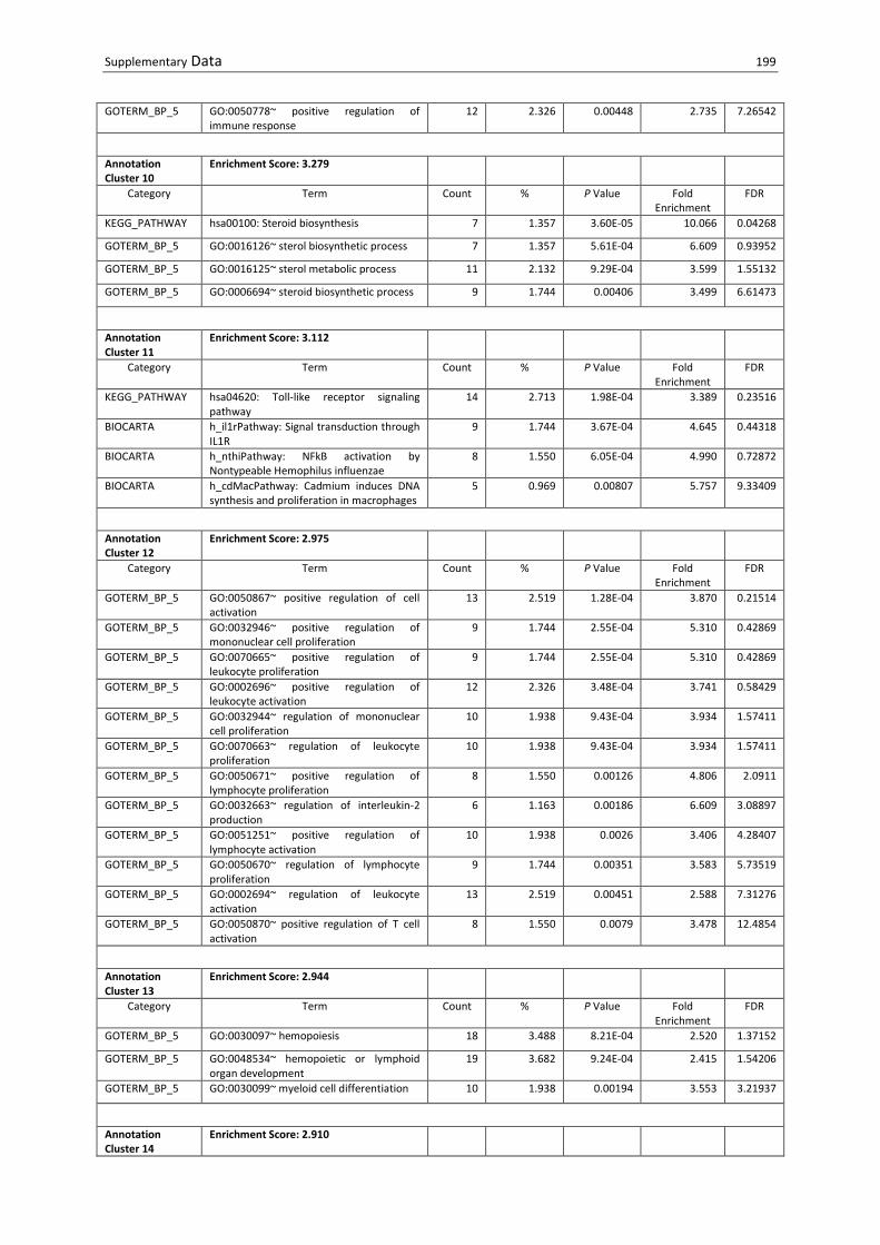
Supplementary Data 199
GOTERM_BP_5 GO:0050778~ positive regulation of immune response
12 2.326 0.00448 2.735 7.26542
Annotation Cluster 10
Enrichment Score: 3.279
Category Term Count % P Value Fold Enrichment
FDR
KEGG_PATHWAY hsa00100: Steroid biosynthesis 7 1.357 3.60E-05 10.066 0.04268
GOTERM_BP_5 GO:0016126~ sterol biosynthetic process 7 1.357 5.61E-04 6.609 0.93952
GOTERM_BP_5 GO:0016125~ sterol metabolic process 11 2.132 9.29E-04 3.599 1.55132
GOTERM_BP_5 GO:0006694~ steroid biosynthetic process 9 1.744 0.00406 3.499 6.61473
Annotation Cluster 11
Enrichment Score: 3.112
Category Term Count % P Value Fold Enrichment
FDR
KEGG_PATHWAY hsa04620: Toll-like receptor signaling pathway
14 2.713 1.98E-04 3.389 0.23516
BIOCARTA h_il1rPathway: Signal transduction through IL1R
9 1.744 3.67E-04 4.645 0.44318
BIOCARTA h_nthiPathway: NFkB activation by Nontypeable Hemophilus influenzae
8 1.550 6.05E-04 4.990 0.72872
BIOCARTA h_cdMacPathway: Cadmium induces DNA synthesis and proliferation in macrophages
5 0.969 0.00807 5.757 9.33409
Annotation Cluster 12
Enrichment Score: 2.975
Category Term Count % P Value Fold Enrichment
FDR
GOTERM_BP_5 GO:0050867~ positive regulation of cell activation
13 2.519 1.28E-04 3.870 0.21514
GOTERM_BP_5 GO:0032946~ positive regulation of mononuclear cell proliferation
9 1.744 2.55E-04 5.310 0.42869
GOTERM_BP_5 GO:0070665~ positive regulation of leukocyte proliferation
9 1.744 2.55E-04 5.310 0.42869
GOTERM_BP_5 GO:0002696~ positive regulation of leukocyte activation
12 2.326 3.48E-04 3.741 0.58429
GOTERM_BP_5 GO:0032944~ regulation of mononuclear cell proliferation
10 1.938 9.43E-04 3.934 1.57411
GOTERM_BP_5 GO:0070663~ regulation of leukocyte proliferation
10 1.938 9.43E-04 3.934 1.57411
GOTERM_BP_5 GO:0050671~ positive regulation of lymphocyte proliferation
8 1.550 0.00126 4.806 2.0911
GOTERM_BP_5 GO:0032663~ regulation of interleukin-2 production
6 1.163 0.00186 6.609 3.08897
GOTERM_BP_5 GO:0051251~ positive regulation of lymphocyte activation
10 1.938 0.0026 3.406 4.28407
GOTERM_BP_5 GO:0050670~ regulation of lymphocyte proliferation
9 1.744 0.00351 3.583 5.73519
GOTERM_BP_5 GO:0002694~ regulation of leukocyte activation
13 2.519 0.00451 2.588 7.31276
GOTERM_BP_5 GO:0050870~ positive regulation of T cell activation
8 1.550 0.0079 3.478 12.4854
Annotation Cluster 13
Enrichment Score: 2.944
Category Term Count % P Value Fold Enrichment
FDR
GOTERM_BP_5 GO:0030097~ hemopoiesis 18 3.488 8.21E-04 2.520 1.37152
GOTERM_BP_5 GO:0048534~ hemopoietic or lymphoid organ development
19 3.682 9.24E-04 2.415 1.54206
GOTERM_BP_5 GO:0030099~ myeloid cell differentiation 10 1.938 0.00194 3.553 3.21937
Annotation Cluster 14
Enrichment Score: 2.910

200 Supplementary Data
Category Term Count % P Value Fold Enrichment
FDR
GOTERM_BP_5 GO:0070304~ positive regulation of stress-activated protein kinase signaling pathway
6 1.163 3.33E-04 9.441 0.558
GOTERM_BP_5 GO:0032874~ positive regulation of stress-activated MAPK cascade
4 0.775 0.002 14.686 3.31084
GOTERM_BP_5 GO:0032872~ regulation of stress-activated MAPK cascade
4 0.775 0.00279 13.217 4.5957
Annotation Cluster 15
Enrichment Score: 2.899
Category Term Count % P Value Fold Enrichment
FDR
GOTERM_BP_5 GO:0050900~ leukocyte migration 9 1.744 2.90E-04 5.217 0.48564
GOTERM_BP_5 GO:0060326~ cell chemotaxis 7 1.357 0.00102 5.931 1.69999
GOTERM_BP_5 GO:0030593~ neutrophil chemotaxis 5 0.969 0.00179 9.179 2.96021
GOTERM_BP_5 GO:0030595~ leukocyte chemotaxis 6 1.163 0.0048 5.358 7.77107
Annotation Cluster 16
Enrichment Score: 2.756
Category Term Count % P Value Fold Enrichment
FDR
GOTERM_BP_5 GO:0032768~ regulation of monooxygenase activity
7 1.357 7.90E-05 9.252 0.13277
GOTERM_BP_5 GO:0051353~ positive regulation of oxidoreductase activity
5 0.969 0.00721 6.354 11.4614
GOTERM_BP_5 GO:0032642~ regulation of chemokine production
4 0.775 0.00947 8.811 14.787
Annotation Cluster 17
Enrichment Score: 2.675
Category Term Count % P Value Fold Enrichment
FDR
GOTERM_BP_5 GO:0019220~ regulation of phosphate metabolic process
28 5.426 0.00167 1.908 2.77594
GOTERM_BP_5 GO:0051174~ regulation of phosphorus metabolic process
28 5.426 0.00167 1.908 2.77594
GOTERM_BP_5 GO:0051347~ positive regulation of transferase activity
17 3.295 0.00256 2.341 4.22485
GOTERM_BP_5 GO:0043549~ regulation of kinase activity 22 4.264 0.00278 2.036 4.57764
Annotation Cluster 18
Enrichment Score: 2.650
Category Term Count % P Value Fold Enrichment
FDR
GOTERM_BP_5 GO:0001893~ maternal placenta development
5 0.969 8.56E-04 11.014 1.42906
GOTERM_BP_5 GO:0001890~ placenta development 8 1.550 0.0021 4.406 3.47608
GOTERM_BP_5 GO:0046697~ decidualization 4 0.775 0.00623 10.167 9.9683
Annotation Cluster 19
Enrichment Score: 2.621
Category Term Count % P Value Fold Enrichment
FDR
GOTERM_BP_5 GO:0032675~ regulation of interleukin-6 production
7 1.357 6.57E-04 6.425 1.09831
GOTERM_BP_5 GO:0032755~ positive regulation of interleukin-6 production
5 0.969 0.00221 8.696 3.64908
GOTERM_BP_5 GO:0032642~ regulation of chemokine production
4 0.775 0.00947 8.811 14.787
Annotation Cluster 20
Enrichment Score: 2.533

Supplementary Data 201
Category Term Count % P Value Fold Enrichment
FDR
GOTERM_BP_5 GO:0050714~ positive regulation of protein secretion
7 1.357 0.00134 5.641 2.22153
GOTERM_BP_5 GO:0050708~ regulation of protein secretion
8 1.550 0.00172 4.558 2.85755
GOTERM_BP_5 GO:0050707~ regulation of cytokine secretion
6 1.163 0.00186 6.609 3.08897
GOTERM_BP_5 GO:0051223~ regulation of protein transport
11 2.132 0.00233 3.188 3.84959
GOTERM_BP_5 GO:0070201~ regulation of establishment of protein localization
11 2.132 0.0036 3.004 5.88774
GOTERM_BP_5 GO:0051222~ positive regulation of protein transport
8 1.550 0.00396 3.945 6.44618
GOTERM_BP_5 GO:0050715~ positive regulation of cytokine secretion
5 0.969 0.00459 7.183 7.43981
GOTERM_BP_5 GO:0051046~ regulation of secretion 14 2.713 0.00834 2.290 13.1338
S Table 10.4 DAVID analysis of downregulated transcripts in B. henselae-infected MACs at d53. Clustering of enriched, functional annotations to related functional groups. Minimum similarity overlap > 3 (P < 0.01).
Annotation Cluster 1
Enrichment Score: 10.621
Category Term Count % P Value Fold Enrichment
FDR
GOTERM_BP_5 GO:0045449~ regulation of transcription 147 22.755 2.75E-12 1.703 4.34E-09
GOTERM_BP_5 GO:0010468~ regulation of gene expression
156 24.149 6.08E-12 1.646 9.58E-09
GOTERM_BP_5 GO:0010556~ regulation of macromolecule biosynthetic process
154 23.839 1.27E-11 1.639 2.00E-08
GOTERM_BP_5 GO:0019219~ regulation of nucleobase, nucleoside, nucleotide and nucleic acid metabolic process
153 23.684 1.67E-11 1.638 2.64E-08
GOTERM_BP_5 GO:0031326~ regulation of cellular biosynthetic process
158 24.458 1.87E-11 1.615 2.95E-08
GOTERM_BP_5 GO:0006355~ regulation of transcription, DNA-dependent
109 16.873 6.04E-11 1.852 9.52E-08
GOTERM_BP_5 GO:0051252~ regulation of RNA metabolic process
110 17.028 1.06E-10 1.828 1.67E-07
GOTERM_MF_5 GO:0046914~ transition metal ion binding 145 22.446 2.56E-10 1.576 3.02E-07
Annotation Cluster 2
Enrichment Score: 3.864
Category Term Count % P Value Fold Enrichment
FDR
BIOCARTA h_tcraPathway: Lck and Fyn tyrosine kinases in initiation of TCR Activation
7 1.084 3.78E-06 13.064 0.004509
GOTERM_BP_5 GO:0030217~ T cell differentiation 11 1.703 4.98E-05 5.098 0.07849
GOTERM_BP_5 GO:0042110~ T cell activation 15 2.322 7.17E-05 3.587 0.112972
GOTERM_BP_5 GO:0030098~ lymphocyte differentiation 13 2.012 1.48E-04 3.802 0.233619
GOTERM_BP_5 GO:0051249~ regulation of lymphocyte activation
15 2.322 3.96E-04 3.053 0.622524
GOTERM_BP_5 GO:0002521~ leukocyte differentiation 14 2.167 3.99E-04 3.220 0.627556
KEGG_PATHWAY hsa04660: T cell receptor signaling pathway 12 1.858 5.52E-04 3.488 0.638988
KEGG_PATHWAY hsa05340: Primary immunodeficiency 7 1.084 6.95E-04 6.278 0.804308

202 Supplementary Data
Annotation Cluster 3
Enrichment Score: 3.653
Category Term Count % P Value Fold Enrichment
FDR
GOTERM_BP_5 GO:0002696~ positive regulation of leukocyte activation
14 2.167 4.62E-05 3.979 0.072736
GOTERM_BP_5 GO:0030217~ T cell differentiation 11 1.703 4.98E-05 5.098 0.07849
GOTERM_BP_5 GO:0050867~ positive regulation of cell activation
14 2.167 7.50E-05 3.800 0.118177
GOTERM_BP_5 GO:0002694~ regulation of leukocyte activation
17 2.632 1.25E-04 3.085 0.196494
GOTERM_BP_5 GO:0050870~ positive regulation of T cell activation
11 1.703 1.92E-04 4.361 0.302447
GOTERM_BP_5 GO:0051251~ positive regulation of lymphocyte activation
12 1.858 3.53E-04 3.727 0.554805
GOTERM_BP_5 GO:0051249~ regulation of lymphocyte activation
15 2.322 3.96E-04 3.053 0.622524
GOTERM_BP_5 GO:0050863~ regulation of T cell activation 13 2.012 4.91E-04 3.347 0.77067
GOTERM_BP_5 GO:0045619~ regulation of lymphocyte differentiation
8 1.238 0.00464 3.826 7.072051
Annotation Cluster 4
Enrichment Score: 3.445
Category Term Count % P Value Fold Enrichment
FDR
BIOCARTA h_tcraPathway: Lck and Fyn tyrosine kinases in initiation of TCR Activation
7 1.084 3.78E-06 13.064 0.004509
BIOCARTA h_tcrPathway: T Cell Receptor Signaling Pathway
10 1.548 1.61E-05 6.038 0.019234
KEGG_PATHWAY hsa04640: Hematopoietic cell lineage 12 1.858 7.14E-05 4.380 0.082903
BIOCARTA h_tcytotoxicPathway: T Cytotoxic Cell Surface Molecules
6 0.929 1.35E-04 10.264 0.160658
BIOCARTA h_tcrmolecule: T Cell Receptor and CD3 Complex
4 0.619 4.10E-04 20.529 0.4885
BIOCARTA h_il17Pathway: IL 17 Signaling Pathway 6 0.929 4.56E-04 8.211 0.542926
KEGG_PATHWAY hsa04660: T cell receptor signaling pathway 12 1.858 5.52E-04 3.488 0.638988
BIOCARTA h_CSKPathway: Activation of Csk by cAMP-dependent Protein Kinase Inhibits Signaling through the T Cell Receptor
6 0.929 8.71E-04 7.245 1.035101
BIOCARTA h_ctla4Pathway: The Co-Stimulatory Signal During T-cell Activation
6 0.929 0.00152 6.483 1.797537
BIOCARTA h_thelperPathway: T Helper Cell Surface Molecules
5 0.774 0.0018 8.554 2.130141
BIOCARTA h_ctlPathway: CTL mediated immune response against target cells
5 0.774 0.00445 6.843 5.187692
BIOCARTA h_tcapoptosisPathway: HIV Induced T Cell Apoptosis
4 0.619 0.00723 9.124 8.300512
Annotation Cluster 5
Enrichment Score: 3.334
Category Term Count % P Value Fold Enrichment
FDR
BIOCARTA h_tcraPathway: Lck and Fyn tyrosine kinases in initiation of TCR Activation
7 1.084 3.78E-06 13.064 0.004509
GOTERM_BP_5 GO:0050778~ positive regulation of immune response
15 2.322 3.21E-04 3.117 0.504624
GOTERM_BP_5 GO:0002768~ immune response-regulating cell surface receptor signaling pathway
8 1.238 4.10E-04 5.739 0.644569

Supplementary Data 203
GOTERM_BP_5 GO:0050851~ antigen receptor-mediated signaling pathway
7 1.084 6.58E-04 6.391 1.032685
GOTERM_BP_5 GO:0002757~ immune response-activating signal transduction
8 1.238 0.00153 4.635 2.38663
GOTERM_BP_5 GO:0002764~ immune response-regulating signal transduction
8 1.238 0.00237 4.304 3.671966
GOTERM_BP_5 GO:0002253~ activation of immune response
10 1.548 0.00389 3.205 5.952457
Annotation Cluster 6
Enrichment Score: 2.712
Category Term Count % P Value Fold Enrichment
FDR
GOTERM_BP_5 GO:0001910~ regulation of leukocyte mediated cytotoxicity
6 0.929 0.00119 7.231 1.863695
GOTERM_BP_5 GO:0002717~ positive regulation of natural killer cell mediated immunity
5 0.774 0.00157 9.415 2.447332
GOTERM_BP_5 GO:0045954~ positive regulation of natural killer cell mediated cytotoxicity
5 0.774 0.00157 9.415 2.447332
GOTERM_BP_5 GO:0002715~ regulation of natural killer cell mediated immunity
5 0.774 0.002 8.861 3.10764
GOTERM_BP_5 GO:0042269~ regulation of natural killer cell mediated cytotoxicity
5 0.774 0.002 8.861 3.10764
GOTERM_BP_5 GO:0001912~ positive regulation of leukocyte mediated cytotoxicity
5 0.774 0.00453 7.173 6.908978

204 Acknowledgements
11. Acknowledgements

Acknowledgements 205
206 Peer Reviewed Publications
12. Peer Reviewed Publications
12.1 Scientific publications
F. O’Rourke, T. Mändle, C. Urbich , S. Dimmeler, U. R. Michaelis, R. P. Brandes, M. Flötenmeyer, C. Döring, M.-L. Hansmann, K. Lauber, W. Ballhorn, V. A.J. Kempf. (2015) Reprogramming of Myeloid Angiogenic Cells by Bartonella henselae leads to microenvironmental regulation of pathological angiogenesis. Cellular Microbiology. e-pub ahead of print
P. O. Kaiser, T. Riess, F. O'Rourke, D. Linke, V. A.J. Kempf. (2011) Bartonella spp.: Throwing light on uncommon human infections. Internation Journal of Medical Microbiology, 301 (1): 7-15
F. O'Rourke, T. Schmidgen, P. O. Kaiser, D. Linke, V. A.J. Kempf: Adhesins of Bartonella spp. (2011); Chapter in: “Bacterial Adhesion: Biology, Chemistry, and Physics”. Springer Verlag (Book series "Advances in Experimental Biology and Medicine"). 715 : 51-70
C. Beerlage, F. O’Rourke, V. A.J. Kempf. (2011) Wenn Bartonellen ihre Anker auswerfen- Bartonella henselae verbindet Infektionsforschung mit Blutgefäßwachstum. Forschung Frankfurt. 2: 26-29
12.2 Oral presentations
F. O'Rourke, T. Mändle, C. Urbich, S. Dimmeler, U. R. Michaelis, R. P. Brandes, M. Flötenmeyer, C. Döring, M.-L. Hansmann, K. Lauber W. Ballhorn, V. A.J. Kempf: Reprogramming of myeloid angiogenic cells by Bartonella henselae leads to microenvironmental regulation of pathological angiogenesis, 66th annual Meeting of the German Society of Microbiology and Infection Control (DGHM), 05.-08.10.2014, Dresden
F. O'Rourke: Interactions of the intercellular pathogen Bartonella henselae with myeloid angiogenic cells; implications for infection associated angiogenesis, Invited Speaker, Sick Kids Hospital, Inflammatory Bowel Disease (IBD) Center, 06.01.2014, Toronto Canada
F. O'Rourke, T. Mändle, C. Urbich, S. Dimmeler, U. R. Michaelis, R. P. Brandes, K. Lauber, V. A.J. Kempf: Infection of myeloid angiogenic cells with Bartonella henselae results in increased pro-angiogenic potential, 5th Congress of European Microbiologists FEMS, 7-11.06.2013, Leipzig
F O'Rourke, T. Mändle, C. Urbich, S. Dimmeler, K. Lauber, V. A.J. Kempf : Infection of myeloid angiogenic cells with Bartonella henselae induces vessel-like growth in vitro, 64th annual Meeting of the German Society of Microbiology and Infection Control (DGHM), 30.09.-03.10.2012, Hamburg
F. O'Rourke, T. Mändle, C. Urbich, S. Dimmeler, U. R. Michaelis, R. P. Brandes, K. Lauber, V. A.J. Kempf: Infection of myeloid angiogenic cells with Bartonella henselae induces vessel-like growth in vitro, Symposium of the DGHM-Focus Group Microbial Pathogenicity, 18-20.06.2012, Bad Urach
12.3 Poster presentations
F. O'Rourke, T. Mändle, C. Urbich, S. Dimmeler, U. R. Michaelis, R. P. Brandes, M. Flötenmeyer, C. Döring, M.-L. Hansmann, K. Lauber, V. A.J. Kempf: Infection of myeloid angiogenic cells with Bartonella henselae results in increased pro-angiogenic effects, 65th annual Meeting of the German Society of Microbiology and Infection Control (DGHM), 22-25.09.2013, Rostock

Peer Reviewed Publications 207
F O'Rourke, T. Mändle, C. Urbich, S. Dimmeler, K. Lauber, V. A.J. Kempf : Infection of human endothelial progenitor cells with Bartonella henselae induces vessel-like growth in vitro, IVBM 2012, 17th International Vascular Biology Meeting, 2-5.06.2012, Wiesbaden
F O'Rourke, T. Mändle, C. Urbich, S. Dimmeler, K. Lauber, V. A.J. Kempf : Infection of human endothelial progenitor cells with Bartonella henselae induces vessel-like growth in vitro, annual Meeting of the Association for General and Applied Microbiology (VAAM), 18.-21.03.2012, Tübingen
F O'Rourke, T. Mändle, C. Urbich, S. Dimmeler, K. Lauber, M. Schaller, V. A.J. Kempf : Infection of human endothelial progenitor cells with Bartonella henselae induces vessel growth in vitro, 9th annual Meeting of the International Society for Stem Cell Research, 15-18.06.2011, Toronto, Canada
F O'Rourke, T. Mändle, C. Urbich, S. Dimmeler, K. Lauber, V. A.J. Kempf : Infection of human endothelial progenitor cells with Bartonella henselae induces vessel growth in vitro, 2nd International Symposium of the SFB 766 Bacterial Cell Envelope, 23-25.05.2011, Kaufbeuren
F. O'Rourke, T. Mändle, C. Urbich, S. Dimmeler, U. R. Michaelis, R. P. Brandes, K. Lauber, V. A.J. Kempf: Infection of human endothelial progenitor cells with Bartonella henselae induces vessel growth in vitro, 63th annual Meeting of the German Society of Microbiology and Infection Control (DGHM), 25-28.09.2011, Essen
F O'Rourke, T. Mändle, C. Urbich, S. Dimmeler, K. Lauber, M. Schaller, V. A.J. Kempf : Infection of human endothelial progenitor cells with Bartonella henselae induces vessel growth in vitro, 2011 Joint Meeting of the European Society of Microcirculation and the German Society of Microcirculation and Vascular Biology (GfMVB), 13-16.10.2011, Munich

208 Curriculum Vitae
13. Curriculum Vitae

Curriculum Vitae 209