pengaruh age diversity, gender diversity dan occupational ...
Upload
cinco-itesmCategory
view
0download
0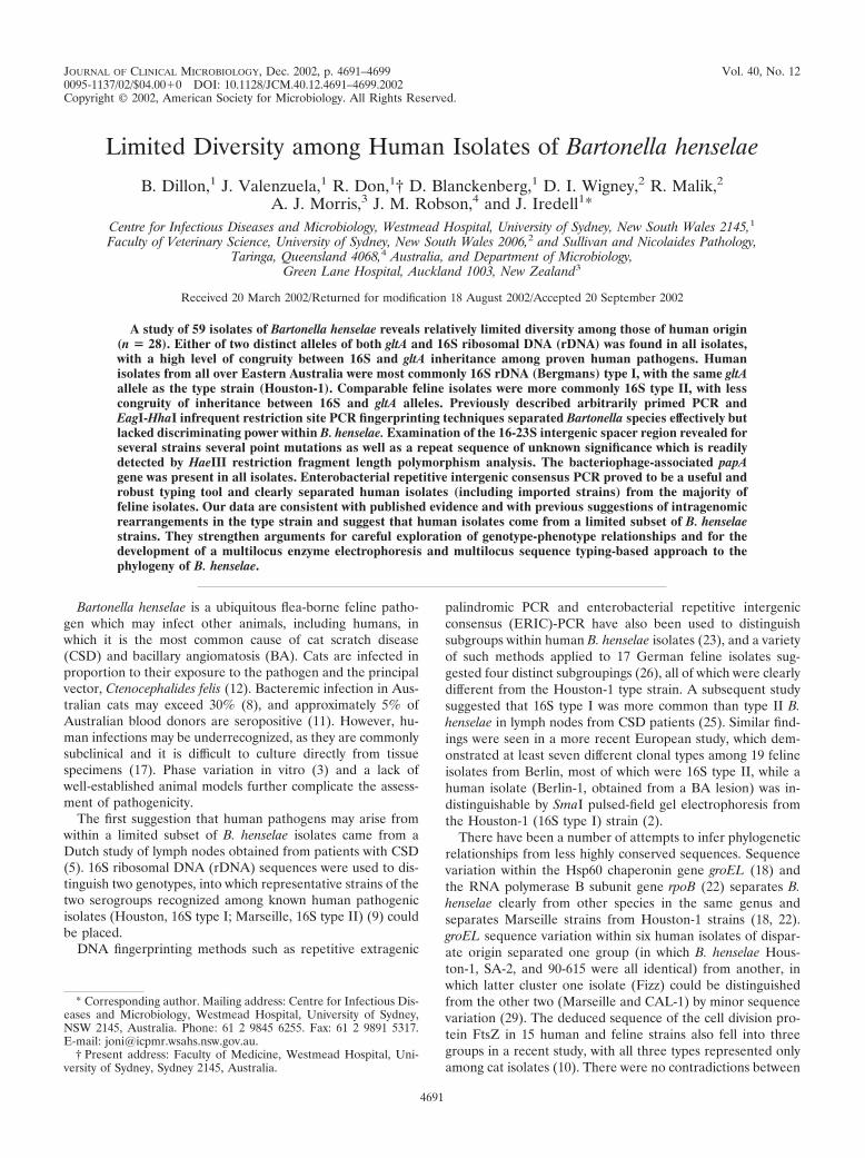
JOURNAL OF CLINICAL MICROBIOLOGY, Dec. 2002, p. 4691–4699 Vol. 40, No. 120095-1137/02/$04.00�0 DOI: 10.1128/JCM.40.12.4691–4699.2002Copyright © 2002, American Society for Microbiology. All Rights Reserved.
Limited Diversity among Human Isolates of Bartonella henselae
B. Dillon,1 J. Valenzuela,1 R. Don,1† D. Blanckenberg,1 D. I. Wigney,2 R. Malik,2A. J. Morris,3 J. M. Robson,4 and J. Iredell1*
Centre for Infectious Diseases and Microbiology, Westmead Hospital, University of Sydney, New South Wales 2145,1
Faculty of Veterinary Science, University of Sydney, New South Wales 2006,2 and Sullivan and Nicolaides Pathology,Taringa, Queensland 4068,4 Australia, and Department of Microbiology,
Green Lane Hospital, Auckland 1003, New Zealand3
Received 20 March 2002/Returned for modification 18 August 2002/Accepted 20 September 2002
A study of 59 isolates of Bartonella henselae reveals relatively limited diversity among those of human origin(n � 28). Either of two distinct alleles of both gltA and 16S ribosomal DNA (rDNA) was found in all isolates,with a high level of congruity between 16S and gltA inheritance among proven human pathogens. Humanisolates from all over Eastern Australia were most commonly 16S rDNA (Bergmans) type I, with the same gltAallele as the type strain (Houston-1). Comparable feline isolates were more commonly 16S type II, with lesscongruity of inheritance between 16S and gltA alleles. Previously described arbitrarily primed PCR andEagI-HhaI infrequent restriction site PCR fingerprinting techniques separated Bartonella species effectively butlacked discriminating power within B. henselae. Examination of the 16-23S intergenic spacer region revealed forseveral strains several point mutations as well as a repeat sequence of unknown significance which is readilydetected by HaeIII restriction fragment length polymorphism analysis. The bacteriophage-associated papAgene was present in all isolates. Enterobacterial repetitive intergenic consensus PCR proved to be a useful androbust typing tool and clearly separated human isolates (including imported strains) from the majority offeline isolates. Our data are consistent with published evidence and with previous suggestions of intragenomicrearrangements in the type strain and suggest that human isolates come from a limited subset of B. henselaestrains. They strengthen arguments for careful exploration of genotype-phenotype relationships and for thedevelopment of a multilocus enzyme electrophoresis and multilocus sequence typing-based approach to thephylogeny of B. henselae.
Bartonella henselae is a ubiquitous flea-borne feline patho-gen which may infect other animals, including humans, inwhich it is the most common cause of cat scratch disease(CSD) and bacillary angiomatosis (BA). Cats are infected inproportion to their exposure to the pathogen and the principalvector, Ctenocephalides felis (12). Bacteremic infection in Aus-tralian cats may exceed 30% (8), and approximately 5% ofAustralian blood donors are seropositive (11). However, hu-man infections may be underrecognized, as they are commonlysubclinical and it is difficult to culture directly from tissuespecimens (17). Phase variation in vitro (3) and a lack ofwell-established animal models further complicate the assess-ment of pathogenicity.
The first suggestion that human pathogens may arise fromwithin a limited subset of B. henselae isolates came from aDutch study of lymph nodes obtained from patients with CSD(5). 16S ribosomal DNA (rDNA) sequences were used to dis-tinguish two genotypes, into which representative strains of thetwo serogroups recognized among known human pathogenicisolates (Houston, 16S type I; Marseille, 16S type II) (9) couldbe placed.
DNA fingerprinting methods such as repetitive extragenic
palindromic PCR and enterobacterial repetitive intergenicconsensus (ERIC)-PCR have also been used to distinguishsubgroups within human B. henselae isolates (23), and a varietyof such methods applied to 17 German feline isolates sug-gested four distinct subgroupings (26), all of which were clearlydifferent from the Houston-1 type strain. A subsequent studysuggested that 16S type I was more common than type II B.henselae in lymph nodes from CSD patients (25). Similar find-ings were seen in a more recent European study, which dem-onstrated at least seven different clonal types among 19 felineisolates from Berlin, most of which were 16S type II, while ahuman isolate (Berlin-1, obtained from a BA lesion) was in-distinguishable by SmaI pulsed-field gel electrophoresis fromthe Houston-1 (16S type I) strain (2).
There have been a number of attempts to infer phylogeneticrelationships from less highly conserved sequences. Sequencevariation within the Hsp60 chaperonin gene groEL (18) andthe RNA polymerase B subunit gene rpoB (22) separates B.henselae clearly from other species in the same genus andseparates Marseille strains from Houston-1 strains (18, 22).groEL sequence variation within six human isolates of dispar-ate origin separated one group (in which B. henselae Hous-ton-1, SA-2, and 90-615 were all identical) from another, inwhich latter cluster one isolate (Fizz) could be distinguishedfrom the other two (Marseille and CAL-1) by minor sequencevariation (29). The deduced sequence of the cell division pro-tein FtsZ in 15 human and feline strains also fell into threegroups in a recent study, with all three types represented onlyamong cat isolates (10). There were no contradictions between
* Corresponding author. Mailing address: Centre for Infectious Dis-eases and Microbiology, Westmead Hospital, University of Sydney,NSW 2145, Australia. Phone: 61 2 9845 6255. Fax: 61 2 9891 5317.E-mail: [email protected].
† Present address: Faculty of Medicine, Westmead Hospital, Uni-versity of Sydney, Sydney 2145, Australia.
4691
FtsZ and groEL-derived groupings, although few commonstrains were tested, and the reported differences were minor.
The citrate synthase (gltA) gene sequence can be used todistinguish between Bartonella species (6, 20) and between theMarseille and Houston-1 strains of B. henselae (6). Sequencevariation in the riboflavin synthase gene ribC (4) was describedfor the (Sander) variant II group of 17 previously describedfeline isolates, all of which were quite distinct from Houston-1,and for the Bergmans 16S rDNA type II isolates (26).
We are therefore faced with a confusing array of typingmethods and systems which have been developed in differentstrain sets, often with small sample numbers (particularly ofproven human pathogenic strains). There appear to be twodistinct serotypes which infect humans (9), and sequence di-vergence in conserved regions points to at least two lineages.The Houston-1 (type) and Marseille strains may thus representdistinct phylogenetic groupings, but our understanding of geo-graphical distribution is incomplete and clear genotype-patho-
genicity correlations remain undefined. We therefore com-pared a group of human and feline isolates in an Australiansetting, using a number of these methods.
(This study was presented in part at the ASR-Bartonellajoint conference, Big Sky, Mont., August 2001.)
MATERIALS AND METHODS
Bacterial strains and growth conditions. Thirty-one Bartonella isolates fromcats and 28 B. henselae isolates from humans were obtained from the sourceslisted in Table 1. Human isolates were obtained by telephone calls and letters toevery large medical microbiology laboratory in Australia to identify existingisolates (most were freezer-stored isolates) and comprise all of the isolates inAustralia available to us at the commencement of the study. Fresh blood wascollected by aseptic venipuncture of cats anesthetized after injury or for castra-tion and was added as soon as possible after collection to an equal volume ofpresterilized lysing solution (0.6 g of saponin and 0.6 g of liquoid in 100 ml of0.9% saline solution) in a sterile centrifuge tube. After mixing and standing for5 min, sediment obtained at 5,000 � g was plated onto chocolate blood agar(Oxoid blood agar base no. 2 with 5% defibrinated sheep blood and vitaminK-hemin) and incubated for 10 days. All strains were subcultured in the labora-
TABLE 1. B. henselae isolates studieda
Isolate(s) Source 16S type gltA type AP type ERIC type IRS type
Feline isolates (n � 31)NU4695 Blood I H H E4H 1RMC10† Blood I H H E4H 1HC54 Blood I H H E4H 2HC69 Blood I H M E4H 2HC62† Blood I H H E1 2RMC3* Blood I H H E5 2NU4428† Blood I H M E2 2HC71 Blood I M H E2 2NU4714*† Blood II H H E4H 1HC35*, HC55 Blood II H H E3a 1RMC8* Blood II H Ha E3a 2NU4681* Blood II H H E3a 1HC60† Blood II H H E3b 1RMC1* Blood II H H E1 2RMC12 Blood II H M E3a 1NU4423† Blood II M H E4H 2RMC11 Blood II M M E7 1RMC4 Blood II M M E7 2HC41, HC48†, HC61, HC63†, HC77 Blood II M M E2 2RMC2† Blood II M M E6 2RMC5 Blood II M M E1 2RMC6†, RMC9† Blood II M M E5 2RMC7, Berlin-2 Blood II M M E5 2NU4713 Blood II M H E2 2
Human isolates (n � 28)BH2 CSD I H H E4H 1ATCC 49793/NZRm3492 (s4) BA I H H E4H 1ATCC 49882 (Houston-1)†, Berlin-1 BA I H H E4H 2BH3(s4), BH5s2*, R987, JR1, JR2, JR3, JR5, JR6,
JR7, JR8, JR9, JR12, JR13, JR14, JR15, JR17,JR18, JR19, JR20, JR22, JR23
CSD I H H E4H 2
R1073† CSD II H H E4H 2BH4 CSD II M M E4M 2URLLY-8 (Marseille)† BA II M M E4M 2
a Symbols: *, Isolate embeds into solid agar and displays ratchet motility at this passage number; †, 16S type was determined by direct sequencing. s2, second passageafter initial isolation; (s4), third to fifth passage after initial isolation. If not specified, RMC-prefix isolates (this work) were studied at passage numbers 2 to 3; HC-prefixisolates were a gift of Tom Gottlieb (8) and were studied at passage numbers 4 to 6; NU-prefix isolates (15) were studied at passage numbers 3 to 5; R- and JR-prefixstrains (this work and Fournier et al. [12a]) were all studied at passage numbers 4 to 7. Other isolates were isolated in our laboratory or obtained from the following:Mardjan Arvand, Hygiene-Institute, Heidelberg, Germany (2) (Berlin-1 and -2); R. Bunter, Goulburn Valley Base Hospital, Goulburn, NSW, Australia (BH2); the NewZealand quality control strain NZRm3492 is thought to be a lab-passaged derivative of the widely used strain ATCC 49793, and they are inseparable by all typingmethods; Jock Harkness, Royal North Shore Hospital, Sydney, Australia [BH3(s4)]; T.H.E. Pathology, Fairfield Heights, NSW, Sydney, Australia [BH5(S2)]; and D.Raoult, Unite des Rickettsies, Marseille, France (9) (URLLY-8 [Marseille]). Strains with the ATCC prefix were obtained from the American Type Culture Collection,Manassas, Va. 16S-23S regions were sequenced in all feline strains, in all Sydney and imported human strains, and in JR2.
4692 DILLON ET AL. J. CLIN. MICROBIOL.
tory on chocolate blood agar (Oxoid blood agar base no. 2) containing 5% horseblood (CBA) at 37°C in 5% CO2 for 5 to 7 days or were grown in liquid medium(27) at 37°C in 5% CO2 with intermittent shaking (100 rpm) in cotton wool-plugged Pyrex Erlenmeyer flasks (as specified in text). Bartonella quintana (J.Robson) and Bartonella vinsonii (ATCC 51672) were cultured under similarconditions. Bartonella bacilliformis (ATCC 35686) was grown on a CBA slope at26°C for 10 to 14 days. The passage number and agar-pitting phenotype for B.henselae strains is listed in Table 1.
Preparation of DNA and lysates. Bacteria grown on CBA plates were washedand resuspended in 1 ml of 0.01 M phosphate-buffered saline, pH 7.2, to a finaloptical density at 550 nm of 0.5, incubated for 10 min at 95°C, and centrifuged at13,000 � g for 30 s. Supernatants (lysates) were used as templates in PCRs.Genomic DNA was extracted from 2 ml of early-stationary-phase (7-day) brothculture with a commercially available kit (Promega Corporation, Madison, Wis.)according to the manufacturer’s instructions.
PCR, biological reagents, gel electrophoresis, and sequencing reactions. Un-less otherwise stated, the following concentrations of reagents were used forPCR: deoxynucleoside triphosphates, 200 �M each (Astral Scientific, Sydney,Australia); MgCl2, 1.5 mM (Astral Scientific); 2 U of Taq polymerase (Promega);1� PCR buffer (Promega); and 2 �l of bacterial lysate in 50-�l reaction volumes.Control strain lysates and distilled water served as controls. DNA amplificationwas performed in a PC-960C thermal cycler (Corbett Research, Sydney, Austra-lia). PCR products were purified with the GeneClean kit (Bio 101 Inc., Carlsbad,Calif.), and both strands were sequenced by use of an automated sequencer (ABI373 Genetic Analyzer; Perkin-Elmer Biosystems). Alignments were performedwith EclustalW (ANGIS), and finalized sequences were submitted to the Gen-Bank database.
Electrophoresis of DNA was performed in 1 to 2% (wt/vol) agarose (Promega)and visualized using ethidium bromide on an UV transilluminator using 667Polaroid film. Images were scanned with an HP ScanJet 5300C (Hewlett Pack-ard) and Adobe PhotoShop 4.0 (Microsoft).
16S rRNA type-specific PCR and sequencing. Primers BH1 and BH2 wereused in conjunction with the broad-host-range primer 16SF according to themethod of Bergmans et al. (5) at 100 nM and at annealing temperatures of 54 to56°C, as specified in the text. 16S sequencing was performed using primers 16SF[5�-AGAGTTTGATCCTGG(CT)TCAG-3�] and 16SR [5�-CTTTACGCCCA(AG)TAA(AT)TCCG-3�] (19).
Amplification of gltA and 16S-23S intergenic region and ITS restriction frag-ment length polymorphism. 16S-23S intergenic spacer region (ITS)-PCR wasperformed as previously described (19), with minor modifications. External prim-ers RPC5 (5�-AAGTCGTAACAAGGTA-3�) and R23S2693 (5�-TACTGGTTCACTATCGGTCA-3�) (200 nM) were used in a 100-�l PCR mixture (19). In-ternal primers BHITSF (5�-AAGAGGATGCCCGGGAAGGT-3�) andBHITSR (5�-GCGTTCTCTGCCTTGTGCAA-3�) (200 nM) were designed (thisstudy) and used to amplify a partial ITS region for sequence analysis as follows:5-min denaturation at 95°C; 35 cycles of 95°C for 30 s, 50°C for 60 s, and 72°C for90 s; and final extension at 72°C for 10 min. Restriction enzymes (HaeIII andAluI) from New England Biolabs were used according to the manufacturer’sinstructions to digest 16S-23S amplicons. Previously described primers(BhCS.1137 [5�-AATGCAAAAAGAACAGTAAACA-3�] and CS140f [5�-TTACTTATGATCCKGGYTTTA-3�] [200 nM]) were used to generate and se-quence gltA amplicons (6).
AP-PCR and ERIC-PCR. Arbitrarily primed (AP)-PCR was performed withM13 bacteriophage core sequence primer (5�-GAGGGTGGCGGTTCT-3� [200nM]) as described previously (26). Primers targeting the ERIC sequence,ERIC-1R (5�-ATGTAAGCTCCTGGGGATTCAC-3�) and ERIC-2 (5�-AAGTAAGTGACTGGGGTGAGAGCG-3�) both at 200 nM), were used with 4 �l ofbacterial lysate in the ERIC-PCR (28) under the following conditions: initialdenaturing at 95°C for 5 min; 35 cycles of denaturing at 95°C for 20 s, annealingat 50°C for 60 s, and extension at 72°C for 2 min; and a final extension at 72°Cfor 10 min. ERIC-PCR fingerprints were analyzed by applying the Dice coeffi-cient to peaks, using GelCompar software, version 4 (Applied Maths, Kortijk,Belgium), according to the manufacturer’s instructions. The unweighted pairgroup method with arithmetic means was used with a 1.2% tolerance in bandpositions to analyze clustering of fingerprint patterns.
IRS-PCR with HhaI and EagI. PCR reagent concentrations and amplificationconditions for infrequent restriction site (IRS)-PCR have been described byHandley and Regnery (13). PCR products were separated on a 6.5% polyacryl-amide gel in 1� Tris-borate-EDTA buffer for 3 h at 150 V.
pap PCR. Amplification reactions were carried out in a 50-�l final volumecontaining 200 nM (each) PAP-1 and PAP-2 primers (1). Amplifications wereperformed as follows: initial denaturation at 95°C for 5 min; 30 cycles of 95°C for30 s, 50°C for 1 min, and 72°C for 90 s; and final extension at 72°C for 10 min.
Statistical analysis. Chi-square analysis was performed where indicated, usingMinitab for Windows release 12 (Minitab Inc., State College, Pa.).
Nucleotide sequence accession numbers. The 16S-23S rRNA intergenic spacerregion sequences of the following isolates from this study have been deposited inthe GenBank and EMBL nucleotide sequence databases under the followingaccession numbers: HC54, AJ439687; HC69, AJ439688; RMC3, AJ441257;RMC10, AJ441256; BH2, AJ457178; and JR2, AJ457177. The gltA sequence ofisolate BH4 has been deposited under accession number AJ439406.
RESULTS
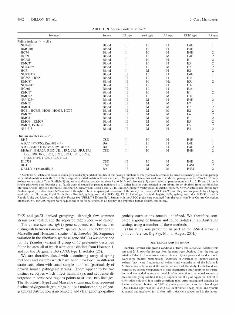
16S rDNA type I is more common in human isolates. Westudied 59 isolates, half of human origin and half of felineorigin (Table 1). Human isolates were mostly from easternAustralia (New South Wales [NSW] and Queensland) and thefeline isolates were from two Sydney (NSW) collections andone New Zealand collection. The majority of feline isolatesfrom Sydney (and New Zealand) were 16S rDNA type II (Ta-ble 1). In contrast, almost all (22 of 24) Australian humanisolates yielded positive results in the type I-specific PCR; oneBrisbane strain (R1073) and one Sydney strain (BH4) wereclearly type II. Clear-cut results were obtained from boiledlysates in the majority (48 of 59) of cases; 11 samples (HC48,HC60, HC62, HC63, NU4423, NU4428, NU4714, RMC2,RMC6, RMC9, and RMC10) reacted in both assays at lowstringency (Tm, 54°C) (Fig. 1). For five of these samples, ac-curate and unambiguous results were also obtained by PCR ata slightly higher stringency (Tm, 56°C). Typing of these 11 andcertain additional strains was confirmed by direct sequencingas indicated in Table 1.
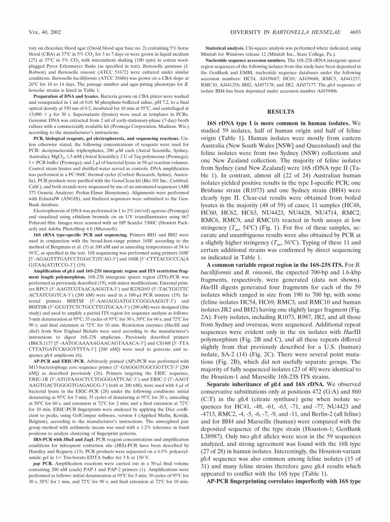
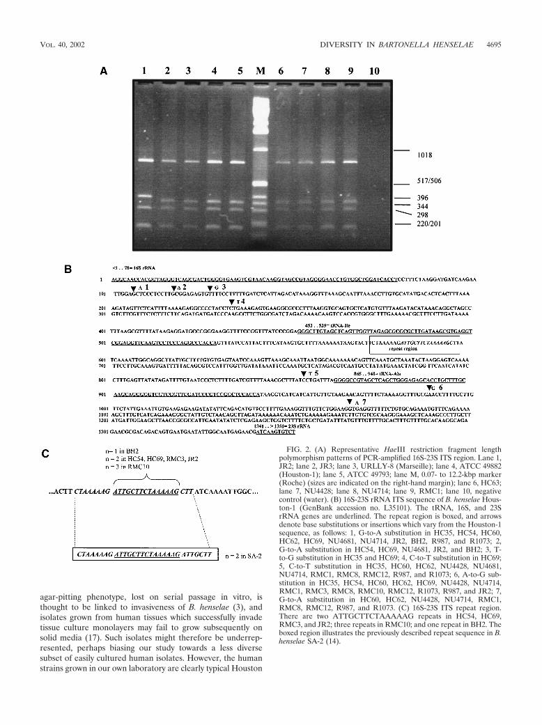
A common variable repeat region in the 16S-23S ITS. For B.bacilliformis and B. vinsonii, the expected 700-bp and 1.6-kbpfragments, respectively, were generated (data not shown).HaeIII digests generated four fragments for each of the 59isolates which ranged in size from 190 to 700 bp, with some(feline isolates HC54, HC69, RMC3, and RMC10 and humanisolates JR2 and BH2) having one slightly larger fragment (Fig.2A). Forty isolates, including R1073, R987, JR2, and all thosefrom Sydney and overseas, were sequenced. Additional repeatsequences were evident only in the six isolates with HaeIIIpolymorphism (Fig. 2B and C), and all these repeats differedslightly from that previously described for a U.S. (human)isolate, SA-2 (14) (Fig. 2C). There were several point muta-tions (Fig. 2B), which did not usefully separate groups. Themajority of fully sequenced isolates (23 of 40) were identical tothe Houston-1 and Marseille 16S-23S ITS strains.
Separate inheritance of gltA and 16S rDNA. We observedconservative substitutions only at positions 472 (G:A) and 860(C:T) in the gltA (citrate synthase) gene when isolate se-quences for HC41, -48, -61, -63, -71, and -77, NU4423 and-4713, RMC2, -4, -5, -6, -7, -9, and -11, and Berlin-2 (all feline)and for BH4 and Marseille (human) were compared with thedeposited sequence of the type strain (Houston-1; GenBankL38987). Only two gltA alleles were seen in the 59 sequencesanalyzed, and strong agreement was found with the 16S type(27 of 28) in human isolates. Interestingly, the Houston-variantgltA sequence was also common among feline isolates (15 of31) and many feline strains therefore gave gltA results whichappeared to conflict with the 16S type (Table 1).
AP-PCR fingerprinting correlates imperfectly with 16S type
VOL. 40, 2002 DIVERSITY IN BARTONELLA HENSELAE 4693
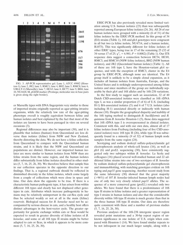
and gltA. Single-primer (M13) AP-PCR distinguished B.henselae Houston-1 from the Marseille strain (Fig. 3), andalmost all (58 of 59) isolates yielded one of these two simplepatterns upon agarose gel electrophoresis (Table 1). One iso-late (RMC8) exhibited altered migration of the 460-bp frag-ment (Fig. 3). Results for all four imported human isolates and23 of the 24 Australian human isolates were consistent with the16S and gltA data, with the Houston-1 (H) pattern dominant.The one exception among the 28 human isolates was againR1073 (16S type II), for which gltA and AP-PCR patterns wereboth H-type. Half (14 of 24) of the Sydney feline isolates wereAP-PCR Marseille (M) type, with only one of these (HC69;Bergmans 16S type 1) conflicting with the (H-variant) gltAsequence. In contrast, AP-PCR correlated poorly with the gltAallele for New Zealand feline isolates, the majority of whichyielded the H pattern on AP-PCR (Table 1).
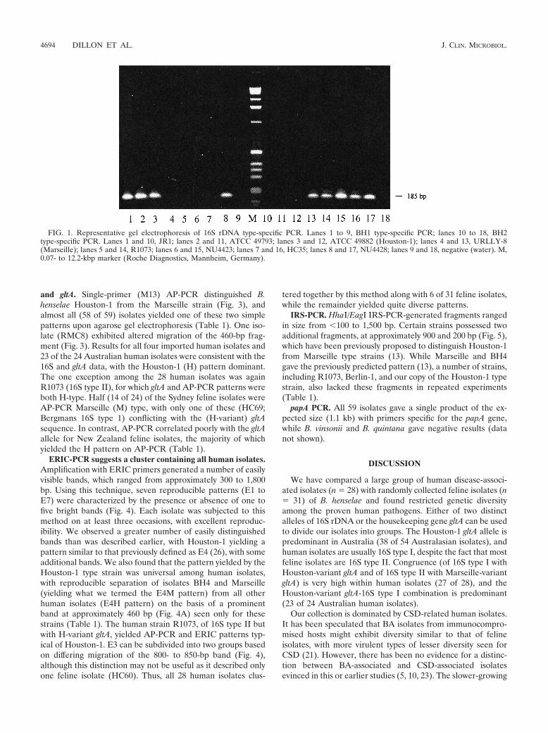
ERIC-PCR suggests a cluster containing all human isolates.Amplification with ERIC primers generated a number of easilyvisible bands, which ranged from approximately 300 to 1,800bp. Using this technique, seven reproducible patterns (E1 toE7) were characterized by the presence or absence of one tofive bright bands (Fig. 4). Each isolate was subjected to thismethod on at least three occasions, with excellent reproduc-ibility. We observed a greater number of easily distinguishedbands than was described earlier, with Houston-1 yielding apattern similar to that previously defined as E4 (26), with someadditional bands. We also found that the pattern yielded by theHouston-1 type strain was universal among human isolates,with reproducible separation of isolates BH4 and Marseille(yielding what we termed the E4M pattern) from all otherhuman isolates (E4H pattern) on the basis of a prominentband at approximately 460 bp (Fig. 4A) seen only for thesestrains (Table 1). The human strain R1073, of 16S type II butwith H-variant gltA, yielded AP-PCR and ERIC patterns typ-ical of Houston-1. E3 can be subdivided into two groups basedon differing migration of the 800- to 850-bp band (Fig. 4),although this distinction may not be useful as it described onlyone feline isolate (HC60). Thus, all 28 human isolates clus-
tered together by this method along with 6 of 31 feline isolates,while the remainder yielded quite diverse patterns.
IRS-PCR. HhaI/EagI IRS-PCR-generated fragments rangedin size from �100 to 1,500 bp. Certain strains possessed twoadditional fragments, at approximately 900 and 200 bp (Fig. 5),which have been previously proposed to distinguish Houston-1from Marseille type strains (13). While Marseille and BH4gave the previously predicted pattern (13), a number of strains,including R1073, Berlin-1, and our copy of the Houston-1 typestrain, also lacked these fragments in repeated experiments(Table 1).
papA PCR. All 59 isolates gave a single product of the ex-pected size (1.1 kb) with primers specific for the papA gene,while B. vinsonii and B. quintana gave negative results (datanot shown).
DISCUSSION
We have compared a large group of human disease-associ-ated isolates (n � 28) with randomly collected feline isolates (n� 31) of B. henselae and found restricted genetic diversityamong the proven human pathogens. Either of two distinctalleles of 16S rDNA or the housekeeping gene gltA can be usedto divide our isolates into groups. The Houston-1 gltA allele ispredominant in Australia (38 of 54 Australasian isolates), andhuman isolates are usually 16S type I, despite the fact that mostfeline isolates are 16S type II. Congruence (of 16S type I withHouston-variant gltA and of 16S type II with Marseille-variantgltA) is very high within human isolates (27 of 28), and theHouston-variant gltA-16S type I combination is predominant(23 of 24 Australian human isolates).
Our collection is dominated by CSD-related human isolates.It has been speculated that BA isolates from immunocompro-mised hosts might exhibit diversity similar to that of felineisolates, with more virulent types of lesser diversity seen forCSD (21). However, there has been no evidence for a distinc-tion between BA-associated and CSD-associated isolatesevinced in this or earlier studies (5, 10, 23). The slower-growing
FIG. 1. Representative gel electrophoresis of 16S rDNA type-specific PCR. Lanes 1 to 9, BH1 type-specific PCR; lanes 10 to 18, BH2type-specific PCR. Lanes 1 and 10, JR1; lanes 2 and 11, ATCC 49793; lanes 3 and 12, ATCC 49882 (Houston-1); lanes 4 and 13, URLLY-8(Marseille); lanes 5 and 14, R1073; lanes 6 and 15, NU4423; lanes 7 and 16, HC35; lanes 8 and 17, NU4428; lanes 9 and 18, negative (water). M,0.07- to 12.2-kbp marker (Roche Diagnostics, Mannheim, Germany).
4694 DILLON ET AL. J. CLIN. MICROBIOL.
agar-pitting phenotype, lost on serial passage in vitro, isthought to be linked to invasiveness of B. henselae (3), andisolates grown from human tissues which successfully invadetissue culture monolayers may fail to grow subsequently onsolid media (17). Such isolates might therefore be underrep-resented, perhaps biasing our study towards a less diversesubset of easily cultured human isolates. However, the humanstrains grown in our own laboratory are clearly typical Houston
FIG. 2. (A) Representative HaeIII restriction fragment lengthpolymorphism patterns of PCR-amplified 16S-23S ITS region. Lane 1,JR2; lane 2, JR3; lane 3, URLLY-8 (Marseille); lane 4, ATCC 49882(Houston-1); lane 5, ATCC 49793; lane M, 0.07- to 12.2-kbp marker(Roche) (sizes are indicated on the right-hand margin); lane 6, HC63;lane 7, NU4428; lane 8, NU4714; lane 9, RMC1; lane 10, negativecontrol (water). (B) 16S-23S rRNA ITS sequence of B. henselae Hous-ton-1 (GenBank accession no. L35101). The tRNA, 16S, and 23SrRNA genes are underlined. The repeat region is boxed, and arrowsdenote base substitutions or insertions which vary from the Houston-1sequence, as follows: 1, G-to-A substitution in HC35, HC54, HC60,HC62, HC69, NU4681, NU4714, JR2, BH2, R987, and R1073; 2,G-to-A substitution in HC54, HC69, NU4681, JR2, and BH2; 3, T-to-G substitution in HC35 and HC69; 4, C-to-T substitution in HC69;5, C-to-T substitution in HC35, HC60, HC62, NU4428, NU4681,NU4714, RMC1, RMC8, RMC12, R987, and R1073; 6, A-to-G sub-stitution in HC35, HC54, HC60, HC62, HC69, NU4428, NU4714,RMC1, RMC3, RMC8, RMC10, RMC12, R1073, R987, and JR2; 7,G-to-A substitution in HC60, HC62, NU4428, NU4714, RMC1,RMC8, RMC12, R987, and R1073. (C) 16S-23S ITS repeat region.There are two ATTGCTTCTAAAAAG repeats in HC54, HC69,RMC3, and JR2; three repeats in RMC10; and one repeat in BH2. Theboxed region illustrates the previously described repeat sequence in B.henselae SA-2 (14).
VOL. 40, 2002 DIVERSITY IN BARTONELLA HENSELAE 4695
or Marseille types with DNA fingerprints very similar to thoseof imported strains originally reported as agar-pitting invasiveorganisms, while the relatively low rate of the agar-pittingphenotype overall is roughly equivalent between feline andhuman isolates and best explained by the fact that most of theisolates are known to have been passaged in vitro on severaloccasions (3).
Regional differences may also be important (30), and it isplausible that isolates (human) from Queensland are less di-verse than isolates (feline) from NSW and New Zealand,thereby distorting the data. We do not have a feline collectionfrom Queensland to compare with the Queensland humanstrains, and it is likely that the NSW and Queensland catpopulations are distinct. However, our imported human iso-lates are more similar to human isolates from NSW than arefeline strains from the same region, and the human isolatesdiffer substantially from feline isolates described in other stud-ies (5, 7, 16, 25, 26, 30). We therefore believe that sample biasin our collection operates in the opposite direction to ourfindings. That is, a regional outbreak should be reflected indiminished diversity in the feline isolates, which come largelyfrom a couple of collections which were close in space andtime. Our data show the opposite to be true. If there is acharacteristic human pathogenic type, it has arisen from withindifferent 16S types and clearly has not displaced other geno-types in cats. Attributes which increase pathogenicity in hu-mans may be relatively unimportant in the cat-flea cycle andthus be selected weakly or not at all in the main (feline)reservoir. Biological success for B. henselae need not be ac-companied by serious disease in cats, and a healthy host offersdistinct advantages in the host-vector cycle. The greater op-portunity for genetic exchange within cats or fleas would beexpected to result in greater diversity of feline isolates of B.henselae, and some or all 16S type II strains might be betteradapted to cats or fleas, in which it appears to be more com-mon (5, 7, 16, 25, 26, 30).
ERIC-PCR has also previously revealed more limited vari-ation among U.S. human isolates (23) than was subsequentlyreported among European feline isolates (24, 26). All 28 of ourhuman isolates were grouped with a minority (6 of 31) of thefeline isolates by the ERIC-PCR method. In this group of 34(E4) strains (Table 1), 16S and gltA genotypes were congruentin all but two (a feline isolate, NU4714, and a human isolate,R1073). This was significantly different for feline isolates ofother ERIC types, being true in 17 of the remaining 25 (32 of34 versus 17 of 25; �2
1 � 6.981; P � 0.0082) (Table 1). Our ITSsequence data suggest a connection between HC54, HC69,RMC3, and RMC10 (NSW feline isolates), BH2 (NSW humanisolates), and JR2 (Queensland human isolate) (Table 1). Allof these are 16S type I, have the Houston-variant gltA se-quence, and with the exception of RMC3, cluster in the E4group by ERIC-PCR, although none are identical. The E4group itself is unlikely to be a simple clonal expansion, as itincludes all human isolates from Australia, Europe, and theUnited States and is strikingly underrepresented among felineisolates and since members of the group are individually sep-arable by their gltA and 16S alleles and by 16S-23S variations.
In the first study to suggest such an association, 32 of 40Dutch CSD-associated isolates were classified as 16S rDNAtype I, as was a similar proportion (5 of 6) of U.S. (includingH-1) BA-associated isolates (5) and 6 of 7 U.S. isolates (alsoincluding H-1) associated directly or indirectly with humandisease (10). Despite the geographical disparity and inability ofthe 16S typing method to distinguish B. bacilliformis and B.quintana from B. henselae Houston-1 (5), those data suggestedthat 16S rDNA type I is overrepresented in clinical isolates.Consistent with this, and with our own data, almost all of 17feline isolates from Freiburg (including four of five CSD-asso-ciated isolates) were 16S type II (26), while type II was subse-quently found in a minority (9 of 32) of human lymph nodesamples from the same region (24).
Serotyping and sodium dodecyl sulfate-polyacrylamide gelelectrophoresis analysis of whole-cell lysates (16), as well asgltA (6) and groEL sequencing (30), have consistently sug-gested only two subtypes within B. henselae. La Scola andcolleagues (16) placed several well-studied human and 22 ad-ditional feline strains into one of two serotypes of B. henselaeby sodium dodecyl sulfate-polyacrylamide gel electrophoresisand immunoblotting, with matching genotypes evident on 16Styping and pap31 gene sequencing. Another recent study fromthe same laboratory (30) showed that the great majority(�96%) of 107 B. henselae-infected human lymph nodes hadone of only two very closely related pap31 alleles. The se-quence of a more conserved gene (groEL) revealed only twoalleles. We have found that there is a predominance of 16Stype II strains in feline isolates and a greater representation oftype I strains in human isolates and also that the considerablediversity within 16S type II strains is much greater than that inthe three human 16S type II strains. Our data are thereforequite consistent with these and a number of previous studies(5, 7, 16, 25, 26, 30).
Previous analyses of the 16S-23S intergenic spacer regionrevealed point mutations and a 30-bp repeat region of un-known significance in one isolate of U.S. origin when com-pared with Houston-1 (14). We have found similar repeats tobe not infrequent in our much larger sample, along with a
FIG. 3. AP-PCR representative gel. Lane 1, ATCC 49882 (Hous-ton-1); lane 2, JR2; lane 3, RMC1; lane 4, BH2; lane 5, RMC8; lane 6,URLLY-8 (Marseille); lane 7, HC63; lane 8, HC77; lane 9, BH4; lane10, NU4428; M, pGEM marker (Promega; molecular size in base pairsis given along the right border).
4696 DILLON ET AL. J. CLIN. MICROBIOL.
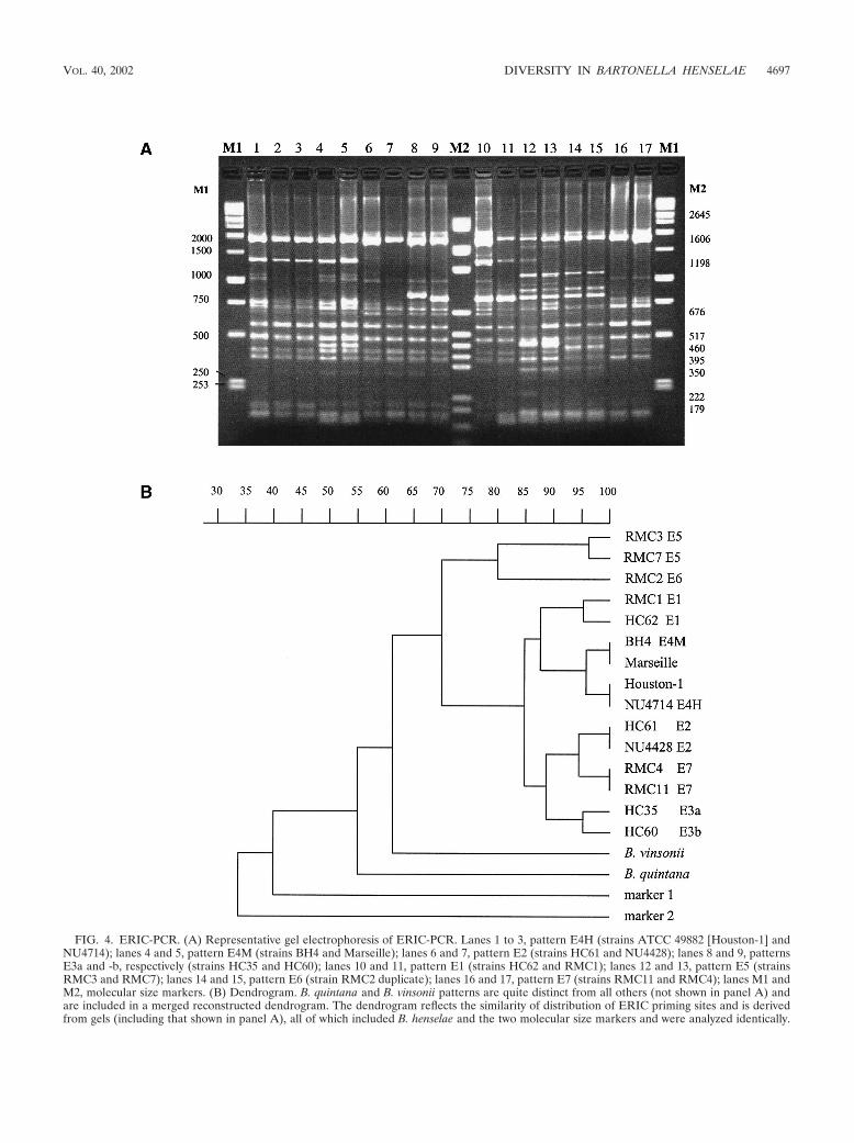
FIG. 4. ERIC-PCR. (A) Representative gel electrophoresis of ERIC-PCR. Lanes 1 to 3, pattern E4H (strains ATCC 49882 [Houston-1] andNU4714); lanes 4 and 5, pattern E4M (strains BH4 and Marseille); lanes 6 and 7, pattern E2 (strains HC61 and NU4428); lanes 8 and 9, patternsE3a and -b, respectively (strains HC35 and HC60); lanes 10 and 11, pattern E1 (strains HC62 and RMC1); lanes 12 and 13, pattern E5 (strainsRMC3 and RMC7); lanes 14 and 15, pattern E6 (strain RMC2 duplicate); lanes 16 and 17, pattern E7 (strains RMC11 and RMC4); lanes M1 andM2, molecular size markers. (B) Dendrogram. B. quintana and B. vinsonii patterns are quite distinct from all others (not shown in panel A) andare included in a merged reconstructed dendrogram. The dendrogram reflects the similarity of distribution of ERIC priming sites and is derivedfrom gels (including that shown in panel A), all of which included B. henselae and the two molecular size markers and were analyzed identically.
VOL. 40, 2002 DIVERSITY IN BARTONELLA HENSELAE 4697
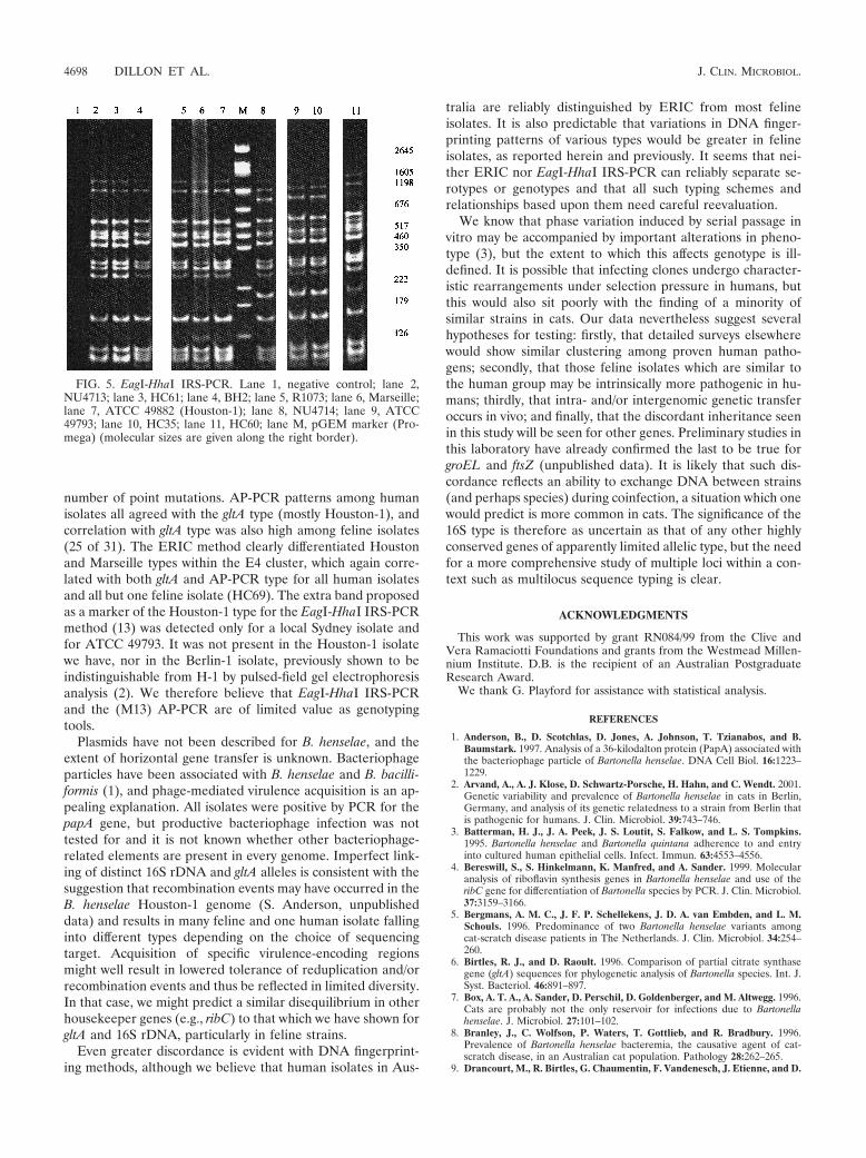
number of point mutations. AP-PCR patterns among humanisolates all agreed with the gltA type (mostly Houston-1), andcorrelation with gltA type was also high among feline isolates(25 of 31). The ERIC method clearly differentiated Houstonand Marseille types within the E4 cluster, which again corre-lated with both gltA and AP-PCR type for all human isolatesand all but one feline isolate (HC69). The extra band proposedas a marker of the Houston-1 type for the EagI-HhaI IRS-PCRmethod (13) was detected only for a local Sydney isolate andfor ATCC 49793. It was not present in the Houston-1 isolatewe have, nor in the Berlin-1 isolate, previously shown to beindistinguishable from H-1 by pulsed-field gel electrophoresisanalysis (2). We therefore believe that EagI-HhaI IRS-PCRand the (M13) AP-PCR are of limited value as genotypingtools.
Plasmids have not been described for B. henselae, and theextent of horizontal gene transfer is unknown. Bacteriophageparticles have been associated with B. henselae and B. bacilli-formis (1), and phage-mediated virulence acquisition is an ap-pealing explanation. All isolates were positive by PCR for thepapA gene, but productive bacteriophage infection was nottested for and it is not known whether other bacteriophage-related elements are present in every genome. Imperfect link-ing of distinct 16S rDNA and gltA alleles is consistent with thesuggestion that recombination events may have occurred in theB. henselae Houston-1 genome (S. Anderson, unpublisheddata) and results in many feline and one human isolate fallinginto different types depending on the choice of sequencingtarget. Acquisition of specific virulence-encoding regionsmight well result in lowered tolerance of reduplication and/orrecombination events and thus be reflected in limited diversity.In that case, we might predict a similar disequilibrium in otherhousekeeper genes (e.g., ribC) to that which we have shown forgltA and 16S rDNA, particularly in feline strains.
Even greater discordance is evident with DNA fingerprint-ing methods, although we believe that human isolates in Aus-
tralia are reliably distinguished by ERIC from most felineisolates. It is also predictable that variations in DNA finger-printing patterns of various types would be greater in felineisolates, as reported herein and previously. It seems that nei-ther ERIC nor EagI-HhaI IRS-PCR can reliably separate se-rotypes or genotypes and that all such typing schemes andrelationships based upon them need careful reevaluation.
We know that phase variation induced by serial passage invitro may be accompanied by important alterations in pheno-type (3), but the extent to which this affects genotype is ill-defined. It is possible that infecting clones undergo character-istic rearrangements under selection pressure in humans, butthis would also sit poorly with the finding of a minority ofsimilar strains in cats. Our data nevertheless suggest severalhypotheses for testing: firstly, that detailed surveys elsewherewould show similar clustering among proven human patho-gens; secondly, that those feline isolates which are similar tothe human group may be intrinsically more pathogenic in hu-mans; thirdly, that intra- and/or intergenomic genetic transferoccurs in vivo; and finally, that the discordant inheritance seenin this study will be seen for other genes. Preliminary studies inthis laboratory have already confirmed the last to be true forgroEL and ftsZ (unpublished data). It is likely that such dis-cordance reflects an ability to exchange DNA between strains(and perhaps species) during coinfection, a situation which onewould predict is more common in cats. The significance of the16S type is therefore as uncertain as that of any other highlyconserved genes of apparently limited allelic type, but the needfor a more comprehensive study of multiple loci within a con-text such as multilocus sequence typing is clear.
ACKNOWLEDGMENTS
This work was supported by grant RN084/99 from the Clive andVera Ramaciotti Foundations and grants from the Westmead Millen-nium Institute. D.B. is the recipient of an Australian PostgraduateResearch Award.
We thank G. Playford for assistance with statistical analysis.
REFERENCES
1. Anderson, B., D. Scotchlas, D. Jones, A. Johnson, T. Tzianabos, and B.Baumstark. 1997. Analysis of a 36-kilodalton protein (PapA) associated withthe bacteriophage particle of Bartonella henselae. DNA Cell Biol. 16:1223–1229.
2. Arvand, A., A. J. Klose, D. Schwartz-Porsche, H. Hahn, and C. Wendt. 2001.Genetic variability and prevalence of Bartonella henselae in cats in Berlin,Germany, and analysis of its genetic relatedness to a strain from Berlin thatis pathogenic for humans. J. Clin. Microbiol. 39:743–746.
3. Batterman, H. J., J. A. Peek, J. S. Loutit, S. Falkow, and L. S. Tompkins.1995. Bartonella henselae and Bartonella quintana adherence to and entryinto cultured human epithelial cells. Infect. Immun. 63:4553–4556.
4. Bereswill, S., S. Hinkelmann, K. Manfred, and A. Sander. 1999. Molecularanalysis of riboflavin synthesis genes in Bartonella henselae and use of theribC gene for differentiation of Bartonella species by PCR. J. Clin. Microbiol.37:3159–3166.
5. Bergmans, A. M. C., J. F. P. Schellekens, J. D. A. van Embden, and L. M.Schouls. 1996. Predominance of two Bartonella henselae variants amongcat-scratch disease patients in The Netherlands. J. Clin. Microbiol. 34:254–260.
6. Birtles, R. J., and D. Raoult. 1996. Comparison of partial citrate synthasegene (gltA) sequences for phylogenetic analysis of Bartonella species. Int. J.Syst. Bacteriol. 46:891–897.
7. Box, A. T. A., A. Sander, D. Perschil, D. Goldenberger, and M. Altwegg. 1996.Cats are probably not the only reservoir for infections due to Bartonellahenselae. J. Microbiol. 27:101–102.
8. Branley, J., C. Wolfson, P. Waters, T. Gottlieb, and R. Bradbury. 1996.Prevalence of Bartonella henselae bacteremia, the causative agent of cat-scratch disease, in an Australian cat population. Pathology 28:262–265.
9. Drancourt, M., R. Birtles, G. Chaumentin, F. Vandenesch, J. Etienne, and D.
FIG. 5. EagI-HhaI IRS-PCR. Lane 1, negative control; lane 2,NU4713; lane 3, HC61; lane 4, BH2; lane 5, R1073; lane 6, Marseille;lane 7, ATCC 49882 (Houston-1); lane 8, NU4714; lane 9, ATCC49793; lane 10, HC35; lane 11, HC60; lane M, pGEM marker (Pro-mega) (molecular sizes are given along the right border).
4698 DILLON ET AL. J. CLIN. MICROBIOL.
Raoult. 1996. New serotype of Bartonella henselae in endocarditis and cat-scratch disease. Lancet 347:441–443.
10. Ehrenborg, C., L. Wesslen, A. Jakobson, G. Friman, and M. Holmberg. 2000.Sequence variation in the ftsZ gene of Bartonella henselae isolates and clin-ical samples. J. Clin. Microbiol. 38:682–687.
11. Flexman, J. P., S. C. Chen, D. J. Dickeson, J. W. Pearman, and G. L. Gilbert.1997. Detection of antibodies to Bartonella henselae in clinically diagnosedcat scratch disease. Med. J. Aust. 166:532–535.
12. Foley, J. E., B. Chomel, Y. Kikuchi, K. Yamamoto, and N. C. Pedersen. 1998.Seroprevalence of Bartonella henselae in cattery cats: association with catteryhygiene and flea infestation. Vet. Q. 20:1–5.
12a.Fournier, P. E. J. Robson, Z. Zeaiter, R. McDougall, S. Byrne, and D.Raoult. 2002. Improved culture from lymph nodes of patients with catscratch disease and genotypic characterization of Bartonella henselae isolatesin Australia. J. Clin. Microbiol. 40:3620–3624.
13. Handley, S. A., and R. L. Regnery. 2000. Differentiation of pathogenicBartonella species by infrequent restriction site PCR. J. Clin. Microbiol.38:3010–3015.
14. Houpikian, P., and D. Raoult. 2001. 16S/23S rRNA intergenic spacer regionsfor phylogenetic analysis, identification, and subtyping of Bartonella species.J. Clin. Microbiol. 39:2768–2778.
15. Joseph, A. K., C. W. Wood, J. M. Robson, S. L. Paul, and A. J. Morris. 1997.Bartonella henselae bacteraemia in domestic cats from Auckland. N. Z. VetJ. 45:185–187.
16. La Scola, B., Z. Liang, Z. Zeaiter, P. Houpikian, P. A. D. Grimont, and D.Raoult. 2002. Genotypic characteristics of two serotypes of Bartonellahenselae. J. Clin. Microbiol. 40:2002–2008.
17. La Scola, B., and D. Raoult. 1999. Culture of Bartonella quintana and Bar-tonella henselae from human samples: a 5-year experience (1993 to 1998).J. Clin. Microbiol. 37:1899–1905.
18. Marston, E. L., J. W. Sumner, and R. L. Regnery. 1999. Evaluation ofintraspecies genetic variation within the 60 kDa heat-shock protein gene(groEL) of Bartonella species. Int. J. Syst. Bacteriol. 49:1015–1023.
19. Matar, G. M., B. Swaminathan, S. B. Hunter, L. N. Slater, and D. F. Welch.1993. Polymerase chain reaction-based restriction fragment length polymor-phism analysis of a fragment of the ribosomal operon from Rochalimaeaspecies for subtyping. J. Clin. Microbiol. 31:1730–1740.
20. Norman, A. F., R. Regnery, P. Jameson, C. Greene, and D. C. Krause. 1995.Differentiation of Bartonella-like isolates at the species level by PCR-restric-tion fragment length polymorphism in the citrate synthase gene. J. Clin.Microbiol. 33:1797–1803.
21. Relman, D. A. 1998. Are all Bartonella henselae strains created equal? Clin.Infect. Dis. 26:1300–1301.
22. Renesto, P., J. Gouvernet, M. Drancourt, V. Roux, and D. Raoult. 2001. Useof rpoB gene analysis for detection and identification of Bartonella species.J. Clin. Microbiol. 39:430–437.
23. Rodriguez-Barradas, M. C., R. J. Hamill, E. D. Houston, P. R. Georghiou,J. E. Clarridge, R. L. Regnery, and J. E. Koehler. 1995. Genomic finger-printing of Bartonella species by repetitive element PCR for distinguishingspecies and isolates. J. Clin. Microbiol. 33:1089–1093.
24. Sander, A., C. Buhler, K. Pelz, E. von Cramm, and W. Bredt. 1997. Detectionand identification of two Bartonella henselae variants in domestic cats inGermany. J. Clin. Microbiol. 35:584–587.
25. Sander, A., M. Posselt, N. Bohm, M. Ruess, and M. Altwegg. 1999. Detectionof Bartonella henselae DNA by two different PCR assays and determinationof the genotypes of strains involved in histologically defined cat scratchdisease. J. Clin. Microbiol. 37:993–997.
26. Sander, A., M. Ruess, S. Bereswill, M. Schuppler, and B. Steinbrueckner.1998. Comparison of different DNA fingerprinting techniques for moleculartyping of Bartonella henselae isolates. J. Clin. Microbiol. 36:2973–2981.
27. Schwartzman, W. A., C. A. Nesbit, and E. J. Baron. 1993. Development andevaluation of a blood-free medium for determining growth curves and op-timizing growth of Rochalimaea henselae. J. Clin. Microbiol. 31:1882–1885.
28. Versalovic, J., T. Loeuth, and J. R. Lupski. 1991. Distribution of repetitiveDNA sequences in eubacteria and application to fingerprinting of bacterialgenomes. Nucleic Acids Res. 19:6823–6831.
29. Zeaiter, Z., P. E. Fournier, H. Ogata, and D. Raoult. 2002. Phylogeneticclassification of Bartonella species by comparing groEL sequences. Int. J.Syst. Evol. Microbiol. 52:165–171.
30. Zeaiter, Z., P. E. Fournier, and D. Raoult. 2002. Genomic variation ofBartonella henselae strains detected in lymph nodes of patients with catscratch disease. J. Clin. Microbiol. 40:1023–1030.
VOL. 40, 2002 DIVERSITY IN BARTONELLA HENSELAE 4699
Copyright © 2022 FDOKUMEN