Antimicrobial Resistance in Nosocomial Isolates of Gram ...
19
antibiotics Article Antimicrobial Resistance in Nosocomial Isolates of Gram-Negative Bacteria: Public Health Implications in the Latvian Context Nityanand Jain 1, * , Inese Jansone 2 , Tatjana Obidenova 2 , Raimonds Simanis 3 ,J¯ anis Meisters 2 , Dagnija Straupmane 2 and Aigars Reinis 1,2 Citation: Jain, N.; Jansone, I.; Obidenova, T.; Simanis, R.; Meisters, J.; Straupmane, D.; Reinis, A. Antimicrobial Resistance in Nosocomial Isolates of Gram-Negative Bacteria: Public Health Implications in the Latvian Context. Antibiotics 2021, 10, 791. https://doi.org/10.3390/ antibiotics10070791 Academic Editor: Maria Bagattini Received: 25 May 2021 Accepted: 29 June 2021 Published: 29 June 2021 Publisher’s Note: MDPI stays neutral with regard to jurisdictional claims in published maps and institutional affil- iations. Copyright: © 2021 by the authors. Licensee MDPI, Basel, Switzerland. This article is an open access article distributed under the terms and conditions of the Creative Commons Attribution (CC BY) license (https:// creativecommons.org/licenses/by/ 4.0/). 1 Department of Biology and Microbiology, Faculty of Medicine, Riga Stradin , š University, LV-1007 Riga, Latvia; [email protected] 2 Joint Laboratory, Pauls Stradin , š Clinical University Hospital, LV-1002 Riga, Latvia; [email protected] (I.J.); [email protected] (T.O.); [email protected] (J.M.); [email protected] (D.S.) 3 Department of Infectology, Faculty of Medicine, Riga Stradin , š University, LV-1007 Riga, Latvia; [email protected] * Correspondence: [email protected]; Tel.: +371-67061584 Abstract: Antimicrobial resistance (AMR) is one of the most serious threats in modern medicine which requires the constant monitoring of emerging trends amongst clinical isolates. However, very limited surveillance data is available in the Latvian context. In the present study, we conducted a retrospective analysis of microbiological data from one of the largest public multispecialty hospitals in Latvia from 2017 to 2020. AMR trends for 19 gram-negative bacterial (GNB) genera were investigated. During the study period, 11,437 isolates were analyzed with Escherichia spp. (34.71%), Klebsiella spp. (19.22%) and Acinetobacter spp. (10.05%) being the most isolated. Carbapenems like Meropenem and Ertapenem were the most effective against GNBs (3% and 5.4% resistance rates, respectively) while high resistance rates (>50%) were noted against both Ampicillin and Amoxicillin/Clavulanic acid. Enterobacter spp. and Klebsiella spp. showed a significant increase in resistance rate against Ertapenem (p = 0.000) and Trimethoprim-Sulfamethoxazole (p = 0.000), respectively. A decrease in the prevalence of Extended-Spectrum Beta-Lactamase positive (ESBL+) Enterobacterales was noted. Despite the lower prescription levels of the penicillin group antimicrobials than the European average (as reported in ESAC-Net Surveillance reports), GNBs showed high average resistant rates, indicating the role of ESBL+ isolates in driving the resistance rates. Constant and careful vigilance along with proper infection control measures are required to track the emerging trends in AMR in GNBs. Keywords: AMR; antimicrobial resistance; antibiotics; nosocomial infection; gram-negative bacteria; ESBL 1. Introduction Antimicrobial resistance (AMR) represents a global crisis that endangers the efficacy of the antibiotics, and consequently, also the efficacy of the treatment provided. According to WHO, AMR is defined as the resistance in different types of microorganisms against various agents like antibacterial, antiviral, antiparasitic and antifungal drugs [1]. Under normal conditions, AMR is acquired naturally, however, acquisition rates can be accelerated by antibiotic overuse, inappropriate prescription and polypharmacy as well as a lack of new drug development by the pharmaceutical industry, which is precipitated with strict regulations [2]. The problem gets amplified further when one considers the aging demographics in most of the developed countries and the increase in immunocompromised patients [3]. Another challenge associated with AMR is the increasing incidence of HAIs (hospital-acquired infections) or nosocomial infections, which are defined as infections that occur more than 48 h post-admission in a hospital or medical care center and are Antibiotics 2021, 10, 791. https://doi.org/10.3390/antibiotics10070791 https://www.mdpi.com/journal/antibiotics
-
Upload
khangminh22 -
Category
Documents
-
view
1 -
download
0
Transcript of Antimicrobial Resistance in Nosocomial Isolates of Gram ...
antibiotics
Article
Antimicrobial Resistance in Nosocomial Isolates ofGram-Negative Bacteria: Public Health Implications in theLatvian Context
Nityanand Jain 1,* , Inese Jansone 2, Tatjana Obidenova 2, Raimonds Simanis 3 , Janis Meisters 2 ,Dagnija Straupmane 2 and Aigars Reinis 1,2
�����������������
Citation: Jain, N.; Jansone, I.;
Obidenova, T.; Simanis, R.; Meisters,
J.; Straupmane, D.; Reinis, A.
Antimicrobial Resistance in
Nosocomial Isolates of
Gram-Negative Bacteria: Public
Health Implications in the Latvian
Context. Antibiotics 2021, 10, 791.
https://doi.org/10.3390/
antibiotics10070791
Academic Editor: Maria Bagattini
Received: 25 May 2021
Accepted: 29 June 2021
Published: 29 June 2021
Publisher’s Note: MDPI stays neutral
with regard to jurisdictional claims in
published maps and institutional affil-
iations.
Copyright: © 2021 by the authors.
Licensee MDPI, Basel, Switzerland.
This article is an open access article
distributed under the terms and
conditions of the Creative Commons
Attribution (CC BY) license (https://
creativecommons.org/licenses/by/
4.0/).
1 Department of Biology and Microbiology, Faculty of Medicine, Riga Stradin, š University, LV-1007 Riga, Latvia;[email protected]
2 Joint Laboratory, Pauls Stradin, š Clinical University Hospital, LV-1002 Riga, Latvia;[email protected] (I.J.); [email protected] (T.O.); [email protected] (J.M.);[email protected] (D.S.)
3 Department of Infectology, Faculty of Medicine, Riga Stradin, š University, LV-1007 Riga, Latvia;[email protected]
* Correspondence: [email protected]; Tel.: +371-67061584
Abstract: Antimicrobial resistance (AMR) is one of the most serious threats in modern medicinewhich requires the constant monitoring of emerging trends amongst clinical isolates. However, verylimited surveillance data is available in the Latvian context. In the present study, we conducted aretrospective analysis of microbiological data from one of the largest public multispecialty hospitals inLatvia from 2017 to 2020. AMR trends for 19 gram-negative bacterial (GNB) genera were investigated.During the study period, 11,437 isolates were analyzed with Escherichia spp. (34.71%), Klebsiella spp.(19.22%) and Acinetobacter spp. (10.05%) being the most isolated. Carbapenems like Meropenemand Ertapenem were the most effective against GNBs (3% and 5.4% resistance rates, respectively)while high resistance rates (>50%) were noted against both Ampicillin and Amoxicillin/Clavulanicacid. Enterobacter spp. and Klebsiella spp. showed a significant increase in resistance rate againstErtapenem (p = 0.000) and Trimethoprim-Sulfamethoxazole (p = 0.000), respectively. A decrease inthe prevalence of Extended-Spectrum Beta-Lactamase positive (ESBL+) Enterobacterales was noted.Despite the lower prescription levels of the penicillin group antimicrobials than the European average(as reported in ESAC-Net Surveillance reports), GNBs showed high average resistant rates, indicatingthe role of ESBL+ isolates in driving the resistance rates. Constant and careful vigilance along withproper infection control measures are required to track the emerging trends in AMR in GNBs.
Keywords: AMR; antimicrobial resistance; antibiotics; nosocomial infection; gram-negativebacteria; ESBL
1. Introduction
Antimicrobial resistance (AMR) represents a global crisis that endangers the efficacyof the antibiotics, and consequently, also the efficacy of the treatment provided. Accordingto WHO, AMR is defined as the resistance in different types of microorganisms againstvarious agents like antibacterial, antiviral, antiparasitic and antifungal drugs [1]. Undernormal conditions, AMR is acquired naturally, however, acquisition rates can be acceleratedby antibiotic overuse, inappropriate prescription and polypharmacy as well as a lackof new drug development by the pharmaceutical industry, which is precipitated withstrict regulations [2]. The problem gets amplified further when one considers the agingdemographics in most of the developed countries and the increase in immunocompromisedpatients [3]. Another challenge associated with AMR is the increasing incidence of HAIs(hospital-acquired infections) or nosocomial infections, which are defined as infectionsthat occur more than 48 h post-admission in a hospital or medical care center and are
Antibiotics 2021, 10, 791. https://doi.org/10.3390/antibiotics10070791 https://www.mdpi.com/journal/antibiotics
Antibiotics 2021, 10, 791 2 of 19
caused by a myriad of microbes ranging from bacteria to fungi to viruses [4]. Inside thehospital setting, however, intrinsic factors like clustering of highly vulnerable patients,extensive use of invasive procedures, and high rates of antibiotic use are the concerningfactors contributing to the development of AMR in HAIs [5]. All these factors ultimatelylead to a huge financial burden on both the patients and hospitals besides the prolongationof hospitalization time.
Amongst the commonly isolated HAIs, gram-negative bacteria (GNBs) are of particu-lar concern, especially given the significantly higher rate of associated complications [6].Further, previous studies have indicated that GNBs are becoming resistant to most ofthe antibiotic drug options available, thereby creating situations that may mimic the pre-antibiotic era [2,5,7]. Hence, it becomes crucial to overcome and tackle the dual AMR-HAIcrisis by understanding the patterns in AMR acquisition and spread to adjust our antimi-crobial prescriptions accordingly. One of the tools available for such surveillances areAntibiograms, which show the development of antibiotic resistance and susceptibility overcontinuous time periods [8]. Studying such graphs helps us to compile and profile theresistance rates of specific bacteria to individual antimicrobial agents which are routinelytested and prescribed in clinics, thus empowering the clinicians with a method of moretargeted and effective selection of empiric antibiotics [8]. Additionally, such studies can actas a cross-sectional audit of antimicrobial use in hospitals.
Within the GNBs identified as HAIs, ESKAPE pathogens are the most worrisomeand burdensome. They account for a majority of the reported nosocomial infections andcan escape the bactericidal effects of the commonly used antibiotics [9,10]. The termESKAPE encompasses six pathogens–two gram-positive (Enterococcus faecium and Staphy-lococcus aureus) and four gram-negative (Klebsiella pneumoniae, Acinetobacter baumannii,Pseudomonas aeruginosa, and Enterobacter species). Other common and emerging speciesof concern include the ones belonging to the order Enterobacterales like Citrobacter spp.,Escherichia spp., Morganella spp., Proteus spp. and Serratia spp. The members of this ordercan produce antibiotic inactivating enzymes (especially β-lactamases) that make themresistant to a variety of different classes of antibiotics. Outside of this order, well-knownnosocomial agents are Campylobacter spp., Neisseria spp., Moraxella spp. and Burkholderiaspp, while an increasing number of cases are being reported for the others like Achro-mobacter spp., Aeromonas spp., Bacteroides spp., Chryseobacterium spp., Prevotella spp. andStenotrophomonas spp.
Amongst the commonly tested and prescribed antibiotics, penicillin group antibi-otics are the first choice to treat gram-negative bacterial infections. Ampicillin (AMP)works by binding and inactivating receptors called membrane-bound penicillin-bindingproteins (PBPs), which arrest cell growth and formation of the cell wall (via inhibitionof peptidoglycan synthesis) [11]. Amoxicillin-clavulanic acid (AMC) is a combination ofamoxicillin, a broad spectrum β-lactam penicillin derivative that works in a similar wayto AMP, and clavulanic acid, a β-lactamase inhibitor (an enzyme secreted by bacteria as adefense against antibiotic), which protects amoxicillin from degradation [12,13]. Like AMC,piperacillin-tazobactam (TPZ) represents a β-lactam/β-lactamase inhibitor combination.
Aminoglycosides like amikacin (AMK) and gentamycin (GEN) are the next choiceand are often used in combination with penicillin to treat severe nosocomial infections.They block protein synthesis by binding to 30S bacterial ribosomal subunits, which leadto their bactericidal effects [14,15]. Very similar to aminoglycosides, amphenicols likechloramphenicol (CHL) inhibit protein synthesis via binding to a 50S bacterial ribosomalsubunit [16]. Quinolones like Ciprofloxacin (CIP) are used in patients predisposed togram-negative bacteria (immunosuppression, prolonged hospitalization, co-morbidity etc.),which inhibits DNA replication by blocking the action of bacterial DNA topoisomeraseand DNA-gyrase [17,18]. Trimethoprim-sulfamethoxazole (SXT), another commonly usedantibiotic agent, works by blocking the bacterial biosynthesis of essential nucleic acids andproteins [19].
Antibiotics 2021, 10, 791 3 of 19
Finally, the excessive use of antibiotics belonging to the carbapenem and 3rd genera-tion cephalosporin classes, increased in the last years the AMR trends which have beenand still are a cause of concern worldwide. Carbapenems like Ertapenem (ETP), Imipenem(IPM) and Meropenem (MEM) belong to β-lactam class of antibiotics, which work by bind-ing and inhibiting PBPs. However, unlike penicillin, carbapenems are relatively resistant todegradation by β-lactamases, which makes them extremely useful in the case of multi-drugresistance gram-negative bacteria [20]. Third generation cephalosporins like ceftazidime(CAZ) and cefotaxime (CTX) also bind to PBPs and inhibit cell wall formation, which leadsto cell death due to osmotic lysis, and, like carbapenems, they are relatively resistant to theaction of β-lactamases [21].
Hence, in the present study, we aimed to collect and analyze AMR data along withreporting the frequency and source of HAIs caused by 19 different GNB genera, withthe intention to improve the overall public health management of GNB-associated HAIsin Latvia and its implications on antimicrobial usage patterns across Latvian healthcarecenters. The present study was conducted from 2017 to 2020 at the Joint Laboratoryof Pauls Stradin, š Clinical University Hospital (PSCUH), Riga, Latvia, which is one ofthe largest multispecialty hospitals in Latvia. Furthermore, our laboratory since 2004,has been providing AMR data to EARS-Net (European Antimicrobial Resistance Surveil-lance Network), which monitors AMR data on seven bacterial pathogens commonly caus-ing infections in humans [22] along with providing reference services for other regionalmicrobiological laboratories.
2. Results2.1. Annual Prevalence of Various Gram-Negative Bacteria
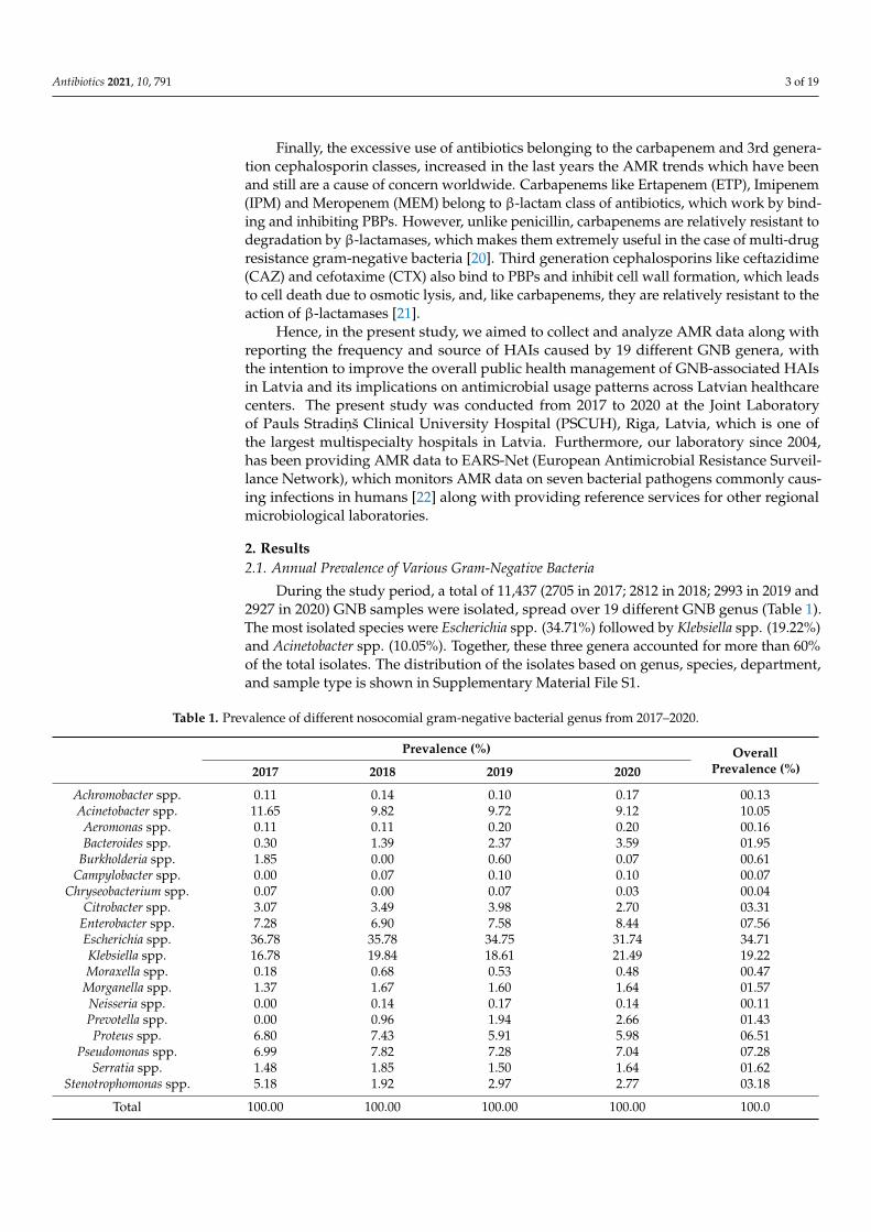
During the study period, a total of 11,437 (2705 in 2017; 2812 in 2018; 2993 in 2019 and2927 in 2020) GNB samples were isolated, spread over 19 different GNB genus (Table 1).The most isolated species were Escherichia spp. (34.71%) followed by Klebsiella spp. (19.22%)and Acinetobacter spp. (10.05%). Together, these three genera accounted for more than 60%of the total isolates. The distribution of the isolates based on genus, species, department,and sample type is shown in Supplementary Material File S1.
Table 1. Prevalence of different nosocomial gram-negative bacterial genus from 2017–2020.
Prevalence (%) OverallPrevalence (%)2017 2018 2019 2020
Achromobacter spp. 0.11 0.14 0.10 0.17 00.13Acinetobacter spp. 11.65 9.82 9.72 9.12 10.05
Aeromonas spp. 0.11 0.11 0.20 0.20 00.16Bacteroides spp. 0.30 1.39 2.37 3.59 01.95
Burkholderia spp. 1.85 0.00 0.60 0.07 00.61Campylobacter spp. 0.00 0.07 0.10 0.10 00.07
Chryseobacterium spp. 0.07 0.00 0.07 0.03 00.04Citrobacter spp. 3.07 3.49 3.98 2.70 03.31
Enterobacter spp. 7.28 6.90 7.58 8.44 07.56Escherichia spp. 36.78 35.78 34.75 31.74 34.71Klebsiella spp. 16.78 19.84 18.61 21.49 19.22Moraxella spp. 0.18 0.68 0.53 0.48 00.47
Morganella spp. 1.37 1.67 1.60 1.64 01.57Neisseria spp. 0.00 0.14 0.17 0.14 00.11Prevotella spp. 0.00 0.96 1.94 2.66 01.43Proteus spp. 6.80 7.43 5.91 5.98 06.51
Pseudomonas spp. 6.99 7.82 7.28 7.04 07.28Serratia spp. 1.48 1.85 1.50 1.64 01.62
Stenotrophomonas spp. 5.18 1.92 2.97 2.77 03.18
Total 100.00 100.00 100.00 100.00 100.0
Antibiotics 2021, 10, 791 4 of 19
2.2. Weighted Cumulative Antibiotic Resistance Rates and Trends over the Study Period
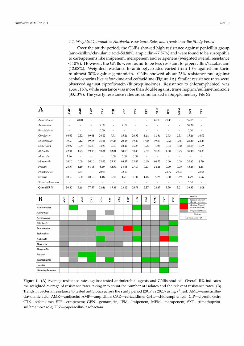
Over the study period, the GNBs showed high resistance against penicillin group(amoxicillin/clavulanic acid–50.80%; ampicillin–77.57%) and were found to be susceptibleto carbapenems like imipenem, meropenem and ertapenem (weighted overall resistance< 10%). However, the GNBs were found to be less resistant to piperacillin/tazobactam(12.08%). Weighted resistance to aminoglycosides varied from 10% against amikacinto almost 30% against gentamicin. GNBs showed about 25% resistance rate againstcephalosporins like cefotaxime and ceftazidime (Figure 1A). Similar resistance rates wereobserved against ciprofloxacin (fluoroquinolone). Resistance to chloramphenicol wasabout 16%, while resistance was more than double against trimethoprim/sulfamethoxazole(33.13%). The yearly resistance rates are summarized in Supplementary File S2.
Figure 1. (A) Average resistance rates against tested antimicrobial agents and GNBs studied. Overall R% indicatesthe weighted average of resistance rates taking into count the number of isolates and the relevant resistance rates. (B)Trends in bacterial resistance to tested antibiotics across the study period (2017 vs 2020) using χ2 test. AMC—amoxicillin-clavulanic acid; AMK—amikacin; AMP—ampicillin; CAZ—ceftazidime; CHL—chloramphenicol; CIP—ciprofloxacin;CTX—cefotaxime; ETP—ertapenem; GEN—gentamicin; IPM—Imipenem; MEM—meropenem; SXT—trimethoprim-sulfamethoxazole; TPZ—piperacillin-tazobactam.
Antibiotics 2021, 10, 791 5 of 19
The χ2 test was used to analyze the antimicrobial resistance rates for each bacterialgenus in the first (2017) and last (2020) study year as shown in Figure 1B. In 90 out of111 (81.1%) bacteria–antimicrobial agent combinations, no significant change in resistancerates were observed. Nineteen (17.1%) bacteria–antimicrobial agent combinations showeda significant decrease in resistance rates. Only two combinations showed a significantincrease in resistance rates—Enterobacter spp. showed a significant increase of 18.4% inresistance rates against Ertapenem (ETP) (p = 0.000; Φ = 0.241) while Klebsiella spp. showeda significant increase of 12.4% in resistance rates against Trimethoprim-Sulfamethoxazole(SXT) (p = 0.000; Φ = 0.124).
2.3. Overall Weighted Multiple Antibiotic Resistance (MAR) Index
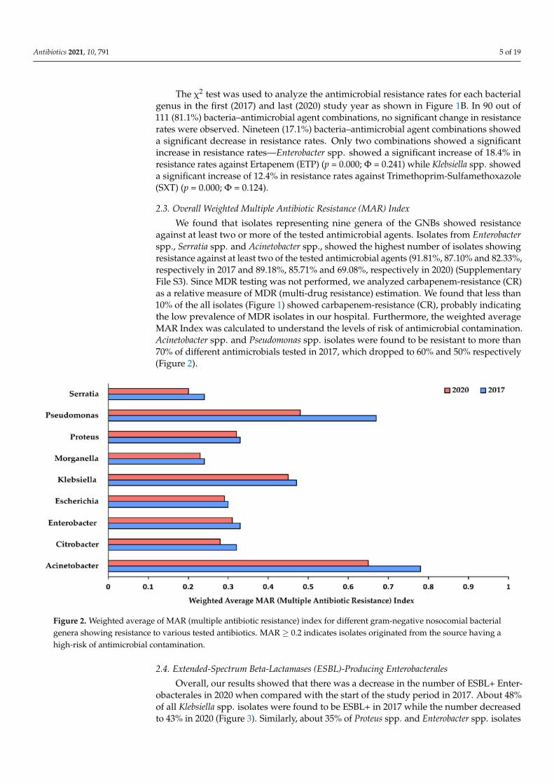
We found that isolates representing nine genera of the GNBs showed resistanceagainst at least two or more of the tested antimicrobial agents. Isolates from Enterobacterspp., Serratia spp. and Acinetobacter spp., showed the highest number of isolates showingresistance against at least two of the tested antimicrobial agents (91.81%, 87.10% and 82.33%,respectively in 2017 and 89.18%, 85.71% and 69.08%, respectively in 2020) (SupplementaryFile S3). Since MDR testing was not performed, we analyzed carbapenem-resistance (CR)as a relative measure of MDR (multi-drug resistance) estimation. We found that less than10% of the all isolates (Figure 1) showed carbapenem-resistance (CR), probably indicatingthe low prevalence of MDR isolates in our hospital. Furthermore, the weighted averageMAR Index was calculated to understand the levels of risk of antimicrobial contamination.Acinetobacter spp. and Pseudomonas spp. isolates were found to be resistant to more than70% of different antimicrobials tested in 2017, which dropped to 60% and 50% respectively(Figure 2).
Figure 2. Weighted average of MAR (multiple antibiotic resistance) index for different gram-negative nosocomial bacterialgenera showing resistance to various tested antibiotics. MAR ≥ 0.2 indicates isolates originated from the source having ahigh-risk of antimicrobial contamination.
2.4. Extended-Spectrum Beta-Lactamases (ESBL)-Producing Enterobacterales
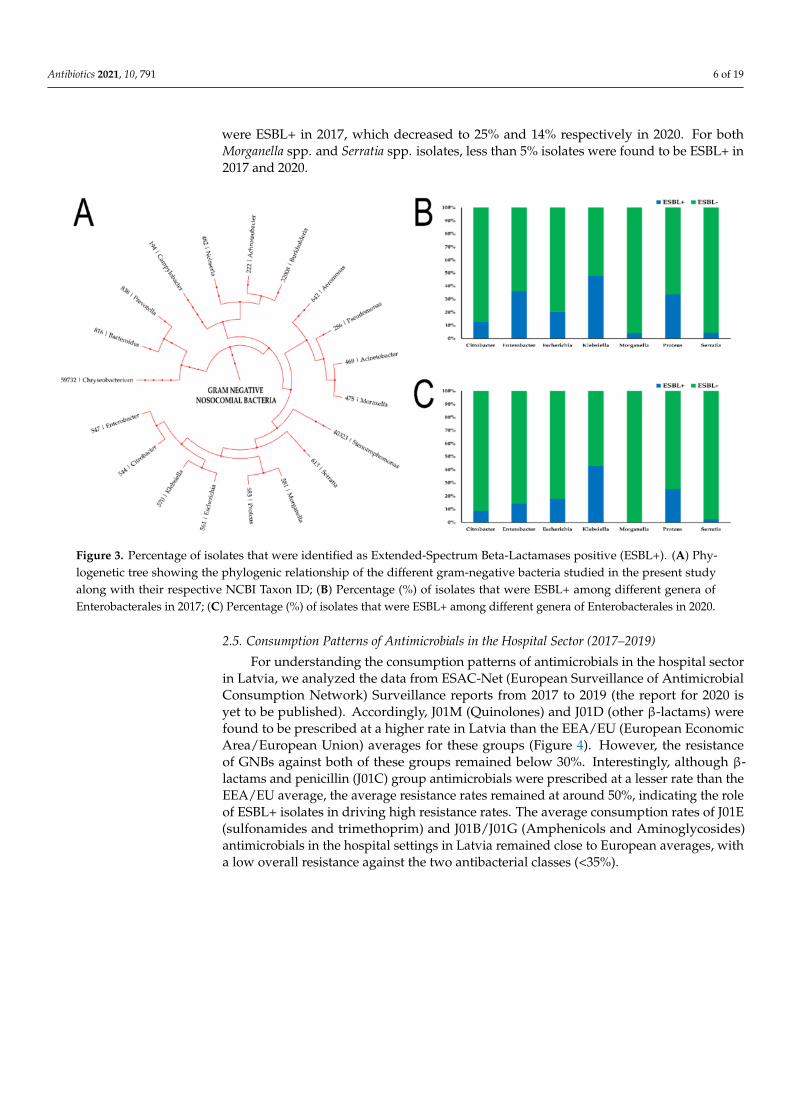
Overall, our results showed that there was a decrease in the number of ESBL+ Enter-obacterales in 2020 when compared with the start of the study period in 2017. About 48%of all Klebsiella spp. isolates were found to be ESBL+ in 2017 while the number decreasedto 43% in 2020 (Figure 3). Similarly, about 35% of Proteus spp. and Enterobacter spp. isolates
Antibiotics 2021, 10, 791 6 of 19
were ESBL+ in 2017, which decreased to 25% and 14% respectively in 2020. For bothMorganella spp. and Serratia spp. isolates, less than 5% isolates were found to be ESBL+ in2017 and 2020.
Figure 3. Percentage of isolates that were identified as Extended-Spectrum Beta-Lactamases positive (ESBL+). (A) Phy-logenetic tree showing the phylogenic relationship of the different gram-negative bacteria studied in the present studyalong with their respective NCBI Taxon ID; (B) Percentage (%) of isolates that were ESBL+ among different genera ofEnterobacterales in 2017; (C) Percentage (%) of isolates that were ESBL+ among different genera of Enterobacterales in 2020.
2.5. Consumption Patterns of Antimicrobials in the Hospital Sector (2017–2019)
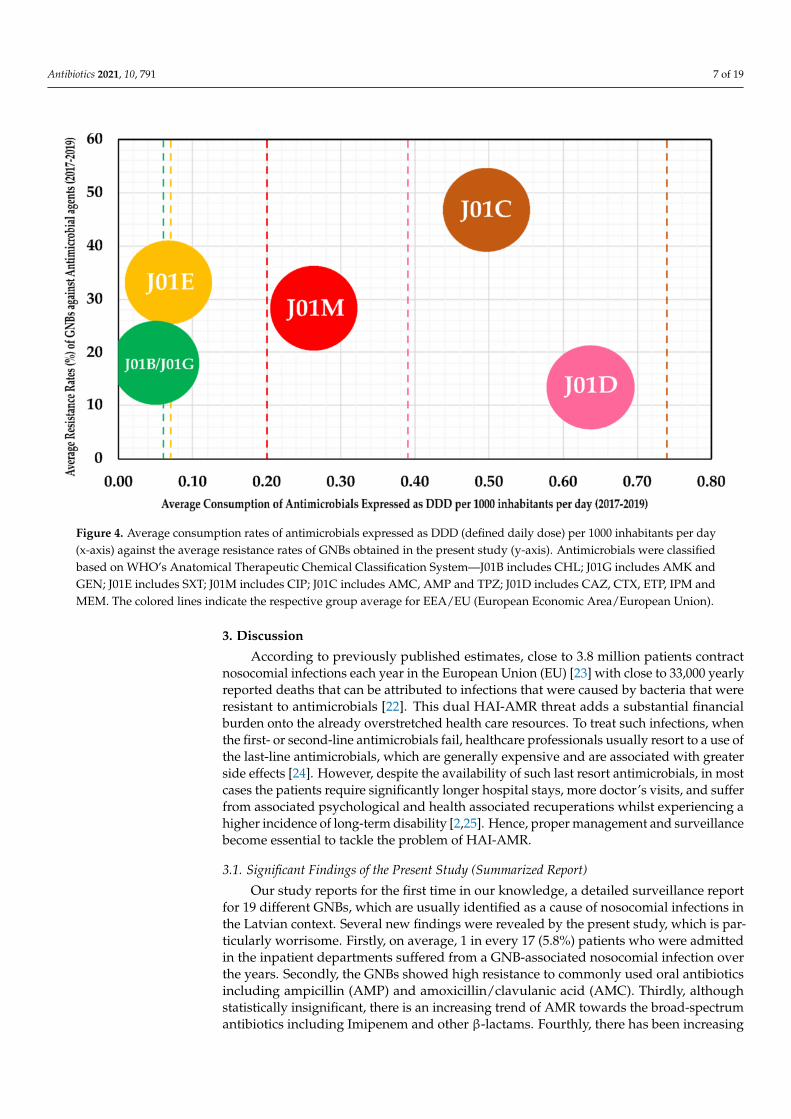
For understanding the consumption patterns of antimicrobials in the hospital sectorin Latvia, we analyzed the data from ESAC-Net (European Surveillance of AntimicrobialConsumption Network) Surveillance reports from 2017 to 2019 (the report for 2020 isyet to be published). Accordingly, J01M (Quinolones) and J01D (other β-lactams) werefound to be prescribed at a higher rate in Latvia than the EEA/EU (European EconomicArea/European Union) averages for these groups (Figure 4). However, the resistanceof GNBs against both of these groups remained below 30%. Interestingly, although β-lactams and penicillin (J01C) group antimicrobials were prescribed at a lesser rate than theEEA/EU average, the average resistance rates remained at around 50%, indicating the roleof ESBL+ isolates in driving high resistance rates. The average consumption rates of J01E(sulfonamides and trimethoprim) and J01B/J01G (Amphenicols and Aminoglycosides)antimicrobials in the hospital settings in Latvia remained close to European averages, witha low overall resistance against the two antibacterial classes (<35%).
Antibiotics 2021, 10, 791 7 of 19
Figure 4. Average consumption rates of antimicrobials expressed as DDD (defined daily dose) per 1000 inhabitants per day(x-axis) against the average resistance rates of GNBs obtained in the present study (y-axis). Antimicrobials were classifiedbased on WHO’s Anatomical Therapeutic Chemical Classification System—J01B includes CHL; J01G includes AMK andGEN; J01E includes SXT; J01M includes CIP; J01C includes AMC, AMP and TPZ; J01D includes CAZ, CTX, ETP, IPM andMEM. The colored lines indicate the respective group average for EEA/EU (European Economic Area/European Union).
3. Discussion
According to previously published estimates, close to 3.8 million patients contractnosocomial infections each year in the European Union (EU) [23] with close to 33,000 yearlyreported deaths that can be attributed to infections that were caused by bacteria that wereresistant to antimicrobials [22]. This dual HAI-AMR threat adds a substantial financialburden onto the already overstretched health care resources. To treat such infections, whenthe first- or second-line antimicrobials fail, healthcare professionals usually resort to a use ofthe last-line antimicrobials, which are generally expensive and are associated with greaterside effects [24]. However, despite the availability of such last resort antimicrobials, in mostcases the patients require significantly longer hospital stays, more doctor’s visits, and sufferfrom associated psychological and health associated recuperations whilst experiencing ahigher incidence of long-term disability [2,25]. Hence, proper management and surveillancebecome essential to tackle the problem of HAI-AMR.
3.1. Significant Findings of the Present Study (Summarized Report)
Our study reports for the first time in our knowledge, a detailed surveillance reportfor 19 different GNBs, which are usually identified as a cause of nosocomial infections inthe Latvian context. Several new findings were revealed by the present study, which is par-ticularly worrisome. Firstly, on average, 1 in every 17 (5.8%) patients who were admittedin the inpatient departments suffered from a GNB-associated nosocomial infection overthe years. Secondly, the GNBs showed high resistance to commonly used oral antibioticsincluding ampicillin (AMP) and amoxicillin/clavulanic acid (AMC). Thirdly, althoughstatistically insignificant, there is an increasing trend of AMR towards the broad-spectrumantibiotics including Imipenem and other β-lactams. Fourthly, there has been increasing
Antibiotics 2021, 10, 791 8 of 19
resistance in Klebsiella spp. and Enterobacter spp. isolates against second-line or last resortantimicrobials. Finally, there were higher national rates of consumption of Quinolones(J01M) and other β lactams (J01D) in the hospital settings when compared with theEEA/EU average.
However, our findings also indicate some encouraging trends in HAI-AMR that needto be pursued further to tackle the problem of AMR. Firstly, there has been a decrease in thenumber of ESBL+ isolates over the years. This decrease is crucial, since these GNBs produceenzymes called beta-lactamases, which breakdown the beta-lactam rings in antibiotics likepenicillin and amoxicillin, rendering the antibiotics useless against infections [26]. This isevident in the case of Proteus spp. Isolates, as the number of ESBL+ isolates decreased from2017 to 2020, the AMR against ampicillin (AMP) also decreased significantly (p = 0.039;Φ = 0.113). Secondly, apart from Klebsiella spp. and Enterobacter spp. isolates, other GNBsshowed either no significant trend changes or rather showed a significant decrease in AMRtrends. This is very crucial, especially in the context of members of genera Acinetobacter spp.and Pseudomonas spp. since both are frequently known to cause life-threatening conditionsthat are difficult to treat due to their intrinsic resistance to many antimicrobial agents.
Nonetheless, in comparison with the EU/EEA average, the AMR rates remainedhigh in Latvia. For example, the EU AMR average against Piperacillin-Tazobactam forPseudomonas spp. isolates were around 16.7% from 2017 to 2019, whilst our isolates showedalmost double AMR rates averaging around 31% (Supplementary File S2) [22]. A similarsituation was noted for carbapenems with resistance in our isolates around 28% againstthe European average of 17%. For Acinetobacter spp. isolates, the situation is even worse.Whilst the EU average resistance rates were about 32.5% (2017–2019) against carbapenems,our samples showed an average resistance of 72%. We found that the resistance ratesare 2- to 3-fold higher in our isolates than the EU average for Acinetobacter spp. againstaminoglycosides like gentamycin and amikacin.
Apart from the traditional GNBs, species like Escherichia spp., Klebsiella spp., Pseu-domonas spp., Acinetobacter spp., Enterobacter spp. and many other GNBs, genera arebecoming an increasing concern in the hospital setting. GNBs like Citrobacter spp., Mor-ganella spp., Serratia spp. and Stenotrophomonas spp. accounted for about 10% of the totalGNB isolates in our study, indicating the need for increased reporting and surveillanceof these genera as well. Furthermore, despite the low prevalence rates of ESBL+ iso-lates of Citrobacter spp., Morganella spp. and Serratia spp., the AMR against ampicillin(AMP) and amoxicillin/clavulanic acid (AMC) remained very high (above 80%). How-ever, they showed minimal resistance to aminoglycosides, which can serve as a potentialtreatment avenue.
3.2. ESKAPE Pathogens–AMR in Klebsiella spp.
Members of this genus are well known to clinicians, both in the outpatient andinpatient setting. They are the causative agents behind community-acquired pneumonia,especially in chronic alcoholics [27]. However, the majority of the infection cases are ratherassociated with hospitalization. As an opportunistic pathogen, the two most importantmembers of the genus—K. pneumoniae and K. oxytoca—are commonly found to targetpatients with underlying conditions like diabetes mellitus (DM) or chronic obstructivepulmonary disease (COPD) [28]. Blood products, contaminated medical equipment, thegastrointestinal tract of patients and the hands of hospital personnel represent the primaryreservoirs and transmission source for Klebsiella species [29,30]. Certain medical procedureslike an endoscopy can also lead to the transmission of the bacteria [31]. While K. pneumoniaeis commonly associated with urinary tract infections (UTI), bronchopneumonia, soft tissueinfections, and bacteremia, K. oxytoca is additionally associated with colitis and infectiveendocarditis [32]. The situation is extremely worrisome, given the frequent isolation of theKlebsiella species from the neonatal departments and wards.
In our study, roughly 15% of the Klebsiella spp. isolates were identified as K. oxytoca,while the vast majority (approximately 80%), were identified as K. pneumoniae. About a
Antibiotics 2021, 10, 791 9 of 19
third of the samples were collected from urine specimens, followed by bronchoalveolarlavage (20%) and blood specimens (7%). Based on departments, intensive care, lungdiseases and thoracic surgery, and nephrology accounted for close to 45% of the isolates.On the other hand, the neonatal department reported a rather lower caseload (close to 1.5%)in the study period (Supplementary File S1). Two major mechanisms have been reportedto be responsible for inferring antimicrobial resistance to K. pneumoniae. The first beingproduction of ESBL and the second being point mutations in genes encoding penicillin-binding proteins (PBPs) [33,34]. Furthermore, it has been shown that indiscriminate useof antibiotics upregulates the expression of ESBL [35]. We found that >40% of the isolateswere ESBL+ with isolates showing complete resistance against ampicillin (average 99.93%).
Another resistance mechanism described is against quinolones (J01M; ciprofloxacin).Point mutations in DNA gyrase genes gyrA and gyrB along with topoisomerase IV genesparC and parE are thought to be the main mechanisms against quinolone resistance [36].Although no significant change in resistance against CIP was noted, a downward trend wasstill noted with an average resistance rate of around 38%, indicating a relatively moderateprevalence of such mutations in the isolates. Additionally, a significant reduction in AMRrates was noted against GEN, TPZ and CHL. The % of carbapenem resistant isolates alsoremained low (<10% for ETP; <1% for IPM and MEM). However, more than a third of theisolates were found to be resistant against third generation cephalosporins like CTX andCAZ. Furthermore, the weighted MAR index remained close to 0.5, indicating that theisolates were collected from departments with a high antimicrobial use and around 53%of the isolates showed resistance against at least two of the tested antimicrobial agents(Supplementary File S3).
3.3. ESKAPE Pathogens—AMR in Acinetobacter spp.
This genus represents a heterogeneous group of ubiquitous free-living saprophytesthat are part of the normal flora of the skin, mucous membranes, pharynx, and humanrespiratory secretions [37–39]. However, members of the genus have also been isolatedfrom the forehead, conjunctiva, hand, vagina, perineum, axillae, groin, and toe webs inhealthy individuals [40]. Due to their ability to survive dry conditions over long periods,they are frequently isolated from reusable medical equipment like ventilator tubing, arterialpressure monitoring devices, humidifiers, washbasins, plastic urinals, respirometers, theskin of healthcare personnel and bed linens and mattress [41,42]. We found that mostof the specimens were isolated from bronchoalveolar lavage, tracheal aspirate, woundsite and urine samples (Supplementary File S1). A. baumannii infections usually involveorgan systems that contain high levels of fluids like the urinary and respiratory tract andperitoneal cavity [39]. It is frequently associated with hospital-acquired pneumonia withan isolate frequency of around 3–5% in ICUs (intensive care units) [43]. This is interesting,since we recorded more than a 10× isolation rate of around 35% in ICUs. A. baumannii hasalso been associated with bacteremia, UTI, trauma and wound infections, meningitis, andendocarditis [39]. About 10% of our isolates were collected from the neurology department,along with 15% from the cardiology department, which makes it essential to control thespread of Acinetobacter species.
Species in this genus have been labeled as “naturally transformable” due to their re-markable capacity for the acquisition of foreign genetic material including AMR genes [44].Several mechanisms of gene transfer have been described, including transformation [45],conjugation [46], transduction [47] and mobile genetic elements like plasmids, transposonsand integrons [48]. Resistance to AMK and GEN (aminoglycosides) is usually due in thepresence of gene coding for aminoglycoside-modifying enzymes (AME) in integrons [38],while resistance to CIP is due to mutations in the gyrA and parC genes. While resistanceagainst CIP was not tested, resistance against AMK and GEN remained very high, withAMR rates of about 70% and 60%, respectively. Carbapenem-resistant A. baumannii is a ma-jor concern, with the WHO (World Health Organization) putting it in the priority categoryagainst which research and development of new antibiotics is critical [49]. Production of
Antibiotics 2021, 10, 791 10 of 19
carbapenemase, altered function of membrane-associated proteins and activation of drugefflux pumps have been described as the resistance mechanism against carbapenems [50].Resistance rates against IPM remained very high (>70%) amongst our isolates. Finally, theweighted MAR index remained one of the highest in Acinetobacter spp., indicating that theisolates were resistant to most of the tested antimicrobial agents.
3.4. ESKAPE Pathogens–AMR in Pseudomonas spp.
Another frequent and difficult-to-treat causative agent, Pseudomonas spp., are knownto colonize moist dark places like medical ventilators, oxygen respirators, humidifiers,sinks, taps, toilets, and dialysis machines [51,52]. In the hospital environment, Pseudomonasspp. are commonly identified as causative agents of bacteremia, pneumonia, urosepsis,and wound infection including the secondary infection of burns [53]. Multiple risk factors,including chemotherapy-induced neutropenia, surgical incisions, insertion of urinaryand vascular catheters, endotracheal tubes, multiple organ injury and failure along withunderlying comorbidities (like DM, COPD, cystic fibrosis etc.), have been reported [52,53].Our analysis showed that about a third of the specimens were isolated from urine samplesfollowed by wound site, ulcers and bronchoalveolar lavage. Based on departments, thehighest caseload was in the Nephrology department, followed by lung disease and thoracicsurgery and ICU.
Generally, the antibiotic resistance mechanisms in this genus are classified as in-trinsic (low outer membrane permeability, efflux pumps and production of antibiotic-inactivating enzymes), acquired (mutational changes and horizontal gene transfer) andadaptive (formation of biofilms) [54–57]. This makes the genus resistant to a wide varietyof antibiotic classes, including aminoglycosides, quinolones, and β-lactams [54] and earnsit (carbapenem resistant P. aeruginosa) a place right with A. baumannii in the WHO classof priority category against which new antibiotics are critically needed [49]. Our isolatesshowed about 20–30% resistance rates against 3rd generation cephalosporins, quinolonesand carbapenems with a significant decrease in resistance rates in 2020 compared to 2017.Resistance against aminoglycosides (AMK) remained very low (around 2%), which couldbe used for the treatment of pseudomonal infections. Furthermore, amongst ESKAPEpathogens, Pseudomonas spp. and Enterobacter spp. isolates remained relatively less com-monly isolated. Nonetheless, the weighted MAR index remained close to 0.5 in 2020,indicating resistance against half of the tested antimicrobial agents.
3.5. ESKAPE Pathogens–AMR in Enterobacter spp.
Finally, in terms of the last of the Gram-negative ESKAPE pathogens, members of thisgenus are responsible for causing nosocomial UTI, lower respiratory infections, soft tissueinfections, osteomyelitis, surgical site infections, bacteremia and endocarditis, among manyothers [58]. Some members of the genus are known to be part of the normal microfloraof gastrointestinal and respiratory tracts [59,60]. Transmission in hospital departmentsis mainly due to contaminations caused by intravenous injection fluids, blood products,stethoscopes, cotton swabs, and colonized hands of healthcare professionals [61]. Amongstthe known 22 species, E. cloacae remains the most isolated and studied in the contextof nosocomial infections. Indeed, E. cloacae remained the most isolated species (>85%)in our samples followed by E. aerogenes and E. asburiae (Supplementary File S1). Urine,bronchoalveolar lavage and wound site remained the most common samples from whichEnterobacter species were isolated (>50%). Nephrology, lung diseases and thoracic surgeryalong with ICU remained the departments with the highest caseload.
Enterobacter spp. showcase intrinsic resistance to penicillin, amoxicillin-clavulanate,first-generation cephalosporins, and cefoxitin due to their ability to produce AmpC β-lactamase [62]. It was evident in our isolates as well, achieving close to 100% resistancerates against AMC and AMP. Although the prevalence of ESBL+ Enterobacter spp. decreasedfrom close to 35% in 2017 to about 15% in 2020, the resistance against beta-lactams did notchange, indicating the presence of certain acquired resistance mechanisms in the isolates.
Antibiotics 2021, 10, 791 11 of 19
Resistance to carbapenems is conferred due to its carbapenemase production capability.Although resistance against ETP remained close to 19% (a significant increase from 2017 wasnoted), resistance against MEM and IPM remained below 1%, indicating low prevalencerates of carbapenem-resistant Enterobacter spp. and over-dependence on ETP for treatmentof enterobacterial infections. Weighted MAR index also remained relatively low at 0.3,indicating that the isolates showed resistance against 30% of the tested antimicrobial agents.Resistance against 3rd generation cephalosporins showed no significant trend change, withaverage resistance rates at around 40%.
3.6. AMR Rates in Other Enterobacterales Order Members (CESP and beyond)
Enterobacterales include a large order of ESBL-producing gram-negative bacteriawhich have been identified as causative nosocomial agents. CESP represents a subgroupwithin this order which are known to produce AmpC-encoded β-lactamases. The subgroupincludes Citrobacter spp., Enterobacter spp., Serratia spp. and Providencia spp. (data notshown). Outside this subgroup, Morganella spp. and Pseudomonas spp. are also knownto produce AmpC-encoded β-lactamases [63]. Escherichia spp. and Proteus spp. were theremaining order members that were included in the present study.
The genus Citrobacter is represented by more than ten species that are known to resideinside the intestines of animals and humans. Of these species, C. freundii is associatedwith gastroenteritis, UTI, neonatal meningitis, and septicemia, while C. koseri causesneonatal meningitis and brain abscesses [64,65]. Citrobacter spp. are often considered asemerging nosocomial agents, especially in developing countries. Invasive procedureslike catheterization or genitourinary instrumentation, along with immune suppressionand prolonged hospitalization durations, put the patients at risk of infection [66]. In ourhospital, the Citrobacter spp. caseload was around 3% of all the GNB genera studied.Although C. freundii and C. koseri were the most isolated species, there was a more than 10×increase in isolates of C. braakii. Most of the specimens were isolated from urine, sputum,wound site, bronchoalveolar lavage and stool samples. Nephrology, neurology, and generalsurgery, along with thoracic surgery and cardio surgery departments, showed the highestincidence of the isolates. Like other genera in Enterobacterales, resistance against AMP andAMC remained high (>80%). No significant change in AMR rates was observed, except asignificant decrease against TPZ during the study period. Resistance against carbapenemsremained below 1% for IPM and MEM and around 8% against ETP, while resistance againstthird generation cephalosporins remained at around 26%.
Infections caused by Serratia spp. are rarely community acquired; however, they areincreasingly being recognized as a major cause of nosocomial infections. Of the 14 knownspecies, S. marcescens, S. liquefaciens and S. odorifera are the most known and clinically im-plicated [67,68]. As an opportunistic bacterium, it is generally known to cause bacteremia,UTI and respiratory tract infections, with patients undergoing invasive procedures likeindwelling and intravenous catheterization and respiratory intubation [69]. Our resultsindicate that there was a decrease in the % of S. marcescens, while an increase in S.liquefacienswas noted. Bronchoalveolar lavage, tracheal aspirate, urine, and sputum samples were themost common isolation samples. Thoracic surgery, ICU, cardiology, and vascular surgerydepartments remained the most burdened with the caseload. Resistance to penicillinremained at 100%, while resistance against carbapenems and cephalosporins remainedbelow 5%. Resistance against other classes as well remained below or around 5%. ESBL+samples were rarely isolated (5% in 2017 which decreased to 3% in 2020).
Morganella morganii is an unusual opportunistic bacterium that has been known torarely cause nosocomial infection [70]. It is known to cause UTI and postoperative woundinfections along with sepsis, abscess, purple urine bag syndrome, chorioamnionitis, andcellulitis [71]. It is often considered a bacterium with increased levels of resistance andvirulence. Old age, concomitant bacteremia, prolonged hospitalization, recent surgery, andconcurrent antibiotic use have been established as risk factors for Morganella infection [72].Furthermore, transmission from animals (like dogs, snakes) via bites or scratches to an
Antibiotics 2021, 10, 791 12 of 19
immunocompromised host is also seen as a risk factor [71,73]. Administration of suchpatients should not be overlooked and could be potentially dangerous, given the associ-ated high mortality rates [71]. We isolated most specimens from urine and trophic ulcersamples (approx. 40%) with nephrology and general surgery departments showcasing thehighest caseload. The bacterium has an intrinsic resistance to penicillin, while it remainsgenerally susceptible to aminoglycosides, quinolones, carbapenems and third generationcephalosporins [74]. We also found that isolates showed complete resistance against AMCand AMP but less than 10% resistance rates against all other antibiotic classes tested.
Escherichia spp. are known to be involved in a broad spectrum of nosocomial infections,including UTI, septicemia, pneumonia, neonatal meningitis, peritonitis, and gastroenteri-tis [75]. In our hospital, the genus remained the most isolated (34%) with urine, blood andwound site samples showing the most isolates. Based on the department, most isolateswere collected from nephrology, general surgery, gastroenterology, and endocrinologydepartments. About 15–20% of the isolates showcased ESBL activity with moderate resis-tance rates against AMC. Resistance against SXT and AMP were relatively higher thanAMC. The bacterium showed low resistance against aminoglycosides and carbapenems.Finally, members of the genus Proteus spp. are usually involved in community-acquiredinfections. However, about 10% of the cases are reported to be nosocomial [76]. Theyare most associated as causative agents of UTI, which usually occurs due to bacterialmigration along the mucosal sheath of the catheter or up the catheter lumen from contami-nated urine [77,78]. Female sex, immunodeficiency, unprotected sex, longer duration ofcatheterization, improper catheter cleaning or care etc., are the established risk factors forinfection by Proteus species. P. mirabilis and P. vulgaris remain the most isolated speciesin our hospital. Most of the specimens were isolated from urine and wound site sampleswith the highest caseload reported by nephrology, neurology, endocrinology, and ICUdepartments. Resistance against penicillin, amphenicols, quinolones and SXT remainedmoderate to high, while resistance to carbapenems and third generation cephalosporinsremained low. About 25–35% of the samples showed ESBL activity.
3.7. AMR Rates in Other Studied Gram-Negative Genera
Of the remaining 10 genera included in the present study, AMR trends were observedfor species belonging to four genera—Aeromonas spp., Burkholderia spp., Moraxella spp.,and Stenotrophomonas spp. Resistance against other genera was not tested, mainly due tolow isolation frequency. Aeromonads are emerging nosocomial enteric pathogens that arepart of the normal intestinal microflora [79]. They usually colonize the water distributionsystem by forming biofilms in the water channels, especially during the summer [79,80].A. caviae is the predominant isolate with A. hydrophila and A. veronii showing specificgeographical distribution. Apart from their association with diarrhea (acute, chronic andtraveler’s), multiple extra-intestinal manifestations like skin and soft tissue infections, andlower respiratory and urinary tract infections have also been reported [80]. They are uni-versally resistant against penicillin; however, they show susceptibility to aminoglycosides,tetracycline, chloramphenicol, trimethoprim-sulfamethoxazole, and quinolones [79–81].Over four years of the study, only 18 isolates were recorded with nephrology, surgery, andICU departments with the highest caseload. Our isolates showed less than 10% resistanceagainst CIP and CAZ, while moderate resistance against SXT was noted.
Burkholderia cepacia causes severe lung infections in cystic fibrosis and immunocom-promised patients. Transmission is usually due to exposure to contaminated solutions suchas antiseptics, disinfectants, nebulizer solution, and dextrose solution in a hospitalized pa-tient [82–84]. Most of the isolates in the present study were collected from bronchoalveolarlavage, tracheal aspirate, and the wound site. ICU and cardiology departments remainedthe most burdened. All our isolates were found to be susceptible to CAZ and SXT withno resistance observed. Moraxella is a common nosocomial agent in ventilated patients,causing upper and lower respiratory tract infections [85]. Crowding, winter season, pro-longed stay in a medical institution, and respiratory therapy are some of the established
Antibiotics 2021, 10, 791 13 of 19
risk factors for infection [85,86]. Most specimens were collected from sputum and bron-choalveolar lavage in the present study, with thoracic surgery and cardiology having thehighest caseload. Resistance was found to be less than 3% against AMC and 0% againstamphenicols, quinolones and third-generation cephalosporins.
Finally, Stenotrophomonas spp. cause infections that are usually associated with highmortality and morbidity and have been associated with bacteremia, pneumonia, endocardi-tis, meningitis, UTI, soft tissue and gastrointestinal infections [87]. We isolated most ofthe specimens from urine and bronchoalveolar lavage samples with surgery, cardiology,and thoracic surgery departments with the highest isolation frequency. Resistance againstSXT remained low at roughly 3%, despite the high intrinsic resistance against multipleantimicrobial classes. Amongst the other remaining six genera we included, incidencefrequency was high for Bacteroides and Prevotella. Only a few isolated cases were observedfor Achromobacter, Campylobacter, Chryseobacterium and Neisseria. Mostly, patients with theseisolates were treated empirically and were based on relevant national and EU guidelines.
3.8. Infection Control and Controlling of AMR Rates
Antimicrobial Stewardship is a crucial and much-needed remedy in controlling AMRrates in nosocomial isolates. It requires a precise and delicate balance involving selection,dosage, and duration of antimicrobial treatment with the end goal being to achieve thebest clinical outcome and prevention of infection with minimal impact on subsequentresistance [88]. To achieve the goal, a collaborative effort is required at the hospital levelwith infectious disease physicians, pharmacists, microbiologists, hospital epidemiologists,and administrative staff joining hands to formulate the necessary strategy [89]. A de-escalating backend approach is seen as the most efficient approach towards achievingstewardship [90,91]. It involves monitoring the current antibiotic prescription patternsand then formulating recommendations for clinicians to either continue, adjust, change, ordiscontinue the patterns based on all available resources and data [89].
Since this study represents the first of such monitoring efforts on a larger scale,encompassing yearly trends in our hospital, the next logical steps would be to formulateand implement proper strategies to control AMR rates, including antibiotic switching andcycling, dose optimization and prescription controls, in line with EU and ECDC guidelines.Furthermore, we would expand the surveillance and monitoring program to encompassmore GNBs apart from ESKAPE and other traditional GNBs. The present study alsohelps us to determine the baseline levels for caseloads that could, in the future, be used tounderstand and manage outbreaks in the hospital. In addition to controlling AMR rates, itis equally important to reduce the incidence and caseload of GNBs.
In our hospital, regular meetings and discussions are conducted between laboratoryspecialists and clinical infectious disease physicians to formulate recommendations re-garding the AMR trends and various approaches that can be used in limiting the spreadof inter- and intra-departmental nosocomial infections. Our laboratory is available forconsultations and offers educational programs including postgraduate training to nurses,physicians, and clinical microbiologists to help them be better prepared to tackle thetopical issues of antibiotic misuse and antimicrobial resistance. The obtained data andresults from the present study will be used for further training of these specialists, alongwith the implementation of more stringent and proper infection control methods andhygiene standards.
3.9. Limitations of the Present Study
Studies like the present one are essential for the implementation of proper infectioncontrol measures, to formulate national prescription guidelines and to control the problemof AMR in HAIs. However, the present study has several limitations. The study did notconduct resistance testing for all antimicrobials against the bacteria isolated, mainly due tolimited financial resources. However, testing was done based on the national and EUCASTguidelines and the prescription patterns in the Latvian healthcare settings. We did not
Antibiotics 2021, 10, 791 14 of 19
perform genotyping to identify specific clonal outbreaks and did not test patients in theoutpatient department (community AMR rates). Finally, since this is a single center study,the results may not completely reflect the national AMR rates. However, upon comparisonwith data available with ECDC, our results agreed with the Latvian national levels asavailable in ECDC, EARS-Net and ESAC-Net Surveillance reports. In the present study, wedid not investigate the prevalence of multidrug-resistant (MDR), extensively drug-resistant(XDR) and pandrug-resistant (PDR) species (due to insufficient testing against all agentsaccording to the guidelines and/or lack of molecular testing). However, according tothe WHO guidelines, it is not necessary to define isolates included in the global prioritylist of AR bacteria as MDR or XDR if they are carbapenem resistant. This is becausecarbapenemase-encoding genes are carried on mobile genetic elements that usually carrygenetic determinants for resistance to other antibiotics [92]. Hence, we just labeled theisolates as carbapenem resistant.
4. Materials and Methods4.1. Study Design and Location
A retrospective analysis of antimicrobial susceptibility data from microbiology lab-oratory was done for the years 2017–2020 (full years from Jan to Dec). The study wasconducted at Paul Stradins Clinical University Hospital (PSCUH), Riga, Latvia. PSCUHis a multispecialty hospital providing treatment and care in 26 medical fields catering topatients from both Riga (48%) and other cities and rural areas of Latvia (52%), with anestimated 310,000 patients receiving treatment/consultations each year (as of 2018) [93].Microbiological data of patients irrespective of age, gender etc., who fulfilled the ECDC(European Center for Disease Prevention and Control) criteria for nosocomial infections,were considered in the present study [94]. Ethical permission for the study was providedby the Clinical Research Ethics Committee of the PSCUH wide no. 290421-16L (29.04.2021).
4.2. Sample Collection and Laboratory Analysis
Patient specimens were collected by the clinicians or nurses in respective departmentsof the hospital and sent for microbiological investigation to the Joint Laboratory. Specimenscollected included pus, tracheal swab, blood, urine, catheter, bronchoalveolar lavage etc.Specimens with improper labeling and those with inadequate patient identifiers wereexcluded from the present study. Furthermore, similar bacterial species isolated from dif-ferent samples of the same patient were considered as a single isolate. Specimen processingand handling were done in accordance with the relevant EUCAST (European Committee onAntimicrobial Susceptibility Testing) guidelines. Genus and species identification was doneusing a Vitek2 analyzer or MALDI-TOF MS (matrix assisted laser desorption ionization-time of flight mass spectrometry). Antimicrobial susceptibility for various antibiotics wasdetermined using a standard disk diffusion method in accordance with EUCAST guide-lines. A summary of breakpoints used in the present study is shown in SupplementaryFile 2. EUCAST recommendations on breakpoint for interpretation of zone diameters v11.0(applicable from Jan 2021) were used for resistance rate analysis [95]. The breakpoints wereapplied retrospectively to eliminate any variances in interpretation of the results. Isolatesidentified as Intermediate (I) or Susceptible, Increased exposure (I) were considered asSusceptible (S). The national average antibiotic consumption patterns in the hospital settingwere obtained from ESAC-Net (European Surveillance of Antimicrobial ConsumptionNetwork) Surveillance reports 2017–2019 and were plotted against the average resistancerates we obtained in the present study.
4.3. Multiple Antibiotic Resistance (MAR) Index
By analyzing the resistance rates of a particular bacterial isolate against the totalnumber of tested antimicrobial agents, one can calculate the MAR Index [96]. This toolis an extremely useful source for tracking and analyzing the risk of multi-drug resistancein isolates. The index is calculated as the ratio of number of antimicrobials to which the
Antibiotics 2021, 10, 791 15 of 19
isolate is resistant, and the total number of antimicrobials exposed to the isolate [96,97].A MAR Index < 0.2 indicated low risk, while MAR Index ≥ 0.2 indicates a high risk ofantimicrobial contamination [96,97].
4.4. Data Collection and Analysis
The data about antimicrobial susceptibility, sample type, species, and the departmentfrom where the sample was isolated, was downloaded from the laboratory database andanalyzed for the period 2017–2020. Differences in antimicrobial susceptibility between thestart and the end of the study for each bacterial species was assessed using χ2 test. Phi(Φ) was used to determine the significance of positive or negative trends in changes inantimicrobial susceptibility over time. SPSS (v27.0, IBM Corp., Armonk, NY, USA) wasused for statistical analysis.
5. Conclusions
Antimicrobial resistance (AMR) in gram-negative bacteria (GNBs) poses a significantthreat, especially in the healthcare setting. To address the current emerging AMR ratesand the increased prevalence of non-traditional GNBs, constant monitoring, along with therevision of antimicrobial guidelines, is needed. The most significant findings of the presentstudy are:
Over the past four years (2017–2020), there have been no significant changes inAMR rates amongst nosocomial isolates of most GNBs. However, the overall resis-tance rates and the number of GNB isolates in Latvia remain relatively high compared toEuropean averages.
Furthermore, significantly increasing AMR rates in Klebsiella spp. and Enterobacter spp.isolates need to be handled with care and with proper infection control management.
Supplementary Materials: The following are available online at https://www.mdpi.com/article/10.3390/antibiotics10070791/s1. Supplementary File S1: Microbial Profile and Epidemiology; Sup-plementary File S2: Antimicrobial Resistance Results; Supplementary File S3: Multiple AntibioticResistance (MAR) Index Results.
Author Contributions: The present study was conceptualized and designed by N.J. and A.R. Patientspecimen collection and processing was done by D.S. Disk diffusion and AMR result interpretationwas done by I.J. and T.O. Data analysis, statistics, literature search and visualization of the data ofdone by N.J. Project supervision was done by R.S., J.M., D.S. and A.R. Initial draft was written by N.J.while reviewing and editing was done by N.J., R.S., J.M., D.S. and A.R. All authors have read andagreed to the published version of the manuscript.
Funding: This research received no external funding.
Institutional Review Board Statement: Ethical permission for the study was provided by the ClinicalResearch Ethics Committee of the PSCUH wide no. 290421-16L (29.04.2021). The protocol wasdesigned in accordance with the Declaration of Helsinki guidelines.
Informed Consent Statement: Patient consent was waived as the study was part of assessment ofquality control for institutional infection control and prevention.
Data Availability Statement: All data analyzed have been presented in a summarized way in Supple-mentary Files 1 and 2. Patient data cannot be provided due to confidentiality and privacy concerns.
Acknowledgments: We would like to extend our gratitude to the management of Pauls StradinšClinical University Hospital (PSCUH) for their support in the present study. The authors would alsolike to thank all the supporting hospital and laboratory staff.
Conflicts of Interest: The authors declare no competing interest in the present study. Furthermore,the hospital administration had no role in the design of the study; in the collection, analysis, orinterpretation of the data; in the writing of the manuscript, or in the decision to publish the results.
Antibiotics 2021, 10, 791 16 of 19
References1. World Health Organization (WHO). Q&A Detail. 27 July 2017. Available online: https://www.who.int/news-room/q-a-detail/
antimicrobial-resistance (accessed on 15 April 2021).2. Ventola, C.L. The antibiotic resistance crisis: Part 1: Causes and threats. Pharm. Ther. 2015, 40, 277–283.3. Friedrich, A.W. Control of hospital acquired infections and antimicrobial resistance in Europe: The way to go. Wien. Med.
Wochenschr. 2019, 169, 25–30. [CrossRef]4. Reed, D.; Kemmerly, S.A. Infection control and prevention: A review of hospital-acquired infections and the economic implications.
Ochsner J. 2009, 9, 27–31.5. Golkar, Z.; Bagasra, O.; Pace, D.G. Bacteriophage therapy: A potential solution for the antibiotic resistance crisis. J. Infect. Dev.
Ctries. 2014, 8, 129–136. [CrossRef] [PubMed]6. Jang, T.N.; Kuo, B.I.; Shen, S.H.; Fung, C.P.; Lee, S.H.; Yang, T.L.; Huang, C.S. Nosocomial gram-negative bacteremia in critically
ill patients: Epidemiologic characteristics and prognostic factors in 147 episodes. J. Formos. Med. Assoc. 1999, 98.7. Rossolini, G.M.; Arena, F.; Pecile, P.; Pollini, S. Update on the antibiotic resistance crisis. Curr. Opin. Pharmacol. 2014, 18, 56–60.
[CrossRef] [PubMed]8. Al-Tawfiq, J.A.; Rabaan, A.A.; Saunar, J.V.; Bazzi, A.M. Antimicrobial resistance of gram-negative bacteria: A six-year longitudinal
study in a hospital in Saudi Arabia. J. Infect. Public Health 2020, 13, 737–745. [CrossRef]9. Rice, L.B. Federal Funding for the Study of Antimicrobial Resistance in Nosocomial Pathogens: No ESKAPE. J. Infect. Dis. 2008,
197, 1079–1081. [CrossRef]10. Mulani, M.S.; Kamble, E.; Kumkar, S.N.; Tawre, M.S.; Pardesi, K.R. Emerging Strategies to Combat ESKAPE Pathogens in the Era
of Antimicrobial Resistance: A Review. Front. Microbiol. 2019, 10, 539. [CrossRef]11. Tipper, D.J. Mode of action of β-lactam antibiotics. Pharmacol. Ther. 1985, 27, 1–35. [CrossRef]12. Brogden, R.N.; Carmine, A.; Heel, R.C.; Morley, P.A.; Speight, T.M.; Avery, G.S. Amoxycillin/clavulanic acid: A review of its
antibacterial activity, pharmacokinetics and therapeutic use. Drugs 1981, 22, 337–362. [CrossRef]13. Stein, G.E.; Gurwith, M.J. Amoxicillin-potassium clavulanate, a beta-lactamase-resistant antibiotic combination. Clin. Pharm.
1984, 3, 591–599.14. Sizar, O.; Rahman, S.; Sundareshan, V. Amikacin. [Updated 2021 Feb 19]. In StatPearls [Internet]; StatPearls Publishing: Treasure
Island, FL, USA, 2021. Available online: https://www.ncbi.nlm.nih.gov/books/NBK430908/ (accessed on 10 April 2021).15. Beganovic, M.; Luther, M.K.; Rice, L.B.; Arias, C.A.; Rybak, M.J.; Laplante, K.L. A Review of Combination Antimicrobial Therapy
for Enterococcus faecalis Bloodstream Infections and Infective Endocarditis. Clin. Infect. Dis. 2018, 67, 303–309. [CrossRef]16. LiverTox: Clinical and Research Information on Drug-Induced Liver Injury [Internet]; National Institute of Diabetes and Digestive and
Kidney Diseases: Bethesda, MD, USA, 2012. [PubMed]17. Campoli-Richards, D.M.; Monk, J.P.; Price, A.; Benfield, P.; Todd, P.A.; Ward, A. Ciprofloxacin. A review of its antibacterial
activity, pharmacokinetic properties and therapeutic use. Drugs 1988, 35, 373–447. [CrossRef]18. Thai, T.; Salisbury, B.H.; Zito, P.M. Ciprofloxacin. [Updated 2020 Sep 29]. In StatPearls [Internet]; StatPearls Publishing: Treasure
Island, FL, USA, 2021. Available online: https://www.ncbi.nlm.nih.gov/books/NBK535454/ (accessed on 10 April 2021).19. Kemnic, T.R.; Coleman, M. Trimethoprim Sulfamethoxazole. [Updated 2020 Dec 11]. In StatPearls [Internet]; StatPearls Publish-
ing: Treasure Island, FL, USA, 2021. Available online: https://www.ncbi.nlm.nih.gov/books/NBK513232/ (accessed on 10April 2021).
20. Papp-Wallace, K.M.; Endimiani, A.; Taracila, M.A.; Bonomo, R.A. Carbapenems: Past, Present, and Future. Antimicrob. AgentsChemother. 2011, 55, 4943–4960. [CrossRef]
21. Sarkar, P.; Yarlagadda, V.; Ghosh, C.; Haldar, J. A review on cell wall synthesis inhibitors with an emphasis on glycopeptideantibiotics. MedChemComm 2017, 8, 516–533. [CrossRef]
22. European Centre for Disease Prevention and Control (ECDC). European Antimicrobial Resistance Surveillance Network (EARS-Net). 2021. Available online: https://www.ecdc.europa.eu/en/about-us/partnerships-and-networks/disease-and-laboratory-networks/ears-net (accessed on 20 April 2021).
23. Kärki, T.; Plachouras, D.; Cassini, A.; Suetens, C. Burden of healthcare-associated infections in European acute care hospitals.Wien. Med. Wochenschr. 2019, 169, 3–5. [CrossRef]
24. Lushniak, B.D. Antibiotic Resistance: A Public Health Crisis. Public Health Rep. 2014, 129, 314–316. [CrossRef]25. Centers for Disease Control and Prevention, Office of Infectious Disease Antibiotic resistance threats in the United States. 2013.
Available online: http://www.cdc.gov/drugresistance/threat-report-2013 (accessed on 10 April 2021).26. Pitout, J.D.D.; Laupland, K.B. Extended-spectrum beta-lactamase-producing Enterobacteriaceae: An emerging public-health
concern. Lancet Infect Dis. 2008, 8, 159–166. [CrossRef]27. Carpenter, J.L. Klebsiella Pulmonary Infections: Occurrence at One Medical Center and Review. Clin. Infect. Dis. 1990, 12, 672–682.
[CrossRef] [PubMed]28. Podschun, R.; Ullmann, U. Klebsiella spp. as Nosocomial Pathogens: Epidemiology, Taxonomy, Typing Methods, and Pathogenic-
ity Factors. Clin. Microbiol. Rev. 1998, 11, 589–603. [CrossRef]29. Ransjö, U.; Good, Z.; Jalakas, K.; Kühn, I.; Siggelkow, I.; Äberg, B.; Anjou, E. An outbreak ofKlebsiella oxytocasepticemias
associated with the use of invasive blood pressure monitoring equipment. Acta Anaesthesiol. Scand. 1992, 36, 289–291. [CrossRef]30. Montgomerie, J.Z. Epidemiology of Klebsiella and Hospital-Associated Infections. Clin. Infect. Dis. 1979, 1, 736–753. [CrossRef]
Antibiotics 2021, 10, 791 17 of 19
31. Moya, C.; Maicas, S. Antimicrobial Resistance in Klebsiella pneumoniae Strains: Mechanisms and Outbreaks. Proceedings 2020,66, 11. [CrossRef]
32. Neog, N.; Phukan, U.; Puzari, M.; Sharma, M.; Chetia, P. Klebsiella oxytoca and Emerging Nosocomial Infections. Curr. Microbiol.2021, 78, 1115–1123. [CrossRef] [PubMed]
33. Nordmann, P.; Dortet, L.; Poirel, L. Carbapenem resistance in Enterobacteriaceae: Here is the storm! Trends Mol. Med. 2012, 18,263–272. [CrossRef]
34. Lee, M.; Dhar, S.; De Benedetti, S.; Hesek, D.; Boggess, B.; Blázquez, B.; Mathee, K.; Mobashery, S. Muropeptides in Pseudo-monasaeruginosa and their role as elicitors of β-lactam-antibiotic resistance. Angew. Chem. Int. Ed. Engl. 2016, 55, 6882–6886. [CrossRef]
35. Jiang, W.; Yang, W.; Zhao, X.; Wang, N.; Ren, H. Klebsiella pneumoniae presents antimicrobial drug resistance for β-lactamthrough the ESBL/PBP signaling pathway. Exp. Ther. Med. 2020, 19, 2449–2456. [CrossRef]
36. Dutta, S.; Kawamura, Y.; Ezaki, T.; Nair, G.B.; Iida, K.-I.; Yoshida, S.-I. Alteration in the GyrA Subunit of DNA Gyrase and theParC Subunit of Topoisomerase IV in Quinolone-Resistant Shigella dysenteriae Serotype 1 Clinical Isolates from Kolkata, India.Antimicrob. Agents Chemother. 2005, 49, 1660–1661. [CrossRef]
37. Munoz-Price, L.S.; Weinstein, R.A. Acinetobacter Infection. N. Engl. J. Med. 2008, 358, 1271–1281. [CrossRef]38. Jung, J.; Park, W. Acinetobacter species as model microorganisms in environmental microbiology: Current state and perspectives.
Appl. Microbiol. Biotechnol. 2015, 99, 2533–2548. [CrossRef]39. Almasaudi, S.B. Acinetobacter spp. as nosocomial pathogens: Epidemiology and resistance features. Saudi J. Biol. Sci. 2018, 25,
586–596. [CrossRef]40. Seifert, H.; Dijkshoorn, L.; Gerner-Smidt, P.; Pelzer, N.; Tjernberg, I.; Vaneechoutte, M. Distribution of Acinetobacter species on
human skin: Comparison of phenotypic and genotypic identification methods. J. Clin. Microbiol. 1997, 35, 2819–2825. [CrossRef]41. Kanafani, A.Z.; Kanj, S.S. Ministry of Health, Kingdome of Saudi Arabia. 2014. Available online: http://www.uptodate.com/
contents/Acinetobacter-infection-treatment-and-prevention (accessed on 15 April 2021).42. Beggs, C.; Kerr, K.; Snelling, A.; Sleigh, P. Acinetobacter spp. and the clinical environment. Indoor Built Environ. 2006, 15, 19–24.
[CrossRef]43. Doughari, H.J.; Ndakidemi, P.A.; Human, I.S.; Benade, S. The Ecology, Biology and Pathogenesis of Acinetobacter spp.: An
Overview. Microbes Environ. 2011, 26, 101–112. [CrossRef]44. Gallagher, L.A.; Ramage, E.; Weiss, E.J.; Radey, M.; Hayden, H.S.; Held, K.G.; Huse, H.K.; Zurawski, D.V.; Brittnacher, M.J.;
Manoil, C. Resources for genetic and genomic analysis of emerging pathogen Acinetobacter baumannii. J. Bacteriol. 2015, 197,2027–2035. [CrossRef] [PubMed]
45. Juni, E.; Janik, A. Transformation of Acinetobacter calco-aceticus (Bacterium anitratum). J. Bacteriol. 1969, 98, 281–288. [CrossRef]46. Yang, H.; Hu, L.; Liu, Y.; Ye, Y.; Li, J. Detection of the plasmid-mediated quinolone resistance determinants in clinical isolates of
Acinetobacter baumannii in China. J. Chemother. 2016, 28, 443–445. [CrossRef]47. Utnes, A.L.G.; Sørum, V.; Hülter, N.F.; Primicerio, R.; Hegstad, J.; Kloos, J.; Nielsen, K.M.; Johnsen, P.J. Growth phase-specific
evolutionary benefits of natural transformation in Acinetobacter baylyi. ISME J. 2015, 9, 2221–2231. [CrossRef]48. Fournier, P.E.; Richet, H.; Weinstein, R.A. The Epidemiology and Control of Acinetobacter baumannii in Health Care Facilities.
Clin. Infect. Dis. 2006, 42, 692–699. [CrossRef]49. Tacconelli, E.; Carrara, E.; Savoldi, A.; Harbarth, S.; Mendelson, M.; Monnet, D.L.; Pulcini, C.; Kahlmeter, G.; Kluytmans, J.;
Carmeli, Y.; et al. Discovery, research, and development of new antibiotics: The WHO priority list of antibiotic-resistant bacteriaand tuberculosis. Lancet Infect. Dis. 2018, 18, 318–327. [CrossRef]
50. Sen, B.; Joshi, S.G. Studies on Acinetobacter baumannii involving multiple mechanisms of carbapenem resistance. J. Appl.Microbiol. 2016, 120, 619–629. [CrossRef]
51. Bassetti, M.; Vena, A.; Croxatto, A.; Righi, E.; Guery, B. How to manage Pseudomonas aeruginosa infections. Drugs Context 2018,7, 1–18. [CrossRef] [PubMed]
52. Litwin, A.; Rojek, S.; Gozdzik, W.; Duszynska, W. Pseudomonas aeruginosa device associated–healthcare associated infectionsand its multidrug resistance at intensive care unit of University Hospital: Polish, 8.5-year, prospective, single-centre study. BMCInfect. Dis. 2021, 21, 1–8. [CrossRef]
53. Kerr, K.; Snelling, A. Pseudomonas aeruginosa: A formidable and ever-present adversary. J. Hosp. Infect. 2009, 73, 338–344.[CrossRef]
54. Hancock, R.; Speert, D.P. Antibiotic resistance in Pseudomonas aeruginosa: Mechanisms and impact on treatment. Drug Resist.Updates 2000, 3, 247–255. [CrossRef]
55. Breidenstein, E.B.; de la Fuente-Núñez, C.; Hancock, R.E. Pseudomonas aeruginosa: All roads lead to resistance. Trends Microbiol.2011, 19, 419–426. [CrossRef]
56. Drenkard, E. Antimicrobial resistance of Pseudomonas aeruginosa biofilms. Microbes Infect. 2003, 5, 1213–1219. [CrossRef]57. Pang, Z.; Raudonis, R.; Glick, B.R.; Lin, T.-J.; Cheng, Z. Antibiotic resistance in Pseudomonas aeruginosa: Mechanisms and
alternative therapeutic strategies. Biotechnol. Adv. 2019, 37, 177–192. [CrossRef]58. Ramirez, D.; Giron, M. Enterobacter Infections. [Updated 2020 Jun 30]. In StatPearls [Internet]; StatPearls Publishing: Treasure
Island, FL, USA, 2021. Available online: https://www.ncbi.nlm.nih.gov/books/NBK559296/ (accessed on 15 April 2021).59. Salimiyan rizi, K.; Ghazvini, K.; Farsiani, H. Clinical and pathogenesis overview of Enterobacter infections. Rev. Clin. Med. 2020,
6, 146–154. [CrossRef]
Antibiotics 2021, 10, 791 18 of 19
60. Pati, N.B.; Doijad, S.P.; Schultze, T.; Mannala, G.K.; Yao, Y.; Jaiswal, S.; Ryan, D.; Suar, M.; Gwozdzinski, K.; Bunk, B.; et al.Enterobacter bugandensis: A novel enterobacterial species associated with severe clinical infection. Sci. Rep. 2018, 8, 1–11.[CrossRef]
61. Dos Santos, G.S.; Solidônio, E.G.; Costa, M.C.V.V.; Melo, R.; de Souza, I.F.A.C.; Silva, G.; Sena, K.X.F.R. Study of the Enterobacteri-aceae group CESP (Citrobacter, Enterobacter, Serratia, Providencia, Morganella and Hafnia): A review. Battle Against Microb.Pathog. Basic Sci. Technol. Adv. Educ. Programs 2015, 2, 794–805.
62. Stock, I.; Grüger, T.; Wiedemann, B. Natural antibiotic susceptibility of strains of the Enterobacter cloacae complex. Int. J.Antimicrob. Agents 2001, 18, 537–545. [CrossRef]
63. Vasques, M.R.G.; Bello, A.R.; da Cruz Lamas, C.; Correa, J.; Pereira, J.A.A. β-lactamase producing Enterobacteria isolated fromsurveillance swabs of patients in a cardiac intensive care unit in Rio de Janeiro, Brazil. Braz. J. Infect. Dis. 2011, 15, 28–33, ISSN1678-4391. [CrossRef]
64. Murray, P.R.; Holmes, B.; Aucken, H.M. Citrobacter, Enterobacter, Klebsiella, Plesiomonas, Serratia, and Other Members of theEnterobacteriaceae. In Topley & Wilson’s Microbiology and Microbial Infections; Mahy, B.W., Meulen, V.t., Borriello, S.P., Murray, P.R.,Funke, G., Kaufmann, S.H., Steward, M.W., Merz, W.G., Hay, R.J., Cox, F., et al., Eds.; Wiley: Hoboken, NJ, USA, 2010. [CrossRef]
65. Peerapur, B.V.; Metri, B.C.; Jyothi, P. Antibiotic resistance in Citrobacter spp. isolated from urinary tract infection. Urol. Ann. 2013,5, 312–313. [CrossRef]
66. Ranjan, K.P.; Ranjan, N. Citrobacter: An emerging health care associated urinary pathogen. Urol. Ann. 2013, 5, 313–314.67. Mahlen, S.D. Serratia Infections: From Military Experiments to Current Practice. Clin. Microbiol. Rev. 2011, 24, 755–791. [CrossRef]68. Farmer, J.J., III. Enterobacteriaceae: Introduction and Identification. In Manual of Clinical Microbiology; Murray, P.R., Baron,
E.J., Pfaller, M.A., Tenover, F.C., Yolken, R.H., Eds.; American Society for Microbiology Press: Washington, DC, USA, 1995;pp. 438–449.
69. Yu, V.L. Serratia infection in the surgical patient. Infect. Surg. 1984, 3, 127–134.70. Farmer, J.J.; Davis, B.R.; Hickman-Brenner, F.W.; McWhorter, A.; Huntley-Carter, G.P.; Asbury, M.A.; Riddle, C.; Wathen-Grady,
H.G.; Elias, C.; Fanning, G.R. Biochemical identification of new species and biogroups of Enterobacteriaceae isolated from clinicalspecimens. J. Clin. Microbiol. 1985, 21, 46–76. [CrossRef]
71. Liu, H.; Zhu, J.; Hu, Q.; Rao, X. Morganella morganii, a non-negligent opportunistic pathogen. Int. J. Infect. Dis. 2016, 50, 10–17.[CrossRef] [PubMed]
72. McDermott, C.; Mylotte, J.M. Morganella morganii: Epidemiology of Bacteremic Disease. Infect. Control. 1984, 5, 131–137.[CrossRef] [PubMed]
73. Chen, C.-M.; Wu, K.-G.; Wang, C.-M. Bacterial infection in association with snakebite: A 10-year experience in a northern Taiwanmedical center. J. Microbiol. Immunol. Infect. 2011, 44, 456–460. [CrossRef]
74. Stock, I.; Wiedemann, B. Identification and Natural Antibiotic Susceptibility of Morganella morganii. Diagn. Microbiol. Infect. Dis.1998, 30, 153–165. [CrossRef]
75. Camins, B.C.; Marschall, J.; De Vader, S.R.; Maker, D.E.; Hoffman, M.W.; Fraser, V.J. The clinical impact of fluoroquinoloneresistance in patients with E coli bacteremia. J. Hosp. Med. 2011, 6, 344–349. [CrossRef]
76. Jamil, R.T.; Foris, L.A.; Snowden, J. Proteus Mirabilis Infections. [Updated 2020 Nov 30]. In StatPearls [Internet]; StatPearlsPublishing: Treasure Island, FL, USA, 2021. Available online: https://www.ncbi.nlm.nih.gov/books/NBK442017/ (accessed on20 April 2021).
77. Odoki, M.; Aliero, A.A.; Tibyangye, J.; Maniga, J.N.; Wampande, E.; Kato, C.D.; Agwu, E.; Bazira, J. Prevalence of Bacterial UrinaryTract Infections and Associated Factors among Patients Attending Hospitals in Bushenyi District, Uganda. Int. J. Microbiol. 2019,2019, 1–8. [CrossRef] [PubMed]
78. Meini, S.; Tascini, C.; Cei, M.; Sozio, E.; Rossolini, G.M. AmpC β-lactamase-producing Enterobacterales: What a clinician shouldknow. Infection 2019, 47, 363–375. [CrossRef] [PubMed]
79. Igbinosa, I.H.; Igumbor, E.; Aghdasi, F.; Tom, M.; Okoh, A.I. EmergingAeromonasSpecies Infections and Their Significance inPublic Health. Sci. World J. 2012, 2012, 1–13. [CrossRef]
80. Batra, P.; Mathur, P.; Misra, M.C. Aeromonas spp.: An Emerging Nosocomial Pathogen. J. Lab. Physicians 2016, 8, 001–004.[CrossRef]
81. Janda, J.M.; Abbott, S.L. The Genus Aeromonas: Taxonomy, Pathogenicity, and Infection. Clin. Microbiol. Rev. 2010, 23, 35–73.[CrossRef]
82. Estivariz, C.F.; Bhatti, L.I.; Pati, R.; Jensen, B.; Arduino, M.; Jernigan, D.; Lipuma, J.J.; Srinivasan, A. An Outbreak of Burkholderiacepacia Associated with Contamination of Albuterol and Nasal Spray. Chest 2006, 130, 1346–1353. [CrossRef]
83. Matrician, L.; Ange, G.; Burns, S.; Fanning, W.L.; Kioski, C.; Cage, G.D.; Komatsu, K.K. Outbreak of NosocomialBurkholderiacepaciaInfection and Colonization Associated with Intrinsically Contaminated Mouthwash. Infect. Control. Hosp. Epidemiol. 2000,21, 739–741. [CrossRef] [PubMed]
84. Srinivasan, S.; Arora, N.; Sahai, K. Report on the newly emerging nosocomial Burkholderia cepacia in a tertiary hospital. Med. J.Armed Forces India 2016, 72, S50–S53. [CrossRef]
85. Warnke, P.; Köller, T.; Kreikemeyer, B.; Barrantes, I.; Mach, H.; Podbielski, A. Molecular epidemiology study of a nosocomialMoraxella catarrhalis outbreak in a neurological rehabilitation unit. J. Hosp. Infect. 2019, 103, 27–34. [CrossRef] [PubMed]
Antibiotics 2021, 10, 791 19 of 19
86. Ejlertsen, T.; Thisted, E.; Ebbesen, F.; Olesen, B.; Renneberg, J. Branhamella catarrhalis in children and adults. A study ofprevalence, time of colonisation, and association with upper and lower respiratory tract infections. J. Infect. 1994, 29, 23–31.[CrossRef]
87. Alqahtani, J.M. Emergence of Stenotrophomonas maltophilia nosocomial isolates in a Saudi children’s hospital. Saudi Med. J.2017, 38, 521–527. [CrossRef]
88. Gerding, D.N. The Search for Good Antimicrobial Stewardship. Jt. Comm. J. Qual. Improv. 2001, 27, 403–404. [CrossRef]89. Doron, S.; Davidson, L.E. Antimicrobial Stewardship. Mayo Clin. Proc. 2011, 86, 1113–1123. [CrossRef]90. Solomon, D.H.; Van Houten, L.; Glynn, R.J.; Baden, L.; Curtis, K.; Schrager, H.; Avorn, J. Academic detailing to improve use of
broad-spectrum antibiotics at an academic medical center. Arch. Intern. Med. 2001, 161, 1897–1902. [CrossRef]91. Fraser, G.L. Antibiotic optimization. An evaluation of patient safety and economic outcomes. Arch. Intern. Med. 1997, 157,
1689–1694. [CrossRef]92. World Health Organization. Global Priority List of Antibiotic-Resistant Bacteria to Guide Research, Discovery, and Development
of New Antibiotics. 2017. Available online: https://www.who.int/medicines/publications/global-priority-list-antibiotic-resistant-bacteria/en/ (accessed on 27 May 2021).
93. Pauls Stradin, š Clinical University Hospital. About the Hospital. 2021. Available online: https://www.stradini.lv/lv/content/par-slimnicu (accessed on 20 April 2021).
94. European Centre for Disease Prevention and Control. EU case definitions. 2021. Available online: https://www.ecdc.europa.eu/en/surveillance-and-disease-data/eu-case-definitions (accessed on 21 April 2021).
95. The European Committee on Antimicrobial Susceptibility Testing. Breakpoint tables for interpretation of MICs and zonediameters. Version 11.0. 2021. Available online: http://www.eucast.org (accessed on 21 April 2021).
96. Akande, E.B.; Abodunrin, T.F.; Oladejo, B.O.; Oladunmoye, M.K. Antibiogram and plasmid profiling of resistance bacteriaisolated from the blood of Hepatitis C Virus positive individuals. J. Microbiol. Exp. 2019, 7, 156–165.
97. Nandi, S.; Mandal, S. Bacteriological Profiling of Commercially Available Eye Cosmetics and their Antibiotic SusceptibilityPattern. Transl. Biomed. 2016, 7. [CrossRef]