Classification of sentiment reviews using n-gram machine ...
Upload
khangminh22Category
view
0download
0
fphys-12-758862 January 3, 2022 Time: 12:41 # 1
ORIGINAL RESEARCHpublished: 07 January 2022
doi: 10.3389/fphys.2021.758862
Edited by:Kayvan Etebari,
The University of Queensland,Australia
Reviewed by:Carlos Peres Silva,
Federal University of Santa Catarina,Brazil
Mark Austin Hanson,Swiss Federal Institute of Technology
in Lausanne, Switzerland
*Correspondence:Yeon Soo Han
[email protected] Hun Jo
Specialty section:This article was submitted to
Invertebrate Physiology,a section of the journalFrontiers in Physiology
Received: 15 August 2021Accepted: 01 December 2021
Published: 07 January 2022
Citation:Ko HJ, Patnaik BB, Park KB,
Kim CE, Baliarsingh S, Jang HA,Lee YS, Han YS and Jo YH (2022)
TmIKKε Is Required to ConferProtection Against Gram-Negative
Bacteria, E. coli by the Regulation ofAntimicrobial Peptide Production in
the Tenebrio molitor Fat Body.Front. Physiol. 12:758862.
doi: 10.3389/fphys.2021.758862
TmIKKε Is Required to ConferProtection Against Gram-NegativeBacteria, E. coli by the Regulation ofAntimicrobial Peptide Production inthe Tenebrio molitor Fat BodyHye Jin Ko1, Bharat Bhusan Patnaik2, Ki Beom Park1, Chang Eun Kim1,Snigdha Baliarsingh2, Ho Am Jang1, Yong Seok Lee3, Yeon Soo Han1* andYong Hun Jo1*
1 Department of Applied Biology, Institute of Environmentally-Friendly Agriculture, College of Agriculture and Life Sciences,Chonnam National University, Gwangju, South Korea, 2 Department of Biosciences and Biotechnology, Fakir MohanUniversity, Balasore, India, 3 Department of Biology, College of Natural Sciences, Soonchunhyang University, Asan,South Korea
The inhibitor of nuclear factor-kappa B (NF-κB) kinase (IKK) is the core regulator ofthe NF-κB pathway against pathogenic invasion in vertebrates or invertebrates. IKKβ,-ε and -γ have pivotal roles in the Toll and immune deficiency (IMD) pathways. Inthis study, a homolog of IKKε (TmIKKε) was identified from Tenebrio molitor RNAsequence database and functionally characterized for its role in regulating immunesignaling pathways in insects. The TmIKKε gene is characterized by two exons and oneintron comprising an open reading frame (ORF) of 2,196 bp that putatively encodes apolypeptide of 731 amino acid residues. TmIKKε contains a serine/threonine proteinkinases catalytic domain. Phylogenetic analysis established the close homology ofTmIKKε to Tribolium castaneum IKKε (TcIKKε) and its proximity with other IKK-relatedkinases. The expression of TmIKKε mRNA was elevated in the gut, integument,and hemocytes of the last-instar larva and the fat body, Malpighian tubules, andtestis of 5-day-old adults. TmIKKε expression was significantly induced by Escherichiacoli, Staphylococcus aureus, and Candida albicans challenge in whole larvae andtissues, such as hemocytes, gut, and fat body. The knockdown of the TmIKKε
messenger RNA (mRNA) expression significantly reduced the survival of the larvaeagainst microbial challenges. Further, we investigated the induction patterns of 14T. molitor antimicrobial peptides (AMPs) genes in TmIKKε gene-silencing model aftermicrobial challenges. While in hemocytes, the transcriptional regulation of most AMPswas negatively regulated in the gut and fat body tissue of T. molitor, AMPs, suchas TmTenecin 1, TmTenecin 4, TmDefensin, TmColeoptericin A, TmColeoptericin B,TmAttacin 1a, and TmAttacin 2, were positively regulated in TmIKKε-silenced individualsafter microbial challenge. Collectively, the results implicate TmIKKε as an importantfactor in antimicrobial innate immune responses in T. molitor.
Keywords: Tenebrio molitor, IMD pathway, IKKε, antimicrobial peptides, RNAi
Frontiers in Physiology | www.frontiersin.org 1 January 2022 | Volume 12 | Article 758862

fphys-12-758862 January 3, 2022 Time: 12:41 # 2
Ko et al. TmIKKε Regulates AMPs in Fat Body
INTRODUCTION
The innate immune response represents the first line of defensein vertebrates and it is the only defense arsenal in invertebratesagainst microbial infections (Hoffmann et al., 1999). This isbecause of a lack of adaptive immune strategy in invertebratesthat necessitates the over-reliance on the innate immune cascadesfor defense against microbial infection (Hoffmann et al., 1999;Li and Xiang, 2013). Antimicrobial peptides (AMPs) productionrepresents one of the crucial effector mechanisms of innateimmunity in insects. AMPs attribute insects most resistant tobacterial infections and shed applications as novel microbicides(Wu et al., 2018). Since the discovery of the first insect AMPcalled cecropin from the giant silk moth Hyalophora cecropia(Hultmark et al., 1980), over 150 AMPs have been identified,purified, and characterized from insects. Drosomycin, anantifungal peptide, and diptericin, an antibacterial peptide, havebeen identified and characterized in Drosophila melanogaster(Lemaitre et al., 1996; Nicolas et al., 1996). Attacin B, which isone of two attacin genes identified from Hyphantria cunea, isstrongly induced by gram-positive and gram-negative bacteria(Kwon et al., 2008). Another attacin gene identified fromSpodoptera exigua has antimicrobial activity against Escherichiacoli DH5α strain, Pseudomonas cichorii, Bacillus subtilis, andListeria monocytogenes (Bang et al., 2012). In the Tenebrio model,in silico analysis and induction patterns of AMP genes thatinclude tenecin-1 (defensin family), -2 (coleoptericin family),-3 (thaumatin-like protein family), and -4 (attacin family),thaumatin-like protein (TLP)-1, and -2, Attacin-1a, -1b, and -2, Defensin and Defensin-like, Coleoptericin-A, -B, and -C, andCecropin-2 have been studied (Kim et al., 1998, 2017; Roh et al.,2009; Chae et al., 2012; Johnston et al., 2013; Zhu et al., 2014;Noh and Jo, 2016; Jo et al., 2018; Maistrou et al., 2018; Jang et al.,2020a,b; Ali Mohammadie Kojour et al., 2021).
In insects, AMPs are induced through the activation of twokey signaling cascade mechanisms – the Toll and immunedeficiency (IMD) pathways. In Drosophila, the IκB kinase(DmIKK) complex, which is the major component of the IMDpathway, stimulates the activation of the nuclear factor-kappaB (NF-κB) protein Relish by phosphorylation (Georgel et al.,2001; Vidal et al., 2001; Chen et al., 2002; Choe et al., 2002,2005; Gottar et al., 2002; Ramet et al., 2002; Cha et al., 2003;Leulier et al., 2003; Silverman et al., 2003; Kaneko et al., 2004).In addition, lipopolysaccharide (LPS) and peptidoglycan (PGN)from gram-negative bacteria stimulate the IMD pathway inDrosophila (Leulier et al., 2003; Kaneko et al., 2004). Further,IKKε in D. melanogaster not only plays a principal role in IMDregulation but also phosphorylates DIAP1 and controls januskinase (JNK) activation and apoptosis (downstream of IMD)(Gan et al., 2021). This study demonstrated that the activation ofthe IMD pathway against sindbis virus (SINV) infection is highlydependent on the microbiota present in the gut (GT) of Aedesaegypti (Barletta et al., 2017). In another insect model, Bombyxmori, the expression of CecropinA1 is regulated by Relish inresponse to gram-negative bacteria (Hua et al., 2016). In addition,the direct functions of B. mori peptidoglycan recognition proteinL1 (BmPGRP-L1) and BmIMD in the IMD pathway are suspectedbut not clearly identified (Zhan et al., 2018). In Plutella xylostella,
IMD RNA interference (RNAi) affected the expression of thedownstream genes of the IMD pathway (Lin et al., 2018). In fact,several studies have characterized the Toll and IMD pathwayresponses of diverse insects including Plautia stali stink bugs(Nishide et al., 2019), aphids (International Aphid GenomicsConsortium, 2010), kissing bugs (Mesquita et al., 2015; Salcedo-Porras and Lowenberger, 2019), or other arthropods such asTetranychus mites (Palmer and Jiggins, 2015; Santos-Matos et al.,2017) and shrimp (Li et al., 2019). Additionally, the crosstalkbetween Toll and IMD pathways in stink bugs by the RNAiexperiments of Toll and IMD pathways-related genes, includingPsImd, PsMyD88, PsDorsal, PsPGRP-L1a, PsPGRP-L1b, PsPGRP-L2, PsLysM, PsGNBP1 have been suggested (Nishide et al., 2019).
In the beetle Tribolium castaneum, the PGRP-LA may bea pivotal sensor of the IMD pathway for both gram-negativeand gram-positive bacteria, and both PGRP-LC and -LE actsas IMD pathway-associated sensors, mainly for gram-negativebacteria (Koyama et al., 2015). Based on comparative genomicanalysis, around 300 candidate defense proteins were identifiedand clustered depending on the immune pathway such as Toll,IMD, and JAK-STAT pathways (Zou et al., 2007). Inducibleimmune-related genes, including Toll, PGRP, and AMP genessuch as ferritin, defensin, and others against crude LPS wereidentified using the suppression subtractive hybridization (SSH)method, and antifungal activity of recombinant TLP was assayedin T. castaneum (Altincicek et al., 2008).
In contrast, a comprehensive study of the IMD pathwayin innate immune responses against infections by variouspathogens has been partially performed in T. molitor with thefunctional characterization of TmPGRP-LE, TmIMD, TmIKKγ,and TmRelish (Tindwa et al., 2013; Jo et al., 2019; AliMohammadie Kojour et al., 2020; Keshavarz et al., 2020b,c,d;Ko et al., 2020). In this study, we identified the IκB kinaseε (IKKε) gene, one of the important components for theIMD pathway, from T. molitor RNA and DNA sequencedatabase. We investigated the mRNA expression patterns ofTmIKKε depending on different developmental stages, tissues,and microbial challenges to the host. Moreover, we investigatedthe effects of TmIKKε-specific knockdown on larval mortality,AMP production, and expression of NF-κB genes against variouspathogens. The findings that IKKε knockdown beetles areespecially susceptible to E. coli but not a gram-positive bacteriumor fungus parallels a recent study wherein fruit flies lackingtheir major AMP genes were specifically susceptible to gram-negative bacteria, but not so much gram-positive bacteria orfungi (Hanson et al., 2019). Collectively, our data provide abetter understanding of the IMD pathway in the Tenebrio innateimmune response.
MATERIALS AND METHODS
Insect RearingTenebrio molitor larvae were reared in the dark at 27◦C ± 1◦Cand 60% ± 5% relative humidity in an environmental chamberestablished in the laboratory. The reared larvae were fed anartificial diet (170 g wheat flour, 20 g roasted soy flour,10 g protein, 100 g wheat bran, 0.5 g sorbic acid, 0.5 mL
Frontiers in Physiology | www.frontiersin.org 2 January 2022 | Volume 12 | Article 758862

fphys-12-758862 January 3, 2022 Time: 12:41 # 3
Ko et al. TmIKKε Regulates AMPs in Fat Body
propionic acid, and 0.5 g chloramphenicol in 200 mL of distilledwater, sterilized by autoclaving at 121◦C for 15 min). Healthy10th to 12th instar larvae (1.2–1.5 cm length) were usedfor experiments.
Preparation of MicroorganismsThree microorganisms—the gram-negative bacterium E. colistrain K12, gram-positive bacterium S. aureus strain RN4220,and the fungus C. albicans—were used for immune challengeexperiments. E. coli and S. aureus were cultivated in Luria-Bertani(LB) broth (MB cell, South Korea). C. albicans suspension wasprepared by culturing the fungi in Sabouraud dextrose broth(MB cell) at 37◦C overnight. The microorganisms were harvestedand washed twice in 1× phosphate-buffered saline (PBS) (8.0 gNaCl, 0.2 g KCl, 1.42 g Na2HPO4, 0.24 g of KH2PO4 in 1 Lof distilled water, pH 7.0) and centrifuged at 1,700 × g for10 min. The washed microorganisms were resuspended in 1×PBS and the optical density at 600 nm (OD600) of the suspensionwas measured using a spectrophotometer (Eppendorf, Germany).The concentration of microbial cells was adjusted to 1 × 10⁶
cells/µL of E. coli and S. aureus and 5× 10⁴ cells/µL of C. albicansfor the immune challenge studies. The relevant optimization ofmicrobial concentration has been adjusted based on the previousstudies (Chae et al., 2012; Jo et al., 2017; Park et al., 2019).
Identification and in silico Analysis ofTmIKKεThe TmIKKε sequence was retrieved from T. molitor RNAsequencing (RNA-seq) (unpublished) and expressed sequencetag (EST) databases. Local-tblastn analysis was performedusing T. castaneum IKKε amino acid sequence (EEZ99267.2)as a query. The full-length cDNA and deduced amino acidsequences of TmIKKε were determined using the blastx andblastp algorithm, respectively, on the National Center forBiotechnology Information (NCBI) website. ComplementaryDNA (cDNA) translation and predictions of the deduced proteinwere analyzed using BioPHP mini tools software (http://www.biophp.org). FGENESH eukaryotic gene prediction was used topredict the TmIKKε open reading frame (ORF) region. Thedomain architecture of the protein sequences was retrievedusing the InterProScan domain analysis program. RepresentativeIKKε protein sequences from other insects were obtained fromGenBank and were used for multiple sequence alignments, andpercentage identity analysis using Clustal X2.1 (Larkin et al.,2007). A phylogenetic tree was constructed based on the aminoacid sequence alignments using the Maximum likelihood method(bootstrap trial set to 1000) with IKKε/TBK1 proteins fromrepresentative insects (Supplementary Table 1). The phylogramwas analyzed using Tree Explorer view with the MolecularEvolutionary Genetics Analysis (MEGA) version 7.0 program(Kumar et al., 2016) (https://megasoftware.net).
Sample Collection and MicroorganismChallengeThe TmIKKε mRNA expression was investigated in differentdevelopmental stages of T. molitor, eggs (EG), young instar larvae(YL; 10th–12th instar larvae), late-instar larvae (LL; 19th–20th
instar larvae), prepupae (PP), 1- to 7-day-old pupae (P1–P7), and1- to 5-day-old adults (A1–A5). TmIKKε mRNA expression wasalso measured in the different tissues that included integument(IT), hemocytes (HC), GT, fat body (FB), and Malpighian tubules(MT) dissected under a stereoscopic microscope (SMZ645,Nikon, Japan). The tissues were dissected from both LL andadults. Ovary (OV) and testis (TE) were additionally dissectedfrom 5-day-old adults.
To investigate induction patterns of TmIKKε mRNA againstmicrobial challenge, 1× 10⁶ cells/larva of E. coli and S. aureus, or5 × 104 cells/larva of C. albicans were injected into 12th–15thinstar larvae using microinjector with microcapillary. Samples(whole body, HC, GT, and FB) were collected at 3, 6, 9, 12, and24 h following injection of microorganisms.
Total RNA Extraction and cDNASynthesisThe total RNA was isolated from the developmental stages,tissues, and time-course samples using a Clear-STM TotalRNA extraction kit (Invirustech Co., Gwangju, South Korea)according to the manufacturer’s instructions. The total RNA(2 µg) was used as the template to synthesize cDNA using theOligo(dT)12−18 primer on MyGenie96 Thermal Block (Bioneer,South Korea) and AccuPower R© RT PreMix (Bioneer) accordingto the manufacturer’s instructions. The cDNA was stored at−20◦C until required.
Expression and Induction Analysis of theTmIKKε mRNAThe relative expression level of TmIKKε mRNA was investigatedby performing quantitative real-time polymerase chain reaction(qRT-PCR) using an AccuPower R© 2× GreenstarTM qPCRMaster Mix (Bioneer, Daejeon, Korea) and synthesized cDNAs,and TmIKKε gene-specific primers were designed using thePrimer 3 plus program (https://primer3plus.com/cgi-bin/dev/primer3plus.cgi) (Supplementary Table 2). The PCR conditionsincluded an initial denaturation at 95◦C for 5 min, followed by40 cycles of denaturation at 95◦C for 15 s, and annealing andextension at 60◦C for 30 s. The qRT-PCR assays were performedon an AriaMx Real-Time PCR System (Agilent Technologies,United States). The results were analyzed using AriaMx Real-Time PCR software. The 2−11Ct method (Livak and Schmittgen,2001) was employed to analyze the TmIKKε mRNA expressionlevels. The mRNA expression levels were normalized to thoseof T. molitor ribosomal protein L27a (TmL27a), which acted asan internal control. The results represent mean ± standard error(SE) of three biological replicates (3 pools of 20 T. molitor larvae).
TmIKKε Gene-SilencingFor the RNA interference (RNAi) experiments of TmIKKε, adouble-strand RNA (dsRNA) fragment of TmIKKε gene wassynthesized. Briefly, dsDNA fragment of TmIKKε was amplifiedusing PCR with gene-specific primers conjugated with T7promoter sequences (Supplementary Table 2). The primers weredesigned using Snapdragon software (https://www.flyrnai.org)to prevent any cross-silencing effects. The primary PCR forthe TmIKKε gene was carried out using an AccuPower Pfu
Frontiers in Physiology | www.frontiersin.org 3 January 2022 | Volume 12 | Article 758862
fphys-12-758862 January 3, 2022 Time: 12:41 # 4
Ko et al. TmIKKε Regulates AMPs in Fat Body
PCR PreMix (Bioneer) with cDNA and specific primers for theTmIKKε gene (Supplementary Table 2). The second PCR wasconducted with primers tailed with T7 promoter sequences and100× dilution of the second PCR products.
Polymerase chain reaction was conducted using an initialdenaturation at 94◦C for 5 min, followed by 35 cyclesof denaturation at 94◦C for 30 s, annealing at 53◦C for1 min, and extension at 72◦C for 30 s on a MyGenie96Thermal Block (Bioneer). The PCR products purified usingthe AccuPrep R© PCR Purification Kit (Bioneer) were used tosynthesize the dsRNA using the EZTM T7 High Yield in vitrotranscription kit (Enzynomics, South Korea). The dsRNAfor enhanced green fluorescent protein (dsEGFP) synthesizedfrom pEGFP-C1 plasmid DNA as described above acted as anegative control. The dsRNA products were purified using thephenol:chloroform:isoamyl alcohol (PCI) method, precipitatedwith 5 M ammonium acetate, and washed with 70 and 90%ethanol by centrifugation at 10,000 × g for 15 min at 4◦C. Thedried pellet was resuspended in DNase and RNase-free water. Thesynthesized dsRNA was stored at −20◦C until required. For theknockdown of TmIKKε mRNA, 1 µg of dsRNA for EGFP andTmIKKε were injected into T. molitor 10th to 12th instar larvae.
Mortality AssayTo measure mortality, microorganisms (1× 10⁶ cells/µl of E. colior S. aureus, and 5 × 10⁴ cells/µl of C. albicans) were injectedinto TmIKKε gene-silenced T. molitor larvae. Dead larvae werecounted each day for up to 10 days following the injection ofthe microorganisms. Ten insect larvae were used for each groupin the mortality assay. Each assay was performed in triplicate.Kaplan–Meier method was used to plot cumulative survivalcurves of larvae after inoculation, the log-rank chi-squared testwas used to assess differences in survival between treatments(Goel et al., 2010).
Effects of TmIKKε RNAi on theExpression of Antimicrobial Peptide andNF-κB GenesTo further characterize the function of the TmIKKε gene inthe humoral innate immune response, the TmIKKε silencedindividuals were challenged with microorganisms and theexpression levels of 14 AMP genes as well as three NF-κBgenes were investigated. After treatment of TmIKKε dsRNA intoT. molitor 10th to 12th instar larvae, E. coli (1 × 10⁶ cells/larva),S. aureus (1× 10⁶ cells/larva), or C. albicans (5× 10⁴ cells/larva)were injected into T. molitor larvae. Twenty-four hours post-injection, over 20 larvae (as a group) were dissected and thesamples (HC, GT, and FB) were collected. 1× PBS was used as aninjection control. The expression levels of the following 14 AMPgenes were measured by qRT-PCR with 14 AMP gene-specificprimers (Supplementary Table 2): TmTenecin 1, 2, 3, and 4(TmTene1, 2, 3, and 4), TmDefensin and TmDefensin-like (TmDefand TmDef-like), TmColeoptericin A and B (TmColeA and B),TmAttacin 1a, 1b, and 2 (TmAtt1a, 1b, and 2), TmCecropin 2(TmCec2), and TmThaumatin-like protein 1 and 2 (TmTLP1 and2). In addition, the expression profiles of NF-κB genes such as
TmDorsal isoform X1 and X2 (TmDorX1 and X2), and TmRelish,were investigated by qRT-PCR. A relative quantitative PCR wasperformed as mentioned above using the AMP and NF-κB genespecific primers.
Statistical AnalysesAll experiments were carried out in triplicate and the data weresubjected to one-way ANOVA. Tukey’s multiple range tests wereused to evaluate the difference between groups (p < 0.05).
RESULTS
Gene Organization, Open Reading Frameand in silico Analyses of TmIKKεThe organization of the TmIKKε gene was deciphered fromthe T. molitor nucleotide database using the TcIKKε aminoacid sequence as a query in a tBLASTn analysis. TmIKKε
includes two exons interrupted by a single intron (Figure 1A andSupplementary Figure 1). Exon 1 and exon 2 of TmIKKε were423 bp and 1,773 bp, respectively. The ORF sequence of 2,196 bpstarts with the “ATG” initiation codon and ends with the “TGA”stop codon. The ORF sequence of TmIKKε encoded a proteinof 731 amino acid residues (Supplementary Figure 2). Domainanalysis predicted a serine/threonine-protein kinase catalyticdomain (residues 12–258), a ubiquitin-like domain (residues310–387), and a TANK-binding kinase 1 coiled-coil domain1 (residues 412–653) in TmIKKε protein. A 5′-untranslatedregion (UTR) sequence of 581 bp and 3′-UTR of 930 bp wasalso predicted for TmIKKε. The 3′-UTR sequence contains aconsensus polyadenylation signal (AATAAA) located in 173–178 bp following termination codon (TGA). The TmIKKε cDNAsequence and deduced protein sequence have been submitted toGenBank (GenBank ID: MZ708789).
The alignment of the predicted amino acid sequence(restricted to the serine/threonine protein kinase catalyticdomain) of TmIKKε with IKKε and serine/threonine proteinkinase TBK1 from other known insect IKKs revealed a highdegree of conservation (Figure 1B). A phylogenetic tree wasconstructed using the Clustal X2 alignment file and MEGA 7.0program to assess the evolutionary position of TmIKKε amongthe orthologs (Figure 1C). Homo sapiens IKKε (HsIKKε) wasused as the outgroup in the phylogenetic analysis. Two clear cladedivisions were observed. TmIKKε was placed with TcIKKε andTBK1 from other beetles (AglTBK1, LdTBK1, and AtTBK1). Thesame clade also placed the IKKε isoforms from the order Dipterain a separate cluster. The high bootstrap values supported the treetopology in this clade. While the mosquito IKKε/TBK1 formedone sub-cluster, the Drosophila and blowfly IKKε/TBK1 formedthe other sub-cluster. In the second clade, the orthologs of otherinsect IKKε proteins were placed.
Expression of TmIKKε DuringDevelopment and in Different TissuesIn order to examine the TmIKKε mRNA expression duringdevelopment and in different tissues of T. molitor, we performed
Frontiers in Physiology | www.frontiersin.org 4 January 2022 | Volume 12 | Article 758862

fphys-12-758862 January 3, 2022 Time: 12:41 # 5
Ko et al. TmIKKε Regulates AMPs in Fat Body
FIGURE 1 | Genomic organization and phylogenetics of the Tenebrio molitor IκB kinase ε (IKKε) (TmIKKε) gene. (A) Schematic representation of the TmIKKε gene.Two exons (E1 and E2) are separated by a single intron (I1). E1 starts with the translation start codon “ATG,” and E2 ends with the translation stop codon “TGA”.(B) Clustal X2-based multiple sequence alignment of IKKε/TBK1 proteins. “*”-Highly conserved residues; “:”-Conserved residues; “.”-Less conserved residues. Theresidues are colored automatically and represents physico-chemical properties of amino acid residues. (C) Maximum likelihood analysis based on the alignment ofIKKε/TBK1 amino acid sequences. Bootstrap values (1,000 replicates) are indicated at the nodes.
qRT-PCR using SYBR Green dye-binding assay. Developmentalexpression patterns indicate that TmIKKε mRNA was highlyexpressed in the pupal stage-2, -3, and -4 followed by variable
expression in the adult stages (Figure 2A). Further, the expressionof TmIKKε mRNA was measured in tissues of T. molitorlate-instar larvae and 5-day old adults using qRT-PCR. In the
Frontiers in Physiology | www.frontiersin.org 5 January 2022 | Volume 12 | Article 758862

fphys-12-758862 January 3, 2022 Time: 12:41 # 6
Ko et al. TmIKKε Regulates AMPs in Fat Body
FIGURE 2 | Expression of TmIKKε during the developmental stages and in different tissues of T. molitor. (A) qRT-PCR was used to measure the expression ofTmIKKε mRNA in the different developmental stages of the insect. YL, early instar larvae; LL, late-instar larvae; PP, pre-pupa; P1–P7, 1- to 7-day-old pupa; A1–A5,1- to 5-day-old adult. (B) Expression of TmIKKε mRNA in tissues of T. molitor LL as analyzed using real-time polymerase chain reaction (qRT-PCR). (C) Expression ofTmIKKε messenger RNA (mRNA) in the tissues of T. molitor adults as analyzed using qRT-PCR. GT, Gut; IT, integument; FB, fat body; MT, Malpighian tubules; HC,hemocytes. T. molitor ribosomal protein 27a (TmL27a) was used as an internal control to normalize the concentration of templates between samples. Vertical barsrepresent mean ± SE (n = 20).
late-instar larval tissues, the TmIKKε mRNA was expressed ina tissue-dependent manner (Figure 2B). TmIKKε mRNA wasfound to be higher (especially in FB, MT, and TE) in the 5-dayold adult T. molitor (Figure 2C).
Temporal Expression of TmIKKε AfterMicrobial InfectionTo understand the biological function of TmIKKε in innateimmunity of T. molitor, the mRNA expression levels weremonitored at different time-points (3, 6, 9, 12, and 24 h) inwhole-larvae, HC, GT, and FB after exposure to PBS (as injectioncontrols), and microorganisms (E. coli, S. aureus, and C. albicans)challenge (Figure 3). The expression level of TmIKKε at varioustime points was analyzed relative to PBS control. In the wholebody, TmIKKε mRNA was induced early at 3 h and declinedat 6, 9, and 12 h post-injection of microorganisms towards thelevel of PBS-injected control. At 24 h, the expression of TmIKKε
mRNA was found to be lower compared to the expression at3 h (Figure 3A). In hemocytes, TmIKKε mRNA was induced at3 and 12 h post-injection by E. coli. S. aureus induced TmIKKε
mRNA early at 3 and 9 h post-challenge (Figure 3B). But inthe gut tissue, expression of TmIKKε mRNA was drasticallyhigh at 6 h-post-injection of S. aureus, E. coli, and C. albicans(Figure 3C). Expression of TmIKKε mRNA in the FB was higherat 9 h post-injections with E. coli and is decreased at 12 hpirelative even to 3 hpi (Figure 3D).
Effects of TmIKKε RNAi on LarvalSurvivabilityTo further substantiate the function of TmIKKε in the hostimmunity against pathogens, we silenced TmIKKε mRNA bysynthesizing dsRNA and injected it into the T. molitor larvae.The silencing of TmIKKε mRNA was compared relative tothe injection of dsEGFP (as negative control) to a separateset of larvae. The RNAi efficiency of TmIKKε was foundto be approximately 75% relative to the dsEGFP controlin the whole body of the larvae (Figure 4A). In addition,the tissue-specific knockdown efficiency by injecting TmIKKε
dsRNA was investigated, which showed the results that theexpression of TmIKKε was significantly down-regulated by
Frontiers in Physiology | www.frontiersin.org 6 January 2022 | Volume 12 | Article 758862

fphys-12-758862 January 3, 2022 Time: 12:41 # 7
Ko et al. TmIKKε Regulates AMPs in Fat Body
FIGURE 3 | Expression of TmIKKε in response to microbial challenge. Escherichia coli, Staphylococcus aureus, or Candida albicans were injected into 10th to 12thinstar larvae, and samples were collected at specific time points (3, 6, 9, 12, and 24 h). TmIKKε mRNA expression in the whole body (A), hemocytes (B), gut (C),and fat body (D) of T. molitor larvae. The TmIKKε mRNA expression was normalized to that in the phosphate-buffered saline (PBS) group. TmL27a was used as aninternal control. Data represent the mean ± SE. Significance is set at p < 0.05. *Denotes significant difference over the control.
dsTmIKKε-treatment in all tissues including FB (86%), GT (68%),HC (89%), and integuments (84%) (Supplementary Figure 3).
The survival of the larvae was recorded for 10 days afterinjection of E. coli, S. aureus, and C. albicans to TmIKKε-silencedlarvae. After E. coli challenge to TmIKKε-silenced larvae, themortality observed was 100% at 6-day post-challenge but wasnot significant compared to dsEGFP-treated larvae (log-rankchi-squared test; p = 1.28E-09) (Figure 4B). The mortality ofTmIKKε-silenced larvae was 70% at 10-day post-challenge withS. aureus (log-rank chi-squared test; p = 0.1129) (Figure 4C).After infection with the fungus, C. albicans, the mortality wasclose to 90% in TmIKKε-silenced larvae (log-rank chi-squaredtest; p = 0.0378) (Figure 4D). The results indicate that depletionof TmIKKε caused increased larval mortality against E. coli, notS. aureus and C. albicans.
Antimicrobial Peptide Expression Levelsin TmIKKε Knockdown T. molitor LarvaeIn order to investigate the requirement of TmIKKε genein the regulation of AMP production in immune organs(hemocytes, gut, and fat body) of T. molitor larvae, weinjected gram-negative bacteria E. coli, gram-positive bacteriaS. aureus or fungus C. albicans into TmIKKε knockdownT. molitor larvae. The transcriptional expression levels offourteen T. molitor AMP genes were measured in TmIKKε
knockdown individuals in comparison with dsEGFP-treatedgroup. In the hemocytes of T. molitor larvae, the expression ofTmTene2 was significantly upregulated in TmIKKε knockdownindividuals post-inoculation with E. coli, S. aureus, andC. albicans (Figure 5B). TmTene1 (Figure 5A) and TmTene3(Figure 5C) expression were also upregulated post-inoculationof E. coli and C. albicans, respectively. Further, the expression ofTmAtta1a was upregulated post-inoculation of E. coli (Figure 5J)and TmAtta1b upregulated post-inoculation of E. coli andS. aureus (Figure 5K) in TmIKKε knockdown individuals. Theupregulation of AMPs in response to TmIKKε gene knockdownsuggests negative regulation during the pathogenic challenge.
Alternatively, in the gut, the expression of eight AMP genesincluding TmTene1 (Figure 6A), TmTene4 (Figure 6D), TmDef(Figure 6E), TmColeA (Figure 6G), TmColeB (Figure 6H),TmAtta1a (Figure 6J), TmAtta1b (Figure 6K), and TmAtta2(Figure 6L) out of the fourteen AMP genes were significantlydecreased in TmIKKε knockdown individuals. On the otherhand, the expression of only two AMP genes including TmTene2(Figure 6B) and TmDef-like (Figure 6F) was significantlyupregulated by TmIKKε RNAi. Overall, in the gut of T. molitorlarvae, TmIKKε RNAi leads to decreased transcriptionalregulation of most AMPs and might be putatively involved in thesurvival of the larvae against pathogenic stress. We also noticedthe downregulation of TmIKKε transcripts in the gut tissuefollowing systemic injection of dsTmIKKε. Moreover, in the
Frontiers in Physiology | www.frontiersin.org 7 January 2022 | Volume 12 | Article 758862

fphys-12-758862 January 3, 2022 Time: 12:41 # 8
Ko et al. TmIKKε Regulates AMPs in Fat Body
FIGURE 4 | TmIKKε gene-silencing and larval survival against pathogenic challenge. (A) RNA interference (RNAi) efficiency of TmIKKε mRNA after dsTmIKKε
injection into T. molitor measured using qRT-PCR. The EGFP dsRNA-treated larvae served as a negative control. Larval survival was measured until 10 days afterinjection with E. coli (B), S. aureus (C), and C. albicans (D). The Kaplan–Meier survival plots (log-rank chi-squared test) were used to analyze the larval mortality.*Denotes significant difference over control.
silenced individuals, eight AMPs were downregulated suggestingputative role in gut immunity.
In T. molitor FB, the expression of twelve AMP genesincluding TmTene1 (Figure 7A), -2 (Figure 7B) and -4(Figure 7D), TmDef (Figure 7E) and -2 (Figure 7F), TmColeA(Figure 7G) and -2 (Figure 7H), TmAtta1a (Figure 7J), -1b (Figure 7K), and -2 (Figure 7L), TmTLP1 (Figure 7M),and -2 (Figure 7N), out of fourteen AMP genes weresignificantly decreased by TmIKKε RNAi. Downregulation ofAMPs in TmIKKε knockdown individuals after challenge withmicroorganisms ascertains the role of TmIKKε in the innateimmunity of the insect.
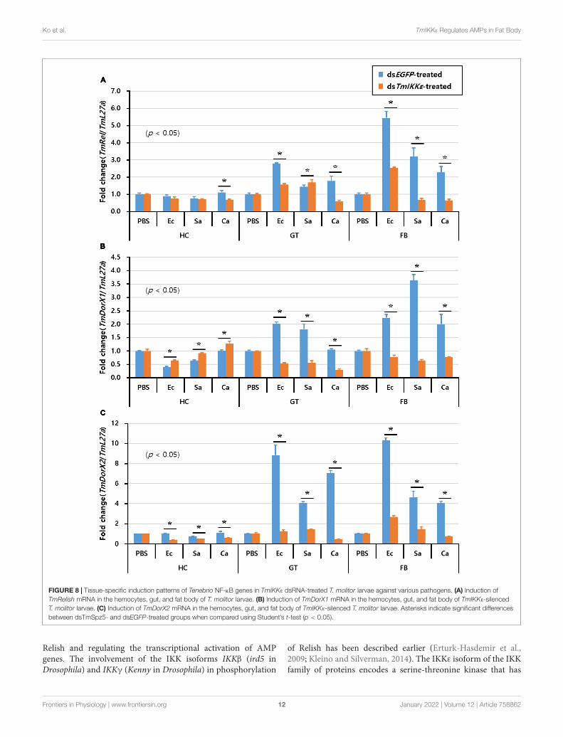
Effects of TmIKKε RNAi on theExpression of Tenebrio NF-κB GenesFurthermore, to understand the effect of TmIKKε RNAi onexpression of Tenebrio NF-κB genes, TmIKKε-silenced T. molitorlarvae were challenged with microorganisms and the expressionpatterns of Tenebrio NF-κB genes such as TmRelish, TmDorX1,and TmDorX2 were investigated at 24 h by qPCR analysis. Theresults showed that the mRNA level of three NF-κB genes was
dramatically decreased by TmIKKε RNAi in the FB of T. molitorlarva. In addition, the expression of TmDorX1 and TmDorX2transcripts were significantly decreased by TmIKKε RNAi in theGT (Figure 8).
DISCUSSION
The Toll and IMD pathways constitute an important defensearsenal to protect insects from non-self-discriminatingpathogens. Our research group has been successful in elucidatingkey genes of the Toll and IMD intracellular pathways, which arerelevant in the context of humoral immunity in the coleopteranpest T. molitor (Patnaik et al., 2013, 2014; Tindwa et al.,2013). Our focus has been on the transcriptional activationof AMPs elicited by diverse groups of microorganisms andmediated through the Toll/IMD signaling cascade (Jo et al.,2017, 2019; Keshavarz et al., 2019, 2020a,b,d; Park et al., 2019;Ali Mohammadie Kojour et al., 2020; Edosa et al., 2020a,b;Ko et al., 2020). The IKK family of proteins act upstream of theNF-κB factor Relish in the IMD pathway by phosphorylating
Frontiers in Physiology | www.frontiersin.org 8 January 2022 | Volume 12 | Article 758862

fphys-12-758862 January 3, 2022 Time: 12:41 # 9
Ko et al. TmIKKε Regulates AMPs in Fat Body
FIGURE 5 | The antimicrobial peptides (AMPs) expression levels in TmIKKε-knockdown T. molitor larval hemocytes upon microorganism challenge. E. coli (Ec), S.aureus (Sa), or C. albicans (Ca) were injected into TmIKKε-silenced T. molitor larvae. The transcriptional expression levels of TmTene1 (A), TmTene2 (B), TmTene3(C), TmTene4 (D), TmDef (E), TmDef-like (F), TmColeA (G), TmColeB (H), TmCec2 (I), TmAtta1a (J), TmAtta1b (K), TmAtta2 (L), TmTLP1 (M), and TmTLP2 (N)were measured using qRT-PCR. EGFP dsRNA was used as a silencing control, and TmL27a was used as an internal control. Data represent the mean ± SE of threeindependent biological replicates. Asterisks indicate significant differences between dsTmSpz5- and dsEGFP-treated groups when compared using Student’s t-test(p < 0.05).
Frontiers in Physiology | www.frontiersin.org 9 January 2022 | Volume 12 | Article 758862

fphys-12-758862 January 3, 2022 Time: 12:41 # 10
Ko et al. TmIKKε Regulates AMPs in Fat Body
FIGURE 6 | AMP expression levels in TmIKKε-knockdown T. molitor larval gut upon microorganism challenge. E. coli (Ec), S. aureus (Sa), or C. albicans (Ca) wereinjected into TmIKKε-silenced T. molitor larvae. The transcriptional expression levels of TmTene1 (A), TmTene2 (B), TmTene3 (C), TmTene4 (D), TmDef (E),TmDef-like (F), TmColeA (G), TmColeB (H), TmCec2 (I), TmAtta1a (J), TmAtta1b (K), TmAtta2 (L), TmTLP1 (M), and TmTLP2 (N) were measured using qRT-PCR.EGFP dsRNA was used as a silencing control, and TmL27a was used as an internal control. Data represent the mean ± SE of three independent biologicalreplicates. Asterisks indicate significant differences between dsTmSpz5- and dsEGFP-treated groups when compared using Student’s t-test (p < 0.05).
Frontiers in Physiology | www.frontiersin.org 10 January 2022 | Volume 12 | Article 758862

fphys-12-758862 January 3, 2022 Time: 12:41 # 11
Ko et al. TmIKKε Regulates AMPs in Fat Body
FIGURE 7 | AMP expression levels in TmIKKε-knockdown T. molitor larval fat body upon microorganism challenge. E. coli (Ec), S. aureus (Sa), or C. albicans (Ca)were injected into TmIKKε-silenced T. molitor larvae. The transcriptional expression levels of TmTene1 (A), TmTene2 (B), TmTene3 (C), TmTene4 (D), TmDef (E),TmDef-like (F), TmColeA (G), TmColeB (H), TmCec2 (I), TmAtta1a (J), TmAtta1b (K), TmAtta2 (L), TmTLP1 (M), and TmTLP2 (N) were measured using qRT-PCR.EGFP dsRNA was used as a silencing control, and TmL27a was used as an internal control. Data represent the mean ± SE of three independent biologicalreplicates. Asterisks indicate significant differences between dsTmSpz5- and dsEGFP-treated groups when compared using Student’s t-test (p < 0.05).
Frontiers in Physiology | www.frontiersin.org 11 January 2022 | Volume 12 | Article 758862
fphys-12-758862 January 3, 2022 Time: 12:41 # 12
Ko et al. TmIKKε Regulates AMPs in Fat Body
FIGURE 8 | Tissue-specific induction patterns of Tenebrio NF-κB genes in TmIKKε dsRNA-treated T. molitor larvae against various pathogens. (A) Induction ofTmRelish mRNA in the hemocytes, gut, and fat body of T. molitor larvae. (B) Induction of TmDorX1 mRNA in the hemocytes, gut, and fat body of TmIKKε-silencedT. molitor larvae. (C) Induction of TmDorX2 mRNA in the hemocytes, gut, and fat body of TmIKKε-silenced T. molitor larvae. Asterisks indicate significant differencesbetween dsTmSpz5- and dsEGFP-treated groups when compared using Student’s t-test (p < 0.05).
Relish and regulating the transcriptional activation of AMPgenes. The involvement of the IKK isoforms IKKβ (ird5 inDrosophila) and IKKγ (Kenny in Drosophila) in phosphorylation
of Relish has been described earlier (Erturk-Hasdemir et al.,2009; Kleino and Silverman, 2014). The IKKε isoform of the IKKfamily of proteins encodes a serine-threonine kinase that has
Frontiers in Physiology | www.frontiersin.org 12 January 2022 | Volume 12 | Article 758862

fphys-12-758862 January 3, 2022 Time: 12:41 # 13
Ko et al. TmIKKε Regulates AMPs in Fat Body
been implicated in NF-κB activation. The isoform also forms anessential component of the interferon regulatory factor 3 (IRF3)signaling pathway (Fitzgerald et al., 2003; Seccareccia et al., 2014;Dubois et al., 2018). IKKε in the black carp (Mylopharyngodonpiceus) was functionally characterized to participate in activatingthe expression of interferons in zebrafish and epitheliomapapulosum cyprini (EPC) cells (Qu et al., 2015). TBK1, whichis structurally identical to other IKK proteins, may also have aputative role as an important immunoregulator for IRF3 andIFNγ induction in chickens (Wang et al., 2017). However, thefunctional characterization of the IKKε homolog in insects isless described. In this study, we characterized the IKKε isoformin the coleopteran pest T. molitor by identifying its sequence,tissue distribution, and possible role in humoral immunity bystudying the transcriptional regulation of 14 T. molitor AMPgenes after microorganism challenge. We have also examined theexpression of downstream NF-κB factors, including TmDorX1and TmDorX2, which are involved in the Toll signaling pathway,and Relish, which is involved in the IMD signaling pathway,under TmIKKε-silenced conditions. Our results have improvedunderstanding of the putative regulatory pathway involvingTmIKKε.
The induction pattern of TmIKKε against injection ofrepresentative gram-negative bacteria, gram-positive bacteria,and fungi indicated that TmIKKε mRNA expression was mainlyinduced by E. coli challenge in the hemocytes. Furthermore,the knockdown of TmIKKε transcripts resulted in larval deathupon E. coli challenge. The findings suggest that TmIKKε may beputatively involved in the defense against gram-negative bacteria.Interestingly, the TmIKKε transcript was significantly induced byS. aureus in hemocytes. This result may indicate the involvementof TmIKKε in the canonical IMD pathway. Further, the roleof S. aureus in the activation of the Toll signaling cascade iswell established in insects, including T. molitor (Patnaik et al.,2014). However, in the gut defense system of D. melanogaster.IMD pathway is required for clearance of S. aureus, possiblyindependently from AMP expression and via Duox system thatproduces reactive oxygen species (ROS) (Hori et al., 2018).
The transcriptional regulation of fourteen AMP genes inT. molitor in TmIKKε-silenced condition was investigated inhemocytes, gut, and fat body tissues post-injection with gram-negative bacteria E. coli, gram-positive bacteria S. aureus,and the fungus C. albicans. Interestingly, most AMP geneswere not positively affected by TmIKKε RNAi in hemocytes,excepting to some extent TmDef. Other critical AMP geneswere mostly negatively regulated after microorganisms challengein TmIKKε-silenced individuals. Further, the inconsequentialrole of TmIKKε knockdown on the activation of the NF-κBgenes including TmRelish, TmDorX1, and TmDorX2 suggest thatTmIKKε is not required for AMP production in hemocytes.Contrastingly, in the gut, seven AMP genes were significantlydownregulated by TmIKKε dsRNA-treatment post-injection ofE. coli. We also find that the TmDorX1 and TmDorX2 mRNA(NF-κB regulator of Toll signaling pathway) were criticallydownregulated in the gut in TmIKKε-silenced individuals. Thispossibly suggests that TmIKKε regulates the transcriptionalactivation of seven AMP genes in the host gut in response to
the systemic infection of microorganisms. Studies in mosquitoeshighlight the promiscuous intervention of hemocytes in theactivation of the anti-plasmodial gut immune system (Ramirezet al., 2014; Castillo et al., 2017). These pieces of evidencesuggest the relationship between systemic infections and gutinnate immune system.
In the fat body tissue of T. molitor larvae, the expressionof ten AMP genes was downregulated and all the three NF-κBgenes were significantly affected by TmIKKε RNAi in responseto microbial challenge. These results indicate that TmIKKε isa key regulator for Toll and IMD pathways in the fat body ofT. molitor larvae. Cross-talk between Toll and IMD pathways isproposed in D. melanogaster (Tanji et al., 2007), T. castaneum(Yokoi et al., 2012a,b), P. stali (Nishide et al., 2019), and T. molitor(Ko et al., 2020). It was also suggested that several AMP geneswere co-regulated by those pathways in D. melanogaster (DeGregorio et al., 2002). Further, DmIKKε phosphorylates DIAP1leading to DIAP1 degradation and apoptosis in developmentdownstream of IMD. In Drosophila, the function of DmIKKε
is not especially Toll or IMD related, particularly for its rolein IMD activation (Kuranaga et al., 2006; Oshima et al., 2006).In addition, the activation of Toll and IMD pathways is totallydependent on invading microbes. For instance, the gram-positivebacteria including Micrococcus luteus, Bacillus subtilis, Bacillusmegaterium, Enterobacter cloacae, and fungi such as Beauveriabassiana, Saccharomyces cerevisiae, Metarhizium anisopliae, andGeotrichum candidum activate the IMD pathway (Hedengren-Olcott et al., 2004). Drosophila Cecropin A was induced by thegram-positive bacteria M. luteus and S. aureus independent of theNF-κB factor Relish. The transcriptional activation of AMPs suchas Cecropin A1 and Cecropin A2 in response to M. luteus infectionrequired Relish and Dif, respectively. Even, it is well known thatthe Tenecin 3 protects T. molitor against infection by the fungusBeauveria bassiana exemplified by increased larval survivability(Maistrou et al., 2018). Contrastingly, our results showed that theTmIKKε RNAi does not mainly affect the expression of Tenecin 3gene and larval mortality against fungal infection.
Taken together, TmIKKε plays a critical function in theproduction of nine AMPs in the fat body by regulating bothToll (TmDorX1 and -X2) and IMD (TmRelish) pathways.Interestingly, seven AMP genes were positively regulated byTmIKKε RNAi in the gut, carefully suggesting that the systemicinfection may positively regulate AMP production in the gutthrough the Toll (TmDorX1 and -X2) pathway.
DATA AVAILABILITY STATEMENT
The original contributions presented in the study are includedin the article/Supplementary Material, further inquiries can bedirected to the corresponding authors.
AUTHOR CONTRIBUTIONS
YH and YJ: conceptualization, methodology, supervision, andproject administration. YH: software, validation, resources,
Frontiers in Physiology | www.frontiersin.org 13 January 2022 | Volume 12 | Article 758862

fphys-12-758862 January 3, 2022 Time: 12:41 # 14
Ko et al. TmIKKε Regulates AMPs in Fat Body
and funding acquisition. HK and YJ: formal analysis andvisualization. HK, KP, CK, and HJ: investigation. HK, KP, andCK: data curation. HK, YH, YJ, and BP: writing – originaldraft preparation. BP, SB, and YL: writing – review and editing.All authors have read and agreed to the published versionof the manuscript.
FUNDING
This research was supported by the Basic Science ResearchProgram through the National Research Foundation of Korea(NRF) funded by the Ministry of Science, ICT and Future
Planning (Grant No. 2018R1A2A2A05023367) and KoreaInstitute of Planning and Evaluation for Technology in Food,Agriculture, Forestry and Fisheries (IPET) through ExportPromotion Technology Development Program (Grant No.617077-5), funded by Ministry of Agriculture, Food and RuralAffairs (MAFRA).
SUPPLEMENTARY MATERIAL
The Supplementary Material for this article can be foundonline at: https://www.frontiersin.org/articles/10.3389/fphys.2021.758862/full#supplementary-material
REFERENCESAli Mohammadie Kojour, M., Han, Y. S., and Jo, Y. H. (2020). An overview of insect
innate immunity. Entomol. Res. 50, 282–291. doi: 10.1111/1748-5967.12437Ali Mohammadie Kojour, M., Jang, H. A., Edosa, T. T., Keshavarz, M., Kim,
B. B., Bae, Y. M., et al. (2021). Identification, in silico characterization, andexpression analysis of Tenebrio molitor Cecropin-2. Entomol. Res. 51, 74–82.doi: 10.1111/1748-5967.12476
Altincicek, B., Knorr, E., and Vilcinskas, A. (2008). Beetle immunity: identificationof immune-inducible genes from the model insect Tribolium castaneum. Dev.Comp. Immunol. 32, 585–595. doi: 10.1016/j.dci.2007.09.005
Bang, K., Park, S., Yoo, J. Y., and Cho, S. (2012). Characterization and expressionof attacin, an antibacterial protein-encoding gene, from the beet armyworm,Spodoptera exigua (Hubner) (Insecta: Lepidoptera: Noctuidae). Mol. Biol. Rep.39, 5151–5159. doi: 10.1007/s11033-011-1311-3
Barletta, A. B., Nascimento-Silva, M. C., Talyuli, O. A., Oliveira, J. H., Pereira,L. O., Oliveira, P. L., et al. (2017). Microbiota activates IMD pathway and limitsSindbis infection in Aedes aegypti. Parasit. Vectors 10:103. doi: 10.1186/s13071-017-2040-9
Castillo, J. C., Ferreira, A. B. B., Trisnadi, N., and Barillas-Mury, C. (2017).Activation of mosquito complement antiplasmodial response requires cellularimmunity. Sci. Immunol. 2:eaal1505. doi: 10.1126/sciimmunol.aal1505
Cha, G. H., Cho, K. S., Lee, J. H., Kim, M., Kim, E., Park, J., et al. (2003). Discretefunctions of TRAF1 and TRAF2 in Drosophila melanogaster mediated by c-JunN-terminal kinase and NF-kappaB-dependent signaling pathways. Mol. Cell.Biol. 23, 7982–7991. doi: 10.1128/MCB.23.22.7982-7991.2003
Chae, J. H., Kurokawa, K., So, Y. I., Hwang, H. O., Kim, M. S., Park, J. W.,et al. (2012). Purification and characterization of tenecin 4, a new anti-Gram-negative bacterial peptide, from the beetle Tenebrio molitor. Dev. Comp.Immunol. 36, 540–546. doi: 10.1016/j.dci.2011.09.010
Chen, W., White, M. A., and Cobb, M. H. (2002). Stimulus-specific requirementsfor MAP3 kinases in activating the JNK pathway. J. Biol. Chem. 277, 49105–49110. doi: 10.1074/jbc.M204934200
Choe, K. M., Lee, H., and Anderson, K. V. (2005). Drosophila peptidoglycanrecognition protein LC (PGRP-LC) acts as a signal-transducing innate immunereceptor. Proc. Natl. Acad. Sci. U.S.A. 102, 1122–1126. doi: 10.1073/pnas.0404952102
Choe, K. M., Werner, T., Stoven, S., Hultmark, D., and Anderson, K. V.(2002). Requirement for a peptidoglycan recognition protein (PGRP) in Relishactivation and antibacterial immune responses in Drosophila. Science 296,359–362. doi: 10.1126/science.1070216
De Gregorio, E., Spellman, P. T., Tzou, P., Rubin, G. M., and Lemaitre, B. (2002).The Toll and Imd pathways are the major regulators of the immune response inDrosophila. EMBO J. 21, 2568–2579. doi: 10.1093/emboj/21.11.2568
Dubois, N., Berendsen, S., Henry, A., Nguyen, M., Bours, V., and Robe, P. (2018).I-Kappa-B kinase-epsilon activates nuclear factor-kappa B and STAT5B andsupports glioblastoma growth but amlexanox shows little therapeutic potentialin these tumors. Cancer Transl. Med. 4, 1–8. doi: 10.4103/ctm.ctm_3_18
Edosa, T. T., Jo, Y. H., Keshavarz, M., Bae, Y. M., Kim, D. H., Lee, Y. S., et al.(2020a). TmSpz4 plays an important role in regulating the production of
antimicrobial peptides in response to Escherichia coli and Candida albicansinfections. Int. J. Mol. Sci. 21:1878. doi: 10.3390/ijms21051878
Edosa, T. T., Jo, Y. H., Keshavarz, M., Bae, Y. M., Kim, D. H., Lee, Y. S., et al.(2020b). TmSpz6 is essential for regulating the immune response to EscherichiaColi and Staphylococcus Aureus infection in Tenebrio molitor. Insects 11:105.doi: 10.3390/insects11020105
Erturk-Hasdemir, D., Broemer, M., Leulier, F., Lane, W. S., Paquette, N., Hwang,D., et al. (2009). Two roles for the Drosophila IKK complex in the activation ofRelish and the induction of antimicrobial peptide genes. Proc. Natl. Acad. Sci.U.S.A. 106, 9779–9784. doi: 10.1073/pnas.0812022106
Fitzgerald, K. A., McWhirter, S. M., Faia, K. L., Rowe, D. C., Latz, E., Golenbock,D. T., et al. (2003). IKKepsilon and TBK1 are essential components of the IRF3signaling pathway. Nat. Immunol. 4, 491–496. doi: 10.1038/ni921
Gan, T., Fan, L., Zhao, L., Misra, M., Liu, M., Zhang, M., et al. (2021). JNKSignaling in Drosophila aging and longevity. Int. J. Mol. Sci. 22:9649. doi: 10.3390/ijms22179649
Georgel, P., Naitza, S., Kappler, C., Ferrandon, D., Zachary, D., Swimmer, C., et al.(2001). Drosophila immune deficiency (IMD) is a death domain protein thatactivates antibacterial defense and can promote apoptosis. Dev. Cell 1, 503–514.doi: 10.1016/S1534-5807(01)00059-4
Goel, M. K., Khanna, P., and Kishore, J. (2010). Understanding survival analysis:Kaplan-Meier estimate. Int. J. Ayurveda Res. 1, 274–278. doi: 10.4103/0974-7788.76794
Gottar, M., Gobert, V., Michel, T., Belvin, M., Duyk, G., Hoffmann, J. A., et al.(2002). The Drosophila immune response against Gram-negative bacteria ismediated by a peptidoglycan recognition protein. Nature 416, 640–644. doi:10.1038/nature734
Hanson, M. A., Dostalova, A., Ceroni, C., Poidevin, M., Kondo, S., and Lemaitre,B. (2019). Synergy and remarkable specificity of antimicrobial peptides in vivousing a systematic knockout approach. Elife 8:e44341. doi: 10.7554/eLife.44341
Hedengren-Olcott, M., Olcott, M. C., Mooney, D. T., Ekengren, S., Geller, B. L.,and Taylor, B. J. (2004). Differential activation of the NF-kappa B-like factorsrelish and Dif in Drosophila melanogaster by fungi and Gram-positive bacteria.J. Biol. Chem. 279, 21121–21127. doi: 10.1074/.M313856200jbc
Hoffmann, J. A., Kafatos, F. C., Janeway, C. A., and Ezekowitz, R. A. (1999).Phylogenetic perspectives in innate immunity. Science 284, 1313–1318. doi:10.1126/science.284.5418.1313
Hori, A., Kurata, S., and Kuraishi, T. (2018). Unexpected role of the IMD pathwayin Drosophila gut defense against Staphylococcus aureus. Biochem. Biophys. Res.Commun. 495, 395–400. doi: 10.1016/j.bbrc.2017.11.004
Hua, X. T., Ma, X. J., Xue, R. J., Cheng, T. C., Wang, F., and Xia, Q. Y. (2016).Characterization of the Bombyx mori Cecropin A1 promoter regulated by IMDpathway. Insect Sci. 23, 297–304. doi: 10.1111/1744-7917.12210
Hultmark, D., Steiner, H., Rasmuson, T., and Boman, H. G. (1980). Insectimmunity. Purification and properties of three inducible bactericidal proteinsfrom hemolymph of immunized pupae of Hyalophora cecropia. Eur. J. Biochem.106, 7–16. doi: 10.1111/j.1432-1033.1980.tb05991.x
International Aphid Genomics Consortium (2010). Genome sequence of the peaaphid Acyrthosiphon pisum. PLoS Biol. 8:e1000313. doi: 10.1371/journal.pbio.1000313
Frontiers in Physiology | www.frontiersin.org 14 January 2022 | Volume 12 | Article 758862

fphys-12-758862 January 3, 2022 Time: 12:41 # 15
Ko et al. TmIKKε Regulates AMPs in Fat Body
Jang, H. A., Park, K. B., Kim, B. B., Ali Mohammadie Kojour, M., Bae, Y. M.,Baliarsingh, S., et al. (2020a). Bacterial but not fungal challenge up-regulatesthe transcription of Coleoptericin genes in Tenebrio molitor. Entomol. Res. 50,440–449. doi: 10.1111/1748-5967.12465
Jang, H. A., Park, K. B., Kim, B. B., Ali Mohammadie Kojour, M., Bae, Y. M.,Baliarsingh, S., et al. (2020b). In silico identification and expression analysesof Defensin genes in the mealworm beetle Tenebrio molitor. Entomol. Res. 50,575–585. doi: 10.1111/1748-5967.12468
Jo, Y. H., Kim, Y. J., Park, K. B., Seong, J. H., Kim, S. G., Park, S., et al. (2017).TmCactin plays an important role in Gram-negative and -positive bacterialinfection by regulating expression of 7 AMP genes in Tenebrio molitor. Sci. Rep.7:46459. doi: 10.1038/srep46459
Jo, Y. H., Park, S., Park, K. B., Noh, M. Y., Cho, J. H., Ko, H. J., et al. (2018). In silicoidentification, characterization and expression analysis of attacin gene family inresponse to bacterial and fungal pathogens in Tenebrio molitor. Entomol. Res.48, 45–54. doi: 10.1111/1748-5967.12287
Jo, Y. H., Patnaik, B. B., Hwang, J., Park, K. B., Ko, H. J., Kim, C. E., et al.(2019). Regulation of the expression of nine antimicrobial peptide genes byTmIMD confers resistance against Gram-negative bacteria. Sci. Rep. 9:10138.doi: 10.1038/s41598-019-46222-8
Johnston, P. R., Makarova, O., and Rolff, J. (2013). Inducible defenses stay uplate: temporal patterns of immune gene expression in Tenebrio molitor. G3(Bethesda) 4, 947–955. doi: 10.1534/g3.113.008516
Kaneko, T., Goldman, W. E., Mellroth, P., Steiner, H., Fukase, K., Kusumoto, S.,et al. (2004). Monomeric and polymeric gram-negative peptidoglycan but notpurified LPS stimulate the Drosophila IMD pathway. Immunity 20, 637–649.doi: 10.1016/s1074-7613(04)00104-9
Keshavarz, M., Jo, Y. H., Edosa, T. T., Bae, Y. M., and Han, Y. S. (2020a). TmPGRP-SA regulates antimicrobial response to bacteria and fungi in the fat bodyand gut of Tenebrio molitor. Int. J. Mol. Sci. 21:2113. doi: 10.3390/ijms21062113
Keshavarz, M., Jo, Y. H., Edosa, T. T., and Han, Y. S. (2020b). Tenebrio molitorPGRP-LE plays a critical role in gut antimicrobial peptide production inresponse to Escherichia coli. Front. Physiol. 11:320. doi: 10.3389/fphys.2020.00320
Keshavarz, M., Jo, Y. H., Edosa, T. T., and Han, Y. S. (2020c). Two roles forthe Tenebrio molitor relish in the regulation of antimicrobial peptides andautophagy-related genes in response to Listeria monocytogenes. Insects 11:188.doi: 10.3390/insects11030188
Keshavarz, M., Jo, Y. H., Patnaik, B. B., Park, K. B., Ko, H. J., Kim, C. E., et al.(2020d). TmRelish is required for regulating the antimicrobial responses toEscherichia coli and Staphylococcus aureus in Tenebrio molitor. Sci. Rep. 10:4258.doi: 10.1038/s41598-020-61157-1
Keshavarz, M., Jo, Y. H., Park, K. B., Ko, H. J., Edosa, T. T., Lee, Y. S., et al. (2019).TmDorX2 positively regulates antimicrobial peptides in Tenebrio molitor gut,fat body, and hemocytes in response to bacterial and fungal infection. Sci. Rep.9:16878. doi: 10.1038/s41598-019-53497-4
Kim, D. H., Lee, Y. T., Lee, Y. J., Chung, J. H., Lee, B. L., Choi, B. S., et al. (1998).Bacterial expression of tenecin 3, an insect antifungal protein isolated fromTenebrio molitor, and its efficient purification. Mol. Cells 8, 786–789.
Kim, D. H., Noh, M. Y., Park, K. B., and Jo, Y. H. (2017). Expression profilesof two thaumatin-like protein (TmTLP) genes in responses to various micro-organisms from Tenebrio molitor. Entomol. Res. 47, 35–40. doi: 10.1111/1748-5967.12197
Kleino, A., and Silverman, N. (2014). The Drosophila IMD pathway in theactivation of the humoral immune response. Dev. Comp. Immunol. 42, 25–35.doi: 10.1016/j.dci.2013.05.014
Ko, H. J., Jo, Y. H., Patnaik, B. B., Park, K. B., Kim, C. E., Keshavarz, M., et al. (2020).IKKγ/NEMO is required to confer antimicrobial innate immune responses inthe yellow mealworm, Tenebrio Molitor. Int. J. Mol. Sci. 21:6734. doi: 10.3390/ijms21186734
Koyama, H., Kato, D., Minakuchi, C., Tanaka, T., Yokoi, K., and Miura, K. (2015).Peptidoglycan recognition protein genes and their roles in the innate immunepathways of the red flour beetle, Tribolium castaneum. J. Invertebr. Pathol. 132,86–100. doi: 10.1016/j.jip.2015.09.003
Kumar, S., Stecher, G., and Tamura, K. (2016). MEGA7: molecular evolutionarygenetics analysis version 7.0 for bigger datasets. Mol. Biol. Evol. 33, 1870–1874.doi: 10.1093/molbev/msw054
Kuranaga, E., Kanuka, H., Tonoki, A., Takemoto, K., Tomioka, T., Kobayashi, M.,et al. (2006). Drosophila IKK-related kinase regulates non-apoptotic function ofcaspases via degradation of IAPs. Cell 126, 583–596. doi: 10.1016/j.cell.2006.05.048
Kwon, Y. M., Kim, H. J., Kim, Y. I., Kang, Y. J., Lee, I. H., Jin, B. R., et al. (2008).Comparative analysis of two attacin genes from Hyphantria cunea. Comp.Biochem. Physiol. B Biochem. Mol. Biol. 151, 213–220. doi: 10.1016/j.cbpb.2008.07.002
Larkin, M. A., Blackshields, G., Brown, N. P., Chenna, R., McGettigan, P. A.,McWilliam, H., et al. (2007). Clustal W and Clustal X version 2.0. Bioinformatics23, 2947–2948. doi: 10.1093/bioinformatics/btm404
Lemaitre, B., Nicolas, E., Michaut, L., Reichhart, J. M., and Hoffmann, J. A.(1996). The dorsoventral regulatory gene cassette spatzle/Toll/cactus controlsthe potent antifungal response in Drosophila adults. Cell 86, 973–983. doi:10.1016/s0092-8674(00)80172-5
Leulier, F., Parquet, C., Pili-Floury, S., Ryu, J. H., Caroff, M., Lee, W. J., et al. (2003).The Drosophila immune system detects bacteria through specific peptidoglycanrecognition. Nat. Immunol. 4, 478–484. doi: 10.1038/ni922
Li, C., Wang, S., and He, J. (2019). The two NF-kappaB pathways regulatingbacterial and WSSV infection of shrimp. Front. Immunol. 10:1785. doi: 10.3389/fimmu.2019.01785
Li, F., and Xiang, J. (2013). Signaling pathways regulating innate immune responsesin shrimp. Fish Shellfish Immunol. 34, 973–980. doi: 10.1016/j.fsi.2012.08.023
Lin, J., Xia, X., Yu, X. Q., Shen, J., Li, Y., Lin, H., et al. (2018). Gene expressionprofiling provides insights into the immune mechanism of Plutella xylostellamidgut to microbial infection. Gene 647, 21–30. doi: 10.1016/j.gene.2018.01.001
Livak, K. J., and Schmittgen, T. D. (2001). Analysis of relative gene expressiondata using real-time quantitative PCR and the 2(-Delta Delta C(T)) Method.Methods 25, 402–408. doi: 10.1006/meth.2001.1262
Maistrou, S., Paris, V., Jensen, A. B., Rolff, J., Meyling, N. V., and Zanchi, C. (2018).A constitutively expressed antifungal peptide protects Tenebrio molitor duringa natural infection by the entomopathogenic fungus Beauveria bassiana. Dev.Comp. Immunol. 86, 26–33. doi: 10.1016/j.dci.2018.04.015
Mesquita, R. D., Vionette-Amaral, R. J., Lowenberger, C., Rivera-Pomar, R.,Monteiro, F. A., Minx, P., et al. (2015). Genome of Rhodnius prolixus, aninsect vector of Chagas disease, reveals unique adaptations to hematophagyand parasite infection. Proc. Natl. Acad. Sci. U.S.A. 112, 14936–14941. doi:10.1073/pnas.1506226112
Nicolas, E., Nappi, A. J., and Lemaitre, B. (1996). Expression of antimicrobialpeptide genes after infection by parasitoid wasps in Drosophila. Dev. Comp.Immunol. 20, 175–181.
Nishide, Y., Kageyama, D., Yokoi, K., Jouraku, A., Tanaka, H., Futahashi, R.,et al. (2019). Functional crosstalk across IMD and toll pathways: insightinto the evolution of incomplete immune cascades. Proc. R. Soc. B Biol. Sci.286:20182207. doi: 10.1098/rspb.2018.2207
Noh, M. Y., and Jo, Y. H. (2016). Identification and sequence analysis of twothaumatin-like protein (TmTLP) genes from Tenebrio molitor. Entomol. Res.46, 354–359. doi: 10.1111/1748-5967.12198
Oshima, K., Takeda, M., Kuranaga, E., Ueda, R., Aigaki, T., Miura, M., et al. (2006).IKKε regulates F-actin assembly and interacts with Drosophila IAP1 in cellularmorphogenesis. Curr. Biol. 16, 1531–1537. doi: 10.1016/j.cub.2006.06.032
Palmer, W. J., and Jiggins, F. M. (2015). Comparative genomics reveals the originsand diversity of arthropod immune systems. Mol. Biol. Evol. 32, 2111–2129.doi: 10.1093/molbev/msv093
Park, S., Jo, Y. H., Park, K. B., Ko, H. J., Kim, C. E., Bae, Y. M., et al. (2019). TmToll-7 plays a crucial role in innate immune responses against gram-negative bacteriaby regulating 5 AMP genes in Tenebrio molitor. Front. Immunol. 10:310. doi:10.3389/fimmu.2019.00310
Patnaik, B. B., Kang, S. M., Seo, G. W., Lee, H. J., Patnaik, H. H., Jo, Y. H., et al.(2013). Molecular cloning, sequence characterization and expression analysisof a CD63 homologue from the coleopteran beetle, Tenebrio molitor. Int. J. Mol.Sci. 14, 20744–20767. doi: 10.3390/ijms141020744
Patnaik, B. B., Patnaik, H. H., Seo, G. W., Jo, Y. H., Lee, Y. S., Lee, B. L., et al.(2014). Gene structure, cDNA characterization and RNAi-based functionalanalysis of a myeloid differentiation factor 88 homolog in Tenebrio molitorlarvae exposed to Staphylococcus aureus infection. Dev. Comp. Immunol. 46,208–221. doi: 10.1016/j.dci.2014.04.009
Frontiers in Physiology | www.frontiersin.org 15 January 2022 | Volume 12 | Article 758862

fphys-12-758862 January 3, 2022 Time: 12:41 # 16
Ko et al. TmIKKε Regulates AMPs in Fat Body
Qu, Y., Zhou, M., Peng, L., Li, J., Yan, J., Yang, P., et al. (2015). Molecular cloningand characterization of IKKepsilon gene from black carp Mylopharyngodonpiceus. Fish Shellfish Immunol. 47, 122–129. doi: 10.1016/j.fsi.2015.08.033
Ramet, M., Manfruelli, P., Pearson, A., Mathey-Prevot, B., and Ezekowitz, R. A. B.(2002). Functional genomic analysis of phagocytosis and identification of aDrosophila receptor for E-coli. Nature 416, 644–648. doi: 10.1038/nature735
Ramirez, J. L., Garver, L. S., Brayner, F. A., Alves, L. C., Rodrigues, J., Molina-Cruz,A., et al. (2014). The role of hemocytes in Anopheles gambiae antiplasmodialimmunity. J. Innate Immun. 6, 119–128. doi: 10.1159/000353765
Roh, K. B., Kim, C. H., Lee, H., Kwon, H. M., Park, J. W., Ryu, J. H., et al. (2009).Proteolytic cascade for the activation of the insect toll pathway induced by thefungal cell wall component. J. Biol. Chem. 284, 19474–19481. doi: 10.1074/jbc.M109.007419
Salcedo-Porras, N., and Lowenberger, C. (2019). The innate immune system ofkissing bugs, vectors of chagas disease. Dev. Comp. Immunol. 98, 119–128.doi: 10.1016/j.dci.2019.04.007
Santos-Matos, G., Wybouw, N., Martins, N. E., Zele, F., Riga, M., Leitao, A. B., et al.(2017). Tetranychus urticae mites do not mount an induced immune responseagainst bacteria. Proc. Biol. Sci. 284:20170401. doi: 10.1098/rspb.2017.0401
Seccareccia, E., Pinard, M., Wang, N., Li, S., Burnier, J., Dankort, D., et al. (2014).The inhibitor of kappa B kinase-epsilon regulates MMP-3 expression levels andcan promote lung metastasis. Oncogenesis 3:e116. doi: 10.1038/oncsis.2014.28
Silverman, N., Zhou, R., Erlich, R. L., Hunter, M., Bernstein, E., Schneider, D., et al.(2003). Immune activation of NF-kappaB and JNK requires Drosophila TAK1.J. Biol. Chem. 278, 48928–48934. doi: 10.1074/jbc.M304802200
Tanji, T., Hu, X., Weber, A. N., and Ip, Y. T. (2007). Toll and IMD pathwayssynergistically activate an innate immune response in Drosophila melanogaster.Mol. Cell. Biol. 27, 4578–4588. doi: 10.1128/MCB.01814-06
Tindwa, H., Patnaik, B. B., Kim, D. H., Mun, S., Jo, Y. H., Lee, B. L., et al. (2013).Cloning, characterization and effect of TmPGRP-LE gene silencing on survivalof Tenebrio molitor against Listeria monocytogenes infection. Int. J. Mol. Sci. 14,22462–22482. doi: 10.3390/ijms141122462
Vidal, S., Khush, R. S., Leulier, F., Tzou, P., Nakamura, M., and Lemaitre, B.(2001). Mutations in the Drosophila dTAK1 gene reveal a conserved functionfor MAPKKKs in the control of rel/NF-kappaB-dependent innate immuneresponses. Genes Dev. 15, 1900–1912. doi: 10.1101/gad.203301
Wang, H., Xu, Q., Xu, X., Hu, Y., Hou, Q., Zhu, Y., et al. (2017). Ctenopharyngodonidella IKKbeta interacts with PKR and IkappaBalpha. Acta Biochim. Biophys.Sin. (Shanghai) 49, 729–736. doi: 10.1093/abbs/gmx065
Wu, Q., Patocka, J., and Kuca, K. (2018). Insect Antimicrobial peptides, a minireview. Toxins (Basel) 10:461. doi: 10.3390/toxins10110461
Yokoi, K., Koyama, H., Ito, W., Minakuchi, C., Tanaka, T., and Miura, K. (2012a).Involvement of NF-kappaB transcription factors in antimicrobial peptide geneinduction in the red flour beetle, Tribolium castaneum. Dev. Comp. Immunol.38, 342–351. doi: 10.1016/j.dci.2012.06.008
Yokoi, K., Koyama, H., Minakuchi, C., Tanaka, T., and Miura, K. (2012b).Antimicrobial peptide gene induction, involvement of Toll and IMDpathways and defense against bacteria in the red flour beetle, Triboliumcastaneum. Results Immunol. 2, 72–82. doi: 10.1016/j.rinim.2012.03.002
Zhan, M. Y., Yang, P. J., and Rao, X. J. (2018). Molecular cloning andanalysis of PGRP-L1 and IMD from silkworm Bombyx mori. Comp.Biochem. Physiol. B Biochem. Mol. Biol. 215, 19–30. doi: 10.1016/j.cbpb.2017.10.002
Zhu, J. Y., Wu, G. X., and Zhang, Z. (2014). Upregulation of coleoptericintranscription in Tenebrio molitor parasitized by Scleroderma guani. J. Asia Pac.Entomol. 17, 339–342. doi: 10.1016/j.aspen.2014.03.001
Zou, Z., Evans, J. D., Lu, Z., Zhao, P., Williams, M., Sumathipala, N., et al. (2007).Comparative genomic analysis of the Tribolium immune system. Genome Biol.8:R177. doi: 10.1186/gb-2007-8-8-r177
Conflict of Interest: The authors declare that the research was conducted in theabsence of any commercial or financial relationships that could be construed as apotential conflict of interest.
Publisher’s Note: All claims expressed in this article are solely those of the authorsand do not necessarily represent those of their affiliated organizations, or those ofthe publisher, the editors and the reviewers. Any product that may be evaluated inthis article, or claim that may be made by its manufacturer, is not guaranteed orendorsed by the publisher.
Copyright © 2022 Ko, Patnaik, Park, Kim, Baliarsingh, Jang, Lee, Han and Jo.This is an open-access article distributed under the terms of the Creative CommonsAttribution License (CC BY). The use, distribution or reproduction in other forumsis permitted, provided the original author(s) and the copyright owner(s) are creditedand that the original publication in this journal is cited, in accordance with acceptedacademic practice. No use, distribution or reproduction is permitted which does notcomply with these terms.
Frontiers in Physiology | www.frontiersin.org 16 January 2022 | Volume 12 | Article 758862
Copyright © 2022 FDOKUMEN