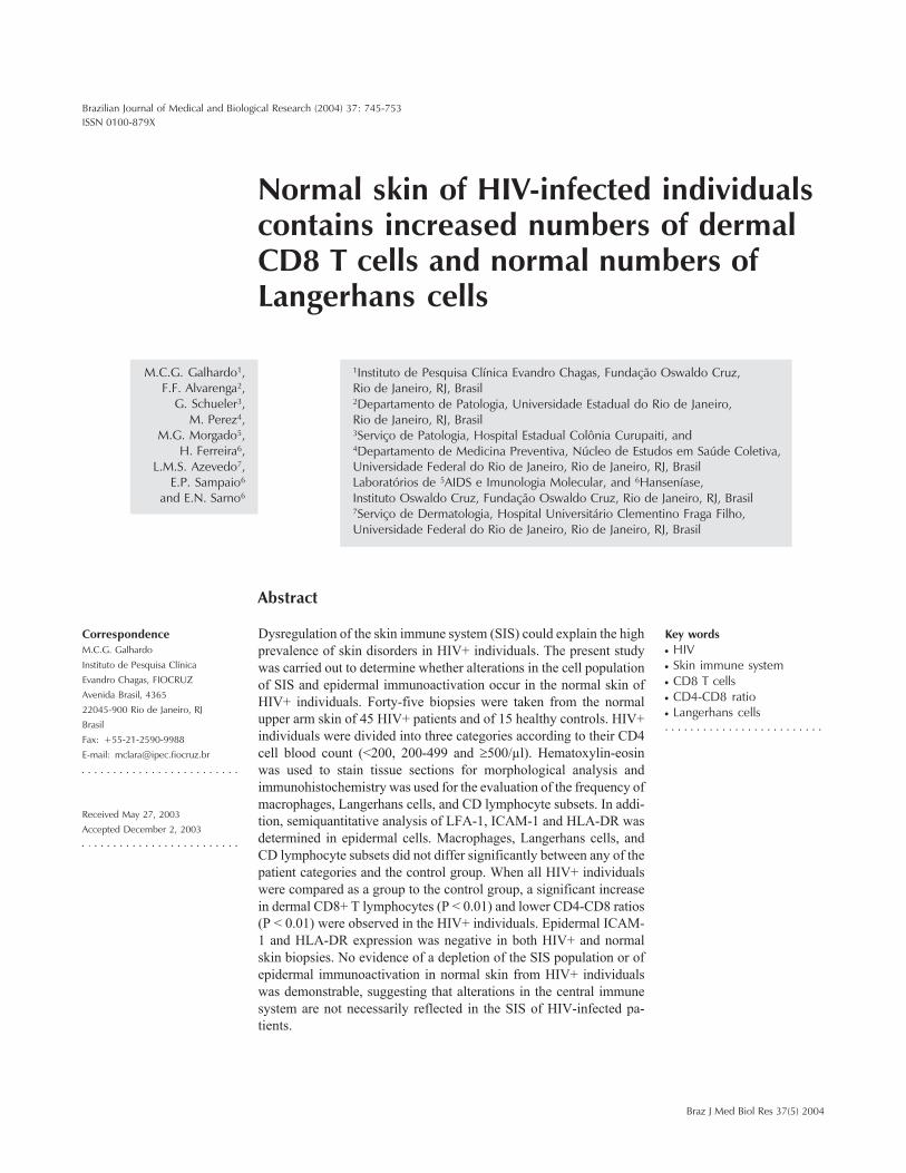
Normal skin of HIV-infected individuals contains increased numbers of dermal CD8 T cells and normal...
9
Braz J Med Biol Res 37(5) 2004 Brazilian Journal of Medical and Biological Research (2004) 37: 745-753 ISSN 0100-879X Normal skin of HIV-infected individuals contains increased numbers of dermal CD8 T cells and normal numbers of Langerhans cells 1 Instituto de Pesquisa Clínica Evandro Chagas, Fundação Oswaldo Cruz, Rio de Janeiro, RJ, Brasil 2 Departamento de Patologia, Universidade Estadual do Rio de Janeiro, Rio de Janeiro, RJ, Brasil 3 Serviço de Patologia, Hospital Estadual Colônia Curupaiti, and 4 Departamento de Medicina Preventiva, Núcleo de Estudos em Saúde Coletiva, Universidade Federal do Rio de Janeiro, Rio de Janeiro, RJ, Brasil Laboratórios de 5 AIDS e Imunologia Molecular, and 6 Hanseníase, Instituto Oswaldo Cruz, Fundação Oswaldo Cruz, Rio de Janeiro, RJ, Brasil 7 Serviço de Dermatologia, Hospital Universitário Clementino Fraga Filho, Universidade Federal do Rio de Janeiro, Rio de Janeiro, RJ, Brasil M.C.G. Galhardo 1 , F.F. Alvarenga 2 , G. Schueler 3 , M. Perez 4 , M.G. Morgado 5 , H. Ferreira 6 , L.M.S. Azevedo 7 , E.P. Sampaio 6 and E.N. Sarno 6 Abstract Dysregulation of the skin immune system (SIS) could explain the high prevalence of skin disorders in HIV+ individuals. The present study was carried out to determine whether alterations in the cell population of SIS and epidermal immunoactivation occur in the normal skin of HIV+ individuals. Forty-five biopsies were taken from the normal upper arm skin of 45 HIV+ patients and of 15 healthy controls. HIV+ individuals were divided into three categories according to their CD4 cell blood count (<200, 200-499 and ≥500/μl). Hematoxylin-eosin was used to stain tissue sections for morphological analysis and immunohistochemistry was used for the evaluation of the frequency of macrophages, Langerhans cells, and CD lymphocyte subsets. In addi- tion, semiquantitative analysis of LFA-1, ICAM-1 and HLA-DR was determined in epidermal cells. Macrophages, Langerhans cells, and CD lymphocyte subsets did not differ significantly between any of the patient categories and the control group. When all HIV+ individuals were compared as a group to the control group, a significant increase in dermal CD8+ T lymphocytes (P < 0.01) and lower CD4-CD8 ratios (P < 0.01) were observed in the HIV+ individuals. Epidermal ICAM- 1 and HLA-DR expression was negative in both HIV+ and normal skin biopsies. No evidence of a depletion of the SIS population or of epidermal immunoactivation in normal skin from HIV+ individuals was demonstrable, suggesting that alterations in the central immune system are not necessarily reflected in the SIS of HIV-infected pa- tients. Correspondence M.C.G. Galhardo Instituto de Pesquisa Clínica Evandro Chagas, FIOCRUZ Avenida Brasil, 4365 22045-900 Rio de Janeiro, RJ Brasil Fax: +55-21-2590-9988 E-mail: [email protected] Received May 27, 2003 Accepted December 2, 2003 Key words • HIV • Skin immune system • CD8 T cells • CD4-CD8 ratio • Langerhans cells
-
Upload
independent -
Category
Documents
-
view
4 -
download
0
Transcript of Normal skin of HIV-infected individuals contains increased numbers of dermal CD8 T cells and normal...
745
Braz J Med Biol Res 37(5) 2004
T cells in the skin of HIV-infected individualsBrazilian Journal of Medical and Biological Research (2004) 37: 745-753ISSN 0100-879X
Normal skin of HIV-infected individualscontains increased numbers of dermalCD8 T cells and normal numbers ofLangerhans cells
1Instituto de Pesquisa Clínica Evandro Chagas, Fundação Oswaldo Cruz,Rio de Janeiro, RJ, Brasil2Departamento de Patologia, Universidade Estadual do Rio de Janeiro,Rio de Janeiro, RJ, Brasil3Serviço de Patologia, Hospital Estadual Colônia Curupaiti, and4Departamento de Medicina Preventiva, Núcleo de Estudos em Saúde Coletiva,Universidade Federal do Rio de Janeiro, Rio de Janeiro, RJ, BrasilLaboratórios de 5AIDS e Imunologia Molecular, and 6Hanseníase,Instituto Oswaldo Cruz, Fundação Oswaldo Cruz, Rio de Janeiro, RJ, Brasil7Serviço de Dermatologia, Hospital Universitário Clementino Fraga Filho,Universidade Federal do Rio de Janeiro, Rio de Janeiro, RJ, Brasil
M.C.G. Galhardo1,F.F. Alvarenga2,
G. Schueler3,M. Perez4,
M.G. Morgado5,H. Ferreira6,
L.M.S. Azevedo7,E.P. Sampaio6
and E.N. Sarno6
Abstract
Dysregulation of the skin immune system (SIS) could explain the highprevalence of skin disorders in HIV+ individuals. The present studywas carried out to determine whether alterations in the cell populationof SIS and epidermal immunoactivation occur in the normal skin ofHIV+ individuals. Forty-five biopsies were taken from the normalupper arm skin of 45 HIV+ patients and of 15 healthy controls. HIV+individuals were divided into three categories according to their CD4cell blood count (<200, 200-499 and ≥500/µl). Hematoxylin-eosinwas used to stain tissue sections for morphological analysis andimmunohistochemistry was used for the evaluation of the frequency ofmacrophages, Langerhans cells, and CD lymphocyte subsets. In addi-tion, semiquantitative analysis of LFA-1, ICAM-1 and HLA-DR wasdetermined in epidermal cells. Macrophages, Langerhans cells, andCD lymphocyte subsets did not differ significantly between any of thepatient categories and the control group. When all HIV+ individualswere compared as a group to the control group, a significant increasein dermal CD8+ T lymphocytes (P < 0.01) and lower CD4-CD8 ratios(P < 0.01) were observed in the HIV+ individuals. Epidermal ICAM-1 and HLA-DR expression was negative in both HIV+ and normalskin biopsies. No evidence of a depletion of the SIS population or ofepidermal immunoactivation in normal skin from HIV+ individualswas demonstrable, suggesting that alterations in the central immunesystem are not necessarily reflected in the SIS of HIV-infected pa-tients.
CorrespondenceM.C.G. Galhardo
Instituto de Pesquisa Clínica
Evandro Chagas, FIOCRUZ
Avenida Brasil, 4365
22045-900 Rio de Janeiro, RJ
Brasil
Fax: +55-21-2590-9988
E-mail: [email protected]
Received May 27, 2003
Accepted December 2, 2003
Key words• HIV• Skin immune system• CD8 T cells• CD4-CD8 ratio• Langerhans cells
746
Braz J Med Biol Res 37(5) 2004
M.C.G. Galhardo et al.
Introduction
During HIV infection, the skin is an or-gan of major morbidity. Increasingly severecutaneous disorders most commonly occurduring the course of the disease and some-times they provide the earliest clue to theexistence of HIV or of its progression (1-3).Suggestive of both local permissiveness andresponsiveness to the presence of HIV, achange in the cutaneous environment alsoappears to take place. As such, an dysregula-tion of the skin immune system (SIS) couldbe the prime reason for the increasing preva-lence of skin disorders in HIV+ individuals(4-7). However, few studies have been con-ducted on the SIS of HIV+ patients, in whom,similar to what occurs in other specializedlymphoid systems, changes in the immunecells associated with the SIS would be pre-dictable (8-10).
Most of the studies on SIS in HIV infec-tion have focused on the role of Langerhanscells (Lc), most probably because of theirimportant function as antigen-presenting cellsand because of their expression of CD4 cells.Although results continue to be contradic-tory, the hypothesis that the Lc populationmay suffer alterations during infection hasbeen the subject of numerous studies (11-16). Few data are available regarding therole of T cells and the other components.Skin mast cell density has been shown toremain unchanged in HIV+ individuals (17).Even though keratinocytes do not becomeinfected with HIV, they are capable of se-creting a variety of immunomodulatory cy-tokines (interleukin (IL)-1, IL-6 and tumornecrosis factor-α) which may enhance HIVreplication and the spreading of the virus inthe skin (7,18). In addition, as can be seen ina wide variety of dermatoses, keratinocytesmay become immunoactivated by means ofthe induction of expression molecules, suchas the intercellular adhesion molecule 1(ICAM-1) and human leukocyte antigen(HLA)-DR, responsible for the increase in
lymphocyte traffic in the epidermis and itssusceptibility to inflammatory reactions (19).
The skin-related mechanisms in HIV in-fection are unknown, although SIS dysregu-lation has been consistently thought to play arole in triggering dermatoses. Using histo-logical and immunohistochemical tech-niques, we attempted to determine whetherthe SIS becomes impaired in the clinicallynormal-appearing skin of HIV+ patients atdifferent stages of infection. The number ofskin immune cells (Lc, dermal dendrite cells,T cell subsets and macrophages), the extentof perivascular infiltrates, and the presenceof immunoactivated cell components (HLA-DR, ICAM-1 and leukocyte function-associ-ated antigen-1 (LFA-1)) were determined insitu.
Material and Methods
Subjects
After obtaining written informed consentfrom patients and control subjects and ap-proval from the Ethics Committee of theOswaldo Cruz Foundation, 45 HIV+ patients(27 males and 18 females; mean age, 37.2 ±6.6; range, 25-50 years) were randomly se-lected to participate in this cross-sectionalstudy. Patients with systemic or cutaneousdiseases that might interfere with the studywere excluded from the investigation. HIVpatients were further grouped according totheir CD4+ cell count as follows: <200/µl(category I, AIDS patients, N = 15); 200 to499/µl (category II, N = 15); ≥500/µl (cat-egory III, N = 15). Thirty-one (68%) patientswere taking antiretroviral agents at the timeof the study; 15 were receiving two drugs(mean time, 7.8 months; range, 2 to 14months); 15, triple drug therapy (mean time,6.3 months; range, 1 to 10 months), and 1,quadruple drug therapy (1 month; nucleo-side reverse transcriptase inhibitors, zidovu-dine, didanosine, dideoxycytidine, and la-mivudine in addition to the protease inhibi-
747
Braz J Med Biol Res 37(5) 2004
T cells in the skin of HIV-infected individuals
tors saquinavir and ritonavir) in a variety ofcombinations (Table 1). The control groupconsisted of 15 healthy individuals (8 malesand 7 females; mean age, 35.4 ± 7.4; range,20-42 years).
Skin biopsies
Punch biopsies (6 mm) were obtainedfrom clinically normal skin on the sun-pro-tected inner side of the upper arm from HIVpatients and control subjects. Biopsy sampleswere cut into two parts and processed forhistological and immunohistochemical evalu-ation as described below.
Histopathological analysis
The skin fragment was fixed in 10% neu-tral-buffered formalin overnight, embeddedin paraffin, and stained with hematoxylin-eosin. The material was submitted to thefollowing morphological determinations: 1)count of the number of layers in the epider-mis and analysis of the keratinocytes in eachlayer; 2) evaluation of the dermal and subcu-taneous adnexa and blood vessels, and 3)measurement of the extension of the inflam-matory infiltrate. Samples were examinedby two independent observers and morpho-logical features were recorded. Quantitativemeasurements (mean %) of the infiltratedarea were made during microscopic exami-nation (100X magnification) of histologicalsections using a ProLite image analyzer,which permitted the viewing of fields with aNikon E 450 microscope. For each sample,three different fields of the upper dermiswere scanned (20X). The most extendedinfiltrates (four of them) were identified andmeasured in each field (40X), and the meanof each sample was calculated.
Immunohistochemistry
Biopsies were snap-frozen and kept inliquid nitrogen until the time for use. Frozen
sections were cut with a cryostat, air dried,and fixed in cold acetone for 10 min. Thesections were incubated with the primaryantibody for 60 min, washed in PBS, andthen incubated with the biotinylated second-ary antibody (Dako Corporation, Carpinte-ria, CA, USA), followed by the avidin-bi-otin-peroxidase complex (Vectastain ABCkit; Vector Laboratories, Inc., Burlingame,CA, USA), and processed according to manu-facturer instructions. Finally, sections weredeveloped with aminoethylcarbazole as thechromogenic substrate. In control specimens,the primary monoclonal antibody was omit-ted or isotype control antibodies of irrel-evant specificity were used. The sectionswere analyzed by light microscopy and pho-tomicrographs were taken with a NikonMicrophot system.
Antibodies
Mouse anti-human monoclonal antibodieswere used for the identification of specific celltypes, proteins, and surface molecules. Anti-CD1 (Lc) antibody, anti-CD3 (pan T cellmarker), anti-CD4, anti-CD8 T cell subsets,
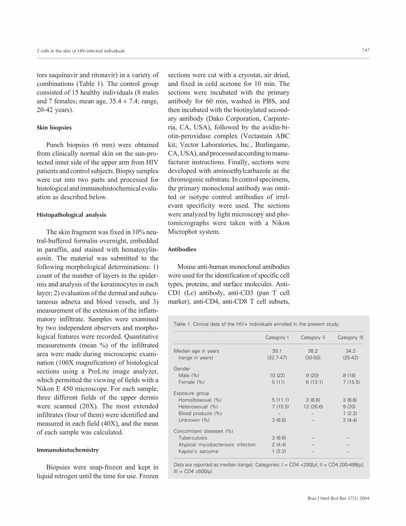
Table 1. Clinical data of the HIV+ individuals enrolled in the present study.
Category I Category II Category III
Median age in years 39.1 38.2 34.2(range in years) (32.7-47) (30-50) (25-42)
GenderMale (%) 10 (22) 9 (20) 8 (18)Female (%) 5 (11) 6 (13.1) 7 (15.5)
Exposure groupHomo/bisexual (%) 5 (11.1) 3 (6.6) 3 (6.6)Heterosexual (%) 7 (15.5) 12 (26.6) 9 (20)Blood products (%) - - 1 (2.2)Unknown (%) 3 (6.6) - 2 (4.4)
Concomitant diseases (%)Tuberculosis 3 (6.6) - -Atypical mycobacteriosis infection 2 (4.4) - -Kaposi’s sarcoma 1 (2.2) - -
Data are reported as median (range). Categories: I = CD4 <200/µl; II = CD4 200-499/µl;III = CD4 ≥500/µl.
748
Braz J Med Biol Res 37(5) 2004
M.C.G. Galhardo et al.
anti-CD45RO (previous activated T cells), andanti-CD45RA (naive T cells) were obtainedfrom Becton & Dickinson (San Jose, CA,USA) and were diluted 1:25 in PBS. Anti-CD68 (macrophage marker; Dako Corpora-tion) and anti-HLA-DR (Becton & Dickinson)were diluted 1:100. Anti-ICAM-1 and anti-LFA-1 were obtained from Immunotech
(Marseille, France) and were used at 1:50dilution. The antibody concentration used wasfound to be optimal, providing maximum spe-cific staining with the lowest background sig-nal. Positive cells were identified by reddish-brown staining and compared with the controlslides from which the primary antibody hadbeen omitted. Staining for CD1 cells was con-sidered to be positive when the cell body wasseen to have one or more dendritic processes.The total number of CD1-positive epidermalcells per field was determined by using a 40Xobjective for the length of the epidermis. Theresult was the ratio between the number ofpositive cells and the number of fields cover-ing the length of the epidermis. For quantifica-tion of CD3, CD4, CD8, CD45RO, CD45RA,and macrophages in the dermal infiltrate, count-ing was performed by focusing on the largestinfiltrates in five fields in which the totalnumber of positive cells per field was countedwith 40X and 100X objectives. For HLA-DR,ICAM-1 and LFA-1 expression, semiquanti-tative analysis was performed as follows: forHLA-DR, (0) = only Lcs were stained, (+) =epidermal cells were stained not including allthe layers or the total length of the epidermis,and (++) = all positive epidermal cells werestained. For ICAM-1, (0) = when the epider-mis was negative, (+) = only one area stainedpositive, and (++) = more than one area stainedpositive. For LFA-1, (0) = absent staining,(+) = sparse staining, (++) = moderate stain-ing, and (+++) = intense staining.
Viral load and T lymphocyte subsets.Blood samples were collected by venipunc-ture from all participants at the time of bi-opsy into Vacutainer tubes containing EDTAand heparin for the determination of viralload and T lymphocyte subsets, respectively.
HIV-1 RNA was determined by quantita-tive nucleic acid sequence-based amplifica-tion (Organon, Teknika, Boxtel, The Nether-lands), as recommended by the manufac-turer. The detection limit of the assay was<400 copies/ml. CD3, CD4 and CD8 lym-phocyte counts were determined by four-
A
B
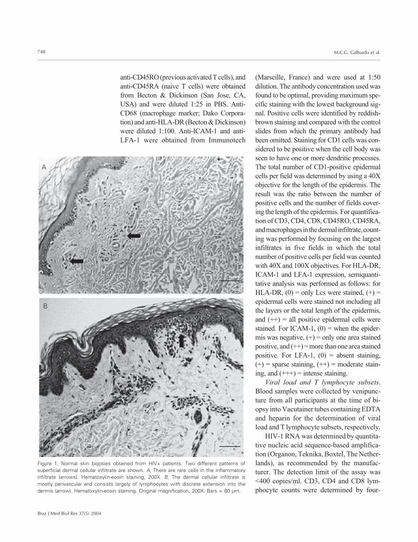
Figure 1. Normal skin biopsies obtained from HIV+ patients. Two different patterns ofsuperficial dermal cellular infiltrate are shown. A, There are rare cells in the inflammatoryinfiltrate (arrows). Hematoxylin-eosin staining, 200X. B, The dermal cellular infiltrate ismostly perivascular and consists largely of lymphocytes with discrete extension into thedermis (arrow). Hematoxylin-eosin staining, Original magnification, 200X. Bars = 80 µm.
749
Braz J Med Biol Res 37(5) 2004
T cells in the skin of HIV-infected individuals
color staining (CD45, CD3, CD4, CD8) us-ing standard flow cytometry.
Statistical analysis
Results are reported as mean ± SD. Asimple description of variables was elaboratedin order to compare the different HIV catego-ries and controls with respect to clinical andlaboratory data. ANOVA (Scheffé) and thechi-square test (Fischer) were used to measurethe effect of HIV staging associated to inde-pendent variables. In addition, the Pearson-product-moment correlation coefficient (r) wascalculated. The level of significance was set atP < 0.05 in all analyses.
Results
Histological analysis
Hematoxylin-eosin staining showed asimilar histological pattern and a nonspe-cific dermis infiltrate in HIV patients andcontrol subjects. No major differences inmean percent area occupied by the infiltratewere noted between the patient categoriesand the control group, with values of 702.8 ±621.6 for category I, 448.8 ± 274.8 for cat-egory II, 626.4 ± 408.6 for category III, and749.5 ± 462.3 for controls. Similarly, therewas no statistically significant differencebetween HIV+ individuals (592.6 ± 760.1)and controls (749.5 ± 462.3; Figure 1A,B).
Immunohistological evaluation
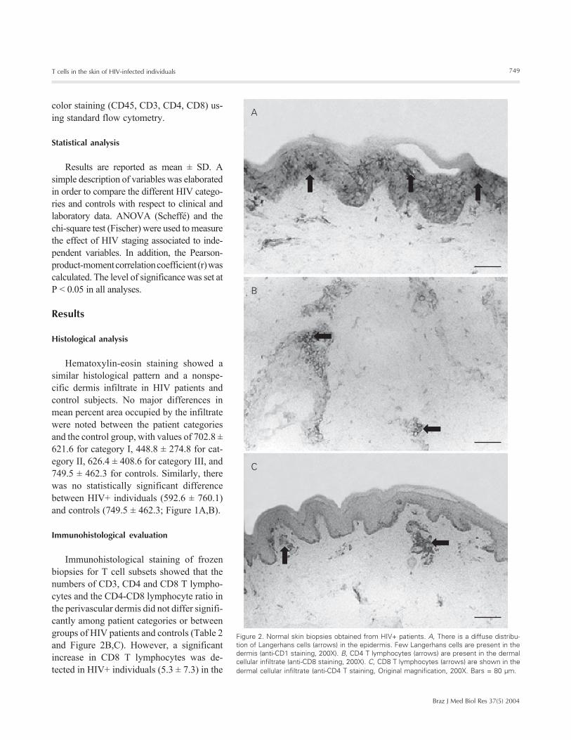
Immunohistological staining of frozenbiopsies for T cell subsets showed that thenumbers of CD3, CD4 and CD8 T lympho-cytes and the CD4-CD8 lymphocyte ratio inthe perivascular dermis did not differ signifi-cantly among patient categories or betweengroups of HIV patients and controls (Table 2and Figure 2B,C). However, a significantincrease in CD8 T lymphocytes was de-tected in HIV+ individuals (5.3 ± 7.3) in the
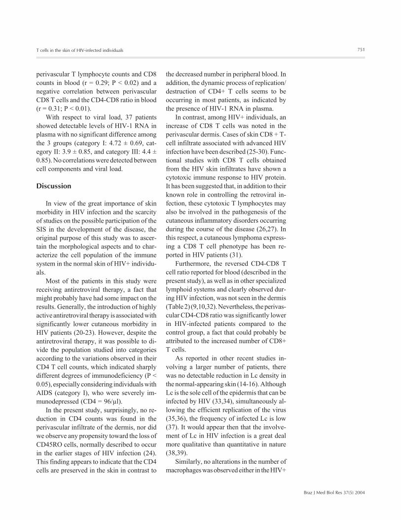
Figure 2. Normal skin biopsies obtained from HIV+ patients. A, There is a diffuse distribu-tion of Langerhans cells (arrows) in the epidermis. Few Langerhans cells are present in thedermis (anti-CD1 staining, 200X). B, CD4 T lymphocytes (arrows) are present in the dermalcellular infiltrate (anti-CD8 staining, 200X). C, CD8 T lymphocytes (arrows) are shown in thedermal cellular infiltrate (anti-CD4 T staining, Original magnification, 200X. Bars = 80 µm.
A
B
C
750
Braz J Med Biol Res 37(5) 2004
M.C.G. Galhardo et al.
22.5 ± 14.4 and 1.0 ± 1.4 for the controlgroup (Figure 2A). Likewise, the mean mac-rophage cell density in the dermis was notsignificantly different between the HIV+categories and the control group or amongthe different patient categories (data notshown). Epidermal HLA-DR and ICAM-1expression was negative in the keratinocytes(0+) and LFA-1 and ICAM-1 were expressedconstitutively in the blood vessels and indermal lymphocytes.
Correlation between CD8 T lymphocytes inthe perivascular dermis infiltrate and Tlymphocyte subsets in the blood
The number of CD3-, CD4- and CD8-positive cells in blood was estimated in allthe individuals enrolled in the study. Asexpected, CD4 cell averages were signifi-cantly different between the various catego-ries (except category III, CD4 ≥500/µl) andthe control group, and individuals with AIDSpresented the lowest averages (Table 2).When data for all HIV+ individuals werepooled, lower mean CD4-CD8 ratios weredetected (0.44 ± 0.45) compared to the con-trol group (2.12 ± 1.24; P < 0.001). More-over, evaluation of the cellular infiltrateshowed a positive correlation between CD8
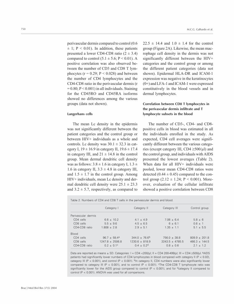
Table 2. Numbers of CD4 and CD8 T cells in the perivascular dermis and blood.
Category I Category II Category III Control group
Perivascular dermisCD4 cells 6.6 ± 10.2 4.1 ± 4.9 7.06 ± 6.4 5.8 ± 6CD8 cells 5.5 ± 9.6 4.5 ± 6.5 6 ± 6.1 0.6 ± 1CD4-CD8 ratio 1.808 ± 2.8 2.9 ± 5.1 1.35 ± 1.1 5.1 ± 5.5
BloodCD4 cells 96.7 ± 58.4a 344.0 ± 76.6b 756.0 ± 38.8 809.9 ± 201.8CD8 cells 1247.8 ± 2506.8 1230.6 ± 818.9 3243.5 ± 4785.5 466.3 ± 144.5CD4-CD8 ratio 0.2 ± 0.1c 0.4 ± 0.2d 0.8 ± 0.6 2.1 ± 1.2
Data are reported as means ± SD. Categories: I = CD4 <200/µl; II = CD4 200-499/µl; III = CD4 ≥500/µl. aAIDSpatients had significantly lower numbers of CD4 lymphocytes in blood compared with category II (P < 0.02),category III (P < 0.001), and control (P < 0.001). bIn category II, CD4 numbers were also significantly lowercompared to category III (P < 0.001), and to control (P < 0.001). cThe CD4-CD8 T lymphocyte ratio wassignificantly lower for the AIDS group compared to control (P < 0.001), and for dcategory II compared tocontrol (P < 0.001). ANOVA was used for all comparisons.
perivascular dermis compared to control (0.6± 1; P < 0.01). In addition, these patientspresented a lower CD4-CD8 ratio (2 ± 3.4)compared to control (5.1 ± 5.6; P < 0.01). Apositive correlation was also observed be-tween the number of CD3 and CD8 T lym-phocytes (r = 0.29; P < 0.028) and betweenthe number of CD4 lymphocytes and theCD4-CD8 ratio in the perivascular dermis (r= 0.80; P < 0.001) in all individuals. Stainingfor the CD45RO and CD45RA isoformsshowed no differences among the variousgroups (data not shown).
Langerhans cells
The mean Lc density in the epidermiswas not significantly different between thepatient categories and the control group orbetween HIV+ individuals as a whole andcontrols. Lc density was 30.1 ± 32.3 in cat-egory I, 19 ± 16.9 in category II, 19.6 ± 17.4in category III, and 21 ± 14.8 in the controlgroup. Mean dermal dendritic cell densitywas as follows: 3.8 ± 1.6 in category I, 1.3 ±1.6 in category II, 3.3 ± 4.8 in category III,and 1.5 ± 1.7 in the control group. AmongHIV+ individuals, mean Lc density and der-mal dendritic cell density were 25.1 ± 23.3and 3.2 ± 5.7, respectively, as compared to
751
Braz J Med Biol Res 37(5) 2004
T cells in the skin of HIV-infected individuals
perivascular T lymphocyte counts and CD8counts in blood (r = 0.29; P < 0.02) and anegative correlation between perivascularCD8 T cells and the CD4-CD8 ratio in blood(r = 0.31; P < 0.01).
With respect to viral load, 37 patientsshowed detectable levels of HIV-1 RNA inplasma with no significant difference amongthe 3 groups (category I: 4.72 ± 0.69, cat-egory II: 3.9 ± 0.85, and category III: 4.4 ±0.85). No correlations were detected betweencell components and viral load.
Discussion
In view of the great importance of skinmorbidity in HIV infection and the scarcityof studies on the possible participation of theSIS in the development of the disease, theoriginal purpose of this study was to ascer-tain the morphological aspects and to char-acterize the cell population of the immunesystem in the normal skin of HIV+ individu-als.
Most of the patients in this study werereceiving antiretroviral therapy, a fact thatmight probably have had some impact on theresults. Generally, the introduction of highlyactive antiretroviral therapy is associated withsignificantly lower cutaneous morbidity inHIV patients (20-23). However, despite theantiretroviral therapy, it was possible to di-vide the population studied into categoriesaccording to the variations observed in theirCD4 T cell counts, which indicated sharplydifferent degrees of immunodeficiency (P <0.05), especially considering individuals withAIDS (category I), who were severely im-munodepressed (CD4 = 96/µl).
In the present study, surprisingly, no re-duction in CD4 counts was found in theperivascular infiltrate of the dermis, nor didwe observe any propensity toward the loss ofCD45RO cells, normally described to occurin the earlier stages of HIV infection (24).This finding appears to indicate that the CD4cells are preserved in the skin in contrast to
the decreased number in peripheral blood. Inaddition, the dynamic process of replication/destruction of CD4+ T cells seems to beoccurring in most patients, as indicated bythe presence of HIV-1 RNA in plasma.
In contrast, among HIV+ individuals, anincrease of CD8 T cells was noted in theperivascular dermis. Cases of skin CD8 + T-cell infiltrate associated with advanced HIVinfection have been described (25-30). Func-tional studies with CD8 T cells obtainedfrom the HIV skin infiltrates have shown acytotoxic immune response to HIV protein.It has been suggested that, in addition to theirknown role in controlling the retroviral in-fection, these cytotoxic T lymphocytes mayalso be involved in the pathogenesis of thecutaneous inflammatory disorders occurringduring the course of the disease (26,27). Inthis respect, a cutaneous lymphoma express-ing a CD8 T cell phenotype has been re-ported in HIV patients (31).
Furthermore, the reversed CD4-CD8 Tcell ratio reported for blood (described in thepresent study), as well as in other specializedlymphoid systems and clearly observed dur-ing HIV infection, was not seen in the dermis(Table 2) (9,10,32). Nevertheless, the perivas-cular CD4-CD8 ratio was significantly lowerin HIV-infected patients compared to thecontrol group, a fact that could probably beattributed to the increased number of CD8+T cells.
As reported in other recent studies in-volving a larger number of patients, therewas no detectable reduction in Lc density inthe normal-appearing skin (14-16). AlthoughLc is the sole cell of the epidermis that can beinfected by HIV (33,34), simultaneously al-lowing the efficient replication of the virus(35,36), the frequency of infected Lc is low(37). It would appear then that the involve-ment of Lc in HIV infection is a great dealmore qualitative than quantitative in nature(38,39).
Similarly, no alterations in the number ofmacrophages was observed either in the HIV+

752
Braz J Med Biol Res 37(5) 2004
M.C.G. Galhardo et al.
patients or between them and the controlgroup. Previous studies have also failed todetect any HIV viral particles or viruses insuch cells (40). On the other hand, in othersites such as the lung, where HIV can beisolated from 50% of the macrophages inbronchoalveolar lavage fluids, numerical,phenotypical, and functional changes in mac-rophages have been reported to occur (3).Similarly, no increase in HLA-DR or ICAM-1 expression was observed in keratinocytes,suggesting that there is no inflammatory re-sponse in the normal skin of HIV+ individu-als.
The present study has shown the preser-vation of immune cells such as dermal CD4+T cells, dermal dendritic cells, macrophages,and Lc in the normal skin of HIV patients. Itcan now be convincingly argued that the skinis not a major site of HIV replication and thatthe SIS in HIV infection acts differently
from other local immune systems where HIV-related changes take place. Conversely, itwould seem that subtle interactions do oc-cur, as indicated mainly by the increasednumber of CD8+ T cells in the skin. Al-though in the present study we did not assessthe activation of CD8 cells, the presence ofan increased number of cells in the perivas-cular normal skin may somehow lead totissue damage, and, thereby actually contrib-uting to the pathogenesis of skin disease inHIV infection.
Acknowledgments
The authors wish to thank Dr. SérgioLuiz Gomes Antunes for helpful advice andthe Laboratório de Produção e Tratamentode Imagens, Instituto Oswaldo Cruz, Rio deJaneiro, RJ, Brazil, for the figures.
References
1. Coldiron BM & Bergstresser PR (1989). Prevalence and clinicalspectrum of skin disease in patient infected with human immuno-deficiency virus. Archives of Dermatology, 125: 357-361.
2. Uthayakumar S, Nanwani R, Drinkwater T, Nayagam AT & DarleyCR (1997). The prevalence of skin disease in HIV infection and itsrelationship to the degree of immunosuppression. British MedicalJournal, 137: 595-598.
3. Spira R, Mignard M, Doutre MS, Morlat P & Dabis F (1998). Preva-lence of cutaneous disorders in a population of HIV-infected pa-tients. Archives of Dermatology, 34: 1208-1212.
4. Duvic M (1995). Human immunodeficiency virus and the skin: se-lected controversies. Journal of Investigative Dermatology, 105(Suppl): 117S-121S.
5. Tschachler E, Bergstresser PR & Stingl G (1996). HIV-related skindiseases. Lancet, 348: 659-663.
6. Henry M & Tschachler E (1996). The skin immune system in thecourse of HIV-1 infection. Photochemistry and Photobiology, 64:275-279.
7. Blauvelt A & Katz SI (1995). The skin as target, vector, and effectororgan in human immunodeficiency virus disease. Journal of Investi-gative Dermatology, 105: 122S-126S.
8. Bray DH, Squire SB, Bagdades E, Mulvenna PM, Johnson MA &Poulter LW (1992). Alveolar macrophage populations are distortedin immunocompromised patients with pneumonitis. European Res-piratory Journal, 5: 545-552.
9. Lim SG, Condez A, Lee CA, Johnson MA, Elias C & Poulter L (1993).Loss of mucosal CD4 lymphocytes is an early feature of HIV infec-tion. Clinical and Experimental Immunology, 92: 448-454.
10. Olaitan A, Johnson MA, MacLean A & Poulter LW (1996). Thedistribution of immunocompetent cells in the genital tract of HIV-positive women. AIDS, 10: 759-764.
11. Belsito DV, Sanchez MR, Baer RL, Valentine F & Thorbecke J(1984). Reduced Langerhans’ cell Ia antigen and ATPase activity inpatients with the acquired immunodeficiency syndrome. New Eng-land Journal of Medicine, 310: 1279-1282.
12. Dréno B, Milpied B, Bignon JD, Stalder JF & Litoux P (1988).Prognostic value of Langerhans cells in the epidermis of HIV pa-tients. British Journal of Dermatology, 118: 481-486.
13. Kanitakis J, Marchand C, Su H, Thivolet J, Zambruno G, Schmitt D &Gazzolo L (1989). Immunohistochemical study of normal skin ofHIV-1 infected patients shows no evidence of infection of epidermalLangerhans cells by HIV. AIDS Research and Human Retroviruses,5: 293-302.
14. Kalter DC, Greenhouse JJ, Orenstein JM, Schnittman SM,Gendelman HE & Meltzer MS (1991). Epidermal Langerhans cellsare not principal reservoirs of virus in HIV disease. Journal of Immu-nology, 146: 3396-3404.
15. Nandwani R, Gazzard BG, Barton SE, Hawkins DA, Zemelman V &Staughton RC (1996). Does HIV disease progression influence epi-dermal Langerhans cell density? British Journal of Dermatology,134: 1087-1092.
16. Comptom CC, Kupper TS & Nadire KB (1996). HIV-1 infected Lan-gerhans cells constitute a significant proportion of the epidermalLangerhans cell population throughout the course of HIV disease.Journal of Investigative Dermatology, 107: 822-826.
17. Galhardo MC, Perez M, Morgado MG, Almeida S, Azevedo LMS,

753
Braz J Med Biol Res 37(5) 2004
T cells in the skin of HIV-infected individuals
Geog I, Ferreira H & Sarno EN (2000). Search for evidence of a Th2profile in HIV+ patients. International Journal of Dermatology, 39:109-115.
18. Oxholm A, Oxholm P & Permin H (1989). Epidermal tumour necro-sis factor α and interleukin 6-like activities in AIDS-related Kaposi’ssarcoma. Acta Pathologica et Microbiologica Scandinavica, 97: 533-538.
19. Wantzing GL, Ralfkiaer E, Lisby S & Rothlein R (1989). The role ofintercellular adhesion molecules in inflammatory skin reactions.British Journal of Dermatology, 119: 141-145.
20. Mirmirani P, Maurer TA, Berger TG, Sands LP & Chren MM (2002).Skin-related quality of life in HIV-infected patients on highly activeantiretroviral therapy. Journal of Cutaneous Medicine and Surgery,6: 10-18.
21. Johnson RA (1999). Human immunodeficiency virus disease in theera of HAART: a reevaluation of the mucocutaneous manifesta-tions. Current Clinical Topics in Infectious Diseases, 19: 252-286.
22. Costner M & Cockerell CJ (1998). The changing spectrum of cuta-neous manifestations of HIV infection. Archives of Dermatology,134: 1290-1292.
23. Calista D, Morri M, Stagno A & Boschini A (2002). Changing morbid-ity of cutaneous diseases in patients with HIV after the introductionof antiretroviral therapy including a protease inhibitor. AmericanJournal of Clinical Dermatology, 3: 359-362.
24. Gruters RA, Terpstra FG, De Goede RE, Mulder JW, De Wolf F,Schellekens PT, Van Lier RA, Tersmette M & Miedema F (1991).Immunological and virological markers in individuals progressingfrom seroconversion to AIDS. AIDS, 5: 837-844.
25. Janier M, Katlama C, Flageul B, Valensi F, Moulonguet I, Sigaux F,Dompmartin D & Civatte J (1989). The pseudo-Sezary-syndromewith CD8 phenotype in a patient with the acquired immunodeficien-cy syndrome (AIDS). Annals of Internal Medicine, 110: 738-740.
26. Bachelez H, Hadida F & Gorochov G (1996). Massive infiltration ofthe skin by human immunodeficiency virus-specific cytotoxic CD8 Tcells (Letter). New England Journal of Medicine, 335: 61-62.
27. Bachelez H, Hadida F, Parizot C, Flageul B, Kemula M, Dubertret L,Debree P & Gorochov G (1998). Oligoclonal expansion of immuno-deficiency virus-specific cytotoxic CD8 T cells in the skin of humanimmunodeficiency virus-1 infected patients with pseudolymphoma.Journal of Clinical Investigation, 101: 2506-2516.
28. Friedler S, Parisi MT, Waldo E, Wieczorek R, Sidhu G & Rico MJ(1999). Atypical cutaneous lymphoproliferative disorder in patientswith HIV infection. International Journal of Dermatology, 38: 111-118.
29. Guitart J, Variakojis D, Kuzel T & Rosen S (1999). Cutaneous CD8+ Tcell infiltrates in advanced HIV infection. Journal of the American
Academy of Dermatology, 41: 722-727.30. Pirovano S, Signorini L, Facchetti F, Santoro A, Albertin A & Imberti
L (2001). Polyclonal T-cell expansion in a HIV+ patient with atypicalcutaneous lymphoproliferative disorder, large granular lymphocyteproliferation and SENV infection. Haematologica, 86: 881-882.
31. Burns MK & Cooper KD (1993). Cutaneous T-cell lymphoma associ-ated with HIV infection. Journal of the American Academy of Der-matology, 29: 394-399.
32. Bofill M, Janossy G, Lee CA, MacDonald-Burns D, Phillips AN,Sabin C, Timms A, Johnson MA & Kernof PBA (1992). Laboratorycontrol values for CD4 and CD8 T lymphocytes. Implications forHIV-1 diagnosis. Clinical and Experimental Immunology, 88: 243-252.
33. Tschachler E, Groh V, Popovic M, Mann DL, Konrad K, Safai B, EronL, Veronese FM, Wolff K & Sting G (1987). Epidermal Langerhanscells - a target for HTLV-III/LAV infection. Journal of InvestigativeDermatology, 88: 233-237.
34. Giannetti A, Zambruno G, Cimarelli A, Marconi A, Negroni M,Girolomoni G & Bertazzoni U (1993). Direct detection of HIV-1 inepidermis Langerhans cells of HIV-1 infected patients. Journal ofAcquired Immune Deficiency Syndromes, 6: 329-333.
35. Kawamura T, Cohen SS, Borris DL, Aquilino EA, Glushakova S,Margolis LB, Orenstein JM, Offord RE, Neurath AR & Blauvelt A(2000). Candidate microbicides block HIV-1 infection of human im-mature Langerhans cells within epithelial tissue explants. Journal ofExperimental Medicine, 192: 1491-1500.
36. Reece JC, Handley AJ, Anstee EJ, Morrison WA, Crowe SM &Cameron PU (1998). HIV-1 selection by epidermal dendritic cellsduring transmission across human skin. Journal of ExperimentalMedicine, 187: 1623-1631.
37. Cimarelli A, Zambruno G, Marconi A, Giralomoni G, Bertazzoni U &Giannetti A (1994). Quantitation by competitive PCR of HIV-1 provi-ral DNA in epidermal Langerhans cells of HIV-1 infected patients.Journal of Acquired Immune Deficiency Syndromes, 7: 230-235.
38. Blauvelt A, Clerici M, Lucey DR, Steinberg SM, Yarchoan R, WalkerR, Shearer GM & Katz SI (1995). Functional studies of epidermalLangerhans cells and blood monocytes in HIV-1 infected persons.Journal of Immunology, 154: 3506-3515.
39. Blauvelt A, Chougnet C, Shearer GM & Katz SI (1996). Modulation ofT cell responses to recall antigens presented by Langerhans cells inHIV-discordant identical twins by anti-interleukin (IL)-10 antibodiesand IL-12. Journal of Clinical Investigation, 97: 1550-1555.
40. Kalter DC, Gendelman HE & Meltzer MS (1991). Monocytes, den-dritic cells, and Langerhans cells in human immunodeficiency virusinfection. Dermatologic Clinics, 9: 415-428.