Collagen containing microcapsules: smart containers for disease controlled therapy
7
Collagen containing microcapsules: Smart containers for disease controlled therapy Laura Pastorino a,⇑ , Svetlana Erokhina b , Federico Caneva Soumetz a , Paolo Bianchini c , Oleg Konovalov d , Alberto Diaspro c , Carmelina Ruggiero a , Victor Erokhin e,f a Department of Communication, Computer and System Sciences, University of Genova, Via all’Opera Pia 13, 16145 Genova, Italy b Department of Biochemistry and Molecular Biology, University of Parma, Viale Usberti 23 A, 43100 Parma, Italy c Nanophysics, IIT—Italian Institute of Technology, 16163 Genoa, Italy d European Synchrotron Radiation Facility, 38043 Grenoble Cedex, France e IPCF-CNR, Rome 00185, Italy f Department of Physics, University of Parma, Viale Usberti 23 A, 43100 Parma, Italy article info Article history: Received 11 October 2010 Accepted 3 February 2011 Available online 24 February 2011 Keywords: Collagen Matrix metalloproteinase Layer-by-layer self assembly Microcapsules Disease-dependent release abstract The protein collagen is the major component of connective tissue and it is involved in many biological functions. Its degradation is at the basis of different pathological processes. The up-regulated expression of matrix metalloproteinases and the down-regulated expression of their inhibitors are the causes for such degradation. The aim of this work was to evaluate the possibility to fabricate collagen based con- tainers for drug encapsulation and release by cellular demand by the action of matrix metalloproteinases. In present work collagen type I based microcapsules were fabricated by means of the layer-by-layer assembly of oppositely charged collagen and poly (stirene sulfonate) onto colloidal particles, followed by removal of the cores to obtain hollow microcapsules. The process of shell growth on planar supports was monitored by quartz crystal microbalance. X-ray reflectivity measurements were carried out at the solid/water interface to study the interaction of matrix metalloproteinase 1 with LbL films of collagen. The morphology of hollow capsules was characterized by scanning electron microscopy, and compared to that of capsules exposed to the matrix metalloproteinase 1. Finally the matrix metalloproteinase 1 mediated permeability of capsules variation was studied by Confocal Laser Scanning Microscopy. The results demonstrated the possibility to fabricate a drug delivery system where the release of the drug is dependent on the biochemistry of the pathological state. Ó 2011 Elsevier Inc. All rights reserved. 1. Introduction Collagens are the most abundant structural proteins of the extracellular [1]. Different collagen types are found in different types of connective tissues in the body. At least, 26 genetically distinct collagen types have been isolated, having in common a triple-helix configuration of three polypeptide subunits known as a-chains and which differ in the length of the helix and the nature and size of the non-helical portions [2]. The primary function of collagens is to contribute to tissues stability and to maintain their structural integrity. Collagens represent the main proteins of mam- malian connective tissues such as cartilage, ligaments, tendons, bone and skin. Collagen degradation and disturbed metabolism are at the basis of pathological states such as osteoarthritis (OA) [3]. In OA, the degradation of the extracellular matrix exceeds its synthesis, leading to a decrease in the amount of cartilage matrix and finally to the complete erosion of the cartilage overlying the bone at the joint surface [4]. Cartilage damage has been related to an up-regulated expression of the collagenolytic enzymes matrix metalloproteinases (MMPs) and to a down-regulated expression of their inhibitors [5,6]. MMPs are a family of Zn 2+ dependent endopeptidases that degrade all the components of the extracellular matrix [7]. Among MMPs, MMP1 belongs to collagenase subgroup in MMPs family and can cleave fibrillar collagen types I, II, III, VII, VIII, X, at one specific site resulting in the generation of fragments, which then denature to gelatin at body temperature. Beside its involve- ment in cartilage degeneration during OA, MMP1 has been also demonstrated to be involved in tumor invasion and metastasis, pulmonary emphysema, fibrotic disorders, intestinal and dermal ulcerations and many other human diseases [8,9]. In this respect the patho-physiological relevance of MMP1 is highly recognized as well as the high potentiality of MMP1 inhibitors as therapeutic agents for the development of new treatment strategies in differ- ent pathologies, including OA. The development of a successful treatment regime largely de- pends also on the adopted drug delivery strategy. Since many 0021-9797/$ - see front matter Ó 2011 Elsevier Inc. All rights reserved. doi:10.1016/j.jcis.2011.02.010 ⇑ Corresponding author. Fax: +39 010 3532154. E-mail addresses: [email protected] (L. Pastorino), svetlana.erokhina@fi- s.unipr.it (S. Erokhina), [email protected] (F.C. Soumetz), [email protected] (P. Bianchini), [email protected] (O. Konovalov), [email protected] (A. Diaspro), [email protected] (C. Ruggiero), victor.erokhin@fis.unipr.it (V. Erokhin). Journal of Colloid and Interface Science 357 (2011) 56–62 Contents lists available at ScienceDirect Journal of Colloid and Interface Science www.elsevier.com/locate/jcis
Transcript of Collagen containing microcapsules: smart containers for disease controlled therapy
Journal of Colloid and Interface Science 357 (2011) 56–62
Contents lists available at ScienceDirect
Journal of Colloid and Interface Science
www.elsevier .com/locate / jc is
Collagen containing microcapsules: Smart containers for disease controlled therapy
Laura Pastorino a,⇑, Svetlana Erokhina b, Federico Caneva Soumetz a, Paolo Bianchini c, Oleg Konovalov d,Alberto Diaspro c, Carmelina Ruggiero a, Victor Erokhin e,f
a Department of Communication, Computer and System Sciences, University of Genova, Via all’Opera Pia 13, 16145 Genova, Italyb Department of Biochemistry and Molecular Biology, University of Parma, Viale Usberti 23 A, 43100 Parma, Italyc Nanophysics, IIT—Italian Institute of Technology, 16163 Genoa, Italyd European Synchrotron Radiation Facility, 38043 Grenoble Cedex, Francee IPCF-CNR, Rome 00185, Italyf Department of Physics, University of Parma, Viale Usberti 23 A, 43100 Parma, Italy
a r t i c l e i n f o a b s t r a c t
Article history:Received 11 October 2010Accepted 3 February 2011Available online 24 February 2011
Keywords:CollagenMatrix metalloproteinaseLayer-by-layer self assemblyMicrocapsulesDisease-dependent release
0021-9797/$ - see front matter � 2011 Elsevier Inc. Adoi:10.1016/j.jcis.2011.02.010
⇑ Corresponding author. Fax: +39 010 3532154.E-mail addresses: [email protected] (L. Past
s.unipr.it (S. Erokhina), [email protected] (F.C. So(P. Bianchini), [email protected] (O. Konovalov), [email protected] (C. Ruggiero), victor.erokhin@fis.
The protein collagen is the major component of connective tissue and it is involved in many biologicalfunctions. Its degradation is at the basis of different pathological processes. The up-regulated expressionof matrix metalloproteinases and the down-regulated expression of their inhibitors are the causes forsuch degradation. The aim of this work was to evaluate the possibility to fabricate collagen based con-tainers for drug encapsulation and release by cellular demand by the action of matrix metalloproteinases.In present work collagen type I based microcapsules were fabricated by means of the layer-by-layerassembly of oppositely charged collagen and poly (stirene sulfonate) onto colloidal particles, followedby removal of the cores to obtain hollow microcapsules. The process of shell growth on planar supportswas monitored by quartz crystal microbalance. X-ray reflectivity measurements were carried out at thesolid/water interface to study the interaction of matrix metalloproteinase 1 with LbL films of collagen.The morphology of hollow capsules was characterized by scanning electron microscopy, and comparedto that of capsules exposed to the matrix metalloproteinase 1. Finally the matrix metalloproteinase 1mediated permeability of capsules variation was studied by Confocal Laser Scanning Microscopy. Theresults demonstrated the possibility to fabricate a drug delivery system where the release of the drugis dependent on the biochemistry of the pathological state.
� 2011 Elsevier Inc. All rights reserved.
1. Introduction and finally to the complete erosion of the cartilage overlying the
Collagens are the most abundant structural proteins of theextracellular [1]. Different collagen types are found in differenttypes of connective tissues in the body. At least, 26 geneticallydistinct collagen types have been isolated, having in common atriple-helix configuration of three polypeptide subunits known asa-chains and which differ in the length of the helix and the natureand size of the non-helical portions [2]. The primary function ofcollagens is to contribute to tissues stability and to maintain theirstructural integrity. Collagens represent the main proteins of mam-malian connective tissues such as cartilage, ligaments, tendons,bone and skin. Collagen degradation and disturbed metabolismare at the basis of pathological states such as osteoarthritis (OA)[3]. In OA, the degradation of the extracellular matrix exceeds itssynthesis, leading to a decrease in the amount of cartilage matrix
ll rights reserved.
orino), svetlana.erokhina@fi-umetz), [email protected]@iit.it (A. Diaspro),unipr.it (V. Erokhin).
bone at the joint surface [4]. Cartilage damage has been relatedto an up-regulated expression of the collagenolytic enzymesmatrix metalloproteinases (MMPs) and to a down-regulatedexpression of their inhibitors [5,6]. MMPs are a family of Zn2+
dependent endopeptidases that degrade all the components ofthe extracellular matrix [7].
Among MMPs, MMP1 belongs to collagenase subgroup in MMPsfamily and can cleave fibrillar collagen types I, II, III, VII, VIII, X, atone specific site resulting in the generation of fragments, whichthen denature to gelatin at body temperature. Beside its involve-ment in cartilage degeneration during OA, MMP1 has been alsodemonstrated to be involved in tumor invasion and metastasis,pulmonary emphysema, fibrotic disorders, intestinal and dermalulcerations and many other human diseases [8,9]. In this respectthe patho-physiological relevance of MMP1 is highly recognizedas well as the high potentiality of MMP1 inhibitors as therapeuticagents for the development of new treatment strategies in differ-ent pathologies, including OA.
The development of a successful treatment regime largely de-pends also on the adopted drug delivery strategy. Since many
L. Pastorino et al. / Journal of Colloid and Interface Science 357 (2011) 56–62 57
drugs, growth factors, and other bioactive molecules may exertnegative side-effects, such as nephrotoxicity, hepatitis or gastroin-testinal ulceration, drug delivery strategies must provide a meansof targeting and localizing drugs to specific tissues. Moreover, sub-cutaneous, intravenous, or intra-articular delivery of therapeuticagents frequently requires concentrations much higher than phys-iological levels due to molecular diffusion and protein deactivation.Accordingly, new drug therapies must develop highly efficient,controlled systems capable of safely delivering drugs to damagedtissues and to release them in a programmable way. In this respect,biomimetic release systems where the release of drugs depends onthe biochemistry of damaged tissues are of particular interest.
In present paper we propose the fabrication of a collagen baseddrug delivery system to be used for the treatment of MMP1 relateddiseases where the release is caused by the collagenolytic activityof over-expressed MMP1 molecules.
A large variety of drug delivery systems has been proposed inthe literature (e.g. liposomes, polymeric micelles, colloids) andoptimized to increase their efficiency. In particular the layer-by-layer technique (LbL), do to its versatility, provides a simple andinexpensive method for the fabrication of nanoengineered poly-meric capsules (NPCs) for intelligent drug delivery. This techniquewas firstly proposed by Iler in 1966 [10] for the alternate assemblyof oppositely charged layers of colloidal particles. In the 1990s De-cher and co-workers reported the fabrication of multicompositefilms of charged materials trough LbL adsorption from aqueoussolutions [11]. Since then, extensive work has been carried outon the application of this technique to the fabrication of multilay-ered ultrathin films of a wide variety of charged compounds,including synthetic polyions, biopolymers, ceramics and nanopar-ticles [12]. This method allows the creation of multilayered ultra-thin films, with molecular resolution, on surfaces of any shapeand size, ranging from flat surfaces to round template particles. Ithas been demonstrated that the deposition of LbL multilayers ontocore particles, having a diameter of 50 nm up to a few tens of mi-crons, followed by core dissolution yields hollow NPCs [13–17].
The interesting property of these NPCs is the possibility to openand close the pores in their shell as a result of the variation of thesolvent pH or composition. This property allows to fulfill the inter-nal part of the capsule with some specific substances, such as drugmolecules or enzymes [18–20].
The mentioned NPCs properties have determined a wide inter-est to these objects as very perspective candidates for the design ofsmart drug delivery/release systems [21–27]. Specifically in thiswork, collagen type I (COL) has been assembled in alternation witha polyanion onto the surface of microscale templates, followed bytemplate removal for the fabrication of collagen NPCs. The deposi-tion process onto flat surfaces has been characterized by liquidquartz crystal microbalance while the NPCs have been imaged byoptical microscopy and scanning electron microscopy (SEM). Theinteraction of COL multilayers with MMP1 has been studied byX-ray reflectivity (XRR) measurements at the solid/water interface[28]. Morphology of MMP1 treated and untreated 2D surfaces andNPCs has been characterized by SEM. The collagenolytic activity ofMMP1 towards collagen NPCs has been studied by Confocal LaserScanning Microscopy (CLSM) of MMP1 treated and untreated NPCsin order to demonstrate the possibility to obtain a disease-activity-dependent release system of drugs by cellular demand.
2. Experimental procedure
2.1. Materials
Cationic type I collagen from calf skin (COL, 1 mg/mL, in 0.1 Macetic acid solution, Sigma product number C8919), anionic poly
(stirene sulfonate) (PSS, average Mw � 200.000, Aldrich productnumber 561959) and cation poly (ethyleneimine) (PEI, averageMw � 25.000, Aldrich product number 40,872-7) were used forpolyelectrolytes deposition and for the capsule shell formation.
PSS and PEI solutions were prepared in pure water at a concentra-tion of 2 mg/ml, whereas COL was used as received. The pH values ofPSS and PEI solutions were adjusted to 4 with 0.1 M HCl. MMP1(10 lg/ml) from Sigma (product number M9195) was diluted inpure water at concentration values of 5 lg/ml and 2 lg/ml. Allchemicals were used as received without any further purification.
The polymer microcapsules were assembled on positively chargedMnCO3 cores (PlasmaChem GmbH, Berlin), 6 lm in diameter.
8-Hydroxypirene-1,3,6-trisulfonic acid trisodium salt (HPTS0.025 mg/ml pH 3, fluorescence excitation and emission maximaof 460 and 510 nm, respectively; Fluka product number 56360)and ATTO 647 (0.01 mg/ml pH 7.5, fluorescence excitation andemission maxima of 645 and 670 nm, respectively; Atto-Tec,Siegen, Germany) were used as dyes.
Water, used in the experiments for the solutions preparationand washing, was purified by Milli-Q system and had theresistance of 18.2 MX cm.
2.2. Preparation of COL based nanoengineered polymeric capsules
NPCs were prepared in the following way. MnCO3 particleswere covered by successively deposited layers of PSS and COL.0.02 g of MnCO3 micro-templates were dispersed in 1.5 ml of ultra-pure water at pH 4 using a bath sonicator. As MnCO3 micro-tem-plates are positively charged, the shell assembly was started withthe deposition of a negatively charged layer of PSS. In this casethe deposition was performed by adding the PSS solution (2 mg/ml at pH 4) to that with templates with successive expositionduring 10 min. PSS deposition was followed by three successivewashings in water at pH 4. Separation of covered micro-templatesfrom the solution was performed by particles precipitation forabout 20 min. COL was then deposited by adding its solution(1 mg/ml) to that with PSS covered micro-templates with succes-sive exposition during 20 min. Also in this case COL depositionwas followed by three washings in water at pH 4.
The above described steps were repeated to deposit a shell hav-ing the following structure (PSS/COL)4. Hollow NPCs were obtainedby micro-template dissolution after three washings in water at pH2. The obtained hollow NPCs were then rinsed by pure water.
2.3. Quartz crystal microbalance (QCM)
Study of the process of shell growth was performed on planarsurfaces using a quartz crystal microbalance instrument workingin liquid environment (QCM-Z500, KSV Instruments, Helsinki, Fin-land). The QCM-Z500 instrument measuring principle is based onthe analysis of the quartz crystal impedance at multiple overtones[29]. An equivalent circuit model is fitted to the impedance curveand the obtained parameters are used to calculate the mechanicalproperties of added layers such as mass, density and thickness[30,31]. PSS/COL multilayes were deposited on gold-coated5 MHz AT-cut quartz crystals. Before adsorption, the quartz crys-tals were cleaned with H2SO4 at 150 �C for 20 min followed byextensive washing in pure water. The quartz crystals were re-newed after each use. Taking into account that the quartz crystalsurface is negatively charged, due to partial oxidation in air, a firstlayer of PEI was deposited to impart a positive charge to the sur-face for the following deposition of the structure (PSS/COL)4. A Tef-lon liquid chamber with a volume of 2 ml was used in theexperiments. Specific solutions were alternatively introduced intothe measurement chamber and were left in contact with the quartzcrystal for 10 min for PSS deposition and for 20 min for COL

Fig. 1. QCM monitoring for LbL film thickness growth during successive layersdeposition of the structure PEI/(PSS/COL)4.
58 L. Pastorino et al. / Journal of Colloid and Interface Science 357 (2011) 56–62
deposition. After each adsorption step, water at pH 4 was purredinto the chamber and left in contact with the crystal for 1 min inorder to remove the unabsorbed molecules. The data analysisand calculation of thickness were performed using the QCMImpedance Analysis software (KSV Instruments, version 3.11).
2.4. Interaction of COL LbL multilayers with MMP1
The interaction of COL LbL multilayers with MMP1 moleculeswas characterized by X-ray reflectivity (XRR) measurements atthe solid/water interface in model planar.
X-ray reflectivity measurements were performed on ID10Bbeam-line station in European Synchrotron Radiation Facilities(ESRF, Grenoble) at solid–liquid interface according to the devel-oped method [28]. Silicon supports of size 20 � 40 mm2 were usedfor the deposition of COL layers. COL layers were deposited ontothe following structure: ðPEI=PSSÞH2O
=ðPAH=PSSÞH2O=ðPAH=PSSÞNaCl
10 .This structure was decorated by the exposition to 0.25 M CdCl2
solution for 6 h. Such procedure was shown to guarantee the suffi-cient contrast for the measurements at the solid/liquid interface[28].
The following values of the electron densities were taken for thedata analysis: water 0.334 e/A3, silicon 0.699 e/A3, sublayer0.423 e/A3.
The possibility to perform a disease-dependent release ofencapsulated molecules was evaluated by SEM and CLSM.
As a first step, model planar structures composed of PSS/COLbilayers were assembled on silicon substrates and were studied.Before adsorption, silicon substrates, having an area of0.5 � 0.5 mm2, were cleaned with H2SO4 at 150 �C for 20 min fol-lowed by extensive washing in pure water. As the surface of siliconslides is negatively charged, a first layer of PEI was deposited to im-part a positive charge to the surface for the following deposition ofthe following structures (PSS/COL)1, (PSS/COL)5, (PSS/COL)10. SEMimages were acquired before and after their exposition to MMP1solution for 1.5 h. As a second step, hollow COL-containing NPCswere exposed to a solution of MMP1 and morphological changesof the shells respect to untreated NPCs were analyzed. A knownamount of hollow NPCs was dispersed into a solution of MMP1having a final protein concentration of 5 lg/ml and incubated at37 �C for 2, 4, 6, 14, 24 and 48 h. The morphology of treated and un-treated NPCs was characterized by SEM. SEM images were ac-quired with Zeiss SUPRA 40 instrument. Measurements wereperformed on samples prepared by drying of diluted solutions ofNPCs on silicon substrates. Also in this case silicon substrates werecleaned as described above and directly used for the adsorption ofpositive NPCs, as COL was the external layer of the shell.
For permeability variation test experiments CLSM imaging wascarried out. Two types of dye molecules with distinct emissionwavelengths were used for this reason. Permeability variationwas studied simultaneous analysis of the release of encapsulateddye molecules (HPTS) from capsules and entrance of the dye mol-ecules (ATTO) from the environmental solution into the capsulesvolume. Loading with HPTS was performed by exposing the NPCsto the solution of the dye at pH 3, when NPCs are in an open con-figuration, for 24 h with continuous shaking at 300 rpm/min. After-ward, the capsules were precipitated, the supernatant wasremoved and buffer solution (NaCl 50 mM) at pH 9 was added inorder to close the capsules. A known amount of loaded capsuleswas disperse into a solution of MMP1 (final concentration 5 lg/ml) and incubated at 37 �C for 24 h. Investigations were performedon loaded untreated and treated NPCs by exposing them to a solu-tion of ATTO 647 in order to put better in evidence the MMP1 med-iated degradation of the NPCs shell. The measurements wereacquired by means of a Leica TCS SP5 AOBS (Leica MicrosystemsCMS, Mannheim, Germany) inverted confocal laser scanning
microscope equipped with a set of lasers covering the 458-, 476-,488-, 496-, 514-, 543-and 633-nm lines. Images were collectedusing a Leica 63xPL APO1.4 NA oil immersion objective (LeicaMicrosystems CMS, Mannheim, Germany). Images were obtainedusing the 488 nm and 633 nm laser lines. Under this imaging con-figuration, typical confocal resolution is of the order of 250 nm inthe lateral and 500 nm in the axial direction.
3. Results and discussion
The assembly of the capsule shell having the architecture PEI/(PSS/COL)4 was characterized by QCM.
The frequency shift due to the deposition of each successivelayer of polyelectrolyte onto the quartz crystal revealed a gradualgrowth of the film with the number of deposition cycles (Fig. 1)[25,32]. Because it is difficult to measure the thickness of polyelec-trolyte capsule, QCM measurements were used to estimate shellthickness. Previous studies have found that layer thickness esti-mated from QCM measurements of dried layers, is half of the thick-ness on micro-templates [21–23]. Such difference is ascribed to thehydrated state of the capsule shell.
The liquid QCM set up was used in order to determine the thick-ness of the wet capsule shell (working conditions). The total thick-ness of the shell was estimated to be about 27 nm and thethickness of the PSS/COL bilayer was estimated to be about 6 nm.
Interaction of the collagen with MMP1 in model planar struc-tures was investigated with X-ray reflectivity measurements.
X-ray reflectivity (XRR) measurements are very useful to studythe structure of thin films (down to monomolecular layers). Thistechnique has been widely used for the characterization of Lang-muir–Blodgett (LB) monolayers at the air–water interface [33],and LB and LbL films deposited at a solid surface [34,35]. RecentlyXRR has been also proposed for the characterization at the solid–li-quid interface giving the possibility to characterize the structureand function of nanoscale films with biomolecules that have neverbeen exposed to the air [28]. Avoiding exposure to air guaranteesthe preservation of the structure and activity of biomolecules aswell as the absence of any reorganization of the layers due to thevariation of external conditions. In our case the most importantparameters that can be obtained from fitting of experimental dataare film thickness and film roughness. It is to note that the secondparameter is even more important as we cannot expect that theinteraction with the enzyme will be homogeneous over the wholefilm surface. As the measurements were done in a small-anglerange, we can say that practically whole sample area was underthe investigation. Therefore, the obtained values are averaged
L. Pastorino et al. / Journal of Colloid and Interface Science 357 (2011) 56–62 59
through the whole sample, what is practically impossible to dowith such a powerful tool as AFM on the area of 2 cm � 4 cm.
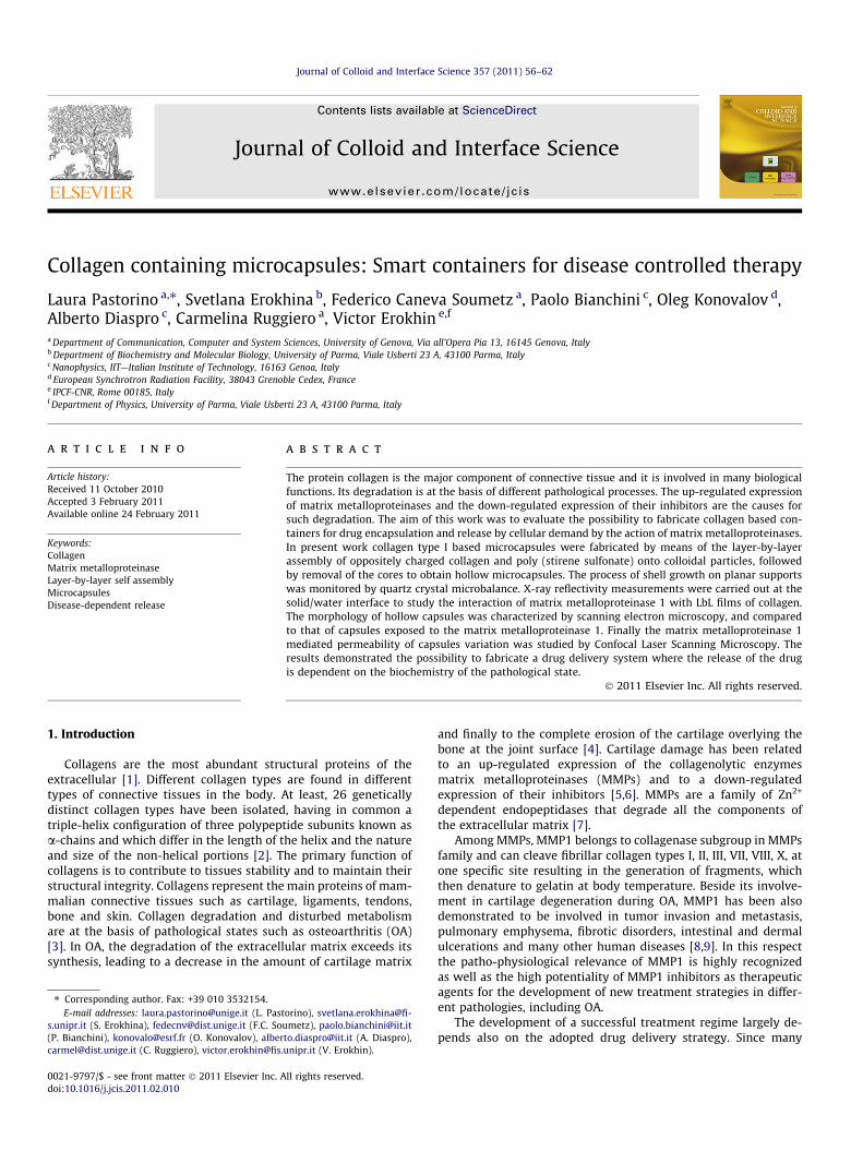
Reflectivity curves from the multilayer covered by one last COLlayer before and after treatment by MMP1 for 3 h are shown inFig. 2 left and right, respectively.
Data treatment has revealed the increase of the layer thicknessfor 0.9 nm, what is much smaller than expected if we would attri-bute it to the simple adsorption of the protein onto the film. Theroughness of the layer-water interface, on the other hand, was sig-nificantly increase from the 1.09 nm before the interactions to3.14 nm after it, what is very significant for the total layer thick-
Fig. 2. X-ray reflectivity data (points) and best fit (curve) of the structure ððPEI=PSSÞH2 O=
(right) interactions with MMP1.
Fig. 3. SEM images of PEI/(PSS/COL)1 deposited onto silicon before (le
Fig. 4. SEM images of PEI/(PSS/COL)5 deposited onto a silicon before (
ness and can be considered as the first indication of the strong var-iation of the layer structure and morphology due to the enzymeaction.
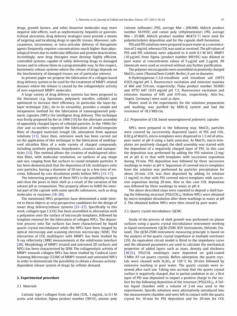
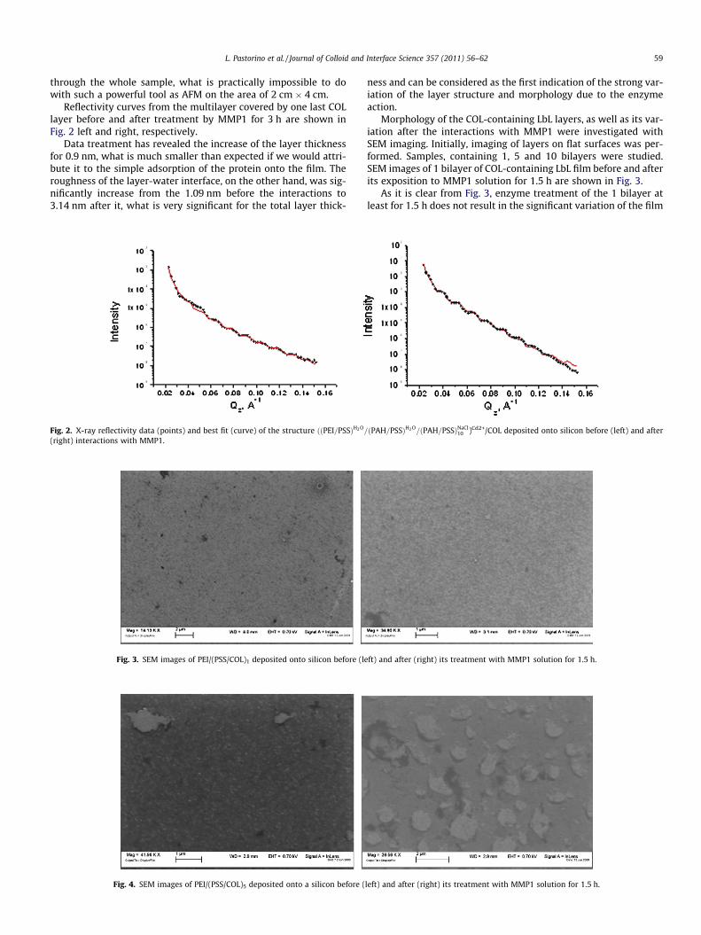
Morphology of the COL-containing LbL layers, as well as its var-iation after the interactions with MMP1 were investigated withSEM imaging. Initially, imaging of layers on flat surfaces was per-formed. Samples, containing 1, 5 and 10 bilayers were studied.SEM images of 1 bilayer of COL-containing LbL film before and afterits exposition to MMP1 solution for 1.5 h are shown in Fig. 3.
As it is clear from Fig. 3, enzyme treatment of the 1 bilayer atleast for 1.5 h does not result in the significant variation of the film
ðPAH=PSSÞH2 O=ðPAH=PSSÞNaCl
10 )Cd2+/COL deposited onto silicon before (left) and after
ft) and after (right) its treatment with MMP1 solution for 1.5 h.
left) and after (right) its treatment with MMP1 solution for 1.5 h.

Fig. 5. SEM images of PEI/(PSS/COL)1 (left), and PEI/(PSS/COL)5 (right) after their exposition to MMP1 solution for 48 h.
Fig. 6. SEM image of the COL-containing microcapsule (shell architecture (PSS/COL)4).
60 L. Pastorino et al. / Journal of Colloid and Interface Science 357 (2011) 56–62
morphology. However, for thicker films the situation was found tobe completely different. Fig. 4 represents the SEM images of the 5bilayers COL-containing LbL films before and after their treatmentby MMP1 solution for 1.5 h. In the case of 10 bilayer films the re-sults are similar.
For both film thicknesses (5 and 10 bilayers) the treatment hasresulted in the significant variation of the layers morphology, whatis in the agreement with the X-ray reflectivity measurements,
Fig. 7. SEM images of COL-containing microcapsules after the interaction w
considering the fact that SEM images reveal the variation of thein-plain film morphology, while X-ray reflectivity measurementsshow the variation of the film roughness in the direction perpen-dicular to the film plain. As it was expected, the enzyme-inducedvariations occur in some microns size zones of the layer, varyingtheir roughness (very likely, the roughness in these zones is evenmore than 3 nm, measured by X-ray reflectivity, because this valueis an averaged one).
We can observe the formation of clearly visible domains afterthe treatment, that were not present in both initial films. It is inter-esting to note that even the thinnest layer varies its morphology ifthe exposition time is increased. Fig. 5 represents SEM images ofthe 1 (left) and (right) bilayers after the interaction with MMP1for 48 h.
Longer treatment does not vary significantly the layermorphology.
Successive study was performed on layers deposited on thespherical supports. Typical image of the 6 lm MnCO3 particles cov-ered with 3 COL-containing LbL bilayers is shown in Fig. 6.
SEM images of the same capsules treated by MMP1 for 4 (a) and14 h (b) respectively are shown in Fig. 7.
As it is clear from the images, the action of the MMP1 results inthe significant variation of the capsule structure. Long exposition(14 h) results in the practically destruction of the capsule shell,what can be a basis for the release of the encapsulated material.
The confocal image of a loaded untreated NPC is presented inFig. 8a.
As it is clear from the Fig. 8a and e (lower curve), the loading ofcapsules with HPTS was successful – the dye molecules are only in-side NPCs and not in the environmental solution. However, their
ith MMP1 for 4 (left) and 14 h (right) (shell architecture (PSS/COL)4).
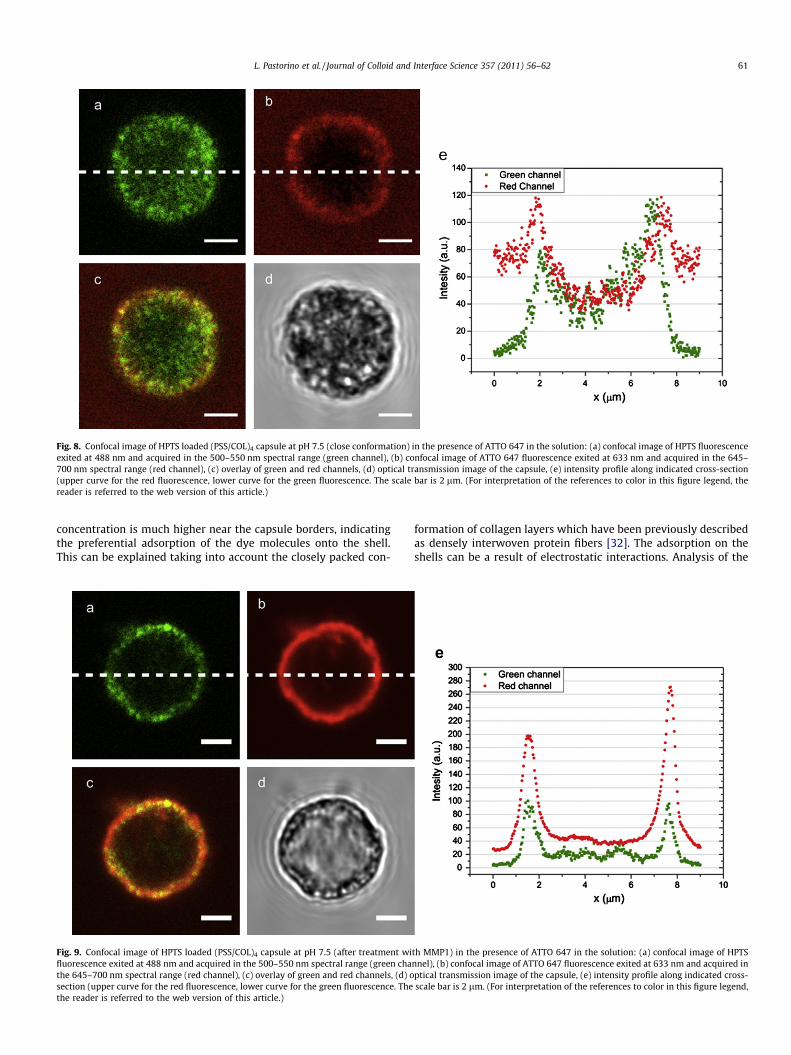
Fig. 8. Confocal image of HPTS loaded (PSS/COL)4 capsule at pH 7.5 (close conformation) in the presence of ATTO 647 in the solution: (a) confocal image of HPTS fluorescenceexited at 488 nm and acquired in the 500–550 nm spectral range (green channel), (b) confocal image of ATTO 647 fluorescence exited at 633 nm and acquired in the 645–700 nm spectral range (red channel), (c) overlay of green and red channels, (d) optical transmission image of the capsule, (e) intensity profile along indicated cross-section(upper curve for the red fluorescence, lower curve for the green fluorescence. The scale bar is 2 lm. (For interpretation of the references to color in this figure legend, thereader is referred to the web version of this article.)
L. Pastorino et al. / Journal of Colloid and Interface Science 357 (2011) 56–62 61
concentration is much higher near the capsule borders, indicatingthe preferential adsorption of the dye molecules onto the shell.This can be explained taking into account the closely packed con-
Fig. 9. Confocal image of HPTS loaded (PSS/COL)4 capsule at pH 7.5 (after treatment wifluorescence exited at 488 nm and acquired in the 500–550 nm spectral range (green chathe 645–700 nm spectral range (red channel), (c) overlay of green and red channels, (d) osection (upper curve for the red fluorescence, lower curve for the green fluorescence. Thethe reader is referred to the web version of this article.)
formation of collagen layers which have been previously describedas densely interwoven protein fibers [32]. The adsorption on theshells can be a result of electrostatic interactions. Analysis of the
th MMP1) in the presence of ATTO 647 in the solution: (a) confocal image of HPTSnnel), (b) confocal image of ATTO 647 fluorescence exited at 633 nm and acquired inptical transmission image of the capsule, (e) intensity profile along indicated cross-scale bar is 2 lm. (For interpretation of the references to color in this figure legend,

62 L. Pastorino et al. / Journal of Colloid and Interface Science 357 (2011) 56–62
red fluorescence (Fig. 8b and e (upper curve)) indicates that theATTO 647 dye molecules, added to the solution, cannot penetratethe capsule volume (the intensity within capsule is significantlylower than in the environmental solution). However, the adsorp-tion of the dye molecules on the capsule shell can be seen also inthis case. Fig. 8c reports the situation when green and red channelsare superimposed. The closed conformation of capsules is con-firmed, as the red dye Atto 647, added to the solution, is adsorbedonly onto the shell but is not found into the capsule interior, whilethe previously encapsulated HPTS is only inside capsules and not inthe environmental solution.
The same measurement was performed onto loaded MMP1treated capsules. Results are reported in Fig. 9.
It is useful to compare the results reported in Fig. 9 with thosein Fig. 8. For the green fluorescence of the encapsulated dye (HPTS)we can see a significant decrease of the fluorescence in the internalpart of the capsule (Fig. 9a and e (lower curve). Instead, for the redfluorescence of the dye molecules added to the environmentalsolution we can see the increase of the intensity in the internal partof capsules (Fig. 9b and e (upper curve). However, the higher inten-sity at the boundary of the capsules was observed for both dyemolecules, confirming their preferential adsorption on the poly-electrolyte layers. Such behavior indicates the increase of the cap-sule shell permeability that is due to the formation of poresinduced by the collagenolytic action of MMP1.
4. Conclusions
The reported results are the first steps toward the developmentof new types of therapeutic preparations with smart release prop-erties. Both X-ray reflectivity and SEM characterization resultshave revealed that the action of MMP1 on the COL layer is not justthe physical adsorption, but significant variation of the layerroughness, that can be connected to the pore formation and/orcomplete destruction of the capsule shell. In fact, prolungatedexposition of the collagen-containing capsules to MMP1 resultsin the significant variation of the capsule shape and in the forma-tion of pores in the shell. The last finding allows to suggest thatsuch micro-containers can be considered for the smart drug deliv-ery. Encapsulated pharmaceutical material inside COL-containingcapsule can be released only in the medium where MMP1 is pres-ent. Such behavior will provide therapy only in the desirable zonesavoiding, therefore, hazard side effects.
Acknowledgments
We acknowledge the European Synchrotron Radiation Facilityfor provision of synchrotron radiation facilities. One of the Authors
(S.E.) wishes to acknowledge the support of Regione Emilia-Romagna under project SITEIA. The authors would like to thank Fi-lippo Romani for table of contents graphic preparation.
References
[1] K.A. Piez, in: J.I. Kroschwitz (Ed.), Encyclopedia of Polymer Science andEngineering, Wiley-Interscience, New York, 1985, p. 699.
[2] E.J. Miller, in: M.E. Nimni (Ed.), Collagen – Biochemistry, vol. I, CRC Press, BocaRaton, 1988, p. 139.
[3] K. von der Mark, Dynamics of Bone and Cartilage Metabolism, Academic Press,Orlando, 1999.
[4] P. Sarzi-Puttini, M.A. Cimmino, R. Scarpa, R. Caporali, F. Parazzini, A. Zaninelli,F. Atzeni, B. Canesi, Semin. Arthritis Rheum. 35 (2005) 1.
[5] A. Okimura, Y. Okada, S. Makihira, H. Pan, L. Yu, K. Tanne, K. Imai, H. Yamada, T.Kawamoto, M. Noshiro, W. Yan, Y. Kato, Arthritis Rheum. 40 (1997) 1029.
[6] Y. Rengel, C. Ospelt, S. Gay, Arthritis Res. Ther. 9 (2007) 221.[7] H. Nagase, J.F. Woessner, J. Biol. Chem. 31 (1999) 21491.[8] A. Pardo, M. Selman, Int. J. Biochem. Cell Biol. 37 (2005) 283.[9] G. Murphy, H. Nagase, Mol. Aspects Med. 29 (2008) 290.
[10] R. Iler, J. Colloid Interface Sci. 21 (1966) 569.[11] G. Decher, Science 227 (1997) 1232.[12] Y. Lvov, in A. Hubbard (Eds.), Encyclopedia of Surface and Colloid Science,
Marcel Dekker, New York, 2002, p. 321.[13] E. Donath, G. Sukhorukov, F. Caruso, S. Davis, H. Mohwald, Angew. Chem., Int.
Ed. 37 (1998) 2201.[14] G.B. Sukhorukov, E. Donath, S. Davis, H. Lichtenfeld, F. Caruso, V.I. Popov, H.
Möhwald, Polym. Adv. Technol. 9 (1998) 759.[15] F. Caruso, R. Caruso, H. Möhwald, Science 282 (1998) 1111.[16] F. Caruso, D. Trau, H. Möhwald, R. Renneberg, Langmuir 16 (2000) 1485.[17] G.B. Sukhorukov, M. Brumen, E. Donath, H. Möhwald, J. Phys. Chem. B 103
(1999) 6434.[18] G.B. Sukhorukov, A.L. Rogach, B. Zebli, T. Liedl, A.G. Skirtach, K. Köhler, A.A.
Antipov, N. Gaponik, A.S. Susha, M. Winterhalter, W.J. Parak, Small 1 (2005)194.
[19] G.B. Sukhorukov, H. Möhwald, Trends Biotechnol. 25 (2007) 93.[20] J.C.M. van Hest, Macromol. Biosci. 10 (2010) 463.[21] Y. Lvov, F. Caruso, Anal. Chem. 73 (2001) 4212.[22] A. Antipov, G. Sukhorukov, E. Donath, H. Möhwald, J. Phys. Chem. B 105 (2001)
2281.[23] H. Ai, S.A. Jones, M.M. de Villiers, Y.M. Lvov, J. Controlled Release 86 (2003) 59.[24] L. Pastorino, S. Erokhina, F. Caneva-Soumetz, C. Ruggiero, J. Nanosci.
Nanotechnol. 9 (2009) 6753.[25] J. Landoulsi, C.J. Roy, C. Dupont-Gillain, S. Demoustier-Champagne,
Biomacromolecules 10 (2009) 1021.[26] T.G. Shutava, S.S. Balkundi, Y.M. Lvov, J. Colloid Interface Sci. 330 (2009) 276.[27] S. Erokhina, L. Benassi, P. Bianchini, A. Diaspro, V. Erokhin, M.P. Fontana, J. Am.
Chem. Soc. 131 (2009) 9800.[28] S. Erokhina, T. Berzina, L. Cristofolini, V. Erokhin, C. Folli, O. Konovalov, I.G.
Marino, M.P. Fontana, Langmuir 24 (2008) 12093.[29] S.J. Martin, V.E. Granstaff, G.C. Frye, Anal. Chem. 63 (1991) 2272.[30] H.L. Bandey, S.J. Martin, R.W. Cernosek, A.R. Hillman, Anal. Chem. 71 (1999)
2205.[31] T. Viitala, J.T. Hautala, J. Vuorinen, S.K. Wiedmer, Langmuir 23 (2007) 609.[32] G.G.S. Grant, D.S. Koktysh, B. Yun, R.L. Matts, N.A. Kotov, Biomed. Microdevices
3 (2001) 301.[33] G. Brezesinski, A. Dietrich, B. Struth, C. Bohm, W.G. Bouwman, K. Kjaer, H.
Mohwald, Chem. Phys. Lipids 76 (1995) 145.[34] U. Pietsch, U. Hohne, H. Mohwald, Langmuir 9 (1993) 208.[35] G. Reiter, C. Bubeck, M. Stamm, Langmuir 8 (1992) 1881.