Non-Invasive Molecular Imaging of Fibrosis Using a Collagen-Targeted Peptidomimetic of the Platelet...
10
Non-Invasive Molecular Imaging of Fibrosis Using a Collagen-Targeted Peptidomimetic of the Platelet Collagen Receptor Glycoprotein VI Julien Muzard 1,5. , Laure Sarda-Mantel 2,5,6. , Ste ´ phane Loyau 1,5 , Alain Meulemans 2,5,6 , Liliane Louedec 1,5 , Claudie Bantsimba-Malanda 4,5 , Florence Hervatin 5 , Joe ¨ lle Marchal-Somme 4,5 , Jean Baptiste Michel 1,5,6 , Dominique Le Guludec 2,5,6 , Philippe Billiald 3 , Martine Jandrot-Perrus 1,5,6 * 1 INSERM, U698, Ho ˆ pital Bichat, Paris, France, 2 INSERM, U773, CRB3, Faculte ´ Xavier Bichat, Paris, France, 3 Museum National d’Histoire Naturelle, CNRS-FRE 3206, Paris, France, 4 INSERM, U700, Faculte ´ Xavier Bichat, Paris, France, 5 Universite ´ Paris7, Paris, France, 6 AP-HP, Ho ˆ pital Bichat, Paris, France Abstract Background: Fibrosis, which is characterized by the pathological accumulation of collagen, is recognized as an important feature of many chronic diseases, and as such, constitutes an enormous health burden. We need non-invasive specific methods for the early diagnosis and follow-up of fibrosis in various disorders. Collagen targeting molecules are therefore of interest for potential in vivo imaging of fibrosis. In this study, we developed a collagen-specific probe using a new approach that takes advantage of the inherent specificity of Glycoprotein VI (GPVI), the main platelet receptor for collagens I and III. Methodology/Principal Findings: An anti-GPVI antibody that neutralizes collagen-binding was used to screen a bacterial random peptide library. A cyclic motif was identified, and the corresponding peptide (designated collagelin) was synthesized. Solid-phase binding assays and histochemical analysis showed that collagelin specifically bound to collagen (Kd 10 27 M) in vitro, and labelled collagen fibers ex vivo on sections of rat aorta and rat tail. Collagelin is therefore a new specific probe for collagen. The suitability of collagelin as an in vivo probe was tested in a rat model of healed myocardial infarctions (MI). Injecting Tc-99m-labelled collagelin and scintigraphic imaging showed that uptake of the probe occurred in the cardiac area of rats with MI, but not in controls. Post mortem autoradiography and histological analysis of heart sections showed that the labeled areas coincided with fibrosis. Scintigraphic molecular imaging with collagelin provides high resolution, and good contrast between the fibrotic scars and healthy tissues. The capacity of collagelin to image fibrosis in vivo was confirmed in a mouse model of lung fibrosis. Conclusion/Significance: Collagelin is a new collagen-targeting agent which may be useful for non-invasive detection of fibrosis in a broad spectrum of diseases. Citation: Muzard J, Sarda-Mantel L, Loyau S, Meulemans A, Louedec L, et al. (2009) Non-Invasive Molecular Imaging of Fibrosis Using a Collagen-Targeted Peptidomimetic of the Platelet Collagen Receptor Glycoprotein VI. PLoS ONE 4(5): e5585. doi:10.1371/journal.pone.0005585 Editor: Annarosa Leri, Harvard Medical School, United States of America Received October 29, 2008; Accepted April 17, 2009; Published May 18, 2009 Copyright: ß 2009 Muzard et al. This is an open-access article distributed under the terms of the Creative Commons Attribution License, which permits unrestricted use, distribution, and reproduction in any medium, provided the original author and source are credited. Funding: This work was supported by INSERM, University Paris 7, the Leducq Foundation, grant 2004002917 from the Fondation de France, and ET7-464 from the Fondation de l’Avenir. J. Muzard was supported by Stago (Asnieres, France) and the Fondation pour la Recherche Medicale and M.Jandrot-Perrus by the Bettencourt-Schueller Foundation. The funders had no role in study design, data collection and analysis, decision to publish, or preparation of the manuscript. Competing Interests: The authors have declared that no competing interests exist. * E-mail: [email protected] . These authors contributed equally to this work. Introduction Collagen, a major component of the extracellular matrix (ECM), is one of the determinants of tissue structure. Fibrosis is characterized by the pathological accumulation of collagen, and is increasingly recognized as an important feature of many chronic diseases, and as such, represents an enormous health burden [1]. It is estimated that 45% of deaths in the United States can be attributed to conditions associated with fibrosis. In the absence of a non-invasive specific marker, the only method available for quantifying fibrosis is tissue biopsy, which is invasive and carries a risk of complications in a variety of organs and cannot be easily repeated. Functional tests are currently used to assess the degree to which organs are affected, but functional impairment only occurs in the presence of a relatively high degree of fibrosis. This means that we still need non-invasive specific methods for the early diagnosis and follow-up of fibrosis in many disorders in which fibrosis is of major prognostic interest. For this purpose, quantitative imaging methods have the advantage over blood biomarkers of being able both to quantify and localize the fibrotic process. Recent studies have shown that transient echography or MRI elastography provide ways to assess liver fibrosis by non- invasively measuring liver stiffness in adult patients [2,3]. Preliminary experiments have also been performed using diffu- sion-weighted MRI to quantify liver fibrosis [4]. However, these techniques are not specific for fibrosis and may suffer from a lack of sensitivity, high levels of fibrosis being necessary before tissue elasticity and diffusion properties are impaired. Recently, PLoS ONE | www.plosone.org 1 May 2009 | Volume 4 | Issue 5 | e5585
-
Upload
independent -
Category
Documents
-
view
1 -
download
0
Transcript of Non-Invasive Molecular Imaging of Fibrosis Using a Collagen-Targeted Peptidomimetic of the Platelet...
Non-Invasive Molecular Imaging of Fibrosis Using aCollagen-Targeted Peptidomimetic of the PlateletCollagen Receptor Glycoprotein VIJulien Muzard1,5., Laure Sarda-Mantel2,5,6., Stephane Loyau1,5, Alain Meulemans2,5,6, Liliane
Louedec1,5, Claudie Bantsimba-Malanda4,5, Florence Hervatin5, Joelle Marchal-Somme4,5, Jean Baptiste
Michel1,5,6, Dominique Le Guludec2,5,6, Philippe Billiald3, Martine Jandrot-Perrus1,5,6*
1 INSERM, U698, Hopital Bichat, Paris, France, 2 INSERM, U773, CRB3, Faculte Xavier Bichat, Paris, France, 3 Museum National d’Histoire Naturelle, CNRS-FRE 3206, Paris,
France, 4 INSERM, U700, Faculte Xavier Bichat, Paris, France, 5 Universite Paris7, Paris, France, 6 AP-HP, Hopital Bichat, Paris, France
Abstract
Background: Fibrosis, which is characterized by the pathological accumulation of collagen, is recognized as an importantfeature of many chronic diseases, and as such, constitutes an enormous health burden. We need non-invasive specificmethods for the early diagnosis and follow-up of fibrosis in various disorders. Collagen targeting molecules are therefore ofinterest for potential in vivo imaging of fibrosis. In this study, we developed a collagen-specific probe using a new approachthat takes advantage of the inherent specificity of Glycoprotein VI (GPVI), the main platelet receptor for collagens I and III.
Methodology/Principal Findings: An anti-GPVI antibody that neutralizes collagen-binding was used to screen a bacterialrandom peptide library. A cyclic motif was identified, and the corresponding peptide (designated collagelin) wassynthesized. Solid-phase binding assays and histochemical analysis showed that collagelin specifically bound to collagen(Kd 1027 M) in vitro, and labelled collagen fibers ex vivo on sections of rat aorta and rat tail. Collagelin is therefore a newspecific probe for collagen. The suitability of collagelin as an in vivo probe was tested in a rat model of healed myocardialinfarctions (MI). Injecting Tc-99m-labelled collagelin and scintigraphic imaging showed that uptake of the probe occurred inthe cardiac area of rats with MI, but not in controls. Post mortem autoradiography and histological analysis of heart sectionsshowed that the labeled areas coincided with fibrosis. Scintigraphic molecular imaging with collagelin provides highresolution, and good contrast between the fibrotic scars and healthy tissues. The capacity of collagelin to image fibrosis invivo was confirmed in a mouse model of lung fibrosis.
Conclusion/Significance: Collagelin is a new collagen-targeting agent which may be useful for non-invasive detection offibrosis in a broad spectrum of diseases.
Citation: Muzard J, Sarda-Mantel L, Loyau S, Meulemans A, Louedec L, et al. (2009) Non-Invasive Molecular Imaging of Fibrosis Using a Collagen-TargetedPeptidomimetic of the Platelet Collagen Receptor Glycoprotein VI. PLoS ONE 4(5): e5585. doi:10.1371/journal.pone.0005585
Editor: Annarosa Leri, Harvard Medical School, United States of America
Received October 29, 2008; Accepted April 17, 2009; Published May 18, 2009
Copyright: � 2009 Muzard et al. This is an open-access article distributed under the terms of the Creative Commons Attribution License, which permitsunrestricted use, distribution, and reproduction in any medium, provided the original author and source are credited.
Funding: This work was supported by INSERM, University Paris 7, the Leducq Foundation, grant 2004002917 from the Fondation de France, and ET7-464 fromthe Fondation de l’Avenir. J. Muzard was supported by Stago (Asnieres, France) and the Fondation pour la Recherche Medicale and M.Jandrot-Perrus by theBettencourt-Schueller Foundation. The funders had no role in study design, data collection and analysis, decision to publish, or preparation of the manuscript.
Competing Interests: The authors have declared that no competing interests exist.
* E-mail: [email protected]
. These authors contributed equally to this work.
Introduction
Collagen, a major component of the extracellular matrix
(ECM), is one of the determinants of tissue structure. Fibrosis is
characterized by the pathological accumulation of collagen, and is
increasingly recognized as an important feature of many chronic
diseases, and as such, represents an enormous health burden [1]. It
is estimated that 45% of deaths in the United States can be
attributed to conditions associated with fibrosis. In the absence of a
non-invasive specific marker, the only method available for
quantifying fibrosis is tissue biopsy, which is invasive and carries
a risk of complications in a variety of organs and cannot be easily
repeated. Functional tests are currently used to assess the degree to
which organs are affected, but functional impairment only occurs
in the presence of a relatively high degree of fibrosis. This means
that we still need non-invasive specific methods for the early
diagnosis and follow-up of fibrosis in many disorders in which
fibrosis is of major prognostic interest. For this purpose,
quantitative imaging methods have the advantage over blood
biomarkers of being able both to quantify and localize the fibrotic
process. Recent studies have shown that transient echography or
MRI elastography provide ways to assess liver fibrosis by non-
invasively measuring liver stiffness in adult patients [2,3].
Preliminary experiments have also been performed using diffu-
sion-weighted MRI to quantify liver fibrosis [4]. However, these
techniques are not specific for fibrosis and may suffer from a lack
of sensitivity, high levels of fibrosis being necessary before tissue
elasticity and diffusion properties are impaired. Recently,
PLoS ONE | www.plosone.org 1 May 2009 | Volume 4 | Issue 5 | e5585

molecular imaging of cardiac fibrosis was reported using
radiotracers specific for targets co-expressed or co-located with
fibrosis in patients and mice with post-infarction cardiomyopathy:
18F-fluorobenzoyl-lisinopril specific for angiotensin-converting
enzyme [5], Tc-99m losartan specific for angiotensinII receptors
[6], 99mTc-Cy5.5 RGD imaging peptide targeting proliferating
myofibroblats [7,8]. However such indirect tracers are not adapted
to all clinical situations involving fibrosis, because of different
physiopathology and the need to detect fibrosis as well as
fibrogenesis. Specific and direct tracers for the molecular imaging
of fibrosis, especially collagen-targeting molecules, constitute a
challenge and a potentially wide field of interest for imaging
methods, including radionucleide imaging and MRI [9].
The inherent collagen binding properties of the collagen
receptors should make them good models for developing collagen
probes. Collagen receptors interact with the triple helical structures
of collagen fibrils [10]. Several members of the integrin family,
including the alpha1beta1, alpha2beta1 and alpha1beta1 integrins,
are widely expressed collagen receptors, but since they also bind to
other matrix proteins, they are not suitable for specifically targeting
collagen. The immunoadhesin glycoprotein VI [11,12] has good
affinity and high specificity for type-I and type-III collagens and has
been extensively characterized. GPVI seems to be an attractive
target for the development of collagen probes. Soluble recombinant
GPVI has even been proposed as a tool for in vivo imaging of collagen
exposed by unstable atherosclerotic plaques. However, an efficient
collagen probe must be small enough to gain access to the interstitial
space. We therefore decided to focus on peptides that mimicGPVI,
and have taken advantage of a monoclonal antibody, 9O12.2, which
binds GPVI with a high affinity, and neutralizes the interaction
between GPVI and collagen in vitro and in vivo [13,14]. We
hypothesized that the 9O12.2 epitope must, at least in part, overlap
with the collagen binding-site on GPVI. Using a bacterial display
approach, a peptidomimetic of GPVI has been identified. This
peptide, designated collagelin, exhibits collagen-binding properties
both in vitro and ex vivo. The suitability of collagelin as a probe for the
molecular imaging of fibrosis is assessed in vivo by isotopic imaging of
scars in a rat model of healed myocardial infarction and a mouse
model of lung fibrosis.
Results
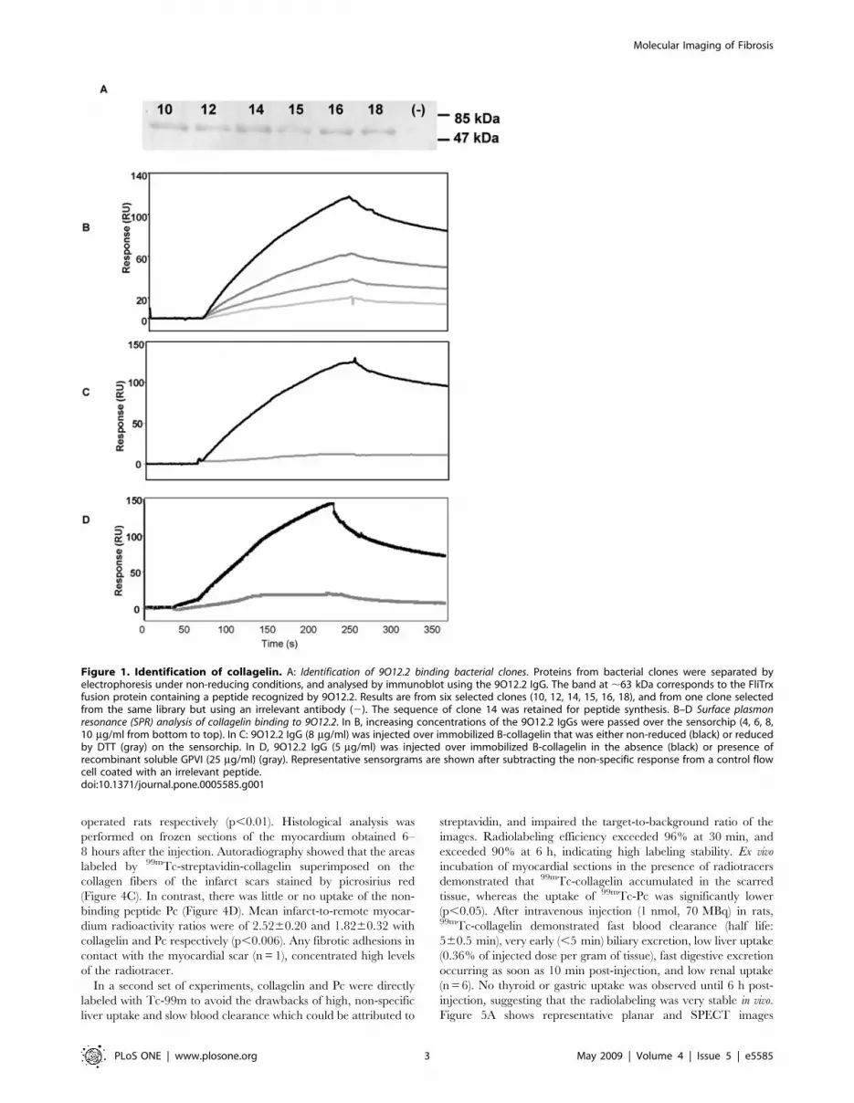
Identification of 9O12.2-binding peptidesAfter five rounds of panning the combinatorial bacterial peptide
library using 9O12.2 IgG, 20 clones were selected that produced a
flagellar fusion protein recognized by 9O12.2 on immunoblots
(Figure 1A). DNA sequencing of all the inserts showed some
redundancy and identified 9 peptide sequences (Table 1) with 7
common residues and differing from each other by one to 5
residues. None of these sequences was registered in any database.
Sequence 14, RVMHGLHLGDDE (single letter amino acid
code), was selected for synthesis. A constrained peptide, designated
collagelin, was synthesized and conjugated with biotin (B–
collagelin). The molecular masses of the peptide and the conjugate
(2155 and 2404 Da respectively) and their purity (.95%) were
determined by mass spectrometry.
Surface plasmon resonance experiments showed that 9O12.2
IgG bound to immobilized B–collagelin in a dose-dependent
manner (Figure 1B) with a KD of 1026 M. The 9O12.2 IgG did
not bind to reduced collagelin (Figure 1C), confirming that the
9O12.2 epitope is conformational [13]. Binding of 9O12.2 IgG to
collagelin was inhibited in the presence of soluble GPVI
(Figure 1D), indicating that GPVI and collagelin competed for
binding to 9O12.2.
Binding to collagenSince collagelin mimicked the 9O12.2epitope, at least in part, it
was assumed that it also mimicked the collagen binding site of
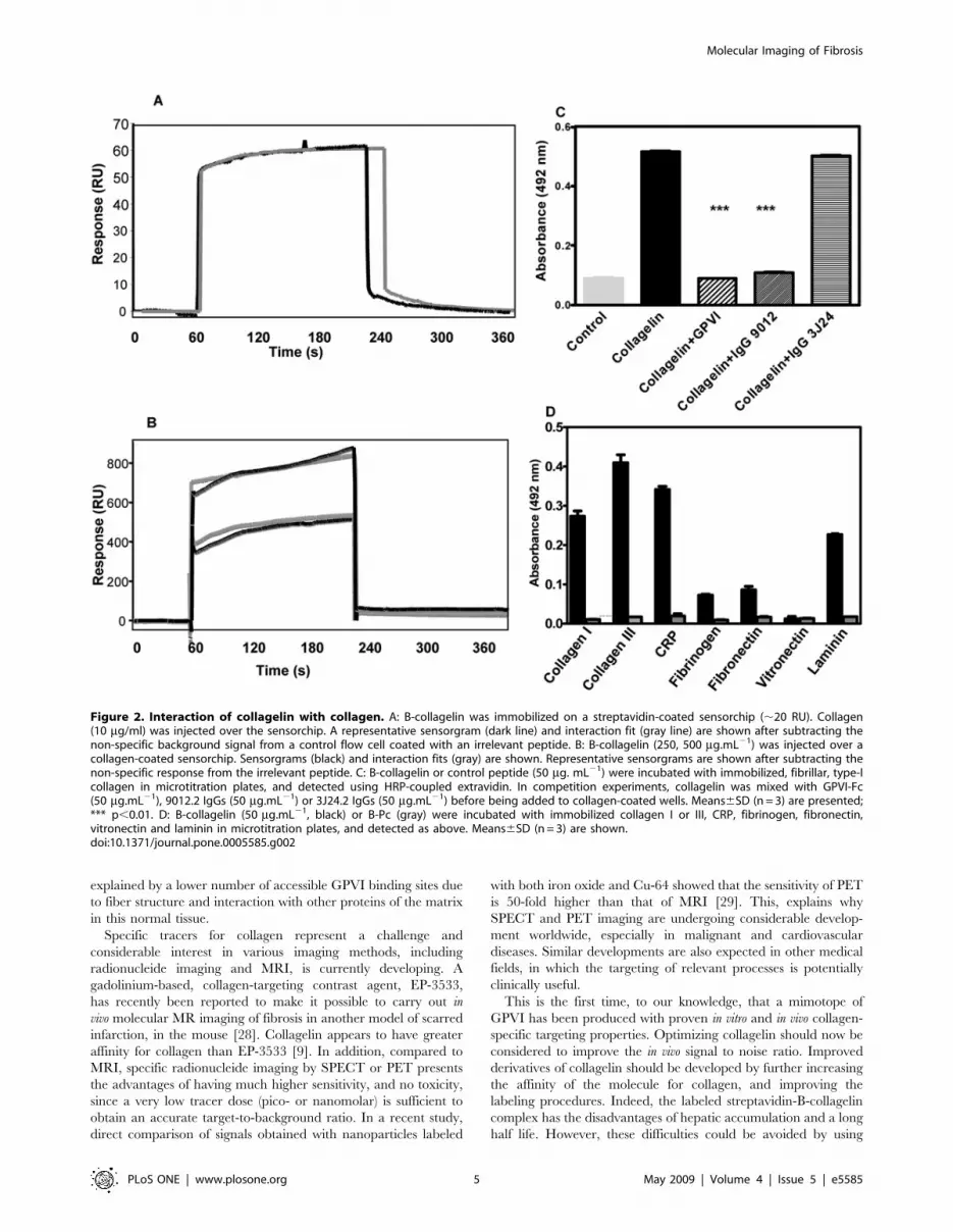
GPVI. Using SPR, type I collagen was found to bind to a B-
collagelin-coated sensorchip (Figure 2A). Furthermore, B-collage-
lin bound to a sensorchip coated with fibrillar type I collagen in a
dose-dependent manner with a KD of 1.1027 M. (Figure 2B). In
contrast, a non-relevant biotinylated peptide (B-Pc), did not bind
to collagen. B–collagelin also bound to collagen-coated plates, and
this binding was completely inhibited in the presence of GPVI-Fc
or of 9O12.2 IgGs (Figure 2C), whereas the anti-GPVI
monoclonal antibody 3J24.2, which binds to an epitope distinct
from 9O12.2 [15], did not inhibit B-collagelin interaction with
collagen. These results demonstrate that collagelin and GPVI bind
to sites on collagen that must overlap or be identical.
The binding specificity of collagelin was determined using the
GPVI specific ligand, collagen related peptide (CRP), and various
different macromolecules from the extracellular matrix. The non-
relevant peptide (Pc) did not bind to any of these proteins.
Collagelin bound to type III collagen and to CRP, confirming its
GPVI-like specificity. The binding of collagelin to vitronectin
fibrinogen or fibronectin was found either to be weak or not to
occur at all (Figure 2D). In contrast, collagelin consistently bound
to laminin, previously identified as a GPVI accessory ligand [16];
the sites of GPVI that interact with laminin and collagen thus
share a common structure that is mimicked by collagelin.
Nevertheless, SPR analysis indicated that the affinity of collagelin
for laminin was two orders lower than for collagen (KD of 1.83
1025 M).
Ex vivo labeling of collagen with collagelinThe capacity of collagelin to interact with collagen was analyzed
histochemically on frozen sections of rat aorta and tail tendon.
Positive and specific brown staining developed when sections were
treated with B-collagelin, but not with B-Pc. The staining
produced by collagelin coincided with that of collagen stained
with picrosirius red in serial sections (Figure 3). The intensity of the
staining by collagelin decreased in the presence of 9O12.2 IgG,
confirming the specificity of the interaction.
In vivo isotopic molecular imaging of fibrosisCollagelin was evaluated as a potential probe for fibrosis in vivo
in rats. A model of healed myocardial infarction was used so that
the uptake of the probe in healthy tissues could be compared to
that in the collagen-rich fibrotic scar formed after myocardial
remodeling. Imaging was performed at least three weeks after MI
after the acute inflammatory phase and when the scar is well
formed.
First, B-collagelin and B-Pc were mixed with 99mTc-streptavi-
din. Ex vivo, the uptake of 99mTc-streptavidin-coupled-collagelin by
rat aortic collagen was three times greater than the uptake of the
control peptide Pc (p,0.001), and the uptake of labeled collagelin
was inhibited by 9012.2 IgG. After intravenous injection of 1 nmol
(70 MBq) to rats, SPECT imaging revealed high uptake by the
liver, and slow blood clearance. Visual analysis of planar and
SPECT images 4 h–6 h post injection revealed significant uptake
of 99mTc-streptavidin-collagelin in the cardiac area of all rats with
an MI scar (n = 6), whereas no tracer uptake was observed in the
cardiac area of sham-operated rats (n = 6) (Figures 4A–4B).
Significant tracer uptake was also observed in the thoracotomy
scar in 2 out of 6 rats with MI, and in 2 of 6 sham-operated rats.
Mean heart-to-lung ratios which represent the contrast between
the target and the surrounding tissues, were calculated on planar
images as being of 2.7660.36 and 1.9560.28 for MI and sham-
Molecular Imaging of Fibrosis
PLoS ONE | www.plosone.org 2 May 2009 | Volume 4 | Issue 5 | e5585
operated rats respectively (p,0.01). Histological analysis was
performed on frozen sections of the myocardium obtained 6–
8 hours after the injection. Autoradiography showed that the areas
labeled by 99mTc-streptavidin-collagelin superimposed on the
collagen fibers of the infarct scars stained by picrosirius red
(Figure 4C). In contrast, there was little or no uptake of the non-
binding peptide Pc (Figure 4D). Mean infarct-to-remote myocar-
dium radioactivity ratios were of 2.5260.20 and 1.8260.32 with
collagelin and Pc respectively (p,0.006). Any fibrotic adhesions in
contact with the myocardial scar (n = 1), concentrated high levels
of the radiotracer.
In a second set of experiments, collagelin and Pc were directly
labeled with Tc-99m to avoid the drawbacks of high, non-specific
liver uptake and slow blood clearance which could be attributed to
streptavidin, and impaired the target-to-background ratio of the
images. Radiolabeling efficiency exceeded 96% at 30 min, and
exceeded 90% at 6 h, indicating high labeling stability. Ex vivo
incubation of myocardial sections in the presence of radiotracers
demonstrated that 99mTc-collagelin accumulated in the scarred
tissue, whereas the uptake of 99mTc-Pc was significantly lower
(p,0.05). After intravenous injection (1 nmol, 70 MBq) in rats,99mTc-collagelin demonstrated fast blood clearance (half life:
560.5 min), very early (,5 min) biliary excretion, low liver uptake
(0.36% of injected dose per gram of tissue), fast digestive excretion
occurring as soon as 10 min post-injection, and low renal uptake
(n = 6). No thyroid or gastric uptake was observed until 6 h post-
injection, suggesting that the radiolabeling was very stable in vivo.
Figure 5A shows representative planar and SPECT images
Figure 1. Identification of collagelin. A: Identification of 9O12.2 binding bacterial clones. Proteins from bacterial clones were separated byelectrophoresis under non-reducing conditions, and analysed by immunoblot using the 9O12.2 IgG. The band at ,63 kDa corresponds to the FliTrxfusion protein containing a peptide recognized by 9O12.2. Results are from six selected clones (10, 12, 14, 15, 16, 18), and from one clone selectedfrom the same library but using an irrelevant antibody (2). The sequence of clone 14 was retained for peptide synthesis. B–D Surface plasmonresonance (SPR) analysis of collagelin binding to 9O12.2. In B, increasing concentrations of the 9O12.2 IgGs were passed over the sensorchip (4, 6, 8,10 mg/ml from bottom to top). In C: 9O12.2 IgG (8 mg/ml) was injected over immobilized B-collagelin that was either non-reduced (black) or reducedby DTT (gray) on the sensorchip. In D, 9O12.2 IgG (5 mg/ml) was injected over immobilized B-collagelin in the absence (black) or presence ofrecombinant soluble GPVI (25 mg/ml) (gray). Representative sensorgrams are shown after subtracting the non-specific response from a control flowcell coated with an irrelevant peptide.doi:10.1371/journal.pone.0005585.g001
Molecular Imaging of Fibrosis
PLoS ONE | www.plosone.org 3 May 2009 | Volume 4 | Issue 5 | e5585

acquired two hours after injection, which reveal a significant
uptake of the probe in the cardiac area of all but one of the rats
with MI scars (n = 8). No tracer uptake was observed in the cardiac
area of sham-operated rats (n = 6). Heart-to-lung ratios calculated
on planar images were 2.0860.17 and 1.6160.23 in MI and
sham-operated rats respectively (p,0.01). Comparison of autora-
diography and histology findings for frozen myocardial sections
showed that the 99mTc-collagelin coincided with the collagen fibers
of the scars (Figure 5B). There was little or no uptake of Pc by the
scar (Figure 5C). Mean infarct-to-remote myocardium activity
ratios were 2.9260.53 and 1.8360.3 for collagelin and Pc
respectively (p,0.008). Any fibrotic adhesions in contact with
the myocardial scar (n = 3) also concentrated high levels of 99mTc-
B-collagelin.
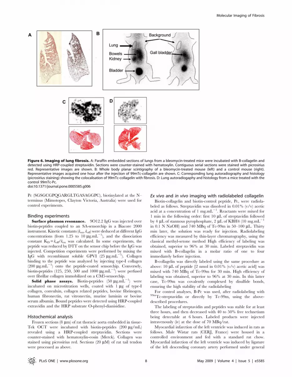
In order to confirm the specificity of imaging with collagelin, the
tracer was tested in a mouse model of lung fibrosis. First, an
histochemical analysis was performed on sections of healthy and
fibrotic lungs (fig 6A). Staining by collagelin coincided with the
collagen rich lesions. In vivo imaging was next realized 14 days after
instillation of bleomycin. One hour after injection of 99mTc-B-
collagelin, scintigraphic imaging showed the uptake of the tracer in
the pulmonary area of the mice that received bleomycin as
compared to control mice (fig 6B) with lung to muscle ratios of
3.4260.35 and 1.860;14 respectively (p,0.005, n = 5). Autora-
diographic studies confirmed higher 99mTc-B-collagelin uptake on
sections of lung of mice with pulmonary fibrosis as compared to
controls with heterogeneous distribution matched with that of
picrosirius staining (fig 6C).
Discussion
We have developed a specific probe for collagen which makes
possible to obtain in vivo, specific SPECT imaging of fibrosis, using
a new approach combining the advantages of the specificity of a
collagen receptor, and the diffusing properties of a peptide.
Combinatorial peptide libraries are usually screened for epitope
mapping such as, for example, the epitopes of antibodies directed
towards vWF and to its platelet receptor GPIb [17,18]. In the case
of the anti–GPVI 9O12.2 moAb, the use of a linear phage display
library [19], and of a constrained bacterial library (this study)
indicated that the epitope is conformational, but cannot be used to
identify a linear or discontinuous motif. Structural studies; such as
X-ray analysis and/or modeling, will help to determine the
epitope structure.
Another increasing application of peptide libraries is to identify
peptides with specific biological activities [20,21]. Peptides
mimicking proteins of interest obtained by screening libraries
versus blocking antibodies, designated mimotopes, are proposed as
potential targets, with either therapeutic or imaging applications.
The peptide we are describing here reproduces the specificity of
GPVI and thus. mimics, at least to some degree, the collagen-
binding site of GPVI. It binds in vitro to type-I collagen with high
affinity (1027 M) and also to type III collagen. Ex vivo collagen
staining on sections of rat tail tendon, which is composed of type I
collagen, and of the vascular wall containing high levels of types I
and III collagens, indicates that collagelin acts as a collagen probe.
A GPVI-based method of isotopic imaging has been proposed
[22]. I125/I123-labeled soluble recombinant GPVI-Fc was used for
non-invasive imaging of vulnerable atherosclerotic plaques in
ApoE2/2 mice in vivo by SPECT, and ex vivo by autoradiography.
However, slow blood clearance leading to high background
activity and low signal-to-noise ratio was observed. GPVI-Fc,
which consists of the extracellular part of GPVI fused to the IgG
Fc domain [12], has a high molecular mass that prevents it from
diffusing into interstitial tissues, and also has antithrombotic
properties [23]. In contrast, collagelin has the advantages of being
small, which facilitates its diffusion to its target, and of having no
effect on platelet function.
Peptides with collagen-binding properties have previously been
reported: cyclic peptides mimicking the collagen-binding site of
vWF bound to rat tail collagen, and inhibited vWF binding to
calfskin and human collagen, but only at high concentrations [24];
phage display also made it possible to identify the collagen-binding
protein rNecH1 from Necator americanus [25], but no further use of
these peptides or proteins has been reported. Recently, CNA35,
the collagen binding part of a bacterial protein, has been
successfully used as a fluorescent probe for collagen [26].
However, it cannot be used for in vivo imaging of organs since it
does not cross the endothelial barrier [27]. Cy5.5-RGD which
targets integrins on myofibroblasts and a sequence in procollagen
is an indirect probe for fibrinogenesis but it does not bind to
mature collagen type I or III fibers [8].
Since collagelin is a low molecular mass collagen-specific probe,
we postulated that it could be used as a collagen tracer in vivo. The
results reported here demonstrate that radiolabeled collagelin does
not accumulate to any significant degree in healthy tissues or in
blood, but that it clearly accumulates in post-infarct myocardial
scars. This experimental model yields a clearly identifiable, dense
but limited area of fibrosis, generally located at the apex of the
heart. Specific accumulation of radiolabeled collagelin provided
good contrast molecular imaging of the fibrotic scar, clearly
distinguishable from the healthy myocardium. Images obtained by
planar scintigraphy in this model, in which the scar is often thin
and small in volume, indicate that collagelin represents a powerful
probe for detecting fibrosis. This was confirmed by the specific
accumulation of the radiotracer in lung fibrotic lesions. In this
model, fibrosis that is looser than in the MI scar, was nevertheless
identified by the probe.
At the time of the observation, we did not notice any
accumulation of the tracer in the skin of the animals, that would
have interfered with the signal of interest. We however have
observed a low level of collagelin uptake in bone marrow. It thus
appears there is a preferential uptake of the tracer by the fibrous
collagen as compared to constitutive collagen in organs. Indeed,
GPVI does not bind to monomeric collagen but interacts with a
structural motif present on the cross-linked helicoidal structure of
collagen [10]. The lower uptake of collagelin by the organs
normally rich in collagen as compared to fibrosis, could be
Table 1. (A): Amino-acid alignment (Fasta format) of the 20clones sequenced after screening of the FliTrx randompeptide display library against immobilized 9O12 IgG.
clones 1–10 RFMHGLQLWADE
clone11 RVMHGLQLWADE
clones12–13 RVMHGLQLWADE
clone14 RVMHGLHLGDDE
clone15 RVMHGLHLWDDE
clone16 RVMHGLQLWDDE
clone17 RVMHGLHLWADE
clone18 FVMHGLHLGDDE
clone19 PVMHGLHLWDDE
clone20 RVMHGLLLGADE
The sequence of the peptide that has been selected for synthesis is underlined.doi:10.1371/journal.pone.0005585.t001
Molecular Imaging of Fibrosis
PLoS ONE | www.plosone.org 4 May 2009 | Volume 4 | Issue 5 | e5585
explained by a lower number of accessible GPVI binding sites due
to fiber structure and interaction with other proteins of the matrix
in this normal tissue.
Specific tracers for collagen represent a challenge and
considerable interest in various imaging methods, including
radionucleide imaging and MRI, is currently developing. A
gadolinium-based, collagen-targeting contrast agent, EP-3533,
has recently been reported to make it possible to carry out in
vivo molecular MR imaging of fibrosis in another model of scarred
infarction, in the mouse [28]. Collagelin appears to have greater
affinity for collagen than EP-3533 [9]. In addition, compared to
MRI, specific radionucleide imaging by SPECT or PET presents
the advantages of having much higher sensitivity, and no toxicity,
since a very low tracer dose (pico- or nanomolar) is sufficient to
obtain an accurate target-to-background ratio. In a recent study,
direct comparison of signals obtained with nanoparticles labeled
with both iron oxide and Cu-64 showed that the sensitivity of PET
is 50-fold higher than that of MRI [29]. This, explains why
SPECT and PET imaging are undergoing considerable develop-
ment worldwide, especially in malignant and cardiovascular
diseases. Similar developments are also expected in other medical
fields, in which the targeting of relevant processes is potentially
clinically useful.
This is the first time, to our knowledge, that a mimotope of
GPVI has been produced with proven in vitro and in vivo collagen-
specific targeting properties. Optimizing collagelin should now be
considered to improve the in vivo signal to noise ratio. Improved
derivatives of collagelin should be developed by further increasing
the affinity of the molecule for collagen, and improving the
labeling procedures. Indeed, the labeled streptavidin-B-collagelin
complex has the disadvantages of hepatic accumulation and a long
half life. However, these difficulties could be avoided by using
Figure 2. Interaction of collagelin with collagen. A: B-collagelin was immobilized on a streptavidin-coated sensorchip (,20 RU). Collagen(10 mg/ml) was injected over the sensorchip. A representative sensorgram (dark line) and interaction fit (gray line) are shown after subtracting thenon-specific background signal from a control flow cell coated with an irrelevant peptide. B: B-collagelin (250, 500 mg.mL21) was injected over acollagen-coated sensorchip. Sensorgrams (black) and interaction fits (gray) are shown. Representative sensorgrams are shown after subtracting thenon-specific response from the irrelevant peptide. C: B-collagelin or control peptide (50 mg. mL21) were incubated with immobilized, fibrillar, type-Icollagen in microtitration plates, and detected using HRP-coupled extravidin. In competition experiments, collagelin was mixed with GPVI-Fc(50 mg.mL21), 9012.2 IgGs (50 mg.mL21) or 3J24.2 IgGs (50 mg.mL21) before being added to collagen-coated wells. Means6SD (n = 3) are presented;*** p,0.01. D: B-collagelin (50 mg.mL21, black) or B-Pc (gray) were incubated with immobilized collagen I or III, CRP, fibrinogen, fibronectin,vitronectin and laminin in microtitration plates, and detected as above. Means6SD (n = 3) are shown.doi:10.1371/journal.pone.0005585.g002
Molecular Imaging of Fibrosis
PLoS ONE | www.plosone.org 5 May 2009 | Volume 4 | Issue 5 | e5585
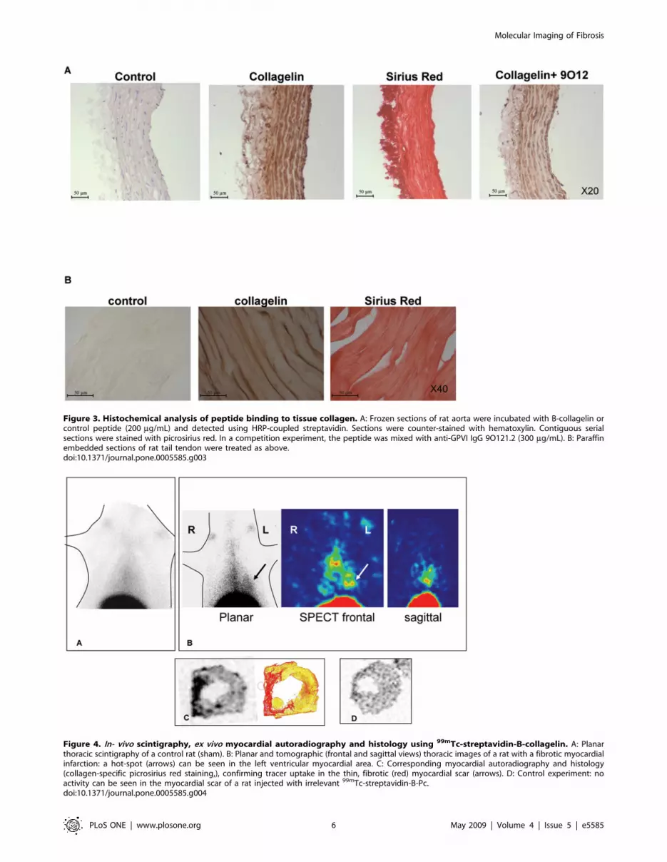
Figure 3. Histochemical analysis of peptide binding to tissue collagen. A: Frozen sections of rat aorta were incubated with B-collagelin orcontrol peptide (200 mg/mL) and detected using HRP-coupled streptavidin. Sections were counter-stained with hematoxylin. Contiguous serialsections were stained with picrosirius red. In a competition experiment, the peptide was mixed with anti-GPVI IgG 9O121.2 (300 mg/mL). B: Paraffinembedded sections of rat tail tendon were treated as above.doi:10.1371/journal.pone.0005585.g003
Figure 4. In- vivo scintigraphy, ex vivo myocardial autoradiography and histology using 99mTc-streptavidin-B-collagelin. A: Planarthoracic scintigraphy of a control rat (sham). B: Planar and tomographic (frontal and sagittal views) thoracic images of a rat with a fibrotic myocardialinfarction: a hot-spot (arrows) can be seen in the left ventricular myocardial area. C: Corresponding myocardial autoradiography and histology(collagen-specific picrosirius red staining,), confirming tracer uptake in the thin, fibrotic (red) myocardial scar (arrows). D: Control experiment: noactivity can be seen in the myocardial scar of a rat injected with irrelevant 99mTc-streptavidin-B-Pc.doi:10.1371/journal.pone.0005585.g004
Molecular Imaging of Fibrosis
PLoS ONE | www.plosone.org 6 May 2009 | Volume 4 | Issue 5 | e5585

directly labeled collagelin, which looks very promising on the basis
of our data. The circulation time of the peptide could be
prolonged by a moderate increase in its size, such as, for example,
by PEGylation or by constructing a dimeric peptide with increased
avidity for collagen. Radiolabeling with positron emitters could
also be considered for the absolute PET quantification of fibrosis in
vivo, which would allow the non invasive therapeutic follow-up of
fibrotic diseases.
Collagelin is thus a new and original collagen-specific probe
with a very wide field of possible applications as a tracer of fibrotic
tissues, in both non-vascular and vascular disorders.
Material and Methods
Screening the FliTrxTM peptide library against the anti-GPVI antibody
Anti-GPVI 9O12.2 IgGs and soluble recombinant GPVI-Fc
were obtained as previously described [13]. The cyclic dodecapep-
tide bacterial FliTrxTM Random Peptide Display Library was
from Invitrogen (San Diego, CA). Bacterial cultures and general
panning methods were conducted according to the manufacturer’s
protocol. Briefly, the pFliTrxTM vector with the PL promoter from
the bacteriophage that drives expression was propagated in E. coli
strain GI826. after induction of the expression of the thioredoxin–
flagellin fusion proteins containing the peptide inserts, bacterial
culture in NaCl (150 mM) containing a-methyl mannoside
(10 mg.mL21) and non-fat dry milk (10 mg.mL21) were incubated
in tissue culture plates coated with 9O12.2 IgG. After several
washing steps, bound bacteria were detached and amplified before
being subjected to a new round of panning. After five rounds,
bacterial colonies plated on solid medium were randomly taken,
amplified and induced for further identification.
Characterization of the clonesIdentification of positive clones was done by Western blotting
using 9O12 IgG. Briefly, proteins of selected bacterial clones were
separated by SDS-PAGE, transferred to nitrocellulose and
immunoblotted with the 9O12. IgG and a phosphatase alkaline-
coupled secondary antibody to mouse IgG [13]. Plasmid DNA was
isolated using standard protocols, and the nucleotide sequences
were determined using the FLITrxTM forward sequencing primer
(59-ATT CAC CTG ACT GAC GA-39). The peptide sequences
were deduced from DNA sequencing.
Peptide synthesisOne peptide base on the sequence of the selected clone
(underlined) [SGSGCPGRVMHGLHLGDDEGPC] was synthe-
sized. The peptide was or was not coupled to biotin at its N-
terminus via a short flexible spacer (SGSG), and made cyclic by
disulfide bridging of the flanking Cys. The carboxyl function at the
C-terminus was substituted by an amide. Peptides were prepared
by NeoMPS (Strasbourg, France), and their quality controlled by
HPLC and mass spectrometry.
The irrelevant, non-cyclic, biotinylated peptide P1
(SGSGVNVYAVTKENTIINPSENGD) and the cyclic peptide
Figure 5. In vivo scintigraphy, ex vivo myocardial autoradiography and histology using 99mTc-collagelin. A: Planar thoracic scintigraphyof a rat with fibrotic myocardial infarction: a clear hot-spot (arrows) xcan be seen in the left ventricular myocardial area. B: From left to right,corresponding myocardial histology (Masson’s trichrome, picrosirius red) and autoradiography, confirming tracer uptake in the thin, fibrotic (red)myocardial scar (arrow heads). C: Control experiment: very low activity is observed in the myocardial infarction in a rat injected with irrelevant 99mTc-Pc.doi:10.1371/journal.pone.0005585.g005
Molecular Imaging of Fibrosis
PLoS ONE | www.plosone.org 7 May 2009 | Volume 4 | Issue 5 | e5585
Pc (SGSGCGPQGARGLTGASAGGPC), biotinylated at the N–
terminus (Mimotopes, Clayton Victoria, Australia) were used for
control experiments.
Binding experimentsSurface plasmon resonance. 9O12.2 IgG was injected over
biotin-peptides coupled to an SA-sensorchip in a Biacore 2000
instrument. Kinetic constants (kon, koff) were deduced at different IgG
concentrations (from 1.25 to 10 mg.mL21), and the dissociation
constant KD = koff/kon was calculated. In some experiments, the
peptide was reduced by DTT on the sensor chip before the IgG was
injected. Competition experiments were performed by mixing the
IgG with recombinant soluble GPVI (25 mg.mL21). Collagen
binding to the peptide was analyzed by injecting type-I collagen
(200 mg.mL21) onto the peptide-coated sensorchip. Conversely,
biotin-peptides (125, 250, 500 and 1000 mg.mL21) were perfused
over fibrillar collagen immobilized on a CM5-sensorchip.
Solid phase assays. Biotin-peptides (50 mg.mL21) were
incubated on microtitration wells, coated with 1 mg of type-I
collagen, convulxin, collagen related peptides, bovine fibrinogen,
human fibronectin, rat vitronectin, murine laminin or bovine
serum albumin. Bound peptides were detected using HRP-coupled
extravidin and the HRP substrate O-phenyl-dianisidine.
Histochemical analysisFrozen sections (8 mm) of rat thoracic aorta embedded in tissue-
Tek OCT were incubated with biotin-peptides (200 mg/mL)
revealed using a HRP-coupled streptavidin. Sections were
counter-stained with hematoxylin-eosin (Merck). Collagen was
stained using picrosirius red. Sections (20 mM) of rat tail tendon
were processed as above.
Ex vivo and in vivo imaging with radiolabeled collagelinBiotin-collagelin and biotin-control peptide, Pc, were radiola-
beled as follows. Streptavidin was dissolved in 0.01% (v/v) acetic
acid at a concentration of 1 mg.mL21. Reactants were mixed for
1 min in the following order: first 10 mL of streptavidin followed
by 4 mL of stannous pyrophosphate, 2 mL of KBH4 (10 mg.mL21
in 0.1 N NaOH) and 740 MBq of Tc-99m in 50–100 mL. Thirty
min later, the solution was ready for injection. Radiolabeling
efficiency was measured by thin-layer chromatography, using the
classical methyl-setone method High efficiency of labeling was
obtained, superior to 96% at 30 min. Labeled streptavidin was
mixed with B-collagelin in a molar ratio of one to four
immediately before injection.
B-collagelin was directly labeled using the same procedure as
above: 10 mL of peptide [2 nmol in 0.01% (v/v) acetic acid] was
mixed with 740 MBq of Tc-99m for 30 min. High efficiency of
labeling was obtained, superior to 96% at 30 min. In this latter
case, Tc-99m was covalently complexed by disulfide bonds,
ensuring the high stability of the radiolabeling
For control analyses, B-Pc was used, after radiolabeling with99mTc-streptavidin or directly by Tc-99m, using the above-
described procedures.
The labeling of streptavidin and peptides was stable for at least
three hours, and then decreased with 40 to 50% free technetium
being detectable at 6 hours. Labeled products were injected
intravenously (iv) at the dose of 70 MBq/rat.
Myocardial infarction of the left ventricle was induced in rats as
follows. Male Wistar rats (CERJ, France) were housed in a
controlled environment and fed with a standard rat chow.
Myocardial infarction of the left ventricle was induced by ligature
of the left descending coronary artery performed under general
Figure 6. Imaging of lung fibrosis. A: Paraffin embedded sections of lungs from a bleomycin-treated mice were incubated with B-collagelin anddetected using HRP-coupled streptavidin. Sections were counter-stained with hematoxylin. Contiguous serial sections were stained with picrosiriusred. Representative images are shown. B: Whole body planar scintigraphy of a bleomycin-treated mouse (left) and a control mouse (right).Representative images acquired one hour after the injection of 99mTc-collagelin are shown. C: Corresponding lung autoradiography and histology(picrosirius staining) showing the colocalisation of 99mTc-collagelin with fibrosis. D: Lung autoradiogaphy and histology from a mice treated with thecontrol 99mTc-Pc.doi:10.1371/journal.pone.0005585.g006
Molecular Imaging of Fibrosis
PLoS ONE | www.plosone.org 8 May 2009 | Volume 4 | Issue 5 | e5585

anesthesia [1 mL.kg21 i.p. ketamine (Imalgene 500, Merial) and
0.5 mL.kg21 i.p. xylazine (Rompun, Bayer)] and positive pressure
ventilation, as described [30]. This protocol was performed under
a permit from the French Veterinary Services Directorate.
Two types of control animals were used: healthy rats, and
healthy rats which had undergone a simple throracotomy (sham-
operated). 99mTc-biotin-collagelin blood kinetics and biodistribu-
tion were investigated by counting blood samples (taken from a
jugular catheter) for 6 h after i.v. injection, and organs (extracted
after sacrifice) 2 h after injection in a gamma-counter, together
with aliquots of the injected preparation (CobraII, Packard,
Meriden, USA). Ex vivo myocardial labeling and isotopic imaging
were performed 4 weeks after the coronary ligature (or
throracotomy), when the lesions had had the time to heal.
Rat aorta and rat myocardial scar sections were incubated in
the presence of 99mTc-biotin-collagelin, +/29012 moAb, or99mTc-biotin-control peptide Pc (120 MBq/ml of RPMI) for 2 h.
The tissues were then rinsed 5 times for periods of 1 minute, and
exposed to the gamma-camera as well as to the instant imager for
quantitative autoradiography (Instant Imager, Packard) for 15 h.
For in vivo experiments, radiotracers (70 MBq) were adminis-
tered i.v. to anesthetized animals (pentobarbital i.p. 6 mg/100 g
BW, Ceva Sante Animale, France) within 2 h of radiolabeling.
Scintigraphic images were obtained 0–2 h, 4 h, 6 h, 10 h and
24 h after injecting 99mTc-streptavidin-biotin peptides and 0–3 h
after injecting 99mTc-biotinylated peptides. Planar and tomo-
graphic 1-h acquisitions were performed using a dedicated small
animal cIMAGER system (Biospace Lab, Paris, France) equipped
with 2 parallel low-energy high-resolution collimators (matrix
1286128, 15% energy window centered on 140 KeV). Tracer
uptake in the left cardiac area was assessed visually. Two regions of
interest were drawn on the scintigrams, over the heart and over
the right lung. The mean activity (cpm) per pixel was determined
in each region of interest. Then heart-to-lung activity ratios (HLR)
were calculated on both planar and transversal tomographic
images.
After sacrificing the animals, the heart was removed and frozen,
then 20-micrometer thick myocardial sections were cut perpen-
dicular to the short axis of the ventricles in a cryostat, and exposed
in a radioimager for 16 h. According to calibration studies
performed with activity standards of tissue-equivalent homoge-
nates, 50 counts/mm2 of 99m Tc-labeled tracers corresponded to
,210 kBq/mg in autoradiography [31]. The myocardial sections
used for autoradiography, and contiguous heart sections (5 mm)
fixed in acetone (220uC), were stained with hematoxylin-eosin,
Masson’s trichrome and picrosirius red to determine the location
and extent of the fibrotic myocardial scarring.
The lung fibrosis model was realized as already described [32].
Male C57BL/6J mice, aged 6–7 weeks were kept in accordance
with INSERM rules. On day 0, mice were administered 80 mg of
bleomycin hydrochloride (Bleomycine Bellon, Aventis, France)
intratracheally. Mortality was assessed daily over a 14 day period.
Naıve mice were used as controls.
At day 14 mice received one intravenous injection of 99mTc- B-
collagelin or of 99mTc- B-Pc (3 MBq). Then planar whole-body
scintigraphic imaging (60 min duration) was performed 1 hour
after tracer injection as above. At the end of the experiment,
animals were sacrificed and lung were dissected for gamma
counting, autoradiography, and histology.
Statistical analysisScintigraphic and autoradiographic quantitative parameters are
expressed as means6SEM. The unpaired t-test was used to
compare collagelin data in 2 groups of animals (rats with
myocardial scar versus sham-operated rats), or radiotracers in
the rats with a myocardial scar (collagelin versus control peptide
Pc). The level of significance was set at p,0.05.
Acknowledgments
We thank Dr. Jean-Michel Camadro (Jacques Monod Institute, University
Paris 6 and 7, Paris, France) for his help in carrying out and interpreting
the SPR analysis, Dr Mary Osborne-Pellegrin and Michele Coutard for
helpful discussion.
Author Contributions
Conceived and designed the experiments: LSM DLG PB MJP. Performed
the experiments: JM SL AM LL FH JMS JBM. Analyzed the data: JM
LSM PB MJP. Contributed reagents/materials/analysis tools: JM LSM SL
LL CBM JBM PB MJP. Wrote the paper: LSM PB MJP.
References
1. Murray CJ, Lopez AD (1997) Alternative projections of mortality and disability
by cause 1990–2020: Global Burden of Disease Study. Lancet 349: 1498–
1504.
2. de Ledinghen V, Le Bail B, Rebouissoux L, Fournier C, Foucher J, et al. (2007)
Liver stiffness measurement in children using FibroScan: feasibility study and
comparison with Fibrotest, aspartate transaminase to platelets ratio index, and
liver biopsy. J Pediatr Gastroenterol Nutr 45: 443–450.
3. Talwalkar JA, Kurtz DM, Schoenleber SJ, West CP, Montori VM (2007)
Ultrasound-based transient elastography for the detection of hepatic fibrosis:
systematic review and meta-analysis. Clin Gastroenterol Hepatol 5: 1214–1220.
4. Taouli B, Tolia AJ, Losada M, Babb JS, Chan ES, et al. (2007) Diffusion-
weighted MRI for quantification of liver fibrosis: preliminary experience. AJR
Am J Roentgenol 189: 799–806.
5. Dilsizian V, Eckelman WC, Loredo ML, Jagoda EM, Shirani J (2007) Evidence
for tissue angiotensin-converting enzyme in explanted hearts of ischemic
cardiomyopathy using targeted radiotracer technique. J Nucl Med 48: 182–187.
6. Verjans JW, Lovhaug D, Narula N, Petrov AD, Indrevoll B, et al. (2008)
Noninvasive imaging of angiotensin receptors after myocardial infarction. JACC
Cardiovasc Imaging 1: 354–362.
7. van den Borne SW, Isobe S, Zandbergen HR, Li P, Petrov A, et al. (2009)
Molecular imaging for efficacy of pharmacologic intervention in myocardial
remodeling. JACC Cardiovasc Imaging 2: 187–198.
8. van den Borne SW, Isobe S, Verjans JW, Petrov A, Lovhaug D, et al. (2008)
Molecular imaging of interstitial alterations in remodeling myocardium after
myocardial infarction. J Am Coll Cardiol 52: 2017–2028.
9. Caravan P, Das B, Dumas S, Epstein FH, Helm PA, et al. (2007) Collagen-
targeted MRI contrast agent for molecular imaging of fibrosis. Angew Chem Int
Ed Engl 46: 8171–8173.
10. Miura Y, Takahashi T, Jung SM, Moroi M (2002) Analysis of the interaction of
platelet collagen receptor glycoprotein VI (GPVI) with collagen. A dimeric form
of GPVI, but not the monomeric form, shows affinity to fibrous collagen. J Biol
Chem 277: 46197–46204.
11. Clemetson JM, Polgar J, Magnenat E, Wells TN, Clemetson KJ (1999) The
platelet collagen receptor glycoprotein VI is a member of the immunoglobulin
superfamily closely related to FcalphaR and the natural killer receptors. J Biol
Chem 274: 29019–29024.
12. Jandrot-Perrus M, Busfield S, Lagrue AH, Xiong X, Debili N, et al. (2000)
Cloning, characterization, and functional studies of human and mouse
glycoprotein VI: a platelet-specific collagen receptor from the immunoglobulin
superfamily. Blood 96: 1798–1807.
13. Lecut C, Feeney LA, Kingsbury G, Hopkins J, Lanza F, et al. (2003) Human
platelet glycoprotein VI function is antagonized by monoclonal antibody-derived
Fab fragments. J Thromb Haemost 1: 2653–2662.
14. Ohlmann P, Hechler B, Ravanat C, Loyau S, Herrenschmidt N, et al. (2008) Ex
vivo inhibition of thrombus formation by an anti-GPVI Fab fragment in non-
human primates without modification of GPVI expression. J Thromb Haemost.
15. Lagrue-Lak-Hal AH, Debili N, Kingbury G, Lecut C, Le Couedic JP, et al.
(2001) Expression and function of the collagen receptor GPVI during
megakaryocyte maturation. J Biol Chem 276: 15316–15325.
16. Inoue O, Suzuki-Inoue K, McCarty OJ, Moroi M, Ruggeri ZM, et al. (2006)
Laminin stimulates spreading of platelets through integrin alpha6beta1-
dependent activation of GPVI. Blood 107: 1405–1412.
17. Cauwenberghs N, Vanhoorelbeke K, Vauterin S, Westra DF, Romo G, et al.
(2001) Epitope mapping of inhibitory antibodies against platelet glycoprotein
Ibalpha reveals interaction between the leucine-rich repeat N-terminal and C-
terminal flanking domains of glycoprotein Ibalpha. Blood 98: 652–660.
Molecular Imaging of Fibrosis
PLoS ONE | www.plosone.org 9 May 2009 | Volume 4 | Issue 5 | e5585

18. Vanhoorelbeke K, Depraetere H, Romijn RA, Huizinga EG, De Maeyer M, et
al. (2003) A consensus tetrapeptide selected by phage display adopts theconformation of a dominant discontinuous epitope of a monoclonal anti-VWF
antibody that inhibits the von Willebrand factor-collagen interaction. J Biol
Chem 278: 37815–37821.19. Lecut C, Arocas V, Ulrichts H, Elbaz A, Villeval JL, et al. (2004) Identification
of residues within human glycoprotein VI involved in the binding to collagen:evidence for the existence of distinct binding sites. J Biol Chem 279:
52293–52299.
20. Nelson TJ, Alkon DL (2007) Protection against beta-amyloid-induced apoptosisby peptides interacting with beta-amyloid. J Biol Chem 282: 31238–31249.
21. Aina OH, Liu R, Sutcliffe JL, Marik J, Pan CX, et al. (2007) From combinatorialchemistry to cancer-targeting peptides. Mol Pharm 4: 631–651.
22. Gawaz M, Konrad I, Hauser AI, Sauer S, Li Z, et al. (2005) Non-invasiveimaging of glycoprotein VI binding to injured arterial lesions. Thromb Haemost
93: 910–913.
23. Massberg S, Konrad I, Bultmann A, Schulz C, Munch G, et al. (2004) Solubleglycoprotein VI dimer inhibits platelet adhesion and aggregation to the injured
vessel wall in vivo. Faseb J 18: 397–399.24. Depraetere H, Viaene A, Deroo S, Vauterin S, Deckmyn H (1998) Identification
of peptides, selected by phage display technology, that inhibit von Willebrand
factor binding to collagen. Blood 92: 4207–4211.25. Viaene A, Crab A, Meiring M, Pritchard D, Deckmyn H (2001) Identification of
a collagen-binding protein from Necator americanus by using a cDNA-expression phage display library. J Parasitol 87: 619–625.
26. Krahn KN, Bouten CV, van Tuijl S, van Zandvoort MA, Merkx M (2006)
Fluorescently labeled collagen binding proteins allow specific visualization of
collagen in tissues and live cell culture. Anal Biochem 350: 177–185.
27. Megens RT, Oude Egbrink MG, Cleutjens JP, Kuijpers MJ, Schiffers PH, et al.
(2007) Imaging collagen in intact viable healthy and atherosclerotic arteries
using fluorescently labeled CNA35 and two-photon laser scanning microscopy.
Mol Imaging 6: 247–260.
28. Helm PA, Caravan P, French BA, Jacques V, Shen L, et al. (2008) Postinfarction
myocardial scarring in mice: molecular MR imaging with use of a collagen-
targeting contrast agent. Radiology 247: 788–796.
29. Nahrendorf M, Zhang H, Hembrador S, Panizzi P, Sosnovik DE, et al. (2008)
Nanoparticle PET-CT imaging of macrophages in inflammatory atherosclerosis.
Circulation 117: 379–387.
30. Fishbein MC, Maclean D, Maroko PR (1978) Experimental myocardial
infarction in the rat: qualitative and quantitative changes during pathologic
evolution. Am J Pathol 90: 57–70.
31. Petegnief Y, Petiet A, Peker MC, Bonnin F, Meulemans A, et al. (1998)
Quantitative autoradiography using a radioimager based on a multiwire
proportional chamber. Phys Med Biol 43: 3629–3638.
32. Fabre A, Marchal-Somme J, Marchand-Adam S, Quesnel C, Borie R, et al.
(2008) Modulation of bleomycin-induced lung fibrosis by serotonin receptor
antagonists in mice. Eur Respir J 32: 426–436.
Molecular Imaging of Fibrosis
PLoS ONE | www.plosone.org 10 May 2009 | Volume 4 | Issue 5 | e5585