In vivo response to electrochemically aligned collagen bioscaffolds
9
In vivo response to electrochemically aligned collagen bioscaffolds Vipuil Kishore, 1 Jorge Alfredo Uquillas, 1 Alexandra Dubikovsky, 1 Musa A. Alshehabat, 2 Paul W. Snyder, 2 Gert J. Breur, 2 Ozan Akkus 1 1 Weldon School of Biomedical Engineering, Purdue University, West Lafayette, Indiana 47907-2032 2 School of Veterinary Medicine, Purdue University, West Lafayette, Indiana 47907-2032 Received 21 February 2011; revised 18 July 2011; accepted 5 September 2011 Published online 16 December 2011 in Wiley Online Library (wileyonlinelibrary.com). DOI: 10.1002/jbm.b.31962 Abstract: Collagen-based biomaterials are a viable option for tendon reconstruction and repair. However, the weak me- chanical strength of collagen constructs is a major limitation. We have previously reported a novel methodology to form highly oriented electrochemically aligned collagen (ELAC) threads with mechanical properties converging on those of the natural tendon. In this study, we assessed the in vivo response of rabbit patellar tendon (PT) to braided ELAC bio- scaffolds. Rabbit PTs were incised longitudinally and the ELAC bioscaffold was inlaid in one limb along the length of the tendon. The contralateral limb served as the sham-oper- ated control. Rabbits were euthanized at 4 or 8 months post- operatively. High-resolution radiographs revealed the absence of ectopic bone formation around the bioscaffolds. Four months post-implantation, the histological sections showed that the ELAC bioscaffold underwent limited degra- dation and was associated with a low-grade granulomatous inflammation. Additionally, quantitative histology revealed that the cross-sectional areas of PTs with the ELAC bioscaf- fold were 29% larger compared with the controls. Further- more, ELAC-treated PTs were significantly stiffer compared with the controls. The volume fraction of the tendon fascicle increased in the ELAC-treated PT compared with the controls. By 8 months, the ELAC bioscaffold was mostly absorbed and the enlargement in the area of tendons with implants sub- sided along with the resolution of the granulomatous inflam- mation. We conclude that ELAC is biocompatible and biodegradable and has the potential to be used as a biomate- rial for tendon tissue engineering applications. V C 2011 Wiley Periodicals, Inc. J Biomed Mater Res Part B: Appl Biomater 100B: 400–408, 2012. Key Words: tendon, tissue engineering, collagen, in vivo, granulomatous inflammation How to cite this article: Kishore V, Uquillas JA, Dubikovsky A, Alshehabat MA, Snyder PW, Breur GJ, Akkus O. 2012. In vivo response to electrochemically aligned collagen bioscaffolds. J Biomed Mater Res Part B 2012:100B:400–408. INTRODUCTION Tendon injuries pose a significant economical burden to the US each year. 1 Suturing provides satisfactory fixation of soft tissue in tendon and ligament reconstruction. However, these techniques are often associated with difficulties in placement, tissue reconstruction, cutting or pulling through tissue and pull out from bone. 2 Autografts and allografts provide potential alternative strategies, but each of them is associated with limitations such as donor site morbidity 3 and risk of disease transmission from the donor to the re- cipient, respectively. Therefore, Functional Tissue Engineer- ing strategies have gained considerable interest among researchers as a practical option to treat damaged tendons. 4 Tissue engineering utilizes a combination of scaffolds and cells to generate functional replacements for damaged tissue. Selection of the biomaterial to be used to fabricate scaffolds critically depends on the specific tissue of interest. Various biomaterials, both natural and synthetic, have been used to fabricate implantable bioscaffolds with the goal of repairing damaged tendons and ligaments. 5–13 Collagen type I, a major component of the tendon extracellular matrix (ECM), is the most widely studied biomaterial for tendon tissue engineering applications. 14–23 Collagen type I forms 70% of tendon’s dry weight. 24,25 Law et al. were one of the first groups to evaluate the in vivo response of a biomaterial made entirely with type I collagen fibers. 22 They crosslinked the collagen fibers either with glutaraldehyde or by dehy- dration followed by exposure to cyanamide and reported that the collagen implant showed excellent biocompatibility. Goldstein et al. demonstrated that collagenous tendon pros- thesis promoted rapid formation of neo-tendon. 18 Implanta- tion of collagen bioscaffolds seeded with mesenchymal stem cells (MSCs) into the rabbit patellar tendon (PT) has been shown to significantly improve the biomechanical properties of the repair tissue. 15 However, a major concern related to the application of MSCs to promote tendon healing is their Correspondence to: O. Akkus; e-mail: [email protected] Contract grant sponsor: Alfred Mann Institute, Purdue University Contract grant sponsor: National Institute of Health; contract grant number: NIH 1R21AR056060 Contract grant sponsor: National Science Foundation; contract grant number: CBET-0754442 400 V C 2011 WILEY PERIODICALS, INC.
Transcript of In vivo response to electrochemically aligned collagen bioscaffolds
In vivo response to electrochemically aligned collagen bioscaffolds
Vipuil Kishore,1 Jorge Alfredo Uquillas,1 Alexandra Dubikovsky,1 Musa A. Alshehabat,2
Paul W. Snyder,2 Gert J. Breur,2 Ozan Akkus1
1Weldon School of Biomedical Engineering, Purdue University, West Lafayette, Indiana 47907-20322School of Veterinary Medicine, Purdue University, West Lafayette, Indiana 47907-2032
Received 21 February 2011; revised 18 July 2011; accepted 5 September 2011
Published online 16 December 2011 in Wiley Online Library (wileyonlinelibrary.com). DOI: 10.1002/jbm.b.31962
Abstract: Collagen-based biomaterials are a viable option for
tendon reconstruction and repair. However, the weak me-
chanical strength of collagen constructs is a major limitation.
We have previously reported a novel methodology to form
highly oriented electrochemically aligned collagen (ELAC)
threads with mechanical properties converging on those of
the natural tendon. In this study, we assessed the in vivo
response of rabbit patellar tendon (PT) to braided ELAC bio-
scaffolds. Rabbit PTs were incised longitudinally and the
ELAC bioscaffold was inlaid in one limb along the length of
the tendon. The contralateral limb served as the sham-oper-
ated control. Rabbits were euthanized at 4 or 8 months post-
operatively. High-resolution radiographs revealed the
absence of ectopic bone formation around the bioscaffolds.
Four months post-implantation, the histological sections
showed that the ELAC bioscaffold underwent limited degra-
dation and was associated with a low-grade granulomatous
inflammation. Additionally, quantitative histology revealed
that the cross-sectional areas of PTs with the ELAC bioscaf-
fold were 29% larger compared with the controls. Further-
more, ELAC-treated PTs were significantly stiffer compared
with the controls. The volume fraction of the tendon fascicle
increased in the ELAC-treated PT compared with the controls.
By 8 months, the ELAC bioscaffold was mostly absorbed and
the enlargement in the area of tendons with implants sub-
sided along with the resolution of the granulomatous inflam-
mation. We conclude that ELAC is biocompatible and
biodegradable and has the potential to be used as a biomate-
rial for tendon tissue engineering applications. VC 2011 Wiley
Periodicals, Inc. J Biomed Mater Res Part B: Appl Biomater 100B:
400–408, 2012.
Key Words: tendon, tissue engineering, collagen, in vivo,
granulomatous inflammation
How to cite this article: Kishore V, Uquillas JA, Dubikovsky A, Alshehabat MA, Snyder PW, Breur GJ, Akkus O. 2012. In vivoresponse to electrochemically aligned collagen bioscaffolds. J Biomed Mater Res Part B 2012:100B:400–408.
INTRODUCTION
Tendon injuries pose a significant economical burden to theUS each year.1 Suturing provides satisfactory fixation of softtissue in tendon and ligament reconstruction. However,these techniques are often associated with difficulties inplacement, tissue reconstruction, cutting or pulling throughtissue and pull out from bone.2 Autografts and allograftsprovide potential alternative strategies, but each of them isassociated with limitations such as donor site morbidity3
and risk of disease transmission from the donor to the re-cipient, respectively. Therefore, Functional Tissue Engineer-ing strategies have gained considerable interest amongresearchers as a practical option to treat damaged tendons.4
Tissue engineering utilizes a combination of scaffoldsand cells to generate functional replacements for damagedtissue. Selection of the biomaterial to be used to fabricatescaffolds critically depends on the specific tissue of interest.Various biomaterials, both natural and synthetic, have been
used to fabricate implantable bioscaffolds with the goal ofrepairing damaged tendons and ligaments.5–13 Collagen typeI, a major component of the tendon extracellular matrix(ECM), is the most widely studied biomaterial for tendontissue engineering applications.14–23 Collagen type I forms70% of tendon’s dry weight.24,25 Law et al. were one of thefirst groups to evaluate the in vivo response of a biomaterialmade entirely with type I collagen fibers.22 They crosslinkedthe collagen fibers either with glutaraldehyde or by dehy-dration followed by exposure to cyanamide and reportedthat the collagen implant showed excellent biocompatibility.Goldstein et al. demonstrated that collagenous tendon pros-thesis promoted rapid formation of neo-tendon.18 Implanta-tion of collagen bioscaffolds seeded with mesenchymal stemcells (MSCs) into the rabbit patellar tendon (PT) has beenshown to significantly improve the biomechanical propertiesof the repair tissue.15 However, a major concern related tothe application of MSCs to promote tendon healing is their
Correspondence to: O. Akkus; e-mail: [email protected]
Contract grant sponsor: Alfred Mann Institute, Purdue University
Contract grant sponsor: National Institute of Health; contract grant number: NIH 1R21AR056060
Contract grant sponsor: National Science Foundation; contract grant number: CBET-0754442
400 VC 2011 WILEY PERIODICALS, INC.
tendency to induce ectopic bone formation14 possibly dueto high cell–cell contact and extensive contraction of the col-lagen gels.26 Careful selection of cell–collagen ratios havebeen recommended to prevent the formation of ectopicbone and accelerate tendon healing.27
A key limitation of the existing collagen constructs fortendon tissue engineering applications is their weak me-chanical strength. For a scaffold to be functional in vivo, itneeds to be mechanically strong with sufficient complianceto bear daily in vivo loads. We have previously developedand reported a novel methodology to align collagen mole-cules using the principles of isoelectric focusing and formhighly oriented and densely packed electrochemicallyaligned collagen (ELAC) threads.28 Crosslinking the ELACthread with genipin resulted in a mechanically strong bio-material with properties converging upon the native tendon.Genipin is a naturally occurring crosslinking agent(extracted from gardenia fruit), which has been reported tobe biocompatible and 10,000 times less cytotoxic comparedwith glutaraldehyde.29 Although the in vitro degradationand cellular response of ELAC have been investigated,30 thein vivo response of this novel material is unknown. Addi-tionally, little is known about the long-term in vivo responseto such densely packed collagen-based bioscaffolds. In thecurrent study, we hypothesize that the densely packed ELACbioscaffold is biocompatible and biodegradable. To test this
hypothesis, ELAC bioscaffolds were implanted into the PT ofrabbits and the long term in vivo response to the materialwas investigated. Cross-sectional area of tendon, degrada-tion of the scaffold, inflammatory response to the scaffold,and matrix organization of the harvested tissue were exam-ined by qualitative histology, quantitative histology, andimmunohistochemistry at 4 or 8 months post-implantation.
MATERIALS AND METHODS
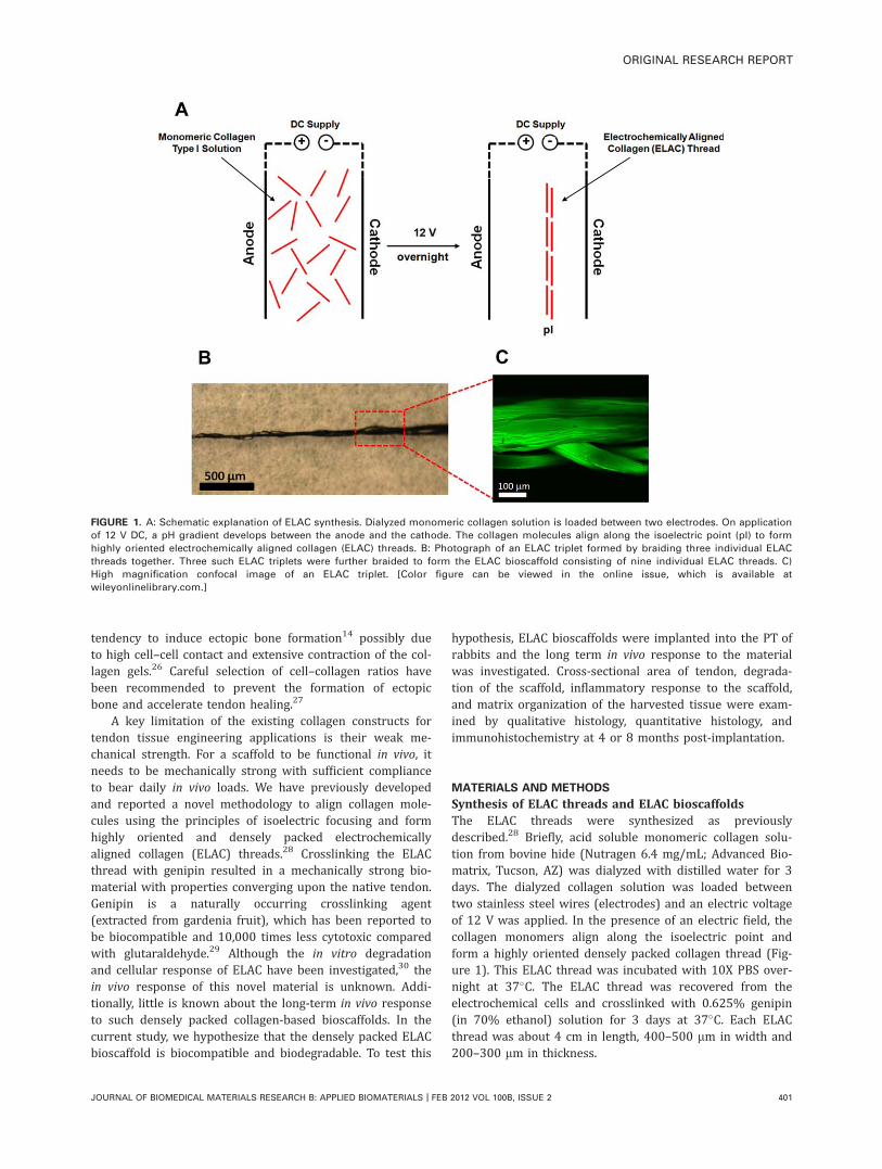
Synthesis of ELAC threads and ELAC bioscaffoldsThe ELAC threads were synthesized as previouslydescribed.28 Briefly, acid soluble monomeric collagen solu-tion from bovine hide (Nutragen 6.4 mg/mL; Advanced Bio-matrix, Tucson, AZ) was dialyzed with distilled water for 3days. The dialyzed collagen solution was loaded betweentwo stainless steel wires (electrodes) and an electric voltageof 12 V was applied. In the presence of an electric field, thecollagen monomers align along the isoelectric point andform a highly oriented densely packed collagen thread (Fig-ure 1). This ELAC thread was incubated with 10X PBS over-night at 37�C. The ELAC thread was recovered from theelectrochemical cells and crosslinked with 0.625% genipin(in 70% ethanol) solution for 3 days at 37�C. Each ELACthread was about 4 cm in length, 400–500 lm in width and200–300 lm in thickness.
FIGURE 1. A: Schematic explanation of ELAC synthesis. Dialyzed monomeric collagen solution is loaded between two electrodes. On application
of 12 V DC, a pH gradient develops between the anode and the cathode. The collagen molecules align along the isoelectric point (pI) to form
highly oriented electrochemically aligned collagen (ELAC) threads. B: Photograph of an ELAC triplet formed by braiding three individual ELAC
threads together. Three such ELAC triplets were further braided to form the ELAC bioscaffold consisting of nine individual ELAC threads. C)
High magnification confocal image of an ELAC triplet. [Color figure can be viewed in the online issue, which is available at
wileyonlinelibrary.com.]
ORIGINAL RESEARCH REPORT
JOURNAL OF BIOMEDICAL MATERIALS RESEARCH B: APPLIED BIOMATERIALS | FEB 2012 VOL 100B, ISSUE 2 401
Three individual genipin crosslinked ELAC threads weremanually braided to form an ELAC triplet. Both ends of theELAC triplet were glued using medical grade adhesive (Loc-tite 4851). Three individual ELAC triplets were braidedagain in a similar manner to form the ELAC bioscaffold con-sisting of nine ELAC threads. As the ELAC bioscaffold ismade out of pure collagen, standard medical device sterili-zation techniques that include physical treatments (UV,gamma radiation, etc.), chemical treatments (ethylene oxide,etc.) and heat treatment (autoclave) were not employed tocompletely ensure that the collagen structure is not cross-linked or denatured unduly. Given that the genipin cross-linking of the ELAC threads within the scaffold was carriedout by using 70% ethanol as the solvent, sterilization of thebioscaffold post-crosslinking was also conducted in thesame solvent. After sterilization, the ELAC bioscaffold waswashed extensively with DI water to completely removetraces of ethanol and incubated in culture medium (DMEMþ 1% penicillin/streptomycin) overnight before surgicallyimplanting it into the PT defects in a rabbit model. At leastin this model, the 70% ethanol treatment step for steriliza-tion was efficacious enough that we have not observedinfection in any of the rabbits included in the study.
Surgical implantation and PT collectionELAC bioscaffolds were surgically implanted into the PT offemale adult New Zealand white rabbits following anapproved protocol by Purdue Animal Care and Use Commit-tee (PACUC). Rabbits were in the range of 2–3 years of ageand weighed 4.5 6 1.0 kg. Rabbits were induced with keta-mine, xylazine, and butorphenol (35, 5, and 0.1 mg/kg IM,respectively), intubated, and maintained on a mixture of iso-flurane and oxygen. The PT was exposed and a longitudinalincision through the thickness of the tendon was made overthe central axis from the patellar to the tibial enthesis, with-out penetrating the parapatellar fat pad. A 1 mm wide bonetrough was burred on the patellar and tibial ends of the PTto facilitate anchoring of the ELAC bioscaffold. On one ran-domly assigned knee, the ELAC bioscaffold was sandwichedbetween the medial and lateral strut of the PT and anchoredin the troughs using an absorbable tissue adhesive (Tis-sueMed II, Veterinary Products Laboratory, Phoenix, AZ).The longitudinal incision was closed with three or foursuperficially placed simple interrupted sutures (4-0 PDS,Ethicon). The contralateral knee served as the sham-oper-ated control in which a 1 mm wide bone trough was burredand a longitudinal PT incision was made but the ELAC bio-scaffold was not implanted. The animals were euthanizedwith pentobarbital (1.5 mL IV, Beuthanasia, Schering-PloughAnimal Health Corporation) at 4 (n ¼ 3) or 8 (n ¼ 3)months. Following necropsy, the patella-PT-tibia units werecollected for analysis. Each collected unit was evaluated forectopic bone formation by radiography (Faxitron MX20, 35kV, 0.3 mA, 300 s). The PT complex was then transectedmidway between the patella and tibia; the proximal halfwas used for mechanical testing, and the distal half wasfixed in 10% neutral buffered formalin for histology andimmunohistochemistry.
Qualitative and quantitative histologyThe PT samples for histological evaluation were fixed in10% neutral buffered formalin. After fixation, the tendonwas dehydrated, embedded in paraffin, and cut at 5 lmthickness in transverse direction. Tissue sections were ana-lyzed by qualitative and quantitative histology by stainingwith hematoxylin and eosin or Masson’s trichrome. Thecross-sectional area of the entire tendon, the area of thegranulomatous inflammatory core (without the ELAC bio-scaffold), and the area of the ELAC bioscaffold itself in theexperimental knee was measured using perimeter tracingon the sections stained with Masson’s trichrome stain. TheVv(fascicle), the volume fraction of the tendon occupied bytendon fascicles, was determined using point counting tech-niques by overlaying a transparent sheet consisting of anarray of points over a Masson trichrome-stained section.The remaining fraction was considered interstitium. All ele-ments of the quantitative analysis were done using an Opti-phot-2 microscope (Nikon, Tokyo, Japan) fitted with a com-puter controlled stage and camera (Optronics, Goleta, CA).Measurements were made using semiautomated image anal-ysis software (Stereo Investigator, MBF Bioscience, Williston,VT).
ImmunohistochemistryImmunohistochemical staining was performed for Type Iand Type III collagen. The protocol followed was modifiedfrom previously published literature.31,32 Briefly, the histo-logical sections were deparaffinized with xylene, rehydratedwith a graded series of ethanol, and subjected to the follow-ing enzymatic treatment for epitope recovery. At first, thesections were incubated with 0.25% trypsin in tris-bufferedsaline (TBS) with pH ¼ 7.6 at 37�C for 30 min. The tissuesections were then washed with TBS and incubated with hy-aluronidase type I-S (1.45 IU/mL) and chondroitinase ABC(0.25 IU/mL) at 37�C for 1 h. After epitope recovery, thesections were washed with TBS and treated with 3% hydro-gen peroxide to block endogenous peroxidase activity. Fol-lowing this, the sections were washed with TBS and incu-bated with 5% goat serum (in TBS) for 30 min at roomtemperature to prevent nonspecific binding of the primaryantibodies. Rabbit specific primary antibodies for Type I (I-8H5, EMD Chemicals) and Type III (III-53, EMD Chemicals)collagen were diluted in an antibody diluent (Dako, Carpin-teria, CA) at a concentration of 10 lg/mL and 100 ng/mL,respectively. The goat serum was drained and the sectionswere incubated overnight with the primary antibody atroom temperature, washed with TBS, and then incubatedwith a biotinylated anti-mouse secondary antibody (JacksonImmunoResearch, West Grove, PA) at a dilution of 1:500 (inTBS) for 1 h. After washing with TBS, the sections wereincubated with streptavidin-HRP (Jackson ImmunoResearch,West Grove, PA) at a concentration of 2 lg/mL for 30 min.Finally, the sections were completely covered with 3,30-dia-minobenzidine chromogen solution (Dako, Carpinteria, CA)protected from light for 4 min. The sections were thenwashed with DI water, counterstained with hematoxylin,
402 KISHORE ET AL. IN VIVO RESPONSE TO ELAC

and coverslipped. Sections treated similarly but without theprimary antibody served as the negative control.
Biomechanical evaluation of matrix modulusBlocks of harvested tissue samples from the proximal halfof the PT were cut transversely to the longer axis of PT(hereafter called as the transverse plane) for biomechanicaltesting. Samples were wrapped in wet gauze, inserted into anylon bag, and stored at �20�C until analysis. Each trans-verse block was about 2-mm-thick. In order to standardizethe sample size between the ELAC treated and control PTs,each 2-mm-thick sample was sectioned in the transversedirection with a surgical blade to obtain 1-mm-thick slices.Disc samples were punched out from these slices using 3.5mm diameter biopsy punches (Miltex, York, PA) using amethodology adopted from Williams et al.33 In this fashion,two to three cylindrical discs were obtained per transverseslice, resulting in a sample size of N ¼ 10–12 in the ELAC-treated group, and N ¼ 7–8 in the control group. Immedi-ately after, digital images of the cylindrical discs wereobtained to calculate their cross-sectional areas. The cross-sectional area was obtained by processing of digital imagesof discs by ImageJ Software (Rasband, W.S., ImageJ, USNational Institutes of Health, Bethesda, MD). The compres-sion device was custom-built using three translation stagesassembled to form an XYZ positioner (CR4500 Series,Parker Automation, Rohnert Park, CA), and a miniature 5-lbload cell (Omega, Stamford, CN). A loading head wasmounted to the translation stage. The portion of the loadinghead contacting the disc sample was a 6 mm hemisphericalglass which could freely rotate in 3D (Edmund Optics, Bar-rington, NJ). The tendon discs were placed in a polycarbon-ate reservoir containing 1x PBS and the reservoir wasmounted on the load cell. Load readings over time wereobtained using Lab VIEW (National Instruments, Austin,TX). Each sample was compressed to a displacement corre-sponding to 50% of its thickness. The thickness was meas-ured by the difference of the displacement at which theloading head contacted the reservoir surface and the dis-placement at which the loading head engaged the sample(as determined by a reading of 0.1N). All samples werecompressed at a strain rate of 30% per second. Data wereprocessed in MATLAB (MathWorks, Natick, MA) to calculatethe modulus as the slope of the steepest increase of loadduring initial compression.
Statistical analysisThe cross-sectional areas and the Vv fascicle data of theELAC treated and the control PTs (n ¼ 3/group) at 4 and 8months were analyzed statistically by using a paired t-test.The change in the area of the ELAC bioscaffold and the areaof the granulomatous inflammatory core between 4 and 8months were assessed using a one-tailed two-sample t-test.A sample size of n ¼ 3 at the observed difference in meanof 0.13 and standard deviation of 0.03 provided a power of99% for the change in area of the ELAC bioscaffold between4 and 8 months. Similarly, a sample size of n ¼ 3 at theobserved difference in mean of 0.40 and standard deviation
of 0.18 provided a power of 72% for the change in the areaof the granulomatous inflammatory core between 4 and 8months. The compressive Young’s modulus data was ana-lyzed between different treatments (ELAC and Control) andtime points (4 and 8 months) using the General LinearModel. If statistical differences were established, a nonpara-metric Mann-Whitney U-test was performed between groupsthat showed significant differences. Statistical significancethreshold was set at p � 0.05.
RESULTS
No ectopic bone formation was observed in the tendonproper in both the ELAC treated PT and the sham-operatedcontrol (Figure 2).
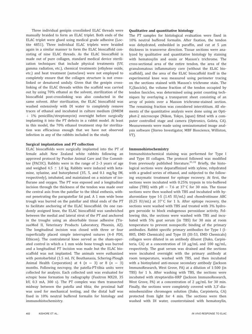
Quantitative measurements performed on the Massontrichrome stained sections showed that the cross-sectionalareas of the PTs treated with ELAC were about 29% larger(p ¼ 0.05) compared to the controls 4 months post-implan-tation [Figure 3(A)]. The inflammatory core and the implanttogether accounted for less than 5% of the total tendonarea [Figure 3(B)]. By 8 months, the cross-sectional areas ofthe PTs between the ELAC treated and the control groupswere comparable [Figure 3(A)].
At 4 months postoperatively, Vv(fascicle), the percentageof the tendon cross section occupied by tendon fascicles,was higher (p ¼ 0.10) in the ELAC group compared the con-trol group (Figure 4).
The ELAC bioscaffold underwent limited degradationbetween zero and 4 months [Figure 5(A)]. However,between 4 and 8 months, the area of the ELAC bioscaffolddecreased significantly (p ¼ 0.016) from 0.2 mm2 at 4months to less than 0.1 mm2 by 8 months [Figure 5(A,C,D)].The granulomatous inflammation area was also observed to
FIGURE 2. Typical radiographic images of ELAC treated and sham-
operated control PTs. Ectopic bone calcification was not evident in
the tendon proper in both groups at 4 or 8 months post-implantation
(P – Patella, T – Tibia, PT – Patellar Tendon).
ORIGINAL RESEARCH REPORT
JOURNAL OF BIOMEDICAL MATERIALS RESEARCH B: APPLIED BIOMATERIALS | FEB 2012 VOL 100B, ISSUE 2 403
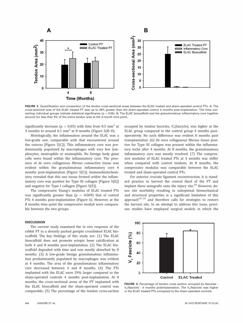
significantly decrease (p ¼ 0.05) with time from 0.5 mm2 at4 months to around 0.1 mm2 at 8 months (Figure 5(B–D).
Histologically, the inflammation around the ELAC was alow-grade one, comparable with that encountered aroundthe sutures [Figure 5(C)]. This inflammatory core was pre-dominantly populated by macrophages with very few lym-phocytes, neutrophils or eosinophils. No foreign body giantcells were found within the inflammatory core. The pres-ence of de novo collagenous fibrous connective tissue wasevident within the granulomatous inflammatory core 4months post-implantation [Figure 5(C)]. Immunohistochem-istry revealed that this neo tissue formed within the inflam-matory core was positive for Type III collagen [Figure 5(E)]and negative for Type I collagen [Figure 5(F)].
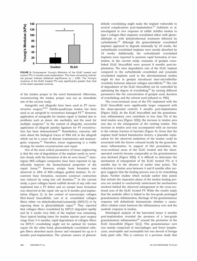
The compressive Young’s modulus of ELAC treated PTswas significantly greater than (p ¼ 0.009) that of controlPTs 4 months post-implantation (Figure 6). However, at the8 months time point the compressive moduli were compara-ble between the two groups.
DISCUSSION
The current study examined the in vivo response of therabbit PT to a densely packed genipin crosslinked ELAC bio-scaffold. The key findings of this study are: (1) The ELACbioscaffold does not promote ectopic bone calcification atboth 4 and 8 months post-implantation. (2) The ELAC bio-scaffold degraded with time and was mostly absorbed by 8months. (3) A low-grade benign granulomatous inflamma-tion predominantly populated by macrophages was evidentat 4 months. The area of the granulomatous inflammatorycore decreased between 4 and 8 months. (4) The PTsimplanted with the ELAC were 29% larger compared to thesham-operated controls 4 months post-implantation. At 8months, the cross-sectional areas of the PT implanted withthe ELAC bioscaffold and the sham-operated control wascomparable. (5) The percentage of the tendon cross-section
occupied by tendon fascicles, Vv(fascicle), was higher in theELAC group compared to the control group 4 months post-operatively. No such difference was evident 8 months posttransplantation. (6) De novo collagenous fibrous tissue posi-tive for Type III collagen was present within the inflamma-tory niche after 4 months. At 8 months, the granulomatousinflammatory core was mostly resolved. (7) The compres-sive modulus of ELAC treated PTs at 4 months was stifferwhen compared with control tendons. At 8 months, thecompressive modulus was comparable between the ELACtreated and sham-operated control PTs.
For anterior cruciate ligament reconstruction, it is stand-ard practice to harvest the central third of the PT andimplant these autografts onto the injury site.34 However, do-nor site morbidity resulting in suboptimal biomechanicaland structural properties is a significant limitation of thisapproach35–39 and therefore calls for strategies to restorethe harvest site. In an attempt to address this issue, previ-ous studies have employed surgical models in which the
FIGURE 3. Quantification and comparison of the tendon cross-sectional areas between the ELAC treated and sham-operated control PTs. A: The
cross-sectional area of the ELAC treated PT was up to 29% greater than the sham-operated control 4 months post-implantation. The lines con-
necting individual groups indicate statistical significance (p � 0.05). B: The ELAC bioscaffold and the granulomatous inflammatory core together
account for less than 5% of the entire tendon area at the 4-month time point.
FIGURE 4. Percentage of tendon cross section occupied by fascicles -
Vv(fascicle) - 4 months postimplantation. The Vv(fascicle) was higher
in the ELAC treated PTs compared to the sham-operated controls.
404 KISHORE ET AL. IN VIVO RESPONSE TO ELAC
central third of the PT is removed and efforts are made torestore the injury site using natural9 or synthetic materi-als14,19 with or without cells. Healing in such surgical mod-els require significant regeneration of the lost tissue toregain full functionality. In contrast, the surgical modelemployed in the current study is less invasive. Although a
longitudinal incision was made along the length of the PT,no part of the tissue was harvested during surgery. Healinginvolved closure of the wound, body’s response to the for-eign material and the integration/degradation of the scaffoldwithin the tendon proper. This surgical model was chosenfor the current study to solely evaluate the in vivo response
FIGURE 5. Histological and immunohistochemical characterization of the ELAC treated PTs. Quantitative histology indicated that (A) the area of
the ELAC bioscaffold decreases significantly after 4 months and is mostly absorbed by the end of 8 months and (B) the area of the granuloma-
tous inflammatory core also reduces significantly with time and is largely resolved by 8 months. The lines connecting individual groups indicate
statistical significance (p � 0.05). C, D: High magnification images of the granulomatous inflammatory core (C) at 4 months and (D) at 8 months
post-implantation. The ‘arrow heads’ point to the ELAC bioscaffold and the dotted ellipse surrounds the sutures. Scale bar: 0.5 mm. E, F: Immu-
nohistochemical images of the ELAC treated PTs 4 months post-implantation stained with (E) Type III collagen and (F) Type I collagen. The de
novo collagenous fibrous tissue formed within the granulomatous inflammatory niche was positive for Type III collagen and negative for Type I
collagen. The ‘arrow heads’ point to the ELAC bioscaffold and ‘*’ indicates the de novo collagenous fibrous tissue formed within the granuloma-
tous inflammatory niche. Scale bar: 0.5 mm. [Color figure can be viewed in the online issue, which is available at wileyonlinelibrary.com.]
ORIGINAL RESEARCH REPORT
JOURNAL OF BIOMEDICAL MATERIALS RESEARCH B: APPLIED BIOMATERIALS | FEB 2012 VOL 100B, ISSUE 2 405
of the tendon proper to the novel biomaterial. Otherwise,reconstructing the tendon proper was not an immediateaim of the current study.
Autografts and allografts have been used in PT recon-structive surgery.40,41 Patella-quadriceps tendon has beenused as an autograft to reconstruct damaged PT.41 However,application of autografts for tendon repair is limited due toproblems such as donor site morbidity and the need formultiple surgeries.3 In the context of allografts, successfulapplication of allograft patellar ligament for PT reconstruc-tion has been demonstrated.40 Nonetheless, concerns stillexist about the biological traces of DNA left in the allograftwhich can be a cause of disease transmission and immuno-genic response.42 Therefore, tissue engineering is a viablestrategy for tendon reconstruction and repair.
One of the most critical parameters of tissue engineeringis that the rate of degradation of the implant needs to corre-late closely with the formation of the de novo tissue.43 Auto-logous MSC-collagen composites have been reported to sig-nificantly improve the biomechanical properties of therepair tissue.14 However, ectopic bone formation wasobserved in 28% of MSC-collagen grafted tendons. To cir-cumvent bone formation, excessive construct contractionwas reduced by using low cell densities.19 In the currentstudy, a pure collagen based scaffold devoid of any cells wasimplanted into a PT defect and no ectopic bone formationwas observed at the repair site up to 8 months post-implan-tation (Figure 2). In the context of macroscopic collagenfiber-based bioscaffolds, Law et al. crosslinked collagenfibers either via dehydrothermal/cyanamide (DHT/C) or byexposing them to glutaraldehyde vapor.22 They reportedthat collagen fibers crosslinked by DHT/C degraded rapidlyand by 6 weeks very little of the implant was remaining.Since typical healing times for tendon injuries post surgeryrange from 3–6 months, rapid degradation of collagen fibersvia DHT/C crosslinking might not be optimal for tendonrepair. On the other hand, glutaraldehyde crosslinked colla-gen fibers absorbed much slower and remained for up to 6months post-implantation. The cytotoxic nature of glutaral-
dehyde crosslinking might make the implant vulnerable toseveral complications post-implantation.29 Goldstein et al.investigated in vivo response of rabbit Achilles tendon totype I collagen fiber implants crosslinked either with glutar-aldehyde or with dehydrothermal treatment followed bycarbodiimide.18 Although the glutaraldehyde crosslinkedimplants appeared to degrade minimally by 20 weeks, thecarbodiimide crosslinked implants were mostly absorbed by10 weeks. Additionally, the carbodiimide crosslinkedimplants were reported to promote rapid formation of neo-tendon. In the current study, remnants of genipin cross-linked ELAC bioscaffold were present 8 months post-im-plantation. The slow degradation rate of the ELAC implantcompared to the carbodiimide crosslinked and cynamidecrosslinked implants used in the aforementioned studiesmight be due to genipin introduced inter-microfibrillarcrosslinks between adjacent collagen microfibrils.44 The rateof degradation of the ELAC bioscaffold can be controlled byoptimizing the degree of crosslinking45 by varying differentparameters like the concentration of genipin used, durationof crosslinking, and the solvent used (ethanol vs. PBS).
The cross-sectional areas of the PTs implanted with theELAC bioscaffold were significantly larger compared withthe sham-operated controls 4 months post-implantation[Figure 3(A)]. As the ELAC bioscaffold and the granuloma-tous inflammatory core contribute to less than 5% of thetotal tendon area [Figure 3(B)], the increase in tendon areawas due to the enlargement of the existing tendon. Thisincrease in tendon area was accompanied with an increasein the volume fraction of fascicles (Figure 4). Given that theimplant itself lacked bioinductive factors, a plausible expla-nation for the observed anabolism of the tendon could beassociated with the factors released locally by the granulom-atous inflammation. In support of this postulation, thecross-sectional areas of the ELAC treated and the sham-operated controls became comparable as the granulomatousarea declined [Figure 3(B)]. It is difficult to determine themechanism of enlargement of the ELAC treated PTs at 4months due to the absence of earlier time points. Thereduction is tendon area between 4 and 8 months after sur-gery suggests that the healing process was in its remodelingphase. Further studies which include earlier time pointsthat include the reparative phase of the tendon healing pro-cess are needed to conclusively understand the mechanismsinvolved behind the observed enlargement in the cross-sec-tional area of the ELAC treated PT. While the results implythat the anabolic effect is linked to the low-grade prolongedgranulomatous inflammation, blockage of the granulomatousresponse will definitively demonstrate whether a cause–effect relation exists between the inflammatory core and theanabolic response in tendon.
Histological analysis of the harvested tissue 4 monthspost-implantation revealed the presence of a low-gradegranulomatous inflammation46 around the perimeter of theELAC bioscaffold [Figure 5(C)]. This granulomatous corewas mainly comprised of macrophages and fewer lympho-cytes, neutrophils and eosinophils but was devoid of foreignbody giant cells. This is contrary to a previous study that
FIGURE 6. Compressive Young’s Modulus of the ELAC treated and
control PTs 4 months post-implantation. The lines connecting individ-
ual groups indicate statistical significance (p � 0.05). The Young’s
modulus of the ELAC treated PTs was significantly greater than that
of the sham-operated controls.
406 KISHORE ET AL. IN VIVO RESPONSE TO ELAC
reported the presence of foreign body giant cells activelyinvolved in the phagocytosis of the collagen implant from asearly as 10 days up to 6 months post-implantation.22 Thismight be due to the differences in the size of the implantand the crosslinking agents used between studies. The gran-ulomatous inflammation observed in the present study wassimilar to that reported for injectable collagen implantsused as facial fillers to correct for scarring and wrinkles.47–50
The scarcity of neutrophils and eosinophils within theinflammatory core suggests that the sterilization procedureadopted prior to the surgical implantation of the ELAC bio-scaffold was adequate and that degradation byproducts werenot eliciting acute inflammation. The granulomatous inflam-matory core was negative for collagen type I and rich in col-lagen type III at 4 months indicating the replacement ofinflammation with scar tissue [Figure 5(E,F)]. This is inagreement with previous studies using MSC-collagen compo-sites that report strong staining of collagen types III and Vtwelve weeks post-implantation.19 The decrease in the areaof the granulomatous inflammatory core by 8 months wascoupled with a reduction in tendon area indicating anongoing remodeling of the scar tissue.
There are two limitations to the current study. First, asthe aim of the current study was to solely evaluate the invivo response to a novel collagen-based bioscaffold withinthe tendon proper, the changes in the construct attachmentsite were not investigated. Although the implant was anch-ored into the bone at both the patella and tibial ends usinga tissue adhesive, it was merely done to hold the implantwithin the tendon proper. Future studies will focus on thedevelopment of the ELAC bioscaffold with a goal to effi-ciently anchor the construct into the bone insertion sites.The efficacy of the construct attachment and the changes inthe attachment site will be investigated via biomechanicaltesting and histological analysis.
The second limitation of the current study is that biome-chanical evaluation of the whole PTs with the construct wasnot performed due to limited sample size. However, com-pression biomechanical testing on disc shaped slices of ten-don was performed to evaluate the effects of the ELAC bio-scaffold on the neighboring tendon stiffness (Figure 6).Therefore, albeit not physiologically relevant, the methodused here allowed assessing the effects of the implant.Others have also utilized compression to observe compres-sive stiffness of the bursal and articular sides of the supra-spinatus tendon.51 Williams et al. studied the anisotropiccompressive mechanical properties of the rabbit PT in thetransverse and longitudinal direction of tendon fibers.33
They found the compressive modulus at strain rates of 10%per second in the longitudinal direction as 3.26 (63.49)kPa. This value is smaller than the longitudinal compressivemodulus of our control group at 4 months: 20.46 (614.67)kPa. However, it is known that in viscoelastic solids such astendons the stress increases as the applied strain rateincreases. The strain rate used in the current study is aboutthree times higher than that used by Williams et al. whichmight explain the difference in modulus. Additionally, Wil-liams et al. computed compressive moduli in small strain
regions of the monotonic loading curves where as in thecurrent study the compressive moduli of the time-depend-ent stress curves were calculated in the region of the steep-est slope. The increased moduli in the tendon from PTsimplanted with ELAC are likely due to changes in the matrixcomposition as seen in the increased fascicle volume frac-tion in ELAC-treated PTs at 4 months (Figure 4). Secretionof collagen and various proteoglycans from the resident ten-don cells may have resulted in a stiffer matrix whichremains to be confirmed by analyzing the expression ofECM molecules by real-time PCR as well as biochemicalanalysis of the ECM.
In conclusion, this study demonstrates that the ELACbioscaffold when implanted in a rabbit PT model: (a)degrades over a time scale of months, (b) does not promoteectopic bone formation, (c) does not induce severe inflam-matory reaction, and (d) increases the volume fraction ofthe fascicles within the tendon proper. Therefore, ELAC hasthe potential to be translated to the clinical environment forthe regeneration of atrophied musculoskeletal tissues.
ACKNOWLEDGMENTS
The authors thank Carol Bain at Purdue University Histopa-thology service laboratory for her help with the histologicalwork.
REFERENCES1. Praemer A, Furner S, Rice D. Musculoskeletal condition in the
United States. Am Acad Orthop Surg 1992.
2. Krackow KA, Thomas SC, Jones LC. Ligament-tendon fixation:
Analysis of a new stitch and comparison with standard techni-
ques. Orthopedics 1988;11:909–917.
3. Kartus J, Movin T, Karlsson J. Donor-site morbidity and anterior
knee problems after anterior cruciate ligament reconstruction
using autografts. Arthroscopy 2001;17:971–980.
4. Butler DL, Juncosa-Melvin N, Boivin GP, Galloway MT, Shearn
JT, Gooch C, Awad H. Functional tissue engineering for tendon
repair: A multidisciplinary strategy using mesenchymal stem
cells, bioscaffolds, and mechanical stimulation. J Orthop Res
2008;26:1–9.
5. Cooper JA, Lu HH, Ko FK, Freeman JW, Laurencin CT. Fiber-based
tissue-engineered scaffold for ligament replacement: Design con-
siderations and in vitro evaluation. Biomaterials 2005;26:
1523–1532.
6. Fan H, Liu H, Toh SL, Goh JC. Anterior cruciate ligament regener-
ation using mesenchymal stem cells and silk scaffold in large ani-
mal model. Biomaterials 2009;30:4967–4977.
7. Fan H, Liu H, Wong EJ, Toh SL, Goh JC. In vivo study of anterior
cruciate ligament regeneration using mesenchymal stem cells
and silk scaffold. Biomaterials 2008;29:3324–3337.
8. Funakoshi T, Majima T, Iwasaki N, Suenaga N, Sawaguchi N, Shi-
mode K, Minami A, Harada K, Nishimura S. Application of tissue
engineering techniques for rotator cuff regeneration using a chi-
tosan-based hyaluronan hybrid fiber scaffold. Am J Sports Med
2005;33:1193–1201.
9. Karaoglu S, M BF, Woo SL, Fu YC, Liang R, Abramowitch SD. Use
of a bioscaffold to improve healing of a patellar tendon defect af-
ter graft harvest for ACL reconstruction: A study in rabbits.
J Orthop Res 2008;26:255–263.
10. Lu HH, Cooper JA, Jr, Manuel S, Freeman JW, Attawia MA, Ko
FK, Laurencin CT. Anterior cruciate ligament regeneration using
braided biodegradable scaffolds: In vitro optimization studies.
Biomaterials 2005;26:4805–4816.
11. Moffat KL, Kwei AS, Spalazzi JP, Doty SB, Levine WN, Lu HH.
Novel nanofiber-based scaffold for rotator cuff repair and aug-
mentation. Tissue Eng Part A 2009;15:115–126.
ORIGINAL RESEARCH REPORT
JOURNAL OF BIOMEDICAL MATERIALS RESEARCH B: APPLIED BIOMATERIALS | FEB 2012 VOL 100B, ISSUE 2 407
12. Ouyang HW, Goh JC, Thambyah A, Teoh SH, Lee EH. Knitted
poly-lactide-co-glycolide scaffold loaded with bone marrow stro-
mal cells in repair and regeneration of rabbit Achilles tendon. Tis-
sue Eng 2003;9:431–439.
13. Yokoya S, Mochizuki Y, Nagata Y, Deie M, Ochi M. Tendon-bone
insertion repair and regeneration using polyglycolic acid sheet in the
rabbit rotator cuff injury model. Am J Sports Med 2008;36:1298–1309.
14. Awad HA, Boivin GP, Dressler MR, Smith FN, Young RG, Butler
DL. Repair of patellar tendon injuries using a cell-collagen com-
posite. J Orthop Res 2003;21:420–431.
15. Awad HA, Butler DL, Boivin GP, Smith FN, Malaviya P, Huibregtse
B, Caplan AI. Autologous mesenchymal stem cell-mediated repair
of tendon. Tissue Eng 1999;5:267–277.
16. Chvapil M, Speer DP, Holubec H, Chvapil TA, King DH. Collagen
fibers as a temporary scaffold for replacement of ACL in goats. J
Biomed Mater Res 1993;27:313–325.
17. Fini M, Torricelli P, Giavaresi G, Rotini R, Castagna A, Giardino R.
In vitro study comparing two collageneous membranes in view of
their clinical application for rotator cuff tendon regeneration. J
Orthop Res 2007;25:98–107.
18. Goldstein JD, Tria AJ, Zawadsky JP, Kato YP, Christiansen D, Sil-
ver FH. Development of a reconstituted collagen tendon prosthe-
sis. A preliminary implantation study. J Bone Joint Surg Am
1989;71:1183–1191.
19. Juncosa-Melvin N, Boivin GP, Gooch C, Galloway MT, West JR,
Dunn MG, Butler DL. The effect of autologous mesenchymal stem
cells on the biomechanics and histology of gel-collagen sponge
constructs used for rabbit patellar tendon repair. Tissue Eng 2006;
12:369–379.
20. Koob TJ, Willis TA, Hernandez DJ. Biocompatibility of NDGA-
polymerized collagen fibers. I. Evaluation of cytotoxicity with ten-
don fibroblasts in vitro. J Biomed Mater Res 2001;56:31–39.
21. Koob TJ, Willis TA, Qiu YS, Hernandez DJ. Biocompatibility of
NDGA-polymerized collagen fibers. II. Attachment, proliferation,
and migration of tendon fibroblasts in vitro. J Biomed Mater Res
2001;56:40–48.
22. Law JK, Parsons JR, Silver FH, Weiss AB. An evaluation of puri-
fied reconstituted type 1 collagen fibers. J Biomed Mater Res
1989;23:961–977.
23. Young RG, Butler DL, Weber W, Caplan AI, Gordon SL, Fink DJ.
Use of mesenchymal stem cells in a collagen matrix for Achilles
tendon repair. J Orthop Res 1998;16(4):406–13.
24. Amiel D, Frank C, Harwood F, Fronek J, Akeson W. Tendons and
ligaments: a morphological and biochemical comparison. J
Orthop Res 1984;1(3):257–65.
25. Fujii K, Yamagishi T, Nagafuchi T, Tsuji M, Kuboki Y. Biochemical
properties of collagen from ligaments and periarticular tendons
of the human knee. Knee Surg Sports Traumatol Arthrosc 1994;2:
229–233.
26. Harris MT, Butler DL, Boivin GP, Florer JB, Schantz EJ, Wenstrup
RJ. Mesenchymal stem cells used for rabbit tendon repair can
form ectopic bone and express alkaline phosphatase activity in
constructs. J Orthop Res 2004;22:998–1003.
27. Juncosa-Melvin N, Boivin GP, Galloway MT, Gooch C, West JR,
Sklenka AM, Butler DL. Effects of cell-to-collagen ratio in mesen-
chymal stem cell-seeded implants on tendon repair biomechanics
and histology. Tissue Eng 2005;11(3–4):448–457.
28. Cheng X, Gurkan UA, Dehen CJ, Tate MP, Hillhouse HW, Simpson
GJ, Akkus O. An electrochemical fabrication process for the as-
sembly of anisotropically oriented collagen bundles. Biomaterials
2008;29:3278–3288.
29. Sung HW, Huang RN, Huang LL, Tsai CC. In vitro evaluation of cy-
totoxicity of a naturally occurring cross-linking reagent for biolog-
ical tissue fixation. J Biomater Sci Polym Ed 1999;10:63–78.
30. Gurkan UA, Cheng X, Kishore V, Uquillas JA, Akkus O. Compari-
son of morphology, orientation, and migration of tendon derived
fibroblasts and bone marrow stromal cells on electrochemically
aligned collagen constructs. J Biomed Mater Res A 2010;94:
1070–1079.
31. Kumagai J, Sarkar K, Uhthoff HK. The collagen types in the
attachment zone of rotator cuff tendons in the elderly: An immu-
nohistochemical study. J Rheumatol 1994;21:2096–2100.
32. Kumagai J, Sarkar K, Uhthoff HK, Okawara Y, Ooshima A. Immu-
nohistochemical distribution of type I, II and III collagens in the
rabbit supraspinatus tendon insertion. J Anat 1994;185 (Pt 2):
279–284.
33. Williams LN, Elder SH, Bouvard JL, Horstemeyer MF. The aniso-
tropic compressive mechanical properties of the rabbit patellar
tendon. Biorheology 2008;45:577–586.
34. Tomita F, Yasuda K, Mikami S, Sakai T, Yamazaki S, Tohyama H.
Comparisons of intraosseous graft healing between the doubled
flexor tendon graft and the bone-patellar tendon-bone graft in an-
terior cruciate ligament reconstruction. Arthroscopy 2001;17:
461–476.
35. Atkinson TS, Atkinson PJ, Mendenhall HV, Haut RC. Patellar ten-
don and infrapatellar fat pad healing after harvest of an ACL graft.
J Surg Res 1998;79:25–30.
36. Beynnon BD, Proffer D, Drez DJ, Jr, Stankewich CJ, Johnson RJ.
Biomechanical assessment of the healing response of the rabbit
patellar tendon after removal of its central third. Am J Sports
Med 1995;23:452–457.
37. Burks RT, Haut RC, Lancaster RL. Biomechanical and histological
observations of the dog patellar tendon after removal of its cen-
tral one-third. Am J Sports Med 1990;18:146–153.
38. Miyashita H, Ochi M, Ikuta Y. Histological and biomechanical
observations of the rabbit patellar tendon after removal of its cen-
tral one-third. Arch Orthop Trauma Surg 1997;116:454–462.
39. Proctor CS, Jackson DW, Simon TM. Characterization of the repair
tissue after removal of the central one-third of the patellar liga-
ment. An experimental study in a goat model. J Bone Joint Surg
Am 1997;79:997–1006.
40. Cushing MV, Lundy DW, Keating JG, Ogden JA. Patellar ligament
reconstruction using allograft patellar ligament: a case report. Am
J Orthop (Belle Mead NJ) 1999;28:263–266.
41. Williams RJ, III, Brooks DD, Wickiewicz TL. Reconstruction of the
patellar tendon using a patella-quadriceps tendon autograft.
Orthopedics 1997;20:554–558.
42. Eastlund T. Bacterial infection transmitted by human tissue allo-
graft transplantation. Cell Tissue Bank 2006;7:147–166.
43. Liu Y, Ramanath HS, Wang DA. Tendon tissue engineering using
scaffold enhancing strategies. Trends Biotechnol 2008;26:201–209.
44. Sung HW, Chang WH, Ma CY, Lee MH. Crosslinking of biological
tissues using genipin and/or carbodiimide. J Biomed Mater Res A
2003;64:427–438.
45. Liang HC, Chang Y, Hsu CK, Lee MH, Sung HW. Effects of cross-
linking degree of an acellular biological tissue on its tissue regen-
eration pattern. Biomaterials 2004;25:3541–3552.
46. Williams GT, Williams WJ. Granulomatous inflammation—A
review. J Clin Pathol 1983;36:723–733.
47. Alcalay J, Alkalay R, Gat A, Yorav S. Late-onset granulomatous
reaction to Artecoll. Dermatol Surg 2003;29:859–862.
48. Barr RJ, Stegman SJ. Delayed skin test reaction to injectable col-
lagen implant (Zyderm). The histopathologic comparative study. J
Am Acad Dermatol 1984;10:652–658.
49. Overholt MA, Tschen JA, Font RL. Granulomatous reaction to col-
lagen implant: Light and electron microscopic observations. Cutis
1993;51:95–98.
50. Swanson NA, Stoner JG, Siegle RJ, Solomon AR. Treatment site
reactions to Zyderm Collagen Implantation. J Dermatol Surg
Oncol 1983;9:377–380.
51. Lee SB, Nakajima T, Luo ZP, Zobitz ME, Chang YW, An KN. The
bursal and articular sides of the supraspinatus tendon have a dif-
ferent compressive stiffness. Clin Biomech (Bristol, Avon) 2000;
15:241–247.
408 KISHORE ET AL. IN VIVO RESPONSE TO ELAC