Population genetic structure and cladistic analysis of Trypanosoma brucei isolates
Upload
independentCategory
view
4download
0
Intracellular Transport of A Variant Surface Glycoprotein in Trypanosoma brucei Michael Duszenko,* Ivan Emanuilov Ivanov,~ Michael A. J. Ferguson,* H. Plesken, and George A. M. Cross* *The Rockefeller University, New York, New York 10021; and Department of Cell Biology, New York University Medical Center, New York, New York 10016.
Abstract. Trypanosome variant surface glycoproteins (VSGs) have a novel glycan-phosphatidylinositol mem- brane anchor, which is cleavable by a phosphatidyl- inositol-specific phospholipase C. A similar structure serves to anchor some membrane proteins in mam- malian cells. Using kinetic and ultrastructural ap- proaches, we have addressed the question of whether this structure directs the protein to the cell surface by a different pathway from the classical one described in other cell types for plasma membrane and secreted glycoproteins. By immunogold labeling on thin cryo- sections we were able to show that, intracellularly, VSG is associated with the rough endoplasmic reticu- lum, all Golgi cisternae, and tubulovesicular elements and flattened cisternae, which form a network in the area adjacent to the trans side of the Golgi apparatus.
Our data suggest that, although the glycan-phospha- tidylinositol anchor is added in the endoplasmic retic- ulum, VSG is nevertheless subsequently transported along the classical intracellular route for glycoproteins, and is delivered to the flagellar pocket, where it is in- tegrated into the surface coat. Treatment of trypano- somes with 1 ~ monensin had no effect on VSG transport, although dilation of the trans-Golgi stacks and lysosomes occurred immediately. Incubation of trypanosomes at 20~ a treatment that arrests intra- cellular transport from the trans-Golgi region to the cell surface in mammalian cells, caused the accumula- tion of VSG molecules in structures of the trans-Golgi network, and retarded the incorporation of newly syn- thesized VSG into the surface coat.
M OST cell-surface membrane proteins are anchored via a hydrophobic peptide spanning the lipid bilayer (41). Recently, for some otherwise unrelated mem-
brane proteins, a new type of membrane anchor, glycan- phosphatidylinositol, has been described (reviewed in 10, 26). Structurally, the variant surface glycoproteins (VSGs) ~ of Trypanosoma brucei are the best characterized examples of this new class of membrane proteins. T. brucei is covered by a densely packed surface coat consisting of ~107 essen- tially identical glycoprotein molecules, accounting for *10% of total cellular protein synthesis (8, 9, 38). Each trypano- some contains the genetic information to sequentially ex- press hundreds of VSGs, enabling the parasite population to evade the host's immune response. The physiological role of this novel membrane anchor is unknown, but trypanosome VSGs are a major substrate for an endogenous glycan-
Michael Duszenko's current address is Physiologisch Chemisches Institut der Universitht, T/ibingen, D-7400 Tiibingen, Federal Republic of Germany. Michael A. J. Ferguson's current address is Department of Biochemistry, Oxford University, Oxford OX1 3QU, United Kingdom.
1. Abbreviations used in this paper: ER, endoplasmic reticulum; GPI-PLC, glycan-phosphatidylinositol phospholipase C; VSG, variant surface glyco- protein.
phosphatidylinositol-specific phospholipase C (GPI-PLC; 6, 16, 21). An enzyme of similar specificity has recently been purified from rat liver membranes (17). Specific endogenous phospholipases could release such membrane bound pro- teins in a soluble form either continuously, or in response to certain stimuli, or at certain stages during the cell cycle.
Biosynthesis and intracellular transport of cell membrane and secretory proteins have been extensively investigated, and a common scheme has been elaborated, which is widely accepted (11, 33, 40). Usually, an amino-terminal leader se- quence (signal peptide) targets the nascent polypeptide to the outer surface of the endoplasmic reticulum (ER) where it is translocated through the ER membrane. Signal peptide cleavage and the initial transfer of core oligosaccharides to asparagine residues occur cotranslationally. After trimming of peripheral glucose and mannose residues, which is ef- fected by ER and Golgi enzymes, further processing steps, including incorporation of galactose, sialic acid, and fucose, O-glycosylation, sulfation or phosphorylation, occur within the Golgi apparatus (12). From here, glycoproteins are directed by vesicular transport to their destined location in- side or outside the cell.
Like membrane proteins with a single transmembrane dis- position, VSGs are synthesized with a transient amino-
�9 The Rockefeller University Press, 0021-9525/88/01/77/10 $2.00 The Journal of Cell Biology, Volume 106, January 1988 77-86 77

terminal signal peptide (5). The completed polypeptide is presumed to be transiently anchored in the ER membrane by the hydrophobic carboxy-terminal amino acid sequence, which is rapidly replaced by a glycophospholipid with a core structure containing ethanolamine, mannose, glucosamine and sn-l,2-dimyristyl phosphatidyl-inositol (13-15). This replacement reaction is insensitive to tunicamycin, whereas asparagine glycosylation, which occurs in different VSGs at one or more positions within the molecule, is completely in- hibited. Tunicamycin has no effect on the kinetics of VSG transport to the cell surface, which has a t,~ of ~ min, suggesting that N-glycosylation does not serve as a sorting signal (3, 15, 36).
The early posttranslational glypiation (10) and the fact that trypanosome N-linked oligosaccharides are generally of a high mannose type, lacking the terminal sugars normally added in the Golgi apparatus (22, 28, 30, 31), raised the pos- sibility that VSGs might be delivered to the cell surface by a novel route bypassing this organelle. In preliminary ex- periments (15) we found that monensin, an ionophore for monovalent cations that in many cases inhibited transport of glycoproteins from the Golgi apparatus to the cell sur- face (reviewed in 11), had no effect on VSG transport. The same result was reported elsewhere (4). One earlier report (18) suggested that VSG not only passed through the Golgi apparatus, but that the carboxy-terminal peptide tail was cleaved in this organelte, rather than in the ER. This conclu- sion is clearly at variance with more recent reports (3, 4, 15).
We have investigated the intracellular transport of VSG in pulse-chase experiments and by immunogold labeling of thin cryosections. For these studies we used T. brucei variant clone ll7a. The mature 117a VSG contains seven methionine residues and one N-linked glycan that lacks galactose and is linked to an asparagine residue at position 420 in the 470- residue mature VSG polypeptide (1).
Materials and Methods
Reagents [3SS]Methionine (1,050 Ci/mmol) was purchased from Amersham Corp., (Arlington Heights, IL). Monensin was purchased from Sigma Chemical Co. (St. Louis, MO). Polyclonal antibodies were isolated from rabbit an- tisera, raised against purified VSG, by affinity chromatography (15).
Preparation of Trypanosomes T. brucei strain 427, Molteno Institute (Cambridge, U.K.) antigenic type MITat 1.4 (clone l17a), was used for all the experiments. Trypanosomes were grown in rats and isolated as described (8).
psS]Methionine Labeling of T. brucei Isolated trypanosomes were washed twice in MEM containing additional glucose (33 mM final concentration), Hepes (7.14 g/l), adenosine (12 rag/l), and BSA (l g/l), but lacking sodium bicarbonate and methionine. Trypano- somes were diluted to a cell density of 3 x 10⁷/ml in the same medium and preincubated for 10 rain in a shaking water bath at 37~ or 20~ as required before [35S]methionine was added. For monensin treatment, cells were preincubated for 10 min at 37~ before monensin (1 Ixm, final concentration) was added from a 1,000-fold concentrated stock solution in ethanol. Ethanol alone had no effect on trypanosomes at the concentration used, as judged by motility, [35S]methionine incorporation, and ultrastructural observations. After an additional 10 min incubation, [35S]methionine was added. For all continuous labeling experiments, 50-gl samples were withdrawn at intervals and precipitated immediately in 1 ml 5 % (wt/vol) ice-cold TCA. Precipita-
25
t _o
• 20
13_ (..)
Z _o
< 15 a:; o n
~ / _z aa I0 7 N ~
"l-
ILl
~ 5 �9
CO , , I , i ,I , , I , , I 50 60 90 120
TIME (MIN) Figure 1. Continuous incorporation of [35S]methionine into total TCA-precipitable protein. Trypanosomes (3 x 10⁷ cells/ml) were incubated with [3~S]methionine (10 gCi/ml), as described in Ma- terials and Methods. (o) Control cells; (�9 monensin-treated cells; (-) cells incubated at 20~
tion was completed overnight in the presence of BSA (50 ttg/ml) as carrier. The samples were filtered (glass microfiber filters GF/C; Whatrnan Chemi- cal Separation Inc., Clifton, NJ), washed three times with ice-cold TCA (5% wt/vol) and once with ice-cold acetic acid (1% vol/vol), air dried, and counted in a liquid scintillation counter. All samples were taken in duplicate.
For pulse-chase experiments, the procedure was as described above ex- cept that [35S]methionine was added for 5 min before being chased by ad- dition of unlabeled methionine to a final concentration of 0.3 mM from a stock solution of 30 mM in MEM. 1-rnl samples were withdrawn at inter- vals, cooled, and centrifuged for 20 s in an Eppendorf microfuge (Brink- mann Instruments, Westbury, NY). Cells were tysed by adding 200 ttl dis- tilled water containing 0.1 mM phenylmethylsulfonyl, fluoride (PMSF) and 0.1 mM N-a-p-tosyl-L-lysine chloromethyl ketone, for 5 min at 20~ when action of the endogenous GPt-PLC was stopped by addition of 2 ~tl 50 mM ZnCI2 (7). To avoid higher temperatures during handling and centrifuga- tion of the ceils, the 20~ water bath and the microfuge were placed in a cold room at 4~ Samples were analyzed by SDS-PAGE (15).
Electron Microscopy of Epon-embedded Trypanosomes Trypanosomes were fixed for electron microscopy either directly after isola- tion from rat blood or at intervals during incubations in vitro. Fixation was performed in 2% (vol/vol) glutaraldehyde in 0.1 M sodium cacodylate
The Journal of Cell Biology, Volume 106, 1988 78

buffer, containing 0.12 M sucrose, for 1 h at 4~ Cells were postfixed in OsO4 (1.5%, wt/vol) and stained in 0.5% (wt/vol) uranylacetate. Dehydra- tion in ethanol, clearing in propylene oxide and embedding in Epon were performed according to standard procedures. Sections were stained with 5 % wt/vol) uranylacetate and 0.4% (wt/vol) lead citrate.
lmmunogold Labeling on Frozen Thin Sections Untreated and treated trypanosomes (incubated for 60 min at 20~ or with 1 I~l monensin for 60 min at 37~ were fixed in 2% (vol/vol) glutaralde- hyde in (3.1 M sodium cacodylate buffer. Cell pellets were infused with 2.3 M sucrose in PBS and cryo-sectioned as described earlier (32, 37). The sections were incubated with whole anti-VSG serum or VSG-aflinity purified antibodies diluted 1:i0 in PBS to give, in both cases, a final specific antibody concentration of •50 Ixg/ml. After washing, the sections were in- cubated with protein A-10-nm gold particle conjugates. Finally, the sections were embedded in LR White resin (London Resin Co., Lid., Basingstoke, Hampshire, England) and positively stained according to our modification of the method of Keller et al. (23).
Results
Incorporation of psSIMethionine into Acid-precipitable Material
To establish appropriate conditions for subsequent experi-
4 / o �9
o / �9
i t/A/A " ~ ' ' ' ~ � 9
'2 f l ~ l L . . / ~ ~ u.........n,-.~ " - ~ - ~ ' n x Or
o 3'0 8 B
2 �9 o �9 I,.~ d~e" ,
o r do ~ 30 do T ime (rain)
Figure 3. Appearance of newly synthesized VSG in the surface coat. (A) supernatant (surface-released) VSG bands from the SDS-PAGE gels (Fig. 2) were cut out, digested in Protosol and counted in a liq- uid scintillation counter. (o) Control cells; (*) monensin-treated cells; (n) cells incubated at 20~ (B and C) Data from a second experiment where cells were preincubated at 37~ labeled for 10 min at 37~ then chased at 37~ (B), or 20~ (C). The VSG bands were cut out from SDS-PAGE as before: (A) solubilized (surface) VSG and (m) pellet (intracellular) VSG.
Figure 2. Autoradiographs of proteins from [35S]methionine pulse- labeled cells. Trypanosomes (3 x 10 7 cells/ml) were incubated with [35S]methionine (17 ~tCi/ml), as described in Materials and Methods. After SDS-PAGE, the gels were stained in Coomassie Brilliant Blue. Since in all cases an identical cell concentration was used, the protein stains of control, monensin-treated, and 20~ - treated samples were identical (data not shown). Only the relevant part of the autoradiographs is shown; the VSG (vsg) and tubulin (tub) bands are marked. Lanes 1-7show the pellet (p) and the super- natant (sn) fractions of hypotonically lysed cells taken at chase times of 5, 10, 20, 30, 45, 60, and 90 min.
ments, total protein synthesis was measured by continuous incorporation of [35S]methionine into TCA-precipitable ma- terial. As shown in Fig. 1, protein synthesis decreased significantly when trypanosomes were incubated in the pres- ence of monensin, and dropped to a very low level when cells were incubated at 20~ instead of 37~ Despite the effect on protein synthesis, cell viability was not significantly affected by monensin nor by incubation at 20~ Samples checked by light microscopy at regular intervals during the 2-h incubation showed no loss of motility or alteration in shape. Moreover, cell counting by hemocytometer after the 2-h incubation gave live cell values of 3.3 • 107/ml for the control sample, 3.2 • 107/ml for the 20~ sample, and 2.7 x 10⁷ for the monensin-treated sample. In the control ex- periment at 37~ incorporation of radiolabeled methionine decreased after 40 min and stopped after 90 min, probably due to exhaustion of methionine, initially present at 16 nM.
Kinetics of p~S]Methionine-labeled VSG Secretion
In pulse-chase experiments, we followed the appearance of newly formed VSG on the cell surface, using osmotic shock to activate the endogenous GPI-PLC, which only releases VSG already incorporated into the surface coat (15). Hence, comparison of cleavable and non-cleavable radiolabeled VSG at different chase times indicates the kinetics of intra- cellular VSG-transport to the cell surface. This interpreta- tion is upheld by the comparable results obtained by using a crosslinking assay to quantitate VSG newly incorporated into the cell surface (4). As judged from protein staining after
Duszenko et al. Trypanosome Surface Glycoprotein Secretion 79
SDS-PAGE (data not shown), the bulk of VSG was accessi- ble to the endogenous GPI-PLC. After autoradiography (Fig. 2), the control experiment at 37~ showed the expected increasing intensities of the VSG bands in the supernatant fractions at longer chase times, accompanied by an equiva- lent decrease in the intensities of the corresponding bands in the pellet fractions. Essentially the same result was obtained in the presence of 1 gM monensin, although the total incor- poration of radiolabeled methionine was less, as expected from the decrease of total protein synthesis. However, when cells were incubated at 20~ only a very small proportion of VSG was released by endogenous GPI-PLC during os- motic lysis. These observations were quantitated, as shown in Fig. 3 A. Since the appearance of newly formed VSG on
the cell surface showed essentially the same kinetics as in the control experiment, it seemed that monensin, although de- scribed as a potential inhibitor of glycoprotein transport from trans-Golgi apparatus to the plasma membrane, had no specific effect on intracellular VSG transport in trypano- somes. In contrast, incubation of trypanosomes at 20~ a temperature that blocks or retards glycoprotein transport from the trans-Golgi apparatus to the plasma membrane in other cells (19, 27), showed a different pattern. Virtually no radiolabeled VSG was released during the first 30 min, but, at later time points, a small but steadily increasing amount of labeled VSG reached the cell surface. However, since pro- tein synthesis was drastically inhibited at an incubation tem- perature of 20~ the amount of radiolabeled VSG was very
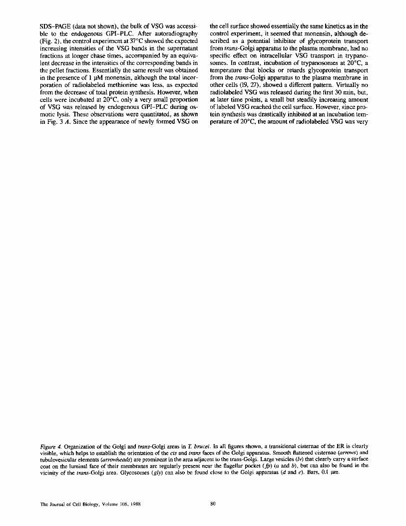
Figure 4. Organization of the Golgi and trans-Golgi areas in T. brucei. In all figures shown, a transitional cisternae of the ER is clearly visible, which helps to establish the orientation of the c/s and trans faces of the Golgi apparatus. Smooth flattened cisternae (arrows) and tubulovesicular elements (arrowheads) are prominent in the area adjacent to the trans-Golgi. Large vesicles (lv) that clearly carry a surface coat on the luminal face of their membranes are regularly present near the flagellar pocket (fp) (a and b), but can also be found in the vicinity of the trans-Golgi area. Glycosomes (gly) can also be found close to the Golgi apparatus (d and e). Bars, 0.1 tan.
The Journal of Cell Biology, Volume 106, 1988 80

Figure 5. Effect of monensin on the organization of the Golgi apparatus and expansion of the system of cisternae and tubulovesicular elements of the post trans-Golgi area in trypanosomes incubated at 20~ 1 0M monensin for 60 min at 37~ led to dilation of the trans-Golgi cisternae, whereas the cis Golgi compartment seemed to be unaffected (a). In addition, multivesicular bodies (MVB) were also dilated (b). After incubation at 20~ trypanosomes showed an increase in the amount of smooth flattened cisternae (arrows) and tubulovesicular elements (arrowheads) present in the area adjacent to the trans-Gotgi apparatus (c-f). Some cisternae in Golgi stacks were partially mis- aligned (e-f). A number of large vesicles (Iv) carrying a surface coat were also observed in the vicinity of the flagellar pocket (fp) (g). Bars, 0.1 0m.
low, which made it difficult to interpret the data. Therefore the experiment was modified by performing the preincuba- tion and pulse-labeling at 37~ After addition of cold methi- thine, the cells were rapidly cooled to and further incubated at 20~ Again, as compared to the control experiment, the rate of VSG transport to the cell surface was significantly re- duced (Fig. 3, B and C). This result is consistent with the assumption that, as in mammalian cells (19, 27, 32, 34), VSG becomes trapped in the trans-Golgi apparatus or post trans- Golgi compartment at this temperature, retarding its delivery to the cell surface.
Organization o f the Golgi Apparatus and the Post trans-Golgi Region in T. brucei and the Effects of Monensin and Incubation at 20~
In T. brucei the Golgi apparatus is usually located near the flagellar pocket and consists of 4-6 well-aligned stacked cisternae. A transitional ER cisterna, mostly free of ribo- somes, is nearly always found near the Golgi apparatus, facilitating the identification of the cis face of this organelle. Numerous small vesicles are present in the area between the transitional and Golgi cisternae, and images suggesting their
Duszenko et al. Trypanosome Surface Glycoprotein Secretion 81

fusion or budding from the cisternal membranes are fre- quently seen. Small vesicles are also present in the periphery of the Golgi stacks (Fig. 4, a-e). In trypanosomes isolated from very highly infected rats (parasitemia ~,,1.5 x 109/ml), the Golgi apparatus was poorly developed and seldom con- sisted of more than three stacked cisternae. This probably reflects the greatly diminished growth rate of trypanosomes at this parasitemia, which is equivalent to an in vitro culture approaching the stationary phase.
The area adjacent to the trans face of the Golgi apparatus is characterized by the presence of a prominent system of flattened cisternae and tubulovesicular elements. Frequently, the inner faces of the membranes of some of the elements of this system contain an electron-dense material similar to the surface coat (Fig. 4 e). Vesicles larger in size (i00 nm) than those in the transitional zone and covered by a well formed luminal coat, are regularly found near the flageUar pocket, as well as in the periphery of the trans-Golgi region (Fig. 4, a and b).
Treatment with 1 taM monensin led, within 1 min, to a dra- matic swelling of several trans-Golgi cisternae (Fig. 5, a and b), and to the appearance of other large vacuoles, not asso- ciated with the Golgi apparatus, containing variable amounts of amorphous material and some small vesicles, and proba- bly representing swollen lysosomes or multivesicular bodies. In spite of these effects observed in the electron micrographs, the shape and motility of the trypanosomes, as judged by light microscopy, were unaffected. This was the case when incubation was prolonged for up to 120 min, but monensin concentrations exceeding 5 IxM led to cell death of this clone within 15 min. Interestingly, some other trypanosome clones (e.g., MITat 1.2, variant clone 221a) tolerated monensin concentrations up to 10 p.M. Contrasting with the effect of monensin on the trans-Golgi cisternae, the flattened cister- nae and post trans-Golgi tubulovesicular elements were not swollen after monensin treatment.
In trypanosomes incubated at 20~ the post trans-Golgi tubulovesicular system was more highly developed (Fig. 5, c-g), and the alignment of Golgi cisternae within the stacks became somewhat looser (Fig. 5, c, e, and f ) . Large vesicles carrying a luminal coat were very distinct in the region of the flagellar pocket (Fig. 5, c and g).
Immunolocalization of VSG
The distribution of VSG in normal trypanosomes and in cells treated with monensin or incubated at 20~ was determined by immunogold labeling of thin sections. In all cases, VSG was localized over the entire cell surface, including the flagellum and flagellar pocket. Intracellularly, in normal cells the gold label was found over the rough ER, the Golgi apparatus, and the tubulovesicular elements and flattened cisternae in the post trans-Golgi area as well as in the luminaUy-coated large (100 nm) vesicles found near the flagellar pocket (Fig. 6). Most importantly, significant con- centrations of VSG were found throughout all the cisternae in the Golgi apparatus (Fig. 6, a-c). A few gold particles were regularly found over the luminal content of ',,50% of the glycosomes (29).
In monensin-treated cells, VSG was present in the large vacuoles derived from the trans-Golgi cisternae and in the remaining undilated cis-Golgi cisternae and elements of the
post trans-Golgi tubulovesicular system (data not shown). In cells incubated at 20~ the concentration of gold particles increased in the trans-Golgi cisternae and in membranes of the overdeveloped system of the tubulovesicular elements, flattened cisternae and large vesicles in the area adjacent to the trans-Golgi complex (Fig. 7).
Discussion
The parasitic-flagellated protozoa comprising the family Trypanosomatidae represent one of the best characterized lower eukaryotes at the cellular, biochemical and molecular levels. Their ancient divergence in the eukaryotic evolution- ary lineage is exemplified by the unique peculiarities of each of the basic cellular processes that have been studied in trypanosomatids. At the start of the work presented in this paper, VSGs were the only proteins for which definitive chemical evidence existed for posttranslational processing by carboxy-terminal hydrophobic peptide cleavage and addi- tion of a glycan phosphatidylinositol moiety (10). Thus, we felt it appropriate to evaluate whether VSG was transported to the trypanosome surface via the Golgi apparatus, espe- cially since published data (22) suggested that terminal modification of the VSG N-linked glycan core structure that is added in the ER was not obligatory, and did not occur in the variant used in the present studies. Our immunocyto- chemical data revealed VSG in the rough ER and throughout the cisternae of the Golgi apparatus, as well as in flattened cisternae and tubulovesicular elements adjacent to the trans- Golgi area. We therefore conclude that the classical transport route for the delivery of integral membrane proteins to the cell surface in higher eukaryotes, via the Golgi apparatus, is also used for VSG in trypanosomes. Within the limits of resolution of the present studies, the addition of glycolipid does not appear to override or modify normal sorting mecha- nisms. Immunolabeling strongly suggests that, after it tra- verses the Golgi stacks, VSG enters a post trans-Golgi tubu- lovesicular network that effects its delivery to the cell surface, most likely at the region of the flagellar pocket, which is also thought to be the point of secretion and endocytosis in try- panosomes (25, 35). Although we did not distinguish be- tween VSG in biosynthetic or endocytic pathways, VSG turnover only occurs at a low rate under the experimental conditions used (Duszenko, M., unpublished observations). Thus, it seems likely that the majority of immunolabeled VSG in the Golgi apparatus and ER can be attributed to the biosynthetic pathway.
The presence, in trypanosomes, of an extensive system of cisternal and tubulovesicular elements in the area adjacent to the trans side of the Golgi apparatus has been recognized by other investigators, who suggested that it plays a role in secretion (39), or is involved in endocytosis and intracellular digestion (25). It seems likely that some of the post trans- Golgi elements represent the counterpart of the trans-Golgi network recently described in mammalian cells, for which a role in the sorting of newly synthesized secretory, lyso- somal, and membrane proteins has been suggested (20). In fact we found that, in trypanosomes incubated at 20~ as is the case with mammalian cells incubated at the same tem- perature (19, 20, 34), the post-Golgi system was expanded
The Journal of Cell Biology, Volume 106, 1988 82

Figure 6. Immunolocalization of VSG in trypanosomes. Thin frozen sections of cells fixed in 2% glutaraldehyde were labeled using anti-VSG antibody and protein A-gold conjugates (10 nm). The cell surface, including the flagellar pocket (d-e), was homogeneously la- beled. Intracellulary, the membranes of the rough ER cisternae showed low levels of labeling, but it was clear that the gold particles were bound to the luminal face of the membrane (e). A prominent amount of the marker was found over the cisternae of the Golgi apparatus (GA) (a-c). Tubulovesicular elements (arrowheads) and flattened cisternae (arrows) adjacent to the trans-Golgi apparatus and the flagellar pocket (fp) were also labeled (a-d, g). Large vesicles (Iv), 100 nm in diameter, usually found close to the flagellar pocket, were systemati- cally labeled on the luminal side of their membranes (d). Bar, 0.1 ttm.
Duszenko et al, T~panosome Surface Glycoprotein Secretion 83

Figure 7. Accumulation of VSG in the expanded system of flattened cisternae and tubulovesicular elements of the post trans-Golgi area of trypanosomes incubated at 20~ After incubation for 60 min at 20~ cells were processed for immunolabeling as described in the legend for Fig. 6. Intracellularly, the VSG marker was predominantly located on trans-Golgi cisternae (b, large arrowhead), and on an increased amount of flattened cisternae (arrows), and tubulovesicular elements (arrowheads) present in the post trans-Golgi area, which are characteristic of these experimental conditions (a-f). Bar, 0.1 tun.
The Journal of Cell Biology, Volume 106, 1988 84

and contained higher concentrations of VSG, and that the delivery of newly synthesized VSG to the cell surface was significantly delayed.
The precise route by which the VSG concentrated in the trypanosomal trans-Golgi network reaches the flagellar pocket remains to be elucidated. It is possible that the cister- nae of the trans-Golgi network intermittently communicate directly with the cell surface or, more likely, that small vesi- cles budding from them transfer VSG to the plasma mem- brane. Large vesicles, of ~100 nm diameter, that are found near the flagellar pocket were consistently labeled for VSG. However, these vesicles contained a well-formed surface coat over the luminal aspect of their membranes, and it seems likely that they are endocytic in nature and contain VSG de- rived from the surface coat. Other authors have shown that similar vesicles can be labeled with endocytosed markers (25).
The lack of effect of monensin on VSG transport is perhaps surprising in view of the clear effects that this ionophore had on Golgi structure. However, cases have been reported of glycoproteins, such as the hemagglutinin of influenza virus synthesized in Madin-Darby canine kidney (MDCK) cells (2), whose transport to the cell surface was not affected by monensin.
Elucidation of the details of modifications occuring in each cellular compartment of the trypanosome secretory pathway will require further studies. Current data (3, 4, 15) are con- sistent with the hypothesis that glypiation occurs as an im- mediate posttranslational event in the ER, probably by addi- tion of a preformed glycolipid precursor in a reaction that is closely coupled to cleavage of the polypeptide tail (24). Re- cent studies have shown that a putative precursor of the protein-bound glycolipid lacks galactose (Menon, A. K., S. Mayor, M. A. J. Ferguson, M. Duszenko, and G. A. M. Cross, unpublished observations). Thus, further modifica- tion of the protein-bound glycolipid may occur in the ER and in the Golgi apparatus.
We thank N. H. Lanners and E. Gubert for helpful advice and excellent technical assistance, Mrs. Jody Culkin for her help in producing the plates, and Mrs. M. Cort for help in the text revisions. We are also very grateful to Dr. David Sabatini for his advice during the work and the preparation of this paper.
This work was supported by National Institutes of Health grants A121531 (G. A. M. Cross), GM 20277, and AG 01461 (I. E. Ivanov and H. Pleskin), and by the Deutsche Forschungsgemeinschafi (M. Duszenko).
Received for publication 23 June 1987, and in revised form 5 October 1987.
References
1. Allen, G., L. P. Guruett, and G. A. M. Cross. 1982. Complete amino acid sequence of a variant surface glycoprotein (VSG 117) from Trypanosoma brucei. J. MoL BioL 157:527-546.
2. Alonso, F. V., and R. W. Compans. 1981. Differential effect of monensin on enveloped viruses that tbrm at distinct plasma membrane domains. J. Cell Biol. 89:700-705.
3. Bangs, J. D., D. Hereld, J. L. Krakow, G. W. Hart, and P. T. Englund. 1985. Rapid processing of the carboxyl terminus ofa trypanosome variant surface glycoprotein. Proc. Natl. Acad. Sci. USA. 82:3207-3211.
4. Bangs, J. D., N. W. Andrews, G. W. Hart, and P. T. Englund. t986. Post- translational modification and intracellular transport of a trypanosome variant surface glycoprotein. J. Cell Biol. 103:255-263.
5. Boothroyd, J. C., C. A. Paynter, G. A. M. Cross, A. Beruards, and P. Borst. t981. Variant surface glycoproteins of Trypanosoma brucei are synthesized with cleavable hydrophobic sequences at the carboxy and amino termini. NucL Acids Res. 9:4735-4743.
6. Bulow, R., and P. Overath. 1986. Purification and characterization of the membrane-form variant surface glycoprotein hydrolase of Trypanosoma brucei. J. Biol. Chem. 261:11918-1t923.
7. Cardoso de Almeida, M. L. and M. J. Turner. 1983. The membrane form of variant surface gtycoproteins of Trypanosoma brucei. Nature (Lond.). 302:349-352.
8. Cross, G. A. M. 1975. Identification, purification and properties of clone specific glycoprotein antigens constituting the surface coat of Trypano- soma brucei. Parasitology. 71:393-417.
9. Cross, G. A. M. 1984. Structure of the variant glycoproteins and surface coat of Trypanosoma brucei. Phil. Trans. R. Soc. Lond. B. 370:3-12.
I0. Cross, G. A. M. 1987. Eukaryotic protein modification and membrane at- tachment via phosphatid)'linositol. Cell. 48:179-181.
I I. Farqnhar, M. G. t985. Progress in unraveling pathways of Golgi traffic. Annu. Rev. Cell Biol. 1:477-488.
12. Farquhar, M. G., and G. E. Palade. 1981. The Golgi apparatus (complex) (1954-1981)-from artifact to center stage. J. Cell Biol. 91:77s-103s.
13. Ferguson, M. A. J,, and G. A. M. Cross. 1984. Myristylation of the mem- brane form of a Trypanosoma brucei variant surface glycoprotein. J. Biol. Chem. 259:3011-3015.
14. Ferguson, M. A. J., M. G. Low, and G. A. M. Cross. 1985. Glycosyl-sn- 1,2-dimyristyl phosphatidylinositol is covalently linked to Trypanosoma brucei variant surface glycoprotein. J. BioL Chem. 260:4963--4968.
15. Ferguson, M. A. J., M. Duszenko, G. S. Lamont, P. Overath, and G. A. M. Cross. 1986. Biosynthesis of Trypanosoma brucei variant surface glyco- proteins: N-glycosylation and addition of a phosphatidylinositol mem- brane anchor. 3. Biol. Chem. 261:356-362.
16. Fox, J. A., M. Duszenko, M. A. J. Ferguson, M. G. Low, and G. A. M. Cross. 1986. Purification and characterization of a novel glycan-phos- phoinositide-specific phospholipase C from Trypanosoma brucei. J. BioL Chemo 261 : 15767-15771.
17. Fox, J. A., N. M. Soliz, and A. R. Saltiel. 1987. Purification and character- ization ofa phosphatidylinositol glycan specific phospholipase C from he- patic plasma membranes. Proc. Natl. Acad. Sci. USA. 84:2663-2668.
18. Grab, D. J., P. Webster, and Y. Verjee. 1984. The intracellular pathway and assembly of newly formed variable surface glycoprotein of Trypano- soma brucei. Proc. Natl. Acad. Sci. USA. 81:7703-7707.
19. Griffiths, G., S. Pfeiffer, K. Simons, and K. Matlin. 1985. Exit of newly synthesized membrane proteins from the trans cisterna of the Golgi com- plex to the plasma membrane. J. Cell Biol. 101:949-964.
20. Griffiths, G., and K. Simons. 1986. The trans-Golgi network: sorting at the exit site of the Golgi complex. Science (Wash. DC). 234:438-443.
21. Hereld, D., J. L. Krakow, J. D. Bangs, G. W. Hart, and P. T. Englund. 1986. A phospholipase C from Trypanosoma brucei which specifically cleaves the glycolipid on the variant surface gtycoprotein. J. BioL Chem. 261:12813-13819.
22. Holder, A. A., and G. A. M. Cross. 1981. Glycopeptides from variant sur- face glycoproteins of Trypanosoma brucei: C-terminal location of anti- genitally cross-reacting carbohydrate moieties. Mol. Biochem. Parasitol. 2:135-150.
23. Keller, G. A., K. T, Tokuyasu, A. H. Dutton, and S. J. Singer. 1984. An improved procedure for immunoelectron microscopy: altrathin plastic embedding of immunolabeled ultrathin frozen sections. Proe. NatL Acad. Sci. USA. 81:5744-5747.
24. Krakow, J. L., D. Hereld, J. D. Bangs, G. W. Hart, and P. T. Englund. 1986. Identification of a glycolipid precursor of the Trypanosoma brucei variant surface glycoprotein. J. Biol. Chem. 261 : 12147-12153.
25. Langreth, S. G., and A. E. Balber. 1975. Protein uptake and digestion in bloodstream and culture forms of Trypanosoma brucei. J. Protozool. 22:40-53.
26. Low, M. G. 1987. Biochemistry of the glycosyl-phosphatidyl-inositol membrane protein anchors. Biochem. J. 244:t-13.
27. Matlin, K., and K. Simons. 1983. Reduced temperature prevents transfer of a membrane glycoprotein to the cell surface but does not prevent termi- nal glycosylation. Cell. 34:233-243.
28. Medelzon, D. H,, J. O. Previato, and A. J. Parodi. 1986. Characterization of protein-linked oligosaccharides in trypanosomatid flagellates. Mol. Biochem. Parasitol. 18:355-369.
29. Opperdoes, F. R,, P. Baudhuin, I. Coppens, C. De Roe, S. W. Edwards, P. J. Weijers, and O. Misset. 1985. 1984. Purification, morphometric analysis, and characterization of the glycosomes (microbodies) of the pro- tozoan hemoflagellate Trypanosoma brucei. Z Cell BioL 98:1178-1184.
30. Parodi, A. J., L. A. Q. Anue, andJ. J. Cazzulo. 1981. Pathway of protein glycosylation in Crithidia fasciculata. Proc. Natl. Acad. Sci. USA. 78: 6201-6205.
31. Previato, L O., D, H. Mendelzon, and A. J. Parodi. 1986. Characterization of dolichol monophosphate- and dolichol diphosphate-linked saccharides in trypanosomatid flagellates. Mol. Biochem. Parasitol. 18:343-355.
32. Rindler, M. J., I. E. Ivanov, H. Plesken, and D. D. Sabatini. 1985. Polar- ized delivery of viral glycoproteins to the apical and basolateral plasma membranes of Madin-Darby canine kidney cells infected with tempera- ture-sensitive viruses. J. Cell Biol. 100:136-14t.
33. Sabatini, D. D., G. Kreibich, T. Morimoto, and M. Adesnik. t982. Mecha- nisms for the incorporation of proteins in membranes and organelles. J. Cell Biol. 92:1-22.
Duszenko et al. Trypanosome SurJhce Glycoprotein Secretion 85

34. Saraste, J., and E. Kuismanen, 1984. Pre- and post-Golgi-vacuoles operate in the transport of Semliki forest virus membrane glycoproteins to the cell surface. Cell. 38:535-549.
35. Steiger, R. 197 t. Some aspects of surface coat formation in Trypanosoma bruceL Acta Tropica. 28:341-346.
36. Strickler, J. E., and C. L. Patton. 1980, Trypanosoma brucei: inhibition of glycosylation of the major variable surface coat glycoprotein by tunicamycin. Proc. Natl. Acad. Sci, USA. 77:1529-1533.
37. Tokuyasu, K. T. 1980. Immunocytochemistry on ultrathin frozen sections.
Histochem. J. 12:381--403. 38. Turner, M. J. 1982. Biochemistry of the variant surface glycoproteins of
salivarian trypanosomes. Adv. ParasitoL 21:69-153. 39. Vickerman, K. 1969. The fine structure of Trypanosoma congotense in its
bloodstream phase. J. Protozol. 16:54-69. 40. Walter, P., R. Gilmore, and W. Blobel. 1984~ Protein translocation across
the endoplasmic reticulum. Celt. 38:5-8. 41. Wickner, W. T., and H. F. Lodish. 1985. Multiple mechanisms of protein
insertion into and across membranes. Science (Wash. DC). 230:400-407.
The Journal of Cell Biology, Volume 106, 1988 86
Copyright © 2022 FDOKUMEN