Glycoprotein gonadotropin in the plasma and its cellular origin ...
11
HAL Id: hal-01600232 https://hal.archives-ouvertes.fr/hal-01600232 Submitted on 2 Jun 2020 HAL is a multi-disciplinary open access archive for the deposit and dissemination of sci- entific research documents, whether they are pub- lished or not. The documents may come from teaching and research institutions in France or abroad, or from public or private research centers. L’archive ouverte pluridisciplinaire HAL, est destinée au dépôt et à la diffusion de documents scientifiques de niveau recherche, publiés ou non, émanant des établissements d’enseignement et de recherche français ou étrangers, des laboratoires publics ou privés. Distributed under a Creative Commons Attribution - ShareAlike| 4.0 International License Glycoprotein gonadotropin in the plasma and its cellular origin in the adenohypophysis of sham-operated and ovariectomized rainbow trout, Salmo gairdneri. L J. van Putten, J. Peute, P.G. van Oordt, H.J. Goos, Bernard Breton To cite this version: L J. van Putten, J. Peute, P.G. van Oordt, H.J. Goos, Bernard Breton. Glycoprotein gonadotropin in the plasma and its cellular origin in the adenohypophysis of sham-operated and ovariectomized rainbow trout, Salmo gairdneri.. Cell and Tissue Research, Springer Verlag, 1981, 218 (2), pp.439- 448. 10.1007/BF00210356. hal-01600232
-
Upload
khangminh22 -
Category
Documents
-
view
1 -
download
0
Transcript of Glycoprotein gonadotropin in the plasma and its cellular origin ...

HAL Id: hal-01600232https://hal.archives-ouvertes.fr/hal-01600232
Submitted on 2 Jun 2020
HAL is a multi-disciplinary open accessarchive for the deposit and dissemination of sci-entific research documents, whether they are pub-lished or not. The documents may come fromteaching and research institutions in France orabroad, or from public or private research centers.
L’archive ouverte pluridisciplinaire HAL, estdestinée au dépôt et à la diffusion de documentsscientifiques de niveau recherche, publiés ou non,émanant des établissements d’enseignement et derecherche français ou étrangers, des laboratoirespublics ou privés.
Distributed under a Creative Commons Attribution - ShareAlike| 4.0 InternationalLicense
Glycoprotein gonadotropin in the plasma and its cellularorigin in the adenohypophysis of sham-operated and
ovariectomized rainbow trout, Salmo gairdneri.L J. van Putten, J. Peute, P.G. van Oordt, H.J. Goos, Bernard Breton
To cite this version:L J. van Putten, J. Peute, P.G. van Oordt, H.J. Goos, Bernard Breton. Glycoprotein gonadotropinin the plasma and its cellular origin in the adenohypophysis of sham-operated and ovariectomizedrainbow trout, Salmo gairdneri.. Cell and Tissue Research, Springer Verlag, 1981, 218 (2), pp.439-448. �10.1007/BF00210356�. �hal-01600232�

Cell Tissue Res (1981) 218:439-448 Cell and Tissue Research �9 Springer-Verlag 1981
Glycoprotein Gonadotropin in the Plasma and Its Cellular Origin in the Adenohypophysis of Sham-operated and Ovariectomized Rainbow Trout, Salmogairdneri
Leonie J.A. Van Putten, J. Peute, P.G.W.J. Van Oordt, H.J.Th. Goos, and B. Breton*
Zoological Laboratory, Section Comparative Endocrinology, University of Utrecht, The Netherlands * Laboratoire de Physiologic des Poissons, I.N.R.A., Rennes, France
Summary. Among the cells of the pituitary generally believed to produce glycoprotein gonadotropin (GTH) five forms were distinguished, based on the amount and the diameter of granules and globules and the appearance of the rough endoplasmic reticulum. In sham-operated trout so-called"globular" cells predominated, whereas after ovariectomy these were replaced by so-called "cisternal" cells, suggesting that both belong to one GTH-cell type. In addition, ovariectomy caused a strong increase in plasma GTH-levels. This indicates that the transition from globular to cisternal cells is accompanied by extrusion of GTH, and thus points to a storage of G T H in the granules and globules. It is argued that one of the five forms has the morphological characteristics of thyrotropic cells and may not produce glycoprotein GTH.
Key words: Pituitary salmonids - GTH-cells - Ultrastructure - Ovariectomy - Radioimmunoassay
Cells producing gonadotropic hormone (GTH) can be found throughout the adenohypophysis of vertebrates, although very seldom in the pars intermedia. They are basophilic, staining, e.g., with periodic acid-Schiff, alcian blue and aldehyde fuchsin. These cells are also characterized by irregular cisternae of the rough endoplasmic reticulum (RER); in many vertebrates they not only contain secretory granules but also large globules. Both granules and globules are storage forms of secretory material (see reviews by Holmes and Ball 1974; van Oordt 1979). This also holds true for teleost fishes, where such GTH-cells tend to concentrate in the ventral
Send offprint requests to: Prof. Dr. P.G.W.J. van Oordt, Zoological Laboratory, Transitorium III, Padualaan 8, 3508 TB Utrecht, The Netherlands
The authors are indebted to Mr. L.W. van Veenendaal for preparing the illustrations and the photographic layout
0302-766X/81/0218/0439/$02.00

440 L.J.A. Van Putten et al.
b o r d e r o f the p r o x i m a l pa rs distal is (p .p.d.) ( for reviews, see v a n O o r d t 1968; Ball
a n d B a k e r 1969; S c h r e i b m a n et al. 1973). In s a lmon ids these " g l o b u l a r ' basoph i l s
a re n o t res t r i c ted to the p.p.d. , b u t a re also s i tua ted be tween the fo l l icu lar ly
a r r a n g e d p r o l a c t i n cells in the ros t r a l pa rs distal is (r .p.d.) ( C o o k a n d v a n O v e r b e e k e 1972; N a g a h a m a 1973; B o d d i n g i u s 1975; O l i v e r e a u 1976, 1978; E k e n g r e n et al.
1978b, c; Peu te et al. 1978; U e d a 1980). D u r i n g the a n n u a l cycle o f the r a i n b o w
t r o u t the g l o b u l a r G T H - c e l l s p r e d o m i n a t e s o m e m o n t h s a f te r s p a w n i n g (Peute et al. 1978). S t o r a g e o f G T H wi th in these cells cou ld be d e m o n s t r a t e d by m e a n s o f
i m m u n o h i s t o c h e m i c a l t echn iques , us ing an t i - ca rp a n d a n t i - s a l m o n g l y c o p r o t e i n
G T H ( M c K e o w n a n d v a n O v e r b e e k e 1971 ; E k e n g r e n et al. 1978b, c). T h e c o n t e n t s
o f b o t h the sec re to ry g ranu les a n d g lobu les reac t to such a n t i - G T H - s e r u m (Leun i s sen et al. 1980).
In a d d i t i o n to the g l o b u l a r G T H - c e l l s , "ves i cu l a r " o r "c i s t e rna l " cells can be f o u n d a m o n g the basoph i l s o f the s a l m o n i d pars distalis. C o o k a n d v a n O v e r b e e k e
(1972), O l i v e r e a u (1976, 1978), a n d U e d a (1980) cons ide r ed the l a t t e r to be a s e p a r a t e type o f G T H - c e l l , whe rea s E k e n g r e n et al. (1978 b, c) a n d Peu te et al. (1978)
s u p p o s e d the g l o b u l a r a n d the c i s te rna l ba soph i l s to r ep resen t d i f fe ren t s tages o f a
s ingle type o f G T H - c e l l . In an a t t e m p t to reso lve these conf l i c t ing views, in the
p re sen t s tudy the pe rcen t ages o f the g l o b u l a r a n d c is ternal cells were c o r r e l a t e d wi th
t he p l a s m a levels o f g l y c o p r o t e i n G T H in s h a m - o p e r a t e d a n d o v a r i e c t o m i z e d
r a i n b o w t r o u t (Salmo gairdneri).
Materials and Methods
The fish were obtained from a hatchery in Vaassen, Holland. Nine mature females weighing 700-1000 g were used. On October 26th five of them were ovariectomized; four others were sham-operated. For ovariectomy the animals were anaesthetized in 0.1% phenoxyaethanol. The ovaries were exposed by a 5 cm ventral cut and removed in the experimental animals. The wound was closed with 4 to 5 stitches with chrom-catgut. During the operation the gills were perfused with a 0.1% solution of the anaesthetic. Five weeks later the animals were anaesthetized with CO2, and blood samples were taken, followed by decapitation. During dissection 8 % glutaraldehyde in 0.1 M Na-cacodylate buffer was dropped on to the pituitary. After removal of the pituitaries they were submerged in 4 % glutaraldehyde in 0.1 M Na- cacodylate buffer (pH 7.2) at 0 ~ C. After 1 to 2h an equal volume of2% OsO 4 in 0.1 M Na-cacodylate buffer was added (0 ~ C in the dark). After 45 min the tissues were postfixed in 2 % OsO 4 in 0.1 M Na- cacodylate buffer at 0 ~ C for another hour. Subsequently, the tissues were dehydrated in graded alcohols and propylene oxide and embedded in Epon.
Sections of I ~tm thickness were cut on a Porter-Blum microtome using glass knives and stained with methylene-blue-azurII (Richardson 1960) for light microscopy. Ultrathin sections were cut on a Reichert OMU 3 ultramicrotome, collected on 200 mesh copper grids, stained with uranyl acetate and lead citrate (Reynolds 1963) and examined with a Zeiss EM 9A electron microscope.
At three times during the experimental period blood samples were taken, i.e., immediately before the operation, two weeks later and on the last day of the experiment. GTH-content of the blood samples was determined with a homologous radioimmunoassay for salmonid fishes, using Oncorhynchus tschawytscha-GTH (Breton et al. 1978) for label and reference, and an antibody directed against Salmo gairdneri-GTH (Breton et al. 1976). The antibody was raised in guinea pigs. The salmon-GTH was labeled with I12s, using NaI 125 IMS 300 from Amersham, England. Conditions of labeling and further purifications were the same as those described for carp-GTH radioimmunoassay (Breton et al. 1971). The antibody was used at a final dilution of 0.5 �9 10 s, the incubation lasted 4 days at 4 ~ free and bound hormone was separated due to the second antibody method (rabbit sera directed against guinea pig globulins). Calculation was performed after logit-log transformation.

Ultrastructure of GTH-Cells and Plasma Levels of GTH in Trout
1 2 3
441
4 5 F ig1
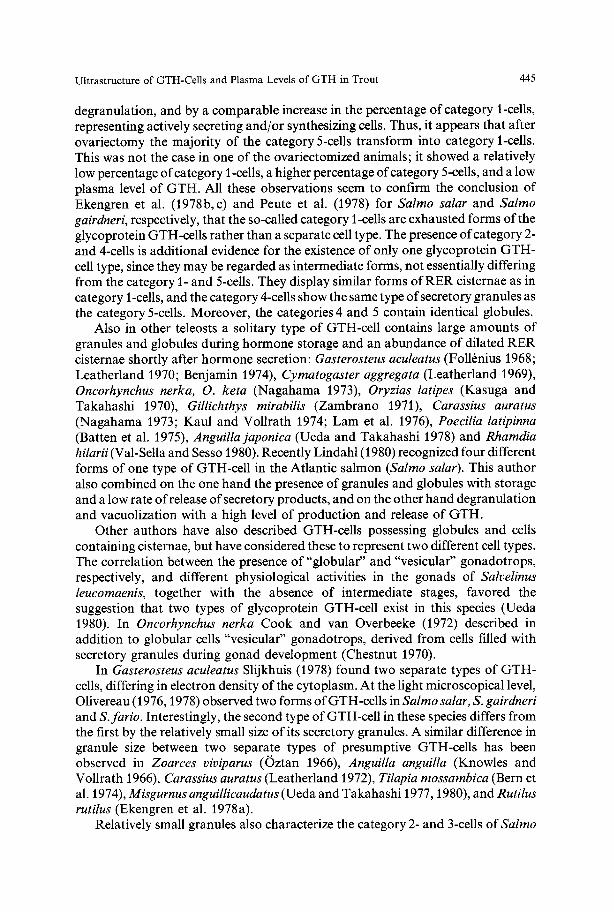
Fig. 1. Classification of GTH-cells in the morphological categories I-5, based on the amount and diameter of granules or globules and the presence of RER. RER rough endoplasmic reticulum; gr granule; gl globule
~00%"
S0%"
O%-
DiIIeN.11 Iorms of GTH.ce . I (in%) 1
,N 3118 3133
Ovarlec tomized
1
3127
iL__ 3136 NROF TROUT
1-5(SEE FIG1)
100%"
50%"
0%-
Sham_operated
Diflerent forms of GTH_r {in%) 5
A A A 1 1 1 1 3102 3103 3107 3113 N• OF TROUT
! 5~UE f'Oll Fig2
Fig. 2. The percentage of cells in each category for each pituitary of ovariectomized and sham-operated trout. Note the appearance of GTH-cells in the "cisternal" stage and the decrease of"globular" GTH- cells after ovariectomy
Quantification of GTH-CelIs
The putative GTH-cells were divided according to a subjective scale into five different categories on the basis of the amount and diameter o f the granules and globules, and the appearance of the RER material (Fig. 1). With such a division it is possible to study the effect of ovariectomy on the GTH-cells in a semi- standardized manner. The chosen sequence of these categories 1-5 is purely morphological and does not necessarily reflect five consecutive physiological stages.
Category I consists of cells with more or less strongly dilated RER cisternae and lacking any granules and globules.
Category 2-cells contain dilated cisternae of the RER and some round electron-dense granules with a diameter o f 75-280 nm (average diameter 155 nm). These cells are also characterized by the presence of an active Golgi system and many mitochondria.

442 L.J.A. Van Putten et al.
Category 3 consists of ceils in which the area containing granules exceeds the area containing RER cisternae. These cisternae are not strongly dilated. The diameters of the granules measure between 90- 280 nm (average diameter 166 nm). Most granules are round, although fusion may result in elongated forms. In cells of categories 2 and 3 globules are absent.
Category 4-cells contain some globules. These are oval or circular in shape, are less electron-dense than the granules and measure 500-1,200 nm. The granules measure between 90-500 nm (average diameter 260 nm). In these cells a considerable portion of the cytoplasm is filled with dilated cisternae of the RER. Most granules are circular in outline, but can become elongated by fusion. Coalescence of granules with globules, and of different globules with each other also occurs.
Category 5-cells are filled with granules and globules. The RER is poorly developed and consists of a few small and somewhat rounded cisternae. The globules and granules show the same characteristics as those of the category 4-cells.
For the quantification of the five categories in each pituitary, about one hundred presumptive GTH- cells were chosen from randomly selected electron-micrographs of the p.p.d., and classified. For each pituitary the percentages of cells in each category are given in Fig. 2.
Results
a) Ultrastrueture of the GTH-Cells
In the sham-operated female t rout at the end o f the experiment _+ 60 % o f the presumptive GTH-cel ls belong to category 5 (Fig. 2), and are filled with numerous secretory granules and globules (Fig. 3). Of the remaining cells _+ 25 % belong to category 3, only 2-9 % to category 2, and 3-8 % to category 4. It appears that in all pituitaries the cells belonging to category 3 show a tendency to be associated with branches o f the neurohypophysis in the rostral region o f the p.p.d., whereas the cells belonging to the other categories show a more r andom distribution. The pituitaries o f the sham-opera ted t rout did not contain cells in category 1, i.e., cells with cytoplasm largely occupied by dilated cisternae o f the R E R (Fig. 3).
On the other hand, in four out o f five pituitaries o f ovariectomized trout, as much as _+ 75 % o f the presumptive GTH-cells can be classified in category I (Fig. 4) and only 5 - 2 0 % belong to category5. Compared to the control group, the percentage o f cells in category 2 had slightly increased to 7-16 %, and that o f cells in category 3 had decreased to 6-12 %. Cells in category 4 remained at about the same percentage (Fig. 2).
The pituitary o f t rout nr 3123 differs f rom those o f the other ovariectomized animals. It had a relatively low percentage (6 %) o f cells in category 1. Relatively high percentages o f cells containing secretory granules and/or globules were found in the categories 2 (35 %), 3 (22 %), and 5 (35 %).
In contrast to the si tuation in other cell types, where electron-dense vesicles are found in the Golgi area, most Golgi vesicles in the GTH-cells are electron lucent.
b) Plasma Levels of GTH (Table 1)
The plasma levels o f G T H o f all animals at the start o f the experiment varied between 3.20 and 4.22 ng G T H per ml plasma. In sham-operated t rout this level did not change during the experimental period. However, the ovariectomized animals
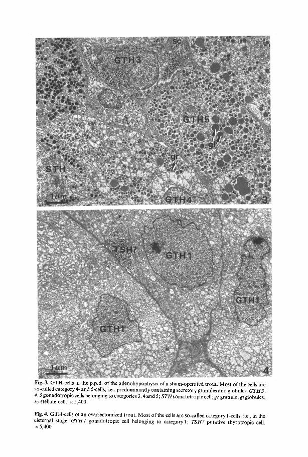
Fig. 3. GTH-cells in the p.p.d, of the adenohypophysis of a sham-operated trout. Most of the cells are so-called category 4- and 5-cells, i.e., predominantly containing secretory granules and globules. GTH3, 4, 5 gonadotropic cells belonging to categories 3, 4 and 5; S T H somatotropic cell; gr granule; gl globules; sc stellate cell. • 5,400
Fig. 4, GTH-cells of an ovariectomized trout. Most of the cells are so-called category 1-cells, i.e., in the cisternal stage. GTH1 gonadotropic cell belonging to category 1; TSH? putative thyrotropic cell. x 5,400

444 L.J.A. Van Putten et al.
Table 1. GTH-content of the blood plasma immediately before the operation, two weeks later and on the last day of the experiment
Treatment Trout nr. GTH ng/ml GTH ng/ml GTH ng/ml 26-10-77 10-11~77 30-11~77
Sham-operated 3102 * 3,48 * 3103 * 4.18 2.77 3107 * 3.20 3.58 3112 * 4.22 3.45
Ovariectornized 3118 4.13 29.44 16,63 3123 4.07 19.07 5,28 3126 5.13 13.90 15.35 3127 4.47 14.76 15.81 3136 3.61 * 15.49
* Not measured
showed a strong, although not homogeneous, response. In the animal with the strongest rise after two weeks, the GTH-level had dropped from 29.44 to 16.63 ng/ml by the end of the experiment. Two other animals showed their highest GTH-level (15.35 and 15.81 ng) at the end of the experiment. The final GTH-level in the plasma of four ovariectomized animals was almost identical. In the fifth animal of this group (nr3123) the GTH-level rose over a period of two weeks to a maximum (19.07), but dropped to about control levels (5.28ng) during the last weeks of the experiment.
Discussion
The low plasma levels of glycoprotein GTH found in female trout at the start of the experiment in October and in the sham-operated controls at the end of the experiment in November correspond to the levels measured by Billard et al. (1978). During this period, preceding the profuse secretion of glycoprotein GTH necessary for the final stages ofoocyte maturation and ovulation (Billard et al. 1978; van den Hurk and Peute 1979), the pituitary is probably storing GTH rather than secreting it. The high percentage of cells filled with numerous granules and globules in the control group is in accordance with this concept. Moreover, recent immunocyto- chemical studies have shown the presence if glycoprotein GTH inside these granules and globules (Leunissen et al. 1980). These data confirm earlier observations on the trout by Peute et al. (1978), who found a positive correlation between a high GTH-content in the pituitary and the presence of many GTH-cells with granules and globules.
Within a period of two weeks the concentration of GTH in the plasma of the ovariectomized animals rose to levels exceeding the maximum normally found during ovulation (Billard et al. 1978). This may partly be due to an abnormally high GTH-secretion by the pituitary, but also partly due to the reduction in number of GTH-binding sites after removal of the ovaries. Twenty days later the plasma GTH-concentration remained high in four ovariectomized trout and was accompanied by a strong decrease in the percentage of category 5-cells, caused by
Ultrastructure of GTH-Cells and Plasma Levels of GTH in Trout 445
degranulation, and by a comparable increase in the percentage of category 1-cells, representing actively secreting and/or synthesizing cells. Thus, it appears that after ovariectomy the majority of the category 5-cells transform into category 1-cells. This was not the case in one of the ovariectomized animals; it showed a relatively low percentage of category 1-cells, a higher percentage of category 5-cells, and a low plasma level of GTH. All these observations seem to confirm the conclusion of Ekengren et al. (1978b, c) and Peute et al. (1978) for Salmo salar and Salmo gairdneri, respectively, that the so-called category 1-cells are exhausted forms of the glycoprotein GTH-cells rather than a separate cell type. The presence of category 2- and 4-cells is additional evidence for the existence of only one glycoprotein GTH- cell type, since they may be regarded as intermediate forms, not essentially differing from the category 1- and 5-cells. They display similar forms ofRER cisternae as in category 1-cells, and the category 4-cells show the same type of secretory granules as the category 5-cells. Moreover, the categories 4 and 5 contain identical globules.
Also in other teleosts a solitary type of GTH-cell contains large amounts of granules and globules during hormone storage and an abundance of dilated RER cisternae shortly after hormone secretion: Gasterosteus aculeatus (Foll6nius 1968; Leatherland 1970; Benjamin 1974), Cymatogaster aggregata (Leatherland 1969), Oncorhynchus nerka, O. keta (Nagahama 1973), Oryzias latipes (Kasuga and Takahashi 1970), Gillichthys mirabilis (Zambrano 1971), Carassius auratus (Nagahama 1973; Kaul and Vollrath 1974; Lam et al. 1976), Poecilia latipinna (Batten et al. 1975), Anguillajaponica (Ueda and Takahashi 1978) and Rhamdia hilarii (Val-Sella and Sesso 1980). Recently Lindahl (1980) recognized four different forms of one type of GTH-cell in the Atlantic salmon (Salmo salar). This author also combined on the one hand the presence of granules and globules with storage and a low rate of release of secretory products, and on the other hand degranulation and vacuolization with a high level of production and release of GTH.
Other authors have also described GTH-cells possessing globules and cells containing cisternae, but have considered these to represent two different cell types. The correlation between the presence of "globular" and "vesicular" gonadotrops, respectively, and different physiological activities in the gonads of Salvelinus leucomaenis, together with the absence of intermediate stages, favored the suggestion that two types of glycoprotein GTH-cell exist in this species (Ueda 1980). In Oncorhynehus nerka Cook and van Overbeeke (1972) described in addition to globular cells "vesicular" gonadotrops, derived from cells filled with secretory granules during gonad development (Chestnut 1970).
In Gasterosteus aculeatus Slijkhuis (1978) found two separate types of GTH- cells, differing in electron density of the cytoplasm. At the light microscopical level, Olivereau (1976, 1978) observed two forms of GTH-cells in Salmo salar, S. gairdneri and S.fario. Interestingly, the second type of GTH-cell in these species differs from the first by the relatively small size of its secretory granules. A similar difference in granule size between two separate types of presumptive GTH-cells has been observed in Zoarces viviparus (Oztan 1966), Anguilla anguilla (Knowles and Vollrath 1966), Carassius auratus (Leatherland 1972), Tilapia mossambica (Bern et al. 1974), Misgurnus anguillicaudatus (Ueda and Takahashi 1977, 1980), and Rutilus rutilus (Ekengren et al. 1978a).
Relatively small granules also characterize the category 2- and 3-cells of Salmo

446 L.J.A. Van Putten et al.
gairdneri. As mentioned above, the category 2-cells may belong to the glycoprotein GTH-cell type on the basis of similarities in the appearance of their RER with that of the category 1- and 4-cells. This is, however, not the case with the category 3-cells. Moreover, the category 3-cells differ from the other categories by the fact that they do not show a random distribution, and are frequently associated with branches of the neurohypophysis in the rostral region of the p.p.d. Recent immunofluorescent studies on trout pituitaries using anti-human-/%thyrotropin (TSH) resulted in a specific reaction of cells showing the same location within the p.p.d. (unpublished results). This location and the small size of the granules measuring approximately 160 nm are common characteristics of TSH-cells (Nagahama 1973, 1977; Bern et al. 1974; Benjamin 1975). This does, of course, not implicate that all category 3-cells belong to the TSH-cell type. The percentage of category 3-cells diminished after ovariectomy in four out of five experimental animals. It might be argued that category 3-cells have a double function, secreting both TSH and GTH, as has been suggested for similar cells in Oncorhynchus kisutch by Chestnut (1970); however, it seems unlikely that they produce glycoprotein GTH, since according to Leunissen et al. (1980) the granules of such cells do not bind anti-carp-/~-GTH. A combined immunocytochemical and electron-microscopical study on the rat pituitary pointed out that also in mammals some gonadotropic cells mimic the thyrotropic cells at the ultrastructural level (Tougard et al. 1980). According to these authors, identi- fication of all gonadotropic cells is difficult without application of im- munocytochemistry.
Recent biochemical studies point to the production of two different gonadotropic hormones in teleosts, including the salmonid Oncorhynchus keta (Ng and Idler 1978; Idler and Ng 1979), only one hormone being a glycoprotein. It is tempting to postulate that these two hormones originate from morphologically different cell types. However, from a comparative endocrinological point of view it seems more logical to suppose that the two gonadotropic hormones are produced by one and the same cell type (cf. van Oordt 1979). This supposition as well as the exact nature of the category 3-cells requires further clarification.
References
Ball JN, Baker BI (1969) The pituitary gland: anatomy and histophysiology. In: Hoar WS, Randall DJ (eds) Fish physiology. Academic Press, New York London, Vol 2, pp 1 110
Batten T, Ball JN, Benjamin M (1975) Ultrastructure of the adenohypophysis in the teleost Poecilia latipinna. Cell Tissue Res 161:239-261
Benjamin M (1974) A morphometric study of the pituitary cell types in the freshwater stickleback, Gasterosteus aculeatus, form leiurus. Cell Tissue Res 152:69-92
Benjamin M (1975) The adenohypophysis of the flounder, Pleuronectus flesus, and the minnow, Phoxinus phoxinus. Cell Tissue Res 157:391M09
Bern HA, Nishioka RS, Nagahama Y (1974) The relationship between nerve fibers and adenohyp- ophysial cell types in the cichlid teleost Tilapia mossambica. Recherches biologiques contemporaines 179-194
Billard R, Breton B, Fostier A, Jalabert B, Well C (1978) Endocrine control of the teleost reproductive cycle and its relation to external factors: salmonid and cyprinid models. In: Gaillard PJ, Boer HH (eds) Comparative Endocrinology. Elsevier/North-Holland Biomedical Press, Amsterdam, pp 37M8
Boddingius J (1975) The cell types of the adenohyopohysis in the rainbow trout (Salmo irideus). A histological study. Thesis Univ Groningen, Holywell Press Ltd, Oxford, pp 1-130

Ultrastructure of GTH-Cells and Plasma Levels of GTH in Trout 447
Breton B, Kann G, Burzawa-G6rard E, Billard R (1971) Dosage radioimmunologique d'une hormone gonadotropique de carpe, Cyprinus carpio. C R Acad Sci, Paris, Ser D 272:1515-1517
Breton B, Jalabert B, Reinaud P (1976) Purification of gonadotropin from rainbow trout (Salmo gairdneri R.) pituitary glands. Ann Biol Anim Biochem Biophys 16:25-36
Breton B, Prunet P, Reinaud P (1978) Sexual differences in salmon gonadotropin. Ann Biol Anita Biochem Biophys 18: 754-765
Chestnut CW (1970) The pituitary gland of coho salmon (Oneorhynehus kisutch, Walbaum) and its function in gonad maturation and thyroid activity. PH D Thesis, Simon Frazer University, Barnaby, British Columbia, Canada, pp 1-130
Cook H, Overbeeke AP van (1972) Ultrastructure of the pituitary gland (pars distalis) in sockeye salmon (Oncorhynchus nerka) during gonad maturation. Z Zellforsch 130:338-350
Ekengren B, Lindahl K, Fridberg G (1978 a) Immunocytology and innervation of the gonadotropic cells in the teleost fish Rutilus rutilus. Acta Zool 59:125-133
Ekengren B, Peute J, Fridberg G (1978b) The distribution and nature of gonadotropic cells in the rostral pars distalis of the Atlantic salmon, Salmo salar. Ann Biol Anita Bioch Biophys 18:799-804
Ekengren B, Peute J, Fridberg G (1978c) Gonadotropic ceils in the Atlantic salmon, Salmo salar. Cell Tissue Res 191 : 187-203
Foll6nius E (1968) Analyse de la structure fine des diff6rents types de cellules hypophysaires des poissons t616ost6ens. Pathol Biol 16:619-632
Holmes RL, Ball JN (1974) The pituitary gland. A comparative account. Biological structure and function. In: Harrison R J, McMinn RMH, Treherne JE (eds). Cambridge University Press 4:1-397
Hurk R van, Peute J (1979) Cyclic changes in the ovary of the rainbow trout, Salmo gairdneri, with special reference to sites of steroidogenesis. Cell Tissue Res 199:289-306
Idler DR, Ng TB (1979) Studies on two types of gonadotropins from both salmon and carp pituitaries. Gen Comp Endocrinol 38:421-440
Kasuga S, Takahashi H (1970) Some observation on neurosecretory innervation in the pituitary gland of the medaka, Oryzias latipes. Bull Fac Sci Hokkaido Univ 21:79-89
Kaul S, Vollrath L (1974) The goldfish pituitary. I, Cytology. Cell Tissue Res 154:211-230 Knowles FGW, Vollrath L (1966) Neurosecretory innervation of the pituitary of the eels Anguilla and
Conger. II. The structure and innervation of the pars distalis at different stages of the life-cycle. Phil Trans B 250:329-342
Lam TJ, Pandey S, Nagahama Y, Hoar WS (1976) Effect of synthetic luteinizing hormone-releasing hormone (LH-RH) on ovulation and pituitary cytology of the goldfish, Carassius auratus. Canad J Zool 54:816--824
Leatherland JF (1969) Studies on the structure and ultrastructure of the intact and "methallibure"- treated meso-adenohypophysis of the viviparous teleost Cymatogaster aggregata Gibbons. Z Zellforsch 98:122-134
Leatherland JF (1970) Seasonal variation in the structure and ultrastructure of the pituitary in the marine form (traehurus) of the threespine stickleback, Gasterosteus aculeatus L. II. Proximal pars distalis and neurointermediate lobe. Z Zellforsch 104:318-336
Leatherland J F (1972) Histophysiology and innervafion of the pituitary gland of the goldfish, Carassius auratus L. : a light and electron microscope investigation. Canad J Zool 50:835-844
Leunissen JLM, Leeuw AM de, Peute J, Elbers PF (1980) Morphology of a few cell types in the pituitary gland of the rainbow trout (Salmo gairdneri) and immunocytochemistry of gonadotropic hormone- producing cells as revealed by cryo-ultramicrotomy. Ultramicr 5:109
Lindahl K (1980) The gonadotropic cell in parr, precocious parr male and smolt of the Atlantic salmon, Salmo salar. An immunocytological, light and electron microscopical study. Acta Zool, Stockholm 61:117-125
McKeown BA, Overbeeke AP van (1971) Immunohistochemical identification of pituitary hormone- producing cells in the sockeye salmon (Oneorhynchus nerka, Walbaum). Z Zellforsch 112:350-362
Nagahama Y (1973) Histo-physiological studies on the pituitary gland of some teleost fishes, with special reference to the classification of hormone-producing cells in the adenohypophysis. Mere Fac Fish, Hokkaido Univ 21:1-63
Nagahama Y, Clarke WG, Hoar WS (1977) Influence of salinity on ultrastructure of the secretory cells of the adenohypophyseal pars distalis in yearling coho salmon (Oncorhynchus kisutch). Canad J Zool 55:183-198
Ng TB, Idler DR (1978) A vitellogenic hormone with a large and a small form from salmon pituitaries. Gen Comp Endocrinol 35:189-195

448 L.J.A. Van Putten et al.
Olivereau M (1976) Les cellules gonadotropes hypophysaires du saumon de l'Atlantique: unicit6 ou dualit6? Gen Comp Endocrinol 28:82-95
Olivereau M (1978) Les cellules gonadotropes chez les salmonides. Ann Biol Anim Bioch Biophys 18:793-798
Oordt PGWJ van (1968) The analysis and identification of the hormone-producing cells of the adenohypophysis. In: Barrington EJW, Barker Jorgensen C (eds) Perspectives in Endocrinology. Hormones in the lives of lower vertebrates. Academic Press, New York London, pp 406-467
Oordt PGWJ van (1979) A typology of the gnathostome adenohypophysis with some emphasis on its gonadotropic function. Basic and Applied Histochem 23:187-202
Oztan N (1966) The fine structure of the adenohypophysis ofZoarces viviparus L. Z Zellforsch 69:699- 718
Peute J, Goos HJTh, Bruyn MGA de, Oordt PGWJ van (1978) Gonadotropic cells of the rainbow trout pituitary during the annual cycle. Ultrastructure and hormone content. Ann Biol Anim Bioch Biophys 18:905-910
Reynolds ES (1963) The use of lead citreate at high pH as an electron-opaque stain in electron microscopy. J Cell Biol 17:208-212
Richardson KC, Jarett L, Finke EH (1960) Embedding in epoxy resins for ultrathin sectioning in electron microscopy. Stain Technol 35:313-323
Schreibman MP, Leatherland JF, McKeown BA (1973) Functional morphology of the teleost pituitary gland. Am Zool 13:719-742
Slijkhuis H (1978) Ultrastructural evidence for two types ofgonadotropic cells in the pituitary gland of the male three-spined stickleback, Gasterosteus aculeatus. Gen Comp Endocrinol 36:639-641
Tougard C, Picart R, Tixier-Vidal A (1980) Immunocytochemical localization of glycoprotein hormones in the rat anterior pituitary. J Histochem Cytochem 28:101-114
Ueda H (1980) Changes of two types of pituitary gonadotrophs in white spotted char, Salvelinus leucomaenis, during gonadal development. Bull Fac Fish, Hokkaido Univ 31:1 15
Ueda H, Takahashi H (1977) Promotion of ovarian maturation accompanied with ovulation and changes of pituitary gonadotrophs after ovulation in the loach, Misgurnus anguillicaudatus, treated with clomiphene citrate. Bull Fac Fish, Hokkaido Univ 28:106-117
Ueda H, Takahashi H (1978) Changes of pituitary gonadotrophs associated with artificial maturation in silver females of the Japanese eel, Anguillajaponica. Bull Fac Fish, Hokkaido Univ 29:299-307
Ueda H, Takahashi H (1980) Responses of two different types of pituitary gonadotrophs of the loach, Misgurnus anguillicaudatus, to gonadectomy and to exogenous sex steroids. Gen Comp Endocrinol 40:463~472
Val-Sella MV, Sesso A (1980) Thin section and freeze fracture studies of the hypophyseal proximal pars distalis in a teleost (Rhamdia hilarii Val.) during different stages of the reproductive cycle. Cell Tissue Res 208:433-444
Zambrano D (1971) The nucleus lateralis tuberis system of the gobiid fish Gillichthys mirabilis. III. Functional modifications of the neurons and gonadotropic cells. Gen Comp Endocrinol 17:164-182
Accepted March 24, 1981