ER Regulation of Gonadotropin Responses during ... - MDPI
20
International Journal of Molecular Sciences Review ERβ Regulation of Gonadotropin Responses during Folliculogenesis Eun B. Lee 1,2 , V. Praveen Chakravarthi 1,2,3 , Michael W. Wolfe 2,3 and M. A. Karim Rumi 1,3, * Citation: Lee, E.B.; Chakravarthi, V.P.; Wolfe, M.W.; Rumi, M.A.K. ERβ Regulation of Gonadotropin Responses during Folliculogenesis. Int. J. Mol. Sci. 2021, 22, 10348. https://doi.org/10.3390/ ijms221910348 Academic Editor: Annunziata Mauro Received: 17 August 2021 Accepted: 22 September 2021 Published: 26 September 2021 Publisher’s Note: MDPI stays neutral with regard to jurisdictional claims in published maps and institutional affil- iations. Copyright: © 2021 by the authors. Licensee MDPI, Basel, Switzerland. This article is an open access article distributed under the terms and conditions of the Creative Commons Attribution (CC BY) license (https:// creativecommons.org/licenses/by/ 4.0/). 1 Department of Pathology and Laboratory Medicine, University of Kansas Medical Center, Kansas City, KS 66160, USA; [email protected] (E.B.L.); [email protected] (V.P.C.) 2 Department of Molecular and Integrative Physiology, University of Kansas Medical Center, Kansas City, KS 66160, USA; [email protected] 3 Institute for Reproduction and Perinatal Research, University of Kansas Medical Center, Kansas City, KS 66160, USA * Correspondence: [email protected]; Tel.: +1-913-588-8059 Abstract: Gonadotropins are essential for regulating ovarian development, steroidogenesis, and gametogenesis. While follicle stimulating hormone (FSH) promotes the development of ovarian follicles, luteinizing hormone (LH) regulates preovulatory maturation of oocytes, ovulation, and formation of corpus luteum. Cognate receptors of FSH and LH are G-protein coupled receptors that predominantly signal through cAMP-dependent and cAMP-independent mechanisms that activate protein kinases. Subsequent vital steps in response to gonadotropins are mediated through activation or inhibition of transcription factors required for follicular gene expression. Estrogen receptors, classical ligand-activated transcriptional regulators, play crucial roles in regulating gonadotropin secretion from the hypothalamic–pituitary axis as well as gonadotropin function in the target organs. In this review, we discuss the role of estrogen receptor β (ERβ) regulating gonadotropin response during folliculogenesis. Ovarian follicles in Erβ knockout (Erβ KO ) mutant female mice and rats cannot develop beyond the antral state, lack oocyte maturation, and fail to ovulate. Theca cells (TCs) in ovarian follicles express LH receptor, whereas granulosa cells (GCs) express both FSH receptor (FSHR) and LH receptor (LHCGR). As oocytes do not express the gonadotropin receptors, the somatic cells play a crucial role during gonadotropin induced oocyte maturation. Somatic cells also express high levels of estrogen receptors; while TCs express ERα and are involved in steroidogenesis, GCs express ERβ and are involved in both steroidogenesis and folliculogenesis. GCs are the primary site of ERβ-regulated gene expression. We observed that a subset of gonadotropin-induced genes in GCs, which are essential for ovarian follicle development, oocyte maturation and ovulation, are dependent on ERβ. Thus, ERβ plays a vital role in regulating the gonadotropin responses in ovary. Keywords: estrogen receptor β; follicle stimulating hormone; luteinizing hormone; steroidogenesis; follicle development; oocyte maturation; ovulation 1. Introduction Follicle stimulating hormone (FSH) and luteinizing hormone (LH) are called go- nadotropins due to their effects on gonadal development and function [1,2] Gonadotropins are secreted from the anterior pituitary gland and act on the ovary and testis [1,2]. In the ovary, gonadotropins interact with intraovarian factors to regulate steroidogenesis, follicle development, oocyte maturation, ovulation, and formation of the corpus luteum [1–5] Estrogens synthesized in the ovary during folliculogenesis in turn act on the hypothalamic– pituitary (H–P) axis to regulate gonadotropin secretion [2]. While estrogens generally exert a negative regulatory effect on gonadotropin secretion, a high level of estrogens during the preovulatory period induces a surge of gonadotropins, which is essential for oocyte maturation and induction of ovulation. Int. J. Mol. Sci. 2021, 22, 10348. https://doi.org/10.3390/ijms221910348 https://www.mdpi.com/journal/ijms
-
Upload
khangminh22 -
Category
Documents
-
view
1 -
download
0
Transcript of ER Regulation of Gonadotropin Responses during ... - MDPI

International Journal of
Molecular Sciences
Review
ERβ Regulation of Gonadotropin Responsesduring Folliculogenesis
Eun B. Lee 1,2, V. Praveen Chakravarthi 1,2,3, Michael W. Wolfe 2,3 and M. A. Karim Rumi 1,3,*
�����������������
Citation: Lee, E.B.; Chakravarthi,
V.P.; Wolfe, M.W.; Rumi, M.A.K. ERβ
Regulation of Gonadotropin
Responses during Folliculogenesis.
Int. J. Mol. Sci. 2021, 22, 10348.
https://doi.org/10.3390/
ijms221910348
Academic Editor: Annunziata Mauro
Received: 17 August 2021
Accepted: 22 September 2021
Published: 26 September 2021
Publisher’s Note: MDPI stays neutral
with regard to jurisdictional claims in
published maps and institutional affil-
iations.
Copyright: © 2021 by the authors.
Licensee MDPI, Basel, Switzerland.
This article is an open access article
distributed under the terms and
conditions of the Creative Commons
Attribution (CC BY) license (https://
creativecommons.org/licenses/by/
4.0/).
1 Department of Pathology and Laboratory Medicine, University of Kansas Medical Center,Kansas City, KS 66160, USA; [email protected] (E.B.L.); [email protected] (V.P.C.)
2 Department of Molecular and Integrative Physiology, University of Kansas Medical Center,Kansas City, KS 66160, USA; [email protected]
3 Institute for Reproduction and Perinatal Research, University of Kansas Medical Center,Kansas City, KS 66160, USA
* Correspondence: [email protected]; Tel.: +1-913-588-8059
Abstract: Gonadotropins are essential for regulating ovarian development, steroidogenesis, andgametogenesis. While follicle stimulating hormone (FSH) promotes the development of ovarianfollicles, luteinizing hormone (LH) regulates preovulatory maturation of oocytes, ovulation, andformation of corpus luteum. Cognate receptors of FSH and LH are G-protein coupled receptors thatpredominantly signal through cAMP-dependent and cAMP-independent mechanisms that activateprotein kinases. Subsequent vital steps in response to gonadotropins are mediated through activationor inhibition of transcription factors required for follicular gene expression. Estrogen receptors,classical ligand-activated transcriptional regulators, play crucial roles in regulating gonadotropinsecretion from the hypothalamic–pituitary axis as well as gonadotropin function in the target organs.In this review, we discuss the role of estrogen receptor β (ERβ) regulating gonadotropin responseduring folliculogenesis. Ovarian follicles in Erβ knockout (ErβKO) mutant female mice and ratscannot develop beyond the antral state, lack oocyte maturation, and fail to ovulate. Theca cells (TCs)in ovarian follicles express LH receptor, whereas granulosa cells (GCs) express both FSH receptor(FSHR) and LH receptor (LHCGR). As oocytes do not express the gonadotropin receptors, the somaticcells play a crucial role during gonadotropin induced oocyte maturation. Somatic cells also expresshigh levels of estrogen receptors; while TCs express ERα and are involved in steroidogenesis, GCsexpress ERβ and are involved in both steroidogenesis and folliculogenesis. GCs are the primary siteof ERβ-regulated gene expression. We observed that a subset of gonadotropin-induced genes in GCs,which are essential for ovarian follicle development, oocyte maturation and ovulation, are dependenton ERβ. Thus, ERβ plays a vital role in regulating the gonadotropin responses in ovary.
Keywords: estrogen receptor β; follicle stimulating hormone; luteinizing hormone; steroidogenesis;follicle development; oocyte maturation; ovulation
1. Introduction
Follicle stimulating hormone (FSH) and luteinizing hormone (LH) are called go-nadotropins due to their effects on gonadal development and function [1,2] Gonadotropinsare secreted from the anterior pituitary gland and act on the ovary and testis [1,2]. In theovary, gonadotropins interact with intraovarian factors to regulate steroidogenesis, follicledevelopment, oocyte maturation, ovulation, and formation of the corpus luteum [1–5]Estrogens synthesized in the ovary during folliculogenesis in turn act on the hypothalamic–pituitary (H–P) axis to regulate gonadotropin secretion [2]. While estrogens generally exerta negative regulatory effect on gonadotropin secretion, a high level of estrogens duringthe preovulatory period induces a surge of gonadotropins, which is essential for oocytematuration and induction of ovulation.
Int. J. Mol. Sci. 2021, 22, 10348. https://doi.org/10.3390/ijms221910348 https://www.mdpi.com/journal/ijms

Int. J. Mol. Sci. 2021, 22, 10348 2 of 20
Ovarian follicles consist of oocytes surrounded by two types of somatic cells, granulosacells (GCs) and theca cells (TCs). These somatic cells are involved in steroidogenesis, andregulation of oocyte development from the dormant stage to ovulation. While TCs aremainly involved in steroidogenesis, GCs are responsible for steroidogenesis, as well asregulation of oocyte maturation. The gonadotropin receptors, FSH receptor (FSHR) andLH/chorionic gonadotropin (CG) receptor (LHCGR), are expressed on the somatic cells,but not on the oocytes. Thus, gonadotropin response that leads to oocyte maturation ismediated through the signaling within somatic cells [6].
Estrogen signaling not only regulates the gonadotropin secretion, but it also controlsthe gonadotropin functions in the ovary [7]. Estrogen receptors are abundantly expressedin the H–P axis as well as in somatic cells in the ovary. While TCs express estrogen receptorα (ERα), GCs cells express estrogen receptor β (ERβ) [8]. ERβ is the predominant estrogenreceptor in the ovary, and the adult ovary is the site associated with the highest level ofERβ expression in females [9]. Thus, it is highly likely that ERβ plays a crucial role inregulating ovarian functions, including those mediated by gonadotropins. Loss of ERβis associated with a decreased estrogen level, and attenuated preovulatory gonadotropinsurge associated with complete failure of ovulation [10–14]. In this review, we discuss therole of ERβ in regulating the gonadotropin responses in ovaries.
2. Estrogen Regulation of Gonadotropin Secretion
Estrogen signaling plays an essential role throughout the hypothalamic–pituitary–ovarian (H–P–O) axis. Estrogens are synthesized in the ovary during folliculogenesisand circulating estrogens acts on the kisspeptin neurons in the hypothalamus to regulatekisspeptin production. Kisspeptins stimulate gonadotropin releasing hormone (GnRH)neurons in the hypothalamus leading to the secretion of GnRH [15] (Figure 1). Finally,GnRH acts on the gonadotrophs in the anterior pituitary and induces gonadotropin syn-thesis and release.
There are two distinct populations of kisspeptin neurons in the hypothalamus; onethat is repressed by estrogens and a second that is induced by estrogens [16–21]. Kisspeptinneurons in the arcuate nucleus are repressed by estrogen signaling. These neurons areresponsible for basal secretion of gonadotropins, which are essential for steroidogenesis,and development of ovarian follicles [16–23]. Throughout the estrous cycle, low levelsof ovarian-derived estradiol inhibit GnRH secretion via negative feedback on kisspeptinneurons until the proestrus evening, when elevated estradiol induces a preovulatory GnRHsurge [21,24–26]. In rodents, kisspeptin neurons in the anteroventral periventricular nucleiand neighboring paraventricular nuclei mediate estrogen induced positive feedback on LHsurge [16–21]. This high level of estrogen during the preovulatory period that induces thegonadotropin surge is required for oocyte maturation and ovulation.
Estrogen receptors are expressed in the kisspeptin neurons, GnRH neurons in thehypothalamus as well as in the pituitary gonadotrophs [22]. Estrogen mediated repressionof kisspeptin neurons in the arcuate nucleus is mediated by ERα [23]. In the absence of ERαin ERαKO mice and rats, kisspeptin secretion is increased due to the lack of the repressiveeffects [27–29]. An elevated level of kisspeptin results in augmented GnRH release in thehypothalamus, which leads to an increased secretion of gonadotropins from the anteriorpituitary [27–29]. Ultimately, a high level of gonadotropins acting on the ovaries synthesizean increased amount of estrogens. A high level of gonadotropins associated with elevatedlevels of estrogens lead to acyclic anovulation and infertility [27–29].
In ERβKO mice and rats, steroidogenesis and follicle maturation are significantlyreduced, which is associated with an attenuated gonadotropin surge [10–14]. Until recently,it was thought that ERα is the predominant estrogen receptor in the H–P axis with ERβhaving a negligible regulatory role on gonadotropin secretion. Using subfertile ErβKO
female mice, it was shown that ERβ is not necessary within the H–P axis for generationof the gonadotropin surge [14]. This study emphasizes the presence of ERβ within theovary for providing the required signals to the H–P axis, and suggests that estradiol alone

Int. J. Mol. Sci. 2021, 22, 10348 3 of 20
may not be sufficient to induce the gonadotropin surge [14]. In contrast, a recent studyhas demonstrated that expression of ERβ in hypothalamic GnRH neurons is essential forinduction of the preovulatory gonadotropin surge [30]. Moreover, loss of ERβ also reducespulsatile GnRH production, and this mutation led to delayed onset of puberty in the ErβKO
female mice [30].
Int. J. Mol. Sci. 2021, 22, x FOR PEER REVIEW 3 of 20
Figure 1. Estrogen receptor β (ERβ) regulation of gonadotropin production and function. Estradiol
(E2) secreted from ovarian follicles acts on the kisspeptin (KP) neurons in the hypothalamus to reg‐
ulate KP expression and release. KP acts on GnRH neurons to induce GnRH release in the hypotha‐
lamic–pituitary (H–P) axis. GnRH stimulates the gonadotrophs in the anterior pituitary to induce
gonadotropin (FSH and LH) secretion. Gonadotropins act on the ovary to induce follicle develop‐
ment, oocyte maturation, ovulation, and luteinization. Estrogen receptors ERα and ERβ are ex‐
pressed in hypothalamic neurons, as well as in gonadotrophs. While ERα plays a predominant role
in KP neurons, ERβ regulates GnRH release and secretion of gonadotropins. Moreover, ERβ is the
major estrogen receptor in ovarian follicles. Thus, ERβ plays a vital role in the levels of gonadotropin
production and gonadotropin function.
In ERβKO mice and rats, steroidogenesis and follicle maturation are significantly re‐
duced, which is associated with an attenuated gonadotropin surge [10–14]. Until recently,
it was thought that ERα is the predominant estrogen receptor in the H–P axis with ERβ
having a negligible regulatory role on gonadotropin secretion. Using subfertile ErβKO fe‐
male mice, it was shown that ERβ is not necessary within the H–P axis for generation of
the gonadotropin surge [14]. This study emphasizes the presence of ERβ within the ovary
for providing the required signals to the H–P axis, and suggests that estradiol alone may
not be sufficient to induce the gonadotropin surge [14]. In contrast, a recent study has
demonstrated that expression of ERβ in hypothalamic GnRH neurons is essential for in‐
duction of the preovulatory gonadotropin surge [30]. Moreover, loss of ERβ also reduces
pulsatile GnRH production, and this mutation led to delayed onset of puberty in the ErβKO
female mice [30].
Estrogen receptors also play an important role in the level of gonadotropin secretion
from the pituitary gland [31,32]. ERα has been found essential for regulating LH and FSH
Figure 1. Estrogen receptor β (ERβ) regulation of gonadotropin production and function. Estra-diol (E2) secreted from ovarian follicles acts on the kisspeptin (KP) neurons in the hypothalamusto regulate KP expression and release. KP acts on GnRH neurons to induce GnRH release in thehypothalamic–pituitary (H–P) axis. GnRH stimulates the gonadotrophs in the anterior pituitary toinduce gonadotropin (FSH and LH) secretion. Gonadotropins act on the ovary to induce follicledevelopment, oocyte maturation, ovulation, and luteinization. Estrogen receptors ERα and ERβ areexpressed in hypothalamic neurons, as well as in gonadotrophs. While ERα plays a predominant rolein KP neurons, ERβ regulates GnRH release and secretion of gonadotropins. Moreover, ERβ is themajor estrogen receptor in ovarian follicles. Thus, ERβ plays a vital role in the levels of gonadotropinproduction and gonadotropin function.
Estrogen receptors also play an important role in the level of gonadotropin secretionfrom the pituitary gland [31,32]. ERα has been found essential for regulating LH and FSHsecretion from the pituitary gonadotrophs, and thus female fertility [31]. It has been re-ported that ERβ can partially compensate the ERα deficiency in pituitary gonadotrophs [32].Taken together, we can conclude that ERβ plays an important role in gonadotropin secretionfrom the H–P axis.

Int. J. Mol. Sci. 2021, 22, 10348 4 of 20
3. Ovarian Responses to Gonadotropins
Gonadotropins play a vital role in ovarian development and onset of puberty [1].Impaired gonadotropin secretion results in a something is missing here [30,33]. In theadult females, gonadotropins regulate the major ovarian functions: steroidogenesis andoogenesis [2,6]. Follicle assembly, activation of primordial follicles, and the early stageof follicle development to the preantral stage are independent of gonadotropins [2,34,35](Figure 2). However, development of ovarian follicles beyond the early antral stage isdependent on FSH and LH stimulation [2,34,35]. The intraovarian regulators such as an-drogens, IGF1, EGF, activin, GDF9, BMP15, and connexins play vital roles in the acquisitionof FSH dependence in preantral follicles [1]. Formation of the TC layer on secondaryfollicles is a key step for acquiring FSH dependence [1]. GC-derived KL and IGF1 recruitsTCs to secondary follicles [36–38], and oocyte-derived GDF9 induces differentiation of theTCs [39–42]. These events are followed by expression of FSHR on GCs and LHCGR on TCsin preantral follicles [1]. TCs synthesize androgens that play important roles in the growth,survival, and acquisition of FSH dependence in preantral follicles [1]. Androgens bindto ARs in GCs to induce the expression of Fshr. IGF1 induces the expression of Fshr andCyp19a1 in GCs during the preantral-to-antral transition [43]. Expression of FSHR is highestin GCs of small antral follicles and the expression is decreased with further developmentand follicular selection [43,44]. In contrast, expression of LHCGR is increased in the GCs oflarger antral follicles after selection and dominance [43,44]. IGF1, estradiol, and IL-6 canenhance the expression of Lhcgr gene that is induced by FSH stimulation [45–47]. WhileFSH-stimulation upregulates the expression of Lhcgr on GCs, LH-signaling downregulatesit dramatically [45–47]. Limited information is available regarding the regulation of Fshrgene expression [48]. Activin and TGFβ can upregulate the expression of Fshr, but themechanism remains unclear [48].
Int. J. Mol. Sci. 2021, 22, x FOR PEER REVIEW 5 of 20
Figure 2. A schematic representation of ovarian follicle development and ovulation. At birth, a fixed number of primor‐
dial follicles are present in the ovary. Throughout a woman’s reproductive years, follicles are recruited and activated from
the pool of dormant follicles. The initial recruitment of primordial follicles to form primary follicles, and their development
into secondary follicles are regulated by intraovarian factors, which are independent of gonadotropins. When secondary
follicles reach the preantral stage, developmental mechanisms of follicles shift from intraovarian to FSH responsiveness.
Subsequent development of preantral follicles to early antral and then antral stage is FSH dependent. Thereafter, follicle
selection is accomplished, follicles acquire LH‐dependence and LH stimulation gives rise to the development of graafian
follicles. LH‐signaling is also crucial for the final stages of oocyte maturation, ovulation, and luteinization of GCs.
LH and FSH have an identical α subunit, but the β subunit is different in each. This
difference is responsible for the specific binding of each hormone to its cognate receptor
[52]. However, the receptor binding is not exclusive of the β subunit because the α subunit
also interacts with the gonadotropin receptors [52]. As we have mentioned above, only
the somatic cells in ovarian follicles express the gonadotropin receptors. TCs express
LHCGR and respond to LH stimulation, whereas mural GCs express both FSHR and
LHCGR and respond to both gonadotropins [6,53,54]. As oocytes do not express gonado‐
tropin receptors, the gonadotropin response from TCs or GCs is conveyed to them
through vectorial transfer of information [6]. FSHR and LHCGR are G‐protein coupled
receptors (GPCRs) that activate adenyl cyclase, PKA, PI3K‐AKT, and MEK1‐ERK1/2 path‐
ways. Gonadotropin responses can also be grouped into cAMP‐dependent and cAMP‐
independent. Although both gonadotropins are thought to activate similar protein kinase
pathways, the fundamental difference between FSH and LH response in the ovary results
from cell‐type specific expression of their receptors, and the dynamic differences in their
pulsatile and bolus secretion from the anterior pituitary gland.
Figure 2. A schematic representation of ovarian follicle development and ovulation. At birth, a fixed number of primordial

Int. J. Mol. Sci. 2021, 22, 10348 5 of 20
follicles are present in the ovary. Throughout a woman’s reproductive years, follicles are recruited and activated from thepool of dormant follicles. The initial recruitment of primordial follicles to form primary follicles, and their developmentinto secondary follicles are regulated by intraovarian factors, which are independent of gonadotropins. When secondaryfollicles reach the preantral stage, developmental mechanisms of follicles shift from intraovarian to FSH responsiveness.Subsequent development of preantral follicles to early antral and then antral stage is FSH dependent. Thereafter, follicleselection is accomplished, follicles acquire LH-dependence and LH stimulation gives rise to the development of graafianfollicles. LH-signaling is also crucial for the final stages of oocyte maturation, ovulation, and luteinization of GCs.
The development of early antral follicles to small antral follicles is dependent onFSH-induced follicular growth, whereas the development of antral follicles to the Graafianstage is mediated by LH-induced follicular (and oocyte) maturation [1,2,6] (Figure 2). Bothgrowth and maturation phases of follicle development are accompanied by gonadotropin-induced steroidogenesis in TCs and GCs [1,2,6]. Pulsatile secretion of low levels of LHstimulates TCs to synthesize progestins, and androgens [49], which are taken up by theadjacent GCs and converted into estrogens [50,51]. A surge of gonadotropin secretionis triggered by the rising estrogen level synthesized by the GCs of maturing follicles.Preovulatory oocyte maturation, induction of ovulation, and luteinization of GCs aredependent on the gonadotropin surge.
LH and FSH have an identical α subunit, but the β subunit is different in each.This difference is responsible for the specific binding of each hormone to its cognatereceptor [52]. However, the receptor binding is not exclusive of the β subunit becausethe α subunit also interacts with the gonadotropin receptors [52]. As we have mentionedabove, only the somatic cells in ovarian follicles express the gonadotropin receptors. TCsexpress LHCGR and respond to LH stimulation, whereas mural GCs express both FSHRand LHCGR and respond to both gonadotropins [6,53,54]. As oocytes do not expressgonadotropin receptors, the gonadotropin response from TCs or GCs is conveyed to themthrough vectorial transfer of information [6]. FSHR and LHCGR are G-protein coupledreceptors (GPCRs) that activate adenyl cyclase, PKA, PI3K-AKT, and MEK1-ERK1/2pathways. Gonadotropin responses can also be grouped into cAMP-dependent and cAMP-independent. Although both gonadotropins are thought to activate similar protein kinasepathways, the fundamental difference between FSH and LH response in the ovary resultsfrom cell-type specific expression of their receptors, and the dynamic differences in theirpulsatile and bolus secretion from the anterior pituitary gland.
3.1. FSH Signaling in the Ovary
FSHR is expressed in the GCs of multilayered secondary follicles, however, FSHstimulation is essential for follicle development beyond the preantral stage [2,34,35,55](Figure 2). Secondary follicles acquire FSH dependence during the transition from pre-antral to early antral stage and these changes determine the fate of follicles [2,34,35]. InFshβKO mice, activation of primordial follicles and subsequent growth to preantral follicleswas intact, but follicles were arrested at the preantral stage, and no antral follicles wereobserved [34,56]. These findings indicate that FSH is indispensable for follicle growth andantrum formation during the preantral-to-antral stage transition [34,56].
FSH activates the GCs both in a cAMP-dependent and a cAMP-independent man-ner [55,57]. Binding of FSH to FSHR activates adenyl cyclase and increases cAMP levels,which subsequently activates the PKA pathway [55,57] (Figure 3). FSH signaling canactivate GRKs and associate with β-arrestins, which results in GPCR desensitization andG-protein independent signaling [58–61]. FSHR interacts with APPL1, and activates thePI3K-AKT and calcium ion mobilization essential for follicle selection and acquisition ofdominance [62,63]. Activated FSHR can interact with the adapter protein 14-3-3τ, whichcan also mediate AKT-activation [64,65]. Activated PI3K-AKT phosphorylates and de-activates FOXO1A [66,67] that leads to upregulation of GC-genes involved in cellularproliferation [68]. FSH induced PI3K-AKT activation also inhibits apoptosis of GCs inantral follicles and prevents follicle atresia [2]. FSHR can interact with a PDZ protein, GIPC,

Int. J. Mol. Sci. 2021, 22, 10348 6 of 20
that promotes the intracellular MAPK [69]. FSH signaling can also activate MEK1 andERK1/2 by stimulating RAS–RAF–MEK pathway [62] (Figure 3). FSH can also stimulatethe TGFβ pathway and activate transcription factors like SMAD2/3 and SMAD4 [70].Thus, FSH signaling regulates the expression of target genes including Lhcgr, steroidogenicenzymes, protein kinases, and growth factors that positively impact steroidogenesis andgametogenesis [71–78] (Figure 3). Recent studies suggest that estrogen signaling increasesthe ovarian responses to FSH. Particularly, estradiol augments the FSH effects during theadvanced stages of follicle development [79,80].
Int. J. Mol. Sci. 2021, 22, x FOR PEER REVIEW 7 of 20
Figure 3. FSH signaling in the ovarian follicles. FSH signaling is necessary for the development of
follicles during preantral to antral transition. Binding of FSH to FSHR can activate GCs in both a
cAMP‐dependent and independent manner. Upon FSH binding, FSHR recruits Gs and AC, leading
to activation of the cyclic AMP/protein kinase A (cAMP/PKA) pathway. Alternatively, PI3K/AKT
can be activated upon FSHR interaction with APPL1. Through phosphorylation, PI3K/AKT directly
inhibits FOXO1A, which leads to upregulation of FOXO‐regulated genes involved in cell prolifera‐
tion. In addition, PI3K/AKT activation of Ca2+ channel leads to an increase in intracellular calcium
concentration, which is crucial for follicle selection and dominance. PI3K/AKT can also activate the
RAS/RAF/MEK singling that plays an important role in the induction of Fshr, Lhcgr, Cyp19a1 expres‐
sion, gap junction formation, steroidogenesis, and inhibition of apoptosis.
3.2. LH Signaling in the Ovary
Development of antral follicles to the Graafian stage occurs after follicle selection and
dominance via LH‐dependent mechanisms that increase estrogen synthesis and activate
IGF1 signaling [1,2,81–86]. In antral follicles, LHCGR is expressed in both TCs, and mural
GCs but not in cumulus GCs or oocytes [6]. FSH signaling in association with the in‐
traovarian factors like IGF1, IL6 and estradiol induces Lhcgr expression in mural GCs [48],
whereas it is repressed in cumulus GCs by GDF9 secreted from oocytes [87–89]. LhβKO
mice suffer from arrested antral follicle growth, and fail to develop preovulatory follicles,
indicating that LH signaling is essential for further maturation of antral follicles [90–92].
LH signaling in TCs plays an essential role in initiating steroidogenesis, whereas LH
binding to LHCGR induces differentiation of GCs, which is required for cumulus expan‐
sion, oocyte maturation, ovulation, and luteinization [6,93]. The low level of LH bound to
LHCGR readily activates Gs and stimulates cAMP synthesis. However, in the presence of
a large quantity of LH and higher LHCGR expression during the preovulatory period, LH
signaling can also activate Gq/11, stimulate phospholipase C, and increase second messen‐
gers like inositol phosphates, calcium, and diacylglycerol [94–97] (Figure 4).
Figure 3. FSH signaling in the ovarian follicles. FSH signaling is necessary for the development offollicles during preantral to antral transition. Binding of FSH to FSHR can activate GCs in both acAMP-dependent and independent manner. Upon FSH binding, FSHR recruits Gs and AC, leadingto activation of the cyclic AMP/protein kinase A (cAMP/PKA) pathway. Alternatively, PI3K/AKTcan be activated upon FSHR interaction with APPL1. Through phosphorylation, PI3K/AKT directlyinhibits FOXO1A, which leads to upregulation of FOXO-regulated genes involved in cell proliferation.In addition, PI3K/AKT activation of Ca2+ channel leads to an increase in intracellular calciumconcentration, which is crucial for follicle selection and dominance. PI3K/AKT can also activatethe RAS/RAF/MEK singling that plays an important role in the induction of Fshr, Lhcgr, Cyp19a1expression, gap junction formation, steroidogenesis, and inhibition of apoptosis.
3.2. LH Signaling in the Ovary
Development of antral follicles to the Graafian stage occurs after follicle selection anddominance via LH-dependent mechanisms that increase estrogen synthesis and activateIGF1 signaling [1,2,81–86]. In antral follicles, LHCGR is expressed in both TCs, andmural GCs but not in cumulus GCs or oocytes [6]. FSH signaling in association with theintraovarian factors like IGF1, IL6 and estradiol induces Lhcgr expression in mural GCs [48],whereas it is repressed in cumulus GCs by GDF9 secreted from oocytes [87–89]. LhβKO

Int. J. Mol. Sci. 2021, 22, 10348 7 of 20
mice suffer from arrested antral follicle growth, and fail to develop preovulatory follicles,indicating that LH signaling is essential for further maturation of antral follicles [90–92].
LH signaling in TCs plays an essential role in initiating steroidogenesis, whereasLH binding to LHCGR induces differentiation of GCs, which is required for cumulusexpansion, oocyte maturation, ovulation, and luteinization [6,93]. The low level of LHbound to LHCGR readily activates Gs and stimulates cAMP synthesis. However, in thepresence of a large quantity of LH and higher LHCGR expression during the preovulatoryperiod, LH signaling can also activate Gq/11, stimulate phospholipase C, and increasesecond messengers like inositol phosphates, calcium, and diacylglycerol [94–97] (Figure 4).
Int. J. Mol. Sci. 2021, 22, x FOR PEER REVIEW 8 of 20
Figure 4. LH signaling in the ovarian follicles. The final stages of follicle maturation and ovulation
are dependent on binding of LH to the LHCGR in mural granulosa cells (GCs). The binding of LH
to the LHCGR activates Gs, which increases cAMP levels within mural GCs. LH stimulated GCs
express growth factors including AREG and EREG that can stimulate the EGFR signaling. This re‐
sults in an activation of RAS–RAF–MEK pathways that phosphorylate ERK1/2. Activated pERK1/2
stimulates the expression of Pgr and Ptgs2, which are necessary to achieve successful ovulation. In
contrast, cAMP and ERK1/2 pathways inhibit expression Nppc mRNA (that encodes CNP) and
NPR2, respectively. As CNP and NPR2 plays an important role in the maintenance of meiotic arrest
in preovulatory follicles, the inhibition of CNP/NPR2 signaling allows oocytes to resume meiosis.
LH signaling via LHCGR interacts with an RTK family member, EGFR, and a guan‐
ylyl cyclase NPR26. LH stimulated mural GCs express EGFR ligands EREG, AREG, and
others, which can activate EGFR [98–101] (Figure 4). These factors trigger RAS–RAF–
MAPK pathways, and increase the expression of Ptgs2, Has2, and Tnfaip6 in GCs, which
are essential for the induction of ovulation [98]. In mutant mouse studies, disruption of
the EGF pathway [102] or ERK1/2 [103] resulted in failure of ovulation despite a normal
follicle growth. Thus, ERK1/2 may mediate the response of EGFR signaling in activated
GCs [104]. LH stimulated mural GCs also express high levels of Nppc mRNA that encodes
C‐type natriuretic peptide ligand (CNP), which can activate NPR2 to increase the cGMP
production crucial for follicle maturation [105,106].
3.3. Interaction between FSH and LH Signaling
FSHR can interact with other related GPCRs like LHCGR, and thus provide diversity
in regulation of gonadotropin responses [107–109]. Studies have suggested that heterom‐
erization of the FSHR with LHCGR plays a key role in regulating the follicular growth
and selection [110,111]. Intracellular signals delivered by LHCGR may be modulated by
the presence of FSHR on GCs, and vice versa. While unliganded FSHR can amplify
LHCGR signals, LHCGR can inhibit FSHR‐dependent cAMP production [110,112]. FSHR
Figure 4. LH signaling in the ovarian follicles. The final stages of follicle maturation and ovulationare dependent on binding of LH to the LHCGR in mural granulosa cells (GCs). The binding ofLH to the LHCGR activates Gs, which increases cAMP levels within mural GCs. LH stimulatedGCs express growth factors including AREG and EREG that can stimulate the EGFR signaling.This results in an activation of RAS–RAF–MEK pathways that phosphorylate ERK1/2. ActivatedpERK1/2 stimulates the expression of Pgr and Ptgs2, which are necessary to achieve successfulovulation. In contrast, cAMP and ERK1/2 pathways inhibit expression Nppc mRNA (that encodesCNP) and NPR2, respectively. As CNP and NPR2 plays an important role in the maintenance ofmeiotic arrest in preovulatory follicles, the inhibition of CNP/NPR2 signaling allows oocytes toresume meiosis.
LH signaling via LHCGR interacts with an RTK family member, EGFR, and a guanylylcyclase NPR26. LH stimulated mural GCs express EGFR ligands EREG, AREG, and others,which can activate EGFR [98–101] (Figure 4). These factors trigger RAS–RAF–MAPKpathways, and increase the expression of Ptgs2, Has2, and Tnfaip6 in GCs, which areessential for the induction of ovulation [98]. In mutant mouse studies, disruption of theEGF pathway [102] or ERK1/2 [103] resulted in failure of ovulation despite a normalfollicle growth. Thus, ERK1/2 may mediate the response of EGFR signaling in activatedGCs [104]. LH stimulated mural GCs also express high levels of Nppc mRNA that encodes
Int. J. Mol. Sci. 2021, 22, 10348 8 of 20
C-type natriuretic peptide ligand (CNP), which can activate NPR2 to increase the cGMPproduction crucial for follicle maturation [105,106].
3.3. Interaction between FSH and LH Signaling
FSHR can interact with other related GPCRs like LHCGR, and thus provide diversityin regulation of gonadotropin responses [107–109]. Studies have suggested that heteromer-ization of the FSHR with LHCGR plays a key role in regulating the follicular growth andselection [110,111]. Intracellular signals delivered by LHCGR may be modulated by thepresence of FSHR on GCs, and vice versa. While unliganded FSHR can amplify LHCGRsignals, LHCGR can inhibit FSHR-dependent cAMP production [110,112]. FSHR also inter-acts with RTKs including IGF1R and EGFR, which is important for the AKT and ERK1/2activation required for gonadotropin induced differentiation of GCs [62,113,114].
4. ERβ Regulation of the Gonadotropin Responses
For successful ovulation, ovarian follicles need to develop to full maturity in responseto gonadotropin stimulation that leads to follicle rupture [100]. Estrogen signaling plays acrucial role in mediating an effective gonadotropin response on the ovarian follicles [10,100].Thus, disruption of estrogen signaling by loss of estrogen receptors or aromatase preventsantral follicles from developing to the Graafian stage and to ovulate [10,13,115–119]. Theexpression and function of ERα are predominant at the H–P level, and that of ERβ areprominent within the ovary. Thus, ERα is important for gonadotropin secretion whereasERβ is essential for gonadotropin responses in the ovary [10]. Nevertheless, ERβ alsoregulates gonadotropin secretion acting in GnRH neurons [30] and ERα also regulatessteroidogenesis acting in TCs.
An effective interaction between estrogen signaling and gonadotropin responses isrequired for the ovarian follicle maturation and ovulation. As the somatic cells expressthe gonadotropin receptors, it is likely that gonadotropin signaling interacts with theestrogen signaling within these cells. Loss of either ERα in TCs or loss of ERβ in GCsaffects the gonadotropin responses regulating ovarian functions [120]. Somatic cells areprimarily involved in steroidogenesis and regulation of oocyte maturation in response togonadotropins [53,54]. While LH signaling initiates steroidogenesis in TCs, both FSH andLH signaling complete the final steps of steroidogenesis in GCs [53,54,121,122]. Further,LH stimulated GCs contribute to oocyte maturation, induction of ovulation, and formationof the corpus luteum [53,54,121,122].
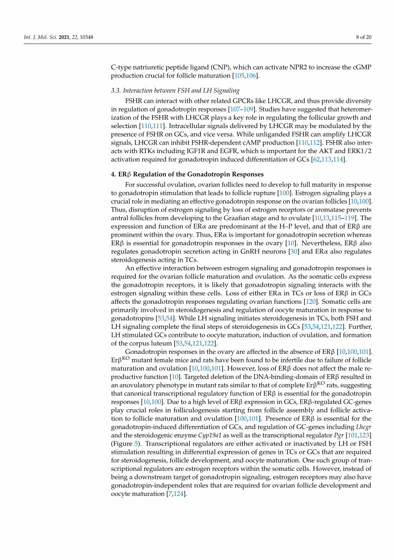
Gonadotropin responses in the ovary are affected in the absence of ERβ [10,100,101].ErβKO mutant female mice and rats have been found to be infertile due to failure of folliclematuration and ovulation [10,100,101]. However, loss of ERβ does not affect the male re-productive function [10]. Targeted deletion of the DNA-binding-domain of ERβ resulted inan anovulatory phenotype in mutant rats similar to that of complete ErβKO rats, suggestingthat canonical transcriptional regulatory function of ERβ is essential for the gonadotropinresponses [10,100]. Due to a high level of ERβ expression in GCs, ERβ-regulated GC-genesplay crucial roles in folliculogenesis starting from follicle assembly and follicle activa-tion to follicle maturation and ovulation [100,101]. Presence of ERβ is essential for thegonadotropin-induced differentiation of GCs, and regulation of GC-genes including Lhcgrand the steroidogenic enzyme Cyp19a1 as well as the transcriptional regulator Pgr [101,123](Figure 5). Transcriptional regulators are either activated or inactivated by LH or FSHstimulation resulting in differential expression of genes in TCs or GCs that are requiredfor steroidogenesis, follicle development, and oocyte maturation. One such group of tran-scriptional regulators are estrogen receptors within the somatic cells. However, instead ofbeing a downstream target of gonadotropin signaling, estrogen receptors may also havegonadotropin-independent roles that are required for ovarian follicle development andoocyte maturation [7,124].

Int. J. Mol. Sci. 2021, 22, 10348 9 of 20Int. J. Mol. Sci. 2021, 22, x FOR PEER REVIEW 10 of 20
Figure 5. ERβ regulation of gonadotropin responses. ERβ is the predominant estrogen receptor in the ovary involved in
transcriptional regulation of gene expression. While ERα is expressed in theca cells (TCs), ERβ is expressed in granulosa
cells (GCs). As GCs express both FSHR and LHCGR, we analyzed the role of ERβ in gonadotropin‐induced gene expres‐
sion in GCs. We identified that a subset of PMSG (that activates FSHR) or hCG (that activates LHCGR) regulated genes
failed to respond in the absence of ERβ expression in GCs. In early antral follicles, expression of FSHR‐induced genes
including Cyp19A1, Cyp11a1, Lhcgr, Gata4, Npr2, Jaml, Galnt6, Znf750, and Dusp9 was dependent on ERβ. Moreover, pres‐
ence of ERβ was found to be essential for the expression of LHCGR‐induced genes, such as Egfr, Kiss1, Ptgs2, Adamts1,
Wnt16, Mageb16, Pgr, Runx2, and Jaml. Disruption of ERβ signaling results in dysregulation of these genes and is associated
with failure of follicle maturation, and ovulation. As ovulation does not occur in the absence of ERβ, the potential role of
ERβ in luteinization has not been studied.
4.1. ERβ Regulation of FSH Responses
FSH signaling stimulates early antral follicles to develop to the antral stage [2]. It has
been shown that FSH stimulation of small antral follicles alone is insufficient for induction
of maturation, which must be facilitated by estrogen signaling [126]. Loss of ERβ does not
impact the development of ovarian follicles prior to the antral stage [101]. However, fail‐
ure of ErβKO follicles to mature following LH stimulation suggests that those follicles may
not possess the factors required for a proper LH response [101]. Gene expression analyses
48h after PMSG stimulation (PMSG acts on rodent FSHR) revealed that many of the genes
that are differentially expressed in wildtype ovaries fail to do so in the absence of ERβ
[10,100,101].
Most studies suggest a primary role for ERβ in the GCs as being essential for FSH
induced ovarian follicle development. Differentiation of GCs in response to FSH is de‐
pendent on ERβ‐mediated estrogen signaling [127]. Despite an increased expression of
FSHR, administration of PMSG fails to induce the genes required for an effective LH re‐
sponse [101,128]. Although there was no change in FSH‐induced genes such as Star, ex‐
pression of Lhcgr, Cyp11a1, Cyp19a1, Gata4, and Npr2 failed to upregulate in ErβKO GCs
[10,100,101,129] (Figure 5). These findings suggest that expression of a subset of FSH‐in‐
duced genes is dependent on the presence of ERβ in GCs [101].
In the absence of ERβ, FSH‐induced cAMP production is markedly reduced in GCs
[79]. However, the molecular mechanism underlying such reduced cAMP production in
GCs remains unclear [79]. In vitro and in vivo studies have also demonstrated defective
Figure 5. ERβ regulation of gonadotropin responses. ERβ is the predominant estrogen receptor in the ovary involved intranscriptional regulation of gene expression. While ERα is expressed in theca cells (TCs), ERβ is expressed in granulosacells (GCs). As GCs express both FSHR and LHCGR, we analyzed the role of ERβ in gonadotropin-induced gene expressionin GCs. We identified that a subset of PMSG (that activates FSHR) or hCG (that activates LHCGR) regulated genes failed torespond in the absence of ERβ expression in GCs. In early antral follicles, expression of FSHR-induced genes includingCyp19A1, Cyp11a1, Lhcgr, Gata4, Npr2, Jaml, Galnt6, Znf750, and Dusp9 was dependent on ERβ. Moreover, presence ofERβ was found to be essential for the expression of LHCGR-induced genes, such as Egfr, Kiss1, Ptgs2, Adamts1, Wnt16,Mageb16, Pgr, Runx2, and Jaml. Disruption of ERβ signaling results in dysregulation of these genes and is associated withfailure of follicle maturation, and ovulation. As ovulation does not occur in the absence of ERβ, the potential role of ERβ inluteinization has not been studied.
ERβ is a ligand-activated transcription factor. However, loss of ERβ disrupts thefinal stages of follicle development and oocyte maturation, when gene transcription isminimal in oocytes. Studies have shown that ERβ can induce the expression of miR-NAs [125] and it can directly interact with AGO2 [125]. Thus, ERβ can also be involvedin posttranscriptional regulation of gene expression. Nevertheless, most of the studiessuggesting a post-transcriptional regulatory function of ERβ refer to cancer cells, and itremains unknown whether such mechanisms also occur in normal ovarian follicles.
4.1. ERβ Regulation of FSH Responses
FSH signaling stimulates early antral follicles to develop to the antral stage [2]. It hasbeen shown that FSH stimulation of small antral follicles alone is insufficient for inductionof maturation, which must be facilitated by estrogen signaling [126]. Loss of ERβ doesnot impact the development of ovarian follicles prior to the antral stage [101]. However,failure of ErβKO follicles to mature following LH stimulation suggests that those folliclesmay not possess the factors required for a proper LH response [101]. Gene expressionanalyses 48h after PMSG stimulation (PMSG acts on rodent FSHR) revealed that many ofthe genes that are differentially expressed in wildtype ovaries fail to do so in the absence ofERβ [10,100,101].

Int. J. Mol. Sci. 2021, 22, 10348 10 of 20
Most studies suggest a primary role for ERβ in the GCs as being essential for FSHinduced ovarian follicle development. Differentiation of GCs in response to FSH is de-pendent on ERβ-mediated estrogen signaling [127]. Despite an increased expression ofFSHR, administration of PMSG fails to induce the genes required for an effective LHresponse [101,128]. Although there was no change in FSH-induced genes such as Star,expression of Lhcgr, Cyp11a1, Cyp19a1, Gata4, and Npr2 failed to upregulate in ErβKO
GCs [10,100,101,129] (Figure 5). These findings suggest that expression of a subset ofFSH-induced genes is dependent on the presence of ERβ in GCs [101].
In the absence of ERβ, FSH-induced cAMP production is markedly reduced inGCs [79]. However, the molecular mechanism underlying such reduced cAMP produc-tion in GCs remains unclear [79]. In vitro and in vivo studies have also demonstrateddefective antrum formation, associated with decreased cumulus expansion after FSH treat-ment [120,129,130]. Due to the reduced levels of Cyp11a1 and Cyp19a1, GCs in ErβKO
preovulatory follicles exhibit significantly lower levels of FSH-induced estrogen synthe-sis [120,130]. A decreased level of Cyp19a1 can interrupt the development of antral folliclesto the Graafian stage. Similar to ErβKO mice, Cyp19a1 knockout mice are able to developantral follicles but failed to mature or ovulate [118]. ErβKO GCs also have a reduced levelof Gata-4 expression, which decreases the proliferation of GCs and that impairs folliclematuration [131,132]. In contrast, in vitro culture experiments with ErαKO models detecteda minimal role for ERα in the differentiation of GCs and their gene regulation [120,130].
4.2. ERβ Regulation of LH Responses
ERβ plays a very important role in the LH-induced differentiation of GCs required forfollicle maturation and induction of ovulation [130]. A reduced level of Lhcgr expressionin ErβKO GCs in response to FSH causes failure of those antral follicles to respond to LH,which is essential for their development to Graafian follicles [6,133]. Expression of LHtarget genes that regulate steroidogenesis, cumulus cell expansion, oocyte maturation,and ovulation, were markedly impaired in ErβKO ovaries due to the failure of Lhcgrupregulation in ErβKO GCs [14,101]. It is important to note that Lhcgr knockout micealso suffered from lack of follicle development beyond the antral stage and failed to formGraafian follicles [91]. We recently reported a similar ovarian phenotype in gonadotropin-induced ErβKO rats [101].
Our recent study revealed that a subset of LH-induced genes in GCs is also depen-dent on the presence of ERβ [100,101]. We observed that hCG-stimulation (hCG activatesLHCGR) failed to upregulate the expression of Pgr, Runx2, Egfr, Ptgs2, Adamts1, and Kiss1in ErβKO GCs [101] (Figure 5). Pgr, Runx2, Ptgs2, and Adamts1 were also found to bedownregulated in GCs isolated from hCG treated ErβKO mice [123]. We previously demon-strated that loss of ERβ results in failure of LH-induced Kiss1 gene expression in ErβKO
rat GCs [100,101]. Our recent findings suggest that ERβ-regulated ovarian kisspeptin mayplay an important role in preovulatory maturation of oocytes [129]. However, it remainsunknown if ovarian kisspeptin has any role in regulating GnRH neurons. In addition tothe known LH-regulated genes, we identified that loss of ERβ also alters the expression ofseveral novel GC-genes including Jaml, Galnt6, Znf750, and Dusp9 [101]. Differential ex-pression of these LH-regulated genes in GCs may be responsible for the lack of maturation,and ovulation of ErβKO ovarian follicles [101].
LH signaling also plays an important role in TCs, however, the major estrogen receptorin TCs is ERα. Therefore, it is less likely to be impacted by ERβ. However, development ofthe TC layer, and differentiation of TCs can be affected by the loss of ERβ in GCs or oocytes,because these mechanisms are dependent on GC-derived KL and IGF1 [36–38] and oocytederived GDF9 [39–42]. We have observed that serum androstenedione and progesteronelevels can be lower in Erβ mutant rats [100]. However, studies have not yet analyzed thechanges in the gene expression profile in ErβKO TCs.

Int. J. Mol. Sci. 2021, 22, 10348 11 of 20
5. Chorionic Gonadotropins in Ovarian Biology
Two placenta-derived gonadotropins (chorionic gonadotropins) are commonly used inovarian biology research and in clinical settings. Human chorionic gonadotropin (hCG) isa polypeptide hormone produced by the trophoblast cells of the placenta. Equine chorionicgonadotropin (eCG), also known as pregnant mare serum gonadotropin (PMSG), is anothercommonly used placenta-derived gonadotropin hormone. Chorionic gonadotropins arecomposed of two dissimilar subunits of glycoproteins like that of pituitary gonadotropins.The α subunit is common to chorionic and pituitary gonadotropins while the β subunit,which is unique for each specific hormone, is responsible for selective receptor binding.The β subunit of hCG (β-hCG) has an 85% homology with the β subunit of pituitary LH,but in equids, the β subunit of chorionic gonadotropin and pituitary LH are expressedfrom the same gene, differing only by the glycosylation pattern. β-hCG is mostly similar toβ-LH, differing in the carboxy terminal region. β-hCG has a carboxy terminal extensionthat includes four glycosylated serine residues that is responsible for its longer half-life.hCG can bind and activate LHCGR in humans as well as in experimental animals likerodents. Interestingly, PMSG has only LH-like activity in equids, but in other speciesincluding rodents, it has FSH-like activity due to its preferred binding to FSHR. PMSGis also preferred over pituitary extracts of gonadotropins due its longer half-life. hCGprepared from the urine of pregnant women and PMSG purified from pregnant horseserum are used in research, however, recombinant hCG or PMSG have been developedand approved for clinical use.
Physiologically, CGs are important only during pregnancy in humans, primates, andhorses [134,135]. These mammals sustain their initial period of pregnancy by steroidhormones produced by the corpora lutea. Extension of normal corpus luteum life isachieved by placental secretion of chorionic gonadotropins and their binding to andregulation of LHCGRs within the corpus luteum. Subsequently, they experience a lutealto placental shift, and placental steroid production becomes essential for continuing theirpregnancy [134,135]. In contrast, the rodent corpora lutea are responsible for steroidhormone production throughout gestation. Therefore, the rodents do not express CGsin placenta to sustain their pregnancy [134,135]. In animal experiments, exogenous CGS(PMSG and hCG) are administered into mice or rats for synchronized induction of ovarianfollicle development, as well as for the induction of ovulation. PMSG is administered to actlike FSH while hCG is administered to act like LH. hCG can bind the LHCGR and induceresponses like that of LH signaling. Injections of hCG mimic the LH surge that is necessaryfor oocyte maturation and induction of ovulation. hCG is also used in the therapy of femaleinfertility, particularly in assisted reproductive techniques. PMSG is also administeredwith progesterone to induce ovulation in livestock prior to artificial insemination.
Another importance of CG is the potential role of hCG in cancer progression dueto its proangiogenic properties [136]. Ovarian cancer cells express hCG and its receptorLHCGR [137]. Such aberrant expression of hCG can be used as a tumor marker in non-pregnant females [138,139]. It has been shown that hCG stimulates angiogenesis in theovary by inducing the expression of VEGF and increasing the proliferation of vascularendothelial cells [137,140]. However, there has been no correlation between hCG expres-sion and the survival of ovarian cancer patients [141]. An interesting aspect of LHCGRexpression outside the H–P–O axis is the association and sensitivity of the expression sitewith estrogen signaling [137,140]. Tissues that express LHCGR also respond to changes inestrogen levels [142], which suggest that either estrogen can modulate the expression ofLHCGR or estrogen signaling interacts with LH signaling. Thus, cancer cells that expressLHCGR may also express ERα and ERβ and respond to estrogen signaling. However,further studies are required to clarify that.
6. ERβ and Gonadotropins in Ovarian Diseases
In contrast, hCG acts on increasing the growth and angiogenesis of ovarian cancersas mentioned above. However, it remains unclear how gonadotropin signaling and ERβ

Int. J. Mol. Sci. 2021, 22, 10348 12 of 20
signaling interact in ovarian cancer cells. ERβ is the predominant estrogen receptor inthe ovary [143–146]. ERβ polymorphisms and mutations in women have been linked toovulatory dysfunctions, including complete ovarian failure [147–150]. PCOS, a commonclinical condition among women that causes failure of ovulation and infertility, is associatedwith high levels of LH and androgens [151,152]. Recent genomewide association studieshave linked FSH and LH receptor variants to the development of PCOS [153]. Due to theintricate connection between gonadotropin response and estrogen signaling in the ovary, itis likely that estrogen signaling plays an important role in the pathogenesis of PCOS. Theloss of ERα induces polycystic like changes in mutant mouse [154] and rat [115] ovaries. Butthere are no such cystic changes in the ErβKO mouse [12,13] or rat [10] ovaries. Rather, thepresence of ERβ was found essential for the development of polycystic changes in ErαKO
mice [146]. Based on these findings, it may be assumed that loss of ERα in TCs associatedwith a normal or increased ERβ activity in GCs may lead to the development of PCOS.However, studies on human PCOS tissues only partially support the assumption [155–158].Another ovarian disease that has been linked to estrogen signaling is ovarian cancer [159].Estrogen receptors are also frequently detected in ovarian cancers, however the exact roleof estrogen receptors in ovarian cancer prognosis remains unclear [159–163]. ERβ acts as atumor suppressor and inhibits the progression of ovarian cancers [164,165]. As expected,expression of ERβ is very low in advanced ovarian cancers [166,167] and loss of ERβexpression in ovarian cancers correlates with a shorter survival rate [168,169]. In contrast,hCG acts on increasing the growth and angiogenesis of ovarian cancers as mentioned above.However, it remains unclear how gonadotropin signaling and ERβ signaling interact inovarian cancer cells.
7. Future Perspectives
Estrogen signaling is essential for mediating effective gonadotropin responses withinthe ovary. Gonadotropin receptors are expressed in TCs and GCs. The presence of ERαin TCs, and ERβ in GCs are essential for gonadotropin induced steroidogenesis andgametogenesis. However, it remains unclear how gonadotropin signaling interacts withestrogen signaling, and the hierarchy in these signaling mechanisms in those somaticcells. It has been demonstrated that FSH induced Lhcgr expression in GCs depends onthe presence of ERβ [100,101]. As loss of ERβ reduces estrogen synthesis in GCs, it maybe hypothesized that ERβ-dependent estrogen signaling positively regulates Lhcgr geneexpression in GCs. In contrast, the expression of Fshr is increased in the absence of ERβ inthe ovary [100,101], which suggest that ERβ may negatively regulate Fshr expression inGCs. Nevertheless, the molecular mechanisms underlying ERβ regulation of gonadotropinreceptors in GCs remain unknown.
ERβ is the predominant estrogen receptor in the ovary, where it functions to regulateexpression of genes involved in follicle development and oocyte maturation [120,170–172].GCs in growing ovarian follicles express the highest level of ERβ. However, in vitro studieson GCs are limited by spontaneous differentiation of GCs in culture. Moreover, GCs rapidlylose the expression of ERβ in cell culture. Thus, the results obtained from in vitro studies ofGCs may differ from the exact molecular mechanisms that exist in vivo. Another limitationin ERβ research is the lack of a specific antibody [173]. Although a mouse monoclonalantibody has been reported to be efficient in detecting human ERβ, it fails to detect ERβ inthe rodents [173].
Our studies have shown that ERβ plays a major role in regulating the GC-genesthat are important for oocyte maturation and induction of ovulation [10,100,101,129]. Ad-ministration of gonadotropins for ovarian stimulation is a common practice in assistedreproductive technologies [174,175]. Some of the patients that receive gonadotropins donot respond well and are investigated for predisposing conditions underlying the defec-tive gonadotropin responses [174]. A more directed focus on ERβ may help identify theunderlying pathologies and lead to an effective treatment to overcome ineffective follicledevelopment and oocyte maturation following gonadotropin stimulation.

Int. J. Mol. Sci. 2021, 22, 10348 13 of 20
Author Contributions: M.A.K.R. planned the manuscript. E.B.L. and V.P.C. contributed to prepara-tion of the manuscript and making the illustrations. M.W.W. and M.A.K.R. edited the manuscriptand submitted for publication. All authors have read and agreed with the contents of the manuscript.All authors have read and agreed to the published version of the manuscript.
Funding: This work was supported by funding from the KUMC SOM, COBRE (P30 GM122731), andK-INBRE (P20 GM103418).
Institutional Review Board Statement: Not applicable.
Informed Consent Statement: Not applicable.
Data Availability Statement: SRA PRJNA551764 and PRJNA551766.
Acknowledgments: Brandi Miller, Department of Pathology and Laboratory Medicine, KUMC, andShari Standiferd, Molecular and Integrative Physiology, KUMC, for their continued support andadministrative assistance.
Conflicts of Interest: The authors declare no conflict of interest.
References1. Orisaka, M.; Miyazaki, Y.; Shirafuji, A.; Tamamura, C.; Tsuyoshi, H.; Tsang, B.K.; Yoshida, Y. The role of pituitary gonadotropins
and intraovarian regulators in follicle development: A mini-review. Reprod. Med. Biol. 2021, 20, 169–175. [CrossRef] [PubMed]2. McGee, E.A.; Hsueh, A.J. Initial and cyclic recruitment of ovarian follicles. Endocr. Rev. 2000, 21, 200–214. [CrossRef] [PubMed]3. Yazawa, T.; Imamichi, Y.; Sekiguchi, T.; Miyamoto, K.; Uwada, J.; Khan, M.R.I.; Suzuki, N.; Umezawa, A.; Taniguchi, T.
Transcriptional regulation of ovarian steroidogenic genes: Recent findings obtained from stem cell-derived steroidogenic cells.BioMed Res. Int. 2019, 2019, 8973076. [CrossRef] [PubMed]
4. Eppig, J.J. Oocyte control of ovarian follicular development and function in mammals. Reproduction 2001, 122, 829–838. [CrossRef][PubMed]
5. Jones, A.S.; Shikanov, A. Follicle development as an orchestrated signaling network in a 3D organoid. J. Biol. Eng. 2019, 13, 2.[CrossRef]
6. Richards, J.S.; Ascoli, M. Endocrine, paracrine, and autocrine signaling pathways that regulate ovulation. Trends Endocrinol.Metab. 2018, 29, 313–325. [CrossRef]
7. Richards, J.S. Maturation of ovarian follicles: Actions and interactions of pituitary and ovarian hormones on follicular celldifferentiation. Physiol. Rev. 1980, 60, 51–89. [CrossRef]
8. Jefferson, W.N.; Couse, J.F.; Banks, E.P.; Korach, K.S.; Newbold, R.R. Expression of estrogen receptor beta is developmentallyregulated in reproductive tissues of male and female mice. Biol. Reprod. 2000, 62, 310–317. [CrossRef]
9. Drummond, A.E.; Fuller, P.J. The importance of ERbeta signalling in the ovary. J. Endocrinol. 2010, 205, 15–23. [CrossRef]10. Rumi, M.A.K.; Singh, P.; Roby, K.F.; Zhao, X.; Iqbal, K.; Ratri, A.; Lei, T.; Cui, W.; Borosha, S.; Dhakal, P.; et al. Defining the role of
estrogen receptor beta in the regulation of female fertility. Endocrinology 2017, 158, 2330–2343. [CrossRef]11. Antal, M.C.; Krust, A.; Chambon, P.; Mark, M. Sterility and absence of histopathological defects in nonreproductive organs of a
mouse ERbeta-null mutant. Proc. Natl. Acad. Sci. USA 2008, 105, 2433–2438. [CrossRef] [PubMed]12. Maneix, L.; Antonson, P.; Humire, P.; Rochel-Maia, S.; Castaneda, J.; Omoto, Y.; Kim, H.J.; Warner, M.; Gustafsson, J.A. Estrogen
receptor beta exon 3-deleted mouse: The importance of non-ERE pathways in ERbeta signaling. Proc. Natl. Acad. Sci. USA 2015,112, 5135–5140. [CrossRef] [PubMed]
13. Dupont, S.; Krust, A.; Gansmuller, A.; Dierich, A.; Chambon, P.; Mark, M. Effect of single and compound knockouts of estrogenreceptors alpha (ERalpha) and beta (ERbeta) on mouse reproductive phenotypes. Development 2000, 127, 4277–4291. [CrossRef][PubMed]
14. Jayes, F.L.; Burns, K.A.; Rodriguez, K.F.; Kissling, G.E.; Korach, K.S. The naturally occurring luteinizing hormone surge isdiminished in mice lacking estrogen receptor Beta in the ovary. Biol. Reprod. 2014, 90, 24. [CrossRef]
15. Novaira, H.J.; Sonko, M.L.; Hoffman, G.; Koo, Y.; Ko, C.; Wolfe, A.; Radovick, S. Disrupted kisspeptin signaling in GnRH neuronsleads to hypogonadotrophic hypogonadism. Mol. Endocrinol. 2014, 28, 225–238. [CrossRef]
16. Cheong, R.Y.; Porteous, R.; Chambon, P.; Abrahám, I.; Herbison, A.E. Effects of neuron-specific estrogen receptor (ER) α and ERβdeletion on the acute estrogen negative feedback mechanism in adult female mice. Endocrinology 2014, 155, 1418–1427. [CrossRef]
17. Handa, R.J.; Mani, S.K.; Uht, R.M. Estrogen receptors and the regulation of neural stress responses. Neuroendocrinology 2012, 96,111–118. [CrossRef]
18. McEwen, B.S.; Akama, K.T.; Spencer-Segal, J.L.; Milner, T.A.; Waters, E.M. Estrogen effects on the brain: Actions beyond thehypothalamus via novel mechanisms. Behav. Neurosci. 2012, 126, 4–16. [CrossRef]
19. Moenter, S.M.; Chu, Z.; Christian, C.A. Neurobiological mechanisms underlying oestradiol negative and positive feedbackregulation of gonadotrophin-releasing hormone neurones. J. Neuroendocrinol. 2009, 21, 327–333. [CrossRef]
20. Radovick, S.; Levine, J.E.; Wolfe, A. Estrogenic regulation of the GnRH neuron. Front. Endocrinol. 2012, 3, 52. [CrossRef]

Int. J. Mol. Sci. 2021, 22, 10348 14 of 20
21. Wintermantel, T.M.; Campbell, R.E.; Porteous, R.; Bock, D.; Gröne, H.J.; Todman, M.G.; Korach, K.S.; Greiner, E.; Pérez, C.A.;Schütz, G.; et al. Definition of estrogen receptor pathway critical for estrogen positive feedback to gonadotropin-releasinghormone neurons and fertility. Neuron 2006, 52, 271–280. [CrossRef]
22. Harter, C.J.L.; Kavanagh, G.S.; Smith, J.T. The role of kisspeptin neurons in reproduction and metabolism. J. Endocrinol. 2018, 238,R173–R183. [CrossRef]
23. Rønnekleiv, O.K.; Qiu, J.; Kelly, M.J. Arcuate kisspeptin neurons coordinate reproductive activities with metabolism. Semin.Reprod. Med. 2019, 37, 131–140. [CrossRef]
24. Khan, A.R.; Kauffman, A.S. The role of kisspeptin and RFamide-related peptide-3 neurones in the circadian-timed preovulatoryluteinising hormone surge. J. Neuroendocrinol. 2012, 24, 131–143. [CrossRef] [PubMed]
25. Tolson, K.P.; Chappell, P.E. The changes they are A-timed: Metabolism, endogenous clocks, and the timing of puberty. Front.Endocrinol. 2012, 3, 45. [CrossRef] [PubMed]
26. Fink, G.; Knobil, E.; Neill, J. Gonadotropin Secretion and Its Control; Raven Press: Isle of Skye, UK, 1988; pp. 1349–1377.27. Couse, J.F.; Bunch, D.O.; Lindzey, J.; Schomberg, D.W.; Korach, K.S. Prevention of the polycystic ovarian phenotype and
characterization of ovulatory capacity in the estrogen receptor-alpha knockout mouse. Endocrinology 1999, 140, 5855–5865.[CrossRef] [PubMed]
28. Couse, J.F.; Yates, M.M.; Rodriguez, K.F.; Johnson, J.A.; Poirier, D.; Korach, K.S. The intraovarian actions of estrogen receptor-alphaare necessary to repress the formation of morphological and functional Leydig-like cells in the female gonad. Endocrinology 2006,147, 3666–3678. [CrossRef]
29. Taniguchi, F.; Couse, J.F.; Rodriguez, K.F.; Emmen, J.M.; Poirier, D.; Korach, K.S. Estrogen receptor-alpha mediates an intrao-varian negative feedback loop on thecal cell steroidogenesis via modulation of Cyp17a1 (cytochrome P450, steroid 17alpha-hydroxylase/17,20 lyase) expression. FASEB J. 2007, 21, 586–595. [CrossRef] [PubMed]
30. Novaira, H.J.; Negron, A.L.; Graceli, J.B.; Capellino, S.; Schoeffield, A.; Hoffman, G.E.; Levine, J.E.; Wolfe, A.; Wondisford, F.E.;Radovick, S. Impairments in the reproductive axis of female mice lacking estrogen receptor beta in GnRH neurons. Am. J. Physiol.Endocrinol. Metab. 2018, 315, E1019–E1033. [CrossRef]
31. Gieske, M.C.; Kim, H.J.; Legan, S.J.; Koo, Y.; Krust, A.; Chambon, P.; Ko, C. Pituitary gonadotroph estrogen receptor-alpha isnecessary for fertility in females. Endocrinology 2008, 149, 20–27. [CrossRef]
32. Sánchez-Criado, J.E.; Trudgen, K.; Millán, Y.; Blanco, A.; Monterde, J.; Garrido-Gracia, J.C.; Gordon, A.; Aguilar, R.; de Las Mulas,J.M.; Ko, C. Estrogen receptor (ESR) 2 partially offsets the absence of ESR1 in gonadotropes of pituitary-specific Esr1 knockoutfemale mice. Reproduction 2012, 143, 549–558. [CrossRef]
33. Lapatto, R.; Pallais, J.C.; Zhang, D.; Chan, Y.M.; Mahan, A.; Cerrato, F.; Le, W.W.; Hoffman, G.E.; Seminara, S.B. Kiss1-/- miceexhibit more variable hypogonadism than Gpr54-/- mice. Endocrinology 2007, 148, 4927–4936. [CrossRef]
34. Kumar, T.R.; Wang, Y.; Lu, N.; Matzuk, M.M. Follicle stimulating hormone is required for ovarian follicle maturation but not malefertility. Nat. Genet. 1997, 15, 201–204. [CrossRef]
35. Cattanach, B.M.; Iddon, C.A.; Charlton, H.M.; Chiappa, S.A.; Fink, G. Gonadotrophin-releasing hormone deficiency in a mutantmouse with hypogonadism. Nature 1977, 269, 338–340. [CrossRef]
36. Huang, C.T.; Weitsman, S.R.; Dykes, B.N.; Magoffin, D.A. Stem cell factor and insulin-like growth factor-I stimulate luteinizinghormone-independent differentiation of rat ovarian theca cells. Biol. Reprod. 2001, 64, 451–456. [CrossRef]
37. Orisaka, M.; Tajima, K.; Mizutani, T.; Miyamoto, K.; Tsang, B.K.; Fukuda, S.; Yoshida, Y.; Kotsuji, F. Granulosa cells promotedifferentiation of cortical stromal cells into theca cells in the bovine ovary. Biol. Reprod. 2006, 75, 734–740. [CrossRef] [PubMed]
38. Honda, A.; Hirose, M.; Hara, K.; Matoba, S.; Inoue, K.; Miki, H.; Hiura, H.; Kanatsu-Shinohara, M.; Kanai, Y.; Kono, T.; et al.Isolation, characterization, and in vitro and in vivo differentiation of putative thecal stem cells. Proc. Natl. Acad. Sci. USA 2007,104, 12389–12394. [CrossRef] [PubMed]
39. Elvin, J.A.; Yan, C.; Wang, P.; Nishimori, K.; Matzuk, M.M. Molecular characterization of the follicle defects in the growthdifferentiation factor 9-deficient ovary. Mol. Endocrinol. 1999, 13, 1018–1034. [CrossRef] [PubMed]
40. Wu, X.; Chen, L.; Brown, C.A.; Yan, C.; Matzuk, M.M. Interrelationship of growth differentiation factor 9 and inhibin in earlyfolliculogenesis and ovarian tumorigenesis in mice. Mol. Endocrinol. 2004, 18, 1509–1519. [CrossRef]
41. Solovyeva, E.V.; Hayashi, M.; Margi, K.; Barkats, C.; Klein, C.; Amsterdam, A.; Hsueh, A.J.; Tsafriri, A. Growth differentiationfactor-9 stimulates rat theca-interstitial cell androgen biosynthesis. Biol. Reprod. 2000, 63, 1214–1218. [CrossRef]
42. Orisaka, M.; Jiang, J.Y.; Orisaka, S.; Kotsuji, F.; Tsang, B.K. Growth differentiation factor 9 promotes rat preantral follicle growthby up-regulating follicular androgen biosynthesis. Endocrinology 2009, 150, 2740–2748. [CrossRef]
43. Zhou, J.; Kumar, T.R.; Matzuk, M.M.; Bondy, C. Insulin-like growth factor I regulates gonadotropin responsiveness in the murineovary. Mol. Endocrinol. 1997, 11, 1924–1933. [CrossRef] [PubMed]
44. Jeppesen, J.V.; Kristensen, S.G.; Nielsen, M.E.; Humaidan, P.; Dal Canto, M.; Fadini, R.; Schmidt, K.T.; Ernst, E.; Yding Andersen,C. LH-receptor gene expression in human granulosa and cumulus cells from antral and preovulatory follicles. J. Clin. Endocrinol.Metab. 2012, 97, E1524–E1531. [CrossRef] [PubMed]
45. Hirakawa, T.; Minegishi, T.; Abe, K.; Kishi, H.; Ibuki, Y.; Miyamoto, K. A role of insulin-like growth factor I in luteinizing hormonereceptor expression in granulosa cells. Endocrinology 1999, 140, 4965–4971. [CrossRef] [PubMed]

Int. J. Mol. Sci. 2021, 22, 10348 15 of 20
46. Ikeda, S.; Nakamura, K.; Kogure, K.; Omori, Y.; Yamashita, S.; Kubota, K.; Mizutani, T.; Miyamoto, K.; Minegishi, T. Effectof estrogen on the expression of luteinizing hormone-human chorionic gonadotropin receptor messenger ribonucleic acid incultured rat granulosa cells. Endocrinology 2008, 149, 1524–1533. [CrossRef] [PubMed]
47. Imai, F.; Kishi, H.; Nakao, K.; Nishimura, T.; Minegishi, T. IL-6 up-regulates the expression of rat LH receptors during granulosacell differentiation. Endocrinology 2014, 155, 1436–1444. [CrossRef] [PubMed]
48. Kishi, H.; Kitahara, Y.; Imai, F.; Nakao, K.; Suwa, H. Expression of the gonadotropin receptors during follicular development.Reprod. Med. Biol. 2018, 17, 11–19. [CrossRef]
49. Fortune, J.; Armstrong, D. Androgen production by theca and granulosa isolated from proestrous rat follicles. Endocrinology 1977,100, 1341–1347. [CrossRef]
50. ERICKSON, G.F.; Hsueh, A. Stimulation of aromatase activity by follicle stimulating hormone in rat granulosa cells in vivo andin vitro. Endocrinology 1978, 102, 1275–1282. [CrossRef]
51. Liu, Y.-X.; Hsueh, A.J. Synergism between granulosa and theca-interstitial cells in estrogen biosynthesis by gonadotropin-treatedrat ovaries: Studies on the two-cell, two-gonadotropin hypothesis using steroid antisera. Biol. Reprod. 1986, 35, 27–36. [CrossRef]
52. Jiang, X.; Fischer, D.; Chen, X.; McKenna, S.D.; Liu, H.; Sriraman, V.; Yu, H.N.; Goutopoulos, A.; Arkinstall, S.; He, X. Evidence forfollicle-stimulating hormone receptor as a functional trimer. J. Biol. Chem. 2014, 289, 14273–14282. [CrossRef]
53. Gromoll, J.; Simoni, M.; Nordhoff, V.; Behre, H.M.; De Geyter, C.; Nieschlag, E. Functional and clinical consequences of mutationsin the FSH receptor. Mol. Cell. Endocrinol. 1996, 125, 177–182. [CrossRef]
54. Moudgal, N. Gonadotropins and Gonadal Function; Elsevier: Amsterdam, The Netherlands, 2012.55. Casarini, L.; Crépieux, P. Molecular mechanisms of action of FSH. Front. Endocrinol. 2019, 10. [CrossRef] [PubMed]56. Dierich, A.; Sairam, M.R.; Monaco, L.; Fimia, G.M.; Gansmuller, A.; LeMeur, M.; Sassone-Corsi, P. Impairing follicle-stimulating
hormone (FSH) signaling in vivo: Targeted disruption of the FSH receptor leads to aberrant gametogenesis and hormonalimbalance. Proc. Natl. Acad. Sci. USA 1998, 95, 13612–13617. [CrossRef] [PubMed]
57. De Pascali, F.; Tréfier, A.; Landomiel, F.; Bozon, V.; Bruneau, G.; Yvinec, R.; Poupon, A.; Crépieux, P.; Reiter, E. Follicle-stimulatinghormone receptor: Advances and remaining challenges. Int. Rev. Cell Mol. Biol. 2018, 338, 1–58.
58. Troispoux, C.; Guillou, F.; Elalouf, J.-M.; Firsov, D.; Iacovelli, L.; De Blasi, A.; Combarnous, Y.; Reiter, E. Involvement of Gprotein-coupled receptor kinases and arrestins in desensitization to follicle-stimulating hormone action. Mol. Endocrinol. 1999, 13,1599–1614. [CrossRef]
59. Krishnamurthy, H.; Galet, C.; Ascoli, M. The association of arrestin-3 with the follitropin receptor depends on receptor activationand phosphorylation. Mol. Cell. Endocrinol. 2003, 204, 127–140. [CrossRef]
60. Crépieux, P.; Poupon, A.; Langonné-Gallay, N.; Reiter, E.; Delgado, J.; Schaefer, M.H.; Bourquard, T.; Serrano, L.; Kiel, C.A comprehensive view of the β-arrestinome. Front. Endocrinol. 2017, 8, 32. [CrossRef]
61. De Pascali, F.; Reiter, E. β-arrestins and biased signaling in gonadotropin receptors. Minerva Ginecol. 2018, 70, 525–538. [CrossRef]62. Wayne, C. FSH-induces multiple signaling cascades: Evidence that activation of SRC, RAS and the EGF receptor are critical for
granulosa cell differentiation. Mol. Endocrinol. 2007, 21, 1940–1957. [CrossRef] [PubMed]63. Thomas, R.M.; Nechamen, C.A.; Mazurkiewicz, J.E.; Ulloa-Aguirre, A.; Dias, J.A. The adapter protein APPL1 links FSH receptor
to inositol 1, 4, 5-trisphosphate production and is implicated in intracellular Ca2+ mobilization. Endocrinology 2011, 152, 1691–1701.[CrossRef]
64. Cohen, B.D.; Nechamen, C.A.; Dias, J.A. Human follitropin receptor (FSHR) interacts with the adapter protein 14-3-3τ. Mol. Cell.Endocrinol. 2004, 220, 1–7. [CrossRef] [PubMed]
65. Dias, J.A.; Mahale, S.D.; Nechamen, C.A.; Davydenko, O.; Thomas, R.M.; Ulloa-Aguirre, A. Emerging roles for the FSH receptoradapter protein APPL1 and overlap of a putative 14-3-3τ interaction domain with a canonical G-protein interaction site. Mol. Cell.Endocrinol. 2010, 329, 17–25. [CrossRef] [PubMed]
66. Gonzalez-Robayna, I.; Falender, A.; Ochsner, S.; Firestone, G.; Richards, J. FSH stimulates phosphorylation and activation ofprotein kinase B (PKB/Akt) and serum and glucocorticoid-induced kinase (Sgk): Evidence for A-kinase independent signaling ingranulosa cells. Mol. Endocrinol. 2000, 14, 1283–1300. [CrossRef] [PubMed]
67. Nechamen, C.A.; Thomas, R.M.; Cohen, B.D.; Acevedo, G.; Poulikakos, P.I.; Testa, J.R.; Dias, J.A. Human follicle-stimulatinghormone (FSH) receptor interacts with the adaptor protein APPL1 in HEK 293 cells: Potential involvement of the PI3K pathwayin FSH signaling. Biol. Reprod. 2004, 71, 629–636. [CrossRef] [PubMed]
68. Fan, H.-Y.; Liu, Z.; Cahill, N.; Richards, J.S. Targeted disruption of Pten in ovarian granulosa cells enhances ovulation and extendsthe life span of luteal cells. Mol. Endocrinol. 2008, 22, 2128–2140. [CrossRef]
69. Jean-Alphonse, F.; Bowersox, S.; Chen, S.; Beard, G.; Puthenveedu, M.A.; Hanyaloglu, A.C. Spatially restricted G protein-coupledreceptor activity via divergent endocytic compartments. J. Biol. Chem. 2014, 289, 3960–3977. [CrossRef]
70. Park, Y.; Maizels, E.T.; Feiger, Z.J.; Alam, H.; Peters, C.A.; Woodruff, T.K.; Unterman, T.G.; Lee, E.J.; Jameson, J.L.; Hunzicker-Dunn,M. Induction of cyclin D2 in rat granulosa cells requires FSH-dependent relief from FOXO1 repression coupled with positivesignals from Smad. J. Biol. Chem. 2005, 280, 9135–9148. [CrossRef]
71. Friedmann, S.; Dantes, A.; Amsterdam, A. Ovarian transcriptomes as a tool for a global approach of genes modulated bygonadotropic hormones in human ovarian granulosa cells. Endocrine 2005, 26, 259–265. [CrossRef]

Int. J. Mol. Sci. 2021, 22, 10348 16 of 20
72. Perlman, S.; Bouquin, T.; van den Hazel, B.; Jensen, T.H.; Schambye, H.T.; Knudsen, S.; Okkels, J.S. Transcriptome analysis of FSHand FSH variant stimulation in granulosa cells from IVM patients reveals novel regulated genes. Mol. Hum. Reprod. 2006, 12,135–144. [CrossRef]
73. Herndon, M.K.; Law, N.C.; Donaubauer, E.M.; Kyriss, B.; Hunzicker-Dunn, M. Forkhead box O member FOXO1 regulates themajority of follicle-stimulating hormone responsive genes in ovarian granulosa cells. Mol. Cell. Endocrinol. 2016, 434, 116–126.[CrossRef]
74. Plant, T.M.; Marshall, G.R. The functional significance of FSH in spermatogenesis and the control of its secretion in male primates.Endocr. Rev. 2001, 22, 764–786. [CrossRef] [PubMed]
75. Hillier, S.G. Gonadotropic control of ovarian follicular growth and development. Mol. Cell. Endocrinol. 2001, 179, 39–46. [CrossRef]76. Choi, J.H.; Wong, A.S.; Huang, H.F.; Leung, P.C. Gonadotropins and ovarian cancer. Endocr. Rev. 2007, 28, 440–461. [CrossRef]
[PubMed]77. Amsterdam, A.; Sasson, R.; Keren-Tal, I.; Aharoni, D.; Dantes, A.; Rimon, E.; Land, A.; Cohen, T.; Dor, Y.; Hirsh, L. Alternative
pathways of ovarian apoptosis: Death for life. Biochem. Pharmacol. 2003, 66, 1355–1362. [CrossRef]78. Casarini, L.; Reiter, E.; Simoni, M. β-arrestins regulate gonadotropin receptor-mediated cell proliferation and apoptosis by
controlling different FSHR or LHCGR intracellular signaling in the hGL5 cell line. Mol. Cell. Endocrinol. 2016, 437, 11–21.[CrossRef]
79. Deroo, B.J.; Rodriguez, K.F.; Couse, J.F.; Hamilton, K.J.; Collins, J.B.; Grissom, S.F.; Korach, K.S. Estrogen receptor beta is requiredfor optimal cAMP production in mouse granulosa cells. Mol. Endocrinol. 2009, 23, 955–965. [CrossRef]
80. Hunzicker-Dunn, M.; Maizels, E.T. FSH signaling pathways in immature granulosa cells that regulate target gene expression:Branching out from protein kinase A. Cell Signal 2006, 18, 1351–1359. [CrossRef]
81. Fortune, J.E. Ovarian follicular growth and development in mammals. Biol. Reprod. 1994, 50, 225–232. [CrossRef]82. Ginther, O.J.; Beg, M.A.; Bergfelt, D.R.; Donadeu, F.X.; Kot, K. Follicle selection in monovular species. Biol. Reprod. 2001, 65,
638–647. [CrossRef]83. Beg, M.A.; Ginther, O.J. Follicle selection in cattle and horses: Role of intrafollicular factors. Reproduction 2006, 132, 365–377.
[CrossRef]84. Thierry van Dessel, H.J.; Chandrasekher, Y.; Yap, O.W.; Lee, P.D.; Hintz, R.L.; Faessen, G.H.; Braat, D.D.; Fauser, B.C.; Giudice,
L.C. Serum and follicular fluid levels of insulin-like growth factor I (IGF-I), IGF-II, and IGF-binding protein-1 and -3 during thenormal menstrual cycle. J. Clin. Endocrinol. Metab. 1996, 81, 1224–1231. [CrossRef]
85. Jolly, P.D.; Tisdall, D.J.; Heath, D.A.; Lun, S.; McNatty, K.P. Apoptosis in bovine granulosa cells in relation to steroid synthesis,cyclic adenosine 3′,5′-monophosphate response to follicle-stimulating hormone and luteinizing hormone, and follicular atresia.Biol. Reprod. 1994, 51, 934–944. [CrossRef]
86. Hattori, K.; Orisaka, M.; Fukuda, S.; Tajima, K.; Yamazaki, Y.; Mizutani, T.; Yoshida, Y. Luteinizing hormone facilitates antralfollicular maturation and survival via thecal paracrine signaling in cattle. Endocrinology 2018, 159, 2337–2347. [CrossRef] [PubMed]
87. Eppig, J.J.; Wigglesworth, K.; Pendola, F.; Hirao, Y. Murine oocytes suppress expression of luteinizing hormone receptormessenger ribonucleic acid by granulosa cells. Biol. Reprod. 1997, 56, 976–984. [CrossRef]
88. Elvin, J.A.; Clark, A.T.; Wang, P.; Wolfman, N.M.; Matzuk, M.M. Paracrine actions of growth differentiation factor-9 in themammalian ovary. Mol. Endocrinol. 1999, 13, 1035–1048. [CrossRef]
89. Chang, H.M.; Qiao, J.; Leung, P.C. Oocyte-somatic cell interactions in the human ovary-novel role of bone morphogenetic proteinsand growth differentiation factors. Hum. Reprod. Update 2016, 23, 1–18. [CrossRef] [PubMed]
90. Ma, X.; Dong, Y.; Matzuk, M.M.; Kumar, T.R. Targeted disruption of luteinizing hormone beta-subunit leads to hypogonadism,defects in gonadal steroidogenesis, and infertility. Proc. Natl. Acad. Sci. USA 2004, 101, 17294–17299. [CrossRef] [PubMed]
91. Lei, Z.M.; Mishra, S.; Zou, W.; Xu, B.; Foltz, M.; Li, X.; Rao, C.V. Targeted disruption of luteinizing hormone/human chorionicgonadotropin receptor gene. Mol. Endocrinol. 2001, 15, 184–200. [CrossRef]
92. Zhang, F.P.; Poutanen, M.; Wilbertz, J.; Huhtaniemi, I. Normal prenatal but arrested postnatal sexual development of luteinizinghormone receptor knockout (LuRKO) mice. Mol. Endocrinol. 2001, 15, 172–183. [CrossRef] [PubMed]
93. Hernandez-Gonzalez, I.; Gonzalez-Robayna, I.; Shimada, M.; Wayne, C.M.; Ochsner, S.A.; White, L.; Richards, J.S. Gene expressionprofiles of cumulus cell oocyte complexes during ovulation reveal cumulus cells express neuronal and immune-related genes:Does this expand their role in the ovulation process? Mol. Endocrinol. 2006, 20, 1300–1321. [CrossRef]
94. Breen, S.M.; Andric, N.; Ping, T.; Xie, F.; Offermans, S.; Gossen, J.A.; Ascoli, M. Ovulation involves the luteinizing hormone-dependent activation of G(q/11) in granulosa cells. Mol. Endocrinol. 2013, 27, 1483–1491. [CrossRef]
95. Gudermann, T.; Birnbaumer, M.; Birnbaumer, L. Evidence for dual coupling of the murine luteinizing hormone receptor toadenylyl cyclase and phosphoinositide breakdown and Ca2+ mobilization. Studies with the cloned murine luteinizing hormonereceptor expressed in L cells. J. Biol. Chem. 1992, 267, 4479–4488. [CrossRef]
96. Gudermann, T.; Nichols, C.; Levy, F.O.; Birnbaumer, M.; Birnbaumer, L. Ca2+ mobilization by the LH receptor expressed inXenopus oocytes independent of 3′,5′-cyclic adenosine monophosphate formation: Evidence for parallel activation of twosignaling pathways. Mol. Endocrinol. 1992, 6, 272–278. [CrossRef]
97. Zhu, X.; Gilbert, S.; Birnbaumer, M.; Birnbaumer, L. Dual signaling potential is common among Gs-coupled receptors anddependent on receptor density. Mol. Pharmacol. 1994, 46, 460–469. [PubMed]

Int. J. Mol. Sci. 2021, 22, 10348 17 of 20
98. Shimada, M.; Hernandez-Gonzalez, I.; Gonzalez-Robayna, I.; Richards, J. Paracrine and autocrine regulation of EGF-like factorsin cumulus oocyte complexes (COCs) and granulosa cells: Key roles for prostanglandin synthase 2 (Ptgs2) and progesteronereceptor (Pgr). Mol. Endocrinol. 2006, 20, 1352–1365. [CrossRef] [PubMed]
99. Conti, M.; Hsieh, M.; Park, J.-Y.; Su, Y.-Q. Role of the epidermal growth factor network in ovarian follicles. Mol. Endocrinol. 2006,20, 715–723. [CrossRef]
100. Khristi, V.; Chakravarthi, V.P.; Singh, P.; Ghosh, S.; Pramanik, A.; Ratri, A.; Borosha, S.; Roby, K.F.; Wolfe, M.W.; Rumi, M.A.K.ESR2 regulates granulosa cell genes essential for follicle maturation and ovulation. Mol. Cell. Endocrinol. 2018, 474, 214–226.[CrossRef] [PubMed]
101. Chakravarthi, V.P.; Ratri, A.; Masumi, S.; Borosha, S.; Ghosh, S.; Christenson, L.K.; Roby, K.F.; Wolfe, M.W.; Rumi, M.A.K.Granulosa cell genes that regulate ovarian follicle development beyond the antral stage: The role of estrogen receptor β. Mol. Cell.Endocrinol. 2021, 528, 111212. [CrossRef]
102. Hsieh, M.; Lee, D.; Panigone, S.; Horner, K.; Chen, R.; Theologis, A.; Lee, D.C.; Threadgill, D.W.; Conti, M. Luteinizing hormone-dependent activation of the epidermal growth factor network is essential for ovulation. Mol. Cell. Biol. 2007, 27, 1914–1924.[CrossRef]
103. Fan, H.-Y.; Liu, Z.; Shimada, M.; Sterneck, E.; Johnson, P.F.; Richards, J.S. ERK1/2 in Ovarian Granulosa Cells Are Essential for FemaleFertility; Oxford University Press: Oxford, UK, 2009.
104. Bliss, S.P.; Miller, A.; Navratil, A.M.; Xie, J.; McDonough, S.P.; Fisher, P.J.; Landreth, G.E.; Roberson, M.S. ERK signaling in thepituitary is required for female but not male fertility. Mol. Endocrinol. 2009, 23, 1092–1101. [CrossRef]
105. Zhang, M.; Su, Y.Q.; Sugiura, K.; Xia, G.; Eppig, J.J. Granulosa cell ligand NPPC and its receptor NPR2 maintain meiotic arrest inmouse oocytes. Science 2010, 330, 366–369. [CrossRef]
106. Liu, X.; Xie, F.; Zamah, A.M.; Cao, B.; Conti, M. Multiple pathways mediate luteinizing hormone regulation of cGMP signaling inthe mouse ovarian follicle. Biol. Reprod. 2014, 91, 9. [CrossRef] [PubMed]
107. Jonas, K.C.; Rivero-Müller, A.; Huhtaniemi, I.T.; Hanyaloglu, A.C. G protein-coupled receptor transactivation: From molecules tomice. Methods Cell Biol. 2013, 117, 433–450.
108. Rivero-Müller, A.; Chou, Y.-Y.; Ji, I.; Lajic, S.; Hanyaloglu, A.C.; Jonas, K.; Rahman, N.; Ji, T.H.; Huhtaniemi, I. Rescue of defectiveG protein–coupled receptor function in vivo by intermolecular cooperation. Proc. Natl. Acad. Sci. USA 2010, 107, 2319–2324.[CrossRef] [PubMed]
109. Ulloa-Aguirre, A.; Reiter, E.; Crepieux, P. FSH receptor signaling: Complexity of interactions and signal diversity. Endocrinology2018, 159, 3020–3035. [CrossRef] [PubMed]
110. Feng, X.; Zhang, M.; Guan, R.; Segaloff, D.L. Heterodimerization between the lutropin and follitropin receptors is associated withan attenuation of hormone-dependent signaling. Endocrinology 2013, 154, 3925–3930. [CrossRef]
111. Casarini, L.; Santi, D.; Simoni, M.; Potì, F. ‘Spare’luteinizing hormone receptors: Facts and fiction. Trends Endocrinol. Metab. 2018,29, 208–217. [CrossRef] [PubMed]
112. Jonas, K.C.; Chen, S.; Virta, M.; Mora, J.; Franks, S.; Huhtaniemi, I.; Hanyaloglu, A. Temporal reprogramming of calcium signallingvia crosstalk of gonadotrophin receptors that associate as functionally asymmetric heteromers. Sci. Rep. 2018, 8, 2239. [CrossRef]
113. Baumgarten, S.C.; Convissar, S.M.; Fierro, M.A.; Winston, N.J.; Scoccia, B.; Stocco, C. IGF1R signaling is necessary for FSH-induced activation of AKT and differentiation of human Cumulus granulosa cells. J. Clin. Endocrinol. Metab. 2014, 99, 2995–3004.[CrossRef]
114. Rougny, A.; Gloaguen, P.; Langonné, N.; Reiter, E.; Crepieux, P.; Poupon, A.; Froidevaux, C. A logic-based method to buildsignaling networks and propose experimental plans. Sci. Rep. 2018, 8, 7830. [CrossRef] [PubMed]
115. Rumi, M.A.; Dhakal, P.; Kubota, K.; Chakraborty, D.; Lei, T.; Larson, M.A.; Wolfe, M.W.; Roby, K.F.; Vivian, J.L.; Soares, M.J.Generation of Esr1-knockout rats using zinc finger nuclease-mediated genome editing. Endocrinology 2014, 155, 1991–1999.[CrossRef] [PubMed]
116. Lubahn, D.B.; Moyer, J.S.; Golding, T.S.; Couse, J.F.; Korach, K.S.; Smithies, O. Alteration of reproductive function but not prenatalsexual development after insertional disruption of the mouse estrogen receptor gene. Proc. Natl. Acad. Sci. USA 1993, 90,11162–11166. [CrossRef] [PubMed]
117. Couse, J.F.; Hewitt, S.C.; Bunch, D.O.; Sar, M.; Walker, V.R.; Davis, B.J.; Korach, K.S. Postnatal sex reversal of the ovaries in micelacking estrogen receptors alpha and beta. Science 1999, 286, 2328–2331. [CrossRef]
118. Fisher, C.R.; Graves, K.H.; Parlow, A.F.; Simpson, E.R. Characterization of mice deficient in aromatase (ArKO) because of targeteddisruption of the cyp19 gene. Proc. Natl. Acad. Sci. USA 1998, 95, 6965–6970. [CrossRef]
119. Britt, K.L.; Drummond, A.E.; Cox, V.A.; Dyson, M.; Wreford, N.G.; Jones, M.E.; Simpson, E.R.; Findlay, J.K. An age-related ovarianphenotype in mice with targeted disruption of the Cyp 19 (aromatase) gene. Endocrinology 2000, 141, 2614–2623. [CrossRef]
120. Emmen, J.M.; Couse, J.F.; Elmore, S.A.; Yates, M.M.; Kissling, G.E.; Korach, K.S. In vitro growth and ovulation of follicles fromovaries of estrogen receptor (ER){alpha} and ER{beta} null mice indicate a role for ER{beta} in follicular maturation. Endocrinology2005, 146, 2817–2826. [CrossRef]
121. Greep, R.O.; Van Dyke, H.; CHOW, B.F. Gonadotropins of the swine pituitary I. Various biological effects of purified thylakentrin(FSH) and pure metakentrin (ICSH). Endocrinology 1942, 30, 635–649. [CrossRef]
122. LOSTROH, A.J.; JOHNSON, R.E. Amounts of interstitial cell-stimulating hormone and follicle-stimulating hormone required forfollicular development, uterine growth and ovulation in the hypophysectomized rat. Endocrinology 1966, 79, 991–996. [CrossRef]

Int. J. Mol. Sci. 2021, 22, 10348 18 of 20
123. Binder, A.K.; Rodriguez, K.F.; Hamilton, K.J.; Stockton, P.S.; Reed, C.E.; Korach, K.S. The absence of ER-beta results in altered geneexpression in ovarian granulosa cells isolated from in vivo preovulatory follicles. Endocrinology 2013, 154, 2174–2187. [CrossRef]
124. Shupnik, M. Oestrogen receptors, receptor variants and oestrogen actions in the hypothalamic-pituitary axis. J. Neuroendocrinol.2002, 14, 85–94. [CrossRef]
125. Tarallo, R.; Giurato, G.; Bruno, G.; Ravo, M.; Rizzo, F.; Salvati, A.; Ricciardi, L.; Marchese, G.; Cordella, A.; Rocco, T.; et al. Thenuclear receptor ERβ engages AGO2 in regulation of gene transcription, RNA splicing and RISC loading. Genome Biol. 2017,18, 189. [CrossRef]
126. Richards, J.S. Hormonal control of gene expression in the ovary. Endocr. Rev. 1994, 15, 725–751. [CrossRef]127. Nakano, R.; Nakayama, T.; Iwao, M. Inhibition of ovarian follicle growth by a chemical antiestrogen. Horm. Res. 1982, 16, 230–236.
[CrossRef]128. Gershon, E.; Dekel, N. Newly Identified Regulators of Ovarian Folliculogenesis and Ovulation. Int. J. Mol. Sci. 2020, 21, 4565.
[CrossRef]129. Chakravarthi, V.P.; Ghosh, S.; Housami, S.M.; Wang, H.; Roby, K.F.; Wolfe, M.W.; Kinsey, W.H.; Rumi, M.A.K. ERβ regulated
ovarian kisspeptin plays an important role in oocyte maturation. Mol. Cell. Endocrinol. 2021, 527, 111208. [CrossRef] [PubMed]130. Couse, J.F.; Yates, M.M.; Deroo, B.J.; Korach, K.S. Estrogen receptor-beta is critical to granulosa cell differentiation and the
ovulatory response to gonadotropins. Endocrinology 2005, 146, 3247–3262. [CrossRef]131. Bennett, J.; Baumgarten, S.C.; Stocco, C. GATA4 and GATA6 silencing in ovarian granulosa cells affects levels of mRNAs
involved in steroidogenesis, extracellular structure organization, IGF-I activity, and apoptosis. Endocrinology 2013, 154, 4845–4858.[CrossRef] [PubMed]
132. Efimenko, E.; Padua, M.B.; Manuylov, N.L.; Fox, S.C.; Morse, D.A.; Tevosian, S.G. The transcription factor GATA4 is required forfollicular development and normal ovarian function. Dev. Biol. 2013, 381, 144–158. [CrossRef] [PubMed]
133. LaPolt, P.S.; Oikawa, M.; Jia, X.-C.; Dargan, C.; Hsueh, A.J. Gonadotropin-induced up-and down-regulation of rat ovarian LHreceptor message levels during follicular growth, ovulation and luteinization. Endocrinology 1990, 126, 3277–3279. [CrossRef]
134. Ratajczak, C.K.; Fay, J.C.; Muglia, L.J. Preventing preterm birth: The past limitations and new potential of animal models. Dis.Models Mech. 2010, 3, 407–414. [CrossRef] [PubMed]
135. Ratajczak, C.K.; Muglia, L.J. Insights into parturition biology from genetically altered mice. Pediatr. Res. 2008, 64, 581–589.[CrossRef] [PubMed]
136. Zygmunt, M.; Herr, F.; Keller-Schoenwetter, S.; Kunzi-Rapp, K.; Münstedt, K.; Rao, C.V.; Lang, U.; Preissner, K.T. Characterizationof human chorionic gonadotropin as a novel angiogenic factor. J. Clin. Endocrinol. Metab. 2002, 87, 5290–5296. [CrossRef]
137. Szajnik, M.; Nowak-Markwitz, E.; Szczepanski, M.J.; Spaczynski, M. Assessment of expression of luteinizing hormone(LH)/human chorionic gonadotropin (hCG) receptor (LH/hCGR) and hCG protein in ovarian cancer tissues. Ginekol. Pol.2007, 78, 939–943.
138. Goldstein, J.; Pandey, P.; Fleming, N.; Westin, S.; Piha-Paul, S. A non-pregnant woman with elevated beta-HCG: A case ofpara-neoplastic syndrome in ovarian cancer. Gynecol. Oncol. Rep. 2016, 17, 49–52. [CrossRef]
139. Sisinni, L.; Landriscina, M. The role of human chorionic gonadotropin as tumor marker: Biochemical and clinical aspects. Adv.Exp. Med. Biol. 2015, 867, 159–176. [CrossRef]
140. Wulff, C.; Dickson, S.E.; Duncan, W.C.; Fraser, H.M. Angiogenesis in the human corpus luteum: Simulated early pregnancy byHCG treatment is associated with both angiogenesis and vessel stabilization. Hum. Reprod. 2001, 16, 2515–2524. [CrossRef]
141. Lenhard, M.; Tsvilina, A.; Schumacher, L.; Kupka, M.; Ditsch, N.; Mayr, D.; Friese, K.; Jeschke, U. Human chorionic gonadotropinand its relation to grade, stage and patient survival in ovarian cancer. BMC Cancer 2012, 12, 2. [CrossRef]
142. Yarram, S.J.; Perry, M.J.; Christopher, T.J.; Westby, K.; Brown, N.L.; Lamminen, T.; Rulli, S.B.; Zhang, F.P.; Huhtaniemi, I.;Sandy, J.R.; et al. Luteinizing hormone receptor knockout (LuRKO) mice and transgenic human chorionic gonadotropin(hCG)-overexpressing mice (hCG alphabeta+) have bone phenotypes. Endocrinology 2003, 144, 3555–3564. [CrossRef]
143. Byers, M.; Kuiper, G.G.; Gustafsson, J.A.; Park-Sarge, O.K. Estrogen receptor-beta mRNA expression in rat ovary: Down-regulationby gonadotropins. Mol. Endocrinol. 1997, 11, 172–182. [CrossRef] [PubMed]
144. Słomczynska, M.; Duda, M.; Galas, J. Estrogen receptor alpha and beta expression in the porcine ovary. Folia Histochem. Cytobiol.2001, 39, 137–138.
145. Pelletier, G.; El-Alfy, M. Immunocytochemical localization of estrogen receptors alpha and beta in the human reproductive organs.J. Clin. Endocrinol. Metab. 2000, 85, 4835–4840. [CrossRef]
146. Couse, J.F.; Yates, M.M.; Sanford, R.; Nyska, A.; Nilson, J.H.; Korach, K.S. Formation of cystic ovarian follicles associated withelevated luteinizing hormone requires estrogen receptor-beta. Endocrinology 2004, 145, 4693–4702. [CrossRef]
147. Asadi, M.; Ghafouri-Fard, S.; Zare-Abdollahi, D.; Ebrahim-Habibi, A.; Matin, N. Estrogen receptor mutation in a girl with primaryamenorrhea. Clin. Genet. 2013, 83, 497–498. [CrossRef]
148. Lang-Muritano, M.; Sproll, P.; Wyss, S.; Kolly, A.; Hurlimann, R.; Konrad, D.; Biason-Lauber, A. Early-onset complete ovarianfailure and lack of puberty in a woman with mutated estrogen receptor beta (ESR2). J. Clin. Endocrinol. Metab. 2018, 103,3748–3756. [CrossRef]
149. Kim, J.J.; Choi, Y.M.; Choung, S.H.; Yoon, S.H.; Lee, G.H.; Moon, S.Y. Estrogen receptor beta gene +1730 G/A polymorphism inwomen with polycystic ovary syndrome. Fertil. Steril. 2010, 93, 1942–1947. [CrossRef]

Int. J. Mol. Sci. 2021, 22, 10348 19 of 20
150. Sundarrajan, C.; Liao, W.X.; Roy, A.C.; Ng, S.C. Association between estrogen receptor-beta gene polymorphisms and ovulatorydysfunctions in patients with menstrual disorders. J. Clin. Endocrinol. Metab. 2001, 86, 135–139. [CrossRef]
151. Witchel, S.F.; Oberfield, S.E.; Peña, A.S. Polycystic ovary syndrome: Pathophysiology, presentation, and treatment with emphasison adolescent girls. J. Endocr. Soc. 2019, 3, 1545–1573. [CrossRef] [PubMed]
152. Xu, X.L.; Deng, S.L.; Lian, Z.X.; Yu, K. Estrogen receptors in polycystic ovary syndrome. Cells 2021, 10, 459. [CrossRef] [PubMed]153. Laven, J.S.E. Follicle stimulating hormone receptor (FSHR) polymorphisms and polycystic ovary syndrome (PCOS). Front.
Endocrinol. 2019, 10, 23. [CrossRef] [PubMed]154. Schomberg, D.W.; Couse, J.F.; Mukherjee, A.; Lubahn, D.B.; Sar, M.; Mayo, K.E.; Korach, K.S. Targeted disruption of the estrogen
receptor-alpha gene in female mice: Characterization of ovarian responses and phenotype in the adult. Endocrinology 1999, 140,2733–2744. [CrossRef]
155. Jakimiuk, A.J.; Weitsman, S.R.; Yen, H.W.; Bogusiewicz, M.; Magoffin, D.A. Estrogen receptor alpha and beta expression in thecaand granulosa cells from women with polycystic ovary syndrome. J. Clin. Endocrinol. Metab. 2002, 87, 5532–5538. [CrossRef][PubMed]
156. Hulchiy, M.; Nybacka, Å.; Sahlin, L.; Hirschberg, A.L. Endometrial expression of estrogen receptors and the androgen receptor inwomen with polycystic ovary syndrome: A lifestyle intervention study. J. Clin. Endocrinol. Metab. 2016, 101, 561–571. [CrossRef][PubMed]
157. Walters, K.A. Polycystic ovary syndrome: Is it androgen or estrogen receptor? Curr. Opin. Endocr. Metab. Res. 2020, 12, 1–7.[CrossRef]
158. Wu, P.-F.; Li, R.-Z.; Zhang, W.; Hu, H.-Y.; Wang, W.; Lin, Y. Polycystic ovary syndrome is causally associated with estrogenreceptor–positive instead of estrogen receptor–negative breast cancer: A Mendelian randomization study. Am. J. Obstetr. Gynecol.2020, 223, 583–585. [CrossRef] [PubMed]
159. Brandenberger, A.W.; Tee, M.K.; Jaffe, R.B. Estrogen receptor alpha (ER-alpha) and beta (ER-beta) mRNAs in normal ovary,ovarian serous cystadenocarcinoma and ovarian cancer cell lines: Down-regulation of ER-beta in neoplastic tissues. J. Clin.Endocrinol. Metab. 1998, 83, 1025–1028. [CrossRef]
160. Lenhard, M.; Tereza, L.; Heublein, S.; Ditsch, N.; Himsl, I.; Mayr, D.; Friese, K.; Jeschke, U. Steroid hormone receptor expressionin ovarian cancer: Progesterone receptor B as prognostic marker for patient survival. BMC Cancer 2012, 12, 553. [CrossRef]
161. Lau, K.M.; Mok, S.C.; Ho, S.M. Expression of human estrogen receptor-alpha and -beta, progesterone receptor, and androgenreceptor mRNA in normal and malignant ovarian epithelial cells. Proc. Natl. Acad. Sci. USA 1999, 96, 5722–5727. [CrossRef]
162. Hillier, S.G.; Anderson, R.A.; Williams, A.R.; Tetsuka, M. Expression of oestrogen receptor alpha and beta in cultured humanovarian surface epithelial cells. Mol. Hum. Reprod. 1998, 4, 811–815. [CrossRef]
163. Syed, V.; Ulinski, G.; Mok, S.C.; Yiu, G.K.; Ho, S.M. Expression of gonadotropin receptor and growth responses to key reproductivehormones in normal and malignant human ovarian surface epithelial cells. Cancer Res. 2001, 61, 6768–6776.
164. Dannenmann, C.; Shabani, N.; Friese, K.; Jeschke, U.; Mylonas, I.; Brüning, A. The metastasis-associated gene MTA1 is upregulatedin advanced ovarian cancer, represses ERbeta, and enhances expression of oncogenic cytokine GRO. Cancer Biol. Ther. 2008, 7,1460–1467. [CrossRef]
165. Zhu, J.; Hua, K.; Sun, H.; Yu, Y.; Jin, H.; Feng, Y. Re-expression of estrogen receptor β inhibits the proliferation and migration ofovarian clear cell adenocarcinoma cells. Oncol. Rep. 2011, 26, 1497–1503. [CrossRef]
166. Issa, R.M.; Lebeau, A.; Grob, T.; Holst, F.; Moch, H.; Terracciano, L.; Choschzick, M.; Sauter, G.; Simon, R. Estrogen receptor geneamplification occurs rarely in ovarian cancer. Mod. Pathol. 2009, 22, 191–196. [CrossRef]
167. De Stefano, I.; Zannoni, G.F.; Prisco, M.G.; Fagotti, A.; Tortorella, L.; Vizzielli, G.; Mencaglia, L.; Scambia, G.; Gallo, D. Cytoplasmicexpression of estrogen receptor beta (ERβ) predicts poor clinical outcome in advanced serous ovarian cancer. Gynecol. Oncol.2011, 122, 573–579. [CrossRef]
168. Chan, K.K.; Wei, N.; Liu, S.S.; Xiao-Yun, L.; Cheung, A.N.; Ngan, H.Y. Estrogen receptor subtypes in ovarian cancer: A clinicalcorrelation. Obstetr. Gynecol. 2008, 111, 144–151. [CrossRef]
169. Halon, A.; Nowak-Markwitz, E.; Maciejczyk, A.; Pudelko, M.; Gansukh, T.; Györffy, B.; Donizy, P.; Murawa, D.; Matkowski,R.; Spaczynski, M.; et al. Loss of estrogen receptor beta expression correlates with shorter overall survival and lack of clinicalresponse to chemotherapy in ovarian cancer patients. Anticancer Res. 2011, 31, 711–718.
170. Dekel, N.; Beers, W.H. Rat oocyte maturation in vitro: Relief of cyclic AMP inhibition by gonadotropins. Proc. Natl. Acad. Sci.USA 1978, 75, 4369–4373. [CrossRef]
171. Gómez, E.; Tarín, J.J.; Pellicer, A. Oocyte maturation in humans: The role of gonadotropins and growth factors. Fertil. Steril. 1993,60, 40–46. [CrossRef]
172. Hegele-Hartung, C.; Siebel, P.; Peters, O.; Kosemund, D.; Müller, G.; Hillisch, A.; Walter, A.; Kraetzschmar, J.; Fritzemeier, K.H.Impact of isotype-selective estrogen receptor agonists on ovarian function. Proc. Natl. Acad. Sci. USA 2004, 101, 5129–5134.[CrossRef] [PubMed]
173. Andersson, S.; Sundberg, M.; Pristovsek, N.; Ibrahim, A.; Jonsson, P.; Katona, B.; Clausson, C.M.; Zieba, A.; Ramström, M.;Söderberg, O.; et al. Insufficient antibody validation challenges oestrogen receptor beta research. Nat. Commun. 2017, 8, 15840.[CrossRef] [PubMed]

Int. J. Mol. Sci. 2021, 22, 10348 20 of 20
174. Bosch, E.; Alviggi, C.; Lispi, M.; Conforti, A.; Hanyaloglu, A.C.; Chuderland, D.; Simoni, M.; Raine-Fenning, N.; Crépieux, P.;Kol, S.; et al. Reduced FSH and LH action: Implications for medically assisted reproduction. Hum. Reprod. 2021, 36, 1469–1480.[CrossRef] [PubMed]
175. National Institute of Diabetes and Digestive and Kidney Diseases. Gonadotropins. In LiverTox: Clinical and Research Information onDrug-Induced Liver Injury; National Institute of Diabetes and Digestive and Kidney Diseases: Bethesda, MD, USA, 2012.