Self-assembled collagen/hydroxyapatite composite materials
7
Chemical Engineering Journal 160 (2010) 794–800 Contents lists available at ScienceDirect Chemical Engineering Journal journal homepage: www.elsevier.com/locate/cej Self-assembled collagen/hydroxyapatite composite materials Anton Ficai a,∗ , Ecaterina Andronescu a , Georgeta Voicu a , Cristina Ghitulica a , Bogdan Stefan Vasile a , Denisa Ficai a , Viorica Trandafir b a POLITEHNICA University of Bucharest, Faculty of Applied Chemistry and Material Science, Gh Polizu Street 1-7, 011061 Bucharest, Romania b National Research & Development Institute for Textiles and Leather (INCDTP) – Leather and Footwear Research Institute, 93 Ion Minulescu st., 031215 Bucharest, Romania article info Article history: Received 16 December 2009 Received in revised form 22 March 2010 Accepted 30 March 2010 Keywords: Nanocomposite Thermal properties Scanning Electron Microscopy (SEM) Transmission Electron Microscopy (TEM) Self-assembly Orientation degree abstract The purpose of this study is the preparation and characterization of highly oriented colla- gen/hydroxyapatite (COLL/HA) composite materials, through a self-assembling method, starting from collagen gel and hydroxyapatite precursors by an in vitro modified mineralization method. Briefly, this method can be descript as follow: collagen gel (containing 3.21% collagen) is firstly let 24 h in contact with Ca(OH) 2 suspension in order that Ca 2+ neutralize the COOH groups than, in the second stage, the corresponding NaH 2 PO 4 is added, in order to form HA. The synthesis of COLL/HA nanocomposite is per- formed under controlled experimental conditions: 37 ◦ C, pH = 9 and air drying. The morphology of the composite material is strongly influenced by the drying method, especially due to the drying time. If the freeze drying method is used the obtained material is highly porous, but no orientation can be observed. In air, the drying is slow enough so that the wet composite material can reorganize and become uniaxial oriented. Our results prove that in certain conditions, oriented COLL/HA nanocomposites materials can be obtained, starting from collagen and hydroxyapatite precursors, through a very simple and accessible method. It is quite difficult to quantify the orientation degree of the composite, but, most of the fibres are uniaxialy oriented, the average orientation degree being 97.46%. The resulted composite materials were characterized by Fourier Transform Infrared Spectroscopy (FTIR), X-ray diffraction (XRD), Differential Thermal Analysis coupled with Thermal Gravimetry Analysis (DTA-TG), Scanning Electron Microscopy (SEM) and Transmission Electron Microscopy (TEM). © 2010 Elsevier B.V. All rights reserved. 1. Introduction On the US medical devices market the orthopaedics (24%) play a very important role, being advanced only by the cardiovascu- lar sector [1]. Because of the increasing number of bone grafts needed every year and the limited amount of available autografts and allografts, the demand for synthetic bone grafts substitutes increased very much. Comparatively, we can analyse what hap- pened on the US market in 1998 and 2003. In 1998, about 266,000 US patients needed chirurgical intervention for bone grafting [2] and, in 2003, about 500,000 bone restorative chirurgical interven- tions were reported [3]. Collagen and hydroxyapatite are the main components of the bone tissue [4,5]. The self-assembling process of the organic phase (mainly collagen), mineral phase (mainly hydroxyapatite) and water, gives to bone the special mechanical, chemical and physical properties [6]. The main roles of bone are: to provide the mechani- cal support for the body, to protect the organs and to act as a mineral reservoir, for Ca 2+ and PO 4 3− , especially [5,7]. ∗ Corresponding author. Tel.: +40 74 2902633; fax: +40 21 3181001. E-mail address: anton fi[email protected] (A. Ficai). There are two fundamental types of bony tissue: trabecular and cortical. The cortical bone is composed by osteons which are min- eralized collagen fibres, disposed parallel each to other. Even if the two types of bony tissue are compositionally similar, due to the different hierarchical structuring the properties are very different. In order to obtain bone-like COLL/HA composite materials, having similar properties with the trabecular bone, the collagen fibres have to be unidirectionally orientated [8,9]. In order to obtain oriented nanostructure a lot of parameters have to be rigorously controlled. The most important parameters which have to be set are: col- lagen gel concentration and pH; contact time collagen—Ca(OH) 2 ; the adding rate of phosphate; the drying methods and conditions. The influence of pH over the collagen conformation was investi- gated by Li et al. [10]; they demonstrate that slightly basic media are proper for fibrillogenesis, especially due to the elongated form of the collagen molecules. Gel concentration is very important in order to form collagen fibres. The drying time is very important because the kinetic of fibrillogenesis is quite slow so, long time drying is necessary in order that collagen fibres could be formed. Thereby, free drying is compulsory because when freeze drying is used the wet material is frozen and so the rate of reorganiza- tion and drying time drastically decreases and limited orientation occurs. 1385-8947/$ – see front matter © 2010 Elsevier B.V. All rights reserved. doi:10.1016/j.cej.2010.03.088
-
Upload
independent -
Category
Documents
-
view
1 -
download
0
Transcript of Self-assembled collagen/hydroxyapatite composite materials
S
ADa
b
a
ARRA
KNTSTSO
1
alnaipUat
b(wpcr
1d
Chemical Engineering Journal 160 (2010) 794–800
Contents lists available at ScienceDirect
Chemical Engineering Journal
journa l homepage: www.e lsev ier .com/ locate /ce j
elf-assembled collagen/hydroxyapatite composite materials
nton Ficaia,∗, Ecaterina Andronescua, Georgeta Voicua, Cristina Ghitulicaa, Bogdan Stefan Vasilea,enisa Ficaia, Viorica Trandafirb
POLITEHNICA University of Bucharest, Faculty of Applied Chemistry and Material Science, Gh Polizu Street 1-7, 011061 Bucharest, RomaniaNational Research & Development Institute for Textiles and Leather (INCDTP) – Leather and Footwear Research Institute, 93 Ion Minulescu st., 031215 Bucharest, Romania
r t i c l e i n f o
rticle history:eceived 16 December 2009eceived in revised form 22 March 2010ccepted 30 March 2010
eywords:anocompositehermal propertiescanning Electron Microscopy (SEM)ransmission Electron Microscopy (TEM)elf-assembly
a b s t r a c t
The purpose of this study is the preparation and characterization of highly oriented colla-gen/hydroxyapatite (COLL/HA) composite materials, through a self-assembling method, starting fromcollagen gel and hydroxyapatite precursors by an in vitro modified mineralization method. Briefly, thismethod can be descript as follow: collagen gel (containing 3.21% collagen) is firstly let 24 h in contactwith Ca(OH)2 suspension in order that Ca2+ neutralize the COOH groups than, in the second stage, thecorresponding NaH2PO4 is added, in order to form HA. The synthesis of COLL/HA nanocomposite is per-formed under controlled experimental conditions: 37 ◦C, pH = 9 and air drying. The morphology of thecomposite material is strongly influenced by the drying method, especially due to the drying time. If thefreeze drying method is used the obtained material is highly porous, but no orientation can be observed.In air, the drying is slow enough so that the wet composite material can reorganize and become uniaxial
rientation degree oriented. Our results prove that in certain conditions, oriented COLL/HA nanocomposites materials canbe obtained, starting from collagen and hydroxyapatite precursors, through a very simple and accessiblemethod. It is quite difficult to quantify the orientation degree of the composite, but, most of the fibres areuniaxialy oriented, the average orientation degree being 97.46%. The resulted composite materials werecharacterized by Fourier Transform Infrared Spectroscopy (FTIR), X-ray diffraction (XRD), Differential
d witElectr
Thermal Analysis couple(SEM) and Transmission
. Introduction
On the US medical devices market the orthopaedics (24%) playvery important role, being advanced only by the cardiovascu-
ar sector [1]. Because of the increasing number of bone graftseeded every year and the limited amount of available autograftsnd allografts, the demand for synthetic bone grafts substitutesncreased very much. Comparatively, we can analyse what hap-ened on the US market in 1998 and 2003. In 1998, about 266,000S patients needed chirurgical intervention for bone grafting [2]nd, in 2003, about 500,000 bone restorative chirurgical interven-ions were reported [3].
Collagen and hydroxyapatite are the main components of theone tissue [4,5]. The self-assembling process of the organic phasemainly collagen), mineral phase (mainly hydroxyapatite) and
ater, gives to bone the special mechanical, chemical and physicalroperties [6]. The main roles of bone are: to provide the mechani-al support for the body, to protect the organs and to act as a mineraleservoir, for Ca2+ and PO43−, especially [5,7].
∗ Corresponding author. Tel.: +40 74 2902633; fax: +40 21 3181001.E-mail address: anton [email protected] (A. Ficai).
385-8947/$ – see front matter © 2010 Elsevier B.V. All rights reserved.oi:10.1016/j.cej.2010.03.088
h Thermal Gravimetry Analysis (DTA-TG), Scanning Electron Microscopyon Microscopy (TEM).
© 2010 Elsevier B.V. All rights reserved.
There are two fundamental types of bony tissue: trabecular andcortical. The cortical bone is composed by osteons which are min-eralized collagen fibres, disposed parallel each to other. Even if thetwo types of bony tissue are compositionally similar, due to thedifferent hierarchical structuring the properties are very different.In order to obtain bone-like COLL/HA composite materials, havingsimilar properties with the trabecular bone, the collagen fibres haveto be unidirectionally orientated [8,9]. In order to obtain orientednanostructure a lot of parameters have to be rigorously controlled.The most important parameters which have to be set are: col-lagen gel concentration and pH; contact time collagen—Ca(OH)2;the adding rate of phosphate; the drying methods and conditions.The influence of pH over the collagen conformation was investi-gated by Li et al. [10]; they demonstrate that slightly basic mediaare proper for fibrillogenesis, especially due to the elongated formof the collagen molecules. Gel concentration is very important inorder to form collagen fibres. The drying time is very importantbecause the kinetic of fibrillogenesis is quite slow so, long time
drying is necessary in order that collagen fibres could be formed.Thereby, free drying is compulsory because when freeze dryingis used the wet material is frozen and so the rate of reorganiza-tion and drying time drastically decreases and limited orientationoccurs.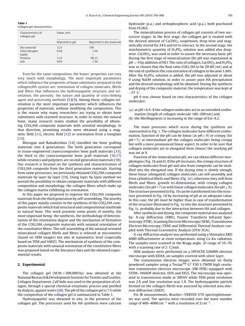
A. Ficai et al. / Chemical Engineering
Table 1Collagen gel characteristics.
Characteristics ofcollagen gel
Value, wt%
Reported to gel Reported to dry materials
Dry material 3.21 100Total nitrogen 0.56 1.56
vwcaeaepisbitn[
mttwOCfmpct
moptmnotm(bpwi
2
NClbt
c
Lipids – –Proteins 3.15 98.13Ash 0.05 1.56
Even for the same composition, the bones’ properties can varyery much with morphology. The most important parametershich influence the properties of bone substitutes prepared in the
ollagen/HA system are: orientation of collagen molecules, fibrilsnd fibres that influences the hydroxyapatite structure and ori-ntation; the porosity; the nature and quantity of cross-linkinggent and processing methods [5,8,9]. Among these, collagen ori-ntation is the most important parameter which influences theroperties of materials, without modifying the composition. This
s the reason why many researchers are trying to obtain boneubstitutes with oriented structure. In order to mimic the naturalone, many research teams studied the possibility of obtain-
ng COLL/HA composite materials with oriented morphology. Inhat direction, promising results were obtained using a mag-etic field [11], electric field [12] or orientation from a template13].
Murugan and Ramakrishna [14] classified the bone graftingaterials into 4 generations. The forth generation correspond
o tissue-engineered (nano)composites bone graft materials [15],he third to the (nano)composite bone graft materials [16,17]hile ceramics and polymers are second generation materials [18].ur research is focused on the synthesis and characterization ofOLL/HA composite from the third generation materials. Starting
rom same precursors, we previously obtained COLL/HA compositeaterials by layer by layer [19]. Using layer by layer method we
roved the possibility to obtain composite materials with differentomposition and morphology, the collagen fibres which make uphe collagen matrix exhibiting no orientation.
In this paper we proposed to improve the COLL/HA compositeaterials from the third generation by self-assembling. The novelty
f this paper mainly consists in the synthesis of the COLL/HA com-osite materials which had structural and compositional similitudeo natural bone. This work presents some points of novelty, the
ost important being: the synthesis, the methodology of determi-ation of the orientation degree and the mechanism of formationf the COLL/HA composite materials with uniaxial orientation ofhe constitutive fibres. The self-assembling of the uniaxial oriented
ineralized collagen fibrils and fibres is relieved at micrometricbased on SEM images) but also at nanometric level (especiallyased on TEM and SAED). The mechanism of synthesis of the com-osite materials with uniaxial orientation of the constitutive fibresas proposed based on the literature data and the obtained exper-
mental results.
. Experimental
The collagen gel (M.W. = 300,000 Da) was obtained at theational Research & Development Institute for Textiles and Leather,ollagen Department. Calf hide was used to the preparation of col-
agen, through a special chemical-enzymatic process and purifiedy dialysis, against water [20]. The pH of the collagen gel is 7.5 whilehe composition of the collagen gel is summarized in Table 1.
Hydroxyapatite was obtained in situ, in the presence of theollagen gel. The precursors used for HA synthesis were calcium
Journal 160 (2010) 794–800 795
hydroxide (p.a.) and orthophosphoric acid (p.a.), both purchasedfrom FLUKA.
The mineralization process of collagen gel consists of two suc-cessive stages. In the first stage, the collagen gel is treated withthe desired amount of Ca(OH)2 suspension, drop-wise and mag-netically stirred for 24 h and let to interact. In the second stage, thestoichiometric quantity of H3PO4 solution was added also drop-wise. Ca(OH)2 was used in order to assure the necessary basic pH.During the first stage of mineralization the pH was maintained atpH � 9 by addition of HCl. The ratio of collagen, Ca(OH)2 and H3PO4was so chosen that the final ratio COLL:HA to be 20:80 (wt) and atthe end of synthesis the concentration of collagen to became 1.66%.After the H3PO4 solution is added, the pH was adjusted at about9 using NaOH solution, in order to assure pure HA precipitationand the desired morphology will be obtained. During the synthesisand drying of the composite material, the temperature was kept at∼37 ◦C.
pH 9 was choose based on two characteristics of the collagenmolecules:
(a) at pH = 6.9–8 the collagen molecules are in an extended confor-mation (length of collagen molecule 180–200 nm) and,
(b) the fibrillogenesis is increasing in the range of 6.6–9.2.
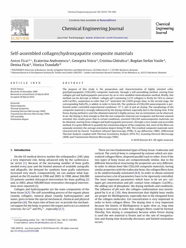
The main processes which occur during the synthesis arerepresented in Fig. 1. The collagen molecules have different confor-mation, function of the pH can be linear (at pH ≥ 9) or crimpy (forpH < 7); at intermediate pH the collagen molecules being crimpybut with a more pronounced linear aspect. In order to be sure thatcollagen molecules are in elongated form (linear) the working pHwas set at 9.
Function of the mineralization pH, we can obtain different mor-phologies (Fig. 1b and d). If the pH increases, the crimpy structure ofcollagen molecules from gel and also from composite can be mod-ified into the elongated one. If the drying time is slowly enough,these linear (elongated) collagen molecules can self-assembly andform cylindrical fibrils and fibres (Fig. 1e), otherwise they will formfibrils and fibres, growth tri-dimensional, with crimpy collagenmolecules (for pH < 7) or with linear collagen molecules (for pH ≥ 9).The structure presented in Fig. 1b can be transformed into the struc-ture presented in Fig. 1d by increasing of pH at an adequate value.In this case, the pH must be higher than in case of transformationof the structure illustrated in Fig. 1a into the structure presented inFig. 1c, probably due to the interaction between HA and collagen.
After synthesis and drying, the composite material was analysedby: X-ray diffraction (XRD), Fourier Transform Infrared Spec-troscopy (FTIR), Scanning Electron Microscopy (SEM), TransmissionElectron Microscopy (TEM) and Differential Thermal Analysis cou-pled with Thermal Gravimetric Analysis (DTA-TGA).
X-ray diffraction analysis was performed using a Shimadzu XRD6000 diffractometer at room temperature, using Cu K� radiation.The samples were scanned in the Bragg angle, 2� range of 10–70,with a scanning rate of 2 ◦C/min.
SEM analyses were performed on a HITACHI S2600N electronmicroscope with EDAX, on samples covered with silver layer.
The transmission electron images were obtained on finelypowdered samples using a TecnaiTM G2 F30 S-TWIN high resolu-tion transmission electron microscope (HR-TEM) equipped withSTEM—HAADF detector, EDX and EELS. The microscope was oper-ated in transmission mode at 300 kV while TEM point resolutionwas 2 Å and line resolution was 1 Å. The hydroxyapatite particle
formed on the collagen fibrils was assessed by selected area elec-tron diffraction (SAED).For IR measurements, Brucker—VERTEX V70 spectrophotome-ter was used. The spectra were recorded over the wave numberrange of 400–4000 cm−1 with a resolution of 2 cm−1.
796 A. Ficai et al. / Chemical Engineering Journal 160 (2010) 794–800
ing th
g5
3
X
ctpHHATiat
Fig. 1. Specific processes occurred in solution dur
The differential thermal analysis (DTA) coupled with thermoravimetric analysis (TGA) was performed on a Shimadzu DTG-TA-0H, at a heating rate of 10 ◦C/min, in static air.
. Results and discussion
XRD was used to evidentiate the mineralization process. TheRD spectrum, in Fig. 2, shows the formation of HA.
In the case of COLL/HA composite obtained by self-assemblyomparing with the HA obtained by precipitation, the X-ray diffrac-ion pattern is evidenciating a much higher intensity for the 2 1 1eak reported to the intensity of the other peaks characteristic toA. This result can be attributed to a preferential growth of theA crystals in the 2 1 1 direction due to the collagen influence.
s it can see, the composite material also contains NaCl (Fig. 2a).he removal of NaCl can be easily made by washing the compos-te materials with distilled water, following the next procedure:fter drying, the COLL/HA composite material with uniaxial orien-ation of the constitutive fibres is crosslinked with glutaraldehyde,e self-assembly of COLL/HA composite materials.
washed with plenty of water and dried. Following this procedure,the chloride was completely removed (Fig. 2b) without altering themineral phase, especially from the point of view of crystallinity andpreferential crystallization direction.
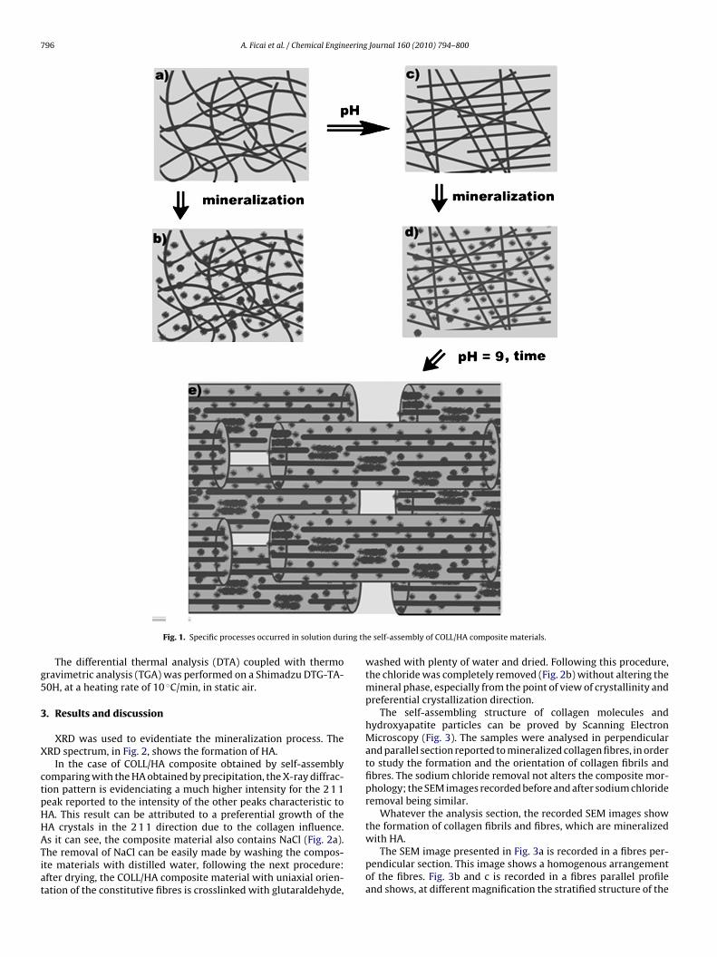
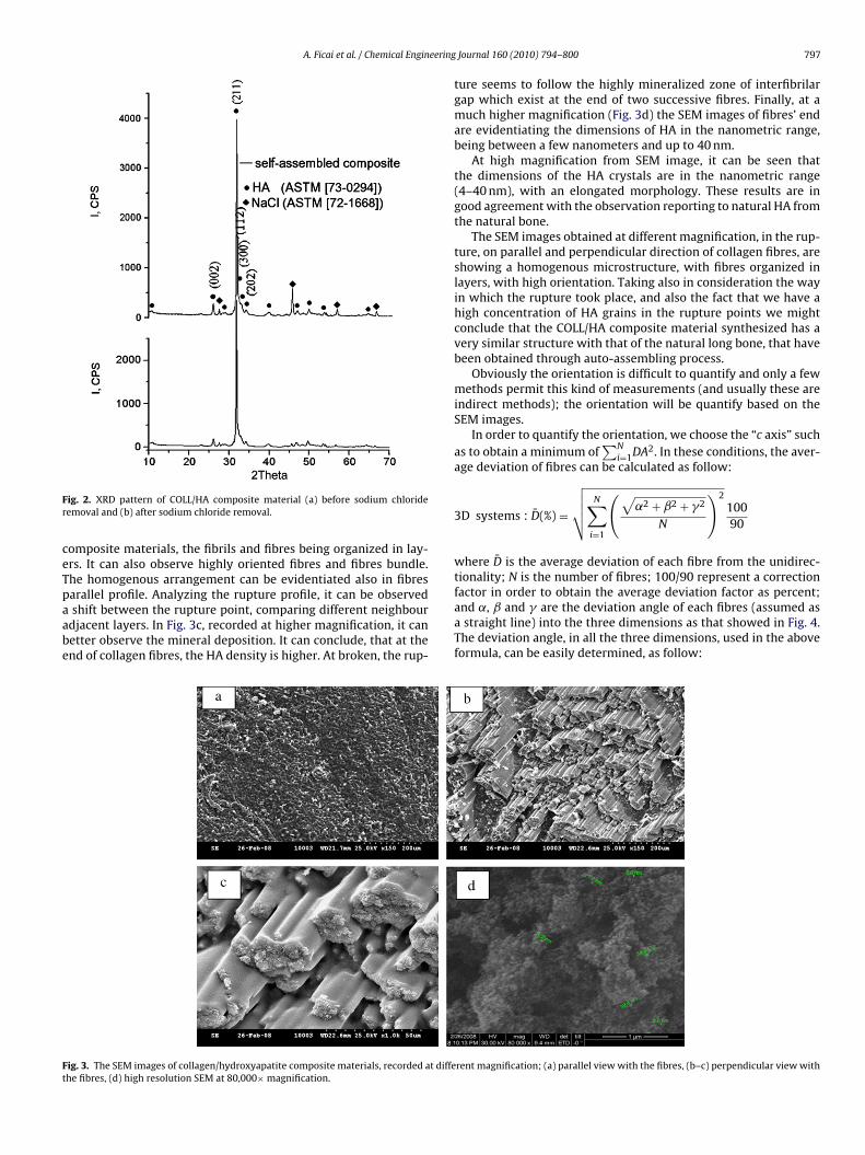
The self-assembling structure of collagen molecules andhydroxyapatite particles can be proved by Scanning ElectronMicroscopy (Fig. 3). The samples were analysed in perpendicularand parallel section reported to mineralized collagen fibres, in orderto study the formation and the orientation of collagen fibrils andfibres. The sodium chloride removal not alters the composite mor-phology; the SEM images recorded before and after sodium chlorideremoval being similar.
Whatever the analysis section, the recorded SEM images showthe formation of collagen fibrils and fibres, which are mineralized
with HA.The SEM image presented in Fig. 3a is recorded in a fibres per-pendicular section. This image shows a homogenous arrangementof the fibres. Fig. 3b and c is recorded in a fibres parallel profileand shows, at different magnification the stratified structure of the
A. Ficai et al. / Chemical Engineering
Fr
ceTpaabe
factor in order to obtain the average deviation factor as percent;
Ft
ig. 2. XRD pattern of COLL/HA composite material (a) before sodium chlorideemoval and (b) after sodium chloride removal.
omposite materials, the fibrils and fibres being organized in lay-rs. It can also observe highly oriented fibres and fibres bundle.he homogenous arrangement can be evidentiated also in fibres
arallel profile. Analyzing the rupture profile, it can be observedshift between the rupture point, comparing different neighbourdjacent layers. In Fig. 3c, recorded at higher magnification, it canetter observe the mineral deposition. It can conclude, that at thend of collagen fibres, the HA density is higher. At broken, the rup-
ig. 3. The SEM images of collagen/hydroxyapatite composite materials, recorded at diffehe fibres, (d) high resolution SEM at 80,000× magnification.
Journal 160 (2010) 794–800 797
ture seems to follow the highly mineralized zone of interfibrilargap which exist at the end of two successive fibres. Finally, at amuch higher magnification (Fig. 3d) the SEM images of fibres’ endare evidentiating the dimensions of HA in the nanometric range,being between a few nanometers and up to 40 nm.
At high magnification from SEM image, it can be seen thatthe dimensions of the HA crystals are in the nanometric range(4–40 nm), with an elongated morphology. These results are ingood agreement with the observation reporting to natural HA fromthe natural bone.
The SEM images obtained at different magnification, in the rup-ture, on parallel and perpendicular direction of collagen fibres, areshowing a homogenous microstructure, with fibres organized inlayers, with high orientation. Taking also in consideration the wayin which the rupture took place, and also the fact that we have ahigh concentration of HA grains in the rupture points we mightconclude that the COLL/HA composite material synthesized has avery similar structure with that of the natural long bone, that havebeen obtained through auto-assembling process.
Obviously the orientation is difficult to quantify and only a fewmethods permit this kind of measurements (and usually these areindirect methods); the orientation will be quantify based on theSEM images.
In order to quantify the orientation, we choose the “c axis” suchas to obtain a minimum of
∑Ni=1DA2. In these conditions, the aver-
age deviation of fibres can be calculated as follow:
3D systems : D̄(%) =
√√√√ N∑i=1
(√˛2 + ˇ2 + �2
N
)210090
where D̄ is the average deviation of each fibre from the unidirec-tionality; N is the number of fibres; 100/90 represent a correction
and ˛, ˇ and � are the deviation angle of each fibres (assumed asa straight line) into the three dimensions as that showed in Fig. 4.The deviation angle, in all the three dimensions, used in the aboveformula, can be easily determined, as follow:
rent magnification; (a) parallel view with the fibres, (b–c) perpendicular view with
798 A. Ficai et al. / Chemical Engineering Journal 160 (2010) 794–800
Ft
----
Fa
paghrp
Fs
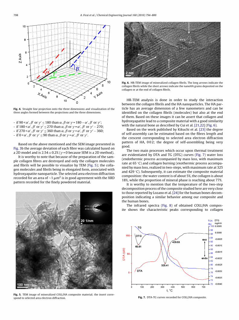
ig. 4. Straight line projection onto the three dimensions and visualization of thehree angles formed between the projections and the three dimensions.
if 90 < ˛′, ˇ′ or � ′ ≤ 180 than ˛, ˇ or � = 180 − ˛′, ˇ′ or � ′;if 180 < ˛′, ˇ′ or � ′ ≤ 270 than ˛, ˇ or � = ˛′, ˇ′ or � ′ − 270;if 270 < ˛′, ˇ′ or � ′ ≤ 360 than ˛, ˇ or � = ˛′, ˇ′ or � ′ − 360;if 0 < ˛′, ˇ′ or � ′ ≤ 90 than ˛, ˇ or � = ˛′, ˇ′ or � ′.
Based on the above mentioned and the SEM image presented inig. 3b the average deviation of each fibre was calculated based on2D model and is 2.54 ± 0.2% (� = 0 because SEM is a 2D method).
It is worthy to note that because of the preparation of the sam-le collagen fibres are destroyed and only the collagen moleculesnd fibrils will be possible to visualize by TEM (Fig. 5); the colla-
en molecules and fibrils being in elongated form, associated withydroxyapatite nanoparticle. The selected area electron diffractionecorded for an area of ∼1 �m2 is in good agreement with the XRDattern recorded for the finely powdered material.ig. 5. TEM image of mineralized COLL/HA composite material; the insert corre-pond to selected area electron diffraction.
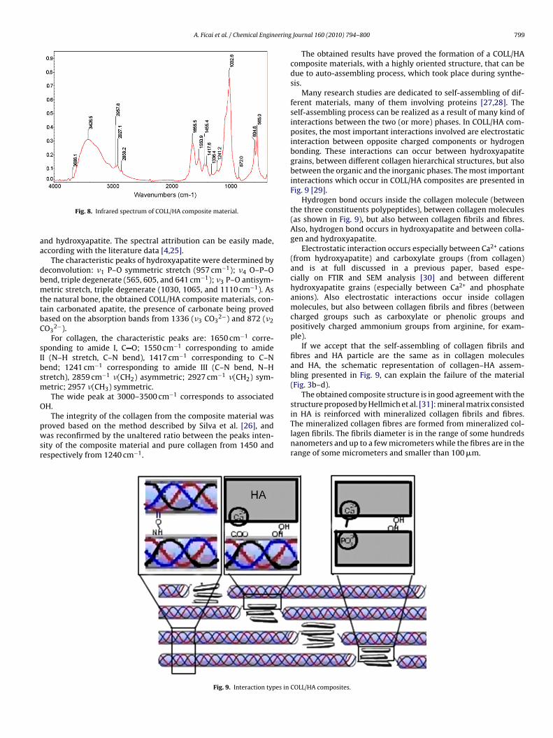
Fig. 6. HR-TEM image of mineralized collagen fibrils. The long arrows indicate thecollagen fibrils while the short arrows indicate the nanoHA grains deposited on thecollagen or at the end of collagen fibrils.
HR-TEM analysis is done in order to study the interactionbetween the collagen fibrils and the HA nanoparticles. The HA par-ticle has an average dimension of a few nanometers and can beidentified on the collagen fibrils (molecules) but also at the endof them. Based on these images it can be assert that collagen andhydroxyapatite lead to a composite material with a good similaritywith the natural bone as described by Cui et al. [21,22] (Fig. 6).
Based on the work published by Kikuchi et al. [23] the degreeof self-assembly can be estimated based on the fibres length andthe crescent corresponding to selected area electron diffractionpattern of HA, 0 0 2; the degree of self-assembling being verygood.
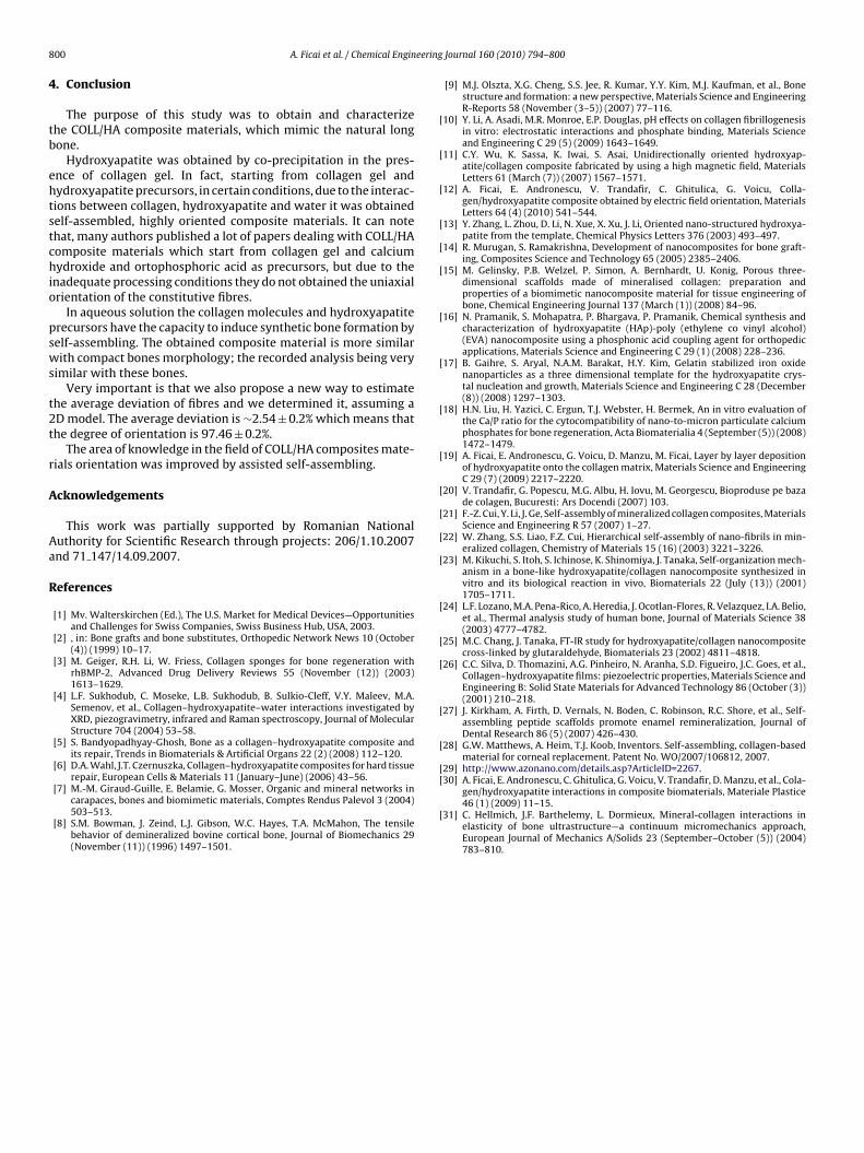
The two main processes which occur upon thermal treatmentare evidentiated by DTA and TG (DTG) curves (Fig. 7) water loss(endothermic process accompanied by mass loss, with maximumrate at 65 ◦C) and collagen burning (exothermic process accompa-nied by mass loss, realized in two-steps, with maximum rate at 325and 429 ◦C). Subsequently, it can estimate the composite materialcomposition: the water content is of about 5%, the collagen is about18%, while the proportion of mineral phase is reaching about 77%.
It is worthy to mention that the temperature of the two-stepdecomposition process of the composite studied here are very closeto those reported by Lozano et al. [24] for the human bones decom-
position indicating a similar behavior among our composite andthe human bones.The infrared spectra (Fig. 8) of obtained COLL/HA compos-ite shows the characteristic peaks corresponding to collagen
Fig. 7. DTA-TG curves recorded for COLL/HA composite.
A. Ficai et al. / Chemical Engineering
aa
dbmttbC
sIbsm
O
pwsr
in HA is reinforced with mineralized collagen fibrils and fibres.
Fig. 8. Infrared spectrum of COLL/HA composite material.
nd hydroxyapatite. The spectral attribution can be easily made,ccording with the literature data [4,25].
The characteristic peaks of hydroxyapatite were determined byeconvolution: �1 P–O symmetric stretch (957 cm−1); �4 O–P–Oend, triple degenerate (565, 605, and 641 cm−1); �3 P–O antisym-etric stretch, triple degenerate (1030, 1065, and 1110 cm−1). As
he natural bone, the obtained COLL/HA composite materials, con-ain carbonated apatite, the presence of carbonate being provedased on the absorption bands from 1336 (�3 CO3
2−) and 872 (�2O3
2−).For collagen, the characteristic peaks are: 1650 cm−1 corre-
ponding to amide I, C O; 1550 cm−1 corresponding to amideI (N–H stretch, C–N bend), 1417 cm−1 corresponding to C–Nend; 1241 cm−1 corresponding to amide III (C–N bend, N–Htretch), 2859 cm−1 �(CH2) asymmetric; 2927 cm−1 �(CH2) sym-etric; 2957 �(CH3) symmetric.The wide peak at 3000–3500 cm−1 corresponds to associated
H.The integrity of the collagen from the composite material was
roved based on the method described by Silva et al. [26], andas reconfirmed by the unaltered ratio between the peaks inten-
ity of the composite material and pure collagen from 1450 andespectively from 1240 cm−1.
Fig. 9. Interaction types in
Journal 160 (2010) 794–800 799
The obtained results have proved the formation of a COLL/HAcomposite materials, with a highly oriented structure, that can bedue to auto-assembling process, which took place during synthe-sis.
Many research studies are dedicated to self-assembling of dif-ferent materials, many of them involving proteins [27,28]. Theself-assembling process can be realized as a result of many kind ofinteractions between the two (or more) phases. In COLL/HA com-posites, the most important interactions involved are electrostaticinteraction between opposite charged components or hydrogenbonding. These interactions can occur between hydroxyapatitegrains, between different collagen hierarchical structures, but alsobetween the organic and the inorganic phases. The most importantinteractions which occur in COLL/HA composites are presented inFig. 9 [29].
Hydrogen bond occurs inside the collagen molecule (betweenthe three constituents polypeptides), between collagen molecules(as shown in Fig. 9), but also between collagen fibrils and fibres.Also, hydrogen bond occurs in hydroxyapatite and between colla-gen and hydroxyapatite.
Electrostatic interaction occurs especially between Ca2+ cations(from hydroxyapatite) and carboxylate groups (from collagen)and is at full discussed in a previous paper, based espe-cially on FTIR and SEM analysis [30] and between differenthydroxyapatite grains (especially between Ca2+ and phosphateanions). Also electrostatic interactions occur inside collagenmolecules, but also between collagen fibrils and fibres (betweencharged groups such as carboxylate or phenolic groups andpositively charged ammonium groups from arginine, for exam-ple).
If we accept that the self-assembling of collagen fibrils andfibres and HA particle are the same as in collagen moleculesand HA, the schematic representation of collagen–HA assem-bling presented in Fig. 9, can explain the failure of the material(Fig. 3b–d).
The obtained composite structure is in good agreement with thestructure proposed by Hellmich et al. [31]: mineral matrix consisted
The mineralized collagen fibres are formed from mineralized col-lagen fibrils. The fibrils diameter is in the range of some hundredsnanometers and up to a few micrometers while the fibres are in therange of some micrometers and smaller than 100 �m.
COLL/HA composites.
8 eering
4
tb
ehtstchio
psws
t2t
r
A
Aa
R
[
[
[
[
[
[
[
[
[
[
[
[
[
[
[
[
[
[
[
[[
00 A. Ficai et al. / Chemical Engin
. Conclusion
The purpose of this study was to obtain and characterizehe COLL/HA composite materials, which mimic the natural longone.
Hydroxyapatite was obtained by co-precipitation in the pres-nce of collagen gel. In fact, starting from collagen gel andydroxyapatite precursors, in certain conditions, due to the interac-ions between collagen, hydroxyapatite and water it was obtainedelf-assembled, highly oriented composite materials. It can notehat, many authors published a lot of papers dealing with COLL/HAomposite materials which start from collagen gel and calciumydroxide and ortophosphoric acid as precursors, but due to the
nadequate processing conditions they do not obtained the uniaxialrientation of the constitutive fibres.
In aqueous solution the collagen molecules and hydroxyapatiterecursors have the capacity to induce synthetic bone formation byelf-assembling. The obtained composite material is more similarith compact bones morphology; the recorded analysis being very
imilar with these bones.Very important is that we also propose a new way to estimate
he average deviation of fibres and we determined it, assuming aD model. The average deviation is ∼2.54 ± 0.2% which means thathe degree of orientation is 97.46 ± 0.2%.
The area of knowledge in the field of COLL/HA composites mate-ials orientation was improved by assisted self-assembling.
cknowledgements
This work was partially supported by Romanian Nationaluthority for Scientific Research through projects: 206/1.10.2007nd 71 147/14.09.2007.
eferences
[1] Mv. Walterskirchen (Ed.), The U.S. Market for Medical Devices—Opportunitiesand Challenges for Swiss Companies, Swiss Business Hub, USA, 2003.
[2] , in: Bone grafts and bone substitutes, Orthopedic Network News 10 (October(4)) (1999) 10–17.
[3] M. Geiger, R.H. Li, W. Friess, Collagen sponges for bone regeneration withrhBMP-2, Advanced Drug Delivery Reviews 55 (November (12)) (2003)1613–1629.
[4] L.F. Sukhodub, C. Moseke, L.B. Sukhodub, B. Sulkio-Cleff, V.Y. Maleev, M.A.Semenov, et al., Collagen–hydroxyapatite–water interactions investigated byXRD, piezogravimetry, infrared and Raman spectroscopy, Journal of MolecularStructure 704 (2004) 53–58.
[5] S. Bandyopadhyay-Ghosh, Bone as a collagen–hydroxyapatite composite andits repair, Trends in Biomaterials & Artificial Organs 22 (2) (2008) 112–120.
[6] D.A. Wahl, J.T. Czernuszka, Collagen–hydroxyapatite composites for hard tissuerepair, European Cells & Materials 11 (January–June) (2006) 43–56.
[7] M.-M. Giraud-Guille, E. Belamie, G. Mosser, Organic and mineral networks incarapaces, bones and biomimetic materials, Comptes Rendus Palevol 3 (2004)503–513.
[8] S.M. Bowman, J. Zeind, L.J. Gibson, W.C. Hayes, T.A. McMahon, The tensilebehavior of demineralized bovine cortical bone, Journal of Biomechanics 29(November (11)) (1996) 1497–1501.
[
Journal 160 (2010) 794–800
[9] M.J. Olszta, X.G. Cheng, S.S. Jee, R. Kumar, Y.Y. Kim, M.J. Kaufman, et al., Bonestructure and formation: a new perspective, Materials Science and EngineeringR-Reports 58 (November (3–5)) (2007) 77–116.
10] Y. Li, A. Asadi, M.R. Monroe, E.P. Douglas, pH effects on collagen fibrillogenesisin vitro: electrostatic interactions and phosphate binding, Materials Scienceand Engineering C 29 (5) (2009) 1643–1649.
11] C.Y. Wu, K. Sassa, K. Iwai, S. Asai, Unidirectionally oriented hydroxyap-atite/collagen composite fabricated by using a high magnetic field, MaterialsLetters 61 (March (7)) (2007) 1567–1571.
12] A. Ficai, E. Andronescu, V. Trandafir, C. Ghitulica, G. Voicu, Colla-gen/hydroxyapatite composite obtained by electric field orientation, MaterialsLetters 64 (4) (2010) 541–544.
13] Y. Zhang, L. Zhou, D. Li, N. Xue, X. Xu, J. Li, Oriented nano-structured hydroxya-patite from the template, Chemical Physics Letters 376 (2003) 493–497.
14] R. Murugan, S. Ramakrishna, Development of nanocomposites for bone graft-ing, Composites Science and Technology 65 (2005) 2385–2406.
15] M. Gelinsky, P.B. Welzel, P. Simon, A. Bernhardt, U. Konig, Porous three-dimensional scaffolds made of mineralised collagen: preparation andproperties of a biomimetic nanocomposite material for tissue engineering ofbone, Chemical Engineering Journal 137 (March (1)) (2008) 84–96.
16] N. Pramanik, S. Mohapatra, P. Bhargava, P. Pramanik, Chemical synthesis andcharacterization of hydroxyapatite (HAp)-poly (ethylene co vinyl alcohol)(EVA) nanocomposite using a phosphonic acid coupling agent for orthopedicapplications, Materials Science and Engineering C 29 (1) (2008) 228–236.
17] B. Gaihre, S. Aryal, N.A.M. Barakat, H.Y. Kim, Gelatin stabilized iron oxidenanoparticles as a three dimensional template for the hydroxyapatite crys-tal nucleation and growth, Materials Science and Engineering C 28 (December(8)) (2008) 1297–1303.
18] H.N. Liu, H. Yazici, C. Ergun, T.J. Webster, H. Bermek, An in vitro evaluation ofthe Ca/P ratio for the cytocompatibility of nano-to-micron particulate calciumphosphates for bone regeneration, Acta Biomaterialia 4 (September (5)) (2008)1472–1479.
19] A. Ficai, E. Andronescu, G. Voicu, D. Manzu, M. Ficai, Layer by layer depositionof hydroxyapatite onto the collagen matrix, Materials Science and EngineeringC 29 (7) (2009) 2217–2220.
20] V. Trandafir, G. Popescu, M.G. Albu, H. Iovu, M. Georgescu, Bioproduse pe bazade colagen, Bucuresti: Ars Docendi (2007) 103.
21] F.-Z. Cui, Y. Li, J. Ge, Self-assembly of mineralized collagen composites, MaterialsScience and Engineering R 57 (2007) 1–27.
22] W. Zhang, S.S. Liao, F.Z. Cui, Hierarchical self-assembly of nano-fibrils in min-eralized collagen, Chemistry of Materials 15 (16) (2003) 3221–3226.
23] M. Kikuchi, S. Itoh, S. Ichinose, K. Shinomiya, J. Tanaka, Self-organization mech-anism in a bone-like hydroxyapatite/collagen nanocomposite synthesized invitro and its biological reaction in vivo, Biomaterials 22 (July (13)) (2001)1705–1711.
24] L.F. Lozano, M.A. Pena-Rico, A. Heredia, J. Ocotlan-Flores, R. Velazquez, I.A. Belio,et al., Thermal analysis study of human bone, Journal of Materials Science 38(2003) 4777–4782.
25] M.C. Chang, J. Tanaka, FT-IR study for hydroxyapatite/collagen nanocompositecross-linked by glutaraldehyde, Biomaterials 23 (2002) 4811–4818.
26] C.C. Silva, D. Thomazini, A.G. Pinheiro, N. Aranha, S.D. Figueiro, J.C. Goes, et al.,Collagen–hydroxyapatite films: piezoelectric properties, Materials Science andEngineering B: Solid State Materials for Advanced Technology 86 (October (3))(2001) 210–218.
27] J. Kirkham, A. Firth, D. Vernals, N. Boden, C. Robinson, R.C. Shore, et al., Self-assembling peptide scaffolds promote enamel remineralization, Journal ofDental Research 86 (5) (2007) 426–430.
28] G.W. Matthews, A. Heim, T.J. Koob, Inventors. Self-assembling, collagen-basedmaterial for corneal replacement. Patent No. WO/2007/106812, 2007.
29] http://www.azonano.com/details.asp?ArticleID=2267.30] A. Ficai, E. Andronescu, C. Ghitulica, G. Voicu, V. Trandafir, D. Manzu, et al., Cola-
gen/hydroxyapatite interactions in composite biomaterials, Materiale Plastice46 (1) (2009) 11–15.
31] C. Hellmich, J.F. Barthelemy, L. Dormieux, Mineral-collagen interactions inelasticity of bone ultrastructure—a continuum micromechanics approach,European Journal of Mechanics A/Solids 23 (September–October (5)) (2004)783–810.