Hydroxyapatite for Biomedical Applications: A Short Overview
22
ceramics Review Hydroxyapatite for Biomedical Applications: A Short Overview Elisa Fiume 1 , Giulia Magnaterra 1 , Abbas Rahdar 2 , Enrica Verné 1 and Francesco Baino 1, * Citation: Fiume, E.; Magnaterra, G.; Rahdar, A.; Verné, E.; Baino, F. Hydroxyapatite for Biomedical Applications: A Short Overview. Ceramics 2021, 4, 542–563. https:// doi.org/10.3390/ceramics4040039 Academic Editors: Anna Lukowiak and Gilbert Fantozzi Received: 30 June 2021 Accepted: 24 September 2021 Published: 28 September 2021 Publisher’s Note: MDPI stays neutral with regard to jurisdictional claims in published maps and institutional affil- iations. Copyright: © 2021 by the authors. Licensee MDPI, Basel, Switzerland. This article is an open access article distributed under the terms and conditions of the Creative Commons Attribution (CC BY) license (https:// creativecommons.org/licenses/by/ 4.0/). 1 Department of Applied Science and Technology (DISAT), Institute of Materials Physics and Engineering, Politecnico di Torino, 10129 Turin, Italy; elisa.fi[email protected] (E.F.); [email protected] (G.M.); [email protected] (E.V.) 2 Department of Physics, University of Zabol, Zabol 98613-35856, Iran; [email protected] * Correspondence: [email protected] Abstract: Calcium phosphates (CaPs) are biocompatible and biodegradable materials showing a great promise in bone regeneration as good alternative to the use of auto- and allografts to guide and support tissue regeneration in critically-sized bone defects. This can be certainly attributed to their similarity to the mineral phase of natural bone. Among CaPs, hydroxyapatite (HA) deserves a special attention as it, actually is the main inorganic component of bone tissue. This review offers a comprehensive overview of past and current trends in the use of HA as grafting material, with a focus on manufacturing strategies and their effect on the mechanical properties of the final products. Recent advances in materials processing allowed the production of HA-based grafts in different forms, thus meeting the requirements for a range of clinical applications and achieving enthusiastic results both in vitro and in vivo. Furthermore, the growing interest in the optimization of three-dimensional (3D) porous grafts, mimicking the trabecular architecture of human bone, has opened up new challenges in the development of bone-like scaffolds showing suitable mechanical performances for potential use in load bearing anatomical sites. Keywords: bioceramics; calcium phosphate; hydroxyapatite; bone; tissue engineering 1. Introduction Since the early 1900s, the scientific community began to be interested in calcium phos- phates (CaPs) as materials for the production of bone substitutes in biomedical applications. The first study dates back to 1920, when CaPs were used as filler material to repair critical size bone defects in rabbits [1]. Owing to their physico-mechanical properties, which are similar to those of human bone, as well as osteogenic potential both in vivo and in vitro [2]. Today CaPs are widely used in different fields of medicine, such as otolaryngology, skull and maxillofacial recon- struction, spinal surgery, orthopedics, treatment of osseous fractures and bone disorders, percutaneous implants and dentistry/periodontal surgery [3]. A study conducted in the USA showed that in 2010 about 1.3 billion dollars were invested in CaP-based bone substitutes only [4]. The term “CaPs” refers to a family of minerals containing calcium cations (Ca 2+ ) and metaphosphate (PO - 3 ), orthophosphate (PO 4 ) 3- , or pyrophosphate (P 2 O 7 ) 4- anions [3], as well as hydroxyl (OH - ) or hydrogen (H + ) ions [5]. CaPs are the main constituents of tooth enamel (~90 wt.%) and bone (~60 wt.%) [5], and this is the main reason why they are studied in bone repair and regeneration. They exhibit crystalline structure and chemical properties very similar to those of bone apatite [6] and excellent biocompatibility with living cells [7,8]. CaPs are well known to have osteoconductive properties that permit the attachment, proliferation and migration of bone cells, regardless of the form in which they are used (e.g., coating, powder, bulk or porous scaffolds). Soluble and/or nano-sized CaPs were also reported to exhibit osteoinductive properties, thus actively promoting the growth and regeneration of new bone [9,10]. Ceramics 2021, 4, 542–563. https://doi.org/10.3390/ceramics4040039 https://www.mdpi.com/journal/ceramics
-
Upload
khangminh22 -
Category
Documents
-
view
0 -
download
0
Transcript of Hydroxyapatite for Biomedical Applications: A Short Overview

ceramics
Review
Hydroxyapatite for Biomedical Applications: A Short Overview
Elisa Fiume 1, Giulia Magnaterra 1, Abbas Rahdar 2 , Enrica Verné 1 and Francesco Baino 1,*
�����������������
Citation: Fiume, E.; Magnaterra, G.;
Rahdar, A.; Verné, E.; Baino, F.
Hydroxyapatite for Biomedical
Applications: A Short Overview.
Ceramics 2021, 4, 542–563. https://
doi.org/10.3390/ceramics4040039
Academic Editors: Anna Lukowiak
and Gilbert Fantozzi
Received: 30 June 2021
Accepted: 24 September 2021
Published: 28 September 2021
Publisher’s Note: MDPI stays neutral
with regard to jurisdictional claims in
published maps and institutional affil-
iations.
Copyright: © 2021 by the authors.
Licensee MDPI, Basel, Switzerland.
This article is an open access article
distributed under the terms and
conditions of the Creative Commons
Attribution (CC BY) license (https://
creativecommons.org/licenses/by/
4.0/).
1 Department of Applied Science and Technology (DISAT), Institute of Materials Physics and Engineering,Politecnico di Torino, 10129 Turin, Italy; [email protected] (E.F.); [email protected] (G.M.);[email protected] (E.V.)
2 Department of Physics, University of Zabol, Zabol 98613-35856, Iran; [email protected]* Correspondence: [email protected]
Abstract: Calcium phosphates (CaPs) are biocompatible and biodegradable materials showing agreat promise in bone regeneration as good alternative to the use of auto- and allografts to guideand support tissue regeneration in critically-sized bone defects. This can be certainly attributed totheir similarity to the mineral phase of natural bone. Among CaPs, hydroxyapatite (HA) deserves aspecial attention as it, actually is the main inorganic component of bone tissue. This review offers acomprehensive overview of past and current trends in the use of HA as grafting material, with a focuson manufacturing strategies and their effect on the mechanical properties of the final products. Recentadvances in materials processing allowed the production of HA-based grafts in different forms, thusmeeting the requirements for a range of clinical applications and achieving enthusiastic results bothin vitro and in vivo. Furthermore, the growing interest in the optimization of three-dimensional (3D)porous grafts, mimicking the trabecular architecture of human bone, has opened up new challengesin the development of bone-like scaffolds showing suitable mechanical performances for potentialuse in load bearing anatomical sites.
Keywords: bioceramics; calcium phosphate; hydroxyapatite; bone; tissue engineering
1. Introduction
Since the early 1900s, the scientific community began to be interested in calcium phos-phates (CaPs) as materials for the production of bone substitutes in biomedical applications.The first study dates back to 1920, when CaPs were used as filler material to repair criticalsize bone defects in rabbits [1].
Owing to their physico-mechanical properties, which are similar to those of humanbone, as well as osteogenic potential both in vivo and in vitro [2]. Today CaPs are widelyused in different fields of medicine, such as otolaryngology, skull and maxillofacial recon-struction, spinal surgery, orthopedics, treatment of osseous fractures and bone disorders,percutaneous implants and dentistry/periodontal surgery [3]. A study conducted inthe USA showed that in 2010 about 1.3 billion dollars were invested in CaP-based bonesubstitutes only [4].
The term “CaPs” refers to a family of minerals containing calcium cations (Ca2+) andmetaphosphate (PO−
3), orthophosphate (PO4)3−, or pyrophosphate (P2O7)4− anions [3],as well as hydroxyl (OH−) or hydrogen (H+) ions [5].
CaPs are the main constituents of tooth enamel (~90 wt.%) and bone (~60 wt.%) [5],and this is the main reason why they are studied in bone repair and regeneration.
They exhibit crystalline structure and chemical properties very similar to those ofbone apatite [6] and excellent biocompatibility with living cells [7,8].
CaPs are well known to have osteoconductive properties that permit the attachment,proliferation and migration of bone cells, regardless of the form in which they are used(e.g., coating, powder, bulk or porous scaffolds). Soluble and/or nano-sized CaPs werealso reported to exhibit osteoinductive properties, thus actively promoting the growth andregeneration of new bone [9,10].
Ceramics 2021, 4, 542–563. https://doi.org/10.3390/ceramics4040039 https://www.mdpi.com/journal/ceramics
Ceramics 2021, 4 543
During their permanence into the human body, most of CaPs can be partially dis-solved within body fluids. The local increment in Ca2+ and PO4
3− ions at the bone/implantinterface determines the supersaturation of the biological environment and the consequentprecipitation of apatite nanocrystals on the material surface [11]. This newly-formed layercan adsorb proteins from the surrounding environment, thus promoting the attachment,proliferation and differentiation of osteoprogenitor stem cells and the consequent miner-alization of the tissue and the formation of osteoids. The angiogenesis progression willfinally result in local bone induction and potential incorporation of the CaP implant withinthe natural bone [11].
The rate of formation of the surface apatite layer depends on the type of bioceramicconsidered; it takes 30 days for hydroxyapatite, and about 14 days for β-tricalcium phos-phate [12]. For the purpose of comparison, bioactive glasses, which have a faster reactivityin contact with biological fluids, are able to form a surface apatite layer in the range of fewhours to few days [13].
The distinction between osteoconductive and osteoinductive biomaterials is obviouslydictated also by the time scale of reactivity in vitro and in vivo; this is the reason why highly-reactive materials such as bioactive glasses or soluble CaPs are classified as belonging to thelatter group, while non-porous hydroxyapatite, which has an almost negligible solubility,is a traditional example of the former [14].
Qui and Ducheyne have described the general reactions occurring at the interfacebetween CaPs and biological environment through a 11-step sequence [11]:
1. Dissolution of CaPs;2. Precipitation from the solution on the surface of CaPs;3. Ion transfer and structural adjustment at the tissue/CaP interface;4. Dispersion from the boundary surface layer in the CaPs;5. Effects mediated by the solution on cell activity;6. Organic and mineral phase deposition without integration into the CaP surface;7. Deposition with integration of CaPs into the surface;8. Chemotaxis to the surface of CaPs;9. Cells attachment and proliferation;10. Differentiation of cells;11. ECM formation.
CaPs are also good vehicles for bioactive peptides, growth factors and various celltypes [4]. They are helpful in mesenchymal stem cell differentiation and influence theexpression of osteoblastic differentiation markers such as alkaline phosphatase (ALP), bonemorphogenetic proteins (BMPs) and collagen type I (COL1) [3].
The intrinsic micropores of CaP materials produced by sintering of powders can havea filtering effect and store the growth factors from the surrounding fluids [15].
Osteoinduction is strongly influenced not only by porosity, which can accelerate solu-bility and promote interactions with cells and biomolecules, but also by other parameterssuch as composition, degree of crystallinity (high crystallinity indicates low degradationrates) and, in general, surface area (e.g., granular product vs. bulk blocks) [16].
Typically, higher degradation rates lead to a better osteoinductive potential [11].Sometimes the osteoinductivity of CaPs can be increased by adding particular os-
teoinductive signaling molecules (extrinsic osteoinductivity) or by performing a chemicaland/or structural optimization of the material itself (intrinsic osteoinductivity) [17].
The various types of CaPs differ for their specific Ca/P ratio, which involves a dif-ferent release of calcium and phosphate ions that will play a key role in bone mineraliza-tion [3,6,18].
The chemical stability of CaPs in terms of dissolution rate is influenced by pH changesand can be designed at the material’s synthesis stage by acting on the temperature, type ofsolvent, pressure and kind of precursors used [19].
A “technological” strategy to control dissolution relies on modulating the availablesurface area by changing the sintering temperature. By decreasing the sintering tempera-

Ceramics 2021, 4 544
ture of CaPs, the residual inter-particle microporosity will increase and, consequently, thespecific surface area will increase too. In contrast, by raising the sintering temperature,the size of the micropores decreases as well as their volume, leading to a decrease in thespecific surface area [11].
Despite the crystallographic similarity with the mineral phase of bone, CaPs sufferfrom some limitations including the lack of an organic phase (e.g., collagen), which can bepartially mitigated by the development of composites [20], low mechanical strength andhigh brittleness, which make them unsuitable for load-bearing prosthetic applications, inwhich tougher metallic implant are typically preferred.
For dense bioceramics, the strength is a function of the grain size: the smaller thecrystals, the more resistant the ceramic. Moreover, the mechanical properties increase as thecrystalline phase increases and porosity decreases. If the crystalline phase is predominantover the amorphous fraction, CaPs will be characterized by greater compressive and tensilestrength and fracture toughness [4,10].
It was estimated that, in order to achieve maximum packing and minimum shrinkageafter sintering—and hence higher mechanical strength—, it is advisable to use a percentageof coarse powders equal to 70% of the total, with the remaining 30% of fine powders [4].
In summary, CaPs are brittle polycrystalline materials with mechanical propertiesrelated to solubility in vitro and in vivo, which strictly depend on the composition, crys-tallinity, grain size and porosity. A strategy to finely modulate the mechanical properties ofCaPs relies on producing multiphasic CaP compounds. This approach involves the prepa-ration of homogeneous mixtures of two (biphasic), three (triphasic) or more (multiphasic)single phases of CaPs with different solubility [7].
Table 1 shows the main CaPs used in biomedical applications and their major char-acteristics. As regards the most commonly-used CaPs, the solubility and dissolution ratefollow this trend: α-TCP > β-TCP > HA > FA.
Table 1. Existing CaPs and their major properties, adapted from [4].
Material Chemical Formula Ca/P Molar Ratio Solubility at 25 ◦C,g/L
pH Stability Range inAqueous Solutions (25 ◦C)
Monocalciumphosphate
monohydrate (MCPM)Ca(H2PO4)2·H2O 0.5 ~18 0.0–2.0
Dicalcium phosphatedehydrate (DCPD),
mineral brushiteCaHPO4·2H2O 1.0 ~0.088 2.0–6.0
Octacalcium phosphate(OCP) Ca8(HPO4)2(PO4)4·5H2O 1.33 ~0.0081 5.5–7.0
α–Tricalciumphosphate (α-TCP) α-Ca3(PO4)2 1.5 ~0.0025 a
β–Tricalciumphosphate (β-TCP) β-Ca3(PO4)2 1.5 ~0.0005 a
Amorphous calciumphosphate (ACP)
CaxHy(PO4)z·nH2O,n = 3–4.5, 15–20% H2O 1.0–2.2 c b 5.0–12.0
Hydroxyapatite (HA) Ca10(PO4)6(OH)2 1.67 ~0.0003 9.5–12.0
Fluorapatite (FA) Ca10(PO4)6F2 1.67 ~0.0002 7.0–12.0
Oxyapatite (OA) Ca10(PO4)6O 1.67 ~0.087 a
Tetracalciumphosphate (TTC) Ca10(PO4)2O 2.0 ~0.0007 a
a These compounds cannot be precipitated from aqueous solutions. b The solubility of ACP cannot be measured precisely. However, thefollowing values of log(Ks) were found, as reported in [6]: 25.7 ± 0.1 (pH = 7.40), 29.9 ± 0.1 (pH = 6.00) and 32.7 ± 0.1 (pH = 5.28). Thecomparative extent of dissolution in acidic buffer is: ACP >> α-TCP >> β-TCP >> HA > FA [6]. c A Ca/P ratio lower than 1 was reportedunder special experimental conditions [18].
Ceramics 2021, 4 545
CaPs are mainly used in tissue engineering in the form of granules and micro-particles,cements and injectable pastes, coatings and scaffolds [5]. More recently, they have startedbeing used to create nano-sized systems (e.g., nano-tubes or nano-needles) [21] or in theform of dispersed nanoparticles.
Injectable CaPs (e.g., water-based pastes) can be easily transported through a non-invasive method to the defect site, where they subsequently harden, repair the defect andsupport the regeneration of new tissue over time. This also allows them to be used as adrug delivery system or to treat a defect in difficult areas, such as the craniofacial complexor the spine (vertebroplasty) [16]. A good overview of commercial products used in clinicsis reported by Islam et al. [16].
Among the many existing CaPs, undoubtedly hydroxyapatite (HA) and tricalciumphosphate (TCP) play a very important role in the development of bone tissue engineering(BTE) products [8,9]. TCP is an osteoconductive material and supports the formation ofnew bone through the release of calcium and phosphate ions; it also degrades quicklyupon contact with human body fluids. HA, as already pointed out, is known for itsbiocompatibility and high osteoconductivity, together with a higher chemical stability incontact with body fluids than TCP [22]. Thanks to these properties, HA has been and still isamong the most studied and used materials in the field of BTE [8] and, given its relevancein medicine, will be the topic of the present review.
2. Chemical Structure of HA and Its Properties
The term “apatite” derives from the Greek for “apatáo” meaning “I am mistaken” as,in the past, it has been often confused with several other minerals such as fluorite andberyl. It was first used by Werner in 1786, and includes a family of compounds with similarstructure (hexagonal system, space group P63/m); however, the exact structure of apatitesmay change due to the different types of morphologies and non-stoichiometric variationsthat exist.
The general formula for apatite is:
M10(PO4)6Z2
where M = bivalent cation and Z = monovalent anion.The specific name and properties of each apatitic compound depend on M and Z. As
regards HA, the bivalent cation (M) corresponds to Ca2+ and Z to the hydroxyl radical(OH−); hence, the chemical formula of HA is:
[Ca10(PO4)6](OH)2
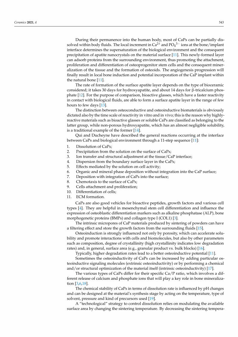
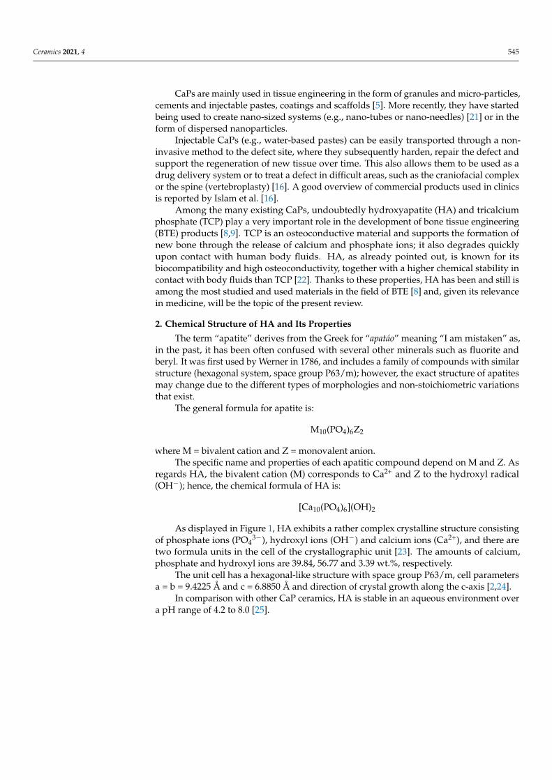
As displayed in Figure 1, HA exhibits a rather complex crystalline structure consistingof phosphate ions (PO4
3−), hydroxyl ions (OH−) and calcium ions (Ca2+), and there aretwo formula units in the cell of the crystallographic unit [23]. The amounts of calcium,phosphate and hydroxyl ions are 39.84, 56.77 and 3.39 wt.%, respectively.
The unit cell has a hexagonal-like structure with space group P63/m, cell parametersa = b = 9.4225 Å and c = 6.8850 Å and direction of crystal growth along the c-axis [2,24].
In comparison with other CaP ceramics, HA is stable in an aqueous environment overa pH range of 4.2 to 8.0 [25].
Ceramics 2021, 4 546
Figure 1. The crystalline structure of HA [5].
In each unit cell, the calcium ions can occupy two sites labelled as I and II, and thePO4
3− ions are divided into two planes at a crystal height of 1⁄4 and 3⁄4, respectively, yieldingthe creation of two different types of channels across the c axis named channel A andchannel B [26]. In channel A, there are oxygen atoms of the phosphate group and ions ofcalcium type II (Ca(II)) arranged at the vertices of two equilateral triangles that are rotatedof 60◦ from each other. The type B channel, with a diameter of 2 Å, contains type I calciumions (Ca(I)) only.
Each unit cell comprises/share:
• 14 Ca2+ ions: 6 of them inside the cell, and 8 shared with adjacent cells; total = 10 Ca2+
ions/unit cell• 10 PO4
3− ions: 2 of them located inside the cell, and 8 peripheral ions shared with asmany adjacent cells; total = 6 PO4
3− ions/unit cell• 8 OH− ions: being along the edges, they all belong to the cell by 1⁄4; total = 2 OH−
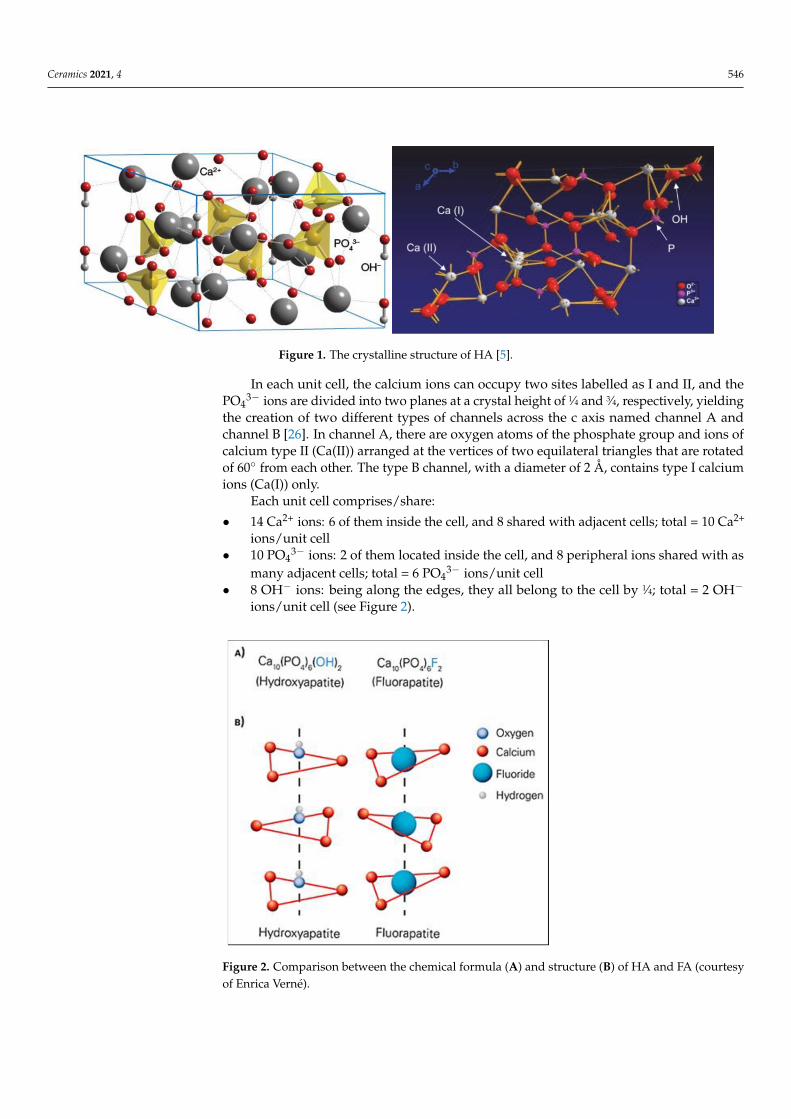
ions/unit cell (see Figure 2).
Figure 2. Comparison between the chemical formula (A) and structure (B) of HA and FA (courtesyof Enrica Verné).

Ceramics 2021, 4 547
Due to a different distribution of OH− ions, it is possible to distinguish two types ofHA: stoichiometric HA (or hexagonal HA) and monoclinic HA. In the stoichiometric HA,there are OH− radicals with alternate orientations at the center of type A channels.
Monoclinic HA is more thermodynamically stable and ordered than the hexagonalone; it forms at high temperatures and there is no evidence of its presence in calcifiedtissues.
The crystalline structure of HA can accommodate substitutions by various other ionsfor the Ca2+, PO4
3− and OH− groups. Ionic replacements can affect crystal morphology,crystallinity, solubility, lattice parameters, thermal stability and biological response of HA.Cationic substitutions occur at sites that are normally occupied by calcium atoms, whereasanionic substitutions may involve either phosphates or hydroxyl ions. Chlorapatite andfluorapatite are common examples of anion-substituted HA, where OH− ions are replacedby Cl− and F− ions, respectively (Figure 2).
Substitution of hydroxyl or phosphate ions with CO32− ions leads to the formation
of carbonate apatite that, although being classifiable as a bone apatite, cannot be useddirectly for osseous repair as it would induce inflammatory response upon implantation.Carbonate apatite is typically used after being sintered so that it partially decomposesat high temperature. Doy et al. [27] reported that specimens of carbonate apatite with12 wt.% of carbonate content could be sintered at 600–750 ◦C and retained around 6 wt.%of carbonate substitution in the lattice, which was comparable to that of bone apatite. Ingeneral, the reactivity of carbonate apatite in body fluids is higher than that of HA, thusyielding to fat dissolution rates depending on pH and particle/granule size [28].
HA is characterized by a Ca/P atomic ratio of 1.67, similar to that of biologicalapatite in human bones and teeth, thus making it very suitable for orthopedic, dental andmaxillofacial repair [29,30].
In general, the closer the Ca/P value to 1.67, the greater the stability of HA inside thehuman body [26].
Ca-deficient HAs with Ca/P < 1.67 exhibit a certain degree of solubility; they mayform, for example, as transient CaPs on the surface of bioactive glasses in vitro but thentend to evolve to stoichiometric HA during the bioactivity process [31–33].
It has also been shown that the mechanical strength of CaPs increases with increasingCa/P ratio and reaches its maximum value if the Ca/P ratio is ~1.67 (stoichiometric HA),while it decreases suddenly if the Ca/P ratio exceeds 1.67 [4]. Furthermore, by altering theCa/P molar ratio, it is possible to finely design the dissolution rate of CaPs [2].
Table 2 summarizes the principal properties of HA [5].
Table 2. Principal properties of HA [5].
Property Value Property Value
Density 3.16 g/cm3 Poisson’s ratio 0.27
Decomposition temperature >1000 ◦C Fracture Energy 2.3–20 J/m2
Dielectric costant 7.40–10.47 Fracture toughness 0.7–1.2 MPa·m1⁄2 (decreasewith porosity)
Thermal conductivity 0.013 W/cm·K Fracture hardness 3–7 GPa (dense HA)
Melting point 1614 ◦C Biocompatibility High
Tensile strength 38–300 MPa (dense HA)~3 MPa (porous HA) Biodegradation Low
Bending strength 38–250 MPa (dense HA)2–11 MPa (porous HA) Bioactivity High
Compressive strength 120–900 MPa (dense HA)2–100 MPa (porous HA) Osteoconduction High
Young’s elastic modulus 35–120 GPa Osteoinduction Nil
Ceramics 2021, 4 548
In addition to the composition, the shape, size and distribution of HA crystals also sig-nificantly affect the mechanical, biochemical and biological properties the material [34,35].In turn, these characteristics are dependent on the technique used for making HA powders.
3. HA Synthesis Techniques
The synthesis techniques for producing man-made HA can be divided into four maingroups, which are briefly described in the following sections.
The most commonly-used synthesis routes are also summarized in Table 3; the inter-ested Reader can find more details elsewhere [35].
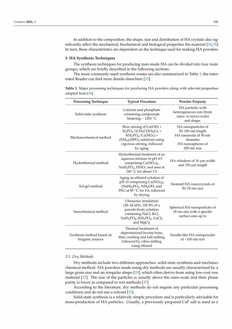
Table 3. Major processing techniques for producing HA powders along with relevant properties;adapted from [36].
Processing Technique Typical Procedure Powder Property
Solid-state synthesisCalcium and phosphatecontaining compounds.
Sintering ~ 1250 ◦C
HA particles withheterogeneous size (from
nano- to micro-scale)and shape.
Mechanochemical method
Slow mixing of Ca(OH) +H3PO4/(CH3COO)2Ca +
KH2PO4/Ca(NO3) +(NH4)2HPO4 solutions using
vigorous stirring, followedby aging
HA nanoparticles of50–100 nm length.
HA nanorods of 50 nmdiameter.
HA nanospheres of200 nm size
Hydrothermal method
Hydrothermal treatment of anaqueous mixture of pH 4.5
comprising Ca(NO3)2,NaH2PO4, HNO3 and urea at
160 ◦C for about 3 h
HA whiskers of 10 µm widthand 150 µm length
Sol-gel method
Aging an ethanol solution ofpH 10 comprising Ca(NO3)2,
(NaH)2PO4, NH4OH, andPEG at 85 ◦C for 4 h, followed
by drying
Sintered HA nanocrystals of50–70 nm size
Sonochemical method
Ultrasonic irradiation(28–34 kHz, 100 W) of apseudo-body solutioncontaining NaCl, KCl,
NaH2PO4, KH2PO4, CaCl2and MgCl2
Spherical HA nanoparticles of18 nm size with a specific
surface area up to
Synthesis method based onbiogenic sources
Thermal treatment ofdeproteinized bovine bone,
then crushing and ball milling,followed by vibro-milling
using ethanol
Needle-like HA nanopowderof ~100 nm size
3.1. Dry Methods
Dry methods include two different approaches: solid-state synthesis and mechano-chemical method. HA powders made using dry methods are usually characterized by alarge grain size and an irregular shape [35], which often derive from using low-cost rawmaterial [37]. The size of the particles is usually above the nano-scale and their phasepurity is lower as compared to wet methods [37].
According to the literature, dry methods do not require any particular processingconditions and do not use a solvent [35].
Solid-state synthesis is a relatively simple procedure and is particularly advisable formass-production of HA particles. Usually, a previously prepared CaP salt is used as a
Ceramics 2021, 4 549
precursor, which is then ground and calcined at high temperature (e.g., 1000 ◦C). The highcalcination temperature leads to the formation of a good crystalline structure. The finalparticles are heterogeneous and rather irregularly shaped.
The second strategy, known as mechano-chemical method or mechanical alloying,allows obtaining a powder with a much more defined structure as compared to the solid-state method, owing to perturbation of surface-bonded species as a result of pressure,which improves kinetic and thermodynamic reactions between solids [35]. In a typicalprocess, the materials are ground by using a planetary mill and the molar ratio between thereagents is kept at the stoichiometric ratio. This technique is relatively simple and easilyreproducible [35]; the principal processing variables are the type of reagents, the type ofmilling medium, the rotational speed and the duration of working phases and intervalsteps [35]. During their experiments, Nasiri-Tabrizi et al. [38] have proven that the averagesize of powder decrease with the increase of milling time; this also yields an increase oflattice strain.
The high reproducibility combined with low processing costs make dry methods themost suitable ones for the production of HA micro-particles in large amounts [34].
3.2. Wet Methods
Unlike dry methods, wet methods allow obtaining HA nanoparticles with a regularmorphology. For this reason, they are the most widely-used methods for the synthesis ofnano-powders [39].
One of the main disadvantages of wet methods is the low temperature used duringpreparation, which leads to the formation of CaP phases other than HA and/or traces ofimpurities in the crystalline structure due to ions from the aqueous solution used for thesynthesis [35].
These methods can be divided into six subgroups, i.e., conventional chemical precip-itation, hydrolysis, sol-gel, hydrothermal method, emulsion method and sono-chemicalmethod.
Chemical precipitation represents one of the easiest wet methods used for HA powderpreparation. It is based on the fact that, at pH 4.2 and room temperature, HA is usuallystable and not very soluble in an aqueous solution; however, the precipitation reactionis usually conducted at pH > 4.2 [35]. For this technique, CaP-containing precursorssuch as calcium nitrate or calcium hydroxide and diammonium hydrogen phosphate ororthophosphoric acid [35] are typically used. Usually, the reagents should be added drop-by-drop, under mild conditions and continuous stirring, checking that the molar Ca/Pratio remains at approximately 1.67. Then the suspension, after being carefully filteredand dried, is either aged for a period at atmospheric pressure or immediately reducedto powder [35]. HA particles produced by this method are, however, non-stoichiometricand poorly crystalline [19]. In order to obtain powders with higher phase purity, theprecipitation reaction must be conducted at higher temperature or higher pH or both; inthis way, the risk of formation of phase impurities decreases as well [35].
Hydrolysis method involves the preparation of HA nanoparticles by hydrolysis ofother CaP phases, such as dicalcium phosphate dihydrate (DCPD), tricalcium phosphate(TCP) under certain conditions and, also, octacalcium phosphate (OCP). To date, OCPhas almost completely been abandoned since it tends to incorporate impurities duringits transformation into HA [35]. Tenhuisen et al. [40] reported the production of HAfrom TCP and observed a linear relationship between the hydrolysis temperature and theresulting surface area of HA. It was also noted that, by increasing the reaction temperature,the regularity of the crystals increases [35]. Park et al. [41] have demonstrated that theaspect ratio, thermal stability and stoichiometry of HA strongly depend on the pH duringhydrolysis.
Sol-gel method allows obtaining fine and homogeneous powders thanks to the mixingof the reagents at molecular level and the possibility of using a low processing temper-ature [35]. The relevant reactions lead to the formation of a solid gel from a colloidal
Ceramics 2021, 4 550
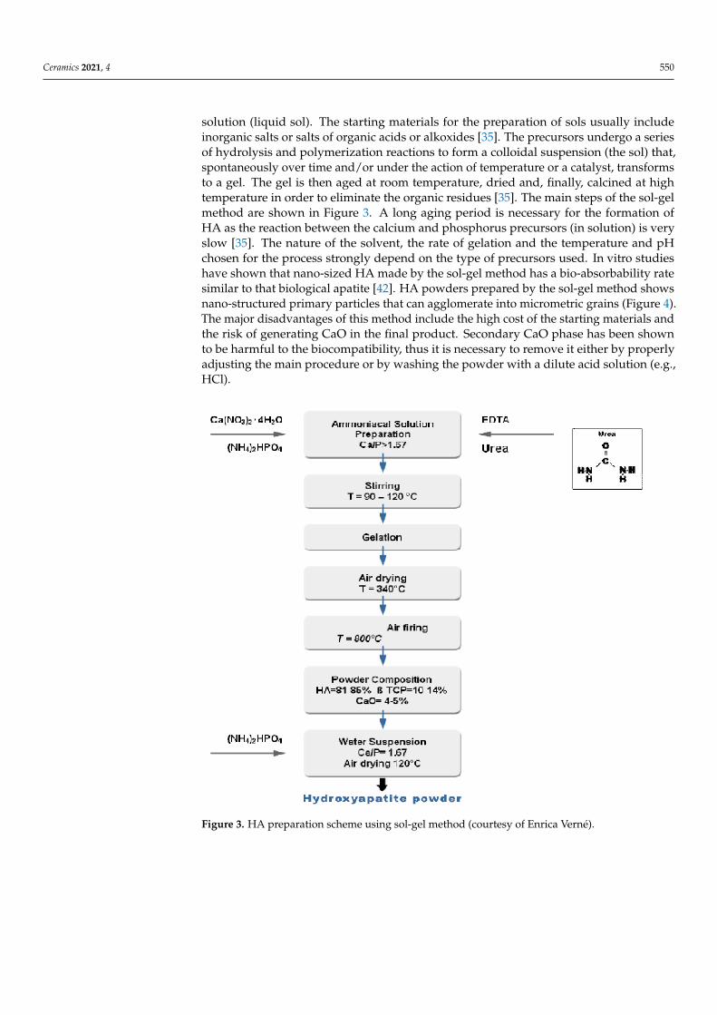
solution (liquid sol). The starting materials for the preparation of sols usually includeinorganic salts or salts of organic acids or alkoxides [35]. The precursors undergo a seriesof hydrolysis and polymerization reactions to form a colloidal suspension (the sol) that,spontaneously over time and/or under the action of temperature or a catalyst, transformsto a gel. The gel is then aged at room temperature, dried and, finally, calcined at hightemperature in order to eliminate the organic residues [35]. The main steps of the sol-gelmethod are shown in Figure 3. A long aging period is necessary for the formation ofHA as the reaction between the calcium and phosphorus precursors (in solution) is veryslow [35]. The nature of the solvent, the rate of gelation and the temperature and pHchosen for the process strongly depend on the type of precursors used. In vitro studieshave shown that nano-sized HA made by the sol-gel method has a bio-absorbability ratesimilar to that biological apatite [42]. HA powders prepared by the sol-gel method showsnano-structured primary particles that can agglomerate into micrometric grains (Figure 4).The major disadvantages of this method include the high cost of the starting materials andthe risk of generating CaO in the final product. Secondary CaO phase has been shownto be harmful to the biocompatibility, thus it is necessary to remove it either by properlyadjusting the main procedure or by washing the powder with a dilute acid solution (e.g.,HCl).
Figure 3. HA preparation scheme using sol-gel method (courtesy of Enrica Verné).

Ceramics 2021, 4 551
Figure 4. HA powder prepared via sol-gel [43].
Hydrothermal method is characterized by high working temperature and pressure.It can be considered as a chemical precipitation method in which the aging phase isconducted at high temperature [35]. The procedure is carried out in an autoclave, wherethe high temperature in a closed environment favors the formation of solvent vapor anda subsequent increase in pressure. Using a high-temperature results in a higher phasepurity than that achievable by traditional sol-gel methods and a suitable Ca/P ratio.However, high temperatures and pressures require expensive equipment, making thisprocess less affordable than other wet methods [35]. The hydrothermal method typicallyallows obtaining HA crystals with irregular morphology, at most spherical or rod-like;hexagonal prisms with 0.1 mm length were obtained by strictly controlling temperature,pH and in the presence of ethylenediamine tetraacetic acid (EDTA) [44].
Emulsion method is one of the most effective strategies for reducing particle sizeand achieving a controlled morphology and fine structure by limiting particle agglomer-ation [35]. This technique was originally used to made porous materials [45]. The mostcommon reagents are phosphoric acid and calcium nitrate because they are quite cheapand easy to be found on the market [44]. The most used surfactants are polyoxyethy-lene, cetyltrimethyl ammonium bromide and dioctyl sodium sulfosuccinate salt. It is arather simple method and uses low processing temperatures and mild synthesis condi-tions [46–48].
Sonochemical method is based on chemical reactions activated by powerful ultrasonicwaves. The physical mechanism behind the synthesis is acoustic cavitation in an aqueousphase. The reactivity of chemicals is stimulated to accelerate heterogeneous reactionsbetween liquid and solid reagents. It has recently been demonstrated that HA particlessynthesized with this process possess more uniform, smaller and purer crystals [35]. HazarYoruç and Ipek [49] have proved that the HA particle size decreases significantly byincreasing the ultrasonic powder up to 300 W. Due to the high speed and high kineticenergy stored during the synthesis, it is more likely that HA particles can collide with eachother and create a more uniform crystal lattice. This characteristic can improve mechanicalproperties of final product.
Ceramics 2021, 4 552
3.3. High-Temperature Processes
These processes are characterized by the need for using high temperatures to par-tially or completely burn the precursors [35]. There are two different methods based oncombustion and pyrolysis.
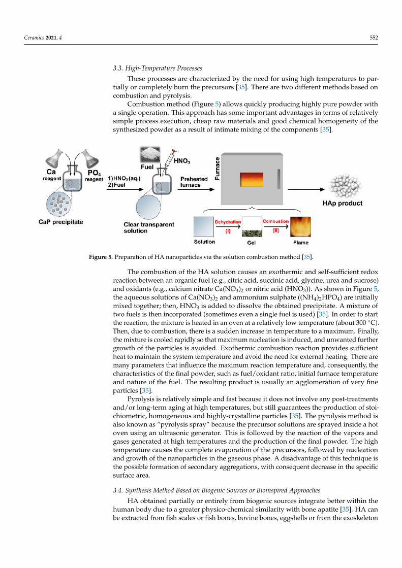
Combustion method (Figure 5) allows quickly producing highly pure powder witha single operation. This approach has some important advantages in terms of relativelysimple process execution, cheap raw materials and good chemical homogeneity of thesynthesized powder as a result of intimate mixing of the components [35].
Figure 5. Preparation of HA nanoparticles via the solution combustion method [35].
The combustion of the HA solution causes an exothermic and self-sufficient redoxreaction between an organic fuel (e.g., citric acid, succinic acid, glycine, urea and sucrose)and oxidants (e.g., calcium nitrate Ca(NO3)2 or nitric acid (HNO3)). As shown in Figure 5,the aqueous solutions of Ca(NO3)2 and ammonium sulphate ((NH4)2HPO4) are initiallymixed together; then, HNO3 is added to dissolve the obtained precipitate. A mixture oftwo fuels is then incorporated (sometimes even a single fuel is used) [35]. In order to startthe reaction, the mixture is heated in an oven at a relatively low temperature (about 300 ◦C).Then, due to combustion, there is a sudden increase in temperature to a maximum. Finally,the mixture is cooled rapidly so that maximum nucleation is induced, and unwanted furthergrowth of the particles is avoided. Exothermic combustion reaction provides sufficientheat to maintain the system temperature and avoid the need for external heating. There aremany parameters that influence the maximum reaction temperature and, consequently, thecharacteristics of the final powder, such as fuel/oxidant ratio, initial furnace temperatureand nature of the fuel. The resulting product is usually an agglomeration of very fineparticles [35].
Pyrolysis is relatively simple and fast because it does not involve any post-treatmentsand/or long-term aging at high temperatures, but still guarantees the production of stoi-chiometric, homogeneous and highly-crystalline particles [35]. The pyrolysis method isalso known as “pyrolysis spray” because the precursor solutions are sprayed inside a hotoven using an ultrasonic generator. This is followed by the reaction of the vapors andgases generated at high temperatures and the production of the final powder. The hightemperature causes the complete evaporation of the precursors, followed by nucleationand growth of the nanoparticles in the gaseous phase. A disadvantage of this technique isthe possible formation of secondary aggregations, with consequent decrease in the specificsurface area.
3.4. Synthesis Method Based on Biogenic Sources or Bioinspired Approaches
HA obtained partially or entirely from biogenic sources integrate better within thehuman body due to a greater physico-chemical similarity with bone apatite [35]. HA canbe extracted from fish scales or fish bones, bovine bones, eggshells or from the exoskeleton

Ceramics 2021, 4 553
of marine organisms. A period of annealing which lasts for several hours is generallyrequired to obtain HA. In this way, the organic part of the bone is removed, and the pureHA particles remain.
Simple thermal annealing is not the only extraction process that can be used; thefollowing are also possible: plasma processing, subcritical water processing, enzymatichydrolysis and alkaline hydrothermal hydrolysis [35]. All these methods make it possibleto remove organic substances from bone and produce pure HA with an average yield of65%.
Several experiments have been conducted to try decreasing the particle size at thenanoscale by means of the vibro-milling method that was used, for example, by Ruksudjaritet al. [50] who obtained HA from bovine bone. The bovine bone was deproteinized in hotwater and calcined at 800 ◦C, crushed into small pieces and finally milled in a ball mill potfor a minimum of 24 h.
In order to improve the properties of the final product, one or more of the methodsdescribed above can be combined. Among the various possibilities, combinations ofhydrothermal-hydrolysis and hydrothermal-microemulsion are most widely used [35].
Almost all of the techniques listed above allow obtaining HA with a stoichiometricratio similar to that of bio-apatite. The most important difference with biological HA is theabsence of ionic replacements that occur spontaneously within the human body [11]. Infact, natural HA contains a certain amount of ionic substitutions and impurities, such asMg2+, F−, K+, Na+, Cl− and Zn2+ [51].
A highly interesting line of research aims at obtaining HA products with bone-likestructural organization deriving from natural templates.
Historically, the most famous example is given by coralline HA, which was firstintroduced in the 1970s [52]. First, the coralline template is thermally treated at 900 ◦Cto allow the removal of all organic substances along with the decomposition of CaCO3into CaO, while the porous microstructure of the original coral is preserved. Upon ahydrothermal process with (NH4)2HPO4, conversion to β-tricalcium phosphate occurs and,eventually, HA is formed being the most thermodynamically stable calcium phosphatephase [53]. The use of KH2PO4 as a mineralizer was also experimented to accelerate the ionexchange process and to avoid the formation of intermediary phases; it was also reported abetter preservation of the original coralline architecture [54].
Recently, coralline HA was doped with strontium, which is known to have boneantiresorptive properties (anti-osteoporotic effect) [55].
Coral-derived HA is commercially available to surgeons in the form of porous granulesfor filling osseous defects, porous scaffolds for bone augmentation and spine surgery,porous plates for orbital floor repair and porous spheres used as orbital implants inenucleated patients [56].
More recently, Rattan wood has a strong similarity with bone as it exhibits a totalporosity of 85 vol.% and large macropores (diameter around 250 µm) organized in a systemof channels (mimicking the Haversian system in bone) interconnected with a networkof smaller canaliculi (mimicking the Volkmann system) [57]. In this regard, Tampieri’sresearch team developed a process of biomorphic transformation of Rattan wood in bone-like HA [58–60]. All the organic substances of wood are eliminated by pyrolysis, thusleaving behind a carbon skeleton which replicates the porous organization of the naturaltemplate. Carbonized wood is then converted to porous HA by a sequence of ion-exchangechemical reactions. A careful control of the chemical parameters of the process as well asthe kinetics of the reactions is the key to allow phase transformations to faithfully occurat a molecular level. Rattan-derived biomorphic porous HA is formed by needle-likenano-sized crystals, mimicking the mineral phase of bone, and exhibits a compressivestrength along the channel axis (4 MPa) comparable to that of calcified tissues [56]. In vitroinvestigation using MG-63 osteoblast-like cells confirmed the good biocompatibility of thewood-templated HA and in vivo tests in critical femoral defects of rats proved extensive
Ceramics 2021, 4 554
bone formation inside the HA channels without inflammation nor encapsulation insideconnective capsule after 1 month of follow-up [61].
4. Dense and Porous Hydroxyapatite
Once HA powder is produced, dense or porous HA-based products can be obtaineddepending on the sintering method chosen. Sintering treatment is carried out in specialelectric furnaces that reach high temperatures in a controlled manner, where the maximumtemperature has to be properly set in order to achieve proper densification of the mate-rial [4]. The heating rate, sintering temperature and holding time depend on the materialto treat; for HA, these values are in the typical ranges of 0.5–3 ◦C/min, 1000–1250 ◦C and2–5 h, respectively [4].
As a result of the sintering process, the ceramic powders can firmly bind together,while water, carbonates and other volatile chemicals are removed in the form of gaseousproducts. In most cases, this causes a considerable shrinkage which has to be considered atthe product design stage [4]
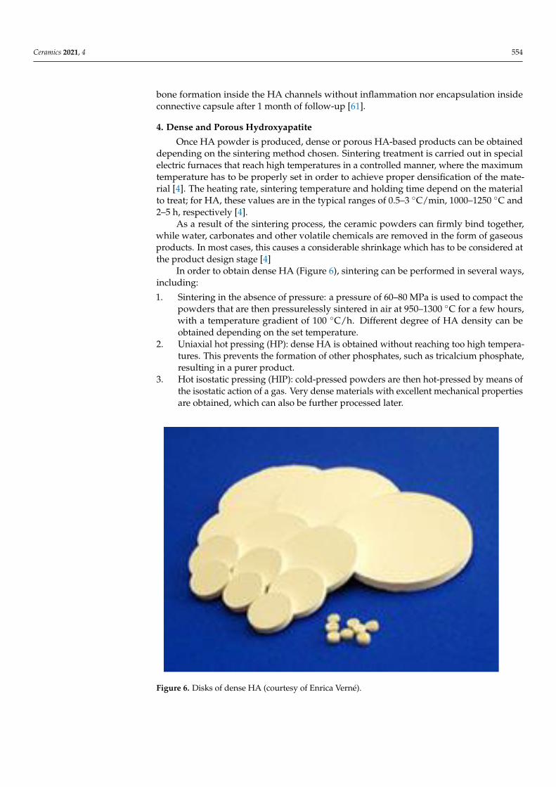
In order to obtain dense HA (Figure 6), sintering can be performed in several ways,including:
1. Sintering in the absence of pressure: a pressure of 60–80 MPa is used to compact thepowders that are then pressurelessly sintered in air at 950–1300 ◦C for a few hours,with a temperature gradient of 100 ◦C/h. Different degree of HA density can beobtained depending on the set temperature.
2. Uniaxial hot pressing (HP): dense HA is obtained without reaching too high tempera-tures. This prevents the formation of other phosphates, such as tricalcium phosphate,resulting in a purer product.
3. Hot isostatic pressing (HIP): cold-pressed powders are then hot-pressed by means ofthe isostatic action of a gas. Very dense materials with excellent mechanical propertiesare obtained, which can also be further processed later.
Figure 6. Disks of dense HA (courtesy of Enrica Verné).
Ceramics 2021, 4 555
These three techniques allow decreasing the grain size, thus obtaining HA with quitehigh density, fine microstructures, high thermal stability and high mechanical proper-ties [62].
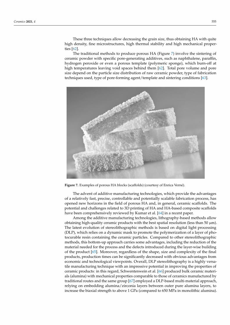
The traditional methods to produce porous HA (Figure 7) involve the sintering ofceramic powder with specific pore-generating additives, such as naphthalene, paraffin,hydrogen peroxide or even a porous template (polymeric sponge), which burn-off athigh temperatures leaving void spaces behind them [62]. Total pore volume and poresize depend on the particle size distribution of raw ceramic powder, type of fabricationtechniques used, type of pore-forming agent/template and sintering conditions [63].
Figure 7. Examples of porous HA blocks (scaffolds) (courtesy of Enrica Verné).
The advent of additive manufacturing technologies, which provide the advantagesof a relatively fast, precise, controllable and potentially scalable fabrication process, hasopened new horizons in the field of porous HA and, in general, ceramic scaffolds. Thepotential and challenges related to 3D printing of HA and HA-based composite scaffoldshave been comprehensively reviewed by Kumar et al. [64] in a recent paper.
Among the additive manufacturing technologies, lithography-based methods allowobtaining high-quality ceramic products with the best spatial resolution (less than 50 µm).The latest evolution of stereolithographic methods is based on digital light processing(DLP), which relies on a dynamic mask to promote the polymerization of a layer of pho-tocurable resin containing the ceramic particles. Compared to other stereolithographicmethods, this bottom-up approach carries some advantages, including the reduction of thematerial needed for the process and the defects introduced during the layer-wise buildingof the product [65]. Moreover, regardless of the shape, size and complexity of the finalproducts, production times can be significantly decreased with obvious advantages fromeconomic and technological viewpoints. Overall, DLP stereolithography is a highly versa-tile manufacturing technique with an impressive potential in improving the properties ofceramic products: in this regard, Schwentenwein et al. [66] produced bulk ceramic materi-als (alumina) with mechanical properties comparable to those of ceramics manufactured bytraditional routes and the same group [67] employed a DLP-based multi-material approach,relying on embedding alumina/zirconia layers between outer pure alumina layers, toincrease the biaxial strength to above 1 GPa (compared to 650 MPa in monolithic alumina).
Ceramics 2021, 4 556
In general, however, ceramic scaffolds—including the HA ones—obtained by addi-tive manufacturing technologies typically exhibit a relatively simple porous architecturewith grid-like arrangements of macro-channels (i.e., the structure of the CAD file usedfor printing) and, thus, do not closely replicate the trabecular architecture of cancellousbone as ceramic foams instead do. In order to overcome this limitation and further expandthe potential of additive manufacturing in biomedicine, HA scaffolds were recently fabri-cated by DLP stereolithography using a micro-tomographic reconstruction of an open-cellpolymeric foam as a CAD model [68]. As a result, truly bone-like HA scaffolds with 3Dtrabecular architecture, pore size, intrinsic permeability, elastic modulus and compressivestrength comparable to those of human cancellous bone were obtained.
HA-based porous ceramic can develop a strong junction with natural bone as thepores allow achieving a strong mechanical interlocking with regenerating tissue, whichyields a stronger fixation of the structure [61]. In general, porous HA is more resorbableand osteoconductive than dense HA. In fact, thanks to the higher surface area, more bonecells are able to attach and proliferate on the implant [63].
5. Mechanical Properties
From a technological viewpoint, the mechanical properties of HA-based productsindeed depend on the method used for powder preparation, which determines the grainsize and shape, Ca/P ratio and purity and the sintering conditions, which dictates thecharacteristics of microporosity [10]. If production of macroporous HA is a goal, sinteringconditions and type of pore-forming agent also dictate the characteristics of macroporosity.
In general, density, grain size, compressive/torsional/flexural strength and elasticmodulus increase as the sintering temperature increases. The mechanical properties of HAdecrease significantly with increasing microporosity, amorphous phase and grain size. Onthe contrary, low porosity, high crystallinity and small grain size increase tensile strength,compressive strength, fracture toughness and stiffness. Fracture toughness can decreasedue to the presence of TCP, that can form during sintering at high temperatures.
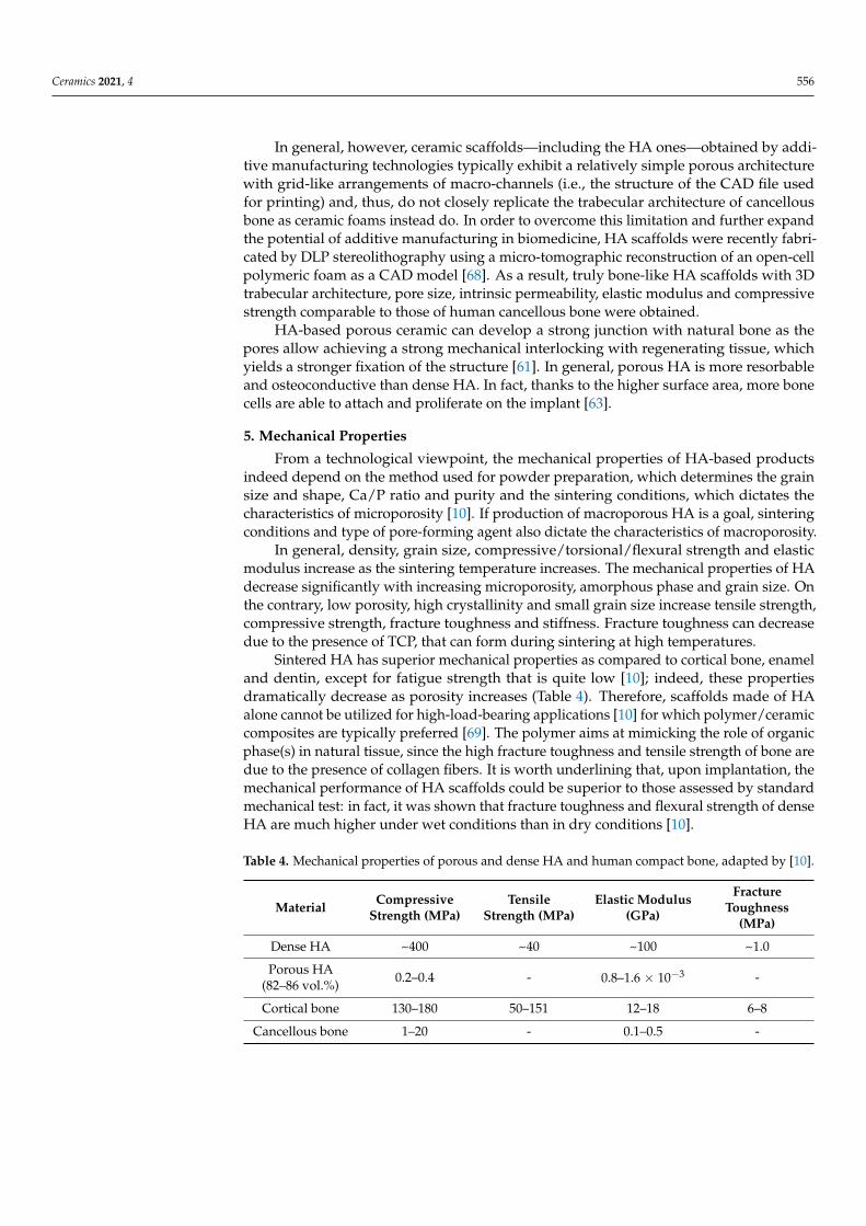
Sintered HA has superior mechanical properties as compared to cortical bone, enameland dentin, except for fatigue strength that is quite low [10]; indeed, these propertiesdramatically decrease as porosity increases (Table 4). Therefore, scaffolds made of HAalone cannot be utilized for high-load-bearing applications [10] for which polymer/ceramiccomposites are typically preferred [69]. The polymer aims at mimicking the role of organicphase(s) in natural tissue, since the high fracture toughness and tensile strength of bone aredue to the presence of collagen fibers. It is worth underlining that, upon implantation, themechanical performance of HA scaffolds could be superior to those assessed by standardmechanical test: in fact, it was shown that fracture toughness and flexural strength of denseHA are much higher under wet conditions than in dry conditions [10].
Table 4. Mechanical properties of porous and dense HA and human compact bone, adapted by [10].
Material CompressiveStrength (MPa)
TensileStrength (MPa)
Elastic Modulus(GPa)
FractureToughness
(MPa)
Dense HA ~400 ~40 ~100 ~1.0
Porous HA(82–86 vol.%) 0.2–0.4 - 0.8–1.6 × 10⁻³ -
Cortical bone 130–180 50–151 12–18 6–8
Cancellous bone 1–20 - 0.1–0.5 -

Ceramics 2021, 4 557
The use of additive manufacturing technologies allowed significantly improvingthe strength of HA scaffolds. For example, grid-like HA scaffolds produced by DLPstereolithography exhibited a compressive strength comparable to that of cancellous bone(1.45–1.92 MPa), although the porosity was lower (49–52 vol.%) [70]. The same fabricationmethod was recently combined with micro-tomographic imaging to obtain trabecular-like HA scaffolds with compressive strength of 1.60 ± 0.79 MPa and bone-like porosity(80 vol.%) and permeability (0.75–1.74·10−9 m2) [68]. Prolonged immersion in simulatedbody fluid (SBF) for up to 2 months lad to no significant variation in the mechanical strengthof these bone-like HA scaffolds, which can be justified by the very low dissolution rate ofHA. Interestingly, these values of compressive strength are from four to eight times higherthan those obtained for foam-replicated HA scaffolds with analogous porosity (Table 4).Another study revealed that using nano-sized HA particles (diameter around 150 nm) tocoat the polyurethane sponge in the foam-replica method yielded just minor improvementin the compressive strength (0.51 MPa) [71]. This suggests that the scaffold productionmethod plays a major role over the particle size in affecting the mechanical performance ofthe final macroporous product.
6. Applications of HA in Tissue Engineering
In 1969, Levitt et al. [72] wrote one of the first scientific papers on the possible applica-tions of HA to make complex implants in contact with bone to repair and regenerate hardtissues, thus figuring out the use of HA in BTE.
HA powders are mainly used to produce coatings on metallic implants, as startingmaterial for making scaffolds, or as fillers for polymer-matrix composites [73].
HA powders as coatings began to be used mainly in the late 1980s; more specifically,Furlong and Osborn [74] were the first to begin studying HA coatings for clinical applica-tions and, since then, these coatings have shown good results with a 2% failure rate for a10-year follow-up study. In this way, the use of CaPs has somewhat expanded even in thefield of load-bearing applications, as metal substrates help to increase the poor mechanicalstrength of ceramics [12,25].
Usually, HA coatings are applied on metallic implants by plasma treatment; clinicalresults reveal that HA-coated implants have a longer lifetime as compared to uncoatedimplants, which can be very beneficial especially for younger patients. Thanks to thepresence of HA, bone tissue can better integrate with the implant (osteoconduction) and, inaddition, HA reduces the release of metal ions from the implant, thus avoiding undesirableinflammatory reactions or toxic effects [62].
In order to achieve positive results, it is crucial to choose the right thickness of thecoating to obtain satisfactory adhesion and interfacial stability; it has been shown that goodoutcomes are obtained with a thickness in the range of 40 to 200 µm [62].
An important use of porous HA is for the controlled drug release in bone disorderssuch as bone tumors or osteoporosis. Porous HA granules or scaffolds loaded with drugsensures a localized and prolonged release of drugs, which speeds bone healing [62]. Arecent study by Ferreira-Ermita et al. [75] showed in a rabbit model that HA associatedwith magnetite nanoparticles was an effective platform for the release of ciprofloxacin inthe treatment of osteomyelitis.
Custom-made porous HA scaffolds are also widely used as gap filler to treat bone de-fects through promoting bone ingrowth, especially in cases where conventional techniqueshave failed. For example, Quarto et al. [76] implanted HA scaffolds seeded with in vitroexpanded autologous bone marrow cells to cure large bone diseases (4–7 cm) of the ulna,humerus and tibia in patients of different ages. In another study, Vacanti et al. [77] proposeda natural coral-derived implant (porous HA; pore size 500 µm, ProOsteon) functionalizedin vitro with autologous periosteal cells to treat a traumatic avulsion of the distal phalanxof a man thumb. Following a similar approach, Morishita et al. [78] treated a disease dueto tumors in a tibia and in a femur by using HA scaffolds seeded with in vitro expandedautologous bone marrow stromal cells.

Ceramics 2021, 4 558
For the preparation of 3D scaffolds, HA can be used alone or in combination withdifferent polymers such as poly(lactic-co-glycolic) acid (PLGA), poly(L-lactic acid) (PLLA)and polycaprolactone (PCL) to improve the mechanical properties or impart a controllableresorbability [79].
Ignjatovic et al. [80] and Wang et al. [81] fabricated HA/PLLA and HA/polyethylenecomposites, respectively, and in both cases these composites had a sufficient mechanicalstrength to be utilized in BTE. HA/polyethylene composites are also commercialized underthe tradename of HAPEX and clinically used for the replacement of middle ear bonesand orbital floor repair. A comprehensive picture of the latest advances in HA/polymercomposites and cell-seeded HA constructs has been recently reported by Kattimai et al. [82].
The application of bone synthetic grafts has further evolved in BTE thanks to improve-ment of different additive manufacturing techniques, including selective laser sintering,laser cladding, 3D printing and stereolithography [64]. Various combinations of HA withother materials, including polymers, bioactive glasses and other crystalline ceramics, arealso possible through multi-material printing to obtain composites; this approach has beentypically limited to robocasting techniques, but recent stereolithographic systems have alsobeen adapted to process more than one ceramic material simultaneously (alumina andzirconia in early trials) [67].
Surgeons often prefer injectable biomaterials as compared to rigid scaffolds, as thepastes are easy to handle and conform to the anatomy of the osseous defect. In this regard,there are many commercially-available CaP-based cements that form a workable paste afterbeing mixed with an aqueous saline solution [83]. This putty can be easily shaped duringsurgery to perfectly match the geometry of the bone defect prior to set within 10–20 min.Upon setting, the reactants reprecipitate until the entire material is converted to finelyporous HA. After being implanted in vivo, CaP cements are gradually resorbed over timeand replaced with newly-formed bone.
Mixing with collagen was apparently regarded as a good strategy to improve thebiomimetic properties of bone cements. In this regard, Cuzmar et al. [84] analyzed theosteogenic capacity of Ca-deficient HA microspheres with or without collagen obtained byemulsification of a CaP cement paste and compared the performance with an injectableceramic cement having the same composition. After implantation in femur condyles ofrabbits, histological analysis revealed that the cements presented cellular activity only inthe margins of the material, whereas the microspheres were well coated with osteogeniccells. As a result, bone ingrowth was enhanced by the microspheres, with a tenfold increaseas compared to the conventional cement, which was associated to the higher accessibilityfor the cells provided by the macroporous network between the microspheres and thelarger surface area available for osteoconduction. Interestingly, no significant differenceswere found in terms of bone formation associated with the presence of collagen in themicrospheres, although a more extensive resorption of the collagen-containing materialwas observed.
Some examples of commercially-available HA products for osseous repair and BTEare also listed in Table 5.

Ceramics 2021, 4 559
Table 5. Various examples of commercially-available HA products for BTE, adapted from [73].
Tradename Producer Country
Actifuse ApaTech UKApaPore ApaTech UK
Apaceram Pentax JapanBonefil Pentax Japan
Bonetite Pentax JapanBonoceram Sumitomo Osaka Cement Japan
Bioroc Depuy—Bioland FranceCerapatite Ceraver France
BoneSource Stryker Orthopaedics NJ, USACalcitite Zimmer IN, USA
Osteograf Ceramed CO, USA
Although being mainly employed for the repair of bone defects, HA has also beenrecently proposed in some emerging applications in contact with soft tissues, includingophthalmology, wound regeneration and anticancer therapies; a comprehensive picture ofthese novel research topics has been recently provided by Kargozar et al. [85]. As regardsocular applications, HA is marketed and clinically used since the 1980s in the form ofporous orbital implants for enucleation (e.g., coralline or synthetic HA spheres) [86].
7. Conclusions
Considering all the various biomaterials currently available in bone regenerativestrategies, calcium phosphates (CaPs) and, specifically, hydroxyapatite (HA), are amongthe most commonly-used due to their compositional and structural similarities to naturalbone and teeth, as well as exceptional biocompatibility and biological behavior in contactwith body fluids, which make them materials of choice for producing synthetic bone graftsto be used as an alternative to transplantation (auto- and allografts).
Due to an excellent osteoconductive potential, HA has been highly favored in boneBTE applications, being the main representative of commercial products currently used inclinical practice. Several studies demonstrated the capability of HA to create a favorableenvironment for promoting new bone tissue ingrowth and regeneration, resulting in aperfect integration of the graft without leading to the development of a severe immuneresponse. These appealing properties made it an optimal candidate in different clinicalfields. To date, the main biomedical applications of HA include osseous defect filling(e.g., augmentation and stabilization of the jawbone in maxillofacial reconstruction), spinalfusion, bone filling after tumor operation, replacement of middle ear bones and repair oforbital floor fractures. Great results were also achieved in the development of drug deliverysystems and carriers of bioactive peptides and/or various cell types.
In recent years, the growing interest in three-dimensional (3D), in vivo-like bone tissueengineering approaches made the biomedical research focus on the development of HAporous grafts mimicking the trabecular architecture of human spongy bone. In this regard,the main challenge to be addressed in the future is certainly represented by the need toachieve adequate mechanical properties to allow a safe use of the graft. Like all ceramicmaterials, in fact, HA has low tensile strength and an intrinsic brittleness that limits itsapplication in load-bearing anatomical sites. Currently, the most common strategy toovercome this drawback is represented by the production of polymer/ceramic compositescaffolds, which have been directly inspired by the peculiar compositional features ofbiological bone, where the presence of collagen fibers confers high fracture toughness andtensile strength to the tissue. Looking at the future, there is a great expectation in theuse of additive manufacturing technologies combined with computer aided design (CAD)and physical simulation tools, with the ambitious aim of introducing the use of advancedtailored—and even personalized—BTE products in clinical practice.
Ceramics 2021, 4 560
Author Contributions: All the authors equally contributed to the present work. All authors haveread and agreed to the published version of the manuscript.
Funding: This research received no external funding.
Institutional Review Board Statement: Not applicable.
Informed Consent Statement: Not applicable.
Data Availability Statement: The data reported in this work, being a review paper, can be found inthe original sources cited in the reference list.
Conflicts of Interest: The authors declare no conflict of interest.
References1. Albee, F.H.; Morrison, H.F. Studies in bone growth triple calcium phosphate as a stimulus to osteogenesis. Ann. Surg. 1920, 71,
32–39.2. Jazayeri, H.E.; Rodriguez-Romero, M.; Razavi, M.; Tahriri, M.; Ganjawalla, K.; Rasoulianboroujeni, M.; Malekoshoaraie, M.H.;
Khoshroo, K.; Tayebi, L. The cross-disciplinary emergence of 3D printed bioceramic scaffolds in orthopedic bioengineering.Ceram. Int. 2018, 44, 1–9. [CrossRef]
3. Jeong, J.; Kim, J.H.; Shim, J.H.; Hwang, N.S.; Heo, C.Y. Bioactive calcium phosphate materials and applications in boneregeneration. Biomater. Res. 2019, 23, 4. [CrossRef]
4. Dorozhkin, S.V. Calcium orthophosphate bioceramics. Ceram. Int. 2015, 41, 13913–13966. [CrossRef]5. Eliaz, N.; Metoki, N. Calcium Phosphate Bioceramics: A Review of Their History, Structure, Properties, coating Technologies and
Biomedical Applications. Materials 2017, 10, 334. [CrossRef] [PubMed]6. Dorozhkin, S.V. Calcium orthophosphates as bioceramics: State of the art. J. Funct. Biomater. 2010, 1, 22–107. [CrossRef]7. Dorozhkin, S.V. Multiphasic calcium orthophosphate (CaPO4) bioceramics and their biomedical applications. Ceram. Int. 2016,
42, 6529–6554. [CrossRef]8. Carvalho, B.; De Rompen, E.; Lecloux, G.; Schupbach, P.; Dory, E. Effect of sintering on in vivo biological performance of bovine
hydroxyapatite. Materials 2019, 12, 3946. [CrossRef] [PubMed]9. Mohamad Yunos, D.; Bretcanu, O.; Boccaccini, A.R. Polymer-bioceramic composites for tissue engineering scaffolds. J. Mater. Sci.
2008, 43, 4433–4442. [CrossRef]10. Rezwan, K.; Chen, Q.Z.; Blaker, J.J.; Boccaccini, A.R. Biodegradable and bioactive porous polymer/inorganic composite scaffolds
for bone tissue engineering. Biomaterials 2006, 27, 3413–3431. [CrossRef] [PubMed]11. Owen, R.G.; Dard, M.; Larjava, H. Hydoxyapatite/beta-tricalcium phosphate biphasic ceramics as regenerative material for the
repair of complex bone defects. J. Biomed. Mater. Res. B Appl. Biomater. 2018, 106, 2493–2512. [CrossRef]12. Søballe, K. Hydroxyapatite ceramic coating for bone implant fixation: Mechanical and histological studies in dogs. Acta Orthop.
Scand. 1993, 64, 1–58. [CrossRef]13. Fiume, E.; Barberi, J.; Verné, E.; Baino, F. Bioactive Glasses: From Parent 45S5 Composition to Scaffold-Assisted Tis-sue-Healing
Therapies. J. Funct. Biomater. 2018, 9, 24. [CrossRef]14. Cao, W.; Hench, L.L. Bioactive materials. Ceram. Int. 1996, 22, 493–507. [CrossRef]15. Horowitz, R.A.; Mazor, Z.; Foitzik, C.; Prasad, H.; Rohrer, M.; Palti, A. β-Tricalcium Phosphate As Bone Substitute Material. J.
Osseointegr. 2010, 1, 60–68.16. Islam, M.T.; Felfel, R.M.; Abou Neel, E.A.; Grant, D.M.; Ahmed, I.; Hossain, K.M.Z. Bioactive calcium phosphate–based glasses
and ceramics and their biomedical applications: A review. J. Tissue Eng. 2017, 8, 2041731417719170. [CrossRef] [PubMed]17. Ebrahimi, M.; Botelho, M.G.; Dorozhkin, S.V. Biphasic calcium phosphates bioceramics (HA/TCP): Concept, physico-chemical
properties and the impact of standardization of study protocols in biomaterials research. Mater. Sci. Eng. C 2017, 71, 1293–1312.[CrossRef]
18. Habraken, W.J.E.M.; Tao, J.; Brylka, L.J.; Friedrich, H.; Bertinetti, L.; Schenk, A.; Verch, A.; Dmitrovic, V.; Bomans, P.H.H.; Frederik,P.M.; et al. Ion-association complexes unite classical and non-classical theories for the biomimetic nucleation of calcium phosphate.Nat. Commun. 2013, 4, 1507. [CrossRef] [PubMed]
19. Kumta, P.N.; Sfeir, C.; Lee, D.H.; Olton, D.; Choi, D. Nanostructured calcium phosphates for biomedical applications: Novelsynthesis and characterization. Acta Biomater. 2005, 1, 65–83. [CrossRef] [PubMed]
20. Lawson, A.C.; Czernuszka, J.T. Collagen-calcium phosphate composites. Proc. Inst. Mech. Eng. H 1998, 212, 413–425. [CrossRef][PubMed]
21. Albulescu, R.; Popa, A.-C.; Enciu, A.-M.; Albulescu, L.; Dudau, M.; Popescu, I.D.; Mihai, S.; Codrici, E.; Pop, S.; Lupu, A.-R.;et al. Comprehensive In Vitro Testing of Calcium Phosphate-Based Bioceramics with Orthopedic and Den-tistry Applications.Materials 2019, 10, 3704. [CrossRef] [PubMed]
22. Szczes, A.; Hołysz, L.; Chibowski, E. Synthesis of hydroxyapatite for biomedical applications. Adv. Colloid Interface Sci. 2017, 249,321–330. [CrossRef] [PubMed]
23. Wopenka, B.; Pasteris, J.D. A mineralogical perspective on the apatite in bone. Mater. Sci. Eng. C 2005, 25, 131–143. [CrossRef]

Ceramics 2021, 4 561
24. Astala, R.; Stott, M.J. First principles investigation of mineral component of bone: CO3 substitutions in hydroxyapatite. Chem.Mater. 2005, 17, 4125–4133. [CrossRef]
25. Best, S.M.; Porter, A.E.; Thian, E.S.; Huang, J. Bioceramics: Past, present and for the future. J. Eur. Ceram. Soc. 2008, 28, 1319–1327.[CrossRef]
26. Rivera-Muñoz, E.M. Hydroxyapatite-Based Materials: Synthesis and Characterization. In Biomedical Engineering: Frontiers andChallenges; Fazel-Rezai, R., Ed.; Intech Open: Rijeka, Croatia, 2011.
27. Doi, Y.; Shibutani, T.; Moriwaki, Y.; Kajimoto, T.; Iwayama, Y. Sintered carbonate apatites as bioresorbable bone substitutes. J.Biomed. Mater. Res. 1998, 39, 603–610. [CrossRef]
28. Rahyussalim, A.J.; Supriadi, S.; Marsetio, A.F.; Pribadi, P.M.; Suharno, B. The potential of carbonate apatite as an alternative bonesubstitute material. Med. J. Indones. 2019, 28, 92–97. [CrossRef]
29. Wang, A.N.; Wu, L.G.; Li, X.L.; Sun, Y.D.; Wang, J.; Wang, S.W.; Jia, A.X.; Wang, C.; Zhang, Y.Y.; Fu, Q.Q.; et al. Study on the BlendFilm Prepared by Chitosan and Gelatin. Adv. Mater. Res. 2011, 201–203, 2866–2869. [CrossRef]
30. Bhattacharjee, A.; Fang, Y.; Hooper, T.J.N.; Kelly, N.L.; Gupta, D.; Balani, K.; Manna, I.; Baikie, T.; Bishop, P.T.; White, T.J.; et al.Crystal chemistry and antibacterial properties of cupriferous hydroxyapatite. Materials 2019, 12, 1814. [CrossRef]
31. López-Noriega, A.; Arcos, D.; Izquierdo-Barba, I.; Sakamoto, Y.; Terasaki, O.; Vallet-Regí, M. Ordered mesoporous bioactiveglasses for bone tissue regeneration. Chem. Mater. 2006, 18, 3137–3144. [CrossRef]
32. Baino, F.; Fiume, E.; Miola, M.; Leone, F.; Onida, B.; Verné, E. Fe-doped bioactive glass-derived scaffolds produced by sol-gelfoaming. Mater. Lett. 2019, 235, 207–211. [CrossRef]
33. Fiume, E.; Serino, G.; Bignardi, C.; Verné, E.; Baino, F. Bread-derived bioactive porous scaffolds: An innovative and sus-tainableapproach to bone tissue engineering. Molecules 2019, 24, 2954. [CrossRef] [PubMed]
34. Silva, C.; Pinheiro, A.; De Oliveira, R.; Goes, J.C.; Aranha, N.; De Oliveira, L.; Sombra, A. Properties and in vivo investigation ofnanocrystalline hydroxyapatite obtained by mechanical alloying. Mater. Sci. Eng. C 2004, 24, 549–554. [CrossRef]
35. Sadat-Shojai, M.; Khorasani, M.T.; Dinpanah-Khoshdargi, E.; Jamshidi, A. Synthesis methods for nanosized hydroxyapatite withdiverse structures. Acta Biomater. 2013, 9, 7591–7621. [CrossRef] [PubMed]
36. Roy, M.; Bandyopadhyay, A.; Bose, S. Chapter 6: Ceramics in Bone Grafts and Coated Implants. In Materials for Bone Disorders;Bose, S., Bandyopadhyay, A., Eds.; Academic Press (Elsevier): London, UK, 2017; pp. 265–314.
37. Li, B.; Webster, T. Orthopedic Biomaterials: Advances and Applications; Springer: Cham, Switzerland, 2017.38. Nasiri-Tabrizi, B.; Honarmandi, P.; Ebrahimi-Kahrizsangi, R. Synthesis of nanosize single-crystal hydroxyapatite via
mechanochemical method. Mater. Lett. 2009, 63, 543–546. [CrossRef]39. Zhan, J.; Tseng, Y.H.; Chan, J.C.C.; Mou, C.Y. Biomimetic formation of hydroxyapatite nanorods by a single-crystal-to-single-
crystal transformation. Adv. Funct. Mater. 2005, 15, 2005–2010. [CrossRef]40. Tenhuisen, K.S.; Brown, P.W. Formation of calcium-deficient hydroxyapatite from α-tricalcium phosphate. Biomaterials 1998, 19,
2209–2217. [CrossRef]41. Park, H.C.; Baek, D.J.; Park, Y.M.; Yoon, S.Y.; Stevens, R. Thermal stability of hydroxyapatite whiskers derived from the hydrolysis
of α-TCP. J. Mater. Sci. 2004, 39, 2531–2534. [CrossRef]42. Fathi, M.; Hanifi, A.; Mortazavi, V. Preparation and bioactivity evaluation of bone-like hydroxyapatite nanopowder. J. Mater.
Process. Technol. 2008, 202, 536–542. [CrossRef]43. Sopyan, I.; Singh, R.; Hamdi, M. Synthesis of nano sized hydroxyapatite powder using sol-gel technique and its conver-sion to
dense and porous bodies. Indian J. Chem. A 2008, 47, 1626–1631.44. Zhu, R.; Yu, R.; Yao, J.; Wang, D.; Ke, J. Morphology control of hydroxyapatite through hydrothermal process. J. Alloys Compd.
2008, 457, 555–559. [CrossRef]45. Chun, H.J.; Park, K.; Kim, C.-H.; Khang, G. Novel Biomaterials for Regenerative Medicine, 1st ed.; Springer: Singapore, 2018.46. Guo, G.; Sun, Y.; Wang, Z.; Guo, H. Preparation of hydroxyapatite nanoparticles by reverse microemulsion. Ceram. Int. 2005, 31,
869–872. [CrossRef]47. Sun, Y.; Guo, G.; Wang, Z.; Guo, H. Synthesis of single-crystal HAP nanorods. Ceram. Int. 2006, 32, 951–954. [CrossRef]48. Li, H.; Zhu, M.Y.; Li, L.H.; Zhou, C.R. Processing of nanocrystalline hydroxyapatite particles via reverse microemul-sions. J.
Mater. Sci. 2008, 43, 384–389. [CrossRef]49. Hazar Yoruç, A.B.; Ipek, Y. Sonochemical synthesis of hydroxyapatite nanoparticles with different precursor reagents. Acta Phys.
Pol. A 2012, 121, 230–232. [CrossRef]50. Ruksudjarit, A.; Pengpat, K.; Rujijanagul, G.; Tunkasiri, T. Synthesis and characterization of nanocrystalline hydroxyapatite from
natural bovine bone. Curr. Appl. Phys. 2008, 8, 270–272. [CrossRef]51. Qu, H.; Fu, H.; Han, Z.; Sun, Y. Biomaterials for bone tissue engineering scaffolds: A review. RSC Adv. 2019, 9, 26252–26262.
[CrossRef]52. White, R.A.; Weber, J.N.; White, E.W. Replamineform: A New Process for Preparing Porous Ceramic, Metal, and Poly-mer
Prosthetic Materials. Science 1972, 176, 922–924. [CrossRef]53. White, E.W.; Weber, J.N.; Roy, D.M.; Owen, E.L.; Chiroff, R.T.; White, R.A. Replamineform Porous Biomaterials for Hard Tissue
Implant Applications. J. Biomed. Mater. Res. 1975, 6, 23–27. [CrossRef]54. Xu, Y.; Wang, D.; Yang, L.; Tang, H. Hydrothermal Conversion of Coral into Hydroxyapatite. Mater. Charact. 2001, 47, 83–87.
[CrossRef]
Ceramics 2021, 4 562
55. Liu, W.; Wang, T.; Shen, Y.; Pan, H.; Peng, S.; Lu, W.W. Strontium Incorporated Coralline Hydroxyapatite for Engineering Bone.ISRN Biomater. 2013, 2013, 649163. [CrossRef]
56. Damien, E.; Revell, P.A. Coralline Hydroxyapatite Bone Graft Substitute: A Review of Experimental Studies and BiomedicalApplications. J. Appl. Biomater. Biomech. 2004, 2, 65–73. [PubMed]
57. Tampieri, A.; Sprio, S.; Ruffini, A.; Celotti, G.; Lesci, I.G.; Roveri, N. From Wood to Bone: Multi-Step Process to Convert WoodHierarchical Structures into Biomimetic Hydroxyapatite Scaffolds for Bone Tissue Engineering. J. Mater. Chem. 2009, 19, 4973–4980.[CrossRef]
58. Ruffini, A.; Sprio, S.; Tampieri, A. Wood Structures with Organized Morphology for Bone Substitutes. J. Appl. Biomater. Biomech.2007, 5, 207.
59. Sprio, S.; Ruffini, A.; Valentini, F.; D’Alessandro, T.; Sandri, M.; Panseri, S.; Tampieri, A. Biomimesis and Biomorphic Transforma-tions: New Concepts Applied to Bone Regeneration. J. Biotechnol. 2011, 156, 347–355. [CrossRef]
60. Ruffini, A.; Sprio, S.; Tampieri, A. Study of the Hydrothermal Transformation of Wood-Derived Calcium Carbonate into 3DHierarchically Organized Hydroxyapatite. Chem. Eng. J. 2013, 217, 150–158. [CrossRef]
61. Ruffini, A.; Sprio, S.; Tampieri, A. Towards Hierarchically Organized Scaffolds for Bone Substitutes from Wood Structures. KeyEng. Mater. 2008, 361–363, 959–962.
62. Suchanek, W.; Yoshimura, M. Processing and properties of hydroxyapatite-based biomaterials for use as hard tissue re-placementimplants. J. Mater. Res. 1998, 13, 94–117. [CrossRef]
63. Al-Naib, U.M.B. Introductory chapter: A Brief Introduction to Porous Ceramic. In Recent Advances in Porous Ceramics; Al-Naib,U.M.B., Ed.; Intech Open: Rijeka, Croatia, 2018.
64. Kumar, A.; Kargozar, S.; Baino, F.; Han, S.S. Additive Manufacturing Methods for Producing Hydroxyapatite and Hydroxyapatite-Based Composite Scaffolds: A Review. Front. Mater. 2019, 6, 313. [CrossRef]
65. Potestio, I. Lithoz: How lithography-based ceramic AM is expanding the opportunities for technical ceramics. Powder Inject.Mould. Int. 2019, 13, 2–5.
66. Schwentenwein, M.; Homa, J. Additive manufacturing of dense alumina ceramics. Int. J. Appl. Ceram. Technol. 2015, 12, 1–7.[CrossRef]
67. Schlacher, J.; Hofer, A.-K.; Geier, S.; Kraleva, I.; Papšík, R.; Schwentenwein, M.; Bermejo, R. Additive manufacturing of high-strength alumina through a multi-material approach. Open Ceram. 2021, 5, 100082. [CrossRef]
68. Baino, F.; Magnaterra, G.; Fiume, E.; Schiavi, A.; Tofan, L.; Schwentenwein, M.; Verné, E. Digital light processing stereolithographyof hydroxyapatite scaffolds with bone-like architecture, permeability, and mechanical properties. J. Am. Ceram. Soc. 2021, 1–10.[CrossRef]
69. Tanner, K.E. Bioactive ceramic-reinforced composites for bone augmentation. J. R. Soc. Interface 2012, 7, S541–S557. [CrossRef]70. Feng, C.; Zang, K.; He, R.; Ding, G.; Xia, M. Additive manufacturing of hydroxyapatite bioceramic scaffolds: Dispersion, digital
light processing, sintering, mechanical properties, and biocompatibility. J. Adv. Ceram. 2020, 9, 360–373. [CrossRef]71. Gervaso, F.; Scalera, F.; Kunjalukkal Padmanabhan, S.; Sannino, A.; Licciulli, A. High performace hydroxyapatite scaffolds for
bone tissue engineering applications. Int. J. Appl. Ceram. Technol. 2012, 9, 507–516. [CrossRef]72. Levitt, G.E.; Crayton, P.H.; Monroe, E.A.; Condrate, R.A. Forming methods for apatite prosthesis. J. Biomed. Mater. Res. 1969, 3,
683–685. [CrossRef]73. Dorozhkin, S. Medical Application of Calcium Orthophosphate Bioceramics. BIO 2011, 1, 1–51. [CrossRef]74. Furlong, R.J.; Osborn, J.F. Fixation of hip prostheses by hydroxyapatite ceramic coatings. J. Bone Jt. Surg. 1991, 73, 741–745.
[CrossRef]75. Ferreira-Ermita, D.A.; Valente, F.L.; Carlo-Reis, E.C.; Araújo, F.R.; Ribeiro, I.M.; Cintra, C.C.; Borges, A.P. Characterization and
in vivo biocompatibility analysis of synthetic hydroxyapatite compounds associated with magnetite nanoparticles for a drugdelivery system in osteomyelitis treatment. Results Mater. 2020, 5, 100063. [CrossRef]
76. Quarto, R.; Mastrogiacomo, M.; Cancedda, R.; Kutepov, S.M.; Mukhachev, V.; Lavroukov, A.; Kon, E.; Marcacci, M. Repair of largebone defects with the use of autologous bone marrow stromal cells. N. Engl. J. Med. 2001, 344, 385–386. [CrossRef] [PubMed]
77. Vacanti, C.A.; Bonassar, L.J.; Vacanti, M.P.; Shufflebarger, J. Replacement of an Avulsed Phalanx with Tissue-Engineered Bone. N.Engl. J. Med. 2001, 344, 1511–1514. [CrossRef] [PubMed]
78. Morishita, T.; Honoki, K.; Ohgushi, H.; Kotobuki, N.; Matsushima, A.; Takakura, Y. Tissue engineering approach to the treatmentof bone tumors: Three cases of cultured bone grafts de-rived from patients’ mesenchymal stem cells. Artif. Organs 2006, 30,115–118. [CrossRef] [PubMed]
79. Haider, A.; Haider, S.; Han, S.S.; Kang, I.K. Recent advances in the synthesis, functionalization and biomedical applica-tions ofhydroxyapatite: A review. RSC Adv. 2017, 7, 7442–7458. [CrossRef]
80. Ignjatovic, N.; Savic, V.; Najman, S.; Plavsic, M.; Uskokovic, D. A study of HAp/PLLA composite as a substitute for bone powder.Biomaterials 2001, 22, 571–575. [CrossRef]
81. Wang, M.; Joseph, R.; Bonfield, W. Hydroxyapatite-polyethylene composites for bone substitution: Effects of ceramic particle sizeand morphology. Biomaterials 1998, 19, 2357–2366. [CrossRef]
82. Kattimani, V.S.; Kondaka, S.; Lingamaneni, K.P. Hydroxyapatite—Past, Present, and Future in Bone Regeneration. Bone TissueRegen. Insights 2016, 7, 9–19. [CrossRef]

Ceramics 2021, 4 563
83. Baino, F. Ceramics for bone replacement: Commercial products and clinical use. In Advances in Ceramic Biomaterials; Palmero, P.,Cambier, F., De Barra Editors, E., Eds.; Woodhead Publishing (Elsevier): Cambridge, UK, 2017; pp. 249–278.
84. Cuzmar, E.; Perez, R.A.; Manzanares, M.-C.; Ginebra, M.-P.; Franch, J. In Vivo Osteogenic Potential of Biomimetic Hydroxyap-atite/Collagen Microspheres: Comparison with Injectable Cement Pastes. PLoS ONE 2015, 10, e0131188.
85. Kargozar, S.; Singh, R.K.; Kim, H.W.; Baino, F. “Hard” ceramics for “Soft” tissue engineering: Paradox or opportunity? ActaBiomater. 2020, 115, 1–28. [CrossRef]
86. Baino, F.; Vitale-Brovarone, C. Ceramics for oculo-orbital surgery. Ceram. Int. 2015, 41, 5213–5231. [CrossRef]