Targeted PARACEST nanoparticle contrast agent for the detection of fibrin
Upload
independentCategory
view
0download
0
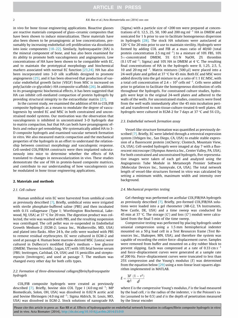
Acta Biomaterialia xxx (2014) xxx–xxx
Contents lists available at ScienceDirect
Acta Biomaterialia
journal homepage: www.elsevier .com/locate /actabiomat
Effects of hydroxyapatite on endothelial network formation incollagen/fibrin composite hydrogels in vitro and in vivo
http://dx.doi.org/10.1016/j.actbio.2014.03.0101742-7061/� 2014 Acta Materialia Inc. Published by Elsevier Ltd. All rights reserved.
⇑ Corresponding author. Tel.: +1 734 764 8313; fax: +1 734 647 4834.E-mail address: [email protected] (J.P. Stegemann).
Please cite this article in press as: Rao RR et al. Effects of hydroxyapatite on endothelial network formation in collagen/fibrin composite hydrogelsand in vivo. Acta Biomater (2014), http://dx.doi.org/10.1016/j.actbio.2014.03.010
Rameshwar R. Rao, Jacob Ceccarelli, Marina L. Vigen, Madhu Gudur, Rahul Singh, Cheri X. Deng,Andrew J. Putnam, Jan P. Stegemann ⇑Department of Biomedical Engineering, University of Michigan, 1101 Beal Ave., Ann Arbor, MI 48109, USA
a r t i c l e i n f o a b s t r a c t
Article history:Received 27 October 2013Received in revised form 5 March 2014Accepted 11 March 2014Available online xxxx
Keywords:CollagenFibrinHydroxyapatiteVasculogenesisIn vivo
Co-culture of endothelial cells (EC) and mesenchymal stem cells (MSC) results in robust vascular networkformation in constrained 3-D collagen/fibrin (COL/FIB) composite hydrogels. However, the ability to formendothelial networks is lost when such gels are allowed to compact via cell-mediated remodeling. In thisstudy, we created co-cultures of human EC and human MSC in both constrained and unconstrained COL/FIB matrices and systematically added nanoparticulate hydroxyapatite (HA, 0–20 mg ml�1), a bone-likemineral that has been shown to have pro-vasculogenic effects. Constructs cultured for 7 days wereassayed for gel compaction, vascular network formation, and mechanical properties. In vitro, robustendothelial network formation was observed in constrained COL/FIB constructs without HA, but thisresponse was significantly inhibited by addition of 5, 10, or 20 mg ml�1 HA. In unconstrained matrices,network formation was abolished in pure COL/FIB constructs but was rescued by 1.25 or 2.5 mg ml�1
HA, while higher levels again inhibited vasculogenesis. HA inhibited gel compaction in a dose-dependentmanner, which was not correlated to endothelial network formation. HA affected initial stiffness of thegels, but gel remodeling abrogated this effect. Subcutaneous implantation of COL/FIB with 0, 2.5 or20 mg ml�1 HA in the mouse resulted in increased perfusion at the implant site, with no significant dif-ferences between materials. Histology at day 7 showed both host and human CD31-stained vasculatureinfiltrating the implants. These findings are relevant to the design of materials and scaffolds fororthopedic tissue engineering, where both vasculogenesis and formation of a mineral phase are requiredfor regeneration.
� 2014 Acta Materialia Inc. Published by Elsevier Ltd. All rights reserved.
1. Introduction
Transplantation and engraftment of engineered tissues requirescreation of a vascular supply, either through vasculogenesis (the denovo formation of blood vessels) or through angiogenesis (the cre-ation of new vessels via sprouting from existing vasculature) [1].The diffusive limit for nutrient transport in most tissues has beensuggested to be only a few hundred microns, so a new vascularsupply to implanted tissue must be created to provide convectivetransport to the region [2]. A variety of model systems have beencreated to study the process of vasculogenesis, including 3-D sys-tems using extracellular matrix proteins such as collagen, fibrinand Matrigel� [3–6]. Previous work in our laboratory has showncomposite collagen/fibrin (COL/FIB) matrices to be permissive toendothelial network formation in vitro when human umbilical veinendothelial cells (EC) are co-cultured with bone-marrow-derived
mesenchymal stem cells (MSC) [7]. The degree of vasculogenesiswas shown to be dependent on the EC:MSC ratio and the composi-tion of the matrix.
In most studies of vasculogenesis in 3-D hydrogels in vitro, vol-ume reduction of the matrix is prevented by constraining the sam-ple at its boundaries. This technique prevents remodeling andcompaction of the matrix by the contractile forces exerted byembedded cells. Unconstrained gel compaction leads to increasedmatrix density and a concomitant increase in matrix stiffness[8,9]. It has been suggested that matrix mechanics play an impor-tant role in regulating endothelial network formation. Increasedstiffness has been shown to promote angiogenesis in some studies[10,11], while other studies have shown an inverse relationship be-tween increased matrix stiffness and neovessel growth, bothin vitro and in vivo [3,7,12–15]. In addition, unconstrained gelcompaction has been shown to result in the regression of endothe-lial networks in vitro [16,17].
Bioceramics have been included in vasculogenesis and angio-genesis models to promote neovessel growth both in vitro and
in vitro
2 R.R. Rao et al. / Acta Biomaterialia xxx (2014) xxx–xxx
in vivo for bone tissue engineering applications. Bioactive glassesare reactive materials composed of glass–ceramic composites thathave been shown to induce mineralization. These materials havealso been shown to be proangiogenic at low concentrations, pre-sumably by increasing endothelial cell proliferation via dissolutioninto ionic components [18–22]. Similarly, hydroxyapatite (HA) isthe mineral component of bone, and has also been examined forits ability to promote both vasculogenesis and angiogenesis. Lowconcentrations of HA have been shown to be compatible with EC,and to maintain the prototypical morphology and biochemicalmarkers associated with normal EC function [23,24]. HA has alsobeen incorporated into 3-D silk scaffolds designed to promoteangiogenesis [25], and it has been observed that production of vas-cular endothelial growth factor (VEGF) from MSC is increased onpoly(lactide-co-glycolide)–HA composite scaffolds [26]. In additionto its proangiogenic biochemical effects, it has been suggested thatHA can inhibit cell-mediated compaction of protein hydrogels byproviding structural integrity to the extracellular matrix [27].
In the current study, we examined the addition of HA to COL/FIBcomposite hydrogels as a means to modulate the degree of vascu-logenesis by seeded EC and MSC in both constrained and uncon-strained model systems. Our motivation was the observation thatvasculogenesis is inhibited in unconstrained 3-D hydrogels dueto matrix compaction, but that HA can both have proangiogenic ef-fects and reduce gel remodeling. We systematically added HA to 3-D composite hydrogels and examined vascular network formationin vitro. We also measured matrix compaction and the mechanicalproperties of the hydrogels in an effort to understand the relation-ship between construct morphology and vasculogenic response.Cell-seeded COL/FIB/HA constructs were then implanted subcuta-neously into mice to determine whether the effects of HAtranslated to changes in neovascularization in vivo. These studiesdemonstrate the use of HA in protein-based composite matrices,and contribute to our understanding of how vasculogenesis canbe modulated in bone tissue engineering applications.
2. Materials and methods
2.1. Cell culture
Human umbilical vein EC were harvested from umbilical cordsas previously described [7]. Briefly, umbilical veins were irrigatedwith sterile phosphate-buffered saline (PBS) and then incubatedwith 0.1% collagenase (Type I; Worthington Biochemical, Lake-wood, NJ, USA) at 37 �C for 20 min. The digestion product was col-lected, the vein was washed with PBS, and the resulting suspensionwas centrifuged. The cell pellet was re-suspended in EndothelialGrowth Medium-2 (EGM-2; Lonza Inc., Walkersville, MD, USA)and plated into flasks. After 24 h, the cells were washed with PBSto remove residual erythrocytes. EC were cultured in EGM-2 andused at passage 4. Human bone marrow-derived MSC (Lonza) werecultured in Dulbecco’s modified Eagle’s medium – low glucose(DMEM; Thermo Scientific; Logan, UT) with 10% fetal bovine serum(FBS; Invitrogen, Carlsbad, CA, USA) and 1% penicillin and strepto-myocin (Invitrogen), and used at passage 7. The medium waschanged every other day for both cells types.
2.2. Formation of three-dimensional collagen/fibrin/hydroxyapatitehydrogels
COL/FIB composite hydrogels were created as previouslydescribed [7]. Briefly, bovine skin COL Type I (4.0 mg ml�1; MPBiomedicals, Solon, OH, USA) was dissolved in 0.02 N acetic acidand bovine fibrinogen (4.0 mg ml�1; Sigma Aldrich, St. Louis, MO,USA) was dissolved in EGM-2. Stock solutions of nanograde HA
Please cite this article in press as: Rao RR et al. Effects of hydroxyapatite on endand in vivo. Acta Biomater (2014), http://dx.doi.org/10.1016/j.actbio.2014.03.0
(Sigma) with a particle size of <200 nm were prepared at concen-trations of 0, 12.5, 25, 50, 100 and 200 mg ml�1 HA in DMEM andsonicated for 1 h prior to use to facilitate homogeneous dispersionin hydrogels [28]. The stock HA solutions were autoclaved at120 �C for 20 min prior to use to maintain sterility. Hydrogels wereformed by adding COL and FIB at a mass ratio of 40/60 (totalprotein concentration 2.5 mg ml�1) to a mixture of 10% FBS, 10%5�-concentrated DMEM, 5% 0.1 N NaOH, 2% thrombin(0.1 UT ml�1; Sigma) and 10% HA in DMEM at 4 �C. The resultingfinal concentrations of HA in the hydrogels were 0, 1.25, 2.5, 5,10 and 20 mg ml�1. Matrix mixtures (500 ll) were placed into a24-well plate and gelled at 37 �C for 45 min. Both EC and MSC wereadded directly into the gel mixture to at a ratio of 1:1 EC:MSC, witha total cell concentration of 2.4 � 105 cells ml�1. Cells were addedprior to gelation to facilitate the homogeneous distribution of cellsthroughout the hydrogels. For constrained culture studies, hydro-gels were kept in the original 24-well plates and adhered to thewalls of the wells. For unconstrained studies, constructs were freedfrom the well walls immediately after the 45 min incubation peri-od and transferred to non-tissue-culture-treated 6-well plates. Allhydrogels were cultured in EGM-2 for 7 days at 37 �C and 5% CO2.
2.3. Endothelial network formation assay
Vessel-like structure formation was quantified as previously de-scribed [7]. Briefly, EC were labeled through a retroviral expressionsystem (Orbigen Inc., San Diego, CA, USA) to enable stable expres-sion of a fluorescent protein (mCherry; Clontech, Mountain View,CA, USA). Cell-seeded hydrogels were imaged at day 7 with a fluo-rescent microscope (Olympus America Inc., Center Valley, PA, USA).For both constrained and unconstrained hydrogels, five representa-tive images were taken of each gel and analyzed using theAngiogenesis Tube Module in Metamorph Premier Software(Molecular Devices Inc., Sunnyvale, CA, USA). The total networklength of vessel-like structures formed in vitro was calculated bysetting a minimum width, maximum width and intensity overbackground.
2.4. Mechanical properties testing
Gel rheology was performed on acellular COL/FIB/HA hydrogelsas previously described [7]. Briefly, pre-formed COL/FIB/HA solu-tions were loaded into a gel rheometer (AR-G2, TA Instruments,New Castle, DE, USA) and a time sweep was conducted for45 min at 37 �C. The storage (G0) and loss (G00) moduli were calcu-lated from the final 5 min of the time sweep.
Compressive testing was performed by placing hydrogels underuniaxial compression using a 1.5 mm hemispherical indentermounted on a 50 g load cell in a Test Resources frame (Test Re-sources Inc., Shakopee, MN, USA), and therefore the system wascapable of recording the entire force–displacement curve. Sampleswere removed from buffer and mounted on a dry rubber block toprevent slipping. Each was compressed at a rate of 0.33 cm s�1
and force–displacement curves were generated at a sample rateof 200 Hz. Force–displacement curves were truncated to less than25% compression and the Young’s modulus (E) was determinedfrom the equation below [29] using a non-linear least squares algo-rithm implemented in MATLAB.
E ¼ 3F
4r12
ð1� m2Þd
32
where E is the compressive Young’s modulus, F is the load measuredby the load cell, r is the radius of the indenter, m is the Poisson’s ra-tio (assumed to be 0.5) and d is the depth of penetration measuredby the linear encoder
othelial network formation in collagen/fibrin composite hydrogels in vitro10
R.R. Rao et al. / Acta Biomaterialia xxx (2014) xxx–xxx 3
We assumed a Poisson’s ratio of 0.5 based on the fact that thehydrogels were tested at small strains (<10%), and we consistentlyanalyzed the data within the elastic response of each curve. Underthese loads these materials behave as a rubbery elastic solid, due tothe highly crosslinked nature of the gel and the intrinsic elasticityof the fibers.
2.5. Measurement of gel compaction and 3-D ultrasound imaging
Representative images of unconstrained hydrogels were takenusing a standard CCD camera in manual mode with a constantexposure setting at days 0, 1, 3, 5 and 7 to qualitatively demon-strate gel compaction throughout the time course. Ultrasoundimaging of the constructs was performed with a Vevo 770(VisualSonics Inc., Toronto, Canada) using an RMV 707B imagingprobe with a nominal 30 MHz center frequency, 15–45 MHz band-width (�6 dB), 12.7 mm focal distance and 2.2 mm depth of focus(�6 dB), as described previously [28]. For each gel, a 3-D ultra-sound image data containing a series of consecutive B-modeimages with a spatial interval of 100 lm was collected. The inter-val between adjacent A-lines in a B-mode cross-sectional imagewas 50 lm. Volumes of these gels were measured from the ultra-sound images using a semi-automated segmentation procedure,in which an edge detection algorithm identified the contour ofthe gel in an ultrasound B-mode image. Subsequent contours inadjacent frames or images were drawn semi-automatically untilthe contours in all frames were drawn, thus providing the volumeof the gel within the contoured region.
2.6. Subcutaneous implants
All animal studies were conducted in accordance with theNational Institutes of Health Guidelines following a protocolapproved by the University of Michigan Committee on Use andCare of Animals. Seven-week old male C.B.-17/SCID mice (TaconicLabs, Hudson, NY, USA) were administered an anesthetic analgesicdrug cocktail containing ketamine (95 mg kg�1, Fort Dodge AnimalHealth, Fort Dodge, IA, USA), xylazine (9.5 mg kg�1, Lloyd Laborato-ries, Shenandoah, IA, USA) and buprenorphine (0.059 mg kg�1,Bedford Laboratories, Bedford, OH, USA) via intraperitoneal injec-tion. Each mouse was shaved and sterilized with Betadine�
(Thermo Fisher Scientific, Fremont, CA, USA) on its dorsal surfaceand wiped with alcohol prior to injection. COL/FIB/HA constructscontaining 0, 2.5 or 20 mg ml�1 HA and a total cell concentrationof 10.0 � 106 cells ml�1 were used for the in vivo studies. Acellularconstructs served as controls. Two implants per animal were cre-ated with gel solutions of 300 ll. After injection, animals were keptstationary for 5 min and then placed in fresh cages. A total of fiveanimals were used for each variable investigated.
2.7. Laser Doppler perfusion imaging
Mice were anesthetized as previously described and blood flowthrough the implant was imaged using laser Doppler perfusionimaging (LDPI; Perimed AB, Sweden) [30]. Mice were imaged intriplicate and a region of interest was overlayed over the implantarea to calculate the mean perfusion through each implant. Resultswere calculated as fold change from perfusion values over thebaseline (before implantation).
2.8. Histology and immunohistochemistry
Explants were retrieved at day 7 and then fixed in zinc-bufferedformalin (Z-Fix; Anatech Ltd., Battle Creek, MI, USA) overnight.Samples were sent to AML Laboratories (Baltimore, MD, USA) forembedding in paraffin and sectioned into 5 lm sections. Sections
Please cite this article in press as: Rao RR et al. Effects of hydroxyapatite on endand in vivo. Acta Biomater (2014), http://dx.doi.org/10.1016/j.actbio.2014.03.0
were then demineralized in a solution of 10% ethylenediamine tet-raacetic acid (Sigma) and Z-Fix for 3 h at 4 �C. Tissue sections wererehydrated, steamed in a vegetable steamer for 25 min in antigenretrieval solution (Dako, Carpinteria, CA, USA) and then equili-brated in Tris-buffered saline with Tween 20 (TBS-T; Sigma). Sec-tions were then incubated at 4 �C in the primary antibody, eitherhuman anti-mouse CD31 (Dako) or human anti-mouse a-SMA(Abcam, Cambridge, MA, USA), diluted 1:50 in TBS-T overnightand then treated with horseradish peroxidase-conjugated anti-mouse secondary antibody. Hematoxylin and eosin (H&E) wasused as a counterstain.
2.9. Quantification of in vivo vessel formation
Blood vessels found within the implants were quantified man-ually by three blinded observers. Ten random images per sampleat 20� magnification were used to quantify the number of humanCD-31 stained vessels (i.e. those that arose from implanted cells) aswell as the total number of vessels within the implant region. Allvessels were quantified if they displayed a lumen containing eryth-rocytes and human vessels were identified if they displayed a po-sitive CD31 stain around the lumen.
2.10. Statistical analysis
All quantitative analyses were performed using a one-way anal-ysis of variance test with a protected Fisher’s least significant dif-ference post hoc test. Statistical significance was set at p < 0.05.Numerical values and bar charts are presented as mean ± standarderror.
3. Results
3.1. Vasculogenesis in constrained and unconstrained COL/FIB/HAhydrogels
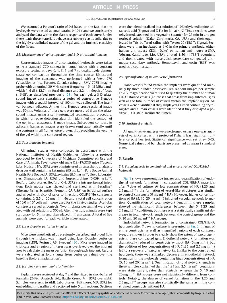
Fig. 1 shows representative images and quantification of endo-thelial network formation in constrained COL/FIB/HA materialsafter 7 days of culture. At low concentrations of HA (1.25 and2.5 mg ml�1), the formation of vessel-like structures was similarto control constructs (0 mg ml�1 HA). However, higher concentra-tions of HA (5, 10, 20 mg ml�1) inhibited vascular network forma-tion. Quantification of total network length in these samplesshowed no significant differences between the 0, 1.25 and2.5 mg ml�1 conditions, but there was a statistically significant de-crease in total network length between the control group and the5, 10 and 20 mg ml�1 HA groups.
Endothelial network formation in unconstrained COL/FIB/HAhydrogels after 7 days in culture is presented in Fig. 2. Images ofentire constructs, as well as magnified regions of each constructtype, are shown in order to clearly show the extent of vasculogen-esis in these compacted gels. Endothelial network formation wasdramatically reduced in constructs without HA (0 mg ml�1), butthe addition of low concentrations of HA (1.25 and 2.5 mg ml�1)led to a recovery of vascular networks. Similar to the constrainedhydrogels, there was a marked decrease in endothelial networkformation in the hydrogels containing high concentrations of HA(5, 10 and 20 mg ml�1). Quantification of total network length inthese samples confirmed that the 1.25 and 2.5 mg ml�1 conditionswere statistically greater than controls, whereas the 5, 10 and20 mg ml�1 HA groups were not statistically different from con-trols. Notably, the degree of vessel formation in the 1.25 and2.5 mg ml�1 groups was also statistically the same as in the con-strained constructs without HA.
othelial network formation in collagen/fibrin composite hydrogels in vitro10
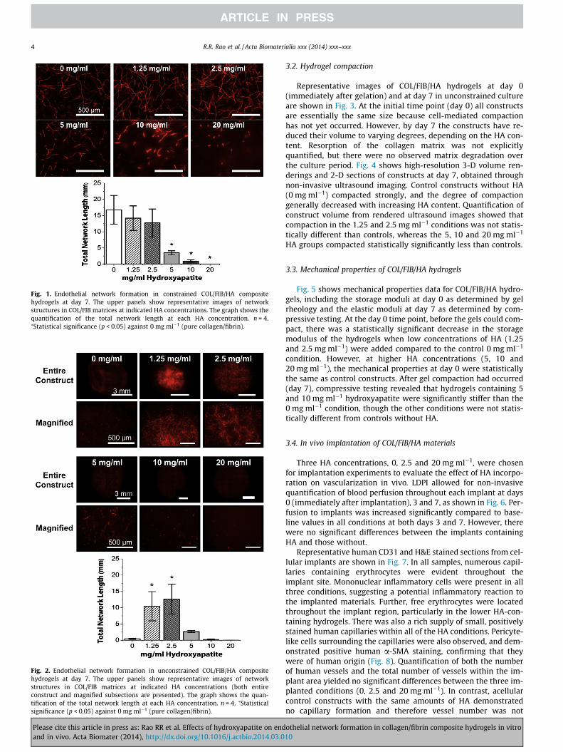
Fig. 1. Endothelial network formation in constrained COL/FIB/HA compositehydrogels at day 7. The upper panels show representative images of networkstructures in COL/FIB matrices at indicated HA concentrations. The graph shows thequantification of the total network length at each HA concentration. n = 4.⁄Statistical significance (p < 0.05) against 0 mg ml�1 (pure collagen/fibrin).
Fig. 2. Endothelial network formation in unconstrained COL/FIB/HA compositehydrogels at day 7. The upper panels show representative images of networkstructures in COL/FIB matrices at indicated HA concentrations (both entireconstruct and magnified subsections are presented). The graph shows the quan-tification of the total network length at each HA concentration. n = 4. ⁄Statisticalsignificance (p < 0.05) against 0 mg ml�1 (pure collagen/fibrin).
4 R.R. Rao et al. / Acta Biomaterialia xxx (2014) xxx–xxx
Please cite this article in press as: Rao RR et al. Effects of hydroxyapatite on endand in vivo. Acta Biomater (2014), http://dx.doi.org/10.1016/j.actbio.2014.03.0
3.2. Hydrogel compaction
Representative images of COL/FIB/HA hydrogels at day 0(immediately after gelation) and at day 7 in unconstrained cultureare shown in Fig. 3. At the initial time point (day 0) all constructsare essentially the same size because cell-mediated compactionhas not yet occurred. However, by day 7 the constructs have re-duced their volume to varying degrees, depending on the HA con-tent. Resorption of the collagen matrix was not explicitlyquantified, but there were no observed matrix degradation overthe culture period. Fig. 4 shows high-resolution 3-D volume ren-derings and 2-D sections of constructs at day 7, obtained throughnon-invasive ultrasound imaging. Control constructs without HA(0 mg ml�1) compacted strongly, and the degree of compactiongenerally decreased with increasing HA content. Quantification ofconstruct volume from rendered ultrasound images showed thatcompaction in the 1.25 and 2.5 mg ml�1 conditions was not statis-tically different than controls, whereas the 5, 10 and 20 mg ml�1
HA groups compacted statistically significantly less than controls.
3.3. Mechanical properties of COL/FIB/HA hydrogels
Fig. 5 shows mechanical properties data for COL/FIB/HA hydro-gels, including the storage moduli at day 0 as determined by gelrheology and the elastic moduli at day 7 as determined by com-pressive testing. At the day 0 time point, before the gels could com-pact, there was a statistically significant decrease in the storagemodulus of the hydrogels when low concentrations of HA (1.25and 2.5 mg ml�1) were added compared to the control 0 mg ml�1
condition. However, at higher HA concentrations (5, 10 and20 mg ml�1), the mechanical properties at day 0 were statisticallythe same as control constructs. After gel compaction had occurred(day 7), compressive testing revealed that hydrogels containing 5and 10 mg ml�1 hydroxyapatite were significantly stiffer than the0 mg ml�1 condition, though the other conditions were not statis-tically different from controls without HA.
3.4. In vivo implantation of COL/FIB/HA materials
Three HA concentrations, 0, 2.5 and 20 mg ml�1, were chosenfor implantation experiments to evaluate the effect of HA incorpo-ration on vascularization in vivo. LDPI allowed for non-invasivequantification of blood perfusion throughout each implant at days0 (immediately after implantation), 3 and 7, as shown in Fig. 6. Per-fusion to implants was increased significantly compared to base-line values in all conditions at both days 3 and 7. However, therewere no significant differences between the implants containingHA and those without.
Representative human CD31 and H&E stained sections from cel-lular implants are shown in Fig. 7. In all samples, numerous capil-laries containing erythrocytes were evident throughout theimplant site. Mononuclear inflammatory cells were present in allthree conditions, suggesting a potential inflammatory reaction tothe implanted materials. Further, free erythrocytes were locatedthroughout the implant region, particularly in the lower HA-con-taining hydrogels. There was also a rich supply of small, positivelystained human capillaries within all of the HA conditions. Pericyte-like cells surrounding the capillaries were also observed, and dem-onstrated positive human a-SMA staining, confirming that theywere of human origin (Fig. 8). Quantification of both the numberof human vessels and the total number of vessels within the im-plant area yielded no significant differences between the three im-planted conditions (0, 2.5 and 20 mg ml�1). In contrast, acellularcontrol constructs with the same amounts of HA demonstratedno capillary formation and therefore vessel number was not
othelial network formation in collagen/fibrin composite hydrogels in vitro10
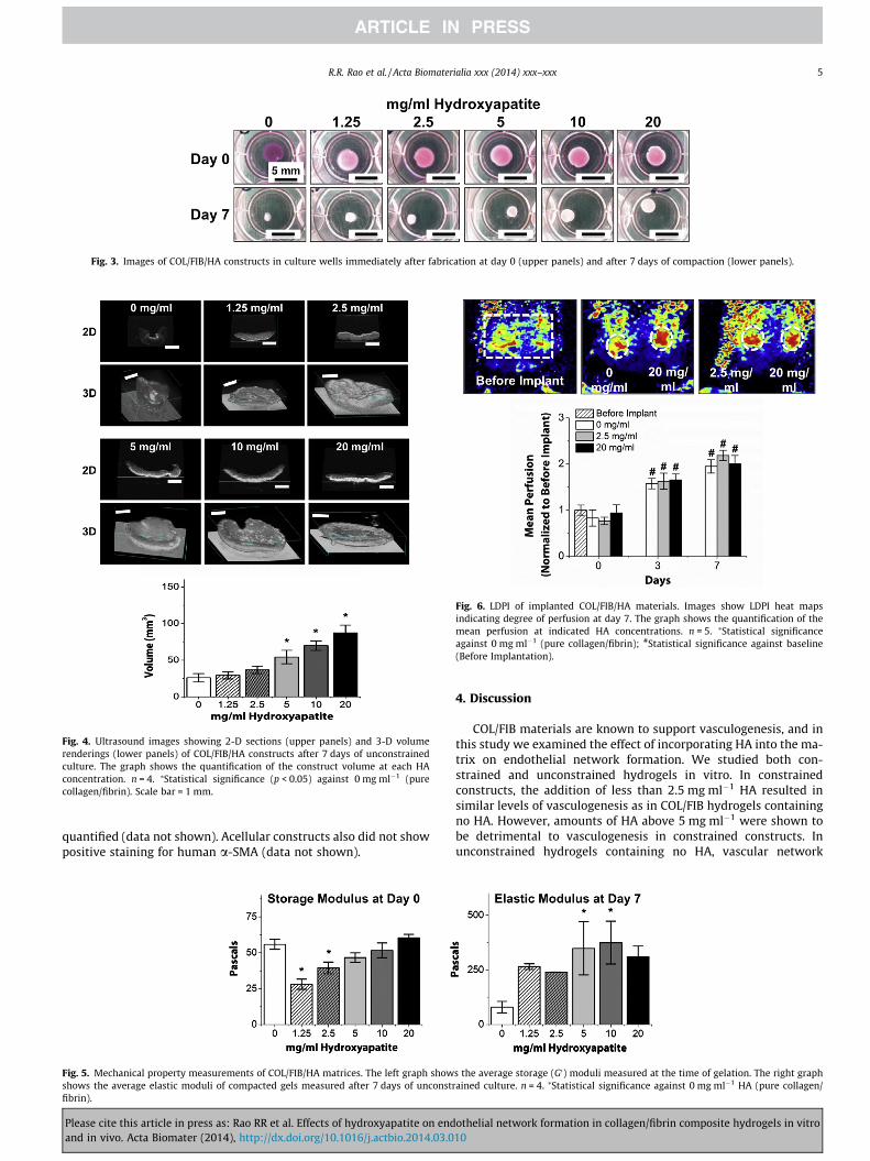
Fig. 3. Images of COL/FIB/HA constructs in culture wells immediately after fabrication at day 0 (upper panels) and after 7 days of compaction (lower panels).
Fig. 4. Ultrasound images showing 2-D sections (upper panels) and 3-D volumerenderings (lower panels) of COL/FIB/HA constructs after 7 days of unconstrainedculture. The graph shows the quantification of the construct volume at each HAconcentration. n = 4. ⁄Statistical significance (p < 0.05) against 0 mg ml�1 (purecollagen/fibrin). Scale bar = 1 mm.
Fig. 6. LDPI of implanted COL/FIB/HA materials. Images show LDPI heat mapsindicating degree of perfusion at day 7. The graph shows the quantification of themean perfusion at indicated HA concentrations. n = 5. ⁄Statistical significanceagainst 0 mg ml�1 (pure collagen/fibrin); #Statistical significance against baseline(Before Implantation).
R.R. Rao et al. / Acta Biomaterialia xxx (2014) xxx–xxx 5
quantified (data not shown). Acellular constructs also did not showpositive staining for human a-SMA (data not shown).
Fig. 5. Mechanical property measurements of COL/FIB/HA matrices. The left graph showshows the average elastic moduli of compacted gels measured after 7 days of unconstrfibrin).
Please cite this article in press as: Rao RR et al. Effects of hydroxyapatite on endand in vivo. Acta Biomater (2014), http://dx.doi.org/10.1016/j.actbio.2014.03.0
4. Discussion
COL/FIB materials are known to support vasculogenesis, and inthis study we examined the effect of incorporating HA into the ma-trix on endothelial network formation. We studied both con-strained and unconstrained hydrogels in vitro. In constrainedconstructs, the addition of less than 2.5 mg ml�1 HA resulted insimilar levels of vasculogenesis as in COL/FIB hydrogels containingno HA. However, amounts of HA above 5 mg ml�1 were shown tobe detrimental to vasculogenesis in constrained constructs. Inunconstrained hydrogels containing no HA, vascular network
s the average storage (G0) moduli measured at the time of gelation. The right graphained culture. n = 4. ⁄Statistical significance against 0 mg ml�1 HA (pure collagen/
othelial network formation in collagen/fibrin composite hydrogels in vitro10
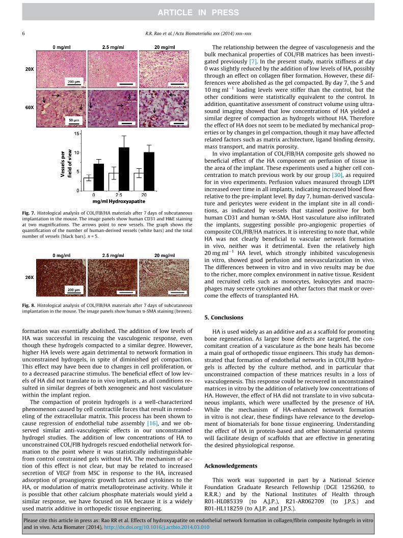
Fig. 7. Histological analysis of COL/FIB/HA materials after 7 days of subcutaneousimplantation in the mouse. The image panels show human CD31 and H&E stainingat two magnifications. The arrows point to new vessels. The graph shows thequantification of the number of human-derived vessels (white bars) and the totalnumber of vessels (black bars). n = 5.
Fig. 8. Histological analysis of COL/FIB/HA materials after 7 days of subcutaneousimplantation in the mouse. The image panels show human a-SMA staining (brown).
6 R.R. Rao et al. / Acta Biomaterialia xxx (2014) xxx–xxx
formation was essentially abolished. The addition of low levels ofHA was successful in rescuing the vasculogenic response, eventhough these hydrogels compacted to a similar degree. However,higher HA levels were again detrimental to network formation inunconstrained hydrogels, in spite of diminished gel compaction.This effect may have been due to changes in cell proliferation, orto a decreased paracrine stimulus. The beneficial effect of low lev-els of HA did not translate to in vivo implants, as all conditions re-sulted in similar degrees of both xenogeneic and host vasculaturewithin the implant region.
The compaction of protein hydrogels is a well-characterizedphenomenon caused by cell contractile forces that result in remod-eling of the extracellular matrix. This process has been shown tocause regression of endothelial tube assembly [16], and we ob-served similar anti-vasculogenic effects in our unconstrainedhydrogel studies. The addition of low concentrations of HA tounconstrained COL/FIB hydrogels rescued endothelial network for-mation to the point where it was statistically indistinguishablefrom control constrained gels without HA. The mechanism of ac-tion of this effect is not clear, but may be related to increasedsecretion of VEGF from MSC in response to the HA, increasedadsorption of proangiogenic growth factors and cytokines to theHA, or modulation of matrix metalloproteinase activity. While itis possible that other calcium phosphate materials would yield asimilar response, we have focused on HA because it is a widelyused matrix additive in orthopedic tissue engineering.
Please cite this article in press as: Rao RR et al. Effects of hydroxyapatite on endand in vivo. Acta Biomater (2014), http://dx.doi.org/10.1016/j.actbio.2014.03.0
The relationship between the degree of vasculogenesis and thebulk mechanical properties of COL/FIB matrices has been investi-gated previously [7]. In the present study, matrix stiffness at day0 was slightly reduced by the addition of low levels of HA, possiblythrough an effect on collagen fiber formation. However, these dif-ferences were abolished as the gel compacted. By day 7, the 5 and10 mg ml�1 loading levels were stiffer than the control, but theother conditions were statistically equivalent to the control. Inaddition, quantitative assessment of construct volume using ultra-sound imaging showed that low concentrations of HA yielded asimilar degree of compaction as hydrogels without HA. Thereforethe effect of HA does not seem to be mediated by mechanical prop-erties or by changes in gel compaction, though it may have affectedrelated factors such as matrix architecture, ligand binding density,mass transport, and matrix porosity.
In vivo implantation of COL/FIB/HA composite gels showed nobeneficial effect of the HA component on perfusion of tissue inthe area of the implant. These experiments used a higher cell con-centration to match previous work by our group [30], as requiredfor in vivo experiments. Perfusion values measured through LDPIincreased over time in all implants, indicating increased blood flowrelative to the pre-implant level. By day 7, human-derived vascula-ture and pericytes were evident in the implant site in all condi-tions, as indicated by vessels that stained positive for bothhuman CD31 and human a-SMA. Host vasculature also infiltratedthe implants, suggesting possible pro-angiogenic properties ofcomposite COL/FIB/HA matrices. It is interesting to note that, whileHA was not clearly beneficial to vascular network formationin vivo, neither was it detrimental. Even the relatively high20 mg ml�1 HA level, which strongly inhibited vasculogenesisin vitro, showed good perfusion and neovascularization in vivo.The differences between in vitro and in vivo results may be dueto the richer, more complex environment in native tissue. Residentand recruited cells such as monocytes, leukocytes and macro-phages may secrete cytokines and other factors that mask or over-come the effects of transplanted HA.
5. Conclusions
HA is used widely as an additive and as a scaffold for promotingbone regeneration. As larger bone defects are targeted, the con-comitant creation of a vasculature as the bone heals has becomea main goal of orthopedic tissue engineers. This study has demon-strated that formation of endothelial networks in COL/FIB hydro-gels is affected by the culture method, and in particular thatunconstrained compaction of these matrices results in a loss ofvasculogenesis. This response could be recovered in unconstrainedmatrices in vitro by the addition of relatively low concentrations ofHA. However, the effect of HA did not translate to in vivo subcuta-neous implants, which were unaffected by the presence of HA.While the mechanism of HA-enhanced network formationin vitro is not clear, these findings have relevance to the develop-ment of biomaterials for bone tissue engineering. Understandingthe effect of HA in protein-based and other biomaterial systemswill facilitate design of scaffolds that are effective in generatingthe desired physiological response.
Acknowledgements
This work was supported in part by a National ScienceFoundation Graduate Research Fellowship (DGE 1256260, toR.R.R.) and by the National Institutes of Health throughR01-HL085339 (to A.J.P.), R21-AR062709 (to J.P.S.) andR01-HL118259 (to A.J.P. and J.P.S.).
othelial network formation in collagen/fibrin composite hydrogels in vitro10
R.R. Rao et al. / Acta Biomaterialia xxx (2014) xxx–xxx 7
Appendix A. Figures with essential colour discrimination
Certain figures in this article, particularly Figs. 1–4 and 6–8, aredifficult to interpret in black and white. The full colour images canbe found in the on-line version, at http://dx.doi.org/10.1016/j.actbio.2014.03.010.
References
[1] Jain RK. Molecular regulation of vessel maturation. Nat Med 2003;9:685–93.[2] Novosel EC, Kleinhans C, Kluger PJ. Vascularization is the key challenge in
tissue engineering. Adv Drug Deliv Rev 2011;63:300–11.[3] Allen P, Melero-Martin J, Bischoff J. Type I collagen, fibrin and PuraMatrix
matrices provide permissive environments for human endothelial andmesenchymal progenitor cells to form neovascular networks. J Tissue EngRegen Med 2011;5:e74–86.
[4] Dietrich F, Lelkes PI. Fine-tuning of a three-dimensional microcarrier-basedangiogenesis assay for the analysis of endothelial–mesenchymal cell co-cultures in fibrin and collagen gels. Angiogenesis 2006;9:111–25.
[5] Martineau L, Doillon CJ. Angiogenic response of endothelial cells seededdispersed versus on beads in fibrin gels. Angiogenesis 2007;10:269–77.
[6] Melero-Martin JM, De Obaldia ME, Kang SY, Khan ZA, Yuan L, Oettgen P, et al.Engineering robust and functional vascular networks in vivo with human adultand cord blood-derived progenitor cells. Circ Res 2008;103:194–202.
[7] Rao RR, Peterson AW, Ceccarelli J, Putnam AJ, Stegemann JP. Matrixcomposition regulates three-dimensional network formation by endothelialcells and mesenchymal stem cells in collagen/fibrin materials. Angiogenesis2012;15:253–64.
[8] Fernandez P, Bausch AR. The compaction of gels by cells: a case of collectivemechanical activity. Integr Biol (Camb) 2009;1:252–9.
[9] Lund AW, Bilgin CC, Hasan MA, McKeen LM, Stegemann JP, Yener B, et al.Quantification of spatial parameters in 3D cellular constructs using graphtheory. J Biomed Biotechnol 2009;2009:928286.
[10] Mason BN, Starchenko A, Williams RM, Bonassar LJ, Reinhart-King CA. Tuningthree-dimensional collagen matrix stiffness independently of collagenconcentration modulates endothelial cell behavior. Acta Biomater2013;9:4635–46447.
[11] Lee PF, Bai Y, Smith RL, Bayless KJ, Yeh AT. Angiogenic responses are enhancedin mechanically and microscopically characterized, microbialtransglutaminase crosslinked collagen matrices with increased stiffness. ActaBiomater 2013;9:7178–90.
[12] Critser PJ, Kreger ST, Voytik-Harbin SL, Yoder MC. Collagen matrix physicalproperties modulate endothelial colony forming cell-derived vessels in vivo.Microvasc Res 2010;80:23–30.
[13] Ghajar CM, Blevins KS, Hughes CC, George SC, Putnam AJ. Mesenchymal stemcells enhance angiogenesis in mechanically viable prevascularized tissues viaearly matrix metalloproteinase upregulation. Tissue Eng 2006;12:2875–88.
[14] Ghajar CM, Chen X, Harris JW, Suresh V, Hughes CC, Jeon NL, et al. The effect ofmatrix density on the regulation of 3-D capillary morphogenesis. Biophys J2008;94:1930–41.
Please cite this article in press as: Rao RR et al. Effects of hydroxyapatite on endand in vivo. Acta Biomater (2014), http://dx.doi.org/10.1016/j.actbio.2014.03.0
[15] Kniazeva E, Kachgal S, Putnam AJ. Effects of extracellular matrix density andmesenchymal stem cells on neovascularization in vivo. Tissue Eng Part A2011;17:905–14.
[16] Davis GE, Davis GE, Pintar Allen KA, Salazar R, Maxwell SA. Matrixmetalloproteinase-1 and -9 activation by plasmin regulates a novelendothelial cell-mediated mechanism of collagen gel contraction andcapillary tube regression in three-dimensional collagen matrices. J Cell Sci2001;114:917–30.
[17] Saunders WB, Bayless KJ, Davis GE. MMP-1 activation by serine proteases andMMP-10 induces human capillary tubular network collapse and regression in3D collagen matrices. J Cell Sci 2005;118:2325–40.
[18] Bi L, Jung S, Day D, Neidig K, Dusevich V, Eick D, et al. Evaluation of boneregeneration, angiogenesis, and hydroxyapatite conversion in critical-sized ratcalvarial defects implanted with bioactive glass scaffolds. J Biomed Mater ResA 2012;100:3267–75.
[19] Gorustovich AA, Roether JA, Boccaccini AR. Effect of bioactive glasses onangiogenesis: a review of in vitro and in vivo evidences. Tissue Eng Part B Rev2010;16:199–207.
[20] Leach JK, Kaigler D, Wang Z, Krebsbach PH, Mooney DJ. Coating of VEGF-releasing scaffolds with bioactive glass for angiogenesis and boneregeneration. Biomaterials 2006;27:3249–55.
[21] Leu A, Leach JK. Proangiogenic potential of a collagen/bioactive glass substrate.Pharm Res 2008;25:1222–9.
[22] Leu A, Stieger SM, Dayton P, Ferrara KW, Leach JK. Angiogenic response tobioactive glass promotes bone healing in an irradiated calvarial defect. TissueEng Part A 2009;15:877–85.
[23] Pezzatini S, Solito R, Morbidelli L, Lamponi S, Boanini E, Bigi A, et al. The effectof hydroxyapatite nanocrystals on microvascular endothelial cell viability andfunctions. J Biomed Mater Res A 2006;76:656–63.
[24] Rücker M, Laschke MW, Junker D, Carvalho C, Tavassol F, Mülhaupt R, et al.Vascularization and biocompatibility of scaffolds consisting of differentcalcium phosphate compounds. J Biomed Mater Res A 2008;86:1002–111.
[25] Sun L, Parker ST, Syoji D, Wang X, Lewis JA, Kaplan DL. Direct-write assemblyof 3D silk/hydroxyapatite scaffolds for bone co-cultures. Adv Healthc Mater2012;1:729–35.
[26] He J, Decaris ML, Leach JK. Bioceramic-mediated trophic factor secretion bymesenchymal stem cells enhances in vitro endothelial cell persistence andin vivo angiogenesis. Tissue Eng Part A 2012;18:1520–8.
[27] Liu Y, Williams DJ. Incorporation of hydroxyapatite sol into collagen gel toregulate the contraction mediated by human bone marrow-derived stromalcells. IEEE Trans Nanobiosci 2010;9:1–11.
[28] Gudur M, Rao RR, Hsiao YS, Peterson AW, Deng CX, Stegemann JP. Noninvasive,quantitative, spatiotemporal characterization of mineralization in three-dimensional collagen hydrogels using high-resolution spectral ultrasoundimaging. Tissue Eng Part C Methods 2012;18:935–46.
[29] Gent AN. On the relation between indentation hardness and Young’s modulus.IRI Trans 1958;34:46–57.
[30] Grainger SJ, Carrion B, Ceccarelli J, Putnam AJ. Stromal cell identity influencesthe in vivo functionality of engineered capillary networks formed byco-delivery of endothelial cells and stromal cells. Tissue Eng Part A 2013;19:1209–22.
othelial network formation in collagen/fibrin composite hydrogels in vitro10
Copyright © 2022 FDOKUMEN