Elimination of fibrin γ-chain cross-linking by FXIIIa ... - PNAS
12
Elimination of fibrin γ-chain cross-linking by FXIIIa increases pulmonary embolism arising from murine inferior vena cava thrombi Cédric Duval a , Adomas Baranauskas a , Tímea Feller a , Majid Ali a , Lih T. Cheah a , Nadira Y. Yuldasheva a , Stephen R. Baker a , Helen R. McPherson a , Zaher Raslan a , Marc A. Bailey a , Richard M. Cubbon a , Simon D. Connell b , Ramzi A. Ajjan a , Helen Philippou a , Khalid M. Naseem a , Victoria C. Ridger c , and Robert A. S. Ariëns a,1 a Leeds Thrombosis Collective, Discovery & Translational Science Department, Leeds Institute of Cardiovascular and Metabolic Medicine, University of Leeds, Leeds LS2 9NL, United Kingdom; b School of Physics and Astronomy, University of Leeds, Leeds LS2 3AR, United Kingdom; and c Department of Infection, Immunity and Cardiovascular Disease, The Medical School, University of Sheffield, Sheffield S10 2RX, United Kingdom Edited by Barry S. Coller, The Rockefeller University, New York, NY, and approved May 25, 2021 (received for review March 4, 2021) The onset of venous thromboembolism, including pulmonary embo- lism, represents a significant health burden affecting more than 1 million people annually worldwide. Current treatment options are based on anticoagulation, which is suboptimal for preventing further embolic events. In order to develop better treatments for thrombo- embolism, we sought to understand the structural and mechanical properties of blood clots and how this influences embolism in vivo. We developed a murine model in which fibrin γ-chain cross-linking by activated Factor XIII is eliminated (FGG3X) and applied methods to study thromboembolism at whole-body and organ levels. We show that FGG3X mice have a normal phenotype, with overall coagulation parameters and platelet aggregation and function largely unaf- fected, except for total inhibition of fibrin γ-chain cross-linking. Elim- ination of fibrin γ-chain cross-linking resulted in thrombi with reduced strength that were prone to fragmentation. Analysis of embolism in vivo using Xtreme optical imaging and light sheet microscopy dem- onstrated that the elimination of fibrin γ-chain cross-linking resulted in increased embolization without affecting clot size or lysis. Our findings point to a central previously unrecognized role for fibrin γ-chain cross- linking in clot stability. They also indirectly indicate mechanistic targets for the prevention of thrombosis through selective modulation of fi- brin α-chain but not γ-chain cross-linking by activated Factor XIII to reduce thrombus size and burden, while maintaining clot stability and preventing embolism. fibrin | Factor XIII | thromboembolism | mechanical properties | clot structure T hrombosis is complicated by life-threatening embolic events, caused by parts of an intravascular blood clot breaking off and traveling downstream to block other blood vessels supplying critical organs. Thromboembolism occurs in both the venous and arterial circulation and is associated with life-threatening pulmo- nary embolism (PE) (1) and ischemic stroke (2). PE occurs when thrombi in the deep veins of the limb embolise and passage with the flowing blood through the inferior vena cava, the right atrium, and ventricle of the heart to the lungs (3), causing pulmonary hypertension and respiratory failure (4). Venous thromboembolism (VTE), comprising deep vein thrombosis (DVT) and PE, which globally affects over 1 million people each year (1), results in sub- stantial healthcare costs (5) and is a major cause of death worldwide (1, 6). Thromboembolism is clinically challenging to treat. Anti- coagulation with vitamin K antagonists or direct oral anticoagulants are used to treat VTE and prophylactically to prevent VTE re- currence (7). In PE, localized thrombolysis with plasminogen activators is challenging and often only used as a last resort to help remove emboli resistant to anticoagulation (8). Improve- ments in treatment and prevention of thromboembolic disor- ders are therefore urgently needed. Recent studies indicate that structural and functional prop- erties of the clot could be critical in thromboembolism, and these parameters may offer novel areas for therapeutic intervention. Hypofibrinolysis was reported to increase the risk of a first DVT (9), while changes in clot properties (increased clot formation rate and fiber density, reduced fibrinolysis) promoted the recurrence of DVT (10). Abnormalities in the establishment of clot viscoelastic properties have been shown to increase risk of PE (11), and re- duced clot elastic modulus has been associated with VTE recur- rence (12). However, the exact mechanisms linking altered clot properties to increased thromboembolic risk are unclear, and therefore, treatment options remain limited and rely on dissolution of fibrin networks and prevention of future clot formation, which carry significant risk of bleeding events. A key regulator of clot mechanical properties is coagulation Factor XIII (FXIII), a protransglutaminase that is converted into the active transglutaminase (FXIIIa) by thrombin (13). FXIIIa catalyzes the formation of γ-glutamyl-e-lysine isopeptide bonds between adjacent molecules within the fibrin fibers to substan- tially increase elastic moduli and reduce storage moduli of both Significance Pulmonary embolism and stroke are thromboembolic diseases affecting >1 million people annually worldwide. Thromboem- bolism involves clot fragments affecting vital downstream or- gans such as the lung or brain. The mechanisms underpinning thromboembolism are not understood and require clarification in order to devise more effective treatments. Here, we devel- oped a thromboembolism protocol supported by state-of-the- art in vivo imaging, coupled to a genetically modified murine model of reduced clot strength caused by mutations in the cross-linking sites of fibrin, which provides the mechanical scaffold of the clot. We find that fibrin γ-chain cross-linking is essential for clot stability and reduces embolism. These find- ings introduce an important concept indicating that mainte- nance of clot stability during thrombosis treatment is essential to prevent thromboembolism. Author contributions: C.D. and R.A.S.A. designed research; C.D., A.B., T.F., M.A., L.T.C., N.Y.Y., S.R.B., H.R.M., and Z.R. performed research; M.A.B. and R.M.C. contributed new reagents/analytic tools; C.D., S.D.C., R.A.A., H.P., K.M.N., V.C.R., and R.A.S.A. analyzed data; C.D. and R.A.S.A. wrote the paper; and A.B., T.F., M.A., L.T.C., N.Y.Y., S.R.B., H.R.M., Z.R., M.A.B., R.M.C., S.D.C., R.A.A., H.P., K.M.N., and V.C.R. critically reviewed the manuscript. The authors declare no competing interest. This article is a PNAS Direct Submission. This open access article is distributed under Creative Commons Attribution License 4.0 (CC BY). 1 To whom correspondence may be addressed. Email: [email protected]. This article contains supporting information online at https://www.pnas.org/lookup/suppl/ doi:10.1073/pnas.2103226118/-/DCSupplemental. Published June 28, 2021. PNAS 2021 Vol. 118 No. 27 e2103226118 https://doi.org/10.1073/pnas.2103226118 | 1 of 12 MEDICAL SCIENCES Downloaded by guest on February 4, 2022
-
Upload
khangminh22 -
Category
Documents
-
view
3 -
download
0
Transcript of Elimination of fibrin γ-chain cross-linking by FXIIIa ... - PNAS

Elimination of fibrin γ-chain cross-linking by FXIIIaincreases pulmonary embolism arising from murineinferior vena cava thrombiCédric Duvala, Adomas Baranauskasa, Tímea Fellera, Majid Alia, Lih T. Cheaha, Nadira Y. Yuldashevaa,Stephen R. Bakera, Helen R. McPhersona
, Zaher Raslana, Marc A. Baileya, Richard M. Cubbona, Simon D. Connellb,
Ramzi A. Ajjana, Helen Philippoua
, Khalid M. Naseema, Victoria C. Ridgerc, and Robert A. S. Ariënsa,1
aLeeds Thrombosis Collective, Discovery & Translational Science Department, Leeds Institute of Cardiovascular and Metabolic Medicine, University of Leeds,Leeds LS2 9NL, United Kingdom; bSchool of Physics and Astronomy, University of Leeds, Leeds LS2 3AR, United Kingdom; and cDepartment of Infection,Immunity and Cardiovascular Disease, The Medical School, University of Sheffield, Sheffield S10 2RX, United Kingdom
Edited by Barry S. Coller, The Rockefeller University, New York, NY, and approved May 25, 2021 (received for review March 4, 2021)
The onset of venous thromboembolism, including pulmonary embo-lism, represents a significant health burden affecting more than 1million people annually worldwide. Current treatment options arebased on anticoagulation, which is suboptimal for preventing furtherembolic events. In order to develop better treatments for thrombo-embolism, we sought to understand the structural and mechanicalproperties of blood clots and how this influences embolism in vivo.We developed a murine model in which fibrin γ-chain cross-linkingby activated Factor XIII is eliminated (FGG3X) and applied methods tostudy thromboembolism at whole-body and organ levels. We showthat FGG3X mice have a normal phenotype, with overall coagulationparameters and platelet aggregation and function largely unaf-fected, except for total inhibition of fibrin γ-chain cross-linking. Elim-ination of fibrin γ-chain cross-linking resulted in thrombi with reducedstrength that were prone to fragmentation. Analysis of embolismin vivo using Xtreme optical imaging and light sheet microscopy dem-onstrated that the elimination of fibrin γ-chain cross-linking resulted inincreased embolizationwithout affecting clot size or lysis. Our findingspoint to a central previously unrecognized role for fibrin γ-chain cross-linking in clot stability. They also indirectly indicate mechanistic targetsfor the prevention of thrombosis through selective modulation of fi-brin α-chain but not γ-chain cross-linking by activated Factor XIII toreduce thrombus size and burden, while maintaining clot stability andpreventing embolism.
fibrin | Factor XIII | thromboembolism | mechanical properties |clot structure
Thrombosis is complicated by life-threatening embolic events,caused by parts of an intravascular blood clot breaking off
and traveling downstream to block other blood vessels supplyingcritical organs. Thromboembolism occurs in both the venous andarterial circulation and is associated with life-threatening pulmo-nary embolism (PE) (1) and ischemic stroke (2). PE occurs whenthrombi in the deep veins of the limb embolise and passage withthe flowing blood through the inferior vena cava, the right atrium,and ventricle of the heart to the lungs (3), causing pulmonaryhypertension and respiratory failure (4). Venous thromboembolism(VTE), comprising deep vein thrombosis (DVT) and PE, whichglobally affects over 1 million people each year (1), results in sub-stantial healthcare costs (5) and is a major cause of death worldwide(1, 6). Thromboembolism is clinically challenging to treat. Anti-coagulation with vitamin K antagonists or direct oral anticoagulantsare used to treat VTE and prophylactically to prevent VTE re-currence (7). In PE, localized thrombolysis with plasminogenactivators is challenging and often only used as a last resort tohelp remove emboli resistant to anticoagulation (8). Improve-ments in treatment and prevention of thromboembolic disor-ders are therefore urgently needed.Recent studies indicate that structural and functional prop-
erties of the clot could be critical in thromboembolism, and these
parameters may offer novel areas for therapeutic intervention.Hypofibrinolysis was reported to increase the risk of a first DVT(9), while changes in clot properties (increased clot formation rateand fiber density, reduced fibrinolysis) promoted the recurrence ofDVT (10). Abnormalities in the establishment of clot viscoelasticproperties have been shown to increase risk of PE (11), and re-duced clot elastic modulus has been associated with VTE recur-rence (12). However, the exact mechanisms linking altered clotproperties to increased thromboembolic risk are unclear, andtherefore, treatment options remain limited and rely on dissolutionof fibrin networks and prevention of future clot formation, whichcarry significant risk of bleeding events.A key regulator of clot mechanical properties is coagulation
Factor XIII (FXIII), a protransglutaminase that is converted intothe active transglutaminase (FXIIIa) by thrombin (13). FXIIIacatalyzes the formation of γ-glutamyl-e-lysine isopeptide bondsbetween adjacent molecules within the fibrin fibers to substan-tially increase elastic moduli and reduce storage moduli of both
Significance
Pulmonary embolism and stroke are thromboembolic diseasesaffecting >1 million people annually worldwide. Thromboem-bolism involves clot fragments affecting vital downstream or-gans such as the lung or brain. The mechanisms underpinningthromboembolism are not understood and require clarificationin order to devise more effective treatments. Here, we devel-oped a thromboembolism protocol supported by state-of-the-art in vivo imaging, coupled to a genetically modified murinemodel of reduced clot strength caused by mutations in thecross-linking sites of fibrin, which provides the mechanicalscaffold of the clot. We find that fibrin γ-chain cross-linking isessential for clot stability and reduces embolism. These find-ings introduce an important concept indicating that mainte-nance of clot stability during thrombosis treatment is essentialto prevent thromboembolism.
Author contributions: C.D. and R.A.S.A. designed research; C.D., A.B., T.F., M.A., L.T.C.,N.Y.Y., S.R.B., H.R.M., and Z.R. performed research; M.A.B. and R.M.C. contributed newreagents/analytic tools; C.D., S.D.C., R.A.A., H.P., K.M.N., V.C.R., and R.A.S.A. analyzeddata; C.D. and R.A.S.A. wrote the paper; and A.B., T.F., M.A., L.T.C., N.Y.Y., S.R.B.,H.R.M., Z.R., M.A.B., R.M.C., S.D.C., R.A.A., H.P., K.M.N., and V.C.R. critically reviewedthe manuscript.
The authors declare no competing interest.
This article is a PNAS Direct Submission.
This open access article is distributed under Creative Commons Attribution License 4.0(CC BY).1To whom correspondence may be addressed. Email: [email protected].
This article contains supporting information online at https://www.pnas.org/lookup/suppl/doi:10.1073/pnas.2103226118/-/DCSupplemental.
Published June 28, 2021.
PNAS 2021 Vol. 118 No. 27 e2103226118 https://doi.org/10.1073/pnas.2103226118 | 1 of 12
MED
ICALSC
IENCE
S
Dow
nloa
ded
by g
uest
on
Feb
ruar
y 4,
202
2

individual fibrin fibers and fibrin networks (14–17), thereforemaking the clot more elastic and less viscous. FXIIIa cross-linksfibrin γ-chain residues Q398 and Q399 with K406 (18, 19), andα-chain residues Q221, Q237, Q328, and Q366 with numerouslysine residues (20–22). We previously demonstrated a criticalrole for γ-chain cross-linking by FXIIIa in generating clot vis-coelastic properties, in particular by increasing the elastic orYoung’s modulus, using a human recombinant fibrinogen γ-3X(γ-Q398N/Q399N/K406R) mutant of the essential γ-chain cross-linking sites (23–25).The fibrin γ-chain cross-linking sites for FXIIIa are highly con-
served, and based on our previous in vitro data (23–25), we havenow generated a genetically modified mouse in which the fibrinγ-chain cross-linking sites are mutated (FGG3X) to understand therole of fibrin fiber cross-linking in predisposition to embolic dis-ease. We confirm the importance of γ-chain cross-linking in en-hancing clot mechanical properties in vivo. Furthermore, using twoprotocols to study VTE, we demonstrate that lack of γ-chain cross-linking by FXIIIa increases thromboembolism using advancedwhole-body and whole-organ imaging. We further show that fibrinfibers lacking γ-chain cross-linking are more prone to rupture atlower stress. These data indicate that fibrin γ-chain cross-linkingenhances the resistance of fibrin fibers to rupture, consequentlyreducing clot fragmentation and thromboembolism.
ResultsGeneration and Characterization of the FGG3X Model. FGG3X micewere generated on a C57BL/6 background, in which the con-served fibrinogen γ-chain cross-linking residues Q398/Q399 andK406 (SI Appendix, Fig. S1) were mutated to N and R residues,respectively, using homologous recombination (SI Appendix, Fig.S2). These mutations recapitulate the mutations generated inour previously described recombinant human fibrinogen variant(γ-3X), tested in vitro, which prevents γ-chain cross-linking byFXIIIa (23). In comparison to wild type (WT), FGG3X miceshowed no difference in growth over 12 wk after birth (Fig. 1A).In addition, no differences in bleeding time (Fig. 1B), fibrinogenantigen level (Fig. 1C), FXIII activity (Fig. 1D), and whole bloodcounts (Table 1) were observed, while there were no rebleedingevents in both WT and FGG3X mice. However, FGG3X miceshowed a total elimination of fibrin γ-chain cross-linking, whileα-chain cross-linking remained unaffected (Fig. 1E), in agree-ment with our previous in vitro human recombinant fibrinogenstudies (23).
Whole–Blood Clot Firmness Is Decreased in FGG3X Mice, while ClotContraction Is Unaffected. We investigated the effects of the lack offibrin γ-chain cross-linking on whole–blood clot formation andstability ex vivo. Rotational thromboelastometry (ROTEM) anal-ysis was performed using both EXTEM (tissue factor Fig. 2A) andFIBTEM (platelet inhibitor cytochalasin D, Fig. 2B) protocols toinvestigate the role of fibrin γ-chain cross-linking in the presence orabsence of platelet activity, respectively. Whole–blood clotting timewas prolonged by 1.3-fold in EXTEM (Fig. 2C) and 1.6-fold (P <0.01) in FIBTEM (Fig. 2E) for FGG3X compared to WT mice,indicating a role for γ-chain cross-linking in early stabilization ofnascent fibrin fibers. Maximum clot firmness was decreased sig-nificantly (P < 0.001) by 1.6- and 1.7-fold in EXTEM (Fig. 2D) andFIBTEM (Fig. 2F), respectively, for FGG3X compared to WTmice. These data demonstrate a role for fibrin γ-chain cross-linkingin providing firmness to the whole blood clot in the presence andabsence of platelets and are in agreement with our previous find-ings of a decreased Young’s modulus for clots made withrecombinant human γQ398N/Q399N/K406R fibrinogen (23).When a range of tissue plasminogen activator (tPA) concen-tration was added to EXTEM (Fig. 2G), no difference in clotlysis time (Fig. 2H) and other lysis parameters (Fig. 2 I–K) wereobserved between FGG3X and WT mice, indicating no role for
fibrin γ-chain cross-linking in determining whole–blood clotlysis efficiency.Next, we examined whether the effects of FGG3X on clot
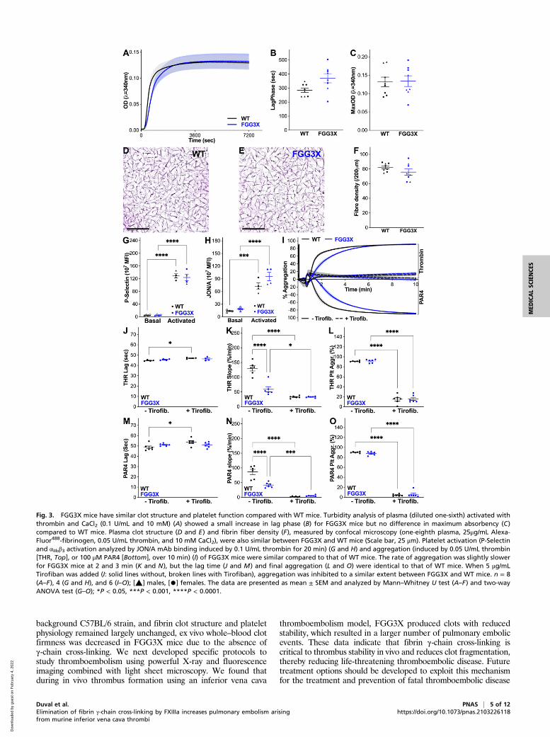
firmness by ROTEM could have been attributed to changes in clotstructure or platelet function. Clots were made with plasma col-lected from both strains of mice and analyzed by turbidity mea-surements and confocal microscopy. In vitro, turbidity analysis(Fig. 3A) of plasma clots from FGG3X mice showed a 1.3-foldincrease in lag phase (Fig. 3B), in agreement with our previouspublication (24) and ROTEM data, but unchanged maximum ab-sorbency (Fig. 3C) compared to WT mice, indicating no majordifferences in fibrin fibers thickness and density. This was con-firmed by confocal microscopy analysis of clots (Fig. 3 D and E),where the fiber density (Fig. 3F) was similar between FGG3X andWT mice. These data indicate that the architecture of clots fromFGG3X and WT mice is not different and therefore not respon-sible for the differences in clot firmness observed by ROTEM.Using flow cytometry, we found that agonist-induced platelet ex-pression of P-selectin (Fig. 3G) and activation of αIIbβ3 (Fig. 3H) inwhole blood were indistinguishable between FGG3X and WTmice. We next examined aggregation using thrombin and alsoPAR4-specific peptide to account for any potential differences dueto the conversion of fibrinogen to fibrin and cross-linking of fibrinby FXIII (Fig. 3I). The lag time to aggregation (Fig. 3 J andM) andoverall aggregation response (Fig. 3 L and O) was unchanged be-tween FGG3X and WT mice, regardless of agonist. We observed aminor reduction in the rate of aggregation for the FGG3X mice upto 3 min, but afterward, the aggregation response was normal(Fig. 3 I, K, and N). Furthermore, the presence of Tirofiban, aninhibitor of αIIbβ3 binding to fibrin(ogen), abolished aggregation inboth mouse strains, confirming that fibrinogen-mediated aggrega-tion of FGG3X platelets was largely unaffected (Fig. 3 I–O). To-gether, these data show that the reduction in clot firmness byROTEM in the FGG3X model is attributable to the lack of fibrinγ-chain cross-linking and not to major changes in platelet functionor clot architecture.We then investigated the effect of the lack of fibrin γ-chain
cross-linking on whole–blood clot contraction, washed plateletclot retraction, and in vivo clot size in an inferior vena cava stasismodel of thrombosis. We found that whole–blood clot contrac-tion kinetics, final clot weight, and final serum hemoglobincontent (SI Appendix, Fig. S3 A–D) were comparable betweenFGG3X and WT mice, indicating that the lack of γ-chain cross-linking does not affect blood clot contraction, in agreement withprevious data showing that red blood cell retention is dependenton α-chain cross-linking and not γ-chain cross-linking (26). Theability of washed platelet clots to retract was also not affected bythe loss of fibrin γ-chain cross-linking (SI Appendix, Fig. S3 E andF). These data were further supported by inferior vena cava li-gation experiments (SI Appendix, Fig. S3 G and H), whichshowed similar clot weight after 24 h between FGG3X and WTmice, thus indicating that the lack of γ-chain cross-linking doesnot affect overall clot formation or size in vivo.
FGG3X Mice Form Clots that Are Less Stable. A previously describedintravital microscopy model of venous thrombosis (27) using FeCl3application to the femoral vein was used to study in vivo clotformation. In this model, thrombus formation in the femoral veinwas followed over time (SI Appendix, Fig. S4), with measurementsof fibrin fluorescence, hence clot size, every 5 to 10 min over thefirst hour in the FGG3X (Fig. 4A) andWT (Fig. 4B) mice. Despitethe average clot area for each time point being similar betweenboth strains, analyses of the individual kinetic data were consis-tent, with FGG3X mice demonstrating more frequent drops inclot size between subsequent time points compared with WTmice.When this was quantified, with drops in clot size greater than 25%between time points counted as embolic events (Fig. 4C), FGG3Xmice showed a significant 2.1-fold (P < 0.01) increase in embolic
2 of 12 | PNAS Duval et al.https://doi.org/10.1073/pnas.2103226118 Elimination of fibrin γ-chain cross-linking by FXIIIa increases pulmonary embolism arising
from murine inferior vena cava thrombi
Dow
nloa
ded
by g
uest
on
Feb
ruar
y 4,
202
2

events compared to WT mice. The time for the first embolic eventto occur (Fig. 4D) was significantly shorter (1.8-fold, P < 0.05) inFGG3X mice. The percentage of clot area loss per 5 min was alsoquantified for all time points (Fig. 4E) and showed a significant1.5-fold increase (P < 0.05) in the overall amount of clot embolismin FGG3X mice compared to WT mice. These data suggest thatthe lack of fibrin γ-chain cross-linking renders the clot more proneto release fragments (emboli) during clot formation.We next examined fibrin fiber mechanical behavior using lateral
atomic force microscopy, to probe fibers made with fibrinogenpurified from FGG3X and WTmice. Individual fibers were pulled
using a lateral force–sensing atomic force microscope until rup-ture, and the resulting stress–strain curves (Fig. 4 F and G) wereanalyzed. In the absence of cross-linking by FXIIIa, stiffness at low(slope 1, Fig. 4H) and high (slope 2, Fig. 4I) strains, strain stiff-ening (s2/s1, Fig. 4J), maximum stress before rupture (Fig. 4K),and toughness (amount of energy absorbed prior to rupture;Fig. 4L) were all similar between both types of fibrinogen. How-ever, after cross-linking by FXIIIa, while initial stiffness wasslightly increased (1.3-fold), there was significant increase for largestrain stiffness (2.2-fold, P < 0.001), strain stiffening (1.7-fold, P <0.0001), maximum stress before rupture (2.1-fold, P < 0.001), andtoughness (2.0-fold, P < 0.01) in WT but not FGG3X fibers.Therefore, cross-linked FGG3X fibers were less stiff before rup-ture (35%, P < 0001), exhibited reduced strain stiffening (75%,P < 0.01), ruptured at a lower stress (45%, P < 0.001), and hadlower toughness (47%, P < 0.01) compared to WT fibers, indi-cating that the lack of γ-chain cross-linking by FXIII renders theFGG3X fibrin fibers more prone to rupture at lower stress relativeto WT.
Thromboembolism Models Show Increased Embolism in FGG3X Mice.In order to investigate the effects of γ-chain cross-linking byFXIIIa on clot stability and embolism in a pathophysiologicalsetting, we developed two protocols to specifically evaluate thelevel of thromboembolism to the lungs (PE) from thrombi inthe inferior vena cava. First, we used optical imaging coupled toX-ray to observe live appearance of emboli into the lungs ofmice undergoing inferior vena cava injury using FeCl3 (SI
Fig. 1. Mouse model characterization. WT and FGG3X mice showed no difference in growth, measured by weight (A), over the first 12 wk (n = 10). Tailsectioning bleeding time (B), plasma fibrinogen concentration (C), and plasma FXIII activity measured by thrombin- and CaCl2-induced (2 U/mL and 10 mM)pentylamine-biotin incorporation to fibrin (D) were not different between these two mouse strains. Fibrinogen, purified from plasma by ammonium sulfateprecipitation, was assayed for cross-linking of 5 μg fibrin by FXIII (10 μg/mL) activated with thrombin and CaCl2 (0.5 U/mL and 10 mM) (E) and showed fullinhibition of fibrin γ-chain cross-linking (no production of γ-γ dimers and continued presence of γ-monomers) in FGG3X, compared to WT, while the α-chaincross-linking was not affected, as shown by the normal disappearance of α-monomers and appearance of polymers (n = 4). n = 8 (A–D) and 4 (E); [▲] males,[•] females. The data are presented as mean ± SEM and analyzed by two-way ANOVA test (A) and Mann–Whitney U test (B–D).
Table 1. Hematological parameters in WT and FGG3X mice
Parameters WT FGG3X P values*
RBC (1012/mL) 5.34 ± 0.05 5.51 ± 0.07 0.127HGB (g/dL) 8.83 ± 0.11 9.08 ± 0.11 0.226HCT (%) 29.91 ± 0.32 29.86 ± 0.33 0.940PLT (1011/mL) 5.99 ± 0.71 7.79 ± 0.47 0.083WBC (109/mL) 3.33 ± 0.51 3.14 ± 0.22 0.775LYM (%) 81.89 ± 1.23 84.41 ± 0.86 0.111NEU (%) 8.48 ± 0.77 8.10 ± 0.53 0.857MXD (%) 9.71 ± 0.71 7.58 ± 0.43 0.066
RBC: red blood cells; HGB: hemoglobin, HCT: hematocrit; PLT: platelets;WBC: white blood cells; LYM: lymphocytes; NEU: neutrophils; and MXD:mixed white blood cells.*Data analyzed by Mann–Whitney U test. n = 8.
Duval et al. PNAS | 3 of 12Elimination of fibrin γ-chain cross-linking by FXIIIa increases pulmonary embolism arisingfrom murine inferior vena cava thrombi
https://doi.org/10.1073/pnas.2103226118
MED
ICALSC
IENCE
S
Dow
nloa
ded
by g
uest
on
Feb
ruar
y 4,
202
2

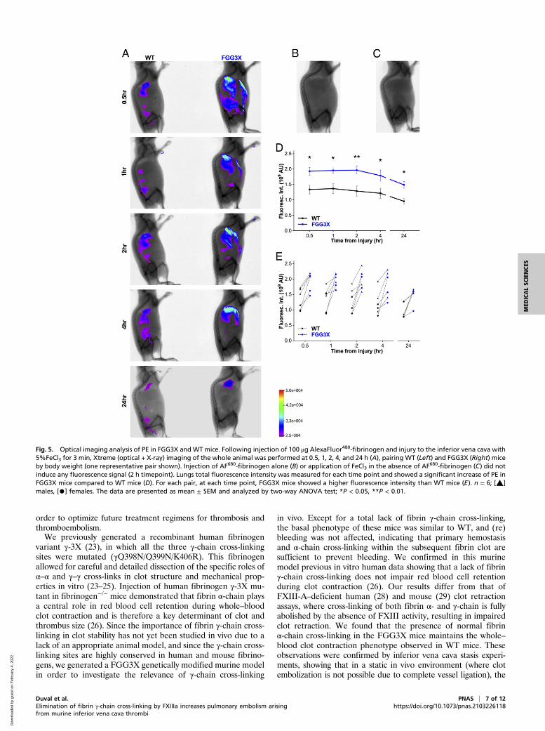
Appendix, Fig. S5), after prior injection of fluorescent fibrino-gen into the mice. The whole-body fluorescence levels inFGG3X and WT mice were observed 0.5, 1, 2, 4, and 24 hpostinjury (Fig. 5A). We observed that fluorescence accumu-lated specifically in the lungs over time after inferior vena cavathrombosis due to clot embolism. In contrast, fibrin accumu-lation was not observed elsewhere in the circulation of the mice.Control experiments with only fluorophore injected (Fig. 5B) orFeCl3 injury without fluorophore present (Fig. 5C) showed nosignal in these mice 2 h postinjury. Quantification of total fluo-rescence in the lungs showed that 30 min postinjury, FGG3X miceshowed a significant 1.4-fold increase (P < 0.05) in clot embolicompared to WT mice (Fig. 5D). This increase in fluorescenceintensity was also significantly higher in FGG3Xmice at 1 (1.4-fold,P < 0.05), 2 (1.5-fold, P < 0.01), 4 (1.5-fold, P < 0.05), and 24 h(1.6-fold, P < 0.05), compared to WT (Fig. 5D). Imaging wasperformed in pairs of matching gender and weight. For each pair,FGG3X mice showed higher fluorescence intensity in thelungs than their WT counterparts at every time point (Fig. 5E).Next, we used light sheet microscopy to image and quantify clot
emboli in the lungs of mice where the inferior vena cava was in-jured with FeCl3 following tail vein injection of fluorescent
fibrinogen to visualize clots. Mice underwent whole-body fixationand perfusion with fluorescent albumin (in gelatin) 1 h postsurgeryin order to visualize the vasculature. Lungs were imaged by lightsheet microscopy, and three-dimensional (3D) fluorescence re-constructions of organs were created using IMARIS (Fig. 6A). Inthis model, FGG3X mice showed a significant 1.5-fold increase(P < 0.001) in pulmonary emboli count compared to WT (Fig. 6B).The distribution of the emboli size (Fig. 6C) was also significantlydifferent (P < 0.05) between both strains, since FGG3X miceexhibited a higher number of pulmonary emboli for eachvolume range compared to WT mice. Together, these datademonstrate that the lack of fibrin γ-chain cross-linking in-creases embolism in the venous circulation, leading to an in-creased number and volume of pulmonary thromboembolicevents.
Discussion
We describe a murine model of fibrinogen, FGG3X, in which allthree conserved fibrin γ-chain cross-linking sites have been mu-tated, leading to a total lack of γ-chain cross-linking by FXIIIa.While blood parameters were otherwise identical to that of the
Fig. 2. FGG3X mice form blood clots that are less firm thanWT mice. EXTEM (7 μL + 3 μL TBS) (A, C, and D) and FIBTEM (7 μL + 3 μL TBS) (B, E, and F) analysis ofwhole blood (103 μL + 7 μL STARTEM) samples showed that clotting time (C and E) was not significantly different between WT and FGG3X mice, whilemaximum clot firmness (D and F) was significantly decreased in FGG3X mice compared to WT mice. EXTEM in the presence of a range of tPA concentration(2.5 to 100 nM) (G) showed that lysis time (H), % lysis at 30 (I), 45 (J), and 60 min (K), were similar between WT (dashed lines) and FGG3X mice (solid lines). n =8; [▲] males, [•] females. The data analyzed are presented as mean ± SEM and analyzed by Mann–Whitney U test; **P < 0.01, ***P < 0.001.
4 of 12 | PNAS Duval et al.https://doi.org/10.1073/pnas.2103226118 Elimination of fibrin γ-chain cross-linking by FXIIIa increases pulmonary embolism arising
from murine inferior vena cava thrombi
Dow
nloa
ded
by g
uest
on
Feb
ruar
y 4,
202
2
background C57BL/6 strain, and fibrin clot structure and plateletphysiology remained largely unchanged, ex vivo whole–blood clotfirmness was decreased in FGG3X mice due to the absence ofγ-chain cross-linking. We next developed specific protocols tostudy thromboembolism using powerful X-ray and fluorescenceimaging combined with light sheet microscopy. We found thatduring in vivo thrombus formation using an inferior vena cava
thromboembolism model, FGG3X produced clots with reducedstability, which resulted in a larger number of pulmonary embolicevents. These data indicate that fibrin γ-chain cross-linking iscritical to thrombus stability in vivo and reduces clot fragmentation,thereby reducing life-threatening thromboembolic disease. Futuretreatment options should be developed to exploit this mechanismfor the treatment and prevention of fatal thromboembolic disease
Fig. 3. FGG3X mice have similar clot structure and platelet function compared with WT mice. Turbidity analysis of plasma (diluted one-sixth) activated withthrombin and CaCl2 (0.1 U/mL and 10 mM) (A) showed a small increase in lag phase (B) for FGG3X mice but no difference in maximum absorbency (C)compared to WT mice. Plasma clot structure (D and E) and fibrin fiber density (F), measured by confocal microscopy (one-eighth plasma, 25μg/mL Alexa-Fluor488-fibrinogen, 0.05 U/mL thrombin, and 10 mM CaCl2), were also similar between FGG3X and WT mice (Scale bar, 25 μm). Platelet activation (P-Selectinand αIIbβ3 activation analyzed by JON/A mAb binding induced by 0.1 U/mL thrombin for 20 min) (G and H) and aggregation (induced by 0.05 U/mL thrombin[THR, Top], or 100 μM PAR4 [Bottom], over 10 min) (I) of FGG3X mice were similar compared to that of WT mice. The rate of aggregation was slightly slowerfor FGG3X mice at 2 and 3 min (K and N), but the lag time (J and M) and final aggregation (L and O) were identical to that of WT mice. When 5 μg/mLTirofiban was added (I: solid lines without, broken lines with Tirofiban), aggregation was inhibited to a similar extent between FGG3X and WT mice. n = 8(A–F), 4 (G and H), and 6 (I–O); [▲] males, [•] females. The data are presented as mean ± SEM and analyzed by Mann–Whitney U test (A–F) and two-wayANOVA test (G–O); *P < 0.05, ***P < 0.001, ****P < 0.0001.
Duval et al. PNAS | 5 of 12Elimination of fibrin γ-chain cross-linking by FXIIIa increases pulmonary embolism arisingfrom murine inferior vena cava thrombi
https://doi.org/10.1073/pnas.2103226118
MED
ICALSC
IENCE
S
Dow
nloa
ded
by g
uest
on
Feb
ruar
y 4,
202
2

by specifically targeting fibrin α-chain cross-linking–dependenterythrocyte retention, therefore clot size and burden, while allow-ing for normal FXIIIa-mediated fibrin γ-chain cross-linking toprevent embolism.Clinically, treatment of thrombosis is largely based on anti-
coagulation, with little attention for the quality or stability of theblood clot. Clot quality and stability, however, have been reported
to play a role in thromboembolism (9–12), an important anddangerous sequela of thrombosis, leading to PE. As such, it isimportant to fully understand the role of fibrin cross-linking in theformation and stability of thrombi and their subsequent emboli-zation. On the basis of these considerations, we consider that amore-complete understanding of the implications of cross-linkingby FXIIIa on clot stability and embolism is urgently needed in
Fig. 4. Clot stability and fiber resistance to rupture are reduced in FGG3X mice. Following injection of 100 μg AlexaFluor488-fibrinogen and injury to thefemoral vein with 10% FeCl3 for 3 min, clot size was measured over time by intravital fluorescence microscopy in WT (A) and FGG3X (B) mice, with the red linerepresenting the average clot area and bold lines representing clot size decreases greater than 25%/5 min (embolic event). The number of embolic events (C)was significantly increased, the time for the first embolic event to occur (D) was significantly lower, while the overall size of the emboli (E) were significantlylarger in FGG3X mice compared to WT mice. Fibrin fiber pulling characteristics were measured by lateral atomic force microscopy using purified FGG3X andWT fibrinogens in the absence (F) or presence (G) of FXIIIa. Low strain stiffness (H) was slightly increased, while maximal stiffness prior to rupture (I), strainstiffening (J), maximum stress before rupture (K), and fiber toughness (L) were significantly increased by cross-linking in WT but not FGG3X fibrin. n = 8 (A–E)and 18 to 22 (F–L); [▲] males, [•] females. The data are presented as mean ± SEM and analyzed by Mann–Whitney U test (C), χ2 test (D), Kolmogorov–Smirnov test (E), and one-way ANOVA test (H–L); *P < 0.05, **P < 0.01, ***P < 0.001, ****P < 0.0001.
6 of 12 | PNAS Duval et al.https://doi.org/10.1073/pnas.2103226118 Elimination of fibrin γ-chain cross-linking by FXIIIa increases pulmonary embolism arising
from murine inferior vena cava thrombi
Dow
nloa
ded
by g
uest
on
Feb
ruar
y 4,
202
2
order to optimize future treatment regimens for thrombosis andthromboembolism.We previously generated a recombinant human fibrinogen
variant γ-3X (23), in which all the three γ-chain cross-linkingsites were mutated (γQ398N/Q399N/K406R). This fibrinogenallowed for careful and detailed dissection of the specific roles ofα–α and γ–γ cross-links in clot structure and mechanical prop-erties in vitro (23–25). Injection of human fibrinogen γ-3X mu-tant in fibrinogen−/− mice demonstrated that fibrin α-chain playsa central role in red blood cell retention during whole–bloodclot contraction and is therefore a key determinant of clot andthrombus size (26). Since the importance of fibrin γ-chain cross-linking in clot stability has not yet been studied in vivo due to alack of an appropriate animal model, and since the γ-chain cross-linking sites are highly conserved in human and mouse fibrino-gens, we generated a FGG3X genetically modified murine modelin order to investigate the relevance of γ-chain cross-linking
in vivo. Except for a total lack of fibrin γ-chain cross-linking,the basal phenotype of these mice was similar to WT, and (re)bleeding was not affected, indicating that primary hemostasisand α-chain cross-linking within the subsequent fibrin clot aresufficient to prevent bleeding. We confirmed in this murinemodel previous in vitro human data showing that a lack of fibrinγ-chain cross-linking does not impair red blood cell retentionduring clot contraction (26). Our results differ from that ofFXIII-A–deficient human (28) and mouse (29) clot retractionassays, where cross-linking of both fibrin α- and γ-chain is fullyabolished by the absence of FXIII activity, resulting in impairedclot retraction. We found that the presence of normal fibrinα-chain cross-linking in the FGG3X mice maintains the whole–blood clot contraction phenotype observed in WT mice. Theseobservations were confirmed by inferior vena cava stasis experi-ments, showing that in a static in vivo environment (where clotembolization is not possible due to complete vessel ligation), the
Fig. 5. Optical imaging analysis of PE in FGG3X and WT mice. Following injection of 100 μg AlexaFluor480-fibrinogen and injury to the inferior vena cava with5%FeCl3 for 3 min, Xtreme (optical + X-ray) imaging of the whole animal was performed at 0.5, 1, 2, 4, and 24 h (A), pairing WT (Left) and FGG3X (Right) miceby body weight (one representative pair shown). Injection of AF680-fibrinogen alone (B) or application of FeCl3 in the absence of AF680-fibrinogen (C) did notinduce any fluorescence signal (2 h timepoint). Lungs total fluorescence intensity was measured for each time point and showed a significant increase of PE inFGG3X mice compared to WT mice (D). For each pair, at each time point, FGG3X mice showed a higher fluorescence intensity than WT mice (E). n = 6; [▲]males, [•] females. The data are presented as mean ± SEM and analyzed by two-way ANOVA test; *P < 0.05, **P < 0.01.
Duval et al. PNAS | 7 of 12Elimination of fibrin γ-chain cross-linking by FXIIIa increases pulmonary embolism arisingfrom murine inferior vena cava thrombi
https://doi.org/10.1073/pnas.2103226118
MED
ICALSC
IENCE
S
Dow
nloa
ded
by g
uest
on
Feb
ruar
y 4,
202
2

lack of γ-chain cross-linking does not impact on the formation andsize of venous thrombi. Additionally, the mutations of γ-Q398N,γ-Q399N, and γ-K406R residues, located near the αIIbβ3 bindingsite (γ-404 to 411 in mouse and human) (30), did not affect washedplatelets activation, aggregation, and clot retraction. A study byJiroušková et al. (31) showed that an antibody (7E9) directedagainst the fibrin γ-chain C terminus, which sterically hindersfibrinogen–αIIbβ3 interaction and fibrin cross-linking by FXIIIa,resulted in reduced platelet aggregation and clot contraction.Additionally, in an arterial thrombosis model, γ-Δ5 mice (whichlack the N-terminal residues of the γ-chain responsible for αIIbβ3interaction) and WT mice administered with the 7E9 antibodyformed smaller thrombi with limited embolization. Our data showthat the conservative Q-N and K-R mutations in the three cross-linking sites for FXIIIa that are close to integrin binding site donot influence platelet binding nor clot contraction, yet γ-chaincross-linking is completely abolished. Importantly, in vivo experi-ments using the FGG3X mice and intravital microscopy imagingon clots formed in the femoral vein following application of FeCl3(27) showed that these mice form clots that are less stable, mostlikely due to the reduced toughness (increase rupture potential)observed by single-fiber pulling experiments using lateral atomicforce microscopy, resulting in an increased number of embolicevents in the early stages of clot formation.Next, two inferior vena cava thrombosis and PE protocols were
developed, showing a critical role for fibrin γ-chain cross-linking inpreventing clot embolism in the venous circulation. Our modeland protocol show that a loss of fibrin γ-chain cross-linking and
increased susceptibility to fiber rupture correlated with the for-mation of less-stable venous thrombi that were more prone tofragmentation and subsequent embolization to the lungs. Wefound that FGG3X mice produced a higher degree of PE thanWT mice at all time points observed. At 2 h post–vena cavathrombosis, the fluorescence intensity in the lungs of both FGG3Xand WT mice decreased at a similar rate, most likely due to fi-brinolysis of the emboli taking place. The rate of lysis was similarbetween both strains, in agreement with our ex vivo ROTEM datashowing that clot lysis was not affected by the lack of fibrin γ-chaincross-linking. With clot lysis being unaffected, our data insteadindicate that clots without fibrin γ-chain cross-linking are morereadily deformed under shear stress, with dissociation of knob-hole and other noncovalent bonds, which are not as strong ascovalent bonds (e.g., γ-chain cross-links present in WT but notFGG3X fibrin) and hence rupture of individual fibers, whichrender the clots more prone to fragmentation and embolism.A paradigm is proposed in which in the absence of γ-chain
cross-linking, the force of flow applied to fibrin protofibrilslikely leads to the dissociation of the D–E–D interactions, slippageof the fibrin molecules along the protofibrils, fiber rupture, andpermanent clot deformation (Fig. 7). Fibrin α-chain cross-linkingmay in part rescue the integrity of the clots via intra- and inter-protofibril cross-links. However, in places where those interactionsdo not occur, due to the random nature of the α-chain cross-linkingorientation, rupture of the fibers results in subsequent clot embo-lism. This phenomenon will be less common in clots from WTmice, as fiber rupture and clot fragmentation are prevented by the
Fig. 6. Light sheet microscopy of clot emboli in the lungs of FGG3X and WT mice. Following injection of 100 μg AlexaFluor647-fibrinogen and injury to theinferior vena cava with 5% FeCl3 for 3 min, perfusion fixation of the mice with PFA after 57 min, and injection of FITC-albumin in the circulation priorcollection and clearing of the lungs, light sheet fluorescence microscopy imaging of the lungs (A) showed that the total emboli count per mouse (B) wassignificantly increased in FGG3X mice. The distribution of all the emboli volumes (C) showed that FGG3X mice produced significantly more emboli, irrespectiveof their size, than WT mice (Scale bar, 1 mm). n = 8; [▲] males, [•] females. The data are presented as mean ± SEM and analyzed by Mann–Whitney U test (B)and Kolmogorov–Smirnov test (C); *P < 0.05, ***P < 0.001.
8 of 12 | PNAS Duval et al.https://doi.org/10.1073/pnas.2103226118 Elimination of fibrin γ-chain cross-linking by FXIIIa increases pulmonary embolism arising
from murine inferior vena cava thrombi
Dow
nloa
ded
by g
uest
on
Feb
ruar
y 4,
202
2

presence of fibrin γ-chain cross-links. This model is further sup-ported by a previous study by Liu et al. on in vitro fibrin mechanicalproperties, which concluded that “fast-forming γ–γ cross-linksalong the axis may enhance elasticity and prevent rupture of the(nascent) fibers” (17). Our data indicate that this is also the casein vivo using protocols to study thromboembolism, suggesting a keyrole for γ-chain cross-linking in the pathophysiology of this disease.These findings are further supported by a recent clinical studyshowing that plasma samples from patients with recurrent VTEhave reduced elastic modulus compared to samples from patientswith nonrecurrent VTE (12), indicating that optimal clot elasticityis critical to prevent VTE recurrence.Prior in vivo studies investigating thromboembolism involved
injection of exogenous clots or thrombotic substance in the ve-nous system (e.g., thrombin or collagen/epinephrine) with theuse of histology to detect the emboli within the lungs (32). Shayaet al. recently applied FeCl3 to the femoral vein to study thrombusformation and performed histology of the lungs to analyze theeffect of dabigatran (a direct thrombin inhibitor) on PE (33).Interestingly, these authors found that dabigatran decreased clotstability and increased PE in this model, which they attributed toreduced FXIII cross-linking activity since untreated FXIII−/− miceshowed similar rates of PE as WT mice treated with dabigatran.
We chose FeCl3 as trigger for thrombosis due to its reproducibilityin terms of the size of the ensuing thrombus. Furthermore, ouraim was to study clot stability and embolism rather than the de-velopment of the clot itself, justifying the use of FeCl3 as a suitablemodel to test our hypothesis. However, future studies may com-bine whole-body/organ emboli imaging described here with othermodels of venous thrombosis such as inferior vena cava ligationand stenosis models, which have also been shown to lead toembolization (34).We developed and characterized methodological approaches
to study thromboembolism. We used live in vivo optical imagingcoupled with X-ray to analyze thromboembolism in real time inliving mice. Advantages of this method are that the fluorescentlylabeled fibrin is stable enough to allow measurements for at least24 h and that the method can be applied to other models ofthrombosis which tolerate animal recovery. Limitations are thatintensity measurements do not provide detailed information onthe number and volume of the emboli generated or their preciselocation within the lungs and that imaging is only possible in thoseparts of the body that are not masked by bones (e.g., the lungs butnot the brain) or very opaque tissues. Information on emboli toorgans covered by bone, however, can be provided by the second,complementary imaging method that we used, involving terminal
Fig. 7. Fibrin γ-chain cross-linking increases clot stability and reduces embolism. Fibrinogen is a soluble heterotetrametric molecule, consisting of two α-, twoβ-, and two γ-chains (A, Left), which is proteolytically converted by thrombin into insoluble fibrin. Fibrin monomers polymerize in a half-staggered ar-rangement to form protofibrils which interact laterally via α-chains to form fibers (A, Middle). FXIIIa stabilizes the clots by forming longitudinal intra-protofibril cross-links between glutamine 398 or 399 and lysine 406 (398/399 and 406 in human) of adjacent γ-chains, as well as lateral interprotofibril cross-links between multiple glutamine and lysine residues of adjacent α-chains (A, Right). In normal conditions (B, WT mice), blood flow induces strain (F) on thefibrin fibers, resulting in deformation of the clots which is reduced by γ-chain (intraprotofibril, yellow rectangles) and α-chain (intra- and interprotofibrils, bluestars) cross-linking. Clots from FGG3X mice (C) exhibit a lack of γ-chain cross-linking, resulting in dissociation of the D–E–D interactions, slippage of the fibrinmolecules along the protofibrils, and permanent deformation and rupture of protofibrils and hence individual fibers. While fibrin α-chain cross-linkingpartially rescues the integrity of the clots, the compromised γ-chain cross-linking predisposes to clot fragmentation and release of emboli (Insert) in places thatlack (randomly orientated) interprotofibril α-chain cross-links (images were created with BioRender.com).
Duval et al. PNAS | 9 of 12Elimination of fibrin γ-chain cross-linking by FXIIIa increases pulmonary embolism arisingfrom murine inferior vena cava thrombi
https://doi.org/10.1073/pnas.2103226118
MED
ICALSC
IENCE
S
Dow
nloa
ded
by g
uest
on
Feb
ruar
y 4,
202
2

light sheet microscopy following perfusion fixation and tissueclearing. The light sheet microscopy data 1 h postinjury were inperfect agreement with the 1-h time point from the optical im-aging experiments, showing that FGG3X mice demonstrate anincreased number of pulmonary emboli and that for each volumebracket, FGG3X produced more emboli, which should result in anoverall higher fluorescence intensity. Advantages of this techniqueare the detailed quantification of emboli numbers and volumes andaccurate location of these emboli in the organ as well as the im-aging of any organ in the body, regardless of its location. Limita-tions are that this is a terminal procedure, therefore requiring alarger number of animals if more than one time point is required.Thus, the combination of both light sheet microscopy and whole-body combined optical and X-ray imaging provides powerful andcomprehensive analysis of thromboembolic disease. Furthermore,it may be possible to combine both protocols in sequence, per-forming optical imaging first, followed by light sheet imaging at theterminal endpoint. This would require that the type of injury allowsfor recovery and that fluorophores and filters are compatible withboth approaches.Previous clot contraction assays and our current in vivo throm-
boembolization data provide strong evidence that fibrin α- andγ-chain cross-linking play complementary roles in venous throm-bosis via different mechanisms. Byrnes et al. showed that fibrinα-chain (but not γ-chain) cross-linking plays an important role inred blood cell retention within the clot (26), thereby increasing thesize of venous thrombi. Our study demonstrates that γ-chain cross-linking is critical for the stability of venous thrombi, thereforereducing breakdown and embolism. While anticoagulation is cur-rently used as treatment for thromboembolic diseases, any possibleeffects of this treatment on embolism have so far been largely ig-nored. Based on our current data, and in agreement with Byrneset al. (26), we propose that specific inhibitors of fibrin α-chaincross-linking should be developed to reduce red blood cell con-tent within thrombi, therefore size and burden of venous thrombi.Moreover, specific α-chain cross-linking inhibitors would not im-pact on the critical fibrin γ-chain cross-linking by FXIIIa, therebypreventing thromboembolic effects as shown in our current study.In conclusion, we present the development and characterization
of a genetically modified murine model in which the residues re-sponsible for fibrin γ-chain cross-linking are mutated. We alsodeveloped and characterized protocols to study thromboembolismusing powerful imaging methods. These models demonstrate theessential importance of γ-chain cross-linking in clot viscoelasticproperties. We show a critical role of fibrin γ-chain cross-linkingby FXIIIa in stabilizing clots and reducing thromboembolic eventsin the venous (PE) circulation in vivo. These data demonstrateimportant mechanisms related to clot mechanical properties inthromboembolic disease, indicating a key future target for thera-peutic intervention and prevention of this leading cause of deaththat remains poorly treated to date.
MethodsEthics. All procedures were approved by the University of Leeds and theUniversity of Sheffield Ethics Committees and performed under projectlicense numbers 70/8115 and P144DD0D6 (held by Stephen Wheatcroft atLeeds University) and 70/8532 (held by Victoria Ridger, University of Sheffield)according to the Home Office Animals (Scientific Procedures) Act 1986. Bothmales and females were used, aged 6 to 8 wk at the time of the experiments.
Materials. Recombinant murine thrombin (Haematologic Technologies Inc) wasreconstituted to 250 U/mL in ddH2O and stored at−80 °C. Recombinant humantPA (Pathway Diagnostics) was diluted in Tris-buffered saline (TBS; 0.05 MTris HCl, 0.1 M NaCl, pH 7.4) to 1,400 nM and stored at −80 °C. EZ-linkpentylamine-biotin (Thermo Fisher Scientific) was diluted in ddH2O to 30 μMand stored at −20 °C. AlexaFluor488, AlexaFluor647, and AlexaFluor680 proteinlabeling kits were purchased from Thermo Fisher Scientific. All other chemicalswere obtained from Sigma unless stated otherwise.
Generation and Maintenance of FGG3X Mice. A genetically modified murineFGG3X line, in which the fibrinogen γ-chain cross-linking sites have beenmutated, were generated by GenOway as follows: Human residues γ-Q398,γ-Q399, and γ-K406 (mature sequence) are conserved in murine fibrinogen(γ-Q398, γ-Q399, and γ-K406; SI Appendix, Fig. S1), with the correspondingcodons located in exon 9 of the murine Fgg gene. Homologous recombinationin embryonic stem (ES) cells was performed using a targeting-vector–containingregion homologous to the murine genomic Fgg sequences. The targeting vec-tor (ARI1-HR) was generated by cloning themouse genomic DNA encompassingthe murine Fgg gene regions surrounding the target exon 9 (exon 7 to 5′-UTR[untranslated region]) into the targeting vector, inducing the three point mu-tations (Q398N, Q399N, and K406R), and inserting a neomycin selection cassette(Neo) (SI Appendix, Fig. S2). The vector was linearized and transfected into EScells by electroporation, and cells were selected for resistance to neomycin,before the correct recombination events were validated by PCR and Southernblot. The ES cells containing the correct recombination were then injected intoblastocysts (3.5-d-old embryos), which were subsequently implanted into C57BL/6 pseudopregnant females, resulting in the generation of chimeric mice. Malemice with chimerism rate above 50% were mated with C57BL/6-Cre females togenerate heterozygous mice carrying the Neo-excised knock-in allele, whichwere bred for the generation of homozygous mice. Upon transfer to the Uni-versity of Leeds, FGG3X mice were backcrossed onto our C57BL/6J strain over 10generations and maintained in individually ventilated cages at 21 °C, 50 to 70%humidity. The light/dark cycle was 12/12 h, andmice were fed on standard chowdiet ad-libitum. Experimental units were one mouse per cage.
Mouse Growth and Blood Sampling. WT and FGG3X mice were weighed once aweek, at the same time of the day from 2 to 12 wk old. Experiments wereperformed with eight mice per group. For blood sampling, animals were bledfrom the inferior vena cava under terminal anesthesia using 10% vol/vol0.109 M sodium citrate as anticoagulant. Blood was centrifuged at 14,000 g for10 min at room temperature (RT) for plasma preparation. The platelet-poorplasma supernatant was collected, aliquoted, and stored at −80 °C.
Bleeding Time and Rebleeding Events. Tomeasure bleeding time, the distal 3-mmsegment of themouse tail tipwas sectionedusing a scalpel under anesthesia andimmersed in a microtube containing isotonic saline (37 °C) (35). Bleeding timewas determined using a stop clock. Measurement of rebleeding events wasperformed using a method adapted from Molina et al. (36); the mice werereturned to individual cages 5 min after cessation of bleeding, and a filter paperwas applied to the end of the mouse tail every 15 min over a 1-h period. Ap-pearance of fresh blood on the filter paper was counted as a rebleeding event.Experiments were performed with eight mice per group.
Fibrinogen Levels and FXIII Activation Rate. Plasma fibrinogen levels were de-termined using a murine fibrinogen total antigen enzyme-linked immunosor-bent assay (ELISA) Kit (MyBioSource) following manufacturer’s instructions.Measurement of FXIII activation was performed using a modified 5-(bio-tinamido)pentylamine incorporation assay (27). Nunc-Immuno 96-MicroWellplates were coated with 100 μL 10 μg/mL N,N-dimethylated casein for 40 min atRT and blocked with 300 μL 1% bovine serum albumin (BSA) in TBS (pH 8.3) for90 min at 37 °C. Plates were washed with 4× 300 μL TBS (pH 8.3) and 10 μLplasma samples (diluted one-tenth in TBS [pH 8.3]) were added to the wells intriplicate. A total of 90 μL activation mix (111 μM dithiothreitol [DTT], 0.3 μMbiotinylated pentylamine, 11 mM CaCl2, and 2.2 U/mL thrombin) were addedand the reactions were stopped at 0, 20, 40, 60, 80, 100, and 120 min by adding200 μL 200 mM ethylenediaminetetraacetic acid. Plates were washed with 4×300 μL 0.1% [vol/vol] Tween 20 in TBS (pH 8.3), and 100 μL 2μg/mL streptavidinin 1% [wt/vol] BSA (in TBS-Tween) were added and incubated for 60 min at37 °C. Following washes with 4× 300 μL TBS-Tween, 100 μL 1 mg/mL phos-phatase substrate (in 1 M diethanolamine) were added for 7 min, and the re-action was stopped by adding 100 μL 4 M NaOH. Absorbency was measured at405 nm using a PowerWave HT Microplate Spectrophotometer (BioTek). Therate of pentylamine incorporation over time was used as an indicator of FXIIIactivation. Measurements were performed in triplicate, with eight miceper group.
Fibrinogen Purification. Fibrinogen was purified from pooled mouse plasma byammonium and ethanol precipitations, using a protocol adapted from Dietrichet al. (37). All steps were performed at 4 °C. A protease inhibitor mixture(55 mM e-aminocaproic acid, 55 mM benzamidine, 11 μM pepstatin, 11 μMleupeptin, and 1.1 mM phenylmethylsulfonyl fluoride, in TBS) was added (1:10vol/vol) to plasma and 1 vol saturated ammonium sulfate (760 g/L) was slowly(drop-by-drop) added to 3 vol plasma and incubated for 2 h. Following centri-fugation at 12,000 g for 15 min, the pellet was resuspended in 2-(N-morpholino)
10 of 12 | PNAS Duval et al.https://doi.org/10.1073/pnas.2103226118 Elimination of fibrin γ-chain cross-linking by FXIIIa increases pulmonary embolism arising
from murine inferior vena cava thrombi
Dow
nloa
ded
by g
uest
on
Feb
ruar
y 4,
202
2

ethanesulfonic acid (MES) buffer (55 mM e-aminocaproic acid, 55 mM ben-zamidine, 1.1 μM pepstatin, 1.1 μM leupeptin, 110 μM phenylmethylsulfonylfluoride, 22 mM 2-(N-morpholino)ethanesulfonic acid, in ddH2O, pH 6.6),and the whole process was repeated a second time, followed by pelletresuspension and dialysis in TBS for 1 h. Then, 1 vol ice-cold 100% ethanol wasadded drop-by-drop to 13 vol ice-cold suspension and incubated on ice for 1 h.Following centrifugation at 12,000 g for 15 min, the pellet was resuspendedand dialyzed against TBS overnight before the concentration was determinedusing a ND-100 Spectrophotometer (Thermo Fisher Scientific).
Fibrin Cross-Linking. Clottingmixtures (20 μL) containing fibrinogen (0.25mg/mL),zymogen FXIII (10μg/mL), thrombin (0.5 U/mL), and CaCl2 (10 mM) were in-cubated at 37 °C for 0, 2, 5, 10, 15, 20, 30, 60, and 120 min. Reactions werestopped by adding 4× NuPAGE lithium dodecyl sulphate (LDS) Sample Buffer(ThermoFisher Scientific) and 10× NuPAGE Sample Reducing Agent (ThermoFisher Scientific), immediately followed by heating at 90 °C for 10min. Samplesand molecular weight marker (Precision Plus Protein Dual Color Standards;BioRad) were run onto a NuPAGE 4 to 12% Bis-Tris Protein Gel (Thermo FisherScientific), and gels were stained using InstantBlue (Expedeon). Protein bandswere visualized and quantified using Genesys and GeneTools softwares (Syn-gene). Band quantification for each lane was relative to the amount of B-βchain staining. Experiments were performed in triplicate.
Fibrinogen Labeling. For in vivo experiments, fibrinogen purified from FGG3Xand WT mice was labeled with AlexaFluor 488, 647, or 680 depending on thefilters available for each setup. Labeling was performed following manu-facturer’s instructions (Thermo Fisher Scientific).
Hematological Blood Parameters. Freshly obtained blood samples were runonto a KX-21N Automated Hematology Analyzer (Sysmex). Discriminator val-ues were changed for leukocyte counts as follows: LD 30 fl, T1 66 fl, and T2 84fl. Experiments were performed in triplicate with eight mice per group.
Rotational Thromboelastometry. Freshly obtained blood samples were run on aROTEM-Delta (Werfen) for thromboelastic analysis. For clotting analysis, 103 μLblood was mixed with 3 μL TBS, 7 μL EXTEM or FIBTEM, and 7 μL STARTEM(CaCl2) reagents before data were acquired using the EXTEM and FIBTEMchannels (respectively). For lysis analysis, 107 μL blood was mixed with 7 μLEXTEM and 7 μL STARTEM reagents and 3 μL tPA (final concentration range 2.5to 100 nM) before data were acquired using the EXTEM channel. Measurementswere performed with eight mice per group.
Fibrin Polymerization and Clot Structure. Polymerization of fibrin was studiedby turbidity analysis as previously described (24). A total of 25 μL plasma(one-third) were transferred into 384-well plates in triplicate. A total of 25 μLCaCl2 and thrombin mix (0.1 U/mL and 10 mM final reaction concentrations)were added to initiate clotting, and absorbency was measured at 340 nm every12 s for 2 h at 32 °C using a PowerWave HT Microplate Spectrophotometer(BioTek). Clot structure was analyzed by laser-scanning confocal microscopy,as previously described (24). Plasma, AlexaFluor488-fibrinogen, CaCl2, andthrombin (one-eighth, 25 μg/mL, 10 mM, and 0.05 U/mL final concentrations,respectively) were mixed, transferred into the channel of an uncoated Ibidiμ-slide VI (Thistle Scientific), and incubated in a dark humidity chamber for60 min. Clots were imaged using an inverted Zeiss LSM-880 microscope (CarlZeiss) with a 40× oil immersion objective lens. Optical z-stacks were obtainedevery 0.5 μm over 10 μm and combined into single projected images. Fiberdensity was determined by counting the number of fibers crossing 10 arbitrarylines of fixed length (200 μm) drawn through a single optical section. Experi-ments were performed in triplicate with eight mice per group.
Platelet Functions. Platelet activation was measured in whole blood, supple-mented with 10 μM Gly-Pro-Arg-Pro-NH2 and fluorescein isothiocyanate (FITC)-conjugated anti–P-selectin (BD Biosciences) or PE-conjugated JON/A (anti-αIIbβ3;Emfret) antibodies, incubated with or without thrombin (0.1 U/mL) for 20 minat 37 °C (38). Whole blood was subsequently fixed with 1% paraformalde-hyde (PFA) (in phosphate-buffered saline [PBS]) for 10 min and analyzed byfluorescence-activated cell sorting using a CytoFLEX Flow Cytometer (Beck-man Coulter). A compensation matrix was applied, and analysis was per-formed with the CytExpert software version 2.1 (Beckman Coulter).Experiments were performed in four mice per group.
Platelet aggregation was measured using suspended washed platelets.Platelet-rich plasma was obtained by centrifugation of whole blood supple-mented with 200 μL modified Tyrode’s buffer (MTB; 150 mM NaCl, 5 mMHepes, 0.55 mM NaH2PO4, 7 mM NaHCO3, 2.7 mM KCl, 0.5 mM MgCl2, 5.6 mM
d-glucose, pH 7.4) at 100 g for 5 min at RT. The resulting pellet was resuspendedin MTB and recentrifuged at 1,000 g in the presence of PGI2 (200 nM) for 6 min,the washed platelet pellet was resuspended in MTB, and counts were adjustedto 2.5 × 10⁸ plt/mL using a Z1 Coulter Particle Counter (Beckman Coulter).Platelet aggregation was performed using 250 μL washed platelets at 2 × 10⁸
plt/mL, calibrated against MTB. Platelets were stimulated with thrombin(0.05 U/mL) or PAR4 (100 μM) with or without the pretreatment of Tirofiban(5 μg/mL), and aggregation was recorded under constant stirring conditions(1,000 rpm) for 10 min at 37 °C using an AggRAM aggregometer (Helena Bio-sciences Europe). Experiments were performed in six mice per group.
Intravital Microscopy. In vivo visualization of clot formation was performed aspreviously described (27). Mice (6- to 8-wk-old) were anesthetized by intra-peritoneal injection of ketamine/atropine/xylazine. The carotid artery wascannulated to allow for maintenance of anesthesia and injection of 100 μgAlexaFluor488-fibrinogen 5 min prior to exposure of the femoral vein andapplication of a 10% [vol/vol] FeCl3-saturated filter paper (3 × 2 mm) for3 min. Real-time observation of clot formation started 2 min after removalof the FeCl3 filter paper and washing with isotonic saline using an uprightNikon Eclipse E600-FN microscope (Nikon) equipped for fluorescence mi-croscopy with a water-immersion 40/0.80-W objective. The green channel(488 nm) was recorded using Slidebook Imaging Software version 5.0 (In-telligent Imaging Innovation). Clot size for each time point was determinedas a combination of area and intensity of green pixels. Experiments wereperformed with eight mice per group.
Lateral Atomic Force Microscopy. The mechanical response of individual fibrinfibers upon lateral stretching was measured as described previously (17).Briefly, clots were made on a striated surface (25, 39), with resulting fibersforming over the trenches. Individual fibers were pulled laterally with theatomic force microscope (AFM) cantilever, and the lateral deflection of thecantilever was registered and used to calculate the force and the stress onthe fiber. Details are provided in SI Appendix, Methods.
Optical and X-ray Imaging. Mice (8-wk-old) were anesthetized with 1.5%isoflurane (Piramal Critical Care) in oxygen (2 l/min flow rate). 100 μg ofAlexaFluor680-fibrinogen per 10 g of mouse body weight were injected intothe tail vein, and the abdomen, chest, and sides of the mice were shaved toallow for imaging. The inferior vena cava was exposed after midline lapa-rotomy and exteriorization of the bowels and separated from the abdomi-nal aorta using a plastic spacer (40). A filter paper (2 × 3 mm) soaked with5% FeCl3 was applied onto the isolated vessel for 3 min before removing thefilter paper and washing the abdominal cavity with saline. The bowels weremoved back into the abdominal cavity with saline, and the muscle and skinlayers were sutured sequentially. Animals were immediately injected with100 μL 0.1 mg/mL Vetergesic (Ceva Animal Health Ltd) for pain relief. Usingthe in vivo Xtreme II optical imaging system (Bruker), fluorescence (excita-tion 680 nm, emission 700 nm) and X-ray in whole mice was imaged underanesthesia at 0.5, 1, and 2 h postinjury, before animals were allowed torecover, and then at 4 and 24 h postinjury. Animals were imaged in threepositions (frontal, sagittal left, and sagittal right) for each time point. UsingMolecular Imaging software version 7.5.2.22464 (Bruker) and X-ray images,the lungs were delineated using the “ROI free form” tool (SI Appendix, Fig.S5), and fluorescence intensity was quantified for each plane. The values foreach image plane were added, determining the total fluorescence intensity.Each set of experiments comprised of one FGG3X and one WT mouse in pairs,determined by gender and weight. Experiments were performed with six miceper group.
Light SheetMicroscopy.Mice (6- to 8-wk-old)were anesthetizedby intraperitonealinjection of ketamine/atropine/xylazine. AlexaFluor647-fibrinogen (100 μL per10 g of body weight) was injected into the tail vein. The inferior vena cava wasisolated over a plastic sheet spacer (40), and a 2.5% FeCl3-soaked filter paper(2 × 3 mm) was applied for 3 min. Remaining FeCl3 was washed off with isotonicsaline, and the mice were kept in the dark for a further 57 min. The mice werethen slowly perfused with 20 mL PBS (+50 U/mL heparin), 15 mL 4% PFA, and10 mL hydrogel (41) (0.8 mg/mL FITC-Albumin, 2% wt/vol gelatin, in PBS). Micewere immediately placed on ice for at least 30 min before the lungs wereharvested and transferred to 4% PFA overnight, at 4 °C, in the dark. All subse-quent steps were performed in the dark. Next day, the lungs were dehydrated in20, 40, 60, 80, and 100% methanol solutions (in ddH2O) for 1 h each, shaking at200 rpm at RT, before being left overnight in fresh methanol solution. The leftand right lungs were then surgically separated before being optically cleared byincubation in 66% dichloromethane (DCM)/34%methanol solution for 3 h at RT,then twice in 100% DCM for 15 min, shaking at 200 rpm. Finally, the lungs were
Duval et al. PNAS | 11 of 12Elimination of fibrin γ-chain cross-linking by FXIIIa increases pulmonary embolism arisingfrom murine inferior vena cava thrombi
https://doi.org/10.1073/pnas.2103226118
MED
ICALSC
IENCE
S
Dow
nloa
ded
by g
uest
on
Feb
ruar
y 4,
202
2

transferred into benzyl-ether (DBE) for at least for 72 h until imaging (42). Thelungs were imaged using a LaVision Ultramicroscope II light sheet microscopecoupled with a 0.63× MV PLAPO 2XC objective (Olympus) with lens protector,and samples were immersed in an imaging chamber filled with ethyl cinnamate(same refractive index as DBE). Samples excitation was performed with 470- and630-nm lasers, producing a 5-μm-thick light sheet, with emitted light collectedusing 525/550- and 680/630-nm filters, respectively. Two separate image z-stacksfor the green (hydrogel in blood vessels) and red (emboli) channels were gen-erated using Imspector Pro software version 5.1.328 (Lavision Biotec GmbH).Analysis of the datasets was performed using IMARIS software versin 9.3.0(Oxford Instruments). The whole image stack was used to create a 3D imagereconstruction using identical image parameters for all samples. Volumetricanalysis of the emboli was performed using the “volume” tool, with the smallestvolume cutoff of 50 μm3 (based on smallest capillaries being around 4 to 5 μm indiameter). The volume of individual emboli and the total number of emboliwere collected by the software. Experiments were performed with eight miceper group, and data acquisition was blinded.
Data Analysis. All datasets were processed in GraphPad PRISM version 7.05 andare presented as mean ± SEM. Data were statistically analyzed using Mann–Whitney U test, unless otherwise stated: *P < 0.05, **P < 0.01, ***P < 0.001, and****P < 0.0001.
Data Availability. All study data are included in the article and/or SI Appendix.
ACKNOWLEDGMENTS. This research was funded by a British Heart Founda-tion (BHF) Programme Grant (Grant RG/13/3/30104, renewal RG/18/11/34036).Funding from the Wellcome Trust (Grant 208276/Z/17/Z) supported the purchaseof our LaVision Ultramicrosope II light sheet microscope. M.A.B is supported by aBHF Intermediate Fellowship (Grant FS/18/12/33270). R.M.C is funded by a BHFIntermediate Fellowship (Grant FS/12/80/29821). The Experimental and PreclinicalImaging Centre (ePIC) is funded through a BHF Strategic Infrastructure award(Grant SI/14/1/30718). We thank Dr. Michael Drozd and Ms. Ruth Hughes fortheir support with light sheet microscopy imaging and analysis and Mr. JohnWright and Ms. Joanna Koch-Paszkowski for their support in ePIC.
1. K. R. Machlus, M. M. Aleman, A. S. Wolberg, Update on venous thromboembolism: Riskfactors, mechanisms, and treatments.Arterioscler. Thromb. Vasc. Biol. 31, 476–478 (2011).
2. S. Jame, G. Barnes, Stroke and thromboembolism prevention in atrial fibrillation.Heart 106, 10–17 (2020).
3. M. V. Huisman et al., Pulmonary embolism. Nat. Rev. Dis. Primers 4, 18028 (2018).4. J. Haythe, Chronic thromboembolic pulmonary hypertension: A review of current
practice. Prog. Cardiovasc. Dis. 55, 134–143 (2012).5. S. D. Grosse, R. E. Nelson, K. A. Nyarko, L. C. Richardson, G. E. Raskob, The economic
burden of incident venous thromboembolism in the United States: A review of esti-mated attributable healthcare costs. Thromb. Res. 137, 3–10 (2016).
6. A. T. Cohen et al.; VTE Impact Assessment Group in Europe (VITAE), Venous throm-boembolism (VTE) in Europe. The number of VTE events and associated morbidity andmortality. Thromb. Haemost. 98, 756–764 (2007).
7. T. Tritschler, N. Kraaijpoel, G. Le Gal, P. S. Wells, Venous Thromboembolism: Advancesin Diagnosis and Treatment. JAMA 320, 1583–1594 (2018).
8. S. V. Konstantinides et al.; 2019 ESC Guidelines for the diagnosis and management ofacute pulmonary embolism developed in collaboration with the European Respira-tory Society (ERS): The Task Force for the diagnosis and management of acute pul-monary embolism of the European Society of Cardiology (ESC). Eur. Respir. J. 54,1901647 (2019).
9. A. Karasu, T. P. Baglin, R. Luddington, C. A. Baglin, A. van Hylckama Vlieg, Prolonged clotlysis time increases the risk of a first but not recurrent venous thrombosis. Br. J. Haematol.172, 947–953 (2016).
10. J. Cieslik, S. Mrozinska, E. Broniatowska, A. Undas, Altered plasma clot propertiesincrease the risk of recurrent deep vein thrombosis: A cohort study. Blood 131,797–807 (2018).
11. M. R. Martinez et al., Enhanced lysis and accelerated establishment of viscoelasticproperties of fibrin clots are associated with pulmonary embolism. Am. J. Physiol.Lung Cell. Mol. Physiol. 306, L397–L404 (2014).
12. S. R. Baker et al., Recurrent venous thromboembolism patients form clots with lowerelastic modulus than those formed by patients with non-recurrent disease. J. Thromb.Haemost. 17, 618–626 (2019).
13. L. Lorand, Activation of blood coagulation factor XIII. Ann. N. Y. Acad. Sci. 485, 144–158(1986).
14. L. Lorand, J. Downey, T. Gotoh, A. Jacobsen, S. Tokura, The transpeptidase systemwhich crosslinks fibrin by gamma-glutamyle-episilon-lysine bonds. Biochem. Biophys.Res. Commun. 31, 222–230 (1968).
15. S. Mataci�c, A. G. Loewy, The identification of isopeptide crosslinks in insoluble fibrin.Biochem. Biophys. Res. Commun. 30, 356–362 (1968).
16. E. A. Ryan, L. F. Mockros, J. W. Weisel, L. Lorand, Structural origins of fibrin clot rheol-ogy. Biophys. J. 77, 2813–2826 (1999).
17. W. Liu et al., Fibrin fibers have extraordinary extensibility and elasticity. Science 313,634 (2006).
18. R. Chen, R. F. Doolittle, γ-γ cross-linking sites in human and bovine fibrin. Biochemistry10, 4487–4491 (1971).
19. L. Purves, M. Purves, W. Brandt, Cleavage of fibrin-derived D-dimer into monomers byendopeptidase from puff adder venom (Bitis arietans) acting at cross-linked sites ofthe gamma-chain. Sequence of carboxy-terminal cyanogen bromide gamma-chainfragments. Biochemistry 26, 4640–4646 (1987).
20. B. A. Cottrell, D. D. Strong, K. W. Watt, R. F. Doolittle, Amino acid sequence studies onthe alpha chain of human fibrinogen. Exact location of cross-linking acceptor sites.Biochemistry 18, 5405–5410 (1979).
21. Y. V. Matsuka, L. V. Medved, M. M. Migliorini, K. C. Ingham, Factor XIIIa-catalyzedcross-linking of recombinant alpha C fragments of human fibrinogen. Biochemistry35, 5810–5816 (1996).
22. J. H. Sobel, M. A. Gawinowicz, Identification of the alpha chain lysine donor sitesinvolved in factor XIIIa fibrin cross-linking. J. Biol. Chem. 271, 19288–19297 (1996).
23. K. F. Standeven et al., Functional analysis of fibrin gamma-chain cross-linking by ac-tivated factor XIII: Determination of a cross-linking pattern that maximizes clotstiffness. Blood 110, 902–907 (2007).
24. C. Duval et al., Roles of fibrin α- and γ-chain specific cross-linking by FXIIIa in fibrinstructure and function. Thromb. Haemost. 111, 842–850 (2014).
25. C. C. Helms, R. A. Ariëns, S. Uitte de Willige, K. F. Standeven, M. Guthold, α-α Cross-links increase fibrin fiber elasticity and stiffness. Biophys. J. 102, 168–175 (2012).
26. J. R. Byrnes et al., Factor XIIIa-dependent retention of red blood cells in clots is me-diated by fibrin α-chain crosslinking. Blood 126, 1940–1948 (2015).
27. C. Duval et al., Factor XIII A-subunit V34L variant affects thrombus cross-linking in amurine model of thrombosis. Arterioscler. Thromb. Vasc. Biol. 36, 308–316 (2016).
28. M. M. Aleman et al., Factor XIII activity mediates red blood cell retention in venousthrombi. J. Clin. Invest. 124, 3590–3600 (2014).
29. K. Kasahara et al., Impaired clot retraction in factor XIII A subunit-deficient mice.Blood 115, 1277–1279 (2010).
30. T. A. Springer, J. Zhu, T. Xiao, Structural basis for distinctive recognition of fibrinogengammaC peptide by the platelet integrin alphaIIbbeta3. J. Cell Biol. 182, 791–800(2008).
31. M. Jirousková, I. Chereshnev, H. Väänänen, J. L. Degen, B. S. Coller, Antibody block-ade or mutation of the fibrinogen gamma-chain C-terminus is more effective in in-hibiting murine arterial thrombus formation than complete absence of fibrinogen.Blood 103, 1995–2002 (2004).
32. J. Campos, A. Brill, By word of mouse: Using animal models in venous thrombosisresearch. Platelets 31, 447–454 (2019).
33. S. A. Shaya et al., Comparison of the effect of dabigatran and dalteparin on thrombusstability in a murine model of venous thromboembolism. J. Thromb. Haemost. 14,143–152 (2016).
34. I. Canobbio et al., Platelet amyloid precursor protein is a modulator of venousthromboembolism in mice. Blood 130, 527–536 (2017).
35. Y. Liu, N. L. Jennings, A. M. Dart, X. J. Du, Standardizing a simpler, more sensitive andaccurate tail bleeding assay in mice. World J. Exp. Med. 2, 30–36 (2012).
36. E. S. Molina, A. Fujita, M. C. Sogayar, M. A. Demasi, A quantitative and humane tailbleeding assay for efficacy evaluation of antihaemophilic factors in haemophilia Amice. Haemophilia 20, e392–e398 (2014).
37. M. Dietrich et al., Fibrin-based tissue engineering: Comparison of differentmethods of autologous fibrinogen isolation. Tissue Eng. Part C Methods 19,216–226 (2013).
38. M. Berger et al., Alterations in platelet alpha-granule secretion and adhesion on colla-gen under flow in mice lacking the atypical rho GTPase RhoBTB3. Cells 8, 149 (2019).
39. S. Baker et al., The mechanical properties of dry, electrospun fibrinogen fibers.Mater.Sci. Eng. C 32, 215–221 (2012).
40. R. S. Robins et al., Vascular Gas6 contributes to thrombogenesis and promotes tissuefactor up-regulation after vessel injury in mice. Blood 121, 692–699 (2013).
41. E. Lugo-Hernandez et al., 3D visualization and quantification of microvessels in thewhole ischemic mouse brain using solvent-based clearing and light sheet microscopy.J. Cereb. Blood Flow Metab. 37, 3355–3367 (2017).
42. N. Renier et al., Mapping of brain activity by automated volume analysis of imme-diate early genes. Cell 165, 1789–1802 (2016).
12 of 12 | PNAS Duval et al.https://doi.org/10.1073/pnas.2103226118 Elimination of fibrin γ-chain cross-linking by FXIIIa increases pulmonary embolism arising
from murine inferior vena cava thrombi
Dow
nloa
ded
by g
uest
on
Feb
ruar
y 4,
202
2