Small Particle Transport in Fibrin Gels and High Throughput ...
Upload
independentCategory
view
0download
0
A Targeted PARACEST Nanoparticle Contrast Agent for theDetection of Fibrin
Patrick M. Winter1, Kejia Cai1, Junjie Chen1, Christopher R. Adair2, Garry E. Kiefer2,Phillip S. Athey3, Patrick J. Gaffney4, Carolyn E. Buff5, J. David Robertson5, Shelton D.Caruthers1,6, Samuel A. Wickline1, and Gregory M. Lanza11 Washington University, St. Louis, MO2 Macrocyclics, Dallas, TX3 The Dow Chemical Co., Freeport, TX4 St. Thomas’ Hospital, London, UK5 University of Missouri Research Reactor, Columbia, MO6 Philips Medical Systems, Cleveland, OH
AbstractA lipid-encapsulated perfluorocarbon nanoparticle molecular imaging contrast agent utilizing aPARAmagnetic Chemical Exchange Saturation Transfer (PARACEST) chelate is presented.PARACEST agents are ideally suited for molecular imaging applications because the contrast canbe switched on and off at will simply by adjusting pulse sequence parameters. This obviates theneed for pre- and post-injection images in order to define contrast agent binding. Spectroscopy(4.7 T) of PARACEST nanoparticles revealed a bound water peak at 52 ppm, in agreement withresults from the water-soluble chelate. Imaging of control nanoparticles showed no appreciablecontrast, while PARACEST nanoparticles produced >10% signal enhancement. PARACESTnanoparticles were targeted to clots via anti-fibrin antibodies and produced a contrast-to-noiseratio of 10 at the clot surface.
KeywordsMRI; contrast agent; nanoparticle; fibrin; molecular imaging
Myocardial disease and stroke continue to be the nation’s leading killer, responsible fornearly 1 million (42%) of all American deaths annually. Of these losses, approximately160,000 are individuals between 35 to 64 years of age (1) for whom the current earlydiagnosis techniques offered little effectiveness. New early detection strategies are needed toprevent a patient’s first symptomatic presentation from being the last. Although a variety ofinvasive approaches can be used to characterize atherosclerotic plaques and follow theirchanges serially (2), these techniques are primarily confined to research studies due to theincreased risk, cost, and time required.
We have proposed that early recognition and quantification of microthrombi in rupturedplaques could provide an important biomarker to justify and guide aggressive therapeutic
Corresponding Author: Patrick M. Winter, Ph.D., Washington University School of Medicine, 4320 Forest Park Blvd., Cortex Suite101, Campus Box 8215, St. Louis, MO 63108, Ph: 314-454-8635, FAX: 314-454-7490, [email protected].
NIH Public AccessAuthor ManuscriptMagn Reson Med. Author manuscript; available in PMC 2010 August 12.
Published in final edited form as:Magn Reson Med. 2006 December ; 56(6): 1384–1388. doi:10.1002/mrm.21093.
NIH
-PA Author Manuscript
NIH
-PA Author Manuscript
NIH
-PA Author Manuscript
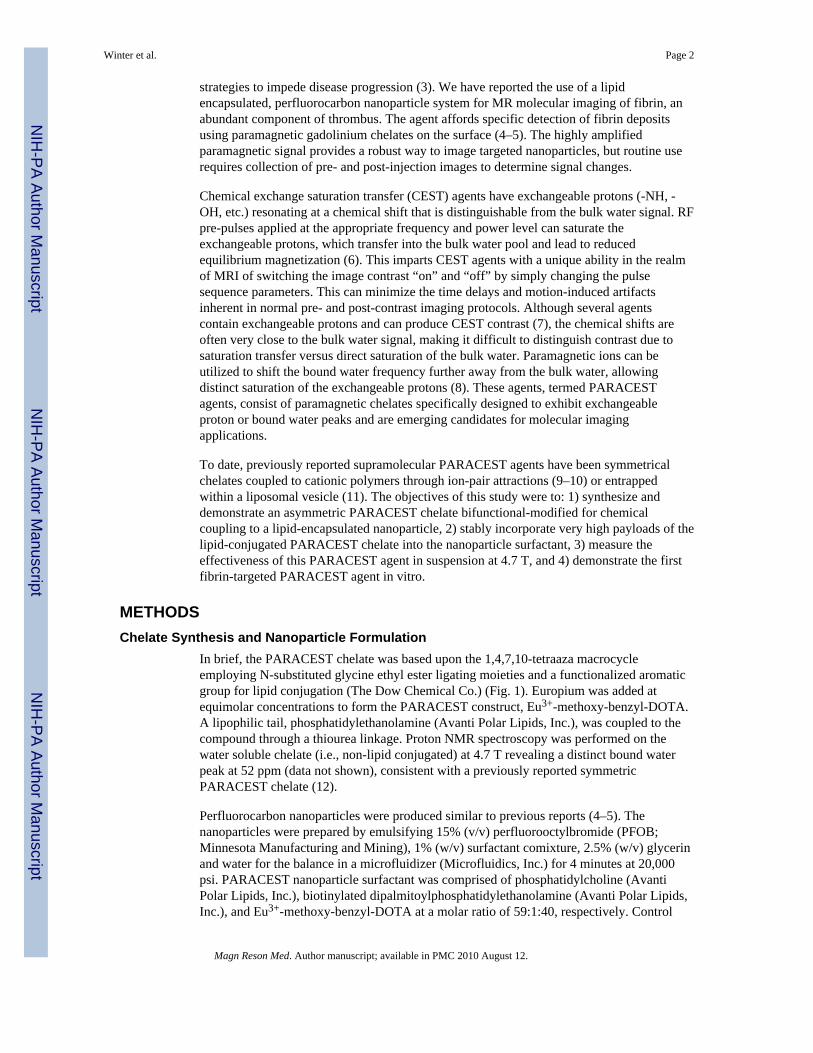
strategies to impede disease progression (3). We have reported the use of a lipidencapsulated, perfluorocarbon nanoparticle system for MR molecular imaging of fibrin, anabundant component of thrombus. The agent affords specific detection of fibrin depositsusing paramagnetic gadolinium chelates on the surface (4–5). The highly amplifiedparamagnetic signal provides a robust way to image targeted nanoparticles, but routine userequires collection of pre- and post-injection images to determine signal changes.
Chemical exchange saturation transfer (CEST) agents have exchangeable protons (-NH, -OH, etc.) resonating at a chemical shift that is distinguishable from the bulk water signal. RFpre-pulses applied at the appropriate frequency and power level can saturate theexchangeable protons, which transfer into the bulk water pool and lead to reducedequilibrium magnetization (6). This imparts CEST agents with a unique ability in the realmof MRI of switching the image contrast “on” and “off” by simply changing the pulsesequence parameters. This can minimize the time delays and motion-induced artifactsinherent in normal pre- and post-contrast imaging protocols. Although several agentscontain exchangeable protons and can produce CEST contrast (7), the chemical shifts areoften very close to the bulk water signal, making it difficult to distinguish contrast due tosaturation transfer versus direct saturation of the bulk water. Paramagnetic ions can beutilized to shift the bound water frequency further away from the bulk water, allowingdistinct saturation of the exchangeable protons (8). These agents, termed PARACESTagents, consist of paramagnetic chelates specifically designed to exhibit exchangeableproton or bound water peaks and are emerging candidates for molecular imagingapplications.
To date, previously reported supramolecular PARACEST agents have been symmetricalchelates coupled to cationic polymers through ion-pair attractions (9–10) or entrappedwithin a liposomal vesicle (11). The objectives of this study were to: 1) synthesize anddemonstrate an asymmetric PARACEST chelate bifunctional-modified for chemicalcoupling to a lipid-encapsulated nanoparticle, 2) stably incorporate very high payloads of thelipid-conjugated PARACEST chelate into the nanoparticle surfactant, 3) measure theeffectiveness of this PARACEST agent in suspension at 4.7 T, and 4) demonstrate the firstfibrin-targeted PARACEST agent in vitro.
METHODSChelate Synthesis and Nanoparticle Formulation
In brief, the PARACEST chelate was based upon the 1,4,7,10-tetraaza macrocycleemploying N-substituted glycine ethyl ester ligating moieties and a functionalized aromaticgroup for lipid conjugation (The Dow Chemical Co.) (Fig. 1). Europium was added atequimolar concentrations to form the PARACEST construct, Eu3+-methoxy-benzyl-DOTA.A lipophilic tail, phosphatidylethanolamine (Avanti Polar Lipids, Inc.), was coupled to thecompound through a thiourea linkage. Proton NMR spectroscopy was performed on thewater soluble chelate (i.e., non-lipid conjugated) at 4.7 T revealing a distinct bound waterpeak at 52 ppm (data not shown), consistent with a previously reported symmetricPARACEST chelate (12).
Perfluorocarbon nanoparticles were produced similar to previous reports (4–5). Thenanoparticles were prepared by emulsifying 15% (v/v) perfluorooctylbromide (PFOB;Minnesota Manufacturing and Mining), 1% (w/v) surfactant comixture, 2.5% (w/v) glycerinand water for the balance in a microfluidizer (Microfluidics, Inc.) for 4 minutes at 20,000psi. PARACEST nanoparticle surfactant was comprised of phosphatidylcholine (AvantiPolar Lipids, Inc.), biotinylated dipalmitoylphosphatidylethanolamine (Avanti Polar Lipids,Inc.), and Eu3+-methoxy-benzyl-DOTA at a molar ratio of 59:1:40, respectively. Control
Winter et al. Page 2
Magn Reson Med. Author manuscript; available in PMC 2010 August 12.
NIH
-PA Author Manuscript
NIH
-PA Author Manuscript
NIH
-PA Author Manuscript

nanoparticles lacked the europium chelate, which was substituted with an equivalentincrease in phosphatidylcholine.
Particle size, using quasi-elastic light scattering, and zeta potential, based upon anelectrophoretic light scattering/laser Doppler velocimetry method, was measured indeionized water at 25°C with a Brookhaven ZetaPlus analyzer (Brookhaven InstrumentCorp.). The europium content of the emulsion was determined by standard comparatorinstrumental neutron activation analysis at the University of Missouri Research Reactor(MURR). Specifically, Eu3+ was quantified by measuring the 842 keV gamma ray from thebeta decay of 152mEu (t1/2 = 9.31 h) produced through neutron capture by 151Eu. Thesamples and comparator standards were irradiated in a thermal flux of ~ 5 × 10¹³ n/(s*cm2)for 60 seconds, allowed to decay for several hours, and counted on a high-resolutiongamma-ray spectrometer for 30 minutes. The minimum detection limit for this procedure is2 ng of Eu3+.
PARACEST Imaging and SpectroscopyImaging and spectroscopy studies were performed on a 4.7 T Varian Inova scanner at roomtemperature (23°C) using a 3 cm diameter custom-built circular surface coil. Bulk waterspectra were collected from both PARACEST and control nanoparticle samples (50 μl)using a 2 second presaturation pulse at a power level of 28 dB. The saturation frequency wasstepped between ±100 ppm (relative to the bulk water frequency at 0 ppm) in 1 ppmincrements and the bulk water signal was integrated using a Matlab program (TheMathWorks, Inc.). The difference between the integrated signals measured at equivalentpositive and negative saturation frequencies was plotted, yielding saturation transfer profilesfor the PARACEST and control nanoparticles.
The effectiveness of the PARACEST nanoparticles was compared to control particles usinga two-chamber phantom constructed with an inner 1 cm diameter chamber that contained theundiluted nanoparticle emulsion. The outer 1.8 cm diameter chamber contained phosphatebuffered saline (PBS). Gradient echo images of the two-chamber phantom were collectedusing a 2.5 second presaturation pulse at a power level of 38 dB with a frequency offset of±52 ppm relative to the bulk water peak. Other imaging parameters were: TR = 2.52 s, TE =4.4 ms, number of averages = 8, in-plane resolution = 156 μm by 156 μm, slice thickness =4mm. Image intensity was normalized with respect to the signal from the PBS chamber,images were subtracted pixel-by-pixel and saturation transfer signal enhancement wascalculated. The signal enhancement of PARACEST nanoparticles was also measuredfollowing presaturation pulses of 1, 2 or 3 seconds duration at power levels of 26, 29, 32, 35or 38 dB using low resolution imaging (TR = 3.1 s, TE = 2.9 ms, number of averages= 2, in-plane resolution = 390 μm by 390 μm, slice thickness = 4 mm).
The concept of a fibrin-targeted PARACEST nanoparticle was studied using cylindricalplasma clots suspended in sterile saline inside plastic snap-cap tubes. The acellular clotswere formed in a 5 mm diameter plastic mold prepared by combining fresh dog plasma, 100mM calcium chloride (3:1 v/v), and 5 U thrombin around a 4-0 silk suture. Clots wereserially incubated with 150 μg biotinylated anti-fibrin antibodies (1H10) (13–15) overnightat 4°C, followed by 50 μg avidin for 1 hr at 25°C, and then 250 μl of PARACEST (n=5) orcontrol (n=4) nanoparticles for 1 hr at 25°C to complete the binding. Clots were rinsed threetimes with sterile saline after each incubation step to remove unbound reactants. Gradientecho images were collected of each clot using identical parameters as used for the two-chamber phantom. The clot surface was manually traced in Matlab and the contrast-to-noiseratio (CNR) was calculated relative to the standard deviation of the image intensity in air.The tracing was repeated in triplicate for each clot by a single observer and averaged.
Winter et al. Page 3
Magn Reson Med. Author manuscript; available in PMC 2010 August 12.
NIH
-PA Author Manuscript
NIH
-PA Author Manuscript
NIH
-PA Author Manuscript
Statistical AnalysisAll studies followed a completely randomized design. Data were analyzed by student’s t-Test or ANOVA. Means were separated when appropriate using the least significantdifference (LSD) method and declared significant at an alpha level of 0.05 using a beta levelof 0.80. Unless otherwise specified, averages are presented as a mean ± SEM.
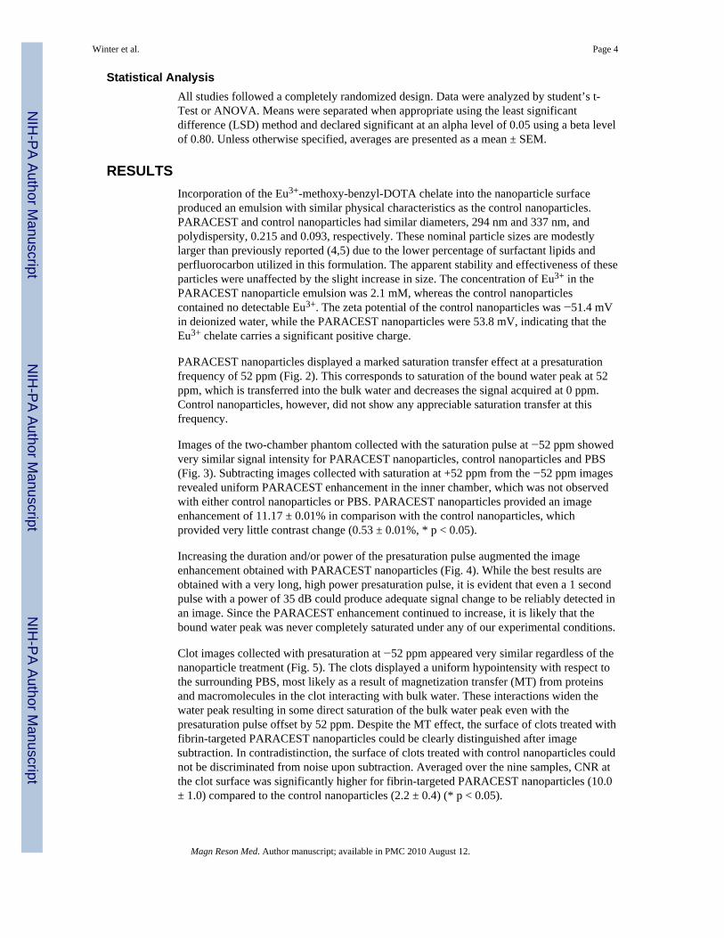
RESULTSIncorporation of the Eu3+-methoxy-benzyl-DOTA chelate into the nanoparticle surfaceproduced an emulsion with similar physical characteristics as the control nanoparticles.PARACEST and control nanoparticles had similar diameters, 294 nm and 337 nm, andpolydispersity, 0.215 and 0.093, respectively. These nominal particle sizes are modestlylarger than previously reported (4,5) due to the lower percentage of surfactant lipids andperfluorocarbon utilized in this formulation. The apparent stability and effectiveness of theseparticles were unaffected by the slight increase in size. The concentration of Eu3+ in thePARACEST nanoparticle emulsion was 2.1 mM, whereas the control nanoparticlescontained no detectable Eu3+. The zeta potential of the control nanoparticles was −51.4 mVin deionized water, while the PARACEST nanoparticles were 53.8 mV, indicating that theEu3+ chelate carries a significant positive charge.
PARACEST nanoparticles displayed a marked saturation transfer effect at a presaturationfrequency of 52 ppm (Fig. 2). This corresponds to saturation of the bound water peak at 52ppm, which is transferred into the bulk water and decreases the signal acquired at 0 ppm.Control nanoparticles, however, did not show any appreciable saturation transfer at thisfrequency.
Images of the two-chamber phantom collected with the saturation pulse at −52 ppm showedvery similar signal intensity for PARACEST nanoparticles, control nanoparticles and PBS(Fig. 3). Subtracting images collected with saturation at +52 ppm from the −52 ppm imagesrevealed uniform PARACEST enhancement in the inner chamber, which was not observedwith either control nanoparticles or PBS. PARACEST nanoparticles provided an imageenhancement of 11.17 ± 0.01% in comparison with the control nanoparticles, whichprovided very little contrast change (0.53 ± 0.01%, * p < 0.05).
Increasing the duration and/or power of the presaturation pulse augmented the imageenhancement obtained with PARACEST nanoparticles (Fig. 4). While the best results areobtained with a very long, high power presaturation pulse, it is evident that even a 1 secondpulse with a power of 35 dB could produce adequate signal change to be reliably detected inan image. Since the PARACEST enhancement continued to increase, it is likely that thebound water peak was never completely saturated under any of our experimental conditions.
Clot images collected with presaturation at −52 ppm appeared very similar regardless of thenanoparticle treatment (Fig. 5). The clots displayed a uniform hypointensity with respect tothe surrounding PBS, most likely as a result of magnetization transfer (MT) from proteinsand macromolecules in the clot interacting with bulk water. These interactions widen thewater peak resulting in some direct saturation of the bulk water peak even with thepresaturation pulse offset by 52 ppm. Despite the MT effect, the surface of clots treated withfibrin-targeted PARACEST nanoparticles could be clearly distinguished after imagesubtraction. In contradistinction, the surface of clots treated with control nanoparticles couldnot be discriminated from noise upon subtraction. Averaged over the nine samples, CNR atthe clot surface was significantly higher for fibrin-targeted PARACEST nanoparticles (10.0± 1.0) compared to the control nanoparticles (2.2 ± 0.4) (* p < 0.05).
Winter et al. Page 4
Magn Reson Med. Author manuscript; available in PMC 2010 August 12.
NIH
-PA Author Manuscript
NIH
-PA Author Manuscript
NIH
-PA Author Manuscript

DISCUSSIONThis study presents the incorporation of a bifunctional, asymmetric PARACEST chelate intothe surfactant of a fibrin-targeted perfluorocarbon nanoparticle for molecular imaging.Fibrin-targeted PARACEST nanoparticles provide a contrast mechanism that may beactivated or deactivated by changing the offset frequency of the presaturation pulse, whichallows easy detection of the targeted agent without collecting images before and aftercontrast administration. In contradistinction to liposomal-based agents which create contrastthrough a chemical shift effect (11), the contrast mechanism of the fibrin-targeted particlesis due to PARACEST interactions.
The importance of detecting and treating unstable plaque before further progression tomyocardial infarction or stroke occurs cannot be overstated. Despite the clinical need forthis information, the cost, risk, and time required to perform invasive techniques such asintravascular ultrasound (16), thermography (17), and coherent optical tomography (18)have dampened the general enthusiasm for their use beyond the research community.Noninvasive MRI in conjunction with fibrin-targeted molecular imaging agents (3) ispositioned to be a favored modality for the detection and characterization of unstable plaque,however, the current need for pre- and post-contrast imaging offset by 30 to 60 minutes ismore complicated than desirable. In the future, one could imagine administering a fibrin-targeted PARACEST agent to a patient at the time of presentation and imaging the patientanytime over the next 30 minutes to two hours without interference from circulating contrastor need to have performed a baseline scan before treatment. The practical implication is anearlier diagnosis and treatment, which would be available at lower cost to more patients peravailable scanner. These clear clinical advantages fuel the mounting research anddevelopment interest in PARACEST for molecular imaging.
MT contrast is commonly used in a variety of clinical MRI applications (19). Similar toPARACEST, MT uses a presaturation pulse to suppress some of the signals in the body.While these techniques utilize similar pulse sequences, PARACEST requires a much largerfrequency offset and is asymmetric with regards to the bulk water. These differences allowdetection of PARACEST agents despite the underlying MT contrast. The hypointenseappearance of the clots in Figure 5 probably reflects MT due to the interactions of watermolecules with clot proteins. Presaturation at +52 ppm and −52 ppm, however, haveidentical MT effects, allowing image subtraction to cancel out the MT contributions. Nocontrast is observed on the subtraction images in areas with MT but no PARACESTparticles, such as the clot interior.
LIMITATIONSWhile the techniques used in this paper clearly demonstrate PARACEST enhancement withtargeted nanoparticles, optimization of the chemistry, coil and pulse sequence is needed totruly capitalize on the strengths of this agent. The Eu3+-methoxy-benzyl-DOTA chelatedisplayed a bound water peak both in solution as well as coupled to nanoparticles. We didnot, however, measure the exchange rate, thus 4.7 T may not be the optimal field strength. Inaddition, the cationic nature of the PARACEST particles in this study is not desirable invivo due to the potential interaction with anionic proteins and membranes, which couldintroduce significant formulation, pharmacokinetic, pharmacodynamics, and biosafetychallenges.
We utilized a very simple surface coil, which provided efficient transmission of thesaturation pulses. More uniform saturation and sensitivity, however, may be required for invivo imaging of deep tissues. Moreover, a long, low-power presaturation pulse was insertedinto a standard gradient echo imaging sequence, which effectively saturated the bound water
Winter et al. Page 5
Magn Reson Med. Author manuscript; available in PMC 2010 August 12.
NIH
-PA Author Manuscript
NIH
-PA Author Manuscript
NIH
-PA Author Manuscript

peak, but did not optimize RF deposition or imaging efficiency. Sequences utilizing multi-echo acquisitions or short saturation pulse trains may shorten imaging times and reduce RFdeposition. Given the early stage of PARACEST contrast agent development, furtherresearch on the chemistry, in vivo particle clearance and several imaging hardware issues isneeded. With optimization of the agent and imaging hardware, comparisons between variousagents, including CEST agents as well as conventional paramagnetic or superparamagneticagents (3), can be performed for specific molecular imaging applications.
CONCLUSIONSThis report describes a targeted PARACEST nanoparticle agent with developmentalpotential for detection of fibrin in unstable atherosclerotic plaques. This agent is particularlyattractive because it can be activated through the pulse sequence parameters of the MRIscanner, effectively turning the contrast agent on and off at will. Targeted PARACESTnanoparticles may allow robust detection of contrast agents at a single time point instead ofcomparing images collected at pre- and post-injection, which would have significantpractical benefits in the clinic.
AcknowledgmentsThe authors thank Dr. Joseph J. H. Ackerman, Professor and Chair, Department of Chemistry, and Director of theBiomedical MR Laboratory at Washington University, for technical assistance and support. This research wassupported in part by the NIH (CO-07121, HL-42950, HL-59865 and EB-01704), Philips Medical Systems and theAmerican Heart Association.
References1. American Heart Association. 2005.2. Rudd JH, Davies JR, Weissberg PL. Imaging of atherosclerosis -- can we predict plaque rupture?
Trends Cardiovasc Med 2005;15(1):17–24. [PubMed: 15795159]3. Wickline SA, Neubauer AM, Winter P, Caruthers S, Lanza G. Applications of nanotechnology to
atherosclerosis, thrombosis, and vascular biology. Arterioscler Thromb Vasc Biol 2006;26(3):435–441. [PubMed: 16373609]
4. Lanza GM, Wallace KD, Scott MJ, Cacheris WP, Abendschein DR, Christy DH, Sharkey AM,Miller JG, Gaffney PJ, Wickline SA. A novel site-targeted ultrasonic contrast agent with broadbiomedical application. Circulation 1996;94(12):3334–3340. [PubMed: 8989148]
5. Winter PM, Caruthers SD, Yu X, Song SK, Chen J, Miller B, Bulte JW, Robertson JD, Gaffney PJ,Wickline SA, Lanza GM. Improved molecular imaging contrast agent for detection of humanthrombus. Magn Reson Med 2003;50(2):411–416. [PubMed: 12876719]
6. Ward KM, Aletras AH, Balaban RS. A new class of contrast agents for MRI based on protonchemical exchange dependent saturation transfer (CEST). J Magn Reson 2000;143(1):79–87.[PubMed: 10698648]
7. Zhou J, Wilson DA, Sun PZ, Klaus JA, Van Zijl PC. Quantitative description of proton exchangeprocesses between water and endogenous and exogenous agents for WEX, CEST, and APTexperiments. Magn Reson Med 2004;51(5):945–952. [PubMed: 15122676]
8. Terreno E, Castelli DD, Cravotto G, Milone L, Aime S. Ln(III)-DOTAMGly complexes: a versatileseries to assess the determinants of the efficacy of paramagnetic chemical exchange saturationtransfer agents for magnetic resonance imaging applications. Invest Radiol 2004;39(4):235–243.[PubMed: 15021328]
9. Aime S, Delli Castelli D, Terreno E. Supramolecular adducts between poly-L-arginine and[TmIIIdotp]: a route to sensitivity-enhanced magnetic resonance imaging-chemical exchangesaturation transfer agents. Angew Chem Int Ed Engl 2003;42(37):4527–4529. [PubMed: 14520757]
10. McMahon MT, Gilad AA, Zhou J, Sun PZ, Bulte JW, van Zijl PC. Quantifying exchange rates inchemical exchange saturation transfer agents using the saturation time and saturation powerdependencies of the magnetization transfer effect on the magnetic resonance imaging signal
Winter et al. Page 6
Magn Reson Med. Author manuscript; available in PMC 2010 August 12.
NIH
-PA Author Manuscript
NIH
-PA Author Manuscript
NIH
-PA Author Manuscript

(QUEST and QUESP): Ph calibration for poly-L-lysine and a starburst dendrimer. Magn ResonMed 2006;55(4):836–847. [PubMed: 16506187]
11. Aime S, Delli Castelli D, Terreno E. Highly sensitive MRI chemical exchange saturation transferagents using liposomes. Angew Chem Int Ed Engl 2005;44(34):5513–5515. [PubMed: 16052647]
12. Zhang S, Winter P, Wu K, Sherry AD. A novel europium(III)-based MRI contrast agent. J AmChem Soc 2001;123(7):1517–1518. [PubMed: 11456734]
13. Edgell T, McEvoy F, Webbon P, Gaffney PJ. Monoclonal antibodies to human fibrin: interactionwith other animal fibrins. Thromb Haemost 1996;75(4):595–599. [PubMed: 8743185]
14. Raut S, Gaffney PJ. Evaluation of the fibrin binding profile of two anti-fibrin monoclonalantibodies. Thromb Haemost 1996;76(1):56–64. [PubMed: 8819252]
15. Tymkewycz PM, Creighton Kempsford LJ, Gaffney PJ. Generation and partial characterization offive monoclonal antibodies with high affinities for fibrin. Blood Coagul Fibrinolysis 1993;4(2):211–221. [PubMed: 7684615]
16. Escolar E, Weigold G, Fuisz A, Weissman NJ. New imaging techniques for diagnosing coronaryartery disease. Cmaj 2006;174(4):487–495. [PubMed: 16477061]
17. Toutouzas K, Drakopoulou M, Stefanadi E, Siasos G, Stefanadis C. Intracoronary thermography:does it help us in clinical decision making? J Interv Cardiol 2005;18(6):485–489. [PubMed:16336430]
18. Regar E, Schaar JA, Mont E, Virmani R, Serruys PW. Optical coherence tomography. CardiovascRadiat Med 2003;4(4):198–204. [PubMed: 15321058]
19. Rumboldt Z, Marotti M. Magnetization transfer, HASTE, and FLAIR imaging. Magn ResonImaging Clin N Am 2003;11(3):471–492. [PubMed: 14768730]
Winter et al. Page 7
Magn Reson Med. Author manuscript; available in PMC 2010 August 12.
NIH
-PA Author Manuscript
NIH
-PA Author Manuscript
NIH
-PA Author Manuscript

FIG. 1.Chemical structure of lipid conjugated PARACEST contrast agent. Eu3+ is chelated tomethoxy-benzyl-DOTA, which is functionalized with a phospholipid moiety forincorporation into the lipid membrane of perfluorocarbon nanoparticles.
Winter et al. Page 8
Magn Reson Med. Author manuscript; available in PMC 2010 August 12.
NIH
-PA Author Manuscript
NIH
-PA Author Manuscript
NIH
-PA Author Manuscript

FIG. 2.Saturation profile of PARACEST nanoparticles shows a clear saturation transfer effect at 52ppm (arrow) confirming saturation of the bound water peak and effective transfer ofmagnetization. Control nanoparticles do not show any saturation transfer effects at thischemical shift.
Winter et al. Page 9
Magn Reson Med. Author manuscript; available in PMC 2010 August 12.
NIH
-PA Author Manuscript
NIH
-PA Author Manuscript
NIH
-PA Author Manuscript

FIG. 3.Imaging of two-chambered phantom containing PARACEST nanoparticles (left) or controlnanoparticles (right) in the inner chamber and PBS in the outer chamber. Original imagescollected with saturation at −52 ppm (top) show no differences between PARACEST andcontrol nanoparticles. Subtraction images (middle) reveal uniform signal enhancement in thePARACEST chamber and no enhancement in the outer PBS chambers or with controlnanoparticles. The signal enhancement (bottom) in the PARACEST nanoparticle chamberwas significantly higher than control nanoparticles (* p < 0.05).
Winter et al. Page 10
Magn Reson Med. Author manuscript; available in PMC 2010 August 12.
NIH
-PA Author Manuscript
NIH
-PA Author Manuscript
NIH
-PA Author Manuscript

FIG. 4.Increasing the power or duration of the saturation pulse increases the signal enhancementobtained from PARACEST nanoparticles. Presaturation pulses as short as 1 second canproduce a 5% change in image intensity.
Winter et al. Page 11
Magn Reson Med. Author manuscript; available in PMC 2010 August 12.
NIH
-PA Author Manuscript
NIH
-PA Author Manuscript
NIH
-PA Author Manuscript

FIG. 5.Imaging of fibrin-targeted PARACEST (left) or control (right) nanoparticles bound toplasma clots. Images obtained with saturation at −52 ppm (top) show no differencesbetween clots treated with PARACEST or control nanoparticles. Subtraction images(middle) reveal signal enhancement on the surface of the clot treated with PARACESTnanoparticles and no enhancement of the clot treated with control nanoparticles. The CNRcalculated at the clot surface (bottom) was significantly higher with PARACESTnanoparticles compared to control nanoparticles (* p < 0.05).
Winter et al. Page 12
Magn Reson Med. Author manuscript; available in PMC 2010 August 12.
NIH
-PA Author Manuscript
NIH
-PA Author Manuscript
NIH
-PA Author Manuscript
Copyright © 2022 FDOKUMEN