Biomedical exploitation of self assembled peptide based nanostructures
8
Send Orders for Reprints to [email protected] Current Protein and Peptide Science, 2013, 14, 000-000 1 1389-2037/13 $58.00+.00 © 2013 Bentham Science Publishers Biomedical Exploitation of Self Assembled Peptide Based Nanostructures Anupam Roy 1 , Octavio L. Franco 2 and Santi M. Mandal 1,* 1 Central Research Facility, Indian Institute of Technology Kharagpur, Kharagpur-721302, India; 2 Centro de Análises Proteômicas e Bioquímicas, Pós-Graduação em Ciências Genômicas e Biotecnologia UCB, Brasília-DF, Brazil Abstract: Nowadays, peptide based disease prevention is an important topic in biomedical science, which may radically change the traditional use of biomaterials and improve the life quality. Self-assembled nanostructured peptides have been receiving extreme attention in the drug delivery field due their high biocompatibility levels, better loading capacity, ex- tended circulation and localization in required target site. This article focuses on the composition and synthesis of differ- ent forms of self-assembled peptide nanostructures as nanotubes, nanofibers, nanoparticles, nanotapes and nanogels. The most important properties for their self assembled mechanism and their biomedical applications are also discussed. Vari- ous potential applications of nanostructures peptide could be developed designed for therapeutic agent’s delivery, biosen- sors, anticancerous and antimicrobial activities. Keywords: Self assemble, peptide nanostructures, drug delivery, disease prevention. INTRODUCTION In last decade, extensive efforts have been given in de- veloping of several nanoscale-carriers in biomedical nanotechnology to improve the drug delivery systems. Bio- medical nanotechnology offers different forms of nanostruc- tures, such as nanoparticles, nanocapsules, nanotubes, na- nomicelles, nanoconjugates and several others [1] for the development of suitable carriers with site specific and con- trolled release of small to large bioactive agents [2, 3]. Cati- onic surfactant peptides have been proposed to be useful as carriers for targeted drug delivery. Development of peptide based nanostructures for entrapment and delivery of bioac- tive agents are emerging for safer in vivo delivery. Peptide self assembled based delivery systems have the ability to deliver therapeutic proteins, peptides, small molecules, anti- biotics and nucleic acids such as oligonucleotides, DNA, ds RNA, RNAi, sh RNA, micro RNA etc [4]. In addition to the diversity of nanoscale-carriers, an effi- cient nano form must possess the high biocompatibility lev- els, loading capacity, extended circulation and target speci- ficity. In recent years, self assembled peptide based nano structure offers several advantages in biomedical applica- tions due to their remarkable recognition capabilities, me- chanical rigidity, amenability to high precision processing and stability under specific chemical conditions [5]. This is due to its unique structural folding and stability, which is not likely to be seen for the other metal or biomolecule based nanostructures [6-7]. Molecular self-assembly is the sponta- neous association of molecules under equilibrium conditions, structurally well defined aggregates connected with nonco- valent bonds [8]. Peptide based nano synthesis is relatively simple in functionalization, highly biocompatible, biode- *Address correspondence to this author at the Central Research Facility, Indian Institute of Technology Kharagpur, Kharagpur-721302, Índia; Tel: + 91-3222- 282486; Fax: +91-3222-282481; Email: [email protected] gradable and folds into specific structures based upon the sequence and environmental responses which makes peptide nanoparticles much more advantageous over other nanopar- ticles. Self-assembling of peptides follow the bottom-up ap- proach where specific modifications and function can be easily incorporated that increase the possibility of more spe- cific target. Self-integration of simple peptide building blocks makes advantage over top-down method where the uses of sophisticated lithography and etching techniques have reached a physical limit [9]. This review particularly focus on the design and synthesis of different nano forms of self-assembled peptides with special emphasis on their di- verse applications in biomedical nanotechnology, such as drug delivery, anticancer, antimicrobial, sensors, bioelectron- ics, etc. DESIGN AND DEVELOPMENT Biological peptides or proteins have the inherent ability to self-assemble and give rise to different forms of nano structures. Proteinaceous natural fibrils are extensively stud- ied and well embedded with detailed theoretical and “in silico” models (Fig. 1). The self assemble of a peptide de- pends upon their charge distribution, degree of polymeriza- tion, and hydrophobicity. Peptide requires at least two addi- tional positively or negatively charged amino acids per pep- tide for their self assembly. ‘In silico” molecular tool is widely used for designing the amino acid sequence, which is important to consider both the net charge and position of the charged amino acids [10]. Nature forms complex of mul- ticomponent three-dimensional structures through spontane- ous association. The amino acids that constitute the peptide motifs having unique properties over other building blocks in nature as they represent a large variety of chemical moieties enable facile chemical and biological modifications [11]. There is an interest in using enzymatic procedures to synthe- size specific peptide motifs. The use of free amino acids as
Transcript of Biomedical exploitation of self assembled peptide based nanostructures
Send Orders for Reprints to [email protected]
Current Protein and Peptide Science, 2013, 14, 000-000 1
1389-2037/13 $58.00+.00 © 2013 Bentham Science Publishers
Biomedical Exploitation of Self Assembled Peptide Based Nanostructures
Anupam Roy1, Octavio L. Franco2 and Santi M. Mandal1,*
1Central Research Facility, Indian Institute of Technology Kharagpur, Kharagpur-721302, India;
2Centro de Análises
Proteômicas e Bioquímicas, Pós-Graduação em Ciências Genômicas e Biotecnologia UCB, Brasília-DF, Brazil
Abstract: Nowadays, peptide based disease prevention is an important topic in biomedical science, which may radically change the traditional use of biomaterials and improve the life quality. Self-assembled nanostructured peptides have been receiving extreme attention in the drug delivery field due their high biocompatibility levels, better loading capacity, ex-tended circulation and localization in required target site. This article focuses on the composition and synthesis of differ-ent forms of self-assembled peptide nanostructures as nanotubes, nanofibers, nanoparticles, nanotapes and nanogels. The most important properties for their self assembled mechanism and their biomedical applications are also discussed. Vari-ous potential applications of nanostructures peptide could be developed designed for therapeutic agent’s delivery, biosen-sors, anticancerous and antimicrobial activities.
Keywords: Self assemble, peptide nanostructures, drug delivery, disease prevention.
INTRODUCTION
In last decade, extensive efforts have been given in de-veloping of several nanoscale-carriers in biomedical nanotechnology to improve the drug delivery systems. Bio-medical nanotechnology offers different forms of nanostruc-tures, such as nanoparticles, nanocapsules, nanotubes, na-nomicelles, nanoconjugates and several others [1] for the development of suitable carriers with site specific and con-trolled release of small to large bioactive agents [2, 3]. Cati-onic surfactant peptides have been proposed to be useful as carriers for targeted drug delivery. Development of peptide based nanostructures for entrapment and delivery of bioac-tive agents are emerging for safer in vivo delivery. Peptide self assembled based delivery systems have the ability to deliver therapeutic proteins, peptides, small molecules, anti-biotics and nucleic acids such as oligonucleotides, DNA, ds RNA, RNAi, sh RNA, micro RNA etc [4].
In addition to the diversity of nanoscale-carriers, an effi-cient nano form must possess the high biocompatibility lev-els, loading capacity, extended circulation and target speci-ficity. In recent years, self assembled peptide based nano structure offers several advantages in biomedical applica-tions due to their remarkable recognition capabilities, me-chanical rigidity, amenability to high precision processing and stability under specific chemical conditions [5]. This is due to its unique structural folding and stability, which is not likely to be seen for the other metal or biomolecule based nanostructures [6-7]. Molecular self-assembly is the sponta-neous association of molecules under equilibrium conditions, structurally well defined aggregates connected with nonco-valent bonds [8]. Peptide based nano synthesis is relatively simple in functionalization, highly biocompatible, biode-
*Address correspondence to this author at the Central Research Facility, Indian Institute of Technology Kharagpur, Kharagpur-721302, Índia; Tel: + 91-3222- 282486; Fax: +91-3222-282481; Email: [email protected]
gradable and folds into specific structures based upon the sequence and environmental responses which makes peptide nanoparticles much more advantageous over other nanopar-ticles. Self-assembling of peptides follow the bottom-up ap-proach where specific modifications and function can be easily incorporated that increase the possibility of more spe-cific target. Self-integration of simple peptide building blocks makes advantage over top-down method where the uses of sophisticated lithography and etching techniques have reached a physical limit [9]. This review particularly focus on the design and synthesis of different nano forms of self-assembled peptides with special emphasis on their di-verse applications in biomedical nanotechnology, such as drug delivery, anticancer, antimicrobial, sensors, bioelectron-ics, etc.
DESIGN AND DEVELOPMENT
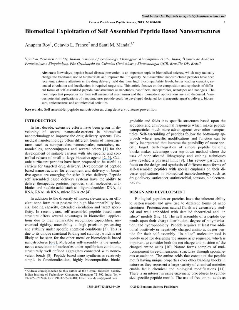
Biological peptides or proteins have the inherent ability to self-assemble and give rise to different forms of nano structures. Proteinaceous natural fibrils are extensively stud-ied and well embedded with detailed theoretical and “in silico” models (Fig. 1). The self assemble of a peptide de-pends upon their charge distribution, degree of polymeriza-tion, and hydrophobicity. Peptide requires at least two addi-tional positively or negatively charged amino acids per pep-tide for their self assembly. ‘In silico” molecular tool is widely used for designing the amino acid sequence, which is important to consider both the net charge and position of the charged amino acids [10]. Nature forms complex of mul-ticomponent three-dimensional structures through spontane-ous association. The amino acids that constitute the peptide motifs having unique properties over other building blocks in nature as they represent a large variety of chemical moieties enable facile chemical and biological modifications [11]. There is an interest in using enzymatic procedures to synthe-size specific peptide motifs. The use of free amino acids as
2 Current Protein and Peptide Science, 2013, Vol. 14, No. 6 Roy et al.
opposed to Fmoc derivatives as starting materials has great economic appeal.
Fig. (1). 3-D structure of naturally occurred peptide nano filament. Images were constructed by using Pymol software. (a) the 1GK4.pdb file of human vimentin coil 2B fragment and (b) 3TNU.pdb file of human keratin 5 taken from online protein data bank. The building block of intermediate filament contains alpha-helical segments in an elongated coiled-coil manner.
However, several new routes have been developed to produce novel materials using short peptides to proteins that remarkably useful in pharmaceutical industry. Manufactur-ing of new functionalized materials using the self assembly mechanism of biomolecules is an emerging area to comple-ment the highly toxic materials or polymers. This is an enormous challenge to design the molecular building blocks that can undergo spontaneous organization into a stable structure using non-covalent bonds. This mechanism is rela-tively common in peptides which offer new types of bio-molecules formation with diverse well-defined nanostruc-tures such as nanotubes, nanofibers, nanoparticles, nano-tapes, hydrogels, nanorods and others. The supramolecular nanostructures functionalized with several bioactive mole-cules have been actively explored as promising materials in the field of biotechnology [12].
NANOTUBES
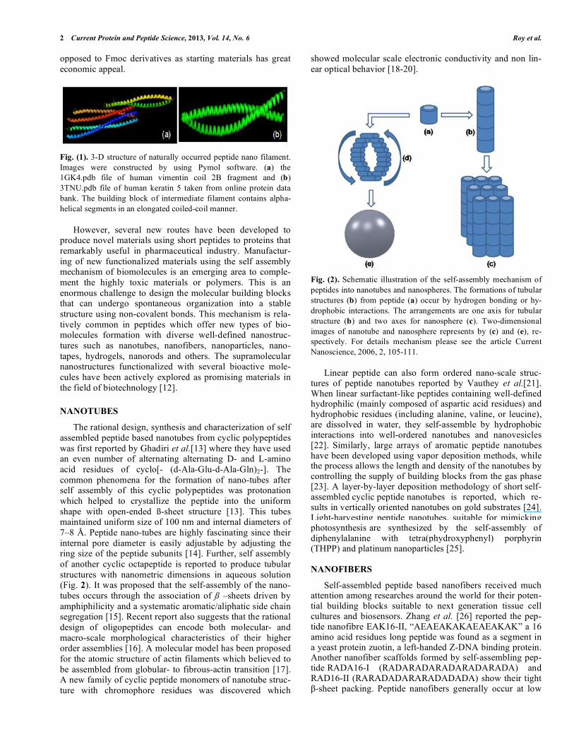
The rational design, synthesis and characterization of self assembled peptide based nanotubes from cyclic polypeptides was first reported by Ghadiri et al.[13] where they have used an even number of alternating alternating D- and L-amino acid residues of cyclo[- (d-Ala-Glu-d-Ala-Gln)2-]. The common phenomena for the formation of nano-tubes after self assembly of this cyclic polypeptides was protonation which helped to crystallize the peptide into the uniform shape with open-ended ß-sheet structure [13]. This tubes maintained uniform size of 100 nm and internal diameters of 7–8 Å. Peptide nano-tubes are highly fascinating since their internal pore diameter is easily adjustable by adjusting the ring size of the peptide subunits [14]. Further, self assembly of another cyclic octapeptide is reported to produce tubular structures with nanometric dimensions in aqueous solution (Fig. 2). It was proposed that the self-assembly of the nano-tubes occurs through the association of ß –sheets driven by amphiphilicity and a systematic aromatic/aliphatic side chain segregation [15]. Recent report also suggests that the rational design of oligopeptides can encode both molecular- and macro-scale morphological characteristics of their higher order assemblies [16]. A molecular model has been proposed for the atomic structure of actin filaments which believed to be assembled from globular- to fibrous-actin transition [17]. A new family of cyclic peptide monomers of nanotube struc-ture with chromophore residues was discovered which
showed molecular scale electronic conductivity and non lin-ear optical behavior [18-20].
Fig. (2). Schematic illustration of the self-assembly mechanism of peptides into nanotubes and nanospheres. The formations of tubular structures (b) from peptide (a) occur by hydrogen bonding or hy-drophobic interactions. The arrangements are one axis for tubular structure (b) and two axes for nanosphere (c). Two-dimensional images of nanotube and nanosphere represents by (c) and (e), re-spectively. For details mechanism please see the article Current Nanoscience, 2006, 2, 105-111.
Linear peptide can also form ordered nano-scale struc-tures of peptide nanotubes reported by Vauthey et al.[21]. When linear surfactant-like peptides containing well-defined hydrophilic (mainly composed of aspartic acid residues) and hydrophobic residues (including alanine, valine, or leucine), are dissolved in water, they self-assemble by hydrophobic interactions into well-ordered nanotubes and nanovesicles [22]. Similarly, large arrays of aromatic peptide nanotubes have been developed using vapor deposition methods, while the process allows the length and density of the nanotubes by controlling the supply of building blocks from the gas phase [23]. A layer-by-layer deposition methodology of short self-assembled cyclic peptide nanotubes is reported, which re-sults in vertically oriented nanotubes on gold substrates [24]. Light-harvesting peptide nanotubes, suitable for mimicking photosynthesis are synthesized by the self-assembly of diphenylalanine with tetra(phydroxyphenyl) porphyrin (THPP) and platinum nanoparticles [25].
NANOFIBERS
Self-assembled peptide based nanofibers received much attention among researches around the world for their poten-tial building blocks suitable to next generation tissue cell cultures and biosensors. Zhang et al. [26] reported the pep-tide nanofibre EAK16-II, “AEAEAKAKAEAEAKAK” a 16 amino acid residues long peptide was found as a segment in a yeast protein zuotin, a left-handed Z-DNA binding protein. Another nanofiber scaffolds formed by self-assembling pep-tide RADA16-I (RADARADARADARADARADA) and RAD16-II (RARADADARARADADADA) show their tight
-sheet packing. Peptide nanofibers generally occur at low
Peptide Nanostructures in Biomedical Technology Current Protein and Peptide Science, 2013, Vol. 14, No. 6 3
pH (4.0) suggesting that the extended peptide backbone con-formation caused by the electrostatic repulsive force in acid solution is crucial for the peptide to self-assembly into nan-ofibers [27]. Amyloid fibrils are a key example of protein self assembly into ordered nano-scale fibrillar structures, associated with neurodegenerative diseases as Alzheimer’s and Parkinson’s diseases [28]. Amyloid fibril are remarkable stiff and poses similar value of E compared to the other most rigid proteinaceous materials such as silk, collagen, and keratin etc. [29]. In a recent work on diphenyla-lanine peptide which are self-assemble into a core-branched nanostructure through non-covalent interactions in aqueous solution. The pre-assemblies further assembled into nanofibers and microvesicles on the glass surface [30]. The oppositely charged amyloid inspired peptides (AIPs), rapidly self-assemble into nanofibers by noncovalent interac-tions at pH 7 upon mixing in water. The pH -induced self-assembly of a peptide-amphiphile fibrous nanostructured have been designed which are able to form a composite ma-terial after cross-linking [31].
NANOTAPES AND NANOGELS
Stacking of peptide -sheets result formation of peptide nanotapes . This peptide nanotapes tends to interacts with each other and form double layers nanotape structures [32]. When this structure exceeds the threshold limit, they form often hydrogel [33]. Several physical parameters such as pH, ionic strength, temperature and salinity effectively change the properties of nanotapes and nanogel formation have been elaborately described in a nice tutorial review article by Lowik et al. [34]. It is well documented that Dipeptide, Ile–Phe, can form transparent, thermoreversible hydrogel with fibrilar structures at 1.5 wt% at pH 5.8 [35]. Self-assembly of an , -dehydrophenylalanine residue ( Phe) containing dipeptide H-Phe- Phe-OH, forms a hydrogel at a concentra-tion as low as 0.2 wt% in a buffer solution offering a gel matrices with highly dense nano fibers network [36]. The self-assembly of a pentapeptide fragment of the amyloid -peptide NH2-KLVFF-COOH reported to produce a hydrogel in phosphate-buffered saline (PBS) solution [37].Self-assembled oligopeptides, H2N-Gly-Ala-Ile-Leu-COOH and H2N-Gly-Phe-Ile-Leu-COOH, form hydrogels at physiologi-cal pH 6.6. Nanofibrillar network structures obtained from self-assembled -helical linear peptide-based gels were also well documented [38]. Peptides with various hydrophobic conjugates including aromatic moieties like fluoren-ylmethoxycarbonyl, naphthalene, and pyrene can also pro-vide reliable opportunities to be assembled in water to form a nanofibrillar hydrogel network [39-41]. Molecular self as-sembly of a mixture of two aromatic short peptide deriva-tives: Fmoc-FF (Fluorenylmethoxycarbonyldiphenylalanine), N-terminally protected peptide sequence Nap-FFGRGD and Fmoc-RGD (arginine-glycineaspartate) reported to offer a peptide based bioactive hydrogel which acts as a 3D-scaffold for anchorage-dependent cells [42-43]. A facile temperature induced self-assembly and self-crosslinking method has been developed for preparing bioreducible nanogels/ microgels. The importance of this process is any stabilizer, catalyst or additional crosslinking agent is not required here and the size of formed nanogels/microgels can also be easily tuned via the polymer concentration [44].
NANO PARTICLES AT SPHERICAL FORM
Peptide based nanoparticles are small well-defined struc-tures formed by different monomers. Peptide, HA16B, hav-ing a hydrophobic -helical body and a hydrophilic polar head was synthesized and hydrated in water by sonication resulting formation of an -helical conformation in the dis-persion. Spherical assemblies were formed with an ex-tremely narrow size distribution (diameter 30 40 nm) [45]. Diphenylglycine, the simplest aromatic peptide, efficiently forms spherical nanometric assemblies and offers remarkable stability [46].One artificial viral-sized peptide-nanospheres self-assemblly was found from three-Way Junctions of -Sheet-forming peptides ( (FKFE)2) [47]. In addition to this, peptide nanotubes, of extended tubular -sheet-like struc-tures, are constructed by the self-assembly of flat, ring-shaped peptide subunits. A rational design of nano-sphere of a novel C3-symmetric peptide conjugate bearing three beta-sheet-forming peptides was developed by mimicking the formation of spherical viruses and clathrin [48]. Synthetic analogs of transmembrane domains of membrane proteins self-assemble into uniform spherical nanoparticles. Struc-tural transition of the peptide results predominantly a beta-hairpin conformation in aqueous solutions but folds into an alpha-helix upon spontaneous fusion of the nanoparticles with cell membrane [49].
STABILITY OF SELF ASSEMBLED PEPTIDE
The self assembly process offers extra stability to peptide under extreme physical and chemical conditions. Peptides are prone to proteolytic degradation but the tendency of pro-teolytic degradation in peptide based nanostructure is re-duced or retarded. Stability of the peptide nanostructures was attained by designing the building block with either L- or D-amino acid isomers. Peptide building blocks that composed of the non natural D-amino acids offered stability to prote-olytic degradation. Chauhan and his coworkers [50] reported that self-assembly process of a dipeptide containing a non-coded, achiral, , -dehydrophenylalanine residue ( Phe), introduced the Phe residue in the peptide sequence. This resulted formation of conformationally rigid distinct nanotu-bular structure, offering resistance to proteolytic degradation. Peptide H3 N+- -Ala (1)- Ala(2)-COO- and H3 N+- -Ava(1)-Phe(2)- COO- contains an N-terminally positioned
-amino acid residue ( -alanine/ -amino valeric acid) which form nanotubes in the solid state as well as in an aqueous solution. The self associated nanotubes are stable proteolytically, thermally, and over a wide range of pH val-ues [51]. Several self assembled hollow nanotubes from dif-ferent water-soluble short peptides each having a common motif, a hybrid of , -amino acid residues ( -Ala-L-Xaa, Xaa = Val/Ile/Phe) offered stability at 80° C, in a wide range of pH (2–10), and against proteolytic degradation [52]. In general, peptide building blocks are stable at high tempera-tures, in organic solvents, in extreme pHs and from enzy-matic degradation.
BIOMEDICAL APPLICATIONS
Drug Delivery
Peptide self assembled nanostructures offer some signifi-cant advantages over traditional delivery mechanisms. This
4 Current Protein and Peptide Science, 2013, Vol. 14, No. 6 Roy et al.
is due to their size which facilitates the increased transporta-tion of the therapeutic compounds in the blood stream. Self assembled peptide nano carriers mimic the capabilities of conventional lipid systems, and often provide improved drug delivery qualities [53]. Peptide nano tube structure can serve as nanocontainers, being proposed to be effective for drug delivery. Artificial membrane ion channels were developed based on a self-assembled cylindrical -sheet peptide archi-tecture which essentially stacks of peptide rings that display good channel-mediated ion-transport activity with rates ex-ceeding 107 ions s 1[10]. The amphiphilic nature, hollow architecture, biocompatibility, low cytotoxicity and the abil-ity to engender a favorable, biologically-inspired environ-ment favors self assembled peptide suitable for cell adhesion and growth. Rosette nanotubes as an effective deliver system of hydrophobic drugs as dexamethasone. It has been used to promote the osteoblast (bone-forming cell) functions over long periods of time [54].
Cationic surfactant peptides have been proposed to be useful as carriers for encapsulation and delivery of a number of small water-insoluble molecules and large biological mo-lecular systems, including negatively charged nucleic acids [55]. Cyclic peptide nanotubes composed of (Trp-D-Leu) 4-Gln-D-Leu is capable of transporting the antitumor drug 5-fluorouracil (5-FU) [56]. Diethylene glycol functionalized self-assembled peptide nanofibers offers potential delivery of hydrophobic drugs [57]. The nanofibre, RAD16-II is highly efficient to stabilizes the hydrophobic drugs such as pyrene in aqueous solution and deliver it into lipophilic environment [58] Integrin avb3-targeted lipid-coated nanogels with cross-linked human serum albumin in the core for carrying drugs of distinct chemotype, including paclitaxel, docetaxel, borte-zomib, 17-AAG, sorafenib, sunitinib, bosutinib, and dasatinib therapeutic cargoes exhibited potent activity in tumor cell proliferation assays. It showed 15-fold improve-ment in antitumor activity relative to abraxane [59].
Several self-assembling peptide amphiphiles (PAs) con-taining a lysine -amine-derivatized hydrazide placed at dif-ferent positions along the backbone of the peptide sequence C(16)V(2)A(2)E(2) (where C(16) = palmitic acid) reported to self-assembled into nanofibers and release Prodan, which is typically used to probe cell membranes [60]. Cationic dipeptides, NH2–Phe–Phe–NH2 self-assembles into nano-tubes at neutral pH and rearrange into spherical structures. As the tubes can be absorbed by cells through endocytosis upon spontaneous conversion into vesicles, it is used to de-liver oligonucleotides into the interior of the cells as a proof of concept of the potential applications of the system in gene and drug delivery [61]. Further, most important that few nanofiber hydrogel can also function as a control releaser of functional proteins and targeted siRNA delivery [62]. Self-assembled oligopeptide nanostructures are also used for co-delivery of drug and gene with synergistic therapeutic effect [63]. Thus self assembled peptides based nanostructures pro-posed to be potential candidate as drug delivery vehicle for disease prevention.
Biosensors
Peptide nanotubes have the functional groups on their surface that enables it as a bioactive platform easier to attach antibody. These properties have been applied to develop
biological sensor. By incorporating luminescent complexes in peptide nanotubes the photoluminescent peptide was de-veloped. This principle further flourished with the develop-ment of an optical biosensor for the detection of neurotoxins, and hydrogen peroxide in live cells [64]. In another research, a conductive polymer, polypyrrole, was attached on the sur-face of the nanofibers to increase the conductivity which increase efficacy of dopamine detection limit [65]. Some-times enzymes were immobilized on the surface of self-assembled peptide nanostructures by poly [66] or glutaralde-hyde matrices and were then attached to nanotubes on the surface of metallic electrodes using polymer matrices for the detection and quantification of glucose and ethanol [67-68].
Peptide nanotube based immunosensors were developed by using cyclic peptides deposited onto the screen-printed carbon paste electrode. The antibodies against Escherichia coli O157:H7 were reacted onto the peptide nanotubes. The electron-transfer resistance is seen to be systematically in-creased with the immobilization of antibodies and antigen onto the surface of peptide nanotubes [69]. A label-free sen-sor chip assembled from specific antibody conjugated pep-tide nanotubes was developed which enables the electrical detection of viruses with an extremely low detection limit. A pair of electrodes is bridged by peptide nanotubes and bind-ing event between the viruses in the sample, its antibody on the peptide nanotube was detected by a capacitance change between the electrodes [70]. This group further used micro-fabricated nanotubes with transducer arrays for bacterial detection. The sensitivity of this microarray is as low as 102 cells of E. coli or S. typhi within one hour [71]. A novel self assembled nanoparticle, functionalized with soma-tostatin or bombesin peptide was designed for drug targeting. The specialty of such nanoparticles is radio labeling with technetium-99 or other radionuclide which can be used in diagnosis and treatment of cancer [72]. A hybrid optical probe incorporated with nanocrescent particle and peptides with artificial tag molecules shows high specificity to PSA, prominent prostate cancer markers. Enhanced extrinsic Ra-man spectral signal from the tag by the nanocrescent was monitored. Sensitive detection of cancer-related serine prote-ase activity of PSA proteins in low concentrations and small volumes of biofluid is effectively carried out [73]. In a recent study strategy to fabricate electrochemical biosensor is pro-posed by fine-tuning the scan pulse frequency of square wave voltammeter (SWV). The surface electron transfer (ET) dynamics of the peptide-based biosensor has been em-ployed for the assay of amyloid 1-42 (A 1-42) soluble oligomer, which is among the most neurotoxic species of A peptide [74].
Anticancerous Activity
The self-assembled nanostructure peptide K5, signifi-cantly suppressed the release of tumor-necrosis-factor- (TNF-) and prostaglandin E2 (PGE2) from RAW264.7 cells. The K5 also inhibits the mRNA expression of cy-clooxygenase- (COX-2) and suppress the translocation of the transcription factors activator protein (AP-1), indicating that the inhibition is at the transcriptional level and controls the cancer cell proliferation. The EAK-EPT complex (EAK16-II encapsulated with ellipticine) was potentially active against A549 human lung carcinoma cell line rather than individual

Peptide Nanostructures in Biomedical Technology Current Protein and Peptide Science, 2013, Vol. 14, No. 6 5
one [75]. Self-assembled, pH-responsive nanoparticles of mPEGylated peptide dendron-doxorubicin conjugates with neutral charged surface, globular morphology with diameter around 80 nm effectively induces apoptosis on the 4T1 breast tumor model and showed strong antitumor activity. Recently, a genetically encoded peptide polymer solution composed of a thermally responsive elastin-like polypeptide (ELP) was used which labeled with 131I. This peptide poly-mer solution self-assembles into radionuclide seeds upon intratumoral injection offers prolonged intratumoral retention ( 85% ID/ tumor 7 days postinjection) of the radionuclide, elicited a tumor growth delay in 100% of the tumors in two human xenografts (FaDu and PC-3). It reduced in more than 67% of tumor-bearing animals after a single administration of labeled self assembled ELP [76].
Antimicrobial Activity
Several antimicrobial peptides with linear, -helices or -sheet-like structures show antimicrobial activity [77-78]. The antimicrobial peptides generally insert into negatively charged bacterial cell surfaces or form -helical bundles after self-association in solution which helps to disintegrate the bacterial cell membrane. In general, cationic peptides act on Gram-positive and Gram-negative bacterial membranes that increase the membrane permeability, collapse transmem-brane ion potentials, and cause rapid cell death [79]. A novel class of core–shell cholesterol conjugated G3R6TAT (CG3R6TAT) cationic peptide nanoparticles was formed by self-assembly of an amphiphilic peptide, which shows strong antimicrobial properties against a wide range of bacteria, yeasts and fungi. Increasing the local density of positive charge and peptide mass is proposed to be the causative agent of enhancement of the antimicrobial properties of the self assembled peptide [80]. These cationic nanoparticles were further used for the treatment of pathogenic yeasts Cryptococcus neoformans [81]. Furthermore a Fmoc-peptide-based self-assembled nanofibers equipped with nu-merous carboxylic acid and thiol groups on their exterior as scaffolds offers the mineralization of silver nanoparti-cles (Ag-PepNFs). These Ag-PepNFs nanocomposites are found to be stable for 3 months of storage at room tempera-ture in air and possess a highly effective and long-term anti-bacterial activity against both Gram-positive bacteria (B. subtilis) and Gram-negative bacteria (E. coli DH5 ) [82]. Hydrogels formed by the self-assembling of -hairpin pep-tides, with a high content of arginine, show high antibacterial activity against both Gram-positive and -negative bacteria, including multi-drug resistant Pseudomonas aeruginosa. Surface of a hydrogel scaffold from the self-assembling pep-tide-MAX1 also exhibited the inherent antibacterial activities against Gram-positive (S. epidermidis, S. aureus, and S. pyo-genes) and Gram-negative (K. pneumoniae and E. coli) bac-teria. The hydrogel may cause inner and outer membrane disruption and releases beta-galactosidase from the cyto-plasm of lactose permease-deficient E. coli ML-35 [83]. An-other -hairpin peptide-based injectable hydrogel profi-ciently kill the methicillin-resistant S. aureus (MRSA). So, such gels can be used as coatings to inhibit MRSA infection or syringe-delivered to a contaminated surface where the gel kills MRSA on contact [84]. Self-assembling of cyclic D,L-
-peptides hold considerable potential as a new rational su-
pramolecular approach toward the design and discovery of broad-spectrum antiviral agents [85]. Thus, self assembled peptides have several biomedical applications which are summarized by schematic presentation in Fig. 3.
Fig. (3). Schematic representation of self assembled peptide nanos-tructures and their biomedical applications. Inside the first diagonal line towards centre, the triangle arrows are indicating the formation of different form of nanostructures. Different beneficial effects of peptide nanostructures are listed between the inner and outer diago-nal line. FUTURE PERSPECTIVES AND CONCLUSION
Self assemble synthesis of peptides nanostructures can be offered in non-harsh conditions, without use of any special-ized equipment, easy to functionalize for specific properties and biocompatibility made it more advantageous over other bio materials. But underneath the apparent advantages lies the ugly truth of improper size control during synthesis, sta-bility in liquid environments, problems in manipulation and immobilization which should be critically addressed to over-come. Low conductivity is the major issue of a self-assembled peptide nanostructure which are using for sensing or diagnosis. Several substitutes such as graphene, metal and other fluorescence molecules incorporation into nanostruc-tures might be improve its applications as biosensors or di-agnosis. Biocompatibilities of several synthesized nanostruc-tures are not studied well or sometimes not reported. How-ever, it is important to be conscious in near future about the toxicity of self-assembled nanostructures and essential to address the pharmacokinetics or pharmacodynamics in blood.
There is no doubt that self-assembled peptide becomes a bench mark research issue in 21st century. Further research attention needs to be focused on the mechanism of self as-sembly of different peptides with its novel mechanistic result in the versatile field of science and technology. The key challenge behind the development of novel structure with specific function that requires extensive studies are not only
6 Current Protein and Peptide Science, 2013, Vol. 14, No. 6 Roy et al.
over the peptide building block, but also on their behavior in different physiological conditions (pH, temperature-thermoreversibility, solvents etc.) and interacting material. Established facts like the efficient drug delivery, antimicro-bial activity, biosensors, siRNA delivery, imaging, antican-cerous and antimicrobial activities are further to be explored under the light of new designed structure with its function on the developing way of mankind to make the world for better sustainable life.
CONFLICT OF INTEREST
The author(s) confirm that this article content has no con-flicts of interest.
ACKNOWLEDGEMENT
Declared none.
REFERENCES
[1] Goldberg, M.; Langer, R.; Jia, X., Nanostructured materials for
applications in drug delivery and tissue engineering. J Biomater Sci Polym Ed, 2007. 18(3): p. 241–268.
[2] Nahar, M,; Dutta, T.; Murugesan, S.; Asthana, A.; Mishra, D.; Rajkumar, V.; Tare, M.; Saraf, S.; Jain, N.K., Functional po-
lymeric nanoparticles: an efficient and promising tool for active delivery of bioactives. Crit Rev Ther Drug Carrier Syst. 2006; 23(4): p. 259-318
[3] Min, K. H.; Park, K.; Kim, Y. S.; Bae, S. M.; Lee, S.; Jo, H. G.; Park, R. W.; Kim, I. S.; Jeong, S. Y.; Kim, K.; Kwon, I. C., Hydrophobically modified glycol chitosan nanoparticles-
encapsulated camptothecin enhance the drug stability and tumor targeting in cancer therapy. J Control Release. 2008. 8: p. 208.
[4] Zhang, S.; Building from the bottom up. Materials Today. 2003. 6(5): p. 20-27.
[5] Lakshmanan, A.; Zhang, S; Hauser, C. A., Short self-assembling peptides as building blocks for modern nanodevices. Trends Bio-technol. 2012 .30(3):p. 155-165.
[6] Scheibel, T., Conducting nanowires built by controlled self assem-bly of amyloid fibers and selective metal deposition. Proc. Natl. Acad. Sci. U.S.A, 2003. 108: p. 4527–4532
[7] Cavalli, S., Amphiphilic peptides and their crossdisciplinary role as building blocks for nanoscience. Chem. Soc. Rev, 2010. 39: p. 241–263
[8] Whitesides, G. M.; Mathias, J. P.; Seto, C. T., Molecular self-assembly and nanochemistry: a chemical strategy for the synthesis of nanostructures. Science, 1991, 254: p. 1312.
[9] Dinca, V.; Kasotakis, E., Catherine, J.; Mourka, A.; Ranella, A.; Ovsianikov, A., Chichkov, B. N.; Farsari, M., Mitraki, A.; Fotakis. C., Directed three dimensional patterning of self-assembled peptide fibrils. Nano Lett, 2008. 8(2): p. 538-543.
[10] Hidenori Yokoi, H.; Kinoshita,T., Strategy for Designing Self-Assembling Peptides to Prepare Transparent Nanofiber Hydrogel at Neutral pH. J Nanomaterials, 2012, doi:10.1155/2012/537262.
[11] Meital Reches, M.; Gazit, E., Molecular Self-Assembly of Peptide Nanostructures: Mechanism of Association and Potential Uses. Curr Nanosci, 2006, 2(2). p. 105-111.
[12] Lim, Y. B., Moon, K. S., Lee, M., Recent advances in functional supramolecular nanostructures assembled from bioactive building
blocks, Chem Soc Rev. 2009.38(4): p. 925-934. [13] Ghadiri, M. R.; Granja, J. R.; Milligan, R. A.; Mcree, D. E.;
Khazanovich, N., Self-assembling organic nanotubes based on a cyclic peptide architecture. Nature, 1993. 366, 324.
[14] Ghadiri, M. R., Cyclic peptide tube. US Patent 2003; 20036613875.
[15] Valéry, C.; Paternostre, M.; Robert, Bruno.; Gulik-Krzywicki, T.; Narayanan, T.; Dedieu, J. C.; Keller, G.; Torres, M. L.; Cherif- Cheikh, R.; Calvo, P.; Artzner, F., Biomimetic organization: Oc-tapeptide self-assembly into nanotubes of viral capsid-like dimen-
sion. Proc. Natl. Acad. Sci. USA, 2003. 100: p. 10258. [16] Pouget, E.; Fay, N.; Dujardin, E.; Jamin, N.; Berthault, P.; Perrin,
L; Pandit, A.; Rose, O. T.; Valery, C.; Thomas D.; Paternostre, M.,
Artzner, F., Elucidation of the Self-Assembly Pathway of Lanreo-
tide Octapeptide into _-Sheet Nanotubes: Role of Two Stable In-termediates. J Am Chem Soc, 2010. 132: p. 4230-4241.
[17] Oda, T.; Iwasa, M.; Aihara, T.; Maeda, Y.; Narita, A., The nature of the globular- to fibrous-actin transition .Nature, 2009. 457: p. 441–445.
[18] McGimpsey, W. G., Cyclic peptide structures for molecular scale electronic and photonic devices. US Patent 2003; 20030144185A1.
[19] McGimpsey, W. G., Cyclic peptide nanotube structures for molecu-lar scale electronic and photonic devices. US Patent 2005; 20050124535A1.
[20] McGimpsey, W. G., Grant W. Cyclic peptide structures for mo-lecular scale electronic and photonic devices. US Patent 2005; 20056902720.
[21] Vauthey, S.; Santoso, S.; Gong, H. Y.; Watson, N.; Zhang, S, Mo-
lecular self-assembly of surfactant-like peptides to form nanotubes and nanovesicles. Proc Natl Acad Sci USA, 2002. 99(8): 5355-5360.
[22] Zhang, S.; Vauthey, S.; Surfactant peptide nanostructures, and uses thereof. WO Patent 2003; 03006043A1.
[23] Adler-Abramovich, L.; Aronov D.; Beker P.; Yevnin,M.; Stem-pler,S.; Buzhansky,L.; Rosenman, G.; Gazit, E., Self-assembled ar-rays of peptide nanotubes by vapour deposition. Nat Nanotechnol, 2009. 4: p. 849 – 854.
[24] Mizrahi, M.; Zakrassov, A.; Lerner-Yardeni, J.; Ashkenasy, N., Charge transport in vertically aligned, self assembled peptide nanotube junctions. Nanoscale. 2012 . 4(2):p. 518-524.
[25] Kim, J.H.; Lee, M,; Lee, J.S.; Park, C.B., Self-Assembled Light-Harvesting Peptide Nanotubes for Mimicking Natural Photosynthe-sis. Angew Chem Int Ed Engl. 2011 51(2):p. 517-520.
[26] Zhang, S.; Lockshin, C.; Herbert, A.; Winter, E.; Rich, A., Zuotin a
putative Z-DNA binding protein in Saccharomyces cerevisiae. EMBO J, 1992. 11: p. 3787–3796.
[27] Zhang, S.; Lockshin, C.; Cook R.; Rich A., Unusually stable beta-sheet formation of an ionic self-complementary oligopeptide. Bio-polymers, 1994. 34: p. 663–672.
[28] Kelly, J. W.; Balch, W. E., Amyloid as a natural product. J Cell Biol, 2003. 161: p. 461 462.
[29] Knowles, T. P.J.; Buehler, M. J., Nanomechanics of functional and
pathological amyloid materials. Nat Nanotechnol. 2011. 31: 6(8): p. 469-479.
[30] Huang, R.; Su, R.; Qi, W.; Zhao, J.; He, Z., Hierarchical, interface-induced self assem-
bly of diphenylalanine: formation of peptide nanofibers andmicrovesicles.Nanotechnology. 2011. 22(24): p. 245609.
[31] Hartgerink, J. D., Beniash, E., Stupp, S. I., Self-assembly and min-eralization of peptide-amphiphile nanofibers, Science, 2001, 294, p. 1684-1688.
[32] Fishwick, C. W. G.; Beevers, A. J.; Carrick, L. M.; Whitehouse, C. D.; Aggeli, A.; Boden, N., Structures of helical beta-tapes and twisted ribbons: The role of side-chain interactions on twist and
bend behavior. Nano Lett, 2003. 3: p. 1475-1479. [33] Aggeli, A.; Bell, M.;Boden, N., Responsive gels formed by the
spontaneous selfassembly of peptides into polymeric beta-sheet tapes. Nature, 1997. 386: 259-262.
[34] Löwik, D. W., Leunissen, E. H., van den Heuvel, M., Hansen, M. B., van Hest, J. C., Stimulus responsive peptide based materials. Chem Soc Rev, 2010. 39(9): p. 3394-3412.
[35] de Groot, N. S.; Parella, T.; Aviles, F. X. ; Vendrell, J.; Ventura, S., Ile-Phe Dipeptide Self-Assembly: Clues to Amyloid Formation. Biophys J, 2007. 92: p. 1732–1741.
[36] Gazit, E., Mechanisms of amyloid fibril self-assembly and inhibi-tion. Model short peptides as a key research tool. FEBS J, 2005. 272: p. 5971–5978.
[37] Krysmann, M. J.; Castelletto, V.; Kelarakis, A., Hamley, I. W.; Hule, R. A.; Pochan, D. J., Self-Assembly and Hydrogelation of an Amyloid Peptide Fragment, Biochemistry, 2008. 47: p. 4597–4605.
[38] Banwell, E. F.; Abelardo, E. S.; Adams, D. J. ; Birchall, M. A. ; Corrigan, A.; Donald, A. M.; Kirkland, M.; Serpell, L. C.; Butler, M. F.; Woolfson, D. N., Rational design and application of respon-sive -helical peptide hydrogels, Nat Mater, 2009. 8: p.596–600.
[39] Zhang, Y.; Yang, Z.; Yuan, F. ; Gu, H. ; Gao, P. ; Xu, B., Molecu-lar Recognition Remolds the Self-Assembly of Hydrogelators and Increases the Elasticity of the Hydrogel by 106-Fold" J Am Chem Soc, 2004. 126: p. 15028–15029.

Peptide Nanostructures in Biomedical Technology Current Protein and Peptide Science, 2013, Vol. 14, No. 6 7
[40] Adhikari, B.; Nanda, J.; Banerjee, A., Multicomponent hydrogels from enantiomeric amino acid derivatives: helical nanofibers, handedness and self-sorting, Soft Matter, 2011. 7: p. 8913–8922.
[41] Chen, L.; Morris, K.; Laybourn, A.; Elias, D.; Hicks, M. R.; Rod-ger, A.; Serpell L.; Adams, D. J., Self-assembly mechanism for a
naphthalene-dipeptide leading to hydrogelation, Langmuir, 2010. 26: 5232–5242.
[42] Liu H.; Hu, Y.; Wang, H.; Wang, J.; Kong, D.; Wang, L.; Chen L.; Yang, Z., A thixotropic molecular hydrogel selectively en-hances Flk1 expression in differentiated murine embryonic stem cells. Soft Matter, 2011, 7: p. 5430–5436.
[43] Wang, Z.; Wang, H.; Zheng, W.; Zhang, J.; Zhao, Q.; Wang, S.; Yang, Z.; Kong, D., Highly stable surface modifications of poly(3-caprolactone) (PCL) films by molecular self-assembly to promote cells adhesion and proliferation. Chem. Commun., 2011, 47: p. 8901-8903.
[44] Yu, Z.Q.; Sun, J.T.; Pan, C.Y.; Hong , C.Y.; Bioreduci-
ble nanogels/microgels easily prepared via temperature induced self-assembly and self-crosslinking. Chem Commun. 2012. 48(45): p. 5623-5625.
[45] Fujita, K.; Kimura, S.; Imanishi, Y., Spherical Self-Assembly of a
Synthetic -Helical Peptide in Water, Langmuir, 1999. 15: p. 4377. [46] Reches M.; Gazit E. Formation of Closed-Cage Nanostructures by
Self-Assembly of Aromatic Dipeptides. Nano Lett, 2004. 4: p. 581. [47] Matsuura, K.; Murasato, K.; Kimizuka, N., Artificial Peptide-
Nanospheres Self-Assembled from Three-Way Junctions of â-Sheet-Forming Peptides. J Am Chem Soc, 2005. 127: p.10148-10149.
[48] Hartgerink, J. D.; Granja, J. R.; Milligan, R. A.; Ghadiri, M. R., Self-Assembling Peptide Nanotubes. J Am Chem Soc, 1996, 118: p.43.
[49] Tarasov, S.G.; Gaponenko, V.; Howard, O.M.; Chen, Y.; Oppen-heim, J.J.; Dyba, M.A.; Subramaniam, S.; Lee, Y.; Michejda, C; Tarasova, N.I., Structural plasticity of a transmembrane peptide al-lows self-assembly into biologically active nanoparticles. Proc Natl Acad Sci U S A. 108(24): p. 9798-9803.
[50] Gupta,M.; Bagaria, A.; Mishra, A.; Mathur, P.; Basu, A.; Rama-kumar, S.; Chauhan, V.S., Self-Assembly of a Dipeptide- Contain-ing Conformationally Restricted Dehydrophenylalanine Residue to Form Ordered Nanotubes. Adv. Mater. 2007, 19: p. 858–861.
[51] Guha, S.; Drew , M.G.B, ; Banerjee, A., Dipeptide Nanotubes, with N-Terminally Located -Amino Acid Residues, That are Stable Proteolytically, Thermally, and Over a Wide Range of pH. Chem. Mater., 2008, 20: p. 2282–2290.
[52] Guha, S.; Drew , M.G.B, ; Banerjee, A., Self-Assembled Robust Dipeptide Nanotubes and Fabrication of Dipeptide-Capped Gold Nanoparticles on the Surface of these Nanotubes. Adv. Funct. Ma-ter., 2009, 19: p. 1949–1961.
[53] Haag, R., Supramolecular drug-delivery systems based on polym-eric core–shell architectures. Angew Chem Int Ed, 2004; 43: p. 278–282.
[54] Chen, Y.; Song, S.; Yan, Z.; Fenniri, H; Webster, T. J., Self assem-bled rosette nanotubes encapsulate and slowly release dexametha-
sone. Int J Nanomed, 2011. 6: p. 1035-1044. [55] von Maltzahn, G.; Vauthey, S.; Santoso, S.; Zhang, S. U., Posi-
tively charged surfactant-like peptides self-assemble into nanos-tructures. Langmuir, 2003. 19(10): p. 4332- 4337.
[56] Liu, H.; Chen, J.; Shen, Q.; Fu, W.; Wu, W., Molecular Insights on the Cyclic Peptide Nanotube-Mediated Transportation of Antitu-mor Drug 5-Fluorouracil. MolPharmaceut, 2010. 7(6): p. 1985-1994.
[57] Sadatmousavi, P.; Mamo, T.; Chen, P., Diethylene glycol function-alized self-assembling peptide nanofibers and their hydrophobic
drug delivery potential. Acta Biomater, 2012. 8(9): p. 3241-3250. [58] Li, F.; Wang, J.; Tang, F.; Lin, J.; Zhang, Y.; Zhang, E. Y.; Wei,
C.; Shi, Y. K.; Zhao, X., Fluorescence Studies on a Designed Self-Assembling Peptide of RAD16-II as a Potential Carrier for Hydro-
phobic Drug. J Nanosci Nanotechno, 2009. 9: p. 1611-1614. [59] Murphy, E. A.; Majeti, B. K.; Mukthavaram, R.; Acevedo, L. M.;
Barnes, L. A.; Cheresh, D. A., Targeted Nanogels: A Versatile Platform for Drug Delivery to Tumors, Mol Cancer Ther, 2011. 10(6): p. 972-982.
[60] Matson, J.B.; Newcomb, C.J.; Bitton, R.; Stupp, S.I., Nanostruc-
ture templated control of drug release from peptide amphiphile nanofiber gels. Soft Matter. 2012. 8(13): p.3586-3595.
[61] Yan, X.; Dr, Q.H.; Kewei Wang, K.; Duan L.; Dr. Y.C.;1, Li ., J,; Transition of Cationic Dipeptide Nanotubes into Vesicles and Oli-gonucleotide Delivery Angew. Chem., Int. Ed, 2007. 46 : p. 2431–2434.
[62] Koutsopoulos, S.; Unsworth, L. D.; Nagaia, Y.; Zhang, S. G., Con-
trolled release of functional proteins through designer self-assembling peptide nanofiber hydrogel scaffold. Proc Natl Acad Sci U S A., 2009. 106(12): p. 4623-4628.
[63] Wiradharma, N.; Tong.,Y. W.; Yang, Y.Y., Self-assembled oli-gopeptide nanostructures for co-delivery of drug and gene with synergistic therapeutic effect . Biomaterials , 2009, 30: p 3100–3109
[64] Kim, J. H.; Ryu, J.; Park, C. B., Selective Detection of Neurotoxin by Photoluminescent Peptide Nanotubes. Small, 2011. 7(6): p. 718-722.
[65] Sasso, L., Vedarethinam, I., Emnéus, J., Svendsen, W. E.; Castillo-León, J., Self- Assembled Diphenylalanine Nanowires for Cellular
Studies and Sensor Applications. J Nanosci Nanotechnol, 2011. 12: p. 3077-83.
[66] Cipriano, T. C.; Takahashi, P. M.; de Lima, D., Oliveira Jr., V. X.; Souza, J. A.; Martinho, H.; Alves, W. A., Spatial organization of
peptide nanotubes for electrochemical devices. J Mat Sci, 2010. 45: p. 18: 5101-5108.
[67] Yemini, M., Reches, M., Rishpon, J.; Gazit, E., Novel electro-chemical biosensing platform using self-assembled peptide nano-
tubes. Nano Lett, 2005. 5(1): p. 183-186. [68] Yang, H., Fung, S. Y., Pritzker, M.; Chen, P., Ionic-
Complementary Peptide Matrix for Enzyme Immobilization and Biomolecular Sensing. Langmuir, 2009. 25: p. 7773-7777.
[69] Cho, E. C., Choi, J. W., Lee, M. Y.; Koo, K. K., Fabrication of an electrochemical immunosensor with self-assembled peptide nano-
tubes. Colloid and Surface A, 2009. 313: p. 95-99. [70] de la Rica, R., Mendoza, E., Lechuga, L. M.; Matsui, H., Label-
Free Pathogen Detection with Sensor Chips Assembled from Pep-tide Nanotubes. Angew Chem, 2008. 47: p. 9752-9755.
[71] de la Rica, R.; Pejoux, C.; Fernandez-Sanchez, C.; Baldi, A., Ma-tsui, H., Peptide- Nanotube Biochips for Label-Free Detection of
Multiple Pathogens. Small, 2010. 6(10): p. 1092-1095. [72] Graff, A.; Trpel, D.; Raman, S. K.; Machaide, G.; Lustig, A.;
Burkhard, P., Peptide nanoparticles for cancer therapy and diag-nosis. NSTI-Nanotech, www.nsti.org, ISBN 0-9769985-0-6, 2005.
[73] Liu, G. L.; Chen, F. F.; Ellman, J. A.; Lee, L. P., Peptide-nanoparticle hybrid SERS probe for dynamic detection of active
cancer biomarker enzymes. Conf Proc IEEE Eng Med Biol Soc. 2006. 1: p. 795-798.
[74] Li, H.; Cao, Y.; Wu, X.; Ye, Z., Li, G., Peptide-based electro-chemical biosensor for amyloid 1-42 soluble oligomer assay.
Talanta. 2012 .93: p.358-363. [75] Wu, Y.; Sadatmousavi, P.; Wang, R.; Lu, S.; Yuan, Y-F.; Chen, P.,
Self-assembling peptide-based nanoparticles enhance anticancer effect of ellipticine in vitro and in vivo. Int J Nanomedicine, 2012. 7: p. 3221–3233.
[76] Liu, W.; McDaniel, J.; Li, X.; Asai, D.; Quiroz, F. G.; Schaal, J.; Park, J. S.; Zalutsky, M.; Chilkoti, A., Brachytherapy Using In-jectable Seeds That Are Self-Assembled from Genetically Encoded
Polypeptides In Situ. Cancer Res, 2012. 15: p. 5956-5965. [77] Mandal, S. M.; Migliolo, L.; Das, S.; Mandal, M.; Franco, O. L.;
Hazra, T. K., Identification and characterization of a bactericidal and proapoptotic peptide from Cycas revoluta seeds with DNA binding properties. J Cell Biochem, 2012. 113(1): p. 184-193.
[78] Mandal, S. M.; Migliolo, L.; Franco, O. L.; Ghosh, A. K., Identification of an antifungal peptide from Trapa natans fruits with inhibitory effects on Candida tropicalis biofilm formation. Peptides, 2011. 32(8): p. 1741-1747.
[79] Mandal, S. M.; Dey, S.; Mandal, M.; Sarkar, S.; Maria-Neto, S.; Franco, O. L., Identification and structural insights of three novel antimicrobial peptides isolated from green coconut water. Pep-tides, 2009. 30(4): p. 633-637.
[80] Liu,L.; Xu, K.; Wang, H.; Tan, P. K.J.; Fan, W.; Venkatraman, S. S.; Li, L.;Yang Y. Y., Self-assembled cationic peptide nanoparti-cles as an efficient antimicrobial agent. Nat Nanotechnol, 2009. 4(7): p. 457-463.
[81] Wang, H.; Xu K., Liu L.; Tan, J. P. K.; Chen, Y.; Li, Y.; Fan, W.; Wei, Z.; Sheng J.; Yang Y. Y.; Li L., The efficacy of self-assembled cationic antimicrobial peptide nanoparticles against
8 Current Protein and Peptide Science, 2013, Vol. 14, No. 6 Roy et al.
Cryptococcus neoformans for the treatment of meningitis. Biomate-rials, 2010. 31: p. 2874–2881.
[82] Wang, Y.; Cao, L.; Guan, S,; Shi, G.; Luo, Q.; Miao, L.; Thistleth-waite, I.; Huang, Z.; Xu, J.; Liu, J., Silver mineralization on self-assembled peptide nanofibers for long term antimicrobial effect. J Mater Chem, 2012. 22: p. 2575-2581.
[83] Salick, D. A.; Kretsinger, J. K.; Pochan, D. J.; Schneider, J. P., Inherent antibacterial activity of a peptide-based beta-hairpin hy-drogel. J Am Chem Soc, 2007. 129(47): p. 14793e9.
[84] Salick, D. A.; Pochan, D. J.; Schneider, J. P.; Design of an in-jectable beta-hairpin peptide hydrogel that kills methicillin-resistant staphylococcus aureus. Adv Mater, 2009. 21(41): p. 4120e3.
[85] Horne, W. S.; Wiethoff, C. M.; Cui, C. L., Antiviral cyclic D,L-alpha-peptides: Targeting a general biochemical pathway in virus infections. Bioorg Med Chem, 2005. 13: p. 5145-5153.
Received: ???????????????? Revised: ???????????????? Accepted: ????????????????