
Building plasmonic nanostructures with DNA
9
268 NATURE NANOTECHNOLOGY | VOL 6 | MAY 2011 | www.nature.com/naturenanotechnology T here is a growing interest in the optical properties of nanos- cale metals owing to their unique surface-plasmon resonances. Unlike propagating plasmons supported on a bulk metal sur- face, nanoparticle plasmons are quantized electron oscillations con- fined to nanoscale volumes, which provide a means for manipulating light–matter interactions while circumventing the diffraction limit. Furthermore, these properties could be used to develop applications such as miniaturized optical 1 and electronic 2,3 devices, sensors 4 and photonic circuits 5 , and medical diagnostics and therapeutics 6,7 . Recent theoretical advances in nanoparticle plasmonics provide a blueprint for the design of plasmonic nanostructures. Although Mie’s theory of light scattering and absorption by gold nanospheres was reported more than a century ago 8 , it is only recently that improved computational approaches and theoretical developments have allowed the mapping of plasmon resonances in more-com- plex nanoparticles and assemblies. In particular, a new theoretical approach known as plasmon hybridization theory 9 draws on the parallels between the behaviour of plasmons in assemblies of metal- lic nanoparticles and that of electrons in quantum molecular orbit- als. is theory predicts that the plasmons on neighbouring metallic nanostructures interact, mix and hybridize just like the electronic wave functions of simple atomic and molecular orbitals. Based on this it is possible to envision a future in which engineers are able to rationally assemble elementary plasmonic nanoparticles (which we can think of as plasmonic atoms) into well-defined plasmonic mol- ecules, polymers and crystals with customizable optical properties. A critical prerequisite for the assembly of these materials is the production of high-quality metallic nanoparticles with tunable size and controllable shapes to tailor their distinct plasmonic signatures. is can be achieved through wet chemical synthesis techniques, in which careful optimization of synthesis conditions allows rational control over nanoparticle sizes and morphologies 10,11 . e ‘periodic’ table in Fig. 1 highlights the substantial progress in the synthesis of metallic nanoparticles, and reflects the diversity of ‘plasmonic elements’ in terms of shape and dimensionality. Each row illus- trates a different level of dimensionality and complexity, including spherical 12 and rod-like 13–19 shapes, two-dimensional (2D) polygo- nal shapes 20–25 , three-dimensional (3D) polyhedral shapes 26–30 , branched structures 31–33 , more complex structures 32,34–38 , and hollow structures 7,30,39–43 . In each row, the geometric order of the structures (in terms of aspect ratio, number of sides and facets, or number of branches) increases from leſt to right. Building plasmonic nanostructures with DNA Shawn J. Tan 1 , Michael J. Campolongo 1 , Dan Luo 1 and Wenlong Cheng 2 * Plasmonic structures can be constructed from precise numbers of well-defined metal nanoparticles that are held together with molecular linkers, templates or spacers. Such structures could be used to concentrate, guide and switch light on the nanoscale in sensors and various other devices. DNA was first used to rationally design plasmonic structures in 1996, and more sophisticated motifs have since emerged as effective and versatile species for guiding the assembly of plasmonic nanoparticles into structures with useful properties. Here we review the design principles for plasmonic nanostructures, and discuss how DNA has been applied to build finite-number assemblies (plasmonic molecules), regularly spaced nanoparticle chains (plasmonic polymers) and extended two- and three-dimensional ordered arrays (plasmonic crystals). Although considerable progress has been made in synthesizing these elementary building blocks, it remains a significant challenge to rationally assemble them into well-defined molecule-like archi- tectures. However, the unique base-pairing rules and structural features of DNA can be used to programme the assembly of plas- monic nanostructures. Moreover, a vast assortment of micro- and nanoscale DNA structures have been recently developed that allow for precise positioning of plasmonic nanoparticles into well-defined architectures 44–49 for various biotechnological applications 50–54 . In this Review, we highlight the recent success in DNA-based con- struction of plasmonic nanostructures from a materials design per- spective. We first introduce modern plasmonic theory to outline the basic design principles, and then illustrate how various DNA motifs can be used as powerful soſt handles to manipulate plasmonic nano- particles. We then cover ongoing DNA-based research into the con- struction of plasmonic molecules, polymers and crystals, and also discuss future directions for research in this field. Design principles for nanoparticle plasmonics Plasmons are electron excitations that occur in metals and semi- conductors in response to visible electromagnetic waves, result- ing in the collective oscillation of conduction band electrons — a phenomenon known as plasmon resonance. Unlike a bulk metal or an extended metal surface where plasmons are free to propagate, a metal nanoparticle imposes a boundary condition that confines plasmons to a finite volume. Consequently, nanoparticles in close proximity exhibit strong near-field coupling and field enhancement effects that have profound implications in subwavelength optics applications. is serves as the foundation for the nascent field of nanoparticle plasmonics, which seeks to precisely manipulate light– matter interactions on the nanoscale while circumventing the dif- fraction limit. In this section, we explore the fundamental theories describing the plasmon resonances of individual nanoparticles and systems containing numerous nanoparticles. Plasmonic theory of individual nanoparticles. A fascinat- ing aspect of plasmonic atoms is that their optical properties are strongly affected by structural parameters such as size and shape, as well as material composition and the surrounding dielectric envi- ronment 55–57 . Although plasmons have been described by quantum theory, classical electrodynamics (Mie theory in particular 58 ) can serve as a basic theoretical rationale for predicting the plasmonic 1 Department of Biological and Environmental Engineering, Cornell University, Ithaca, New York 14853, USA, 2 Department of Chemical Engineering, Monash University, Clayton, Victoria 3150, Australia. *e-mail: [email protected] REVIEW ARTICLE PUBLISHED ONLINE: 17 APRIL 2011 | DOI: 10.1038/NNANO.2011.49 © 2011 Macmillan Publishers Limited. All rights reserved
-
Upload
independent -
Category
Documents
-
view
1 -
download
0
Transcript of Building plasmonic nanostructures with DNA
268 NATURE NANOTECHNOLOGY | VOL 6 | MAY 2011 | www.nature.com/naturenanotechnology
There is a growing interest in the optical properties of nanos-cale metals owing to their unique surface-plasmon resonances. Unlike propagating plasmons supported on a bulk metal sur-
face, nanoparticle plasmons are quantized electron oscillations con-fined to nanoscale volumes, which provide a means for manipulating light–matter interactions while circumventing the diffraction limit. Furthermore, these properties could be used to develop applications such as miniaturized optical1 and electronic2,3 devices, sensors4 and photonic circuits5, and medical diagnostics and therapeutics6,7.
Recent theoretical advances in nanoparticle plasmonics provide a blueprint for the design of plasmonic nanostructures. Although Mie’s theory of light scattering and absorption by gold nanospheres was reported more than a century ago8, it is only recently that improved computational approaches and theoretical developments have allowed the mapping of plasmon resonances in more-com-plex nanoparticles and assemblies. In particular, a new theoretical approach known as plasmon hybridization theory9 draws on the parallels between the behaviour of plasmons in assemblies of metal-lic nanoparticles and that of electrons in quantum molecular orbit-als. This theory predicts that the plasmons on neighbouring metallic nanostructures interact, mix and hybridize just like the electronic wave functions of simple atomic and molecular orbitals. Based on this it is possible to envision a future in which engineers are able to rationally assemble elementary plasmonic nanoparticles (which we can think of as plasmonic atoms) into well-defined plasmonic mol-ecules, polymers and crystals with customizable optical properties.
A critical prerequisite for the assembly of these materials is the production of high-quality metallic nanoparticles with tunable size and controllable shapes to tailor their distinct plasmonic signatures. This can be achieved through wet chemical synthesis techniques, in which careful optimization of synthesis conditions allows rational control over nanoparticle sizes and morphologies10,11. The ‘periodic’ table in Fig. 1 highlights the substantial progress in the synthesis of metallic nanoparticles, and reflects the diversity of ‘plasmonic elements’ in terms of shape and dimensionality. Each row illus-trates a different level of dimensionality and complexity, including spherical12 and rod-like13–19 shapes, two-dimensional (2D) polygo-nal shapes20–25, three-dimensional (3D) polyhedral shapes26–30, branched structures31–33, more complex structures32,34–38, and hollow structures7,30,39–43. In each row, the geometric order of the structures (in terms of aspect ratio, number of sides and facets, or number of branches) increases from left to right.
Building plasmonic nanostructures with DNAShawn J. Tan1, Michael J. Campolongo1, Dan Luo1 and Wenlong Cheng2*
Plasmonic structures can be constructed from precise numbers of well-defined metal nanoparticles that are held together with molecular linkers, templates or spacers. Such structures could be used to concentrate, guide and switch light on the nanoscale in sensors and various other devices. DNA was first used to rationally design plasmonic structures in 1996, and more sophisticated motifs have since emerged as effective and versatile species for guiding the assembly of plasmonic nanoparticles into structures with useful properties. Here we review the design principles for plasmonic nanostructures, and discuss how DNA has been applied to build finite-number assemblies (plasmonic molecules), regularly spaced nanoparticle chains (plasmonic polymers) and extended two- and three-dimensional ordered arrays (plasmonic crystals).
Although considerable progress has been made in synthesizing these elementary building blocks, it remains a significant challenge to rationally assemble them into well-defined molecule-like archi-tectures. However, the unique base-pairing rules and structural features of DNA can be used to programme the assembly of plas-monic nanostructures. Moreover, a vast assortment of micro- and nanoscale DNA structures have been recently developed that allow for precise positioning of plasmonic nanoparticles into well-defined architectures44–49 for various biotechnological applications50–54. In this Review, we highlight the recent success in DNA-based con-struction of plasmonic nanostructures from a materials design per-spective. We first introduce modern plasmonic theory to outline the basic design principles, and then illustrate how various DNA motifs can be used as powerful soft handles to manipulate plasmonic nano-particles. We then cover ongoing DNA-based research into the con-struction of plasmonic molecules, polymers and crystals, and also discuss future directions for research in this field.
Design principles for nanoparticle plasmonicsPlasmons are electron excitations that occur in metals and semi-conductors in response to visible electromagnetic waves, result-ing in the collective oscillation of conduction band electrons — a phenomenon known as plasmon resonance. Unlike a bulk metal or an extended metal surface where plasmons are free to propagate, a metal nanoparticle imposes a boundary condition that confines plasmons to a finite volume. Consequently, nanoparticles in close proximity exhibit strong near-field coupling and field enhancement effects that have profound implications in subwavelength optics applications. This serves as the foundation for the nascent field of nanoparticle plasmonics, which seeks to precisely manipulate light–matter interactions on the nanoscale while circumventing the dif-fraction limit. In this section, we explore the fundamental theories describing the plasmon resonances of individual nanoparticles and systems containing numerous nanoparticles.
Plasmonic theory of individual nanoparticles. A fascinat-ing aspect of plasmonic atoms is that their optical properties are strongly affected by structural parameters such as size and shape, as well as material composition and the surrounding dielectric envi-ronment55–57. Although plasmons have been described by quantum theory, classical electrodynamics (Mie theory in particular58) can serve as a basic theoretical rationale for predicting the plasmonic
1Department of Biological and Environmental Engineering, Cornell University, Ithaca, New York 14853, USA, 2Department of Chemical Engineering, Monash University, Clayton, Victoria 3150, Australia. *e-mail: [email protected]
REVIEW ARTICLEPUBLISHED ONLINE: 17 APRIL 2011 | DOI: 10.1038/NNANO.2011.49
© 2011 Macmillan Publishers Limited. All rights reserved

NATURE NANOTECHNOLOGY | VOL 6 | MAY 2011 | www.nature.com/naturenanotechnology 269
properties of metallic nanoparticles. According to Mie theory, the extinction cross-section, Cext, for the scattering of a metallic nano-sphere is given by59:
where R is the radius, c is the speed of light, εm is the dielectric con-stant of the surrounding medium (assumed to be frequency-inde-pendent), ω is the frequency and ε(ω, R) = ε1(ω, R) + iε2(ω, R), such that ε1(ω, R) and iε2(ω, R) are the real and complex parts of the mate-rial dielectric constant, respectively. Evidently, from this equation, size and dielectric properties of both the material and environment are the factors that determine plasmonic signatures of spherical metal nanoparticles. Based on this insight, tuning the size of spheri-cal particles is one feasible way to engineer the position and strength of plasmonic resonance bands60.
Plasmons supported on isotropic spherical particles are dipolar in nature, resulting in only a single plasmon resonance peak. However, anisotropic nanoparticles can support many plasmon modes61. In 1912, Gans extended Mie theory to both oblate and prolate sphe-roidal particles, and predicted two well-defined, distinct plasmon modes for metal nanorods62. For more complex shapes, various numerical modelling methods, such as T-matrix, discrete dipole approximation (DDA), finite-difference time domain (FDTD), finite-element modelling (FEM), and the boundary element method (BEM), have been developed. DDA, in particular, is a powerful numerical tool widely used for investigating shape effects on nano-particle plasmonics as well as for providing practical guidelines for designing new plasmonic shapes. In a DDA simulation, the particle and its surroundings are discretized into elementary subunits that are modelled as dipoles. The subunits are polarized by the incident light, resulting in an electric field that is induced by the collective polarization of the surrounding boxes. This numerical method has been applied towards various geometries in an effort to understand how shape control can be used to tune the optical properties63. In particular, it was observed that the number of resonant frequencies increased with the number of ways the shape could be polarized.
Although other parameters influence plasmonic properties, size and shape are the most important, and controlling these two parameters is normally sufficient to produce the plasmonic proper-ties required for a given application. In general, qualitative design rules for plasmonic atoms are63: (1) the resonance frequencies of spherical particles redshift with increasing particle diameter; (2) the resonance peak intensities for spherical particles increase with increasing particle diameter; (3) the resonance frequencies for non-spherical particles redshift with increasing corner sharpness and particle anisotropy; (4) the intensity of the resonance peak increases if charges separate with mirror symmetry; and (5) the number of resonance peaks increases with the number of ways that the particle can be polarized. Using these design rules in conjunc-tion with sophisticated synthetic techniques, the size and shape can be optimized so as to generate a virtually unlimited variety of plasmonic atoms.
Plasmon hybridization in multinanoparticle systems. To develop technical applications such as optical routing and light switching on subwavelength scales in future plasmonic circuits it will be nec-essary to group plasmonic atoms into well-defined molecule-like nanostructures. Substantial research efforts have sought to under-stand interparticle plasmonic coupling using spherical-shaped nan-oparticles as a model system63. In general, plasmonic coupling does not occur until the edge-to-edge interparticle spacing is less than 2.5 times the particle diameter (that is, the separation-to-diameter ratio, γ, is less than 2.5). Near-field coupling between neighbouring
Cext(ω,R)=12πε2(ω,R)
[ε1(ω,R) + 2εm]2 + ε1(ω,R)2
ωR3ε3/2m
c (1)
particles results in enhanced electric fields that are confined to small regions between nanoparticles, but that decay quickly with increasing distance. For spherical particles, this typically results in a redshift of the single-particle resonant peak, which decays expo-nentially with increasing interparticle spacing until the spectrum approaches that of a single particle64. El-Sayed and co-workers pro-posed a universal relationship between the exponential decay of the spectral shift with respect to interparticle separation65. This relation-ship is described by the following empirical equation:
where Δλ/λ0 is fractional plasmon shift and s/D is the separation-to-diameter ratio.
Coupling between plasmonic modes in metallic nanostructures, though described well by electromagnetic theory, is similar to the way electron orbitals hybridize in molecules. With this insight, a plasmonic hybridization model was developed to describe the cou-pling between plasmon modes in complex nanostructures9,61,66. In this theory, plasmon resonances for complex shapes can be com-puted by deconstructing the nanostructure into simple shapes for which the plasmon resonance is known, and then combining these resonances to generate hybridized modes. Consider a hollow metal-lic nanoshell that can be decomposed into two known geometries: a simple sphere and a hollow shell in bulk metal. Hybridization of the plasmon modes in each of these geometries would result in a split-ting of the resonance peak, described by:
where ωB is the plasma frequency of bulk metal, l represents spher-ical harmonic indices for multipolar modes, and a and b are the inner and outer radius, respectively. Not only can the plasmon hybridization model be used to predict the plasmonic behaviour of nanoshells, but it can also be used to explain multiparticle systems61, such as dimers, trimers, and quadrumers61,67,68. Interactions between multipolar modes must be considered between each set of modes for each nanoparticle in the system. For example, plasmon modes in dimers form linear combinations when the particles are in close proximity, resulting in ‘bonding’ and ‘antibonding’ plasmons61. Consequently, the near-field coupling between adjacent nanoparti-cles can direct the propagation of plasmons down one-dimensional (1D) nanoparticle chains (plasmonic polymers) — a phenomenon that has been observed experimentally by exciting plasmons at one end and observing the energy transfer at the other end5,69–72. Furthermore, the symmetries and orientations of individual nano-particles in multimeric systems can also significantly influence the plasmon modes and resulting optical properties68,73–75. For example, it has been demonstrated that the plasmon shift exhibited a cos2θ dependence based on the relative orientation of adjacent nanorods (θ is the angle between the interparticle axis and the axis of the lon-gitudinal plasmonic mode of the rotated nanorod)73.
Why DNA for plasmonics?Plasmonic theory can predict the optical properties of complex materials by reconstructing them from elementary components while taking into account the hybridization of plasmon modes. Using this as a design principle, well-defined plasmonic materi-als could then be rationally assembled from elementary plasmonic nanoparticles, similar to how chemists design molecules. Despite the recent progress in producing plasmonic atoms (Fig. 1), experi-mental techniques for the assembly of these elementary metallic
∆λλ0
≈ 0.18exp( –(s/D)0.23 ) (2)
√2ω2
B
2l+1 1+4l(l+1)(a/b)2l+11= [ [1±ω2
l± (3)
REVIEW ARTICLENATURE NANOTECHNOLOGY DOI: 10.1038/NNANO.2011.49
© 2011 Macmillan Publishers Limited. All rights reserved

270 NATURE NANOTECHNOLOGY | VOL 6 | MAY 2011 | www.nature.com/naturenanotechnology
nanoparticles into well-defined nanostructures remain limited owing to difficulties in controlling nanoparticle bonding interac-tions. Unlike specific bonding interactions among atoms, interac-tions between nanoparticles are very complex and involve various temporal and spatial forces, such as van der Waals, electrostatic, solvation/depletion, friction/lubrication and capillary forces76,77. Furthermore, most nanoparticles do not self-assemble into their thermodynamically lowest energy states, but rather form kineti-cally trapped non-equilibrium structures. An effective way to pre-vent aggregation caused by strong adhesion forces between hard nanoparticle cores is to coat them with soft organic materials78–80. These soft corona layers can be engineered to provide the appro-priate balance of forces to direct the assembly of highly ordered nanoparticle structures. DNA, in particular, is an outstanding material for nanoparticle corona engineering. Its unique molecu-lar recognition capability and structural versatility provide new approaches for sequence- and structure-based corona engineer-ing, respectively. From a sequence-based perspective, the most important feature of DNA ligands is specific Watson–Crick base pairing, which provides controllable degrees of hydrogen bond-ing that enable the adhesive forces between DNA coronae to be systematically and precisely programmed81–83. Furthermore, this allows interparticle forces to be balanced to meet the thermody-namic conditions for nanoparticle crystallization84,85.
The DNA corona can be also engineered from a structural per-spective. The relative thickness and mechanical strength of the corona are two key parameters that influence how the soft coronae deform when nanoparticles are in close proximity, and thus they also influence the outcome of the nanoparticle assembly. One straight-forward approach to regulating these parameters is to manipulate the DNA ligand length, which can be finely and widely tuned over a larger range than has been achieved with other organic ligands86,87. Alternatively, the mechanical deformability of coronae could be tailored by using other structural forms of DNA nanotechnology. For example, double-stranded DNA may improve the mechanical strength of soft coronae by virtue of its larger persistence length compared with single-stranded DNA (ssDNA). Moreover, rigid DNA motifs44 could potentially increase the stiffness of the DNA corona owing to reduced conformational flexibility.
With careful structural and sequence design, ‘artificial bonds’ between elementary nanoparticles can be engineered to mimic chemical bonds between atoms (Fig. 2). In general, these DNA–nanoparticle systems involve: (1) attractive van der Waals forces between nanoparticle cores and ligands; (2) repulsive steric inter-actions between surface DNA ligands; (3) electrostatic interactions between charged nanoparticle surfaces88,89; (4) electrostatic repul-sion between DNA ligands; and (5) attractive Watson–Crick base pairing. The complex interplay of these forces can be balanced by
4
11
18
25
31
38
5
12
19
26
32
39
6
13
20
27
33
40
7
14
21
28
34
41
8
15
22
29
35
42
3
10
17
24
30
37
1
9
16
23
36
43
2
Spherical
Rod–like
2D polygonal
3D polyhedral
Branched
Complex
Hollow structures
1 – Nanosphere2 – Hollow nanosphere
3 – Nanobar4 – Nanorod5 – Nanobone
7 – Nanobelt8 – Nanowires9 – Hollow nanorod
6 – Nanobeam
30 – Nanopyramid31 – Nanoclover32 – Nanosnow�ake
34 – Nanotree35 – Dendrite36 – Nanocrescent
33 – Nanothorn
37 – Nanoshell (ring)38 – Porous triangles39 – Nanoshell (cubic)
41 – Nanocage42 – Nanoskeleton43 – Nanobox
40 – Truncated octahedron
10 – Triangle11 – Square12 – Pentagon
14 – Truncated triangle15 – Disc16 – Nanoring
13 – Hexagon
17 – Tetrahedron18 – Cube19 – Decahedron
21 – Icosahedron22 – Rhombicuboctahedron23 – Hollow nanocage
20 – Octahedron
24 – Monopod25 – Bipod26 – Tripod
27 – Tetrapod28 – Star-shaped29 – Octapod
Figure 1 | A ‘periodic table’ of plasmonic atoms. Plasmonic nanoparticles can be categorized based on geometrical parameters. Rows one to five contain spherical shapes12, rod-like shapes13–19, 2D polygons20–25, 3D polyhedrons26–30 and branched shapes31–33. From left to right in each row, particles become geometrically higher-ordered in terms of aspect ratios, number of sides and facets, or number of branches. The last particle in each row has a hollow structure. Row six contains nanoparticles of various complexities32,34–38. Row seven contains various other hollow polygonal and polyhedral nanoparticles7,30,39–43. Some images have been cropped, rotated, recoloured and/or had their backgrounds filled in; see the original papers for scale bars and other information. Figure reproduced with permission from: 2–9, 13–16, 19, 23–27, 29, 31, 35–37, 39, 40, refs 12–19, 23, 23–25, 28, 30, 31, 31, 31, 31, 33, 35, 38, 34, 39, 30, 41 respectively, © 2006, 2007, 2008, 2009, 2006, 2008, 2008, 2006, 2005, 2005, 2005, 2004, 2008, 2002, 2003, 2003, 2003, 2003, 2009, 2010, 2008, 2004, 2008, 2002, 2006 respectively ACS; 10, 43, refs 20, 43 respectively © 2001, 2002 respectively AAAS; 11, 22, 34, refs 21, 29, 37 respectively, © 2005, 2010, 2007 respectively RSC; 12, 17, 21, 28, 30, 33, 34, 42, refs 22, 26, 26, 32, 36, 32, 32, 42 respectively © 2010, 2004, 2004, 2008, 2007, 2008, 2008, 2009 respectively Wiley; 18, 20, 41, refs 27, 27, 7 © 2007, 2007, 2009 NPG; 38, ref. 40, © 2007 Elsevier.
REVIEW ARTICLE NATURE NANOTECHNOLOGY DOI: 10.1038/NNANO.2011.49
© 2011 Macmillan Publishers Limited. All rights reserved
NATURE NANOTECHNOLOGY | VOL 6 | MAY 2011 | www.nature.com/naturenanotechnology 271
DNA ligands. For example, van der Waals forces can be balanced by steric hindrance; electrostatic interactions can be controlled through ionic strength; Watson–Crick base-pairing forces can be activated by controlling the sequence and can be fine-tuned with the number density of DNA ligands and the number of DNA bases. By controlling structural parameters in DNA coronae, unique ‘molecu-lar’ configurations, ranging from simple dimer systems to 3D crys-tals, can be achieved.
An alternative to corona engineering is DNA template engineer-ing — a strategy based on the self-assembly of DNA building blocks into well-defined, addressable scaffolds onto which nanoparticles can be organized. Inspired by the transient Holliday junctions that occur in genetic recombination, a plethora of sophisticated branched DNA, such as double crossover (DX) and triple crossover (TX) molecules, have been designed to serve as rigid pieces that can further assemble into complex tiling patterns44,90,91. Subsequently, nanoparticles can be attached onto probe sequences inserted at specific locations in each tile, resulting in highly ordered nanoparticle arrays46–48,92–94.
A more recent development is the DNA-origami strategy, which can be used to engineer almost any arbitrary pattern45. Whereas conventional tile-based strategies typically involve the construc-tion of DNA tiles and subsequent self-assembly into tiled arrays, DNA origami uses a versatile ‘one-pot’ process to generate the scaffold. Rationally designed short single strands of DNA (staple strands) are used to direct the folding of a long single strand into the desired shape45. This strategy has not only been used to organ-ize nanoparticles into discrete supramolecular architectures95, but is also capable of interfacing with top-down lithographic methods96,97. In general, the templating strategies are advantageous in obtaining relatively rigid structures and providing versatility in spatial control over placement of nanoparticles. Nevertheless, these assemblies are sometimes limited in scale and the complex interplay of nanoscale forces in the template could also lead to structures deviating from the desired design.
Building plasmonic molecules with DNAA prerequisite to constructing plasmonic, molecule-like architec-tures is the capability to precisely control the arrangement of nan-oparticles. This can be achieved using DNA as a ligand (through monofunctionalization and anisotropic functionalization) and/or as a template for spatial positioning of functionalized nanoparti-cles. So far, a diverse assortment of plasmonic molecules has been synthesized by exploiting the specific recognition of DNA (Fig. 3, panels 1–18).
Monofunctionalization (namely, attaching a single DNA strand to a single plasmonic nanoparticle at a one-to-one ratio) is non-trivial as it typically involves careful rational design and substan-tial purification techniques81,98–100. In particular, it is exceedingly challenging to construct plasmonic molecules from large metallic nanoparticles that are necessary for plasmonic applications. In a seminal work, Alivisatos and co-workers first demonstrated the feasibility of DNA-monofunctionalization by using ultrasmall 1.4-nm nanoparticles that only allow for a single ssDNA strand to be attached because of surface-area restrictions81. Consequently, these monofunctionalized nanoparticles were assembled into discrete homodimeric and homotrimeric nanoparticle molecules (Fig. 3, panel 2) through Watson–Crick base pairing with ssDNA template strands. Nevertheless, this strategy becomes significantly less feasible as the sizes and surface areas of the nanoparticles increase. Because surface-area restrictions no longer apply for larger nanoparticles, it is necessary to isolate monofunctional-ized nanoparticles from stochiometric mixtures. Improvements in experimental design and the use of electrophoretic isolation eventually led to the DNA-based assembly of larger monofunc-tionalized plasmonic nanoparticles into homodimers (Fig. 3, panel 1)98. Aside from single-component assemblies, multiparticle
systems can also be assembled based on the hybridization of DNA-monofunctionalized nanoparticles. For example, binary plasmonic molecules comprising of 5- and 10-nm nanoparticles have been constructed in the form of heterodimers and heterotrimers in an assortment of triangular, bent and collinear configurations (Fig. 3, panel 8)98. A further development was achieved with the isolation of stable, large 20-nm gold nanoparticles monofunctionalized with short strands of ssDNA through high-performance liquid chromatography purification, which allowed the construction of plasmonic molecules with enhanced surface-plasmon intensities arising from low separation-to-diameter ratios100. Using an alter-native route, Suh and colleagues functionalized 20- and 30-nm gold nanoparticles each with both a target-capture sequence and protecting sequences101. At extremely low ratios of capture-to-pro-tecting sequences (1:99 for 20-nm nanoparticles, 1:199 for 30-nm nanoparticles), large monofunctionalized gold nanoparticles were obtained and subsequently assembled to generate a high yield of dimeric molecules (Fig. 3, panel 7).
More complex plasmonic systems based on monofunctionalized nanoparticles have also been made. For instance, chiral pyramidal groupings of plasmonic molecules have been constructed from four individual nanoparticles monofunctionalized with distinct strands of ssDNA (Fig. 3, panels 4 and 9)102. Specifically, each of the ssDNA strands on four different-sized gold nanoparticles were designed to be complementary to a third of the other strands such that they hybridize around one entire face of a pyramidal configuration. With this design, the degree of optical coupling in the 3D plasmonic mol-ecule could be rationally tuned by manipulating the relative sizes of the DNA scaffold and the nanoparticles used.
Anisotropic functionalization is another route that can generate a diverse selection of discrete plasmonic molecules. Satellite-like plasmonic molecules, consisting of a large 31-nm gold nanoparticle surrounded by numerous smaller 8-nm gold nanoparticles (Fig. 3, panel 12), were first experimentally observed on hybridization of
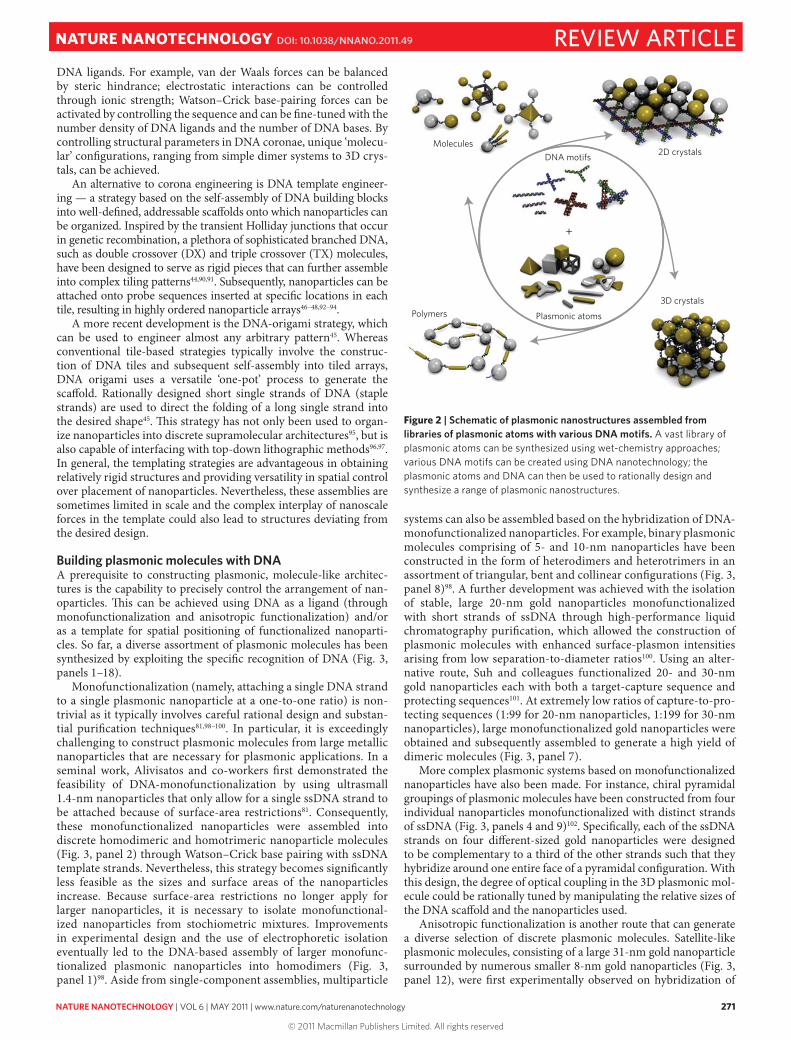
Molecules2D crystals
3D crystalsPolymers
DNA motifs
Plasmonic atoms
+
Figure 2 | Schematic of plasmonic nanostructures assembled from libraries of plasmonic atoms with various DNA motifs. A vast library of plasmonic atoms can be synthesized using wet-chemistry approaches; various DNA motifs can be created using DNA nanotechnology; the plasmonic atoms and DNA can then be used to rationally design and synthesize a range of plasmonic nanostructures.
REVIEW ARTICLENATURE NANOTECHNOLOGY DOI: 10.1038/NNANO.2011.49
© 2011 Macmillan Publishers Limited. All rights reserved

272 NATURE NANOTECHNOLOGY | VOL 6 | MAY 2011 | www.nature.com/naturenanotechnology
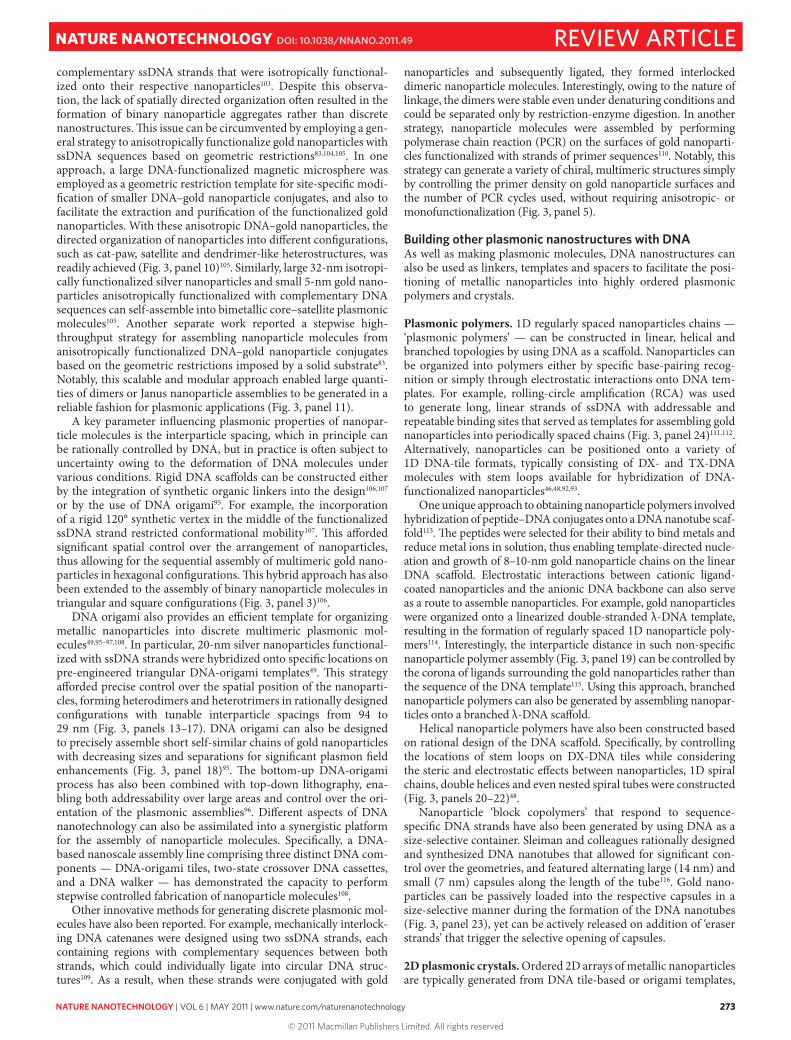
Nan
opar
ticle
pol
ymer
s2D
nan
opar
ticle
cry
stal
s
3D n
anop
artic
le c
ryst
als
Nan
opar
ticle
‘mol
ecul
es’
1 2 3 4 5 6
13 14 15 16 17 18
7 8 9 10 11 12
19 20 21 22 23
24
25 26 27 28 29
1
30 31
Figure 3 | Plasmonic nanostructures rationally organized from metallic ‘nanoparticle atoms’. These spatially directed assemblies include homomeric molecules (panels 1–681,98,102,106,110,123; 13–1749,97), heteromeric molecules (panels 7–1283,98,101–104; 1895), linear ‘polymer’ chains (panels 19–2448,111,115,116), 2D crystalline patterns (panels 25–2946,87,94,117) and 3D nanoparticle crystals (panels 3082 and 3186). Some images have been cropped, rotated, recoloured and/or had their backgrounds filled in; see the original papers for scale bars and other information. Figure reproduced with permission from: 1, 8, 13–15, 17, 24, 31, refs 98, 98, 49, 49, 49, 49, 111, 86 respectively, © 1999, 1999, 2010, 2010, 2010, 2010, 2005, 2010 respectively Wiley; 2, 7, 11, 16, 23, 28–30, refs 81, 101, 83, 97, 116, 117, 87, 82 respectively, © 1996, 2010, 2009, 2010, 2010, 2008, 2009, 2010 respectively NPG; 3–6, 9, 10, 12, 18, 19, 25–27, refs 106, 102, 110, 123, 102, 104, 103, 95, 115, 46, 94, 94 respectively, © 2007, 2009, 2009, 2009, 2009, 2006, 1998, 2010, 2004, 2004, 2006, 2006 respectively ACS; 20–22, ref. 48, © 2009 AAAS.
REVIEW ARTICLE NATURE NANOTECHNOLOGY DOI: 10.1038/NNANO.2011.49
© 2011 Macmillan Publishers Limited. All rights reserved
NATURE NANOTECHNOLOGY | VOL 6 | MAY 2011 | www.nature.com/naturenanotechnology 273
complementary ssDNA strands that were isotropically functional-ized onto their respective nanoparticles103. Despite this observa-tion, the lack of spatially directed organization often resulted in the formation of binary nanoparticle aggregates rather than discrete nanostructures. This issue can be circumvented by employing a gen-eral strategy to anisotropically functionalize gold nanoparticles with ssDNA sequences based on geometric restrictions83,104,105. In one approach, a large DNA-functionalized magnetic microsphere was employed as a geometric restriction template for site-specific modi-fication of smaller DNA–gold nanoparticle conjugates, and also to facilitate the extraction and purification of the functionalized gold nanoparticles. With these anisotropic DNA–gold nanoparticles, the directed organization of nanoparticles into different configurations, such as cat-paw, satellite and dendrimer-like heterostructures, was readily achieved (Fig. 3, panel 10)105. Similarly, large 32-nm isotropi-cally functionalized silver nanoparticles and small 5-nm gold nano-particles anisotropically functionalized with complementary DNA sequences can self-assemble into bimetallic core–satellite plasmonic molecules105. Another separate work reported a stepwise high-throughput strategy for assembling nanoparticle molecules from anisotropically functionalized DNA–gold nanoparticle conjugates based on the geometric restrictions imposed by a solid substrate83. Notably, this scalable and modular approach enabled large quanti-ties of dimers or Janus nanoparticle assemblies to be generated in a reliable fashion for plasmonic applications (Fig. 3, panel 11).
A key parameter influencing plasmonic properties of nanopar-ticle molecules is the interparticle spacing, which in principle can be rationally controlled by DNA, but in practice is often subject to uncertainty owing to the deformation of DNA molecules under various conditions. Rigid DNA scaffolds can be constructed either by the integration of synthetic organic linkers into the design106,107 or by the use of DNA origami95. For example, the incorporation of a rigid 120° synthetic vertex in the middle of the functionalized ssDNA strand restricted conformational mobility107. This afforded significant spatial control over the arrangement of nanoparticles, thus allowing for the sequential assembly of multimeric gold nano-particles in hexagonal configurations. This hybrid approach has also been extended to the assembly of binary nanoparticle molecules in triangular and square configurations (Fig. 3, panel 3)106.
DNA origami also provides an efficient template for organizing metallic nanoparticles into discrete multimeric plasmonic mol-ecules49,95–97,108. In particular, 20-nm silver nanoparticles functional-ized with ssDNA strands were hybridized onto specific locations on pre-engineered triangular DNA-origami templates49. This strategy afforded precise control over the spatial position of the nanoparti-cles, forming heterodimers and heterotrimers in rationally designed configurations with tunable interparticle spacings from 94 to 29 nm (Fig. 3, panels 13–17). DNA origami can also be designed to precisely assemble short self-similar chains of gold nanoparticles with decreasing sizes and separations for significant plasmon field enhancements (Fig. 3, panel 18)95. The bottom-up DNA-origami process has also been combined with top-down lithography, ena-bling both addressability over large areas and control over the ori-entation of the plasmonic assemblies96. Different aspects of DNA nanotechnology can also be assimilated into a synergistic platform for the assembly of nanoparticle molecules. Specifically, a DNA-based nanoscale assembly line comprising three distinct DNA com-ponents — DNA-origami tiles, two-state crossover DNA cassettes, and a DNA walker — has demonstrated the capacity to perform stepwise controlled fabrication of nanoparticle molecules108.
Other innovative methods for generating discrete plasmonic mol-ecules have also been reported. For example, mechanically interlock-ing DNA catenanes were designed using two ssDNA strands, each containing regions with complementary sequences between both strands, which could individually ligate into circular DNA struc-tures109. As a result, when these strands were conjugated with gold
nanoparticles and subsequently ligated, they formed interlocked dimeric nanoparticle molecules. Interestingly, owing to the nature of linkage, the dimers were stable even under denaturing conditions and could be separated only by restriction-enzyme digestion. In another strategy, nanoparticle molecules were assembled by performing polymerase chain reaction (PCR) on the surfaces of gold nanoparti-cles functionalized with strands of primer sequences110. Notably, this strategy can generate a variety of chiral, multimeric structures simply by controlling the primer density on gold nanoparticle surfaces and the number of PCR cycles used, without requiring anisotropic- or monofunctionalization (Fig. 3, panel 5).
Building other plasmonic nanostructures with DNAAs well as making plasmonic molecules, DNA nanostructures can also be used as linkers, templates and spacers to facilitate the posi-tioning of metallic nanoparticles into highly ordered plasmonic polymers and crystals.
Plasmonic polymers. 1D regularly spaced nanoparticles chains — ‘plasmonic polymers’ — can be constructed in linear, helical and branched topologies by using DNA as a scaffold. Nanoparticles can be organized into polymers either by specific base-pairing recog-nition or simply through electrostatic interactions onto DNA tem-plates. For example, rolling-circle amplification (RCA) was used to generate long, linear strands of ssDNA with addressable and repeatable binding sites that served as templates for assembling gold nanoparticles into periodically spaced chains (Fig. 3, panel 24)111,112. Alternatively, nanoparticles can be positioned onto a variety of 1D DNA-tile formats, typically consisting of DX- and TX-DNA molecules with stem loops available for hybridization of DNA-functionalized nanoparticles46,48,92,93.
One unique approach to obtaining nanoparticle polymers involved hybridization of peptide–DNA conjugates onto a DNA nanotube scaf-fold113. The peptides were selected for their ability to bind metals and reduce metal ions in solution, thus enabling template-directed nucle-ation and growth of 8–10-nm gold nanoparticle chains on the linear DNA scaffold. Electrostatic interactions between cationic ligand-coated nanoparticles and the anionic DNA backbone can also serve as a route to assemble nanoparticles. For example, gold nanoparticles were organized onto a linearized double-stranded λ-DNA template, resulting in the formation of regularly spaced 1D nanoparticle poly-mers114. Interestingly, the interparticle distance in such non-specific nanoparticle polymer assembly (Fig. 3, panel 19) can be controlled by the corona of ligands surrounding the gold nanoparticles rather than the sequence of the DNA template115. Using this approach, branched nanoparticle polymers can also be generated by assembling nanopar-ticles onto a branched λ-DNA scaffold.
Helical nanoparticle polymers have also been constructed based on rational design of the DNA scaffold. Specifically, by controlling the locations of stem loops on DX-DNA tiles while considering the steric and electrostatic effects between nanoparticles, 1D spiral chains, double helices and even nested spiral tubes were constructed (Fig. 3, panels 20–22)48.
Nanoparticle ‘block copolymers’ that respond to sequence-specific DNA strands have also been generated by using DNA as a size-selective container. Sleiman and colleagues rationally designed and synthesized DNA nanotubes that allowed for significant con-trol over the geometries, and featured alternating large (14 nm) and small (7 nm) capsules along the length of the tube116. Gold nano-particles can be passively loaded into the respective capsules in a size-selective manner during the formation of the DNA nanotubes (Fig. 3, panel 23), yet can be actively released on addition of ‘eraser strands’ that trigger the selective opening of capsules.
2D plasmonic crystals. Ordered 2D arrays of metallic nanoparticles are typically generated from DNA tile-based or origami templates,
REVIEW ARTICLENATURE NANOTECHNOLOGY DOI: 10.1038/NNANO.2011.49
© 2011 Macmillan Publishers Limited. All rights reserved

274 NATURE NANOTECHNOLOGY | VOL 6 | MAY 2011 | www.nature.com/naturenanotechnology
onto which a DNA-encoded nanoparticle can hybridize. For exam-ple, four-armed branched DNA tiles can be assembled in an A–B tile system, with tile A containing an exposed ssDNA oligo as a hybridization site. Gold nanoparticles functionalized with comple-mentary ssDNA sequences can subsequently be patterned into a 2D array using the A–B tile system as a template47. By modifying the sticky ends and overall size of tile B, the 2D DNA template can be rationally designed to organize DNA-functionalized gold nanopar-ticles in different configurations93. 2D DNA scaffolding assembled from DX-DNA tiles has also successfully templated the patterning of DNA-encoded gold nanoparticles into 2D arrays of aligned gold nanoparticle chains (Fig. 3, panel 25)46. Furthermore, a three-space-spanning DX-DNA motif was used to generate ordered 2D arrays of 5- and 10-nm gold nanoparticles (Fig. 3, panels 26 and 27)94.
As well as the templated strategy, 2D plasmonic crystals can also be generated through a microhole-confined drying process. For example, 2D plasmonic nanoparticle superlattices spontaneously formed on a silicon substrate through micromold-regulated drying (Fig. 3, panel 28)117. 2D plasmonic crystals can even assemble with-out any substrate support and exist in a free-standing format (Fig. 3, panel 29)87. These free-standing superlattice sheets were mechani-cally strong and exhibited both tunable mechanical and plasmonic properties based on DNA ligand length. Interestingly, both of these 2D plasmonic crystal formats were stable constructs in a dehydrated state without requiring any specific Watson–Crick base pairing; rather, non-specific DNA–DNA interactions were responsible for maintaining the integrity of these superlattice structures.
3D plasmonic crystals. DNA molecules have also been success-fully exploited to assemble 3D plasmonic nanoparticle crystals. Solely by programming DNA sequences, plasmonic nanoparticles can self-organize into 3D crystals (Fig. 3, panel 30) with distinct lattice structures that are reversible on changes in temperature84,85. Specifically, nanoparticles could be assembled through a sin-gle DNA linker strand that gave rise to equal binding affinities between particles, resulting in a close-packed face-centred cubic structure; alternatively, nanoparticles could be assembled through two different DNA linker strands, resulting in a binary system that favoured the formation of a non-close-packed body-centred cubic structure. 3D plasmonic crystals have also been constructed without any base pairing in a drying-mediated self-assembly proc-ess (Fig. 3, panel 31)86. Real-time small-angle X-ray scattering revealed that the nature of DNA ligand interactions could tempo-rally regulate the occurrence of nanoparticle crystallization. For example, crystallization of nanoparticles capped with palindromic base-pairing ssDNA occurred at an earlier stage of drying than those with non-base-pairing ssDNA. Based on these observations, 3D nanoparticle crystals can be fabricated with or without DNA base pairing to varying consequences. Furthermore, the construc-tion of 3D DNA-based plasmonic crystals in the dehydrated state represents a promising step towards integration with solid-state lithographical structures.
Summary and outlookThe rapid progress in the development of DNA-based plasmonic nanostructures, which stems from the continued integration of nanoparticle synthesis, surface chemistry, DNA nanotechnology and lithography, will soon lead to real-world applications such as sensing, waveguiding and energy harvesting. Gold and silver nano-particle dimers have already been employed as robust molecular rulers for extended real-time monitoring of single-DNA hybridiza-tion events118. Such plasmon rulers are particularly advantageous over traditional fluorescent resonance energy transfer-based rulers as they are not limited by signal fluctuations, photobleaching, or an upper distance limit of ~10 nm. Interparticle junctions between dimeric plasmonic nanoparticles have also been engineered to
enable highly sensitive single-molecule detection based on strong surface-enhanced Raman-scattering effects101. Practically, it is pos-sible to scale-up the fabrication of DNA-based plasmonic architec-tures by improving yield because the synthesis, conjugation and self-assembly of DNA–nanoparticle systems are reactions typically limited by the availability of materials.
The DNA-based strategy for assembly of plasmonic nanopar-ticles can also be extended to heterogeneous materials systems involving quantum dots, magnetic nanoparticles, carbon nano-tubes, graphene and other nanomaterials. For example, molecular chromophores119 and quantum dots120 have been incorporated into arrays of plasmonic particles using DNA to provide control over interparticle distance. These hybrid systems have been achieved in the form of heterodimeric assemblies119,120, and also in 3D superlat-tices for dramatic enhancements in optical properties121. Hence, as well as tuning plasmonic coupling, DNA can also be used to tailor the coupling in heterogeneous material systems.
Engineering the shapes and sizes of individual plasmonic atoms and programming their assembly into molecule-like architectures through DNA represent a new approach to generating customiz-able optical nanomaterials. So far, DNA has been by far the most successful molecule for guided assembly of elementary plasmonic nanoparticles into an assortment of well-defined nanostructures. Despite this progress, the field of DNA–nanoparticle plasmonics is still in its infancy. Although most reports on successful assem-bly of plasmonic molecules have been predominantly limited to spherical building blocks, more advanced methods of surface modification must be developed to fully exploit the vast library of available plasmonic atoms.
Nevertheless, these challenges can be overcome with further development in nanosurface chemistry and DNA nanotechnol-ogy, thereby permitting both site and stoichiometric control over the functionalization of anisotropic and complex nanoparticles. Such capabilities will allow for precise spatial and vectorial con-trol over different-shaped nanoparticles to facilitate the coordina-tion of sophisticated multiparticle systems in high yield. Another challenge lies in the interfacing of DNA–nanoparticle structures with solid-state devices. Plasmonic molecules have typically been assembled and stabilized in aqueous buffered environments, thus requiring careful design considerations when attempting to inter-face with solid-state devices. However, recent success in com-bining the bottom-up DNA-guided self-assembly of plasmonic nanostructures with top-down lithography87,96,117,122 is an encour-aging step towards the application of DNA-guided plasmonics in solid-state electronics. Based on its versatility and potential as an organizer of nanomaterials, we predict that DNA, beyond its genetic functions, will play a critical role in shaping tomorrow’s optical and electronic devices by directing the synthesis of highly ordered designer materials.
References1. Schuller, J. A. et al. Plasmonics for extreme light concentration and
manipulation. Nature Mater. 9, 193–204 (2010).2. Shipway, A. N., Katz, E. & Willner, I. Nanoparticle arrays on surfaces for
electronic, optical, and sensor applications. ChemPhysChem 1, 18–52 (2000).3. Ozbay, E. Plasmonics: Merging photonics and electronics at nanoscale
dimensions. Science 311, 189–193 (2006).4. Anker, J. N. et al. Biosensing with plasmonic nanosensors. Nature Mater.
7, 442–453 (2008).5. Maier, S. A. et al. Local detection of electromagnetic energy transport below
the diffraction limit in metal nanoparticle plasmon waveguides. Nature Mater. 2, 229–232 (2003).
6. Lal, S., Clare, S. E. & Halas, N. J. Nanoshell-enabled photothermal cancer therapy: Impending clinical impact. Acc. Chem. Rev. 41, 1842–1851 (2008).
7. Yavuz, M. S. et al. Gold nanocages covered by smart polymers for controlled release with near-infrared light. Nature Mater. 8, 935–939 (2009).
8. Mie, G. Beiträge zur optik trüber medien, speziell kolloidaler metallösungen. Ann. Phys. 330, 377–445 (1908).
REVIEW ARTICLE NATURE NANOTECHNOLOGY DOI: 10.1038/NNANO.2011.49
© 2011 Macmillan Publishers Limited. All rights reserved

NATURE NANOTECHNOLOGY | VOL 6 | MAY 2011 | www.nature.com/naturenanotechnology 275
9. Prodan, E., Radloff, C., Halas, N. J. & Nordlander, P. A hybridization model for the plasmon response of complex nanostructures. Science 302, 419–422 (2003).
10. Pérez-Juste, J., Pastoriza-Santos, I., Liz-Marzán, L. M. & Mulvaney, P. Gold nanorods: Synthesis, characterization and applications. Coordin. Chem. Rev. 249, 1870–1901 (2005).
11. Xia, Y., Xiong, Y., Lim, B. & Skrabalak, S. E. Shape-controlled synthesis of metal nanocrystals: Simple chemistry meets complex physics? Angew. Chem. Int. Ed. 48, 60–103 (2009).
12. Schwartzberg, A. M., Olson, T. Y., Talley, C. E. & Zhang, J. Z. Synthesis, characterization, and tunable optical properties of hollow gold nanospheres. J. Phys. Chem. B 110, 19935–19944 (2006).
13. Xiong, Y. et al. Synthesis and mechanistic study of palladium nanobars and nanorods. J. Am. Chem. Soc. 129, 3665–3675 (2007).
14. Ni, W., Yang, Z., Chen, H., Li, L. & Wang, J. Coupling between molecular and plasmonic resonances in freestanding dye-gold nanorod hybrid nanostructures. J. Am. Chem. Soc. 130, 6692–6693 (2008).
15. Wijaya, A., Schaffer, S. B., Pallares, I. G. & Hamad-Schifferli, K. Selective release of multiple DNA oligonucleotides from gold nanorods. ACS Nano 3, 80–86 (2008).
16. Wiley, B. J. et al. Synthesis and electrical characterization of silver nanobeams. Nano Lett. 6, 2273–2278 (2006).
17. Zhao, N. et al. Controlled synthesis of gold nanobelts and nanocombs in aqueous mixed surfactant solutions. Langmuir 24, 991–998 (2008).
18. Huo, Z., Tsung, C-k., Huang, W., Zhang, X. & Yang, P. Sub-two nanometer single crystal au nanowires. Nano Lett. 8, 2041–2044 (2008).
19. Murphy, C. J., Gole, A. M., Hunyadi, S. E. & Orendorff, C. J. One-dimensional colloidal gold and silver nanostructures. Inorg. Chem. 45, 7544–7554 (2006).
20. Jin, R. et al. Photoinduced conversion of silver nanospheres to nanoprisms. Science 294, 1901–1903 (2001).
21. Porel, S., Singh, S. & Radhakrishnan, T. P. Polygonal gold nanoplates in a polymer matrix. Chem. Commun. 2387–2389 (2005).
22. Das, S. K., Das, A. R. & Guha, A. K. Microbial synthesis of multishaped gold nanostructures. Small 6, 1012–1021 (2010).
23. Liu, B., Xie, J., Lee, J. Y., Ting, Y. P. & Chen, J. P. Optimization of high-yield biological synthesis of single-crystalline gold nanoplates. J. Phys. Chem. B 109, 15256–15263 (2005).
24. Ah, C. S. et al. Size-controlled synthesis of machinable single crystalline gold nanoplates. Chem. Mater. 17, 5558–5561 (2005).
25. Jiang, L-P. et al. Ultrasonic-assisted synthesis of monodisperse single-crystalline silver nanoplates and gold nanorings. Inorg. Chem. 43, 5877–5883 (2004).
26. Kim, F., Connor, S., Song, H., Kuykendall, T. & Yang, P. Platonic gold nanocrystals. Angew. Chem. Int. Ed. 43, 3673–3677 (2004).
27. Tao, A., Sinsermsuksakul, P. & Yang, P. Tunable plasmonic lattices of silver nanocrystals. Nature Nanotech. 2, 435–440 (2007).
28. Seo, D. et al. Shape adjustment between multiply twinned and single-crystalline polyhedral gold nanocrystals: Decahedra, icosahedra, and truncated tetrahedra. J. Phys. Chem. C 112, 2469–2475 (2008).
29. Kim, D. Y., Im, S. H., Park, O. O. & Lim, Y. T. Evolution of gold nanoparticles through Catalan, Archimedean, and Platonic solids. CrystEngComm 12, 116–121 (2010).
30. Sun, Y., Mayers, B. T. & Xia, Y. Template-engaged replacement reaction: A one-step approach to the large-scale synthesis of metal nanostructures with hollow interiors. Nano Lett. 2, 481–485 (2002).
31. Chen, S., Wang, Z. L., Ballato, J., Foulger, S. H. & Carroll, D. L. Monopod, bipod, tripod, and tetrapod gold nanocrystals. J. Am. Chem. Soc. 125, 16186–16187 (2003).
32. Liao, H-G., Jiang, Y-X., Zhou, Z-Y., Chen, S-P. & Sun, S-G. Shape-controlled synthesis of gold nanoparticles in deep eutectic solvents for studies of structure-functionality relationships in electrocatalysis. Angew. Chem. Int. Ed. 47, 9100–9103 (2008).
33. Mulvihill, M. J., Ling, X. Y., Henzie, J. & Yang, P. Anisotropic etching of silver nanoparticles for plasmonic structures capable of single-particle SERS. J. Am. Chem. Soc. 132, 268–274 (2009).
34. Lu, Y., Liu, G. L., Kim, J., Mejia, Y. X. & Lee, L. P. Nanophotonic crescent moon structures with sharp edge for ultrasensitive biomolecular detection by local electromagnetic field enhancement effect. Nano Lett. 5, 119–124 (2004).
35. Wang, C. et al. Rational synthesis of heterostructured nanoparticles with morphology control. J. Am. Chem. Soc. 132, 6524–6529 (2010).
36. Lee, J., Hasan, W., Lee, M. H. & Odom, T. W. Optical properties and magnetic manipulation of bimaterial nanopyramids. Adv. Mater. 19, 4387–4391 (2007).
37. Cheng, W. L., Steinhart, M., Gösele, U. & Wehrspohn, R. Tree-like alumina nanopores generated in a non-steady-state anodization. J. Mater. Chem. 17, 3493–3495 (2007).
38. Qin, Y. et al. Ionic liquid-assisted growth of single-crystalline dendritic gold nanostructures with a three-fold symmetry. Chem. Mater. 20, 3965–3972 (2008).
39. Skrabalak, S. E. et al. Gold nanocages: Synthesis, properties, and applications. Acc. Chem. Rev. 41, 1587–1595 (2008).
40. Zou, X., Ying, E. & Dong, S. Preparation of novel silver-gold bimetallic nanostructures by seeding with silver nanoplates and application in surface-enhanced Raman scattering. J. Colloid Interface Sci. 306, 307–315 (2007).
41. Yin, Y., Erdonmez, C., Aloni, S. & Alivisatos, A. P. Faceting of nanocrystals during chemical transformation: From solid silver spheres to hollow gold octahedra. J. Am. Chem. Soc. 128, 12671–12673 (2006).
42. Wang, X. et al. One-pot solution synthesis of cubic cobalt nanoskeletons. Adv. Mater. 21, 1636–1640 (2009).
43. Sun, Y. & Xia, Y. Shape-controlled synthesis of gold and silver nanoparticles. Science 298, 2176–2179 (2002).
44. Seeman, N. C. DNA in a material world. Nature 421, 427–431 (2003).45. Rothemund, P. W. K. Folding DNA to create nanoscale shapes and patterns.
Nature 440, 297–302 (2006).46. Le, J. D. et al. DNA-templated self-assembly of metallic nanocomponent arrays
on a surface. Nano Lett. 4, 2343–2347 (2004).47. Zhang, J., Liu, Y., Ke, Y. & Yan, H. Periodic square-like gold nanoparticle
arrays templated by self-assembled 2D DNA nanogrids on a surface. Nano Lett. 6, 248–251 (2006).
48. Sharma, J. et al. Control of self-assembly of DNA tubules through integration of gold nanoparticles. Science 323, 112–116 (2009).
49. Pal, S., Deng, Z., Ding, B., Yan, H. & Liu, Y. DNA-origami-directed self-assembly of discrete silver-nanoparticle architectures. Angew. Chem. Int. Ed. 49, 2700–2704 (2010).
50. Storhoff, J. J. & Mirkin, C. A. Programmed materials synthesis with DNA. Chem. Rev. 99, 1849–1862 (1999).
51. Katz, E. & Willner, I. Integrated nanoparticle-biomolecule hybrid systems: Synthesis, properties, and applications. Angew. Chem. Int. Ed. 43, 6042–6108 (2004).
52. Niemeyer, C. M. & Simon, U. DNA-based assembly of metal nanoparticles. Eur. J. Inorg. Chem. 3641–3655 (2005).
53. Ofir, Y., Samanta, B. & Rotello, V. M. Polymer and biopolymer mediated self-assembly of gold nanoparticles. Chem. Soc. Rev. 37, 1814–1823 (2008).
54. Choi, C. L. & Alivisatos, A. P. From artificial atoms to nanocrystal molecules: Preparation and properties of more complex nanostructures. Annu. Rev. Phys. Chem. 61, 369–389 (2010).
55. Mulvaney, P. Surface plasmon spectroscopy of nanosized metal particles. Langmuir 12, 788–800 (1996).
56. Kelly, L. K., Coronado, E., Zhao, L. L. & Schatz, G. C. The optical properties of metal nanoparticles: The influence of size, shape, and dielectric environment. J. Phys. Chem. B 107, 668–677 (2003).
57. Liz-Marzán, L. M. Tailoring surface plasmons through the morphology and assembly of metal nanoparticles. Langmuir 22, 32–41 (2006).
58. Bohren, C. F. & Huffmann, D. R. Absorption and Scattering of Light by Small Particles (Wiley, 1983).
59. Kreibig, U. & Vollmer, M. Optical Properties of Metal Clusters (Springer, 1995).
60. Link, S. & El-Sayed, M. A. Size and temperature dependence of the plasmon absorption of colloidal gold nanoparticles. J. Phys. Chem. B 103, 4212–4217 (1999).
61. Wang, H., Brandl, D. W., Nordlander, P. & Halas, N. J. Plasmonic nanostructures: Artificial molecules. Acc. Chem. Res. 40, 53–62 (2007).
62. Gans, R. The shape of ultra microscopic gold nanoparticles. Ann. Phys. 37, 881–900 (1912).
63. Wiley, B. J. et al. Maneuvering the surface plasmon resonance of silver nanostructures through shape-controlled synthesis. J. Phys. Chem. B 110, 15666–15675 (2006).
64. Su, K-H., Wei, Q-H. & Zhang, X. Interparticle coupling effects on plasmon resonances of nanogold particles. Nano Lett. 3, 1087–1090 (2003).
65. Jain, P. K., Huang, W. & El-Sayed, M. A. On the universal scaling behavior of the distance decay of plasmon coupling in metal nanoparticle pairs: A plasmon ruler equation. Nano Lett. 7, 2080–2088 (2007).
66. Prodan, E. & Nordlander, P. Plasmon hybridization in spherical nanoparticles. J. Chem. Phys. 120, 5444–5454 (2004).
67. Nordlander, P., Oubre, C., Prodan, E., Li, K. & Stockman, M. I. Plasmon hybridization in nanoparticle dimers. Nano Lett. 4, 899–903 (2004).
68. Fan, J. A. et al. Self-assembled plasmonic nanoparticle clusters. Science 328, 1135–1138 (2010).
69. Quinten, M., Leitner, A., Krenn, J. R. & Aussenegg, F. R. Electromagnetic energy transport via linear chains of silver nanoparticles. Opt. Lett. 23, 1331–1333 (1998).
70. Wei, Q-H., Su, K-H., Durant, S. & Zhang, X. Plasmon resonance of finite one-dimensional Au nanoparticle chains. Nano Lett. 4, 1067–1071 (2004).
71. Lal, S., Link, S. & Halas, N. J. Nano-optics from sensing to waveguiding. Nature Photon. 1, 641–648 (2007).
72. Lin, S., Dujardin, E., Girard, C. & Mann, S. One-dimensional plasmon coupling by facile self-assembly of gold nanoparticles into branched chain networks. Adv. Mater. 17, 2553–2559 (2005).
REVIEW ARTICLENATURE NANOTECHNOLOGY DOI: 10.1038/NNANO.2011.49
© 2011 Macmillan Publishers Limited. All rights reserved
276 NATURE NANOTECHNOLOGY | VOL 6 | MAY 2011 | www.nature.com/naturenanotechnology
73. Tabor, C., Van Haute, D. & El-Sayed, M. A. Effect of orientation on plasmonic coupling between gold nanorods. ACS Nano 3, 3670–3678 (2009).
74. Sheikholeslami, S., Jun, Y-W., Jain, P. K. & Alivisatos, A. P. Coupling of optical resonances in a compositionally asymmetric plasmonic nanoparticle dimer. Nano Lett. 10, 2655–2660 (2010).
75. Funston, A. M., Novo, C., Davis, T. J. & Mulvaney, P. Plasmon coupling of gold nanorods at short distances and in different geometries. Nano Lett. 9, 1651–1658 (2009).
76. Min, Y. J., Akbulut, M., Kristiansen, K., Golan, Y. & Israelachvili, J. The role of interparticle and external forces in nanoparticle assembly. Nature Mater. 7, 527–538 (2008).
77. Shevchenko, E. V., Talapin, D. V., Kotov, N. A., O’Brien, S. & Murray, C. B. Structural diversity in binary nanoparticle superlattices. Nature 439, 55–59 (2006).
78. Badia, A. et al. Self-assembled monolayers on gold nanoparticles. Chem.-Eur. J. 2, 359–363 (1996).
79. Templeton, A. C., Wuelfing, M. P. & Murray, R. W. Monolayer protected cluster molecules. Acc. Chem. Rev. 33, 27–36 (2000).
80. Cheng, W. L., Dong, S. J. & Wang, E. K. Synthesis and self-assembly of cetyltrimethylammonium bromide-capped gold nanoparticles. Langmuir 19, 9434–9439 (2003).
81. Alivisatos, A. P. et al. Organization of ‘nanocrystal molecules’ using DNA. Nature 382, 609–611 (1996).
82. Maye, M. M., Kumara, M. T., Nykypanchuk, D., Sherman, W. B. & Gang, O. Switching binary states of nanoparticle superlattices and dimer clusters by DNA strands. Nature Nanotech. 5, 116–120 (2010).
83. Maye, M. M., Nykypanchuk, D., Cuisinier, M., van der Lelie, D. & Gang, O. Stepwise surface encoding for high-throughput assembly of nanoclusters. Nature Mater. 8, 388–391 (2009).
84. Nykypanchuk, D., Maye, M. M., van der Lelie, D. & Gang, O. DNA-guided crystallization of colloidal nanoparticles. Nature 451, 549–552 (2008).
85. Park, S. Y. et al. DNA-programmable nanoparticle crystallization. Nature 451, 553–556 (2008).
86. Cheng, W. et al. Probing in real time the soft crystallization of DNA-capped nanoparticles. Angew. Chem. Int. Ed. 49, 380–384 (2010).
87. Cheng, W. et al. Free-standing nanoparticle superlattice sheets controlled by DNA. Nature Mater. 8, 519–525 (2009).
88. Verwey, E. J. W. & Overbeek, J. Theory of the Stability of Lyophobic Colloids Vol. 1 (Elsevier, 1948).
89. Derjaguin, B. V. & Landau, L. Theory of stability of highly charged lyophobic sols and adhesion of highly charged particles in solutions of electrolytes. Acta Physicochim. URS 14, 633–652 (1941).
90. Lin, C., Liu, Y., Rinker, S. & Yan, H. DNA tile based self-assembly: Building complex nanoarchitectures. ChemPhysChem 7, 1641–1647 (2006).
91. LaBean, T. H. & Li, H. Constructing novel materials with DNA. Nano Today 2, 26–35 (2007).
92. Li, H., Park, S. H., Reif, J. H., LaBean, T. H. & Yan, H. DNA-templated self-assembly of protein and nanoparticle linear arrays. J. Am. Chem. Soc. 126, 418–419 (2003).
93. Sharma, J., Chhabra, R., Liu, Y., Ke, Y. & Yan, H. DNA-templated self-assembly of two-dimensional and periodical gold nanoparticle arrays. Angew. Chem. Int. Ed. 45, 730–735 (2006).
94. Zheng, J. et al. Two-dimensional nanoparticle arrays show the organizational power of robust DNA motifs. Nano Lett. 6, 1502–1504 (2006).
95. Ding, B. et al. Gold nanoparticle self-similar chain structure organized by DNA origami. J. Am. Chem. Soc. 132, 3248–3249 (2010).
96. Kershner, R. J. et al. Placement and orientation of individual DNA shapes on lithographically patterned surfaces. Nature Nanotech. 4, 557–561 (2009).
97. Hung, A. M. et al. Large-area spatially ordered arrays of gold nanoparticles directed by lithographically confined DNA origami. Nature Nanotech. 5, 121–126 (2010).
98. Loweth, C. J., Caldwell, W. B., Peng, X., Alivisatos, A. P. & Schultz, P. G. DNA-based assembly of gold nanocrystals. Angew. Chem. Int. Ed. 38, 1808–1812 (1999).
99. Zanchet, D., Micheel, C. M., Parak, W. J., Gerion, D. & Alivisatos, A. P. Electrophoretic isolation of discrete Au nanocrystal/DNA conjugates. Nano Lett. 1, 32–35 (2001).
100. Claridge, S. A., Liang, H. W., Basu, S. R., Fréchet, J. M. J. & Alivisatos, A. P. Isolation of discrete nanoparticle-DNA conjugates for plasmonic applications. Nano Lett. 8, 1202–1206 (2008).
101. Lim, D-K., Jeon, K-S., Kim, H. M., Nam, J-M. & Suh, Y. D. Nanogap-engineerable Raman-active nanodumbbells for single-molecule detection. Nature Mater. 9, 60–67 (2010).
102. Mastroianni, A. J., Claridge, S. A. & Alivisatos, A. P. Pyramidal and chiral groupings of gold nanocrystals assembled using DNA scaffolds. J. Am. Chem. Soc. 131, 8455–8459 (2009).
103. Mucic, R. C., Storhoff, J. J., Mirkin, C. A. & Letsinger, R. L. DNA-directed synthesis of binary nanoparticle network materials. J. Am. Chem. Soc. 120, 12674–12675 (1998).
104. Xu, X., Rosi, N. L., Wang, Y., Huo, F. & Mirkin, C. A. Asymmetric functionalization of gold nanoparticles with oligonucleotides. J. Am. Chem. Soc. 128, 9286–9287 (2006).
105. Pal, S., Sharma, J., Yan, H. & Liu, Y. Stable silver nanoparticle–DNA conjugates for directed self-assembly of core-satellite silver–gold nanoclusters. Chem. Commun. 6059–6061 (2009).
106. Aldaye, F. A. & Sleiman, H. F. Dynamic DNA templates for discrete gold nanoparticle assemblies: Control of geometry, modularity, write/erase and structural switching. J. Am. Chem. Soc. 129, 4130–4131 (2007).
107. Aldaye, F. A. & Sleiman, H. F. Sequential self-assembly of a DNA hexagon as a template for the organization of gold nanoparticles. Angew. Chem. Int. Ed. 45, 2204–2209 (2006).
108. Gu, H., Chao, J., Xiao, S-J. & Seeman, N. C. A proximity-based programmable DNA nanoscale assembly line. Nature 465, 202–205 (2010).
109. Weizmann, Y., Braunschweig, A. B., Wilner, O. I., Cheglakov, Z. & Willner, I. A polycatenated DNA scaffold for the one-step assembly of hierarchical nanostructures. Proc. Natl Acad. Sci. USA 105, 5289–5294 (2008).
110. Chen, W. et al. Nanoparticle superstructures made by polymerase chain reaction: Collective interactions of nanoparticles and a new principle for chiral materials. Nano Lett. 9, 2153–2159 (2009).
111. Deng, Z., Tian, Y., Lee, S-H., Ribbe, A. E. & Mao, C. DNA-encoded self-assembly of gold nanoparticles into one-dimensional arrays. Angew. Chem. Int. Ed. 44, 3582–3585 (2005).
112. Beyer, S., Nickels, P. & Simmel, F. C. Periodic DNA nanotemplates synthesized by rolling circle amplification. Nano Lett. 5, 719–722 (2005).
113. Stearns, Linda A. et al. Template-directed nucleation and growth of inorganic nanoparticles on DNA scaffolds. Angew. Chem. Int. Ed. 48, 8494–8496 (2009).
114. Warner, M. G. & Hutchison, J. E. Linear assemblies of nanoparticles electrostatically organized on DNA scaffolds. Nature Mater. 2, 272–277 (2003).
115. Wang, G. & Murray, R. W. Controlled assembly of monolayer-protected gold clusters by dissolved DNA. Nano Lett. 4, 95–101 (2003).
116. Lo, P. K. et al. Loading and selective release of cargo in DNA nanotubes with longitudinal variation. Nature Chem. 2, 319–328 (2010).
117. Cheng, W. L., Park, N., Walter, M. T., Hartman, M. R. & Luo, D. Nanopatterning self-assembled nanoparticle superlattices by moulding microdroplets. Nature Nanotech. 3, 682–690 (2008).
118. Sonnichsen, C., Reinhard, B. M., Liphardt, J. & Alivisatos, A. P. A molecular ruler based on plasmon coupling of single gold and silver nanoparticles. Nature Biotechnol. 23, 741–745 (2005).
119. Seelig, J. et al. Nanoparticle-induced fluorescence lifetime modification as nanoscopic ruler: Demonstration at the single molecule level. Nano Lett. 7, 685–689 (2007).
120. Maye, M. M., Gang, O. & Cotlet, M. Photoluminescence enhancement in CdSe/ZnS-DNA linked-Au nanoparticle heterodimers probed by single molecule spectroscopy. Chem. Commun. 46, 6111–6113 (2010).
121. Xiong, H., Sfeir, M. Y. & Gang, O. Three-dimensional dynamically tunable multicomponent superlattices. Nano Lett. 10, 4456–4462 (2010).
122. Maune, H. T. et al. Self-assembly of carbon nanotubes into two-dimensional geometries using DNA origami templates. Nature Nanotech. 5, 61–66 (2010).
123. Kim, J-Y. & Lee, J-S. Synthesis and thermally reversible assembly of DNA-gold nanoparticle cluster conjugates. Nano Lett. 9, 4564–4569 (2009).
AcknowledgementsW.L.C. acknowledges financial support from the New Staff Member Research Fund awarded by the Faculty of Engineering, Monash University. S.J.T. is a recipient of the National Science Scholarship awarded by the Agency for Science, Technology and Research, Singapore (A-STAR). M.J.C. is supported by the IGERT Program of the National Science Foundation under Agreement no. DGE-0654112, administered by the Nanobiotechnology Center at Cornell University. D.L. acknowledges financial support from NYSTAR and the NSF CAREER award (grant number: 0547330).
Additional informationThe authors declare no competing financial interests.
REVIEW ARTICLE NATURE NANOTECHNOLOGY DOI: 10.1038/NNANO.2011.49
© 2011 Macmillan Publishers Limited. All rights reserved