DNA-programmed assembly of nanostructures
15
PERSPECTIVE OBC www.rsc.org/obc DNA-programmed assembly of nanostructures Kurt V. Gothelf* a and Thomas H. LaBean* b a Department of Chemistry, Aarhus University, Langelandsgade 140, 8000, Aarhus C, Denmark. E-mail: [email protected]; Tel: (+45) 89423907 b Department of Computer Science, Duke University, Durham, North Carolina, 27708, USA. E-mail: [email protected]; Tel: (919)660-1565 Received 27th July 2005, Accepted 13th September 2005 First published as an Advance Article on the web 6th October 2005 DNA is a unique material for nanotechnology since it is possible to use base sequences to encode instructions for assembly in a predetermined fashion at the nanometre scale. Synthetic oligonucleotides are readily obtained by automated synthesis and numerous techniques have been developed for conjugating DNA with other materials. The exact spatial positioning of materials is crucial for the future development of complex nanodevices and the emerging field of DNA-nanotechnology is now exploring DNA-programmed processes for the assembly of organic compounds, biomolecules, and inorganic materials. Introduction Self-assembly is often one of the key approaches discussed when debating future methods for building nanostructures and nanodevices. 1 Our current ability to form nanostructures by self- assembly is, however, very limited compared to the power of lithographic techniques for the formation of solid structures in bulk materials and, in particular, electronic circuits at the nanoscale. The pace of developing and refining lithographic techniques, new scanning probe microscopy and nanoimprinting techniques is impressive and the current bottom-up approaches are far from able to compete. Self-assembly procedures for the production of some non-biological systems are commonly used and have in some cases found commercial applications. These include self-assembled monolayers for the immobilization of compounds and materials on surfaces, Langmuir–Blodgett films, artificial membranes etc. However, these structures are typically, polydisperse and only “nanoscaled” in one dimension. Kurt Vesterager Gothelf (1968) performed his PhD research in organic synthesis and asymmetric catalysis at Aarhus University, Denmark, where he obtained his PhD degree in 1995 under the guidance of Professor K. B. G. Torssell and Professor K. A. Jørgensen. Following a period as a postdoc in Professor K. A. Jørgensen’s group, he joined Professor M. C. Pirrung’s group at Duke University. He returned to Aarhus University in 1999 to initiate research in organic nanochemistry. Since May 2002 he has been an Associate Professor at Aarhus University. Kurt V. Gothelf Thomas H. LaBean Thomas Henry LaBean (1963) earned his PhD at the University of Pennsylvania in 1993. He studied the folding properties of arbitrary sequence proteins expressed by random, synthetic DNA libraries under the guidance of Professor Stuart A. Kauffman and Professor Tauseef R. Butt. He then moved to Duke University and studied protein design with Professors Jane S. and David C. Richardson, and then worked on DNA-based computation with Professor John H. Reif. He now studies self-assembling biomolecular nanostructures as an Associate Research Professor at Duke University. Formation of more complex monodisperse and two- or three- dimensional nanostructures consisting of multiple building blocks requires precise control over each interaction when the structure undergoes self-assembly. In this regard supramolecular chemistry is in its infancy and the structures that are accessible via this method are mainly limited to a single or a few highly symmetric structures. The motivation and inspiration to continue exploring self- assembly is, on the other hand, obvious when studying the most advanced nanosystem known: the living cell. This unimaginably complex machinery made of organic molecules and polymers is formed by, and operates by, self-assembly. The precision and efficiency of this process derives from specific molecular interactions between proteins, DNA and RNA in particular, and other compounds including lipids, carbohydrates, and small molecules. The question is: how can we use any part of the cell’s self-assembly machinery to assemble artificial nanostructures? A dramatic reduction in complexity is necessary and this should be obtained by focusing on only one or two types of structural element from cells. In this regard DNA (or RNA) is the obvious choice since (i) it is the building block with the highest information content, (ii) it is constructed from only four, quite similar chemical building blocks, (iii) its self- assembly behavior is by far the most predictable compared to other classes of biomolecules, (iv) microgram quantities of oligonucleotides are easily and inexpensively available via automated chemical synthesis and (v) a diverse infrastructure developed for biotechnology provides many tools for DNA manipulation, including amplification via the polymerase chain reaction (PCR). DOI: 10.1039/b510551j This journal is © The Royal Society of Chemistry 2005 Org. Biomol. Chem. , 2005, 3 , 4023–4037 4023
-
Upload
independent -
Category
Documents
-
view
5 -
download
0
Transcript of DNA-programmed assembly of nanostructures

P E R S P E C T I V E
OBC
ww
w.rsc.o
rg/o
bc
DNA-programmed assembly of nanostructures
Kurt V. Gothelf*a and Thomas H. LaBean*b
a Department of Chemistry, Aarhus University, Langelandsgade 140, 8000, Aarhus C, Denmark.E-mail: [email protected]; Tel: (+45) 89423907
b Department of Computer Science, Duke University, Durham, North Carolina, 27708, USA.E-mail: [email protected]; Tel: (919)660-1565
Received 27th July 2005, Accepted 13th September 2005First published as an Advance Article on the web 6th October 2005
DNA is a unique material for nanotechnology since it ispossible to use base sequences to encode instructions forassembly in a predetermined fashion at the nanometrescale. Synthetic oligonucleotides are readily obtained byautomated synthesis and numerous techniques have beendeveloped for conjugating DNA with other materials.The exact spatial positioning of materials is crucial forthe future development of complex nanodevices and theemerging field of DNA-nanotechnology is now exploringDNA-programmed processes for the assembly of organiccompounds, biomolecules, and inorganic materials.
IntroductionSelf-assembly is often one of the key approaches discussedwhen debating future methods for building nanostructures andnanodevices.1 Our current ability to form nanostructures by self-assembly is, however, very limited compared to the power oflithographic techniques for the formation of solid structuresin bulk materials and, in particular, electronic circuits at thenanoscale. The pace of developing and refining lithographictechniques, new scanning probe microscopy and nanoimprintingtechniques is impressive and the current bottom-up approachesare far from able to compete. Self-assembly procedures forthe production of some non-biological systems are commonlyused and have in some cases found commercial applications.These include self-assembled monolayers for the immobilizationof compounds and materials on surfaces, Langmuir–Blodgettfilms, artificial membranes etc. However, these structures aretypically, polydisperse and only “nanoscaled” in one dimension.
Kurt Vesterager Gothelf (1968) performed his PhD research in organic synthesis and asymmetric catalysis at Aarhus University,Denmark, where he obtained his PhD degree in 1995 under the guidance of Professor K. B. G. Torssell and Professor K. A. Jørgensen.Following a period as a postdoc in Professor K. A. Jørgensen’s group, he joined Professor M. C. Pirrung’s group at Duke University. Hereturned to Aarhus University in 1999 to initiate research in organic nanochemistry. Since May 2002 he has been an Associate Professorat Aarhus University.
Kurt V. Gothelf Thomas H. LaBean
Thomas Henry LaBean (1963) earned his PhD at the Universityof Pennsylvania in 1993. He studied the folding properties ofarbitrary sequence proteins expressed by random, synthetic DNAlibraries under the guidance of Professor Stuart A. Kauffman andProfessor Tauseef R. Butt. He then moved to Duke Universityand studied protein design with Professors Jane S. and DavidC. Richardson, and then worked on DNA-based computationwith Professor John H. Reif. He now studies self-assemblingbiomolecular nanostructures as an Associate Research Professorat Duke University.
Formation of more complex monodisperse and two- or three-dimensional nanostructures consisting of multiple buildingblocks requires precise control over each interaction when thestructure undergoes self-assembly. In this regard supramolecularchemistry is in its infancy and the structures that are accessiblevia this method are mainly limited to a single or a few highlysymmetric structures.
The motivation and inspiration to continue exploring self-assembly is, on the other hand, obvious when studying the mostadvanced nanosystem known: the living cell. This unimaginablycomplex machinery made of organic molecules and polymersis formed by, and operates by, self-assembly. The precisionand efficiency of this process derives from specific molecularinteractions between proteins, DNA and RNA in particular,and other compounds including lipids, carbohydrates, and smallmolecules. The question is: how can we use any part of the cell’sself-assembly machinery to assemble artificial nanostructures?A dramatic reduction in complexity is necessary and thisshould be obtained by focusing on only one or two types ofstructural element from cells. In this regard DNA (or RNA)is the obvious choice since (i) it is the building block withthe highest information content, (ii) it is constructed fromonly four, quite similar chemical building blocks, (iii) its self-assembly behavior is by far the most predictable comparedto other classes of biomolecules, (iv) microgram quantitiesof oligonucleotides are easily and inexpensively available viaautomated chemical synthesis and (v) a diverse infrastructuredeveloped for biotechnology provides many tools for DNAmanipulation, including amplification via the polymerase chainreaction (PCR).
DOI:
10.1
039/
b51
0551
j
T h i s j o u r n a l i s © T h e R o y a l S o c i e t y o f C h e m i s t r y 2 0 0 5 O r g . B i o m o l . C h e m . , 2 0 0 5 , 3 , 4 0 2 3 – 4 0 3 7 4 0 2 3

Proteins, antibodies and their small molecule affinity sub-strates are also efficient and highly specific in self-assemblyprocesses and, if only few types of specific interactions arerequired, they might be the best choice. The importance of theirrole in molecular biology, medicine and nanoscience cannotbe underestimated. However, when it comes to the individualencoding of multiple building blocks for the assembly intonanostructues they are less suitable due to their complexity,diversity in structure and differences in the nature of theirinteractions or size. Peptide self-assembly2 has its own, largetechnical literature which will not be discussed here.
Since the pioneering work by Ned Seeman3, non-biologicalDNA-programmed self-assembly has become a major researcharea and several recent reviews have described various aspects ofthe area.3–15 In this perspective we will focus on the application ofDNA and its analogs as a programmable material with which toassemble organic, inorganic, and biomolecular nanostructures,and also for the assembly of complex, nanosized materials andmicrometre-scale materials with nanometre-scale features andpatterns.
Basic considerations regarding DNA-designWhen planning to assemble materials by simple DNA-programmed processes the first things to consider are howto conjugate the materials and DNA (or analogs) and howto design the sequences to obtain the desired assembly.16 Thepower of automated oligonucleotide synthesis makes smalloligonucleotides easily accessible and, furthermore, many DNA-modifiers are commercially available and can be incorporatedduring automated DNA-synthesis. These include phospho-ramidites with modified nucleoside bases, fluorescent dyes, bio-labels such as biotin and a variety of functional chemical groups(FCG).17 Typically, modifications are incorporated at the 5′-endof the DNA sequence using a modifier with a phosphoramiditegroup for coupling with the 5′-OH of the DNA sequence anda protected FCG. DNA sequences containing a 5′-terminal4,4′-dimethoxytrityl (DMTr) ether are often separated fromside products by chromatography immediately after synthesis.Therefore, it is advantageous to use modifiers that also containa DMTr ether. Such modifiers can also be applied to 3′ andinternal modification by incorporation at an earlier stage ofthe automated synthesis. The modifiers typically have FCGssuch as amines, thiols or carboxyl groups. It is relativelyeasy to conjugate DNA with organic molecules that carryfunctional groups which react with the FCGs.17 Biomoleculesand chemically modified biomolecules can be conjugated withDNA in a similar manner, however, an alternative strategy is tointroduce, for example, a biotin modifier in the DNA sequencefor subsequent association with streptavidin.7 Steptavidin hasfour binding sites with a very strong affinity for biotin, andthe remaining three binding sites can be used for binding otherbiotin-labelled biomolecules or materials.
The inorganic material most frequently used in conjugationwith DNA is gold nanoparticles.5,11 DNA sequences with a thiolmodifier can react directly with the naked gold nanoparticle or,by ligand exchange, react with monolayer-covered particles toform thiolate-Au bonds. This method can also be used for thereaction of silver and metal sulfide nanoparticles with thiols.Depending on the stoichiometry this approach typically leadsto nanoparticles with more than one oligonucleotide moleculeattached, however, purification methods have been described.18
Alternatively, inorganic materials modified with an organicmoiety e.g. a maleimide or a N-hydroxysuccinimide ester arelinked to oligonucleotides by the reaction with a thiol or aminemodifier, respectively.
To make DNA assemblies that are stable at room temperatureit is required that DNA-sequences are of sufficient length.Typically 10- to 25-mer DNA sequences are used. A 10-mer dsDNA helix has a melting temperature between 25 and
35 ◦C. Guanine (G) and cytosine (C) interact via three hydrogenbonds leading to higher stabilization (ca. 6 kcal mol−1) than thetwo-hydrogen bond interaction between thymine (T) and ade-nine (A) (ca. 4.5 kcal mol−1) (Fig. 1). Therefore the melting pointis also dependent on the nucleotide composition. Alternatively,the commercially available DNA analog peptide nucleic acids(PNA)19 and locked nucleic acids (LNA)20 can be applied. Theyhave a much higher affinity for duplex formation and fewer basesare required to obtain stable double helix.19–21 Furthermore theyare both stable towards DNases, and by using PNA the negativecharge of the DNA phosphate backbone is avoided.
Fig. 1 The two-hydrogen bond interaction between adenine (A) andthymine (T) and the three-hydrogen bond interaction between guanine(G) and cytosine (C).
In the simplest case it is desired to bring two components,each attached to one oligonucleotide sequence, together viaDNA-programmed assembly (Fig. 2). In principle the same self-complementary (palindromic) sequence can be applied on eachcomponent, however, two different complementary sequencesare, by far, the most often used to increase control of thehybridization process and to avoid the formation of homod-imers if the components are different. If both components areconjugated via the 5′ end to complementary sequences they willbe separated by the dsDNA formed upon hybridization. In thecase where one of the components is conjugated via the 3′-end,the two components will be brought in close proximity. What ispreferred strongly depends on the size of the appended materialsand the details of the particular application. In the templateapproach the sequences attached to the two components are
Fig. 2 Strategies for assembly of two components by DNA-programmed assembly. The coloured boxes symbolize an organic,bioorganic or inorganic material and the curved lines indicate linkersbetween DNA and the material. Arrowheads mark the 3′ ends.
4 0 2 4 O r g . B i o m o l . C h e m . , 2 0 0 5 , 3 , 4 0 2 3 – 4 0 3 7

non-complementary, however they are complementary withtwo successive sequences on a template. Depending on theconjugation point, the components may be aligned head-to-heador head-to-tail (or tail-to-tail). If the alignment of the two com-ponents on the template leads to some kind of signalling, thismay be used for detection of DNA (RNA) sequences containingthe template sequence.22 More complex structures are easilydesigned by also attaching materials to the template and/oraligning several DNA-conjugates on one or more templates.
The use of only two complementary sequences or twosequences and a complementary template may also lead tomore complex assemblies if multiple copies of the strands areattached to the same component (Fig. 3). This is often thecase for materials with multiple attachment points such as goldnanoparticles. If a DNA multiplier i.e. a phosphoramidite linkedto two or more DMTr ethers is applied in the DNA-synthesisother materials with only one attachment point can also befunctionalized with multiple identical sequences.
Fig. 3 Application of materials labelled with one or more identicalDNA strands for formation of more complex assemblies. The arrow-heads mark the 3′ ends.
The conjugation of materials to a modifier in the middle of aDNA-sequence is, in principle, equivalent to the attachment oftwo different DNA-strands to the same component (Fig. 4A).If the modifier is a nucleoside, e.g. having the linker attachedvia the nucleotide base, the copying of the DNA sequenceby PCR may be possible. The material may also, in fact,be attached to two different DNA sequences, either by usingchemical modifiers or by incorporation during the automatedsynthesis using phosphoramidite chemistry (Fig. 4A). These
types of conjugates allow for the formation of well-definedsupramolecular oligomers. The materials can be assembled in aside-by-side fashion or along a double strand depending on theDNA-encoding (Fig. 4B and C). In analogy to the templatedapproach mentioned above for materials attached with oneDNA-sequence, the structures in Fig. 4A can also be alignedon a template, however, in this setup enzymatic ligation of thealigned sequences is in principle possible (Fig. 4D).
Most of the strategies for DNA-programmed assembly in-troduced above have been applied for the assembly of variousmaterials, and several examples will be given in this perspective.However, much more advanced DNA-designs have been usedfor the assembly of pure DNA-structures and, in a few cases,also for the assembly of materials. The following section willgive examples of such structures.
DNA nanostructuresThe last few years have seen a great number of advances in ourability to construct complex nanostructures from nucleic acidbuilding materials.8 The study of artificial DNA structures forapplications in nanotechnology began in the early 80s when See-man sought to design and construct periodic matter and discreteobjects assembled from synthetic DNA oligonucleotides.23 Henoted that simple double-helical DNA could only be used for theconstruction of linear assemblies and that more complex build-ing blocks would be required for two- and three-dimensionalconstructs. He also noted that biological systems make useof branched base-pairing complexes such as forks (three-armjunctions) found in replicating DNA and Holliday intermedi-ates (four-arm junctions) found in homologous recombinationcomplexes. These natural branch junction motifs exposed apotential path toward multi-valent structural units. A Hollidayjunction is formed by four strands of DNA (two identical pairs ofcomplementary strands) where double-helical domains meet ata branch point and exchange base-pairing partner strands. Thebranch junctions in recombination complexes are free to diffuseup and down the paired homologous dsDNA domains sincethe partners share sequence identity along their entire lengths.Seeman showed that by specifically designing sequences whichwere able to exchange strands at a single specified point andby breaking the sequence symmetry which allowed the branchjunction to migrate,24 immobile junctions could be constructedand used in the formation of stable and rigid DNA buildingblocks. These building blocks (or DNA tiles), especially double-crossover (DX) complexes25, became the initial building blocksfor the construction of periodic assemblies and the formationof the first two-dimensional crystals of DNA tiles.26 DX tiles
Fig. 4 Assembly of materials incorporated internally in DNA sequences. (A) Materials attached via a DNA modifier or via two separate sequences.(B) Side-by-side assembly. (C) Assembly along a double strand. (D) Alignment on a template. Arrowheads mark the 3′ ends of strands.
O r g . B i o m o l . C h e m . , 2 0 0 5 , 3 , 4 0 2 3 – 4 0 3 7 4 0 2 5

have also been used to produce beautiful algorithmic assembliesdisplaying fractal design patterns (discussed further, below).27
These large lattices provide multiple attachment sites both withinand between tiles for complex programmed structures and leadto diverse possibilities for scaffolding useful constructs andtemplating interesting chemistries.
A large number of distinct DNA tile types have now beendesigned and prototyped; some examples are shown in Fig. 5.The high thermal stability (Tm up to at least 70 ◦C) of someDNA tiles, the ability to program tile-to-tile association rules viassDNA sticky-ends, and the wide range of available attachmentchemistries make these structures extremely useful as molecularscale building blocks for diverse nanofabrication tasks. DNAtiles produced to date have contained double-helical DNAdomains as structural members and branch junctions crossoversas connectors. The use of paired crossovers greatly increases thestiffness of the tiles over that of linear dsDNA. Following thesuccess of the DX lattices, triple-crossover (TX) tiles, lattices,and computational nanostructures were demonstrated.28–30
Fig. 5 Schematic drawings of four DNA tiles are shown. Colored linesrepresent different oligonucleotide strands with arrowheads marking the3′ ends. DAE and DAO are double crossover complexes (also known asDX), TAO is an example of a triple crossover (or TX) tile, and the4 × 4 tile is composed of four arms each of which contains a four-armjunction.
Since DX and TX tiles are designed with their helices paralleland coplanar, their lattices tend to grow very well in the directionparallel to the helix axes and fairly poorly in the directionperpendicular to it. Elimination of this problem and the growthof lattices with a square aspect ratio was the primary motivationbehind the design of the 4 × 4 cross tile.31 Long DNA nanotubes(up to 15 lm) and large 2D lattices (many square micrometres)have been assembled from 4 × 4 cross tiles (Fig. 6). Variantsof these tiles have been used to pattern proteins and metallicnanoparticles (see below) and provide a versatile toolbox withwhich to organize nanoscale materials.
Fig. 6 AFM images of corrugated (planar) and uncorrugated (tube)versions of 4 × 4 cross tile lattices. The right panel is a 1 lm × 1 lmscan. Adapted with permission from ref. 31.
A variety of other tile shapes have been prototyped beyondthe rectangular and square tiles shown above. Lattices withrhombus-like units have been made in which the helix crossingangles are closer to the relaxed ∼60◦ angles observed inbiological Holliday junctions.32 At least three different versionsof triangular DNA tiles have been prototyped (Fig. 7), one in
which the plane is tiled entirely by triangles33 and two versionswhich form hexagonal patterns.34,35 Such triangular lattices havenot been shown to grow as large as those from rectangular andsquare tiles, but they may be useful for assembly applicationswhere slightly more structural flexibility is desired. They havealso demonstrated some interesting multilayer structures withsymmetrical stacking interactions.35
Fig. 7 Schematic drawings of two different triangular tiles (A and C)and AFM images of resulting 2D lattices assembled from trianglulartiles (B and D). Adapted with permission from ref. 33 (A and B) and ref.34 (C and D).
DNA tiles which hold their helical domains in non-planararrangements have also been designed, for example a three-helix bundle (Fig. 8).36 and a six-helix bundle.37 Non-planartiles represent one strategy for expanding the tiling into the thirddimension, although initial attempts at 3D structures using thesetiles have not yet succeeded.
Besides 3D periodic lattices, another long-term goal of DNAself-assembly studies has been the generation of complex pat-terns on 2D arrays. The most complex pattern yet demonstratedvia molecular self-assembly is the Sierpinski triangle patternshown in Fig. 9.27 These patterns were formed using a smalltile set whose sticky-ends represent tile association rules whichpromote lattice formation according to the specific rules of theencoded algorithm.
Demonstration of this complex algorithmic self-assemblyusing synthetic DNA tiling shows that any arbitrary structurewhich can be specified by a set of encoded association rules canbe expected to form, albeit at some yield < 100% and with someerror rate > 0%. Self-assembling tiles and lattices have also beenconstructed from RNA with an added feature: the production offinite-sized arrays.38 Previous DNA tiling systems all resulted inunbounded growth of the lattice and, consequently, polydisperseproducts following annealing. Demonstration of finite-sizedarrays represents another step toward increased control of self-assembled molecular systems.
Three-dimensional building blocks and periodic matter con-structed from DNA have been long-term goals of this field.Early attempts to build a cube39 and a truncated octahedron40
with dsDNA edges and branch junction vertices met withsome success, but the final constructs were produced in verylow yields. More recently, a tetrahedral unit with short doublehelical edges was constructed in much higher yield.41 Perhapsthe most impressive experimental success yet in DNA-based 3Dnanostructures produced an octahedron with DX-like edges.42
This study was noteworthy in that the 1.7 kilobase DNA strand(which folded with the help of five short oligonucleotides) wasproduced as a single piece by PCR-based assembly, and theoctahedron was formed in sufficient yield to permit structuralcharacterization by cryo-electron microscopy.
4 0 2 6 O r g . B i o m o l . C h e m . , 2 0 0 5 , 3 , 4 0 2 3 – 4 0 3 7
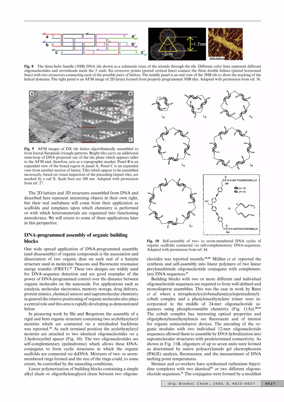
Fig. 8 The three-helix bundle (3HB) DNA tile shown as a schematic trace of the strands through the tile. Different color lines represent differentoligonucleotides and arrowheads mark the 3′ ends. Six crossover points (paired vertical lines) connect the three double helices (paired horizontallines) with two crossovers connecting each of the possible pairs of helices. The middle panel is an end view of the 3HB tile to show the stacking of thehelical domains. The right panel is an AFM image of 2D lattice formed from properly programmed 3HB tiles. Adapted with permission from ref. 36.
Fig. 9 AFM images of DX tile lattice algorithmically assembled toform fractal Sierpinski triangle patterns. Bright tiles carry an additionalstem-loop of DNA projected out of the tile plane which appears tallerto the AFM and, therefore, acts as a topographic marker. Panel B is anexpanded view of the boxed region in panel A. Panel C is an expandedview from another section of lattice. Tiles which appear to be assembledincorrectly, based on visual inspection of the preceding (input) tiles, aremarked by a red X. Scale bars are 100 nm. Adapted with permissionfrom ref. 27.
The 2D lattices and 3D structures assembled from DNA anddescribed here represent interesting objects in their own right,but their real usefulness will come from their application asscaffolds and templates upon which chemistry is performedor with which heteromaterials are organized into functioningnanodevices. We will return to some of these applications laterin this perspective.
DNA-programmed assembly of organic buildingblocksOne wide spread application of DNA-programmed assembly(and disassembly) of organic compounds is the association anddissociation of two organic dyes on each end of a hairpinstructure used in molecular beacons and fluorescent resonanceenergy transfer (FRET).43 These two designs are widely usedfor DNA-sequence detection and are good examples of thepower of DNA-programmed control over the distance betweenorganic molecules on the nanoscale. For applications such ascatalysis, molecular electronics, memory storage, drug delivery,protein mimics, chemical sensors and supramolecular chemistryin general the relative positioning of organic molecules also playsa central role and this area is rapidly developing as demonstratedbelow.
In pionering work by Shi and Bergstrom the assembly of arigid and bent organic structure containing two arylethynylarylmoieties which are connected via a tetrahedral backbonewas reported.44 At each terminal position the arylethynylarylmoieties are attached to two identical oligonucleotides via a2-hydroxyethyl spacer (Fig. 10). The two oligonucleotides areself-complementary (palindromic) which allows these DNA-conjugates to form cyclic structures in which the organicscaffolds are connected via dsDNA. Mixtures of two- to seven-membered rings formed and the size of the rings could, to someextent, be controlled by the annealing conditions.
Linear polymerization of building blocks containing a simplealkyl chain or oligoethyleneglycol chain between two oligonu-
Fig. 10 Self-assembly of two- to seven-membered DNA cycles oforganic scaffolds connected via self-complementary DNA-sequences.Adapted with permission from ref. 44.
cleotides was reported recently.45,46 Mullen et al. reported thesynthesis and self-assembly into linear polymers of two linearperylenediimide–oligonucleotide conjugates with complemen-tary DNA-sequences.47
Building blocks with two or more different and individualoligonucleotide sequences are required to form well-defined andmonodisperse assemblies. This was the case in work by Bunzet al. where a tetraphenylcyclobutadiene(cyclopentadienyl)–cobalt complex and a phenyleneethynylene trimer were in-corporated in the middle of 24-mer oligonucleotide se-quences using phosphoramidite chemistry (Fig. 11A)).48,49
The cobalt complex has interesting optical properties andoligo(phenyleneethynylene)s are fluorescent and of interestfor organic semiconductor devices. The encoding of the or-ganic modules with two individual 12-mer oligonucleotidesequences allowed them to assemble by DNA hybridization intosupramolecular structures with predetermined connectivity. Asshown in Fig. 11B, oligomers of up to seven units were formedas determined by native polyacrylamide gel electrophoresis(PAGE) analysis, fluorescence, and the measurement of DNAmelting point temperatures.
Sleiman and co-workers have synthesized ruthenium bipyri-dine complexes with two identical50 or two different oligonu-cleotide sequences.51 The conjugates were formed by a modified
O r g . B i o m o l . C h e m . , 2 0 0 5 , 3 , 4 0 2 3 – 4 0 3 7 4 0 2 7

Fig. 11 (A) Structures of DNA–organic conjugates. (B). Melting temperature and fluorescence quantum yields for annealed conjugates. Adaptedwith permission from ref. 48.
4 0 2 8 O r g . B i o m o l . C h e m . , 2 0 0 5 , 3 , 4 0 2 3 – 4 0 3 7

phosphoroamidite base strategy. In the latter report both non-complexed and Ru-complexed dioligonucleotide bipyridineswere synthesized. In the presence of two of these modules withcomplementary DNA-sequences they showed a strong tendencyto form cyclic dimers, however, higher order structures andpolymers were also observed. It is notable that the Ru-complexeddioligonucleotide bipyridines showed a higher tendency todimerize than the non-complexed analogs.
In a related approach Han et al. reported on the synthesisof a terpyridine derivative tethered to a DNA sequence.52
When two of the conjugates containing DNA sequences ofdifferent length were mixed in the presence of Fe(II), a sta-ble bis(terpyridine)iron(II) complex formed (Fig. 12). It waspossible to separate heterodimeric modules from homodimericones using PAGE. These dimeric metal complexes are two-way branched oligonucleotides. Three of the oligonucleotidemodified metal-organic modules were capable of self-assemblinginto DNA triangles where the bis(terpyridine)Fe(II) complexesare positioned in the vertexes.
Fig. 12 Assembly terpyridine-DNA conjugates into two-way branchedbis(terpyridine)Fe(II) complexes and DNA-programmed assembly ofthese into DNA triangles. Adapted with permission from ref. 52.
In a series of three papers Majima et al. have reported on theside-by-side assembly of two dsDNA strands by the applicationof multiple cross-linked oligonucleotides.53–55 Two identical 10-mer oligonucleotides were cross-linked by a disulfide tetherattached in the middle of the oligonucleotides. Two 20-mer or30-mer sequences were assembled side-by-side by annealing withtwo or three of the short cross-linked oligonucleotides. In asingle step, well-defined rigid structures controlling the relativeorientation of the two double helix strands were formed.53
Branched DNA structures with three or more arms were in-dependently explored by the two research groups and they bothused an W-branched phosphoramidite with three protected pri-mary hydroxyl groups.56–58 Shchepinov et al. used a W-branchedsynthon to form oligonucleotide dendrimers with two, three, six,nine or twenty-seven arms.57 Hybridization between two of theresulting DNA dendrimers with complementary sequences andalso hybridization between DNA dendrimers with complemen-tary strands on a solid support were investigated by thermalmelting analysis. Von Kiedrowski et al. applied purified andwell-characterized three-armed oligonucleotides to investigatethe self-assembly of two three-armed structures with comple-mentary sequences.58 They suggested, based on PAGE analysis,formation of a dimeric structure interconnected via three paralleldsDNA helices in an “acetylene-like” fashion and a tetramericcyclic structure connected in a “cyclobutadiene-like” fashion,along with higher order structures (Fig. 13). The W-structurewas also applied to the formation of a tetrahedral structure.59
Fig. 13 Structure and assembly of W-branched DNA sequences intodimer and tetramer structures. Adapted with permission from ref 58.
Steward and McLaughlin applied a Ni(II)–cyclam complexas the scaffold for four-armed oligonucleotide conjugates(Fig. 14).60 The branched oligonucleotide conjugates were syn-thesized by an advanced phosphoroamidite-based solid-phasemethod. Initially the first 20-mer oligonucleotide arm was syn-thesized on the solid support in the reverse 3′ to 5′ direction withreverse nucleoside phosphoramidites. One of the four functionalside chains on the Ni-cyclam is attached to the oligonucleotideon the solid support and the synthesis is continued in parallel forthe remaining three hydroxyl groups on the Ni–cyclam, but thistime in the normal 5′ to 3′ direction. This elegant method enablesthe synthesis of four arm branched DNA structures where allfour arms are 5′ terminated. Using a similar synthetic method,the same group has demonstrated the synthesis of a structurewith six oligonucleotide arms by the application of a Ru(II)tris(bipyridyl) complex containing six hydroxyl groups arrangedin an octahedral structures as the central building block.61 The
Fig. 14 (A) Structures of the Ni–cyclam DNA conjugates and (B) assembly of four way branched structures. (B) is adapted with permission fromref. 60.
O r g . B i o m o l . C h e m . , 2 0 0 5 , 3 , 4 0 2 3 – 4 0 3 7 4 0 2 9

four armed Ni-cyclam structures Nc1, Nc2 and Nc3 resultingfrom the former study contain four 20-mer oligonucleotidesequences and are tetrahedral by design (Fig. 14A).60 Thehybridization between Nc1 and four Nc2 (which contains onearm complementary to the four arms of Nc1) was explored.Only at high (100 mM) Mg2+ concentration was it possible toform the pentamer as illustrated in Fig. 14B and it was assumedthat charge repulsion between the 12 unpaired arms is reducedby increased counter ion concentration. Attempts to form a3D DNA crystal were performed by annealing Nc1 with Nc3(all strands complementary to those of Nc1). Higher molecularweight bands were observed by PAGE, however, the structure ofthese assemblies has yet to be determined.
It has been demonstrated above that DNA is excellent forencoding the assembly of organic molecules into well-definedstructures. One of the major draw-backs is the lack of controlover spatial positioning of the organic groups. This is a particularproblem for linear structures and large rings. For some purposes,in which only the average distance between the organic moietiesis of importance, as for example for molecular beacons andFRET, this lack of positional control is not crucial, however,for applications where exact distances between the moleculesare required, this is a serious limitation. Another aspect is thatthe organic compounds, in almost all cases described above,are separated by some length of dsDNA helix, hamperingdirect interaction between the compounds thus limiting theapplicability of such assemblies. To avoid these problems, a newstrategy is to apply DNA-programming both for assemblingthe organic structures and for covalently coupling the organicbuilding blocks into new macromolecular structures.10 Progressin this area is described in the next section.
DNA-programmed covalent coupling of organicbuilding blocksRecent progress in DNA-programmed synthesis has revealedthat a variety of organic reactions can be directed by attachedoligonucleotide sequences.62 The concept of DNA-programmedsynthesis is illustrated in Fig. 15A. The functional groupsFG1 and FG2 can in principle react with each other withouthybridization of the DNA sequences, but at low concentrations(nM to lM) their intermolecular reaction is so slow that practi-cally no conversion occurs. If the DNA sequences attached to thefunctional groups are complementary, they will hybridize even
Fig. 15 Principle of DNA-programmed covalent coupling reactions.(A) The functional groups FG1 and FG2 are brought into close proxim-ity by hybridization of their attached complementary DNA sequencesleading to reaction and formation of a product. (B) Arrangement of thereactants in a DNA hairpin structure. (C) Arrangement of the reactantson a DNA template. Adapted with permission from ref. 10.
at very low concentrations and thereby bring the two functionalgroups into close proximity. Now the local concentrations of thefunctional groups are significantly increased and the groups canreact in a “pseudo-intramolecular” reaction, which will proceedsignificantly faster than the intermolecular reaction. Two otherDNA designs for DNA-directed reactions are based on usingeither DNA hairpins or a DNA template (Fig. 15B and C).
The new field of DNA-programmed chemistry has mainlybeen developed by Liu et al. and they have explored a variety ofapplications of this concept such as e.g. combinatorial chemistryand the discovery of new organic reactions.62–64 The number ofexample applications of DNA-programmed chemistry for nano-related sciences is limited.10 Liu applied a 40-mer DNA-templateto align with ten PNA tetramer building blocks for parallelchemical ligation by reductive amination.65 In a recent reportby Chen and Mao it was demonstrated that the pH inducedswitching between the triplex and duplex DNA structures ofa 5′-carboxylate labelled 40-mer DNA template, annealed withtwo 5′-amino labelled 12-mers, allowed for the selective reactionof the carboxylate with either one of the amino groups.66
Assembly and covalent coupling of three different oligonu-cleotides to a central organic core was reported by vonKiedrowski and co-workers.67 They used the W-branched three-armed DNA structure mentioned previously (Fig. 13) as thetemplate to align three DNA-conjugated chemical functionali-ties at the center. The three individual oligonucleotides, havinga 5′-hydrazide functionality, were aligned to react with a 1,3,5-triformyl benzene resulting in a trishydrazone attached to threedifferent oligonucleotide sequences. In this method the chemicalconnectivity information contained in the W-branched DNAsequences is copied by template-directed linking.
In an early report in this field the DNA-programmed syn-thesis of a organometallic complex was demonstrated. Twosalicylaldehyde conjugated oligonucleotides were aligned on aDNA template in the presence of ethylenediamine and Mn(II) orNi(II) resulting in the formation of metallosalen–DNA complex(Fig. 16).68
In the molecular engineering strategy by Gothelf et al., two orthree salicylaldehyde groups are contained within the same com-pound enabling the assembly and covalent coupling of multiplemodules10,69–72 The linear oligonucleotide-functionalized module(LOM) and tripoidal oligonucleotide-functionalized module(TOM) shown in Fig. 17A were synthesized.69 The backbone ofthe modules is based on oligo(phenylene ethynylene)s to obtaina rigid, conjugated and potentially conducting structure. Themodules contain salicylaldehyde moieties at each terminus. Themodules also contain amide spacers at each terminus, whichare connected to 15-mer oligonucleotides via phosphoramiditechemistry. Oligonucleotides attached at each terminus, areencoded to link up others containing complementary sequences.The salicylaldehyde groups of two modules are brought inclose proximity when their complementary DNA sequencesare annealed together. DNA-programmed coupling of themodules proceeds via manganese–salen formation between twosalicylaldehydes groups in the presence of ethylenediamineand Mn(II).70 The metal–salen forming coupling reaction wasdeliberately chosen, since the linkages between the individualhead groups of the modules will be essentially linear due tothe stereochemistry of the manganese–salen complex formed.The metal–salen link constitutes a potential conducting junctionwith the possibility of varying the central coordinated metal.
Depending on the encoding of LOMs and TOMs withdifferent DNA sequences, assembly and covalent coupling ofthe modules into a variety of predetermined nanostructures canbe formed as depicted for selected structures in Fig. 17B. Theproducts were characterized by denaturing PAGE and dimer andtrimer products were characterized by MALDI-TOF MS. It wasalso found that the melting points of the LOM–LOM, LOM–TOM or TOM–TOM combinations are increased by 15–30 ◦Cafter the coupling of the modules.70
4 0 3 0 O r g . B i o m o l . C h e m . , 2 0 0 5 , 3 , 4 0 2 3 – 4 0 3 7

Fig. 16 DNA-templated formation of a metallosalen complex. Adapted with permission from ref. 68.
Fig. 17 Modular DNA-programmed assembly of conjugated nanostructures. (A) Structure of LOM and TOM modules. (B) Representative couplingsof LOMs and TOMs by formation of multiple manganese–salen complexes between the modules (green colour: ethylenediamine and Mn(OAc)2. (B)is adapted with permission from ref. 10.
The same group later showed that by introducing a disulfidespacer between the organic module and the DNA sequences,it was possible to cleave the DNA sequences off the macro-molecular nanostructures.71,72 It has also been shown that theDNA-programmed coupling could be performed by reductiveamination with ethylenediamine and NaCNBH3 resulting intetrahydrosalen linked structures with significantly increasedstability.72 The principle of using DNA to assemble a macro-molecular organic structure from which the DNA sequences aresubsequently removed is a new aspect of DNA-nanotechnology.Furthermore, recycling of the liberated thiol modified DNAsequences may be possible.
DNA-programmed assembly of biomoleculesAssembly of other biomolecules on DNA templates and arraysmay prove useful for fabrication of biomimetics and otherdevices with applications such as biochips, immunoassays,biosensors, and a variety of nanopatterned materials. The logical
end to the shrinking of microarrays is the self-assembled DNAnanoarray with a library of ligands distributed at addressablelocations to bring analyte detection down to the single moleculelevel. We will return to complex DNA tiling structures momen-tarily, but first we will look at simpler dsDNA systems.
The conjugation of DNA and streptavidin via a covalent linkerwas reported by Niemeyer et al. in 1994, and these conjugateswere applied to DNA-programmed assembly on a macroscopicDNA array on a surface and in a nanoscale array made byaligning DNA-tagged proteins to specific positions along aoligonucleotide template (Fig. 18).7,73,74 The covalent attach-ment of an oligonucleotide to streptavidin provides a specificrecognition domain for a complementary nucleic acid sequence.In addition, the binding capacity for four biotin molecules isutilized as biomolecular adapters for positioning biotinylatedcomponents along a nucleic acid backbone (vide infra).
Biotin labelled oligonucleotides are commercially availableand are routinely used in molecular biology, and their applica-tion for nanostructuring is growing in popularity.7,75 Niemeyer
O r g . B i o m o l . C h e m . , 2 0 0 5 , 3 , 4 0 2 3 – 4 0 3 7 4 0 3 1

Fig. 18 Conjugation of 5′-thiol oligonucleotide and streptavidin, andalignment of the conjugates on a 170-mer RNA template. Adapted withpermission from ref. 7.
applied 169-mer dsDNA sequences labelled with biotin in the5′-positions for the formation of DNA–streptavidin networks(Fig. 19A).76–78 Despite the tetravalent binding capacity ofstreptavidin it serves primarily as a bi- or trivalent linker betweenthe oligonucleotide strands as observed in the AFM picturesof the aggregates shown in Fig. 19B. By thermal denatura-tion and rapid cooling these aggregates are transformed intoDNA–streptavidin nanocircles as imaged by AFM (Fig. 19C).Nanocircles with DNA sequences of sizes varying from 87 to485-mer sequences were formed and, depending on the lengthand concentration, up to 77% nanocircles were formed relativeto other structures for the larger DNA sequences, whereas28% nanocircles were formed from the 87-mer sequences.
Fig. 19 (A) Self-assembly of DNA–streptavidin conjugates. (B) AFMimages of the oligomeric DNA–STV aggregates and (C) of the DNA–streptavidin nanocircles. (D) Ionic-switching of a DNA3–(streptavidin)3
by increased supercoiling of the interconnecting DNA linkers. Adaptedwith permission from ref. 7.
The nanocircles were, furthermore, functionalized with haptengroups such as fluorescein and applied in an immuno-PCRassay.79 During the studies of the aggregates, in this case aDNA3-(streptavidin)3 structure, supercoiling of the dsDNA-sequences induced by increased Mg2+ concentration led to asignificant structural change, decreasing the distance betweenthe streptavidins as illustrated in Fig. 19D.
Besides duplex DNA structures, more complex self-assembling DNA tiling structures have been used to organizebiomolecules into specific spatial patterns. DNA nanostructurescovalently labelled with ligands have been shown to bind proteinmolecules in programmed patterns, for example, making useof the popular biotin/avidin pair, arrays of evenly spacedstreptavidin molecules were assembled on DNA tile lattice.31
On 4 × 4 cross tile lattice, individual streptavidin molecules arevisible as separate peaks in the AFM image (Fig. 20). Singlemolecule detection could be achieved on DNA nanoarraysdisplaying a variety of protein binding ligands.
Fig. 20 (A) Schematic drawing of 4 × 4 cross tile lattice carrying abiotinylated central strand and streptavidin molecules (blue) binding tothe functionalized sites. (B) AFM image showing individual streptavidinproteins at the vertices of the cross tile array. Adapted with permissionfrom ref. 31.
Further design evolution of the 4 × 4 cross tile system to a twotile type (A and B) tile set allowed for somewhat more complexstructures and patterns.80 In this study, some size control oflattice and partial addressability were demonstrated, but thedisplay patterns were still periodic and symmetric (Fig. 21).In ongoing experiments, finite-sized objects with independentaddressing have been used to assemble a range of specificallypatterned protein arrays in high yield.81
Another exciting future use for biomolecules specificallypatterned on self-assembled DNA nanostructures is the specificdeposition of inorganic materials via crystal nucleation. Naturalpeptides and proteins have been implicated in the growth ofnano-patterned silica by living organisms.82 Peptides and RNAsequences have been artificially evolved by in vitro selectionto specifically bind and precipitate or crystallize various semi-conductors and metals.83,84 Patterning these species on 3D DNAlattices could provide a method for bottom-up assembly andcontrolled deposition resulting in a wide variety of complexinorganic structures for use in nanoelectronics, photonics, andother fields.
DNA-programmed assembly of materialsIn analogy to the immobilization of DNA on a variety ofsolid surfaces, the conjugation of DNA and analogs withmetal nanoparticles, semiconductor nanoparticles and polymer
4 0 3 2 O r g . B i o m o l . C h e m . , 2 0 0 5 , 3 , 4 0 2 3 – 4 0 3 7

Fig. 21 Atomic force microscopy images of the programmed self-assembly of streptavidin on 1D DNA nanotracks. Panels a and b are AFM imagesof bare A*B and A*B* nanotrack before streptavidin binding, respectively, where tiles marked with ‘*’ indicate the presence of biotinylated strands.Panels c and d are AFM images of A*B and A*B* nanotrack after binding of streptavidin. All AFM images are 500 nm × 500 nm. Adapted withpermission from ref. 80.
particles is becoming increasingly important. Such DNA-conjugated materials have in several studies found applicationfor biosensors and the reader is addressed to recent excellentreviews for a detailed overview.5,11,85 Here the main focus willbe on the application of DNA to assemble materials into welldefined nanostructures.3 Two important pioneering reports onthe assembly of gold nanoparticles by hybridization of DNA-nanoparticle conjugates were published back-to-back in Naturein 1996.86,87 Mirkin et al. prepared two samples of 13 nmgold nanoparticles functionalized with 3′-thiol-linked DNAsequences and 5′-thiol-linked DNA sequences, respectively.86
If a solution containing the two DNA-nanoparticles is mixedwith a DNA target complementary to both DNA-nanoparticlesequences, hybridization will force the particles to aggregate(see Fig. 3). This change in interparticle distance causes achange in the plasmon absorbance due to plasmon coupling.The resulting color change in the presence of the target is avery efficient and easy method for DNA-detection and hasbeen developed in a series of subsequent reports.18,88,89 In oneextension they reported on the DNA-programmed placement of8 nm gold particles around larger 31 nm gold particles (Fig. 22).90
Depending on the ratio between nanoparticles 1 and 2, the“satellite structures” were observed within extended assembliesor in isolated structures as shown in Fig. 22B.
Whereas the examples mentioned above utilize nanoparticlesfunctionalized with several oligonucleotide strands per nanopar-ticle, the approach described by Alivisatos, Schultz and co-workers applies gold nanoparticles labelled with only singleoligonucleotide molecules.87 Small 1.4 nm gold nanoparticlescontaining one maleimide group per cluster were reacted with 5′-or 3′-thiol modified 18-mer oligonucleotides. By annealing twoor three of these DNA–nanoparticle conjugates with a DNAtemplate, homodimeric or homotrimeric nanoparticle assem-blies were formed as verified by TEM. In more recent studiesheterodimeric and heterotrimeric gold nanoparticle assemblieswere also obtained by using nanoparticles of different sizes.18 InFig. 23 the DNA-programmed arrangement of 5 nm and 10 nmgold nanoparticles into non-periodic assemblies is shown. It is anexcellent example of the power of DNA to encode and assemblematerials, but it also shows the lack of structurally rigidity ofdsDNA, since precise positional arrangement of nanoparticles
is not obtained. In a very recent study, homotetrameric DNA–nanoparticle assemblies containing extendable hairpin loopswere reported.91 Related studies using branched DNA-sequenceswere performed by others.92
Niemeyer et al. have published several papers on DNAconjugated with biomolecules (vide supra) and they have ex-tended this work to the DNA-programmed assembly of goldnanoparticles.74 Short 5′-thiol modified DNA strands wereattached covalently to streptavidin, and gold nanoparticles(1.4 nm) with a single amino substituent were coupled to abiotin moiety. These two components were mixed and four of thebiotinylated-gold nanoparticles were linked to the streptavidin–DNA conjugate due to the strong and specific affinity ofbiotin for the four binding sites of streptavidin. Up to sixof these DNA–streptavidin–gold nanoparticle structures withindividual DNA sequences were annealed with a complementary170-mer RNA sequence, resulting in alignment of the sixnanoparticle–streptavidin complexes in a line as verified byTEM. In another study they reported the functionalization ofgold nanoparticles with up to seven different 3′-thiol and 5′-thiol modified oligonucleotide sequences for the detection ofdifferent target sequences using the same oligofunctional DNAgold nanoparticles.93
In a recent publication a method for reversible switchingof DNA–gold nanoparticle aggregation was developed.94 Goldnanoparticles (23 nm) bound to two different 12-mer oligonu-cleotides (a and b) were applied. In the presence of a templatecontaining sequences a′ and b′, the nanoparticles will aggregateupon hybridization as in the concept developed by Mirkinet al.22 Niemeyer et al. extended this to a system capable ofundergoing DNA-programmed reversible switching between ag-gregation and dispersion by applying two complementary “fuel”oligonucleotides, Fa and Fd (Fig. 24). The base sequence of Fa
is comprised of the template sequences a′ and b′ plus a short 4-mer sequence c′. The hybridization between the Fa template andthe nanoparticle DNA-sequences is disrupted by the additionof the fully complementary fuel strand Fd (Fig. 24, stage III)which strips Fa out of the complex, leading to the formationof a waste duplex and redispersion of the nanoparticles. Theswitching between the aggregated and dispersed nanoparticleswas easily detected by UV–visible spectroscopy due to the
O r g . B i o m o l . C h e m . , 2 0 0 5 , 3 , 4 0 2 3 – 4 0 3 7 4 0 3 3
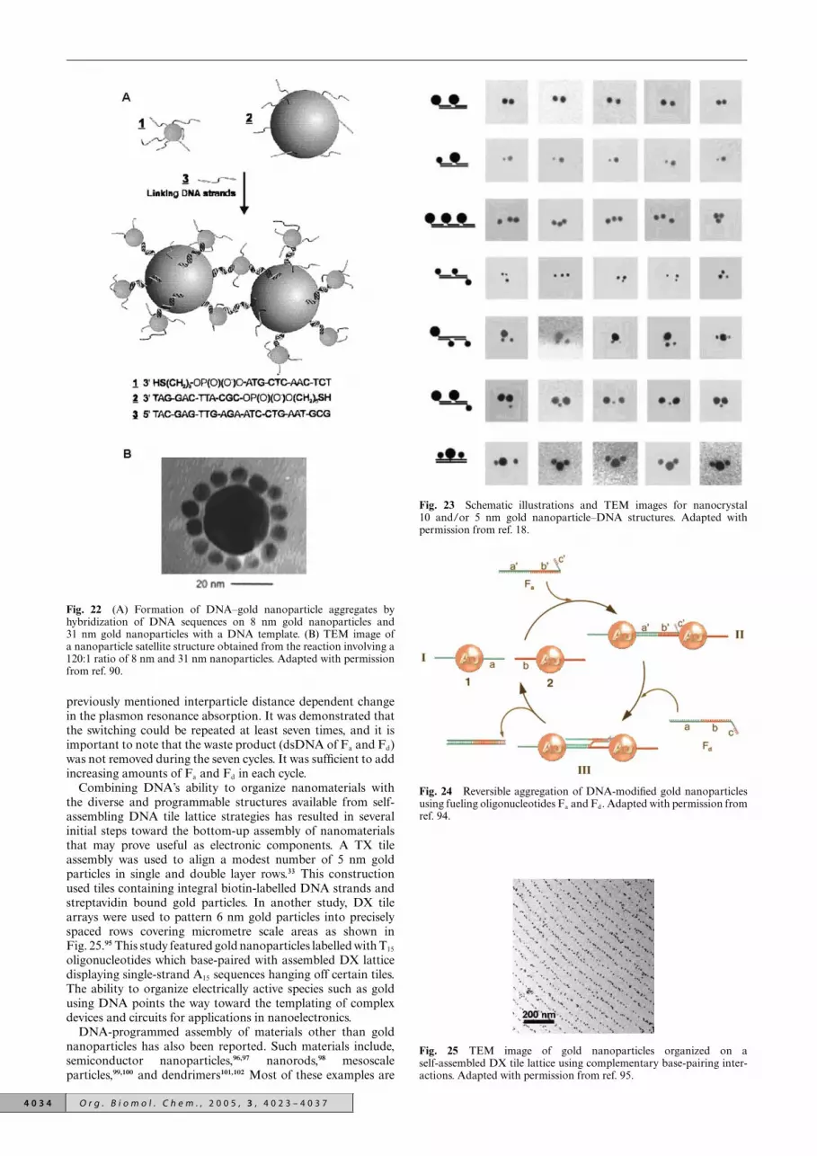
Fig. 22 (A) Formation of DNA–gold nanoparticle aggregates byhybridization of DNA sequences on 8 nm gold nanoparticles and31 nm gold nanoparticles with a DNA template. (B) TEM image ofa nanoparticle satellite structure obtained from the reaction involving a120:1 ratio of 8 nm and 31 nm nanoparticles. Adapted with permissionfrom ref. 90.
previously mentioned interparticle distance dependent changein the plasmon resonance absorption. It was demonstrated thatthe switching could be repeated at least seven times, and it isimportant to note that the waste product (dsDNA of Fa and Fd)was not removed during the seven cycles. It was sufficient to addincreasing amounts of Fa and Fd in each cycle.
Combining DNA’s ability to organize nanomaterials withthe diverse and programmable structures available from self-assembling DNA tile lattice strategies has resulted in severalinitial steps toward the bottom-up assembly of nanomaterialsthat may prove useful as electronic components. A TX tileassembly was used to align a modest number of 5 nm goldparticles in single and double layer rows.33 This constructionused tiles containing integral biotin-labelled DNA strands andstreptavidin bound gold particles. In another study, DX tilearrays were used to pattern 6 nm gold particles into preciselyspaced rows covering micrometre scale areas as shown inFig. 25.95 This study featured gold nanoparticles labelled with T15
oligonucleotides which base-paired with assembled DX latticedisplaying single-strand A15 sequences hanging off certain tiles.The ability to organize electrically active species such as goldusing DNA points the way toward the templating of complexdevices and circuits for applications in nanoelectronics.
DNA-programmed assembly of materials other than goldnanoparticles has also been reported. Such materials include,semiconductor nanoparticles,96,97 nanorods,98 mesoscaleparticles,99,100 and dendrimers101,102 Most of these examples are
Fig. 23 Schematic illustrations and TEM images for nanocrystal10 and/or 5 nm gold nanoparticle–DNA structures. Adapted withpermission from ref. 18.
Fig. 24 Reversible aggregation of DNA-modified gold nanoparticlesusing fueling oligonucleotides Fa and Fd. Adapted with permission fromref. 94.
Fig. 25 TEM image of gold nanoparticles organized on aself-assembled DX tile lattice using complementary base-pairing inter-actions. Adapted with permission from ref. 95.
4 0 3 4 O r g . B i o m o l . C h e m . , 2 0 0 5 , 3 , 4 0 2 3 – 4 0 3 7

based on linear assembly of two complementary DNA strandsleading to dimers or aggregates. Many examples of nanowirestemplated on DNA molecules by a variety of electrolessdeposition protocols (including fabrication of a field effecttransistor103) have also been reported, but these are beyond thescope of this article.
Carbon nanotubes are one of the most promising materialsfor nanoscience due to their unique structure and mechanic andelectronic properties.104 In recent years chemical conjugation oforganic and bioorganic compounds with carbon nanotubes isa field that has developed rapidly.105 The ability to control theexact positioning of multiple carbon nanotubes by means ofDNA-programmed assembly would be a major achievementin nanoscience. In a few reports, the conjugation of carbonnanotubes with DNA106,107 and with PNA has been described.108
In these reports carbon nanotubes were shortened into frag-ments by oxidation, resulting in carbon nanotube fragmentswith carboxyl groups in the terminal positions and, to someextent, in their walls. Covalent coupling of 5′-amino DNA-sequences or PNA to carboxyl groups on the nanotubes ledto the formation of carbon nanotubes coupled with DNA orPNA sequences. Carbon nanotubes containing 12-mer PNA-sequences were annealed with dsDNA sequences containing 12-mer sticky ends and imaged by AFM.108 In the work by Dai et al.,multi-wall carbon nanotubes (MWNTs) and single-wall carbonnanotubes (SWNT) functionalized with 20-mer DNA sequenceswere annealed with complementary sequences attached to goldnanoparticles.107 The resulting aggregates were deposited onmica and imaged by AFM (Fig. 26). The images revealed theoccasional interconnection of individual MWNTs by a goldnanoparticle. The surface plot in Fig. 26B shows a gap betweenthe gold nanoparticle and the MWNT corresponding to the7 nm length of the 20-mer dsDNA connecting the two materials.
Fig. 26 (A) AFM image of the interconnection of two MWNTs by agold nanoparticle (scanning area: 0.55 lm × 0.55 lm). (B) 3-D surfaceplot of (A). Adapted with permission from ref. 107.
In another study, SWNTs were assembled between preposi-tioned metal electrodes via complementary DNA base-pairingby ssDNA on the gold electrodes (thiol-labelled oligos) andthe oxidized SWNTs (3′-amino-labelled oligos).109 Electricalconductivity between the electrode pairs was shown to behighly dependent on the presence of complementary DNAon the electrodes and nanotubes. These initial investigationsof carbon nanotube–DNA conjugates hold great promise forfuture developments in the assembly of nanotube structures withuseful electronic and mechanical properties.
ConclusionsIn the ultimate development of bottom-up nanofabricationstrategies it will be possible to assemble large numbers of easilyavailable building blocks, and depending on the nature andprogramming of the building blocks they will self-assemble intocomplex nanostructures with enzyme-like properties, electroniccircuits with efficient contacts to larger length scales, memorystorage devices, drug delivery robots, multifunctional diagnosticdevices for in vivo application, or even systems capable of self-reproduction. Only the future will tell how much of this will berealized in practice, but whatever develops, DNA-programmedassembly will undoubtedly play a central role.
We are now learning the basics of how to position materialswith DNA. It has been demonstrated that DNA molecules areused to assemble a small number of components (< 10) suchas organic molecules, biomolecules and nanoparticles into welldefined assemblies or aggregates. Furthermore, complex DNAbuilding blocks have been assembled into highly regular 2DDNA-lattices, which in some cases were used for the periodicincorporation of proteins or nanoparticles. Formation of DNA-wires and 3D constructs has also been described. Most of thereports describe model-studies, but some of the systems havefound application, in particular for DNA-sequence detection.85
The area of DNA-nanotechnology will undoubtedly con-tinue to evolve and improve our present ability to positionmaterials using DNA. Specific topics we find of particularimportance and interest include: (i) the development of newDNA-constructs with enhanced properties for materials as-sembly, (ii) the design of new DNA structures which cancontrol the formation of nanoparticle devices for application inelectronics and photonics, (iii) the assembly of 2D DNA latticeson surfaces with individually addressable connecting pointsfor future development of nanoarrays, (vi) major advances inDNA-programmed assembly of advanced carbon nanotube-based architectures, in particular if the problems regarding thesynthesis of monodisperse carbon nanotube fragments of similarsize, structure and properties are solved, (v) further developmentin DNA-programmed assembly and covalent coupling to formmacromolecular nanostructures with potential application inmolecular electronics, catalysis and new macromolecular ar-chitectures, (iv) design of other systems for DNA-programmedassembly, in which the organic, bioorganic or inorganic buildingblocks are connected by other means than DNA-hybridizationleading to structures that are stable after removal of the DNA-sequences.
Since the elucidation of its structure, DNA has fascinatedmankind, as it reduces the information behind all living organ-isms to a code based on only four chemical compounds. Weare now able to engineer DNA and to conjugate it with othermaterials. This has opened almost unlimited possibilities fordesign of structures and for programmed assembly events at thenano-scale.
AcknowledgementsThis work has been supported by grants from the NationalScience Foundation (USA) (EIA-0218376, CCR-0326157) toT. H. LaBean and grants from the Danish Technical ResearchCouncil, the Danish National Research Foundation and theCarlsberg Foundation to K. V. Gothelf.
References1 G. M. Whitesides and B. Gryzbowski, Science, 2002, 295, 2418–
2421.2 Self-Assembling Peptide Systems in Biology, Medicine and Engi-
neering, ed. A. Aggeli, N. Boden and S. ZhangKluwer AcademicPublishing, Dordrecht, Netherlands, 2000.
3 N. C. Seeman, Angew. Chem., Int. Ed., 1998, 37, 3220–3238.4 J. J. Storhoff and C. A. Mirkin, Chem. Rev., 1999, 99, 1849–1869.5 C. M. Niemeyer, Angew. Chem., Int. Ed., 2001, 40, 4128–4158.6 N. C. Seeman, Synlett, 2000, 1536–1548.7 C. M. Niemeyer, Trends Biotechnol., 2002, 9, 395–401.8 N. C. Seeman, Nature, 2003, 421, 427–431.9 J. Wengel, Org. Biomol. Chem., 2004, 2, 227–280.
10 K. V. Gothelf and R. S. Brown, Chem. Eur. J., 2005, 11, 1062–1069.11 E. Katz and I. Willner, Angew. Chem., Int. Ed., 2004, 43, 6042–6108.12 Nanobiotechnology: Concepts, Applications and Perspectives, ed.
C. M. Niemeyer and C. A. Mirkin, Wiley-VCH, Weinheim 2004.13 B. Samori and G. Zucheri, Angew. Chem., Int. Ed., 2005, 44, 1166–
1181.14 N. C. Seeman, Trends Biochem. Sci., 2005, 3, 119–125.15 J. Xu, T. H. LaBean, and S. L. Craig, in Supramolecular Polymers
2, ed. Alberto Ciferri, CRC Press - Taylor & Francis Group, BocaRaton, FL, 2005, pp. 445-480.
O r g . B i o m o l . C h e m . , 2 0 0 5 , 3 , 4 0 2 3 – 4 0 3 7 4 0 3 5

16 P. Pancoska, Z. Moravek and U. M. Moll, Nucleic Acids Res., 2004,32, 4630–4645.
17 S. L. Beaucage and R. P Iyer, Tetrahedron, 1993, 49, 1925–1963.18 C. J. Loweth, W. B. Caldwell, X. Peng, A. P. Alivisatos and P. G.
Schultz, Angew. Chem., Int. Ed., 1999, 38, 1808–1812.19 P. E. Nielsen, Acc. Chem. Res., 1999, 32, 624–630.20 B. Vester and J. Wengel, Biochemistry, 2004, 43, 13233–13241.21 O.-S. Ng and D. E. Bergstrom, Nano Lett., 2005, 5, 107–111.22 R. Elghanian, J. J. Storhoff, R. C. Mucic, R. L. Letsinger and C. A.
Mirkin, Science, 1997, 277, 1078–1081.23 N. C. Seeman, J. Theor. Biol., 1982, 99, 237–247.24 N. C. Seeman, Chem. Intell., 1995, 1, 38–47.25 X. Li, X. Yang, J. Qi and N. C. Seeman, J. Am. Chem. Soc., 1996,
118, 6131–6140.26 E. Winfree, F. Liu, L. A. Wenzler and N. C. Seeman, Nature, 1998,
394, 539–544.27 P. W. K. Rothemund, N. Papadakis and E. Winfree, PLoS Biol.,
2004, 2, 2041–2053.28 T. H. LaBean, H. Yan, J. Kopatsch, F. Liu, E. Winfree, J. H. Reif
and N. C. Seeman, J. Am. Chem. Soc., 2000, 122, 1848–1860.29 C. Mao, T. H. LaBean, J. H. Reif and N. C. Seeman, Nature, 2000,
407, 493–496.30 H. Li, S.-H. Park, J. H. Reif, T. H. LaBean and H. Yan, J. Am.
Chem. Soc., 2004, 126, 418–419.31 H. Yan, S. H. Park, G. Finkelstein, J. H. Reif and T. H. LaBean,
Science, 2003, 301, 1882–1884.32 C. Mao, W. Sun and N. C. Seeman, J. Am. Chem. Soc., 1999, 121,
5437–5443.33 D. Liu, M. Wang, Z. Deng, R. Walulu and C. Mao, J. Am. Chem.
Soc., 2004, 126, 2324–2325.34 B. Ding, R. Sha and N. C. Seeman, J. Am. Chem. Soc., 2004, 126,
10230–10231.35 Y. Brun, M. Gopalkrishnan, D. Reishus, B. Shaw, N. Chelyapov and
L. Adleman, in Proceedings of the 1st Conference on the Foundationsof Nanoscience, 2004, pp. 2–15.
36 S.-H. Park, R. Barish, H. Li, J. H. Reif, G. Finkelstein, H. Yan andT. H. LaBean, Nano Lett., 2005, 5, 693–696.
37 F. Mathieu, S. Liao, J. Kopatsch, T. Wang, C. Mao and N. C.Seeman, Nano Lett., 2005, 5, 661–665.
38 A. Chworos, I. Severcan, A. Y. Koyfman, P. Weinkam, E. Oroudjev,H. G. Hansma and L. Jaeger, Science, 306, 2068–2072.
39 J. Chen and N. C. Seeman, Nature, 1991, 350, 631–633.40 Y. Zhang and N. C. Seeman, N. C., J. Am. Chem. Soc., 1994, 116,
1661–1669.41 R. P. Goodman, R. M. Berry and A. J. Turberfield, Chem. Commun.,
2004, 1372–1373.42 W. M. Shih, J. D. Quispe and G. F. Joyce, Nature, 2004, 427, 618–
621.43 See e.g.:C. M. Niemeyer and M. Adler, Angew. Chem., Int. Ed.,
2002, 41, 2779–3783.44 J. Shi and D. E. Bergstrom, Angew. Chem., Int. Ed., 1997, 36, 111–
113.45 J. Xu, E. A. Fogleman and S. L. Craig, Macromolecules, 2004, 37,
1863–1870.46 E. A. Fogleman, W. C. Yount, J. Xu and S. L. Craig, Angew. Chem.,
Int. Ed., 2002, 41, 4026–4028.47 M. A. Abdalla, J. Bayer, J. O. Radler and K. Mullen, Angew. Chem.,
Int. Ed., 2004, 43, 3967–3970.48 S. M. Waybright, C. P. Singleton, K. Wachter, C. J. Murphy and
U. H. Bunz, J. Am. Chem. Soc., 2001, 123, 1828–1833.49 S. M. Waybright, C. P. Singleton, J. M. Tour, C. J. Murphy and
U. H. F. Bunz, Organometallics, 2000, 19, 368–370.50 I. Vargas-Baca, D. Mitra, H. J. Zulyniak, J. Banerjee and H.
Sleiman, Angew. Chem., Int. Ed., 2001, 40, 4629–4631.51 D. Mitra, N. Di Cesare and H. F. Sleiman, Angew. Chem., Int. Ed.,
2004, 43, 5804–5808.52 J. S. Choi, C. W. Kang, K. Jung, J. W. Yang, Y. G. Kim and H. Y.
Han, J. Am. Chem. Soc., 2004, 126, 8606–8607.53 M. Endo and T. Majima, J. Am. Chem. Soc., 2003, 125, 13654–
13655.54 M. Endo, S. Uegaki and T. Majima, Chem. Commun., 2005, 3153–
3155.55 M. Endo and T. Majima, Chem. Commun., 2004, 1308–1309.56 M. S. Shchepinov, I. A. Udalova, A. J. Bridgman and E. M.
Southern, Nucleic Acids Res., 1997, 25, 4447–4454.57 M. S. Shchepinov, K. U. Mir, J. K. Elder, M. D. Frank-Kamenetskii
and E. M. Southern, Nucleic Acids Res., 1999, 27, 3035–3041.58 M. Scheffler, A. Dorenbeck, S. Jordan, M. Wustefeld and G. von
Kiedrowski, Angew. Chem., Int. Ed., 1999, 38, 3311–3315.59 G. von Kiedrowski, L. H. Eckardt, K. Naumann, W. N. Pankau,
M. Reimold and M. Rein, Pure Appl. Chem., 2003, 75, 609–619.
60 K. M. Steward and L. W. McLaughlin, J. Am. Chem. Soc., 2004,126, 2050–2057.
61 K. M. Steward, J. Rojo and L. W. Mclaughlin, Angew. Chem., Int.Ed., 2004, 43, 5808–5811.
62 X. Li and D. R. Liu, Angew Chem., Int. Ed., 2004, 43, 4848–4870.63 Z. J. Gartner, B. N. Tse, R. Grubina, J. B. Doyon, T. M. Snyder and
D. R. Liu, Science, 2004, 305, 1601–1605.64 M. W. Kanan, M. M. Rozenman, K. Sakurai, T. M. Snyder and
D. R. Liu, Nature, 2004, 431, 545–549.65 D. M. Rosenbaum and D. R. Liu, J. Am. Chem. Soc., 2003, 125,
13924–13925.66 Y. Chen and C. Mao, J. Am. Chem. Soc., 2004, 126, 13240–13241.67 L. H. Eckardt, K. Naumann, W. N. Pankau, M. Rein, M.
Schweitzer, N. Windhab and G. von Kiedrowski, Nature, 2002, 420,286.
68 J. L. Czlapinski and T. L. Sheppard, J. Am. Chem. Soc., 2001, 123,8618–8619.
69 M. Nielsen, A. H. Thomsen, E. Clo, F. Kirpekar and K. V. Gothelf,J. Org. Chem., 2004, 69, 2240–2250.
70 K. V. Gothelf, A. H. Thomsen, M. Nielsen, E. Clo and R. S. Brown,J. Am. Chem. Soc., 2004, 126, 1044–1046.
71 R. S. Brown, M. Nielsen and K. V. Gothelf, Chem. Commun., 2004,1464–1465.
72 M. Nielsen, V. Dauksaite, J. Kjems and K. V. Gothelf, BioconjugateChem., 2005, 16, 681–685.
73 C. M. Niemeyer, T. Sano, C. L. Smith and C. R. Cantor, NucleicAcids Res., 1994, 22, 5530–5539.
74 C. M. Niemeyer, W. Burger and J. Peplies, Angew. Chem., Int. Ed.,1998, 37, 2265.
75 S.-J. Park, A. A. Lazarides, C. A. Mirkin and R. L. Letsinger, Angew.Chem., Int. Ed., 2001, 40, 2909–2919.
76 C. M. Niemeyer, M. Adler, S. Lenhert, S. Gao, H. Fuchs and L. F.Chi, ChemBioChem, 2001, 2, 260–265.
77 C. M. Niemeyer, M. Adler, B. Pignataro, S. Lenhert, S. Gao, L. F.Chi, H. Fuchs and D. Blohm, Nucleic Acids Res., 1999, 27, 4553–4561.
78 C. M. Niemeyer, M. Adler, S. Gao and L. Chi, Angew. Chem., Int.Ed., 2000, 39, 3056–3059.
79 C. M. Niemeyer, R. Wacker and M. Adler, Angew. Chem., Int. Ed.,2001, 40, 3169–3172.
80 S.-H. Park, P. Yin, Y. Liu, J. H. Reif, T. H. LaBean and H. Yan,Nano Lett., 2005, 5, 729–733.
81 S.-H. Park, C. Pistol, S.-J. Ahn, J. H. Reif, A. Lebeck, C. Dwyer andT. H. LaBean, Angew. Chem., Int. Ed., 2005, submitted.
82 J. N. Cha, K. Shimizu, Y. Zhou, S. C. Christiansen, B. F. Chmelka,G. D. Stucky and D. E. Morse, Proc. Natl. Acad. Sci., 1999, 96,361–365.
83 S. R. Whaley, D. S. English, E. L. Hu, P. F. Barbara and A. M.Belcher, Nature, 2000, 405, 665–668.
84 L. A. Gugliotti, D. L. Feldheim and B. E. Eaton, Science, 2004, 304,850–852.
85 N. L. Rosi and C. A. Mirkin, Chem. Rev., 2005, 105, 1547–1562.86 C. A. Mirkin, R. L. Letsinger, R. C. Mucic and J. J. Storhoff, Nature,
1996, 382, 607–609.87 A. P. Alivisatos, K. P. Johnsson, X. Peng, T. E. Wilson, T. C. J.
Loweth, M. P. Bruchez and P. G. Schultz, Nature, 1996, 382, 609–611.
88 J. J. Storhoff, R. Elghanian, R. C. Mucic, C. A. Mirkin and R. L.Letsinger, J. Am. Chem. Soc., 1998, 120, 1959–1964.
89 R. A. Reynolds III, C. A. Mirkin and R. L. Letsinger, J. Am. Chem.Soc., 2000, 122, 3795–3796.
90 R. C. Mucic, J. J. Storhoff, D. A. Mirkin and R. L. Letsinger, J. Am.Chem. Soc., 1998, 120, 12674–12675.
91 S. A. Claridge, S. L. Goh, J. M. J. Frechet, S. C. Williams, C. M.Micheel and A. P. Alivisatos, Chem. Mater., 2005, 17, 1628–1635.
92 M. G. Grimau, D. Iacopino, A. Avino, B. G. de la Torre, A. Ongaro,D. Fitzmaurice, J. Wessels and R. Eritja, Helv. Chim. Acta, 2003,86, 2814–2826.
93 C. M. Niemeyer, B. Ceyhan and P. Hazarika, Angew. Chem., Int.Ed., 2003, 42, 5766.
94 P. Hazarika, B. Ceyhan and C. M. Niemeyer, Angew. Chem., Int.Ed., 2004, 43, 6469–6471.
95 J. D. Le, Y. Pinto, N. C. Seeman, K. Musier-Forsyth, T. A. Tatonand R. A. Kiehl, Nano Lett., 2004, 4, 2343–2347.
96 G. P. Mitchell, C. A. Mirkin and R. L. Letsinger, J. Am. Chem. Soc.,1999, 121, 8122–8123.
97 W. J. Parak, D. Gerion, D. Zanchet, A. S. Woerz, T. Pellegrino,C. Micheel, S. C. Williams, M. Seitz, R. E. Bruehl, Z. Bryant, C.Bustamante, C. R. Bertozzi and A. P. Alivistos, Chem. Mater., 2002,12, 2113–2119.
4 0 3 6 O r g . B i o m o l . C h e m . , 2 0 0 5 , 3 , 4 0 2 3 – 4 0 3 7

98 E. Dujardin, L.-B. Hsin, C. R. C. Wang and S. Mann, Chem.Commun., 2001, 1264–1265.
99 C. M. Soto, A. Srinivasan and B. R. Ratna, J. Am. Chem. Soc.,2002, 124, 8508–8509.
100 V. T. Milam, A. L. Hiddessen, J. C. Crocker, D. J. Graves and D. A.Hammer, Langmuir, 2003, 19, 10317–10323.
101 Y. Choi, A. Mecke, B. G. Orr, M. M. B. Holl and J. R. Baker Jr.,Nano Lett., 2004, 4, 391–397.
102 C. R. DeMattei, B. Huang and D. A. Tomalia, Nano Lett., 2004, 4,771–777.
103 K. Keren, M. Krueger, R. Gilad, G. Ben-Yoseph, U. Sivan and E.Braun, Science, 2002, 297, 72–75.
104 H. Dai, Acc. Chem. Res., 2002, 35, 1035–1044.105 S. Banerjee, T. Hemraj-Benny and S. S. Wong, Adv. Mater., 2005,
17, 17–29.106 C. Dwyer, M. Guthold, M. Falvo, S. Washburn, R. Superfine and
D. Erie, Nanotechnology, 2002, 13, 601–604.107 S. Li, P. He, J. Dong, Z. Guo and L. Dai, J. Am. Chem. Soc., 2005,
127, 14–15.108 K. A. Williams, P. T. M. Veenhuizen, B. G. de la Torre, R. Eritja
and C. Dekker, Nature, 2002, 420, 761.109 M. Hazani, F. Hennrich, M. Kappes, R. Naaman, D. Peled,
V. Sidorov and D. Shvarts, Chem. Phys. Lett., 2004, 391, 389–392.
O r g . B i o m o l . C h e m . , 2 0 0 5 , 3 , 4 0 2 3 – 4 0 3 7 4 0 3 7