Evaluation of sericin/collagen membranes as prospective wound dressing biomaterial
10
Evaluation of sericin/collagen membranes as prospective wound dressing biomaterial Omer Akturk, 1 Aysen Tezcaner, 1,2 Hasan Bilgili, 3 M. Salih Deveci, 4 M. Rusen Gecit, 1 and Dilek Keskin 1,2, ⁎ Department of Engineering Sciences, Middle East Technical University, 06531 Ankara, Turkey, 1 Graduate Department of Biotechnology, Middle East Technical University, 06531 Ankara, Turkey, 2 Faculty of Veterinary Medicine, Department of Surgery, Ankara University, 06110 Ankara, Turkey, 3 and Faculty of Medicine, Department of Pathology, Gulhane Military Medical Academy, Ankara, Turkey 4 Received 21 December 2010; accepted 27 May 2011 Available online 21 June 2011 Sericin, a silk protein, has high potential for use in biomedical applications. In this study, wound dressing membranes of Sericin (S) and Collagen (C) were prepared by glutaraldehyde cross-linking at S/C; 2:1, 1:1, 1:2, and 0:1 weight ratios. They were stable in water for 4 weeks. However, increasing the proportion of sericin had decreasing effect on the membrane stability. Water swelling property of membranes was enhanced with sericin. The highest water swelling was obtained in 1:1 group (9.06 g/g), but increasing collagen or sericin content in the membranes had a diminishing effect. Highest water vapor transmission rate was obtained with 1:2 group (1013.80 g/m 2 /day). Oxygen permeability results showed that 1:2 (7.67 mg/L) and 2:1 (7.85 mg/L) S/C groups were better than the other groups. While sericin decreased the tensile strength and elongation of membranes, it increased modulus. Sericin also increased brittleness of membranes, but their UTS range (24.93–44.92 MPa) was still suitable for a wound dressing. Membranes were not penetrable to microorganisms. Cytotoxicity studies showed that fibroblasts and keratinocytes attached and gained their characteristic morphologies. They also proliferated on membranes significantly. After 1 week of subcutaneous implantation, a fibrous capsule formed around all membranes with an acute inflammation. Sericin containing membranes showed signs of degradation (at 2nd week), while collagen only membranes remained largely intact. Eventually, sericin containing membranes degraded in 3 weeks with moderate inflammatory response. Overall results suggest that sericin/collagen membranes would be favorable as wound dressing material when sericin ratio is less than or equal to the collagen component. © 2011, The Society for Biotechnology, Japan. All rights reserved. [Key words: Sericin; Collagen; Wound dressing; Membrane; Biocompatibility] Severe acute and chronic wounds such as burns, mechanical trauma, pressure and leg ulcers, congenital skin diseases and cancer excision, pose a great challenge to the surgeons. Especially burns and skin ulcers are problematic issues (1,2). Wound dressing is a biomaterial of synthetic or biological origin that is designed to recover the healthy physiological and structural properties of the wounded skin and is life saving in serious complications (3,4). Wound dressing materials have evolved significantly in the past quarter century (5). An ideal wound healing material should be biocompatible, protective from secondary infections and should prevent water loss while controlling water vapor and oxygen permeabilities. In addition to these, wound dressing should have mechanical properties compatible with the skin and improve the healing process by actively attracting the cells to the wound area (6–8). As one of the most commonly used wound dressing material, collagen is processed into various forms including films, mats, fibers, and gels for designing implants and engineering tissues (9,10). Collagen, besides stimulating the cellular elements of healing such as granulocytes and macrophages, also provides a template for cellular attachment, migration and proliferation (3). Cells such as fibroblasts and keratino- cytes specifically recognize collagen substrates (11). In order to improve the properties of wound dressings, other natural materials have drawn the attention of researchers. Among these are silk proteins, sericin and fibroin. Sericin, constituting 25–30% of silk, envelops the fibroin fibers and provides important attributes such as excellent oxygen permeability (Murase, M., Japanese Patent, 06-166850A, 1994), antioxidant action, moisture regulating ability, UV resistance, antibacter- ial, anticancer and anticoagulant properties (12–14). Different areas of sericin use in cosmetics and biomedical applications have been reviewed (13,15). Studies on sericin in wound healing and its potential as a wound dressing gave promising results. Tsubouchi (Tsubouchi, K., Japanese Patent 11-070160A, 1999) developed a fibroin-mixed-sericin wound dressing that could accelerate healing and be peeled off without disturbing the newly formed skin. Tsubouchi et al. (16) found that the attachment of primary cultured human fibroblasts was enhanced with sericin. However, sericin has been implicated as the cause for immune responses to silk when it was used together with fibroin (17). Sericin by itself did not elicit an immune response (18). The recent study conducted by Aramwit and Sangcakul (19) in rats also showed that sericin cream had wound healing effect without causing any allergic reactions. Journal of Bioscience and Bioengineering VOL. 112 No. 3, 279 – 288, 2011 www.elsevier.com/locate/jbiosc ⁎ Corresponding author at: Department of Engineering Sciences, Middle East Technical University, 06531 Ankara, Turkey. Tel.: + 90 312 2102389; fax: + 90 312 2104462. E-mail address: [email protected] (D. Keskin). 1389-1723/$ - see front matter © 2011, The Society for Biotechnology, Japan. All rights reserved. doi:10.1016/j.jbiosc.2011.05.014
Transcript of Evaluation of sericin/collagen membranes as prospective wound dressing biomaterial

Journal of Bioscience and BioengineeringVOL. 112 No. 3, 279–288, 2011
www.elsevier.com/locate/jbiosc
Evaluation of sericin/collagen membranes as prospectivewound dressing biomaterial
Omer Akturk,1 Aysen Tezcaner,1,2 Hasan Bilgili,3 M. Salih Deveci,4 M. Rusen Gecit,1 and Dilek Keskin1,2,⁎
⁎ CorrespondTechnical Univ2104462.
E-mail add
1389-1723/$doi:10.1016/j
Department of Engineering Sciences, Middle East Technical University, 06531 Ankara, Turkey,1 Graduate Department of Biotechnology,Middle East Technical University, 06531 Ankara, Turkey,2 Faculty of Veterinary Medicine, Department of Surgery, Ankara University, 06110 Ankara,
Turkey,3 and Faculty of Medicine, Department of Pathology, Gulhane Military Medical Academy, Ankara, Turkey4
Received 21 December 2010; accepted 27 May 2011Available online 21 June 2011
Sericin, a silk protein, has high potential for use in biomedical applications. In this study, wound dressing membranes ofSericin (S) and Collagen (C) were prepared by glutaraldehyde cross-linking at S/C; 2:1, 1:1, 1:2, and 0:1 weight ratios. They werestable in water for 4 weeks. However, increasing the proportion of sericin had decreasing effect on the membrane stability.Water swelling property of membranes was enhanced with sericin. The highest water swelling was obtained in 1:1 group(9.06 g/g), but increasing collagen or sericin content in the membranes had a diminishing effect. Highest water vaportransmission rate was obtained with 1:2 group (1013.80 g/m2/day). Oxygen permeability results showed that 1:2 (7.67 mg/L)and 2:1 (7.85 mg/L) S/C groups were better than the other groups. While sericin decreased the tensile strength and elongationof membranes, it increased modulus. Sericin also increased brittleness of membranes, but their UTS range (24.93–44.92 MPa)was still suitable for a wound dressing. Membranes were not penetrable to microorganisms. Cytotoxicity studies showed thatfibroblasts and keratinocytes attached and gained their characteristic morphologies. They also proliferated on membranessignificantly. After 1 week of subcutaneous implantation, a fibrous capsule formed around all membranes with an acuteinflammation. Sericin containing membranes showed signs of degradation (at 2nd week), while collagen only membranesremained largely intact. Eventually, sericin containing membranes degraded in 3 weeks with moderate inflammatoryresponse. Overall results suggest that sericin/collagen membranes would be favorable as wound dressing material whensericin ratio is less than or equal to the collagen component.
© 2011, The Society for Biotechnology, Japan. All rights reserved.
[Key words: Sericin; Collagen; Wound dressing; Membrane; Biocompatibility]
Severe acute and chronicwounds such as burns,mechanical trauma,pressure and leg ulcers, congenital skin diseases and cancer excision,pose a great challenge to the surgeons. Especially burns and skin ulcersare problematic issues (1,2). Wound dressing is a biomaterial ofsynthetic or biological origin that is designed to recover the healthyphysiological and structural properties of the wounded skin and is lifesaving in serious complications (3,4). Wound dressing materials haveevolved significantly in the past quarter century (5). An ideal woundhealing material should be biocompatible, protective from secondaryinfections and should prevent water loss while controlling water vaporand oxygen permeabilities. In addition to these, wound dressing shouldhave mechanical properties compatible with the skin and improve thehealing process by actively attracting the cells to the wound area (6–8).
As one of the most commonly used wound dressing material,collagen is processed intovarious forms including films,mats, fibers, andgels for designing implants and engineering tissues (9,10). Collagen,besides stimulating the cellular elementsofhealing suchasgranulocytes
ing author at: Department of Engineering Sciences, Middle Eastersity, 06531 Ankara, Turkey. Tel.: +90 312 2102389; fax: +90 312
ress: [email protected] (D. Keskin).
- see front matter © 2011, The Society for Biotechnology, Japan. All.jbiosc.2011.05.014
and macrophages, also provides a template for cellular attachment,migration and proliferation (3). Cells such as fibroblasts and keratino-cytes specifically recognize collagen substrates (11).
In order to improve the properties of wound dressings, other naturalmaterials have drawn the attention of researchers. Among these are silkproteins, sericin and fibroin. Sericin, constituting25–30%of silk, envelopsthe fibroin fibers and provides important attributes such as excellentoxygen permeability (Murase, M., Japanese Patent, 06-166850A, 1994),antioxidant action,moisture regulating ability, UV resistance, antibacter-ial, anticancer and anticoagulant properties (12–14). Different areas ofsericin use in cosmetics and biomedical applications have been reviewed(13,15). Studies on sericin inwound healing and its potential as awounddressing gave promising results. Tsubouchi (Tsubouchi, K., JapanesePatent 11-070160A, 1999) developed a fibroin-mixed-sericin wounddressing that could accelerate healing and be peeled off withoutdisturbing the newly formed skin. Tsubouchi et al. (16) found that theattachment of primary cultured human fibroblasts was enhanced withsericin. However, sericin has been implicated as the cause for immuneresponses to silk when it was used together with fibroin (17). Sericin byitself did not elicit an immune response (18). The recent study conductedby Aramwit and Sangcakul (19) in rats also showed that sericin creamhad wound healing effect without causing any allergic reactions.
rights reserved.

280 AKTURK ET AL. J. BIOSCI. BIOENG.,
With this study we report the first membrane using sericin andcollagenproteins aswounddressing. It is hypothesized that the inherentproperties of theseproteins arising fromsericin's specific roles in cocoonand collagen being nativematrix element of skin bring extra advantagesin their simultaneous use for wound healing. Membranes preparedat different ratios of two proteins were characterized for oxygenpermeability, water holding capacity, degradation, mechanical proper-ties and biocompatibility.
MATERIALS AND METHODS
Materials Sericin powder of Bombyx mori origin was purchased from NembriIndustrie Tessili s.r.l. (CAS no: 60650-88-6; 60650-89-7, MW: ca.138 kDa) and collagen(Gelfix® Lyophilized Type I Collagen pad) was obtained from Isse InternationalEurosearch (Italy). Glutaraldehyde (50% GTA) was obtained from Sigma-Aldrich(Germany). All other reagents and solvents used were of reagent grade and obtainedfrom Merck (Germany), unless otherwise stated in the text. All cell culture solutionswere purchased from PAA Laboratories (Austria), unless otherwise noted.
Preparation of the membranes Sericin and collagen were separately dissolvedin 0.5 M acetic acid (0.4% each) by stirring and homogenizing several times. Sericin andcollagen solutions were then mixed with different weight ratios (Table 1) and casted(120 mL) onto glass petri plates (12.5 cm in diameter) to be dried at room temperature.Themembraneswere then cross-linkedwith3% (w/v) glutaraldehyde (GTA) solution (20)for 2 h and rinsed with de-ionized water for 30 min. Finally, they were kept in glycinesolution (0.2 M) (Electrophoresis purity reagent, Bio-Rad Laboratories, Hercules, CA, USA)(20) in distilled-water for 30 min to block the unreacted GTA residues.
Membrane degradation studies After measuring the initial dry weight ofsericin-collagen membranes (Wi) they were immersed in distilled-water andincubated in oven at 37°C for 4 weeks. The final dry weights of membranes (Wf)were thenmeasured to calculate their weight loss. The weight loss was calculated usingthe following equation:
Weight Loss =Wi−Wf
Wið1Þ
Equilibrium degree of swelling Water uptake of membranes wasmeasured bya general gravimetric method (21). Membranes from each group were cut into2×2 cm2 pieces and their dry weights (Wd) were measured. Each sample was placed ina plastic bottle and immersed in de-ionized water at 37°C. All bottles were cappedtightly to prevent any evaporative water loss. At different time intervals water insidethe bottles was removed. After removal of excess surface water by gently blotting witha filter paper, the weights of the swollen membranes were measured (Ws). Finally,equilibrium degree of swelling (EDS) was calculated using the following equation:
EDS =Ws−Wd
Wdð2Þ
All tests were repeated five times for each membrane type and all measurementswere done weekly until reaching equilibrium for water uptake.
Water vapor transmission rate (WVTR) Water vapor transmission rates(WVTR) were determined according to ASTM E96-90 Procedure D. Briefly, anevaporimeter was constructed in a closed chamber to prevent variations owing toambient conditions. The system consisted of a plastic container with an air tight cover, adigital hygrometer and a reservoir of a saturated magnesium chloride solution. It wasplaced in an incubator to maintain isothermal ambience at 35°C and relative humidityof 40±2% after equilibration. A cylindrical, plastic permeability cup filled with 20 g ofde-ionized water and sealed with the test membrane at the top was placed inside thesystem. Evaporation of water through the test membrane was monitored by measuringweight change as an indication of daily loss of water. The water vapor transmission rate(WVTR) was found by dividing the daily loss of water with the evaporation area of thepermeability cup. The water vapor evaporation studies were carried out at least inquadruplets (n≥4).
Oxygen penetration Studies for oxygen penetration through membranes wereperformed by gluing each membrane to the top of an Erlenmeyer flask filled with300 mL de-ionized water (a modified method of Wittaya-areekul and Prahsarn (22)).The negative control was the flask closed with an airtight cap, while the positive controlwas open flask allowing oxygen to enter into the flask and dissolve in water. Test flasks
TABLE 1. Sericin/collagen membrane groups.
Group names(sericin/collagen weight ratios, w/w)
Weight of sericin(mg)
Weight of collagen(mg)
0:1 – 1201:1 120 1202:1 240 1201:2 120 240
were placed in an open environment with constant agitation by a magnetic stirrer for24 h. The collected water samples were then analyzed for dissolved oxygen (DO)content according toWinkler's method. The test procedure followed was adopted fromStandard Methods for the Examination of Water and Wastewater (American PublicHealth Association (APHA), 1995).
Microbial penetration The ability of membranes to prevent microbialpenetration was tested by attaching the membranes to the top of glass test tubescontaining 20 mL of standard nutrient broth (adapted from Wittaya-areekul andPrahsarn (22)). Membranes were sterilized by incubating overnight in 70% ethylalcohol supplemented with penicillin/streptomycin (0.1%) followed by exposing to UVlight for 30 min. Before tests, nutrient broth and glass test tubes were sterilized withautoclave for 20 min at 121°C. The negative control was a sterile nutrient broth in glasstest tube closed with cotton ball and aluminum foil while the positive control was asterile nutrient broth in test tube open to air. The cloudiness of the nutrient broth in testtubes after 1 week of incubation at ambient environment was considered as microbialcontamination. Also, spectrophotometric measurements at 600 nm were carried outwith μQuant™ Microplate Spectrophotometer (Biotek Instruments Inc., Winooski, VT,USA). Samples were tested in triplicates.
Mechanical properties The mechanical properties of membranes wereevaluated by applying stretch test with Lloyd LS500 Material Testing Machine (Lloyd,England) using Nexygen computer software. Rectangular samples (4×30 mm) werecut from membranes. To prevent slippage of the membranes from the grips they werecovered with sandpaper at the attachment site. The gauge length and width were 10and 4 mm, respectively. The stretch tests were carried out at 25°C and 50±3% relativehumidity conditions for dry samples. The crosshead speed of the system was adjustedto 10 mm/min to get a constant strain rate of 100%/min. The results of the tests wereobtained as load versus deflection curves which were then converted into stress–straindata by the computer program. Modulus of elasticity, ultimate tensile strength andpercent elongation at break were calculated from the stress–strain curves of samples(n≥6) for each membrane type.
Cytotoxicity studies The HaCat human keratinocyte (DSMZ, Braunschweig,Germany) and 3T3 fibroblast cell lines were cultured in Dulbecco's modified Eagle'smedium (DMEM high glucose–glutamine) supplemented with fetal bovine serum (FBS,10%, v/v) and penicillin/streptomycin (10 U/ml) at 37°C under humidified atmosphereof 5% CO2–95% air in incubator (5215, Shel Lab, Cornelius, OR, USA). When the cellsreached at least 80–90% confluency, they were trypsinized for passaging. Membranes(10 mm diameter) were placed in 24 well plates (15 mmwell diameter) and sterilizedby incubating overnight in 70% ethyl alcohol supplemented with penicillin/strepto-mycin (0.1%) and by exposing to UV light for 30 min. Initial seeding density onto eachmembrane was 3×10⁴ cells/membrane. Collagen only group and polystyrene wellswere used as the controls.
Cell proliferation on membranes was evaluated with MTT (methylthiazolyldiphenyl-tetrazolium bromide, Sigma-Aldrich) assay. This method is based onreduction of tetrazolium salt by mitochondrial dehydrogenases to a dark blue formazanproduct (23). The extent to which MTT is reduced to a formazan product has beencorrelated with the cell viability after incubations of 1, 4 and 7 days. Accordingly, themedium was removed and 500 μL MTT solutions (5 mg/mL in DMEM low glucose) wasadded to each well and incubated for 3 h at 37°C. After removing the MTT solution andwashing with PBS, membranes were transmitted to other plates containing 500 μLdimethyl sulfoxide (DMSO, cell culture grade, min. 99.5% AppliChem, Germany) tosolubilize the formazan crystals formed inside the cells. The absorbance was measuredat 550 nm wavelength using μQuant™ Microplate Spectrophotometer (Biotek In-struments). The morphology of cells on the membranes was analyzed with lightmicroscope and photographed (Nikon TS100, Nikon, Tokyo, Japan).
SEM analysis The cell seeded membranes were treated with glutaraldehydesolution (GTA, 3%) for 1 h for fixing the cells. All prepared samples were initially coatedwith gold before analysis. The surface properties of membranes and the morphology ofcells seeded onto membranes were studied with Scanning Electron Microscopy (SEM,JSM-6400 Electron Microscope, JEOL Ltd., Tokyo, Japan).
In vivo studies A total of 9maleWistar rats (6 months old)were housed one percage. They were maintained at constant temperature in simulated 12 h diurnal cycles.After acclimation,membrane samples (for the groups; collagen only, sericin/collagen, 1:1,and 1:2) were implanted subcutaneously under general anesthesia using ketamine andxylazine. Each rat had 6 membranes (10 mm in diameter each) 2 from each group placedbilaterally to the spinal column at dorsal part. At the end of 1st, 2nd and 3rd weeks, 3 ratswere sacrificed and implant regions were removed for histological analysis.
Histological analysis Subcutaneous implant regions were excised and fixed informaldehyde solution (10% v/v). The samples were embedded in paraffin. The blockswere cut into sections of 4–6 μm thickness with a rotary microtome (Shandon, Finesse325). The sections were stained with Hematoxylin and Eosin for microscopicexaminations. Capsule formation around the implants, degree of inflammation, presenceandamountofmultinuclear giant cellswere investigated for all groups. For theevaluationsof tissue reactions a semi-quantitative scoringwas appliedaccording to thedefined criteriaby De Jong et al. (24).
Statistical analysis In comparing the membrane groups for a single parameteror for comparing the results of same group at different incubation times (in vitro tests)One-way Analysis of Variance (ANOVA) test was used with Tukey's Multiple ComparisonTest for the post-hoc pair-wise comparisons using SPSS 18 Software Program. Differenceswere considered significant for pb0.05.

TABLE
2.Prop
erties
ofsericin/co
llage
nmem
bran
es.
Sericin/co
llage
n(w
eigh
tratio)
Ave
rage
weigh
tloss
ofmem
bran
es(%
)Eq
uilib
rium
degree
ofsw
ellin
gresu
lts
(g/g)
Water
vapo
rtran
smission
rate
g/m
2 /da
yOxy
genpe
rmea
bilityformem
bran
es(d
issolved
oxyg
enam
ounts,
mg/L)
Microbial
pene
tration
(optical
dens
ityof
nutrient
broths
cove
redwithmem
bran
es)
0:1
14.89±
2.82
5.37
±0.45
980.90
±28
.66
7.25
±0.06
a0.04
65±
0.00
09a
1:2
10.57±
1.63
6.22
±0.50
1013
.80±
44.48
7.67
±0.10
a,b,c
0.04
58±
0.00
21a
1:1
17.66±
2.74
9.06
±1.23
c97
6.87
±29
.29
7.64
±0.04
a,b
0.04
58±
0.00
03a
2:1
22.70±
1.93
6.85
±1.43
982.33
±48
.66
7.85
±0.33
a,b,c
0.04
57±
0.00
03a
Ope
nco
ntrol
8.31
±0.27
0.07
88±
0.02
03a
Closed
control
6.95
±0.06
0.04
65±
0.00
25a
Dataareex
pressedas
mea
n±
SD(n
≥4)(For
microbial
pene
trationn=
3).
aSign
ificant
differen
cefrom
theop
enco
ntrolat
pb0.05
.b
Sign
ificant
differen
cefrom
theclosed
controlat
pb0.05
.cSign
ificant
differen
cefrom
thegrou
pco
ntaining
only
colla
genat
pb0.05
.
SERICIN/COLLAGEN MEMBRANES 281VOL. 112, 2011
RESULTS AND DISCUSSION
Membrane degradation studies Preliminary stability studiesconducted on uncross-linked sericin/collagen membranes implied thatthey were prone to lose their structural integrity in water to a greatextent (average weight loss was as high as 78% for 2:1 sericin/collagengroup) before 2 weeks (data not shown). Also, as the sericin content inthe uncross-linked groups increased (3:1, 4:1 S/C) average weight lossincreased as much as 85%. These preliminary studies, hence, constituteda mean to understand the stability of membranes of different pro-portions of sericin and collagen (either cross-linked or uncross-linked)and were used to determine the membrane groups to be characterizedfor the in vitro and in vivo studies. According to these membranestability tests, cross-linked membranes remained mostly stable up to7 weeks (average weight loss was about 32% for 2:1 S/C group). Studieswith selected groups showed that as the sericin ratio increased, percentweight loss also increased (Table 2). GTA cross-linked sericin/gelatinfilms fabricated by Mandal et al. (25) had blending ratios ranging from0.5:5 to 7.5:5 (w/w) and, the degradation studies carried out on thesefilms showed that with an increase in sericin concentration, higherdegradation was observed. Weight loss in membranes was thought tooccur with hydrolytic degradation. Hence, the more hydrophilic theproteins are, the more hydrolytic degradation is expected to take place.Sericin has hydrophilic properties due to the presence of severalhydroxyl groups (26). On the contrary, collagen is more hydrophobicdue to highly non-polar side groups of its amino acids (27). However,the effect of sericin in increasing the hydrophilicity was observed atequal or higher proportion of sericin content. Teramoto et al. (28)produced sericin gel films by gelation during which water-stable net-work of inter-molecular β-sheets was formed. S/C membranes wereproduced without any gelation step which possibly caused them to bemore susceptible to hydrolytic degradation. Sericin might also be lesscross-linked in the membrane structure, thereby making the mem-branes less stable in water.
A skin scaffold having a biodegradation time for about 25 dayscould be suggested for healing acute wounds. While a more rapidbiodegradation rate would make it inappropriate for healing, a tooslow degradation would prevent the wound healing process (29). Inthis study, membranes had high resistance to hydrolytic degrada-tion during 4 weeks of incubation and maintained their structuralintegrity to a great extent. However, the degradation rate is expectedto increase under in vivo conditions.
Equilibrium degree of swelling An important parameter forthe evaluation of a wound dressing material is the ability to absorbwater. As seen in Table 2, 1:1 groups had the highest EDS values(9.0581 g/g membrane). All sericin containing membranes had higherEDS values than pure collagen ones (0:1). This result was compatiblewith the previously stated higher hydrophilicity of sericin compared tocollagen. Besides that, sericin has an important property; it consists ofabout 30% serine amino acid, which is related to themoisture absorbingand desorbing capabilities. Thus, with the presence of this amino acid,sericin becomes an excellent moisturizing agent (12,13). Therefore,sericin is suitable as a wound healing material. Yoshii et al. produced ahydrogel blend of sericin, fibroin and PVA. This hydrogel was shown tohave excellent moisture absorbing and desorbing properties (Yoshii, F.,Kume, T., Makuuchi, K., Sato, F., Japanese Patent, 2000-169736A, 2000).Similarly, polyurethane foams containing sericin had excellent resultsfor the same properties (Nomura, M., Iwasa, Y., Araya, H., JapanesePatent 07-292240A, 1995). Therefore, the experimental results thatsericin containing groups had higher EDS values than the groupscomprising only collagenwere compatible with the inherent propertiesof sericin. This finding was significant in 1:1 ratio group (Table 2).Accordingly, adding sericin into collagen membranes increased EDSclearly up to 1:1 ratio. However, further increasing the ratio of sericin(2:1, S/C) decreased EDS result. Such decrease could be the result of

FIG. 1. Comparison of A) tensile strength, B) tensile moduli, and C) elongation at breakvalues of collagen only (0:1) and sericin/collagen (1:2, 1:1, 2:1) membranes. Data areexpressed as mean±SD (n≥6). Number: Significant difference from the group con-taining collagen (0:1) only. Asterisk: Significant difference from 1:2. pb0.05.
282 AKTURK ET AL. J. BIOSCI. BIOENG.,
highest weight loss observed in the 2:1 ratio group. There are studies inliterature to improve the wound dressing properties with the com-binational use of polymers (20,30). To stabilize the collagen mem-branes, glutaraldehyde cross-linking is commonly used. However, GTAcross-linking was shown to decrease the water uptake (20,30). GTAcross-linked collagen and chitosan films fabricated by casting method(31) had lower EDS ratios (0.3–0.7, g/g) than the cross-linked sericin/collagenmembranes of the current study (6.22–9.06, g/g) (Table 2). EDSof collagen/chitosan nanofibrouswound dressingmembranes preparedby Chen et al. (20)was 9 g/g but decreased to 2.7 g/g after cross-linkingwith GTA. Chitosan-polysaccharide wound dressing films producedbyWittaya-areekul and Prahsarn (22) had EDS range of 4–7 (g/g)whichwas very similar to sericin/collagen membranes produced in this study.Hence, the EDS values of S/C membranes prepared were comparablewith those in literature and were thought to be highly suitable forwound dressing applications.
Water vapor transmission rate (WVTR) All membrane groupshad a range of evaporative water loss rates between 980.90 and1084.03 g/m2/day (Table 2). There were no statistically significant dif-ferences between WVTR values of membrane groups. However, theeffect of sericin was observed with the highest WVTR in 1:2 S/C group(1013.80 g/m2/day). TheWVTR for normal skin is 204 g/m2/day, while itmay range from 279 g/m2/day for a first-degree burn to 5138 g/m2/dayfor a granulating wound (32). Thus, it might be suggested that thesemembranes are suitable for low to medium exudating wounds. WVTRof sericin/collagen wound dressing membranes were also comparablewith those developed by other researchers (i.e. asymmetric chitosanmembranes: 2109.2 to 2792.8 g/m2/day; PVA–clay nanocompositehydrogels: 864–1344 g/m2/day) (8,32).
Oxygen penetration The results of oxygen permeability prop-erties of membranes demonstrated that 2:1 and 1:2 S/C membraneshad significantly higher permeability to oxygen than collagen onlymembranes (0:1) (Table 2). The closed control group had significantlylower dissolved oxygen (DO) values than all groups except forcollagen only membrane. Although none of the membrane groupsreached high DO values as the open control, sericin inclusion inmembranes resulted in a statistical increase in DO value. Therefore, itmight be suggested that addition of sericin to the membranes couldenhance the oxygen permeability. In parallel to our results, Murase(Murase, M., Japanse Patent, 06-166850A, 1994) discovered that filmsmade of sericin and fibroin had excellent oxygen permeability whichwas similar to human cornea in its functional properties.
Microbial penetration Experimental results showed that noneof the membranes allowed the penetration of microorganisms duringa week period (Table 2). The optical density (OD) measurements werestatistically indifferent than the closed control and lower than opencontrol (directly exposed to air). The results were also easily observablewith the blurriness of media only in open control group. The anti-bacterial property of sericin was shown by Zhang et al. (13). Thischaracteristic of sericin will bring extra advantage in healing processagainst secondary infections during in vivo applications. According to aprevious work, 64 layers of gauze were not sufficient to prevent theentry of exogenous bacteria into thewound (3). Hence, sericin/collagenmembranes are considered to have high capacity in wound healing–protection purposes owing to their dense structure and antimicrobialproperties.
Mechanical properties Therewas a decreasing trend in ultimatetensile strength (UTS) of membranes with increasing sericin content(Fig. 1A). Collagen only membranes (0:1) had significantly higher UTS(44.92±3.72 MPa) than all sericin containing ones (1:1, 2:1, S/C)except for 1:2. The UTS of cross-linked collagen membranes was inagreement with the values in literature (47–72 MPa) (33). UTS of 1:2groupwas also significantly higher than 2:1 group. However, UTS of 1:1membraneswasneither statistically different than 1:2 nor 2:1. Yet, eventhe lowest UTS value for highest sericin ratio group (24.93 MPa) was
comparably higher than the tensile strength of skinwhichwas reportedas7.6 MPa (34) andwas comparablewith collagenmatrices in literature(i.e., 33.5±1.49 MPa for cross-linked collagen films (35); 10–30 MPafor antibiotic-loaded collagen–hyaluronic acid matrices (36)). Thedecrease in UTS with addition of sericin could be related with themolecular weight (MW) difference of type I collagen (ca. 283 kDa) (27)and sericin (ca. 138 kDa measured in our lab) and distribution of lowMW sericin molecules between the collagen fibers. Another possible

FIG. 2. Proliferation of (A) keratinocytes and (B) fibroblasts on collagen only (0:1) andsericin containing (1:2, 1:1, 2:1) membranes. Data are expressed as mean±SD (n≥4).Number indicates significant difference from the control for the same incubation time.Asterisk indicates significant difference from 0:1 for the same incubation time. Alphaindicates significant difference from both the control and 0:1 for the same incubationtime. pb0.05. Polystyrene wells were used as the controls.
SERICIN/COLLAGEN MEMBRANES 283VOL. 112, 2011
explanation for the decrease inUTS ofmembranes upon sericin additionmight be the loss of secondary structure (beta-sheets) of sericin proteindue to denaturation of sericin (37) during either extraction from the silkor processing stages of membrane. Also, there seems to be a relationbetween the β-sheets formation and gelation (28). Hence, we inferredthat sericin/collagen membranes produced without the gelation stepmight be the reason for decrease in UTS in this study.
Tensile moduli (E) of 1:2, 1:1 and 2:1 were not significantly differentbut had an increasing trend with increasing sericin content (Fig. 1B). Evalues of collagen only membranes were significantly lower than thoseof 1:1 and 2:1 (S/C) but statistically not different than 1:2 membranes.The E of collagen only membrane was 0.499±0.08 GPa (Fig. 1B) whichwas comparable with the range for collagen fibers 0.4–0.8 GPa reportedby Vepari et al. (33). Besides that, E for skin was found to be between6 and 40 MPa (11), which was much lower than the E values of sericin/collagen membranes.
Ultimate percent elongation at break (EB) of collagen only mem-branes was statistically higher (12.64±1.26) than all other membranegroups (Fig. 1C). This value was within the maximum strain range ofcollagen (10–20%) (38). There was no statistical difference betweenthe EB values of 1:1 and 2:1 S/C groups. EB results showed the sametrendwith UTS where the lowest EB (3.59±0.60) was observed for thehighest sericin ratio group.However, sericinwas reported to increase EBof silk fiber due to its glue-like property resulting from hydrogenbonding between serine residues of sericin and those of fibroin in silkfiber and its capability of β-sheet formation (33,38,39). The differentbehavior for EB observed might have resulted from the differentinteractionof sericin andcollagenas compared to silk fibroin and sericin.It is also reported that membranes of sericin are fragile in the dry state(15). Yet, brittle structure of sericinwas alleviated by cross-linking (40).Despite all, the percent elongation of membranes (Fig. 1C) was com-parable with the tensile strain of collagen/chitosan nanofibrous mem-brane (23.26±1.22% and 1.72±0.73% for uncross-linked and cross-linked ones, respectively) produced by Chen et al. (20).
In vitro cell culture studies Initial attachment of keratinocytesto membranes was higher compared to positive controls, as shown bysignificantly higher OD values for 24 h post seeding (Fig. 2A). The cellattachment on collagen membranes was significantly higher than allother membranes and control. Adhesion proteins such as collagen,laminin, fibronectin, and vitronectin are present in ECM and containan arginine–glycine–aspartic (RGD) acid sequence recognized by cellsurface receptors, integrins (41). The integrins mediate cell–cell andcell–ECM adhesion (42). It was reported that high collagen contentwould be expected to promote cell attachment due to integrin bindingwith cell adhesion domains in collagen (43). Therefore, presence ofcollagen in membranes was thought to promote the attachment ofkeratinocytes via integrin binding sites of collagen. The OD increaseobserved on all membranes between days 1 and 7 was significantindicating the proliferation of keratinocytes. There was no statisticaldifference among membranes but the MTT readings on membraneswere significantly lower than those for the control at days 4 and 7. Thepresence of sericin did not have any significant change on prolifer-ation of keratinocytes compared to collagen only membranes.
Similar to the MTT results of keratinocytes, the initial attachmentof fibroblasts on collagen only membranes was high (Fig. 2B). The ODincrease observed on 1:1 and 2:1 S/C membranes between days 1–4and 1–7 might indicate the proliferation of fibroblasts. The degree ofproliferation on collagen membranes was significantly higher thanall other groups for all incubation times. It was also observed that thecell proliferation on 1:2, 1:1, and 2:1 was not statistically differentfrom each other for any of the incubation times. The assessment of thecell viability and proliferation of fibroblasts made by Mandal et al.(25) on sericin/gelatin films suggested that the films containing highamounts of sericin exhibited lower cellular activity after 4 and 6 daysof culturing and lower rates of proliferation during 1 week.
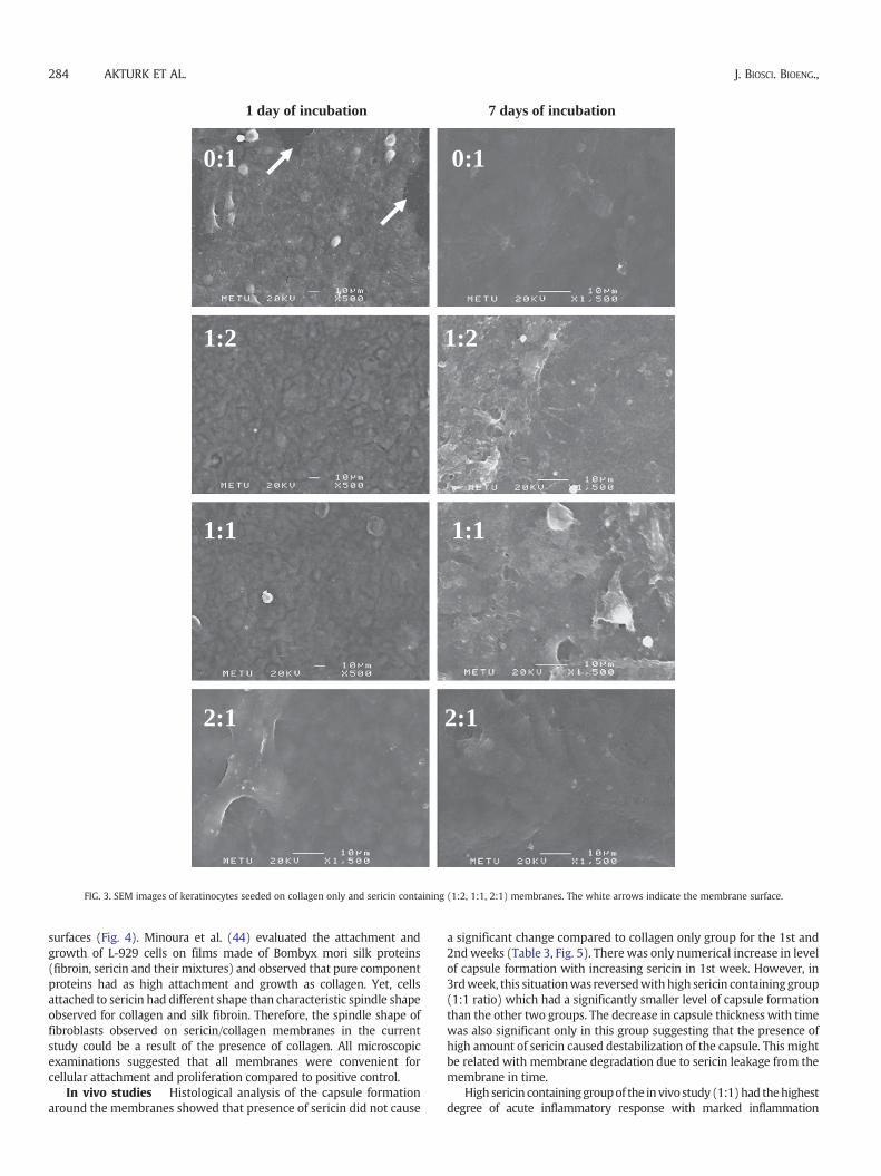
MTT results were in agreementwith light microscopic observations.Keratinocytes spreaded onmembranes after 1 day (figures not shown).For eachmembrane group, confluencywas reached in themiddle of themembrane 1 day after seeding and themembrane area covered by cellsincreased considerably with 7-day incubation. Fibroblasts also attachedand spread on membranes after 1 day (figures not shown). Generally,fibroblasts lost their elongated morphology after 7 days in regionswhere they reached high confluency. Cells seeded on membranes ofhigh sericin ratio groups (2:1, 1:1 S/C)were difficult to distinguish fromthe membrane after 7 days. Yet, the cells on collagen only membraneswere easily detectable. The polygonal morphology of keratinocyteswas also observed by SEM (Fig. 3). At the end of 7 days, the high degreeof conformation of keratinocytes with the surface contours of themembranes apparently indicated effective cell–substrate binding. Aclear distinction was made between the surface of membrane (0:1 S/C)and keratinocytes pointing out the cell-membrane boundaries asindicated by an arrow in figure (Fig. 3). SEM observations also revealedthat fibroblasts gained an elongated morphology on all membranes1 day after seeding and cells maintained this well elongated morphol-ogy for 7 days (Fig. 4). The filopodia of fibroblasts were also observed24-hour post seeding, indicating that they attached strongly to the
1 day of incubation 7 days of incubation
0:1 0:1
1:2 1:2
1:1 1:1
2:1 2:1
FIG. 3. SEM images of keratinocytes seeded on collagen only and sericin containing (1:2, 1:1, 2:1) membranes. The white arrows indicate the membrane surface.
284 AKTURK ET AL. J. BIOSCI. BIOENG.,
surfaces (Fig. 4). Minoura et al. (44) evaluated the attachment andgrowth of L-929 cells on films made of Bombyx mori silk proteins(fibroin, sericin and their mixtures) and observed that pure componentproteins had as high attachment and growth as collagen. Yet, cellsattached to sericin had different shape than characteristic spindle shapeobserved for collagen and silk fibroin. Therefore, the spindle shape offibroblasts observed on sericin/collagen membranes in the currentstudy could be a result of the presence of collagen. All microscopicexaminations suggested that all membranes were convenient forcellular attachment and proliferation compared to positive control.
In vivo studies Histological analysis of the capsule formationaround the membranes showed that presence of sericin did not cause
a significant change compared to collagen only group for the 1st and2ndweeks (Table 3, Fig. 5). There was only numerical increase in levelof capsule formation with increasing sericin in 1st week. However, in3rdweek, this situationwas reversedwith high sericin containinggroup(1:1 ratio) which had a significantly smaller level of capsule formationthan the other two groups. The decrease in capsule thickness with timewas also significant only in this group suggesting that the presence ofhigh amount of sericin caused destabilization of the capsule. This mightbe related with membrane degradation due to sericin leakage from themembrane in time.
High sericin containinggroupof the in vivo study (1:1)had thehighestdegree of acute inflammatory response with marked inflammation

1 day of incubation 7 days of incubation
0:1 0:1
1:2 1:2
1:1 1:1
2:1 2:1
FIG. 4. SEM images of fibroblasts seeded on membranes containing collagen only (0:1), and different weight ratios of sericin/collagen (1:2, 1:1, 2:1). The white arrows indicate cellsand the black ones indicate its filopodias.
SERICIN/COLLAGEN MEMBRANES 285VOL. 112, 2011
(Fig. 6C). This value, however, then decreased to a moderate level withcomparable values to the other groups at 2nd and 3rd weeks (Table 3).Membranes at the implantation site for the sericin containing groupscould not be observed at the 3rd week (Fig. 6A). This was thought tobe related with sericin loss from the membranes which might havedisrupted the membrane structure resulting in complete degradation(Fig. 6B). Considerable increase in the number of multinucleated giantcells (MNGC) in sericin containing groups (1 membrane remaining outof 6) in the 3rdweek suggested that the remnants of themembranewereengulfed by these cells. These cells were observed in high number inhistological sections of these groups at 2nd week (Fig. 6D).
There are several studies to determine the immunogenicity ofsericin. Somestudies related to the evaluation of potential inflammatoryresponse of sericin are encouraging its use in vivo (25,45). The results ofmacrophage response to sericin tests lasting for 1 week period in theresearchofMandal et al. (25) suggested that sericinmaybe assumednotto be very inflammatory in nature. The cytokine levels (TNF-α andIL-1β) weremeasured for silk sericin creamand control groups (normalsaline soaked and cream base-treatedwounds) in the study of Aramwitet al. (45) and it was found that the levels of both inflammatorymediators were lower in sericin-treated wounds than the other controlgroups. This result suggested that the minor irritability to the cells

TABLE 3. Histological evaluations of in vivo responses to membranes aftersubcutaneous implantations.
0:1(Collagen only)
1:2Sericin/collagen (w/w)
1:1Sericin/collagen (w/w)
1 weekCapsule formation 1.50±0.55
(n=6)1.80±0.45(n=5)
2.00±0.82(n=4)
Inflammation 2.00±0.63(n=6)
2.20±0.45(n=5)
3.00±1.41(n=4)
Multinucleatedgiant cell
0.33±0.52(n=6)
1.00±0.71(n=5)
0.75±0.50(n=4)
2 weeksCapsule formation 2.00±0.71
(n=5)1.60±0.55(n=5)
1.67±0.58(n=3)
Inflammation 1.60±0.55(n=5)
1.60±0.55(n=5)
1.67±0.58(n=3)
Multinucleatedgiant cell
0.0±0.0(n=5)
0.80±1.10(n=5)
0.67±1.15(n=3)
3 weeksCapsule formation 2.00±0.71
(n=5)2.00a
(n=1)0.67±0.58(n=3)
Inflammation 1.80±0.45(n=5)
2.00a
(n=1)2.00±0.45(n=2)
Multinucleatedgiant cell
1.00±1.00(n=5)
3.00a
(n=1)3.00±0.0(n=2)
a n shows the number of membranes that could be observed to stay in the implantsite at the end of study period out of 6 samples implanted for that time period.
A
C
B
FIG. 5. Capsule formation around the membranes in 1st week for the sericin/collagengroups (A) 0:1, (B) 1:2, and (C) 1:1.
286 AKTURK ET AL. J. BIOSCI. BIOENG.,
caused by sericin if any, should be considered as an acute rather than achronic inflammation. Panilaitis et al. (18) showed in their study thatsericin, either when attached to fibroin fibers provides better adhesiontomacrophages or primes themacrophages for subsequent stimulationdue to its conformational change upon binding to silk fiber. This couldbe analogous to our case for which the sericin/collagen membranesallowed theadhesionofmacrophages on themembrane surface, leadingto the formation of MNGC. It was reported that adherent macrophagesare fused to form MNGC on the biomaterial surfaces as a result ofmacrophage–biomaterial interaction (46). A significant increase innumber of MNGC after 2 weeks was observed in sericin containinggroups. This was thought to be due to the resorption process of thematerial between second and third weeks, in which the release of thesmall degradation products near themembrane caused a sharp increasein number of MNGC. In accordance with our observations, Yoshikawaet al. (47) showed that fine particles induced multinucleated giant cellsand macrophages resulting in mild inflammatory reaction. Therefore,the formationof giant cells couldbecorrelated to the in vivodegradationof the membranes (46,48). However, upon complete resorption, therewere noMNGCsor capsule at the site of implantation (Fig. 6A). Based onour in vivo observations, the tested membranes could be suggested asbiocompatible with a moderate level of inflammation before completedegradation (Table 3).
Thus, by slowing down the membrane degradation rate, whichmight be related with sericin dissolution, it could be possible todecrease this moderate response. This can be achieved by increasingthe degree of cross-linking of themembranes, using a sericin of highermolecular weight or by gelating the mixtures of sericin/collagenbefore drying to alleviate the degradation related in vivo tissueresponse.
Sericin addition to collagen improved EDS and oxygen permeabilityproperties. In comparison, 1:1 groupshad significantly better EDSvalueswhile 1:2 and 2:1 groups had significantly better oxygen permeabilityproperties than collagen only ones.Membraneswith sericinwere foundas mechanically suitable for wound dressing applications, but thereseems to be a trend that as sericin ratio increases the mechanicalproperties worsen. Together with in vivo degradation of membranes, itcan be suggested that higher proportions of sericin (2:1) might not be
suitable wound dressing materials as the other groups (1:1 and 1:2).Sericin/collagen membranes were biocompatible and they were highlyfavored by keratinocytes indicating their prospective efficiency in skinwound related applications. Presence of sericin in membranes did notcause a significant difference in acute inflammatory and cellularresponse in rats compared to collagen only membranes. Overall, the invivo response to sericin containingmembraneswasmoderate and thesegroups degraded completely at about 3 weeks. In conclusion, sericinaddition to collagen can providemembranes that have better propertiesfor wound healing applications like EDS, oxygen permeability andcellular attachment besides its antibacterial and UV protective proper-ties. As a limitation of the study it should be noted that the materialwas not evaluated for in vivowounddressing applications but rather forin situ characteristics and for in vitro–in vivo biocompatibility andbiodegradability properties. Future studies should be performed inanimalwound/burnmodels in order to investigate its success in healingand treatment.

Collagen
D
Collagen
CA
MNGC
Collagen
B
FIG. 6. Sericin/collagen (1:1) group (A) showing the implantation site at which neither membrane nor capsule has been observed due to complete degradation at the end of 3rd week.There was only mild inflammation on the surrounding tissues. (B) Implantation site at which the membrane structure has degraded into smaller pieces which were attacked by theMNGC in 3rd week. (C) Showing the highest degree of acute inflammatory response with marked inflammation at 1st week. The area inside the red circle shows the abundance ofneutrophils and leukocytes. (D) The engulfment of 1:1 membrane group in 2nd week.
SERICIN/COLLAGEN MEMBRANES 287VOL. 112, 2011
ACKNOWLEDGMENTS
This investigation was financially supported by the Scientificand Technological Research Council of Turkey, TUBITAK (Project no:106M062).
References
1. Supp, D. M. and Boyce, S. T.: Engineered skin substitutes: practices and potentials,Clin. Dermatol., 23, 403–412 (2005).
2. Balasubramani, M., Kumar, T. R., and Babu, M.: Skin substitutes: a review, Burns,27, 534–544 (2001).
3. Sai, K. P. and Babu, M.: Collagen based dressings—a review, Burns, 26, 54–62 (2000).4. Sheridan, R. L. and Tompkins, R. G.: Skin substitutes in burns, Burns, 25, 97–103
(1999).5. Ovington, L. G.: Advances in wound dressings, Clin. Dermatol., 25, 33–38 (2007).6. Pei, H. N., Chen, X. G., Li, Y., and Zhou, H. Y.: Characterization and ornidazole
release in vitro of a novel composite film prepared with chitosan/poly(vinylalcohol)/alginate, J. Biomed. Mater. Res. A, 85, 566–572 (2008).
7. Wang, C. C., Su, C. H., and Chen, C. C.: Characterization and ornidazole release invitro of a novel composite film prepared with chitosan/poly(vinyl alcohol)/alginate,J. Biomed. Mater. Res. A, 84, 1006–1017 (2008).
8. Kokabi, M., Sirousazar, M., and Hassan, Z. M.: PVA–clay nanocomposite hydrogelsfor wound dressing, Eur. Polym. J., 43, 773–781 (2007).
9. Lee, C. H., Singla, A., and Lee, Y.: Biomedical applications of collagen, Int. J. Pharm.,221, 1–22 (2001).
10. Haarer, J. C. and Dee, K. C.: Proteins and amino acid-derived polymers, pp. 122–128in: Guelcher, S.A. and Hollinger, J. O. (Eds.), An Introduction to Biomaterials. Thebiomedical engineering series. CRC, Boca Raton, FL (2006).
11. Silver, F. H.: Biomaterials, medical devices and tissue engineering: an integratedapproach, pp. 46–91. Chapman Hall, London (1994).
12. Kurioka, A., Kurioka, F., and Yamazaki, M.: Characterization of sericin powderprepared from citric acid degraded sericin polypeptides of the silkworm, BombyxMori, Biosci. Biotechnol. Biochem., 68, 774–780 (2004).
13. Zhang, Y. Q.: Applications of natural silk protein sericin in biomaterials, Biotechnol.Adv., 20, 91–100 (2002).
14. Zhaorigetu, S., Yanaka, N., Sasaki, M., Watanabe, H., and Kato, N.: Inhibitoryeffects of silk protein, sericin on UVB-induced acute damage and tumor promotionby reducing oxidative stress in the skin of hairless mouse, J. Photochem. Photobiol.B, 71, 11–17 (2003).
15. Kundu, S. C., Dash, B. C., Dash, R., and Kaplan, D. L.: Natural protective glue protein,sericin bioengineered by silkworms: potential for biomedical and biotechnologicalapplications, Prog. Polym. Sci., 33, 998–1012 (2008).
16. Tsubouchi, K., Igarashi, Y., Takasu, Y., and Yamada, H.: Sericin enhancesattachment of cultured human skin fibroblasts, Biosci. Biotechnol. Biochem., 69,403–405 (2005).
17. Hollander, D. H.: Interstitial cytisis and silk allergy, Med. Hypotheses, 43, 155–160(1994).
18. Panilaitis, B., Altman, G. H., Chen, J., Jin, H. J., Karageorgiou, V., and Kaplan, D. L.:Macrophage responses to silk, Biomaterials, 24, 3079–3085 (2003).
19. Aramwit, P. and Sangcakul, A.: The effects of sericin cream on wound healing inrats, Biosci. Biotechnol. Biochem., 71, 2473–2477 (2007).
20. Chen, J. P., Chang, G. Y., and Chen, J. K.: Electrospun collagen/chitosan nanofibrousmembrane as wound dressing, Colloids Surf. A Physicochem. Eng. Asp., 313–314,183–188 (2007).
21. Tanodekaew, S., Prasitsilp, M., Swasdison, S., Thavornyutikarn, B., Pothsree, T.,and Pateepasen, R.: Preparation of acrylic grafted chitin for wound dressingapplication, Biomaterials, 25, 1453–1460 (2004).
22. Wittaya-areekul, S. andPrahsarn, C.:Developmentand in vitro evaluationof chitosan-polysaccharides composite wound dressings, Int. J. Pharm., 313, 123–128 (2006).

288 AKTURK ET AL. J. BIOSCI. BIOENG.,
23. Mosmann, T.:Rapid colorimetric assay for cellular growth and survival: application toproliferation and cytotoxicity assays, J. Immunol. Methods, 65, 55–63 (1983).
24. De Jong, W. H., Bergsma, J. E., Robinson, J. E., and Bos, R. R. M.: Tissue response topartially in vitro predegraded poly-L-lactide implants, Biomaterials, 26, 1781–1791(2005).
25. Mandal, B. B., Priya, A. S., and Kundu, S. C.: Novel silk sericin/gelatin 3-D scaffoldsand 2-D films: fabrication and characterization for potential tissue engineeringapplications, Acta Biomater., 5, 3007–3020 (2007).
26. Kweon, H. Y. and Cho, C. S.: Biomedical applications of silk protein, Int. J. Ind.Entomol., 3, 1–6 (2001).
27. Li, S. T.: Biologic biomaterials: tissue derived biomaterials (collagen), p. 42.6, in:Park, J. B. and Bronzino, J. D. (Eds.), Biomaterials: principles and applications.CRC, Boca Raton, FL (2003).
28. Teramoto, H., Nakajima, K., and Takabayashi, C.: Preparation of gel film frombombyx mori silk sericin and its characterization as a wound dressing, Biosci.Biotechnol. Biochem., 72, 3189–3196 (2008).
29. Yannas, V. and Burke, J. F.: Design of an artificial skin. 1. Basic design principles,J. Biomed. Mater. Res., 14, 65–81 (1980).
30. Ma, L., Gao, C., Mao, Z., Zhou, J., Shen, J., Hu, X., and Han, C.: Collagen/ chitosanporous scaffolds with improved biostability for skin tissue engineering, Bio-materials, 24, 4833–4841 (2003).
31. Shanmugasundaram, N., Ravichandran, P., Reddy, P. N., Ramamurty, N., Pal, S.,and Rao, K. P.: Collagen–chitosan polymeric scaffolds for the in vitro culture ofhuman epidermoid carcinoma cells, Biomaterials, 22, 1943–1951 (2001).
32. Mi, F. L., Shyu, S. S., Wu, Y. B., Lee, S. T., Shyong, J. Y., and Huang, R. N.: Fabricationand characterization of a sponge-like asymmetric chitosan membrane as a wounddressing, Biomaterials, 22, 165–173 (2001).
33. Vepari, C. andKaplan,D. L.:Silk as abiomaterial, Prog. Polym. Sci.,32, 991–1007 (2007).34. Park, J. B. and Lakes, R. S.: Biomaterials: an introduction, p. 216. Plenum, New
York, London (1992).35. Wang, X. H., Li, D. P.,Wang,W. J., Feng, Q. L., Cui, F. Z., Xu, Y. X., Song, X. H., and van
der Werf, M.: Crosslinked collagen/chitosan matrix for artificial livers, Biomaterials,24, 3213–3220 (2003).
36. Park, S. N., Kim, J. K., and Suh, H.: Evaluation of antibiotic-loaded collagen–hyaluronic acid matrix as a skin substitute, Biomaterials, 25, 3689–3698 (2004).
37. Teramoto, H., Nakajima, K., and Takabayashi, C.: Preparation of elastic silk sericinhydrogel, Biosci. Biotechnol. Biochem., 69, 845–847 (2005).
38. Meyers, M. A., Chen, P. Y., Lin, A. Y. M., and Seki, Y.: Biological materials: structureand mechanical Properties, Prog. Mater. Sci., 53, 1–206 (2008).
39. Ki, C. S., Kim, J. W., Oh, H. J., Lee, K. H., and Park, Y. H.: The effect of residual silksericin on the structure and mechanical property of regenerated silk filament,Int. J. Biol. Macromol., 41, 346–353 (2007).
40. Nagura, M., Ohnishi, R., Gitoh, Y., and Ohkoshi, Y.: Structures and physicalproperties of cross-linked sericin membranes, J. Insect Biotechnol. Sericol., 70,149–153 (2001).
41. Ruoslahti, E. and Pierschbacher, M. D.: New perspectives in cell adhesion: RGDand integrins, Science, 238, 491–497 (1987).
42. Hynes, R. O.: Integrins: versatility, modulation, and signaling in cell adhesion, Cell,69, 11–25 (2000).
43. LeBarron, R. G. and Athanasiou, K. A.: Extracellular matrix cell adhesion peptides:functional applications in orthopaedic materials, Tissue Eng., 6, 85–103 (2000).
44. Minoura, N., Aiba, S., Gotoh, Y., Tsukada, M., and Imai, Y.: Attachment andgrowth of cultured fibroblast cells on silk protein matrices, J. Biomed. Mater. Res.,29, 1215–1221 (1995).
45. Aramwit, P., Kanokpanont, S., De-Eknamkul, W., and Srichana, T.:Monitoring ofinflammatory mediators induced by silk sericin, J. Biosci. Bioeng., 107, 556–561(2009).
46. Anderson, J. M., Rodriguez, A., and Chang, D. T.: Foreign body reaction tobiomaterials, Semin. Immunol., 20, 86–100 (2008).
47. Yoshikawa, M., Hayami, S., and Toda, T.: In vivo estimation of calcium phosphatecements containing chondroitin sulfate in subcutis, Mat. Sci. Eng. C, 20, 135–141(2002).
48. Kao, W. J., Zhao, Q. H., Hiltner, A., and Anderson, J. M.: Theoretical analysis ofin vivo macrophage adhesion and foreign body giant cell formation on poly-dimethylsiloxane, low density polyethylene, and polyetherurethanes, J. Biomed.Mater. Res., 28, 73–90 (1994).