Copper–copper oxide coated nanofibrillar cellulose: a promising biomaterial
10
RSC Advances c3ra42209g PAPER Copper–copper oxide coated nanofibrillar cellulose: a promising biomaterial Shaswat Barua, Gautam Das, Lipika Aidew, Alak K. Buragohain and Niranjan Karak* Cu–CuO nanoparticle-coated nanofibrillar cellulose as a biocompatible and antibacterial biomaterial. Please check this proof carefully. Our staff will not read it in detail after you have returned it. Translation errors between word-processor files and typesetting systems can occur so the whole proof needs to be read. Please pay particular attention to: tabulated material; equations; numerical data; figures and graphics; and references. If you have not already indicated the corresponding author(s) please mark their name(s) with an asterisk. Please e-mail a list of corrections or the PDF with electronic notes attached — do not change the text within the PDF file or send a revised manuscript. Corrections at this stage should be minor and not involve extensive changes. All corrections must be sent at the same time. Please bear in mind that minor layout improvements, e.g. in line breaking, table widths and graphic placement, are routinely applied to the final version. We will publish articles on the web as soon as possible after receiving your corrections; no late corrections will be made. Please return your final corrections, where possible within 48 hours of receipt, by e-mail to: [email protected]
Transcript of Copper–copper oxide coated nanofibrillar cellulose: a promising biomaterial
RSC Advances c3ra42209g
PAPER
Copper–copper oxide coated nanofibrillar cellulose:a promising biomaterial
Shaswat Barua, Gautam Das, Lipika Aidew, Alak K.Buragohain and Niranjan Karak*
Cu–CuO nanoparticle-coated nanofibrillar cellulose as abiocompatible and antibacterial biomaterial.
Please check this proof carefully. Our staff will not read it in detail after you have returned it.
Translation errors between word-processor files and typesetting systems can occur so the whole proof needs to be read.Please pay particular attention to: tabulated material; equations; numerical data; figures and graphics; and references.If you have not already indicated the corresponding author(s) please mark their name(s) with an asterisk. Please e-mail alist of corrections or the PDF with electronic notes attached — do not change the text within the PDF file or send arevised manuscript. Corrections at this stage should be minor and not involve extensive changes. All corrections must besent at the same time.
Please bear in mind that minor layout improvements, e.g. in line breaking, table widths and graphic placement, areroutinely applied to the final version.
We will publish articles on the web as soon as possible after receiving your corrections; no late corrections will bemade.
Please return your final corrections, where possible within 48 hours of receipt, by e-mail to: [email protected]
Queries for the attention of the authors
Journal: RSC Advances
Paper: c3ra42209g
Title: Copper–copper oxide coated nanofibrillar cellulose: a promising biomaterial
Editor’s queries are marked like this... U, and for your convenience line numbers are indicated like this... 5.
Please ensure that all queries are answered when returning your proof corrections so that publication of your article is notdelayed.
QueryReference
Query Remarks
1 [INFO-1] Please carefully check the spelling of allauthor names. This is important for the correctindexing and future citation of your article. No latecorrections can be made. FOR YOUR INFORMATION:You can cite this paper before the page numbers areassigned with: (authors), RSC Adv., DOI: 10.1039/c3ra42209g.
2 Caption of Fig. 5, labels (a), (b) and (c) have beenremoved as they do not appear in the figure.
3 Please check the details for ref. 2, 29 and 32 arecorrect.
1
5
10
15
20
25
30
35
40
45
50
55
1
5
10
15
20
25
30
35
40
45
50
55

Cite this: DOI: 10.1039/c3ra42209g
; Copper–copper oxide coated nanofibrillar cellulose: apromising biomaterial3
Received 4th May 2013,Accepted 24th June 2013
DOI: 10.1039/c3ra42209g
www.rsc.org/advances
Shaswat Barua,a Gautam Das,b Lipika Aidew,c Alak K. Buragohainc
and Niranjan Karak*a
Nanocellulose is gaining impetus as a hierarchical material in many advanced applications. The isolation of
nanocellulose from easily available bio-resources is an area to be delved into thoroughly. This article
highlights the isolation of nanofibrillar cellulose from an abundant natural source, Colocasia esculenta, by
a chemical method. The nanofibrils were coated with copper–copper oxide nanoparticles through a
‘green’ reductive technique using the alcoholic extract of Terminalia chebula fruit. The prepared fibrils and
the nanohybrid were characterized by Fourier transformed infrared spectroscopy, X-ray diffraction and
transmission electron microscopic studies. The coated nanofibrils showed promising antimicrobial activity
against Staphyllococcus aureus, Escherichia coli and Candida albicans. The nanohybrid was quite
compatible with peripheral blood mononuclear cells (PBMC) as well as mammalian red blood cells
(RBCs). The structural integrity of bovine serum albumin (BSA) was unaltered upon interaction with the
nanohybrid. The biocompatible and antimicrobial nanohybrid presented here possesses high potential to
be used as a biomaterial in a suitable niche of modern biomedical fields.
Introduction
Availability and sustainability are the signature attributes ofrenewable resources. The tailoring of innovative products fromrenewable resources for materials science and technology, aswell as for the biomedical domain, generated a global intereston cellulose – the most abundant organic polymer.1 Theplant’s cellulose generally forms a native composite withlignin and other carbohydrates (e.g. hemicelluloses) fromwhich it can be isolated by various physico-chemical techni-ques.1 A scrutiny of the literature reveals a limited number ofreports on the isolation of micro and nanofibrillar cellulose(NFC) from natural and industrial wastes.2,3 Recently, Mandaland Chakrabarty reported the isolation of nanocellulose fromwaste sugarcane bagasse by the acid hydrolysis method.4 Apartfrom plants, certain non-pathogenic bacteria, algae and fungifound in fruits, vegetables etc. have been reported to be usedin the isolation of cellulose.5,6 However, the crystallinity andfiber size are dependent on the isolation conditions as well as
the post- and pre-treatments.7 Another vital factor that dictatesthese features is the original source from which it is isolated.
Again, Colocasia esculenta is a plant that grows abundantlyin the tropical regions across the globe. The nutrientcomposition and the cellulose content of this plant havealready been evaluated thoroughly.8 In the present work, wereport the isolation of nanofibrillar cellulose from C. esculenta,by a chemical method using alkali and acid treatments.
Furthermore, copper nanoparticles have been associatedwith remarkable properties including an excellent antimicro-bial activity.9 Greener approaches for the reduction of metalsalts have been a continuous endeavor,10–14 however, reportson the reduction of copper salts are still limited in theliterature.15 Herein, we wish to report the reduction of acopper acetate salt by ethanolic extracts of the Terminaliachebula fruit. Again, the synthesis of pure metallic coppernanoparticles is still a challenge for researchers, becausecopper is highly susceptible to oxidation in the atmosphere.16
Thus, the present endeavor was to prepare copper–copperoxide (Cu–CuO) nanoparticle-coated cellulose nanofibrils.Recently, Jia et al. described the preparation of coppernanoparticle-coated cellulose films.17 However, they used toxicreductants like NaBH4 for the reduction of the copper salt.17
Green nanomaterials with potential bioactivity have beengaining attention recently.11 The interaction of such nanoma-terials with living systems is of paramount importance fortheir potential biomedical applications. The role of protein–nanoparticle interactions in nanomedicine and nanotoxicol-ogy is a promising field. Reports suggest that distortion of the
aAdvanced Polymer and Nanomaterial Laboratory, Department of Chemical Sciences,
Tezpur University, Napaam-784028, Assam, India.
E-mail: [email protected]; Fax: +913712267006; Tel: +91-3712-267009bDepartment of Environment & Energy Engineering, Gachon University, 1342
Seongnam Daero, Seongnam, Gyeonggi, 461-701, KoreacDepartment of Molecular Biology and Biotechnology, Tezpur University, Napaam-
784028, Assam, India
3 Electronic supplementary information (ESI) available: EDX images of NFC andCu NFCs and UV-visible spectra of Cu NFC are given here. See DOI: 10.1039/c3ra42209g
This journal is � The Royal Society of Chemistry 2013 RSC Adv., 2013, 1–8 | 1
1
5
10
15
20
25
30
35
40
45
50
55
1
5
10
15
20
25
30
35
40
45
50
55
RSC Advances
PAPER

protein may occur upon interaction with nanosurfaces.18
Lynch and Dawson recently studied a collection of proteinsthat associate with nanoparticles in biological fluids.19 Themost abundant protein found in plasma is serum albumin,which plays a key role in many physiological functions such astransport and delivery of fatty acids, porphyrins, bilirubin andsteroids, etc. Bovine serum albumin (BSA) was selected as ourprotein model due to its water-soluble nature, which isimportant for the study of the interaction with nanomater-ials.20
Again, cellulose has extensive utility in the biomedicaldomain, especially in bandage applications due to its porousnature.21 A surgical bandage material demands compatibilitywith blood cells, especially with peripheral blood mononuclearcells (PBMC), a fibroblast cell line that helps in the contractionand healing of wounds.22 To achieve a better comparison within vivo conditions and to evaluate whether the nanohybridsystem shows any toxic effect, studies were conducted onmammalian PBMC, which consist mainly of lymphocytes (e.g.T-cells) and monocytes, and also on red blood cells (RBCs). Inthis endeavor, the aim was to prepare Cu–CuO coatednanofibrillar cellulose with promising antimicrobial potencyand biocompatibility. This nanohybrid may be a potentialmaterial for biomedical applications.
Experimental
Materials
Colocasia esculenta stems were collected from the TezpurUniversity (Assam, India) campus and were cut, sun dried andwashed. The dried sample was ground with a domesticblender. The chemicals used for the extraction of the fiberand the preparation of the nanofibrillar cellulose were sodiumhydroxide, glacial acetic acid and hydrogen peroxide (MERCK,India). Mueller Hinton agar, potato dextrose agar (Himedia,India) and dimethyl sulfoxide (DMSO) (MERCK, India) wereused for the antimicrobial tests. Trypan Blue, a diazo stain,RPMI-1640, the cell culture media and BSA (Sigma Aldrich,India) were used for the compatibility assay with PBMC andthe interaction study of the nanohybrid with the protein.
Method of isolation of the cellulose nanofibrils from the C.esculenta stems
The ground mass of C. esculenta was bleached with a 7%hydrogen peroxide solution (pH 5, adjusted with glacial aceticacid), maintaining a fiber to solution ratio (1 : 50) for 2 h at 45uC. This was followed by successive washings with doubledistilled water to maintain a neutral pH. The treatmentfacilitated the removal of lignin from the fibers and helped inthe partial removal of hemicelluloses. For the completedelignification of the fibers, a 5% sodium hydroxide solutionwas used. Fibers were soaked in 250 mL alkaline solution for 2h at 45 uC. The residue was collected by filtration through amicro filter and washed several times with double distilledwater. The residue was then air dried and treated with 50%
DMSO, at 80 uC for 2 h.4 This was again filtered and washedwith distilled water.
The delignified cellulose fibers were treated with 50% glacialacetic acid for 2 h at 45 uC. The acid treated fibers were washedwith water until the medium reached neutral pH. The acidhydrolysis was quenched by pouring water to the reactor andallowed to cool at room temperature. The fibrils were collectedby centrifugation and washed several times with distilledwater. Then, fibrils were mechanically dispersed in water toobtain a suspension of nanofibrillar cellulose (NFC). The NFCsuspension was frozen by lowering the pressure undervacuum, followed by sublimation of the water using alyophilizer (Labtech Freeze dryer, Daihan Labtech Co.L-DF5512). This resulted in a dry nanofibrillar cellulosepowder.
Preparation of copper-coated nanofibrillar cellulose
About 2 g of Terminalia chebula fruit was washed and groundin a domestic blender. It was stirred for about 30 min in 100mL of ethanol at 40 uC. The alcoholic extract was filteredthrough a muslin cloth. The pH of the extract was 7.4, which isnear neutral. A copper acetate solution was prepared inethanol with varying amounts of the salt: 5, 10 and 15% (w/v),by stirring 15 min at room temperature. Nanofibrillar cellulose(4 g) was dispersed in 10 mL ethanol. The copper acetatesolution was poured onto the dispersed nanofibrillar celluloseand stirred at room temperature for 2 h. The T. chebula extract(1 mL) was added to the above solution and stirred for another6 h at room temperature. The color of the solution changedfrom blue to dark brown and finally to faint black. Thesolution was centrifuged and the fibers were washed severaltimes with ethanol, followed by water. The reaction was carriedout at pH 7.4 and the samples were encoded as Cu NFC 1, CuNFC 2 and Cu NFC 3 for 5, 10 and 15% (w/v) of the copperacetate solution, respectively. The copper–copper oxide coatedfibrils were mechanically dispersed in water and freeze driedunder vacuum using a lyophilizer (Labtech Freez dryer,DaihanLabtech Co. L-DF5512). For comparison, anothersample (Cu NFC 4) was prepared in which copper acetatewas reduced by NaOH according to a reported procedure,where a 0.2 M ethanolic solution of copper acetate wasreduced with 0.01 mol of NaOH.23
Antioxidant activity
The antioxidant activity of the prepared samples was deter-mined by the modified DPPH (19,19-diphenyl picryl-hydrazyle)method as reported previously.10 Then, 0, 50, 100, 150 and 200mL of all the samples were mixed with 2 mL of a 100 mM DPPH(HiMedia, India) solution and vortexed. DPPH scavenging wasmeasured in the dark by measuring the absorbance of thesolutions at 515 nm, using a UV-visible spectrophotometer.Measurements were done in triplicate. The percentage ofscavenging was calculated using the formula:
DPPH scavenging = [(AC 2 AS)/AC] 6 100 (1)
where, AC and AS are the absorption of blank DPPH and thesample-mixed DPPH at 517 nm, respectively.
2 | RSC Adv., 2013, 1–8 This journal is � The Royal Society of Chemistry 2013
1
5
10
15
20
25
30
35
40
45
50
55
1
5
10
15
20
25
30
35
40
45
50
55
Paper RSC Advances

Antimicrobial assay
NFC and Cu NFC were tested against the following microbialstrains: Escherichia coli (ATCC 10536, Gram negative),Staphylococcus aureus (ATCC 11632, Gram positive) andCandida albicans (ATCC 10231). The bacterial strains werecultured on Mueller Hinton agar and the fungal species (C.albicans) was cultured on potato dextrose agar. The agar welldiffusion method was employed as a preliminary test to findout if the samples had antimicrobial activity.10 The fibrils weredispersed in sterile 1% DMSO (MERCK, India) to a concentra-tion of 30 mg mL21. Wells with 8 mm diameter were punchedin the media into which 50 mL samples were loaded.Streptomycin and nystatin (8 mg mL21) were used as thepositive controls for bacteria and C. albicans respectively and1% DMSO was used as the blank. Prepared plates wereincubated at 37 uC for 16 h. The antibacterial activity wasexamined by measuring the diameter of the zone of inhibitionusing a zone scale (HIMEDIA). The tests were carried out intriplicate.
Hemolysis assay
The hemolysis assays were performed to investigate the lysis (ifany) of the RBC membrane by the prepared fibrils. Goat bloodwas collected from a slaughter house in a heparinized tubecontaining 4% sodium citrate. It was then centrifuged (MPW)at 1006 6 g (3000 rpm) for 20 min at 4 uC. The erythrocyteswere washed twice with phosphate buffer saline (PBS). Afterwashing, the packed erythrocytes were resuspended in PBS (10mM at pH = 7.4) to obtain a 5% haematocrit. Varyingconcentrations of the samples (0.39, 0.78, 1.56, 3.12, and6.25 mg mL21) were prepared. 100 mL of the test samples alongwith 1900 mL of haematocrit were added in each microfugetube (Eppendorf) and incubated at 37 uC for 30 min. Cells werekept in an ice bath for 60 s for post-incubation and thencentrifuged for 5 min at 1006 6 g (3000 rpm) at 4 uC. Thehaemoglobin concentration, as a measure of haemolysis, wasdetermined by taking the supernatants and reading theabsorbance at 540 nm.24,25 The results were taken with threereplicates and analyzed with one way ANOVA.
Compatibility with PBMC
In vitro cytotoxicity assays of the samples were carried out onPBMC.22 Goat blood was collected in citrated containers,transported to the laboratory and diluted to 1 : 1 PBS, of which9 mL was layered onto 6 mL of Ficoll-Paque. The suspensionwas then centrifuged for 15 min at 400 6 g (1891 rpm). Theplasma PBS was carefully removed without disturbing theinterface. The interface was collected with a syringe anddiluted to 20 mL in RPMI-1640 (serum free cell culturemedium). PBMC were cultured in a 6 well plate (1 6 105 cells/well) in RPMI-1640. The samples (0.1 mg mL21) were thenadded to the cells and the culture was incubated for 24 h in aCO2 incubator at 37 uC. Cells were stained with Trypan Blue(diazo stain) and counted on a hemocytometer. Cells withoutthe treated sample were used as the control. The experimentwas done in triplicate.
A MTT assay was performed with the cultured PBMC toevaluate the cell viability.11 Twenty microlitres of a sterile
filtered 3-(4,5-dimethylthiazol-2-yl)-2,5-diphenyl tetrazoliumbromide (MTT, Sigma Aldrich) stock solution in PBS at pH7.4 (5 mg mL21) were added to each well. After 4 h ofincubation, the insoluble formazan crystals were dissolved inDMSO (MERCK, India) and measured spectrophotometricallyin an ELISA reader (Thermo Scientific) at a wavelength of 570nm. The relative cell viability (%) with respect to the controlwells containing cell culture medium without nanoparticleswas calculated by the absorbance of (test sample/control) 6100.
Interaction with bovine serum albumin
To study the interaction of the fibrils (both pristine andcopper–copper oxide nanoparticle-coated nanofibrillar cellu-lose) with BSA, 10 mg of the samples were incubated with 500mL of BSA (1 mg mL21) for 16 h at 37 uC.20 After incubation,samples were treated with buffer containing 60 mM Tris (pH6.8), 25% glycerol, 2% sodium dodecyl sulfate (SDS), 14.4 mM2-mercaptoethanol, 0.1% bromophenol blue (HIMEDIA,INDIA) and boiled for 5 min, followed by centrifugation at7155 6 g (8000 rpm) for 1 min at 4 uC. Electrophoresis wasperformed using an Amersham Biosciences Gel system at aconstant voltage of 80 kV for 240 min. After electrophoresis,the gel was stained with the Coomassie Brilliant Blue dye andobserved in a gel imaging system (Bio-Rad, USA).
Characterization
FTIR spectra of samples were recorded in a FTIR spectro-photometer (Impact-410, Nicolet, USA). The dried fibers andprepared nanohybrid were mixed with KBr pellets and pressedin a compressor to obtain a transparent film, which wasanalyzed with the spectrophotometer. UV-visible spectra of thediluted suspensions of the nanohybrid were analyzed by aHitachi (U-2001, Tokyo, Japan) UV spectrophotometer. Theweight percentage of dried Cu–CuO loaded in the nanohybridwas investigated by electron dispersive X-ray (EDX) JSM-6390LV. An X-ray diffractometer (powder XRD), ‘Miniflex’(Rigaku Corporation Japan), was used for the analysis of thedry fibrils (cast on a glass slide) at room temperature (approx.23 uC). The scanning rate of 5u min21 over the range of 2h =10–80u was used. The size and dispersion of the nanoparticleson the cellulose nanofibrils (dispersed in water and dried)were determined by using a JEOL JEMCXII transmissionelectron microscope (TEM) at an operating voltage of 200 kV.
Results and discussions
Isolation of NFC and formation of Cu NFC
Hydrogen peroxide is used extensively for bleaching lignocel-lulosic materials.26 The lignin was partly removed from thecellulosic fiber in this step. The alkali treatment acceleratedthe removal of lignins whereas hemicellulose was partiallyremoved. Finally, the paracrystalline regions of the fibers gothydrolyzed preferentially by the acid treatment, while thecrystalline regions were resistant to the acid attack.27 The useof T. chebula extract is a greener trial to reduce the copper salt.The alcoholic extract was well reported to have many
This journal is � The Royal Society of Chemistry 2013 RSC Adv., 2013, 1–8 | 3
1
5
10
15
20
25
30
35
40
45
50
55
1
5
10
15
20
25
30
35
40
45
50
55
RSC Advances Paper
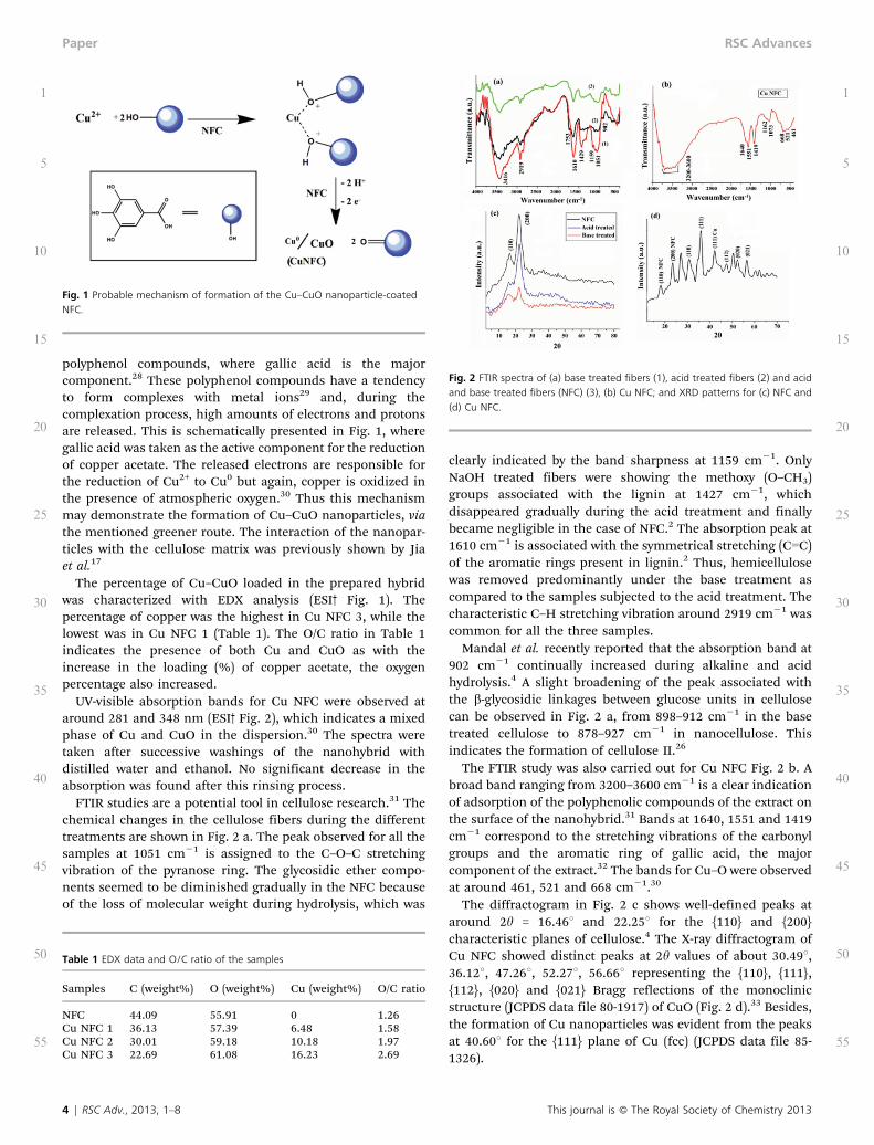
polyphenol compounds, where gallic acid is the majorcomponent.28 These polyphenol compounds have a tendencyto form complexes with metal ions29 and, during thecomplexation process, high amounts of electrons and protonsare released. This is schematically presented in Fig. 1, wheregallic acid was taken as the active component for the reductionof copper acetate. The released electrons are responsible forthe reduction of Cu2+ to Cu0 but again, copper is oxidized inthe presence of atmospheric oxygen.30 Thus this mechanismmay demonstrate the formation of Cu–CuO nanoparticles, viathe mentioned greener route. The interaction of the nanopar-ticles with the cellulose matrix was previously shown by Jiaet al.17
The percentage of Cu–CuO loaded in the prepared hybridwas characterized with EDX analysis (ESI3 Fig. 1). Thepercentage of copper was the highest in Cu NFC 3, while thelowest was in Cu NFC 1 (Table 1). The O/C ratio in Table 1indicates the presence of both Cu and CuO as with theincrease in the loading (%) of copper acetate, the oxygenpercentage also increased.
UV-visible absorption bands for Cu NFC were observed ataround 281 and 348 nm (ESI3 Fig. 2), which indicates a mixedphase of Cu and CuO in the dispersion.30 The spectra weretaken after successive washings of the nanohybrid withdistilled water and ethanol. No significant decrease in theabsorption was found after this rinsing process.
FTIR studies are a potential tool in cellulose research.31 Thechemical changes in the cellulose fibers during the differenttreatments are shown in Fig. 2 a. The peak observed for all thesamples at 1051 cm21 is assigned to the C–O–C stretchingvibration of the pyranose ring. The glycosidic ether compo-nents seemed to be diminished gradually in the NFC becauseof the loss of molecular weight during hydrolysis, which was
clearly indicated by the band sharpness at 1159 cm21. OnlyNaOH treated fibers were showing the methoxy (O–CH3)groups associated with the lignin at 1427 cm21, whichdisappeared gradually during the acid treatment and finallybecame negligible in the case of NFC.2 The absorption peak at1610 cm21 is associated with the symmetrical stretching (CLC)of the aromatic rings present in lignin.2 Thus, hemicellulosewas removed predominantly under the base treatment ascompared to the samples subjected to the acid treatment. Thecharacteristic C–H stretching vibration around 2919 cm21 wascommon for all the three samples.
Mandal et al. recently reported that the absorption band at902 cm21 continually increased during alkaline and acidhydrolysis.4 A slight broadening of the peak associated withthe b-glycosidic linkages between glucose units in cellulosecan be observed in Fig. 2 a, from 898–912 cm21 in the basetreated cellulose to 878–927 cm21 in nanocellulose. Thisindicates the formation of cellulose II.26
The FTIR study was also carried out for Cu NFC Fig. 2 b. Abroad band ranging from 3200–3600 cm21 is a clear indicationof adsorption of the polyphenolic compounds of the extract onthe surface of the nanohybrid.31 Bands at 1640, 1551 and 1419cm21 correspond to the stretching vibrations of the carbonylgroups and the aromatic ring of gallic acid, the majorcomponent of the extract.32 The bands for Cu–O were observedat around 461, 521 and 668 cm21.30
The diffractogram in Fig. 2 c shows well-defined peaks ataround 2h = 16.46u and 22.25u for the {110} and {200}characteristic planes of cellulose.4 The X-ray diffractogram ofCu NFC showed distinct peaks at 2h values of about 30.49u,36.12u, 47.26u, 52.27u, 56.66u representing the {110}, {111},{112}, {020} and {021} Bragg reflections of the monoclinicstructure (JCPDS data file 80-1917) of CuO (Fig. 2 d).33 Besides,the formation of Cu nanoparticles was evident from the peaksat 40.60u for the {111} plane of Cu (fcc) (JCPDS data file 85-1326).
Table 1 EDX data and O/C ratio of the samples
Samples C (weight%) O (weight%) Cu (weight%) O/C ratio
NFC 44.09 55.91 0 1.26Cu NFC 1 36.13 57.39 6.48 1.58Cu NFC 2 30.01 59.18 10.18 1.97Cu NFC 3 22.69 61.08 16.23 2.69
Fig. 2 FTIR spectra of (a) base treated fibers (1), acid treated fibers (2) and acidand base treated fibers (NFC) (3), (b) Cu NFC; and XRD patterns for (c) NFC and(d) Cu NFC.
Fig. 1 Probable mechanism of formation of the Cu–CuO nanoparticle-coatedNFC.
4 | RSC Adv., 2013, 1–8 This journal is � The Royal Society of Chemistry 2013
1
5
10
15
20
25
30
35
40
45
50
55
1
5
10
15
20
25
30
35
40
45
50
55
Paper RSC Advances
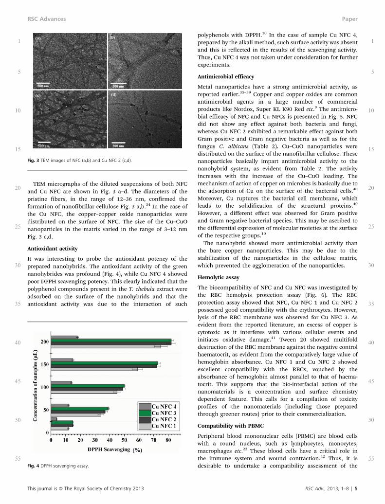
TEM micrographs of the diluted suspensions of both NFCand Cu NFC are shown in Fig. 3 a–d. The diameters of thepristine fibers, in the range of 12–36 nm, confirmed theformation of nanofibrillar cellulose Fig. 3 a,b.34 In the case ofthe Cu NFC, the copper–copper oxide nanoparticles weredistributed on the surface of NFC. The size of the Cu–CuOnanoparticles in the matrix varied in the range of 3–12 nmFig. 3 c,d.
Antioxidant activity
It was interesting to probe the antioxidant potency of theprepared nanohybrids. The antioxidant activity of the greennanohybrides was profound (Fig. 4), while Cu NFC 4 showedpoor DPPH scavenging potency. This clearly indicated that thepolyphenol compounds present in the T. chebula extract wereadsorbed on the surface of the nanohybrids and that theantioxidant activity was due to the interaction of such
polyphenols with DPPH.10 In the case of sample Cu NFC 4,prepared by the alkali method, such surface activity was absentand this is reflected in the results of the scavenging activity.Thus, Cu NFC 4 was not taken under consideration for furtherexperiments.
Antimicrobial efficacy
Metal nanoparticles have a strong antimicrobial activity, asreported earlier.35–39 Copper and copper oxides are commonantimicrobial agents in a large number of commercialproducts like Nordox, Super KL K90 Red etc.9 The antimicro-bial efficacy of NFC and Cu NFCs is presented in Fig. 5. NFCdid not show any effect against both bacteria and fungi,whereas Cu NFC 2 exhibited a remarkable effect against bothGram positive and Gram negative bacteria as well as for thefungus C. albicans (Table 2). Cu–CuO nanoparticles weredistributed on the surface of the nanofibrillar cellulose. Thesenanoparticles basically impart antimicrobial activity to thenanohybrid system, as evident from Table 2. The activityincreases with the increase of the Cu–CuO loading. Themechanism of action of copper on microbes is basically due tothe adsorption of Cu on the surface of the bacterial cells.40
Moreover, Cu ruptures the bacterial cell membrane, whichleads to the solidification of the structural proteins.40
However, a different effect was observed for Gram positiveand Gram negative bacterial species. This may be ascribed tothe differential expression of molecular moieties at the surfaceof the respective groups.10
The nanohybrid showed more antimicrobial activity thanthe bare copper nanoparticles. This may be due to thestabilization of the nanoparticles in the cellulose matrix,which prevented the agglomeration of the nanoparticles.
Hemolytic assay
The biocompatibility of NFC and Cu NFC was investigated bythe RBC hemolysis protection assay (Fig. 6). The RBCprotection assay showed that NFC, Cu NFC 1 and Cu NFC 2possessed good compatibility with the erythrocytes. However,lysis of the RBC membrane was observed for Cu NFC 3. Asevident from the reported literature, an excess of copper iscytotoxic as it interferes with various cellular events andinitiates oxidative damage.41 Tween 20 showed multifolddestruction of the RBC membrane against the negative controlhaematocrit, as evident from the comparatively large value ofhemoglobin absorbance. Cu NFC 1 and Cu NFC 2 showedexcellent compatibility with the RBCs, vouched by theabsorbance of hemoglobin almost parallel to that of haema-tocrit. This supports that the bio-interfacial action of thenanomaterials is a concentration and surface chemistrydependent feature. This calls for a compilation of toxicityprofiles of the nanomaterials (including those preparedthrough greener routes) prior to their commercialization.
Compatibility with PBMC
Peripheral blood mononuclear cells (PBMC) are blood cellswith a round nucleus, such as lymphocytes, monocytes,macrophages etc.22 These blood cells have a critical role inthe immune system and wound contraction.42 Thus, it isdesirable to undertake a compatibility assessment of theFig. 4 DPPH scavenging assay.
Fig. 3 TEM images of NFC (a,b) and Cu NFC 2 (c,d).
This journal is � The Royal Society of Chemistry 2013 RSC Adv., 2013, 1–8 | 5
1
5
10
15
20
25
30
35
40
45
50
55
1
5
10
15
20
25
30
35
40
45
50
55
RSC Advances Paper

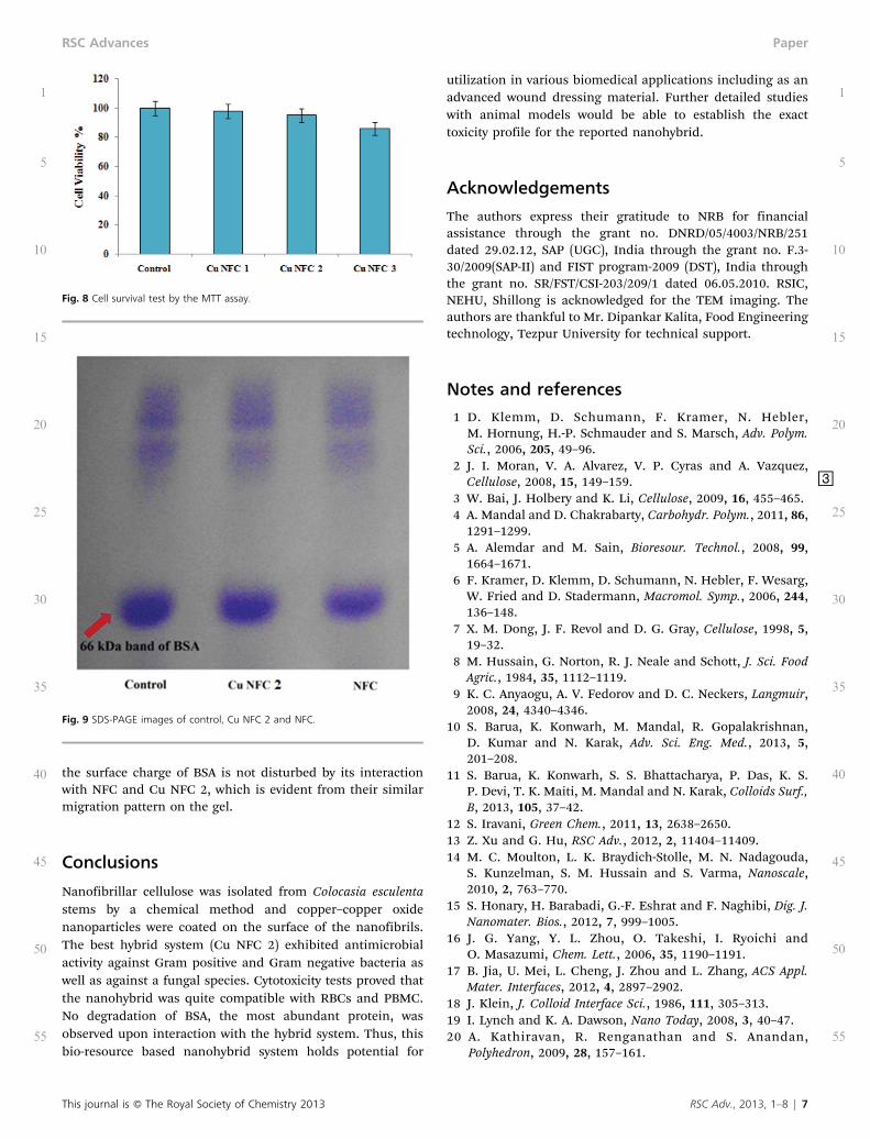
nanomaterials with PBMC, which may reveal their potential tobe used in advanced biomedical applications. The biocompat-ibility of the prepared nanohybrids (Cu NFC) was examinedthrough their interaction with PBMC. It is quite clear fromFig. 7 that there is no change in the morphology of the cellsafter treating with Cu NFC 1 and Cu NFC 2. Cell survival wasfound to be almost the same as compared to the control but
cell death was observed for Cu NFC 3. A MTT assay wasperformed to confirm this observation. It was found that thecell survival rate for Cu NFC 2 is equivalent to that of thecontrol (Fig. 8). The aforementioned bioassays ascertainedthat Cu NFC 2 possesses good antimicrobial activity as well asexcellent biocompatibility. Thus, this nanohybrid is a goodcandidate for its application in biomedical bandages.
Interaction with BSA
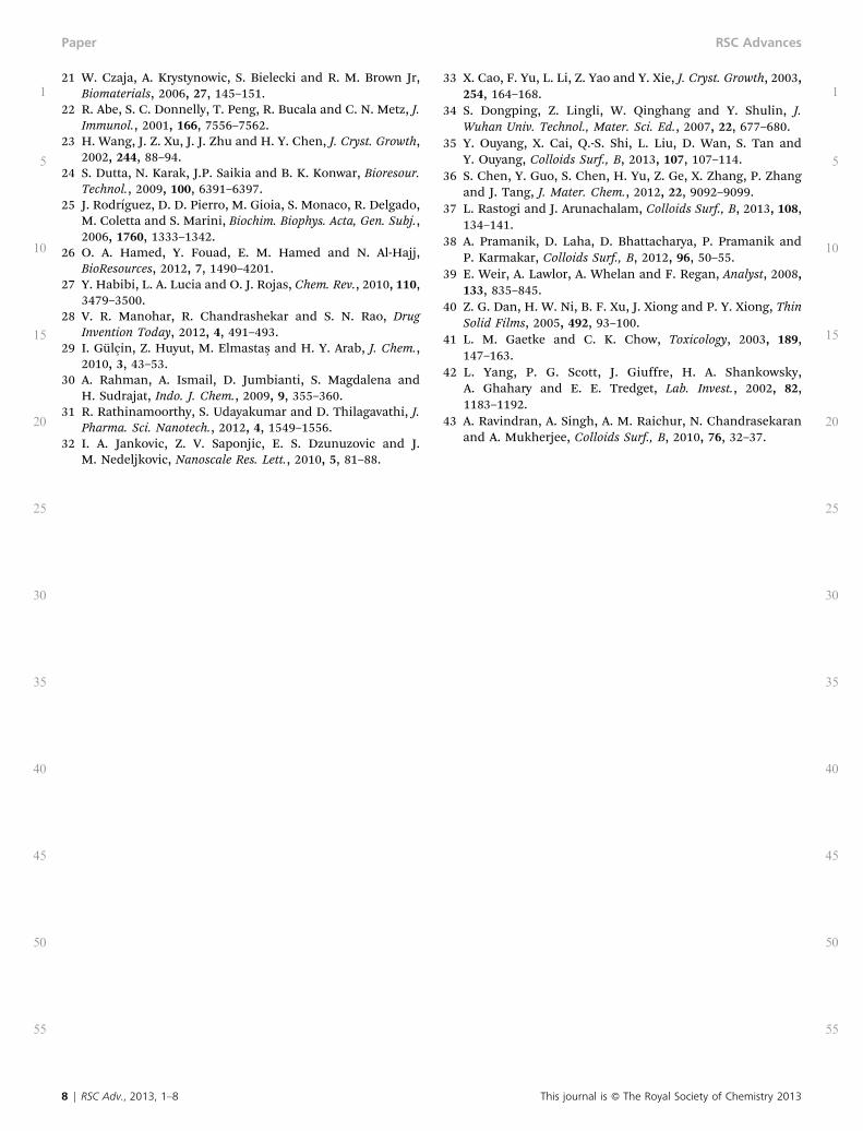
Protein–nanoparticle interaction studies are an emerging fieldin nanomedicine and nanotoxicology.19 Nanomaterials with ahigh surface area can absorb the dynamic layer of proteinswhen they interact with living systems. Kathiravan et al.recently studied the interaction of BSA, a physiologicallyimportant protein, with metal nanoparticles.20 In yet anotherreport, Ravindran et al. concluded that the interaction of BSAwith silver nanoparticles prevented aggregation, which wasconfirmed by spectroscopic methods.43 In this study, wedelved into the interaction of the same protein with NFC andCu NFC 2 by gel electrophoresis (Fig. 9). BSA gives a band at 66KDa and single bands were observed in all the three lanes forboth the samples and the control. This clearly indicated that
Fig. 5 Representative antimicrobial activity of Cu NFC 2 against Staphylococcus aureus, Escherichia coli and Candida albicans. <
Table 2 Antimicrobial activity of Cu NFC
Test organisms S. aureus E. coli C. albicans
Cu 0 14.8 ¡ 0.3 18.16 ¡ 0.2NFC 0 0 0Cu NFC 1 20.2 ¡ 0.18 24.4 ¡ 0.3 22 ¡ 0.1Cu NFC 2 21.93 ¡ 0.06 25.96 ¡ 0.15 22.9 ¡ 0.17Cu NFC 3 22.86 ¡ 0.15 26.06 ¡ 0.2 25 ¡ 0.17Antibiotic 31.9 ¡ 0.2 31.8 ¡ 0.1 30 ¡ 0.15
Fig. 6 Anti-hemolytic activity of NFC and Cu NFCs. Fig. 7 Microscopic images of Trypan Blue stained PBMC treated with Cu NFCs.
6 | RSC Adv., 2013, 1–8 This journal is � The Royal Society of Chemistry 2013
1
5
10
15
20
25
30
35
40
45
50
55
1
5
10
15
20
25
30
35
40
45
50
55
Paper RSC Advances
the surface charge of BSA is not disturbed by its interactionwith NFC and Cu NFC 2, which is evident from their similarmigration pattern on the gel.
Conclusions
Nanofibrillar cellulose was isolated from Colocasia esculentastems by a chemical method and copper–copper oxidenanoparticles were coated on the surface of the nanofibrils.The best hybrid system (Cu NFC 2) exhibited antimicrobialactivity against Gram positive and Gram negative bacteria aswell as against a fungal species. Cytotoxicity tests proved thatthe nanohybrid was quite compatible with RBCs and PBMC.No degradation of BSA, the most abundant protein, wasobserved upon interaction with the hybrid system. Thus, thisbio-resource based nanohybrid system holds potential for
utilization in various biomedical applications including as anadvanced wound dressing material. Further detailed studieswith animal models would be able to establish the exacttoxicity profile for the reported nanohybrid.
Acknowledgements
The authors express their gratitude to NRB for financialassistance through the grant no. DNRD/05/4003/NRB/251dated 29.02.12, SAP (UGC), India through the grant no. F.3-30/2009(SAP-II) and FIST program-2009 (DST), India throughthe grant no. SR/FST/CSI-203/209/1 dated 06.05.2010. RSIC,NEHU, Shillong is acknowledged for the TEM imaging. Theauthors are thankful to Mr. Dipankar Kalita, Food Engineeringtechnology, Tezpur University for technical support.
Notes and references
1 D. Klemm, D. Schumann, F. Kramer, N. Hebler,M. Hornung, H.-P. Schmauder and S. Marsch, Adv. Polym.Sci., 2006, 205, 49–96.
2 J. I. Moran, V. A. Alvarez, V. P. Cyras and A. Vazquez,Cellulose, 2008, 15, 149–159. =
3 W. Bai, J. Holbery and K. Li, Cellulose, 2009, 16, 455–465.4 A. Mandal and D. Chakrabarty, Carbohydr. Polym., 2011, 86,
1291–1299.5 A. Alemdar and M. Sain, Bioresour. Technol., 2008, 99,
1664–1671.6 F. Kramer, D. Klemm, D. Schumann, N. Hebler, F. Wesarg,
W. Fried and D. Stadermann, Macromol. Symp., 2006, 244,136–148.
7 X. M. Dong, J. F. Revol and D. G. Gray, Cellulose, 1998, 5,19–32.
8 M. Hussain, G. Norton, R. J. Neale and Schott, J. Sci. FoodAgric., 1984, 35, 1112–1119.
9 K. C. Anyaogu, A. V. Fedorov and D. C. Neckers, Langmuir,2008, 24, 4340–4346.
10 S. Barua, K. Konwarh, M. Mandal, R. Gopalakrishnan,D. Kumar and N. Karak, Adv. Sci. Eng. Med., 2013, 5,201–208.
11 S. Barua, K. Konwarh, S. S. Bhattacharya, P. Das, K. S.P. Devi, T. K. Maiti, M. Mandal and N. Karak, Colloids Surf.,B, 2013, 105, 37–42.
12 S. Iravani, Green Chem., 2011, 13, 2638–2650.13 Z. Xu and G. Hu, RSC Adv., 2012, 2, 11404–11409.14 M. C. Moulton, L. K. Braydich-Stolle, M. N. Nadagouda,
S. Kunzelman, S. M. Hussain and S. Varma, Nanoscale,2010, 2, 763–770.
15 S. Honary, H. Barabadi, G.-F. Eshrat and F. Naghibi, Dig. J.Nanomater. Bios., 2012, 7, 999–1005.
16 J. G. Yang, Y. L. Zhou, O. Takeshi, I. Ryoichi andO. Masazumi, Chem. Lett., 2006, 35, 1190–1191.
17 B. Jia, U. Mei, L. Cheng, J. Zhou and L. Zhang, ACS Appl.Mater. Interfaces, 2012, 4, 2897–2902.
18 J. Klein, J. Colloid Interface Sci., 1986, 111, 305–313.19 I. Lynch and K. A. Dawson, Nano Today, 2008, 3, 40–47.20 A. Kathiravan, R. Renganathan and S. Anandan,
Polyhedron, 2009, 28, 157–161.
Fig. 9 SDS-PAGE images of control, Cu NFC 2 and NFC.
Fig. 8 Cell survival test by the MTT assay.
This journal is � The Royal Society of Chemistry 2013 RSC Adv., 2013, 1–8 | 7
1
5
10
15
20
25
30
35
40
45
50
55
1
5
10
15
20
25
30
35
40
45
50
55
RSC Advances Paper
21 W. Czaja, A. Krystynowic, S. Bielecki and R. M. Brown Jr,Biomaterials, 2006, 27, 145–151.
22 R. Abe, S. C. Donnelly, T. Peng, R. Bucala and C. N. Metz, J.Immunol., 2001, 166, 7556–7562.
23 H. Wang, J. Z. Xu, J. J. Zhu and H. Y. Chen, J. Cryst. Growth,2002, 244, 88–94.
24 S. Dutta, N. Karak, J.P. Saikia and B. K. Konwar, Bioresour.Technol., 2009, 100, 6391–6397.
25 J. Rodrıguez, D. D. Pierro, M. Gioia, S. Monaco, R. Delgado,M. Coletta and S. Marini, Biochim. Biophys. Acta, Gen. Subj.,2006, 1760, 1333–1342.
26 O. A. Hamed, Y. Fouad, E. M. Hamed and N. Al-Hajj,BioResources, 2012, 7, 1490–4201.
27 Y. Habibi, L. A. Lucia and O. J. Rojas, Chem. Rev., 2010, 110,3479–3500.
28 V. R. Manohar, R. Chandrashekar and S. N. Rao, DrugInvention Today, 2012, 4, 491–493.
29 I. Gulçin, Z. Huyut, M. Elmastas and H. Y. Arab, J. Chem.,2010, 3, 43–53.
30 A. Rahman, A. Ismail, D. Jumbianti, S. Magdalena andH. Sudrajat, Indo. J. Chem., 2009, 9, 355–360.
31 R. Rathinamoorthy, S. Udayakumar and D. Thilagavathi, J.Pharma. Sci. Nanotech., 2012, 4, 1549–1556.
32 I. A. Jankovic, Z. V. Saponjic, E. S. Dzunuzovic and J.M. Nedeljkovic, Nanoscale Res. Lett., 2010, 5, 81–88.
33 X. Cao, F. Yu, L. Li, Z. Yao and Y. Xie, J. Cryst. Growth, 2003,254, 164–168.
34 S. Dongping, Z. Lingli, W. Qinghang and Y. Shulin, J.Wuhan Univ. Technol., Mater. Sci. Ed., 2007, 22, 677–680.
35 Y. Ouyang, X. Cai, Q.-S. Shi, L. Liu, D. Wan, S. Tan andY. Ouyang, Colloids Surf., B, 2013, 107, 107–114.
36 S. Chen, Y. Guo, S. Chen, H. Yu, Z. Ge, X. Zhang, P. Zhangand J. Tang, J. Mater. Chem., 2012, 22, 9092–9099.
37 L. Rastogi and J. Arunachalam, Colloids Surf., B, 2013, 108,134–141.
38 A. Pramanik, D. Laha, D. Bhattacharya, P. Pramanik andP. Karmakar, Colloids Surf., B, 2012, 96, 50–55.
39 E. Weir, A. Lawlor, A. Whelan and F. Regan, Analyst, 2008,133, 835–845.
40 Z. G. Dan, H. W. Ni, B. F. Xu, J. Xiong and P. Y. Xiong, ThinSolid Films, 2005, 492, 93–100.
41 L. M. Gaetke and C. K. Chow, Toxicology, 2003, 189,147–163.
42 L. Yang, P. G. Scott, J. Giuffre, H. A. Shankowsky,A. Ghahary and E. E. Tredget, Lab. Invest., 2002, 82,1183–1192.
43 A. Ravindran, A. Singh, A. M. Raichur, N. Chandrasekaranand A. Mukherjee, Colloids Surf., B, 2010, 76, 32–37.
8 | RSC Adv., 2013, 1–8 This journal is � The Royal Society of Chemistry 2013
1
5
10
15
20
25
30
35
40
45
50
55
1
5
10
15
20
25
30
35
40
45
50
55
Paper RSC Advances