Acanthamoeba castellanii: Morphological analysis of the interaction with human cornea
Upload
independentCategory
view
3download
0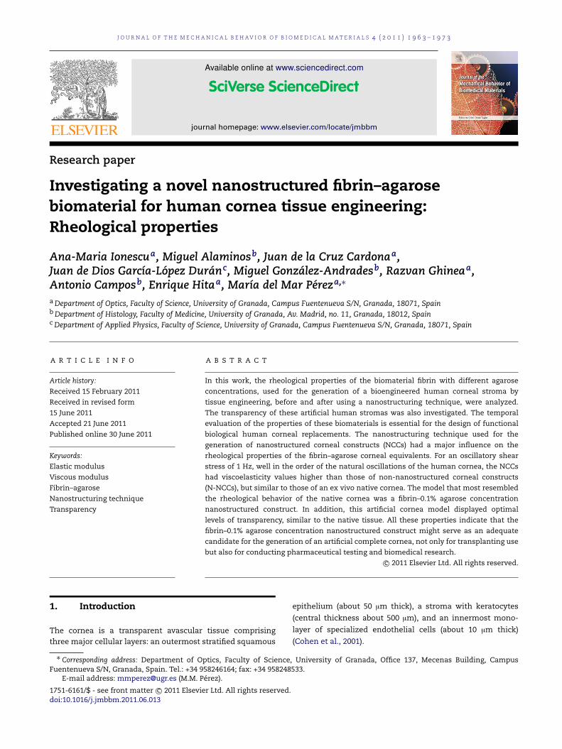
J O U R N A L O F T H E M E C H A N I C A L B E H AV I O R O F B I O M E D I C A L M A T E R I A L S 4 ( 2 0 1 1 ) 1 9 6 3 – 1 9 7 3
Available online at www.sciencedirect.com
journal homepage: www.elsevier.com/locate/jmbbm
Research paper
Investigating a novel nanostructured fibrin–agarosebiomaterial for human cornea tissue engineering:Rheological properties
Ana-Maria Ionescua, Miguel Alaminosb, Juan de la Cruz Cardonaa,Juan de Dios García-López Duránc, Miguel González-Andradesb, Razvan Ghineaa,Antonio Camposb, Enrique Hitaa, María del Mar Péreza,∗
aDepartment of Optics, Faculty of Science, University of Granada, Campus Fuentenueva S/N, Granada, 18071, SpainbDepartment of Histology, Faculty of Medicine, University of Granada, Av. Madrid, no. 11, Granada, 18012, SpaincDepartment of Applied Physics, Faculty of Science, University of Granada, Campus Fuentenueva S/N, Granada, 18071, Spain
A R T I C L E I N F O
Article history:
Received 15 February 2011
Received in revised form
15 June 2011
Accepted 21 June 2011
Published online 30 June 2011
Keywords:
Elastic modulus
Viscous modulus
Fibrin–agarose
Nanostructuring technique
Transparency
A B S T R A C T
In this work, the rheological properties of the biomaterial fibrin with different agarose
concentrations, used for the generation of a bioengineered human corneal stroma by
tissue engineering, before and after using a nanostructuring technique, were analyzed.
The transparency of these artificial human stromas was also investigated. The temporal
evaluation of the properties of these biomaterials is essential for the design of functional
biological human corneal replacements. The nanostructuring technique used for the
generation of nanostructured corneal constructs (NCCs) had a major influence on the
rheological properties of the fibrin–agarose corneal equivalents. For an oscillatory shear
stress of 1 Hz, well in the order of the natural oscillations of the human cornea, the NCCs
had viscoelasticity values higher than those of non-nanostructured corneal constructs
(N-NCCs), but similar to those of an ex vivo native cornea. The model that most resembled
the rheological behavior of the native cornea was a fibrin–0.1% agarose concentration
nanostructured construct. In addition, this artificial cornea model displayed optimal
levels of transparency, similar to the native tissue. All these properties indicate that the
fibrin–0.1% agarose concentration nanostructured construct might serve as an adequate
candidate for the generation of an artificial complete cornea, not only for transplanting use
but also for conducting pharmaceutical testing and biomedical research.c⃝ 2011 Elsevier Ltd. All rights reserved.
4
d
1. Introduction
The cornea is a transparent avascular tissue comprisingthree major cellular layers: an outermost stratified squamous
∗ Corresponding address: Department of Optics, Faculty of SciencFuentenueva S/N, Granada, Spain. Tel.: +34 958246164; fax: +34 9582
E-mail address: [email protected] (M.M. Pérez).
1751-6161/$ - see front matter c⃝ 2011 Elsevier Ltd. All rights reservedoi:10.1016/j.jmbbm.2011.06.013
e, University of Granada, Office 137, Mecenas Building, Campus8533.
epithelium (about 50 µm thick), a stroma with keratocytes
(central thickness about 500 µm), and an innermost mono-
layer of specialized endothelial cells (about 10 µm thick)
(Cohen et al., 2001).
.

1964 J O U R N A L O F T H E M E C H A N I C A L B E H AV I O R O F B I O M E D I C A L M A T E R I A L S 4 ( 2 0 1 1 ) 1 9 6 3 – 1 9 7 3
More than 10 million people worldwide suffer from visionloss due to corneal damage (disease or injury), with trans-plantation using donor tissue being the only widely acceptedtreatment for irreversible cornea blindness (Whitcher et al.,2001). However, the supply of good-quality donor tissue doesnot meet the demand, and clinical indications such as au-toimmune disease and chemical burns may be amenable todonor tissue transplantation (Zerbe et al., 2006). Therefore, acornea substitute as a donor tissue replacement is in urgentdemand. In this context, tissue engineering has been an areaof immense research in recent years owing to its vast poten-tial in the repair and/or replacement of damaged tissues andorgans (Yang et al., 2001; Ma, 2004). It has shown tremendouspromise in creating biological alternatives for harvested tis-sues, implants, and prostheses (Mooney and Mikos, 1999).
The underlying concept of tissue engineering is the beliefthat cells can be isolated from a patient, and their populationthen expanded in a cell culture and seeded onto a carrier.The resulting tissue engineering construct is then graftedback into the same patient to function as the introducedreplacement tissue (Yang et al., 2001). In this approach,a highly porous artificial extracellular matrix (Peters andMooney, 1997), or scaffold, is thought to be needed toaccommodate mammalian cells and guide their growth andtissues regeneration in three dimensions.
The creation of tissues for medical use is already usedto a significant extent in hospitals. These ground-breakingapplications involve fabricated skin (Cooper et al., 1991), liver(Cima et al., 1991a,b), cartilage (Ashiku et al., 1997), bone (Hoet al., 1995; Simske et al., 1997; Cao et al., 1997; Bostrom andMikos, 1997), and cornea (Orwin et al., 2003; Alaminos et al.,2006; González-Andrades et al., 2009; Ionescu et al., 2010).
By using tissue-engineering techniques, several researchgroups have tried to develop an efficient substitute for thecornea that would overcome the present disadvantages ofheterologous corneal transplantation (Reichl et al., 2004;Nishida et al., 2004; Nishida, 2003). Some researchershave proposed different tissue-engineering techniques toconstruct a cornea of animal origin, as the first step in thedevelopment of a human corneal substitute (Minami et al.,1993; Zieske et al., 1994; Schneider et al., 1999). However,although some scientists have developed artificial substitutesthat partially mimic the human cornea (Griffith et al., 1999),a full-thickness, functional human corneal construct hasnot been achieved to date. Most corneal substitutes usethree-dimensional cultures of one or two corneal cell typesand biomaterials (Maurice, 1984). Different biomaterials havebeen used as stromal substitutes. Ideally, a good biomaterialshould be biocompatible, transparent, and consistent, andcorneal cells should be able to adhere and grow on it. Thematerials that have been usedmost often to construct cornealsubstitutes are type I collagen and fibrin. Type I collagen hasbeen extensively used as a scaffold in tissue engineering forthe construction of artificial skin, oral mucosa, corneas, andother tissues (Reichl andMuller-Goymann, 2003; Griffith et al.,1999; Meana et al., 1998; Llames et al., 2004; Orwin and Hubel,2000). However, collagen is an expensive material of animalorigin, and it tends to shrink and lose volume when the cellsare seeded inside the scaffold (Porter et al., 1998; Tegtmeyeret al., 2001). In addition, stromal substitutes made of collagen
are not stable and are quickly degraded (Chen et al., 2005).Human fibrin has been proposed as a stromal substitute toconstruct different tissue substitutes, especially human skin(Meana et al., 1998; Llames et al., 2004), and cornea (Alaminoset al., 2006). It has the advantages of low price, availability,and good tolerance to cells, and it can be produced as acomplete autologous scaffold. In contrast with collagen gels,fibrin gels are not contracted by stromal cells (Meana et al.,1998; Llames et al., 2004). Other materials such as agaroseare less commonly used in tissue engineering of the cornea,although several recent reports mention the use of a mixtureof fibrin and agarose for cornea development (Ionescu et al.,2010; Cardona et al., in press).
In the development of a bioengineered construct thatcould replace a diseased cornea, it is important to accuratelycharacterize its rheological and optical properties to ensureoptimization of its design, since it is accepted that the rhe-ological (mechanical) and optical properties of biomaterialsdirectly affect their clinical performance (Jones et al., 1997a,b;Jones, 1999).
Corneal transparency has been the subject of numerousstudies over the last few years (Meek et al., 2003a). It is nowgenerally accepted that corneal transparency depends on thedestructive interference of the incoming light that is scatteredaway and on the absorption of light by the corneal tissueitself. In this regard, several researchers have demonstratedthat the scattering of light in the human cornea dependsmostly on the stromal extracellular matrix, and that thescattering will increase if the fibril diameter increases or ifthe spatial arrangement or the hydration level of the fibrils isaltered (Freund et al., 1986; Meek et al., 2003b). By contrast,other studies suggest that corneal cells might play a relevantrole and that stromal keratocytes contribute significantly tocorneal light scattering (Maurice, 1957).
Apart from its optical properties (transparency to visiblelight and formation of a nearly perfect optical interface torefract light onto the retina (Ruberti and Zieske, 2008)), thecornea must provide protection to the intraocular contentsand, consequently, it is required that the biomaterial em-ployed as the tissue substitute possess adequate viscoelasticproperties.
Recently, a complete rabbit cornea (Alaminos et al., 2006)and a partial human cornea (González-Andrades et al., 2009;Ionescu et al., 2010) have been generated by tissue engineer-ing using a novel fibrin–agarose biocompatible biomaterial,and all three types of corneal cell (endothelial, stromal, andepithelial) were efficiently cultured, immersed and on top ofthis novel biomaterial. Although some optical properties suchas absorbance, the absorption coefficient, and transmittanceof these substitutes in the UV range (240–400 nm) have beenstudied (Ionescu et al., 2010), the rheological properties andthe optical behavior in the visible light spectrum of thesefibrin–agarose cornea substitutes have not yet been deter-mined.
The present work aims to describe the rheological andoptical properties of an artificial construct based on fibrinand agarose for determining the potential corneal applicationof such artificial biomaterials as corneal substitutes. Also,a new method for the generation of bioengineered corneal
J O U R N A L O F T H E M E C H A N I C A L B E H AV I O R O F B I O M E D I C A L M A T E R I A L S 4 ( 2 0 1 1 ) 1 9 6 3 – 1 9 7 3 1965
constructs that allowed the partial dehydration of thefibrin–agarose biomaterial is proposed.
The rheological and optical characteristics of these nanos-tructured biomaterials are of great interest for the optimiza-tion of their design and performance for possible clinicalusefulness, and they have not been determined to date.
2. Materials and methods
2.1. Construction of an artificial human cornea stromasubstitute (NCCs and N-NCCs)
Due to the difficulty in obtaining intact human corneas, thecorneal cells used in the present experiment were extractedfrom corneal–scleral limbal rims of about 14 mm diameterthat were obtained at the University Hospital of San Cecilio,Granada, Spain, after the removal of ±7 mm central cornealbuttons for corneal transplantation. This work was approvedby the local research and ethical committees and the researchadhered to the tenets of the Declaration of Helsinki.
Primary cultures of human keratocytes were generatedas previously described (Alaminos et al., 2006; González-Andrades et al., 2009). Stromal keratocytes were isolated fromsmall fragments of cornea that remained attached to thescleral–corneal limbus after the keratoplasty procedure, usingcollagenase I (Invitrogen-Gibco) at 37 ◦C for 6 h. Once the cellshad been harvested by centrifugation, stromal keratocyteswere cultured in Dulbecco’s modified Eagle’s medium (DMEM)supplemented with 10% fetal bovine serum (FBS, Sigma-Aldrich), 4 mM L-glutamine, and 1% antibiotic–antimycoticsolution (Invitrogen-Gibco). All cells were incubated at 37 ◦Cin 5% carbon dioxide under standard culture conditions.
Then, we generated 12 types of bioengineered cornealstroma substitute in the laboratory: human fibrin stromaswith increasing concentration of agarose (0%, 0.025%, 0.05%,0.1%, 0.2% and 0.3%) non-nanostructured constructs (N-NCCs), and nanostructured constructs (NCCs). In all cases,21 ml of human plasma were added to 1.8 ml of DMEMin which 250,000 cultured keratocytes had been previouslysuspended, and 200 µL of tranexamic acid (Amchafibrin,Fides-Ecofarma, Valencia, Spain) were added to avoidspontaneous fibrinolysis. Then, 2 ml of 1% of CaCl2 wereadded to the solution to precipitate the polymerizationreaction of the hydrogel. In the case of the fibrin–agarosegels, previously melted type VII agarose dissolved in PBS(phosphate-buffered saline) was supplemented in the laststep. After polymerization, 15 ml of culture medium wereadded to the surface of the bioengineered corneal stromas,which were incubated at 37 ◦C in 5% carbon dioxide. Samplesof the different stromal substitutes were studied weekly untileight weeks of development in culture. Once generated, halfof them were nanostructured (NCCs) and the other half werestudied directly (N-NCCs).
The nanostructuring technique is based on previouslydescribedmethods by Hadjipanayi et al. (2011), who describeda technique that is able to allow fluid ultrafiltrationretaining the fibril structure and to modify the biomechanicalproperties of the biomaterials. The nanostructuring methodproposed here would induce a series of complex interfibrillar
changes at the nanometrical scale (nanostructuring) thatcould in turn modify the properties of this biomaterial.The dehydration of the fibrin–agarose biomaterials wascarried out since it has been reported that small amountsof water can contribute to slippage during the rheologicalmeasurements (Shin et al., 2002).
For nanostructuring, samples were transferred to a specificpolycarbonate chamber in which 4–6 layers of sterile What-man 3MM absorbent paper were put above and below thesample to facilitate dehydration of the artificial tissue. To pre-vent the stromal construct sticking to the paper layers, a filternylon membrane (0.4 µm pore size) was settled between bothfaces of the sample and the paper layers. Then, a flat crystalsurface was set on top of the system and a total of 1000 Pa ofpressure was applied to compress the bioengineered tissues.This process was carried out for 3–5 min, and then the nanos-tructured tissues were removed from the chamber and main-tained in PBS until the moment of the analysis. All cornealconstruct samples (approximately 0.5 mm thick) were gener-ated and analyzed in triplicate.
As controls, ten fresh porcine corneas were obtained fromadult pigs immediately after their death at a local slaughter-house. The eyes selected for the study had integral cornealsurface with a horizontal corneal diameter of 12–14 mm. Thenative porcine cornea was removed using a 16 mm cornealtrephine. The corneas were washed thoroughly in PBS. All tencontrol corneas were subjected to the same mechanical andoptical tests as the bioengineered artificial corneal constructs.
2.2. Histological analysis of the extracellular matrix
For light-microscopy analysis, samples corresponding to N-NCCs and NCCs were fixed in 4% formaldehyde, dehydratedin ethanol series, and embedded in paraffin. Then, 4 µm thicksections were cut. For the analysis of collagen synthesis anddeposition by the cells immersed in the hydrogels, deparaf-finized tissue sections were stained by using picrosirius histo-chemical methods. In short, sections were stained for 30 minin 0.2% picrosirius solution (Sirius Red F3BA, BDH Labora-tory Supplies, UK, in saturated picric acid) and counterstainedwith Harris’s hematoxylin for 5 min at room temperature.After being washed in water, the sections were dehydratedthrough an ethanol and xylene series and covered usingmounting medium.
To identify the synthesis of proteoglycans in the bioengi-neered corneal stroma, safranin O histochemistry was used.In this case, deparaffinized tissue sections were stained inWeigert’s hematoxylin for 10 min and washed in water for10 min. Then, the samples were counterstained in aqueousfast green for 5 min, rinsed in 1% acetic acid and stained in0.1% safranin O for 5 min. Finally, slides were dehydratedin alcohol series and xylene and covered using mountingmedium.
2.3. Rheological measurements
Measurements of the rheological quantities of interest wereperformed for all NCCs and N-NCCs and, also, for the porcinecontrol cornea. A controlled shear stress rheometer (BohlinCS-10, UK) was used. The measuring cell (Bohlin PP4-40) had

1966 J O U R N A L O F T H E M E C H A N I C A L B E H AV I O R O F B I O M E D I C A L M A T E R I A L S 4 ( 2 0 1 1 ) 1 9 6 3 – 1 9 7 3
a plate–plate geometry (the diameter of the rotor plate was40 mm) with a gap thickness of 1 mm. All measurementswere performed at 37.0 ± 0.1 ◦C. During the measurements,the samples were maintained in a water-vapor-saturatedatmosphere to avoid drying. In addition, undesired wall-slipphenomena were prevented by using roughened plates, asrecommended by Barnes (1995).
First, steady-state shear stress versus shear rate (σ versusdγ
dt ) experiments were conducted. A shear stress ramp wasapplied to record the corresponding shear rate at time inter-vals of 3 s. The rheograms showed a plastic behavior, char-acterized by the yield stress required to provoke the viscousdeformation of the materials studied. Hence the yield stressσy was determined by fitting the σ-dγ
dt data in the post-yieldregime to the Bingham equation
σ = σy + ηdγ
dt. (1)
Oscillatory tests were also performed by applying a sinusoidalshear stress (σ = σ0sinωt) and recording the subsequent sinu-soidal strain (γ = γ0 sin(ωt + δ)). The constitutive equation forlinear viscoelasticity can be expressed as (Macosko, 1994)
σ∗(ω, t) = G∗(ω)γ∗(ω, t), (2)
where G∗(ω) is the complex rigidity modulus, σ∗ is the oscil-latory shear stress, and γ∗ is the oscillatory shear strain incomplex notation. The real part of the rigidity modulus, G′,is the elastic or storage modulus, and its imaginary part, G′′,is the viscous or loss modulus. Eq. (2) assumes a linear de-pendence between σ∗ and γ∗, which is usually accomplishedfor low stress amplitude values into the so-called viscoelasticlinear region (VLR).
Two kinds of oscillatory measurement were performed.
(i) VLR determination. A stress of increasing amplitude (σ0)and constant frequency (ω = 1 Hz) was applied to thesample: G′ and G′′ were constant (amplitude independent)until a critical σ0 was reached. This stress corresponds tothe upper limit of the VLR.
(ii) Oscillograms. A stress of amplitude 2 Pa (within the VLR)was applied, with frequencies ranging between 0.1 and10 Hz, and both G′ and G′′ were recorded as a functionof ω.
2.4. Quantification of transparency
The fraction of light transmitted through the cornea hastraditionally been measured as a method for determining theamount of light scattering in the cornea. Previous studieshave demonstrated that the fraction of light transmittedthrough a normal transparent cornea is a function of thewavelength (Farrell et al., 1973; Freund et al., 1986; McCallyand Farrell, 1982, 1988). To determine this parameter, first, thespectral reflectance of the artificial corneal-stroma substitute,and the control porcine cornea was measured using aspectroradiometer (SpectraScan PR-704 Photo Research, Inc,Chatsworth, USA) with 4% measurement accuracy. Then, theKubelka–Munk scattering (S) and absorption (K) coefficientswere calculated algebraically from the spectral reflectancedata of each tissue using the Kubelka–Munk equations(Kubelka, 1948; Lee, 2007; Thennadil, 2008). The fraction oflight transmitted in the 400–700 nm range was calculatedusing the equation
FT = Fo exp[−(µa + µ′s)t] =>
FTF0
= exp[−(µa + µ′s)t], (3)
where FTF0
is the fraction of light transmitted, µa = K/2 is the
absorption coefficient, µ′s =
43S is the scattering coefficient,
and t is the thickness of the sample.
2.5. Statistical analysis
As normality (and homogeneity of variance) assumptionswere not satisfied, non-parametric tests were used. To testthe significance of observed differences between the studygroups, the Kruskal–Wallis one-way analysis of variance byranks (K–W) and the Mann–Whitney U test (M–W) wereapplied. A value of p < 0.05 was considered to be statisticallysignificant.
The Rho-de-Spearman correlation coefficient (ρ) was alsodetermined between all the parameters measured in thisstudy.
The optical study also included the determination ofwhether the differences between the parameters values de-termined for the different days and tissues analyzed wereuniform across the wavelength spectrum, using the VAF(variance accounting for) coefficient with Cauchy–Schwarz’sinequality expressed by
VAF =
700∑
k=400akbk
2
700∑k=400
a2k
700∑
k=400b2k
, (4)
where ak is the value of transparency (for each wavelength)and bk is the equivalent for another measurement. The closerthis coefficient gets to unity (100%), the more similar the twocurves become.
3. Results and discussion
3.1. Sequential analysis of extracellular matrix deposition
Histochemical analysis of the bioengineered cornea stromasusing picrosirius staining revealed the presence of neo-formed collagen fibrils after five weeks of developmentin culture for fibrin scaffolds and after seven weeks ofdevelopment for fibrin–0.025% agarose biomaterials, for boththe N-NCCs and the NCCs. Stromas with higher agaroseconcentrations showed no collagen deposition for the entirestudy period (eight weeks of development in culture).Illustrative images of the sample analysis using picrosiriusstaining are shown in Fig. 1. However, proteoglycans stainingof the corneal substitutes analyzed in this work usingsafranin O histochemistry proved completely negative for allsamples, suggesting that these components were absent fromthe different tissue constructs (data not shown).
J O U R N A L O F T H E M E C H A N I C A L B E H AV I O R O F B I O M E D I C A L M A T E R I A L S 4 ( 2 0 1 1 ) 1 9 6 3 – 1 9 7 3 1967
0%
N-NCC2 weeks
NCC2 weeks
N-NCC7 weeks
NCC7 weeks
N-NCC8 weeks
NCC8 weeks
0.025% 0.05% 0.1% 0.2% 0.3%
Fig. 1 – Histochemical analysis of collagen synthesis as determined by picrosirius staining in the different samplesanalyzed in this work after two, seven, and eight weeks of development in culture. Percentages correspond to the agarosecontents in the fibrin–agarose scaffolds. NCCs: nanostructured cornea constructs. N-NCCs: non-nanostructured corneaconstructs. Arrows indicate collagen deposition. Bars size: 50 µm.
50A B
40
30
Yie
ld s
tres
s σ y
(Pa)
20
10
01 2 3 4 5
Time (weeks)
6 7 8
50
40
30
Yie
ld s
tres
s σ y
(Pa)
20
10
01 2 3 4 5
Time (weeks)
6 7 8
Fig. 2 – Yield stress values corresponding to the different bioengineered corneal-stroma substitutes. N-NCCs (A) and NCCs(B). Fibrin; Fibrin–0.025% agarose; Fibrin–0.05% agarose; Fibrin–0.1% agarose; Fibrin–0.2% agarose;
Fibrin–0.3% agarose; Ex vivo native cornea.
3.2. Rheological properties
3.2.1. Steady-state measurements
The existence of yield stress or minimum stress neededfor the corneal constructs to start to flow was observed.The data in the post-yield regime were fitted to Eq. (1).
From the intercept of this fitting, the yield stress of eachmaterial was determined. Fig. 2 shows the yield stress valuescorresponding to the different bioengineered corneal-stromasubstitutes (N-NCCs (A) and NCCs (B)) and to the controlnative cornea.
This evaluation was made as a function of the agaroseconcentration and the culture time. No degradation was
1968 J O U R N A L O F T H E M E C H A N I C A L B E H AV I O R O F B I O M E D I C A L M A T E R I A L S 4 ( 2 0 1 1 ) 1 9 6 3 – 1 9 7 3
10
1
G' (
Pa)
G' G"
0.1
10
1
G"
(Pa)
0.10.1 1
Frecuency (Hz)
10
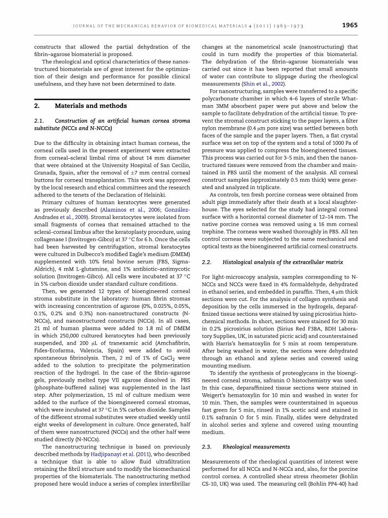
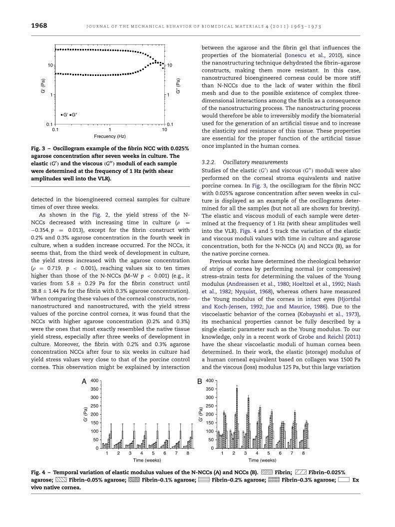
Fig. 3 – Oscillogram example of the fibrin NCC with 0.025%agarose concentration after seven weeks in culture. Theelastic (G′) and the viscous (G′′) moduli of each samplewere determined at the frequency of 1 Hz (with shearamplitudes well into the VLR).
detected in the bioengineered corneal samples for culturetimes of over three weeks.
As shown in the Fig. 2, the yield stress of the N-NCCs decreased with increasing time in culture (ρ =
−0.354,p = 0.013), except for the fibrin construct with0.2% and 0.3% agarose concentration in the fourth week inculture, when a sudden increase occurred. For the NCCs, itseems that, from the third week of development in culture,the yield stress increased with the agarose concentration(ρ = 0.719, p < 0.001), reaching values six to ten timeshigher than those of the N-NCCs (M–W p < 0.001) (e.g., itvaries from 5.8 ± 0.29 Pa for the fibrin construct until38.8 ± 1.44 Pa for the fibrin with 0.3% agarose concentration).When comparing these values of the corneal constructs, non-nanostructured and nanostructured, with the yield stressvalues of the porcine control cornea, it was found that theNCCs with higher agarose concentration (0.2% and 0.3%)were the ones that most exactly resembled the native tissueyield stress, especially after three weeks of development inculture. Moreover, the fibrin with 0.2% and 0.3% agaroseconcentration NCCs after four to six weeks in culture hadyield stress values very close to that of the porcine controlcornea. This observation might be explained by interaction
between the agarose and the fibrin gel that influences theproperties of the biomaterial (Ionescu et al., 2010), sincethe nanostructuring technique dehydrated the fibrin–agaroseconstructs, making them more resistant. In this case,nanostructured bioengineered corneas could be more stiffthan N-NCCs due to the lack of water within the fibrilmesh and due to the possible existence of complex three-dimensional interactions among the fibrils as a consequenceof the nanostructuring process. The nanostructuring processwould therefore be able to irreversibly modify the biomaterialused for the generation of an artificial tissue and to increasethe elasticity and resistance of this tissue. These propertiesare essential for the proper function of the artificial tissueonce implanted in the human cornea.
3.2.2. Oscillatory measurementsStudies of the elastic (G′) and viscous (G′′) moduli were alsoperformed on the corneal stroma equivalents and nativeporcine cornea. In Fig. 3, the oscillogram for the fibrin NCCwith 0.025% agarose concentration after seven weeks in cul-ture is displayed as an example of the oscillograms deter-mined for all the samples (but not all are shown for brevity).The elastic and viscous moduli of each sample were deter-mined at the frequency of 1 Hz (with shear amplitudes wellinto the VLR). Figs. 4 and 5 track the variation of the elasticand viscous moduli values with time in culture and agaroseconcentration, both for the N-NCCs (A) and NCCs (B), as forthe native porcine cornea.
Previous works have determined the rheological behaviorof strips of cornea by performing normal (or compressive)stress–strain tests for determining the values of the Youngmodulus (Andreassen et al., 1980; Hoeltzel et al., 1992; Nashet al., 1982; Nyquist, 1968), whereas others have measuredthe Young modulus of the cornea in intact eyes (Hjortdaland Koch-Jensen, 1992; Jue and Maurice, 1986). Due to theviscoelastic behavior of the cornea (Kobayashi et al., 1973),its mechanical properties cannot be fully described by asingle elastic parameter such as the Young modulus. To ourknowledge, only in a recent work of Grobe and Reichl (2011)have the shear viscoelastic moduli of human cornea beendetermined. In their work, the elastic (storage) modulus ofa human corneal equivalent based on collagen was 1500 Paand the viscous (loss) modulus 125 Pa, but this large variation
400A B350
300
250
200
G' (
Pa)
150
100
50
01 2 3 4 5
Time (weeks)
6 7 8
400
350
300
250
200
G' (
Pa)
150
100
50
01 2 3 4 5
Time (weeks)
6 7 8
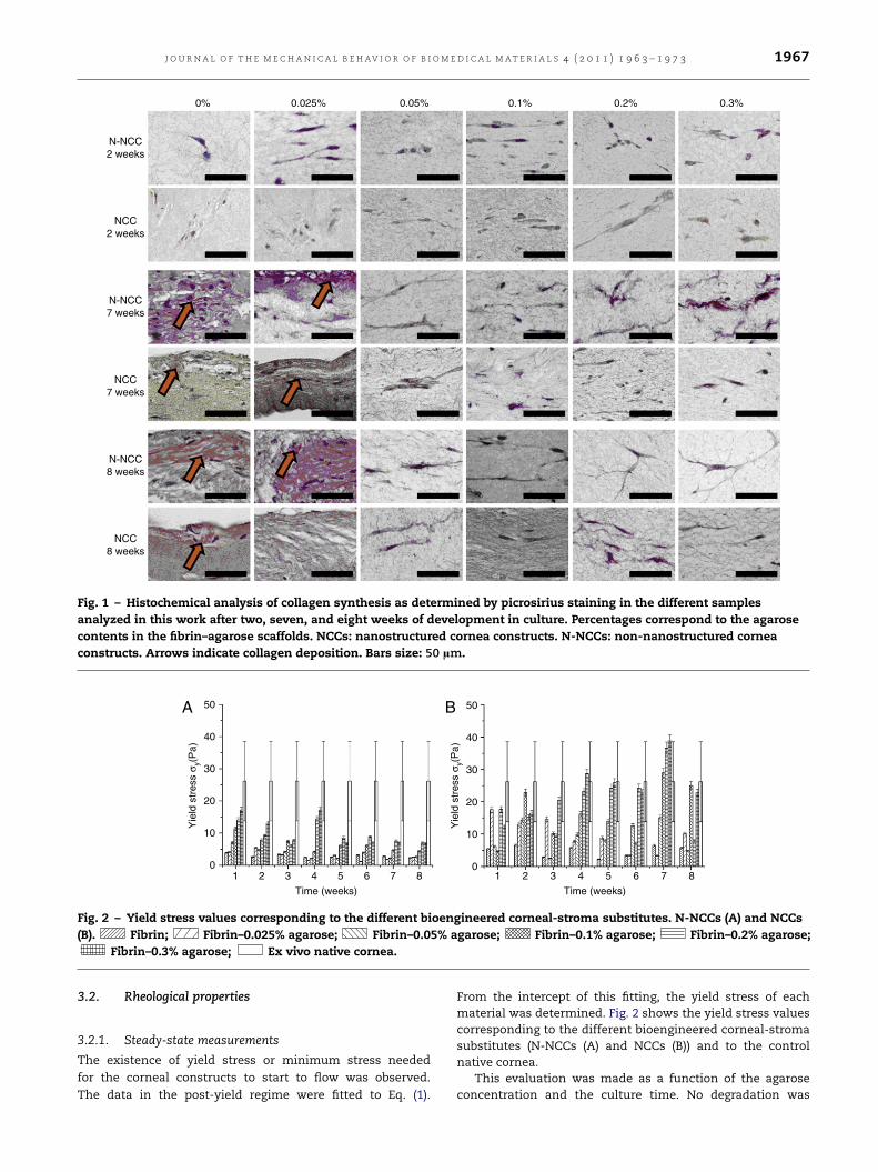
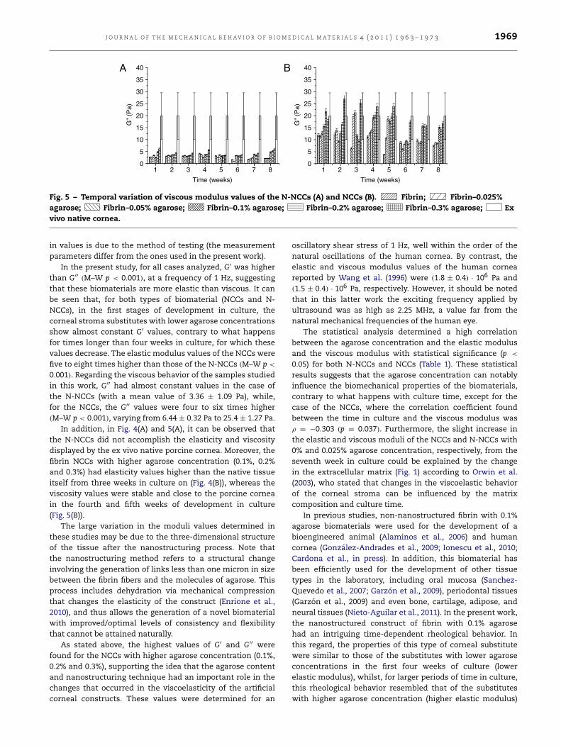
Fig. 4 – Temporal variation of elastic modulus values of the N-NCCs (A) and NCCs (B). Fibrin; Fibrin–0.025%agarose; Fibrin–0.05% agarose; Fibrin–0.1% agarose; Fibrin–0.2% agarose; Fibrin–0.3% agarose; Exvivo native cornea.
J O U R N A L O F T H E M E C H A N I C A L B E H AV I O R O F B I O M E D I C A L M A T E R I A L S 4 ( 2 0 1 1 ) 1 9 6 3 – 1 9 7 3 1969
40A B35
30
25
20
G"
(Pa)
15
10
5
01 2 3 4 5
Time (weeks)
6 7 8
40
35
30
25
20
G"
(Pa)
15
10
5
01 2 3 4 5
Time (weeks)
6 7 8
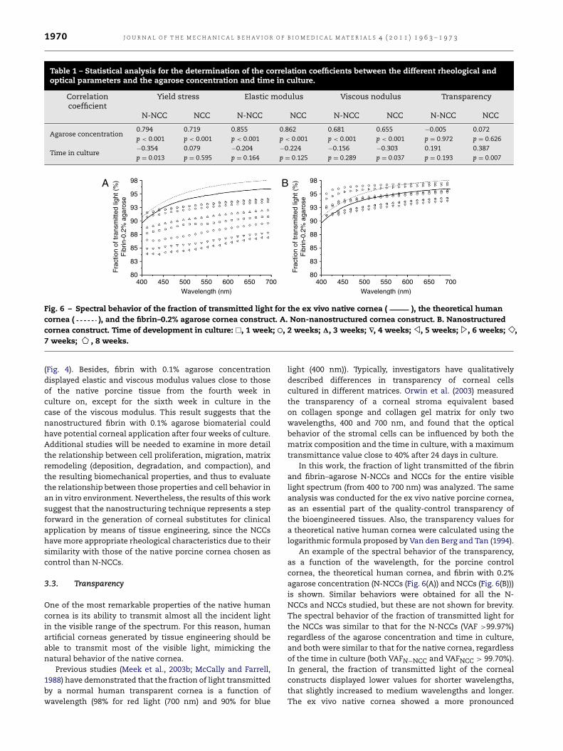
Fig. 5 – Temporal variation of viscous modulus values of the N-NCCs (A) and NCCs (B). Fibrin; Fibrin–0.025%agarose; Fibrin–0.05% agarose; Fibrin–0.1% agarose; Fibrin–0.2% agarose; Fibrin–0.3% agarose; Exvivo native cornea.
in values is due to the method of testing (the measurementparameters differ from the ones used in the present work).
In the present study, for all cases analyzed, G′ was higherthan G′′ (M–W p < 0.001), at a frequency of 1 Hz, suggestingthat these biomaterials are more elastic than viscous. It canbe seen that, for both types of biomaterial (NCCs and N-NCCs), in the first stages of development in culture, thecorneal stroma substitutes with lower agarose concentrationsshow almost constant G′ values, contrary to what happensfor times longer than four weeks in culture, for which thesevalues decrease. The elastic modulus values of the NCCs werefive to eight times higher than those of the N-NCCs (M–W p <
0.001). Regarding the viscous behavior of the samples studiedin this work, G′′ had almost constant values in the case ofthe N-NCCs (with a mean value of 3.36 ± 1.09 Pa), while,for the NCCs, the G′′ values were four to six times higher(M–W p < 0.001), varying from 6.44 ± 0.32 Pa to 25.4 ± 1.27 Pa.
In addition, in Fig. 4(A) and 5(A), it can be observed thatthe N-NCCs did not accomplish the elasticity and viscositydisplayed by the ex vivo native porcine cornea. Moreover, thefibrin NCCs with higher agarose concentration (0.1%, 0.2%and 0.3%) had elasticity values higher than the native tissueitself from three weeks in culture on (Fig. 4(B)), whereas theviscosity values were stable and close to the porcine corneain the fourth and fifth weeks of development in culture(Fig. 5(B)).
The large variation in the moduli values determined inthese studies may be due to the three-dimensional structureof the tissue after the nanostructuring process. Note thatthe nanostructuring method refers to a structural changeinvolving the generation of links less than one micron in sizebetween the fibrin fibers and the molecules of agarose. Thisprocess includes dehydration via mechanical compressionthat changes the elasticity of the construct (Enrione et al.,2010), and thus allows the generation of a novel biomaterialwith improved/optimal levels of consistency and flexibilitythat cannot be attained naturally.
As stated above, the highest values of G′ and G′′ werefound for the NCCs with higher agarose concentration (0.1%,0.2% and 0.3%), supporting the idea that the agarose contentand nanostructuring technique had an important role in thechanges that occurred in the viscoelasticity of the artificialcorneal constructs. These values were determined for an
oscillatory shear stress of 1 Hz, well within the order of thenatural oscillations of the human cornea. By contrast, theelastic and viscous modulus values of the human corneareported by Wang et al. (1996) were (1.8 ± 0.4) · 106 Pa and(1.5 ± 0.4) · 106 Pa, respectively. However, it should be notedthat in this latter work the exciting frequency applied byultrasound was as high as 2.25 MHz, a value far from thenatural mechanical frequencies of the human eye.
The statistical analysis determined a high correlationbetween the agarose concentration and the elastic modulusand the viscous modulus with statistical significance (p <
0.05) for both N-NCCs and NCCs (Table 1). These statisticalresults suggests that the agarose concentration can notablyinfluence the biomechanical properties of the biomaterials,contrary to what happens with culture time, except for thecase of the NCCs, where the correlation coefficient foundbetween the time in culture and the viscous modulus wasρ = −0.303 (p = 0.037). Furthermore, the slight increase inthe elastic and viscous moduli of the NCCs and N-NCCs with0% and 0.025% agarose concentration, respectively, from theseventh week in culture could be explained by the changein the extracellular matrix (Fig. 1) according to Orwin et al.(2003), who stated that changes in the viscoelastic behaviorof the corneal stroma can be influenced by the matrixcomposition and culture time.
In previous studies, non-nanostructured fibrin with 0.1%agarose biomaterials were used for the development of abioengineered animal (Alaminos et al., 2006) and humancornea (González-Andrades et al., 2009; Ionescu et al., 2010;Cardona et al., in press). In addition, this biomaterial hasbeen efficiently used for the development of other tissuetypes in the laboratory, including oral mucosa (Sanchez-Quevedo et al., 2007; Garzón et al., 2009), periodontal tissues(Garzón et al., 2009) and even bone, cartilage, adipose, andneural tissues (Nieto-Aguilar et al., 2011). In the present work,the nanostructured construct of fibrin with 0.1% agarosehad an intriguing time-dependent rheological behavior. Inthis regard, the properties of this type of corneal substitutewere similar to those of the substitutes with lower agaroseconcentrations in the first four weeks of culture (lowerelastic modulus), whilst, for larger periods of time in culture,this rheological behavior resembled that of the substituteswith higher agarose concentration (higher elastic modulus)
1970 J O U R N A L O F T H E M E C H A N I C A L B E H AV I O R O F B I O M E D I C A L M A T E R I A L S 4 ( 2 0 1 1 ) 1 9 6 3 – 1 9 7 3
Table 1 – Statistical analysis for the determination of the correlation coefficients between the different rheological andoptical parameters and the agarose concentration and time in culture.
Correlationcoefficient
Yield stress Elastic modulus Viscous nodulus Transparency
N-NCC NCC N-NCC NCC N-NCC NCC N-NCC NCC
Agarose concentration0.794 0.719 0.855 0.862 0.681 0.655 −0.005 0.072p < 0.001 p < 0.001 p < 0.001 p < 0.001 p < 0.001 p < 0.001 p = 0.972 p = 0.626
Time in culture−0.354 0.079 −0.204 −0.224 −0.156 −0.303 0.191 0.387p = 0.013 p = 0.595 p = 0.164 p = 0.125 p = 0.289 p = 0.037 p = 0.193 p = 0.007
80400 450 500 550
Wavelength (nm)600 650 700
83
85
88
Fra
ctio
n of
tran
smitt
ed li
ght (
%)
Fib
rin-0
.2%
aga
rose
90
93
95
98A B
80400 450 500 550
Wavelength (nm)600 650 700
83
85
88
Fra
ctio
n of
tran
smitt
ed li
ght (
%)
Fib
rin-0
.2%
aga
rose
90
93
95
98
Fig. 6 – Spectral behavior of the fraction of transmitted light for the ex vivo native cornea ( ), the theoretical humancornea ( ), and the fibrin–0.2% agarose cornea construct. A. Non-nanostructured cornea construct. B. Nanostructuredcornea construct. Time of development in culture: �, 1 week; ⃝, 2 weeks; 1, 3 weeks; ∇, 4 weeks; , 5 weeks; , 6 weeks; ,7 weeks; , 8 weeks.
(Fig. 4). Besides, fibrin with 0.1% agarose concentrationdisplayed elastic and viscous modulus values close to thoseof the native porcine tissue from the fourth week inculture on, except for the sixth week in culture in thecase of the viscous modulus. This result suggests that thenanostructured fibrin with 0.1% agarose biomaterial couldhave potential corneal application after four weeks of culture.Additional studies will be needed to examine in more detailthe relationship between cell proliferation, migration, matrixremodeling (deposition, degradation, and compaction), andthe resulting biomechanical properties, and thus to evaluatethe relationship between those properties and cell behavior inan in vitro environment. Nevertheless, the results of this worksuggest that the nanostructuring technique represents a stepforward in the generation of corneal substitutes for clinicalapplication by means of tissue engineering, since the NCCshavemore appropriate rheological characteristics due to theirsimilarity with those of the native porcine cornea chosen ascontrol than N-NCCs.
3.3. Transparency
One of the most remarkable properties of the native humancornea is its ability to transmit almost all the incident lightin the visible range of the spectrum. For this reason, humanartificial corneas generated by tissue engineering should beable to transmit most of the visible light, mimicking thenatural behavior of the native cornea.
Previous studies (Meek et al., 2003b; McCally and Farrell,1988) have demonstrated that the fraction of light transmittedby a normal human transparent cornea is a function ofwavelength (98% for red light (700 nm) and 90% for blue
light (400 nm)). Typically, investigators have qualitativelydescribed differences in transparency of corneal cellscultured in different matrices. Orwin et al. (2003) measuredthe transparency of a corneal stroma equivalent basedon collagen sponge and collagen gel matrix for only twowavelengths, 400 and 700 nm, and found that the opticalbehavior of the stromal cells can be influenced by both thematrix composition and the time in culture, with a maximumtransmittance value close to 40% after 24 days in culture.
In this work, the fraction of light transmitted of the fibrinand fibrin–agarose N-NCCs and NCCs for the entire visiblelight spectrum (from 400 to 700 nm) was analyzed. The sameanalysis was conducted for the ex vivo native porcine cornea,as an essential part of the quality-control transparency ofthe bioengineered tissues. Also, the transparency values fora theoretical native human cornea were calculated using thelogarithmic formula proposed by Van den Berg and Tan (1994).
An example of the spectral behavior of the transparency,as a function of the wavelength, for the porcine controlcornea, the theoretical human cornea, and fibrin with 0.2%agarose concentration (N-NCCs (Fig. 6(A)) and NCCs (Fig. 6(B)))is shown. Similar behaviors were obtained for all the N-NCCs and NCCs studied, but these are not shown for brevity.The spectral behavior of the fraction of transmitted light forthe NCCs was similar to that for the N-NCCs (VAF >99.97%)regardless of the agarose concentration and time in culture,and both were similar to that for the native cornea, regardlessof the time in culture (both VAFN−NCC and VAFNCC > 99.70%).In general, the fraction of transmitted light of the cornealconstructs displayed lower values for shorter wavelengths,that slightly increased to medium wavelengths and longer.The ex vivo native cornea showed a more pronounced
J O U R N A L O F T H E M E C H A N I C A L B E H AV I O R O F B I O M E D I C A L M A T E R I A L S 4 ( 2 0 1 1 ) 1 9 6 3 – 1 9 7 3 1971
A B100
Frac
tion
of tr
ansm
itted
ligh
t (%
)
95
90
85
801 2 3 4 5
Time (weeks)
6 7 8
100
Frac
tion
of tr
ansm
itted
ligh
t (%
)
95
90
85
801 2 3 4 5
Time (weeks)
6 7 8
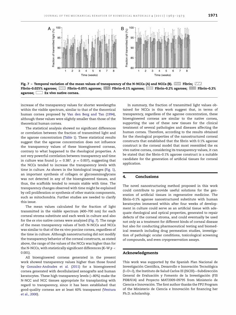
Fig. 7 – Temporal variation of the mean values of transparency of the N-NCCs (A) and NCCs (B). Fibrin;Fibrin–0.025% agarose; Fibrin–0.05% agarose; Fibrin–0.1% agarose; Fibrin–0.2% agarose; Fibrin–0.3%agarose; Ex vivo native cornea.
increase of the transparency values for shorter wavelengthswithin the visible spectrum, similar to that of the theoreticalhuman cornea proposed by Van den Berg and Tan (1994),although these values were slightly smaller than those of thetheoretical human cornea.
The statistical analysis showed no significant differencesor correlation between the fraction of transmitted light andthe agarose concentration (Table 1). These statistical resultssuggest that the agarose concentration does not influencethe transparency values of these bioengineered corneas,contrary to what happened to the rheological properties. Anot very powerful correlation between transparency and timein culture was found (ρ = 0.387, p = 0.007), suggesting thatthe NCCs tended to increase the transparency levels withtime in culture. As shown in the histological images (Fig. 1),an important synthesis of collagen or glycosaminoglycanswas not detected in any of the bioengineered tissues, andthus, the scaffolds tended to remain stable with time. Thetransparency changes observed with timemight be explainedby cell proliferation or synthesis of other matrix components,such as mitochondria. Further studies are needed to clarifythis issue.
The mean values calculated for the fraction of lighttransmitted in the visible spectrum (400–700 nm) for eachcorneal stroma substitute and each week in culture and alsofor the ex vivo native cornea were analyzed (Fig. 7). The rangeof the mean transparency values of both N-NCCs and NCCswas similar to that of the ex vivo porcine cornea, regardless ofthe time in culture. Although nanostructuring did not modifythe transparency behavior of the corneal constructs, as statedabove, the range of the values of the NCCs was higher than forthe N-NCCs, with statistically significant differences (K–W p <
0.001).
All bioengineered corneas generated in the presentwork showed transparency values higher than those foundby Gonzalez-Andrades et al. (2011) for a bioengineeredcornea generated with decellularized xenografts and humankeratocytes. These high transparency levels (>80%) make theN-NCC and NCC tissues appropriate for transplanting withregard to transparency, since it has been established thatgood-quality corneas are at least 60% transparent (Venturaet al., 2000).
In summary, the fraction of transmitted light values ob-tained for NCCs in this work suggest that, in terms oftransparency, regardless of the agarose concentration, thesebioengineered corneas are similar to the native cornea,supporting the use of these new tissues for the clinicaltreatment of several pathologies and diseases affecting thehuman cornea. Therefore, according to the results obtainedfor the rheological properties of the nanostructured cornealconstructs that established that the fibrin with 0.1% agaroseconstruct is the corneal model that most resembled the exvivo native cornea, considering its transparency values, it canbe stated that the fibrin–0.1% agarose construct is a suitablecandidate for the generation of artificial tissues for cornealapplication.
4. Conclusions
The novel nanostructuring method proposed in this workcould contribute to provide useful solutions for the gen-eration of artificial tissues in regenerative medicine. Thefibrin–0.1% agarose nanostructured substitute with humankeratocytes immersed within after four weeks of develop-ment in culture could serve as an artificial tissue with ade-quate rheological and optical properties, generated to repairdefects of the corneal stroma, and could eventually be usednot only as a treatment for different kinds of corneal diseasebut also for conducting pharmaceutical testing and biomed-ical research including drug permeation studies, investiga-tion of pathologic ocular conditions, toxicological screeningof compounds, and even cryopreservation assays.
Acknowledgments
This work was supported by the Spanish Plan Nacional deInvestigación Científica, Desarrollo e Innovación Tecnológica(I+D+I), the Instituto de Salud Carlos III (ISCIII)—SubdirecciónGeneral de Evaluación y Fomento de la Investigación (FISPI08/614) and Proyecto MAT2009-09795 from Ministerio deCiencia e Innovación. The first author thanks the FPU Programof the Ministerio de Ciencia e Innovación for financing herPh.D. scholarship.
1972 J O U R N A L O F T H E M E C H A N I C A L B E H AV I O R O F B I O M E D I C A L M A T E R I A L S 4 ( 2 0 1 1 ) 1 9 6 3 – 1 9 7 3
R E F E R E N C E S
Alaminos, M., Sanchez-Quevedo, M.C., Munoz-Avila, J.I., Serrano,D., Medialdea, S., Carreras, I., Campos, A., 2006. Constructionof a complete rabbit cornea substitute using a fibrin–agarosescaffold. Invest. Ophth. Vis. Sci. 47, 3311–3317.
Andreassen, T.T., Simonsen, A.H., Oxlund, H., 1980. Biomechanicalproperties of keratoconus and normal corneas. Exp. Eye Res.31, 435–441.
Ashiku, S.K., Randolph, M.A., Vacanti, C.A., 1997. Tissueengineering cartilage. Mater. Sci. Forum 250, 129–150.
Barnes, H.A., 1995. A review of the slip (wall depletion) of polymersolutions, emulsions and particle suspensions in viscometers:its cause, character, and cure. J. Non-Newton. Fluid 56, 221–251.
Bostrom, R.D., Mikos, A.G., 1997. Tissue engineering of bone.In: Atala, A., Mooney, A.J. (Eds.), Synthetic BiodegradablePolymers Scaffolds. Birkhäuser, Boston, pp. 215–234.
Cao, Y., Ibarra, C., Vacanti, C.A., 1997. Tissue engineering car-tilage and bone. In: Atala, A., Mooney, A.J. (Eds.), Syn-thetic Biodegradable Polymers Scaffolds. Birkhäuser, Boston,pp. 199–214.
Cardona, J.C., Ionescu, A.M., Gomez-Sotomayor, R., Gonzalez-Andrades, M., Campos, A., Alaminos, M., Perez, M.M., 2011.Transparency in a fibrin and fibrin–agarose corneal stromasubstitute generated by tissue engineering. Cornea (in press).
Chen, J., Li, Q., Huang, Y., Ding, Y., Deng, H., Zhao, S.,Chen, R., 2005. Study on biocompatibility of complexesof collagen–chitosan–sodium hyaluronate and cornea. Artif.Organs 29, 104–113.
Cima, L.G., Ingber, D.E., Vacanti, J.P., Langer, R., 1991a. Hepatocyteculture on biodegradable polymeric substrates. Biotechnol.Bioeng. 38, 145–148.
Cima, L.G., Vacanti, J.P., Vacanti, C., Ingber, D., Mooney, D., Langer,R., 1991b. Tissue engineering by cell transplantation usingdegradable polymer substrates. J. Biomech. Eng. 113, 143–151.
Cohen, D., Chuck, R., Bearman, G., McDonnell, P., Grundfest, W.,2001. Ablation spectra of the human cornea. J. Biomed. Opt. 6(3), 339–343.
Cooper, M.L., Hansbrough, J.F., Spielvogel, R.L., Cohen, R., Bartel,R.L., Naughton, G., 1991. In vivooptimization of a livingdermal substitute employing cultured human fibroblastson a biodegradable polyglycolic acid or polyglactin mesh.Biomaterials 12, 243–248.
Enrione, J., Osorio, F., Lopez, P., Weinstein-Oppenheimer, C.,Fuentes, M.A., Ceriani, R., Brown, D.I., et al., 2010. Charac-terization of a gelatin/chitosan/hyaluronan scaffold-polymer.Electron. J. Biotechnol. 13 (5), http://dx.doi.org/10.2225/vol13-issue5-fulltext-15.
Farrell, R.A., McCally, R.L., Tatham, P.E.R., 1973. Wavelengthdependencies of light scattering in normal and cold swollenrabbit corneas and their structural implications. J. Physiol. 233,589–612.
Freund, D.E., McCally, R.L., Farrell, R.A., 1986. Direct summation offields for light scattering by fibrils with applications to normalcorneas. Appl. Opt. 25, 2739–2746.
Garzón, I., Sánchez-Quevedo, M.C., Moreu, G., González-Jaranay,M., González-Andrades, M., Montalvo, A., Campos, A.,Alaminos, M., 2009. In vitro and in vivo cytokeratin patternsof expression in bioengineered human periodontal mucosa. J.Periodontal Res. 44 (5), 588–597.
Gonzalez-Andrades, M., Cardona, J.C., Ionescu, A.M., Campos, A.,Perez, M.M., Alaminos, M., 2011. Generation of bioengineeredcorneas with decellularized xenografts and human kerato-cytes. Invest. Ophth. Vis. Sci. 52 (1), 215–222.
González-Andrades, M., Garzón, I., Gascón, M.I., Muñoz-Avila,J.I., Sánchez-Quevedo, M.C., Campos, A., Alaminos, M.,2009. Sequential development of intercellular junctions inbioengineered human corneas. J. Tissue Eng. Regen. Med. 3 (6),442–449.
Griffith, M., Osborne, R., Munger, R., Xiong, X., Doillon, C.J.,Laycock, N.L., Hakim, M., Song, Y., Watsky, M.A., 1999.Functional human corneal equivalents constructed from celllines. Science 286, 2169–2172.
Grobe, G., Reichl, S., 2011. Examining the suitability ofRiboflavin/UVA treatment for strengthening the stromalbioequivalent of a human cornea construct. Curr. Eye Res. 36,217–231.
Hadjipanayi, E., Ananta, M., Binkowski, M., Streeter, I., Lu,Z., Cui, Z.F., Brown, R.A., Mudera, V., 2011. Mechanismsof structure generation during plastic compression onnanofibrillar collagen hydrogel scaffolds: towards engineeringof collagen. J. Tissue Eng. Regen. Med. 5, 505–519.
Hjortdal, J.O., Koch-Jensen, P., 1992. In situ mechanical behaviorof the human cornea as evaluated by simultaneous measure-ments of corneal strain, corneal surface contour, and cornealthickness. Invest. Ophth. Vis. Sci. 33 (Suppl.), 1022.
Ho, K.T., Jannetta, C., Vacanti, J.P., et al. Engineered bonefrom polyglycolic acid polymer scaffold and periosteum. In:Proceedings of the 1995 MRS Spring Meeting, Polymers inMedicine and Pharmacy, April 17–19, San Francisco, 1995, pp.91–98.
Hoeltzel, D.A., Altman, P., Buzard, K., Choe, K., 1992. Stripextensiometry for comparison of the mechanical responseof bovine, rabbit, and human corneas. J. Biomech. Eng. 114,202–215.
Ionescu, A.M., Cardona, J.C., González-Andrades, M., Alaminos,M., Campos, A., Hita, E., Pérez, M.M., 2010. UV Absorbance ofa bioengineered corneal stroma substitute in the 240–400 nmrange. Cornea 29, 895–898.
Jones, D.S., Woolfson, A.D., Brown, A.F., 1997a. Textural,viscoelastic and mucoadhesive properties of gels composed ofcellulose polymers. Int. J. Pharm. 151, 223–233.
Jones, D.S., Woolfson, A.D., Brown, A.F., 1997b. Textural analysisand flow rheometry of novel, bioadhesive antimicrobial oralgels. Pharm. Res. 14 (4), 450–457.
Jones, D.S., 1999. Dynamic mechanical analysis of polymericsystems of pharmaceutical and biomedical significance. Int. J.Pharm. 179, 167–178.
Jue, B., Maurice, D.M., 1986. The mechanical properties of therabbit and human cornea. J. Biomech. 19 (10), 847–853.
Kubelka, P., 1948. New contributions to the optics of intenselylight-scattering materials. Part I. J. Opt. Soc. Amer. 38, 448–457.
Kobayashi, A.S., Staberg, L.G., Schlegel, W.A., 1973. Viscoelasticproperties of human corneas. Exp. Mech. 13 (12), 497–503.
Lee, Y.K., 2007. Influence of scattering/absorption characteristicson the color of resin composites. Dent. Mater. 23, 124–131.
Llames, S.G., Del Rio, M., Larcher, F., García, E., García, M., JoseEscamez, M., Jorcano, J.L., Holguín, P., Meana, A., 2004. Humanplasma as a dermal scaffold for the generation of a completelyautologous bioengineered skin. Transplantation 77, 350–355.
Ma, P.X., 2004. Scaffolds for tissue fabrication. Mater. Today 7 (5),30–40.
Macosko, C.W., 1994. Rheology: Principles, Measurements, andApplications. VCH, New York.
Maurice, D.M., 1957. The structure and transparency of thecorneal stroma. J. Physiol. 136 (1), 263–286.
Maurice, D.M., 1984. The cornea and sclera. In: Davson, H. (Ed.),The Eye. Academic Press, Orlando, FL, pp. 1–158.
McCally, R.L., Farrell, R.A., 1982. Structural implications of smallangle light scattering from cornea. Exp. Eye Res. 34, 99–113.

J O U R N A L O F T H E M E C H A N I C A L B E H AV I O R O F B I O M E D I C A L M A T E R I A L S 4 ( 2 0 1 1 ) 1 9 6 3 – 1 9 7 3 1973
McCally, R.L., Farrell, R.A., 1988. Interaction of light andthe cornea: light scattering versus transparency. In: Ca-vanagh, H.D. (Ed.), The Cornea, Transactions of the WorldCongress on the Cornea. ZZZ, New York, pp. 165–171.
Meana, A., Iglesias, J., Del Rio, M., Larcher, F., Madrigal, B.,Fresno, M.F., Martin, C., San Roman, F., Tevar, F., 1998. Largesurface of cultured human epithelium obtained on a dermalmatrix based on live fibroblast-containing fibrin gels. Burns 24,621–630.
Meek, K.M., Dennis, S., Khan, S., 2003a. Changes in the refractiveindex of the stroma and its extrafibrillar matrix when thecornea swells. Biophys. J. 85 (4), 2205–2212.
Meek, K.M., Leonard, D.W., Connon, C.J., Dennis, S., Khan, S.,2003b. Transparency, swelling and scarring in the cornealstroma. Eye 17 (8), 927–936.
Minami, Y., Sugihara, H., Oono, S., 1993. Reconstruction of corneain three-dimensional collagen gel matrix culture. Invest.Ophth. Vis. Sci. 34, 2316–2324.
Mooney, D.J., Mikos, A.G., 1999. Growing new organs. Sci. Am. 280,60–65.
Nash, I.S., Greene, P.R., Foster, C.S., 1982. Comparison ofmechanical properties of keratoconus and normal corneas.Exp. Eye Res. 35, 413–423.
Nieto-Aguilar, R., Serrato, D., Garzón, I., Campos, A., Alaminos,M., 2011. Pluripotential differentiation capability of humanadipose-derived stem cells in a novel fibrin–agarose scaffold.J. Biomater. Appl. 25 (7), 743–768.
Nishida, K., 2003. Tissue engineering of the cornea. Cornea 22,S28–S34.
Nishida, K., Yamato, M., Hayashida, Y., Watanabe, K., Maeda, N.,Watanabe, H., Yamamoto, K., Nagai, S., Kikuchi, A., Tano, Y.,Okano, T., 2004. Functional bioengineered corneal epithelialsheet grafts from corneal stem cells expanded ex vivo on atemperature responsive cell culture surface. Transplantation77, 379–385.
Nyquist, G.W., 1968. Reology of the cornea: experimentaltechniques and results. Exp. Eye Res. 7, 183–188.
Orwin, E.J., Borene, M.L., Hubel, A., 2003. Biomechanical andoptical characteristics of a corneal stromal equivalent. J.Biomech. Eng. 125, 439–444.
Orwin, E.J., Hubel, A., 2000. In vitro culture characteristics ofcorneal epithelial, endothelial, and keratocyte cells in a nativecollagen matrix. Tissue Eng. 6, 307–319.
Peters, M.C., Mooney, D.J., 1997. Synthetic extracellular matricesfor cell transplantation. Mater. Sci. Forum 250, 43–52.
Porter, R.A., Brown, R.A., Eastwood, M., Occleston, N.L., Khaw, P.T.,1998. Ultrastructural changes during contraction of collagenlattices by ocular fibroblasts. Wound Repair. Regen. 6, 157–166.
Reichl, S., Bednarz, J., Muller-Goymann, C.C., 2004. Human cornealequivalent as cell culture model for in vitro drug permeationstudies. Br. J. Ophthalmol. 88, 560–565.
Reichl, S., Muller-Goymann, C.C., 2003. The use of a porcineorganotypic cornea construct for permeation studies fromformulations containing befunolol hydrochloride. Int. J.Pharm. 250, 191–201.
Ruberti, J.W., Zieske, J.D., 2008. Prelude to corneal tissueengineering—Gaining control of collagen organization. Prog.Retin. Eye Res. 27, 549–577.
Sanchez-Quevedo, M.C., Alaminos, M., Capitan, L.M., Moreu, G.,Garzon, I., Crespo, P.V., Campos, A, 2007. Histological andhistochemical evaluation of human oral mucosa constructsdeveloped by tissue engineering. Histol. Histopathol. 22 (6),631–640.
Schneider, A.I., Maier-Reif, K., Graeve, T., 1999. Constructing anin vitro cornea from cultures of the three specific corneal celltypes. In Vitro Cell. Dev. Biol. Anim. 35, 515–526.
Shin, J.E., Cornillion, P., Salim, L., 2002. The effect of centrifugationon agar/sucrose gels. Food Hydrocoll. 16, 89–94.
Simske, S.J., Ayers, R.A., Bateman, T.A., 1997. Porous materials forbone engineering. Mater. Sci. Forum 250, 151–182.
Tegtmeyer, S., Papantoniou, I., Muller-Goymann, C.C., 2001.Reconstruction of an in vitro cornea and its use for drugpermeation studies from different formulations containingpilocarpine hydrochloride. Eur. J. Pharm. Biopharm. 51,119–125.
Thennadil, S.N., 2008. Relationship between the Kubelka–Munkscattering and radiative transfer coefficients. J. Opt. Soc. Amer.A 25 (7), 1480–1485.
Van den Berg, T.J.T.P., Tan, K.E.W.P., 1994. Light transmittance ofthe human cornea from 320 to 700 nm for different ages. Vis.Res. 34, 1453–1456.
Ventura, L., Sousa, S.J., Messias, A.M., Bispo, J.M., 2000. Systemfor measuring the transmission spectrum of ‘in vitro’ corneas.Physiol. Meas. 21, 197–207.
Wang, H., Prendiville, P.L., McDonnell, P.J., Chang, W.V., 1996.An ultrasonic technique for the measurement of the elasticmoduli of human cornea. J. Biomech. 29 (12), 1633–1636.
Whitcher, J.P., Srinivasan, M., Upadhyay, M.P., 2001. Cornealblindness: a global perspective. Bull. World Health Organ. 79(3), 214–221.
Yang, S., Leong, K.F., Du, Z., Chua, C.K., 2001. The design ofscaffolds for use in tissue engineering. Part I. Traditionalfactors. Tissue Eng. 7 (6), 679–689.
Zerbe, B.L., Belin, M.W., Ciolino, J.B., 2006. Results from the mul-ticenter Boston type 1 keratoprothesis study. Ophthalmology113 (10), 1779.e1-7.
Zieske, J.D., Mason, V.S., Wasson, M.E., Meunier, S.F., Nolte,C.J., Fukai, N., Olsen, B.R., Parenteau, N.L., 1994. Basementmembrane assembly and differentiation of cultured cornealcells: importance of culture environment and endothelial cellinteraction.. Exp. Cell Res. 214, 621–633.
Copyright © 2022 FDOKUMEN