Incorporation of silica particles into decellularized tissue biomaterial and its effect on...
29
www.rsc.org/ advances RSC Advances This is an Accepted Manuscript, which has been through the Royal Society of Chemistry peer review process and has been accepted for publication. Accepted Manuscripts are published online shortly after acceptance, before technical editing, formatting and proof reading. Using this free service, authors can make their results available to the community, in citable form, before we publish the edited article. This Accepted Manuscript will be replaced by the edited, formatted and paginated article as soon as this is available. You can find more information about Accepted Manuscripts in the Information for Authors. Please note that technical editing may introduce minor changes to the text and/or graphics, which may alter content. The journal’s standard Terms & Conditions and the Ethical guidelines still apply. In no event shall the Royal Society of Chemistry be held responsible for any errors or omissions in this Accepted Manuscript or any consequences arising from the use of any information it contains. View Article Online View Journal This article can be cited before page numbers have been issued, to do this please use: B. Mendoza- Novelo, M. C. Lona, G. García-González, L. E. Castellano, J. Delgado, P. Cuéllar-Mata, J. M. Flores-
Transcript of Incorporation of silica particles into decellularized tissue biomaterial and its effect on...

www.rsc.org/advances
RSC Advances
This is an Accepted Manuscript, which has been through the Royal Society of Chemistry peer review process and has been accepted for publication.
Accepted Manuscripts are published online shortly after acceptance, before technical editing, formatting and proof reading. Using this free service, authors can make their results available to the community, in citable form, before we publish the edited article. This Accepted Manuscript will be replaced by the edited, formatted and paginated article as soon as this is available.
You can find more information about Accepted Manuscripts in the Information for Authors.
Please note that technical editing may introduce minor changes to the text and/or graphics, which may alter content. The journal’s standard Terms & Conditions and the Ethical guidelines still apply. In no event shall the Royal Society of Chemistry be held responsible for any errors or omissions in this Accepted Manuscript or any consequences arising from the use of any information it contains.
View Article OnlineView Journal
This article can be cited before page numbers have been issued, to do this please use: B. Mendoza-
Novelo, M. C. Lona, G. García-González, L. E. Castellano, J. Delgado, P. Cuéllar-Mata, J. M. Flores-

- 1 -
Incorporation of silica particles into decellularized tissue
biomaterial and its effect on the macrophage activation
Birzabith Mendoza-Noveloa,*
, María C. Lona-Ramosa, Gerardo García-González
b, Laura E.
Castellanoc, Jorge Delgado
a, Patricia Cuellar-Mata
d, J. Mauricio Flores-Moreno
e, Juan
Vargasf, J. Alfredo Gutiérrez
b, Eva E. Ávila
d, José L. Mata-Mata
b
a Depto. de Ingenierías Química, Electrónica y Biomédica, DCI, Universidad de Guanajuato,
Loma del Bosque 103, 37150, León, México
b Depto. de Química, DCNE, Universidad de Guanajuato, Noria alta s/n, 36050, Guanajuato,
México
c Depto. de Ciencias Aplicadas al Trabajo, DCS, Universidad de Guanajuato, Garza Sada 532,
37150, León, México
d Depto. de Biología, DCNE, Universidad de Guanajuato, Noria alta s/n, 36050, Guanajuato,
México
e Centro de Investigación en Óptica A. C., Loma del Bosque 115, 37150, León, México
f IPN-Unidad Profesional Interdisciplinaria de Ingenierías Campus Guanajuato, Industrial
Puerto interior, 36275, Silao de la Victoria, México
*Correspondence: [email protected], Phone: +52-477-7885100 ext 8464
Page 1 of 28 RSC Advances
RS
CA
dvan
ces
Acc
epte
dM
anus
crip
t
Publ
ishe
d on
14
Nov
embe
r 20
14. D
ownl
oade
d by
UN
IVE
RIS
DA
D D
E G
UA
NA
JUA
TO
on
14/1
1/20
14 1
6:05
:04.
View Article OnlineDOI: 10.1039/C4RA08984G

- 2 -
Abstract
This paper describes an optimized procedure to incorporate silica particles by
hydrolysis/polycondensation of sodium silicate into pericardial (ECM) matrix scaffolds and
points out the effect of the biocomposites on the in vitro response of macrophages by
assessment the secretion of signaling molecules. Variables (concentration, pH, time) of the
sol-gel process allow a gradual incorporation of silica into the ECM scaffolds as confirmed by
gravimetry, FT-IR, SEM and EDX microanalysis. The SiO2 incorporation increases the
resistance to in vitro degradation but not alters either denaturation temperature or free amines
of non-crosslinked ECM fibrous scaffolds, however, properties of oligourethane-crosslinked
scaffolds are not modified after silica incorporation. Despite the fact that cell viability is
gradually decreased on the ECM materials crosslinked with oligourethane and functionalized
with silica, murine RAW264.7 macrophages are able to secrete b-FGF, TGF- and VEGF.
Secretion of growth factors by RAW264.7 macrophages after 6 h of culture on scaffolds
containing silica was lower but it was sustained for 24 h as compared to cells cultured on
silica-free materials. Human peripheral blood macrophages cultured with materials containing
silica show a higher production of IL-6, IL-10 or TNF- than with the silica-free counterparts
but in a time-dependent manner from one to four days of culture. Results suggest that
stimulation of macrophages is induced by silica particles deposited onto the ECM fibrous
network, which represents an opportunity to control the cell response to decellularized tissue-
derived biomaterials through strategies intended to stimulate cells via signaling molecules
secreted by macrophages.
Keywords: crosslinked/decellularized scaffolds; silica functionalization; macrophage
activation
Page 2 of 28RSC Advances
RS
CA
dvan
ces
Acc
epte
dM
anus
crip
t
Publ
ishe
d on
14
Nov
embe
r 20
14. D
ownl
oade
d by
UN
IVE
RIS
DA
D D
E G
UA
NA
JUA
TO
on
14/1
1/20
14 1
6:05
:04.
View Article OnlineDOI: 10.1039/C4RA08984G

- 3 -
1. Introduction
Crosslinking and functionalization of extracellular matrix (ECM) scaffolds derived
from animal tissues are strategies intended to tailor the physicochemical properties and
ultimately to improve its biological performance in tissue engineering applications.
Crosslinking of collagen, main component of matrices as dermis or pericardium, is a first
action to slow or prevent its degradation, to inhibit the recognition of surface epitopes by the
host, and to improve mechanical properties.1-2
Our research group has reported a method in
which collagen is crosslinked through isocyanate reactions with water-soluble blocked
urethane oligomers after pH-responsive deblocking of carbamoylsulfonate end groups. This
method allowed the preparation of a porous collagen mesh coated with oligourethanes that
reveals excellent tensile mechanical properties and a degree of crosslinking adjusted tuning
pH, volume, concentration or molecular weight.3-4
Furthermore, effective stabilization of
biological scaffolds with oligourethanes can be achieved simultaneously with incorporation of
silica by the sol-gel process of tetraethyl orthosilicate.5 Thus, although silica is generally
accepted as having low toxicity, the biocompatibility of silicon and silica in its different
forms (i.e. nanoparticles, gels, films) as a “new” class of biomaterial should be revisited.6
Silicate–biopolymer hybrids manufactured in different forms have recently received
considerable attention as templates to repair/replace bone-defective tissue 7-8
, to induce
osteoblastic differentiation 9-13
, to treat chronic wounds 14
, to immobilize cells 15-16
, to delivery
drug 17-21
and morphogenetic proteins 22-23
, and to trigger cell-mediated immunity for tumor
immunotherapy. 24-25
The incorporation of silicate particles into biopolymers has shown that
yields a controllable biodegradability through adjustment of the silicate content 26-27
, and that
influence the cell response through intracellular uptake of the silicate particles and its
dissolution products. The nanoparticulate debris released from biomaterials containing silicon
can reach and penetrate the cell surface affecting its behavior in a manner that dependent of
size/porosity, surface chemistry (e.g. ≡Si-OH group concentration, charge) and dissolution
Page 3 of 28 RSC Advances
RS
CA
dvan
ces
Acc
epte
dM
anus
crip
t
Publ
ishe
d on
14
Nov
embe
r 20
14. D
ownl
oade
d by
UN
IVE
RIS
DA
D D
E G
UA
NA
JUA
TO
on
14/1
1/20
14 1
6:05
:04.
View Article OnlineDOI: 10.1039/C4RA08984G
- 4 -
(biodegradation) rate. 28-33
This is the case of fibroblasts that are internalized by aggregates of
silica nanoparticles, which are dissolved intracellularly and released either as colloidal or
soluble species.34-36
Key aspects in tissue engineering issues such as osteogenic differentiation
of stem cells 10,12
and enhanced angiogenesis 35
have shown be improved by the degradation
products of biomaterials containing silicon.
Macrophages are immune cells key players in the release of messengers and mediators
(e.g. enzymes, cytokines and growth factors) that act as signals to induce migration,
proliferation or differentiation of other cell types. 37
Macrophages mediate the healing
responses to implanted biomaterials, fundamentally by two outcomes: scar tissue formation
(M1 pathway) or regeneration (M2 pathway). 37-38
The modulation of the inflammatory
response by the physical and chemical properties of scaffolds represents a hypothesis
currently assessed in the design of biomaterials. For instance, mechanical strain 39
,
glycoprotein adsorption 40
and chemical composition 41-43
are some aspects related to
biomaterial that are keys to modulate the translation of human peripheral blood-derived
macrophages from a pro-inflammatory process (M1) to an anti-inflammatory process (M2).
Recent reports have described the engagement of cytokines secreted by macrophages in
angiogenesis and neovascularization in co-cultures on both 2D 44
and 3D 45
(on collagen
templates). Thus, pro-inflammatory stimuli have been proposed as an active participant in the
hard and soft tissue engineering context to accelerate healing mechanisms. 44
The stimulation of macrophages to secrete signaling molecules able to contribute to
tissue repair process could be achieved on (crosslinked or non with oligourethane) ECM
templates containing silica. In this study we prepared biocomposites consisting of
decellularized pericardial ECM scaffolds with/without silica particles to evaluate the in vitro
response of macrophages by assessing the secretion of growth factors and signaling
molecules. The particulate silica was incorporated into the non-crosslinked and oligourethane-
crosslinked ECM templates using sodium silicate (Na4SiO4) and a sol-gel process. The
Page 4 of 28RSC Advances
RS
CA
dvan
ces
Acc
epte
dM
anus
crip
t
Publ
ishe
d on
14
Nov
embe
r 20
14. D
ownl
oade
d by
UN
IVE
RIS
DA
D D
E G
UA
NA
JUA
TO
on
14/1
1/20
14 1
6:05
:04.
View Article OnlineDOI: 10.1039/C4RA08984G

- 5 -
viability of fibroblasts, RAW264.7 macrophages and human peripheral blood-derived
macrophages, and the production of IL-6, IL-10, TNF-b-FGF, TGF- and VEGF by
macrophages were evaluated.
2. Experimental Section
2.1. Materials
Acellular pericardial ECM scaffold - Biological scaffolds were prepared by
decellularization of bovine pericardial tissue using a method that combines reversible alkaline
swelling and the use of a non-ionic detergent.46
Briefly, samples were placed in distilled water
(4°C, 18 h) containing calcium oxide (0.8w%) and Triton X-100 (1w%). Afterwards, they
were stirred (2x, RT, 20 min) in a solution containing ammonium sulphate (2%) and
subsequently stirred (RT, 24 h, 20 rpm orbital agitation) in sterilized PIPES buffered saline
solution (PiBS, 30 mM, 0.9% NaCl, pH 7.4) containing Triton X-100 (1w%) and finally
washed with PiBS. At the end, samples were incubated in a solution of nucleases (10 mM
Tris–HCl, pH 7.6, 2.5 mM MgCl2, 0.5 mM CaCl2 containing 0.2 mg mL-1
of DNase and 0.02
mg mL-1
of RNase) and washed (2x, RT, 15 min, 100 mM Tris, pH 8, 30 mM EDTA).
Urethane oligomer crosslinker - The oligourethane was synthesized as previously
described elsewhere.5 Briefly, poly(ethylene glycol) (Mw 1000 g mol
-1) reacted with
hexamethylene diisocyanate in a molar NCO:OH ratio of 4.0:1.0 for 2 h at 100°C. In a second
step, sodium bisulphite (40w% solution in water) was added in the prepolymer and the
reaction continued at 40°C by 2 h. Finally, the solution was diluted with water to give a
product of water-soluble blocked oligourethane.
Page 5 of 28 RSC Advances
RS
CA
dvan
ces
Acc
epte
dM
anus
crip
t
Publ
ishe
d on
14
Nov
embe
r 20
14. D
ownl
oade
d by
UN
IVE
RIS
DA
D D
E G
UA
NA
JUA
TO
on
14/1
1/20
14 1
6:05
:04.
View Article OnlineDOI: 10.1039/C4RA08984G

- 6 -
Oligourethane-crosslinked ECM scaffolds - Pericardial ECM scaffold samples were
crosslinked with oligomers as follows:4 Hydrated samples of known mass were immersed in
PiBS at a 1:5 (w/w) scaffold:PiBS, then oligourethane (15w%) was added and the mixture
was stirred (RT, 2 h, 20 rpm). In a second step, magnesium oxide (0.4w%) was added and the
crosslinking reaction extended (22 h, 20 rpm). Finally, samples were washed with distilled
water and saline solution to remove residual reagents.
Other materials – 3-(4,5-dimethylthiazol-yl)-2,5-diphenyltetrazolium bromide (MTT),
2,4,6-Trinitrobencensulphonic acid (TNBS) and collagenase type I (288 U/mg solid,
Clostridum histolyticum) were acquired from SigmaAldrich (Mexico).
2.2. Incorporation of silica into the scaffolds
Non-crosslinked or oligourethane-crosslinked ECM scaffold samples were
functionalized with silica as follows: Hydrated samples were immersed in a solution of
sodium silicate (Na4SiO4, 0.33 M, pH 7, neutralized with HCl 1 M) under stirring (RT, 30
min). Finally, samples were washed with distilled water (4x) and lyophilized. In preliminary
assays, concentration (0.33, 0.5 and 1 M), time (15, 30, 60 and 120 min) and pH (6, 7 and 8)
of hydrolysis-condensation reaction of sodium silicate was optimized in terms of the amount
of silica incorporated into the ECM scaffolds (see Fig. S1) and the collagenase degradation
resistance (see Fig. S2).
2.3. Composition and microstructure evaluation of biocomposite scaffolds
Gravimetry – Hydrated samples (10 x 10 mm) were weighted and then dried in an
oven at 60°C until weight was constant. Then, samples were heated in an oven at 800°C
during 2 h. The remaining inorganic mass was registered. Finally, content of water, ECM
material or oligourethane/ECM material and silica was calculated.
Page 6 of 28RSC Advances
RS
CA
dvan
ces
Acc
epte
dM
anus
crip
t
Publ
ishe
d on
14
Nov
embe
r 20
14. D
ownl
oade
d by
UN
IVE
RIS
DA
D D
E G
UA
NA
JUA
TO
on
14/1
1/20
14 1
6:05
:04.
View Article OnlineDOI: 10.1039/C4RA08984G

- 7 -
FT-IR – Infrared spectroscopy information of the silica-functionalized ECM scaffolds
was obtained using the ATR technique on a Nicolet iS50 FT-IR Spectrometer. The materials
were dehydrated in ethanol and dried in an oven at 30°C under vacuum. The dry samples
were analyzed on the diamond crystal and ATR-FTIR spectra, averaging 32 scans, were
recorded at 4 cm-1
of resolution from 4000 to 650 cm-1
.
SEM and EDX microanalysis - The morphology of scaffolds covered with gold was
observed by low-vacuum scanning electron microscopy (SEM, JEOL, JSM 6360LV or
FESEM 7600F). Using an EDX analyzer coupled to the scanning electron microscope, the
elemental composition of calcination-remaining inorganic material was obtained.
2.4. Assessment of crosslinking, stability and water uptake of biocomposite
scaffolds
DSC - Thermal transitions were evaluated by differential scanning calorimetry
(Diamond DSC calorimeter, Perkin Elmer). Approximately 5 mg of hydrated samples were
heated from 40 to 100°C with a heating rate of 10°C min-1
. Results from the first scan were
used to report thermal properties of the ECM scaffolds.
Blocked matrix-amines - The extent or degree of crosslinking was also evaluated by
the change in the free amine content in materials by means of the TNBS assay. Samples of
known mass (5 x 5 mm) were conditioned in NaHCO3 (4.0%, 2 mL, 30 min) and then were
reacted with TNBS (0.5w%, 1 mL, 40 °C, 2 h). Subsequently, samples were washed (4x) with
NaCl (0.9w%) and then hydrolyzed in HCl (25%, 1 mL, 60 °C). The hydrolyzate was diluted
to 5 mL and the absorbance at 344 nm was measured. The amine group concentration was
reported in mmol per gram of dry scaffold sample (molar absorption coefficient14,600 mL
mmol-1
cm-1
).
Page 7 of 28 RSC Advances
RS
CA
dvan
ces
Acc
epte
dM
anus
crip
t
Publ
ishe
d on
14
Nov
embe
r 20
14. D
ownl
oade
d by
UN
IVE
RIS
DA
D D
E G
UA
NA
JUA
TO
on
14/1
1/20
14 1
6:05
:04.
View Article OnlineDOI: 10.1039/C4RA08984G

- 8 -
Resistance to degradation and water uptake - The resistance to the in vitro degradation
was studied under enzymatic (125 g mL-1
of type I collagenase, 50 mM Tris•HCl, pH 7.4,
0.03w% NaN3, 5 mM CaCl2•2H2O) or non-enzymatic (DMEM medium) conditions. ECM
scaffold samples of known mass (5 x 10 mm) were incubated in the degradation solution (1
mL, 37°C) under orbital stirring with change of solution every three days. Finally, the mass
loss was calculated gravimetrically.
2.5. Assessment of in vitro cellular response to biocomposite scaffolds
Cytotoxicity test – Primary culture of rat dermal fibroblasts or mouse macrophage
cells (RAW264.7) were routinely grown in DMEM or RPMI-1640 (Gibco®) medium
supplemented with fetal bovine serum (FBS, 10%, Gibco®). Lyophilized ECM scaffolds (5 x
5 cm) were sanitized in an ethanol/peracetic acid (4%/0.1%) solution (RT, 2 h), sterile PBS
(3x) and UV radiation (2 h), and then plated in 96-well microplates. Cells suspended in 200
L were plated at 5x10⁴ cells per well containing samples and then cultured (10% FBS, 37°C,
5% CO2, 95% humidity) for 24 h. The viability of macrophages or fibroblasts was quantified
by MTT on scaffold samples and on wells. For this, after culture, scaffolds were transferred to
empty wells and 20 L of MTT solution was added to remaining polystyrene wells and
scaffold-containing wells. Cells were maintained under these conditions for 3 h, then the
medium was decanted, cells were washed and the blue formazan crystals were dissolved in 2-
propanol and finally optical density of the supernatants was measured at 540 nm. The
absorbance of MTT reduced by cells cultured on wells (without ECM scaffolds) represented
the 100% of cell viability. In addition, morphology of adherent cells on scaffold fixed with
4% paraformaldehyde and covered with gold was examined with the scanning electron
microscope JEOL JSM 6360LV.
Page 8 of 28RSC Advances
RS
CA
dvan
ces
Acc
epte
dM
anus
crip
t
Publ
ishe
d on
14
Nov
embe
r 20
14. D
ownl
oade
d by
UN
IVE
RIS
DA
D D
E G
UA
NA
JUA
TO
on
14/1
1/20
14 1
6:05
:04.
View Article OnlineDOI: 10.1039/C4RA08984G

- 9 -
Quantification of growth factors secreted by RAW264.7 macrophages – ECM
scaffolds (disks 15 mm) were sanitized, as described previously, and plated in wells of 24-
well microplates. Cells suspended in 1000 L of RPMI medium supplemented with 10% FBS
and antibiotics (50 g mL-1
penicillin/streptomycin) were plated at 1x10⁵ per well containing
samples and then cultured (37°C, 5% CO2, 95% humidity) during 6 or 24 h. After that time,
the medium was collected to quantify growth factors. Quantification of the growth factors
TGF-β1, b-FGF and VEGF was performed using ELISA kits (InvitrogenTM
) according to
specifications. The number of viable macrophages on the scaffolds and on the rest of the
surface of the wells after 6 or 24 h of culture was quantified by MTT assay. The production of
growth factors was normalized with the value of absorbance for reduced MTT. The recovered
culture medium and the reduced MTT absorbance from cells cultured in scaffolds-free
conditions were used as control. The results of viability and TGF-1 secretion by
macrophages seeded on wells before the addition of the materials into the culture wells
showed a similar tendency to the obtained results when cells were directly seeded on
materials (Figure S3).
Quantification of cytokines secreted by human monocytes/macrophages – Peripheral
blood cells from healthy donors were separated by centrifugation (700 g, 30 min) on a two-
phase density gradient of Histopaque 1077 and 1119 (SigmaAldrich, Mexico, cat 10771 and
11191). Peripheral blood mononuclear cells (monocytes/lymphocytes) were washed (sterile
PBS, 3x) and centrifuged (700 g, 5 min). Then cells were suspended in RPMI medium
supplemented with 10% FBS and antibiotics (50 g mL-1
penicillin/streptomycin and 25 g
mL-1
amphotericin B) at a cell density of 8x10⁵ cells per mL, and plated to allow the
adherence of monocytes. After 2 h, medium was removed in order to eliminate unattached
cells. Antibiotics, FBS and macrophage colony stimulating factor (M-CSF, 30 pg mL-1
) were
included in the renewed culture medium. Cells were allowed to differentiate for 9 days (37
Page 9 of 28 RSC Advances
RS
CA
dvan
ces
Acc
epte
dM
anus
crip
t
Publ
ishe
d on
14
Nov
embe
r 20
14. D
ownl
oade
d by
UN
IVE
RIS
DA
D D
E G
UA
NA
JUA
TO
on
14/1
1/20
14 1
6:05
:04.
View Article OnlineDOI: 10.1039/C4RA08984G

- 10 -
°C, 5% CO2), after which cells were re-plated every 7 days (3x) and then passed twice weekly
(9x) by trypsinization (0.05% Trypsin-EDTA, Gibco®) and cultured with FBS and antibiotics
but without M-CSF. Human macrophages differentiated from peripheral blood monocytes (12
passes after M-CSF induced differentiation) were plated at 35x103 per well containing sterile
glass slide in 24-well microplates and then cultured (10% FBS, 37°C, 5% CO2, 95%
humidity) for 16 h. Then sanitized ECM scaffolds (disks 15 mm) were placed in wells and the
culture extended for 1, 2 or 4 days, after which medium was collected to quantify the
cytokines. Quantification of the cytokines TGF-β1, TNF-, IL-10 and IL-6 was performed
using ELISA kits (eBioscience) according to specifications. The number of viable
macrophages on the scaffolds and on the rest of the surface of the wells was quantified by
MTT assay. The production cytokines by human macrophages was normalized with the value
of absorbance for reduced MTT. The recovered culture medium and the reduced MTT
absorbance from cells cultured in scaffolds-free conditions were used as control.
2.6 Statistical analysis
Data sets were compared using one-way analysis of variance (ANOVA). The Sidak-Holm
Test was used for the comparison between data groups. The results were considered with
statistically significant difference at P-values less than 0.05 and presented as mean ± standard
deviation (SD). All the experiments were repeated independently at least three times.
Page 10 of 28RSC Advances
RS
CA
dvan
ces
Acc
epte
dM
anus
crip
t
Publ
ishe
d on
14
Nov
embe
r 20
14. D
ownl
oade
d by
UN
IVE
RIS
DA
D D
E G
UA
NA
JUA
TO
on
14/1
1/20
14 1
6:05
:04.
View Article OnlineDOI: 10.1039/C4RA08984G

- 11 -
3. Results
3.1. Silica incorporation on oligourethane/ECM and ECM scaffolds
The silica content in scaffolds was close to 1w% and 2w% for non-crosslinked ECM
scaffold or oligourethane/ECM scaffolds, respectively (Fig 1a). SEM micrographs of sheets
obtained after freeze-drying of hybrid scaffolds revealed silica particles deposited onto the
ECM network (Fig. 1d and 1e). FT-IR spectra showed peaks at 1634 and 1555 cm-1
, attributed
to amide I (C=O stretching) and II (N–H deformation) respectively, and 1078 and 787 cm-1
,
attributed to (Si–O–Si) siloxane bonds (Fig. 1c). The effective deposition of silica particles
onto the collagen fibrous network was resulted of the hydrolysis/polycondensation of sodium
silicate catalyzed by hydrochloride acid under mild conditions and the subsequent freeze-
drying.
Figure 1
Page 11 of 28 RSC Advances
RS
CA
dvan
ces
Acc
epte
dM
anus
crip
t
Publ
ishe
d on
14
Nov
embe
r 20
14. D
ownl
oade
d by
UN
IVE
RIS
DA
D D
E G
UA
NA
JUA
TO
on
14/1
1/20
14 1
6:05
:04.
View Article OnlineDOI: 10.1039/C4RA08984G
- 12 -
On the other hand, silicate-treated oligourethane/ECM or ECM materials dried at 60°
(Fig 1f) and then calcinated at 800°C resulted in inorganic materials (Fig. 1g). The inorganic
material that remains after calcination of hybrid scaffolds is composed mainly by silicon and
oxygen as revealed by EDX analysis (Fig. 1h). At the same time, SEM analysis revealed a
microstructure of the inorganic material that replicates the fibrous structure of the pericardial
ECM scaffold (Fig. 1i).
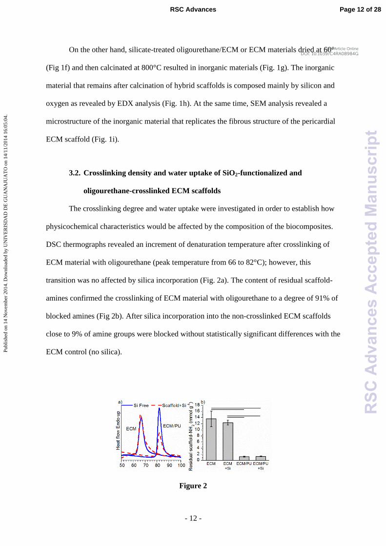
3.2. Crosslinking density and water uptake of SiO2-functionalized and
oligourethane-crosslinked ECM scaffolds
The crosslinking degree and water uptake were investigated in order to establish how
physicochemical characteristics would be affected by the composition of the biocomposites.
DSC thermographs revealed an increment of denaturation temperature after crosslinking of
ECM material with oligourethane (peak temperature from 66 to 82°C); however, this
transition was no affected by silica incorporation (Fig. 2a). The content of residual scaffold-
amines confirmed the crosslinking of ECM material with oligourethane to a degree of 91% of
blocked amines (Fig 2b). After silica incorporation into the non-crosslinked ECM scaffolds
close to 9% of amine groups were blocked without statistically significant differences with the
ECM control (no silica).
Figure 2
Page 12 of 28RSC Advances
RS
CA
dvan
ces
Acc
epte
dM
anus
crip
t
Publ
ishe
d on
14
Nov
embe
r 20
14. D
ownl
oade
d by
UN
IVE
RIS
DA
D D
E G
UA
NA
JUA
TO
on
14/1
1/20
14 1
6:05
:04.
View Article OnlineDOI: 10.1039/C4RA08984G

- 13 -
In presence of collagenase activity, silica-functionalized non-crosslinked ECM
materials gained mass and elapsed its degradation time 1 to 2 days with respect to silica-free
ECM scaffolds, whereas for oligourethane/ECM scaffolds, after 4 days, loss of mass was
observed in silica-containing scaffolds without water uptake process (Fig. 3a). In presence of
collagenase-free DMEM medium, non-crosslinked ECM scaffolds absorbed water until 7
days, after which the mass decreased, while for oligourethane/ECM scaffolds the water
uptake ability was reduced (Fig. 3b). These results indicate that the enzymatic degradation of
ECM scaffolds can be hindered by its crosslinking with oligourethane while the water uptake
of ECM materials can be regulated by both its crosslinking with oligourethane and the
incorporation of silica.
Figure 3
3.3. Cell viability on SiO2-functionalized and oligourethane-crosslinked ECM
scaffolds
In order to evaluate the effect of silica functionalization and the oligourethane
crosslinking on the cytocompatibility of ECM scaffolds, cells were seeded directly on the
lyophilized materials. The SEM micrographs revealed that the macrophages (Fig 4a-4b) and
fibroblasts (Fig 4d-4e) spread on surface of scaffolds. Viable macrophage-like RAW264.7
Page 13 of 28 RSC Advances
RS
CA
dvan
ces
Acc
epte
dM
anus
crip
t
Publ
ishe
d on
14
Nov
embe
r 20
14. D
ownl
oade
d by
UN
IVE
RIS
DA
D D
E G
UA
NA
JUA
TO
on
14/1
1/20
14 1
6:05
:04.
View Article OnlineDOI: 10.1039/C4RA08984G

- 14 -
line cells (Fig. 4c) and primary dermal fibroblasts (Fig. 4f) were detected on all four scaffolds
by means of MTT assay. However, a reduction in the cell viability was detected on silica-
containing scaffolds in both non-crosslinked and crosslinked. In addition, the viability
measured on scaffolds was higher than the viability on the rest of the surface of polystyrene
wells (Fig. 4c and 4f).
Figure 4
Figure 5
Page 14 of 28RSC Advances
RS
CA
dvan
ces
Acc
epte
dM
anus
crip
t
Publ
ishe
d on
14
Nov
embe
r 20
14. D
ownl
oade
d by
UN
IVE
RIS
DA
D D
E G
UA
NA
JUA
TO
on
14/1
1/20
14 1
6:05
:04.
View Article OnlineDOI: 10.1039/C4RA08984G

- 15 -
RAW264.7 macrophages and human blood macrophages were able to proliferate in
the presence of all four scaffolds as evaluated by means of MTT reduction assay (Fig. 5a and
5b) and a fluorescent Live/Dead assay (Fig. 6a and 7a).
3.4. Angiogenic/fibrogenic growth factors secreted by RAW264.7 macrophages
cultured on SiO2-functionalized and oligourethane-crosslinked ECM
scaffolds
To investigate if murine RAW264.7 macrophages respond to particulate silica and
oligourethane crosslinker, the secretion of growth factors involved in angiogenesis and
fibrogenesis processes was quantified when cells were cultured on the whole scaffolds.
Macrophages cultured during 6 h, in contact with oligourethane/ECM and ECM scaffolds,
secreted a higher amount of TGF-1 (Fig. 6b), b-FGF (Fig. 6c) and VEGF (Fig. 6d) compared
vs. control or vs. silica-containing scaffolds. Macrophage proliferation on oligourethane/ECM
and ECM scaffolds from 6-to-24 h did not result in an increment of growth factors
production, in fact, a drop in the secretion was observed. However, secretion of growth
factors by RAW264.7 macrophages cultured in the presence of silica-containing scaffolds was
not decreased with the culture time from 6-to-24 h.
Page 15 of 28 RSC Advances
RS
CA
dvan
ces
Acc
epte
dM
anus
crip
t
Publ
ishe
d on
14
Nov
embe
r 20
14. D
ownl
oade
d by
UN
IVE
RIS
DA
D D
E G
UA
NA
JUA
TO
on
14/1
1/20
14 1
6:05
:04.
View Article OnlineDOI: 10.1039/C4RA08984G

- 16 -
Figure 6
3.5. Cytokines secreted by human blood peripheral derived macrophages
cultured on SiO2-functionalized and oligourethane-crosslinked ECM
scaffolds
The stimulation of primary human macrophages by particulate silica deposited on
ECM scaffolds was investigated by means of the ability to elaborate cytokines involved in
both, pro-inflammatory and anti-inflammatory process. The silica incorporation into the
oligourethane/ECM and ECM scaffolds rendered a higher production of TNF- (Fig. 7b), IL-
6 (Fig. 7c) and IL-10 (Fig. 7e) by macrophages compared to silica-free scaffolds. In addition,
a time-dependent decrease in the production of the three cytokine was detected. ECM
scaffolds containing silica showed a minimal effect on TGF-1 secretion and no time
dependence (Fig 7d). Nonetheless, two different methods were used to active the TGF-1,
Page 16 of 28RSC Advances
RS
CA
dvan
ces
Acc
epte
dM
anus
crip
t
Publ
ishe
d on
14
Nov
embe
r 20
14. D
ownl
oade
d by
UN
IVE
RIS
DA
D D
E G
UA
NA
JUA
TO
on
14/1
1/20
14 1
6:05
:04.
View Article OnlineDOI: 10.1039/C4RA08984G

- 17 -
i.e., to cleave the so called latency-associated peptide (LAP) from TGF-1 protein, secreted
by murine RAW264.7 and human-circulating macrophages, which represents a limitation in
the quantification by ELISA of this signaling molecule (Fig. 6b and 7d). In fact, using the
same protocol to generate the active form of TGF-1, its secretion by murine and human
macrophages was detected after 24 h of culture with scaffolds (Fig. S4).
Figure 7
Page 17 of 28 RSC Advances
RS
CA
dvan
ces
Acc
epte
dM
anus
crip
t
Publ
ishe
d on
14
Nov
embe
r 20
14. D
ownl
oade
d by
UN
IVE
RIS
DA
D D
E G
UA
NA
JUA
TO
on
14/1
1/20
14 1
6:05
:04.
View Article OnlineDOI: 10.1039/C4RA08984G
- 18 -
4. Discussion
An incorporation of bioactive molecules or a promotion of crosslinking in
decellularized matrices offer a chance to enhance the physical and/or the biological
performance by routes involving optimization of surface chemistry, regulation of
biodegradation or controlled releasing of bioactive agents. Silica and its related degradation
products impact the cell behavior which has shown some benefits for tissue engineering
applications, for instance in the wound healing of chronic skin wounds 14
and in osteogenic
differentiation of stem cells.9-13
In the present study, we reported the functionalization of
crosslinked or non-crosslinked ECM scaffolds with particulate silica by
hydrolysis/polycondensation of sodium silicate, and the release of certain soluble signaling
molecules by macrophages when cultured in the presence of scaffolds functionalized with
silica.
The neutralization of sodium silicate solution induces the gelation typically rather
quickly by a single step process. After neutralization of Na4SiO4 solution with HCl, the
polymerization of orthosilicic acid in presence of ECM materials allowed a homogenous
incorporation of silica hydrogel as result of the formation of colloidal silica into aggregates.
Silica aggregates occupied interstitial spaces of decellularized pericardial matrix. Another
important point to mention is that ECM materials containing silica hydrogel served as a
template in the generation of silica material that mimics the microstructure of fibrous tissue
because of the polymerization of silica during the calcination (Fig. 1g-1i). On the other hand,
the freeze-drying of ECM materials containing silica hydrogel induced the deposition of silica
particles onto the fibrous collagen (Fig. 1d-1e). The oligourethane/ECM and ECM materials
functionalized with silica particles were chosen to investigate the effect of silica on the
stability, in vitro degradation and macrophages response.
The increase of the ECM scaffold stability, as evidenced by the increases of collagen
denaturation temperature (Fig. 2a), the decreases of free collagen-amines (Fig. 2b) and the
Page 18 of 28RSC Advances
RS
CA
dvan
ces
Acc
epte
dM
anus
crip
t
Publ
ishe
d on
14
Nov
embe
r 20
14. D
ownl
oade
d by
UN
IVE
RIS
DA
D D
E G
UA
NA
JUA
TO
on
14/1
1/20
14 1
6:05
:04.
View Article OnlineDOI: 10.1039/C4RA08984G

- 19 -
resistance to collagenase degradation (Fig. 3a), was produced by the crosslinking with
oligourethane and was not affected by the incorporation of particulate silica. The silica inside
interstitial spaces in the ECM material can confer an enhanced water uptake process in silica-
ECM scaffolds (Fig. 3a). By contrast, for crosslinked-ECM scaffolds, the reduced interstitial
spaces can hinder the water uptake (Fig. 3a-3b). All these results indicated an effective
incorporation of silica into the ECM scaffolds both, non-crosslinked and oligourethane-
crosslinked.
To have a first idea of the effect of the silica incorporation into the ECM scaffolds on
biocompatibility, we measured the viability of dermal fibroblast and RAW264.7 macrophages
cultured on scaffolds. The cells adhered and viable onto silica-ECM scaffolds were revealed
by SEM micrographs (Fig. 4a-4b and 4d-4e) and MTT reduction assay, respectively. The drop
of the viability of cells after crosslinking (Fig. 4c, 4f) can be induced by a masking of focal
adhesion sites on the collagen network of the decellularized scaffolds. In addition, a drop in
the viability of cells was observed on scaffolds functionalized with silica compared to silica-
free scaffolds (Fig. 4c, 4f). The impact of silica on viability of macrophages and fibroblasts
can be a result of the interaction between cells and particles on the ECM material. The silica
particles and its dissolution products after internalization can disturb the networks that
maintain homeostasis during proliferation, differentiation, and apoptosis. 47
The reactive
oxygen species generated in cells stimulated with different particles is considered a major
factor in particle-induced cytotoxicity. 47-48
In previous studies, it has been reported that silica biomaterials are able to modify the
cell behavior. 6,29-30
Hence, silica-ECM hybrid scaffolds is a suitable platform to characterize
the cellular response to biocomposites that release silicon species. Macrophages are cells of
the innate immune system with a dominant effector activity in the injury site after biomaterial
implantation. 38
Cross-talk between immune cells activates macrophages after which, they
release a variety of signaling molecules. Signaling molecules secreted by macrophages such
Page 19 of 28 RSC Advances
RS
CA
dvan
ces
Acc
epte
dM
anus
crip
t
Publ
ishe
d on
14
Nov
embe
r 20
14. D
ownl
oade
d by
UN
IVE
RIS
DA
D D
E G
UA
NA
JUA
TO
on
14/1
1/20
14 1
6:05
:04.
View Article OnlineDOI: 10.1039/C4RA08984G

- 20 -
as cytokines (as interleukins), growth factors (as b-FGF, VEGF), TGF-1 and TNF-
influence the development of other cell types. 44-45
In fact, the profile of signaling molecules
secretion is commonly evaluated to study the polarization of the macrophage response from
an inflammation and tissue injury process to a repair process 36
or to study angiogenesis and
scaffold vascularization. 45
Macrophages showed the ability to proliferate on all scaffolds, including those that
contain particulate silica (Fig 5a, 5b, 6a and 7a). The secretion of growth factors by
RAW264.7 macrophages in a sustained manner (Fig. 6b-6d) and interleukins by human
macrophages in up-regulated manner (Fig. 7b-7e) when cells were cultured with silica-ECM
scaffolds suggests that silica debris induces the activation of macrophages. Debris of the silica
particles incorporated on ECM scaffolds can be internalized by macrophages, as previously
reported after culture of HepG2 epithelial-like cells 49
and human endothelial cells 50
with
silica nanoparticles. An internalization of silica compounds by macrophages can further
influence signaling pathways to increase the production of signaling molecules. The
macrophages activation with silica nanoparticles has been promoted to assist the DNA
vaccination in the treatment of infectious diseases. 24
The interaction of stem cells with silica
nanoplatelets has induced the osteogenic differentiation. 51
Since the hydrolysis of silicate
materials occurs in aqueous solution under mild conditions, biodegradation products of silica
can be related with the impact of silica on cell response. Porous sol–gel silica particles have
been biodegraded in culture media 52
and under physiological flow conditions 53
by hydrolysis
or dissolution to form orthosilicic acid, Si(OH)4. 54
In fact, stimulation of human skin cells
guided by the release of high molecular weight decomposition products from silicon-based
biodegradable inorganic material has been related with the regulation of wound healing. 14
The incorporation of silica into biodegradable scaffolds has proven to be an effective strategy
to induce the formation of hydroxyapatite on scaffolds 10,55
while allows the growth of
osteoblasts and moderate release of H2O2 by macrophages. 10
A rapid and robust angiogenic
Page 20 of 28RSC Advances
RS
CA
dvan
ces
Acc
epte
dM
anus
crip
t
Publ
ishe
d on
14
Nov
embe
r 20
14. D
ownl
oade
d by
UN
IVE
RIS
DA
D D
E G
UA
NA
JUA
TO
on
14/1
1/20
14 1
6:05
:04.
View Article OnlineDOI: 10.1039/C4RA08984G

- 21 -
response in a quail chorioallantoic membrane model has been supported by collagen films
functionalized with bioglass that contain silicon. 35
The in vitro cytotoxicity and expression of
pro-inflammatory cytokines by macrophages has been reduced by stimulation with
mesoporous silica nanoparticle unlike colloidal silica nanoparticles. 56
Along with these
previous findings, stimulation of macrophages induced by silica particles in situ deposited
onto the ECM network can be explored in the in vitro strategies that combine cell co-culture
44,57 or exogenous stimulating factors
45,58-59 in order to promote the biological response to
ECM biomaterials in different tissue engineering applications. Considering that bone-related
cell lines have showed responsiveness towards silica to maintain the morphogenetic stimuli of
silica on 2D or 3D cultures 60-61
, the series of silica-decellularized tissue composites can offer
a suitable template for enhanced osteogenic potential while the in vivo biodegradation is
adjusted by the crosslinking, which is a subject of ongoing research.
5. Conclusions
Particulate silica was effectively incorporated into the decellularized tissue scaffolds
either non-crosslinked or crosslinked by isocyanate chemistry. The stability and water uptake
of the biocomposites were reliant on crosslinking but were not reliant on the incorporation of
silica. Murine RAW264.7 macrophages cultured on ECM materials crosslinked with
oligourethane and functionalized with silica were able to secrete b-FGF, TGF-1 and VEGF.
Secretion of growth factors by RAW264.7 macrophages after 6 h of culture on scaffolds
containing silica was lower but it was sustained for 24 h as compared to cells cultured on
silica-free materials. The secretion of IL-6, IL-10 and TNF- by human macrophages was
increased with the presence of scaffolds containing silica and it was decreased in a time-
dependent manner from one to four days of culture.
Page 21 of 28 RSC Advances
RS
CA
dvan
ces
Acc
epte
dM
anus
crip
t
Publ
ishe
d on
14
Nov
embe
r 20
14. D
ownl
oade
d by
UN
IVE
RIS
DA
D D
E G
UA
NA
JUA
TO
on
14/1
1/20
14 1
6:05
:04.
View Article OnlineDOI: 10.1039/C4RA08984G

- 22 -
Acknowledgements: We thank to Julio C. Martínez and Mayra C. Rodríguez (UG) for
technical assistance with fibroblast culture and to Rossana F. Vargas and Prof. Juan V. Cauich
(Centro de Investigación Científica de Yucatán) for technical assistance and facilities with
SEM and DSC analyses. GGG thanks to MC Dora A. Huerta Q. for technical assistance with
SEM analysis at LANNBIO Cinvestav-unidad Mérida Yucatán. This research was supported
by the Consejo Nacional de Ciencia y Tecnología (México) through the project
CB2011/164440 and the Fund Convocatoria Interinstitucional CIO-UG 2013.
References
1 S. Rajabi-Zeleti, S. Jalili-Firoozinezhad, M. Azarnia, F. Khayyatan, S. Vahdat, S.
Nikeghbalian, A. Khademhosseini, H. Baharvand and N. Aghdami, Biomaterials, 2014, 35,
970.
2 B. N. Brown, R. Londono, S. Tottey, L. Zhang, K. A. Kukla, M. T. Wolf, K. A. Daly,
J. E. Reing and S. F. Badylak. Acta Biomater., 2012, 8, 978.
3 B. Mendoza-Novelo, D. I. Alvarado-Castro, J. L. Mata-Mata, J. V. Cauich-Rodríguez,
A. Vega-González, E. Jorge-Herrero, F. J. Rojo and G. V. Guinea, Mater. Sci. Eng. C, 2013,
33, 2392.
4 B. Mendoza-Novelo, J. L. Mata-Mata, A. Vega-González, J. V. Cauich-Rodríguez and
A. Marcos-Fernández, J. Mater. Chem. B, 2014, 2, 2874.
5 B. Mendoza-Novelo, G. González-García, J. L. Mata-Mata, L. E. Castellano, P.
Cuéllar-Mata and E. E. Ávila, Mater. Letters, 2013, 106, 369.
6 A.M. Mebert, D. E. Camporotondi, M. L. Foglia, G. S.Alvarez, P. L. S. Orihuela, L. E.
Diaz and M. F. Desimone, J. Biomater. Tissue Eng., 2013, 3, 108.
7 K. Rezwan, Q. Z. Chen, J. J. Blaker and A. R. Boccaccini, Biomaterials, 2006, 27,
3413.
8 K. H. Lee and S. H. Rhee., Biomaterials, 2009, 30, 3444.
Page 22 of 28RSC Advances
RS
CA
dvan
ces
Acc
epte
dM
anus
crip
t
Publ
ishe
d on
14
Nov
embe
r 20
14. D
ownl
oade
d by
UN
IVE
RIS
DA
D D
E G
UA
NA
JUA
TO
on
14/1
1/20
14 1
6:05
:04.
View Article OnlineDOI: 10.1039/C4RA08984G

- 23 -
9 C. Wu, W. Fan, J. Chang and Y. Xiao, J. Mater. Chem., 2011, 21, 18300.
10 W. Zhai, H. Lu, C. Wu, L. Chen, X. Lin, K. Naoki, G. Chen and J. Chang, Acta
Biomater., 2013, 9, 8004.
11 W. E. G. Müller, X. Wang, V. Grebenjuk, B. Diehl-Seifert, R. Steffen, U.
Schloßmacher, A. Trautwein, S. Neumann and H. C. Schröder, Biomater. Sci., 2013, 1, 669.
12 A. I. Rodrigues, M. E. Gomes, I. B. Leonor and R. L. Reis, Acta Biomater., 2012, 8,
3765.
13 G. Tomoaia, A. Mocanu, I. Vida-Simiti, N. Jumate, L. D. Bobos, O. Soritau and M.
Tomoaia-Cotisel, Mater. Sci. Eng. C, 2014, 37, 37.
14 V. Grotheer, M. Goergens, P. C. Fuchs, S. Dunda, N. Pallua, J. Windolf and C. V.
Suschek, Biomaterials, 2013, 34, 7314.
15 M. F. Desimone, C. Hélary, I. B. Rietveld, I. Bataille, G. Mosser, M. M. Giraud-
Guille, J. Livage and T. Coradin, Acta Biomater., 2010, 6, 3998.
16 U. Schloßmacher, H. C. Schröder, X. Wang, Q. Feng, B. Diehl-Seifert, S. Neumann,
A. Trautweina and W. E. G. Müller, RSC Adv., 2013, 3, 11185.
17 A. Angelopoulou, E. K. Efthimiadou and G. Kordas, Mater Lett., 2012, 74, 50.
18 P. C. Balaurea, E. Andronescu, A. M. Grumezescu, A. Ficai, K. S. Huang, C. H. Yang,
C. M. Chifiriuc and Y. S. Lin, Int. J. Pharm., 2013, 441, 555.
19 F. Tang, L. Li and D. Chen, Adv. Mater., 2012, 24, 1504.
20 M. Colilla, B. González and M. Vallet-Regí, Biomater. Sci., 2013, 1, 114.
21 G. S. Alvarez, C. Hélary, A. M. Mebert, X. Wang, T. Coradin and M. F. Desimone, J.
Mater. Chem. B, 2014, 2, 4660.
22 F. M. Chen, M. Zhang and Z. F. Wu, Biomaterials, 2010, 31, 6279.
23 S. Chen, A. Osaka, T. Ikoma, H. Morita, J. Li, M. Takeguchi and N. Hanagata, J.
Mater. Chem., 2011, 21, 10942.
Page 23 of 28 RSC Advances
RS
CA
dvan
ces
Acc
epte
dM
anus
crip
t
Publ
ishe
d on
14
Nov
embe
r 20
14. D
ownl
oade
d by
UN
IVE
RIS
DA
D D
E G
UA
NA
JUA
TO
on
14/1
1/20
14 1
6:05
:04.
View Article OnlineDOI: 10.1039/C4RA08984G

- 24 -
24 J. Wang, R. Zhu, B. Gao, B. Wu, K. Li, X. Sun, H. Liu and S. Wang, Biomaterials
2014, 35, 466.
25 X. Wang, X. Li, A. Ito, Y. Sogo and T. Ohno, Acta Biomater. 2013, 9, 7480.
26 Y. Shirosaki, K. Tsuru, S. Hayakawa, A. Osaka, M. A. Lopes, J. D. Santos and M. H.
Fernandes, Biomaterials, 2005, 26, 485.
27 L. Ren, K. Tsuru, S. Hayakawa and A. Osaka, J. Non-Cryst. Solids, 2001, 285, 116.
28 H. Zhang, D. R. Dunphy, X. Jiang, H. Meng, B. Sun, D. Tarn, M. Xue, X. Wang, S.
Lin, Z. Ji, R. Li, F. L. Garcia, J. Yang, M. L. Kirk, T. Xia, J. I. Zink, A. Nel and C. J. Brinker,
J. Am. Chem. Soc., 2012, 134, 15790.
29 P. Han, C. Wu and Y. Xiao, Biomater. Sci., 2013, 1, 379.
30 A. Hoppe, N. S. Güldal and A. R. Boccaccini, Biomaterials, 2011, 32, 2757.
31 H. S. Yun, J. W. Park, S. H. Kim, Y. J. Kim and J. H. Jang, Acta Biomater., 2011, 7,
2651.
32 J. Kasper, M. I. Hermanns, C. Bantz, O. Koshkina, T. Lang, M. Maskos, C. Pohl, R. E.
Unger and C. J. Kirkpatrick, Arch. Toxicol., 2013, 87, 1053.
33 Z. Mao, X. Zhou and C. Gao, Biomater. Sci., 2013, 1, 896.
34 S. Quignard, G. Mosser, M. Boissière and T. Coradin, Biomaterials, 2012, 33, 4431.
35 G. E. Vargas, L. A. H. Durand, V. Cadena, M. Romero, R. V. Mesones, M. Mackovic,
S. Spallek, E. Spiecker, A. R. Boccaccini and A. A. Gorustovich, J. Mater. Sci. Mater. Med.,
2013, 24, 1261.
36 S. Quignard, C. Hélary, M. Boissière, J. M. Fullana, P. Y. Lagrée and T. Coradin,
Biomater. Sci., 2014, 2, 484.
37 S. Franz, S. Rammelt, D. Scharnweber and J. C. Simon, Biomaterials, 2011, 32, 6692.
38 M. Jaguin, N. Houlbert, O. Fardel and V. Lecureur, Cellular Immunol., 2013, 281, 51.
39 V. Ballotta, A. Driessen-Mol, C. V. C. Bouten and F. P. T. Baaijens, Biomaterials,
2014, 35, 4919.
Page 24 of 28RSC Advances
RS
CA
dvan
ces
Acc
epte
dM
anus
crip
t
Publ
ishe
d on
14
Nov
embe
r 20
14. D
ownl
oade
d by
UN
IVE
RIS
DA
D D
E G
UA
NA
JUA
TO
on
14/1
1/20
14 1
6:05
:04.
View Article OnlineDOI: 10.1039/C4RA08984G

- 25 -
40 J. Maciel, M. I. Oliveira, R. M. Gonçalves and M. A. Barbosa, Acta Biomater., 2012,
8, 3669.
41 J. Kajahn, S. Franz, E. Rueckert, I. Forstreuter, V. Hintze, S. Moeller and J. C. Simon,
Biomatter 2012, 2, 226.
42 M. B. Ariganello, D. T. Simionescu, R. S. Labowa and J. M. Lee, Biomaterials, 2011,
32, 439.
43 R. M. Day and A. R. Boccaccini, J. Biomed. Mater. Res. A, 2005, 73, 73.
44 E. Dohle, I. Bischoff, T. Böse, A. Marsano, A. Banfi, R. E. Unger and C. J.
Kirkpatrick, Eur. Cell. Mater., 2014, 27, 149.
45 K. L. Spiller, R. R. Anfang, K. J. Spiller, J. Ng, K. R. Nakazawa, J. W. Daulton and G.
Vunjak-Novakovic, Biomaterials, 2014, 35, 4477.
46 B. Mendoza-Novelo, E. E. Avila, J. V. Cauich-Rodríguez, E. Jorge-Herrero, F. J.
Rojo, G. V. Guinea and J. L. Mata-Mata, Acta Biomater., 2011, 7, 1241.
47 Q. Mu, G. Jiang, L. Chen, H. Zhou, D. Fourches, A. Tropsha and B. Yan, Chem. Rev.,
2014, 114, 7740.
48 T. Xia, M. Kovochich, J. Brant, M. Hotze, J. Sempf, T. Oberley, C. Sioutas, J. I. Yeh,
M. R. Wiesner and A. E. Nel, Nano Lett., 2006, 6, 1794.
49 L. Hu, Z. Mao, Y. Zhang and C. Gao, J. Nanosci. Lett., 2011, 1, 1.
50 W. Zhai, C. He, L. Wu, Y. Zhou, H. Chen, J. Chang and H. Zhang, J. Biomed. Mater.
Res. B Appl. Biomater., 2012, 100, 1397.
51 A. K. Gaharwar, S. M. Mihaila, A. Swami, A. Patel, S. Sant, R. L. Reis, A. P.
Marques, M. E. Gomes and A. Khademhosseini, Adv. Mater., 2013, 25, 3329.
52 M. Cicuéndez, P. Portolés, M. Montes-Casado, I. Izquierdo-Barba, M. Vallet-Regí,
and M.T. Portolés, J. Mater. Chem. B, 2014, 2, 3469.
53 Q. He, J. Shi, M. Zhu, Y. Chen and F. Chen, Micropor. Mesopor. Mater., 2010, 131,
314.
Page 25 of 28 RSC Advances
RS
CA
dvan
ces
Acc
epte
dM
anus
crip
t
Publ
ishe
d on
14
Nov
embe
r 20
14. D
ownl
oade
d by
UN
IVE
RIS
DA
D D
E G
UA
NA
JUA
TO
on
14/1
1/20
14 1
6:05
:04.
View Article OnlineDOI: 10.1039/C4RA08984G

- 26 -
54 T. Coradin, D. Eglin and J. Livage, Spectroscopy, 2004, 18, 567.
55 C. E. Plazas Bonilla, S. Trujillo, B. Demirdögen, J. E. Perilla, Y. M. Elcin and J. L.
Gómez Ribelles, Mater. Sci. Eng. C, 2014, 40, 418.
56 S. Lee, H. S. Yun and S. H. Kim, Biomaterials, 2011, 32, 9434.
57 K. G. Battiston, J. W. C. Cheung, D. Jain and J. P. Santerre, Biomaterials, 2014, 35,
4465.
58 R. Augustine, E. A. Dominic, I. Reju, B. Kaimal, N. Kalarikkal and S. Thomas, RSC
Adv., 2014, 4, 51528.
59 Y. Rong, T. Zhou, W. Cheng, J. Guo, X. Cui,Y. Liu and W. Chen, Environ. Toxicol.
Pharmacol. 2013, 36, 921.
60 T. Link, X. Wang, U. Schloßmacher, Q. Feng, H. C. Schröder and W. E. G. Müller,
RSC Adv., 2013, 3, 11140.
61 M. Wiens, X. Wang, H. C. Schröder, U. Kolb, U. Schloßmacher, H. Ushijima and W.
E. G. Müller, Biomaterials, 2010, 31, 7716.
Figure captions:
Figure 1. a) Content of silica, oligourethane(PU)/ECM or ECM material and water for
biocomposites (n=4). Representative b) photographs (after freeze-drying), c)ATR-FTIR
spectra, d) and e) SEM micrographs, f) photographs (after dry 60°C), g) photographs (after
calcination 800°C) of the biocomposites. Representative h) EDX spectrum and i) SEM
micrograph of silica material (see Fig. 1g) that remains calcination process.
Figure 2. a) DSC thermographs and b) free ECM material-amines for hydrated materials:
ECM ˗ uncrosslinked materials, ECM/PU ˗ materials crosslinked with oligourethane, Si ˗
materials functionalized with particulate silica; n=3; statistically significant differences for
marked groups.
Page 26 of 28RSC Advances
RS
CA
dvan
ces
Acc
epte
dM
anus
crip
t
Publ
ishe
d on
14
Nov
embe
r 20
14. D
ownl
oade
d by
UN
IVE
RIS
DA
D D
E G
UA
NA
JUA
TO
on
14/1
1/20
14 1
6:05
:04.
View Article OnlineDOI: 10.1039/C4RA08984G

- 27 -
Figure 3. Water uptake /degradation behavior evaluated by means of loss/gain of mass in the
presence of a) collagenase type I and b) DMEM medium for the freeze-dried materials: ECM
˗ uncrosslinked materials, ECM/PU ˗ materials crosslinked with oligourethane, Si ˗ materials
functionalized with particulate silica; n=3.
Figure 4. Representative SEM micrographs of RAW264.7 macrophages seeded on a) ECM
scaffolds and b) silica-ECM scaffolds and fibroblasts seeded on d) ECM scaffolds and e)
silica-ECM scaffolds. Viability of c) macrophages and f) fibroblasts assessed by MTT
reduction by cells on freeze-dried materials (ECM ˗ uncrosslinked materials, ECM/PU ˗
materials crosslinked with oligourethane, Si ˗ materials functionalized with particulate silica)
and polystyrene wells after 24 h of culture. 100% represent viability of cells proliferating on
polystyrene wells free of materials; n=4.
Figure 5. Viability of a) RAW264.7 macrophages and b) human macrophages assessed by
MTT reduction by cells on freeze-dried materials (ECM ˗ uncrosslinked materials, ECM/PU ˗
materials crosslinked with oligourethane, Si ˗ materials functionalized with particulate silica).
Control ˗ cells proliferating on polystyrene wells free of materials; n=4; statistically
significant differences for all marked groups.
Figure 6. a) Representative fluorescent micrographs for RAW264.7 macrophages
proliferating for 6 and 24 h on freeze-dried materials. Growth factor secretion analysis from
RAW264.7 macrophages for b) TGF-1, c) b-FGF and d) VEGF after 6 and 24 h of culture
on freeze-dried materials (ECM ˗ uncrosslinked materials, ECM/PU ˗ materials crosslinked
with oligourethane, Si ˗ materials functionalized with particulate silica). Control ˗ cells
proliferating on polystyrene wells free of materials; n=3; statistically significant differences
for all marked groups.
Figure 7. a) Representative fluorescent micrographs for human peripheral blood derived
macrophages proliferating for 1, 2 and 4 days on freeze-dried materials. Cytokine secretion
Page 27 of 28 RSC Advances
RS
CA
dvan
ces
Acc
epte
dM
anus
crip
t
Publ
ishe
d on
14
Nov
embe
r 20
14. D
ownl
oade
d by
UN
IVE
RIS
DA
D D
E G
UA
NA
JUA
TO
on
14/1
1/20
14 1
6:05
:04.
View Article OnlineDOI: 10.1039/C4RA08984G

- 28 -
analysis from human macrophages for b) TNF-, c) IL-6, d) TGF-1, d) IL-10 after 1, 2 or 4
day of culture on freeze-dried materials (ECM ˗ uncrosslinked materials, ECM/PU ˗ materials
crosslinked with oligourethane, Si ˗ materials functionalized with particulate silica). Control ˗
cells proliferating on polystyrene wells free of materials; n=3; statistically significant
differences for all marked groups.
Page 28 of 28RSC Advances
RS
CA
dvan
ces
Acc
epte
dM
anus
crip
t
Publ
ishe
d on
14
Nov
embe
r 20
14. D
ownl
oade
d by
UN
IVE
RIS
DA
D D
E G
UA
NA
JUA
TO
on
14/1
1/20
14 1
6:05
:04.
View Article OnlineDOI: 10.1039/C4RA08984G