Acanthamoeba castellanii: Morphological analysis of the interaction with human cornea
6
Acanthamoeba castellanii: Morphological analysis of the interaction with human cornea Maritza Omaña-Molina a, * , Arturo González-Robles b , Lizbeth Iliana Salazar-Villatoro b , Ana Ruth Cristóbal-Ramos b , Mónica González-Lázaro b , Edmundo Salinas-Moreno a , Rene Méndez-Cruz a , Manuel Sánchez-Cornejo c , Enrique De la Torre-González c , Adolfo Martínez-Palomo b a School of Superior Studies Iztacala, UNAM. Los Reyes Iztacala, Tlalnepantla, Mexico State, Mexico b Department of Infectomic and Molecular Pathogenesis Center for Research and Advanced Studies, Mexico City, Mexico c Ophthalmology Service, Juarez Hospital, Mexico City, Mexico article info Article history: Received 23 October 2009 Received in revised form 2 February 2010 Accepted 4 February 2010 Available online 8 February 2010 Keywords: Acanthamoeba castellanii Human cornea Phagocytosis Pathogenic mechanisms Acanthamoeba keratitis (AK) abstract The present study demonstrates that when Acanthamoeba castellanii trophozoites are co-cultivated with isolated human corneas, the amoeba can be invasive and cause damage to the intact corneal epithelium without the requirement of previous corneal abrasion. After adhesion, A. castellanii trophozoites migrate between cells forming bumps on the corneal cell layers and reaching Bowman ´ s membrane in 3 h, although no evidence of cell damage was observed until the phagocytic process was detected. Likewise, conditioned medium produced damage to the corneal cells that was proportional to the time of incuba- tion, but this cytophatic effect involved only the most superficial layer of the human cornea and was not enough to explain amoebic invasion of Bowman ´ s membrane. As a result of our observations, we suggest that the mechanical action of the trophozoites and phagocytosis of corneal cells during the process of cor- neal invasion are more important than previously suggested. Ó 2010 Elsevier Inc. All rights reserved. 1. Introduction Free-living amoebae of the genus Acanthamoeba are protozoans that live in a wide variety of habitats, having been isolated from soil, air and water environments (Marciano-Cabral and Cabral, 2003). These amoebae have become medically important since they are the causative agents of granulomatous amebic encephalitis, skin lesions and a corneal infection known as Acanthamoeba kera- titis (AK). This corneal disease is a progressive sight-threatening infection which can occur both in immunocompromised and healthy individuals and is particularly prevalent among contact lens users. In developed countries roughly 83% of the cases are diagnosed in contact lens wearers (Carvalho et al., 2009). Poor hy- giene and insufficient disinfection may be determinants of AK in contact lens users. In contrast, in non developed countries the majority of cases are related with agricultural-based activities and/or ocular trauma (Gopinathan et al., 2009). The signs referred during corneal infection are pain, eye redness, tearing, eyelid swelling, and a ring-shaped ulcer invading the corneal stroma is typical. Amoebae cause thinning and ulceration of the corneal epithelium, eventually leading to necrosis, edema, and stromal inflammatory reaction (Dart et al., 2009). An early diagnosis is critical for the retention of good visual acuity. Acanthamoeba trophozoites or cysts can be diagnosed in corneal scrapings using wet mount stains, histopathologic exami- nation or culture. Sample analysis using stains for the cyst wall or immunostaining may also be employed. PCR is also a well- established diagnostic tool (Ledee et al., 1996; Marciano-Cabral and Cabral, 2003; Schroeder et al., 2001). If the corneal infection is not diagnosed promptly and treated aggressively, the corneal epithelium becomes ulcerated with stroma infiltration that leads to perforation and finally loss of vision (Saeed et al., 2009). A confocal scanning study has suggested that AK may occur more commonly than currently recognized (Tu et al., 2008). A variety of topically applied therapeutic agents are thought to be effective, including propamidine isethionate, clotrimazole, polyhexamethyl- ene biguanide, and chlorhexidine. Various combinations of these and other agents have been employed, often resulting in medical cure (Dart et al., 2009). The pathogenic mechanisms of AK have not been totally eluci- dated. In general it has been established that the pathophysiology of this infection involves an intricate series of sequential events in which the first step is the adhesion of the trophozoites to the host cell. Subsequent to adhesion, the amoebae produce several lytic proteases which are able to degrade the epithelium, inducing 0014-4894/$ - see front matter Ó 2010 Elsevier Inc. All rights reserved. doi:10.1016/j.exppara.2010.02.004 * Corresponding author. Address: FESI UNAM. Av. de los Barrios No.1, Los Reyes Ixtacala, UIICSE, Medicine C. P. 54090, Tlalnepantla, State of Mexico, Mexico. Fax: +52 155 53 90 76 04. E-mail address: [email protected] (M. Omaña-Molina). Experimental Parasitology 126 (2010) 73–78 Contents lists available at ScienceDirect Experimental Parasitology journal homepage: www.elsevier.com/locate/yexpr
-
Upload
independent -
Category
Documents
-
view
1 -
download
0
Transcript of Acanthamoeba castellanii: Morphological analysis of the interaction with human cornea

Experimental Parasitology 126 (2010) 73–78
Contents lists available at ScienceDirect
Experimental Parasitology
journal homepage: www.elsevier .com/locate /yexpr
Acanthamoeba castellanii: Morphological analysis of the interactionwith human cornea
Maritza Omaña-Molina a,*, Arturo González-Robles b, Lizbeth Iliana Salazar-Villatoro b,Ana Ruth Cristóbal-Ramos b, Mónica González-Lázaro b, Edmundo Salinas-Moreno a, Rene Méndez-Cruz a,Manuel Sánchez-Cornejo c, Enrique De la Torre-González c, Adolfo Martínez-Palomo b
a School of Superior Studies Iztacala, UNAM. Los Reyes Iztacala, Tlalnepantla, Mexico State, Mexicob Department of Infectomic and Molecular Pathogenesis Center for Research and Advanced Studies, Mexico City, Mexicoc Ophthalmology Service, Juarez Hospital, Mexico City, Mexico
a r t i c l e i n f o a b s t r a c t
Article history:Received 23 October 2009Received in revised form 2 February 2010Accepted 4 February 2010Available online 8 February 2010
Keywords:Acanthamoeba castellaniiHuman corneaPhagocytosisPathogenic mechanismsAcanthamoeba keratitis (AK)
0014-4894/$ - see front matter � 2010 Elsevier Inc. Adoi:10.1016/j.exppara.2010.02.004
* Corresponding author. Address: FESI UNAM. Av. dIxtacala, UIICSE, Medicine C. P. 54090, Tlalnepantla, S+52 155 53 90 76 04.
E-mail address: [email protected] (
The present study demonstrates that when Acanthamoeba castellanii trophozoites are co-cultivated withisolated human corneas, the amoeba can be invasive and cause damage to the intact corneal epitheliumwithout the requirement of previous corneal abrasion. After adhesion, A. castellanii trophozoites migratebetween cells forming bumps on the corneal cell layers and reaching Bowmans membrane in 3 h,although no evidence of cell damage was observed until the phagocytic process was detected. Likewise,conditioned medium produced damage to the corneal cells that was proportional to the time of incuba-tion, but this cytophatic effect involved only the most superficial layer of the human cornea and was notenough to explain amoebic invasion of Bowmans membrane. As a result of our observations, we suggestthat the mechanical action of the trophozoites and phagocytosis of corneal cells during the process of cor-neal invasion are more important than previously suggested.
� 2010 Elsevier Inc. All rights reserved.
1. Introduction
Free-living amoebae of the genus Acanthamoeba are protozoansthat live in a wide variety of habitats, having been isolated from soil,air and water environments (Marciano-Cabral and Cabral, 2003).
These amoebae have become medically important since theyare the causative agents of granulomatous amebic encephalitis,skin lesions and a corneal infection known as Acanthamoeba kera-titis (AK). This corneal disease is a progressive sight-threateninginfection which can occur both in immunocompromised andhealthy individuals and is particularly prevalent among contactlens users. In developed countries roughly 83% of the cases arediagnosed in contact lens wearers (Carvalho et al., 2009). Poor hy-giene and insufficient disinfection may be determinants of AK incontact lens users. In contrast, in non developed countries themajority of cases are related with agricultural-based activitiesand/or ocular trauma (Gopinathan et al., 2009). The signs referredduring corneal infection are pain, eye redness, tearing, eyelidswelling, and a ring-shaped ulcer invading the corneal stroma istypical. Amoebae cause thinning and ulceration of the corneal
ll rights reserved.
e los Barrios No.1, Los Reyestate of Mexico, Mexico. Fax:
M. Omaña-Molina).
epithelium, eventually leading to necrosis, edema, and stromalinflammatory reaction (Dart et al., 2009).
An early diagnosis is critical for the retention of good visualacuity. Acanthamoeba trophozoites or cysts can be diagnosed incorneal scrapings using wet mount stains, histopathologic exami-nation or culture. Sample analysis using stains for the cyst wallor immunostaining may also be employed. PCR is also a well-established diagnostic tool (Ledee et al., 1996; Marciano-Cabraland Cabral, 2003; Schroeder et al., 2001). If the corneal infectionis not diagnosed promptly and treated aggressively, the cornealepithelium becomes ulcerated with stroma infiltration that leadsto perforation and finally loss of vision (Saeed et al., 2009). Aconfocal scanning study has suggested that AK may occur morecommonly than currently recognized (Tu et al., 2008). A varietyof topically applied therapeutic agents are thought to be effective,including propamidine isethionate, clotrimazole, polyhexamethyl-ene biguanide, and chlorhexidine. Various combinations of theseand other agents have been employed, often resulting in medicalcure (Dart et al., 2009).
The pathogenic mechanisms of AK have not been totally eluci-dated. In general it has been established that the pathophysiologyof this infection involves an intricate series of sequential events inwhich the first step is the adhesion of the trophozoites to the hostcell. Subsequent to adhesion, the amoebae produce several lyticproteases which are able to degrade the epithelium, inducing

74 M. Omaña-Molina et al. / Experimental Parasitology 126 (2010) 73–78
cytolysis and apoptosis of the cellular elements of the cornea. Thisprocess culminates with the penetration of the parasites into thedeeper layers of the cornea and with the dissolution of the collag-enous corneal stroma (Clarke and Niederkorn, 2006).
There have been several attempts to explain the cytopathiceffect of Acanthamoeba spp. on the cornea. Studies aimed to pro-duce amebic keratitis in animal models have been performed byintentionally damaging corneal epithelium; however, none ofthese studies have described the pathogenic stages prior to 12 hof interaction between the amoebae and the corneal epithelium(He et al., 1992; Panjwani et al., 1997; Khan, 2001).
Previously, we reported the morphological and electrophysiolog-ical events that take place during the early stages of the interactionbetween Acanthamoeba castellanii trophozoites and golden Syrianhamster (Mesocricetus auratus) cornea which had not been previ-ously damaged. The process begins with the adhesion of the tropho-zoites to the epithelial surface, followed by the formation of clumpsand the migration of the parasites toward the cell borders, causingthe separation of adjacent cells after 1 h of co-culture. At later stages(2–3 h), some amoebae were found under desquamating epithelialcells whereas others were seen associated with damaged cells orforming amebostome-like structures to ingest detached epithelialcells. Hamster’s corneas incubated with conditioned culture med-ium showed cytoplasmic vacuolization and disruption of epithelialcell junctions (Omaña-Molina et al., 2004).
Although hamster models have been used extensively, there islittle information on the mechanisms of pathogenicity of Acantha-moeba trophozoites on the human cornea. For this reason, wereport the early events that take place during the incubation of A.castellanii trophozoites or conditioned medium with the humancornea, as an initial approach to determine the pathogenic mecha-nisms of these amoebae in the human host.
2. Materials and methods
2.1. Amoebae
This study was undertaken with one of the most frequently iso-lated species of Acanthamoeba from AK cases: A. castellanii, whichwas originally isolated from a contact lens that was not associatedwith a human case of keratitis (Association to prevent blindness inMexico, Luis Sánchez Bulnes Hospital, Mexico City). In this partic-ular case the patient was only suffering from an intense ocularpain. Even though amoebae were isolated from the lens, no para-sites were obtained from the corneal scrapes. The strain reportedin the present study has been shown to be very virulent and inva-sive, as determined by tests carried out in a murine model, as wellas in cell cultures and with human corneas (data not shown).
Amoebae were grown and maintained in axenic culture in 2%Bacto Casitone (pancreatic digest of casein, Becton–Dickinson,Sparks, MD) supplemented with 10% fetal bovine serum (Gibco,Grand Islands, NY). Cultures were incubated at 30 �C in borosilicatetubes (Pyrex, Mexico). All assays were performed with trophozoitesharvested at the end of the logarithmic phase of growth (72 h) bychilling culture tubes at 4 �C and centrifuging them for 5 min at2500 rpm.
2.2. Human corneas
Intact human corneas or segments of corneas free of infectionwere used. The corneal tissue was obtained from the Eye Bank ofthe Ophthalmology Service at Juárez Hospital (Mexico City, Mex-ico). Standard hospital protocols of human corneal tissue use forresearch purposes were fulfilled. Corneas were removed with aperipheral rim of scleral tissue, and manipulation was performed
touching only the scleral zones, leaving the lens untouched.Corneal tissue was evaluated using a stereoscopic microscopebefore carrying out the interaction studies. The integrity of cornealtissue was also corroborated by scanning electron microscopy.
Samples were allowed to interact with amoebae 5–10 h aftersurgery. All samples were maintained at 4 �C in corneal storage med-ium (Optisol, Chiron Ophthalmics, Irvine, California) until used.
2.3. Interaction assays
Segments of approximately 0.5 cm2 of human corneas wereplaced in 24-well styrene plates (Nunclon TM, Denmark). Eachcornea contained a peripheral ring of scleral tissue to allow manip-ulation while leaving the lens untouched.
Three experimental groups of corneal segments were tested. Allinteractions were performed using 2% Bacto Casitone medium:
(a) Segments of human corneas interacting with 2.5 � 105trophozoites/ml of medium.
(b) Segments of human corneas interacting with 1 ml of amoe-bae products (conditioned medium). The cultured mediumin which the amoebas were grown was filtered through0.22 lm filters (Millex GV Durapore PVDF).
(c) Control corneas interacting with 2% Bacto Casitone medium,which was found to be an optimal medium to preservehuman corneal tissue, as determined by scanning electronmicroscopy.
All experimental groups were incubated at 36.5 �C for 1, 2 or3 h, then fixed and processed for scanning electron microscopyand embedded in epoxy resins to obtain semithin sections. All as-says were performed in triplicate.
2.4. Scanning electron microscopy
After incubation, samples were fixed with 2.5% glutaraldehydein 0.1 M cacodylate buffer, washed with distilled water and dehy-drated with increasing concentrations of ethanol. Samples werethen critically point-dried with liquid CO2 using a Samdri 780apparatus (Tousimis Research Corp.), and coated with a thin layer(30 nm) of gold in an Ion sputtering device (JEOL JFC-1100). Spec-imens were examined with a Philips XL 30 ESEM scanning electronmicroscope.
2.5. Light microscopy
After co-incubation, samples were fixed at room temperature,post-fixed with 1% osmium tetroxide in cacodylate buffer, anddehydrated in increasing concentrations of ethanol. Samples weretransferred to propylene oxide, then to a mixture of propyleneoxide/epoxy resin (1/1) and finally embedded in epoxy resins.Semithin sections (0.5 lm), stained with toluidine blue wereexamined with an Axiophot photo-microscope (Carl Zeiss,Germany). Photographs were obtained with a Zeiss AxioCamMRcdigital camera (Carl Zeiss Vision GmbH, Germany).
3. Results
3.1. Control corneas
Examination of control human corneas incubated in Bacto Casi-tone medium in the absence of amoebae showed a normal mor-phology by light microscopy (semi thin sections), as well as byscanning electron microscopy. No evidence of epithelial damage,

Fig. 1. Scanning electron microscopy of Acanthamoeba castellanii trophozoites interacting with human corneas. (A) Control human corneas incubated in Bacto Casitonemedium in the absence of amoebae showed a normal morphology, and no evidence of epithelial damage was observed. (B) After 1 h of co-incubation, abundant trophozoites(T) are seen in contact with corneal epithelial cells (Ec). In a different area, a trophozoite (arrow) is located between epithelial cells (Ec) and partially separates and penetratesthe corneal epithelium. In addition two Acanthamoeba trophozoites (arrowhead) are observed phagocyting a newly detached corneal epithelial cell. Bar = 10 lm. (C) After 3 hof interaction, trophozoites have penetrated all layers of the corneal epithelium and are seen attached to Bowman’s membrane (Bm). Bar = 50 lm. (D) At the same time, someamoebae may have been undergoing in cell division on Bowman’s membrane. Bar = 20 lm.
M. Omaña-Molina et al. / Experimental Parasitology 126 (2010) 73–78 75
pitting, ulceration, wounding, excessive cell exfoliation or anyother defects were observed (Fig. 1A).
3.2. Experimental corneas
3.2.1. Scanning electron microscopyAcanthamoeba castellanii trophozoites co-cultured with human
corneas produced cytopathic changes on the epithelium. First, indi-vidual or clumped trophozoites were seen adhered to the surfaceof the cornea. After adhesion, trophozoites migrated toward thecells borders, causing the separation of adjacent cells after 1 h ofco-culture. At later stages (2–3 h), abundant trophozoites were ob-served beneath the superficial layers of the cornea, forming bulgeson the corneal surface as well as under desquamating epithelialcells. Frequently, the amoebae were observed ingesting detachedcorneal cells (Fig. 1B). Numerous trophozoites continued migratinginto deeper cell layers and adhering to middle (wings) cells. After3 h of co-cultivation most superficial and middle cells of the cor-neal epithelium had been detached and separated from the restof the tissue. The normal architecture of corneal epithelium wasabsent; however, these cells did not show apparent signs of dam-age, since they preserved their normal morphology. In other areas,some trophozoites had reached Bowmans membrane, wherenumerous trophozoites were observed apparently engulfing basalcells (Fig. 1C). In most observations trophozoites maintained theirclassical morphology as seen in axenic cultures; however, we ob-served morphological differences when the trophozoites pene-
trated through cell junctions, where an elongated shape wasadopted to pass through the tight spaces. The morphology adoptedby some trophozoites on the Bowmans membrane indicated thatthey may have been undergoing cell division (Fig. 1D).
3.2.2. Light microscopyInvasion of trophozoites toward the inner layers of the human
corneal epithelium was corroborated by light microscopy. Duringthe first hour of interaction, trophozoites attached to the mostsuperficial corneal cells and penetrated toward deeper layers ofcorneal tissue. At longer times of interaction (2–3 h), superficialand middle (wings) cells of the corneal epithelium had been de-tached and separated from the rest of the tissue, but they appearedto preserve their normal morphology. Afterwards, the amoebaereached Bowmans membrane and finally phagocyted newly de-tached corneal cells. During the interactions, some trophozoitesshowed their classical morphology, in which the nuclei and nucle-oli as well as contractile vacuoles were evident (Fig. 2A and B).However, other amoebae changed their morphology to occupyspaces between basal epithelial cells to reach Bowman’s mem-brane (Fig. 2B).
3.2.3. Interaction with conditioned mediumDuring the interaction of conditioned medium with human cor-
neas, we observed abrasion and loss of continuity only of the mostsuperficial corneal cells. The damage was proportional to the timeof interaction. During the first hour of co-incubation disruption of
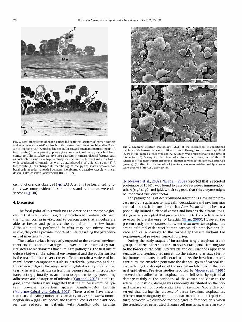
Fig. 2. Light microscopy of epoxy-embedded semi thin sections of human corneasand Acanthamoeba castellanii trophozoites stained with toluidine blue after 2 and3 h of interaction. (A) Amoebae have migrated toward Bowmans membrane (Bm). Atrophozoite (T) is apparently phagocyting an intact and newly detached basalcorneal cell. The amoebae preserve their characteristic morphological features, suchas contractile vacuoles, a large centrally located nucleus (arrow) and a nucleoluswith condensed chromatin as well as acanthopodia of different sizes. (B) Atrophozoite (T) has changed its morphology to occupy the spaces between twobasal cells in order to reach Bowman’s membrane. A digestive vacuole with celldebris is also observed (arrowhead). Bar = 10 lm.
Fig. 3. Scanning electron microscopy (SEM) of the interaction of conditionedmedium with human corneas at different times. Damage to the most superficiallayers of the human cornea was observed, which was proportional to the time ofinteraction. (A) During the first hour of co-incubation, disruption of the celljunctions of the most superficial layer of human corneal epithelium was observed(arrows). (B) After 3 h, the loss of cell junctions was more evident and lytic areaswere observed (arrows). Bar = 50 lm.
76 M. Omaña-Molina et al. / Experimental Parasitology 126 (2010) 73–78
cell junctions was observed (Fig. 3A). After 3 h, the loss of cell junc-tions was more evident in some areas and lytic areas were ob-served (Fig. 3B).
4. Discussion
The focal point of this work was to describe the morphologicalevents that take place during the interaction of Acanthamoeba withthe human cornea in vitro, and to demonstrate that amoebae areable to invade and penetrate the epithelium in a few hours.Although studies performed in vitro may not mirror eventsin vivo, they often provide important clues regarding the pathogen-esis of infection in vivo.
The ocular surface is regularly exposed to the external environ-ment and to potential pathogens; however, it is protected by nat-ural defense mechanisms that eliminate pathogens. The first line ofdefense between the external environment and the ocular surfaceis the tear film that covers the eye. Tears contain a variety of hu-moral defense components such as lactoferrin, lysozyme, and lac-toperoxidase. lgA is the major immunoglobulin isotype in normaltears where it constitutes a frontline defense against microorgan-isms, acting primarily as an immunologic barrier by preventingadherence and adsorption of microbes (Cao et al., 2008). In this re-gard, some studies have suggested that the mucosal immune sys-tem provides protection against Acanthamoeba keratitis(Marciano-Cabral and Cabral, 2003). These studies have shownthat tears of healthy individuals contain anti-Acanthamoeba immu-noglobulin A (IgA) antibodies and that the levels of these antibod-ies are reduced in patients with Acanthamoeba keratitis
(Niederkorn et al., 2002). Na et al. (2002) reported that a secretedproteinase of 12 kDa was found to degrade secretory immunoglob-ulin A (sIgA), IgG, and IgM, which suggests that this enzyme mightbe important virulence factor.
The pathogenesis of Acanthamoeba infection is a multistep pro-cess involving adhesion to host cells, degradation and invasion intocorneal tissues. It is considered that Acanthamoeba attaches to apreviously injured surface of cornea and invades the stroma, thus,it is generally accepted that previous trauma to the epithelium hasto occur before the onset of keratitis (Khan, 2006). However, thepresent study demonstrates that when Acanthamoeba trophozoitesare co-cultured with intact human corneas, the amoebae can in-vade and cause damage to the corneal epithelium without therequirement of previous corneal abrasion.
During the early stages of interaction, single trophozoites orgroups of them adhere to the corneal surface, and then migrateto the border of the cells. Afterwards, the cell junctions appear toseparate and trophozoites move into the intracellular space form-ing bumps and causing cell detachment. As the invasion processcontinues, the amoebae penetrate the deeper layers of corneal tis-sue, inducing the disruption of the normal architecture of the cor-neal epithelium. Previous studies reported by Moore et al. (1991)showed that adhesion of trophozoites is followed by epithelialdamage mainly at the periphery of the cornea and close to thesclera. In our study, damage was randomly distributed on the cor-neal surface without preferential sites of invasion. Moore also ob-served that during the process of tissue invasion, trophozoitesdiffered morphologically from amoebae maintained in liquid cul-ture; however, we observed morphological differences only whenthe trophozoites penetrated through cell junctions, where an elon-

M. Omaña-Molina et al. / Experimental Parasitology 126 (2010) 73–78 77
gated shape was adopted to pass through tight spaces. Ubelakeret al. (1991) reported that trophozoites of A. castellanii were ableto adhere to each other by desmosome-like structures; however,in our study no evidence of this kind of attachment between tro-phozoites was observed. Even though the cell surface of Acantha-moeba is a highly specialized region, we consider that amoeba donot adhere to each other by this mechanism.
The ability of Acanthamoeba to bind to the host tissue is consid-ered a critical step in the pathogenesis of AK. Thus far, a mannose-binding protein expressed on the surface of Acanthamoeba tropho-zoites has been shown to provide an initial attachment site to hostcells and to induce the production of cytopathic factors that in-crease damage (Cao et al., 1998; Hurt et al., 2003a, b; Leheret al., 1998; Yang et al., 1997). Adhesion of Acanthamoeba to cor-neal cells leads to secondary events such as phagocytosis, whichis an actin-dependent process. It has been reported that Acantha-moeba has the ability to form amoebostomes during co-incubationwith host cells, which suggests that phagocytosis plays a majorrole in the pathogenesis of these amoebae (Khan, 2001; Omaña-Molina et al., 2004). Phagocytosis of corneal epithelial cells as wellas of those recently removed was a commonly observed eventwhile Acanthamoeba trophozoites aimed to invade deeper layersof the human cornea, which demonstrates the ability of A. castella-nii to use human corneal epithelial cells as a food source in vitro.This plentiful food supply may allow the organisms to subsistand proliferate in the in vivo corneal tissue for long periods of time.Similar sequences of events have been reported during the co-cul-ture of A. castellanii and A. polyphaga with hamster corneas (Omañ-a-Molina et al., 2004).
Concerning the manner on how Acanthamoeba migrates and in-vades target tissues, it has been proposed that proteases secretedby this amoeba are an important factor in its pathogenesis, ashas been observed in other protozoan parasites (McKerrow et al.,1993). Although the precise role of these proteinases in the patho-genesis of corneal infection of Acanthamoeba is not clear at present,some evidence suggests that proteinases are important virulencefactors in Acanthamoeba keratitis. Several serine proteases havebeen investigated (Mitra et al.,1995; Cho et al., 2000; Kong et al.,2000; Na et al., 2001; Hurt et al., 2003a,b) and have been consid-ered to play a role in the mechanisms of invasion by destroyingthe tissue. Acanthamoeba proteinases can degrade several host pro-teins, including major structural proteins such as collagen, fibro-nectin and laminin (Cho et al., 2000; Kong et al., 2000; Na et al.,2001). In particular, the secretory products of Acanthamoeba havebeen shown to have collagenolytic activity, both in vitro andin vivo (He et al., 1990; Mitro et al., 1994).
We believe that enzymatic activity may play a role in the cyto-lytic mechanisms by facilitating the separation of cells and thusallowing the amoeba to break through the deeper layers of the cor-nea, but only by facilitating the separation of cells and not bydestroying the tissue as has been suggested before.
The pathogenic mechanisms of Acanthamoeba consist of bothcontact dependent and independent processes. Leher et al.(1998), reported that mannose treatment induced Acanthamoebatrophozoites to release cytopathic factors that lyse corneal epithe-lial cells in vitro, and that the cytopathic activity was completelyinhibited by a serine protease inhibitor. Khan (2003) also sug-gested that serine proteases play a crucial role in the pathogenesisof AK, because a serine protease inhibitor abolished cytotoxicity ofAcanthamoeba conditioned medium on corneal epithelial cells.When the interaction with conditioned culture medium was per-formed, we observed a cytophathic effect that was proportionalto the time of interaction with the corneas. However, this cytopha-thic effect involved only the most superficial layer of the corneaand this mechanism is not enough to explain how amoebae invadeBowmans membrane.
As a result of our observations, we propose that the mechanicalaction of the trophozoites during the process of corneal invasion ismore important than previously considered. It is possible that themovement of amoebas between cells exerts a mechanical forcethat promotes separation and allows migration into the deeper lay-ers of corneal tissue, which may help to explain the loss of cornealepithelium architecture, apparently without the loss of integrity ordamage to corneal cells, as has been suggested previously. Whileanalyzing the interaction of A. castellanii trophozoites with MDCKcells, González-Robles et al. (2006) suggested that the mechanicalaction of the trophozoites on target cells plays an important role inthe cytolytic mechanism.
In summary, this is the first study that provides information onthe sequence of morphological changes that occur during the inter-action of A. castellanii with intact human corneas, providing a plat-form for further studies. The mechanisms of pathogenicity shownhere are very similar to those previously reported using animalmodels (Omaña-Molina et al., 2004), which validate our previousfindings.
Thus, the hamster cornea model can be used for future studiesrelated with the pathogenesis of Acanthamoeba keratitis.
Acknowledgments
We wish to thank Q.B.P. Virginia Vanzzini for providing theamoebae used in this study. This study was supported by grantskindly provided by UNAM FES Iztacala PAPCA (Project 2007/2008No. 66) and by the National Council of Science and Technology(CONACyT; project 102188/2008).
References
Cao, Z., Jefferson, D.M., Panjwani, N., 1998. Role of carbohydrate-mediatedadherence in cytopathogenic mechanisms of Acanthamoeba. Journal ofBiological Chemistry 273, 15838–15845.
Cao, Z., Saravanan, C., Goldstein, M.H., Wu, H.K., Pasricha, G., Sharma, S., Panjwani,N., 2008. Effect of human tears on Acanthamoeba-induced cytopathic effect.Archives of Ophthalmology 126, 348–352.
Carvalho, F.R., Foronda, A.S., Mannis, M.J., Höfling-Lima, A.L., Belfort, R.Jr., de Freitas,D., 2009. Twenty years of Acanthamoeba Keratitis. Cornea 28, 516–519.
Cho, J.H., Na, B.K., Kim, T.S., Song, C.Y., 2000. Purification and characterization of anextracellular serine proteinase from Acanthamoeba castellanii. IUBMB Life 50,209–214.
Clarke, D.W., Niederkorn, J.Y., 2006. The pathophysiology of Acanthamoeba keratitis.Trends in Parasitology 22, 175–180.
Dart, J.K., Saw, V.P., Kilvington, S., 2009. Acanthamoeba Keratitis: diagnosis andtreatment update 2009. American Journal of Ophthalmology 148, 487–499.
González-Robles, A., Castañón, G., Cristóbal-Ramos, A.R., Lázaro-Haller, A., Omaña-Molina, M., Bonilla, P., Martínez-Palomo, A., 2006. Acanthamoeba castellaniistructural basis of the cythophathic mechanisms. Experimental Parasitology114, 133–140.
Gopinathan, U., Sharma, S., Garg, P., Rao, G.N., 2009. Review of epidemiologicalfeatures, microbiological diagnosis and treatment outcome of microbialkeratitis: experience of over a decade. Indian Journal of Ophthalmology 57,273–279.
He, Y.G., Niederkorn, J.Y., McCulley, J.P., Stewart, G.L., Meyer, D.R., Silvany, R.,Dougherty, J., 1990. In vivo and in vitro collagenolytic activity of Acanthamoebacastellanii. Investigative Ophthalmology and Visual Science 11, 2235–2240.
He, Y.G., McCulley, J.P., Alizadeh, H., Pidherney, M., Mellon, J., Ubelaker, J.E., Stewart,G.L., Silvany, R.E., Niederkorn, J.Y., 1992. A pig model of Acanthamoeba keratitis:transmission via contaminated contact lenses. Investigative Ophthalmologyand Visual Science 33, 126–133.
Hurt, M., Neelam, S., Niederkorn, J., Alizadeh, H., 2003a. Pathogenic Acanthamoebaspp. secrete a mannose-induced cytolytic protein that correlates with theability to cause disease. Infection and Immunity 71, 6243–6255.
Hurt, M., Niederkorn, J., Alizadeh, H., 2003b. Effects of mannose on Acanthamoebacastellanii proliferation and cytolytic ability to corneal epithelial cells.Investigative Ophthalmolology and Visual Science 44, 3424–3431.
Khan, N.A., 2001. Pathogenicity, morphology and differentiation of Acanthamoeba.Current Microbiology 43, 391–395.
Khan, N.A., 2003. Pathogenesis of Acanthamoeba infections. Microbial Pathogenesis34, 277–285.
Khan, N.A., 2006. Acanthamoeba: biology and increasing importance in humanhealth. FEMS Microbiology Reviews 30, 564–595.
78 M. Omaña-Molina et al. / Experimental Parasitology 126 (2010) 73–78
Kong, H.H., Kim, T.H., Chung, D.I., 2000. Purification and characterization of asecretory proteinase of Acanthamoeba healyi isolates from GAE. Journal ofParasitology 86, 12–17.
Ledee, D.R., Hay, J., Byers, T.H., Seal, D.V., Kirkness, C.M., 1996. Acanthamoeba griffini.Molecular characterization of a new corneal pathogen. InvestigativeOphthalmology and Visual Science 4, 544–550.
Leher, H., Silvany, R., Alizadeh, H., Huang, J., Niederkorn, J.Y., 1998. Mannose inducesthe release of cytopathic factors from Acanthamoeba castellanii. Infection andImmunity 66, 5–10.
Marciano-Cabral, F., Cabral, G., 2003. Acanthamoeba spp. as agents of disease inhumans. Clinical Microbiology Review 16, 273–307.
Mckerrow, J.H., Sun, E., Rosenthal, P.J., Bouvier, J., 1993. The proteases andpathogenicity of parasitic protozoa. Annual Review of Microbiology 47, 821–853.
Mitro, K., Bhagavathiammai, A., Zhou, O.M., Bobbett, G., McKerrow, J.H., Chokshi, R.,Chokshi, B., James, E.R., 1994. Partial characterization of the proteolyticsecretions of Acanthamoeba polyphaga. Experimental Parasitology 4, 377–385.
Mitra, M.M., Alizadeh, H., Gerard, R.D., Niederkorn, J.Y., 1995. Characterization of aplasminogen activator produced by Acanthamoeba castellanii. Molecular andBiochemical Parasitology 73, 157–164.
Moore, M.B., Ubelaker, J.E., Martin, J.H., Silvany, R., Dougherty, J.M., Meyer, D.R.,McCulley, J.P., 1991. In vitro penetration of human corneal epithelium byAcanthamoeba castellanii: a scanning and transmission electron microscopystudy. Cornea 10, 291–298.
Na, B.K., Kim, J.C., Song, C.Y., 2001. Characterization and pathogenetic role ofproteinase from Acanthamoeba castellanii. Microbial Pathogenesis 30, 39–48.
Na, B.K., Cho, J.H., Song, C.Y., Kim, T.S., 2002. Degradation of immunoglobulins,protease inhibitors and interleukin-1 by a secretory proteinase of Acanthamoebacastellanii. Korean Journal of Parasitology 2, 93–99.
Niederkorn, J.Y., Alizadeh, H., Leher, H., Apte, S., El Agha, S., Ling, L., Hurt, M.,Howard, K., Cavanagh, H.D., McCulley, J.P., 2002. Role of tear anti-acanthamoebaIgA in Acanthamoeba keratitis. Advances in experimental medicine and biology506, 845–850.
Omaña-Molina, M., Navarro-García, F., González-Robles, A., Serrano-Luna, J.J.,Campos-Rodríguez, R., Martínez-Palomo, A., Tsutsumi, V., Shibayama, M.,2004. Induction of morphological and electrophysiological changes inhamster cornea after in vitro interaction with trophozoites of Acanthamoebaspp. Infection and Immunity 72, 3245–3251.
Panjwani, N., Zhao, Z., Baum, J., Hazlett, L.D., Yang, Z., 1997. Acanthamoeba bind torabbit corneal epithelium in vitro. Investigative Ophthalmology and VisualScience 38, 1858–1864.
Saeed, A., D’Arcy, F., Stack, J., Collum, L.M., Power, W., Beatty, S., 2009. Risk factors,microbiological findings, and clinical outcomes in cases of microbial keratitisadmitted to a tertiary referral center in Ireland. Cornea 28, 285–292.
Schroeder, J.M., Booton, G.C., Hay, J., Niszl, I.A., Seal, D.V., Markus, M.B., Fuerst, P.A.,Byers, T.J., 2001. Use of subgenic 18S ribosomal DNA PCR and sequencing forgenus and genotype identification of Acanthamoebae from humans withkeratitis and from sewage sludge. Journal of Clinical Microbiology 5, 1903–1911.
Tu, E.Y., Joslin, C.E., Sugar, J., Booton, G.C., Shoff, M.E., Fuerst, P.A., 2008. The relativevalue of confocal microscopy and superficial corneal scrapings in the diagnosisof Acanthamoeba keratitis. Cornea 27, 764–772.
Ubelaker, J.E., Moore, M.B., Martin, J.H., Siveny, R., Dougherty, J.M., Meyer, D.R.,McCulley, J.P., 1991. In vitro intercellular adherence of Acanthamoeba castellanii:a scanning and transmission electron microscopy study. Cornea 10, 299–304.
Yang, Z., Cao, Z., Panjwani, N., 1997. Pathogenesis of Acanthamoeba keratitis:carbohydrate-mediated host–parasite interactions. Infection and Immunity 65,439–445.