CORNEAL DYSTROPHIES - Review of Cornea and Contact ...
44
REVIEW OF CORNEA & CONTACT LENSES NOVEMBER/DECEMBER 2019 ALSO: • THE AGE-OLD COMPLIANCE CONUNDRUM • REFLECTIONS FROM ED BENNETT • SEX AND GENDER DIFFERENCES IN DRY EYE • CONJUNCTIVAL SUTURE CONSIDERATIONS Herpetic Keratouveitis Front to Back, PAGE 12 Hot Topics in Bacterial Keratitis, PAGE 14 Smart Strategies to Treat Corneal Infection, PAGE 18 Take on Neurotrophic Keratitis, PAGE 24 Residual Astigmatism After Salzmann’s, PAGE 38 The ABCs of EBMD, PAGE 40 Unlocking the Mystery of CORNEAL DYSTROPHIES Optometrists are often the first to detect the clues and sleuth out the diagnosis. Here’s what to look for. EARN 1 CE CREDIT Page 28 CORNEAL DISEASE ISSUE
-
Upload
khangminh22 -
Category
Documents
-
view
0 -
download
0
Transcript of CORNEAL DYSTROPHIES - Review of Cornea and Contact ...
REVIEW OF CORNEA & CONTACT LENSES
NOVEMBER/DECEMBER 2019
ALSO: • THE AGE-OLD COMPLIANCE CONUNDRUM • REFLECTIONS FROM ED BENNETT• SEX AND GENDER DIFFERENCES IN DRY EYE • CONJUNCTIVAL SUTURE CONSIDERATIONS
Herpetic Keratouveitis Front to Back, PAGE 12
Hot Topics in Bacterial Keratitis, PAGE 14
Smart Strategies to Treat Corneal Infection, PAGE 18
Take on Neurotrophic Keratitis, PAGE 24
Residual Astigmatism After Salzmann’s, PAGE 38
The ABCs of EBMD, PAGE 40
Unlocking the Mystery ofCORNEAL DYSTROPHIESOptometrists are often the fi rst to detect the clues and sleuth out the diagnosis. Here’s what to look for. EARN 1 CE CREDIT
Page 28
COR N EAL DI S EAS E I SS U E

It’s amazing how something so smallcan make such a big impact.

S M A L L ? P E R H A P S .
S I G N I F I C A N T ?A B S O L U T E L Y .
UNITY BIOSYNC WITH HYDRAMIST RATED…*
Visit unitybiosync.com for more information or to order your fit kit today.

Review of Cornea & Contact Lenses | November/December 2019
departments features
contents
4 REVIEW OF CORNEA & CONTACT LENSES | NOVEMBER/DECEMBER 2019
News Review6Dry eye and contact lenses;
corneal pain location; scleral lens
terminology guide; contact lens
adaptation
My Perspective9New Contacts, Same Old Mistakes
By Joseph P. Shovlin, OD
Practice Progress40The ABCs of EBMD
By Mile Brujic, OD, and David Kading, OD
Hot Topics in
Bacterial Keratitis
Understanding common pathogens and eff ective treatments is essential in managing these patients with the most successful results.
By Christina Cherny, BS,
Pratik Patel, OD, and
Suzanne Sherman, OD
14Smart Strategies for
Using Antibiotics to
Treat Corneal Infection
Understand when to initiate appropriate therapy options.
By Fiza Shuja, OD, and
Suzanne Sherman, OD
18Take On Neurotrophic
Keratitis With These
Clinical Tools
The more it progresses, the more challenging it becomes, so early detection and diagnosis are key.
By Megan Mannen, OD
24
GPs: Now and Beyond
By Robert Ensley, OD,and Heidi Miller, OD
The GP Experts10
A Knotty Problem
By Christine W. Sindt, OD
The Big Picture42
The “Wow” Starts Now
By Cory Collier, OD
Fitting Challenges38
At the Intersection of
Sex and Dry Eye
Biological diff erences aff ect a patient’s risk of dry eye, pathophysiology and treatment response.
By Cecelia Koetting, OD
34
28CE — Unlocking
the Mystery of
Corneal Dystrophies
Optometrists are often the fi rst to detect the clues and sleuth out the diagnosis of corneal dystrophies.
By Mitch Ibach, OD, and
Larae Zimprich, OD
Herpetic Keratouveitis Front
to Back
By Aaron Bronner, OD
Corneal Consult12

SIMPLIFIED FITTING with SmartCurve™ technology
EXPANDED OFFERINGS with Tangible® Hydra-PEG®
and Zen™ Multifocal
FCLSA-CERTIF IED CONSULTANTS
offer individualized support • •
# 1 D O C T O R - P R E S C R I B E D S C L E R A L L E N S *
*Based on US lens sales between December 2018 and February 2019.
Zen, Zenlens, and SmartCurve are trademarks of Bausch & Lomb Incorporated or its affiliates.Tangible and Hydra-PEG are trademarks of Tangible Science, LLC used under license.©2019 Bausch & Lomb Incorporated or its affiliates. ALZN.0052.USA.19
B A L A N C E P O W E R A N D S I M P L I C I T Y

6 REVIEW OF CORNEA & CONTACT LENSES | NOVEMBER/DECEMBER 2019
News Review
Dry Eye: Look Beyond the Lenses
The root of many contact lens wearing patients’ dry eye symptoms may not be
the lenses but a different etiology entirely. While contact lens wear is an established cause of dry eye, a recent study found nearly half of symptomatic contact lens wearers had symptoms of dry eye that were not contact lens–induced.
The investigation enrolled 92 par-ticipants who completed the Berkeley Dry Eye Flow Chart with and without their lenses. Other testing included ocular surface exams and dry eye questionnaires.
The study divided the subjects into three groups: asymptomatic contact lens wearers; symptomatic contact lens wearers who became asymptom-atic after they removed their lenses; and symptomatic lens wearers who did not improve after they stopped wearing their contacts.
The investigators found 40% of subjects were asymptomatic, 33% had contact lens–induced dry eye and 27% had underlying physiologi-cal dry eye.The researchers noted the Visual Analog Scale ratings, Ocular Surface Disease Index and Standard Dry Eye Patient Questionnaire scores were signifi cantly better for the asymptomatic group but did not distinguish contact lens–induced dry
eye from physiological dry eye.Additionally, the study found the
physiological dry eye group was signifi cantly worse than both the lens-induced dry eye and asymp-tomatic groups in pre-corneal noninvasive tear break-up time (8.2 seconds in the physiological group vs. 12.3 seconds in the contact-lens induced group and 14.3 seconds in the asymptomatic group), anterior displacement of the line of Marx and superior conjunctival staining.
The asymptomatic and lens-in-duced dry eye groups showed similar clinical signs, whereas the contact lens–induced dry eye and physiologi-cal dry eye groups were more similar in reported symptoms.
Many contact lens wearers pre-senting with dryness symptoms have an underlying dry eye condition and won’t respond to treatments aimed at changing lenses or solutions, the researchers said. Contradictory re-sults from research studies of dry eye in contact lens wearers could be due in part to a failure to distinguish sub-jects with symptoms resulting from contact lens wear from those whose symptoms have underlying causes unrelated to contact lens wear.
“It is critical for clinicians and researchers both, once a contact lens wearer has presented with symp-toms, to investigate further using a combination of questionnaire instruments, clinical assessments and objective measurements to determine the underlying causes or contributing factors to achieve successful patient treatment and valid, generalizable clinical study results,” the research-ers wrote in their paper on the study.
Molina K, Graham AD, Yeh T. et al. Not all dry eye in contact lens wear is contact lens-in-duced. Eye Contact Lens. September 10, 2019. [Epub ahead of print].
IN BRIEF
■ Researchers recently found that a conjunctival limbal autograft can provide long-term ocular surface stability and good visual outcomes for patients with unilateral total limbal stem cell defi ciency. After performing penetrating or deep lamellar anterior keratoplasty in 44.5% of eyes, best-corrected visual acuity improved from about 20/400 preoperatively to about 20/70 at last follow-up. The study noted two signifi cant side eff ects: microbial keratitis in 14.8% of eyes and ocular hypertension secondary to corticosteroid use in 25.9%.Eslani M, Cheung AY, Kurji K, et al. Long-term outcomes of conjunctival limbal autograft in patients with unilateral total limbal stem cell defi ciency. Ocul Surf. September 6, 2019. [Epub ahead of print].
■ An Australian study determined that corneal and intra-epidermal neuronal loss was more pronounced in advanced diabetic retinopathy (DR) stages, indicating a positive severity correlation between DR and diabetic peripheral neuropathy. The corneal nerves, however, were far more sensitive to DR changes than the neuroretinal layers were. Corneal nerve fi ber length and density were signifi cantly reduced more so in the proliferative DR group than in the non-proliferative DR group. The researchers also found a low correlation between intra-epidermal and corneal fi ber loss for both neurological scores.Hafner J, Zadrazil M, Grisold A, et al. Retinal and corneal neurodegeneration and its association to systemic signs of peripheral neuropathy in type 2 diabetes. Am J Opthalmol. September 19, 2019. [Epub ahead of print].
■ Researchers recently found a potential relationship between psoriasis and keratoconus: the more severe the psoriasis, the greater the topography map changes, with the association persisting both in the beginning stages of the disease and the longer it lasts. They discovered that 26 eyes of 16 patients with psoriasis were keratoconus (KC) suspects, and another two eyes already had a diagnosis of KC. Although the results do not necessarily mean that patients with psoriasis will defi nitely experience KC at some point in their lives, the researchers suggest that it would be useful to conduct eye examinations more often for these patients. Akcam HT, Karagun E, Iritas I, et al. Keratoconus could be associated with psoriasis: novel fi ndings from a comparative study. Cornea. September 30, 2019. [Epub ahead of print]. Changing lenses or solutions might
not affect dry eye in CL wearers.
Photo: H
eidi Wagner, O
D, M
PH

REVIEW OF CORNEA & CONTACT LENSES | NOVEMBER/DECEMBER 2019 7
Location Plays Key Role in Understanding Corneal Pain
Neuropathic pain felt on the periphery or in the center of the cornea may be two dis-
tinct issues that respond differently to topical anesthesia, specifi cally if the pain presents without clinically visible cues, a study reports.
The investigation included 27 eyes of 14 patients who had con-tinuous severe ocular pain but with minimal or no ocular surface signs for at least one year. The patients were also non-responsive to topical lubricants, steroids or cyclosporine.
The investigators used in vivoconfocal microscopy to examine the central and paracentral cornea in the patients with corneal pain and in seven healthy controls. The
researchers also measured corneal epithelial thickness and sub-basal nerve density.
The study found four patients responded to topical anesthesia (the responsive group), indicating peripheral neuropathic corneal pain, while 10 patients showed no im-provement (non-responsive group), which pointed to central neuropath-ic corneal pain.
The investigators also reported the Schirmer 1 was within normal limits in the responsive group but was signifi cantly greater in the non-responsive group. None of the other clinical parameters or corneal epithelial thickness were markedly different.
They also noted that the sub-bas-al nerve density was signifi cantly reduced in corneal pain patients compared with controls, and the stroma of all study participants showed activated keratocytes and spindle, lateral and stump micro-neuromas. The study observed a much greater amount of microneu-romas and activated keratocytes in the responsive group compared with the non-responsive group.
Neuropathic corneal pain with-out visible clinical signs does not represent typical dry eye disease, the researchers added.Ross AR, Al-Aqaba MA, Almaazmi A, et al. Clinical and in vivo confocal microscopic features of neuropathic corneal pain. Br J Ophthalmol. Sep-tember 18, 2019. [Epub ahead of print].
Confused by scleral lens jargon? If so, you’re not alone. With no current
scleral lens standards, a handful of optometrists sought to provide lens fi tting and manufacturing defi nitions to improve uniformity between manufacturers and lens handlers.
A committee of 12 advanced scleral lens clinicians used a litera-ture review to help them develop a list of terms related to scleral lens fi tting and manufacturing. After experts in the fi eld were consulted to validate the terms and their sug-gested defi nitions, a fi nal version was adopted by the Scleral Lens Education Society at the end of last year.
The original team behind this undertaking, led by Langis Michaud, OD, MSc, provided the defi nition of a scleral lens,
addressed the general terminology habitually applied to scleral lenses and described terms specifi cally used when fi tting and manufactur-ing scleral lenses. They then made recommendations to manufactur-ers about the essential elements eye care practitioners need to help them understand the lens design and customize their fi t.
“A common language is key to advancing the science and clinical practice of scleral lens fi tting,” they concluded in their paper on the effort. “The current terminolo-gy will help standardize this fi eld, helping eye care practitioners, educators, speakers and manu-facturers to talk with the same language.”
“I am very proud of this article because it sets the standard for the scleral lens industry,” Dr. Michaud says. “From now, hopefully, every stakeholder will speak the same language for a better understand-ing. It also highlights the evolu-tion of the fi eld in the past years. Now, as an evolved market, scleral lenses should rely on an offi cial terminology.” RCCL
Michaud L, Lipson M, Kramer E, et al. The offi cial guide to scleral lens terminology. Cont Lens Anterior Eye. September 25, 2019. [Epub ahead of print].
Scleral Lens Terminology Guide Developed
Standardized scleral lens fitting and
manufacturing terms aim to improve
clarity in the field.

8 REVIEW OF CORNEA & CONTACT LENSES | NOVEMBER/DECEMBER 2019
News Review
CL Patients Can Adapt Faster
For new contact lens wearers, eye care practitioners typically recommend a gradual wear-
ing schedule to help patients adapt to lens wear. But a team of UK re-searchers suggests this conventional strategy may no longer be needed with some of the latest soft contact lens designs.
Their study, published in Contact Lens & Anterior Eye, found no difference in fast vs. gradual adaptation in patients who wore daily disposable hydrogel or silicon hydrogel (SiHy) contact lenses.
The investigation randomly assigned patients to an adaptation schedule, either fast (10 hours of wear the fi rst day) or gradual (four hours on the fi rst day and two extra hours each day until reaching 10 hours). In the hydrogel lens group, 24 patients were put on the fast schedule, and 21 wore their lenses on a gradual schedule. The SiHy
group included 10 patients on the fast schedule and 10 who were on the gradual schedule.
Masked investigators graded ocular surface physiology and non-invasive tear break-up time. They also recorded a range of subjective scores at the initial visit, after 10 hours of lens wear, four to six days later and 12 to 14 days later.
The study found no difference in ocular surface physiology between the fast and gradual adaptation groups at any time point in either lens type. The researchers also found non-invasive tear break-up time was similar at all time points for both adaptation groups in both lens types with the exception of gradual adaptation SiHy wearers, whose times were slightly longer than the fast adaptation group at 12 to 14 days.
The study noted the subjective scores were similar across the visits and lens types with the exception of “lens awareness” and “ease of lens removal,” which were better in the fast group compared with the gradual adaptation group of hydrogel lens wearers at day seven. Additionally, the fast hydrogel lens group reported less end-of-day discomfort 12 to 14 days compared with the gradual adaptation group.
“There appears to be no benefi t in daily disposable soft contact lens adaptation for neophytes with modern contact lens material,” the researchers wrote in their paper. They also noted that no underpin-ning scientifi c evidence existed for the need for a gradual approach for new lens wearers.
Wolff sohn JS, Dhirajlal H, Vianya-Estopa M, et al. Fast versus gradual adaptation of soft daily disposable contact lenses in neophyte wearers. Cont Lens Anterior Eye. September 20, 2019. [Epub ahead of print].
Advertiser Index
Alcon ....................................... Cover 3
Bausch + Lomb ........................Page 5
Menicon ..................................Cover 4
VSP ............................Cover 2, Page 3
Upon evaluating corneal sub-bas-al nerve alterations in contact lens-naive silicone hydrogel lens wearers and investigating the relationship between structural changes and corneal sensitivity, researchers found that sensory adaptation to lens wear is not mediated through attenuation of the subbasal nerve or reduction of corneal tactile sensitivity.
Kocabeyoglu S, Colak D, Mocan M, et al. Sen-sory adaptation to silicone hydrogel contact lens wear is not associated with alterations in the corneal subbasal nerve plexus. Cornea. 2019;38(9):1142-6.
CORNEAL NERVES SAFE
RCCLRCCLREVIEW OF CORNEA & CONTACT LENSES
11 Campus Blvd., Suite 100
Newtown Square, PA 19073
Telephone (610) 492-1000
Fax (610) 492-1049
Editorial inquiries: (610) 492-1006
Advertising inquiries: (610) 492-1011
Email: [email protected]
EDITORIAL STAFF
EDITOR-IN-CHIEFJack Persico [email protected]
MANAGING EDITORRebecca Hepp [email protected]
ASSOCIATE EDITORCatherine Manthorp [email protected]
ASSOCIATE EDITORMark De Leon [email protected]
CLINICAL EDITORJoseph P. Shovlin, OD, [email protected]
ASSOCIATE CLINICAL EDITORChristine W. Sindt, OD, [email protected]
EXECUTIVE EDITORArthur B. Epstein, OD, [email protected]
CONSULTING EDITORMilton M. Hom, OD, [email protected]
GRAPHIC DESIGNERAshley Schmouder [email protected]
AD PRODUCTION MANAGERScott Tobin [email protected]
BUSINESS STAFF
PUBLISHERJames Henne [email protected]
REGIONAL SALES MANAGER Michele Barrett [email protected]
REGIONAL SALES MANAGER Michael Hoster [email protected]
VICE PRESIDENT, OPERATIONSCasey Foster [email protected]
EXECUTIVE STAFF
CEO, INFORMATION SERVICES GROUPMarc Ferrara [email protected]
SENIOR VICE PRESIDENT, OPERATIONS Jeff Levitz [email protected]
SENIOR VICE PRESIDENT, HUMAN RESOURCES Tammy Garcia [email protected]
VICE PRESIDENT, CREATIVE SERVICES & PRODUCTIONMonica Tettamanzi [email protected]
VICE PRESIDENT, CIRCULATIONEmelda Barea [email protected]
CORPORATE PRODUCTION MANAGERJohn Caggiano [email protected]
EDITORIAL REVIEW BOARD
Mark B. Abelson, MD
James V. Aquavella, MD
Edward S. Bennett, OD
Aaron Bronner, OD
Brian Chou, OD
Kenneth Daniels, OD
S. Barry Eiden, OD
Desmond Fonn, Dip Optom, M Optom
Gary Gerber, OD
Robert M. Grohe, OD
Susan Gromacki, OD
Patricia Keech, OD
Bruce Koffler, MD
Pete Kollbaum, OD, PhD
Jeffrey Charles Krohn, OD
Kenneth A. Lebow, OD
Jerry Legerton , OD
Kelly Nichols, OD
Robert Ryan, OD
Jack Schaeffer, OD
Charles B. Slonim, MD
Kirk Smick, OD
Mary Jo Stiegemeier, OD
Loretta B. Szczotka, OD
Michael A. Ward, FCLSA
Barry M. Weiner, OD
Barry Weissman, OD

By Joseph P. Shovlin, OD
My Perspective
REVIEW OF CORNEA & CONTACT LENSES | NOVEMBER/DECEMBER 2019 9
Asobering report this past summer by Centers for Disease Control and Prevention (CDC)’s
Morbidity and Mortality WeeklyReport highlighted that “one-third of lens wearers recalled never hear-ing any lens care recommendations–even though most eye care providers reported sharing recommendations always or most of the time with their patients.”1 There is hope that we will continue to provide ongo-ing proper lens care education in order to ward off potential risks of complications, especially serious eye infections, but how will the estimat-ed 45 million contact lens wearers in the United States listen?
As eye care professionals, we are acutely aware that improper wearing schedules and poor care behaviors may pose devastating risks to patients wearing contact lenses. Adding another dimension, engaging in hazardous behaviors does not appear to be totally relat-ed to a general lack of knowledge.2
Risky behaviors in lens wearers unfortunately abound, even when seemingly appropriate and ade-quate knowledge of lens care exists. The most common reasons for non-compliance in lens wearers are saving money and forgetting the recommended lens replacement schedule.2 How quickly patients seem to forget or shrug off our warnings.
Nevertheless, non-compliant behavior continues to pose risks to our patients and hinders efforts to maximize safety.3 Historical rates of non-compliance in lens wear-ers range from 40% to 91%, but one model found that only 2% of
patients surveyed demonstrated “good” compliance and only 0.4% were “fully” compliant.3
Fortunately, these behaviors are modifi able with continued efforts from all of us. So, continue to stress the risks of:
1. improper wearing schedules 2. not complying with recom-
mended lens replacement frequencies
3. not washing and drying hands before inserting and removing lenses
4. re-using or “topping-off” lens care solutions
5. not cleaning properly and replacing lens storage case properly or regularly
6. not rubbing or rinsing lens-es with approved lens care solutions
7. swimming and showering in contact lenses and exposing them to contaminated water
8. sleeping in lenses when not approved to do so
The CDC and others have done a fabulous job in providing educational resources and other communication materials about healthy lens care habits that can be displayed in the offi ce and handed or shown to patients. Employing both verbal and written messages seems to be a more effective in communicating with patients and consumers.2
In addition, the CDC has desig-nated a week in August (prior to students returning to school) the past few years as Contact Lens Health Week to emphasize the importance of healthy behaviors in lens care. The CDC recommends combating poor compliance by
using techniques that are easy to understand and specifi c to the message intended, such as repeating messages that minimize jargon and checking for patient understanding of the presented points.1
Practice newsletters, text mes-sages and email missives to your patients may serve as reminders on how to avoid risky behaviors, as well as why compliance is import-ant and the risks of not comply-ing. Unfortunately, the strongest message is heard when a patient experiences a complication, which then provides an opportunity to get that patient’s attention by review-ing what might have contributed to their lens-related complication.
We must continue compliance campaigns with ongoing
pertinent messages that sustain and encourage healthy behaviors. Only then can we potentially avoid the tragic complications that often re-sult in sight-threatening experiences for our patients. Future studies will judge just how effective compliance campaigns really are. Can they be totally effective? Probably not, for a whole host of reasons—but the fi ght must go on. It’s just too important not to make this a major crusade! RCCL
1. Konne NM, Collier SA, Spangler J, Cope JR: Healthy contact lens behaviors communi-cated by eye care providers and recalled by patients-United States, 2018. MMWR Morb Mortal Wkly Rep. 2019;68(32):693-7.2. Steele K. Contact lens compliance: a review. Contact Lens Update. contactlensupdate.com/2018/10/26/contact-lens-compli-ance-a-review. October 26, 2018. Accessed October 3, 2019.3. Robertson DM and Cavanaugh D: Non-com-pliance with contact lens wear and care practices: a comparative analysis. Optom Vis Sci. 2011; 88(12):1402-8.
New Contacts, Same Old MistakesBesides learning from the consequences, how can patients better understand compliance?
By Joseph P. Shovlin, OD
My Perspective

The GP Experts
10 REVIEW OF CORNEA & CONTACT LENSES | NOVEMBER/DECEMBER 2019
This year marked the retirement from aca-demia of one of the true legends of GP lenses, Ed Bennett, OD. For nearly
40 years, Dr. Bennett has served as a clinician, researcher, educator, in-dustry leader and mentor to many. Recently, we picked his brain about how the GP lens industry has evolved over his career and what he expects for its future.
First off, congratulations on your retirement from the University of Missouri-St. Louis after 37 years! Where did you develop your passion for GP lenses? Was there a particular professor or patient that inspired you? I’ve been a rigid lens wearer for more than 52 years, and I person-ally knew the benefi ts. When I was fortunate enough to be a fourth-year extern in a newly developed contact lens research externship at Indiana University under the super-vision of Drs. Sarita Soni and Irvin Borish. That was life-changing.
I was involved in FDA clinical trials for the fi rst extended-wear lenses, the fi rst soft toric and, most importantly, the fi rst viable gas per-meable lens, Polycon I (Art Optical Lens). I was able to observe the edema dissipate in hundreds of polymethylmethacrylate-wearing patients who were refi t into GP lenses during my three years on the faculty at Indiana University. And I was hooked!
At one point, many opined that GP lenses would also become obsolete. What factors do you believe have
led to their longevity and survival? I certainly remember the ‘obsolete’ comments, and I am so excited that idea never came close to fruition. Certainly, the revival—and continuing improvement—of scleral lenses has had a signifi cant impact and will continue to do so well into the future.
I’ve seen GP materials advance such that we have stable and wettable lenses, even in as high as 200 Dk. The introduction of the HydraPEG (Tangible Science) coating allows for these lenses to be worn comfortably for longer time periods and aids with the borderline dry eye patients.
In your opinion, what were the greatest innovations in GP lens technologies that have impacted the industry? Regarding the aforementioned scleral lenses, there are innovations such as molded sclerals and topog-raphy-aided, stable, hyper-Dk lens materials and lens coatings. There is continuing improvement in ortho-keratology (ortho-K) designs and GP multifocal designs, especially
hybrids and sclerals. Major im-provements in manufacturing tech-nology resulting in ultrathin and pseudo-aspheric peripheral designs have all been important as well.
Are there any challenges we still face in GPs that you feel can be improved upon?Of course, the elephant in the room has always been a patient’s initial comfort. Early on, I was involved in a number of studies looking at the relationship between lens design factors and comfort, and the only factor that was signifi cant was diameter. Scleral lenses ultimately supported that fi nding.
However, we did also fi nd in another study that the use of a topical anesthetic was signifi cant in optimizing a patient’s initial experi-ence along with how you presented GPs to a patient. If the optometrist’s presentation to the patient was pro-active and used terms such as “lens awareness” and “lid sensation” as opposed to “discomfort,” they were more likely to be successful.
Today’s lower edge clearance and consistently smooth edge profi le de-signs have resulted in well-centered, better initial comfort results than their predecessors. This is especially important with patients benefi ting from the vision achieved with GP bitoric, multifocal and keratoconus/post-surgical designs, and I hope this will continue.
Do you think the online marketplace will ever become a competitor with GP lenses? It’s diffi cult to say at this time, but my inclination is no. Obviously the
By Robert Ensley, OD, and Heidi Miller, OD
A conversation with prolifi c GP expert Ed Bennett, OD, on what these lenses still have to off er.
GPs: Now and Beyond
Dr. Bennett with Dr. Ensley.

REVIEW OF CORNEA & CONTACT LENSES | NOVEMBER/DECEMBER 2019 11
online marketplace will continue to grow, but one of the strengths of GP lenses is their custom nature, which simply does not lend itself easily to the online marketplace.
GP lenses are often perceived as more time consuming and diffi cult to fi t. What advice would you give to a practitioner wanting to build their GP practice? For the practitioner desiring to start building their GP practice, I would fi rst encourage them to talk to one or more of the Contact Lens Manufacturer’s Association (CLMA) member laboratories about the services and lenses they can provide. Their consultants truly are supportive, and there is no ques-tion too simple for them to answer. With the ability to take pictures and videos of lenses on the eye via iPhone slit lamp adapters, as well as corneal topography, providing the laboratory with this information can lead to great success.
The GP Lens Institute (GPLI, www.gpli.info) has a large num-ber of online resources in all areas (spherical, multifocal, toric, irregu-lar cornea, scleral and ortho-K), in-cluding video tutorials, calculators and almost 100 archived webinars. There is also a lab consultants FAQs module as well as a coding and billing module.
You have also served in many posi-tions and leadership roles. What are you planning on staying involved with in this next phase of life? I’m still keeping busy. As executive director of the GPLI, my intent is to increase my time in helping devel-
op new GP and custom soft lens resources and programs. I’m still in an editorial position for a contact lens publication, and I defi nitely enjoy that role. Likewise, I hope to continue to serve on the Education Committee of the Global Specialty Lens Symposium.
The fi fth edition my text with Vinita Henry, Clinical Manual of Contact Lenses, is coming out any day now. I’m also excited to be joining a new oversight committee for the AAO in anticipation of their 100th anniversary in 2022. And who knows? There might be something else out there for me to be active in.
Care to offer up any projections for the future of GP lenses in the next 25 years and beyond? Certainly, scleral lenses will contin-ue to grow and—with the innova-tions in design including peripheral haptics as well as profi lometry— they will become easier and result in higher patient success. We will see continued design innovations in presbyopia with corneal, scleral and hybrid multifocals, including
decentered optics to optimize vision at all distances.
Myopia control is on the verge of exploding onto the scene, and it only makes sense that ortho-K will have a signifi cant role, as its designs continue to improve. And GP lenses lend themselves to the augmented/virtual reality lenses now under de-velopment, as well as possibly being used as a recording device.
Can you single out any aspect of your career in GP lenses that has been the most rewarding? Did you ever think you would become so infl uential amongst GP lens industry? Thirty-two years ago, Carl Moore, then-president of the CLMA, was taking me to the airport. He asked if I would be interested in becoming executive director of the GPLI.
Taking that position was the greatest professional decision I have ever made and has brought me an enormous amount of personal satisfaction. What a joy to be able to have a passion for something and live out that passion surrounded by those who feel the same way, most notably the CLMA board and representatives and our outstanding GPLI advisory board—many of the most knowledgeable GP experts in the world.
It’s been a wonderful life to have been able to serve the profession in several leadership positions and to be able to author articles and text-books, but it is the opportunity to work for the CLMA in my present capacity that has allowed me to have any infl uence I might pos-sess—and to follow my dreams! RCCLDr. Bennett with Dr. Miller.
By Aaron Bronner, OD
Corneal Consult
12 REVIEW OF CORNEA & CONTACT LENSES | NOVEMBER/DECEMBER 2019
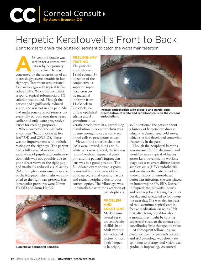
A56-year-old female was sent in for a cornea eval-uation by her primary optometrist. He was
concerned by the progression of an increasingly severe keratitis in her right eye. Treatment was initiated four weeks ago with topical trifl u-ridine 1.0%. When the eye didn’t respond, topical tobramycin 0.3% solution was added. Though the patient had signifi cantly reduced vision, she was not in any pain. She had undergone cataract surgery un-eventfully on both eyes three years earlier and only wore progressive lenses for reading purposes.
When corrected, the patient’s vision was “hand motion at fi ve feet” OD and 20/25 OS. There was no improvement with pinhole testing on the right eye. The patient had a full range of motion, but full evaluation of pupils and confronta-tion fi elds was not possible due to poor direct views of the right pupil and markedly reduced visual acuity (VA), though a consensual response of the left pupil when light was ap-plied to the right was present. Her intraocular pressures were 20mm Hg OD and 8mm Hg OS.
PRELIMINARY
TESTING
The patient’s exam showed 1+ lid edema, 3+ injection of the conjunctiva, a superior super-fi cial crescen-tic marginal infi ltrate from 11 o’clock to 2 o’clock, 2+ diffuse epithelial edema and 4+ granulomatous keratic precipitates in a partial ring distribution. Her endotheliitis was intense enough to cause some red blood cells to precipitate as well.
Views of the anterior chamber (AC) were limited, but 1+ to 2+ white cells were graded, the iris was normal without segmental atro-phy and the patient’s intraocular lens was in a good position. The full dilated exam showed a gross-ly normal but poor view of the optic nerve, retinal vessels, macula and retinal periphery due to poor corneal optics. The fellow eye was unremarkable with the exception of
pseudophakia.
PROBLEM
AND
SOLUTIONS
Marked uni-lateral kera-touveitis/endo-theliitis in an adult without any other risk factors is most likely herpet-ic in origin,
so I questioned the patient about a history of herpetic eye disease, which she denied, and cold sores, which she had developed somewhat frequently in the past.
Though the peripheral keratitis was unusual for the diagnosis (and would be more typical of herpes zoster keratouveitis), my working diagnosis was severe diffuse herpes simplex virus (HSV) endotheliitis and uveitis, as the patient had no known history of zoster-based periocular infection. She was placed on homatropine 5% BID, Durezol (difl uprednate, Novartis) hourly and oral acyclovir 400mg fi ve times per day and scheduled to follow-up the next day. She was also instruct-ed to discontinue topical anti-in-fective medication usage, as I felt that after being dosed for about a month, they might be causing superfi cial stress to the cornea and contributing little therapeutic value.
At subsequent follow-ups, we could see that the patient’s corneal and AC pathology was slowly re-sponding to therapy and vision was gradually improving. As corneal
Don’t forget to check the posterior segment to catch the worst manifestation.
Herpetic Keratouveitis Front to Back
Intense endotheliitis with placoid and partial ring
precipitation of white and red blood cells on the corneal
endothelium.
Superfi cial peripheral keratitis.

REVIEW OF CORNEA & CONTACT LENSES | NOVEMBER/DECEMBER 2019 13
optics improved, so did posterior segment views. With these better views, it became apparent that the patient’s posterior segment was also involved to some degree. There was mild vitritis, an asymmetrically mildly hyperemic nerve and a small amount of segmented columnar oc-clusive material in the primary and secondary retinal arterioles.
Given the previous diagnosis and now posterior involvement, acute retinal necrosis (ARN) needed to be considered. ARN is a rare pathology caused by herpes viruses that produces a diagnostic triad of edematous necrosis of the retina, occlusive vasculitis/arteritis and vitritis. It carries an extremely neg-ative prognosis—it’s estimated that between 20% and 85% of ARN patients develop rhegmatogenous retinal detachment and nearly 50% of patients end up with a best-cor-rected VA of 20/200 following the condition.1 Due to the severity
of the possible problem and its location in the posterior seg-ment, the patient was maintained on topical ther-apy and referred to the University of Washington Uveitis Clinic.
At the clinic, posterior in-volvement of the patient’s uveitis with diffuse retinal vessel leakage, subclinical retinal edema and nerve head leakage was confi rmed. The specialist described the arteriolar involvement as Kyrieleis plaques, which are a source of retinal vasculitis associated with ARN, tuberculosis, syphilis toxoplasmosis and Mediterranean spotted fever. Serology tests for these pathologies were run and subsequently found to
be negative. The primary diagno-
sis of HSV keratouve-itis with possible early ARN was maintained. Given the absence of zonal retinal necrosis, a fi rm diagnosis of ARN was not made, so its treatment protocol, which involves prophy-lactic vitreoretinal sur-gery, was not followed. The uveitis facility added a modest dose of oral prednisone with a protracted taper and asked us to schedule follow-ups every six weeks.
OUTCOMES
After six months of follow-up and gradually tapering therapy, the pa-tient’s panuveitis fully resolved. She now corrects to 20/20 and is very pleased with the outcome.
This case is a good reminder of an important clinical pointer. While the scope of possible pathologies of herpetic eye disease is wide and the vast majority of cases only involve the anterior segment, the disease’s worst manifestation involves the posterior segment. Therefore, peri-odic posterior evaluation of these eyes is necessary. Though it is easy to focus only on the anterior exam when working with impressive cas-es of anterior uveitis, the clinician needs to recognize that these pa-thologies should not be presumed to only involve the anterior segment. Paying equal close attention to the posterior segment is critical to catch panuveitis, which is associated with more profound and longer-lasting vision loss. RCCL
1. Schoenberger SD, Kim SJ, Thorne JE, et al. Diagnosis and treatment of acute retinal necrosis: a report by the American Acad-emy of Ophthalmology. Ophthalmology. 2017;124(3):382-92.
Posterior segment involvement shows occlusive
arteritis of the primary retinal arterioles. These
Kyrieleis plaques are associated with ARN.
Improvement in corneal involvement after one week of
aggressive therapy.

14 REVIEW OF CORNEA & CONTACT LENSES | NOVEMBER/DECEMBER 2019
Bacterial keratitis is a serious ocular condition that can lead to vision loss if not treated promptly,
appropriately and aggressively.1
According to the CDC, an estimat-ed one million clinical visits occur annually in the United States due to keratitis.2 Signifi cant risk factors include contact lens use and ocular surface disease. Contact lens risk factors can be further broken down into overnight lens wear, poor stor-age hygiene and infrequent storage case replacement.2,3
Treatment of bacterial keratitis begins with empirical manage-ment using frequent instillation of topical broad-spectrum antibiotics for coverage of both gram-pos-itive and gram-negative patho-gens. Common treatment starting points depend on the severity of the corneal ulcer and consist of monotherapy with fl uoroquinolo-nes or combination therapy with cephalosporins, aminoglycosides, vancomycin, amikacin or forti-fi ed antibiotics.1,4 This increased fi rst-line usage of broad-spectrum antibiotics, however, has coincided with a signifi cant rise in the number of bacterial pathogens resistant to antimicrobials.5
In this review, we discuss relevant trends in bacterial keratitis, includ-
ing corneal pathogen prevalence, corneal pathogen susceptibility, antibiotic resistance and treatment strategy.
PREVALENCE
According to two long-term ret-rospective case reviews of micro-bial keratitis in the United States, gram-positive organisms were the most commonly isolated bacterial group, followed by gram-negative organisms.6,7 Both studies indi-cated Staphylococci as the most prevalent gram-positive organism and Pseudomonas species as the most frequently isolated gram-neg-ative and overall organism.6,7 The individual proportions of cultured species differed slightly by study.6,7
Another study noted that methicil-lin-resistant Staphylococcus aureus (MRSA) comprised 20% of all isolates, while 5% of all cultured isolates were MRSA.6
International studies show simi-larities and differences in bacterial prevalence compared with national fi ndings.8-15 Similar to the United States, investigations in China, Switzerland, South Korea, Spain and Colombia indicate an overall preponderance of gram-positive or-ganisms, as well as a predominance of Staphylococci and Pseudomonas individual strains.10,12,14,15 India,
ABOUT THE AUTHORS
Ms. Cherny is a third-year optometry candidate at the SUNY College
of Optometry, where she is completing an Advanced Cornea and Contact Lens micro-credential. She received her undergraduate Human Biology, Health and
Society degree at Cornell University. She is interested
in specialty contact lenses and corneal disease management.
Dr. Patel is a graduate of the SUNY College of Optometry. He completed a
contact lens and ocular disease residency at The Ohio State
University Havener Eye Institute. He is currently a clinical instructor at Weill Cornell Medicine Ophthalmology in New York City, where his
focus is anterior segment management and specialty
contact lens fi tting. He is a fellow of the American Academy of Optometry.
Dr. Sherman is an assistant professor of optometric sciences in ophthalmology, director of optometric services at the Columbia University Medical Center and an assistant attending at New
York–Presbyterian Hospital. She specializes in complex and medically necessary contact lens fi ttings, anterior segment disease and primary care. She is board-certifi ed by the American Board of
Optometry and National Board of Examiners in
Optometry. She is a fellow of the American Academy of Optometry, conducts research, contributes to peer-reviewed scientifi c publications and presents at annual meetings.
Understanding common pathogens and eff ective treatments is essential in managing these patients with the most successful results.
HOT TOPICS
in Bacterial KeratitisBy Christina Cherny, BS,
Pratik Patel, OD,and Suzanne Sherman, OD

REVIEW OF CORNEA & CONTACT LENSES | NOVEMBER/DECEMBER 2019 15
Taiwan and the tropics of Malaysia share another similarity with the United States—Pseudomonas was the single most commonly isolated bacterial organism.8,11,13 Of notable contrast to the aforementioned countries is Taiwan. From 2007 to 2016, Taiwan noted gram-negative bacteria as the most commonly iso-lated strain, followed by gram-posi-tive bacteria.13
Temporal trends in the prev-alence of gram-positive strains isolated from bacterial keratitis cases varied by each respective region. In the United States, there was a 1.13-increased odds of culturing MRSA for each one-year increase in culture date.6 The trends for gram-negative organisms also varied considerably by region and species. According to one study, the only bacterial pathogen that increased signifi cantly in propor-tion during the study period was Pseudomonas aeruginosa.7
International trends in the proportions of bacterial isolates follow a similar route, differing by nation. There has been a signifi cant increase in gram-positive isolates in India in recent years, in contrast with the decreasing trends in cer-tain gram-positive strains observed
in China.8,9 In India, there has been a decrease in the overall prevalence of gram-negative organisms, which differs from the increasing trends in certain gram-negative strains observed in China and Taiwan.8,9,13
Retrospective analyses conducted in Switzerland and South Korea in-dicate no difference in proportions of gram-positive and gram-negative organisms, as well as no discernible variation in causative pathogens responsible for microbial kerati-tis during their respective study periods.10,12
SUSCEPTIBILITY
Antibiotic sensitivity is a measure of the antibiotic concentration that will inhibit bacterial growth and is a predictor of the clinical re-sponse to antimicrobial treatment.16
Techniques of bacterial culturing, such as natural agar plates and cor-neal scraping, are used to determine the bacteria in question and its sensitivity.17 Knowledge of bacte-rial susceptibility can be helpful to guide the selection of antibiot-ics to achieve successful clinical outcomes.18
Retrospective analyses of gram-positive organisms conduct-ed in the United States indicate
signifi cant variability in terms of antibiotic susceptibilities.6,7 Gram-positive isolates demonstrate 100% susceptibility to vancomycin over time and are fairly sensitive to gen-tamicin, tetracycline and trimetho-prim sulfamethoxazole.6,7 However, susceptibility to fl uoroquinolones, cefazolin and erythromycin is in-consistent.6,7 Furthermore, research-ers note a decrease in susceptibility of gram-positive organisms to levofl oxacin and gentamicin over time.6
Analyses of gram-negative or-ganisms indicated excellent in vitro susceptibility of Pseudomonas to fl uoroquinolones and aminoglyco-sides.6,7 Fluoroquinolones are also effective in Serratia, Moraxella and other enteric organisms.6,7 Non-Moraxella gram-negative rods exhibit better in vitro sensitivity to ceftazidime than to moxifl oxacin and tobramycin.6
Internationally, studies document differences in antibiotic sensitivities and trends between the various re-gions. Gram-positive isolates have a high susceptibility to vancomy-cin, fl uoroquinolones (including levofl oxacin, moxifl oxacin and gatifl oxacin), aminoglycosides (namely, erythromycin, gentamicin
Strain prevalence characteristics differ markedly between the United States and international regions (comprising India,
China, Switzerland, Malaysia, South Korea, Taiwan, Spain, Colombia).
43%
7%
19%
16%
15%
Gram-negative
• Pseudomonas spp.
• Serratia spp.
• Moraxella spp.
• Other enterics
• Other gram-negative
35%
32%
31%
2%
Gram-positive
• Staphylococcus aureus
• Streptococcus spp.
• Coagulase-negative Staphylococci
• Other gram-positive
UNITED STATES:
23%
17%
18%
15%
12%
6%
6%3%
Gram-positive
• Staphylococcus spp.
• Streptococcus spp.
• Corynebacterium spp.
• Coagulase-negative Staphylococci
26%
16%
10%10%
10%
10%
6%
6%
6% Gram-negative
• Pseudomonas spp.
• Other gram-negative
• Acinetobacter spp.
• Serratia spp.
• Klebsiella
• Bacillus spp.
• Enterococcus spp.
• Other
• Other gram-positive
• Enterobacter spp.
• Escherichia coli
• Proteus mirabilis
• Haemophilus infuenzae
INTERNATIONAL:

16 REVIEW OF CORNEA & CONTACT LENSES | NOVEMBER/DECEMBER 2019
and tobramycin), chloramphenicol and impinemen.8,9,12-15 In China, however, there has been a decrease in susceptibility of gram-positive isolates to levofl oxacin, cefazolin, ceftazidime and chloramphenicol over time.9 Staphylococcus species in particular have a high antibiotic sensitivity to vancomycin, teico-planin and chloramphenicol and a low sensitivity to fl uoroquino-lones, such as ciprofl oxacin.8,13
Susceptibility of Streptococcus pneumoniae is highest to cefazolin and lowest to ciprofl oxacin, which is mirrored by an overall trend of higher susceptibility of gram-posi-tive cocci to cephalosporins relative to fl uoroquinolones.8,9
In regard to gram-negative organisms, most are highly sensi-tive to fl uoroquinolones (including ciprofl oxacin and levofl oxacin), aminoglycosides (namely genta-micin, amikacin and tobramycin), cephalosporins (including ceftazi-dime and cefepime) and carbape-nem.9,11-13 Pseudomonas species in particular demonstrate excellent in vitro sensitivity to fl uoroquinolo-nes, aminoglycosides and certain cephalosporins.8,11-13
Studies conducted in South Korea and Taiwan do not show signifi cant changes in antibiotic sensitivity over time.12,13 In compar-ison, analyses in China indicate a decreasing trend of susceptibilities. In China, fl uoroquinolones were
reported to be the most susceptible antibiotic to gram-negative bacilli, with an increased susceptibility to ofl oxacin observed over time.9
ANTIBIOTIC RESISTANCE
This occurs when bacteria acquire the ability to evade destruction by antibiotics through spontaneous mutation or horizontal gene trans-fer. Resistance is driven largely by overuse of antibiotics. The resis-tance process removes drug-sen-sitive bacteria and leaves resistant strains to proliferate. Causes of an-tibiotic resistance include the vast number of antibiotics prescribed, incorrect prescribing of antibiotics, extensive agricultural use of anti-biotics and the lack of availability of new antibiotics on the market.18
Trends in antibiotic resistance vary regionally, with certain bacterial organisms demonstrating increased resistance over time and other bac-terial strains exhibiting no signifi -cant change in antibiotic resistance.
In the United States, trends in antibiotic resistance have changed over time and vary by region and bacterial strain. Resistance to moxi-fl oxacin increases with each one-year increase in the culture date, and a trend of increasing resis-tance to gentamicin was observed among gram-positive organisms. Additionally, one study found the risk of culturing MRSA seems to increase with time in San Francisco,
while no signifi cant annual trends were noted in the proportions of oxacillin-resistant Staphylococcus aureus and oxacillin-resistant coag-ulase-negative Staphylococci in St. Louis.6,7
Analyses in Malaysia observe no increase in resistance rates for the commonly used antibiotics, although these fi ndings do not hold true for other regions.11 In Colombia, gram-negative organ-isms exhibit higher resistance rates overall, while in Taiwan, resistance is more common in gram-posi-tive pathogens.13,15 Similar to the United States, the risk of culturing MRSA in Taiwan increases with time.13 This differs from fi ndings in Spain and South Korea, where no trends have been observed for methicillin-resistant strains.12,14
Furthermore, Staphylococcusspecies demonstrate a signifi cant increase in the proportion resistant to oxacillin over time. This is in contrast to the pattern seen in the United States.13 In Colombia and South Korea, moderate-to-high resistance is observed for gram-neg-ative isolates to gentamicin, tobramycin, amikacin imipenem, gatifl oxacin and ciprofl oxacin, as well as for gram-positive isolates to Ciprofl oxacin.12,15
TREATMENT STRATEGY
In addition to the current accepted methods used to treat microbial
HOT TOPICS IN BACTERIAL KERATITIS
Fluorescein staining shows an active
bacterial ulcer in a patient who slept
in her contact lenses.
The patient’s corneal ulcer is
resolving.
She was left with this scar after her
ulcer healed.

REVIEW OF CORNEA & CONTACT LENSES | NOVEMBER/DECEMBER 2019 17
keratitis, a review of recent lit-erature indicates several novel approaches may prove valuable for the treatment of bacterial corneal infections.
A broad-spectrum combination of polymyxin B–trimethoprim (PT) and rifampin demonstrat-ed increased potency, improved antibiofi lm activity and more rapid bactericidal activity compared with PT alone for the treatment of Staphylococcus aureus and Pseudomonas corneal infections.19
This novel combination also ex-hibits a lower tendency to develop resistance than either individual agent or moxifl oxacin.19 Increased effi cacy was also observed relative to commercial PT and moxifl oxa-cin in murine models of keratitis.19
Thymosin beta-4 (Tβ4) is a natu-rally occurring amino acid protein that promotes wound healing and host defense and reduces corneal infl ammation. Corneas treated with a combination of Tβ4 and
ciprofl oxacin to fi ght off keratitis caused by Pseudomonas aeruginosa demonstrate the most improvement in disease severity, with minimal impact on host immune response.20
Researchers investigated ar-gon cold plasma as a potential treatment for therapy-resistant corneal infections, specifi cal-ly for Staphylococcus aureus, Staphylococcus epidermidis, Escherichia coli and Pseudomonas aeruginosa. Analyses indicate that argon cold plasma treatments can signifi cantly reduce various corne-al pathogens and improve vision post-treatment without damaging the ocular surface.21
In another study, patients with culture-positive bacterial ker-atitis—negative for fungal and protozoal species—received six or more daily drops of potent topical steroids, including prednisolone acetate 1%, phenylephrine hydro-chloride 0.12%, dexamethasone 0.1% and prednisolone sodium phosphate 0.5%.22 Patients on the high-dose steroid regimen have a 5.5-increased chance of better visual outcomes, although patients with Nocardia keratitis had poorer outcomes.22 Treatment of bacterial keratitis with high-dose steroid reg-imens may prove to be an import-ant clinical tool for improved visual outcomes after corneal infection.22
A working knowledge and understanding of common
pathogens and their respective clinical therapies and current trends is essential to effectively manage bacterial keratitis and improve visual outcomes in patients. Given the widespread use of broad-spec-trum antimicrobials and the subsequent emergence of antibiotic resistance, targeting various patho-gens with treatments that have proven effective and using novel therapies adjunctively or in isola-
tion may help improve success rates in the treatment of corneal ulcers and better preserve vision in those affected. RCCL
1. Bourcier T, Thomas F, Borderie V, et al. Bacterial ker-atitis: predisposing factors, clinical and microbiological review of 300 cases. Br J Ophthalmol. 2003;87(7):834-8.2. Collier SA, Gronostaj MP, MacGurn AK, et al. Estimated burden of keratitis—United States, 2010. Morb Mortal Wkly Rep. 2014;63(45):1027-30.3. Truong DT, Bui MT, Cavanagh HD. Epidemiology and outcome of microbial keratitis: private university versus urban public hospital care. Eye Cont Lens. 2018;44(Sup-pl 1):S82-6.4. Acharya M, Farooqui JH, Jain S, et al. Pearls and paradigms in infective keratitis. Rom J Ophthalmol. 2019;63(2):119-27.5. Odonkor ST, Addo KK. Bacteria resistance to antibi-otics: recent trends and challenges. Int J Biol Med Res. 2011;2(4):1204-10.6. Peng MY, Cevallos V, McLeod SD, et al. Bacterial keratitis: isolated organisms and antibiotic resistance patterns in San Francisco. Cornea. 2018;37(1):84-7.7. Hsu HY, Ernst B, Schmidt EJ, et al. Laboratory results, epidemiologic features, and outcome analyses of microbial keratitis: a 15-year review from St. Louis. Am J Ophthalmol. 2019;198:54-62.8. Das S, Samantaray R, Mallick A, et al. Types of organ-isms and in-vitro susceptibility of bacterial isolates from patients with microbial keratitis: a trend analysis of 8 years. Ind J Ophthalmol. 2019;67(1):49-53.9. Lin L, Duan F, Yang Y, et al. Nine-year analysis of iso-lated pathogens and antibiotic susceptibilities of micro-bial keratitis from a large referral eye center in southern China. Infection and Drug Resistance. 2019;12:1295-302.10. Bograd A, Seiler T, Droz S, et al. Bacterial and fungal keratitis: a retrospective analysis at a univer-sity hospital in Switzerland. Klin Monbl Augenheilkd. 2019;236(4):358-65.11. Khor HG, Cho I, Lee KRCK, et al. Spectrum of micro-bial keratitis encountered in the tropics. Eye Cont Lens. 2019:1542-2321.12. Mun Y, Kim MK, Oh JY. Ten-year analysis of micro-biological profi le and antibiotic sensitivity for bacterial keratitis in Korea. PLoS ONE. 2019;14(3):e0213103.13. Liu HY, Chu HS, Wang IJ, et al. Microbial kerati-tis in Taiwan: a 20-year update. Am J Ophthalmol. 2019;205:74-81.14. Tena D, Rodríguez N, Toribio L, et al. Infectious kerati-tis: microbiological review of 297 cases. Jpn J Infect Dis. 2019;72(2):121-3.15. Galvis V, Parra MM, Tello A, et al. Antibiotic resistance profi le in eye infections in a reference centre in Floridab-lanca, Colombia. Arch Soc Esp Oftalmol. 2019;94(1):4-11.16. Smaill F. Antibiotic susceptibility and resistance test-ing: an overview. Can J Gastroenterol. 2000;14(10):871-5.17. Leck A. Taking a corneal scrape and making a diagno-sis. Comm Eye Health. 2009;22(71):42-3.18. Ventola CL. The antibiotic resistance crisis: part 1: causes and threats. Pharm Therapeut. 2015;40(4):277-83.19. Chojnacki M, Philbrick A, Wucher B, et al. Develop-ment of a broad-spectrum antimicrobial combination for the treatment of Staphylococcus aureus and Pseudomo-nas aeruginosa corneal infections. Antimicrob Agents Chemother. 2018;63(1):e01929-18.20. Carion TW, Ebrahim AS, Kracht D, et al. Thymosin Beta-4 and ciprofl oxacin adjunctive therapy improves Pseudomonas aeruginosa-induced keratitis. Cells. 2018;7(10):145.21. Reitberger HH, Czugala M, Chow C, et al. Argon cold plasma—a novel tool to treat therapy-resistant corneal infections. Am J Ophthalmol. 2018;190:150-63.22. Green M, Hughes I, Hogden J, et al. High-dose steroid treatment of bacterial keratitis. Cornea. 2019;38(2):135-40.
This patient had irregular
epitheliopathy at presentation and
ended up having an active bacterial
infection secondary to contact lenses.
Hyperemia can be seen in this patient.

18 REVIEW OF CORNEA & CONTACT LENSES | NOVEMBER/DECEMBER 2019
According to a recent study in Cornea, ap-proximately one million annual ocular medical
visits in the United States ended up with a bacterial keratitis diagno-sis.1,2 Of these patients, 76.5% re-ceived a prescription for antibiotics from their healthcare provider.1,2
The cost of treating bacterial keratitis is estimated to be around $377 million to $857 million per year.3 Bacterial keratitis is only one cause of ocular infections, but it requires immediate intervention in order to prevent vision loss and minimize complications.
Viral and bacterial infections are the most common etiologies for bacterial keratitis. A proportion of 71,000 cases of severe infectious keratitis a year in America has been estimated, a lower proportion than non-infectious bacterial keratitis.2,4
To treat bacterial keratitis, prac-titioners initiate empirical therapy with broad-spectrum or fortifi ed antibiotics prescriptions; however, overuse has led to a pattern of re-sistance that can cause diffi culty in suitably managing the condition.5
This article explores the changes in trends regarding corneal in-fections and the situations where practitioners should use antibiotic treatment. Understanding common
pathogens and effective treatments is essential in managing these patients with the most successful results.
BACTERIAL CONJUNCTIVITIS
AND KERATITIS
Bacterial conjunctivitis is the second most common cause of con-junctivitis, and it is responsible for 50% to 75% of conjunctivitis cases in children.6 Although conjunctivi-tis involves the conjunctiva specifi -cally, it can affect the surrounding ocular structures that can lead to worsening infections, such as keratitis, which can become serious enough to cause blindness.7
In adults, a bacterial origin is less common than a viral one and is characterized by bacterial over-growth, along with infi ltration of the conjunctival epithelial layer. The origin can either be from direct contact with an infected individu-al’s secretions or advanced through organisms colonizing within the patient’s own nasal and sinus mucosa.8
Bacterial keratitis is an acute or chronic condition that can become sight-threatening if left untreated. These cases can lead to stromal in-fl ammation and progressive tissue destruction, eventually causing per-foration. It is commonly connected
with risk factors that disturb the corneal epithelial integrity. Contact lens wear, trauma, impaired defense mechanism, immunosup-pressive medication use and altered corneal surface structure postoper-atively are all common predispos-ing factors.8,9
The most common risk factor in the US is contact lens wear. Microbial keratitis is approxi-mately 15 times more likely in patients who sleep in their lenses and is positively correlated with the number of days patients wear their contact lenses without removal.8
With the increase of contact lens wear gloally, bacterial keratitis has also increased accordingly.9
Bacterial conjunctivitis can be self-limiting and resolves on its own in one to two weeks due to
ABOUT THE AUTHORS
Dr. Shuja is an optometrist at New York–Presbyterian Hospital.
Dr. Sherman is an assistant professor of optometric
sciences in ophthalmology and director of optometric services at the Columbia University Medical Center and an assistant attending at New
York–Presbyterian Hospital.
SMART STRATEGIESfor Using Antibiotics to TreatCORNEAL INFECTION
Understand when to initiate appropriate therapy options.
By Fiza Shuja, OD, and Suzanne Sherman, OD

REVIEW OF CORNEA & CONTACT LENSES | NOVEMBER/DECEMBER 2019 19
the body’s immune factors.6 Gram-positive cocci and Staphylococcus species are known to inhabit skin cells, skin glands and mucous membranes. Methicillin-resistant Staphylococcus aureus (MRSA) is a well-known gram positive cocci we always have to be on the lookout for. MRSA has a caused an increase of vancomycin use. Propionibacterium acnes (P. acnes) is one of the major causes of postoperative endophthalmitis but an uncommon cause of microbial keratitis.
When caused by dangerous bacterial species, such as Neisseriagonorrhoeae or Streptococcus pyo-genes, bacterial conjunctivitis can be serious and sight-threatening. In rare cases, it may foreshadow a life-threatening systemic disease, such as conjunctivitis caused by Neisseria meningitidis.8
The most common causative bacterial species are Haemophilus infl uenza, Streptococcus pneu-moniae, Staphylococcus aureus and Staphylococcus epidermidiswith staphylococci, specifi cally S. aureus and coagulase-negative Staphylococci (CoNS), reported most often.7,10
Several bacterial species simul-taneously can cause most cases of acute bacterial conjunctivitis. This is why practitioners use broad-spectrum antibiotics as the fi rst line of ophthalmic antibacte-
rial treatment.1 Antibiotics often accelerate clinical resolution and microbiolog-ical remission, while also lessening the risk of recur-rence and the development of complications.1
Some prospective studies show that delaying antibiotic treatment until day three or four will reduce the use of un-necessary medications and not affect outcomes. The practi-tioner only initiated treatment if the signs were worsening, shortening the course and improving symptoms. These studies all advocate that initiation of antibiotics after day four provides fi nite benefi ts.8
Classic antibacterial options include tobramycin, trimetho-prim, ciprofl oxacin, gatifl oxacin and moxifl oxacin.10 However, the widespread use of broad-spectrum antibiotics has resulted in resis-tance to those typical antibiotics.10
Therefore, developing new antibi-otics with high effi cacy and safety against some resistant bacteria is necessary.1
MANAGEMENT
The primary goal when dealing with corneal infections is always to prevent loss of sight and to preserve corneal clarity. It is safer to assume and, therefore, treat any presentation of microbial keratitis
as bacterial keratitis for the best outcome.9 However, it is diffi cult for the practitioner to quickly and effectively manage patients with presumed microbial conjunctivitis or keratitis. Although it would be ideal to have a confi rmed defi nitive diagnosis before initiating therapy, bacterial pathogens can cause irreversible corneal scarring. It is therefore imperative to begin treatment before any damage occurs. The initiation of therapy must occur before an established diagnosis in order to prevent visual disability and limit the bacterial load.8
Preliminary therapy is comprised of empirical topical broad-spec-trum antibiotics. For routine cor-neal ulcers, monotherapy of topical fl uoroquinolones provides compa-rable therapy to combination ther-
apy due to the enhanced penetration obtained with commercially avail-able fl uoroquinolones.8
Fluoroquinolones can be instilled every 30 minutes to 60 minutes for a routine corneal ulcer.
If the ulcer is more se-vere, use a loading dose of every fi ve minutes for 30 minutes to transfer
This child has subcutaneous conjunctival
membranes from a bacterial infection.
Classifi cation of Bacterial Conjunctivitis8
Course of Onset Severity Common Organisms
Slow(days to weeks)
Mild to moderate Staphylococcus aureusMoraxella lacunata
Proteus spp.Enterobacteriaceae
Pseudomonas
Acute or subacute(hours to days)
Moderate to severe Haemophilus infl uenzae biotype IIIHaemophilus infl uenzae
Streptococcus pneumoniaeStaphylococcus aureus
Hyperacute(less than 24 hours)
Severe Neisseria gonorrhoeaeNeisseria meningitidis

20 REVIEW OF CORNEA & CONTACT LENSES | NOVEMBER/DECEMBER 2019
therapeutic concentration to the stroma faster.8
If monotherapy fails and/or the initial ulcer is large, central or atypical, consider combination therapies due to the additional gram-negative activity. In addi-tion, if combination therapy fails or MRSA is suspected, initiate fortifi ed antibiotics, including vancomycin. Fortifi ed antibiot-ics are compounded at increased concentration. Remember that “fortifi eds” can be diffi cult to obtain commercially and have greater corneal toxicity. According to a survey, the majority of corneal specialist respondents in the United States chose to treat corneal ulcers with fortifi ed antibiotics, specifi -cally vancomycin and tobramycin due to low antibiotic resistance, whereas a majority of internation-al corneal specialists respondents chose fl uoroquinolone treatment due to availability.11
Clinical parameters that can be helpful to monitor the response to antibiotic treatment include blunt-ing the stromal infi ltrate perimeter, decreased density of the stromal infi ltrate, reduction of stromal ede-ma and endothelial infl ammatory plaque, reduction in anterior cham-ber infl ammation, reepithelization and cessation of corneal thinning.8
In day-to-day clinical practice, we work side-by-side with anterior segment specialists in a tertiary care setting. Over the years, we have developed our own “smart” strategies for dealing with corneal infections.
1. Take a careful case history. Do not rely only on the informa-tion the patient gives you. Always ask about previous contact lens use, recent activities, surgeries, etc.
2. Ask about previous treatments. Many patients have already gone to a walk-in clinic or someone who is not an eye care provider. If they can’t recall the exact medication given, ask about the cap color and dosage.
3. Conduct a careful slit lamp exam including lid eversion and fl uorescein staining.
4. Use slit lamp photo-graphs to compare images at each visit.
5. Perform IOP measure-ments and dilation are essential. If you haven’t seen a patient recently, you cannot assume the posterior segment is not involved.
Once the presumed etiology is determined based on case history and clinical exam, personal expe-rience shows it is most effective to use monotherapy of topical fl uoroquinolones during the day and an added ointment at night. In the offi ce, fourth generation fl uoro-quinolones are not always avail-able; therefore, we give the patient a sample of the highest-generation drop available and a prescription
for fourth generation fl uoroquino-lones to the pharmacy.
Depending on the severity of the infection, it is helpful to space out follow-ups in order to give the antibiotics time to work and the cornea time to heal. At the follow-up, closely compare the current presentation to previous slit lamp photos. Oftentimes, clinical signs and patient symptoms will have started to improve before culture results have returned.
RISE OF THE RESISTANCE
Though bacterial keratitis requires treatment with antibiotics, it is crucial to understand how over-us-ing and over-prescribing antibiotics can lead to resistance. Bacterial resistance to an antibiotic depends on the mechanism. The most common resistance mechanism, modifi cation, can involve a muta-tion to the target site, making the
USING ANTIBIOTICS TO TREAT CORNEAL INFECTION
Common Causes of Bacterial Keratitis8
Common Uncommon
Staphylococcus aureusStaphylococcus epidermidisStreptococcus pneumoniaePseudomonas aeruginosa
Enterobacteriaceae
Neisseria spp.Moraxella spp.
Mycobacterium spp.Nocardia spp.
Non–spore-forming anaerobesCorynebacterium spp.
This patient had bacterial keratitis (top) that
eventually resolved with antibiotics and was
left only with a small scar (bottom).

REVIEW OF CORNEA & CONTACT LENSES | NOVEMBER/DECEMBER 2019 21
drug ineffective.12 Resistance can be coded into the bacterial genes and then passed between colonies and species, allowing it to spread quickly.12
Antibiotic resistance to penicil-lin can begin soon after the drug is introduced to treat infections.13
Factors to blame for antibiotic resistance include over-prescribing, inappropriate dosing regimen, increased use of antibiotics in agriculture and increased exposure to systemic antibiotics.14,15 When practitioners prescribe, a pattern of resistance can occur if patients are unable to self-administer properly or discontinue medications due to ocular discomfort from adverse effects.15
Many of the antibiotics treating the ocular surface are also used systemically for infections, except besifl oxacin, which was formulated exclusively for ocular use to allow lower resistance rates.14 We have seen some patients who believe they are cured and self-discontinue antibiotics early. Once this hap-pened, the infection reappeared and the treatment course had to be resumed. It is therefore wise for optometrists to prevent over-pre-scribing and make sure the antibi-otic treatment runs its course.
The increase in resistant bacteria over the years has led to stud-ies, such as the Ocular Tracking Resistance in the United States Today (TRUST) and Antibiotic Resistance Monitoring in Ocular micRoorganisms (ARMOR) stud-ies.13,14 Ocular TRUST monitored S. aureus, S. pneumoniae and H. infl uenzae when treated with fl uo-roquinolones, macrolides, amino-glycosides, penicillin, dihydrofolate reductase inhibitors and polypep-tides. Ocular TRUST studies re-ported 16.8% methicillin resistance from 2005 to 2006, which then increased to 50% by 2008.16
The ARMOR study monitors antibiotic resistance in ocular infec-tions against S. aureus, CoNS, S. pneumoniae, H. infl uenzae and P. aeruginosa.16 From 2009 to 2013, methicillin resistance was shown in staphylococcal isolates; however, it did not increase over the fi ve years. Bacteria such as S. pneumoniae and H. infl uenzae were most susceptible to antibiotics, whereas there was multidrug resistance in 86.8% of methicillin-resistant S. aureus iso-lates and 77.3% of methicillin-re-sistant CoNS. The study also noted a higher number of methicillin-re-sistant staphylococcal infections in elderly patients.16 P. aeruginosaand H. infl uenzae isolates showed low resistance against most of the antibiotics tested.
Most of the published data regarding ocular pathogens is col-lected from single centers; however, pathogens differ in prevalence
geographically, reinforcing the need for studies like ARMOR, which are conducted nationwide in the United States. Understanding the location of bacteria can aid eye-care providers to target the more common pathogens—S. aureus is higher in the South and lower in the West, S. pneumoniae is higher in the Midwest and lower in the West and P. aeruginosa is higher in the Midwest and lower in the West.
Globally, antibiotic resistance continues to plague practi-tioners when treating keratitis. Moxifl oxacin has shown increased resistance in India, despite its more “recent” foray in treating bacterial infections; there was low susceptibility of coagulase-nega-tive Staphylococcus (61.2%) and methicillin-resistant Staphylococcus(53.1%) when treated with moxi-fl oxacin. In the US, 26% of all organisms cultured were resistant
Antibiotic Smart Strategies
Routine Corneal
Ulcer
Monotherapy: topical fl uoroquinolones
• Equivalent to combination therapy
Every 30 to 60 minutes, tapered according to clinical response.
More severe presentation: Loading dose every fi ve minutes for 30 minutes.
Second generation:ciprofl oxacin, ofl oxacin
• Pseudomonas coverage
• Lack some gram-positive activity
Third and fourth generation:moxifl oxacin, gatifl oxacin, levofl oxacin, besifl oxacin
• Improved gram-positive activity
• Atypical mycobacterial coverage
• Limited activity against MRSA
Monotherapy
for initial ulcer,
unless ulcer is
large, vision-
threatening or
atypical
Combination therapy
• Active against gram-positive and gram-negative bacteria
Failed
monotherapy
or combination
therapy with
large, vision-
threatening,
MRSA suspected
Fortifi ed antibiotics
• Vancomycin gram +, greater coverage against MRSA

22 REVIEW OF CORNEA & CONTACT LENSES | NOVEMBER/DECEMBER 2019
to moxifl oxacin, while 28% of Staphylococcus aureus bacterial isolates were resistant to ciprofl ox-acin or ofl oxacin.17
ALTERNATIVE THERAPY
There are other treatment mo-dalities to consider when treating bacterial keratitis. The use of ste-roids in treating bacterial keratitis remains a controversial issue. Early use of steroids can help reduce corneal stromal melt, neovascu-larization and corneal scarring that results from the infl ammatory response against the infection. Instilling steroids in conjunction with fortifi ed antibiotics can also help decrease discomfort. The counterargument is that steroids delay corneal healing, leading to a worse infection. Though steroid use leads to improvement for some patients, note that the use of ste-roids for fungal or Acanthamoeba infections can lead to terrible results, such as increased loss of vision or loss of the eye.
Studies have been performed to fi nd new adjunct therapies to treat bacterial keratitis, especially with the rise of antibiotic resistance. Amniotic membranes, typically used during pterygium surgery,
also help resolve epithelial defects and chemical in-jury. Amniotic membrane benefi ts include reepithe-lization by reinforcing bas-al epithelial cell adhesion, induction of epithelial cell migration, differenti-ation and proliferation of conjunctival and limbal epithelial progenitor cells, prevention of epithelial apoptosis and reduction of keratocyte apopto-sis. Studies have shown improvement in epithelial defect, corneal haze and neovascularization when
eyes were treated with amniotic membranes rather than antibiotics alone; however, larger studies needs to be conducted to analyze the full potential of amniotic membranes when treating bacterial keratitis.18
Treatment for corneal perfora-tion with doxycycline has shown improvement in animal studies. In rabbit models, corneal perforation from Pseudomonas ulcers was reduced by 50% with systemic doxycycline use. Unfortunately, the lack of human studies makes it diffi cult to prove doxycycline as an effective therapy.
Collagen crosslinking, used to treat keratoconus, has antimicro-bial properties and can potentially help resolve corneal ulcers from bacterial pathogens. Case reports have shown an improvement in symptoms and the resolution of treatment-resistant infections. Further trials and studies could help establish crosslinking as a viable treatment for those with an-tibiotic resistance and/or to prevent ocular toxicity.19
Though bacterial keratitis needs to be treated aggressively, ex-
ercise caution when using treating topical therapy options, such as
steroids. Clinical assessment is imperative in making the correct diagnosis and managing it appro-priately prevents vision loss. RCCL
1. Collier SA, Gronostaj MP, MacGurn AK, et al. Es-timated burden of keratitis: United States, 2010. Morb Mortal Wkly Rep. 2014;63(45):1027-30.2. Ballouz D, Maganti N, Tuohy M, et al. Medica-tion burden for patients with bacterial keratitis. Cornea. 2019;38(8):933–7.3. Smith AF, Waycaster C. Estimate of the direct and indirect annual cost of bacterial conjunctivitis in the United States. BMC Ophthalmol. 2009;9:13.4. Jeng BH, Gritz DC, Kumar AB, et al. Epidemiol-ogy of ulcerative keratitis in Northern California. Arch Ophthalmol. 2010;128:1022–8.5. Gokhale NS. Medical management approach to infectious keratitis. Indian J Ophthalmol. 2008;56(3):215-20.6. Høvding G. Acute bacterial conjunctivitis. Acta Ophthalmol. 2008;86(1):5-17.7. Teweldemedhin M, Gebreyesus H, Atsbaha AH, et al. Bacterial profi le of ocular infections: a sys-tematic review. BMC Ophthalmology. 2017;17:212.8. American Academy of Ophthalmology. Exter-nal disease and cornea: Basic and Clinical Science Course 2017-2018. 2017.9. Wang JJ, Gao XY, Li HZ, Du SS. The effi cacy and safety of besifl oxacin for acute bacterial conjunctivitis: a Meta-analysis. Int J Ophthalmol. 2019;12(6):1027-36.10. Al-Mujaini A, Al-Kharusi N, Thakral A, Wali UK. Bacterial keratitis: perspective on epidemiology, clinico-pathogenesis, diagnosis and treatment. Sultan Qaboos Univ Med J. 2009;9(2):184–95.11. Austin A, Schallhorn J, Geske M, et al. Empirical treatment of bacterial keratitis: an international survey of corneal specialists. BMJ Open Ophthal-mol. 2016;2:e00047.12. Baum J, Barza M. The evolution of antibiotic therapy for bacterial conjunctivitis and keratitis: 1970-2000. Cornea. 2000;19(5):659-72.13. McDonald M, Blondeau J. Emerging and antibiotic resistance in ocular infections and the role of fl uoroquinolones. J Cataract Refract Surg. 2010;36(9):1588-98.14. Thomas RK, Melton R, Asbell PA. Antibiotic re-sistance among ocular pathogens: current trends from the ARMOR surveillance study (2009-2016). Clin Optom (Auckl). 2019;11:15-26.15. Samarawickrama C, Chan E, Daniell M. Rising fl uoroquinolone resistance rates in corneal isolates: implications for the wider use of antibi-otics within the community. Infection, Disease & Health. 2015;20:128-33.16. Asbell PA, Sanfi lippo CM, Pillar CM, et al. An-tibiotic resistance trends from ocular pathogens in the US-cumulative results from the antibiotic resistance monitoring in ocular microorganisms (ARMOR) surveillance study. US Ophthalmic Review. 2017;10(1):35-8.17. Ung L, Bispo PJM, Shanbhag SS, et. al. The persistent dilemma of microbial keratitis: global burden, diagnosis, and antimicrobial resistance. Surv Ophthalmol. 2019;64:255-7118. Dakhil TAB, Stone DU, Gritz DC. Adjunctive therapies for bacterial keratitis. Middle East Afr J Ophthalmol. 2017;24(1):11–7.19. Austin A, Lietman T, Rose-Nussbaumer J. Up-date on the management of infectious keratitis. Ophthalmology. 2017;124(11):1678–89.
Corneal scarring secondary to chronic
blepharoconjunctivitis caused this case of
keratitis.
USING ANTIBIOTICS TO TREAT CORNEAL INFECTION

Visit our website for the latest information: www.reviewsce.com/eventse-mail: [email protected] or call: 866-658-1772
2020 MEETINGSJoin us for our
Earn up to
18-28 CE
Credits*
FEBRUARY 14-17, 2020 - ASPEN, COAnnual Winter Ophthalmic ConferenceWestin Snowmass Conference CenterProgram Co-chairs: Murray Fingeret, OD, FAAO and Leo P. Semes, OD, FACMO, FAAO
REGISTER ONLINE: www.skivision.com
FAAWWP
R
JUNE 5-7, 2020 - ORLANDO, FLDisney Yacht & Beach ClubProgram Chair: Paul M. Karpecki, OD, FAAO
REGISTER ONLINE: www.reviewsce.com/orlando2020
JJUDP
R
MAY 29-31, 2020 - SAN DIEGO, CAManchester Grand HyattProgram Chair: Paul M. Karpecki, OD, FAAO
REGISTER ONLINE: www.reviewsce.com/sandiego2020
MMP
R
APRIL 16-19, 2020 - AUSTIN, TXOmni Barton CreekJoint Meeting with OCCRS**
Program Chair: Paul M. Karpecki, OD, FAAO
REGISTER ONLINE: www.reviewsce.com/austin2020
AOJJoP
R
NOVEMBER 6-8, 2020 - PHILADELPHIA, PAPhiladelphia Marriott DowntownProgram Chair: Paul M. Karpecki, OD, FAAO
REGISTER ONLINE: www.reviewsce.com/philadelphia2020
NPP
R
Review Education Group partners with Salus University for those ODs who are licensed in states that require university credit.See www.reviewsce.com/events for any meeting schedule changes or updates.
NEW TECHNOLOGIES & TREATMENTS IN
Eye CareNENEW W TNENEW T && T& && T&
EEEyEEy
2020 eearearCCCCye ye CareCCaC re Ca EEyEE00
**17th Annual Education Symposium
Joint Meeting with NT&T in Eye Care*Approval pending
Administered by:
creo
24 REVIEW OF CORNEA & CONTACT LENSES | NOVEMBER/DECEMBER 2019
Neurotrophic keratitis (NK) is a potential-ly sight-threatening condition marked
by decreased or absent corneal sensation. The condition is caused by damage both to the trigeminal nerve along its corneal distribution and at any point in the pathway from the ganglion to the basal plexus. This damage compromises corneal integrity by altering the metabolism and mitosis processes of the corneal epithelium, which results in reduced innate immunity of the ocular surface and delayed healing.1,2 If allowed to progress, damage could advance to the point of ulceration and, in severe cases, corneal perforation.1-3
NK is easy to manage early on in the disease process but remains one of the most challenging corne-al conditions to treat in its severe stages. This article will review the causes, diagnostic fi ndings and treatments available for NK throughout all stages of disease.
TRACE IT TO THE SOURCE
The most common etiologies of NK are herpes simplex virus (HSV) and varicella zoster virus (VZV).1 Among herpes viruses, zoster causes more severe neur-otrophy, as it may infl ict damage
both centrally at the ganglion and peripherally at the basal plexus. Alternatively, HSV only causes peripheral nerve damage. Patients suffering from herpes zoster ophthalmicus experience shorter corneal nerve length and lower corneal nerve count.4 The zoster virus lies dormant in the trigeminal ganglion, and when it reactivates along the nasociliary distribution, it results in corneal compromise.4 In both HSV and VZV, these damaging effects occur quickly and have long-lasting impacts on the integrity of the corneal epithelium.
Beyond viral eye disease, there are a variety of other causes of NK, including repeated corneal surgeries, chemical burns, contact lens abuse, medications, tumors and surgical complications result-ing in trigeminal nerve palsies.1,5
Systemic disorders such as dia-betes also contribute to NK. The longer a patient has diabetes, the more severe their corneal nerve damage might be. This mani-fests in diminished corneal tissue sensation similar to that occur-ring with peripheral neuropathy. Additionally, peripheral treatment with panretinal photocoagula-tion for proliferative diabetic retinopathy worsens the degree
of neurotrophy.5 In fact, corneal confocal microscopy has been shown to be useful in the early diagnosis of peripheral neurop-athy due to its ability to detect subtle reductions in corneal nerve density and length.6 This form of microscopy can assess small nerve fi bers, which neuropathy affects fi rst.6 Because of the link between diabetic neuropathy and corne-al integrity, consider the role of underlying neurotrophy in patients with diabetes and other ocular surface disease.
Regardless of its specifi c source, corneal neurotrophy is typical-ly not associated with pain or discomfort due to the lack of sensation it is accompanied by. These patients will, however, likely report reduced vision, especially as the condition becomes more severe. Medicamentosa is a poten-tial cause of corneal neurotrophy that should always be considered
ABOUT THE AUTHOR
Dr. Mannen practices at a private optometry and ophthalmology clinic in the greater Ogden, Utah area. She completed her residency at the Walla Walla Veteran Aff airs
Medical Center and Pacifi c Cataract and Laser Institute.
She is also a fellow of the American Academy of Optometry.�
The more it progresses, the more challenging it becomes,so early detection and diagnosis are key.
TAKE ON
With These
Clinical Tools
Neurotrophic Keratitis
By Megan Mannen, OD

REVIEW OF CORNEA & CONTACT LENSES | NOVEMBER/DECEMBER 2019 25
as a differential and man-aged appropriately. Topical medications most likely to have noxious ocular surface effects include anesthetics, beta-blockers and non-ste-roidal anti-infl ammatories (NSAIDs). Most of these medications use benzalko-nium chloride as the preser-vative.7 Monitor the ocular surface closely in glaucoma patients using beta-blockers and in post-surgical patients using NSAIDs, as these are the medications most com-monly prescribed to these patients.
Using a greater number of medications further increases the risk of toxicity.7 If topical toxicity is suspected, discontinue medica-tions and monitor the cornea for improvement. Systemic medica-tions have also been shown to perpetuate corneal neurotrophy. These include antihistamines, neuroleptics and antipsychotics.2
Consult primary care providers and consider drug cessation if these mediations are contributing to corneal compromise.
THE THREE STAGES OF NK
There are three traditional clinical stages of NK. The fi rst involves tear dynamic alteration, punc-tate epithelial erosion, superfi cial neovascularization and stromal scarring. These fi ndings are common and can be mistaken for other ocular surface diseases, but decreased corneal sensation, lack of symptoms and patient history will help with the diagnosis.1,2,8
The second stage of NK is marked by a persistent epithelial defect. The defect may be sur-rounded by a loose, edematous or boggy epithelium. Stromal edema and folds in Descemet’s membrane may also accompany these clinical
signs. Cells and fl are, which may occasionally worsen into a sterile hypopyon, are often present in this stage and can make the differenti-ation between microbial keratitis and neurotrophic disease more challenging.1,2,5,8
The third and fi nal stage of neurotrophy involves corneal stroma ulceration. This is defi ned as an epithelial break with under-lying stromal infl ammation. The stroma is made up of organized collagen fi bers called lamellae. Once the stroma is penetrated, the collagen fi brils start to degrade. This damage attracts white blood cells, which infi ltrate the damaged tissue. These immune cells proceed to release matrix metalloprotein-ases (MMPs) and oxygen radicals, which are pro-infl ammatory and cause the stroma to progressively degrade.9 This may lead to cor-neal melt and perforation—an ocular emergency that requires immediate attention and treatment to best preserve vision and globe integrity.1,2,8
CLINICAL DIAGNOSIS
Diagnosing NK involves evalu-ating corneal sensation. Corneal aesthesia can be tested using a few
different methods. Cotton wisps and (non-fl avored) dental fl oss are used most frequently, as they are inexpensive and readily available.
Approach the unaffect-ed eye fi rst and test it in quadrants and centrally. Then, proceed to the other eye and test the same zones, allowing the patient to grade the sensitivity in comparison with its counterpart.
Relative neurotrophy is gradable using both subjec-tive responses and observ-able differences in patient
blink force and recoil. For consis-tency, use a near uniform length of wisp or fl oss each time you test (~3cm is standard) to ensure equal force is applied with each touch. Alternatively, the Cochet-Bonnet esthesiometer is a more repeatable and quantifi able test of aesthesia. It records a patient’s response to contact with a nylon line on a scale between 0cm and 6cm.10
A lower reading indicates more reduced corneal sensitivity.11
TREATMENT STRATEGY
Treatment of NK is based on disease etiology and severity. In stage one, optimizing the ocu-lar surface is the primary goal. Preservative-free artifi cial tears, punctal occlusion and Restasis (Allergan) or Xiidra (Novartis) are frequently prescribed to improve tear volume. Comorbidities, such as meibomian gland dysfunction, exposure keratitis and limbal defi -ciency, should also be addressed to prevent further corneal damage.
When NK progresses to stage two, closure of the defect and prevention of progressive stromal thinning are the primary objec-tives. Use scleral lenses, bandage soft contact lenses and amniotic
Fig. 1. A 51-year-old Caucasian female presented
with progressively worsening vision in the left eye
over the last two months but no more discomfort.
She reported using bacitracin-Polymyxin B-
neomycin-hydrocortisone ophthalmic solution TID
and diclofenac BID for approximately two months
while in the hospital.

26 REVIEW OF CORNEA & CONTACT LENSES | NOVEMBER/DECEMBER 2019
membranes in attempts to close the epithelial defect. Scleral lenses can act as a moisture chamber to offer a fl uid reservoir and buffer against the mechanical force of the blink. Bandage contact lenses provide a short-term barrier to lid friction. Amniotic membranes contain placental tissue and, thus, a variety of growth factors. They have been shown to increase corneal epithelialization, promote tissue repair and heal persistent epithelial defects.12 Additionally, they decrease MMPs, which normally reduce tissue growth and turnover and, thus, infl am-mation.13 Each of these therapies should be combined with anti-microbial prophylaxis to prevent bacterial super-infection.
Consider a temporary lateral tarsorrhaphy to heal persistent defects. A temporary tarsorrhaphy joins the eyelids together with three or four sutures. Wait until the wound resolves to release the tarsorrhaphy, as premature suture removal makes these epithelial de-fects more likely to recur.8,14 Two alternatives to a tarsorrhaphy are a Botox injection into the levator to create a temporary ptosis and palpebral spring use.8,15 These sur-gical interventions may be helpful
in cases of pure exposure keratop-athy as well.
Serum-based eye drops are becoming more widely used ther-apies for stages one and two of NK. These drops are created from blood samples, which are clotted, centrifuged and diluted with either sterile saline or a balanced salt solution.16 All dilution prepara-tions have proved useful in resolv-ing persistent epithelial defects.17
Additionally, umbilical cord se-rum, substance P with insulin-like growth factor 1 and nerve growth factor drops are being explored as future mainstream NK treatments due to their ability to repair nerve damage.2,8,11,18,19 In fact, Oxervate (Dompé)—a recombinant human nerve growth factor—was recently FDA-approved for the treatment of NK. Treatment includes six daily drops over an eight-week course and has shown extremely promising results in repairing persistent corneal defects.20 Nerve growth factor promotes epitheli-al healing and increases corneal sensitivity.21,22
Also gaining popularity is cor-neal neurotization, which restores corneal sensation by transferring healthy nerve tissue to the limbus of the affected cornea. It is most
commonly performed on the distal ends of the supratrocheal and supraorbital nerves.23,24
The donor nerve is drawn together with the dam-aged nerve with the hope of regenerating corneal sensation, which could take up to six months following surgery.23
Although neurotization procedures are extensive, the least invasive tech-niques use cadaver nerve tissue or endoscopy.23
Neurotization is thought
to lead to direct nerve sprouting and provide a potential cure to NK.23
Topical steroids may be helpful in managing neurotrophy caused by chemical burns; however, due to their immunosuppression and delayed healing effects, they are considered controversial in treat-ing pure NK. Further, they have been found to increase the risk of corneal melting and perforation by up-regulating collagenases, which leads to enhanced stromal breakdown. In an effort to prevent this, avoid steroids if possible.1,2,25
If a steroid is necessary, pair it with an appropriate antibiotic to keep normal fl ora from infecting the NK.
Stage three of NK is charac-terized by a sterile ulceration or melting of the stroma with the potential to perforate. It is pro-moted by the prolonged presence of epithelial defects. Collagenase activity is up-regulated by the presence of these chronic epithelial defects. This leads to thinning of the stroma with subsequent scar-ring and warpage of the cornea if re-epithelialization occurs or, worse— in the absence of re-ep-ithelialization—if progression to perforation takes place.5
While steroids are generally avoided due to their ability to potentiate collagenase activity, medications that down-regulate these enzymes can be helpful by decreasing potentiators of melt. These include N-acetylcysteine, oral tetracycline antibiotics and medroxyprogresterone.1,9,25
If perforation is impending or has already occurred, treatment takes on a new level of urgency. Treatments include cyanoacrylate glue or fi brin adhesive application and amniotic membrane trans-plantation as temporizing mea-sures. Adhesive options are ther-
TAKE ON NEUROTROPHIC KERATITIS WITH THESE CLINICAL TOOLS
Fig. 2. Slit lamp exam revealed a perforated
corneal ulcer involving the inferior visual axis
with a collapsed anterior chamber and iris
prolapse in the left eye.

REVIEW OF CORNEA & CONTACT LENSES | NOVEMBER/DECEMBER 2019 27
apeutic via tectonic improvement and polymorphonuclear leukocyte reduction, which arrests stromal lysis via the inhibition of collage-nases.15,25,26 If ulceration continues to worsen, follow this step with a more defi nitive conjunctival fl ap or keratoplasty.
Conjunctival fl aps are a defi n-itive treatment for neurotrophy but come at a signifi cant burden to visual potential. They promote healing by providing fi brovascular tissue rich in growth factors. The primary goal is to preserve globe integrity. Conjunctival tissue is not susceptible to the same mech-anisms that lead to neurotrophic disease and, thus, the potential for perforation is eliminated. Flaps are reserved for chronic ulcers with poor visual prognoses because al-though the conjunctival fl ap tissue thins, it remains vascularized, lim-iting best-corrected visual acuity.14
Once neurotrophy advances to perforation, treatment depends largely on size. Small perforations can be glued, while large per-forations are generally repaired with patch grafts or penetrating
keratoplasties. A penetrating keratoplasty is a full-thickness transplant—the graft type used in most NK cases that lead to perforation. Transplants have a better chance of survival if they can be delayed with temporizing measures.13,27
This allows time for infl amma-tion to subside. Unfortunately, corneal trans-plants in neuro-
trophic patients are more likely to fail due to a propensity for continued or even worsening neu-rotrophy, which leads to chronic, non-healing epithelial defects.5
The prognosis of NK depends largely on severity, duration
and comorbidities. Severely neu-rotrophic corneas are among the most diffi cult corneal pathologies to effectively manage, as sight is threatened and the potential for recurrence is high. The primary objectives for appropriate man-agement should be to optimize the ocular surface, close epithelial de-fects and reduce collagenase activ-ity. Careful attention to the many facets of neurotrophy should be given to provide optimal patient care, maintain ocular integrity and ultimately preserve vision. RCCL
1. Bonini S, Rama P, Olzi D, et al. Neurotrophic kera-titis. Eye (Lond). 2003:17(8):989-95.2. Semeraro F, Forbice E, Romano V, et al. Neurotrophic keratitis. Ophthalmologica. 2014:231(4):191-7.3. Srinivasan S, Lyall DAM. Clinical Gate. Neuro-trophic keratopathy. clinicalgate.com/neurotroph-ic-keratopathy/. August 3, 2015. 4. Hamrah P, Cruzat A, Dastjerdi MH, et al. Unilateral herpes zoster ophthalmicus results in bilateral
corneal nerve alteration: an in vivo confocal micros-copy study. Ophthalmology. 2013;120(1):40-7.5. Chang BH, Groos EB. Neurotrophic keratitis. In: Cornea: Fundamentals, Diagnosis, and Manage-ment. Elsevier; 2017:1035-42.6. Petropoulos IN, Alam U, Fadavi H, et al. Rapid automated diagnosis of diabetic peripheral neu-ropathy with in vivo corneal confocal microscopy. Invest Ophthalmol Vis Sci. 2014;55(4):2071-8.7. Okoro CC, Amiebenomo OM, Aruotu N. Medica-mentosa keratoconjunctivitis: a case report. Afr Vis Eye Health. 2016;75(1).8. Sacchetti M, Lambiase A. Diagnosis and man-agement of neurotrophic keratitis. Clin Ophthalmol. 2014;8:571-9.9. Ralph RA. Tetracyclines and the treatment of corneal stromal ulceration: a review. Cornea. 2000;19(3):274-7.10. Lambiase A, Rama P, Aloe L, et al. Management of neurotrophic keratopathy. Curr Opin Ophthalmol. 1999;10(4):270-6.11. Yoon KC, You IC, Im SK, et al. Application of umbilical cord serum eyedrops for the treat-ment of neurotrophic keratitis. Ophthalmology. 2007;114(9):1637-42.12. Kaufman SC. Anterior segment complications of herpes zoster ophthalmicus. Ophthalmology. 2008;115(2 Suppl):S24-32.13. Solomon A, Meller D, Prabhasawat P, et al. Am-niotic membrane grafts for nontraumatic corneal perforations, descemetoceles, and deep ulcers. Ophthalmology. 2002;109(4):694-703.14. Mantelli F, Nardella C, Tiberi E, et al. Congeni-tal corneal anesthesia and neurotrophic keratitis: diagnosis and management. Biomed Res Int. 2015;2015:805876.15. Portnoy SL, Insler MS, Kaufman HE. Surgical management of corneal ulceration and perforation. Surv Ophthalmol. 1989;34(1):47-58.16. Shaheen BS, Bakir M, Jain S. Corneal nerves in health and disease. Surv Ophthalmol. 2014;59(3):263-85.17. Jeng BH, Dupps WJ Jr. Autologous serum 50% eyedrops in the treatment of persistent corneal epithelial defects. Cornea. 2009;28(10):1104-8.18. Chikama T, Fukuda K, Morishige N, et al. Treatment of neurotrophic keratopathy with sub-stance-P-derived peptide (FGLM) and insulin-like growth factor I. Lancet. 1998;351(9118):1783-4.19. Yanai R, Nishida T, Chikama T, et al. Potential new modes of treatment of neurotrophic keratopa-thy. Cornea. 2015;34(Suppl 11):S121-7.20. Bonini S, Lambiase A, et al. Phase II ran-domized, double-masked, vehicle-controlled trial of recombinant human nerve growth factor for neurotrophic keratitis. Ophthalmology. 2018;125(9):1332-43.21. Bonini S, Lambiase A, Rama P, et al. Topical treatment with nerve growth factor for neurotroph-ic keratitis. Ophthalmology. 2000;107(7):1347-51.22. Lambiase A, Rama P, Bonini S, et al. Top-ical treatment with nerve growth factor for corneal neurotrophic ulcers. N Engl J Med. 1998;338(17):1174-80.23. Koaik M, Baig K. Corneal neurotization. Curr Opin Ophthalmol. 2019;30(4):292-8.24. Terzis JK, Dryer MM, Bodner BI. Corneal neuroti-zation: a novel solution to neurotrophic keratopa-thy. Plast Reconstr Surg. 2009;123(1):112-20.25. Jhanji V, Young AL, Mehta JS, et al. Manage-ment of corneal perforation. Surv Ophthalmol. 2011;56(6):522-38.26. Setlik DE, Seldomridge DL, Adelman RA, et al. The eff ectiveness of isobutyl cyanoacrylate tissue adhesive for the treatment of corneal perforations. Am J Ophthalmol. 2005;140(5):920-1. 27. Nurözler AB, Salvarli S, Budak K, et al. Results of therapeutic penetrating keratoplasty. Jpn J Ophthalmol. 2004;48(4):368-71.
Fig. 3. The patient underwent emergency penetrating
keratoplasty (PK) followed by a repeat PK with cataract
extraction. The culture performed prior to the first
corneal transplant was negative for microbial growth.
Due to a persistent epithelial defect following PK, she
needed multiple Prokera (Bio-Tissue) membranes,
tarsorrhaphy and an amniotic membrane transplant.
Post-op visual acuity was 20/400 with pinhole acuity
ranging from 20/80 to 20/200.

28 REVIEW OF CORNEA & CONTACT LENSES | NOVEMBER/DECEMBER 2019
Corneal dystrophies are a group of progressive in-heritable conditions that, over time, cause bilateral
pathology. The manifestations, which are commonly deposits of material in the anterior cornea and cell atrophy in the posterior cornea, are not associated with trauma, in-fection or infl ammation; rather, they are secondary to genetic mutations.1
Although corneal dystrophy is a rare condition, the familial im-pact makes diagnosis and appro-priate counseling of the utmost importance.
This review aims to unlock the mystery of corneal dystrophies by discussing prevalence, genetic patterns, slit lamp clues, treatments and other pertinent points to help clinicians better diagnose and care for these patients.
Classifi cation of corneal dys-trophies is routinely based on the affected corneal layer. A recent epidemiologic retrospective analysis of managed care patients revealed a prevalence of a single corneal dystrophy at 0.13%.1 Endothelial dystrophies were the most common (60.4% of the total) followed by an-terior dystrophies (15.6%). Lastly, stromal dystrophies were identifi ed as the least frequent.1 However, 26% of cases in this study were listed as “unspecifi ed” dystrophies. ANTERIOR CORNEAL
DYSTROPHIES
Four main corneal dystrophies af-fect the superfi cial cornea: epithelial basement membrane dystrophy (EBMD), Meesmann dystrophy, Reis-Bucklers dystrophy and Thiel-Behnke dystrophy. The epithelium, approximately 50µm thick, serves as a barrier to provide protection and allow the diffusion of oxygen from the tear fi lm. All of these dystro-phies can compromise the epitheli-um, which often leads to pain and irregular astigmatism, which causes poor visual outcomes.
Epithelial basement membrane dystrophy. EBMD is a bilateral, though often asymmetric, condition characterized by thickening and redundancy of the corneal epithe-lial basement membrane and the abnormal adhesion to the basal epi-thelial cells. It is not uncommon for patients to be asymptomatic or even unaware they have the condition.
There are reported cases of EBMD with studied point muta-tions in the TGFβI gene on chromo-some 5 in an autosomal dominant pattern; however, some feel that it is more of a degeneration rather than a true dystrophy.2 The genetic inheritance pattern is rather weak, which leads some people to believe it is a degeneration associated with aging, dry eye and trauma.
Patients who are symptomatic may complain of blurred vision due to induced irregular astigmatism, mild to severe pain, sensitivity to
light, tearing or a gritty, sandy-like feeling. Oftentimes, a patient’s symptoms can be exacerbated upon awakening due to the detachment of damaged epithelium, which may cause signifi cant pain. Once this oc-curs, the cornea may become prone to recurrent corneal erosion (RCE). Approximately 10% of patients with EBMD experience recurrent corneal erosions and, conversely, 50% of patients with RCE evidence EBMD in the contralateral eye.3
Although EBMD is one of the most common corneal dystro-phies, it may present in a variety of ways. Slit-lamp examination of a patient with EBMD may reveal bilateral subepithelial microcysts, chalky patches, thickened gray areas that resemble fi ngerprints or map-like lesions—giving us the term “map-dot-fi ngerprint” dys-
Dr. Ibach is a residency-trained optometrist at Vance Thompson Vision in Sioux Falls, SD. He specializes in anterior segment surgical care,
including cataracts, corneal diseases, glaucoma and
refractive surgeries.
Dr. Zimprich is the current optometry resident at Vance Thompson Vision, where she will specialize in ocular disease as well as comanagement of
refractive, cataract, corneal and glaucoma surgeries. She
recently graduated from Massachusetts College of Pharmacy and Health Sciences.
ABOUT THE AUTHORS
1 CECredit
(COPE APPROVED)
Optometrists are often the fi rst to detect the clues and sleuth out the diagnosis. Here’s what to look for. By Mitch Ibach, OD, and Larae Zimprich, OD
Unlocking the Mystery of
Corneal Dystrophies

REVIEW OF CORNEA & CONTACT LENSES | NOVEMBER/DECEMBER 2019 29
trophy. These fi ndings are confi ned to the epithelial layer, and negative staining in a whorl-like pattern can be observed with the instillation of fl uorescein.
Meesmann dystrophy. This is an autosomal-dominant epitheli-al corneal dystrophy. Meesmann dystrophy typically manifests from a young age and is caused by a defect in the KRTI2 gene, which leads to thickening of the epithelial basement membrane, similarly to EBMD.4
However, slit-lamp examination reveals bilateral, small, punctate or bubble-like intraepithelial cysts concentrated in the interpalpebral region.
Reis-Bucklers and Thiel-Behnke dystrophies. These are Bowman’s layer dystrophies caused by muta-tions in TGFβI that manifest later in life compared with Meesmann cor-neal dystrophy.4 Corneal examina-tion in a patient with Reis-Bucklers or Thiel-Behnke dystrophy may reveal subepithelial deposits of hy-aline-like material that damage and eventually replace Bowman’s layer.2
The cornea may also show dense gray-white subepithelial opacities centrally that worsen with age.
Although clinically similar, they can be differentiated from one an-other by the characteristic fi nding of
curled fi laments within Bowman’s membrane on confocal microsco-py, which is pathognomonic for Thiel-Behnke.4
RCEs may occur in all epithelial and Bowman’s dystrophies due to ruptured cysts and an unstable epithelium; however, they are more common in Reis-Bucklers and Thiel-Behnke dystrophies.4 Otherwise, typical symptoms such as glare, light sensitivity and blurred vision are generally mild.
TREATMENT OF ANTERIOR
DYSTROPHIES
The primary therapy goal in patients with epithelial basement membrane or Bowman’s dystrophies is to maintain the integrity of the ocular surface.
A patient who is mildly symp-tomatic may be managed conserva-tively with lubrication. If symptoms worsen, medical treatments such as punctal plugs, hyperosmotic agents and topical or oral anti-infl amma-tories can be added. Although not a cure, patients can be fi t in scleral lenses. These lenses completely vault over the cornea to protect the fragile epithelium while correcting vision compromised by irregular astigmatism.
The treatment of RCEs is tailored
toward restoring the epithelium and decreasing patient pain, so place-ment of a bandage contact lens or amniotic membrane on the cornea is common. For any patient with an epithelial defect wearing a bandage lens, add a topical antibiotic for infection prophylaxis. Topical and/or oral anti-infl ammatories can decrease matrix-metalloproteinase-9 (MMP-9) as well as speed up heal-ing and prevent recurrent erosions. In a small study of seven patients
Release Date: November 15, 2019Expiration Date: November 15, 2022Estimated time to complete activity: 1 hour
Jointly provided by Postgraduate Institute for Medicine and Review Education Group.
Educational Objectives: After completing this activity, the participant should be better able to:
• Identify the most common traits as well as the characteristic signs and symptoms related to corneal dystrophies.
• Consider the diff erential diagnosis for corneal dystrophies. • Perform the necessary elements of the patient history, ocular
examination/vision testing and laboratory testing required to diagnose corneal dystrophies.
• Distinguish the diff erent types of corneal dystrophies most commonly encountered by optometrists, with at least a cursory understanding of their genetic inheritance.
• Provide, or otherwise obtain, the ocular and systemic treatment that the patient requires.
Target Audience: This activity is intended for optometrists engaged in the care of patients with corneal dystrophies.
Accreditation Statement: In support of improving patient care, this
activity has been planned and implemented by the Postgraduate Institute for Medicine and Review Education Group. Postgraduate Institute for Medicine is jointly accredited by the Accreditation Council for Continuing Medical Education, the Accreditation Council for Pharmacy Education, and the American Nurses Credentialing Center, to provide continuing education for the healthcare team. Postgraduate Institute for Medicine is accredited by COPE to provide continuing education to optometrists.
Faculty/Editorial Board: Mitch Ibach, OD, Vance Thompson Vision, Sioux Falls, SD, and Larae Zimprich, OD, Vance Thompson Vision.
Credit Statement: This course is COPE approved for 1 hour of CE credit. Course ID is 65002-AS. Check with your local state licensing board to see if this counts toward your CE requirement for relicensure.
Disclosure Statements:
Dr. Ibach receives consulting fees from Aerie, Alcon, Avedro, Glaukos, Ocular Therapeutix and fees for non-CME/CE services from Aerie and Glaukos; and has ownership interest in Equinox.Dr. Zimprich has no disclosures. Managers and Editorial Staff : The PIM planners and managers have nothing to disclose. The Review Education Group planners, managers and editorial staff have nothing to disclose.
JOINTLY ACCREDITED PROVIDERTM
INTERPROFESSIONAL CONTINUING EDUCATION
Central epithelial basement
membrane dystrophy on high
magnifi cation.
EBMD in a patient with a history of
fi ngernail trauma.

30 REVIEW OF CORNEA & CONTACT LENSES | NOVEMBER/DECEMBER 2019
with traumatic RCEs who were treated with a combination of topical steroid and oral doxy-cycline, all patients’ epithelial defects healed in 10 days or less with no recurrence of erosion for at least three months.5
All of these treatments sup-port the stability of the ocular surface, but they won’t cure the condition. A more permanent approach is surgical interven-tion. Superfi cial keratectomy, where the epithelium is de-brided with or without excimer laser phototherapeutic keratectomy (PTK), may be considered for super-fi cial abnormalities of the cornea. These can be used to polish corneal irregularities or opacities within the anterior 10% to 20% of the cornea.6
STROMAL DYSTROPHIES
The stroma represents the thick-est layer of the cornea, measuring about 450µm. It is made up of dense, regular connective tissue that contains keratocytes, colla-gen, ground substance and water. Stromal dystrophies reside deep within the cornea and are typically treated in a conservative manner; but if stromal opacifi cation presents, treatment options are limited.
Multiple stromal dystrophies can affect the cornea, but the most com-mon are macular dystrophy, gran-ular dystrophy, lattice dystrophy, Avellino dystrophy and Schnyder dystrophy.
Macular corneal dystrophy (MCD). This is the least common yet the most visually impairing stromal dystrophy. Despite its classifi cation, MCD can also affect Descemet’s membrane and the endothelium. Caused by mutations in the CHST6 gene, MCD is the only autosomal recessive stromal dystrophy and is associated with an aggregation of glycosaminoglycans
and mucopolysaccharides within the stroma.4
In affected patients, the cornea demonstrates bilateral corneal opacities that extend throughout the thickness of the stroma. A key slit-lamp differentiator for MCD is that the stromal opacities commonly extend out to the limbus. As the condition progresses, the opacities coalesce and guttata can develop within the endothelium, both lead-ing to a poor visual outcome within the fi rst three decades of life. These patients are commonly diagnosed at an early age and can experience at-tacks of irritation and photophobia. Further testing will reveal a slightly thinner than average stroma.
Therapeutic options are limited to tinted contact lenses and lu-brication, which help reduce symptoms only to a certain extent. The sole treatment is penetrating keratoplasty (PKP), which is a full-thick-ness corneal transplant that replaces the damaged tissue. Lamellar keratoplasty is typically not indicated due to its high rate of disease re-currence because it involves removing only the anteri-or cornea and may leave a damaged endothelium attached to the graft.4
Granular corneal dys-trophy. This condition is
characterized by bilateral amorphous hyaline deposits that present within the fi rst de-cade of life. It is an autosomal dominant dystrophy that has a mutation in the TGFβI gene.4
Symptoms include glare and photophobia, with decreased vision as it progresses. Patients normally do not experience vision loss until later in life. The cornea will show small breadcrumb-like hyaline de-posits in the central superfi cial
stroma that spare the limbus. In the later phase, these deposits increase in number, enlarge and extend deeper into the stroma. RCEs may also occur. Treatment options in-clude PKP or deep anterior lamellar keratoplasty.
Lattice corneal dystrophy. Caused by a mutation in TGFβI gene, this dystrophy manifests as amyloid deposits located in the an-terior stroma.2 The deposits appear as refractile lines centrally in the anterior stroma, while the peripher-al cornea remains largely unaffected. Later, these refractile lines progress into stromal opacifi cations, which can cause patients to be symptomat-ic with blurred vision. Some patients may lack the typical appearance of
UNLOCKING THE MYSTERY OF CORNEAL DYSTROPHIES
Reis-Bucklers dystrophy with central grayish
opacities in Bowman’s layer.
Granular dystrophy type 2 (Avellino
dystrophy) showing a central combination of
line and circular opacities.

REVIEW OF CORNEA & CONTACT LENSES | NOVEMBER/DECEMBER 2019 31
the stromal disease and only experi-ence symptoms from RCEs.2
Initially, management is focused on treating any RCEs that occur; but if the opacities become visually signifi cant, PKP may be indicat-ed. Unfortunately, there is a high rate of recurrence of lattice cor-neal dystrophy within the graft.4
For surface irregularities, PTK is indicated. Opacities deep within the stroma cannot be treated by PTK because of the depth of involve-ment. Patients risk corneal haze and overall decreased vision the deeper the ablation depth.
Avellino corneal dystrophy. Also known as granular corneal dystro-phy type 2, Avellino is a combina-tion of granular and lattice corneal dystrophies that is caused by a mutation in the TGFβI gene.2 The affected cornea presents with white-gray granular deposits within the anterior stroma, while the mid-pos-terior stroma has lattice-like lesions that move more centrally with age. In the late phase, an overall haze can present throughout the cornea.
Management includes copious lubrication, anti-infl ammatories or PKP.
Schnyder corneal dystrophy. This normally presents within the fi rst decade of life, with multiple fi ne polychromatic crystal deposits that tend to form in a ring shape near the limbus. These deposits are caused by an accumulation of cholesterol and phospholipids that have a strong association with systemic hypercho-lesterolemia.7 Patients under the age of 40 with this presentation should have their fasting serum cholesterol and triglyceride levels measured.
Schnyder corneal dystrophy is caused by a mutation in the UBIAD1 gene and is rare.4 Surgical intervention is seldom needed, as vi-sion is typically unaffected until lat-er in life when the opacities become more central. If vision is signifi cant-
ly affected, a corneal graft may be indicated.2
ENDOTHELIAL
DYSTROPHIES
The endothelial cell layer represents the most pos-terior corneal layer. The endothelium is approx-imately 5µm thick and borders on its basement membrane, Descemet’s. This boundary layer’s main function is to reg-ulate corneal hydration with active fl uid transport to maintain a mostly dehydrated stroma.
Three main corneal dys-trophies affect the posteri-or cornea: Fuchs’ endo-thelial dystrophy (FECD), posterior polymorphous dystrophy (PPCD) and congenital hereditary endothelial dystrophy (CHED). In all three, the endothelial pump function is disrupted, leading to an imbalance of hydration and resultant corneal edema.
Fuchs’ endothelial corneal dys-trophy. This is the most common of all the corneal dystrophies in the United States with a prevalence of approximately 4% of the popula-tion over age 40.1 Fuchs’ dystrophy is inherited in an autosomal domi-nant pattern, affecting women more than men.
In FECD patients, the endothelial “pump” cells atrophy, leaving a fewer number of healthy pumping cells and corneal guttata (areas devoid of endothelial cells with thickened Descemet’s). Two hall-mark endothelial cell morphology changes in FECD are pleomorphism (irregular change to cell shape) and polymegathism (irregular cell growth). As the endothelial cells reduce in number and pump ability continues to worsen, the corneal
fl uid equilibrium is disrupted and corneal edema ensues. Corneal edema results in loss of visual acuity and visual quality for patients where glare and haloes become a frequent-ly cited visual disruption. In late stage FECD with chronic corneal edema, patients may develop painful bullae (blisters), which over time can alter the anterior corneal shape and epithelial integrity.
Posterior polymorphous corneal dystrophy. This autosomal dom-inant corneal dystrophy results in abnormal vesicles with deep gray corneal haze at the level of Descemet’s and endothelium. The pathophysiology is thought to occur during gestation, and its ocular signs most commonly manifest in early childhood. In the corneal develop-ment of patients with PPCD, the en-dothelial layer is lined with variable amounts of epithelium-like squa-mous cells.4 The additional cell lay-ers cause band-like adhesions and
Lattice dystrophy on high magnifi cation. Note
the progressive corneal haze.
Schnyder corneal dystrophy with an overlying
scleral contact lens.

32 REVIEW OF CORNEA & CONTACT LENSES | NOVEMBER/DECEMBER 2019
an abnormally thickened Descemet’s membrane. The abnormal cell bands can extend from the cornea and ad-here to the iris, leading to peripheral anterior synechiae, putting PPCD patients at risk for glaucoma.4
Corneal edema typically isn’t progressive in patients with PPCD, but if vision in late childhood and early adulthood worsens, clinicians should initiate ancillary tests and a treatment plan to reduce edema.
Congenital hereditary endothelial dystrophy (CHED). The least com-mon of the endothelial dystrophies, CHED has two distinctly recogniz-able forms: CHED1 and CHED2.4
A distinguishing factor of both CHED1 and CHED2 is bilateral thickened corneas with a ground glass appearance present at birth or infancy, which is different in onset from other endothelial dystrophies.4
CHED1 (autosomal dominant) patients manifest progressive cor-neal edema, tearing and photopho-bia, but do not have nystagmus.4
CHED2 (autosomal recessive) tends to be non-progressive, but presents with nystagmus plus occasional deafness (corneal dystrophy-per-ceptive deafness, a.k.a., Harboyan syndrome).4
SURGICAL OPTIONS
If any of these endothelial dystro-phies result in progressive visual loss, relieving stromal edema is the most common treatment approach. Hyperosmotics may provide short-term relief in select patients, but for patients with progressing dystro-phies, the best long-term treatment is a surgical approach.
For CHED1 and CHED2, due to stromal ground glass opacifi cation, PKP is the surgical option of choice because corneal edema is less of a concern.4
In PPCD and FECD, in which sur-gical intervention is more dictated by corneal edema, an endothelial
keratoplasty is preferred over a PKP. Endothelial keratoplasties are a better option due to less induced astigmatism, avoidance of incre-mental suture removal, lowered risk of rejection and better visual acuity potential compared with PKP.8 Descemet’s stripping endo-thelial keratoplasty and Descemet’s membrane endothelial keratoplasty (DMEK) are the two most common-ly performed endothelial keratoplas-ties in the United States. Although both provide superior visual acuity results to PKP, DMEK has better vi-sual acuity potential with a quicker visual recovery.9,10
Optometry plays a signifi cant role for patients with corneal
dystrophies because the initial diag-nosis and referral, when necessary, is often made by an OD.
The optometrist’s fundamental task for the diagnosis and manage-ment of corneal dystrophies is to fi rst determine which corneal layer is affected. Along with the case history, regular corneal topogra-phies and corneal imaging can help to monitor for change and aid in the decision-making for manage-ment. Keep in mind that anterior dystrophies most commonly lead to more RCEs and opacifi cation, while posterior dystrophies are associated with more progressive corneal ede-ma. This tenet dictates medical and
surgical treatment approaches. When keratoplasty is necessary,
anterior dystrophies require a full-thickness or anterior lamellar transplant; but for patients with posterior dystrophies, an endothe-lial keratoplasty should be the fi rst choice.
Corneal dystrophies are rare, but correctly diagnosing these condi-tions changes the eye care landscape for a whole family. RCCL
1. Musch DC, Niziol LM, Stein JD, Kamyar RM, Sugar A. Prevalence of corneal dystrophies in the United States: estimates from claims data. Invest Ophthalmol Vis Sci. 2011 Sep 1;52(9):6959-63.2. Krachmer J, Mannis M, Holland E (eds.). Cornea. 2nd ed, Vol 1. Philadelphia, PA: Elsevier; 2005: 898-902.3. Jager RD, Lamkin JC. The Massachusetts Eye and Ear Infi rmary Review Manual for Ophthalmology. 3rd ed. Philadelphia: Lippincott Williams & Wilkins; 2005.4. Klintworth GK. Corneal dystrophies. Orphanet J Rare Dis. 2009 Feb 23;4:7.5. Dursun D, Kim MC, Solomon A, Pfl ugfelder SC. Treatment of recalcitrant recurrent corneal erosions with inhibitors of matrix metalloproteinase-9, doxy-cycline and corticosteroids. Am J Ophthalmol. 2001 Jul;132(1):8-13.6. Paparo LG, Rapuano CJ, Raber IM, et al. Photo-therapeutic keratectomy for Schnyder’s crystalline corneal dystrophy. Cornea. 2000 May;19(3):343-7.7. Bagheri N, Wajda B (eds.). The Wills Eye Manual. 7th ed. Philadelphia: Wolters Kluwer; 2017.8. Lee WB, Jacobs DS, Musch DC, et al. Descemet’s stripping endothelial keratoplasty: safety and out-comes: a report by the American Academy of Oph-thalmology. Ophthalmology. 2009 Sep;116(9):1818-30.9. Tourtas T, Laaser K, Bachmann BO, et al. Descemet membrane endothelial keratoplasty versus Descemet stripping automated endothelial keratoplasty. Am J Ophthalmol. 2012 Jun;153(6):1082-90.10. Hamzaoglu EC, Straiko MD, Mayko ZM, et al. The fi rst 100 eyes of standardized Descemet stripping automated endothelial keratoplasty versus standard-ized Descemet membrane endothelial keratoplasty. Ophthalmology. 2015 Nov;122(11):2193-9.
UNLOCKING THE MYSTERY OF CORNEAL DYSTROPHIES
Posterior polymorphous dystrophy in
a 17-year-old male.
Fuch’s dystrophy with early corneal
edema. Note the classic orange peel
appearance on the endothelium.

REVIEW OF CORNEA & CONTACT LENSES | NOVEMBER/DECEMBER 2019 33
CE TEST ~ NOVEMBER/DECEMBER 2019
1. All of the following are caused by a mutation in the
TGFβI gene, except:a. Epithelial basement membrane dystrophy.b. Schnyder corneal dystrophy.c. Granular dystrophy.d. Lattice dystrophy.
2. What percentage of patients with epithelial basement
membrane dystrophy experience recurrent corneal
erosions?
a. 10%.b. 30%.c. 50%.d. 70%.
3. Phototherapeutic keratectomy may be indicated for
which of the following dystrophies?
a. Fuchs’ endothelial dystrophy.b. Congenital hereditary endothelial dystrophy. c. Macular dystrophy.d. Epithelial basement membranedystrophy.
4. Which of the following is an autosomal recessive inherited dystrophy?a. CHED1.b. Macular dystrophy.c. Granular dystrophy.d. Epithelial basement membrane dystrophy.
5. Which corneal dystrophy is characterized by amyloid deposits within the anterior stroma?a. Granular dystrophy.b. Meesmann dystrophy.c. Reis-Bucklers dystrophy.d. Lattice dystrophy.
6. Which of the following dystrophies is associated with hypercholesteremia?a. Fuchs’ endothelial dystrophy.b. Macular dystrophy.c. Epithelial basement membrane dystrophy. d. Schnyder dystrophy.
7. What is the correct defi nition of pleomorphism?a. Irregular change to cell shape.b. Irregular cell growth.
c. Decreased number of cells. d. Overproduction of cells.
8. Which of the following dystrophies is associated with an increased risk for glaucoma?a. Fuchs’ endothelial dystrophy.b. Posterior polymorphous dystrophy. c. Lattice dystrophy.d. Granular dystrophy.
9. Which of the following dystrophies is associated with nystagmus?a. CHED2.b. Posterior polymorphous dystrophy.c. Macular dystrophy.d. Avellino dystr
ophy.10. Descemet’s membrane endothelial keratoplasty is
indicated for which of the following dystrophies?a. Lattice dystrophy.b. Avellino dystrophy.c. Epithelial basement membrane dystrophy.d. Fuchs’ endothelial dystrophy.
Identifying information (please print clearly):
First Name Last Name Email The following is your: Home Address Business Address Business Name Address City State ZIP Telephone # - - Fax # - -
By submitting this answer sheet, I certify that I have read the lesson in its entirety and completed the self-assessment exam personally based on the material presented. I have not obtained the answers to this exam by fraudulent or improper means.
Signature: ________________________________________ Date: _____________
Please retain a copy for your records. LESSON 118824, RO-RCCL-1119
Post-activity evaluation questions:
Rate how well the activity supported your achievement of these learning objectives: 1=Poor, 2=Fair, 3=Neutral, 4=Good, 5=Excellent
11. Identify the most common traits as well as the characteristic signs and symptoms related to corneal dystrophies.12. Consider the differential diagnosis for corneal dystrophies. 13. Perform the necessary elements of the patient history, ocular examination/vision testing and laboratory testing required to
diagnose corneal dystrophies. 14. Distinguish the different types of corneal dystrophies most commonly encountered by optometrists, with at least a cursory
understanding of their genetic inheritance.15. Provide, or otherwise obtain, the ocular and systemic treatment that the patient requires. 16. Based upon your participation in this activity, do you intend to change your practice behavior? (choose only one of the
following options) A I do plan to implement changes in my practice based on the information presented. B My current practice has been reinforced by the information presented. C I need more information before I will change my practice.17. Thinking about how your participation in this activity will influence your patient care, how many of your patients are likely to benefit?(please use a number)18. If you plan to change your practice behavior, what type of changes do you plan to implement? (check all that apply) A Apply latest guidelines B Change in pharmaceutical therapy C Choice of treatment/management approach D Change in current practice referral E Change in non-pharmaceutical therapy F Change in differential diagnostics G Change in diagnostic testing H Other, please specify: 19. How confident are you that you will be able to make your intended changes? A very confident B somewhat confident C unsure D not confident20. Which of the following do you anticipate will be the
primary barrier to implementing these changes? A Formulary restrictions B Time constraints C System constraints D Insurance/financial issues E Lack of interprofessional team support F Treatment related adverse events G Patient adherence/complianceH Other, please specify:
1 2 3 4 5
1 2 3 4 5
1 2 3 4 5
1 2 3 4 5
Answers to CE exam:
1. A B C D
2. A B C D
3. A B C D
4. A B C D
5. A B C D
6. A B C D
7. A B C D
8. A B C D
9. A B C D
10. A B C D
Rate the quality of the material provided:
1=Strongly disagree2=Somewhat disagree3=Neutral4=Somewhat Agree5=Strongly agree
22. The content was evidence-based.
23. The content was balanced and free of bias.
24. The presentation was clear and effective.
EXAMINATION ANSWER SHEET
Unlocking the Mystery of Corneal DystrophiesValid for credit through November 15, 2022
Online: This exam can also be taken online at www.reviewsce.com. Upon passing the exam, you can view your results immediately and download a real-time CE ceriticate. You can also view your test history at any time from the website. Directions: Select one answer for each question in the exam and completely darken the appropriate circle. A minimum score of 70% is required to earn credit.
Mail to: Jobson Healthcare Information, LLC, Attn.: CE Processing,395 Hudson Street, 3rd Floor New York, New York 10014Payment: Remit $20 with this exam. Make check payable to:Jobson Healthcare Information, LLC.Credit: This lesson is approved for 1 hour of CE credit. Course ID is 65002-AS.Jointly provided by Postgraduate Institute for Medicine and Review Education Group.
Salus University has sponsored the review and approval of this activity.
Processing: There is a four-week processing time for this exam.
1 2 3 4 5
1 2 3 4 5
1 2 3 4 5
21. Additional comments on this course:
1 2 3 4 5

34 REVIEW OF CORNEA & CONTACT LENSES | NOVEMBER/DECEMBER 2019
While sex differences may not matter for many things in eye care, they can
have a signifi cant impact on dry eye disease (DED), according to the Tear Film and Ocular Surface Society’s (TFOS) Dry Eye Workshop II (DEWS II) report.1 In addition to sex, hormones and gender have also been identifi ed as factors that can affect DED diagnosis and treatment.
In the absence of a decent com-prehensive report discussing the biological sex differences at a cellular and molecular level, TFOS commissioned one by the Institute of Medicine to help develop a better understanding.2 The report docu-ments several biological differences between the sexes that manifest themselves in the anatomy, physi-ology and pathophysiology of the lacrimal gland, cornea, conjunctiva,
meibomian glands, nasolacrimal duct and tear fi lm.
In addition, men and women experience pain differently, which impacts the number of patients seeking care for DED and expe-riencing symptom improvement. Studies show that men have a higher pain tolerance than women, with explanations ranging from sociocultural gender roles to brain neurochemistry differences.3,4 Thus, male patients are less likely to complain of DED symptoms and are less likely to seek treatment or be compliant with treatment. When they do, they tend to have a greater decrease in symptoms following LipiFlow (TearScience) treatment.1
Alternately, females may present with symptom complaints that may not match their ocular signs.
Here’s how sex, gender and hor-mones affect a patient’s risk for, and diagnosis of, dry eye.
RISKY BEHAVIOR
Overall, the female sex has a higher risk of DED, and women are diag-nosed an average of six years young-er than their male counterparts.1,5-8
Additionally, certain gender-re-lated behaviors and habits can lead to a higher risk of DED, such as the use of cosmetics and contact lens wear, which is higher in women than men. Women are also more
likely to undergo LASIK refractive surgery, which carries an elevated risk of post-op DED and neuro-pathic pain. Animal studies show females have a higher prevalence of neuropathic pain related to DED.1
Gender- and sex-specifi c medica-tions, such as hormone replacement therapy and oral contraceptives, can also increase the risk of DED.
SIGNS & SYMPTOMS SEE-SAW
The female sex reports more severe symptoms as measured by the Ocular Surface Disease Index and the Symptom Assessment Questionnaire in Dry Eye. Increased visual quality indicators and feelings of depression accompany this in-crease in symptoms.1 The increase in reporting and scoring of symptoms by females may, in part, be why they are diagnosed an average of six years younger than male patients.1
Tear osmolarity testing has be-come common in many optometric offi ces and can aid in diagnosing and managing DED. The DEWS
ABOUT THE AUTHOR
Dr. Koetting is the referral optometric care and externship program coordinator at Virginia Eye Consultants in Norfolk, VA. She is a fellow of the AAO
and a trustee of the Virginia Optometric Association.
AT THE
ofIntersectionSex Dry EyeBiological diff erences aff ect a patient's riskof dry eye, pathophysiology and treatment response.By Cecelia Koetting, OD
AND
The most important first step is un-derstanding the difference between sex and gender. The DEWS II report uses the term sex to classify organ-isms, generally as male or female, according to reproductive organs and functions assigned by chro-mosomal complement. Gender is a person’s self-representation as male or female, which is often rooted in biology but also shaped by environ-ment and experience.1
Work with the Right Definitions

REVIEW OF CORNEA & CONTACT LENSES | NOVEMBER/DECEMBER 2019 35
II report noted that both males and females experience increased tear osmolarity during aging.1 Incorporating tear osmolarity testing in all older patients may lead to increased and earlier diagnosis of DED patients who may otherwise not have been identifi ed.
Neutral lipids are unstable when spread over an aqueous subphase and will collapse, forming lipid droplets that leave the aqueous portion of the tear fi lm unprotected, allowing for rapid evaporation.9,10
This layer is stabilized by the lower polar lipid sublayer, so too few polar or too many neutral lipids can cause a tear fi lm imbalance.9,10
DEWS II researchers note that meibomian gland expression of specifi c polar lipids in meibum was lower in both middle aged males and females.1 Specifi c neutral lipid expression in meibum was higher in older males and females.1 Both of these results correlate with the fi nding that the lipid layer is thicker and less contaminated in males older than age 45.1 It also explains, in part, why in males in the third de-cade of life experience their highest rate of decreased tear break-up time and females in their seventh.1
Within the last few years, more clinicians have started to use meibography to assess patient’s meibomian gland function and structure. The TFOS researchers behind DEWS II found that although males have a higher prevalence of asymptomatic meibomian gland dysfuction (MGD), they have greater lid margin abnormality and gland drop out after age 70.1 This may explain why males have greater decrease in symptoms following LipiFlow treatment.1
Goblet cells found within the conjunctiva are important because they produce and secrete mucins, which in turn hydrate and lubri-cate the surface of the eye, helping to control chronic infl mmation.11
Mucin regulation is targeted by allergy and infl ammatory mediators that alter the function and sur-vival of the goblet cells.11 Allergic conjunctivitis leads to an increase in mucin production, while the opposite occurs with DED.11 With chronic infl ammation, infl ammatory cytokines cause decreased goblet cell survival, leading to decreased mucin production.11
The DEWS II report notes that males have higher goblet cell count, suggesting males may fare better
overall when it comes to goblet cell density loss even when compro-mised by chronic infl ammation. Males’ higher goblet cell count is also likely related to their higher prevalence of conjunctival infl am-mation related to allergies.1
DEWS II also found women experience a depression of goblet cell count around the time of ovu-lation.1 Knowing that females who are ovulating will have cyclically worse dry eye, it may be helpful to individualize their DED treatment and increase their use of medication around their mensturation cycle.
Goblet cell density and pro-duction is also affected by other infl ammatory processes such as superior limbic keratoconjunctivitis,
which is more prevalent in women.1
Pterygia, which orig-inate in the conjunctiva and extend onto the cornea, exhibit areas of goblet cell hyperplasia and can occur from many things, such as ultraviolet radiation, infl ammatory disorders and chronic oc-ular surface irritatiton.12
The DEWS II report fi nds that pterygia occur more often in males but cause greater discomfort in females.1 It is important to keep this in mind when
This postmenopausal female patient
has chronic dry eye and bilateral
Salzmann’s nodules.
Right and left meibography of a 75-year-old female
patient with no diagnosed systemic comorbidities.
Right and left meibography of a 75-year-old male
patient with no diagnosed systemic comorbidities. Note
the male patient of the same age has less gland damage,
truncation and atrophy.
MEIBOGRAPHY COMPARISON BY SEX

36 REVIEW OF CORNEA & CONTACT LENSES | NOVEMBER/DECEMBER 2019
treating pterygia and making surgi-cal decisions. Even if male patients may be asymptomatic, don’t forego the discussion of the risks of pro-gression and don’t delay treatment of underlying causes.
Comorbidities, although wide-ly reported in multiple studies to increase the risk of DED, have not been directly studied when it comes to sex-related differences. Research shows lupus, Sjögren’s syndrome, rosacea, anxiety, hay fe-ver, depression, pelvic pain, irritable bowel syndrome and chronic pain syndrome are all associated with DED.1,5 Clinically, most agree with this and have noted increased DED incidence in patients who suffer from these comorbidities. Worth noting is that an all-male study also found an increased rate of DED in those exhibiting high blood pressure and benign prostatic hyperplasia.6
These conditions are not typically linked with DED, and we may be overlooking these patients.
Both sexes have an increased risk of DED with the use of antidepres-sants, although women are more likely to use them. Not surprisingly, women who are postmenopausal and on hormone therapy had a 70% increased incidence of DED
when on estrogen alone and a 30% increased incidence when on estrogen in combination with progesterone/progestins.5
GET TO KNOW HORMONES
Our endocrine system helps us develop and regulate the ocular surface and adnexa. Hormones play an important role in this system, including androgen, estrogen, progestin, estrogen, glucocorticoids, growth hormone IGF-1, insulin and thyroid hormones.
Androgen. This hormone helps in the development and function of the lacrimal glands, meibomian glands, cornea and conjunctiva. The lacrimal gland is an androgen target organ that uses the hormone to help with cellular architecture, gene expression, protein synthesis, im-mune activity and fl uid and protein secretion.1
Shortage can cause lacrimal gland dysfunction and aqueous tear defi ciency. Women have a decrease in serum androgen and increased primary lacrimal gland defi ciency during menopause, pregnancy, lactation and with use of estrogen containing oral contraceptives.
Sjögren’s patients, more common-ly women than men, have a greater androgen defi ciency within their lacrimal tissues related to the elevat-ed pro-infl ammatory cytokines IL-1, TNF-α and IL-6.1 This androgen defi ciency impairs lacrimal gland function, which is a risk factor for lacrimal gland infl ammation and aqueous-defi cient DED.1
The meibomian gland complex is also an androgen target organ, stim-ulating tissue function and suppress-ing keratinization.1 Androgen stim-ulates the ontologies responsible for lipid biosynthesis and cholesterol, fatty acid, phospholipid and steroid dynamics.1 Androgen defi ciency is a risk factor for the development of MGD and evaporative DED.
Androgen is also important for corneal and conjunctival cell repro-duction and synthesis. Defi ciency can lead to poor wound healing, corneal dystrophies and increased conjunctival and corneal staining.1
Studies exploring topical and sys-temic applications show an im-provement in DED signs and symp-toms in both men and women.1
Estrogen and progesterone. Unlike androgen, less is understood about the roles of estrogen or pro-gesterone in ocular surface function. No intra-tissue data currently exists; however, both hormones have been detected in human tears, correlating with serum levels in premenopausal females.1 Estrogen receptors are found in the meibomian glands, lacrimal gland, cornea, bulbar con-junctival and tarsal plate.1
Estrogen plays an important role within our immune system, the extent of which differs signifi cantly based on concentration and tissue type. This hormone promotes the production of B-cells and antibod-ies, a subset of T-cells, dendritic cells, M2 macrophages and regula-tory cytokines.1
Reports on the effect of estro-gen and progesterone on DED are varied depending on the study and co-factors such as autoimmune conditions.1 Although studies show positive effects of topical testoster-one on MGD and DED, it is cur-rently only available via compound-ing pharmacies.9 Allergan submitted a patent and initiated a Phase II study in 2015, but nothing has been published at this time. Similarly, compounded estradiol suspension or a mixture containing testosterone and progesterone are used by some practitioners to treat DED off label.
Glucocorticoids. Functioning as a regulator of the infl ammatory response and immunosuppressive hormone, glucocorticoid and its synthetic partners are important in
AT THE INTERSECTION OF SEX AND DRY EYE
This female patient reported severe
symptoms, despite signs of only 1+
superficial punctate keratitis.

REVIEW OF CORNEA & CONTACT LENSES | NOVEMBER/DECEMBER 2019 37
anti-infl ammatory activity. Glucocorticoid’s effects on the oc-
ular surface and adnexa are concen-tration dependent and have a higher level of anti-infl ammatory activity in males than females.1 The ocular sur-face of the mucosa is immunopro-tected in part by the autocrine effect in epithelial cell and fi broblasts caused by production of cortisol from endogenous glucocorticoid.1
With a better understanding of these hormones’ role in DED, researchers have developed and continue to develop new treatments. Topical glucocorticoids are already widely used to control infl amma-tion-associated DED. A phase IV study recently fi nished, but has not published results, on topical hydro-cortisone 0.335% (Softacort, Thea Pharmaceuticals) to treat chronic DED and ocular infl ammation. Another ongoing study is looking at the effects of intravenous glucocor-ticoid on the tear fi lm in eyes with thyroid-associated ophthalmopathy.
With the currently available topical steroids, clinicians should take the preservatives within the drug into consideration, as many with DED are also sensitive to these. Compounding pharmacies are a great alternative for preparation of preservative-free topical corticoste-
roids. This option also allows for control of the drug’s concentration, helping to decrease concern for complications with longer-term use.
Growth hormone, IGF-1 and Insulin. The latter of these hor-mones, secreted by the acinar cells within the lacrimal gland, is found within the human tear fi lm and its receptor, IGF-1, is found on the human ocular surface. Growth hor-mone and IGF-1 may help within the meibomian glands to regulate growth and function.1
Insulin helps with corneal wound healing by promoting tissue main-tenance.1 Diabetic patients have delayed corneal healing in part because their tears exhibit increased IGF-binding protein 3, which may attenuate the IGF-1 receptor signal necessary for tissue maintenance.
Diabetes patients often suffer from DED due to the autoimmune destruction of the lacrimal gland from antigen cross activity with the pancreas in Type 1 diabetes. DED in patients with Type 2 diabetes is both hormonal and metabolic in nature stemming from the defective insulin action and hyperglycemia.
Sex hormones are thought to in-fl uence the levels of insulin receptor, suggesting higher levels of sex hor-mones lower the action of insulin within ocular tissues. Patients with insulin resistant conditions such as polycystic ovary syndrome, preg-nancy, anti-androgen therapy and androgen insensitivity syndrome have increased DED symptoms.
Animal studies looking at sys-temic replacement or topical insulin treatment show that signs of DED and wound healing defects can be reversed. Current use of autologous serum, which contains insulin and growth factors, is an effective treat-ment for severe DED and conditions associated with it, although it is widely underused due to prepara-tion and storage inconveniences.
Thyroid. An imbalance of thyroid hormones T3 and T4 has a nega-tive effect on the lacrimal gland, tear fi lm and ocular surface.1 These hormones help to promote lipid and protein synthesis along with tissue growth.1 Females are more prone to thyroid diseases such as Hashimoto’s thyroiditis and Graves’ disease. These patients are also more likely to have Sjögren’s syndrome.
Our understanding of DED has evolved signifi cantly in the
last 10 years, as has our diagnostic technology and treatment options. Understanding the underlying cause and how sex and gender impact the physical, hormonal and behavioral differences are crucial to ensure an appropriate treatment approach. RCCL
1. Sullivan D, Rocha E, Aragona P, et al. TFOS DEWS II sex, gender, and hormones report. Ocul Surf. 2017;15(3):284-333.2. Institute of Medicine (US) Committee on Under-standing the Biology of Sex and Gender Diff erences: Exploring the Biological Contributions to Human Health: Does Sex Matter? Washington, DC: The National Academies Press; 2001. 3. Mogil, Jeff rey S. Sex diff erences in pain and pain inhibition: multiple explanations of a controversial phenomenon. Nat Rev Neurosci. 2012;13(12):859.4. Ruau D, Liu LY, Clark JD, et al. Sex diff erences in reported pain across 11,000 patients captured in electronic medical records. J Pain. 2012;13(3):228-34.5. Schaumberg DA, Sullivan DA, Buring JE, Dana MR. Prevalence of dry eye syndrome among US women. Am J Ophthalmol. 2003;136(2):318-26.6. Schaumberg DA, Dana R, Buring JE, Sullivan DA. Prevalence of dry eye disease among US men: estimates from the Physicians’ Health Studies. Arch Ophthalmol 2009;127:763e8.7. Paulsen AJ, Cruickshanks KJ, Fischer ME, et al. Dry eye in the beaver dam off spring study: preva-lence, risk factors, and health-related quality of life. Am J Ophthalmol. 2014;157(4):799-806.8. Muoz B, West SK, Rubin GS, et al. Causes of blind-ness and visual impairment in a population of older Americans: The Salisbury Eye Evaluation Study. Arch Ophthalmol. 2000;118(6):819-25.9. Green-Church K, Butovich I, Willcox M, et al. The international workshop on meibomian gland dys-function: report of the subcommittee on tear fi lm lipids and lipid-protein interactions in health and disease. Invest Ophthalmol Vis Sci. 2011;52(4):1979-93.10. Shrestha R, Borchman D, Foulks G, et al. Analysis of the composition of lipid in human meibum from normal infants, children, adolescents, adults, and adults with meibomian gland dysfunction using 1H-NMR spectroscopy. Invest Ophthalmol Vis Sci. 2011;52(10):7350-8. 11. Dartt DA, Masli S. Conjunctival epithelial and goblet cell function in chronic infl ammation and ocular allergic infl ammation. Curr Opin Allergy Clin Immunol. 2014;14(5):464-70. 12. Golu T, Mogoantă L, Streba CT, et al. Pterygium: histological and immunohistochemical spects. Rom J Morphol Ebryol. 2011;52(1):153-8.
This poorly controlled diabetic
patient is treated for chronic
severe DED and neurotrophic
keratoconjunctivitis, which has led to
recurrent corneal abrasions.

By Cory Collier, OD
Fitting Challenges
38 REVIEW OF CORNEA & CONTACT LENSES | NOVEMBER/DECEMBER 2019
It can be easy to get lost in the cus-tomization options available with scleral lenses. While most help create healthy and comfortable
lenses, front toric vision correction stands out from the rest. Sometimes, it can be the difference between a satisfi ed patient and a loyal patient.
THE CASE
A 73-year-old female presented for a second opinion on her contact lens correction. The patient’s chief com-plaint was poor visual quality in her right eye due to glare and ghosting. She was diagnosed with Salzmann’s nodular corneal degeneration 15 years earlier and has been wearing corneal gas permeable (GP) lenses for over 40 years. She was refi t with a piggyback lens system three years earlier to improve the centration of her lenses. Lately, however, she has been experiencing frustration as a re-sult of her visual symptoms. She was interested in any contact lens option that would improve visual quality.
Slit-lamp examination revealed superior nasal, elevated gray-blue nodules consistent with her corneal degeneration diagnosis. The ocular surface was otherwise unremarkable.
The patient’s GP lenses were highly decentered inferior temporal in both eyes. The superior nasal nod-ules appeared to play a role in decen-tration, as they displaced the lenses inferior and temporal in both eyes. Acuities were measured at 20/40 OU. A +0.50 spherical over-refrac-tion improved vision to 20/30 OU, and a sphero-cylindrical over-refrac-tion improved it to 20/20 OU. It was unclear if lens decentration, fl exure or true residual astigmatism was resulting in the toric over-refraction.
Although the patient had poten-tial for improved vision based on the results of the over-refraction, the current piggyback lens design made this unlikely for two reasons. First, decentration would continue to be a major limiting factor as the nodules displaced the lens. Even with piggybacking, the current centration was not adequate. This decentration was likely contributing to the glare symptoms as her pupil approached the limits of her optic zone. In addition, if true residual astigmatism were present, a front toric corneal design would be diffi cult to stabilize due to the superior corneal nodules displacing the lens inferiorly. It also has unpredictable rotational stability, as the lens edge contacts the corneal nodules with each blink.
After reviewing possible approach-es, a scleral lens was the best option. This would provide the opportunity for GP optics, centration indepen-dent of corneal anatomy, a larger optic zone to limit interaction with mydriasis in low lighting and the opportunity to incorporate a stable front toric correction if needed.
DIAGNOSTIC FITTING
The diagnostic lens chosen for
both the right and left eyes was the Custom Stable Elite (Valley Contax) -2.00D/45.00D/16.8mm with stan-dard limbal and scleral zones [+6 (fl at)/-4 (steep)].
Following 30 minutes of settling, central clearance OD was 250µm with approximately 75µm of clear-ance over the most elevated corneal nodule located in the mid-peripheral cornea, and the limbal clearance was roughly 25µm in all quadrants. The scleral zone displayed mild edge lift along the fl at meridian (this design uses two laser-marked “Os” to indicate the fl at meridian) with align-ment along the steep meridian.
The fl at meridian was rotated 25 degrees to the right using the hori-zontal meridian as a reference. The left eye displayed 200µm centrally with 50µm of clearance over the most elevated corneal nodule located in the superior-nasal peripheral cornea. The limbal clearance was full at roughly 25µm in all quadrants. The scleral zone displayed moderate edge lift along the fl at meridian with alignment along the steep meridian. The fl at meridian was rotated 30 degrees to the left. Both lenses were well centered.
A spherical over-refraction of +1.50 OD and +1.25 OS improved vision to 20/30+ OD and OS and 20/30+2 OU. A sphero-cylindrical over-refraction yielded +1.75-0.75x050 OD and +1.50-0.75x130 OS. This resulted in acuities of 20/20 OU. Over-keratometry was per-formed with a spherical result, ruling out lens fl exure as the potential cause of the cylindrical over-refraction.
Based on these results, the following lenses were ordered: -0.25-0.75x025 (25-degree right
The “Wow” Starts NowCorrecting residual astigmatism can be the diff erence between a good visual outcome and a great one.
The fl at meridian “O” is rotated 30
degrees to the left, and the front cylinder
vertical reference is at 6 o’clock OS.

REVIEW OF CORNEA & CONTACT LENSES | NOVEMBER/DECEMBER 2019 39
rotation)/45.00D/16.8mm/limbal clearance zone: standard/scleral landing zone: +4 (fl at)/-2 (steep) OD and -0.50-0.75x160 (30-degree left rotation)/45.00D/16.8mm/limbal clearance zone: standard/scleral land-ing zone: +2 (fl at)/-2 (steep) OS.
Both lenses were modifi ed in the fl at meridian of the scleral zone to compensate for edge lift, while the steep meridians were left standard. The over-refraction axis was adjust-ed to compensate for lens rotation.
DISPENSING
The patient returned one week later. As is typical in our offi ce, the pa-tient’s lenses were inserted, and she was given approximately 30 minutes for them to settle.
Upon entering the exam room, the patient said, “I have one word for you—‘Wow!’” She went on to comment that she couldn’t recall the last time her vision felt so crisp and clear. Her acuities were 20/20 OU with a plano over-refraction in both eyes. The central fi t of both lenses was consistent with the diagnos-tic fi t. The fl at meridian markings were rotated to the same degree as they were in the diagnostic fi tting. Within this lens design, an additional vertical laser mark was placed at the anticipated 6 o’clock position for a simple axis check. The patient was educated on all pertinent wear and care instructions.
FOLLOW-UP
The patient returned a week later having worn her lenses for seven hours. She praised her visual quality, reporting excellent initial and lasting comfort and vision with a daily aver-age wear time of 12 hours. In addi-
tion, she had noticed a reduction in glare while driving at night. Her acu-ities were 20/20 OU, and her central clearances were 200µm and 150µm in the right and left eyes, respectively. Roughly 25µm of clearance over the most elevated nodules, full limbal clearance and scleral alignment was observed in both eyes. Rotation was consistent with previous measures.
The patient returned for her one-month follow-up with continued clarity and comfort. The fi t was unchanged, and her ocular surface tolerated lens wear well.
DISCUSSION
Scleral lenses offer excellent stability on the eye. With this stability comes the ability to incorporate front toric vision correction with predictable rotational stability.
Front toric scleral lenses can be stabilized through prism ballast-ing and toric scleral shape. When a front toric correction is added to a lens with a symmetrical scler-al zone design, the lens must be balanced using prism ballasting to minimize lens rotation. The amount of recommended ballasting varies based on lens design. The ballast can be increased if unstable rotation is observed; however, keep gravity and oxygen availability in mind.
With the ballast adding weight to the inferior portion of the lens, infe-rior decentration can be problematic when excess amounts of prism are incorporated. In addition, the thick-ness of the inferior lens can present challenges with oxygen availability to the inferior cornea. Use the least amount of ballast to stabilize the lens and high- to hyper-Dk materials.
A more favorable situation arises
when scleral toricity can be used to stabilize the lens. When toric scleral zones are appropriate, the interaction between the toricity in the shape of the lens and the toricity of the sclera creates rotational stability. This al-lows for stability without the added weight or thickness. A diagnostic set with standard toricity is extremely helpful in determining the lens power to order. By carefully measuring the rotation of the diagnostic lens, the over-refraction axis can be properly compensated.
Considering the right eye of the patient in this case, the diagnostic lens was rotated 25 degrees to the right; thus, the axis of the over-re-fraction was modifi ed from 50 degrees to 25 degrees. Note that this compensation is performed on the over-refraction, not the manifest refraction. We anticipate the ordered lens will rotate the same amount of degrees as the diagnostic lens. If this is the case, the axis will align as intended. Some scleral lens designs provide additional rotation assess-ment lens markings to ensure that the predicted rotation with the diag-nostic lens is the observed rotation with the ordered lens.
During the diagnostic fi tting pro-cess, I always perform both spherical and sphero-cylindrical over-refrac-tions to gauge the patient’s best-cor-rected acuity and identify meaningful amounts of residual astigmatism (I consider a front toric when residu-al astigmatism is 0.75 or greater). By addressing meaningful residual astigmatism, we are able to aid our patients in achieving their full visual potential and improving their overall quality of vision and satisfaction with their lenses. RCCL

40 REVIEW OF CORNEA & CONTACT LENSES | NOVEMBER/DECEMBER 2019
By Mile Brujic, OD, and David Kading, OD
Practice Progress
Epithelial basement mem-brane dystrophy (EBMD) may be more common than we think. Literature
suggests that it affects 2% to 3% of the global population.1,2 This is probably an underestimate, as EBMD is sometimes overlooked because of how subtle its clinical appearance can be. Here’s how to spot and manage it.
CLINICAL PRESENTATION
EBMD happens when the base-ment membrane (BM) extends into the corneal epithelium. Basal corneal epithelial cells produce abnormal BM cells, leading to the region’s irregularity.2-4 As this occurs, the epithelium becomes uneven over the irregularly heaped areas of membrane, creating the elevations clinically seen as areas of negative staining and patterns of mapping, dots and fi ngerprints. Elevated epithelium due to BM irregularities is thinner than corresponding non-el-evated epithelial tissue. This can be easily seen with AS-OCT (Figure 1).
As EBMD progresses, it can cause a progressive reduction in BCVA. Unfortunately, often this leads to variability in vision because of poor tear fi lm qualities over the corneal irregularities and variability in the irregular patterns observed on the cornea. These patients’ refractive errors can vary with manifest refrac-tions over separate visits. Usually this is seen as changes in the axis and amount of astigmatism measured.
VISION OPTIMIZATION
EBMD management strategies focus on promoting the health of the tear
fi lm and ocular surface. If any ab-normalities exist, it is critical to treat them appropriately. In the presence of a normal tear fi lm and ocular sur-face, lubrication via artifi cial tears, punctal plugs, Lacriserts or a combi-nation of the three may be used.
A major concern for patients with EBMD is recurrent corneal ero-sion. Preventative measures include lubrication in the evening with bland and hyperosmotic saline ointments. More aggressive strategies to reduce the risk of recurrent events include epithelial debridement and photo-therapeutic keratectomy.5-7
In the event of a recurrent erosion, the goal is to rehabilitate the cornea. In mild cases, a bandage contact lens can help the cornea heal. If the erosive event is more severe, consider an amniotic membrane.8
For patients who have a diffi cult time seeing well and experience fl uc-tuating vision, there are several con-tact lens options that can improve the visual experience by renormaliz-ing surface irregularities to create a smooth, regular optic surface.
EBMD patients can minimize their need for glasses with the appropriate lens prescription. In mild cases, a soft contact lens can accomplish this. Although no clear guidelines exist for soft lens selection, higher modulus lenses, such as silicone hydrogel options, work well for these patients. With ever-advancing technologies, we now have a handful of daily disposable lens options that are sil-icone hydrogel. For more moder-ate or severe corneal irregularities secondary to EBMD, oftentimes a rigid surface is required to renor-malize the optics entering the eye.
Small-diameter gas permeable lenses work well for EBMD patients. If a patient has a diffi cult time wearing this lens, a hybrid may be an appro-priate alternative. Scleral lenses are also an option and correct corneal irregularities through the post-lens solution reservoir and lens surface.
CASE STUDY
A 45-year-old male presented with a chief complaint of blurred vision. He noted that everything “seemed blurry” and driving at night was be-coming much more diffi cult for him. He reported that his glasses were re-made three times but the prescrip-tion was never correct. His manifest refraction was different than what was in his glasses. His VA was 20/25 OU.
The posterior segment examina-tion was unremarkable. The patient’s corneas were remarkable for signif-icant mapping patterns that were visible when viewed with a cobalt blue light and a written #12 fi lter after fl uorescein was placed on the
Although the presentation of epithelial basement membrane dystrophy may be subtle, its visual outcomes may not be.
The ABCs of EBMD
Fig. 1. Epithelial thickness map shows
thinning where the map pattern is located.

REVIEW OF CORNEA & CONTACT LENSES | NOVEMBER/DECEMBER 2019 41
surface of the eyes (Figure 2). EBMD patients have elevated regions on the corneal surface that appear darker or black when compared with the surrounding cornea because they are not adequately covered by the tear fi lm (Figure 3).
The patient was educated on the reason for his reduced and variable vision. We discussed treatment op-tions, and he wanted to proceed with lenses to improve his vision. He was fi t with scleral lenses of the following parameters: -2.00/16.5mm diame-ter/4200 sagittal depth/toric scleral landing zone OU.
After the lenses were worn for 30 minutes, the central corneal clear-ances measured with a horizontal scan were 220µm OD and 240µm OS. There was adequate limbal clearance, and the landing zone was slightly fl at in the steep meridian. The steep meridian was oriented vertically on the eye with a marking indicating it was located at the 6 o’clock position. The over-refraction was -0.50 SPH 20/15 OD and -0.25 SPH 20/15 OS.
With the diagnostic lenses, the patient reported that his vision was
better than it had been in years. He commented on how sharp every-thing looked. We ordered lenses with the appropriate powers and Hydra-PEG (Tangible Science) coating to optimize the hydrophilic properties of the lens surface and steepened the landing zone in the steep meridian by two steps.
With the lenses, the patient’s VA was 20/20 OU. In addition to improved vision and visual quality, the patient commented on how comfortable the lenses felt. He was taught appropriate wear and care practices before the lenses were dispensed.
The patient noted good comfort and vision at his one-week fol-low-up. His vision was 20/15 OU with no notable over-refraction. He continues to fi nd success with scleral lens wear and experience clearer vision.
Aspecialty contact lens improved the quality of this patient’s
vision and could be the solution for others like him. Practicing appro-priate treatment methods, includ-ing advanced contact lens fi ttings
and surgical remedies, presents an important opportunity to intervene in an EBMD patient’s life and help optimize their vision for the best out-comes. You just need to know where to start and how to proceed. RCCL
1. Hillenaar T, van Cleynenbreugel H, Remei-jer L. How normal is the transparent cornea? Eff ects of aging on corneal morphology. Ophthalmology. 2012;119(2):241-8.2. Waring GO 3rd, Rodrigues MM, Laibson PR. Corneal dystrophies. I. Dystrophies of the epithelium, Bowman’s layer and stroma. Surv Ophthalmol. 1978;23(2):71-122.3. Labbé A, Nicola RD, Dupas B, et al. Epithelial basement membrane dystrophy: evaluation with the HRT II Rostock Cornea Module. Ophthalmology. 2006;113(8):1301-8.4. Karpecki PM, Shechtman DL. Put an end to EBMD. Rev Optom. 2008;145(3).5. Reidy JJ, Paulus MP, Gona S. Recurrent erosions of the cornea: epidemiology and treatment. Cornea. 2000;19(6):767-71.6. Vo RC, Chen JL, Sanchez PJ, et al. Long-term outcomes of epithelial debridement and diamond burr polishing for corneal epithelial irregularity and recurrent corneal erosion. Cornea. 2015;34(10):1259-65.7. Lee WS, Lam CK, Manche EE. Photothera-peutic keratectomy for epithelial basement membrane dystrophy. Clin Ophthalmol. 2016;11:15-22.8. Miller DD, Hasan SA, Simmons NL, et al. Recurrent corneal erosion: a comprehensive review. Clin Ophthalmol. 2019;13:325-35.
Fig. 3. Negative staining associated with
EBMD.
Fig. 2. Normal fl uorescein pattern over
the cornea.

By Christine W. Sindt, OD
The Big Picture
An 18-year-old male with Stevens-Johnson syn-drome presented after glaucoma tube explan-
tation secondary to tube exposure and endophthalmitis. The patient was suffering from a persistent corneal epithelial defect and was referred for scleral lens fi tting to aid in corneal healing of his only remaining eye.
The conjunctival wound had been closed with three running 8-0 vicryl sutures with episcleral bites to anchor to the underlying sclera. There was a suture knot with long tails under the conjunctival fl ap. An elevation-specifi c lens was created with extra clearance over the knot and the corneal epithelial
defect was closed within days. The patient remained on all postop-erative antibiotics, steroids and glaucoma meds during scleral lens wear. At follow up, the suture tails were splayed out under the scleral lens haptic; however, there was no staining or erosion of the conjunc-tival tissue.
Contact lenses are commonly fi t over ocular sutures. When doing so, it is important to follow the patient closely for suture erosion, which may be a vector for infection.
Vicryl (polyglactin 910) is an absorbable suture that holds its tensile strength for approximately 21 days and is completely absorbed within 56 to 70 days. It is used for subcutaneous tissue, muscle reat-
tachment, cornea and conjunctival procedures. It undergoes hydrolytic degradation and has been known to elicit infl ammatory reactions. Granulomas may form around vic-ryl conjunctival/scleral sutures.
It is a braided material, which ac-counts for the splayed appearance seen in this patient. He will need to be refi t with a new elevation-spe-cifi c lens design once the suture is absorbed and the surgical infl am-mation is reduced.
Ethilon (nylon) is a non-absorb-able monofi lament material com-monly used for corneal transplants. This material will remain in the cornea until removed. Fortunate-ly, it is inert, with minimal tissue reaction. RCCL
Sutures pose a challenge for contact lens fi tting, necessitating an elevation-specifi c design.
A Knotty Problem
42 REVIEW OF CORNEA & CONTACT LENSES | NOVEMBER/DECEMBER 2019

SYSTANE ®COMPLETE:
OUR MOST ADVANCED SOLUTION.
ONE SIMPLE CHOICE.
Our most innovative drop supports all layers of the tear film and is designed to provide symptom relief for every major type of dry eye:1-9
Evaporative Dry Eye
Aqueous-defi cient Dry Eye
Mixed Dry Eye
Advanced, lipid nano-droplet technology rapidly delivers the lubricant across the ocular surface — resulting in better coverage* to provide fast-acting hydration, tear evaporation protection, and long-lasting relief1,5-6,8-10
VISIT SYSTANE.COM TO LEARN MORE!
*Compared to SYSTANE® BALANCE Lubricant Eye Drops.
References: 1. Korb D, Blackie C, Meadows D, Christensen M, Tudor M. Evaluation of extended tear stability by two emulsion based artifi cial tears. Poster presented at: 6th International Conference of the Tear Film and Ocular Surface: Basic Science and Clinical Relevance; September 22-25, 2010; Florence, Italy. 2. Moon SW, Hwang JH, Chung SH, Nam KH. The impact of artifi cial tears containing hydroxypropyl guar on mucous layer. Cornea. 2010;29(12):1430-1435. 3. Davitt WF, Bloomenstein M, Christensen M, Martin AE. Effi cacy in patients with dry eye after treatment with a new lubricant eye drop formulation. J Ocul Pharmacol Ther. 2010;26(4):347-353. 4. Willcox MDP, Argueso P, Georgiev GA, et al. TFOS DEWS II tear fi lm report. Ocul Surf. 2017;15:366-403. 5. Ketelson H, Rangarajan R. Pre-clinical evaluation of a novel phospholipid nanoemulsion based lubricant eye drop. Invest Ophthalmol Vis Sci. 2017;58:3929. 6. Ogundele A, Ketelson H, et al. Preclinical evaluation of a novel hydroxypropyl-guar phospholipid nanoemulsion lubricant eye drop for dry eye disease. Poster presented at: The 36th World Ophthalmology Congress (WOC); June 16-19, 2018; Barcelona, Spain. 7.
Craig J, Nichols K, Akpek E, et al. TFOS DEWS II defi nition and classifi cation report. Ocul Surf. 2017;15:276-283. 8. Lane S, Paugh J, et al. An Evaluation of the in vivo Retention Time of a Novel Artifi cial Tear as Compared to a Placebo Control. Invest Ophthalmol Vis Sci. 2009;50(13):4679. 9. Benelli U. Systane® lubricant eye drops in the management of ocular dryness. Clin Ophthalmol. 2011;5:783-790. 10. Torkildsen G. The eff ects of lubricant eye drops on visual function as measured by the Inter-blink interval Visual Acuity Decay test. Clin Ophthalmol. 2009;3:501-506.
© 2018 Novartis 10/18 US-SYS-18-E-0966(1)