Urinary Naphthol Metabolites and Chromosomal Aberrations in 5-Year-Old Children
Upload
independentCategory
view
3download
0
Cl
JDUDCSAS
HWIWWP
1
VsfipTcaeadaiab
oiaasead
Ac9
Journal of Biomedical Optics 14�4�, 044003 �July/August 2009�
J
orneal primary aberrations compensation by obliqueight incidence
ulian Espinosaavid Masniversidad de Alicanteepartment Opticaampus S Vicentean Vicente del Raspeiglicante 03690pain
enryk T. Kasprzakroclaw University of Technology
nstitute of Physicsybrzeze Wyspiankiego 27roclaw 50-370
oland
Abstract. The eye is not a centered system. The line of sight connectsthe fovea with the center of the pupil and is usually tilted in thetemporal direction. Thus, off-axis optical aberrations, mainly comaand oblique astigmatism, are introduced at the fovea. Tabernero et al.�J. Opt. Soc. Am. A 24�10�, 3274–3283 �2007�� showed that a hori-zontal tilt of the crystalline lens generates a horizontal coma aberra-tion that is compensated by the oblique light incidence on the eye.Here we suggest that corneal astigmatism may also play a role incompensation of oblique aberrations, and we propose a simple modelto analyze such a possibility. A theoretical Kooijman eye model with aslight ��0.6 D� with-the-rule astigmatism is analyzed. Light rays atdifferent incidence angles to the optical axis are considered, and thecorresponding point spread functions �PSFs� at the retina are calcu-lated. A quality criterion is used to determine the incidence angle thatprovides the narrowest and highest PSF energy peak. We show thatthe best image is obtained for a tilted incidence angle compatible withmean values of the angle kappa. This suggests that angle kappa, lenstilt, and corneal astigmatism may combine to provide a passive com-pensation mechanism to minimize aberrations on the fovea. © 2009Society of Photo-Optical Instrumentation Engineers. �DOI: 10.1117/1.3158996�
Keywords: astigmatism; angle kappa; ocular aberrations.Paper 09039RR received Feb. 9, 2009; revised manuscript received May 12, 2009;accepted for publication May 12, 2009; published online Jul. 1, 2009.
Introduction
isual perception is a complex task that occurs on two maintages. The first is optical image formation, which imposes therst physical limit to vision. The second is the capture androcessing of the optical image by the retina and the brain.he optical part of the eye, with its biological surfaces that areonstantly under development, is usually optically imperfectnd suffers from asymmetries and aberrations. However, theye’s optics is also robust.1 One of the most significant recentdvancements in physiological optics was the recognition thatifferent optical elements of the eye are balanced to producen improved retinal image. Thus, corneal spherical aberrations balanced by the young crystalline lens.2–5 Other cornealberrations like coma and astigmatism are also compensatedy the internal optics of the eye,6,7 or by artificial lenses.8
Recent work has shown that, in addition to the balancingf the aberrations of different ocular components, the angle ofncidence of light on the cornea could also play a role inberration compensation. Kasprzak and Pierscionek9 proposemodel of astigmatism in the cornea involving gravitational
ag and the distribution in intraocular pressure. They givexamples that, when rays enter the cornea at an appropriatengle, images with significantly reduced aberrations are pro-uced.
ddress all correspondence to: David Mas, Dept. Optica, Universidad de Ali-ante, c/Alicante, s.n. 03690, San Vicente del Raspeig, Alicante Spain. Tel: 346 590 34 00 Ext. 2648; Fax: 34 96 590 34 64; E-mail: [email protected]
ournal of Biomedical Optics 044003-
Some aberrations may also come from misalignment be-tween the eye’s optical elements. The eye is not a centeredsystem and its line of sight deviates from the optical axis. Theangle between these axes is known as the kappa angle and isclosely related to the distance between the pupil center andthe corneal apex.1 Tabernero et al.7 studied the mechanism ofcompensation of ocular aberrations arising from oblique lightincidence, and determined that the eye behaves as an aplanaticoptical system when the actual shape factors of optical sur-faces in the eye are combined with an appropriately tiltedangle of incidence.
In these recent studies, little attention was paid to the com-pensation of residual astigmatism, since it is usually of lowmagnitude. Most ocular astigmatism is due to asymmetries inthe corneal shape. The corneal astigmatism is usually partiallycompensated by the astigmatism of the internal optics of theeye, which provides approximately 0.5 D of the oppositesign.5 Nevertheless, typically there still remains some residualastigmatism that cannot be cancelled by the optics of the eye.Kelly et al.6 show that there is a consistent relationship be-tween the magnitudes of the corneal and internal horizontal/vertical astigmatism aberration coefficients, and they suggestthe existence of a fine-tuning mechanism that is feedbackdriven. However, this compensation mechanism only makesup for about 45% of astigmatism. Satterfield10 found that 63%of young adults between 20 and 30 years exhibited 0.25 D or
1083-3668/2009/14�4�/044003/6/$25.00 © 2009 SPIE
July/August 2009 � Vol. 14�4�1

mtggdutsga
artalm
wctta
weppscac
22
Ttfr
w�et
mitwaei
awT
Espinosa, Mas, and Kasprzak: Corneal primary aberrations compensation by oblique light incidence
J
ore of ocular astigmatism. Fledelius and Stubgaard11 foundhat 46% of the total population had corneal astigmatismreater than 0.5 D, but only 4.7% of the population exhibitedreater that 1.5 D of corneal astigmatism. It has also beenetermined that in young adults the axis of astigmatism issually horizontal �with-the-rule�. The genesis of the astigma-ism and the reasons for this prevalence are not fully under-tood. Several possible factors have been suggested, includingenetic factors, eyelid pressure, extraocular muscles tension,nd visual feedback �see Ref. 12 for review�.
Coma and astigmatism caused by oblique incidence sharecommon origin, since both aberrations appear when tilted
ays enter an optical surface. Although Tabernero et al.7 statehat crystalline lens horizontal tilt generates a horizontal comaberration that is compensated by the kappa angle, they payess attention to the possible effect of residual corneal astig-
atism.Our hypothesis is that the oblique incidence associated
ith the angle kappa may also play a role in compensatingorneal astigmatism. Thus the angle kappa and foveal decen-ration play a double role in passively compensating bothypes of coma aberrations coming from the crystalline lensnd residual corneal astigmatism.
In this work we have simulated a Kooijman theoretical eyeith a slight with-the-rule astigmatism.13 Light rays at differ-
nt angles of incidence are considered. Light patterns areropagated through the eye, and the location of the best imagelane is determined. The results show that the best pointpread function �PSF� is achieved when light is obliquely in-ident with a tilt that is compatible with mean values of thengle kappa, thus suggesting a mechanism of corneal passiveompensation of corneal astigmatism.
Methods.1 Eye Model
he conical curves that are used for approximating oph-halmic surfaces in the eye can be represented in differentorms. One of the most classic forms used to describe theefractive properties of the surfaces is:
z�x,y� =Ry
�1 + Q��1 − �1 −�1 + Q�
Ry� x2
Rx+
y2
Ry��1/2 , �1�
here Rx, Ry, and Q are the two principal radii of curvaturein perpendicular axial planes� and the conic constant param-ter, respectively, and x, y, and z are spatial coordinates, withhe z axis being the optical axis.
We have used the parameters proposed by Kooijman13 toodel the eye. Table 1 shows the values of the eye model. It
s widely known that there is considerable intersubject varia-ion of the astigmatism and its axis. Since our aim in thisork is to suggest a possible compensation role of the corneal
stigmatism, we have proposed a statistically typical youngye: for Rx we have taken values slightly greater than Ry tonduce a slight with-the-rule astigmatism ��0.6 D�.12
In the proposed model, the apices of the optical surfacesre aligned. The center of the entrance pupil is also alignedith the corneal apex, thus making a centered optical system.his is a deviation from real eyes, where the pupil and the
ournal of Biomedical Optics 044003-
crystalline lens are usually displaced from the center of thecornea. Nevertheless, the model is simple and sufficiently ac-curate for our proposals.
2.2 Ray Tracing through the EyeFrom the optical surfaces we have computed the optical pathlength of light passing through the cornea and crystalline lensusing exact ray tracing. Let us consider a normalized ray vec-tor v incident on a surface. This vector is defined using direc-tion cosines. Consider also a normalized vector q that is nor-mal to the surface at the incidence point.
v = �vx,vy,vz� = cos���,cos���,cos���� , �2�
q = �qx,qy,qz� = � �z
�x,�z
�y,1� . �3�
Let k be the direction vector of the refracted ray:
k = �kx,ky,kz� = cos����,cos����,cos����� .
The angles of incidence and refraction angles � and �� can berelated using these vectors through
v · q = cos��� ,
k · q = cos���� ,
v · k = cos�� − ��� . �4�
From the Snell law we can obtain the refracted ray k at eachpoint as:
k = �kx,ky,kz� = � n
n��v � �cos���� �
n
n�cos����q , �5�
where the plus/minus signs stands for a convex or concaverefracting surface.
With these equations we can calculate the optical pathlength from the anterior corneal surface to the posterior sur-face of the crystalline lens surface. To do so, we divide thecalculation into five zones limited by the input and output
Table 1 Kooijman eye model parameters.
SurfaceAnteriorcornea
Posteriorcornea
Anteriorlens
Posteriorlens
Radius �mm� Rx=7.9Ry=7.8
6.5 10.2 −6.0
Conic constant Q −0.25 −0.25 −3.06 −1.0
Cornea Anteriorchamber
Lens Posteriorchamber
Thickness �mm� 0.55 3.05 4.0 16.6
Refractive index 1.3771 1.3374 1.42 1.336
July/August 2009 � Vol. 14�4�2
pfdpl
ctTs�ps
wsts
tpcs
cuostic
aF
Espinosa, Mas, and Kasprzak: Corneal primary aberrations compensation by oblique light incidence
J
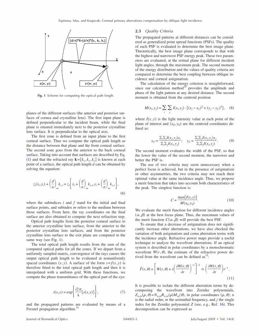
lanes of the different surfaces �the anterior and posterior sur-aces of cornea and crystalline lens�. The first input plane isefined perpendicular to the incident beam, while the finallane is situated immediately next to the posterior crystallineens surface. It is perpendicular to the optical axis.
The first zone is defined from an input plane to the firstorneal surface. Thus we compute the optical path length ashe distance between that plane and the front corneal surface.he second zone goes from the anterior to the back cornealurface. Taking into account that surfaces are described by Eq.1� and that the refracted ray k= kx ,ky ,kz� is known at eachoint of a surface, the optical path length d can be obtained byolving the equation:
zi�xi,yi� + �d
n�
mkz,i = zf�xi + �d
n�
mkx,i,yi + �d
n�
mky,i� ,
�6�
here the subindices i and f stand for the initial and finalurface points, and subindex m refers to the medium betweenhose surfaces. From here, the ray coordinates on the finalurface are also obtained to compute the next refraction step.
Optical path lengths from the posterior corneal surface tohe anterior crystalline lens surface, from the anterior to theosterior crystalline lens surfaces, and from the posteriorrystalline lens surface to the exit plane are computed in theame way �see Fig. 1�.
The total optical path length results from the sum of theomputed optical paths for all the zones. If we depart from aniformly sampled matrix, convergence of the rays causes theutput optical path length to be evaluated at nonuniformlypaced coordinates �x ,y�. A surface of the form s= f�x ,y� isherefore fitted to the total optical path length and then it isnterpolated with a uniform grid. With these functions, weompute the phase transmittance of the optical part of the eye:
t�x,y� = exp� i2�
�s�x,y�� , �7�
nd the propagated patterns are evaluated by means of aresnel propagation algorithm.14
Fig. 1 Scheme for computing the optical path length.
ournal of Biomedical Optics 044003-
2.3 Quality CriteriaThe propagated patterns at different distances can be consid-ered as generalized point spread functions �PSFs�. The qualityof each PSF is evaluated to determine the best image plane.Theoretically, the best image plane corresponds to that withthe highest and narrowest PSF energy peak. These two param-eters are evaluated, at the retinal plane for different incidentlight angles, through the maximum peak. The second momentof the energy distribution and the values of quality criteria arecompared to determine the best coupling between oblique in-cidence and corneal astigmatism.
The calculation of the energy criterion is straightforward,since our calculation method14 provides the amplitude andphase of the light pattern at any desired distance. The secondmoment is obtained from the centroid position
M�xi,yj� = �i
�j
I�xi,yj� · �xi − xC�2 + �yj − yC�2� , �8�
where I�x ,y� is the light intensity value at each point of theplane of interest and �x0 ,y0� are the centroid coordinates de-fined as:
xC =�i� jI�xi,yj�xi
�i� jI�xi,yj�; yC =
�i� jI�xi,yj�yj
�i� jI�xi,yj�. �9�
The second moment evaluates the width of the PSF, so thatthe lower the value of the second moment, the narrower andbetter the PSF is.
The use of two criteria may seem unnecessary when aperfect focus is achieved, but in the presence of astigmatismor other asymmetries, the two criteria may not reach theiroptimal value at the same incidence angle. Thus, we proposea merit function that takes into account both characteristics ofthe peak. The simplest function is:
C =maxI�x,y��
M�x0,y0�. �10�
We evaluate the merit function for different incidence angles�� ,�� at the best focus plane. Thus, the maximum values ofthe merit function C�� ,�� will provide the best PSF.
To ensure that a decrease of astigmatism does not signifi-cantly increase other aberrations, we have also checked thevariation of both astigmatism and coma aberration terms withthe incidence angle. Refractive power maps provide a usefultechnique to analyze the wavefront aberrations. If an opticalsystem is described in polar coordinates by a monochromaticwavefront W�r ,�, the estimate of the refractive power de-rived from the wavefront can be defined as15:
F̂�r,� = �W�r,� + r� �W�r,��r
�−1�−1
� �W�r,��r
�r−1.
�11�
It is possible to isolate the different aberration terms by de-composing the wavefront into Zernike polynomials,Zn,m� ,�=Nn,mRn,m��Mm��, in polar coordinates � ,�; nis the radial order, m the azimuthal frequency, and j the singleindex for the Zernike polynomial Z �see, e.g., Ref. 16�. Thisdecomposition can be expressed as
July/August 2009 � Vol. 14�4�3

wZa
�ta
Fd
wn2vawm
3Uestclyrhwt�
e
Espinosa, Mas, and Kasprzak: Corneal primary aberrations compensation by oblique light incidence
J
W�,� = �j=0
p−1
cjZj�,� , �12�
j =1
2n�n + 2� + m�; n = �− 3 + �9 + 8j
2�;
m = 2j − n�n + 2�;
Rn,m�� = �s=0
�n−�m��/2�− 1�s�n − s�!
s!0.5�n + �m�� − s�!0.5�n − �m�� − s�!n−2s,
Nn,m = �2�n + 1�1 + �m0
�1/2
; Mm�� = �cos�m� m � 0
sin�m� m 0 ,
�13�
here p is the number of terms in the expansion, cj are theernike coefficients associated with their Zernike polynomial,nd �·� denotes the ceiling �round-up� operator.
A more convenient form to analyze the polynomial in Eq.12� involves rearranging the wavefront Zernike decomposi-ion by grouping all the terms with the same radial degree andzimuthal frequency,17
W�,� = �j=0
p−1
gjnMm�� = �
j=0
p−1
gjGj�,� . �14�
rom Eqs. �11� and �14�, the estimate of the refractive powererived from the wavefront can be obtained as
F̂�r,� r−1 �
�r��j=3
p−1
gjrnMm��� = �
j=3
p−1
ngjrn−2Mm�� ,
�15�
here ngj are directly related to the power vector compo-ents. For the case n=2 �j=3, j=4, and j=5�, we obtaing3=−J45, 2g4=−M =−1 /zc, and 2g5=−J0, i.e., the powerector components. If the reference sphere is subtracted, anberration-free wavefront will be planar, i.e., the power mapill be equal to zero. Hence, the PSF is better if the polyno-ial coefficients in Eq. �15� are lower.
Resultssing the values in Table 1, we have generated a Kooijman
ye model. To introduce a slight corneal astigmatism, we haveelected different values for the horizontal and vertical curva-ure radius, i.e., Rx=7.9 mm; Ry =7.8 mm. This differenceauses a total with-the-rule ocular astigmatism of 0.6 D, simi-ar to the mean values of astigmatism observed in the normalouth adult population. Pupil size has been limited to a natu-al pupil size of 4 mm in diameter. The ray incidence angleas been varied in both vertical and horizontal directionsithin the range −8 deg,8 deg� which corresponds to varia-
ions in the interval −82 deg,98 deg� for the direction anglesand �.Figure 2 shows the values obtained for the quality param-
ter in the specified range. The maximum merit function value
ournal of Biomedical Optics 044003-
is reached for �� ,��= �84.8 deg,90 deg� and �� ,��= �95.2 deg,90 deg� equivalent to beams entering the corneawith incidence angles tilted at �5.2 deg in the horizontal di-rection. Different astigmatic corneas will provide maxima ofthe quality function at different incidence angles.
In Fig. 3, we show the values obtained for some of thepower map coefficients described in Eq. �15�. We have se-lected those that are more affected by an oblique incidence,since these will be the main contributors to the final quality ofthe PSF. Thus, we show absolute values of g3 and g5, equiva-lent to J45 and J0, which describe astigmatism, and g7 and g8that are directly related to vertical and horizontal coma, re-spectively. We have varied the incidence angle in the x direc-tion, while the � angle has been fixed at 90 deg. In Fig. 3 wecan see that 45 /135 astigmatism g3, and vertical coma g7, arenot affected by the tilt of the ray, and their values are close tozero. On the other hand, the coefficient g5, related with90 /180 astigmatism, decreases as the tilt angle approachesaround 4.4 deg, as was predicted, but the coefficient for thevertical coma g8 increases. Notice that the best image points
Fig. 2 Quality parameter for different incidence angles.
Fig. 3 Absolute astigmatism and coma Zernike coefficients.
July/August 2009 � Vol. 14�4�4
ab
cmc=m
chmiia�v
PltO=di
4WdtloiIms
Vbo�
Espinosa, Mas, and Kasprzak: Corneal primary aberrations compensation by oblique light incidence
J
re obtained for incidence angles that provide a compromiseetween astigmatism compensation and coma increase.
Figure 2 provides a quantitative description of the passiveompensation of oblique incident astigmatism by an astig-atic cornea. At first glance one can see that aberrations are
anceled in two broad symmetric regions around ��5.8 deg �note that �=90 deg−� if �=90 deg� withaxima PSF peak values reached at �= �5.2 deg.According to Fig. 3, the minimum of g5 is reached at in-
idence angles of �= �4.8 deg. However, at these points theorizontal coma coefficient g8 reaches values near itsaxima. Thus, total cancellation of astigmatism implies an
ncrease in the coma term. The merit function �Fig. 2� takesnto account other contributions aside from only comand astigmatism, and reaches its maximum values at= �5.2 deg. At this point it seems that aberrations are con-eniently balanced to give a PSF of reasonable quality.
To better understand the situation, in Video 1 we show theSFs obtained for �=90 deg and �� −8 deg,8 deg�. To al-
ow better visualization, all the frames have been normalizedo their maximum value, which is shown in the upper part.ne can observe there that the PSF corresponding to ��5.2 deg is better than the ones obtained for other inci-
ence angles. We can also see the appearance of some coman the PSF at this angle, as predicted in Fig. 3.
Discussione have shown that astigmatism induced by oblique inci-
ence can compensate for the astigmatism of the cornea, al-hough this tilt also induces comatic aberration. An apparentimitation of our calculations is the predicted symmetry of theptimal regions, which does not agree with the situation foundn the real eye, where the foveal region is clearly determined.n the literature it is reported that peripheral astigmatism isinimum about one point of the nasal retina.18,19 Our results
eem to agree with this finding, since we find a double mini-
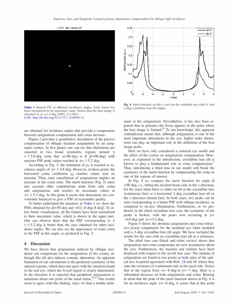
ideo 1 Retinal PSF at different incidence angles. Each frame haseen normalized to its maximum value. Notice that the best image isbtained at �= ±5.2 deg �MPG, 5.4 MG�.URL: http://dx.doi.org/10.1117/1.3158996.1�
ournal of Biomedical Optics 044003-
mum in the astigmatism. Nevertheless, it has also been re-ported that in primates the fovea appears at the point wherethe best image is formed.20 To our knowledge, this apparentcontradiction means that, although astigmatism is one of themost important aberrations in the eye, higher order aberra-tions can play an important role in the definition of the bestimage point.
Here we have only considered a centered eye model andthe effect of the cornea on astigmatism compensation. How-ever, as explained in the introduction, crystalline lens tilt isknown to play a fundamental role in coma compensation.7
Thus, introducing a tilted lens in our model will break thesymmetry of the merit function by compensating the coma inone of the regions of interest.
In Fig. 4 we compare the merit function for angle �=90 deg, i.e., tilting the incident beam only in the x direction,for the cases when there is either no tilt in the crystalline lens�continuous line� or a horizontal 3-deg crystalline lens tilt inthe x direction �dotted line�. In both cases, two peaks can beseen corresponding to a better PSF with oblique incidence, ascompared to on-axis illumination. Furthermore, as we pre-dicted, in the tilted crystalline lens case, the symmetry of thepeaks is broken, with the peaks now occurring at �=−6.0 deg and �=+5.2 deg.
Figure 5 shows the absolute astigmatism and coma refrac-tive power components for the modeled eye when modeledwith a 3-deg crystalline lens tilt angle. We have included theresults for the case with no crystalline lens tilt as a reference.
The tilted lens case �black and white circles� shows thatastigmatism and coma components are now asymmetric aboutthe axis. Furthermore, the maxima and minima have beendisplaced with respect to the no-tilt lens case. The minima ofastigmatism are found in two points at both sides of the opti-cal axis in partial agreement with Refs. 18 and 19, where theystate the existence of a minimum only at the nasal side. Noticethat in the region from �=−6 deg to �=−7 deg, there is asubstantial decrease on both astigmatism and coma. Bearingin mind that the peak of the merit function shown in Fig. 4 isfor an incidence angle �=−6 deg, it seems that at this point
Fig. 4 Merit function on the x axis for the modeled eye with 0- and3-deg crystalline lens tilt angles.
July/August 2009 � Vol. 14�4�5

braPTa
tWtdotb
iicctr
8la
ahPaork
Ff
Espinosa, Mas, and Kasprzak: Corneal primary aberrations compensation by oblique light incidence
J
oth aberrations are conveniently balanced to give a PSF ofeasonable quality around the foveal region.20 Notice also thatccording to the value of the merit function in Fig. 4, the bestSF is better than the one obtained without tilting the lens.his effect is due to the compensation of coma that was notchieved in the centered case.
Summarizing, the simulations presented in this work showhat oblique incidence can compensate corneal astigmatism.
e have modeled a simple theoretical eye with a small with-he-rule corneal astigmatism. Tilting of the rays may intro-uce a new aberration term with opposite sign to the existingne. We have proposed two simple quality criteria to evaluatehe peak: maximum energy and second moment, which haveeen combined in a single merit function.
Although astigmatism is canceled, oblique incidence alsonduces coma in the final image. As noted by Tabernero et al.n Ref. 7, tilt of the crystalline lens may compensate thisoma. Our simple model has shown that oblique incidencean passively compensate the effects of both corneal astigma-ism and lens tilt on the foveal image, thus making the eye aobust optical system.
Normal values for the kappa angle are set between 4 anddeg with an average value of 5 deg. Results presented here
ocate the best image point within this range, which is ingreement with data from real eyes.
It is true that variability of eye parameters �asphericities,stigmatism, kappa angle, etc.� is considerable and the modelere presented is necessarily limited. Kasprzak andierscionek4 suggested that gravitational corneal sag wouldlso play some role in the compensation of aberrations byblique incidence. It would be interesting to investigate cor-elations between lens tilt, corneal astigmatism, and angleappa, as well as with other sources of aberrations in the eye,
ig. 5 Absolute astigmatism and coma refractive power componentsor the modeled eye with 0- and 3-deg crystalline lens tilt angles.
ournal of Biomedical Optics 044003-
but such studies are outside the scope of this communicationand would require applications of more complex eye models.
AcknowledgmentsDavid Mas acknowledges the support of the Generalitat Va-lenciana through the project BEST-2008-148. Henryk T.Kasprzak acknowledges the University of Alicante for its fi-nancial support through the project Senior 08/07.
References1. P. Artal, A. Benito, and J. Tabernero, “The human eye is an example
of robust optical design,” J. Vision 6, 1–7 �2006�.2. S. G. El Hage and F. Berny, “Contribution of the crystalline lens to
the spherical aberration of the eye,” J. Opt. Soc. Am. 63, 205–211�1973�.
3. M. Millodot and J. Sivak, “Contribution of the cornea and lens to thespherical aberration of the eye,” Vision Res. 19, 685–687 �1979�.
4. P. Artal and A. Guirao, “Contribution of cornea and crystalline lens tothe aberrations of the human eye,” Opt. Lett. 23, 1713–1715 �1998�.
5. P. Artal, A. Guirao, E. Berrio, and D. R. Williams, “Compensation ofcorneal aberrations by the internal optics in the human eye,” J. Vision1, 1–8 �2001�.
6. J. E. Kelly, T. Mihashi, and H. C. Howland, “Compensation of cor-neal horizontal/vertical astigmatism, lateral coma and spherical aber-ration by internal optics of the eye,” J. Vision 4, 262–271 �2004�.
7. J. Tabernero, A. Benito, E. Alcón, and P. Artal, “Mechanism of com-pensation of aberrations in the human eye,” J. Opt. Soc. Am. A24�10�, 3274–3283 �2007�.
8. S. Marcos, P. Rosales, L. Llorente, S. Barbero, and I. Jiménez-Alfaro,“Balance of corneal horizontal coma by internal optics in eyes withintraocular artificial lenses: evidence of a passive mechanism,” VisionRes. 48�1�, 70–79 �2008�.
9. H. Kasprzak and B. K. Pierscionek, “Modelling the gravitational sagof the cornea and the subsequent quality of the refracted image,” J.Mod. Opt. 49, 2153–2166 �2002�.
10. D. S. Satterfield, “Prevalence and variation of astigmatism in a mili-tary population,” J. Am. Optom. Assoc. 60, 14–18 �1981�.
11. H. C. Fledelius and M. Subgaard, “Changes in refraction and cornealcurvature during growth and adult life, a cross sectional study,” ActaOphthalmol. 64, 487–491 �1986�.
12. S. A. Read, M. J. Collins, and L. G. Carney, “A review of astigma-tism and its possible genesis,” Clin. Exp. Optom. 90, 5–19 �2007�.
13. A. Kooijman, “Light distribution on the retina of a wide-angle theo-retical eye,” J. Opt. Soc. Am. 73, 1544–1550 �1983�.
14. D. Mas, J. Pérez, C. Hernández, C. Vázquez, J. J. Miret, and C.Illueca, “Fast numerical calculation of Fresnel patterns in convergentsystems,” Opt. Commun. 227, 245–248 �2003�.
15. D. R. Iskander, B. A. Davis, M. J. Collins, and R. Franklin, “Objec-tive refraction from monochromatic wavefront aberrations viaZernike power polynomials,” Ophthalmic Physiol. Opt. 27, 245–255�2007�.
16. L. N. Thibos, R. A. Applegate, J. T. Schweigerling, and R. Webb,“Standards for reporting the optical aberrations of eyes,” J. Refract.Surg. 18, S652–S660 �2002�.
17. J. Espinosa, D. Mas, J. Pérez, and C. Illueca, “Adaptive sampling inconvergent beams,” Opt. Lett. 33, 1960–1962 �2008�.
18. M. C. M. Dunne, G. P. Misson, E. K. White, and D. A. Barnes,“Peripheral astigmatic asymmetry and angle alpha,” OphthalmicPhysiol. Opt. 13, 303–305 �1996�.
19. A. Seidemann, F. Schaeffel, A. Guirao, N. Lopez-Gil, and P. Artal,“Peripheral refractive errors in myopic, emmetropic, and hyperopicyoung subjects,” J. Opt. Soc. Am. A 19, 2363–2373 �2002�.
20. A. D. Springer, “New role for the primate fovea: A retinal excavationdetermines photoreceptor deployment and shape,” Visual Neurosci.16 629–636 �1999�.
July/August 2009 � Vol. 14�4�6
Copyright © 2022 FDOKUMEN