Forecasting characteristic earthquakes in a minimalist model
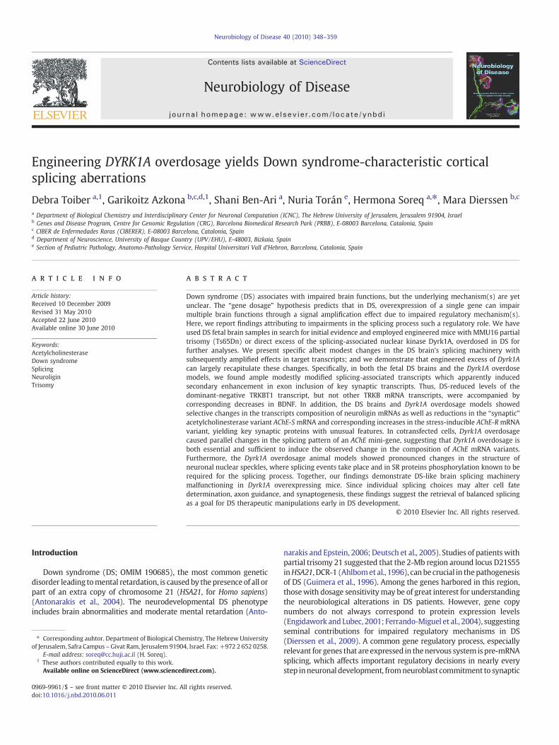
Engineering DYRK1A overdosage yields Down syndrome-characteristic corticalsplicing aberrations
Debra Toiber a,1, Garikoitz Azkona b,c,d,1, Shani Ben-Ari a, Nuria Torán e, Hermona Soreq a,!, Mara Dierssen b,c
a Department of Biological Chemistry and Interdisciplinary Center for Neuronal Computation (ICNC), The Hebrew University of Jerusalem, Jerusalem 91904, Israelb Genes and Disease Program, Centre for Genomic Regulation (CRG), Barcelona Biomedical Research Park (PRBB), E-08003 Barcelona, Catalonia, Spainc CIBER de Enfermedades Raras (CIBERER), E-08003 Barcelona, Catalonia, Spaind Department of Neuroscience, University of Basque Country (UPV/EHU), E-48003, Bizkaia, Spaine Section of Pediatric Pathology, Anatomo-Pathology Service, Hospital Universitari Vall d'Hebron, Barcelona, Catalonia, Spain
a b s t r a c ta r t i c l e i n f o
Article history:Received 10 December 2009Revised 31 May 2010Accepted 22 June 2010Available online 30 June 2010
Keywords:AcetylcholinesteraseDown syndromeSplicingNeuroliginTrisomy
Down syndrome (DS) associates with impaired brain functions, but the underlying mechanism(s) are yetunclear. The “gene dosage” hypothesis predicts that in DS, overexpression of a single gene can impairmultiple brain functions through a signal ampli!cation effect due to impaired regulatory mechanism(s).Here, we report !ndings attributing to impairments in the splicing process such a regulatory role. We haveused DS fetal brain samples in search for initial evidence and employed engineered mice with MMU16 partialtrisomy (Ts65Dn) or direct excess of the splicing-associated nuclear kinase Dyrk1A, overdosed in DS forfurther analyses. We present speci!c albeit modest changes in the DS brain's splicing machinery withsubsequently ampli!ed effects in target transcripts; and we demonstrate that engineered excess of Dyrk1Acan largely recapitulate these changes. Speci!cally, in both the fetal DS brains and the Dyrk1A overdosemodels, we found ample modestly modi!ed splicing-associated transcripts which apparently inducedsecondary enhancement in exon inclusion of key synaptic transcripts. Thus, DS-reduced levels of thedominant-negative TRKBT1 transcript, but not other TRKB mRNA transcripts, were accompanied bycorresponding decreases in BDNF. In addition, the DS brains and Dyrk1A overdosage models showedselective changes in the transcripts composition of neuroligin mRNAs as well as reductions in the “synaptic”acetylcholinesterase variant AChE-SmRNA and corresponding increases in the stress-inducible AChE-RmRNAvariant, yielding key synaptic proteins with unusual features. In cotransfected cells, Dyrk1A overdosagecaused parallel changes in the splicing pattern of an AChE mini-gene, suggesting that Dyrk1A overdosage isboth essential and suf!cient to induce the observed change in the composition of AChE mRNA variants.Furthermore, the Dyrk1A overdosage animal models showed pronounced changes in the structure ofneuronal nuclear speckles, where splicing events take place and in SR proteins phosphorylation known to berequired for the splicing process. Together, our !ndings demonstrate DS-like brain splicing machinerymalfunctioning in Dyrk1A overexpressing mice. Since individual splicing choices may alter cell fatedetermination, axon guidance, and synaptogenesis, these !ndings suggest the retrieval of balanced splicingas a goal for DS therapeutic manipulations early in DS development.
© 2010 Elsevier Inc. All rights reserved.
Introduction
Down syndrome (DS; OMIM 190685), the most common geneticdisorder leading tomental retardation, is caused by the presence of all orpart of an extra copy of chromosome 21 (HSA21, for Homo sapiens)(Antonarakis et al., 2004). The neurodevelopmental DS phenotypeincludes brain abnormalities and moderate mental retardation (Anto-
narakis and Epstein, 2006; Deutsch et al., 2005). Studies of patients withpartial trisomy 21 suggested that the 2-Mb region around locus D21S55inHSA21, DCR-1 (Ahlbomet al., 1996), canbe crucial in thepathogenesisof DS (Guimera et al., 1996). Among the genes harbored in this region,thosewith dosage sensitivitymay be of great interest for understandingthe neurobiological alterations in DS patients. However, gene copynumbers do not always correspond to protein expression levels(Engidawork and Lubec, 2001; Ferrando-Miguel et al., 2004), suggestingseminal contributions for impaired regulatory mechanisms in DS(Dierssen et al., 2009). A common gene regulatory process, especiallyrelevant for genes that are expressed in thenervous system is pre-mRNAsplicing, which affects important regulatory decisions in nearly everystep inneuronal development, fromneuroblast commitment to synaptic
Neurobiology of Disease 40 (2010) 348–359
! Corresponding auhtor. Department of Biological Chemistry, The Hebrew Universityof Jerusalem, Safra Campus – Givat Ram, Jerusalem 91904, Israel. Fax: +972 2 652 0258.
E-mail address: [email protected] (H. Soreq).1 These authors contributed equally to this work.
Available online on ScienceDirect (www.sciencedirect.com).
0969-9961/$ – see front matter © 2010 Elsevier Inc. All rights reserved.doi:10.1016/j.nbd.2010.06.011
Contents lists available at ScienceDirect
Neurobiology of Disease
j ou rna l homepage: www.e lsev ie r.com/ locate /ynbd i

specialization,modulatingproteins ranging from transcription factors tocell adhesion molecules (Li et al., 2007).
Many human disorders are generated by misregulation ofalternative splicing (Licatalosi and Darnell, 2006; Yamada andNabeshima, 2004). Even if small, changes in the level of splicingmachinery components can perturb the composition of splicingcomplexes and thus induce many changes in downstream processedtranscripts. In particular, alternative splicing plays a critical role in thenervous system, where more than 75% of the transcripts arealternatively spliced (Licatalosi and Darnell, 2006; Stamm et al.,2005). The different variants play essential and sometimes inverseroles in ion channel function, receptor speci!city, neuronal cellrecognition, neurotransmission, and learning and memory (Li et al.,2007). Misregulated alternative splicing is responsible for manynervous system diseases such as Alzheimer's, Parkinson's, Pick's, andHuntington's diseases (Licatalosi and Darnell, 2006) but has not yetbeen explored in other disorders affecting cognition, such as themental retardation in DS.
Splicing aberrations can directly cause disease, modify the severityof the disease phenotype or be linked with disease susceptibility(Wang and Cooper, 2007). Among the proteins that are involved inthe splicing machinery, the members of the SR protein (SRp) family ofsplicing factors, which contain Ser/Arg-rich domains are primarilyinvolved in mRNA maturation (Hanamura et al., 1998). SRps act assplicing regulatory proteins, binding to exonic splicing enhancerelements and stimulating exon inclusion. SRp family members aremainly named by their sizes (e.g., SRp20, SRp50, SRp55, etc.) and arein"uenced by protein interactions and posttranslational modi!ca-tions. Different SRp have different roles depending on combinatorialcontributions of cell type, signaling, and posttranslational modi!ca-tions. For example, ASF/SF2 (alternative splicing factor/splicing factor2) affects 5" splice site selection, whereas SC35 (splicing component,35 kDa) plays an opposite role in the splicing of AChE (Meshorer et al.,2005).
Phosphorylation of SRp is required at the onset of spliceosomeassembly (Stamm, 2008). Splicing factors are organized in the highlycompartmentalized mammalian nucleus, concentrated in the splicingspeckles. These structures (about 25/nucleus) are enriched in splicingfactors, pre-messenger RNA, as well as transcription, export, andnuclear structural machinery. They are located in the interchromatinregions, close to highly active transcription sites. The speckles arethought to be a “storage” or assembly place for the splicingmachinery.Upon signaling, their components can possibly be mobilized to thetarget sites. Several kinases and phosphatases are able to modify thesplicing machinery, regulating the accessibility of these factors to thesplicing events (for a detailed review, see Lamond and Spector, 2003).A recent example involves the phosphorylation of ASF/SF2, whichwas suggested to regulate the alternative splicing of tau in DS (Shi etal., 2008).
Expanding this concept, we predicted that splicing ef!ciency andaccuracy at large may be a signi!cant contributor to the DS phenotypeand its variability. To challenge this prediction, we comparativelyanalyzed SRp expression and phosphorylation and the splicing variantpro!les of key target genes involved in neurodevelopment andcognition in fetal cerebral cortices from DS carriers and in engineeredDyrk1A overexpression systems.
The dual-speci!city tyrosine phosphorylation-regulated kinase 1A(DYRK1A), a member of an evolutionarily conserved protein kinasefamily (Becker et al., 1998), has been proposed as an exceptionaldosage-sensitive candidate gene for DS. DYRK1A is a proline-directedserine/threonine-speci!c protein kinase whose activity depends ontyrosine autophosphorylation in the catalytic domain. It maps to theDCR-1 and is highly expressed in fetal and adult brains (Guimera et al.,1999). Multiple reports show involvement of DYRK1A in manydifferent pathways, spanning transcription, translation, and signaltransduction (Arron et al., 2006; Gwack et al., 2006; Sitz et al., 2004;
Yang et al., 2001). In neurons, DYRK1A localizes to both the nucleusand cytoplasm, including neuronal processes and synapses. In thenucleus, it localizes to splicing speckles and overexpression of activeDYRK1A in cultured cells associates with speckles disassembly(Alvarez et al., 2003). Transgenic mice overexpressing Dyrk1A(TgDyrk1A) exhibit hyperactivity and impaired neuromotor develop-ment as well as spatial learning and memory (Altafaj et al., 2001;Martinez de Lagran et al., 2004), a phenotype which is reversible bytargeting Dyrk1A with AAVshRNA (Ortiz-Abalia et al., 2008). Compa-rable alterations are found in the MMU16 chromosome 16 partialtrisomy murine model of DS, Ts65Dn (Galdzicki et al., 2001),suggesting that DYRK1A overexpression in trisomy 21 carriers maycontribute to the DS mental retardation and motor anomaliesphenotype. The long list of DYRK1A substrates includes cyclin L2(de Graaf et al., 2004), SF3b1 (de Graaf et al., 2006), and ASF (Shi et al.,2008), suggesting its possible involvement in the regulation of thesplicing machinery (Alvarez et al., 2003; de Graaf et al., 2004; Deanand Dresbach, 2006; Dierssen and de Lagran, 2006). Therefore, wedecided to preliminarily investigate the splicing changes induced inthe DS brain and compare our !ndings to those in mice with enforcedDyrk1A overexpression. Speci!cally, we analyzed the splicing ma-chinery at the RNA, protein, and phosphorylation levels with aparticular focus on genes involved with the molecular basis oflearning.
Materials and methods
Human brain samples
Cerebral cortices of fetal brains with or without DS (12 males ofeach) at 16–19 weeks gestation (Supplementary Table I) wereobtained from the fetal tissue bank of Hospital Vall d'Hebron(Barcelona, Spain) and Hospital La Fe (Sabadell, Spain). Sampleswere obtained between 6 and 12 h postmortem and were frozen orembedded in paraf!n. Diagnosis of all cases was genetically andhistopathologically con!rmed (Dr. N. Torán and Dr. JC Farreras). Theinclusion criterion for control samples was that fetal demise was notdue to a known genetic cause. Fetal samples were stored at !80 °Cuntil use. DS fetal samples showed elevated DYRK1A protein levels(Supplementary Fig. 1). All experimental procedures were approvedby the local ethical committee (CEE-Vall d'Hebrón and CEE-PRBB) andmet the guidelines of the local and European regulations.
Animals
The production of transgenic mice overexpressing Dyrk1A(TgDyrk1A) has been previously described (Altafaj et al., 2001).Brie"y, the original founder was obtained by insertion of thetransgene into C57BL/6JXSJL (Charles River, Barcelona, Spain)embryos, and the stock is maintained by crossing C57BL/6JXSJLwild type females and transgenic males derived from the originalfounder. MMU16 Ts65Dn segmental trisomic mice, free from the Rdmutation, were purchased from The Jackson Laboratory (Bar Harbor,Maine, USA). Nontransgenic and disomic littermates served ascontrols. Both TgDyrk1A and Ts65Dn mice showed elevated proteinlevels of Dyrk1A compared to strain-matched wild type mice(Supplementary Fig. 1). Animals were housed under 12:12-h light–dark cycle (lights on at 8:00 a.m.) in controlled environmentalconditions, 60% of humidity, and at 22±2 °C. All animal procedureswere approved by the local ethical committee (CEEA-IMIM andCEEA-PRBB) and met the guidelines of the local (law 32/2007) andEuropean regulations (EU directive no. 86/609, EU decree 2001-486)and the Standards for Use of Laboratory Animals no. A5388-01 (NIH).The CRG is authorized to work with genetically modi!ed organisms(A/ES/05/I-13 and A/ES/05/14).
349D. Toiber et al. / Neurobiology of Disease 40 (2010) 348–359

RNA procedures
The RNeasy kit (Qiagen, Valencia, CA) was used as per manufac-turer's instructions for RNA extraction. DNase was applied to removeDNA contamination. RNA integrity was con!rmed by gel electropho-resis, and RNA concentration and purity was assessed spectrophoto-metrically. For cDNA synthesis, 0.4 !g RNA was used for each sample(Promega, Madison, WI). The SpliceChip experiment was performedas described previously (Ben-Ari et al., 2006). Quantitative qRT–PCRwas performed in duplicates using ABI Prism 7900HT and SYBR greenmaster mix (Applied Biosystems, Foster City, CA). ROX, a passivereference dye, was used for signal normalization across the plate, and"-actin and GAPDH mRNA served as reference transcripts. Annealingtemperature was 60 °C for all primers. Serial dilution of samplesserved to evaluate primer's ef!ciency and the appropriate cDNAconcentration that yields linear changes. Melting curve analysis andamplicons sequencing served to verify the end product. Primersemployed are denoted in Supplementary Table 2. Transfection andanalyses of the human AChE minigene was as previously detailed(Meshorer et al., 2005).
Western blots
Protein content was quanti!ed in postmortem fetal humansamples and mouse tissues. Animals were sacri!ced, and brainswere rapidly removed and dissected on ice. Tissues were
homogenized in lysis buffer (10 mM HEPES pH 7.5, 150 mMNaCl, 1 mM EDTA, 0.1 mM MgCl2, phosphate-buffered saline (PBS),0.2% Triton X-100) and a protease inhibitor cocktail (Roche,Mannheim, Germany). After clearance of the lysates by centrifu-gation (1400!g, 20 min at 4 °C), protein quanti!cation wasperformed following the BCA Protein Assay Reagent (Pierce,Rockford, IL, USA) protocol. Western blot analysis was performedusing 50 !g of protein resolved on a 10% SDS–PAGE andelectroblotting onto nitrocellulose membranes (Hybond-C, Amer-sham Pharmacia Biotech, Freiburg, Germany). Membranes wereblocked with 5% nonfat dry milk or albumin from bovine serum(Sigma, Steinheim, Germany) in Tris-buffered saline including 0.1%Tween-20 (TBS-T) and incubated with the primary antibodies in5% nonfat dry milk in TBS-T overnight at 4 °C. The followingdilutions of primary antibodies were used: mouse anti-ASF/SF2(1:100; Zymed, CA, USA), anti-Dyrk1A (1:500; Abnova, Heidelberg,Germany), anti-SR proteins (16H3) and (1H4) (1:100 each;Zymed, CA, USA), and goat anti-SC35 (1:200; Santa Cruz,Heidelberg, Germany), rabbit anti-TRKB (1:1000; Santa Cruz),rabbit anti-TRKBT.1 (C13) (1:200; Santa Cruz), and anti-actin(1:2000; Sigma, St Louis, MO, USA). Incubation with horseradishperoxidase (HRP)-conjugated anti-mouse or anti-goat IgG (Pierce,Rockford, IL, USA), followed by enhanced chemiluminescence (ECL,Pierce) assay allowed detection. Quanti!cation involved densito-metric analysis of nonsaturated !lms (Quantity One imagesoftware).
Fig. 1. SR proteins levels and phosphorylation in DS fetal cerebral cortex. Upper panels: SR proteins; lower panels: posterior densitometric quanti!cation graphed as percentage of controlvalues from cerebral cortices of fetal brains. Actin was used as loading control. Representative protein blot of (A) ASF/SF2, (B) SC35, and (C) SRp50 and SRp55. (D) Representative confocalimages of cortical fetal cells immunostained with an anti-SRp-phospho (1H4) antibody, showing a higher expression of phosphorylated SR proteins in DS cortices compared to controls. Leftpanel: controls; rightpanel:DS. Scale bar=50 !m.Rightupper insert:highermagni!cation (scalebar=5 !m). (E)Representativeproteinblot ofphosphorylatedSRproteins anddensitometricquanti!cation graphed as percentage of control values from cerebral cortex of normal fetal samples. Actinwas used as loading control.N=4per karyotype for immunohistochemistry analysisand 8 per karyotype per protein blot analysis. White bars: controls; black bars: DS fetal cortical tissues. Data are expressed as mean±SEM, *Pb0.05. Mann–Whitney U test.
Fig. 2. Dyrk1A overexpression modi!es the levels of splicing machinery transcripts. (A) SpliceChip results for TgDyrk1A cortices compared to strain matched controls. Bar graphsrepresent genes related to splicing that were up/downregulated signi!cantly in both experiments. Data are presented as log 2 scale showing increase or decrease from control mice(log 2b!0.5 or N0.5). N=3 per genotype. (B) Bar graphs represent percent of change from control in qRT–PCR of individual brain samples for the noted transcripts. N=6 pergenotype. Black bars represent TgDyrk1A and white bars control mice. *Pb0.05. Mann–Whitney U test.
350 D. Toiber et al. / Neurobiology of Disease 40 (2010) 348–359

351D. Toiber et al. / Neurobiology of Disease 40 (2010) 348–359

Coimmunoprecipitation
Cortical extracts (700 !g) were prepared as previously describedand subjected to coimmunoprecipitation in lysis buffer containing0.25% Triton X-100 (overnight, at 4 °C) using rabbit anti-Dyrk1A(kindly provided by Dr. J. Naranjo; CNB, Madrid). After !ve washes inlysis buffer containing 300 mM KCl and 0.25% Triton X-100, proteinswere eluted in SDS sample buffer, resolved in SDS–PAGE, andelectroblotted onto Immobilon-P (Millipore). Blots were developedusing a mouse anti-SR proteins (16H3) (Zymed, CA, USA) or anti-phosphoSer (phosphoserine detection antibody set; Biomol). Proteinswere visualized with HRP-conjugated secondary antibodies followedby ECL (West Dura, Pierce).
Immunohistochemistry
Mice were perfused transcardially with 0.1 M PBS, pH 7.4 and thenwith 4% paraformaldehyde (Sigma, St Louis, MO, USA). After 24 h,brains were embedded in paraf!n, slices (5 !m) were obtained andhandled simultaneously during the protocol, to minimize interslidesvariability. Antigen retrieval was performed with citrate buffer, andquenching was performed in human fetal samples with: 0.3% KMnO4
for 4 min, deionized water wash, 1% K2S2O5+1% oxalic acid (20–40 sec), deionized water wash, and 1% NaBH4, 4 min, deionized waterwash. Nonspeci!c labeling was blocked for 40 min in Tris 10% serumblocking solution containing horse and goat serum, 5% each. Mouseanti-ASF/SF2 (1:100; Zymed, CA, USA), anti-SR-phospho proteins(1H4) (1:100; Zymed, CA, USA), and goat anti-SC35 (1:200; SantaCruz, Heidelberg, Germany) were diluted as for western blots in 2%TBS milk/5% serum mix and applied for 2 h at room temperature orovernight at 4 °C. Corresponding biotin-conjugated secondary anti-bodies with or without Cy3 or Cy2 conjugates (diluted 1:200) andDAPI served to stain the nucleus. Quanti!cation was done using 4pictures of each brain area (frontal cortex and the CA1 and dentategyrus of hippocampus, ImageJ Software). The averages of each pictureyielded the !nal average of the group.
Image analysis
Tissue slices were scanned using an FV-1000 confocal microscope(Olympus, Japan), equipped with an IX81 inverted attachment and anA40!/1.3 oil immersion objective. For DAPI, excitation was 405 nmand emission 430–470 nm; for Cy2, excitation was 488 nm andemission 505–525 nm. Immunostaining intensity was quanti!ed bythe ImageJ 1 1.33 free software (http://rsb.info.nih.gov/ij/), using the“Analyze tool” mean gray value. Using the “freehand selection,” wedrew lines around single cells and analyzed 30–300 cells in eachpicture. Averaged 2–3 empty spots in the tissue served to determinethe background for each picture, which was subtracted from each ofthe values observed for measured cells to normalize light ef!ciencydifferences that could occur between different pictures. The totalaverage for a single experiment was composed of 4 different picturesat least from each mouse and 8 mice per group. Standard deviationvalues were calculated for these averages to challenge the possibilitythat a speci!c change was an outlier which occurred outside thenormal range calculated for the other pictures (averages for differentmice were used and were depicted in the !gures). The data collectedfrom the different cells in each picture were used to derive an averagevalue for that picture. In cell culture experiments, 5–8 differentpictures were used to assess each treatment and were analyzed asnoted above.
ELISA
BDNF levels were determined using BDNF Emax® Immunoassay
(Promega, Madison, WI, USA) by the manufacturer's protocol. Brie"y,
polystyrene-coated ELISA plates were incubated overnight at 4 °C incarbonate-coating buffer containing a polyclonal anti-BDNF antibody.Nonspeci!c binding was blocked by incubating the plate with serumalbumin for 1 h at room temperature followed by one wash with 0.1%TBS-T. A standard curve was created from serial dilutions of knownconcentrations of BDNF ranging from 500 to 508pg/ml. After rinsingthe plate !ve times, samples were incubated in the HRP-conjugatedsecondary antibody for 2.5 h at room temperature. TMB/peroxidasesolution was used as the chromogen to visualize the reaction product.The reaction was allowed to proceed for 10 min and stopped with 1 Nhydrochloric acid. Optical density was measured at 450 nm in anELISA plate reader (Molecular Devices SpectraMax 340 PC, SoftmaxPro version 3.1.2).
AChE and BChE activities
Acetylthiocholine (ACTh) hydrolysis rates were measured asdetailed (Diamant et al., 2006). Readings at 405 nm were repeatedat 1-min intervals for 20 min. Nonenzymatic hydrolysis of substratewas subtracted from the total rate of hydrolysis. Enzyme activity wascalculated using the molar extinction coef!cient for 5-thio-2-nitrobenzoate (13,600 M!1 cm!1) (Ellman et al., 1961).
Statistical analyses
Data were summarized using SPSS 12.0 as mean±standard errorof mean (SEM). The Mann–Whitney U test was used for comparingmedian values whenever two different groups were relatively small(8 samples per karyotype/genotype), assuming nonparametricdistribution. The Wilcoxon test was used when one or moreobservations were “off scale” (more than two standard derivationsfrom the mean). Wilcoxon matched-pairs signed-rank was used tocompare populations of phosphorylated SR proteins. In all tests, adifference was considered to be signi!cant if obtained probabilityreached 0.05.
Results
Aberrant SR protein phosphorylation in the fetal DS cerebral cortex
In immune-blotted extracts from DS brains, ASF/SF2 labelingnormalized to actin content was insigni!cantly lower than in euploidfetal cortices (67.2±524%, P=0.08; see examples in Fig. 1A), SC35levels were comparable between groups (Fig. 1B) and the DS levels ofSRp50 and SRp55 were insigni!cantly higher (SRp50: 90±36%,P=0.09; SRp55: 157±68%, P=0.09; Fig. 1C). Since splicing isregulated by reversible splicing factor phosphorylation (Stamm,2008), we predicted that the phosphorylation consequences ofthese mild SRp changes may be more pronounced despite the smallgroup size and heterogeneous nature of the analyzed samples.Compatible with these predictions, immunohistochemical experi-ments revealed signi!cantly increased SRp phosphorylation in the DScerebral cortex (95.1±5.1%, P=0.02; Fig. 1D), con!rmed by Westernblots (Wilcoxon matched-pairs signed-ranks test; Pb0.05), wheresigni!cant increases were observed in the speci!c phosphorylationlevels of SRp75 (374±43%, P=0.04) and SRp30 (206±9%, P=0.02,Fig. 1E). Thus, splicing protein phosphorylation showed larger dif-ferences than protein amounts, is compatible with reports by others(Liu et al., 2008) and is also shown for DS compared to normal brainsin Supplementary Fig. 1).
Dyrk1A overexpression mimics the DS-induced changes in splicingmachinery
To challenge the hypothesis that Dyrk1A overexpression is causallyinvolved with the deregulated splicing machinery in the DS cerebral
352 D. Toiber et al. / Neurobiology of Disease 40 (2010) 348–359

Fig. 3. Dyrk1A overexpression modi!es splicing factor protein levels. (A) Immunohistochemistry for ASF/SF2 and its quanti!cation of TgDyrk1A frontal cortex. (B) Protein blotquanti!cation for ASF/SF2 of TgDyrk1A and Ts65Dn mice cortex. (C) Western blot quanti!cation for SRp55 of TgDyrk1A and Ts65Dn mice cortex. (D) Coimmunoprecipitation ofmouse protein brains extracts with anti-Dyrk1A, showing SRp55 interaction. (E) Coimmunoprecipitation showing SRp55 phosphorylation at Ser. N=4 per genotype forimmunohistochemistry analysis and 8 per genotype per Western blot analysis. Data are expressed as mean±SEM, *Pb0.05, **Pb0.01. Mann–Whitney U test.
353D. Toiber et al. / Neurobiology of Disease 40 (2010) 348–359

354 D. Toiber et al. / Neurobiology of Disease 40 (2010) 348–359
cortex, pooled RNA samples from the cerebral cortex of 3 TgDyrk1Amice and 3 litter-matched controls were subjected to a spottedmicroarray carrying ~240 splicing related transcripts (SpliceChip,Supplementary Fig. 2). The levels of over 40 out of 252 testedtranscripts were modi!ed by over 40% in TgDyk1A samples, pointingat Dyrk1A as a potential modulator of the splicing machinery. Changeswere observed inmultiple components of the splicingmachinery suchas helicases, snRNPs, hnRNPs, and SR proteins (Fig. 2A). qRT–PCRvalidated key changes in samples from individual brains (Fig. 2B).Analysis of the functional groups of genes did not show signi!cantchanges (Supplementary Fig. 3), suggesting involvement of multiplespeci!c target transcripts rather than groups of mRNAs.
Changes in individual transcript and protein levels did not alwaysmatch. Thus ASF/SF2 mRNA showed up-regulation, but immunohis-tochemistry demonstrated reduced expression of ASF/SF2 in thefrontal cortex of TgDyrk1Amice compared to strain-matched controls(66±16.6%, P=0.05; Fig. 3A), perhaps re"ecting micro-RNA block-ade of ASF/SF2mRNA translation. The trisomic Ts65Dnmice showed areduced expression of ASF/SF2 in the cortex (57.2±4.4%, P=0.002;Fig. 3B). In contrast, the Srp55 splicing factor was overexpressed inTgDyrk1A cortical extracts (155±12%, P=0.03) as well as in theTs65Dn cortex (131±8.6%, P=0.03; Fig. 3C). Furthermore, Dyrk1Awas coimmunoprecipitated from Tg Dyrk1A cortices with an antibodytargeting the SR domain of SR proteins, and corresponding immuno-blots suggested interaction with SRp55 (Fig. 3D), and its phosphor-ylation in Ser (Fig. 3E).
Dyrk1A overdosage enhances SRp phosphorylation and speckledisassembly
In view of the DS changes, we explored if gain of function of theDyrk1A kinase may affect SRp phosphorylation. In the Dyrk1A frontalcortex, immunohistochemistry revealed increased SR protein phos-phorylation (124.1±26.4%, P=0.001; Fig. 4A). This result wascon!rmed by protein blots (Wilcoxon matched-pairs signed-rankstest, Pb0.05), where we could distinguish between the differentproteins according to their sizes (see bar graph). Thus, SRp150phosphorylation was signi!cantly increased (179±15%, P=0.04;Fig. 4B). In cortical extracts from Ts65Dn mice, we also observedenhanced SRp phosphorylation; however, in these mice, SRp30 wasthe most affected (160±27%, P=0.02; Fig. 4D).
Speckle morphology is an index of the transcriptional state of thecell, but can also bemodi!ed by signaling, changing the location of thesplicing machinery. To challenge the role of Dyrk1A as transcriptionalmodulator or as a splicing factor regulator, we labeled nuclearspeckles in cortical sections from TgDyrk1A and litter-matchedcontrol mice with an anti-SC35 antibody. Quanti!cation of SC35-labeled areas revealed no changes upon in vivo Dyrk1A overexpressionin cortex (Figs. 4D and E). However, the nuclear staining lost the spotpattern, becoming more homogeneous in Dyrk1A mice and suggest-ing speckle disassembly or general changes in the nuclear distributionof SC35 (Fig. 4F).
TRKB and AChE splicing variations in the DS fetal cerebral cortex
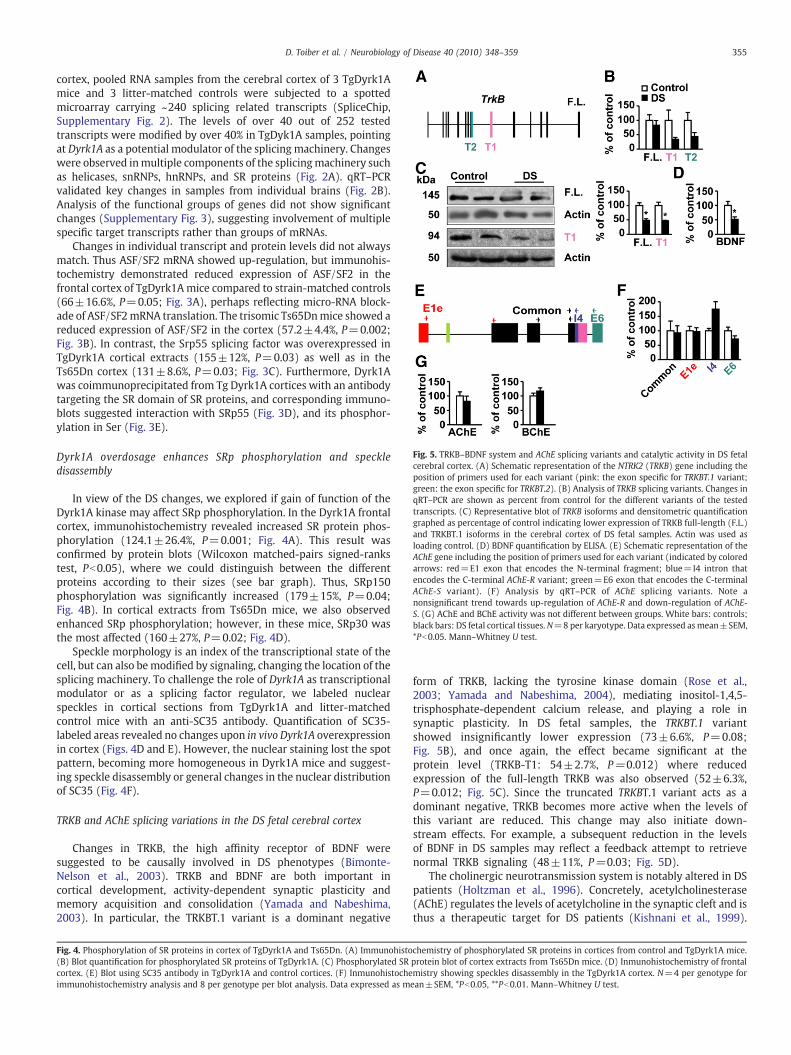
Changes in TRKB, the high af!nity receptor of BDNF weresuggested to be causally involved in DS phenotypes (Bimonte-Nelson et al., 2003). TRKB and BDNF are both important incortical development, activity-dependent synaptic plasticity andmemory acquisition and consolidation (Yamada and Nabeshima,2003). In particular, the TRKBT.1 variant is a dominant negative
form of TRKB, lacking the tyrosine kinase domain (Rose et al.,2003; Yamada and Nabeshima, 2004), mediating inositol-1,4,5-trisphosphate-dependent calcium release, and playing a role insynaptic plasticity. In DS fetal samples, the TRKBT.1 variantshowed insigni!cantly lower expression (73±6.6%, P=0.08;Fig. 5B), and once again, the effect became signi!cant at theprotein level (TRKB-T1: 54±2.7%, P=0.012) where reducedexpression of the full-length TRKB was also observed (52±6.3%,P=0.012; Fig. 5C). Since the truncated TRKBT.1 variant acts as adominant negative, TRKB becomes more active when the levels ofthis variant are reduced. This change may also initiate down-stream effects. For example, a subsequent reduction in the levelsof BDNF in DS samples may re"ect a feedback attempt to retrievenormal TRKB signaling (48±11%, P=0.03; Fig. 5D).
The cholinergic neurotransmission system is notably altered in DSpatients (Holtzman et al., 1996). Concretely, acetylcholinesterase(AChE) regulates the levels of acetylcholine in the synaptic cleft and isthus a therapeutic target for DS patients (Kishnani et al., 1999).
Fig. 4. Phosphorylation of SR proteins in cortex of TgDyrk1A and Ts65Dn. (A) Immunohistochemistry of phosphorylated SR proteins in cortices from control and TgDyrk1A mice.(B) Blot quanti!cation for phosphorylated SR proteins of TgDyrk1A. (C) Phosphorylated SR protein blot of cortex extracts from Ts65Dn mice. (D) Inmunohistochemistry of frontalcortex. (E) Blot using SC35 antibody in TgDyrk1A and control cortices. (F) Inmunohistochemistry showing speckles disassembly in the TgDyrk1A cortex. N=4 per genotype forimmunohistochemistry analysis and 8 per genotype per blot analysis. Data expressed as mean±SEM, *Pb0.05, **Pb0.01. Mann–Whitney U test.
Fig. 5. TRKB–BDNF system and AChE splicing variants and catalytic activity in DS fetalcerebral cortex. (A) Schematic representation of the NTRK2 (TRKB) gene including theposition of primers used for each variant (pink: the exon speci!c for TRKBT.1 variant;green: the exon speci!c for TRKBT.2). (B) Analysis of TRKB splicing variants. Changes inqRT–PCR are shown as percent from control for the different variants of the testedtranscripts. (C) Representative blot of TRKB isoforms and densitometric quanti!cationgraphed as percentage of control indicating lower expression of TRKB full-length (F.L.)and TRKBT.1 isoforms in the cerebral cortex of DS fetal samples. Actin was used asloading control. (D) BDNF quanti!cation by ELISA. (E) Schematic representation of theAChE gene including the position of primers used for each variant (indicated by coloredarrows: red=E1 exon that encodes the N-terminal fragment; blue=I4 intron thatencodes the C-terminal AChE-R variant; green=E6 exon that encodes the C-terminalAChE-S variant). (F) Analysis by qRT–PCR of AChE splicing variants. Note anonsigni!cant trend towards up-regulation of AChE-R and down-regulation of AChE-S. (G) AChE and BChE activity was not different between groups. White bars: controls;black bars: DS fetal cortical tissues.N=8 per karyotype. Data expressed asmean±SEM,*Pb0.05. Mann–Whitney U test.
355D. Toiber et al. / Neurobiology of Disease 40 (2010) 348–359

Alternate promoters and alternative splicing yield different AChEvariants (Meshorer and Soreq, 2006). Transcripts including the 5" E1eexon and the 3" exon 6 encode the N-terminal apoptotic exon 6inclusion N-AChE-S variant (Toiber et al., 2008), whereas the shorterC-terminal “synaptic” (AChE-S) variant is involved in synaptictransmission and neurite growth and the alternative AChE-R variant,which includes the 3" intronic I4 region, is involved in distressresponses (Meshorer et al., 2002). DS fetal samples showedinsigni!cant changes in AChE-R and AChE-S expression and hydrolyticAChE activity in control and DS fetal cortical samples (Figs. 5F and G),suggesting that the reported cholinergic de!cits in the DS braindevelop after birth.
Dyrk1A excess modi!es the splicing patterns of synaptic transcripts
Next, we tested if in the adult murine brain, Dyrk1A overdosemodi!es the splicing pattern of the studied synaptic transcripts.TrkBT.1 was signi!cantly downregulated in TgDyrk1A mice, unlike!ndings by Dorsey et al. (2002), but the full-length TrkB and the exoninclusion TrkBT.2 variant both maintained normal levels (Fig. 6A). Atthe protein level we observed a signi!cant reduction of the full-length(23±3.3%; P=0.014) and T.1 isoforms (40±7%, P=0.012; Fig. 6B) inthe transgenic Dyrk1A cortex but not in Ts65Dn mice (Fig. 6C). ELISAshowed a signi!cant reduction in BDNF in TgDyrk1A (36±6.4%,P=0.04) but not in Ts65Dn mice (Fig. 6C).
Nevertheless, the levels of the AChE common exon 2 and the 5" E1eexon encoding the N-terminal apoptotic variant remained unchangedin the Tg Dyrk1A mouse. In contrast, the AChE-S variant wasdownregulated, whereas the AChE-R variant was signi!cantly upre-gulated, attesting to intron 4 retention in the case of the AChE gene
(Fig. 7A). To !nd out if Dyrk1A overexpression was necessary andsuf!cient for exerting the observed change, we cotransfected 293Tcells with Dyrk1A and a mini-AChE gene containing the introns andexons of the human sequence. Dyrk1A overexpression induced aswitch from AChE-S to AChE-R (Fig. 7B), reminiscent of the changeobserved in Tg Dyrk1A mice and supporting the previous reports onAChE changes in the DS brain.
Next, we tested neuroligins, cell adhesion molecules involved incell signaling and, in particular, in synaptic function (Dean andDresbach, 2006). Mutations or alternative splicing of these transcriptsaffect synaptic fate and function (Levinson and El-Husseini, 2005) andhave been implicated in mental retardation (Fig. 8) (Daoud et al.,2009). In Tg Dyrk1A mice, Neuroligin 1 showed up-regulation of bothvariants A and B in the cerebral cortex (Fig. 7C), whereas its commontranscript remained unchanged. The splicing pattern of neuroligin 3 isnotably modi!ed in autistic patients (Talebizadeh et al., 2006). InTgDyrk1A mice brains, we identi!ed up-regulation of variant B, butneither variant A nor the common form of neuroligin 3 (Fig. 7D). Thus,Dyrk1A overdosage was causally involved in shifting synaptictranscripts toward exon inclusion patterns characteristic of brainmalfunctioning.
Discussion
The “gene-dosage” hypothesis (Epstein, 1986) predicts that in DS,overexpression of a single gene can impair multiple brain functionsthrough a signal ampli!cation effect. Our combined !ndings in thefetal DS brain and engineered mouse models and cultured cells with
Fig. 6. The TRKB–BDNF system in Dyrk1A overexpressing cortices. (A) TrkB splicingvariants expression by qRT–PCR. (B) TRKB full-length (F.L.) and TRKBT.1 and BDNFquantities in TgDyrk1A. (C) TRKB full-length (F.L.) and TRKBT.1 isoforms expression byblot and BDNF quanti!cation in Ts65Dn mice. N=4 per genotype. Data expressed asmean±SEM, *Pb0.05. Mann–Whitney U test.
Fig. 7. Splicing modi!cations target neuronal transcripts. (A) Ache splicing variants(qRT–PCR). (B) Cotransfecting 293T cells with the AChE mini-gene and DYRK1Areduced AChE-S and increased AChE-R compared to controls. (C) Splicing variants ofneuroligin 1. (D) Splicing variants of neuroligin 3 by qRT–PCR . N=4 per genotype. Dataexpressed as mean±SEM, *Pb0.05. Mann–Whitney U test.
356 D. Toiber et al. / Neurobiology of Disease 40 (2010) 348–359

Dyrk1A excess present Dyrk1A as such a gene, which is primarilyinvolved in monitoring the composition of the splicing machinery.Our !ndings demonstrate that stable excess of Dyrk1A alters thephosphorylation and subcellular location of splicing machinerycomponents and modi!es key synaptic transcripts and indicate thatDyrk1A overexpression changes the composition of synapses in themammalian brain. DS is a neurodevelopmental brain disorder, andindividual splicing choices and splicing regulators contribute to cellfate determination, axon guidance, and synaptogenesis. Therefore,our !ndings may provide a mechanistic explanation to at least part ofthe reported cognitive de!ciencies in both DS and the Dyrk1Aoverexpressing mice.
DYRK1A is also a transcription regulator, and overdosage oftranscription factors could principally affect the expression levels ofsplicing factors. In addition, transcription has been coupled to splicingand DYRK1A roles in transcription may also affect splicing. Intrigu-ingly, the levels of splicing factor transcripts in the DS fetal cortexremained largely unchanged, whereas downstream posttranslationalchanges such as protein levels and patterns of phosphorylation gainedsigni!cance (e.g., higher SRp75 and SRp30 phosphorylation in the DSfetal brain and in the two Dyrk1A gene dosage murine models).Together, this suggests that DYRK1A activities as a kinase rather thanas a transcriptional regulator were primarily involved. The phosphor-ylation status of SR proteins affects their RNA binding speci!city,protein–protein interactions, and intracellular distribution, so thatgenetic disorders that affect SRp phosphorylation patterns canreciprocally alter splicing, compatible with the Epstein ampli!cationconcept. The regulation of splicing is further coupled to transcription(Kornblihtt, 2005), highlighting the combinatorial complexity of theconsequences of Dyrk1A excess. Correspondingly, Dyrk1A excess alsoaltered the splicing patterns of key target transcripts.
That Dyrk1A overexpression changed total SR protein propertieswas indicated by the observed speckle disassembly. Indeed, modify-ing the subnuclear location of splicing factors is known to be a centralform of splicing regulation (Melcak et al., 2000). These changes mayre"ect direct interaction of DYRK1A with splicing factors, as waspreviously reported for ASF/SF2 (Shi et al., 2008) and Sf3b1/SAP155(de Graaf et al., 2006). Our current !ndings suggest that Dyrk1A also
interacts directly with SRp55. Consequent to these changes, we foundDyrk1A-mediated exon inclusion events, such as those reported intauopathies that induce the inclusion of exon 10 in the tau transcript(Shi et al., 2008) or bipolar disorderwhich is associatedwith two SNPslocated in the neural cell adhesion molecule (NCAM1) gene within acluster of alternatively spliced exons (Atz et al., 2007). Our !ndingsthus add DYRK1A to other known neuronal-speci!c splicing mod-ulators, e.g., NOVA2, which modi!es the splicing pattern of synapse-related transcripts in a coordinated manner (Ule et al., 2005) and thepolypyrimidine-tract-binding protein (PTB) which blocks entry ofU2AF into the presplicesomal complex, suppressing inclusion of theneuron-speci!c c-srcN1 exon in non-neuronal cells (Matsunaga et al.,1993).
Downstreamtarget geneswe tested includeNeuroligin 1 and 3, TRKB,and AChE. That the changes induced by DYRK1A overexpressionprimarily involved exon inclusions can have a profound effect onNeuroligin–Neurexin interactions. Speci!cally, Neuroligin splicingdictates the formation of inhibitory or excitatory synapses throughchanging its binding characteristics with Neurexin variants (Dean andDresbach, 2006), as was suggested to be the case in autism (Boucard etal., 2005). Downstream changes, induced by those occurring inNeuroligin would predictably change the binding af!nity of theNeuroligin protein products, which can modify synaptic composition.Recently, chronic systemic treatment of Ts65Dn mice with GABAAantagonists at nonepileptic doses caused persistent post-drug recoveryof cognition and long-term potentiation (Fernandez et al., 2007). Thisand other observations (Kurt et al., 2000) indicate the existence of arelative excess of inhibitory synapses in thesemice, compatiblewith ourDyrk1A-related changes in neuroligin alternative variants.
In DS fetal samples, we detected a lower expression of BDNF and ofthe full-length TRKB isoform, which can compromise cell proliferationand differentiation. In previous studies, changes in the lamination ofthe neorcortex of DS fetal brains were correlated with a signi!cantreduction of full-length TRKB (Wisniewski, 1990). In our hands, atrend towards lower expression of the truncated TRKBT.1 variant wasaccompanied by a signi!cant decrease at the protein level. BDNFreduction was also observed in the cortex of Ts65Dn mice (Muller etal., 2009), and BDNF mRNA was reduced in the Down syndrome
Fig. 8. Proposed mechanism of DYRK1A effects on mental retardation. (A) DYRK1A overexpression. (B) Imbalanced splicing machinery, hyperphosphorylated SR proteins, andspeckles disassembly. (C) Changes in SRp. (D) Changes in the nuclear speckles. (E) Changes in target synapses. (F) All of these changes together may affect (G) synaptic plasticity and(H) spine composition and properties.
357D. Toiber et al. / Neurobiology of Disease 40 (2010) 348–359

hippocampus. The diminutions in mice correlated with workingmemory errors; in addition, the low levels were corrected in theYACtg152F7 mice by the use of green tea polyphenols (DYRK1Ainhibitors) (Bimonte-Nelson et al., 2003; Guedj et al., 2009). Likewise,reducing the levels of AChE-S impairs neuritogenesis, compatible withthe neuritogenesis alterations observed in DS patients (Dierssen andRamakers, 2006). Inversely, elevated levels of the stress-inducedAChE-R splicing variant have been observed in the frontal cortex ofover 60-year-old DS brains and in AD patients (Darreh-Shori et al.,2004). That the slight variations in the AChE splicing pattern in DSfetal cortices did not lead to changes in AChE's catalytic activitysuggests stable cholinergic metabolism in DS individuals at earlydevelopmental stages, as was previously proposed (Brooksbank et al.,1989; Kish et al., 1989).
Additional examples of genes regulated by DYRK1A at thetranscription level may highlight speci!c motif(s) that might bepresent in their promoter region. Similarly, wider scope screeningcould be useful in !nding common motifs in the alternatively splicedtarget genes and decipher if this splicing misregulation is synaptic-related, brain-speci!c, or if it enables wider and more robust changes.While the effect of DYRK1A is likely not limited to brain functions, thehigher diversity of splicing variations in the central nervous systempredicts that splicing changes might affect brain development andfunction in a more pronounced manner than other organs (Licatalosiand Darnell, 2006). Others reported that DYRK1A misregulation leadsto impairments in brain function (Altafaj et al., 2001; Fotaki et al.,2002; Martinez de Lagran et al., 2004) and that its overexpression isrelated to pathological aging (Ferrer et al., 2005). Our current worksuggests that DYRK1A regulates the splicing machinery in general, atboth the posttranscriptional and the posttranslational levels, whichhelps to explain those observations. Recent discoveries point at a rolefor alternative splicing machinery in cancer (Kim et al., 2008; Soreq etal., 2008). Compatible with this, DS carriers are prone to leukemia(Zwaan et al., 2008), which could be related to alterations in thehematopoietic splicing machinery.
Funding
This research was supported by the Israel Science Foundationgrants 1876/2008 and 399/07, The German, Israeli and SpanishMinistries of Science and the German-Israel Project, DIP-G 3.2, TheHebrew University's Interdisciplinary Center for Neuronal Computa-tion (ICNC), The CIBERER Foundation, UK, and Jerome LejeuneFoundation, France. G. Azkona received a fellowship (BFI05.48) fromthe Basque Country Government, Spain.
Acknowledgments
We thank Prof. J.R. Naranjo and Dr. B. Mëlstrom for their kind helpin immunoprecipitation experiments and the Dyrk1A plasmid.
Appendix A. Supplementary data
Supplementary data associated with this article can be found, inthe online version, at doi:10.1016/j.nbd.2010.06.011.
References
Ahlbom, B.E., et al., 1996. Molecular analysis of chromosome 21 in a patient with aphenotype of Down syndrome and apparently normal karyotype. Am. J. Med.Genet. 63, 566–572.
Altafaj, X., et al., 2001. Neurodevelopmental delay, motor abnormalities and cognitivede!cits in transgenic mice overexpressing Dyrk1A (minibrain), a murine model ofDown's syndrome. Hum. Mol. Genet. 10, 1915–1923.
Alvarez, M., et al., 2003. DYRK1A accumulates in splicing speckles through a noveltargeting signal and induces speckle disassembly. J. Cell Sci. 116, 3099–3107.
Antonarakis, S.E., Epstein, C.J., 2006. The challenge of Down syndrome. Trends Mol.Med. 12, 473–479.
Antonarakis, S.E., et al., 2004. Chromosome 21 and Down syndrome: from genomics topathophysiology. Nat. Rev. Genet. 5, 725–738.
Arron, J.R., et al., 2006. NFAT dysregulation by increased dosage of DSCR1 and DYRK1Aon chromosome 21. Nature 441, 595–600.
Atz, M.E., et al., 2007. NCAM1 association study of bipolar disorder and schizophrenia:polymorphisms and alternatively spliced isoforms lead to similarities anddifferences. Psychiatr. Genet. 17, 55–67.
Becker, W., et al., 1998. Sequence characteristics, subcellular localization, and substratespeci!city of DYRK-related kinases, a novel family of dual speci!city proteinkinases. J. Biol. Chem. 273, 25893–25902.
Ben-Ari, S., et al., 2006. Modulated splicing-associated gene expression in P19 cellsexpressing distinct acetylcholinesterase splice variants. J. Neurochem. 97 (Suppl 1),24–34.
Bimonte-Nelson, H.A., et al., 2003. Frontal cortex BDNF levels correlate with workingmemory in an animal model of Down syndrome. Behav. Brain Res. 139, 47–57.
Boucard, A.A., et al., 2005. A splice code for trans-synaptic cell adhesion mediated bybinding of neuroligin 1 to alpha- and beta-neurexins. Neuron 48, 229–236.
Brooksbank, B.W., et al., 1989. Neuronal maturation in the foetal brain in Down'ssyndrome. Early Hum. Dev. 18, 237–246.
Daoud, H., et al., 2009. Autism and nonsyndromic mental retardation associated with ade novo mutation in the NLGN4X gene promoter causing an increased expressionlevel. Biol. Psychiatry 66 (10), 904–905.
Darreh-Shori, T., et al., 2004. Long-lasting acetylcholinesterase splice variations inanticholinesterase-treated Alzheimer's disease patients. J. Neurochem. 88, 1102–1113.
de Graaf, K., et al., 2004. Characterization of cyclin L2, a novel cyclin with an arginine/serine-rich domain: phosphorylation by DYRK1A and colocalization with splicingfactors. J. Biol. Chem. 279, 4612–4624.
de Graaf, K., et al., 2006. The protein kinase DYRK1A phosphorylates the splicing factorSF3b1/SAP155 at Thr434, a novel in vivo phosphorylation site. BMC Biochem. 7, 7.
Dean, C., Dresbach, T., 2006. Neuroligins and neurexins: linking cell adhesion, synapseformation and cognitive function. Trends Neurosci. 29, 21–29.
Deutsch, S., et al., 2005. Gene expression variation and expression quantitative traitmapping of human chromosome 21 genes. Hum. Mol. Genet. 14, 3741–3749.
Diamant, S., 2006. Butyrylcholinesterase attenuates amyloid !bril formation in vitro.Proc. Natl. Acad. Sci. USA 103 (23), 8628–8633 (Electronic publication ahead ofprint 2006 May 26).
Dierssen, M., de Lagran, M.M., 2006. DYRK1A (dual-speci!city tyrosine-phosphorylatedand -regulated kinase 1A): a gene with dosage effect during development andneurogenesis. Scienti!c World J. 6, 1911–1922.
Dierssen, M., Ramakers, G.J., 2006. Dendritic pathology in mental retardation: frommolecular genetics to neurobiology. Genes Brain Behav. 5 (Suppl 2), 48–60.
Dierssen, M., et al., 2009. Aneuploidy: from a physiological mechanism of variance toDown syndrome. Physiol. Rev. 89, 887–920.
Dorsey, S.G., et al., 2002. Failure of brain-derived neurotrophic factor-dependentneuron survival in mouse trisomy 16. J. Neurosci. 22, 2571–2578.
Ellman, G.L., et al., 1961. A new and rapid colorimetric determination of acetylcholin-esterase activity. Biochem. Pharmacol. 7, 88–95.
Engidawork, E., Lubec, G., 2001. Protein expression in Down syndrome brain. AminoAcids 21, 331–361.
Epstein, C.J., 1986. Developmental genetics. Experientia 42, 1117–1128.Fernandez, F., et al., 2007. Pharmacotherapy for cognitive impairment in amousemodel
of Down syndrome. Nat. Neurosci. 10, 411–413.Ferrando-Miguel, R., et al., 2004. Protein levels of genes encoded on chromosome 21 in
fetal Down syndrome brain (Part V): overexpression of phosphatidyl-inositol-glycan class P protein (DSCR5). Amino Acids 26, 255–261.
Ferrer, I., et al., 2005. Constitutive Dyrk1A is abnormally expressed in Alzheimerdisease, Down syndrome, Pick disease, and related transgenic models. Neurobiol.Dis. 20, 392–400.
Fotaki, V., et al., 2002. Dyrk1A haploinsuf!ciency affects viability and causesdevelopmental delay and abnormal brain morphology in mice. Mol. Cell. Biol. 22,6636–6647.
Galdzicki, Z., et al., 2001. On the cause of mental retardation in Down syndrome:extrapolation from full and segmental trisomy 16 mouse models. Brain Res. BrainRes. Rev. 35, 115–145.
Guedj, F., et al., 2009. Green tea polyphenols rescue of brain defects induced byoverexpression of DYRK1A. PLoS ONE 4, e4606.
Guimera, J., et al., 1996. A human homologue of Drosophila minibrain (MNB) isexpressed in the neuronal regions affected in Down syndrome and maps to thecritical region. Hum. Mol. Genet. 5, 1305–1310.
Guimera, J., et al., 1999. Human minibrain homologue (MNBH/DYRK1): characteriza-tion, alternative splicing, differential tissue expression, and overexpression inDown syndrome. Genomics 57, 407–418.
Gwack, Y., et al., 2006. A genome-wide Drosophila RNAi screen identi!es DYRK-familykinases as regulators of NFAT. Nature 441, 646–650.
Hanamura, A., et al., 1998. Regulated tissue-speci!c expression of antagonistic pre-mRNA splicing factors. RNA 4, 430–444.
Holtzman, D.M., et al., 1996. Developmental abnormalities and age-related neurodegenera-tion in a mouse model of Down syndrome. Proc. Natl. Acad. Sci. USA 93, 13333–13338.
Kim, E., et al., 2008. Insights into the connection between cancer and alternativesplicing. Trends Genet. 24, 7–10.
Kish, S., et al., 1989. Down's syndrome individuals begin life with normal levels of braincholinergic markers. J. Neurochem. 52, 1183–1187.
Kishnani, P.S., et al., 1999. Cholinergic therapy for Down's syndrome. Lancet 353,1064–1065.
Kornblihtt, A.R., 2005. Promoter usage and alternative splicing. Curr. Opin. Cell Biol. 17,262–268.
358 D. Toiber et al. / Neurobiology of Disease 40 (2010) 348–359

Kurt, M.A., et al., 2000. Synaptic de!cit in the temporal cortex of partial trisomy 16(Ts65Dn) mice. Brain Res. 858, 191–197.
Lamond, A.I., Spector, D.L., 2003. Nuclear speckles: a model for nuclear organelles. Nat.Rev. Mol. Cell. Biol. 4, 605–612.
Levinson, J.N., El-Husseini, A., 2005. Building excitatory and inhibitory synapses:balancing neuroligin partnerships. Neuron 48, 171–174.
Li, Q., et al., 2007. Neuronal regulation of alternative pre-mRNA splicing. Nat. Rev.Neurosci. 8, 819–831.
Licatalosi, D.D., Darnell, R.B., 2006. Splicing regulation in neurologic disease. Neuron 52,93–101.
Liu, F., et al., 2008. Overexpression of Dyrk1A contributes to neuro!brillarydegeneration in Down syndrome. FASEB J. 22, 3224–3233.
Martinez de Lagran, M., et al., 2004. Motor phenotypic alterations in TgDyrk1atransgenic mice implicate DYRK1A in Down syndrome motor dysfunction.Neurobiol. Dis. 15, 132–142.
Matsunaga, T., et al., 1993. Expression of alternatively spliced src messenger RNAsrelated to neuronal differentiation in human neuroblastomas. Cancer Res. 53,3179–3185.
Melcak, I., et al., 2000. Nuclear pre-mRNA compartmentalization: traf!cking of releasedtranscripts to splicing factor reservoirs. Mol. Biol. Cell 11, 497–510.
Meshorer, E., Soreq, H., 2006. Virtues and woes of AChE alternative splicing in stress-related neuropathologies. Trends Neurosci. 29, 216–224.
Meshorer, E., et al., 2002. Alternative splicing and neuritic mRNA translocation underlong-term neuronal hypersensitivity. Science 295, 508–512.
Meshorer, E., et al., 2005. SC35 promotes sustainable stress-induced alternative splicingof neuronal acetylcholinesterase mRNA. Mol. Psychiatry 10, 985–997.
Muller, H.D., et al., 2009. Different postischemic protein expression of the GABA(A)receptor alpha2 subunit and the plasticity-associated protein MAP1B aftertreatment with BDNF versus G-CSF in the rat brain. Restor. Neurol. Neurosci. 27,27–39.
Ortiz-Abalia, J., et al., 2008. Targeting Dyrk1A with AAVshRNA attenuates motoralterations in TgDyrk1A, a mouse model of Down syndrome. Am. J. Hum. Genet. 83,479–488.
Rose, C.R., et al., 2003. Truncated TrkB-T1 mediates neurotrophin-evoked calciumsignalling in glia cells. Nature 426, 74–78.
Shi, J., et al., 2008. Increased dosage of Dyrk1A alters alternative splicing factor (ASF)-regulated alternative splicing of Tau in Down syndrome. J. Biol. Chem. 283,28660–28669.
Sitz, J.H., et al., 2004. Dyrk1A potentiates steroid hormone-induced transcription via thechromatin remodeling factor Arip4. Mol. Cell. Biol. 24, 5821–5834.
Soreq, L., et al., 2008. Advancedmicroarray analysis highlights modi!ed neuro-immunesignaling in nucleated blood cells from Parkinson's disease patients. J. Neuroim-munol. 201–202, 227–236.
Stamm, S., 2008. Regulation of alternative splicing by reversible protein phosphory-lation. J. Biol. Chem. 283, 1223–1227.
Stamm, S., et al., 2005. Function of alternative splicing. Gene 344, 1–20.Talebizadeh, Z., et al., 2006. Novel splice isoforms for NLGN3 and NLGN4 with possible
implications in autism. J. Med. Genet. 43, e21.Toiber, D., et al., 2008.N-acetylcholinesterase-induced apoptosis in Alzheimer's disease.
PLoS ONE 3, e3108.Ule, J., et al., 2005. Nova regulates brain-speci!c splicing to shape the synapse. Nat.
Genet. 37, 844–852.Wang, G.S., Cooper, T.A., 2007. Splicing in disease: disruption of the splicing code and
the decoding machinery. Nat. Rev. Genet. 8, 749–761.Wisniewski, K.E., 1990. Down syndrome children often have brain with maturation
delay, retardation of growth, and cortical dysgenesis. Am. J. Med. Genet. Suppl. 7,274–281.
Yamada, K., Nabeshima, T., 2003. Brain-derived neurotrophic factor/TrkB signaling inmemory processes. J. Pharmacol. Sci. 91, 267–270.
Yamada, K., Nabeshima, T., 2004. Interaction of BDNF/TrkB signaling with NMDAreceptor in learning and memory. Drug News Perspect. 17, 435–438.
Yang, E.J., et al., 2001. Protein kinase Dyrk1 activates cAMP response element-bindingprotein during neuronal differentiation in hippocampal progenitor cells. J. Biol.Chem. 276, 39819–39824.
Zwaan, M.C., et al., 2008. Acute leukemias in children with Down syndrome. Pediatr.Clin. N. Am. 55, 53–70 (x).
359D. Toiber et al. / Neurobiology of Disease 40 (2010) 348–359
Copyright © 2022 FDOKUMEN