Copper–copper oxide coated nanofibrillar cellulose: a promising biomaterial

Anti-Cancer Agents in Medicinal Chemistry, 2009, 9, 185-211 185
1871-5206/09 $55.00+.00 © 2009 Bentham Science Publishers Ltd.
Copper Complexes as Anticancer Agents
Cristina Marzano1,*, Maura Pellei
2, Francesco Tisato
3 and Carlo Santini
2,*
1Dipartimento di Scienze Farmaceutiche, Università di Padova, via Marzolo 5, 35131 Padova, Italy;
2Dipartimento di Scienze Chi-
miche, Università di Camerino, via S. Agostino 1, 62032 Camerino, Macerata, Italy and 3ICIS-C.N.R., Corso Stati Uniti, 4, 35127
Padova, Italy
Abstract: Metal-based antitumor drugs play a relevant role in antiblastic chemotherapy. Cisplatin is regarded as one of the most effective
drugs, even if severe toxicities and drug resistance phenomena limit its clinical use. Therefore, in recent years there has been a rapid ex-pansion in research and development of novel metal-based anticancer drugs to improve clinical effectiveness, to reduce general toxicity
and to broaden the spectrum of activity.
The variety of metal ion functions in biology has stimulated the development of new metallodrugs other than Pt drugs with the aim to ob-
tain compounds acting via alternative mechanisms of action. Among non-Pt compounds, copper complexes are potentially attractive as anticancer agents. Actually, since many years a lot of researches have actively investigated copper compounds based on the assumption
proposal that endogenous metals may be less toxic.
It has been established that the properties of copper-coordinated compounds are largely determined by the nature of ligands and donor at-oms bound to the metal ion. In this review, the most remarkable achievements in the design and development of copper(I, II) complexes
as antitumor agents are discussed. Special emphasis has been focused on the identification of structure-activity relationships for the dif-ferent classes of copper(I,II) complexes. This work was motivated by the observation that no comprehensive surveys of copper com-
plexes as anticancer agents were available in the literature. Moreover, up to now, despite the enormous efforts in synthesizing different classes of copper complexes, very few data concerning the molecular basis of the mechanisms underlying their antitumor activity are
available. This overview, collecting the most significant strategies adopted in the last ten years to design promising anticancer cop-per(I,II) compounds, would be a help to the researchers working in this field.
1. INTRODUCTION
Although medicinal chemistry was almost exclusively based on organic compounds and natural products, during the past three dec-ades metal complexes have gained a growing interest as pharma-ceuticals for the use as diagnostic agents or as chemotherapeutic drugs [1-7].
There is no doubt that the discovery of cisplatin, cis-diammine- dichloroplatinum(II), represents one of the most significant events for cancer chemotherapy in the 20
th century. Cisplatin is highly
effective in treating a variety of cancers, especially testicular can-cer, for which the overall cure rate exceeds 90% [6]. The treatment is limited, however, by several side effects including nephrotoxic-ity, emetogenesis and neurotoxicity [6]. In addition, cisplatin is not orally bioavailable, and both inherited and acquired resistance seri-ously limit its applications [8]. These drawbacks have stimulated an extensive search for other antitumor-active inorganic complexes with improved pharmacological properties. A hot topic of debate has been the relative emphasis that should be placed on molecularly targeted metal-based drugs (designed to bind to and inhibit a single molecular target with high specificity) versus new cytotoxic and broad-spectrum agents. In metal-based drugs, the metal can coordi-nate ligands in a precise three-dimensional configuration, thus al-lowing the tailoring of the molecule to recognize and interact with a specific molecular target. This is further enhanced by different chemical modifications of ligands. Moreover, metal complexes easily undergo redox reactions and ligand substitution which allow them to participate in biological redox chemistry and interact with biological molecules. It is remarkable that investigations in this area are focused on the use of biologically active complexes formed by essential ions, such as copper [9]. Since any essential metal which escapes its normal metabolic pathways can be very toxic to the
*Address correspondence to these authors at the Dipartimento di Scienze
Farmaceutiche, Università di Padova, via Marzolo 5, 35131 Padova, Italy;
Tel: +39 (0)49 8275347; Fax: +39 (0)49 8275366;
E-mail: [email protected] and Dipartimento di Scienze Chimiche,
Università di Camerino, via S. Agostino 1, 62032 Camerino, Macerata,
Italy; Tel/Fax: +39 (0)737 402213; E-mail: [email protected]
organism, complexes of such metals may serve as effective cyto-toxic agents [10].
Copper is found in all living organisms and is a crucial trace element in redox chemistry, growth and development [11]. It is important for the function of several enzymes and proteins involved in energy metabolism, respiration and DNA synthesis, notably cy-tochrome oxidase, superoxide dismutase (SOD), ascorbate oxidase and tyrosinase. The major functions of copper–biological com-pounds involve oxidation–reduction reactions in which copper con-taining biological molecules react directly with molecular oxygen to produce free radicals [12,13]. Copper plays a pivotal role in cell physiology as a catalytic cofactor in the redox chemistry of mito-chondrial respiration, iron absorption, free radical scavenging, and elastin cross-linking [14]. As with Menkes syndrome and Wilson disease [15], which are of genetic origin, altered levels of copper are associated with diseases such as rheumatoid arthritis, gastroin-testinal ulcers, epilepsy, diabets and cancer. Researches on Menkes and Wilson disorders have provided new insights in the field of copper homeostasis and in particular into the understanding of in-tracellular trafficking and distribution of copper at molecular levels. Copper toxicity comes about from its ability to produce reactive oxygen species (ROS), displace other metal ions, peroxidize lipids, and directly cleave DNA and RNA [12]. Elevated levels of copper have been found in many types of human cancers, including pros-tate, breast, colon, lung, and brain [16]. Moreover, a specific amount of local copper appears to be required for angiogenesis to occur [17].
With the exception of platinum(II) compounds, there is rela-tively little mechanistic information on how metal anticancer drugs function, but it is clear that metal ions can work by a variety of different routes. Non-platinum active compounds are likely to have mechanism of action, biodistribution and toxicity which are differ-ent from those of platinum drugs and might be effective against human cancers that are poor chemosensitive or have become resis-tant to conventional platinum drugs. Copper, being an essential element, may be less toxic than non-essential metals such as plati-num. In recent years, several families of copper complexes have been studied as potential antitumor agents. Although only a little

186 Anti-Cancer Agents in Medicinal Chemistry, 2009, Vol. 9, No. 2 Marzano et al.
understanding of the molecular basis of their mechanism of action has been documented, copper complexes have attracted attention based on modes of action different from that of cisplatin (covalent binding to DNA). Therefore, copper complexes may provide, at least in principle, a broader spectrum of antitumor activity.
In this review, the most remarkable achievements in the design and development of copper–based drugs as antitumor agents will be discussed. Special emphasis will be focused on the identification of structure-activity relationships (SAR) for the different classes of copper(I,II) complexes. Recent investigations carried out by the authors concerning new phosphine copper(I) complexes will re-ceive a particular attention.
2. COPPER CHEMISTRY
Humans first used copper about 10000 years ago. A copper pendant discovered in Northern Iraq is thought to date back to around 8700 BC, and for nearly 5000 years copper was the only metal known to man. During the Roman Empire, copper was prin-cipally mined on the island of Cyprus, hence the origin of the name of the metal as Cyprium, "metal of Cyprus", later shortened to Cu-prum, from which the chemical symbol Cu.
Copper is the 29th
chemical element of the Periodic Table and belongs to the first row of Group 11 metals with electronic configu-ration 3d
104s
1. Thus, the Cu(I) ion has a completed 3d
10 shell and
the Cu(II) ion, loosing two electrons, has a partially filled d-block 3d
9 configuration behaving as a common transition metal (vide
infra in Section 5).
Copper is an essential trace nutrient to all high plants and ani-mals. In mammals, it is found primarily in the bloodstream, as a co-factor in various enzymes, and in copper-based pigments. Out of a total of 80-120 mg in a healthy human adult of 70 kg there are 8 mg in liver, 15 mg in heart, spleen, kidneys, brain and blood. However, in sufficiently high amounts, copper can be poisonous and even fatal to organisms. In the form of bivalent ion, copper is very poi-sonous to lower organisms. For example, bacteria and other decay micro-organisms die in water in a copper vessel, and copper com-pounds in general prevent growth of algae [18]. Analogously in humans, it can become toxic for cells at elevated concentrations [19]. Copper was found to bind DNA with high affinity than any other divalent cation, thus promoting DNA oxidation [20]. The binding of copper ions to specific sites can modify the conforma-tional structures of proteins, polynucleotides or DNA and biomem-branes [21]. This binding is dependent on copper size, charge elec-tron affinity and geometry of the formed adduct. It is known that copper ions as Cu
+/Cu
2+ in blue copper proteins act by changing the
redox potential and facilitating electron transfer phenomena. A high degree of selectivity is reached in molecular recognition through transition metal ions, which can perform a change of valence in redox reactions [22].
One of the main mechanisms proposed to explain copper-induced cellular toxicity comes from the propensity of free copper ions to participate in the formation of ROS. Cupric and cuprous copper ions can participate in oxidation and reduction reactions. In the presence of superoxide (*O2
-) or reducing agents such as ascor-
bic acid or glutathione (GSH), Cu(II) can be reduced to Cu(I), which is capable of catalyzing the formation of hydroxyl radicals (OH
•) from hydrogen peroxide (H2O2) via the Haber-Weiss reaction
[23]:
Cu(II) + O2-• Cu(I) + O2
Cu(I) + H2O2 Cu(II) + OH• + OH
-
------------------------------------------------
O2-• + H2O2 O2 + OH
• + OH
- Neat
The highly reactive hydroxyl radical is able to interact with any biological molecule by abstracting the hydrogen from an amino-bearing carbon to form a carboncentered protein radical and from
an unsaturated fatty acid to form a lipid radical. This results in oxi-dative damage of cells [24]. In particular, it has been demonstrated that copper is capable of inducing DNA strand breaks and oxidation of bases by producing ROS. GSH was shown to inhibit free radical formation by copper ions in the presence of hydrogen peroxide, ascorbate and DNA. The protective effect of GSH was attributed to its ability to stabilize Cu(I), preventing redox cycling and the gen-eration of free radicals.
According to the below equation copper(II) also forms thioxyl radicals, RS
•:
RSH + Cu(II) RS• + Cu(I) + H
+
and copper-cysteine and copper-methionine ions, which may pro-duce metallothioneines and disulfides, RSSR, that can be damaging [24]. Anyway, when considering copper as a metal participating in Fenton chemistry, it has to be reminded that intracellular free cop-per is limited to less than one free copper ion per cell, thus suggest-ing a significant overcapacity for chelation of copper in the cell [25].
3. COPPER HOMEOSTASIS
As already mentioned, excess accumulation of copper as well as its deficiency can be deleterious to human health; therefore, copper homeostasis is tightly regulated [11]. A conserved group of proteins that contain unique cysteine, methionine or histidine-rich domains referred to as metal binding sequences maintain the concentration of free Cu in cells at < 10
-18 M [26]. Fig. (1) presents a summary of
the known Cu homeostasis pathways in mammalian cells. Dietary copper is absorbed from the stomach and small intestine and then enters the blood circulation through mainly the action of ATP7A protein (Menkes protein). Most of the copper in normal human serum is bound to ceruloplasmin, an enzyme containing six copper atoms both in Cu(II) and Cu(I) state. Copper in this form is not exchangeable. The exchangeable form of copper is bound to albu-min and amino acids [27]. Copper-histidine complex was identified as the main copper-amino acid complex in human serum. It was also shown that human albumin forms a ternary complex with cop-per-histidine [28, 29].
During the uptake process, Cu(II) is reduced to Cu(I) by a hy-pothetical membrane bound metalloreductase and is absorbed by the cell through the transmembrane transporters. The main copper influx transporter in human cells is the 190 amino acid Cu trans-porter 1 (hCtr1). hCtr1 resides predominatly in the plasma mem-brane and is expressed in highest levels in the liver, kidney and heart, followed by the intestine, with the lowest level in the brain and muscle. It has been suggested that hCtr1 binds copper via the methionine and histidine–rich amino terminal domain and trans-ports it across cell membrane through pores [30]. Competition ex-periments have also demonstrated that hCtr1 is a monovalent metal transporter [31].
Upon entering the cytoplasm, copper may be complexed to a variety of ligands to prevent the interaction of free copper with cellular membranes, proteins, or DNA that can lead to oxidative damage. However, it is thought that the majority of cytoplasmatic copper is complexed to GSH as Cu(I) [32]. The Cu(I)-GS complex can then donate copper to various intracellular proteins such as metallothionein (MT), a family of proteins important for metal detoxification [33]. Another important class of molecules that are vital for copper delivery is copper chaperones. This is a group of cytosolic peptides that includes ATOX1 (HAH1), that delivers Cu to the P-type ATPase ATP7A and ATP7B at the trans-Golgi net-work, COX17 that delivers Cu to cytochrome-c oxidase in mito-condria and CCS1 which loads Cu onto cytoplasmic SOD [34].
To avoid copper excess, mammals cells have two structurally similar P-type ATPases, ATP7A and ATP7B, that mediate the cel-lular efflux of Cu. Defects in the function of ATP7A produce Menkes desease, while defects in ATP7B cause Wilson disease.

Copper Complexes as Anticancer Agents Anti-Cancer Agents in Medicinal Chemistry, 2009, Vol. 9, No. 2 187
ATP7A is expressed in the intestinal epithelium [35] as well as most other tissues other than liver [36]. ATP7B is expressed in liver and kidney and to a lesser extent in brain of normal individuals [37]. Both proteins function as monomers and have 8 membrane spanning domains and 6 repeats of GMTCXXCIE motif that has very high affinity to copper [26]. Binding of copper to ATP7A and ATP7B triggers highly regulated subcellular relocalization that involves movement from their basal position in the trans-Golgi network to other vescicular compartments, and in the case of ATP7A, to the plasma membrane [26].
Recent studies have demonstrated that copper transporters in-cluding hCtr1, ATP7A and ATP7B, are involved in the import, subcellular distribution and export of cisplatin-related drugs. This means that Cu transporters can regulate the sensitivity of human cancer cells to platinum drugs. These observations are undoubtedly appealing, because conventional thinking of the inorganic physiol-ogic chemistry of cisplatin and copper is quite different. Up to now, the mechanisms by which these transporters shuffle platinum-based antitumor agents are largely unknown. The Cu transporters are excellent at discriminating between closely related metal ions, and even between Cu(I) and Cu(II). Thus, their promiscuity with respect to the Pt drug suggests that alterations in platinum drug cellular pharmacology associated with a modified expression of the copper transporters are mediated by secondary effects of Cu on other metabolic pathways, such as MT and GSH levels [26].
4. COPPER, ANGIOGENESIS AND PROTEASOME PATH-WAY
The involvement of copper in angiogenesis has been known for a couple of decades [38, 39]. Folkman [40, 41] pioneered the con-cept that tumor growth requires angiogenesis and this requirement might be an Achilles heel for cancer because in adults there is little requirement for angiogenesis. The concept of antiangiogenic ther-apy for cancer has taken off in the last decade [41]. Researches aimed at understanding the role of copper in promoting angiogene-sis demonstrated that this metal interacts with several pro-angiogenic factors. Despite the functional significance of these interactions remains often unclear, it has been suggested that the role of copper in angiogenesis is referable to several mechanisms: i) copper may act through binding of angiogenic growth factors and increasing their affinity for endothelial cells, as with angiogenin, ii) copper may control the secretion of angiogenic cytokines, as dem-
onstrated with FGF1 and IL-1 and iii) copper may induce expres-sion of angiogenic growth factors such as VEGF [17]. Thus, ther-apy aimed at depleting copper may be a successful anticancer strat-egy which may target multiple angiogenic growth factors. Several anti-copper drugs used in Wilson disease [16] have been evaluated for use in cancer. It has been shown that three copper chelators, such as D-penicillamine, trientine, and tetrathiomolybdate (TM) [42], which quickly and effectively deplete copper stores, have antiangiogenic effects in murine cancer models [43, 44]. In particu-lar, promising results in vitro, in preclinical animal models and in an early (phase I) clinical trial have led to ongoing phase II evalua-tion of TM in patients with advanced cancers [17].
Recently, it has been demonstrated that mixtures of dithiocar-bamates (DTCs) or clioquinol with Cu(II) salts spontaneously bind with tumor cellular copper forming a proteasome inhibitor and an apoptosis inducer. The anti-angiogenesis effects, as well as the potential use of proteasome inhibitors in cancer therapies, have been extensively reviewed [45, 46]. It has been found that, like other established proteasome inhibitors, copper-binding compounds were only effective in inducing ubiquinated protein accumulation and apoptosis in tumor, but not in non-trasformed cells. Therefore, it can be hypothesized that such a strategy could result in highly effective and selective cancer killing that avoids toxicity.
It is important to note that not all copper-binding compounds have proteasome-inhibitory and apoptosis inducing capability. For example, TM and ethylenediaminetetraacetic acid are potent copper chelators but have no proteasome-inhibitory activity when com-bined with copper [47]. However, the ability of some copper-binding compounds to inhibit the proteasome and induce apoptosis by themselves in copper-enriched cancer cells is promising for their development as anticancer compounds in a nontoxic chemothera-peutic strategy [48].
5. COPPER COMPLEXES
Since the approval of cis-[PtCl2(NH3)2] in the clinical practice, many efforts have been made to develop novel platinum- and non platinum-based antitumor drugs to improve clinical effectiveness, to reduce systemic and organo-specific toxicities and to broaden the spectrum of activity. In the field of non-platinum compounds show-ing an antitumor potential, copper-based complexes have been in-vestigated on the assumption that endogenous metals may be less toxic.
Fig. (1). Schematic diagram of pathways involved in cellular copper homeostasis. hCtr1 = human copper transporter1, SOD = superoxide dismutase, CCS1,
COX17 and ATOX1 = copper chaperones, ATP7A = ATPase copper transporting -polypeptide, ATP7B = ATPase copper transporting -polypeptide, GSH =
glutathione, MT = metallothionein.

188 Anti-Cancer Agents in Medicinal Chemistry, 2009, Vol. 9, No. 2 Marzano et al.
The properties of copper-coordinated compounds, whether in classical inorganic coordination complexes, in organometallic com-pounds, or in bioinorganic model compounds are largely deter-mined by the nature of ligands and donor atoms bound to the metal ion [49,50]. Three oxidation states of the metal can be stabilized: Cu(III), Cu(II) and Cu(I), but very few examples of copper(III) compounds are reported [50]. Coordination chemistry of copper is therefore dominated by Cu(II) derivatives with little, but important examples of Cu(I) compounds. Due to the closed-shell d
10 elec-
tronic configuration, Cu(I) complexes are usually colorless solids and strongly prefer ligands having soft donor atoms such as P and aromatic amines. Although two-coordinated linear and three-coordinated trigonal arrangements are known, Cu(I) complexes are mostly four-coordinated species adopting a tetrahedral geometry.
The d9 electronic configuration typical of Cu(II) derivatives
promotes, instead, d-d transitions resulting in intense coloured spe-cies. In these complexes the coordination number varies from four to six, including four-coordinate square planar (sp), five-coordinate trigonal bipyramidal (tbp) and six-coordinate octahedral (oc) geo-metries. Such molecular structures depart from ideal arrangements mostly showing tetragonal distortions. The variety of available arrays allows for a great assortment in the choice of the ligands (from mono- to hexa-dentate chelates), and of the donor atoms (N, O, S and halides). The redox potential of the physiologically acces-sible Cu(I)/Cu(II) couple varies dramatically depending upon the ligand environment due to the donor set, geometry, substituent elec-tronic effect, substituent steric effect, and chelation [51]. For exam-ple, in the one electron oxidation of Cu(I) complexes toward dioxy-gen, a wide range of reduction potentials (from -1.5 to +1.3 V vs NHE) is known for copper complexes. In addition, such an electron transfer always involves important modifications of the stereochem-istry of the pertinent oxidized/reduced complexes. This feature, together with the possibility to release coordinating groups ongoing, for example, from octahedral Cu(II) to tetrahedral Cu(I) species, are chemical factors that illustrate the huge complexity of the Cu(I)/ Cu(II) system in physiological media.
The first evidence that copper complexes may exert antitumor efficacy may be traced back to early 1960s [52]. Copper complexes were subjected to a lot of studies concerning their chemical and biological behaviour. However, despite the enormous efforts in synthesizing different classes of copper complexes, very few data are available concerning their physiological processing.
This chapter includes representative examples of copper com-plexes grouped according to ligand similarity that have been tested for their anti-cancer activity in about the last ten years. It has been established that the copper ion chelation plays a definite role in the biological activity of most of the selected organic ligands, enhanc-ing the anti-tumour activity of the metal-free substances.
5.1. Thiosemicarbazone Complexes
Thiosemicarbazones (TSCs) are a class of compounds of me-dicinal interest whose anticancer activity has been reported as early as 1960s [53,54] and their development is still in progress [55-59]. Some of them, such as Marboran
TM or Triapine
TM, are in the clini-
cal practice [60]. TSCs have been intensively studied due to their inhibitory action on the DNA enzyme ribonucleotide diphosphate reductase and their selectivity towards hormone-responsive cancers [61]. For many years it has been known that a large number of bis-thiosemicarbazones (bTSCs) and series of their copper complexes showed promising antitumour activities [15, 62, 63]. A critical property of many of these copper complexes is the poor water solu-bility and the relatively high in vivo toxicity [64, 65]. Many at-tempts have been made in the last decades to improve the hydro-philicity and reduce the toxic effects by modifying the TSCs frameworks of the copper complexes [66].
For example, the water solubility increased by a factor of about 100 when the 2-pyridine moiety of classical TSCs was replaced by
the 1,2-diazine function (1) [67]; moreover, the diazinyl-derived TSCs turned out to be less cytotoxic, as compared to the pyridyl congeners (by a factor of 50) [68]. It has been shown that the re-placement of the terminal primary or secondary amino function by a tertiary amino group resulted in a marked enhancement of cyto-toxic activity [68].
NN
NNH
S
NH2
Scheme 1.
NN
NNH
S
N
R
R'
R = CH3, R' = H
R = CH3, R' = CH3
R = CH2CH3, R' = H
Scheme 2.
NN
NNH
S
N
R
R = CH3, R' = H
R = CH3, R' = CH3
R'
Scheme 3.
N
N NNH
S
N
R
R = CH3, R' = H, R" = H
R = CH3, R' = CH3, R" = H
R = CH3, R' = H, R" = CH3
R'
R"
Scheme 4.
N
NN
S
N
R
NR'
Cu
Cl
Scheme 5.
These findings led to the design of acyldiazinyl TSCs bearing an N
4-azabicyclo[3.2.2]nonane group and their copper complexes
(2-5) [69]. These compounds inhibited the proliferation of a series of tumor cell lines at nanomolar range, being more potent than free TSCs. In particular, the TSCs ligands 2-4 exhibited potent cytotoxic activities against human acute lymphoblastic leukemia CCRF-CEM cells (IC50 = 0.05-0.77 μM) and colon adenocarcinoma HT-29 cells (IC50 = 0.011-2.22 μM), whereas related copper(II) complexes (5) showed a significant improvement in cytotoxic activity against HT-29 cells by a factor of 3 (IC50 = 0.004-1.51 μM).

Copper Complexes as Anticancer Agents Anti-Cancer Agents in Medicinal Chemistry, 2009, Vol. 9, No. 2 189
As an example of TSCs with non-pyridine heterocyclic rings, thiophene-2-carbaldehyde TSC (6) exhibited a lower cytotoxicity against melanoma B16F10 and Friend erythroleukemia cells than the corresponding Cu(II) complexes [70, 71].
NN
S
NH2
H
S
Scheme 6.
N
NH
S
NH2
O Cu
H2O
2
SO4.2DMSO.6H2O
Scheme 7.
All this notwithstanding, TSCs bearing an aromatic heterocyclic moiety seem to be endowed with an implemented biological activ-ity [72-80]. Ferrari et al. reported a series of TSCs derivatives by changing the 2-pyridine group with salicylaldehyde [81], pyridoxal [72,78] and 5-formyluracil [82-84]. The salicylaldehyde TSC (H2salt), exhibited specificity for copper(II) leading to the dimeric metal complex [Cu(Hsalt)(H2O)]2SO4
.2DMSO
.6H2O] (7). This
complex has been tested in vitro on human leukaemic U937 cells, focusing the experiments on the activity with respect to cell prolif-eration inhibition and apoptosis induction. The results indicated that (7) inhibited about 40% of cell proliferation at 0.3 and 0.5 μg/ml, but DNA fragmentation and apoptosis were not observed at these concentrations. This unusual biological behaviour could be con-nected with the square planar copper coordination geometry shown by (7).
Instead, the monomeric copper complex (8) of the pyridoxal-TSC ligand (9a) [72], and the complexes (10), (11) and (12) of the 5-formyluracil-TSC ligand (13), all adopting five coordinated struc-tures, caused DNA fragmentation leading to apoptosis [82].
HO
N
CH3
O
NNH
S
NH2
Cu
ClOH2
H
Cl
Scheme 8.
OH
N
H3C
OH
NN
S
NR3
R2R1
a) R1 = H; R2 = H; R3 = H
b) R1 = H; R2 = Me; R3 = Me
c) R1 = Me; R2 = H; R3 = Me
d) R1 = H; R2 = H; R3 = E
Scheme 9.
HN
HN
NN
S
NH2
O O Cu
Cl H2O
Scheme 10.
HN
HN
NNH
S
NH2
O O Cu
ClCl
Scheme 11.
HN
HN
NNH
S
NH2
O O Cu
OSO3H2O
Scheme 12.
HN
HN
NNH
S
NH2
O O
Scheme 13.
The dimeric copper compound containing the unsubstituted pyridoxal TSC (9a), [{Cu(9a)(H2O)}2]Cl2
. 2H2O [72]), has been
synthesized [80] and its behavior has been evaluated on different murine and human cell lines in comparison to its monomeric coun-terpart (8). To verify if other dimeric complexes show relevant biological activity, some derivatives with N-substituted pyridoxal TSCs (9) have been synthesized and spectroscopically character-ized. In particular, the dimeric species [Cu(9b)Cl]2
.6H2O, [Cu(9c)
Cl]2Cl2.4H2O and [Cu(9d)Cl]2Cl2
.2H2O, characterized by X-ray
diffractometry, adopted a square pyramidal geometry, with the ligand showing a SNO tridentate donor set, and were tested in vitro on human leukemic cell lines U937, CEM and K562 [85].
Continuing their research on the biological properties of pyri-doxal TSC (9a) derivatives, Ferrari et al. [86] have synthesized copper(II) complexes with nitroprusside as a counterion because of its use in medical practice, thanks to the role of the nitric oxide released [87-89], as well as because of its inductive role in apopto-sis [88, 89]. From the biological viewpoint, the two complexes [Cu(9a)][Fe(CN)5(NO)]
.2H2O and [Cu(9a)]2[Fe(CN)5(NO)]
.6H2O
with 4+1 coordination geometry were found to inhibit leukaemic cell proliferation of both CEM and U937 cell lines and in inducing apoptosis. The study of the effects on the cell cycle revealed an increase of cells in G2/M phase [86].
Hambley and co-workers [90] described that the stability of Cu-TSCs species was increased when the external surface of the com-plex molecule was hydrophobic. On this subject, the -ketoglutaric acid TSC is an aliphatic ligand with many potential donor atoms and with a variety of possible conformations which determine a versatile chelating behavior. Seven N-substituted -ketoglutaric thiosemicarbazides (14), possessing either aliphatic or aromatic groups were synthesized and used for preparing copper complexes [91]. DNA binding constants were determined, and studies of ther-mal denaturation profiles and nuclease activity were also per-

190 Anti-Cancer Agents in Medicinal Chemistry, 2009, Vol. 9, No. 2 Marzano et al.
formed. Tests in vitro on human leukemia U937 cell line were car-ried out concerning with cell growth inhibition, cell cycle effects, and apoptosis induction. Among all the substituents introduced on the aminic nitrogen, the ethyl derivatives led to the highest biologi-cal activity.
A group of methylpyruvate TSCs (15) was obtained by reacting methyl pyruvate with thiosemicarbazides derivatized on the aminic nitrogen with both alkyl or aryl groups [92]. Tests on cell prolifera-tion against human leukemic U937 cell line, showed that the copper complex [Cu(15b)Cl]
.H2O was the most powerful agent, even if it
was not able to induce apoptosis.
NN
S
N
H
R1
R2
O
OH
OHO
a) R1 = Me; R2 = H
b) R1 = Et; R2 = H
c) R1 = vinyl; R2 = H
d) R1 = H; R2 = Me
e) R1 = Me; R2 = Me
f) R1 = Ph; R2 = H
g) R1 = p-tolyl; R2 = H
Scheme 14.
NN
S
N
R2
R1
HCH3
O
O
H3C
a) R1 = Me; R2 = H
b) R1 = Et; R2 = H
c) R1 = Me; R2 = Me
d) R1 = Allyl; R2 = H
e) R1 = Ph; R2 = H
f) R1 = MePh; R2 = H
Scheme 15.
Another series of TSCs (16-19), derived from natural alde-hydes, containing both N and S metal binding sites and an alkyl or terpenic group was designed in order to identify additional SARs [93]. The aromatic moiety of classic TSCs was replaced by an alkyl group of eight carbon backbone. Both ligands and the correspond-ing copper(II) complexes (16, R = H) and (18, R = Ph) showed a remarkable inhibition ability towards human leukemia U937 cell line and were capable of triggering cell apoptosis. These studies showed that the presence of an unsaturated bond in position 2 with respect to the imine on hydrocarbon chain as well as the absence of a substituent on the terminal amino group of TSCs enhanced the cytotoxic activity.
NNH
S
NH
RR = H; Ph
Scheme 16.
NNH
S
NH
RR = H; Ph
Scheme 17.
NNH
S
NH
R
R = Ph; MePh; Me; Et
CH3
CH3
Scheme 18.
NNH
S
NH
R
Scheme 19.
N
HN S
NHCu
Cl
OH2
Scheme 20.
The ligand 9-cis-retinal-TSC and its square planar Cu(II) com-plex (20) have been recently synthesized and characterized by Bis-ceglie et al. [94]. The DNA binding constants and spectroscopic data showed an external binding mode for the ligand and its copper complex. They possess a good lipophilic degree useful for an effi-cient cellular uptake. The metal complex inhibited human leukae-mic U937 cell proliferation at micromolar concentration by activat-ing an apoptotic cell death mechanism.
The antitumour potential of quinonoidal compounds is known since 1974 [95] and several 1,2-napthaquinone derivatives behave as anticancer compounds [96, 97]. Frydman et al. [98] have demon-strated that several 1,2-napthoquinone derivatives can induce DNA–topoisomerase II-mediated cleavages of DNA in an ATP-independent manner and this effect may be crucial for the cytotox-icity of these compounds. In particular, the addition of the TSC-pharmacophore to the parent quinonoidal moiety considerably en-hanced the anti-proliferative activity of the quinone moiety. Re-cently, Afrasiabi et al. [99] reported the synthesis and structural characterization of copper conjugates of 1,2-naphthaquinone TSC (21) and significant antitumour properties against MCF7 breast cancer cell line. This cytotoxic action has been attributed to its topoisomerase II inhibitory activity.
3-Formylchromone TSC (22), the minimal biologically active structural motif of soy isoflavone (genistein), and its square planar copper(II) complexes have been reported by Sarkar et al. [100]. They were stabilized in both Cu
2+/Cu
+ redox states and formed
stronger charge interactions in the kinase domain than genistein, leading to better stabilization in the active pocket. In vitro evalua-tion of copper complexes against hormone-independent and metas-tatic breast (BT20), prostate (PC-3), K-ras mutant (COLO 357) and K-ras wild-type (BxPC-3) pancreatic cancer cells revealed that [Cu(22)Cl2] complex exhibited PKB (Akt protein) inhibitory activ-ity and caused NF kappa B inactivation in COLO 357 cells.
The TSC derivative of 9,10-phenanthrenequinone (23) and its copper complex were synthesized by Padhye et al. [101]. The com-plex exhibited maximum antiproliferative activity against the hu-man breast T47D cancer cell line, probably due to inhibition of steroid binding to the cognitive receptor or by preventing dimeriza-tion of the estrogen receptor.
NN
S
NH2
O
H
Scheme 21.
More recently, the same authors have synthesized novel curcu-min analogs (24) using Knoevenagel condensation to convert enolic diketones of curcumin into non-enolizable ones and Schiff bases

Copper Complexes as Anticancer Agents Anti-Cancer Agents in Medicinal Chemistry, 2009, Vol. 9, No. 2 191
were prepared using a bioactive thiosemicarbazide pharmacophore [102]. Copper(II) conjugates of all synthesized ligands were pre-pared and structurally characterized as well as evaluated for their potential of inhibiting TNF induced NF kappa B activation and proliferation in human leukemic KBM-5 cells. Compound (24c) was found to be more potent than curcumin.
NN
S
NH2
HCH3
O
O
Scheme 22.
NN
S
NH2
HO
Scheme 23.
O
HO
O
OH
N NN N
S NH2SH2N
CH3CH3
R
Cu
a) R = 2-hydroxy
b) R = 2,3-dihydroxy
c) R = 3,4-dihydroxy
Scheme 24.
5.2. Conjugates Schiff Base Complexes
Padhye et al. [103] have modified the structure of nimesulide (25), a well-known cyclooxygenase-2 (COX-2) inhibitor [104,105], with pharmacophores capable of targeting vascular endothelial growth factor (VEGF) [106,107] and anti-apoptotic proteins like Bcl-2 and Bcl-XL, besides targeting the COX enzymes [108-111]. Copper conjugates of Schiff base derivatives of nimesulide (26), were synthesized, structurally characterized and evaluated for their COX selectivity indices and cytotoxicities on pancreatic tumor BxPC-3 (COX-2 positive) and MiaPaCa (COX-2 negative) cell lines. Copper conjugates exhibited distorted square planar geo-metries, as revealed by the single crystal X-ray structure determina-tion of (27), and showed significantly higher growth inhibition in both cell lines (IC50 values 3–26 μM for COX-2 positive and 5–9 μM for COX-2 negative cell line) than nimesulide (35 μM for COX-2 positive and >100 μM for COX-2 negative cell line). It has been reported that the mechanistic pathway responsible for the bio-logical activity involved the inhibition of VEGF and COX-2, as well as the down regulation of antiapoptotic Bcl-2 and Bcl-XL proteins.
A mononuclear complex of Cu(II) (28), with the Schiff base ligand (Z)-2-hydroxy-N’-(2-oxoindolin-3-ylidene)benzohydrazide, has been prepared by Zhong et al. [112]. This complex has been characterized by X-ray crystallography as a distorted octahedral species. The cytotoxicity assays, carried out in four kinds of human
cancer cell lines (SPCA-1, Tb, MGC, and K562), indicated that this complex was substantially more active than related compounds previously reported [113,114]. For the enhanced antitumor activity, the authors claimed the generation of cytotoxic Cu(I) species through intracellular enzymatic reduction [115].
O
HN
NO2
SCH3
O
O
Scheme 25.
O
HN
N
SCH3
O
O
OH
R1
R2
R3
a) R1 = H; R2 = H; R3 = H
b) R1 = H; R2 = OMe; R3 = H
c) R1 = OH; R2 = H; R3 = H
d) R1 = But; R2 = H; R3 = But
Scheme 26.
O
HN
N
SCH3
O
O
O
O
NH
N
SH3C
O
O
O
Cu
Scheme 27.
NH
O
N
N
O
HO
HN
O
N
N
O
OH
Cu
Scheme 28.

192 Anti-Cancer Agents in Medicinal Chemistry, 2009, Vol. 9, No. 2 Marzano et al.
Sarkar et al. [116,117] have shown that the central ketonic function of 3-benzoyl- -methyl benzene acetic acid (ketoprofen) could be easily appended with pharmacophores such as TSCs bear-ing amino groups to yield Schiff base type compounds. Reaction of these ligands with transition metals and, in particular with copper, yielded potent anticancer agents against breast cancer cells. Re-cently, the same authors have synthesized and biologically charac-terized a ketoprofen derivative named (29), as a selective COX-2 inhibitor [118]. Complex (29) exhibited a potent antiproliferative and proapoptotic activity in COX-2 positive cells. However, further escalation of dose than the one needed for inhibition of COX-2 activity indicated that other non COX-2 dependent pathways might also be involved. A similar speculation had been proposed in previ-ous studies reporting the ability of non-steroidal anti-inflammatory drugs (NSAIDs) to inhibit the growth of a colorectal cancer cell line that lacked COX-1 and COX-2 enzymatic expression [108]. The results showing a decrease in the COX-2 mRNA and protein subse-quent to treatment with (29) suggested that it inhibited NF kappa B activation leading to the inhibition of COX-2 expression and its activity, thereby inducing apoptotic cell death.
N
COOH
H3C Cu
HN O
OH
Cl
Cl
Scheme 29.
N
NNH
NH2S
CuCl
Cl
Scheme 30.
N
NNH
O
CuCl
Cl
Z
R
a) R = H; Z = CH
b) R = H; Z = N
c) R = OH; Z = CH
Scheme 31.
Adsule et al. [119] have reported the synthesis of some 1:1 Schiff base copper complexes of quinoline-2-carboxaldehyde ligands (30, 31). The quinoline and related copper-quinoline deriva-tives were tested for their cytotoxic and apoptosis induction effects in prostate cancer cell lines PC-3 and LNCaP. The results showed that these compounds induced apoptosis without causing an oxida-tive stress. Moreover, the introduction of thiocarbonyl side chains enhanced the antitumor potency. Actually, (30) was the most potent
analog which inhibited proteasome activity with a IC50 value of one order of magnitude lower that those of clioquinol and pyrrolidine DTC.
Some isatin-Schiff base copper(II) complexes have been re-cently prepared and spectroscopically characterized by Cerchiaro et al. [120]. These complexes exhibited keto-enol equilibria and a relatively high stability. Studies concerning the cytotoxic properties of this class of oxindolimine-copper(II) complexes revealed that some of them activated the apoptotic program in human promono-cytic U937 and neuroblastoma SH-SY5Y cells [120]. Very recent studies characterized the pro-apoptotic activity of the two most in- teresting isatin-Shiff base copper complexes (32) and (33), obtained from isatin and 1,3-diaminopropane and 2-(2-aminoethyl)pyridine, respectively. It was found that they induced apoptosis via the mito-chondrial pathway through both p-53 dependent and independent mechanisms and the extent of apoptosis correlated well with intra-cellular copper uptake. Therefore, it was suggested that these cop-per complexes were able to vehicle copper into the cell, thus pro-ducing ROS. On the other hand, considering the chemical structure of the isatin-Shiff base, (32) and (33) might behave as delocalized lipophilic cations, thus specifically targeting mitochondria [121].
N
Cu
N
HN NH
O O
2+
2 ClO4-
Scheme 32.
N
Cu
N
NH
O
2+
2 ClO4-
N
N
HN
O
Scheme 33.
5.3. Imidazole, Benzimidazole, Pyrazole and Triazole Com-
plexes
The strong antitumour activity shown by the [trans-bis(acetato) bis(imidazole)]copper(II) complex (34) against the B16 murine melanoma cell line [122] stimulated the synthesis of imidazole-substituted copper complexes. The crystal structure of (35), with the 1-methyl-4,5-diphenylimidazole, indicated that two trans-oriented imidazoles and two acetate ligands were coordinated to the Cu(II) ion forming a distorted octahedral environment. The effects of the bis-phenyl imidazole complex on the induction of cell division delays, as well as of Sister Chromatid Exchanges (SCEs) and on the suppression of Mitotic Indices (MIs) were examined [123]. At con-centrations between 0.77
.10
-7 and 1.54
.10
-6 M, this compound sig-
nificantly induced SCEs and MI and reduced proliferation rate in-dexes (PRIs) in cultured human lymphocytes, indicating a high cytostatic and cytotoxic action of this copper complex. Moreover, an unwinding effect on the plasmid pKS DNA was clearly observed after incubation with low concentrations of (35). In particular, con-centrations above 0.5 mM caused scissions on both closed super-

Copper Complexes as Anticancer Agents Anti-Cancer Agents in Medicinal Chemistry, 2009, Vol. 9, No. 2 193
coiled and open relaxed forms of DNA, and probably drug insertion occurred not randomly, but with some preference to guanosines, as detected by endonuclease inhibition experiments. An interstrand cross-linking of DNA bases were also observed in ds DNA calf thymus and plasmid DNA treated with (35).
N
N
N
N
Cu
O
O
OH3C
CH3O
H
H
Scheme 34.
N
N
N
N
Cu
O
O
OH3C
CH3O
CH3
H3C
Ph
Ph
Ph
Ph
Scheme 35.
Benzimidazole and many of its derivatives are known to exhibit a variety of biological actions, including antibacterial, antiviral, anticancer and antifungal activity [124]. 2-(4-Thiazolyl)benzimida- zole, or thiabendazole (HTBZ), with structural features common to phenantroline and benzimidazole is non-toxic to humans and used as a fungicide in agriculture. Reactions of HTBZ with copper(II) acetate, chloride, nitrate and butanedioate yielded [Cu(HTBZ)2
(H2O)2], [Cu(HTBZ)2Cl]Cl.H2O
.EtOH (36), [Cu(HTBZ)2(NO3)2]
and [Cu(HTBZ)2(CO2-C2H4-CO2)], respectively. The molecular structure of (36) comprised a five-coordinate copper centre with the metal bound to two chelating HTBZ and one chloride ion. The ge-ometry of this complex was best described as trigonal bipyramidal [125]. Testing HTBZ, (37) and [Cu(HTBZ)2(NO3)2] against the human squamous carcinoma tongue cell line (CAL-27) and the malignant melanoma skin cell line (SK-MEL-31), revealed that the chemotherapeutic potential of HTBZ was significantly enhanced upon metal coordination.
N
S
Cu
Cl
N
N
H
N
S
N
N
H
Scheme 36.
In an effort to further develop this class of potential anticancer agents, Devereux et al. [126] studied the synthesis and chemothera-peutic potential of a series of novel copper(II) carboxylate com-plexes derived from HTBZ and 2-(2-pyridyl)benzimidazole (PYBZ). Because of the contemporary presence of the benzimidazole moiety and of the carboxylic group, the ligand potential could be enhanced
by the chance to act both as acid and base [127] suggesting that HTBZ and PYBZ could be either neutral or anionic chelators or that they might simply act as countercations [125,128]. As for HTBZ complexes, the syntheses of mono-, bis- and tris-chelate compounds of PYBZ have been reported [129-133] but, so far, the only authen-ticated structure included mono-chelate derivatives of copper(I) [134]. Complex (37) and the corresponding free ligand (PYBZ) were assessed for their cancer chemotherapeutic potential against the hepatocellular carcinoma (Hep-G2) and kidney adenocarcinoma (A-498) cell lines. The bis-chelate derivatives [Cu(HTBZ)2(BZA)] (BZA)
.0.5(HTBZ)
.H2O (BZA = benzoic acid) and (37) elicited a
significant cytotoxic response. Replacing HTBZ and PYBZ with phenantroline (phen) to give [Cu(BZA)2(phen)(H2O)] did not sig-nificantly increase the anticancer activity. This study [126] sup-ported the hypothesis that the nitrogen donor ligands enabled cop-per complexes to interact with biomolecules, such as DNA.
CuO
O
Ph
N
N
H
N
N
NN
Scheme 37.
N
HN
N
HN
Cu
ClCl
Scheme 38.
An additional series of Cu(II) coordination compounds with benzimidazole-derived bidentate chelating ligands has been pre-pared and characterized by Saczewski et al. [135]. Complex (38) showed a very potent Cu,Zn-SOD activity in vitro with a IC50 value of 0.09 μM, comparable to those described in the literature for the best low molecular weight Cu,Zn-SOD mimics, including imidazo-late-bridged Cu(II)–Zn(II) heterodinuclear complexes [136-139]. In vitro cytotoxicity studies, with seven different human tumor cell lines, showed a moderate cell growth inhibitory activity, being the A-427 lung the most sensitive cancer cell line (IC50 values of 4.76–10.12 μM).
The synthesis, characterization and anti-proliferative activity of mononuclear or binuclear copper(II) complexes with 6-(2-chloro- benzylamino)purine (39a) and 6-(3-chlorobenzylamino)purine (39b) have been reported by Travnycek et al. [140]. The X-ray single-crystal structural analysis of (40) showed that the metal is penta-coordinated, the Cu(II) ion adopting a distorted trigonal bipyrami-dal geometry with the protonated HL2 ligands coordinated in trans apical positions, and the three chloride ions situated on the equato-rial plane. Binuclear complexes were characterized as well, and through comparison with crystallographic parameters observed for similar complexes [141], their structures were proposed as [Cu2(μ-Cl)2(μ-39a)2(H2O)2] and [Cu2(μ-Cl)2(μ-39a)2(39a)Cl2]. Biological activity testing against mouse melanoma B16-FO, human malig-nant melanoma G361, human osteogenic sarcoma HOS and human breast adenocarcinoma MCF7 cell lines were performed for the three complexes and compared with those exhibited by uncoordi-nated (39a) and (39b). The cytotoxic activity of the ligands strongly increased after formation of the Cu(II) complexes, which in turn

194 Anti-Cancer Agents in Medicinal Chemistry, 2009, Vol. 9, No. 2 Marzano et al.
showed potent activities against B16-F0 cells (IC50 from 8.2 to 19 μM). G361, HOS and MCF7 cell lines were less sensitive, with IC50 values ranging from 20 to 88 μM. For the G361 and HOS cell lines, the mononuclear complex was found to be several times less potent than the binuclear Cu(II) species.
N
NH
NH
N
R1
R2
a) R1 = Cl; R2 = H
b) R1 = H; R2 = Cl
Scheme 39.
NHN
NN
Cl
N NH
N N
Cl
Cu
Cl
Cl Cl
H
H Cl-
Scheme 40.
Recently, a series of ligands based on bidentate pyrazolylpyri-dine or tridentate pyrazolyl-bipyridine frameworks, and the corre-sponding metal complexes have been synthesized [142,143]. In particular, Yang et al. [144] reported the synthesis and characteriza-tion of the 2-[1-(naphthalen-1-ylmethyl)-1H-pyrazol-3-yl]pyridine ligand (41) and its octahedral [M(41)3][ClO4]2 complexes (M = Cu(II), Zn(II)). The Cu complex was found to possess a higher affinity toward DNA and a superior cytotoxicity against human leukemia HL-60, human gastric cancer BGC-823 and human mammary gland cancer MDA5 cell lines compared to those shown by the isostructural Zn(II) complex and uncoordinated (41). Also the related complex [Cu(41)2(NO3)][NO3] [145] was evaluated against six different cancer cell lines (HL-60 cells, PC-3M-1E8 human prostate tumor cells, BGC-832 cells, MDA cells, Bel-7402 human hepatoma cells and Hela human cervix cancer cells) show-ing considerable inhibitory rate and cytotoxic specificity. Com-pound (41) and the corresponding metal complexes had an extended aromatic p-system (like those shown by typical bidentate chelating ligands bipy and phen) and they bound to DNA by intercalation mode showing a marked DNA-cleavage activity.
NN N
Scheme 41.
More recently, Padhye et al. [146] have described the synthesis of the bis(3,6-pyrazol-1-yl)pyridazine ligand and of the related Cu(II) complex (42). The superoxide radical scavenging activity of this cationic dimeric species revealed that this compound was among the best SOD mimics (IC50 value of 3.90
.10
-7 M) whose
antioxidant potential was nicely reflected in its antiproliferative and apoptosis inducing effects against estrogen independent breast BT-20 and androgen independent prostate PC-3 cancer cells. The low-est IC50 values observed were 1.73 M and 1.42 M for BT-20 and PC-3 cells, respectively.
N N
NN
N N
Cu CuOH ClCl
Cl ClCl
Scheme 42.
A series of Cu(I) mixed-complexes containing dihydridobis(3-nitro-1,2,4-triazolyl)borate (43) [147], sodium bis(1,2,4-triazol-1-yl)acetate (44) or sodium bis(3,5-dimethyl-pyrazol-1-yl)acetate (45) ligands [148], and phosphine coligands were tested for their cyto-toxic properties against a panel of several human tumor cell lines. Results are reported in the Section 5.5.
N
NN
B
N
NN
O2N NO2
H H
K
Scheme 43.
N
NN
N
NN
O O-Na
+
Scheme 44.
N
NN
N
O O-Na
+
H3C
H3C
CH3
CH3
Scheme 45.
Copper complexes bearing a thioxo group, such as disulfiram, showed strong toxicity against cancer cell lines or cancer xenografts [119,149]. The rich redox chemistry of the thione group [150] can confer interesting biological properties, as demonstrated by the chemopreventive agent (48) [151], which was reported to induce protective biological mechanisms against radical generation.
N
N
S
S
SH3C
Scheme 46.
Dallavalle et al. [152] demonstrated that the 4-amino-5-(pyri- din-2-yl)-2H-1,2,4-triazole-3(4H)-thione copper(II) complex (47)

Copper Complexes as Anticancer Agents Anti-Cancer Agents in Medicinal Chemistry, 2009, Vol. 9, No. 2 195
exhibited in HT1080 human fibrosarcoma cells a cytotoxic activity comparable to that exhibited by cisplatin. Interestingly, normal human fibroblasts (HF) were not sensitive to the cytotoxic effects of (47). These effects were further investigated, leading to the con-clusion that (47) caused a variant of programmed cell death (PCD) clearly distinct from apoptosis [153]. Indeed, the morphological hallmark of the death process induced by (47) was found to consist of a massive cytoplasmic vacuolization. No evidence of nuclear fragmentation neither of caspase-3 activation, biochemical features of apoptosis [154,155], were observed. These results suggested that (47) drove sensitive cells toward a nonapoptotic PCD because it blocked caspase-3-dependent apoptotic pathways. Similar cellular effects have been recently described in human ovarian adenocarci-noma cells treated with some Cu(I) phosphine complexes (vide infra) [156].
N
N
N
N
S
N
Cu
Cl
Cl
HH
H
Scheme 47.
N
N
N
N
S
N
CuCl
Cl
N
N
N
N
S
N
Cu Cl
Cl
Scheme 48.
N
N
N
N
S
NH2
Cu
Cl
Cl
Scheme 49.
To gain insight into the SARs of thioxo-1,2,4-triazole cop-per(II) complexes, Tardito el al. [157] modified the thioamido func-tion of the ligand 4-amino-5-(pyridin-2-yl)-2H-1,2,4-triazole-3(4H)-thione. The resulting Cu(II) thioether complexes (48) and (49) were less potent than (47) showing, in HT1080 cells, IC50 values of 100, 68 and 12 μM, respectively. The complexes (48) and (49) also trig-gered the same peculiar type of cell death observed in (47) treated cells, hallmarked by an extensive cytoplasmic vacuolization in the absence of apoptotic or oncotic features. HT1080 fibrosarcoma cells and HeLa cells exhibited a marked sensitivity to (47), while normal human fibroblasts and SW872 liposarcoma cells were sub-stantially insensitive to the complex. The sensitivity to (47) ap-peared to be correlated with the ability of the complex to facilitate copper accumulation in the cells, which, in turn, might be related to the enhanced stability of (47) conferred by the thioamido coordina-
tion. In HT1080 cells treated with (47), copper uptake was paral-leled by an evident perturbation of the cell oxidative status, as indi-cated by a 5-fold increase of the oxidized form of GSH, pointing to an interaction of the metal with cell thiols.
5.4. Phenanthroline and Bipyridine Complexes
The metal chelator 1,10-phenanthroline (phen) and copper salts form stable bis-phen complexes which showed nuclease activity in the presence of reducing agents and molecular oxygen. The bio-logical activity of such cuprous complexes was investigated by Sigman et al. since the seventies [158] and reviewed in the nineties [159]. The bis-phen complex [Cu(phen)2]
+ (50) was described as an
agent able to oxidatively degrade DNA and RNA by attacking the sugar groups [159]. In addition, it has been shown to exhibit inter-esting clinical activities including antitumoral, antimycobacterial, antifungal and antimicrobial properties [160, 161]. The DNA cleav-age activity of [Cu(phen)2], which was significantly more reactive than the mono-phen complexes [162-175], was supposed to occur according to the following mechanisms [176]: a) [Cu(phen)2]
2+ is
reduced in solution to [Cu(phen)2]+; b) [Cu(phen)2]
+ binds DNA
non-coordinatively; c) [Cu(phen)2]+ is oxidized to [Cu(phen)2]
2+ by
H2O2 produced in situ; d) upon oxidation, the metal ion gains one ligand leading to Cu-“oxo” and/or Cu-“hydroxyl” species, the exact nature of which is still unknown; e) an oxidative attack mediated by these copper compounds leads to DNA cleavage mainly at C-1’, C-4’, or C-5’ of the 2-deoxyribose units [171, 172, 177].
N
N
N
N
Cu
Scheme 50.
Zhou et al. have reported that [Cu(phen)2] induced G1-phase specific apoptosis in liver carcinoma Bel-7402 cell line [178]. More recently, Cai et al. [179] demonstrated that the apoptosis pathway in Bel-7402 cells treated with [Cu(phen)2] might be initiated by the excessive copper in cells transported by the lipophilic phen ligand. Moreover, the catalysis of the redox-active copper with intracellular reductants induced an increase of ROS production and a decrease of the GSH/GSSG ratio. Furthermore, the same complex exhibited a potent cytotoxicity against human leukaemic HL60 cells and human stomachal SGC-7901 cells, with growth inhibition up to 90% [180].
On the other hand, the use of Cu-phen complexes presents some restrictions: a) their formation is rather unfavorable under physio-logical conditions because of the very small association constant of the second phen; b) [Cu(phen)2]
2+ shows a minor DNA sequence
selectivity, although it is not nucleotide specific. In an effort to overcome these limits, Pitié et al. [181] used a serinol bridge (ab-breviated as Clip) to link two phen ligands on positions 2, resulting in [Cu(2-Clip-phen)]
2+ (51) and 3, resulting in [Cu(3-Clip-phen)]
2+
(52) [182]. These chemical modifications led to several advantages: a) the link ensured that the two phen ligands coordinated to Cu; b) [Cu(2-Clip-phen)]
2+ and [Cu(3-Clip-phen)]
2+ complexes turned out
to cleave DNA macromolecules 2 and 60 times more efficiently than [Cu(phen)2]
2+, respectively, using a mechanism similar to that
shown by the bis-phen complex [181,183,184]. The reason for this increase in DNA-cleavage activity was not completely clear, but it was supposed to arise from structural characteristics [185] rather than from electronic properties [182].
The presence of the serinol bridge has allowed the functionali-zation of copper phen compounds with sequence specific anticancer drugs, rendering the compound highly sequence selective and pos-

196 Anti-Cancer Agents in Medicinal Chemistry, 2009, Vol. 9, No. 2 Marzano et al.
sibly even providing it with anticancer properties. In this respect, examples included a conjugate of 3-Clip-phen with a distamycin analog [177,186-188] or a cisplatin derivative [189].
N
N
N
N
Cu++
O O
NH2
Scheme 51.
N
N
N
N
Cu++
O O
NH2
Scheme 52.
N N
N N
Cu+2
O
O
HN (CH2)n N
Pt
NH
Cl Cl
a) n = 6
b) n = 10
2Cl-
Scheme 53.
The combined interest in (i) improving the DNA cleavage spe- cificity of (52) type complexes together with their ability to perform double-strand breaks (DSB) and (ii) circumventing drug resistance owing to the use of cisplatin [190-194] inspired the design of bi-functional molecules (53a) and (53b) [189]. It has been proposed that the amino group of [Cu(3-Clip-Phen)] (52) could be proto-nated, favoring its interaction with the polyanionic structure of DNA [182]. Thus, it could interact via hydrogen bonding with the oxygen atom of the phosphate, thereby pointing toward the minor groove [195,196]. The in vitro cytotoxic activities of (53a) and (53b) have been determined for several human cancer cell lines and a significant antiproliferative activity of (52), (53a) and (53b) has been measured on the L1210 murine leukemia cell line [197]. Re-sults showed that, in most cases, (53a) was more efficient than (53b) [189,198]. These noticeable activities confirmed the high potential of [Cu(3-Clip-Phen)] as an antitumor agent and strength-ened the potential of strategy to prepare hybrid [Cu(3-Clip-Phen)/ cis-Pt] derivatives.
The chemotherapeutic potential of [Cu(phen)2(mal)].2H2O
(malH2 = malonic acid) was determined by Egan et al. [199] using two human cell lines (A-498 kidney carcinoma and Hep-G2 hepa-
tocellular carcinoma). Phen and the copper-phen complex induced a concentration-dependent cytotoxic effect; in particular, the metal complex demonstrated the greatest cytotoxic response, between 3 and 18 times greater than that observed with cisplatin. The [Cu (phen)2(mal)]
.2H2O complex inhibited DNA synthesis which did
not appear to be mediated through intercalation. Also, the potential cancer chemotherapeutic application of this compound was seen to be enhanced by results obtained from Ames tests [200], which showed that this agent and its phase I metabolite were non-muta- genic.
In an effort to throw some light on the SARs of this class of copper complexes, Devereux et al. [201] have studied systems in which only two nitrogen atoms (either as chelates or monodentate ligands) were bound to the copper centre. In particular, a series of Cu(II) carboxylate complexes, some containing one chelating, phen or bipy, have been prepared and characterized. All of the complexes were poor catalase mimics in the presence of imidazole and totally inactive in its absence. Conversely, all the complexes exhibited excellent SOD mimetic activity. The phen derivatives [Cu(CH3
COO)2(phen)] (54) and [Cu(sal)(phen)] (salH = salicylic acid) dis-played potent in vitro cytotoxicity against human hepatic Hep-G, renal A-498 and lung A-549 cancer cell lines, approximately seven times higher than that shown by cisplatin. They were relatively soluble and they had cytotoxicities comparable with those of the copper(II) bis-phen complexes [199]. The lack of correlation be-tween the SOD and cytotoxic activities supports the notion that mechanisms other than SOD mimicking may also be responsible for the anticancer properties of these complexes.
N
N
CuCH3
O
O
H3C
O
O
Scheme 54.
A series of copper-based drugs registered with the name of Casiopeinas
® (Cas) has been developed by Ruiz-Azuara et al.
[202, 203]. These compounds are mixed chelate copper(II) com-plexes with a general condensed formula [Cu(N–N)(A–A)][NO3], where N–N represents neutral diimine donors, either phen or bipy, A–A stands for uninegative N–O or O–O donors, either aminoaci-dates or acetylacetonate [204-215]. Cas were designed as a che-motherapeutic alternative for cancer treatments and, according to some preliminary experiments, some of them have indeed shown antineoplastic activity both in vitro and in vivo [204, 207] and were able to induce apoptosis in murine cancer cell lines, such as L1210 and CH1 [205]. Experiments in rats employing one of the most promising derivatives (55), showed a strong inhibition of cell pro-liferation against C6 glioma cells. It was observed that the drug promoted an increment in ROS which in turn caused subsequent damage to mitochondria followed by apoptosis elicited through both, caspase-dependent and caspase-independent pathways [215].
N
N
CuCH3
O
O
H3C
O
O
Scheme 55.
Copper Complexes as Anticancer Agents Anti-Cancer Agents in Medicinal Chemistry, 2009, Vol. 9, No. 2 197
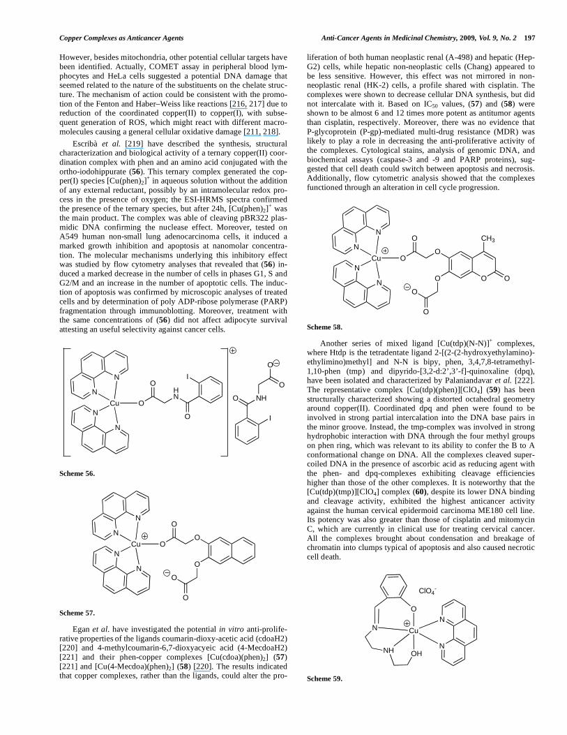
However, besides mitochondria, other potential cellular targets have been identified. Actually, COMET assay in peripheral blood lym-phocytes and HeLa cells suggested a potential DNA damage that seemed related to the nature of the substituents on the chelate struc-ture. The mechanism of action could be consistent with the promo-tion of the Fenton and Haber–Weiss like reactions [216, 217] due to reduction of the coordinated copper(II) to copper(I), with subse-quent generation of ROS, which might react with different macro-molecules causing a general cellular oxidative damage [211, 218].
Escribà et al. [219] have described the synthesis, structural characterization and biological activity of a ternary copper(II) coor-dination complex with phen and an amino acid conjugated with the ortho-iodohippurate (56). This ternary complex generated the cop-per(I) species [Cu(phen)2]
+ in aqueous solution without the addition
of any external reductant, possibly by an intramolecular redox pro- cess in the presence of oxygen; the ESI-HRMS spectra confirmed the presence of the ternary species, but after 24h, [Cu(phen)2]
+ was
the main product. The complex was able of cleaving pBR322 plas-midic DNA confirming the nuclease effect. Moreover, tested on A549 human non-small lung adenocarcinoma cells, it induced a marked growth inhibition and apoptosis at nanomolar concentra-tion. The molecular mechanisms underlying this inhibitory effect was studied by flow cytometry analyses that revealed that (56) in-duced a marked decrease in the number of cells in phases G1, S and G2/M and an increase in the number of apoptotic cells. The induc-tion of apoptosis was confirmed by microscopic analyses of treated cells and by determination of poly ADP-ribose polymerase (PARP) fragmentation through immunoblotting. Moreover, treatment with the same concentrations of (56) did not affect adipocyte survival attesting an useful selectivity against cancer cells.
N
N
N
N
Cu
I
HN
O
O
O
I
NHO
O
O
Scheme 56.
N
N
N
N
CuO
O
O
O
O
O
Scheme 57.
Egan et al. have investigated the potential in vitro anti-prolife- rative properties of the ligands coumarin-dioxy-acetic acid (cdoaH2) [220] and 4-methylcoumarin-6,7-dioxyacyeic acid (4-MecdoaH2) [221] and their phen-copper complexes [Cu(cdoa)(phen)2] (57) [221] and [Cu(4-Mecdoa)(phen)2] (58) [220]. The results indicated that copper complexes, rather than the ligands, could alter the pro-
liferation of both human neoplastic renal (A-498) and hepatic (Hep-G2) cells, while hepatic non-neoplastic cells (Chang) appeared to be less sensitive. However, this effect was not mirrored in non-neoplastic renal (HK-2) cells, a profile shared with cisplatin. The complexes were shown to decrease cellular DNA synthesis, but did not intercalate with it. Based on IC50 values, (57) and (58) were shown to be almost 6 and 12 times more potent as antitumor agents than cisplatin, respectively. Moreover, there was no evidence that P-glycoprotein (P-gp)-mediated multi-drug resistance (MDR) was likely to play a role in decreasing the anti-proliferative activity of the complexes. Cytological stains, analysis of genomic DNA, and biochemical assays (caspase-3 and -9 and PARP proteins), sug-gested that cell death could switch between apoptosis and necrosis. Additionally, flow cytometric analysis showed that the complexes functioned through an alteration in cell cycle progression.
N
N
N
N
CuO
O
O
OO
O
O
CH3
O
Scheme 58.
Another series of mixed ligand [Cu(tdp)(N-N)]+ complexes,
where Htdp is the tetradentate ligand 2-[(2-(2-hydroxyethylamino)-ethylimino)methyl] and N-N is bipy, phen, 3,4,7,8-tetramethyl-1,10-phen (tmp) and dipyrido-[3,2-d:2’,3’-f]-quinoxaline (dpq), have been isolated and characterized by Palaniandavar et al. [222]. The representative complex [Cu(tdp)(phen)][ClO4] (59) has been structurally characterized showing a distorted octahedral geometry around copper(II). Coordinated dpq and phen were found to be involved in strong partial intercalation into the DNA base pairs in the minor groove. Instead, the tmp-complex was involved in strong hydrophobic interaction with DNA through the four methyl groups on phen ring, which was relevant to its ability to confer the B to A conformational change on DNA. All the complexes cleaved super-coiled DNA in the presence of ascorbic acid as reducing agent with the phen- and dpq-complexes exhibiting cleavage efficiencies higher than those of the other complexes. It is noteworthy that the [Cu(tdp)(tmp)][ClO4] complex (60), despite its lower DNA binding and cleavage activity, exhibited the highest anticancer activity against the human cervical epidermoid carcinoma ME180 cell line. Its potency was also greater than those of cisplatin and mitomycin C, which are currently in clinical use for treating cervical cancer. All the complexes brought about condensation and breakage of chromatin into clumps typical of apoptosis and also caused necrotic cell death.
N
N
Cu
O
N
NHOH
ClO4-
Scheme 59.

198 Anti-Cancer Agents in Medicinal Chemistry, 2009, Vol. 9, No. 2 Marzano et al.
N
N
Cu
O
N
NHOH
ClO4-
CH3
CH3
CH3
CH3
Scheme 60.
5.5. Phosphine Complexes
Auranofin (a tetraacetyl thioglucose derivative of triethyl phosphine gold(I)) has been the first metal phosphine complex in-troduced into clinical use to treat rheumatoid arthritis [223, 224]. Moreover, a numer of studies have shown that auranofin elicited potent cytotoxic activity against both cell lines in vitro and antitu-mor activity against murine leukaemia in vivo [218]. These obser-vations have represented the starting point for a series of investiga-tions focused on the biological chemistry and anticancer properties of phosphines and their related metal complexes.
Among these
compounds, extensive research performed by Berners-Price, Sadler and coworkers in the eighties (to which readers are addressed for a comprehensive view of the matter) [218, 225, 226] have established that bisaryldiphosphine (P-P) metal complexes of the type [M(P-P)2][Cl] (M = Au, Ag and Cu; P-P = dppe: 1,2-bis(diphenylphosphi-no)ethane, dppp: 1,2-bis(diphenylphosphino)propane, dppey: 1,2-bis(diphenylphosphino)ethylene) showed cytotoxic activity against P388 murine leukemia [227], B16 melanoma and M5076 reticulum cell sarcoma [218].
For example, the activity of [Cu(dppey)2][Cl]
and [Cu(dppp)2][Cl] was comparable to that exhibited by the free ligands, but al least 20-fold more potent than those shown by Au(I) analogues, suggesting that particular metal ions (Cu
1+/2+ and Au
1+/3+
but not Mg2+
, Zn2+
, Mn2+
, Fe2+
, Co2+
and Cd2+
) regulated (or better potentiated) the cytotoxic properties of arylphosphines [228]. Within this view, as suggested by the authors, such metal ions sur-rounded by chelate phosphines protected the ligand from oxidation and promoted their uptake into cells depending on the lipophilicity of the complex. In other words, the metal ion basically operated as a simple carrier to deliver arylphosphines, the actual supposed cyto-toxic agent. Despite considerable efforts carried out in this field, the presence of several phenyl groups appended to the phosphorus do-nors in bisaryldiphosphine copper and gold compounds caused undesired nephrotoxicity [218]
and cardiovascular toxicity
[229] in
animal models, respectively, thus precluding clinical trials in hu-
mans. The effect of exchanging phenyl substituents with more physiological ethyl groups in [M(eppe)2][Cl] species (eppe = 1-(diethylphosphino),2-(diphenylphosphino)ethane) was, unlikely, to lower significantly the cytotoxic activity [227]. It should be stressed that poor water solubility represents a major problem which makes the formulation for clinical trials difficult. Therefore, many efforts have been focused to the design of more hydrophilic copper(I) de-rivatives as in the case of the partial substitution of the ‘CuP4‘ aryl-phosphine coordination sphere with heterocyclic thiones [230], acetonitrile [231]
and N-heterocycles such as carbazole and ben-
zotriazole producing ‘mixed-ligand’ type compounds. Among these derivatives, [Cu2(dppe)3(CH3CN)2][ClO4]2 (61) exhibited a potent in vitro cytotoxicity in H460 human lung carcinoma cells, compa-rable to that displayed by adriamycin [231].
Mechanistic studies revealed that (61) damaged DNA in vitro (COMET assay) and activated the p53 pathway eventually inhibit-ing the growth of cancer cells by inducing cell cycle arrest and apoptosis, as assessed by flow cytometry studies. The authors also
showed that simultaneous addition of (61) and of the DNA interca-lating agent adriamycin increased the cytotoxicity of either com-pounds, suggesting the potential use of adriamycin with (61) in combination therapy. Evidences based on DNA binding and simula-tion studies indicated an increased binding of adriamycin to DNA modified by (61), that was presumed to occur through the release of labile acetonitrile and formation of stable complexes with guanine residues, thus providing a plausible mechanism for synergistic ac-tion between adriamycin and (61) [231].
Following a similar ‘mixed-ligand’ approach, several N2-scor- pionate ligands have been attached to a coordination vacant lipo-philic ‘CuP2’ moiety (P2 = bidentate dppe or two monodentate aryl-phosphines) [147]. The use of bis(nitrotriazolyl)borate [H2B(tz
NO2)2]
scorpionate co-ligands had the double aim at increasing the water solubility and the kinetic inertness of the resulting mixed-complex. These less hydrophobic copper(I) {[H2B(tz
NO2)2]Cu[(PR3)2]} com-
pounds (R = m-tolyl, 62a; p-fluorophenyl, 62b) were proved to be easier to handle during the in vitro tests, and they retained cytotoxic activity against a panel of human tumor cell lines including exam-ples of ovarian (2008), cervix (A431), and lung (A549) cancer, melanoma (A375) and leukemia (HL60). IC50 values indicated that (62a) and (62b) were appreciably more effective against all cell lines compared to the reference metallodrug cisplatin, and that free phosphine ligands presented IC50 values at least 15-fold higher. In particular, on lung adenocarcinoma (A549) cells, with notoriously poor chemosensisivity to cisplatin, [232] cytotoxicities shown by complexes (62a) and (62b) exceeded that of cisplatin by a factor of about 20 [147].
Cu
PPh
P PPh
P Ph
Cu
Ph Ph
Ph
Ph
Ph
P P
Ph Ph
PhPh
NC-CH3
ClO4
H3C-CN
ClO4
Scheme 61.
N
N
N
B
NN
N
H
HCu
PR3
NO2
NO2
PR3
a) R = m-tolyl
b) R = p-fluorophenyl
Scheme 62.
More recently, the substitution of arylphosphines with hydro-philic tris(hydroxymethyl)phosphine (thp) allowed the preparation of more hydrophilic species stable in aqueous media with improved cytotoxic properties [148]. In particular, the complex {[HC(CO2) (pz
Me2)2]Cu(thp)2} (63) was found to possess antiproliferative activ-
ity against different human tumor cell lines (HL60, A549, MCF-7, A375 and LoVo) markedly higher than that shown by cisplatin. Chemosensitivity tests performed on cisplatin sensitive and resis-tant human cancer cell lines (ovarian cancer 2008 vs C13* and cervix cancer A431 vs A431/Pt) established that (scorpionate)Cu (thp)2 type compounds were able to overcome cisplatin resistance, supporting the hypothesis of a different mechanism of action com-pared to that exhibited by the reference drug. Flow cytometry

Copper Complexes as Anticancer Agents Anti-Cancer Agents in Medicinal Chemistry, 2009, Vol. 9, No. 2 199
NN
B
NNH
HCu
P
CH3
CH3
P
OH
OH
OH
OH
OH
OH
H3C
H3C
Scheme 63.
analysis on 2008 human ovarian carcinoma cells showed that (63) induced a marked enlarge of both cell size and granularity and a significant increase in the fraction of G2/M cells that was not ac-companied by the appearance of a relevant sub-G1 fraction. Be-sides, no evidence of caspase-3 activation was detected. Marzano et al. hypothesized that the cytotoxic activity of these copper(I) com-plexes might be correlated to their ability to trigger paraptosis, a non-apoptotic mechanism of cell death [147].
Cu
P
P P
P
PF6
OHHOHO
HO
OH OH
OH
OH
HO
HO OH
OH
Scheme 64.
Cu
P
P P
PPF6
OHHOHO
HO
OH OH
OH
OH
Scheme 65.
Further mechanistic information were provided by the design of two additional water soluble complexes featuring the ‘CuP4’ coor-dination sphere, namely [Cu(thp)4][PF6] (64) and [Cu(bhpe)2][PF6] (65) (bhpe = bis[bis(hydroxymethyl)phosphino]ethane) [156]. Physico-chemical characterization indicated that they possessed a tetrahedral geometry both in the solid state [233] and in the solution state [147]. ESI-MS data supported the view of an extremely stable substitution-inert entity in the case of (65) showing the molecular ion peak without detectable fragmentation, whereas (64) exhibited some lability showing the ‘CuP4’ molecular ion along with the ‘CuP3’ and ‘CuP2’ molecular fragments at higher concentrations. This remarkable different behaviour in the ion trap had a major impact on the cytotoxic profile of these two species. Complex (65) showed negligible in vitro antitumor activity suggesting that a marked kinetic inertness of the copper species is not a primary re-quirement. On the contrary, it appeared that, as already shown by other cytotoxic copper complexes, labile sites at copper are neces-sary for the pro-drug to display its antiproliferative action. Hence, (64) showed a potent cytotoxic activity on the same panel of tumor cells with IC50 values even over 30 times lower that those obtained with cisplatin; moreover, it overcame cisplatin resistance. Different short-term proliferation assays measuring the damage to various subcellular organelles suggested that lysosomal damage represents the early cellular event associated with (64) cytotoxicity, probably
mediated by an increased production of reactive oxygen species. Cytological stains and flow cytometric analyses indicated that this phosphine copper(I) complex is able to inhibit the growth of tumor cells via G2/M cell cycle arrest and paraptosis accompanied with loss of mitochondrial transmembrane potential.
It is interesting to note that a similar cytotoxic profile was ob-served in HT1080 human fibrosarcoma cells treated with a Cu(II) thioxotriazole complex (47) [152]. Biological data support the view that both Cu(I) phosphine and Cu(II) thioxotriazole complexes behave in the same way, the only difference being the associated IC50 values (in the micromolar range for Cu(II) compared to the sub micromolar range for Cu(I) compounds). This behaviour could be correlated with the more favourable cell trafficking of Cu(I) species [152].
5.6. Miscellaneous Complexes
The sequestering agent 1,2-propylenediamine-N,N,N,N-tetra- acetic acid (PDTA) has been proposed as a good example of a polycarboxylic ligand forming active metal complexes against sev-eral human tumour cells [234-237]. It is well known that Cu(II) complexes with chelating ligands play an important role as models of the associative complexes involved in the substitution reactions at the copper site of SOD enzymes; indeed, strongly donor ligands as PDTA should favour the formation of six-coordinate copper complexes [238]. Based on these arguments, an octahedral mono-meric compound [Cu(PDTA-H2)(H2O)2]
.H2O was synthetized by
Kamah et al. [239]. Potentiometric and electronic measurements identified the ligand as tetradentate, two nitrogen and two oxygen atoms being bonded to the Cu(II) in planar positions. The antican-cer properties of this complex have been evaluated against ovarian tumour TG cell line and solid tumour Sarcoma 180 [239]. The re-sults attested a significant in vitro antitumor activity (ID50 = 2.29 μM at 48h) and an important in vivo antitumor activity with com-plete regression of the tumor at a dose of 12.5 mg/kg body weight.
Quinolones, a commonly used term for the quinolonecarboxylic acids or 4-quinolones, are a group of synthetic antibacterial agents containing the 4-oxo-1,4-dihydroquinoline skeleton. The interaction of diverse metal ions with quinolones has been thoroughly studied [240]. Biological properties of quinolones have been monitored with regards to their interaction with DNA [241, 242], cytotoxicity [243-245] and potential antitumor activity [246, 247]. Hpr-norflox- acin is the N-propyl protected form of norfloxacin and is a typical quinolone that can act as a bidentate ligand through the pyridine oxygen and one carboxylate oxygen atom [240]. The interaction of Cu(II) with Hpr-norfloxacin in the presence of bipy to give com-plex (66) has been studied in an attempt to examine the mode of binding and possible synergistic effects [248]. Complex (66) showed a concentration dependent antiproliferative effect as well as a time- and concentration-dependent necrotic effect on both human
NO
O
O
F N
N
Cu
N
N
Cl
Scheme 66.

200 Anti-Cancer Agents in Medicinal Chemistry, 2009, Vol. 9, No. 2 Marzano et al.
HL-60 promyelocytic leukemia and chronic myelogenous leukemia K562 cells.
Sparfloxacin is the first aminodifluoroquinolone currently available in Japan and United States for clinical use as antibacterial agent [249]. Padhye et al. [250] described the synthesis, characteri-zation and antiproliferative activity against hormone independent BT20 breast cancer cell line of the sparfloxacin mixed-ligand cop-per complexes, with appended ancillary ligands such as bipy, phen and 4,5-diazafluoren-9-one. Amongst the compounds examined, the dimeric copper compound (67) was the most potent derivative. The authors suggested that the facile Cu
2+/Cu
+ redox couple might act as
redox trigger for activating apoptotic processes [250].
Polyamine complexes with pyrrole and others arms substituents have been synthesized and characterized by Yang et al. [251]. The structure of (68) with the N1,N8-bis(1-methyl-4-nitropyrrole-2-carbonyl)triethylenetetramine ligand was investigated with theoreti-cal DFT calculations, and the DNA-binding of the Cu(II) complex and its effects on tumor cell viability were examined. The results showed that the mode of binding of the complex to DNA was the classical intercalation; moreover, (68) efficiently cleaved plasmid pBR322 DNA suggesting that it worked as a new kind of chemical nuclease. At 1 μM concentration, (68) induced a 34% reduction of human QBC-939 biliary tract carcinoma cell viability.
NH
Cu
NH
HN
O
NH
O
N N
H3C CH3
NO2 O2N
Cl
Cl
Scheme 68.
Diethylentetramine (dien), a biologically occurring substance, represents an excellent complexating reagent, capable of coordinat-ing to a number of transition metal ions, including Cu(II). Bolos et
al. [252] have synthetized some mixed-ligand copper complexes with dien and thiazole or imidazole derivatives that showed antipro-liferative activity against a panel of human cancer cells. In particu-lar, the complex (69), with the 2-amino-5-methylthiazole ligand, was cytotoxic against human cervical HeLa, breast T47D, colon HT-29 cancer cells (IC50 values ranging from 41 to 131 μM), whereas normal fibroblasts were not affected. The absence of apop-totic pattern and a strong inhibition of cell cycle progression as well as of DNA synthesis were detected in tumor treated cells. The authors suggested that G2/M cell cycle arrest could be related to p34cdc2 inhibition by tyrosine phosphorylation and/or to induction of cyclin-dependent kinase inhibitor p21
WAF1.
N
Cu
N
N
N
S
N
CH3
N
O
O
O
NO3
HHH
H
H
H
H
Scheme 69.
NHNH
NHCu
Br
Br
Scheme 70.
Medvetz et al. [253] have reported that the copper(II) complex (70) with the 1,4,7-triazacyclononane ligand, possessed an antican-cer activity against the human ovarian NuTu-19 cancer cell line comparable with that shown by cisplatin.
4-Nitropyridine N-oxide and its methyl derivatives represent an important group of compounds due to their potential biological applications as radiosensitizers, cytotoxins, antifungal, antibacterial, anti-inflammatory and tranquilizing substances [254]. It was found that the degree of electron flow between the para positioned elec-
N
O
O
O
F
NNH
F
NH2
N
OO
O
F
N
N
H
F
H2N
Cu
N
O
O
O
F
N N H
F
H2N
N
O O
O
F
N
N
H
F
NH2
Cu
Scheme 67.

Copper Complexes as Anticancer Agents Anti-Cancer Agents in Medicinal Chemistry, 2009, Vol. 9, No. 2 201
tron withdrawing (NO2) and electrodonating (NO) groups is strongly dependent upon the nature of ring substituents [255]. For example, the introduction of one or more methyl groups may mod-ify hydrophobicity with consequences for molecule transport through the cellular membrane [254]. The N-oxide group is known to be a binding site to metal centers, including copper. In vitro stud-ies on the antiproliferative activity of copper complexes containing various methyl substituted 4-nitropyridine N-oxides showed a sig-nificant sensitivity of human MCF-7 breast cancer cells depending upon the number and position of the methyl groups.
Several heterocyclic N-oxides and related compounds such as quinoxaline N-dioxide derivatives can be reduced in hypoxic condi-tions forming cytotoxic species having DNA as the main biological target [256-258]. Recently, Torre el al. [259-261] have developed some copper(II) complexes as selective hypoxic cytotoxins trying to improve the bioavailability and the pharmacological and toxico-logical properties of the 3-aminoquinoxaline-2-carbonitrile N1,N4-dioxide (AQCD) derivatives, (71a) and (71b), which have proved to act as bioreductive compounds [259-261]. In these first series, the cytotoxicity in hypoxia of copper(II) complexes was similar or slightly better than that of the free ligands whereas, under well-oxygenated conditions, the complexes were almost non cytotoxic [261, 262]. Subsequently, a new series of Cu(II) complexes with other AQCD derivatives were synthesized and characterized by different spectroscopic methods [263].
N
N
O
O
CN
NH2
R1
R2
a) R1 = Cl, R2 = Cl
b) R1 = Br, R2 = Br
c) R1 = F, R2 = Cl
d) R1 = H, R2 = H
e) R1 = Cl, R2 = OMe
Scheme 71.
The hypoxic selective cytotoxicity towards V79 cells and the SOD like activity of the complexes were determined and related to physicochemical properties. In particular, the copper(II) complex with (71c) showed cytotoxic selectivity in hypoxia being the most lipophilic compound of the series. On the contrary, the complex [Cu–(71d)] was cytotoxic but not selective and [Cu–(71e)] was not cytotoxic towards V79 cells neither in oxia nor in hypoxia, proba-bly due to its insolubility or its low lipophilicity. In addition, the potency for the hypoxic selective complex (72) and the previously reported complexes [Cu–(71a)] and [Cu–(71b)] [259-261] could be correlated with the sum of Hammett m constants, showing that
compounds with better hypoxic potency have higher values due to the halogen substituents in aromatic rings.
Hydroxamic acids (HAs) are important bioligands [264] in-volved in numerous biological processes including metal-ion trans-port and inhibition of metalloenzymes [265, 266]. Several HAs compounds, including benzo-hydroxamic acid HBHA and substi-tuted-HBHA analogues [267], and phthal-hydroxamic acid HPHA have been synthesized and tested for their antitumour properties, most of them showing significant cytotoxic activity. Furthermore, complexes of the type [Cu(BHA)2] were isolated and evaluated not only for their efficacy in tumour cell growth inhibition and en-hancement of longevity in tumour-bearing mice, but also as correc-tors of the perturbed biological parameters produced by the action of oxygen free radicals [268]. Treatment of Ehrlich ascitic carci-noma bearing mice with [Cu(BHA)2] (25 mg/kg, ip dose/day, 4 days) resulted in a significant cancer growth inhibition (65%) and increase of tumour-bearing mice life span (150%). Additionally, the early perturbed haematological parameters were totally restored by the host [269].
Wang et al. [270] have reported the self-assembly synthesis of the waterwheel-like dimetal HPHA complex (73) appended with four free hydroxamic acid groups. Waterwheel-like dimetal copper and zinc complexes were evaluated for their in vitro cytotoxic ac-tivity against SMMC-7721 and HO-8910 cancer cells. The relative cytotoxicities of copper and zinc compounds were similar and more predominant than those shown by HPHA (IC50: Cu(II) > Zn(II) >> HPHA or Cu(CH3COO)2
.2H2O). Copper complex was 40 and 11-
fold more potent than HPHA toward SMMC-7721 and HO-8910 cells, respectively. It was proposed that these Cu compounds might induce cell death through necrosis pathways.
Choudhuri et al. [271] have described the effect of oxalyl bis(N-phenyl) hydroxamic acid and the metal chelate copper[N-(2-hydroxy-acetophenone)glycinate] (74) [272] on multidrug-resistant P-gp-expressing CEM/ADR5000 T-cell acute lymphoblastic leu-kemia cells. Compound (74) was found to overcome doxorubicin resistance by depleting GSH, by inhibiting GST through formation of (74)-GSH conjugates. Complex (74) also modulated P-gp-mediated drug resistance.
Copper(II) complexes with NSAIDs belonging to the oxicam group, viz piroxicam and meloxicam, have been described [273]. Piroxicam and meloxicam exhibited anticancer properties in various cancer cell lines and in animal models [274-278].
In a recent work by Cini et al. [279], the in vitro cytotoxic ac-tivity of copper(II) piroxicam (75) was tested against a panel of about 50 human tumor cell lines. The highest activity was relevant
Scheme 72.
N NO O
CNNH
ClF
NN OO
NC HN
Cl F
N
N
O
O
CN
HN
Cl
F
N
N
O
O
CN
HN
Cl
F
Cu Cu

202 Anti-Cancer Agents in Medicinal Chemistry, 2009, Vol. 9, No. 2 Marzano et al.
to small lung, non-small lung, central nervous system, melanoma, ovarian and renal cell lines. The growth inhibition values for many of these cells were significantly smaller (ca. 20 μM) than those found for carboplatin that was selected as reference drug [279]. Results from studies on oxygen radical scavenger activity by the octahedral (75) complex [280-282] showed that it was a better scavenger than copper(II) ion and free piroxicam [283].
O O
O O
O
O
Cu
Cu O
O
NH
HO
NH
OH
HN
OH
O
O
HN
HO
O
OH2O
OH2
Scheme 73.
O
CH3
N
Cu
O
O
H2O OH2
OH2
Scheme 74.
S
N
O
HN
O
CH3
O
O
S
N
O
NH
O
H3C
O
O
Cu
H3C
N
HC
CH3
O
N
N
CH3
N
CH
H3C
O
Scheme 75.
6. CONCLUDING REMARKS
Although copper has a long history of medical application, coordination compounds of copper(I,II) have been investigated as potential antiproliferative agents only in the last few decades, par-ticularly after the discovery of cisplatin, the most widely used anti-cancer metallodrug. Copper, as an essential cofactor in a number of enzymes and physiological processes, may be less toxic than non essential metals, such as platinum. Up to now, a great variety of copper complexes have been tested as cytotoxic agents and found to be endowed with an antitumor activity in several in vitro tests (on cultured cancer cell lines) and few in vivo experiments (on murine tumor models). According to the well established coordination chemistry of copper, frequently enriched by the flexible Cu(I/II)
redox behaviour, there still exist an enormous prospect in the design of more potent and less toxic copper based antiproliferative drugs.
This review have summarized developments in this field re-ported in the literature in the last ten years starting from the histori-cal class of copper-TSC compounds, the antitumor activity of which has been reported as early as 1960s. Copper complexes con-taining polydentate Schiff base ligands, five-membered aromatic heterocycles such as imidazoles, pyrazoles and triazoles, six-membered aromatic heterocycles (phen and bipy), and tertiary phosphines complete the survey which ends with a miscellaneous ligands chapter.
Most of the investigated compounds belong to the family of copper(II) complexes showing either five-coordinate environments comprising distorted square pyramidal and trigonal bipyramidal geometries or distorted six-coordinate octahedral arrays. The major-ity of these agents are mononuclear species, but there are few dis-tinctive examples of dimeric compounds exhibiting remarkable antitumor activity. Not many, but noteworthy examples of tetrahe-dral copper(I) complexes having soft tertiary phosphines or aro-matic amines (e.g. phen, bipy) as donor ligands, display antiprolif-erative activity in the sub-micromolar range.
Among various synthetic strategies aiming at the enhancement of the antitumor efficacy, it has to be mentioned the attempt made to improve the hydrophilic character of copper compounds in order to get a suitable solubility in physiological conditions, thus enhanc-ing the bioavailability and reducing the in vivo toxicity.
In general, attempts to correlate the antitumor activity of copper compounds with copper oxidation state, coordination number and geometry is matter of debate and correlations, if exist, are far to be demonstrated. Nevertheless, according to the fact that copper trans-port machinery at the plasma membrane in mammalian cells is driven primarily by the hCtr1 system, a specific monovalent metal transporter, the intracellular trafficking of copper(I) species might be favoured with respect to the internalization of copper(II) com-pounds.
Copper complexes were initially tested with a vision toward behavior analogous to cisplatin, and, consequently, their interaction with nucleic acids were largely investigated. Taken all together, the results of these studies seem to suggest that DNA does not represent the main and/or unique biological target for this class of metal complexes. With the time it has become clear that copper com-pounds can act by different mechanisms. Indeed, besides DNA binding, intercalation and cleavage activity, SOD mimetic activity, as well as generation of ROS by redox-cycling, that both lead to an oxidative cell damage, have been described. Generally, these intra-cellular molecular events trigger cancer cell death through an apop-totic mechanism. Moreover, down regulation of antiapoptotic pro-teins (Bcl-2 and Bcl-XL) has been shown on human cancer cells treated with copper compounds. On the other hand, more recently, a non apoptotic form of programmed cell death (paraptosis) has been evidenced in human cancer cells treated with both copper(I) and copper(II) complexes. This observation is very stimulating because it suggests the ability of triggering cell death even in cancer cells with multiple defects in the normal apoptotic pathway as, for in-stance, cisplatin resistant cells.
Although recent studies are trying to obtain more mechanistic information, it is evident that, up to now, very little understanding of the molecular basis of the mechanisms underlying the action of these classes of copper compounds is available. Efforts in this di-rection could provide additional information useful for the design of ‘second generation’ copper drugs having better selectivity and im-proved antitumor efficiency.
ACKNOWLEDGMENTS
This work was financially supported by University of Padova (Progetto di Ateneo CPDA065113/06), by University of Camerino

Copper Complexes as Anticancer Agents Anti-Cancer Agents in Medicinal Chemistry, 2009, Vol. 9, No. 2 203
(FAR 2007), and by Ministero dell’Istruzione dell’Università e della Ricerca (PRIN 2007). We are grateful to CIRCMSB (Consor-zio Interuniversitario di Ricerca in Chimica dei Metalli nei Sistemi Biologici).
ABBREVIATIONS
AQCD = 3-Aminoquinoxaline-2-carbonitrile N1,N4-dioxide
Bipy = 2,2’-Bipyridine
BZA = benzoic acid
Cas = Casiopeinas
cisplatin = cis-Diamminedichloroplatinum(II)
COX-2 = Cyclooxygenase-2
Dien = Diethylentetramine
DTC = Dithiocarbamate
ESI-HRMS = Electrospray ionization–high resolution mass spectrometry
GSH = Glutathione
HAs = Hydroxamic acid
HBHA = Benzo-hydroxamic acid
HPHA = Phthal-hydroxamic acid
HTBZ = 2-(4'-Thiazolyl)benzimidazole
IC50 = Concentration affording 50% of inhibition
MDR = Multi-drug resistance
MI = Mitotic index
MT = Metallothionein
NMR = Nuclear magnetic resonance
NSAIDs = Non-steroidal anti-inflammatory drugs
PARP = Poly ADP-ribose polymerase
PCD = Programmed cell death
P-gp = P-Glycoprotein
Phen = 1,10-Phenantroline
PRIs = Proliferation rate indexes
PYBZ = 2-(2-Pyridyl)benzimidazole
ROS = Reactive oxygen species
SARs = Structure-activity relationships
SCEs = Sister chromatid exchanges
SOD = Superoxide dismutase
TM = Tetrathiomolybdate
TSC = Thiosemicarbazone
VEGF = Vascular endothelial growth factor
REFERENCES
[1] Hambley, T.W. Metal-Based Therapeutics. Science, 2007, 318
(5855), 1392-1393.
[2] Sigel, H. Metal Ions in Biological Systems. M. Dekker, Inc.: New
York, 1980; Vol. II.
[3] Kepler, B.K. Metal Complexes in Cancer Chemotherapy. VCH,
Weinheim, 1993.
[4] Guo, Z.; Sadler, P.J. Metals in medicine. Angew. Chem. Int. Ed.
Engl., 1999, 38(11), 1512-1531.
[5] Gielen, M.; Tiekink, E.R.T. Metallotherapeutic Drugs and Metal-
Based Diagnostic Agents, The Use of Metals in Medicine. Wiley:
Chichester, 2005.
[6] Zhang, C.X.; Lippard, S.J. New metal complexes as potential
therapeutics. Curr. Opin. Chem. Biol., 2003, 7(4), 481-489.
[7] Sessler, J.L.; Doctrow, S.R.; McMurry, T.J.; Lippard, S.J. Medici-
nal Inorganic Chemistry. ACS: Washington, D.C., 2003.
[8] Kelland, L.R. Preclinical perspectives on platinum resistance.
Drugs, 2000, 59, 1-8.
[9] Petering, D.H. Carcinostatic copper complexes. Met. Ions Biol.
Syst., 1980, 11, 197-229.
[10] Frausto Da Silva, J.J.R.; Williams, R.J.P. The Biological Chemistry
of the Elements. Clarendon: Oxford, 1991.
[11] Linder, M.C. Biochemistry of Copper. Plenum Press: New York,
1991.
[12] Halliwell, B.; Gutteridge, J.M. Role of free radicals and catalytic
metal ions in human disease: an overview. Methods Enzymol.,
1990, 186, 1-85.
[13] Aust, S.D.; Morehouse, L.A.; Thomas, C.E. Role of metals in oxy-
gen radical reactions. J. Free Radic. Biol. Med., 1985, 1(1), 3-25.
[14] Turnlund, J.R.; Keyes, W.R.; Anderson, H.L.; Acord, L.L. Copper
absorption and retention in young men at three levels of dietary
copper by use of the stable isotope copper-65. Am. J. Clin. Nutr.,
1989, 49(5), 870-878.
[15] Wang, T.; Guo, Z.J. Copper in medicine: Homeostasis, chelation
therapy and antitumor drug design. Curr. Med. Chem., 2006, 13(5),
525-537.
[16] Daniel, K.G.; Harbach, R.H.; Guida, W.C.; Dou, Q.P. Copper
storage diseases: Menkes, Wilson's, and cancer. Front. Biosci.,
2004, 9, 2652-2662.
[17] Goodman, V.L.; Brewer, G.J.; Merajver, S.D. Copper deficiency as
an anti-cancer strategy. Endocr.-Relat. Cancer, 2004, 11(2), 255-
263.
[18] Omae, I. Organotin antifouling paints and their alternatives. Appl.
Organomet. Chem., 2003, 17(2), 81-105.
[19] Theophanides, T.; Anastassopoulou, J. Copper and carcinogenesis.
Crit. Rev. Oncol. Hematol., 2002, 42(1), 57-64.
[20] Burkitt, M.J. Copper-DNA adducts. Methods Enzymol., 1994, 234,
66-79.
[21] Kagawa, T.F.; Geierstanger, B.H.; Wang, A.H.J.; Ho, P.S. Cova-
lent modification of guanine bases in double-stranded DNA. The
1.2-.ANG. Z-DNA structure of d(CGCGCG) in the presence of
copper(II) chloride. J. Biol. Chem., 1991, 266(30), 20175-20184.
[22] Frausto Da Silva, J.J.R.; Williams, R.J.P. The biological chemistry
of elements. The inorganic chemistry of life. Oxford: Clarendon
Press, 1994.
[23] Koppenol, W.H. The Haber-Weiss cycle - 70 years later. Redox
Rep., 2001, 6(4), 229-234.
[24] Galaris, D.; Evangelou, A. The role of oxidative stress in mecha-
nisms of metal-induced carcinogenesis. Crit. Rev. Oncol. Hematol.,
2002, 42(1), 93-103.
[25] Valko, M.; Morris, H.; Cronin, M.T.D. Metals, toxicity and oxida-
tive stress. Curr. Med. Chem., 2005, 12(10), 1161-1208.
[26] Safaei, R.; Holzer, A.K.; Katano, K.; Samimi, G.; Howell, S.B. The
role of copper transporters in the development of resistance to Pt
drugs. J. Inorg. Biochem., 2004, 98(10), 1607-1613.
[27] Laussac, J.P.; Sarker, B. Characterization of the copper(II) and
nickel(II) transport site of human serum albumin. Studies of cop-
per(II) and nickel(II) binding to peptide 1-24 of human serum al-
bumin by carbon-13 and proton NMR spectroscopy. Biochemistry,
1984, 23(12), 2832-2838.
[28] Lau, S.J.; Sarkar, B. Ternary coordination complex between human
serum albumin, copper (II), and L-histidine. J. Biol. Chem., 1971,
246(19), 5938-5943.
[29] Tabata, M.; Sarkar, B. Kinetic mechanism of copper(II)-transfer
between the native sequence peptide representing the copper(II)-
transport site of human serum albumin and L-histidine. Can. J.
Chem., 1985, 63(11), 3111-3116.
[30] Lee, J.; Prohaska, J.R.; Thiele, D.J. Essential role for mammalian
copper transporter Ctr1 in copper homeostasis and embryonic de-
velopment. Proc. Natl. Acad. Sci. USA, 2001, 98(12), 6842-6847.
[31] Lee, J.; Petris, M.J.; Thiele, D.J. Characterization of mouse embry-
onic cells deficient in the Ctr1 high affinity copper transporter. J.
Biol. Chem., 2002, 277(43), 40253-40259.
[32] Freedman, J.H.; Ciriolo, M.R.; Peisach, J. The role of glutathione
in copper metabolism and toxicity. J. Biol. Chem., 1989, 264(10),
5598-5605.

204 Anti-Cancer Agents in Medicinal Chemistry, 2009, Vol. 9, No. 2 Marzano et al.
[33] Ferreira, A.M.D.C.; Ciriolo, M.R.; Marcocci, L.; Rotilio, G. Cop-
per(I) transfer into metallothionein mediated by glutathione. Bio-
chem. J., 1993, 292(3), 673-676.
[34] Culotta, V.C.; Klomp, L.W.J.; Strain, J.; Casareno, R.L.B.; Krems,
B.; Gitlin, J.D. The copper chaperone for superoxide dismutase. J.
Biol. Chem., 1997, 272(38), 23469-23472.
[35] Murata, Y.; Kodama, H.; Abe, T.; Ishida, N.; Nishimura, M.;
Levinson, B.; Gitschier, J.; Packman, S. Mutation analysis and ex-
pression of the mottled gene in the macular mouse model of
Menkes disease. Pediatr. Res., 1997, 42(4), 436-442.
[36] Vulpe, C.; Levinson, B.; Whitney, S.; Packman, S.; Gitschier, J.
Isolation of a candidate gene for Menkes disease and evidence that
it encodes a copper-transporting ATPase. Nat. Genet., 1993, 3(1),
7-13.
[37] Tanzi, R.E.; Petrukhin, K.; Chernov, I.; Pellequer, J.L.; Wasco, W.;
Ross, B.; Romano, D.M.; Parano, E.; Pavone, L.; Brzustowicz,
L.M. The Wilson disease gene is a copper transporting ATPase
with homology to the Menkes disease gene. Nat. Genet., 1993,
5(4), 344-350.
[38] Parke, A.; Bhattacherjee, P.; Palmer, R.M.; Lazarus, N.R. Charac-
terization and quantification of copper sulfate-induced vasculariza-
tion of the rabbit cornea. Am. J. Pathol., 1988, 130(1), 173-178.
[39] Lowndes, S.A.; Harris, A.L. The role of copper in tumour angio-
genesis. J. Mammary Gland Biol. Neoplasia, 2005, 10(4), 299-310.
[40] Folkman, J. Tumor angiogenesis: therapeutic implications. N. Engl.
J. Med., 1971, 285(21), 1182-1186.
[41] Gradishar, W.J. An overview of clinical trials involving inhibitors
of angiogenesis and their mechanism of action. Invest. New Drugs,
1997, 15(1), 49-59.
[42] Brewer, G.J. Anticopper therapy against cancer and diseases of
inflammation and fibrosis. Drug Discov. Today, 2005, 10(16),
1103-1109.
[43] Yoshii, J.; Yoshiji, H.; Kuriyama, S.; Ikenaka, Y.; Noguchi, R.;
Okuda, H.; Tsujinoue, H.; Nakatani, T.; Kishida, H.; Nakae, D.;
Gomez, D.E.; De Lorenzo, M.S.; Tejera, A.M.; Fukui, H. The cop-
per-chelating agent, trientine, suppresses tumor development and
angiogenesis in the murine hepatocellular carcinoma cells. Int. J.
Cancer, 2001, 94(6), 768-773.
[44] Brem, S.S.; Zagzag, D.; Tsanaclis, A.M.C.; Gately, S.; Elkouby,
M.P.; Brien, S.E. Inhibition of angiogenesis and tumor growth in
the brain. Suppression of endothelial cell turnover by penicillamine
and the depletion of copper, an angiogenic cofactor. Am. J. Pathol.,
1990, 137(5), 1121-1142.
[45] Adams, J. Development of the proteasome inhibitor PS-341. On-
cologist, 2002, 7(1), 9-16.
[46] Dou, Q.P.; Goldfarb, R.H. Bortezomib Millennium Pharmaceuti-
cals. IDrugs, 2002, 5(8), 828-834.
[47] Daniel, K.G.; Chen, D.; Orlu, S.; Cui, Q.C.; Miller, F.R.; Dou, Q.P.
Clioquinol and pyrrolidine dithiocarbamate complex with copper to
form proteasome inhibitors and apoptosis inducers in human breast
cancer cells. Breast Cancer Res., 2005, 7(6), R897-R908.
[48] Daniel, K.G.; Chen, D.; Yan, B.; Dou, Q.P. Copper-binding com-
pounds as proteasome inhibitors and apoptosis inducers in human
cancer. Front. Biosci., 2007, 12, 135-144.
[49] Hathaway, B.J., In Comprehensive Coordination Chemistry, Wil-
kinson, G.; Gillard, R.D.; McCleverty, J.A., Eds. Pergamon Press:
Oxford (UK), 1987; Vol. 5: pp. 533-594.
[50] Mukherjee, R., Copper. In Comprehensive Coordination Chemistry
II - From Biology to Nanotechnology, McCleverty, J.A.; Meyer,
T.J., Eds. Elsevier Ltd.: Oxford (UK), 2004; Vol. 6: pp. 747-910.
[51] Kitajima, N.; Moro-oka, Y. Copper-Dioxygen Complexes. Inor-
ganic and bioinorganic perspectives. Chem. Rev., 1994, 94(3), 737-
757.
[52] Padhye, S.; Kauffman, G.B. Transition metal complexes of semi-
carbazones and thiosemicarbazones. Coord. Chem. Rev., 1985, 63,
127-160.
[53] Taylor, M.R.; Gabe, E.J.; Glusker, J.P.; Minkin, J.A.; Patterson,
A.L. The crystal structures of compounds with antitumor activity.
2-keto-3-ethoxybutyraldehyde bis(thiosemicarbazone) and its cu-
pric complex. J. Am. Chem. Soc., 1966, 88(8), 1845-1846.
[54] Crim, J.A.; Petering, H.G. The antitumor activity of Cu(II)KTS, the
copper (II) chelate of 3-ethoxy-2-oxobutyraldehyde bis(thiosemi-
carbazone). Cancer Res., 1967, 27(7), 1278-1285.
[55] Lovejoy, D.B.; Richardson, D.R. Novel "hybrid" iron chelators
derived from aroylhydrazones and thiosemicarbazones demonstrate
selective antiproliferative activity against tumor cells. Blood, 2002,
100(2), 666-676.
[56] Ming, L.-J. Structure and function of "metalloantibiotics". Med.
Res. Rev., 2003, 23(6), 697-762.
[57] Wolohan, P.; Yoo, J.; Welch, M.J.; Reichert, D.E. QSAR studies of
copper azamacrocycles and thiosemicarbazones: MM3 parameter
development and prediction of biological properties. J. Med.
Chem., 2005, 48(17), 5561-5569.
[58] Pogni, R.; Baratto, M.C.; Diaz, A.; Basosi, R. EPR characterization
of mono(thiosemicarbazones) copper(II) complexes. Note II. J.
Inorg. Biochem., 2000, 79(1-4), 333-337.
[59] Zhang, H.; Thomas, R.; Oupicky, D.; Peng, F. Synthesis and char-
acterization of new copper thiosemicarbazone complexes with an
ONNS quadridentate system: cell growth inhibition, S-phase cell
cycle arrest and proapoptotic activities on cisplatin-resistant neuro-
blastoma cells. J. Biol. Inorg. Chem., 2008, 13(1), 47-55.
[60] Feun, L.; Modiano, M.; Lee, K.; Mao, J.; Marini, A.; Savaraj, N.;
Plezia, P.; Almassian, B.; Colacino, E.; Fischer, J.; MacDonald, S.
Phase I and pharmacokinetic study of 3-aminopyridine-2-carbox-
aldehyde thiosemicarbazone (3-AP) using a single intravenous dose
schedule. Cancer Chemother. Pharmacol., 2002, 50(3), 223-229.
[61] Murugkar, A.; Unnikrishnan, B.; Padhye, S.; Bhonde, R.; Teat, S.;
Triantafillou, E.; Sinn, E. Hormone anchored metal complexes. 1.
Synthesis, structure, spectroscopy and in vitro antitumor activity of
testosterone acetate thiosemicarbazone and its metal complexes.
Met.-Based Drugs, 1999, 6(3), 177-182.
[62] Saryan, L.A.; Ankel, E.; Krishnamurti, C.; Petering, D.H.; Elford,
H. Comparative cytotoxic and biochemical effects of ligands and
metal complexes of alpha-N-heterocyclic carboxaldehyde thiose-
micarbazones. J. Med. Chem., 1979, 22(10), 1218-1221.
[63] Antholine, W.E.; Knight, J.M.; Petering, D.H. Inhibition of tumor
cell transplantability by iron and copper complexes of 5-substituted
2-formylpyridine thiosemicarbazones. J. Med. Chem., 1976, 19(2),
339-341.
[64] Booth, B.A.; Agrawal, K.C.; Moore, E.C.; Sartorelli, A.C. Alpha-
(N)-heterocyclic carboxaldehyde thiosemicarbazone inhibitors of
ribonucleoside diphosphate reductase. Cancer Res., 1974, 34(6),
1308-1314.
[65] Brockman, R.W.; Thomson, J.R.; Bell, M.J.; Skipper, H.E. Obser-
vations on the antileukemic activity of pyridine-2-carboxaldehyde
thiosemicarbazone and thiocarbohydrazone. Cancer Res., 1956,
16(2), 167-170.
[66] Scovill, J.P.; Klayman, D.L.; Franchino, C.F. 2-Acetylpyridine
thiosemicarbazones. 4. Complexes with transition metals as anti-
malarial and antileukemic agents. J. Med. Chem., 1982, 25(10),
1261-1264.
[67] Easmon, J.; Heinisch, G.; Holzer, W.; Rosenwirth, B. Synthesis
and antiviral activity of thiosemicarbazone derivatives of pyridazi-
necarbaldehydes and alkyl pyridazinyl ketones. Arzneim.-Forsch.,
1989, 39(10), 1196-1201.
[68] Easmon, J.; Heinisch, G.; Holzer, W.; Rosenwirth, B. Pyridazines.
63. Novel thiosemicarbazones derived from formyl- and acyldiazi-
nes: synthesis, effects on cell proliferation, and synergism with an-
tiviral agents. J. Med. Chem., 1992, 35(17), 3288-3296.
[69] Easmon, J.; Puerstinger, G.; Heinisch, G.; Roth, T.; Fiebig, H.H.;
Holzer, W.; Jaeger, W.; Jenny, M.; Hofmann, J. Synthesis, cytotox-
icity, and antitumor activity of copper(II) and iron(II) complexes of
4N-Azabicyclo[3.2.2]nonane thiosemicarbazones derived from acyl
diazines. J. Med. Chem., 2001, 44(13), 2164-2171.
[70] Garcia-Tojal, J.; Garcia-Orad, A.; Serra, J.L.; Pizarro, J.L.;
Lezama, L.; Arriortua, M.I.; Rojo, T. Synthesis and spectroscopic
properties of copper(II) complexes derived from thiophene-2-
carbaldehyde thiosemicarbazone. Structure and biological activity
of [Cu(C6H6N3S2)2]. J. Inorg. Biochem., 1999, 75(1), 45-54.
[71] Garcia-Tojal, J.; Pizarro, J.L.; Garcia-Orad, A.; Perez-Sanz, A.R.;
Ugalde, M.; Alvarez Diaz, A.; Serra, J.L.; Arriortua, M.I.; Rojo, T.
Biological activity of complexes derived from thiophene-2-

Copper Complexes as Anticancer Agents Anti-Cancer Agents in Medicinal Chemistry, 2009, Vol. 9, No. 2 205
carbaldehyde thiosemicarbazone. Crystal structure of [Ni(C6H6N3
S2)2]. J. Inorg. Biochem., 2001, 86(2-3), 627-633.
[72] Belicchi Ferrari, M.; Gasparri Fava, G.; Tarasconi, P.; Albertini,
R.; Pinelli, S.; Starcich, R. Synthesis, spectroscopic and structural
characterization, and biological activity of aquachloro(pyridoxal
thiosemicarbazone)copper(II) chloride. J. Inorg. Biochem., 1994,
53(1), 13-25.
[73] West, D.X.; Liberta, A.E.; Padhye, S.B.; Chikate, R.C.; Sonawane,
P.B.; Kumbhar, A.S.; Yerande, R.G. Thiosemicarbazone com-
plexes of copper(II): structural and biological studies. Coord.
Chem. Rev., 1993, 123(1-2), 49-71.
[74] Byrnes, R.W.; Mohan, M.; Antholine, W.E.; Xu, R.X.; Petering,
D.H. Oxidative stress induced by a copper-thiosemicarbazone
complex. Biochemistry, 1990, 29(30), 7046-7053.
[75] Cory, J.G.; Cory, A.H.; Rappa, G.; Lorico, A.; Liu, M.C.; Lin, T.S.;
Sartorelli, A.C. Inhibitors of ribonucleotide reductase. Comparative
effects of amino- and hydroxy-substituted pyridine-2-carboxalde-
hyde thiosemicarbazones. Biochem. Pharmacol., 1994, 48(2), 335-
344.
[76] Kovala-Demertzi, D.; Yadav, P.N.; Demertzis, M.A.; Coluccia, M.
Synthesis, crystal structure, spectral properties and cytotoxic activ-
ity of platinum (II) complexes of 2-acetyl pyridine and pyridine-2-
carbaldehyde N(4)-ethyl-thiosemicarbazones. J. Inorg. Biochem.,
2000, 78(4), 347-354.
[77] Lakovidou, Z.; Papageorgiou, A.; Demertzis, M.A.; Mioglou, E.;
Mourelatos, D.; Kotsis, A.; Yadav, P.N.; Kovala-Demertzi, D.
Platinum(II) and palladium(II) complexes with 2-acetylpyridine
thiosemicarbazone: cytogenetic and antineoplastic effects. Antican-
cer Drugs, 2001, 12(1), 65-70.
[78] Belicchi Ferrari, M.; Bisceglie, F.; Leporati, E.; Pelosi, G.; Taras-
coni, P. Synthesis, solution chemistry, x-ray structure and biologi-
cal activity of novel pyridoxal thiosemicarbazone derivatives. Bull.
Chem. Soc. Jpn., 2002, 75(4), 781-788.
[79] Casas, J.S.; Rodriguez-Arguelles, M.C.; Russo, U.; Sanchez, A.;
Sordo, J.; Vazquez-Lopez, A.; Pinelli, S.; Lunghi, P.; Bonati, A.;
Albertini, R. Diorganotin(IV) complexes of pyridoxal thiosemicar-
bazone: synthesis, spectroscopic properties and biological activity.
J. Inorg. Biochem., 1998, 69(4), 283-292.
[80] Belicchi Ferrari, M.; Gasparri Fava, G.; Pelizzi, C.; Tarasconi, P.;
Tosi, G. Thiosemicarbazones as coordinating agents. Part 2. Syn-
thesis, spectroscopic characterization, and x-ray structure of
aquachloro(pyridoxal thiosemicarbazone)manganese(II) chloride
and aqua(pyridoxal thiosemicarbazonato)copper(II) chloride mono-
hydrate. J. Chem. Soc., Dalton Trans., 1987, (1), 227-233.
[81] Belicchi Ferrari, M.; Capacchi, S.; Pelosi, G.; Reffo, G.; Tarasconi,
P.; Albertini, R.; Pinelli, S.; Lunghi, P. Synthesis, structural charac-
terization and biological activity of helicin thiosemicarbazone
monohydrate and a copper(II) complex of salicylaldehyde thiose-
micarbazone. Inorg. Chim. Acta, 1999, 286(2), 134-141.
[82] Belicchi Ferrari, M.; Fava, G.G.; Leporati, E.; Pelosi, G.; Rossi, R.;
Tarasconi, P.; Albertini, R.; Bonati, A.; Lunghi, P.; Pinelli, S. Syn-
thesis, characterization and biological activity of three copper(II)
complexes with a modified nitrogenous base: 5-formyluracil
thiosemicarbazone. J. Inorg. Biochem., 1998, 70(2), 145-154.
[83] Baldini, M.; Belicchi-Ferrari, M.; Bisceglie, F.; Pelosi, G.; Pinelli,
S.; Tarasconi, P. Cu(II) complexes with heterocyclic substituted
thiosemicarbazones: The case of 5-Formyluracil. Synthesis, charac-
terization, X-ray structures, DNA interaction studies, and biological
activity. Inorg. Chem., 2003, 42(6), 2049-2055.
[84] Belicchi Ferrari, M.; Bisceglie, F.; Pelosi, G.; Tarasconi, P.; Alber-
tini, R.; Bonati, A.; Lunghi, P.; Pinelli, S. Synthesis, characteriza-
tion, x-ray structure and biological activity of three new 5-formyl-
uracil thiosemicarbazone complexes. J. Inorg. Biochem., 2001,
83(2-3), 169-179.
[85] Belicchi Ferrari, M.; Bisceglie, F.; Pelosi, G.; Tarasconi, P.; Alber-
tini, R.; Dall'Aglio, P.P.; Pinelli, S.; Bergamo, A.; Sava, G. Synthe-
sis, characterization and biological activity of copper complexes
with pyridoxal thiosemicarbazone derivatives. X-ray crystal struc-
ture of three dimeric complexes. J. Inorg. Biochem., 2004, 98(2),
301-312.
[86] Belicchi-Ferrari, M.; Bisceglie, F.; Casoli, C.; Durot, S.; Morgen-
stern-Badarau, I.; Pelosi, G.; Pilotti, E.; Pinelli, S.; Tarasconi, P.
Copper(II) and Cobalt(III) pyridoxal thiosemicarbazone complexes
with nitroprusside as counterion: Syntheses, electronic properties,
and antileukemic activity. J. Med. Chem., 2005, 48(5), 1671-1675.
[87] Butler, A.R.; Megson, I.L. Non-Heme iron nitrosyls in biology.
Chem. Rev., 2002, 102(4), 1155-1165.
[88] Kim, P.K.; Zamora, R.; Petrosko, P.; Billiar, T.R. The regulatory
role of nitric oxide in apoptosis. Int. Immunopharmacol., 2001,
1(8), 1421-1441.
[89] Terwel, D.; Nieland, L.J.; Schutte, B.; Reutelingsperger, C.P.;
Ramaekers, F.C.; Steinbusch, H.W. S-nitroso-N-acetylpenicill-
amine and nitroprusside induce apoptosis in a neuronal cell line by
the production of different reactive molecules. Eur. J. Pharmacol.,
2000, 400(1), 19-33.
[90] Weder, J.E.; Dillon, C.T.; Hambley, T.W.; Kennedy, B.J.; Lay,
P.A.; Biffin, J.R.; Regtop, H.L.; Davies, N.M. Copper complexes
of non-steroidal anti-inflammatory drugs: an opportunity yet to be
realized. Coord. Chem. Rev., 2002, 232(1-2), 95-126.
[91] Baldini, M.; Belicchi-Ferrari, M.; Bisceglie, F.; Dall'Aglio, P.P.;
Pelosi, G.; Pinelli, S.; Tarasconi, P. Copper(II) complexes with
substituted thiosemicarbazones of alpha -Ketoglutaric acid: synthe-
sis, X-ray structures, DNA binding studies, and nuclease and
biological activity. Inorg. Chem., 2004, 43(22), 7170-7179.
[92] Belicchi Ferrari, M.; Bisceglie, F.; Pelosi, G.; Tarasconi, P.; Alber-
tini, R.; Pinelli, S. New methyl pyruvate thiosemicarbazones and
their copper and zinc complexes: synthesis, characterization, X-ray
structures and biological activity. J. Inorg. Biochem., 2001, 87(3),
137-147.
[93] Belicchi Ferrari, M.; Bisceglie, F.; Pelosi, G.; Sassi, M.; Tarasconi,
P.; Cornia, M.; Capacchi, S.; Albertini, R.; Pinelli, S. Synthesis,
characterization and X-ray structures of new antiproliferative and
proapoptotic natural aldehyde thiosemicarbazones and their nickel
(II) and copper(II) complexes. J. Inorg. Biochem., 2002, 90(3-4),
113-126.
[94] Bisceglie, F.; Baldini, M.; Belicchi-Ferrari, M.; Buluggiu, E.;
Careri, M.; Pelosi, G.; Pinelli, S.; Tarasconi, P. Metal complexes of
retinoid derivatives with antiproliferative activity: Synthesis, char-
acterization and DNA interaction studies. Eur. J. Med. Chem.,
2007, 42(5), 627-634.
[95] Driscoll, J.S.; Hazard, G.F., Jr.; Wood, H.B., Jr.; Goldin, A. Struc-
ture-antitumor activity relationships among quinone derivatives.
Cancer Chemother. Rep. 2, 1974, 4(2), 1-362.
[96] Li, C.J.; Zhang, L.J.; Dezube, B.J.; Crumpacker, C.S.; Pardee, A.B.
Three inhibitors of type 1 human immunodeficiency virus long
terminal repeat-directed gene expression and virus replication.
Proc. Natl. Acad. Sci. USA, 1993, 90(5), 1839-1842.
[97] Planchon, S.M.; Wuerzberger, S.; Frydman, B.; Witiak, D.T.; Hut-
son, P.; Church, D.R.; Wilding, G.; Boothman, D.A. Beta-
lapachone-mediated apoptosis in human promyelocytic leukemia
(HL-60) and human prostate cancer cells: a p53-independent re-
sponse. Cancer Res., 1995, 55(17), 3706-3711.
[98] Frydman, B.; Marton, L.J.; Sun, J.S.; Neder, K.; Witiak, D.T.; Liu,
A.A.; Wang, H.-M.; Mao, Y.; Wu, H.-Y.; Sanders, M.M.; Liu, L.F.
Induction of DNA topoisomerase II-mediated DNA cleavage by
beta -lapachone and related naphthoquinones. Cancer Res., 1997,
57(4), 620-627.
[99] Afrasiabi, Z.; Sinn, E.; Chen, J.N.; Ma, Y.F.; Rheingold, A.L.;
Zakharov, L.N.; Rath, N.; Padhye, S. Appended 1,2-naphthoqui-
nones as anticancer agents 1: synthesis, structural, spectral and an-
titumor activities of ortho-naphthaquinone thiosemicarbazone and
its transition metal complexes. Inorg. Chim. Acta, 2004, 357(1),
271-278.
[100] Barve, V.; Ahmed, F.; Adsule, S.; Banerjee, S.; Kulkarni, S.; Kati-
yar, P.; Anson, C.E.; Powell, A.K.; Padhye, S.; Sarkar, F.H. Syn-
thesis, molecular characterization, and biological activity of novel
synthetic derivatives of chromen-4-one in human cancer cells. J.
Med. Chem., 2006, 49(13), 3800-3808.
[101] Afrasiabi, Z.; Sinn, E.; Padhye, S.; Dutta, S.; Padhye, S.; Newton,
C.; Anson, C.E.; Powell, A.K. Transition metal complexes of phe-
nanthrenequinone thiosemicarbazone as potential anticancer
agents: synthesis, structure, spectroscopy, electrochemistry and in
vitro anticancer activity against human breast cancer cell-line,
T47D. J. Inorg. Biochem., 2003, 95(4), 306-314.

206 Anti-Cancer Agents in Medicinal Chemistry, 2009, Vol. 9, No. 2 Marzano et al.
[102] Zambre, A.P.; Kulkarni, V.M.; Padhye, S.; Sandur, S.K.; Aggar-
wal, B.B. Novel curcumin analogs targeting TNF-induced NF-kB
activation and proliferation in human leukemic KBM-5 cells.
Bioorg. Med. Chem., 2006, 14(21), 7196-7204.
[103] Ambike, V.; Adsule, S.; Ahmed, F.; Wang, Z.; Afrasiabi, Z.; Sinn,
E.; Sarkar, F.; Padhye, S. Copper conjugates of nimesulide Schiff
bases targeting VEGF, COX and Bcl-2 in pancreatic cancer cells. J.
Inorg. Biochem., 2007, 101(10), 1517-1524.
[104] Okami, J.; Yamamoto, H.; Fujiwara, Y.; Tsujie, M.; Kondo, M.;
Noura, S.; Oshima, S.; Nagano, H.; Dono, K.; Umeshita, K.; Ishi-
kawa, O.; Sakon, M.; Matsuura, N.; Nakamori, S.; Monden, M.
Overexpression of cyclooxygenase-2 in carcinoma of the pancreas.
Clin. Cancer Res., 1999, 5(8), 2018-2024.
[105] Tucker, O.N.; Dannenberg, A.J.; Yang, E.K.; Zhang, F.; Teng, L.;
Daly, J.M.; Soslow, R.A.; Masferrer, J.L.; Woerner, B.M.; Koki,
A.T.; Fahey, T.J., III Cyclooxygenase-2 expression is up-regulated
in human pancreatic cancer. Cancer Res., 1999, 59(5), 987-990.
[106] Ikeda, N.; Adachi, M.; Taki, T.; Huang, C.; Hashida, H.; Takabaya-
shi, A.; Sho, M.; Nakajima, Y.; Kanehiro, H.; Hisanaga, M.; Na-
kano, H.; Miyake, M. Prognostic significance of angiogenesis in
human pancreatic cancer. Br. J. Cancer, 1999, 79(9-10), 1553-
1563.
[107] Neufeld, G.; Cohen, T.; Gengrinovitch, S.; Poltorak, Z. Vascular
endothelial growth factor (VEGF) and its receptors. FASEB J.,
1999, 13(1), 9-22.
[108] Li, M.; Lotan, R.; Levin, B.; Tahara, E.; Lippman, S.M.; Xu, X.-C.
Aspirin induction of apoptosis in esophageal cancer: a potential for
chemoprevention. Cancer Epidemiol., Biomarkers Prev., 2000,
9(6), 545-549.
[109] Green, D.R.; Reed, J.C. Mitochondria and apoptosis. Science,
1998, 281(5381), 1309-1312.
[110] Tsujimoto, Y.; Shimizu, S. Bcl-2 family: Life-or-death switch.
FEBS Lett., 2000, 466(1), 6-10.
[111] Kroemer, G.; Reed, J.C. Mitochondrial control of cell death. Nat.
Med., 2000, 6(5), 513-519.
[112] Zhong, X.; Wei, H.-L.; Liu, W.-S.; Wang, D.-Q.; Wang, X. The
crystal structures of copper(II), manganese(II), and nickel(II) com-
plexes of a (Z)-2-hydroxy-N'-(2-oxoindolin-3-ylidene)benzohydra-
zide - potential antitumor agents. Bioorg. Med. Chem. Lett., 2007,
17(13), 3774-3777.
[113] Bestwick, C.S.; Milne, L.; Pirie, L.; Duthie, S.J. The effect of
short-term kaempferol exposure on reactive oxygen levels and in-
tegrity of human (HL-60) leukaemic cells. Biochim. Biophys. Acta,
Mol. Basis Dis., 2005, 1740(3), 340-349.
[114] Kostova, I.; Momekov, G.; Zaharieva, M.; Karaivanova, M. Cyto-
toxic activity of new lanthanum(III) complexes of bis-coumarins.
Eur. J. Med. Chem., 2005, 40(6), 542-551.
[115] Li, Y.; Trush, M.A. DNA damage resulting from the oxidation of
hydroquinone by copper: role for a Cu(II)/Cu(I) redox cycle and
reactive oxygen generation. Carcinogenesis, 1993, 14(7), 1303-
1311.
[116] Saha, D.K.; Padhye, S.; Padhye, S. Targeting estrogen receptor
sites in human breast cancer cell line T47D with copper conjugates
of nonsteroidal antiinflammatory drug derivatives: antiproliferative
activity of ketoprofen derivative and its copper complex. Met.-
Based Drugs, 2001, 8(2), 73-77.
[117] Padhye, S.; Afrasiabi, Z.; Sinn, E.; Fok, J.; Mehta, K.; Rath, N.
Antitumor metallothiosemicarbazonates: Structure and antitumor
activity of palladium complex of phenanthrenequinone thiosemi-
carbazone. Inorg. Chem., 2005, 44(5), 1154-1156.
[118] Ahmed, F.; Adsule, S.; Ali, A.S.; Banerjee, S.; Ali, S.; Kulkarni, S.;
Padhye, S.; Sarkar, F.H. A novel copper complex of 3-benzoyl-
alpha methyl benzene acetic acid with antitumor activity mediated
via cyclooxygenase pathway. Int. J. Cancer, 2007, 120(4), 734-
742.
[119] Adsule, S.; Barve, V.; Chen, D.; Ahmed, F.; Dou, Q.P.; Padhye, S.;
Sarkar, F.H. Novel schiff base copper complexes of quinoline-2
carboxaldehyde as proteasome inhibitors in human prostate cancer
cells. J. Med. Chem., 2006, 49(24), 7242-7246.
[120] Cerchiaro, G.; Aquilano, K.; Filomeni, G.; Rotilio, G.; Ciriolo,
M.R.; Ferreira, A.M.D.C. Isatin-Schiff base copper(II) complexes
and their influence on cellular viability. J. Inorg. Biochem., 2005,
99(7), 1433-1440.
[121] Filomeni, G.; Cerchiaro, G.; Da Costa Ferreira, A.M.; De Martino,
A.; Pedersen, J.Z.; Rotilio, G.; Ciriolo, M.R. Pro-apoptotic activity
of novel isatin-schiff base copper(II) complexes depends on oxida-
tive stress induction and organelle-selective damage. J. Biol.
Chem., 2007, 282(16), 12010-12021.
[122] Tamura, H.; Imai, H.; Kuwahara, J.; Sugiura, Y. A new antitumor
complex: bis(acetato)bis(imidazole)copper(II). J. Am. Chem. Soc.,
1987, 109(22), 6870-6871.
[123] Raptopoulou, C.P.; Paschalidou, S.; Pantazaki, A.A.; Terzis, A.;
Perlepes, S.P.; Lialiaris, T.; Bakalbassis, E.G.; Mrozinski, J.; Kyri-
akidis, D.A. Bis(acetato)bis(1-methyl-4,5-diphenylimidazole)copper
(II): preparation, characterization, crystal structure, DNA strand
breakage and cytogenetic effect. J. Inorg. Biochem., 1998, 71(1,2),
15-27.
[124] Habib, N.S.; Rida, S.M.; Badawey, E.A.; Fahmy, H.T.; Ghozlan,
H.A. Synthesis and biological investigations of some novel thia-
zolylbenzimidazoles, and benzimidazolyl-thiazolo[4,5-d]pyrimi-
dines. Pharmazie, 1997, 52(5), 346-350.
[125] Devereux, M.; McCann, M.; O Shea, D.; Kelly, R.; Egan, D.; Dee-
gan, C.; Kavanagh, K.; McKee, V.; Finn, G. Synthesis, antimicro-
bial activity and chemotherapeutic potential of inorganic deriva-
tives of 2-(4'-thiazolyl)benzimidazole{thiabendazole}: X-ray crys-
tal structures of [Cu(TBZH)2Cl]Cl.H2O.EtOH and TBZH2NO3
(TBZH = thiabendazole). J. Inorg. Biochem., 2004, 98(6), 1023-
1031.
[126] Devereux, M.; Shea, D.O.; Kellett, A.; McCann, M.; Walsh, M.;
Egan, D.; Deegan, C.; Kgdziora, K.; Rosair, G.; Mulller-Bunz, H.
Synthesis, X-ray crystal structures and biomimetic and anticancer
activities of novel copper(II)benzoate complexes incorporating 2-
(4'-thiazolyl)benzimidazole (thiabendazole), 2-(2-pyridyl)benzimi-
dazole and 1,10-phenanthroline as chelating nitrogen donor ligands.
J. Inorg. Biochem., 2007, 101(6), 881-892.
[127] Kaniskan, N.; Ogretir, C. Investigation of acid-base behaviour of
some 2-pyridylbenzimidazoles using semi-empirical methods.
Theochem, 2002, 584, 45-52.
[128] Grevy, J.M.; Tellez, F.; Bernes, S.; Noth, H.; Contreras, R.; Barba-
Behrens, N. Coordination compounds of thiabendazole with main
group and transition metal ions. Inorg. Chim. Acta, 2002, 339, 532-
542.
[129] He, X.-F.; Vogels, C.M.; Decken, A.; Westcott, S.A. Pyridyl ben-
zimidazole, benzoxazole, and benzothiazole platinum complexes.
Polyhedron, 2004, 23(1), 155-160.
[130] Choquesillo-Lazarte, D.; Covelo, B.; Gonzalez-Perez, J.M.; Casti-
neiras, A.; Niclos-Gutierrez, J. Metal chelates of N-(2-pyridyl-
methyl)iminodiacetate(2-) ion (pmda). Part I. Two mixed-ligand
copper(II) complexes of pmda with N,N-chelating bases. Synthesis,
crystal structure and properties of H2pmda.0.5H2O, [Cu(pmda)
(pca)].3H2O (pca = alpha-picolylamine) and [Cu(pmda)(Hpb)].5H2O
(Hpb = 2-(2'-pyridyl)benzimidazole). Polyhedron, 2002, 21(14-15),
1485-1495.
[131] Mock, C.; Puscasu, I.; Rauterkus, M.J.; Tallen, G.; Wolff, J.E.A.;
Krebs, B. Novel Pt(II) anticancer agents and their Pd(II) analogues:
syntheses, crystal structures, reactions with nucleobases and cyto-
toxicities. Inorg. Chim. Acta, 2001, 319(1,2), 109-116.
[132] Boca, R.; Boca, M.; Ehrenberg, H.; Fuess, H.; Linert, W.; Renz, F.;
Svoboda, I. Spin crossover in iron(II) tris(2-(2'-pyridyl)benzimida-
zole) complex monitored by variable temperature methods. Syn-
chrotron powder diffraction, DSC, IR spectra, Mossbauer spectra,
and magnetic susceptibility. Chem. Phys., 2003, 293(3), 375-395.
[133] McDevitt, M.R.; Ru, Y.; Addison, A.W. Ruthenium(II) complexes
with phenanthroline-, benzimidazole-, benzothiazole-, and pyri-
dine-derived bidentate and tridentate ligands: reactivity and spec-
troscopic and electrochemical characterization. Transit. Met.
Chem., 1993, 18(2), 197-204.
[134] Zeng, M.H.; Shen, X.C.; Ng, S.W. catena-Poly[copper(I)-micro -2-
(2-pyridyl)-1H-benzimidazolato-k2N1:N3]. Acta Crystallogr., Sect.
E Struct. Rep. Online, 2006, E62(9), m2194-m2195.
[135] Saczewski, F.; Dziemidowicz-Borys, E.; Bednarski, P.J.; Gruenert,
R.; Gdaniec, M.; Tabin, P. Synthesis, crystal structure and biologi-
cal activities of copper(II) complexes with chelating bidentate 2-

Copper Complexes as Anticancer Agents Anti-Cancer Agents in Medicinal Chemistry, 2009, Vol. 9, No. 2 207
substituted benzimidazole ligands. J. Inorg. Biochem., 2006, 100(8),
1389-1398.
[136] Ohtsu, H.; Shimazaki, Y.; Odani, A.; Yamauchi, O.; Mori, W.;
Itoh, S.; Fukuzumi, S. Synthesis and characterization of imidazo-
late-bridged dinuclear complexes as active site models of Cu, Zn-
SOD. J. Am. Chem. Soc., 2000, 122(24), 5733-5741.
[137] Casanova, J.; Alzuet, G.; Ferrer, S.; Latorre, J.; Antonio Ramirez,
J.; Borras, J. Superoxide dismutase activity of ternary copper com-
plexes of sulfathiazole and imidazole derivatives. Synthesis and
properties of [CuL2(R-Him)2] [HL = 4-amino-N-(thiazol-2-yl)ben-
zenesulfonamide, R-Him = 4-methylimidazole, 4,4-dimethylimida-
zoline or 1,2-dimethylimidazole]. Crystal structure of [CuL2(4,4-
dimethylimidazoline)2]. Inorg. Chim. Acta, 2000, 304(2), 170-177.
[138] Szilagyi, I.; Labadi, I.; Hernadi, K.; Palinko, I.; Nagy, N.V.; Ko-
recz, L.; Rockenbauer, A.; Kele, Z.; Kiss, T. Speciation study of an
imidazolate-bridged copper(II)-zinc(II) complex in aqueous solu-
tion. J. Inorg. Biochem., 2005, 99(8), 1619-1629.
[139] Patel, R.N.; Kumar, S.; Pandeya, K.B. E.s.r., visible and SOD
studies of imidazolate bridged Cu2II,II, CuIIZnII and CuIINiII
complexes with pentamethyldiethylenetriamine as capping ligand:
a plausible model for superoxide dismutase. J. Inorg. Biochem.,
2002, 89(1-2), 61-68.
[140] Travnicek, Z.; Malon, M.; Sindelal, Z.; Dolezal, K.; Rolcik, J.;
Krystof, V.; Strnad, M.; Marek, J. Preparation, physicochemical
properties and biological activity of copper(II) complexes with 6-
(2-chlorobenzylamino)purine (HL1) or 6-(3-chlorobenzylamino)
purine (HL2). The single-crystal X-ray structure of. J. Inorg. Bio-
chem., 2001, 84(1-2), 23-32.
[141] Marzotto, A.; Ciccarese, A.; Clemente, D.A.; Valle, G. Coordina-
tion chemistry of adenine (HAde): synthesis and characterization of
[CuII(tren)(nucleobase)]X2 [tren = tris(2-aminoethyl)amine, X = Cl
or NO3] complexes and crystal structure of [CuII(tren)(Ade)]
Cl.2H2O. J. Chem. Soc. Dalton Trans., 1995, (9), 1461-1468.
[142] Manikandan, P.; Padmakumar, K.; Thomas, K.R.; Varghese, B.;
Onodera, H.; Manoharan, P.T. Lattice-dictated conformers in bis
(pyrazolyl)pyridine-based iron(II) complexes: Mossbauer, NMR,
and magnetic studies. Inorg. Chem., 2001, 40(27), 6930-6939.
[143] Willison, S.A.; Jude, H.; Antonelli, R.M.; Rennekamp, J.M.; Eck-
ert, N.A.; Bauer, J.A.K.; Connick, W.B. A Luminescent plati-
num(II) 2,6-Bis(N-pyrazolyl)pyridine complex. Inorg. Chem.,
2004, 43(8), 2548-2555.
[144] Zhang, H.; Liu, C.-S.; Bu, X.-H.; Yang, M. Synthesis, crystal struc-
ture, cytotoxic activity and DNA-binding properties of the cop-
per(II) and zinc(II) complexes with 1-[3-(2-pyridyl)pyrazol-1-
ylmethyl]naphthalene. J. Inorg. Biochem., 2005, 99(5), 1119-1125.
[145] Chen, R.; Liu, C.-S.; Zhang, H.; Guo, Y.; Bu, X.-H.; Yang, M.
Three new Cu(II) and Cd(II) complexes with 3-(2-pyridyl)pyra-
zole-based ligand: Syntheses, crystal structures, and evaluations for
bioactivities. J. Inorg. Biochem., 2007, 101(3), 412-421.
[146] Dutta, S.; Padhye, S.; Ahmed, F.; Sarkar, F. Pyridazolate-bridged
dicopper (II) SOD mimics with enhanced antiproliferative activities
against estrogen and androgen independent cancer cell lines. Inorg.
Chim. Acta, 2005, 358(13), 3617-3624.
[147] Marzano, C.; Pellei, M.; Alidori, S.; Brossa, A.; Lobbia, G.G.;
Tisato, F.; Santini, C. New copper(I) phosphane complexes of di-
hydrobis(3-nitro-1,2,4-triazolyl)borate ligand showing cytotoxic
activity. J. Inorg. Biochem., 2006, 100(2), 299-304.
[148] Marzano, C.; Pellei, M.; Colavito, D.; Alidori, S.; Lobbia, G.G.;
Gandin, V.; Tisato, F.; Santini, C. Synthesis, characterization, and
in vitro antitumor properties of Tris(hydroxymethyl)phosphine cop-
per(I) complexes containing the new Bis(1,2,4-triazol-1-yl)acetate
ligand. J. Med. Chem., 2006, 49(25), 7317-7324.
[149] Chen, D.; Cui, Q.C.; Yang, H.; Dou, Q.P. Disulfiram, a clinically
used anti-alcoholism drug and copper-binding agent, induces apop-
totic cell death in breast cancer cultures and xenografts via inhibi-
tion of the proteasome activity. Cancer Res., 2006, 66(21), 10425-
10433.
[150] Raper, E.S. Copper complexes of heterocyclic thioamides and
related ligands. Coord. Chem. Rev., 1994, 129(1-2), 91-156.
[151] Velayutham, M.; Villamena, F.A.; Navamal, M.; Fishbein, J.C.;
Zweier, J.L. Glutathione-mediated formation of oxygen free radi-
cals by the major metabolite of oltipraz. Chem. Res. Toxicol., 2005,
18(6), 970-975.
[152] Dallavalle, F.; Gaccioli, F.; Franchi-Gazzola, R.; Lanfranchi, M.;
Marchio, L.; Pellinghelli, M.A.; Tegoni, M. Synthesis, molecular
structure, solution equilibrium, and antiproliferative activity of thi-
oxotriazoline and thioxotriazole complexes of copper(II) and palla-
dium(II). J. Inorg. Biochem., 2002, 92(2), 95-104.
[153] Tardito, S.; Bussolati, O.; Gaccioli, F.; Gatti, R.; Guizzardi, S.;
Uggeri, J.; Marchio, L.; Lanfranchi, M.; Franchi-Gazzola, R. Non-
apoptotic programmed cell death induced by a copper(II) complex
in human fibrosarcoma cells. Histochem. Cell Biol., 2006, 126(4),
473-482.
[154] Cummings, B.S.; Schnellmann, R.G. Cisplatin-induced renal cell
apoptosis: caspase 3-dependent and -independent pathways. J.
Pharmacol. Exp. Ther., 2002, 302(1), 8-17.
[155] Kolenko, V.M.; Uzzo, R.G.; Bukowski, R.; Finke, J.H. Caspase-
dependent and -independent death pathways in cancer therapy.
Apoptosis, 2000, 5(1), 17-20.
[156] Marzano, C.; Gandin, V.; Pellei, M.; Colavito, D.; Papini, G.; Lob-
bia, G.G.; Del Giudice, E.; Porchia, M.; Tisato, F.; Santini, C. In vi-
tro antitumor activity of the water soluble copper(I) complexes
bearing the Tris(hydroxymethyl)phosphine ligand. J. Med. Chem.,
2008, 51(4), 798-808.
[157] Tardito, S.; Bussolati, O.; Maffini, M.; Tegoni, M.; Giannetto, M.;
Dall'Asta, V.; Franchi-Gazzola, R.; Lanfranchi, M.; Pellinghelli,
M.A.; Mucchino, C.; Mori, G.; Marchio, L. Thio-amido coordina-
tion in a Thioxo-1,2,4-triazole copper(II) complex enhances non-
apoptotic programmed cell death associated with copper accumula-
tion and oxidative stress in human cancer cells. J. Med. Chem.,
2007, 50(8), 1916-1924.
[158] Sigman, D.S.; Graham, D.R.; D'Aurora, V.; Stern, A.M. Oxygen-
dependent cleavage of DNA by the 1,10-phenanthroline-cuprous
complex. Inhibition of E. coli DNA polymerase I. J. Biol. Chem.,
1979, 254(24), 12269-12272.
[159] Sigman, D.S.; Landgraf, R.; Perrin, D.M.; Pearson, L. Nucleic acid
chemistry of the cuprous complexes of 1,10-phenanthroline and de-
rivatives. Met. Ions Biol. Syst., 1996, 33, 485-513.
[160] Ranford, J.D.; Sadler, P.J.; Tocher, D.A. Cytotoxicity and antiviral
activity of transition-metal salicylato complexes and crystal struc-
ture of bis(diisopropylsalicylato)(1,10-phenanthroline)copper(II). J.
Chem. Soc., Dalton Trans., 1993, (22), 3393-3399.
[161] Saha, D.K.; Sandbhor, U.; Shirisha, K.; Padhye, S.; Deobagkar, D.;
Anson, C.E.; Powell, A.K. A novel mixed-ligand antimycobacterial
dimeric copper complex of ciprofloxacin and phenanthroline.
Bioorg. Med. Chem. Lett., 2004, 14(12), 3027-3032.
[162] Sigman, D.S.; Mazumder, A.; Perrin, D.M. Chemical nucleases.
Chem. Rev., 1993, 93(6), 2295-2316.
[163] Chen, T.; Greenberg, M.M. Model studies indicate that copper
phenanthroline induces direct strand breaks via beta -elimination of
the 2'-deoxyribonolactone intermediate observed in enediyne medi-
ated DNA damage. J. Am. Chem. Soc., 1998, 120(15), 3815-3816.
[164] Hirohama, T.; Kuranuki, Y.; Ebina, E.; Sugizaki, T.; Arii, H.;
Chikira, M.; Tamil Selvi, P.; Palaniandavar, M. Copper(II) com-
plexes of 1,10-phenanthroline-derived ligands: Studies on DNA
binding properties and nuclease activity. J. Inorg. Biochem., 2005,
99(5), 1205-1219.
[165] Thomas, A.M.; Nethaji, M.; Mahadevan, S.; Chakravarty, A.R.
Synthesis, crystal structure, and nuclease activity of planar mono-
heterocyclic base copper(II) complexes. J. Inorg. Biochem., 2003,
94(1-2), 171-178.
[166] Chikira, M.; Tomizawa, Y.; Fukita, D.; Sugizaki, T.; Sugawara, N.;
Yamazaki, T.; Sasano, A.; Shindo, H.; Palaniandavar, M.; An-
tholine, W.E. DNA-fiber EPR study of the orientation of Cu(II)
complexes of 1,10-phenanthroline and its derivatives bound to
DNA: mono(phenanthroline)-copper(II) and its ternary complexes
with amino acids. J. Inorg. Biochem., 2002, 89(3-4), 163-173.
[167] Sigman, D.S. Nuclease activity of 1,10-phenanthroline-copper ion.
Acc. Chem. Res., 1986, 19(6), 180-186.
[168] Pogozelski, W.K.; Tullius, T.D. Oxidative strand scission of nu-
cleic acids: Routes initiated by hydrogen abstraction from the sugar
moiety. Chem. Rev., 1998, 98(3), 1089-1107.

208 Anti-Cancer Agents in Medicinal Chemistry, 2009, Vol. 9, No. 2 Marzano et al.
[169] Kalsani, V.; Schmittel, M.; Listorti, A.; Accorsi, G.; Armaroli, N.
Novel phenanthroline ligands and their kinetically locked copper(I)
complexes with unexpected photophysical properties. Inorg.
Chem., 2006, 45(5), 2061-2067.
[170] Miller, M.T.; Gantzel, P.K.; Karpishin, T.B. Structures of the cop-
per(I) and copper(II) complexes of 2,9-Diphenyl-1,10-phenanthro-
line: Implications for excited-state structural distortion. Inorg.
Chem., 1998, 37(9), 2285-2290.
[171] Meijler, M.M.; Zelenko, O.; Sigman, D.S. Chemical mechanism of
DNA scission by (1,10-Phenanthroline)copper. Carbonyl oxygen of
5-methylenefuranone is derived from water. J. Am. Chem. Soc.,
1997, 119(5), 1135-1136.
[172] Oyoshi, T.; Sugiyama, H. Mechanism of DNA strand scission
induced by (1,10-phenanthroline)copper complex: major direct
DNA cleavage is not through 1',2'-Dehydronucleotide intermediate
nor -elimination of forming ribonolactone. J. Am. Chem. Soc.,
2000, 122(26), 6313-6314.
[173] Lu, L.-P.; Zhu, M.-L.; Yang, P. Crystal structure and nuclease
activity of mono(1,10-phenanthroline) copper complex. J. Inorg.
Biochem., 2003, 95(1), 31-36.
[174] Pratviel, G.; Bernadou, J.; Meuminer, B. Carbon-hydrogen bonds
of DNA sugar units as targets for chemical nucleases and drugs.
Angew. Chem., Int. Ed. Engl., 1995, 34(7), 746-769.
[175] Chen, C.B.; Milne, L.; Landgraf, R.; Perrin, D.M.; Sigman, D.S.
Artificial nucleases. Chembiochem, 2001, 2(10), 735-740.
[176] Thederahn, T.B.; Kuwabara, M.D.; Larsen, T.A.; Sigman, D.S.
Nuclease activity of 1,10-phenanthroline-copper: kinetic mecha-
nism. J. Am. Chem. Soc., 1989, 111(13), 4941-4946.
[177] Pitie, M.; Burrows, C.J.; Meunier, B. Mechanisms of DNA cleav-
age by copper complexes of 3-clip-phen and of its conjugate with a
distamycin analogue. Nucleic Acids Res., 2000, 28(24), 4856-4864.
[178] Zhou, H.; Zheng, C.; Zou, G.; Tao, D.; Gong, J. G1-phase specific
apoptosis in liver carcinoma cell line induced by copper-1,10-
phenanthroline. Int. J. Biochem. Cell Biol., 2002, 34(6), 678-684.
[179] Cai, X.; Pan, N.; Zou, G. Copper-1,10-phenanthroline-induced
apoptosis in liver carcinoma Bel-7402 cells associates with copper
overload, reactive oxygen species production, glutathione depletion
and oxidative DNA damage. BioMetals, 2007, 20(1), 1-11.
[180] Zhang, S.; Zhu, Y.; Tu, C.; Wei, H.; Yang, Z.; Lin, L.; Ding, J.;
Zhang, J.; Guo, Z. A novel cytotoxic ternary copper(II) complex of
1,10-phenanthroline and L-threonine with DNA nuclease activity.
J. Inorg. Biochem., 2004, 98(12), 2099-2106.
[181] Pitie, M.; Donnadieu, B.; Meunier, B. Preparation of the new
bis(phenanthroline) ligand "Clip-Phen" and evaluation of the nu-
clease activity of the corresponding copper complex. Inorg. Chem.,
1998, 37(14), 3486-3489.
[182] Pitie, M.; Boldron, C.; Gornitzka, H.; Hemmert, C.; Donnadieu, B.;
Meunier, B. DNA cleavage by copper complexes of 2- and 3-clip-
phen derivatives. Eur. J. Inorg. Chem., 2003, 3, 528-540.
[183] James, B.R.; Williams, R.J.P. Oxidation-reduction potentials of
some copper complexes. J. Chem. Soc., 1961, 2007-2019.
[184] Pitie, M.; Sudres, B.; Meunier, B. Dramatic increase of the DNA
cleavage activity of Cu(Clip-phen) by fixing the bridging linker on
the C3 position of the phenanthroline units. Chem. Commun., 1998,
23, 2597-2598.
[185] de Hoog, P.; Louwerse, M.J.; Gamez, P.; Pitié, M.; Baerends, E.J.;
Meunier, B.; Reedijk, J. Influence of the copper coordination ge-
ometry on the DNA cleavage activity of clip-phen complexes stud-
ied by DFT. Eur. J. Inorg. Chem., 2008, 4, 612-619.
[186] Pitie, M.; Van Horn, J.D.; Brion, D.; Burrows, C.J.; Meunier, B.
Targeting the DNA cleavage activity of copper phenanthroline and
Clip-Phen to A.T tracts via linkage to a poly-N-methylpyrrole.
Bioconjug. Chem., 2000, 11(6), 892-900.
[187] Bales, B.C.; Kodama, T.; Weledji, Y.N.; Pitie, M.; Meunier, B.;
Greenberg, M.M. Mechanistic studies on DNA damage by minor
groove binding copper-phenanthroline conjugates. Nucleic Acids
Res., 2005, 33(16), 5371-5379.
[188] Pitie, M.; Meunier, B. Preparation of a spermine conjugate of the
bis-phenanthroline ligand Clip-phen and evaluation of the corre-
sponding copper complex. Bioconjug. Chem., 1998, 9(5), 604-611.
[189] de Hoog, P.; Boldron, C.; Gamez, P.; Sliedregt-Bol, K.; Roland, I.;
Pitie, M.; Kiss, R.; Meunier, B.; Reedijk, J. New approach for the
preparation of efficient DNA cleaving agents: Ditopic copper-
platinum complexes based on 3-clip-phen and cisplatin. J. Med.
Chem., 2007, 50(13), 3148-3152.
[190] Fuertes, M.A.; Alonso, C.; Perez, J.M. Biochemical modulation of
cisplatin mechanisms of action: enhancement of antitumor activity
and circumvention of drug resistance. Chem. Rev., 2003, 103(3),
645-662.
[191] Kartalou, M.; Essigmann, J.M. Mechanisms of resistance to cis-
platin. Mutat. Res., 2001, 478(1,2), 23-43.
[192] Kartalou, M.; Essigmann, J.M. Recognition of cisplatin adducts by
cellular proteins. Mutat. Res., 2001, 478(1,2), 1-21.
[193] Holmes, R.J.; McKeage, M.J.; Murray, V.; Denny, W.A.; McFad-
yen, W.D. cis-Dichloroplatinum(II) complexes tethered to 9-
aminoacridine-4-carboxamides: synthesis and action in resistant
cell lines in vitro. J. Inorg. Biochem., 2001, 85(2-3), 209-217.
[194] Martins, E.T.; Baruah, H.; Kramarczyk, J.; Saluta, G.; Day, C.S.;
Kucera, G.L.; Bierbach, U. Design, synthesis, and biological activ-
ity of a novel non-cisplatin-type platinum-acridine pharmacophore.
J. Med. Chem., 2001, 44(25), 4492-4496.
[195] Detmer, C.A., III; Pamatong, F.V.; Bocarsly, J.R. Molecular recog-
nition effects in metal complex mediated double-strand cleavage of
DNA: Reactivity and binding studies with model substrates. Inorg.
Chem., 1997, 36(17), 3676-3682.
[196] Pamatong, F.V.; Detmer, C.A., III; Bocarsly, J.R. Double-strand
cleavage of DNA by a monofunctional transition metal cleavage
agent. J. Am. Chem. Soc., 1996, 118(23), 5339-5345.
[197] Temple, M.D.; McFadyen, W.D.; Holmes, R.J.; Denny, W.A.;
Murray, V. Interaction of cisplatin and dna-targeted 9-aminoacri-
dine platinum complexes with DNA. Biochemistry, 2000, 39(18),
5593-5599.
[198] Ozalp-Yaman, S.; de Hoog, P.; Amadei, G.; Pitie, M.; Gamez, P.;
Dewelle, J.; Mijatovic, T.; Meunier, B.; Kiss, R.; Reedijk, J. Plati-
nated copper(3-Clip-Phen) complexes as effective DNA-cleaving
and cytotoxic agents. Chem. Eur. J., 2008, 14(11), 3418-3426.
[199] Deegan, C.; McCann, M.; Devereux, M.; Coyle, B.; Egan, D.A. In
vitro cancer chemotherapeutic activity of 1,10-phenanthroline
(phen), [Ag2(phen)3(mal)].2H2O, [Cu(phen)2(mal)].2H2O and [Mn
(phen)2(mal)].2H2O (malH2 = malonic acid) using human cancer
cells. Cancer Lett., 2007, 247(2), 224-233.
[200] Maron, D.M.; Ames, B.N. Revised methods for the Salmonella
mutagenicity test. Mutat. Res., Environ. Mutagen. Relat. Subj.,
1983, 113(3-4), 173-215.
[201] Devereux, M.; O'Shea, D.; O'Connor, M.; Grehan, H.; Connor, G.;
McCann, M.; Rosair, G.; Lyng, F.; Kellett, A.; Walsh, M.; Egan,
D.; Thati, B. Synthesis, catalase, superoxide dismutase and antitu-
mour activities of copper(II) carboxylate complexes incorporating
benzimidazole, 1,10-phenanthroline and bipyridine ligands: X-ray
crystal structures of [Cu(BZA)2(bipy)(H2O)], [Cu(SalH)2(BZDH)2]
and [Cu(CH3COO)2(5,6-DMBZDH)2] (SalH2 =salicylic acid;
BZAH = benzoic acid; BZDH = benzimidazole and 5,6-DMBZDH=
5,6-dimethylbenzimidazole). Polyhedron, 2007, 26(15), 4073-4084.
[202] Alemon-Medina, R.; Brena-Valle, M.; Munoz-Sanchez, J.L.; Gra-
cia-Mora, M.I.; Ruiz-Azuara, L. Induction of oxidative damage by
copper-based antineoplastic drugs (Casiopeinas). Cancer Che-
mother. Pharmacol., 2007, 60(2), 219-228.
[203] Ruiz Azuara, L. Preparation of new mixed copper aminoacidate
complexes from phenanthrolines and their alkyl derivatives to be
used as anticancerigenic agents. Eur. Pat. Appl., EP 434445, 10 pp,
1991.
[204] De Vizcaya-Ruiz, A.; Rivero-Muller, A.; Ruiz-Ramirez, L.; How-
arth, J.A.; Dobrota, M. Hematotoxicity response in rats by the
novel copper-based anticancer agent: casiopeina II. Toxicology,
2003, 194(1-2), 103-113.
[205] De Vizcaya-Ruiz, A.; Rivero-Muller, A.; Ruiz-Ramirez, L.; Kass,
G.E.N.; Kelland, L.R.; Orr, R.M.; Dobrota, M. Induction of apop-
tosis by a novel copper-based anticancer compound, Casiopeina II,
in L1210 murine leukemia and CH1 human ovarian carcinoma
cells. Toxicol. in Vitro, 2000, 14(1), 1-5.
[206] Fuentes-Noriega, I.; Ruiz-Ramirez, L.; Tovar, A.T.; Rico-Morales,
H.; Gracia-Mora, I. Development and validation of a liquid chro-
matographic method for Casiopeina IIIi in rat plasma. J. Chroma-
togr. B Anal. Technol. Biomed. Life Sci., 2002, 772(1), 115-121.

Copper Complexes as Anticancer Agents Anti-Cancer Agents in Medicinal Chemistry, 2009, Vol. 9, No. 2 209
[207] Gracia-Mora, I.; Ruiz-Ramirez, L.; Gomez-Ruiz, C.; Tinoco-
Mendez, M.; Marquez-Quinones, A.; Romero-De Lira, L.; Marin-
Hernandez, A.; Macias-Rosales, L.; Bravo-Gomez, M.E. Knight's
move in the periodic table, from copper to platinum, novel antitu-
mor mixed chelate copper compounds, Casiopeinas, evaluated by
an in vitro human and murine cancer cell line panel. Met.-Based
Drugs, 2001, 8(1), 19-28.
[208] Hernandez-Esquivel, L.; Marin-Hernandez, A.; Pavon, N.; Carva-
jal, K.; Moreno-Sanchez, R. Cardiotoxicity of copper-based anti-
neoplastic drugs casiopeinas is related to inhibition of energy me-
tabolism. Toxicol. Appl. Pharmacol., 2006, 212(1), 79-88.
[209] Leal-Garcia, M.; Garcia-Ortuno, L.; Ruiz-Azuara, L.; Gracia-Mora,
I.; Luna-delVillar, J.; Sumano, H. Assessment of acute respiratory
and cardiovascular toxicity of casiopeinas in anaesthetized dogs.
Basic Clin. Pharmacol. Toxicol., 2007, 101(3), 151-158.
[210] Marin-Hernandez, A.; Gracia-Mora, I.; Ruiz-Ramirez, L.; Moreno-
Sanchez, R. Toxic effects of copper-based antineoplastic drugs
(Casiopeinas) on mitochondrial functions. Biochem. Pharmacol.,
2003, 65(12), 1979-1989.
[211] Reyes, L.; Fuentes-Noriega, I.; Ruiz-Ramirez, L.; Macias, L. De-
velopment and validation of a liquid chromatographic method for
Casiopeina IIgly in rat plasma. J. Chromatogr. B Anal. Technol.
Biomed. Life Sci., 2003, 791(1-2), 111-116.
[212] Rivero-Mueller, A.; De Vizcaya-Ruiz, A.; Plant, N.; Ruiz, L.;
Dobrota, M. Mixed chelate copper complex, Casiopeina IIgly binds
and degrades nucleic acids: A mechanism of cytotoxicity. Chem.
Biol. Interact., 2007, 165(3), 189-199.
[213] Rodriguez-Enriquez, S.; Vital-Gonzalez, P.A.; Flores-Rodriguez,
F.L.; Marin-Hernandez, A.; Ruiz-Azuara, L.; Moreno-Sanchez, R.
Control of cellular proliferation by modulation of oxidative phos-
phorylation in human and rodent fast-growing tumor cells. Toxicol.
Appl. Pharmacol., 2006, 215(2), 208-217.
[214] Tovar-Tovar, A.; Ruiz-Ramirez, L.; Campero, A.; Romerosa, A.;
Moreno-Esparza, R.; Rosales-Hoz, M.J. Structural and reactivity
studies on 4,4'-dimethyl-2,2'-bipyridine acetylacetonate copper(II)
nitrate (CASIOPEINA III-ia) with methionine, by UV-visible and
EPR techniques. J. Inorg. Biochem., 2004, 98(6), 1045-1053.
[215] Trejo-Solis, C.; Palencia, G.; Zuniga, S.; Rodriguez-Ropon, A.;
Osorio-Rico, L.; Luvia, S.T.; Gracia-Mora, I.; Marquez-Rosado, L.;
Sanchez, A.; Moreno-Garcia, M.E.; Cruz, A.; Bravo-Gomez, M.E.;
Ruiz-Ramirez, L.; Rodriguez-Enriquez, S.; Sotelo, J. Cas IIgly in-
duces apoptosis in glioma C6 cells in vitro and in vivo through
caspase-dependent and caspase-independent mechanisms. Neopla-
sia, 2005, 7(6), 563-574.
[216] Gaetke, L.M.; Chow, C.K. Copper toxicity, oxidative stress, and
antioxidant nutrients. Toxicology, 2003, 189(1-2), 147-163.
[217] Liochev, S.I.; Fridovich, I. Superoxide radical in Escherichia coli.
Curr. Commun. Cell Mol. Biol., 1992, 5, 213-229.
[218] Berners-Price, S.J.; Sadler, P.J. Phosphines and metal phosphine
complexes: relationship of chemistry to anticancer and other bio-
logical activity. Struct. Bonding, 1988, 70, 27-102.
[219] Barcelo-Oliver, M.; Garcia-Raso, A.; Terron, A.; Molins, E.;
Prieto, M.J.; Moreno, V.; Martinez, J.; Llado, V.; Lopez, I.; Gutier-
rez, A.; Escriba, P.V. Synthesis and mass spectroscopy kinetics of a
novel ternary copper(II) complex with cytotoxic activity against
cancer cells. J. Inorg. Biochem., 2007, 101(4), 649-659.
[220] Thati, B.; Noble, A.; Creaven, B.S.; Walsh, M.; Kavanagh, K.;
Egan, D.A. Apoptotic cell death: A possible key event in mediating
the in vitro anti-proliferative effect of a novel copper(II) complex,
[Cu(4-Mecdoa)(phen)2] (phen = phenanthroline, 4-Mecdoa = 4-
methylcoumarin-6,7-dioxactetate), in human malignant cancer
cells. Eur. J. Pharmacol., 2007, 569(1-2), 16-28.
[221] Thati, B.; Noble, A.; Creaven, B.S.; Walsh, M.; Kavanagh, K.;
Egan, D.A. An in vitro investigation of the induction of apoptosis
and modulation of cell cycle events in human cancer cells by
bisphenanthroline-coumarin-6,7-dioxacetatocopper(II) complex.
Chem.-Biol. Interact., 2007, 168(2), 143-158.
[222] Rajendiran, V.; Karthik, R.; Palaniandavar, M.; Stoeckli-Evans, H.;
Periasamy, V.S.; Akbarsha, M.A.; Srinag, B.S.; Krishnamurthy, H.
Mixed-ligand Copper(II)-phenolate complexes: Effect of coligand
on enhanced DNA and protein binding, DNA cleavage, and anti-
cancer activity. Inorg. Chem., 2007, 46(20), 8208-8221.
[223] Shaw, C.F., III Gold-based therapeutic agents. Chem. Rev., 1999,
99(9), 2589-2600.
[224] Felson, D.T.; Anderson, J.J.; Meenan, R.F. The comparative effi-
cacy and toxicity of second-line drugs in rheumatoid arthritis. Re-
sults of two metaanalyses. Arthritis Rheum., 1990, 33(10), 1449-
1461.
[225] Berners-Price, S.J.; Mirabelli, C.K.; Johnson, R.K.; Mattern, M.R.;
McCabe, F.L.; Faucette, L.F.; Sung, C.M.; Mong, S.M.; Sadler,
P.J.; Crooke, S.T. In vivo antitumor activity and in vitro cytotoxic
properties of bis[1,2-bis(diphenylphosphino)ethane]gold(I) chlo-
ride. Cancer Res., 1986, 46(11), 5486-5493.
[226] Berners-Price, S.J.; Sant, M.E.; Christopherson, R.I.; Kuchel, P.W.
1H and 31P NMR and HPLC studies of mouse L1210 leukemia
cell extracts: the effect of Au(I) and Cu(I) diphosphine complexes
on the cell metabolism. Magn. Reson. Med., 1991, 18(1), 142-158.
[227] Berners-Price, S.J.; Johnson, R.K.; Mirabelli, C.K.; Faucette, L.F.;
McCabe, F.L.; Sadler, P.J. Copper(I) complexes with bidentate ter-
tiary phosphine ligands: solution chemistry and antitumor activity.
Inorg. Chem., 1987, 26(20), 3383-3387.
[228] Snyder, R.M.; Mirabelli, C.K.; Johnson, R.K.; Sung, C.M.; Fau-
cette, L.F.; McCabe, F.L.; Zimmerman, J.P.; Whitman, M.; Hem-
pel, J.C.; Crooke, S.T. Modulation of the antitumor and biochemi-
cal properties of bis(diphenylphosphine)ethane with metals. Cancer
Res., 1986, 46(10), 5054-5060.
[229] Hoke, G.D.; Macia, R.A.; Meunier, P.C.; Bugelski, P.J.; Mirabelli,
C.K.; Rush, G.F.; Matthews, W.D. In vivo and in vitro cardiotoxic-
ity of a gold-containing antineoplastic drug candidate in the rabbit.
Toxicol. Appl. Pharmacol., 1989, 100(2), 293-306.
[230] Adwankar, M.K.; Wycliff, C.; Samuelson, A. In vitro cytotoxic
effect of new diphenylphosphinoethane-copper(I) complexes on
human ovarian carcinoma cells. Indian J. Exp. Biol., 1997, 35(8),
810-814.
[231] Sanghamitra, N.J.; Phatak, P.; Das, S.; Samuelson, A.G.; Soma-
sundaram, K. Mechanism of cytotoxicity of copper(I) complexes of
1,2-Bis(diphenylphosphino)ethane. J. Med. Chem., 2005, 48(4),
977-985.
[232] Zhang, P.; Gao Wei, Y.; Turner, S.; Ducatman Barbara, S. Gleevec
(STI-571) inhibits lung cancer cell growth (A549) and potentiates
the cisplatin effect in vitro. Mol. Cancer, 2003, 2, 1.
[233] Saravana Bharathi, D.; Sridhar, M.A.; Shashidhara Prasad, J.;
Samuelson, A.G. The first copper(I) complex of tris(hydroxyme-
thyl)phosphine. Inorg. Chem. Commun., 2001, 4(9), 490-492.
[234] Clarke, M.J. Ruthenium metallopharmaceuticals. Coord. Chem.
Rev., 2003, 236(1-2), 209-233.
[235] Vilaplana, R.; Romero, M.A.; Quiros, M.; Salas, J.M.; Gonzalez-
Vilchez, F. Synthesis, structure and antitumor properties of a new
1,2-propylenediaminetetraacetate-ruthenium(III) compound. Met.-
Based Drugs, 1995, 2(4), 211-219.
[236] Carballo, M.; Vilaplana, R.; Marquez, G.; Conde, M.; Bedoya, F.J.;
Gonzalez-Vilchez, F.; Sobrino, F. A newly synthesized molecule
derived from ruthenium cation, with antitumor activity, activates
NADPH oxidase in human neutrophils. Biochem. J., 1997, 328(2),
559-564.
[237] Gonzalez Vilchez, F.; Vilaplana, R.; Blasco, G.; Messori, L. Solu-
tion studies of the antitumor complex dichloro 1,2-propylendia-
minetetraacetate ruthenium (III) and of its interactions with pro-
teins. J. Inorg. Biochem., 1998, 71(1-2), 45-51.
[238] Lu, Z.-l.; Duan, C.-y.; Tian, Y.-p.; You, X.-z.; Huang, X.-y. Syn-
thesis, characterization, and crystal structure of a novel copper(II)
complex with an asymmetric coordinated 2,2'-bipyridine deriva-
tive: a model for the associative complex in the ligand-substitution
reactions of [Cu(tren)L]2+? Inorg. Chem., 1996, 35(8), 2253-2258.
[239] Kamah, S.; Vilaplana, R.; Moreno, J.; Akdi, K.; Garcia-Herdugo,
G.; Gonzalez-Vilchez, F. Isolation, characterization and antitumor
properties of the 1,2-propylenediaminetetraacetate-trans-diaqua-
copper (II). Met.-Based Drugs, 2000, 7(4), 219-224.
[240] Turel, I. The interactions of metal ions with quinolone antibacterial
agents. Coord. Chem. Rev., 2002, 232(1-2), 27-47.
[241] Sandstrom, K.; Warmlander, S.; Leijon, M.; Graslund, A. 1H NMR
studies of selective interactions of norfloxacin with double-
stranded DNA. Biochem. Biophys. Res. Commun., 2003, 304(1),
55-59.

210 Anti-Cancer Agents in Medicinal Chemistry, 2009, Vol. 9, No. 2 Marzano et al.
[242] Fan, J.-Y.; Sun, D.; Yu, H.; Kerwin, S.M.; Hurley, L.H. Self-
Assembly of a quinobenzoxazine-Mg2+ complex on DNA: A new
paradigm for the structure of a drug-DNA complex and implica-
tions for the structure of the quinolone bacterial gyrase-DNA com-
plex. J. Med. Chem., 1995, 38(3), 408-424.
[243] Wiles, J.A.; Wang, Q.; Lucien, E.; Hashimoto, A.; Song, Y.;
Cheng, J.; Marlor, C.W.; Ou, Y.; Podos, S.D.; Thanassi, J.A.;
Thoma, C.L.; Deshpande, M.; Pucci, M.J.; Bradbury, B.J. Isothia-
zoloquinolones containing functionalized aromatic hydrocarbons at
the 7-position: Synthesis and in vitro activity of a series of potent
antibacterial agents with diminished cytotoxicity in human cells.
Bioorg. Med. Chem. Lett., 2006, 16(5), 1272-1276.
[244] Fang, K.C.; Chen, Y.L.; Sheu, J.Y.; Wang, T.C.; Tzeng, C.C. Syn-
thesis, antibacterial, and cytotoxic evaluation of certain 7-
substituted norfloxacin derivatives. J. Med. Chem., 2000, 43(20),
3809-3812.
[245] Pfeiffer, E.S.; Hiasa, H. Replacement of ParC alpha 4 helix with
that of GyrA increases the stability and cytotoxicity of topoi-
somerase IV-quinolone-DNA ternary complexes. Antimicrob.
Agents Chemother., 2004, 48(2), 608-611.
[246] Xia, Y.; Yang, Z.-Y.; Xia, P.; Bastow, K.F.; Tachibana, Y.; Kuo,
S.-C.; Hamel, E.; Hackl, T.; Lee, K.-H. Antitumor agents. 181.
Synthesis and biological evaluation of 6,7,2',3',4'-substituted-
1,2,3,4-tetrahydro-2-phenyl-4-quinolones as a new class of antimi-
totic antitumor agents. J. Med. Chem., 1998, 41(7), 1155-1162.
[247] Kamat, A.M.; DeHaven, J.I.; Lamm, D.L. Quinolone antibiotics: a
potential adjunct to intravesical chemotherapy for bladder cancer.
Urology, 1999, 54(1), 56-61.
[248] Efthimiadou, E.K.; Thomadaki, H.; Sanakis, Y.; Raptopoulou,
C.P.; Katsaros, N.; Scorilas, A.; Karaliota, A.; Psomas, G. Structure
and biological properties of the copper(II) complex with the qui-
nolone antibacterial drug N-propyl-norfloxacin and 2,2'-bipyridine.
J. Inorg. Biochem., 2007, 101(1), 64-73.
[249] Schentag, J.J. Sparfloxacin: a review. Clin. Ther., 2000, 22(4), 372-
387.
[250] Shingnapurkar, D.; Butcher, R.; Afrasiabi, Z.; Sinn, E.; Ahmed, F.;
Sarkar, F.; Padhye, S. Neutral dimeric copper-sparfloxacin conju-
gate having butterfly motif with antiproliferative effects against
hormone independent BT20 breast cancer cell line. Inorg. Chem.
Commun., 2007, 10(4), 459-462.
[251] Zhou, C.-Y.; Zhao, J.; Wu, Y.-B.; Yin, C.-X.; Pin, Y. Synthesis,
characterization and studies on DNA-binding of a new Cu(II) com-
plex with N1,N8-bis(1-methyl-4-nitropyrrol-2-ylcarbonyl)triethyl-
enetetramine. J. Inorg. Biochem., 2007, 101(1), 10-18.
[252] Bolos, C.A.; Papazisis, K.T.; Kortsaris, A.H.; Voyatzi, S.; Zam-
bouli, D.; Kyriakidis, D.A. Antiproliferative activity of mixed-
ligand dien-Cu(II) complexes with thiazole, thiazoline and imida-
zole derivatives. J. Inorg. Biochem., 2002, 88(1), 25-36.
[253] Medvetz Doug, A.; Stakleff Kimberly, D.; Schreiber, T.; Custer
Paul, D.; Hindi, K.; Panzner Matthew, J.; Blanco Denise, D.;
Taschner Michael, J.; Tessier Claire, A.; Youngs Wiley, J. Ovarian
cancer activity of cyclic amine and thiaether metal complexes. J.
Med. Chem., 2007, 50(7), 1703-1706.
[254] Zieba-Mizgala, A.; Puszko, A.; Regiec, A.; Kuduk-Jaworska, J.
Electrophilic properties of nitroheterocyclic compounds. Bioelec-
trochemistry, 2005, 65(2), 113-119.
[255] Puszko, A.; Wasylina, L.; Pelczynska, M.; Staszak, Z.; Adach, A.;
Cieslak-Golonka, M.; Kubiak, M. Methyl-substituted 4-nitropyri-
dine N-oxides as ligands: Structural, spectroscopic, magnetic and
cytotoxic characteristics of copper(II) complexes. J. Inorg. Bio-
chem., 2007, 101(1), 117-126.
[256] Cerecetto, H.; Gonzalez, M.; Lavaggi, M.L. Development of hy-
poxia selective cytotoxins for cancer treatment: an update. Med.
Chem., 2006, 2(3), 315-327.
[257] Cerecetto, H.; Gonzalez, M. N-oxides as hypoxia selective cytotox-
ins. Mini-Rev. Med. Chem., 2001, 1(3), 219-231.
[258] Ganley, B.; Chowdhury, G.; Bhansali, J.; Daniels, J.S.; Gates, K.S.
Redox-activated, hypoxia-selective DNA cleavage by quinoxaline
1,4-di-N-oxide. Bioorg. Med. Chem., 2001, 9(9), 2395-2401.
[259] Urquiola, C.; Vieites, M.; Aguirre, G.; Marin, A.; Solano, B.; Ar-
rambide, G.; Noblia, P.; Lavaggi, M.L.; Torre, M.H.; Gonzalez,
M.; Monge, A.; Gambino, D.; Cerecetto, H. Improving anti-trypa-
nosomal activity of 3-aminoquinoxaline-2-carbonitrile N1,N4-
dioxide derivatives by complexation with vanadium. Bioorg. Med.
Chem., 2006, 14(16), 5503-5509.
[260] Noblia, P.; Vieites, M.; Torre, M.H.; Costa-Filho, A.J.; Cerecetto,
H.; Gonzalez, M.; Lavaggi, M.L.; Adachi, Y.; Sakurai, H.; Gam-
bino, D. Novel vanadyl complexes with quinoxaline N1,N4-dioxide
derivatives as potent in vitro insulin-mimetic compounds. J. Inorg.
Biochem., 2006, 100(2), 281-287.
[261] Vieites, M.; Noblia, P.; Torre, M.H.; Cerecetto, H.; Lavaggi, M.L.;
Costa-Filho, A.J.; Azqueta, A.; Lopez de Cerain, A.; Monge, A.;
Parajon-Costa, B.; Gonzalez, M.; Gambino, D. Selective hypoxia-
cytotoxins based on vanadyl complexes with 3-aminoquinoxaline-
2-carbonitrile-N 1,N 4-dioxide derivatives. J. Inorg. Biochem.,
2006, 100(8), 1358-1367.
[262] Torre Maria, H.; Gambino, D.; Araujo, J.; Cerecetto, H.; Gonzalez,
M.; Lavaggi Maria, L.; Azqueta, A.; Lopez de Cerain, A.; Vega
Antonio, M.; Abram, U.; Costa-Filho Antonio, J. Novel Cu(II) qui-
noxaline N1,N4-dioxide complexes as selective hypoxic cytotox-
ins. Eur. J. Med. Chem., 2005, 40(5), 473-80.
[263] Urquiola, C.; Gambino, D.; Cabrera, M.; Lavaggi, M.L.; Cerecetto,
H.; Gonzalez, M.; Lopez de Cerain, A.; Monge, A.; Costa-Filho,
A.J.; Torre, M.H. New copper-based complexes with quinoxaline
N 1,N 4-dioxide derivatives, potential antitumoral agents. J. Inorg.
Biochem., 2008, 102(1), 119-126.
[264] Marmion, C.J.; Griffith, D.; Nolan, K.B. Hydroxamic acids - An
intriguing family of enzyme inhibitors and biomedical ligands. Eur.
J. Inorg. Chem., 2004, (15), 3003-3016.
[265] Rudzka, K.; Arif, A.M.; Berreau, L.M. Chemistry of a Ni(II) aceto-
hydroxamic acid complex: formation, reactivity with water, and at-
tempted preparation of zinc and cobalt analogues. Inorg. Chem.,
2005, 44(20), 7234-7242.
[266] Rudzka, K.; Makowska-Grzyska, M.M.; Szajna, E.; Arif, A.M.;
Berreau, L.M. Neutral acetohydroxamic acid coordination to a
mononuclear Ni(II) center stabilized by an intramolecular hydro-
gen-bonding interaction. Chem. Commun., 2005, (4), 489-491.
[267] Hall, I.H.; Izydore, R.; Hall, E.S.; Miller, M.C., 3rd; Daniels, D.L.;
Debnath, M.L.; Woodard, T. The antineoplastic and cytotoxicity of
benzohydroxamic acids and related derivatives in murine and hu-
man tumor cells. Anticancer Drugs, 1992, 3(3), 273-280.
[268] Khanam, J.A.; Bag, S.P.; Sur, B.; Sur, P. Antineoplastic activity of
copper-benzohydroxamic acid complex against Ehrlich ascites car-
cinoma (EAC) in mice. Indian J. Pharmacol., 1997, 29(3), 157-
161.
[269] Natelson, S.; Pantazis, P.; Natelson, E.A. L-Homoserine hydrox-
amic acid as an antitumor agent. Clin. Chim. Acta, 1994, 229(1-2),
133-145.
[270] Wang, W.-H.; Liu, W.-S.; Wang, Y.-W.; Li, Y.; Zheng, L.-F.;
Wang, D.-Q. Self-assembly and cytotoxicity study of waterwheel-
like dinuclear metal complexes: The first metal complexes ap-
pended with multiple free hydroxamic acid groups. J. Inorg. Bio-
chem., 2007, 101(2), 297-304.
[271] Majumder, S.; Dutta, P.; Mukherjee, P.; Datta, E.R.; Efferth, T.;
Bhattacharya, S.; Choudhuri, S.K. Reversal of drug resistance in P-
glycoprotein-expressing T-cell acute lymphoblastic CEM leukemia
cells by copper N-(2-hydroxy acetophenone) glycinate and oxalyl
bis (N-phenyl) hydroxamic acid. Cancer Lett., 2006, 244(1), 16-23.
[272] Majumder, S.; Panda, G.S.; Choudhuri, S.K. Synthesis, characteri-
zation and biological properties of a novel copper complex. Eur. J.
Med. Chem., 2003, 38(10), 893-898.
[273] Roy, S.; Banerjee, R.; Sarkar, M. Direct binding of Cu(II)-
complexes of oxicam NSAIDs with DNA backbone. J. Inorg. Bio-
chem., 2006, 100(8), 1320-1331.
[274] Sporn, M.B.; Suh, N. Chemoprevention of cancer. Carcinogenesis,
2000, 21(3), 525-530.
[275] Ritland, S.R.; Gendler, S.J. Chemoprevention of intestinal adeno-
mas in the ApcMin mouse by piroxicam: kinetics, strain effects and
resistance to chemosuppression. Carcinogenesis, 1999, 20(1), 51-
58.
[276] Kern, M.A.; Schubert, D.; Sahi, D.; Schoeneweib, M.M.; Moll, I.;
Haugg, A.M.; Dienes, H.P.; Breuhahn, K.; Schirmacher, P.
Proapoptotic and antiproliferative potential of selective cyclooxy-

Copper Complexes as Anticancer Agents Anti-Cancer Agents in Medicinal Chemistry, 2009, Vol. 9, No. 2 211
genase-2 inhibitors in human liver tumor cells. Hepatology, 2002,
36(4), 885-894.
[277] Subbaramaiah, K.; Dannenberg, A.J. Cyclooxygenase 2: a molecu-
lar target for cancer prevention and treatment. Trends Pharmacol.
Sci., 2003, 24(2), 96-102.
[278] Zhang, T.; Otevrel, T.; Gao, Z.; Gao, Z.; Ehrlich, S.M.; Fields, J.Z.;
Boman, B.M. Evidence that APC regulates survivin expression: a
possible mechanism contributing to the stem cell origin of colon
cancer. Cancer Res., 2001, 61(24), 8664-8667.
[279] Cini, R.; Tamasi, G.; Defazio, S.; Hursthouse, M.B. Unusual coor-
dinating behavior by three non-steroidal anti-inflammatory drugs
from the oxicam family towards copper(II). Synthesis, X-ray struc-
ture for copper(II)-isoxicam, -meloxicam and -cinnoxicam-
derivative complexes, and cytotoxic activity for a copper(II)-
piroxicam complex. J. Inorg. Biochem., 2007, 101(8), 1140-1152.
[280] Cini, R.; Giorgi, G.; Cinquantini, A.; Rossi, C.; Sabat, M. Metal
complexes of the antiinflammatory drug piroxicam. Inorg. Chem.,
1990, 29(26), 5197-5200.
[281] Defazio, S.; Cini, R. Synthesis, x-ray structure and molecular mod-
eling analysis of cobalt(II), nickel(II), zinc(II) and cadmium(II)
complexes of the widely used anti-inflammatory drug meloxicam.
J. Chem. Soc. Dalton Trans., 2002, (9), 1888-1897.
[282] Zayed, M.A.; Nour El-Dien, F.A.; Mohamed Gehad, G.; El-Gamel
Nadia, E.A. Structure investigation, spectral, thermal, X-ray and
mass characterization of piroxicam and its metal complexes. Spec-
trochim. Acta A Mol. Biomol. Spectrosc., 2004, 60(12), 2843-2852.
[283] Cini, R.; Pogni, R.; Basosi, R.; Donati, A.; Rossi, C.; Sabadini, L.;
Rollo, L.; Lorenzini, S.; Gelli, R.; Marcolongo, R. Oxygen radical
scavenger activity, EPR, NMR, molecular mechanics and ex-
tended-Hueckel molecular orbital investigation of the bis(piroxi-
cam)copper(II) complex. Met.-Based Drugs, 1995, 2(1), 43-56.
Received: May 5, 2008 Revised: July 30, 2008 Accepted: August 1, 2008

Copyright © 2022 FDOKUMEN


![Substituted dibenzo[ c,h]cinnolines: topoisomerase I-targeting anticancer agents](https://static.fdokumen.com/doc/165x107/631871c065e4a6af370f5e52/substituted-dibenzo-chcinnolines-topoisomerase-i-targeting-anticancer-agents.jpg)