Cannabidiol as potential anticancer drug
28
1 © 2012 The Authors British Journal of Clinical Pharmacology © 2012 The British Pharmacological Society CANNABIDIOL AS POTENTIAL ANTICANCER DRUG 1 Paola Massi 1 , Marta Solinas 2 , Valentina Cinquina 2 , Daniela Parolaro 2 1 Department of Pharmacology, Chemotherapy and Toxicology, University of Milan, Via Vanvitelli 32, 20129 Milan, Italy; 2 Department of Theoretical and Applied Sciences, Biomedical Division, University of Insubria, Via A. da Giussano 10, 21052 Busto Arsizio (VA), Italy Correspondence Daniela Parolaro, Department of Theoretical and Applied Sciences, Biomedical Division, Center of Neuroscience, University of Insubria, Via A. da Giussano 10, 21052 Busto Arsizio (VA), Italy - Tel: +39 0331339417; Fax: +39 0331339459. E-mail: [email protected] Running head: Cannabidiol and cancer Keywords: cannabidiol, cancer cells, proliferation, invasion, metastasization Word count: 3954 words 1 table 4 figures This article has been accepted for publication and undergone full scientific peer review but has not been through the copyediting, typesetting, pagination and proofreading process which may lead to differences between this version and the Version of Record. Please cite this article as an ‘Accepted Article’, doi: 10.1111/1365-2125.2012.04298.x Accepted Article
-
Upload
uninsubria -
Category
Documents
-
view
2 -
download
0
Transcript of Cannabidiol as potential anticancer drug

1 © 2012 The Authors British Journal of Clinical Pharmacology © 2012 The British Pharmacological Society
CANNABIDIOL AS POTENTIAL ANTICANCER DRUG1
Paola Massi1, Marta Solinas2, Valentina Cinquina2, Daniela Parolaro2
1Department of Pharmacology, Chemotherapy and Toxicology, University of Milan, Via Vanvitelli
32, 20129 Milan, Italy; 2Department of Theoretical and Applied Sciences, Biomedical Division,
University of Insubria, Via A. da Giussano 10, 21052 Busto Arsizio (VA), Italy
Correspondence
Daniela Parolaro, Department of Theoretical and Applied Sciences, Biomedical Division, Center of
Neuroscience, University of Insubria, Via A. da Giussano 10, 21052 Busto Arsizio (VA), Italy -
Tel: +39 0331339417; Fax: +39 0331339459. E-mail: [email protected]
Running head: Cannabidiol and cancer
Keywords: cannabidiol, cancer cells, proliferation, invasion, metastasization
Word count: 3954 words
1 table
4 figures
This article has been accepted for publication and undergone full scientific peer review but has not been through the copyediting, typesetting, pagination and proofreading process which may lead to differences between this version and the Version of Record. Please cite this article as an ‘Accepted Article’, doi: 10.1111/1365-2125.2012.04298.x
Acc
epte
d A
rticl
e

2 © 2012 The Authors British Journal of Clinical Pharmacology © 2012 The British Pharmacological Society
Summary
Over the past years, several lines of evidence support an antitumorigenic effect of cannabinoids
including ∆9-tetrahydrocannabinol (∆9-THC), synthetic agonists, endocannabinoids and
endocannabinoid transport or degradation inhibitors. Indeed, cannabinoids possess anti-proliferative
and pro-apoptotic effects and they are known to interfere with tumour neovascularization, cancer
cell migration, adhesion, invasion and metastasization. However, the clinical use of ∆9-THC and
additional cannabinoid agonists is often limited by their unwanted psychoactive side effects, and for
this reason interest in non-psychoactive cannabinoid compounds with structural affinity for ∆9-
THC, such as cannabidiol (CBD), has substantially increased in recent years. The present review
will focus on the efficacy of CBD in the modulation of different steps of tumourigenesis in several
types of cancer and highlights the importance of exploring CBD/CBD analogues as alternative
therapeutic agents.
The endocannabinoid system: a brief overview
The endocannabinoid system (eCB) is a recently discovered signalling system comprising the
cannabinoid CB1 and CB2 receptors, their intrinsic lipid ligands, endocannabinoids (eCBs), such as
the N-arachidonoylethanolamide (anandamide, AEA) and the 2-arachidonoylglycerol (2AG), and
the associated enzymatic machinery (transporters, biosynthetic and degradative enzymes).
The cannabinoid CB1 and CB2 receptors are both G protein-coupled receptors: CB1 receptors are
highly distributed in the central nervous system (CNS), with low to moderate expression in
periphery, whereas CB2 receptors are high in the immune system, with much lower and more
restricted distribution in the CNS [1, 2].
Endogenous ligands for the cannabinoid receptors were discovered soon after their characterization.
The two major known endogenous ligands are anandamide (AEA) and 2-arachidonoylglycerol
(2AG) [3-6]. Both are arachidonic acid derivatives produced from phospholipid precursors through Acc
epte
d A
rticl
e

3 © 2012 The Authors British Journal of Clinical Pharmacology © 2012 The British Pharmacological Society
activity-dependent activation of specific phospholipase enzymes [7]. Later on, a number of other
eCB ligands have been discovered, including N-arachidonoyldopamine, N-
arachidonoylglycerolether and O-arachidonoylethanolamine [8].
AEA and 2-AG do not share the same biosynthetic or metabolic pathways. Different pathways can
produce AEA from the phospholipid precursor N-arachidonoyl-phosphatidylethanolamine, the most
important being a direct conversion catalysed by an N-acyl-phosphatidylethanolamine-selective
phosphodiesterase. 2AG is mainly synthesized through activation of phospholipase C and
subsequent production of diacylglycerol, which is converted to 2AG by diacylglycerol lipase. After
its re-uptake, AEA is hydrolysed by the enzyme fatty acid amide hydrolase (FAAH), producing
arachidonic acid and ethanolamine, while 2AG is primarily metabolized by monoacylglycerol
lipase, leading to the formation of arachidonic acid and glycerol [9]. Apart from their binding to
CB1 and CB2 receptors, eCBs may bind to other receptors: for example, AEA may intracellularly
activate the potential vanilloid receptor type 1 (TRPV1) [10]. Moreover, other putative cannabinoid
receptors are the ‘orphan’ G protein-coupled receptor, GPR55 [11], and the peroxisome
proliferator-activated receptor, PPAR [12, 13]. However, CB1 and CB2 receptors are certainly the
most known targets for AEA and 2AG, which activate them with different affinity: AEA has the
highest affinity in both cases, whereas 2AG has the highest efficacy in both cases [14].
Physiological or pathological stimuli induce synthesis and release of endocannabinoids, which can
subsequently activate cannabinoid receptors. Therefore eCBs are synthesized and released ‘on
demand’ through the cleavage of membrane phospholipid precursors.
Multiple sclerosis and spinal cord injury, neuropathic pain, cancer, atherosclerosis, stroke,
myocardial infarction, hypertension, glaucoma, obesity/metabolic syndrome and osteoporosis are
some of the diseases in which alterations in the eCB system have been demonstrated, thus paving
the way for new therapeutic strategies aimed at restoring normal eCB system functionality [15]. Acc
epte
d A
rticl
e

4 © 2012 The Authors British Journal of Clinical Pharmacology © 2012 The British Pharmacological Society
Currently, the term ‘cannabinoid’ refers to more than 100 terpenophenols derived from Cannabis
sativa [16], as well as to synthetic compounds that directly or indirectly interact with cannabinoid
receptors. ∆9-THC is the most psychoactive component of the plant Cannabis sativa, and its
biological actions as well as the ones of synthetic cannabinoid compounds (synthetic compounds
active on cannabinoid receptors) are primarily mediated by CB1 and CB2 receptors. Cannabinoids
are further classified into phytocannabinoids (subclassified in different categories according to their
chemical structures, such as ∆9-THC, ∆
8-THC, cannabinol, CBD and cannabicyclol), synthetic
compounds active on cannabinoid receptors (i.e., JWH133, WIN55212-2, SR141716) and
endocannabinoids (i.e., AEA and 2- AG) which are produced endogenously (Fig. 1).
Cannabinoids in the treatment of cancer
Cannabinoids are currently used in cancer patients to palliate wasting, emesis and pain that often
accompany cancer. A significant advancement in cannabinoid use in cancer treatment came from
the discovery of a potential utility of these compounds for targeting and killing cancer cells. In 1975
Munson et al. [17] demonstrated that the administration of ∆9-THC, ∆
8-THC and cannabinol
inhibited the growth of Lewis lung adenocarcinoma cells in vitro as well as in vivo after oral
administration in mice. The interest in anticarcinogenic properties of cannabinoids was even
renewed after the discovery of the eCB system and the cloning of the specific cannabinoid
receptors. Since then, several cannabinoids have been shown to exert anti-proliferative, and pro-
apoptotic effects in various cancer types (lung, glioma, thyroid, lymphoma, skin, pancreas, uterus,
breast, prostate and colorectal carcinoma) both in vitro and in vivo [18–26]. Moreover, other
antitumorigenic mechanisms of cannabinoids are currently emerging, showing their ability to
interfere with tumour neovascularisation, cancer cell migration, adhesion, invasion and
metastasization [27]. Acc
epte
d A
rticl
e

5 © 2012 The Authors British Journal of Clinical Pharmacology © 2012 The British Pharmacological Society
However, the clinical use of ∆9-THC and additional synthetic agonists is often limited by their
unwanted psychoactive side effects, and for this reason interest in non-psychoactive
phytocannabinoids, such as CBD, has substantially increased in recent years. Interestingly CBD has
no psychotropic activity and, although its very low affinity for both CB1 and CB2 receptors, it has
been recently reported to act with unexpectedly high potency in vitro as antagonist of CB1 receptors
in the mouse vas deferens [28] and brain [29] tissues. Additionally, CBD displays inverse agonism
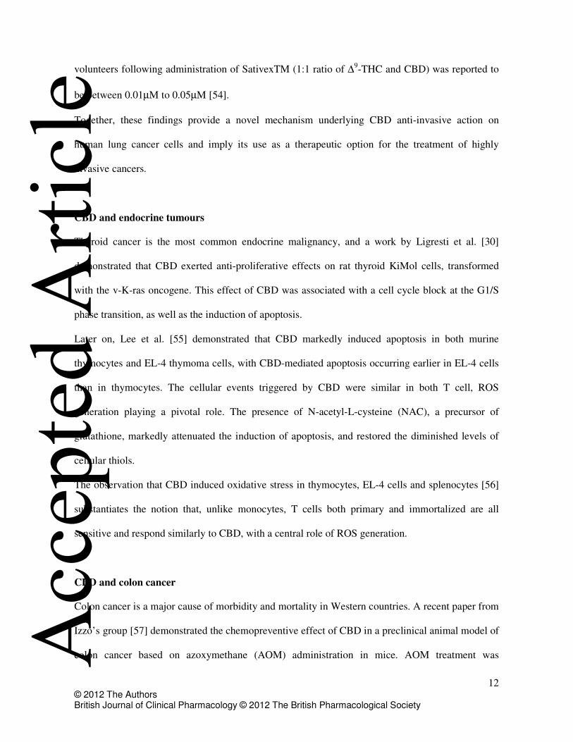
at human CB2 receptors [29]. Moreover, other putative CBD’s molecular targets are TRPV, 5-HT1A,
GPR55 and PPARγ receptors (see Fig. 2). Interestingly CBD does not have any psychotropic
activity. Besides its beneficial effects in the treatment of pain and spasticity and other CNS
pathologies, several reports demonstrated that CBD possesses antiproliferative, pro-apoptotic
effects and inhibits cancer cell migration, adhesion and invasion.
This review will focus on the most recent evidence regarding the efficacy of CBD in the modulation
of tumorigenesis in several types of cancer. The data available so far are summarized in Table 1 and
are discussed in detail in the following paragraphs.
CBD and breast cancer
In 2006 Ligresti et al. [30] demonstrated for the first time that CBD potently and selectively
inhibited the growth of different breast tumour cell lines (MCF7, MDA-MB-231), with an IC50 of
about 6µM, and exhibited significantly lower potency in non-cancer cells. CBD and CBD-rich
extracts (containing approximately 70% CBD together with lesser amounts of other cannabinoids)
also inhibited the growth of xenografts, obtained by s.c. injection into athymic mice of human
MDA-MB-231 cells, and reduced infiltration of lung metastases derived from intrapaw injection of
breast carcinoma cells. Among the possible cellular and molecular mechanisms underlying these
effects, CBD seemed to involve direct TRPV1 activation and/or CB2 indirect activation (via
FAAH), as well as induction of oxidative stress. Later on, McAllister’s group [31] demonstrated Acc
epte
d A
rticl
e

6 © 2012 The Authors British Journal of Clinical Pharmacology © 2012 The British Pharmacological Society
that, besides proliferation, CBD also interfered with two other crucial steps of breast cancer cell
progression: invasion and metastasization. Among the three different groups of cannabinoid
compounds tested (phytocannabinoids with affinity for CB1 and CB2 receptors; phytocannabinoids
with no appreciable affinity for CB1 and CB2 receptors; synthetic compounds with affinity for CB1
and CB2 receptors), CBD was shown to be one of the most effective inhibitors of human breast
cancer cell proliferation, being equipotent to ∆9-THC and CP55940 in inhibiting respectively MDA-
MB-231 and MDA-MB-436 cell growth, and resulting as the most potent inhibitor of the MDA-
MB-231 cell migration. Interestingly, CBD regulated the expression of key genes involved in the
control of cell proliferation and invasion through the downregulation of Id-1 expression, an
inhibitor of basic helix-loop-helix transcription factors, whose overexpression in breast cancer cells
is responsible for proliferation, migration and invasion. Therefore, the ability of CBD to
significantly decrease Id-1 expression in breast cancer cells was associated with its efficacy in
reducing tumour aggressiveness.
Four years later, the same group [32] demonstrated that the observed effect of CBD on Id-1
expression was mediated by the upregulation of the extracellular signal-regulated kinase
phosphorylation (pERK). Indeed, the ERK inhibitor, U0126, partially reverted CBD-induced
inhibition of proliferation and invasion as well as its effect on Id-1 expression. Besides ERK
upregulation, also the production of reactive oxygen species (ROS) was shown to mediate CBD-
induced effects on cell proliferation and Id-1 expression, since the use of a ROS scavenger
(tocopherol) reversed the aforementioned CBD effects. Moreover, these authors demonstrated that
CBD was effective in reducing the primary tumour mass and the size and number of metastatic foci
in vivo.
Finally, the excellent paper of Shrivastava et al. [33] adds fresh light on the cellular mechanism
through which CBD induces cell death in breast cancer cells. These authors showed that CBD
induced a concentration-dependent cell death of both estrogen receptor-positive and estrogen Acc
epte
d A
rticl
e

7 © 2012 The Authors British Journal of Clinical Pharmacology © 2012 The British Pharmacological Society
receptor-negative breast cancer cells with a mechanism independent of CB1, CB2 and TRPV1
receptor activation. Interestingly, the effective concentrations of CBD in tumour cells have little
effect on non tumorigenic, mammary cells. Moreover, electron microscopy revealed morphologies
consistent with the coexistence of autophagy and apoptosis, these events being promoted by the
induction of endoplasmic reticulum (ER) stress and the inhibition of Akt/mTOR/4EBP1 signalling.
In addition, CBD-driven increase in ROS production is fundamental to induce both apoptosis and
autophagy. Examining further the cellular mechanism involved in CBD-induced cell death, they
found that CBD reduced mitochondrial membrane potential, triggered the translocation of the
Beclin2 Interacting Protein (Bid) to the mitochondria and the release of cytochrome C to the cytosol
and, ultimately, the activation of the intrinsic apoptotic pathway.
Finally, the relationship between CBD-induced apoptosis and autophagic cell death was explored
by blocking each form of cell death with specific inhibitors. Caspase inhibition reduced CBD-
induced apoptosis and the expression of protein markers in breast cancer cells, whereas the
inhibition of autophagy enhanced apoptosis as a compensatory/alternative mechanism of cell death.
The apoptosis:autophagy ratio in CBD-induced cell death seemed to be mediated via Beclin1, a key
signalling molecule in the autophagic process. CBD treatment induced the cleavage of Beclin1 and
the subsequent translocation of the cleavage product to the mitochondria where it may induce
apoptosis through the enhancement of cytochrome C release [34, 35].
As a whole this work highlights the presence of a complex balance between autophagy and
mitochondria-mediated apoptosis in CBD-induced breast cancer cell death and strengthens the idea
that CBD can be considered as an alternative agent for breast cancer therapy. Figure 3 shows a
schematic representation of the signalling pathways associated with the effect of CBD in breast
cancer cell proliferation and invasion.
CBD and glioma Acc
epte
d A
rticl
e

8 © 2012 The Authors British Journal of Clinical Pharmacology © 2012 The British Pharmacological Society
CBD possesses anti-tumoural properties also in gliomas, tumours of glial origin characterized by a
high morphological and genetic heterogeneity and considered one of the most devastating
neoplasms, showing high proliferative rate, aggressive invasiveness and insensitivity to radio- and
chemotherapy.
After the seminal paper of Jacobsson et al. (2000) [36] demonstrating a serum-dependent effect of
CBD upon C6 murine glioma cells proliferation, Massi et al. in 2004 [37] reported that CBD was
effective in inhibiting U87-MG and U373 human glioma cell proliferation in vitro through the
induction of apoptosis. Interestingly, CBD did not affect viability of non-transformed primary glial
cells [38]. When tumour xenografts were generated in immune-deficient mice, in vivo intratumoural
treatment with CBD significantly reduced tumour growth [37].
The anti-proliferative effect of CBD was cannabinoid and vanilloid receptors independent; the CB2
receptor antagonist SR144528 reverted CBD effect, but in a weak and transient manner [37]. More
important, this paper demonstrated for the first time that the anti-tumour effect of CBD involved the
induction of oxidative stress, through increased early production of ROS, depletion of intracellular
glutathione and increased GSH-associated enzymatic activity. Accordingly, CBD anti-proliferative
effect was reversed by the anti-oxidant, tocopherol. Importantly, CBD did not induce ROS
production in non-transformed primary glial cells [38].
Among the cellular events involved in glioma cell death, CBD produced a time-dependent release
of cytochrome C and activation of caspase-8, -9 and -3, suggesting the involvement of both the
intrinsic and extrinsic apoptotic pathways [38]. Marcu et al. [39] later confirmed the efficacy of
CBD in inhibiting the growth of multiple glioblastoma cell lines in a more potent way than ∆9-THC.
Interestingly, combined treatment of ∆9-THC with CBD demonstrated that CBD enhanced ∆9-THC
inhibitory effect on glioblastoma cell growth, but not on invasiveness [39]. In line with this, more
recently Torres et al. [40] confirmed that combined treatment with CBD and ∆9-THC greatly
reduced several human glioma cell viability enhancing both autophagy and apoptosis and triggering Acc
epte
d A
rticl
e

9 © 2012 The Authors British Journal of Clinical Pharmacology © 2012 The British Pharmacological Society
caspase-3 activation. Moreover, combined administration of submaximal doses of CBD and ∆9-
THC reduced the growth of U87-MG cell-derived tumour xenografts in nude mice at a higher
extent than the treatment with the individual compounds, suggesting the potential use of a
combinatory therapy which would reduce the amount of the psychoactive ∆9-THC.
The synergistic effect of combined therapy implied in vitro cell cycle arrest, ROS induction and
apoptosis through sustained activation of caspases-3, -7 and -9, as well as specific modulation of the
extracellular signal-regulated kinase, ERK [39]. These effects were not observed with either
compound individually, indicating them as a prerogative of combination treatment. Differently from
Marcu’s data [39], our recent results [41] showed that, both in U87-MG and in ∆9-THC-resistant
T98G human glioma cell lines, CBD per se strongly down-regulates two signalling molecules
crucial in tumour cell proliferation such as ERK and PI3K/Akt. In addition it inhibited the hypoxia-
inducible transcription factor HIF-1α, one of the most critical stimuli for cell survival, motility and
tumour angiogenesis. Thus, inhibition of these three molecules appears as part of the multiple
molecular targets for CBD anti-neoplastic activity [41].
Further biochemical analysis of glioma tumour tissues excised from nude mice treated in vivo with
CBD indicated a significant decrease of activity and content of 5-LOX, as well as a marked
stimulation of FAAH and a decrease of AEA content [42].
Besides cell growth, CBD reduced glioma cell migration [43] and invasiveness in a Boyden
chamber test [39], at concentrations lower than those required to inhibit cell proliferation. CBD
anti-migratory effect was not antagonized by the selective cannabinoid receptor antagonists or by
pretreatment with pertussis toxin, indicating no involvement of classical cannabinoid receptors
and/or Gi/o protein-coupled receptors.
As a whole, CBD seems to counteract glioma cell proliferation and invasion through multiple
mechanisms, as summarized in Figure 4.
Acc
epte
d A
rticl
e

10 © 2012 The Authors British Journal of Clinical Pharmacology © 2012 The British Pharmacological Society
CBD and leukaemia/lymphoma
Gallily et al. [44] provided first evidence of a possible exploitation of CBD in the treatment of
lymphoblastic diseases. They demonstrated that CBD treatment induced apoptosis, through caspase-
3 activation in human acute myeloid leukaemia HL-60 cell line, whereas had no effect on human
monocytes from normal individuals.
Later on, McKallip et al. [45], using the murine EL-4 lymphoma cell line and the human Jurkat and
Molt-4 leukaemia cell lines, demonstrated that CBD exposure led to a significant CB2 receptor-
mediated decrease in the number of viable cells as well as to the induction of apoptosis, both in
vitro and in vivo.
In Jurkat cells, CBD exposure resulted in the activation of caspase-8, -9, and -3, the cleavage of
poly(ADPribose) polymerase, and the decrease in full-length Bid, suggesting a possible cross-talk
between the intrinsic and extrinsic apoptotic pathways. Moreover, exposure to CBD led to the loss
of mitochondrial membrane potential and subsequent release of cytochrome C. As in other tumour
cells, CBD-induced apoptosis required an increase of ROS production. Finally, CBD decreased the
levels of phospho-p38 mitogen-activated protein kinase [45], and this effect was blocked by
treatment with a CB2-selective antagonist or ROS scavenger. In addition, CBD treatment caused a
significant reduction in tumour burden and increased the level of apoptotic tumours in EL-4-bearing
mice [45].
Together, the results suggest that CBD, acting through CB2 receptors and ROS production, may
represent a novel and highly selective treatment for leukaemia. Moreover, previous evidence
indicated that human leukaemias and lymphomas expressed significantly higher levels of CB2
receptors compared to other tumour cell lines, suggesting that tumours of immune origin may be
highly sensitive to the CB2-mediated effects of CBD [46].
CBD and lung cancer Acc
epte
d A
rticl
e

11 © 2012 The Authors British Journal of Clinical Pharmacology © 2012 The British Pharmacological Society
Given the poor response of lung cancer to available therapy and their aggressive biological nature, a
series of targets and new therapeutic strategies for their treatment are currently being investigated
[47-50].
Recently, Ramer et al. [51-53] investigated the effect of CBD on the invasive properties of A549
cells, a line of human lung carcinoma cells expressing both CB1 and CB2, as well as TRPV1
receptors. Using Matrigel invasion assays, they found a CBD-driven impaired invasion of A549
cells that was reversed by CB1 and CB2 receptors as well as TRPV1 antagonists.
CBD treatment concomitantly upregulated tissue inhibitor of matrix metalloproteinases-1 (TIMP-1)
and the CBD-elicited decrease in tumour cell invasiveness was reversed by knocking down TIMP-1
expression through a siRNA approach. These results suggest a causal link between TIMP-1
upregulation and CBD anti-invasive action. CBD was also shown to induce p38 and ERK
phosphorylation as upstream mechanisms for TIMP-1 induction and subsequent decreased
invasiveness. Interestingly all these cellular events were blocked by cannabinoids or TRPV1
receptor antagonists.
The significant inhibition of A549 cell invasion following CBD treatment was also accompanied by
the downregulation of another important factor involved in the regulation of cell spreading, the
plasminogen activator inhibitor PAI-1 [52]. CB1 and CB2 as well as TRPV1 receptor antagonists
suppressed the observed effect of CBD on PAI-1 secretion and cell invasion. Recombinant human
PAI-1 and PAI-1 siRNA led to a concentration-dependent up- and down-regulation of invasiveness,
respectively, suggesting a crucial role of PAI-1 in A549 invasiveness.
Additionally, in vivo studies in thymic aplastic nude mice revealed a significant inhibition of A549
lung metastases following CBD treatment [51] and a significant downregulation of PAI-1 protein
was demonstrated in A549 xenografts of CBD-treated rats [52].
It is worth to note that CBD decreased invasiveness in a range of therapeutically relevant
concentrations (0.01 to 0.05µM), since the peak plasma concentrations of CBD in healthy Acc
epte
d A
rticl
e
12 © 2012 The Authors British Journal of Clinical Pharmacology © 2012 The British Pharmacological Society
volunteers following administration of SativexTM (1:1 ratio of ∆9-THC and CBD) was reported to
be between 0.01µM to 0.05µM [54].
Together, these findings provide a novel mechanism underlying CBD anti-invasive action on
human lung cancer cells and imply its use as a therapeutic option for the treatment of highly
invasive cancers.
CBD and endocrine tumours
Thyroid cancer is the most common endocrine malignancy, and a work by Ligresti et al. [30]
demonstrated that CBD exerted anti-proliferative effects on rat thyroid KiMol cells, transformed
with the v-K-ras oncogene. This effect of CBD was associated with a cell cycle block at the G1/S
phase transition, as well as the induction of apoptosis.
Later on, Lee et al. [55] demonstrated that CBD markedly induced apoptosis in both murine
thymocytes and EL-4 thymoma cells, with CBD-mediated apoptosis occurring earlier in EL-4 cells
than in thymocytes. The cellular events triggered by CBD were similar in both T cell, ROS
generation playing a pivotal role. The presence of N-acetyl-L-cysteine (NAC), a precursor of
glutathione, markedly attenuated the induction of apoptosis, and restored the diminished levels of
cellular thiols.
The observation that CBD induced oxidative stress in thymocytes, EL-4 cells and splenocytes [56]
substantiates the notion that, unlike monocytes, T cells both primary and immortalized are all
sensitive and respond similarly to CBD, with a central role of ROS generation.
CBD and colon cancer
Colon cancer is a major cause of morbidity and mortality in Western countries. A recent paper from
Izzo’s group [57] demonstrated the chemopreventive effect of CBD in a preclinical animal model of
colon cancer based on azoxymethane (AOM) administration in mice. AOM treatment was Acc
epte
d A
rticl
e

13 © 2012 The Authors British Journal of Clinical Pharmacology © 2012 The British Pharmacological Society
associated with Aberrant Crypt Foci (ACF), polyps, and tumour formation, as well as with the
upregulation of phospho-Akt, iNOS and COX-2 and the downregulation of caspase-3. CBD was
effective in reducting ACF, polyps and tumours and counteracted AOM-induced phospho-Akt and
caspase-3 changes. In vitro studies, supported the beneficial effect of CBD. Indeed, in colorectal
carcinoma cell lines, CBD protected DNA from oxidative damage, increased endocannabinoid
levels and reduced cell proliferation in a CB1-, TRPV1- and PPARγ-antagonist sensitive manner.
In the light of its safety records, these results suggest that CBD might be worthy of clinical
consideration in colon cancer prevention.
CBD and angiogenesis
Angiogenesis consists in the formation of new blood vessels from pre-existing ones and represents
another promising therapeutic target for cancer therapy. Collectively, cannabinoids have been
demonstrated to act as anti-angiogenic factors by disposing tumour cells to decrease the production
of pro-angiogenic factors and/or by direct modulation of endothelial cells [58].
Surprisingly, so far no study investigated the effect of CBD on angiogenesis. Our data currently
under publication [59] demonstrated that CBD potently inhibited HUVE cells proliferation,
migration and invasion through the induction of endothelial cell cytostasis without triggering
apoptosis. Interestingly, CBD also affected endothelial cell differentiation into tubular capillaries as
well as the outgrowth of capillary-like structures from HUVEC spheroids in vitro. In addition, the
anti-angiogenic properties of CBD were demonstrated also in vivo, using a matrigel sponge model.
These effects were associated with down-modulation of several molecules associated with
angiogenesis, including MMP2, MMP9, uPA, Endothelin-1, PDGF-AA and CXCL16.
Collectively, these preliminary data demonstrate that, besides its well known pro-apoptotic anti-
proliferative and anti-invasive actions, CBD may also exert anti-angiogenic effects, thus further
strengthening its potential application in cancer therapy. Acc
epte
d A
rticl
e

14 © 2012 The Authors British Journal of Clinical Pharmacology © 2012 The British Pharmacological Society
Conclusion and future directions
Collectively, the non-psychoactive plant-derived cannabinoid CBD exhibits pro-apoptotic and anti-
proliferative actions in different types of tumours and may also exert anti-migratory, anti-invasive,
anti-metastatic and perhaps anti-angiogenic properties. On these bases, evidence is emerging to
suggest that CBD is a potent inhibitor of both cancer growth and spreading.
Interestingly, the anticancer effect of this compound seems to be selective for cancer cells, at least
in vitro, since it does not affect normal cell lines. The efficacy of CBD is linked to its ability to
target multiple cellular pathways that control tumorigenesis through the modulation of different
intracellular signalling depending on the cancer type considered. The most common effect of CBD
is the increase in ROS production that seems to be determinant for triggering its beneficial action in
all the considered cancer cell types. The role of cannabinoid/vanilloid receptors in mediating CBD
effects is more controversial. In some cases (lung, leukaemia, colon) a clear contribution of these
receptors has been demonstrated through the use of specific antagonists, but in other cancer types
(glioma and breast) their relevance appears only marginal or absent. Besides the in vitro data, the
efficacy of CBD in reducing tumour growth and, in some case, metastasization was confirmed in
experimental animal models. However, the potential clinical application of CBD for cancer therapy
needs some consideration. Its low toxicity is certainly a good starting point. CBD behaves as a non
toxic compound; indeed oral administration of CBD 700mg/day for 6 weeks did not show any overt
toxicity in humans [60] suggesting its possible exploitation for prolonged treatment. The route of
administration appears more problematic since CBD oral absorption is slow and unpredictable.
However, 6 weeks of oral CBD treatment 10mg/kg/day provoked a mean plasma level of CBD
between 6 and 11ng/ml (about 0.036µM) [61] that did not differ significantly over the 6 weeks of
administration. Interestingly, this range of concentration was demonstrated to be active in inhibiting
lung cancer cell invasion [52, 53] thus suggesting that in some cases the oral route could be the Acc
epte
d A
rticl
e

15 © 2012 The Authors British Journal of Clinical Pharmacology © 2012 The British Pharmacological Society
appropriate choice. Additionally, experimental data showed that combined treatment with CBD and
∆9-THC could be more effective in reducing cancer cell proliferation, suggesting that the co-
administration may represent a better choice for cancer therapy. Accordingly, oromucosal treatment
with SativexTM 10mg (a formulation including 1:1 ratio of CBD and ∆9-THC recently approved
for multiple sclerosis) provoked CBD plasma level of approximately 0.01µM and up to 0.05µM in
humans, a concentration range effective in reducing lung cell invasion in vitro. Thus, the results
obtained with SativexTM suggest that the use of different associations of phytocannabinoids in a
variable proportion might lead to a better outcome without pharmacokinetic interaction [62].
Moreover, oromucosal administration may represent a first choice in presence of nausea and
vomiting. Finally, the use of CBD/Sativex can be suggested in combination with classical
chemotherapeutic agents to check for the presence of a synergistic effect that might potentially
allow clinical chemotherapeutic dose reduction, thereby reducing toxicity while maintaining
efficacy. In the light of its safety records and considering that CBD is already currently used in
patients with multiple sclerosis, the findings here summarized suggest that CBD might be worthy of
clinical consideration for cancer therapy.
Nomenclature
The drug/molecular target nomenclature conforms top the BJP’s Guide to Receptors and Channels
[63].
Acc
epte
d A
rticl
e

16 © 2012 The Authors British Journal of Clinical Pharmacology © 2012 The British Pharmacological Society
References
1 Howlett AC. The cannabinoid receptors. Prostaglandins Other Lipid Mediat 2002; 68-69: 619-
31.
2 Van Sickle MD, Duncan M, Kingsley PJ, Mouihate A, Urbani P, Mackie K, Stella N,
Makriyannis A, Piomelli D, Davison JS, Marnett LJ, Di Marzo V, Pittman QJ, Patel KD,
Sharkey KA. Identification and functional characterization of brainstem cannabinoid CB2
receptors. Science 2005; 310: 329-32.
3 Devane WA, Hanus L, Breuer A, Pertwee RG, Stevenson LA, Griffin G, Gibson D,
Mandelbaum A, Etinger A, Mechoulam R. Isolation and structure of a brain constituent that
binds to the cannabinoid receptor. Science 1992; 258: 1946-9.
4 Stella N, Schweitzer P, Piomelli D. A second endogenous cannabinoid that modulates long-term
potentiation. Nature 1997; 388: 773-8.
5 Sugiura T, Kishimoto S, Oka S, Gokoh M. Biochemistry, pharmacology and physiology of 2-
arachidonoylglycerol, an endogenous cannabinoid receptor ligand. Prog Lipid Res 2006; 45:
405-46.
6 Mechoulam R, Ben-Shabat S, Hanus L, Ligumsky M, Kaminski NE, Schatz AR, Gopher A,
Almog S, Martin BR, Compton DR, Pertwee RG, Griffin G, Bayewitch M, Barg J, Vogel Z.
Identification of an endogenous 2-monoglyceride, present in canine gut, that binds to
cannabinoid receptors. Biochem Pharmacol 1995 50: 83-90.
7 Piomelli D. The molecular logic of endocannabinoid signalling. Nat Rev Neurosci 2003; 4: 873-
84.
8 De Petrocellis L, Di Marzo V. An introduction to the endocannabinoid system: from the early to
the latest concepts. Best Pract Res Clin Endocrinol Metab 2009; 23: 1-15.
9 Di Marzo V, Petrosino S. Endocannabinoids and the regulation of their levels in health and
disease. Curr Opin Lipidol 2007; 18: 129-40. Acc
epte
d A
rticl
e

17 © 2012 The Authors British Journal of Clinical Pharmacology © 2012 The British Pharmacological Society
10 Ross RA. Anandamide and vanilloid TRPV1 receptors. Br J Pharmacol 2003; 140: 790-801.
11 Ryberg E, Larsson N, Sjögren S, Hjorth S, Hermansson NO, Leonova J, Elebring T, Nilsson K,
Drmota T, Greasley PJ. The orphan receptor GPR55 is a novel cannabinoid receptor. Br J
Pharmacol 2007; 152: 1092-101.
12 O'Sullivan SE. Cannabinoids go nuclear: evidence for activation of peroxisome proliferator-
activated receptors. Br J Pharmacol 2007; 152: 576-82.
13 Pertwee RG, Howlett AC, Abood ME, Alexander SP, Di Marzo V, Elphick MR, Greasley PJ,
Hansen HS, Kunos G, Mackie K, Mechoulam R, Ross RA. International Union of Basic and
Clinical Pharmacology. LXXIX. Cannabinoid receptors and their ligands: beyond CB1 and CB2.
Pharmacol Rev 2010 62: 588-631.
14 McPartland JM, Norris RW, Kilpatrick CW. Coevolution between cannabinoid receptors and
endocannabinoid ligands. Gene 2007; 397: 126-35.
15 Pacher P, Bátkai S, Kunos G. The endocannabinoid system as an emerging target of
pharmacotherapy. Pharmacol Rev 2006; 58: 389-462.
16 Appendino G, Chianese G, Taglialatela-Scalfati O. Cannabinoids: occurrence and medicinal
chemistry. Curr Med Chem 2011; 18: 1085-1099.
17 Munson AE, Harris LS, Friedman MA, Dewey WL, Carchman RA. Antineoplastic activity of
cannabinoids. J Natl Cancer Inst 1975; 55: 597-602.
18 Galve-Roperh I, Sánchez C, Cortés ML, Gómez del Pulgar T, Izquierdo M, Guzmán M. Anti-
tumoral action of cannabinoids: involvement of sustained ceramide accumulation and
extracellular signal-regulated kinase activation. Nat Med; 2000 6: 313-9.
19 Sánchez C, de Ceballos ML, Gomez del Pulgar T, Rueda D, Corbacho C, Velasco G, Galve-
Roperh I, Huffman JW, Ramón y Cajal S, Guzmán M. Inhibition of glioma growth in vivo by
selective activation of the CB(2) cannabinoid receptor. Cancer Res 2001; 61: 5784-9. Acc
epte
d A
rticl
e

18 © 2012 The Authors British Journal of Clinical Pharmacology © 2012 The British Pharmacological Society
20 Casanova ML, Blázquez C, Martínez-Palacio J, Villanueva C, Fernández-Aceñero MJ, Huffman
JW, Jorcano JL, Guzmán M. Inhibition of skin tumour growth and angiogenesis in vivo by
activation of cannabinoid receptors. J Clin Invest 2003; 111: 43–50.
21 Blázquez C, Carracedo A, Barrado L, Real PJ, Fernández-Luna JL, Velasco G, Malumbres M,
Guzmán M. Cannabinoid receptors as novel targets for the treatment of melanoma, FASEB J
2006; 20: 2633–5.
22 Carracedo A, Gironella M, Lorente M, Garcia S, Guzmán M, Velasco G, Iovanna JL.
Cannabinoids induce apoptosis of pancreatic tumor cells via endoplasmic reticulum stress-
related genes. Cancer Res 2006; 66: 6748–55.
23 Cianchi F, Papucci L, Schiavone N, Lulli M, Magnelli L, Vinci MC, Messerini L, Manera C,
Ronconi E, Romagnani P, Donnini M, Perigli G, Trallori G, Tanganelli E, Capaccioli S, Masini
E. Cannabinoid receptor activation induces apoptosis through tumor necrosis factor alpha-
mediated ceramide de nono synthesis in colon cancer cells. Clin Cancer Res 2008; 14: 7691–
700.
24 Bifulco M, Di Marzo V. Targeting the endocannabinoid system in cancer therapy: a call for
further research. Nat Med 2002; 8: 547–50.
25 Bifulco M, Laezza C, Pisanti S, Gazzerro P. Cannabinoids and cancer: pros and cons of an
antitumour strategy. Br J Pharmacol 2006; 148: 123–35.
26 Bifulco M, Malfitano AM, Pisanti S, Laezza C. Endocannabinoids in endocrine and related
tumours. Endocr Relat Cancer 2008; 15: 391–408.
27 Freimuth N, Ramer R, Hinz B. Antitumorigenic effects of cannabinoids beyond apoptosis. J
Pharmacol Exp Ther 2010; 332: 336-44.
28 Pertwee RG, Ross RA, Craib SJ, Thomas A. (-)-Cannabidiol antagonizes cannabinoid receptor
agonists and noradrenaline in the mouse vas deferens. Eur J Pharmacol 2002 456: 99-106. Acc
epte
d A
rticl
e

19 © 2012 The Authors British Journal of Clinical Pharmacology © 2012 The British Pharmacological Society
29 Thomas A, Baillie GL, Phillips AM, Razdan RK, Ross RA, Pertwee RG. Cannabidiol displays
unexpectedly high potency as an antagonist of CB1 and CB2 receptor agonists in vitro. Br J
Pharmacol 2007 150: 613-23.
30 Ligresti A, Moriello AS, Starowicz K, Matias I, Pisanti S, De Petrocellis L, Laezza C, Portella
G, Bifulco M, Di Marzo V. Antitumor activity of plant cannabinoids with emphasis on the
effect of cannabidiol on human breast carcinoma. J Pharmacol Exp Ther 2006; 318: 1375-87.
31 McAllister SD, Christian RT, Horowitz MP, Garcia A, Desprez PY. Cannabidiol as a novel
inhibitor of Id-1 gene expression in aggressive breast cancer cells. Mol Cancer Ther 2007; 6:
2921-7.
32 McAllister SD, Murase R, Christian RT, Lau D, Zielinski AJ, Allison J, Almanza C, Pakdel A,
Lee J, Limbad C, Liu Y, Debs RJ, Moore DH, Desprez PY. Pathways mediating the effects of
cannabidiol on the reduction of breast cancer cell proliferation, invasion, and metastasis. Breast
Cancer Res Treat 2011; 129: 37-47.
33 Shrivastava A, Kuzontkoski PM, Groopman JE, Prasad A. Cannabidiol induces programmed
cell death in breast cancer cells by coordinating the cross-talk between apoptosis and autophagy.
Mol Cancer Ther 2011; 10: 1161-72.
34 Cho DH, Jo YK, Hwang JJ, Lee YM, Roh SA, Kim JC. Caspase-mediated cleavage of
ATG6/Beclin-1 links apoptosis to autophagy in HeLa cells. Cancer Lett 2009; 274: 95-100.
35 Wirawan E, Vande Walle L, Kersse K, Cornelis S, Claerhout S, Vanoverberghe I, Roelandt R,
De Rycke R, Verspurten J, Declercq W, Agostinis P, Vanden Berghe T, Lippens S,
Vandenabeele P. Caspase-mediated cleavage of Beclin-1 inactivates Beclin-1-induced
autophagy and enhances apoptosis by promoting the release of proapoptotic factors from
mitochondria. Cell Death Dis 2010; 1:e18.
36 Jacobsson SO, Rongård E, Stridh M, Tiger G, Fowler CJ. Serum-dependent effects of tamoxifen
and cannabinoids upon C6 glioma cell viability. Biochem Pharmacol 2000 60: 1807-13. Acc
epte
d A
rticl
e

20 © 2012 The Authors British Journal of Clinical Pharmacology © 2012 The British Pharmacological Society
37 Massi P, Vaccani A, Ceruti S, Colombo A, Abbracchio MP, Parolaro D. Antitumor effects of
cannabidiol, a nonpsychoactive cannabinoid, on human glioma cell lines. J Pharmacol Exp Ther
2004; 308: 838-45.
38 Massi P, Vaccani A, Bianchessi S, Costa B, Macchi P, Parolaro D. The non-psychoactive
cannabidiol triggers caspase activation and oxidative stress in human glioma cells. Cell Mol
Life Sci 2006; 63: 2057-66.
39 Marcu JP, Christian RT, Lau D, Zielinski AJ, Horowitz MP, Lee J, Pakdel A, Allison J, Limbad
C, Moore DH, Yount GL, Desprez PY, McAllister SD. Cannabidiol enhances the inhibitory
effects of delta9-tetrahydrocannabinol on human glioblastoma cell proliferation and survival.
Mol Cancer Ther 2010; 9: 180-9.
40 Torres S, Lorente M, Rodríguez-Fornés F, Hernández-Tiedra S, Salazar M, García-Taboada E,
Barcia J, Guzmán M, Velasco G. A combined preclinical therapy of cannabinoids and
temozolomide against glioma. Mol Cancer Ther 2011; 10: 90-103.
41 Massi P, Solinas M, Valenti M, Cinquina V, Bolognini D, Gariboldi M, Monti E, Parolaro D.
Cannabidiol, a non-psychoactive cannabinoid compound, inhibits cell invasion and prosurvival
intracellular pathways in U87-MG human glioma cells. Submitted to European Journal of
Pharmacology (2011).
42 Massi P, Valenti M, Vaccani A, Gasperi V, Perletti G, Marras E, Fezza F, Maccarrone M,
Parolaro D. 5-Lipoxygenase and anandamide hydrolase (FAAH) mediate the antitumor activity
of cannabidiol, a non-psychoactive cannabinoid. J Neurochem 2008;104: 1091-100.
43 Vaccani A, Massi P, Colombo A, Rubino T, Parolaro D. Cannabidiol inhibits human glioma cell
migration through a cannabinoid receptor-independent mechanism. Br J Pharmacol 2005; 144:
1032-6.
Acc
epte
d A
rticl
e

21 © 2012 The Authors British Journal of Clinical Pharmacology © 2012 The British Pharmacological Society
44 Gallily R, Even-Chena T, Katzavian G, Lehmann D, Dagan A, Mechoulam R. Gamma-
irradiation enhances apoptosis induced by cannabidiol, a non-psychotropic cannabinoid, in
cultured HL-60 myeloblastic leukemia cells. Leuk Lymphoma 2003; 44: 1767-73.
45 McKallip RJ, Jia W, Schlomer J, Warren JW, Nagarkatti PS, Nagarkatti M. Cannabidiol-
induced apoptosis in human leukemia cells: A novel role of cannabidiol in the regulation of
p22phox and Nox4 expression. Mol Pharmacol 2006; 70: 897-908.
46 McKallip RJ, Lombard C, Fisher M, Martin BR, Ryu S, Grant S, Nagarkatti PS, Nagarkatti M.
Targeting CB2 cannabinoid receptors as a novel therapy to treat malignant lymphoblastic
disease. Blood 2002; 100: 627-34.
47 Li R, Wang H, Bekele BN, Yin Z, Caraway NP, Katz RL, Stass SA, Jiang F. Identification of
putative oncogenes in lung adenocarcinoma by a comprehensive functional genomic approach.
Oncogene 2006; 25: 2628–35.
48 Adjei AA. Novel combinations based on epidermal growth factor receptor inhibition. Clin
Cancer Res 2006; 12: 4446s–50s.
49 Erler JT, Bennewith KL, Nicolau M, Dornhöfer N, Kong C, Le QT, Chi JT, Jeffrey SS, Giaccia
AJ. Lysyl oxidase is essential for hypoxia-induced metastasis. Nature 2006; 440: 1222–6.
50 Molina JR, Adjei AA, Jett JR. Advances in chemotherapy of non-small cell lung cancer. Chest
2006; 130: 1211–9.
51 Ramer R, Merkord J, Rohde H, Hinz B. Cannabidiol inhibits cancer cell invasion via
upregulation of tissue inhibitor of matrix metalloproteinases-1. Biochem Pharmacol 2010; 79:
955-66.
52 Ramer R, Rohde A, Merkord J, Rohde H, Hinz B. Decrease of plasminogen activator inhibitor-1
may contribute to the anti-invasive action of cannabidiol on human lung cancer cells. Pharm
Res 2010; 27: 2162-74. Acc
epte
d A
rticl
e

22 © 2012 The Authors British Journal of Clinical Pharmacology © 2012 The British Pharmacological Society
53 Ramer R, Hinz B. Inhibition of cancer cell invasion by cannabinoids via increased expression of
tissue inhibitor of matrix metalloproteinases-1. J Natl Cancer Inst 2008; 100: 59–69.
54 Sativex Product Monograph. Salisbury, Wiltshire U.K.: GW Pharma Ltd.; SP4 0JQ Submission
Control No: 091289; April 2003.
55 Lee CY, Wey SP, Liao MH, Hsu WL, Wu HY, Jan TR. A comparative study on cannabidiol-
induced apoptosis in murine thymocytes and EL-4 thymoma cells. Int Immunopharmacol 2008;
8: 732-40.
56 Wu HY, Chu RM, Wang CC, Lee CY, Lin SH, Jan TR. Cannabidiol-induced apoptosis in
primary lymphocytes is associated with oxidative stress-dependent activation of caspase-8.
Toxicol Appl Pharmacol 2008; 226: 260-70.
57 Aviello G, Romano B, Borrelli F, Capasso R, Gallo L, Piscitelli F, Di Marzo V, Izzo AA.
Chemopreventive effect of the non-psychotropic phytocannabinoid cannabidiol on experimental
colon cancer. J Mol Med (Berl) 2012. [Epub ahead of print].
58 Freimuth N, Ramer R, Hinz B. Antitumorigenic effects of cannabinoids beyond apoptosis. J
Pharmacol Exp Ther 2010; 332: 336-44.
59 Solinas M, Massi P, Cantelmo AR, Cattaneo MG, Cammarota R, Cinquina V, Valenti M,
Vicentini L, Noonan D, Albini A, Parolaro D. Cannabidiol inhibits angiogenesis by multiple
mechanisms. Submitted to British Journal of Pharmacology (2012).
60 Consroe P, Laguna J, Allender J, Snider S, Stern L, Sandyk R, Kennedy K, Schram K.
Controlled clinical trial of cannabidiol in Huntington's disease. Pharmacol Biochem Behav 1991
40: 701-8.
61 Consroe P, Kennedy K, Schram K. Assay of plasma cannabidiol by capillary gas
chromatography/ion trap mass spectroscopy following high-dose repeated daily oral
administration in humans. Pharmacol Biochem Behav 1991 40: 517-22. Acc
epte
d A
rticl
e

23 © 2012 The Authors British Journal of Clinical Pharmacology © 2012 The British Pharmacological Society
62 Karschner EL, Darwin WD, Goodwin RS, Wright S, Huestis MA. Plasma cannabinoid
pharmacokinetics following controlled oral delta9-tetrahydrocannabinol and oromucosal
cannabis extract administration. Clin Chem 2011 57: 66-75.
63 Alexander SPH, Mathie A, Peters JA. Guide to Receptors and Channels (GRAC), 5th edition. Br
J Pharmacol 2011 164 (Suppl. 1): S1-S324.
Acc
epte
d A
rticl
e

24 © 2012 The Authors British Journal of Clinical Pharmacology © 2012 The British Pharmacological Society
Table 1 - Effects of cannabidiol on different types of cancer
Cancer In vitro effect Receptor
involvement
ROS
production Molecular cell signalling Autophagy Apoptosis In vivo effect Reference
↓ proliferation CB2;
TRPV1 ↑ NC NC +
↓ xenografts growth
↓ lung metastases [26]
↓ viability
non-CB1;
non-CB2;
non-TRPV1
↑ ↓ pAkt; ↑ cytochrome C
Bid translocation + + NC [29]
Breast
↓ invasion NC ↑ ↓ Id-1; ↑ pERK NC NC
↓ tumour growth
↓ size and number of
metastases
[27, 28]
↓ proliferation
non-CB1;
partial-CB2;
non-TRPV1
↑
↓ pERK; ↓ pAkt; ↓ HIF-1α
↑ cytochrome C
caspase activation
NC + ↓ tumour growth [32, 33, 36, 37]
↓ proliferation and invasiveness NC NC NC NC NC NC [34] Glioma
↓ migration
non-CB1;
non-CB2;
non-TRPV1
NC Ptx insensitive NC NC NC [38]
↓ viability NC NC caspase-3 activation NC + NC [39]
Leukaemia ↓ viability CB2 ↑
↓ p-p38
caspase activation; ↓ Bid
↑ cytochrome C
NC + ↓ tumour burden
↑ tumour cell apoptosis [40]
Lung ↓ invasion
CB1;
CB2;
TRPV1
NC ↑ p-p38; ↑ p-ERK; ↑ TIMP-1
↓ PAI-1 NC NC
↓ lung metastases
↓ PAI-1 in xenografts [46, 47]
cytostatic effect NC NC NC NC + NC [26] Thyroid
Thymoma ↓ viability NC ↑ NC NC + NC [50]
Colon ↓ proliferation
CB1;
TRPV1;
PPARγ
NC ↓ Akt; ↑ 2AG
↑ caspase-3 NC + ↓ ACF, polyps and tumours [52]
NC = not checked
↑ increase; ↓ decrease Acc
epte
d A
rticl
e

25 © 2012 The Authors British Journal of Clinical Pharmacology © 2012 The British Pharmacological Society
Figure 1 - Chemical structures of ∆∆∆∆9-tetrahydrocannabinol (∆∆∆∆
9-THC), cannabidiol (CBD), anandamide (AEA) and WIN55212-2 A
ccep
ted
Arti
cle
26 © 2012 The Authors British Journal of Clinical Pharmacology © 2012 The British Pharmacological Society
Figure 2 – Some of the potential biological targets of CBD Acc
epte
d A
rticl
e

27 © 2012 The Authors British Journal of Clinical Pharmacology © 2012 The British Pharmacological Society
Figure 3 – Schematic representation of the signalling pathways associated with CBD effects on breast cancer Acc
epte
d A
rticl
e

28 © 2012 The Authors British Journal of Clinical Pharmacology © 2012 The British Pharmacological Society
Figure 4 – Schematic representation of the signalling pathways associated with CBD effects on glioma Acc
epte
d A
rticl
e