Block copolymer micelles conjugated with anti-EGFR antibody for targeted delivery of anticancer drug
11
Block Copolymer Micelles Conjugated with anti-EGFR Antibody for Targeted Delivery of Anticancer Drug TAIHO NOH, 1 YEON HEE KOOK, 2 CHIYOUNG PARK, 1 HYEWON YOUN, 2 HANA KIM, 1 EUN TAX OH, 2 EUN KYUNG CHOI, 3 HEON JOO PARK, 2 CHULHEE KIM 1 1 Department of Polymer Science and Engineering, Inha University, Incheon 402-751, Korea 2 Department of Microbiology, Center for Advanced Medical Education by BK21 Project, College of Medicine, Inha University, Incheon, 400-712, Korea 3 Department of Radiation Oncology, College of Medicine, University of Ulsan, Seoul, 138-736, Korea Received 12 June 2008; accepted 21 August 2008 DOI: 10.1002/pola.23036 Published online in Wiley InterScience (www.interscience.wiley.com). ABSTRACT: Antiepidermal growth factor receptor antibody (anti-EGFR antibody) was conjugated with the block copolymer micelle based on poly(ethylene glycol) (PEG) and poly(e-caprolactone) (PCL) for active targeting to EGFR overexpressing cancer cells. Doxorubicin (DOX) was encapsulated in the core of the block copolymer (MePEG-b- PCL) micelle (DOX-micelle). The mean diameters of the DOX-micelle and the anti- EGFR-PEG-b-PCL copolymer micelles loaded with DOX (DOX-anti-EGFR-micelle) were about 25 and 31 nm, respectively. The RKO human colorectal cancer cells expressing moderate degree of EGFR were incubated with free DOX, DOX-micelle, or DOX-anti-EGFR-micelle to study the distribution of DOX in the cells. When cells were incubated with free DOX, moderate degree of DOX fluorescence was observed in the nuclei. In the cells treated with DOX-micelle, the DOX fluorescence intensity in the cytoplasm was much greater than that in the nuclei. On the other hand, the nuclei of the cells treated with DOX-anti-EGFR-micelle exhibited DOX fluorescence intensity similar to that in the cytoplasm. The cytotoxicity of DOX-anti-EGFR-micelle to induce apoptosis in RKO cells was significantly greater than that of free DOX or DOX- micelle. These results demonstrated that the presence of anti-EGFR antibody on the DOX-micelle surface (DOX-anti-EGFR-micelle) increased the internalization of the DOX-micelle and nuclear accumulation of DOX, and enhanced the DOX-induced cell death. V V C 2008 Wiley Periodicals, Inc. J Polym Sci Part A: Polym Chem 46: 7321–7331, 2008 Keywords: anti-EGFR; block copolymers; doxorubicin; drug delivery systems; micelles INTRODUCTION In recent years, there has been great interest in the use of a variety of nanoscale colloidal particles such as liposomes, polymeric micelles, silica nano- particles, and gold nanoparticles to improve the delivery of cytotoxic drug to cancer cells. 1–8 Among these nanocarriers, polymeric micelles have a potential to bring several advantages to therapeutic systems because of the storage capa- bility and chemically tailorable structure. For the use of polymeric micelle as a drug delivery carrier, there are prerequisite requirements such as Journal of Polymer Science: Part A: Polymer Chemistry, Vol. 46, 7321–7331 (2008) V V C 2008 Wiley Periodicals, Inc. Correspondence to: C. Kim (E-mail: [email protected]) and H. J. Park (E-mail: [email protected]) 7321
Transcript of Block copolymer micelles conjugated with anti-EGFR antibody for targeted delivery of anticancer drug

Block Copolymer Micelles Conjugated with anti-EGFRAntibody for Targeted Delivery of Anticancer Drug
TAIHO NOH,1 YEON HEE KOOK,2 CHIYOUNG PARK,1 HYEWON YOUN,2 HANA KIM,1 EUN TAX OH,2
EUN KYUNG CHOI,3 HEON JOO PARK,2 CHULHEE KIM1
1Department of Polymer Science and Engineering, Inha University, Incheon 402-751, Korea
2Department of Microbiology, Center for Advanced Medical Education by BK21 Project, College of Medicine,Inha University, Incheon, 400-712, Korea
3Department of Radiation Oncology, College of Medicine, University of Ulsan, Seoul, 138-736, Korea
Received 12 June 2008; accepted 21 August 2008DOI: 10.1002/pola.23036Published online in Wiley InterScience (www.interscience.wiley.com).
ABSTRACT: Antiepidermal growth factor receptor antibody (anti-EGFR antibody) wasconjugated with the block copolymer micelle based on poly(ethylene glycol) (PEG) andpoly(e-caprolactone) (PCL) for active targeting to EGFR overexpressing cancer cells.Doxorubicin (DOX) was encapsulated in the core of the block copolymer (MePEG-b-PCL) micelle (DOX-micelle). The mean diameters of the DOX-micelle and the anti-EGFR-PEG-b-PCL copolymer micelles loaded with DOX (DOX-anti-EGFR-micelle)were about 25 and 31 nm, respectively. The RKO human colorectal cancer cellsexpressing moderate degree of EGFR were incubated with free DOX, DOX-micelle, orDOX-anti-EGFR-micelle to study the distribution of DOX in the cells. When cells wereincubated with free DOX, moderate degree of DOX fluorescence was observed in thenuclei. In the cells treated with DOX-micelle, the DOX fluorescence intensity in thecytoplasm was much greater than that in the nuclei. On the other hand, the nuclei ofthe cells treated with DOX-anti-EGFR-micelle exhibited DOX fluorescence intensitysimilar to that in the cytoplasm. The cytotoxicity of DOX-anti-EGFR-micelle toinduce apoptosis in RKO cells was significantly greater than that of free DOX or DOX-micelle. These results demonstrated that the presence of anti-EGFR antibody on theDOX-micelle surface (DOX-anti-EGFR-micelle) increased the internalization of theDOX-micelle and nuclear accumulation of DOX, and enhanced the DOX-induced celldeath. VVC 2008 Wiley Periodicals, Inc. J Polym Sci Part A: Polym Chem 46: 7321–7331, 2008
Keywords: anti-EGFR; block copolymers; doxorubicin; drug delivery systems;micelles
INTRODUCTION
In recent years, there has been great interest inthe use of a variety of nanoscale colloidal particles
such as liposomes, polymeric micelles, silica nano-particles, and gold nanoparticles to improve thedelivery of cytotoxic drug to cancer cells.1–8
Among these nanocarriers, polymeric micelleshave a potential to bring several advantages totherapeutic systems because of the storage capa-bility and chemically tailorable structure. For theuse of polymeric micelle as a drug delivery carrier,there are prerequisite requirements such as
Journal of Polymer Science: Part A: Polymer Chemistry, Vol. 46, 7321–7331 (2008)VVC 2008 Wiley Periodicals, Inc.
Correspondence to: C. Kim (E-mail: [email protected]) andH. J. Park (E-mail: [email protected])
7321
biocompatibility, high drug loading capacity, longcirculation time, and controlled release profiles.9–15
For this purpose, water-soluble polymers suchas poly(ethylene glycol) (PEG), poly(2-ethyl-2-oxa-zoline), and polypeptide have been adopted as ahydrophilic block because of their biocompatibleand antibiofouling characteristics.16–18 In addi-tion, biocompatible and biodegradable polyesterssuch poly(D,L-lactic-co-glycolic acid) and poly(e-carprolactone) (PCL) have also been utilized as ahydrophobic block for drug encapsulation.19,20
One of the challenging topics has been the designof dug carriers with active targeting capabilitythat would result in higher intracellular drugconcentrations in a selective manner.21,22 In ourstudies, we have focused on the development ofdrug-loaded polymeric micelle conjugated with atargeting ligand as a target-selective delivery ve-hicle.23–28 Specifically, first, we selected PEG–PCL block copolymer micelle composed of PEG asa hydrophilic block and PCL as a hydrophobicblock for drug encapsulation. Second, anti-EGFRantibody was conjugated with the PEG–PCLblock copolymer micelle for active targeting tocancer cells overexpressing EGFR.
EGFR (ErbB-1; HER1 in humans) is a cell-sur-face receptor for the members of the epidermalgrowth factor family (EGF-family), and is overex-pressed on the surface of a number of differenthuman cancer cells including colorectal, breast,and lung cancer cells.29–31 In recent years, effortshave been made to develop drug delivery systemsexploiting the overexpression of EGFR on the cellsurface. For example, the possibility of usingEGF-conjugated polymer micelle as a vehicle fortargeting hydrophobic drugs to EGFR overex-pressing cancers have been investigated.32 Signif-icant progress has also been made in recent yearsdeveloping new cancer treatment strategies usingvarious EGFR inhibitors and some of them are inclinical trials.33–38 In this study, we have investi-gated the feasibility of using anti-EGFR antibody-conjugated polymer micelle as a vehicle for thedelivery of anticancer drugs DOX to EGFR overex-pressing cancer cells. It is known that the ligand–receptor complexes are internalized via receptor-mediated endocytosis and that the endocytosis ofreceptors to lysosomes leads to degradation of thereceptors. Interestingly, on the other hand, therehas been emerging evidence that ligand–EGFRcomplexes are translocated into nuclei afterEGFR-mediated internalization.34,39,40 Althoughit is unclear how the translocation of the EGFR–ligand complexes into nuclei takes place, it may be
expected that anti-EGFR antibody-micelle mayalso be translocated into nuclei following endocy-tosis, and thus the anti-EGFR antibody-micellemay be used to deliver anticancer drugs such asDOX to nuclei of EGFR overexpressing cancercells.
The mechanism of the anticancer action ofDOX is complex, although DNA replication hasbeen known to be the major target of the drug.39
However, wider clinical use of DOX is limitedbecause of its low water solubility and acute toxic-ity to normal tissue. To reduce the acute toxicityand improve the therapeutic efficacy of DOX, var-ious delivery vehicles for DOX have been pro-posed.2,41,42
This report describes the development of DOX-loaded PEG–PCL block copolymer micelle conju-gated with anti-EGFR antibody (DOX-anti-EGFR-micelle). We elucidated the targeting capa-bility of DOX-anti-EGFR-micelle and its cytotoxic-ity against RKO human colorectal cancer cellsexpressing in vitro.
EXPERIMENTAL
Materials and Equipments
Monomethoxy-poly(ethylene oxide) (MePEG, Mn
¼ 2000, Sigma-Aldrich) and a-carboxy-x-hydroxy-PEG (COOH-PEG, Mn ¼ 2000, Mw/Mn ¼ 1.16,Polymer Sources) were purified by azeotropic dis-tillation with toluene. e-Caprolactone and triethyl-amine (Sigma-Aldrich, Inc., St. Louis, MO) weredried over calcium hydride and distilled underreduced pressure. Mouse monoclonal anti-EGFRantibody (mouse IgG1 isotype, MW 170 kDa,external carbohydrate residue binding antibodyE2760, Sigma-Aldrich), N-hydroxysuccinimide(NHS), N,N0-dicyclohexylcarbodiimide (DCC),stannous octoate [Sn(Oct)2], and phosphate buf-fered saline (PBS) (pH ¼ 7.4, 0.01 M) were usedas received (Sigma-Aldrich). Doxorubicin (DOX)was purchased from Sigma-Aldrich and hydro-phobic DOX was prepared following the literatureprocedure.42 Tetrahydrofuran (THF) and toluenewere distilled before use. The cellulose membrane(molecular weight cutoff ¼ 12,000, Sigma-Aldrich,Inc.) was used for dialysis. The molecular weightsand block compositions of the block copolymerswere determined with the 1H-NMR spectra whichwas recorded on a Varian UNITY INOVA 400 at400 MHz. The molecular weight distributionswere determined using a GPC equipped with aWaters Associates 410 RI detector, 510 HPLC
7322 NOH ET AL.
Journal of Polymer Science: Part A: Polymer ChemistryDOI 10.1002/pola

pump and l-Styragel columns with pore sizes of102, 500, 103, and 104 A. The eluent was THF andthe molecular weights were calibrated with poly-styrene standards. The UV-visible spectra wererecorded on HP-8453A diode array spectrophoto-meter.
Cell Lines and Culture Conditions
The RKO human colorectal cancer cells wereused. The RKO cells are known to express moder-ate amount of EGFR.31 The cells were cultured in25 cm2 plastic tissue culture flasks with Dulbec-co’s modified Eagle’s medium (DMEM, HycloneLaboratories Inc., Logan, UT) supplemented with10% fetal bovine serum (FBS, Hyclone Laborato-ries Inc.) and 1% penicillin/streptomycin (HycloneLaboratories Inc.) in a humidified 5% CO2/95%air incubator at 37 �C. The cells in exponentialgrowth phase were treated with free DOX, DOX-micelles or DOX-anti-EGFR-micelles.
Synthesis of MePEG-b-PCL
Sn(Oct)2 (12.51 lg, 30.9 lmol) was added into thetoluene solution (100 mL) of MePEG (10.0 g) ande-caprolactone (13.13 g, 115 mmol) at room tem-perature under nitrogen. After stirring for 72 h at130 �C, the solvent was removed under reducedpressure. The product was obtained by precipita-tion from THF into n-hexane (yield 19.5 g, 87%).
1H-NMR (400 MHz, CDCl3) d 1.4 (b, 1H,ACOACH2ACH2ACH2ACH2ACH2AOA), 1.6 (b,2H, ACOACH2ACH2ACH2ACH2ACH2AOA),2.25 (b, 1H, ACOACH2ACH2A), 3.6 (m, 4H,ACH2ACH2AOA), 4.1 (b, 1H, ACOACH2ACH2
ACH2AO): Mn ¼ 4500, Mw/Mn ¼ 1.25.
Synthesis of COOH-PEG-b-PCL
A toluene solution (30 mL) of a-carboxy-x-hydroxy-PEG (0.10 g) and e-caprolactone (0.13 g,11.2 mmol) was stirred at room temperatureunder nitrogen. After adding Sn(Oct)2 (4.17 lg, 10lmol), the reaction mixture was stirred for 72 h at130 �C. The product was purified by a repeatedprecipitation from THF into n-hexane (yield 0.21 g,87%).
1H-NMR (400 MHz, CDCl3) d 1.4 (b, 1H,ACOACH2ACH2ACH2ACH2ACH2AOA), 1.6 (b,2H, ACOACH2ACH2ACH2ACH2ACH2AOA),2.25 (b, 1H, ACOACH2ACH2A), 3.6 (m, 4H,ACH2ACH2AOA), 4.1 (b, 1H, ACOACH2ACH2ACH2AOA): Mn ¼ 4600, Mw/Mn ¼ 1.25.
Synthesis of NHS-PEG-b-PCL
A THF solution (1 mL) of NHS (12.5 mg, 0.11mmol) and TEA (0.015 mL, 0.11 mmol) was addedto a THF solution (5 mL) of COOH-PEG-b-PCL(0.10 g), and DCC (0.022 g, 0.11 mmol).32 Afterstirring for 12 h at 45 �C, the precipitated ureawas filtered off. The purified product was obtainedby precipitation from THF into diethyl ether(yield 0.08 g, 80%).
1H-NMR (400 MHz, CDCl3) d 1.4 (b, 50H,ACOACH2ACH2ACH2ACH2ACH2AOA), 1.6 (b,95H, ACOACH2ACH2ACH2ACH2ACH2AOA),2.25 (b, 50H, ACOACH2ACH2A), 2.83 (b, 4H,ACOACH2ACH2ACOA), 3.6 (m, 200H, ACH2
ACH2AOA), 4.1 (b, 46H, ACOACH2ACH2ACH2AOA).
Synthesis of Anti-EGFR-Conjugated PEG-b-PCL(anti-EGFR-PEG-b-PCL)
Anti-EGFR antibody (12.4 mg, 72.9 nmol) wasadded to a PBS solution (0.4 mL, pH ¼ 7.4) ofNHS-PEG-b-PCL (1.7 mg) and stirred for 48 h atroom temperature. The reaction mixture wasthen dialyzed against PBS solution for 48 h. Anti-EGFR-PEG-b-PCL was characterized by usingBCA (bicinchoninic acid, Sigma-Aldrich) proteinassay and dot blot assay.43
Preparation of DOX-Micelle
A THF solution (1.0 mL) of DOX (4.0 mg) andMePEG-b-PCL (40.0 mg) was stirred for 1 h, andthe mixture was then added dropwise to water(35 mL) with stirring. The mixture was thenopened to air overnight, allowing slow evapora-tion of THF and formation of micelles. The resid-ual THF was completely removed under reducedpressure. The solution was dialyzed against waterfor 72 h (molecular weight cutoff: 12,000 Da) andthen lyophilized.
Preparation of DOX-Anti-EGFR-Micelle
A THF solution (1.0 mL) of DOX (4.0 mg) andMePEG-b-PCL (40.0 mg) was stirred for 1 h atroom temperature, and then THF was removedunder nitrogen. The mixture of DOX and MePEG-b-PCL was hydrated with PBS solution (pH ¼ 7.4,30 mL), and then anti-EGFR-PEG-b-PCL (10.1mg) was added to the preformed DOX-micelle so-lution. After stirring at 60 �C for 1 h and addi-tional 12 h at room temperature, the mixture wasdialyzed against water.32,44
SYNTHESIS OF ANTI-EGFR-CONJUGATED PEG-B-PCL 7323
Journal of Polymer Science: Part A: Polymer ChemistryDOI 10.1002/pola

Determination of DOX-Loading Content
The micelle loaded with DOX was dissolved inchloroform/DMSO (1:1, v/v) solution, and theDOX-loading content was determined by meas-uring the UV-vis absorbance at 485 nm.
Dynamic Light Scattering
Dynamic light scattering measurements were per-formed at 25 �Cwith a Brookhaven BI-200SM goni-ometer and BI-9000AT digital autocorrelator. Thescattered light of a He-Ne laser (Research Electro-optics, 35 mW) operated at 632.8 nmwas measuredat an angle of 90� and collected on an autocorrela-tor. The sample solutions were purified by passingthrough a 0.20-lm Millipore filter. The hydrody-namic diameters (d) of micelles were calculatedusing the Stokes-Einstein equation d ¼ kBT/3pgD,where kB is the Boltzmann constant, T is the abso-lute temperature, g is the solvent viscosity, and Dis the diffusion coefficient. The polydispersity factorof micelles, represented as l2/C
2, where l2 is thesecond cumulant of the decay function and C is theaverage characteristic line width, was calculatedfrom the cumulant method. CONTIN algorithmswere used in the Laplace inversion of the autocor-relation function to obtain micelle size distribution.
Transmission Electron Microscopy (TEM)
TEM was performed with a Philips CM 200, oper-ated at an acceleration voltage of 120 kV. For thepreparation of dispersed samples in water, a dropof sample solution (1 g/L) was placed onto a 200-mesh copper grid coated with carbon. About 2 minafter deposition, the grid was tapped with filterpaper to remove surface water, followed by airdrying. Negative staining was performed by usinga droplet of a 5 wt % uranyl acetate solution. Thesamples were air dried before measurement.
Dot Blot Assay
Nitrocellulose membrane was labeled for proteinelution fractions and placed on the top of wetWhatman paper for keeping moist. Samples (2 lLeach) were loaded from each micelle fraction onthe membrane, and allowed to dry. The membranewas incubated in blocking solution of 5% skimmilk for 1 h. After washing twice with Tris-buf-fered saline supplemented with 0.05% Tween-20(TBST), the membrane was immersed into the sec-ondary antibody (antimouse immunoglobulin G;Amersham Pharmacia Biotech) solution, followed
shaking at room temperature for 1 h. The mem-brane was then washed four times for 15 min eachwith TBST buffer. Specific antigen–antibody com-plexes were detected using the ECL western blot-ting detection system (Amersham Pharmacia Bio-tech, Piscataway, NJ). From the membrane, theexcess amount of ECL substrate solution wasdrained, then the membrane was wrapped andexposed to X-ray film to detect anti-EGFR.
In vitro DOX Release Study
The rate of DOX release from DOX-micelle in phe-nol red-free DMEM was investigated. To obtainthe drug release profile, 3 mg of DOX-micelle orDOX-anti-EGFR-micelle was dispersed in me-dium (DMEM) and transferred into dialysis tubes,which were then immersed in the medium at37 �C. At selected time points, the amount ofreleased DOX was determined by measuring theabsorbance at 485 nm.
Flow Cytometric Analysis for Apoptosis
Control and drug-treated RKO cells were har-vested by trypsin treatment. The cells were thenfixed with 70% ethanol and maintained at �20 �Cfor an hour, washed with PBS, and resuspendedin 1-mL PBS containing 30 units of DNase-freeRNase. After incubation with propidium iodide(PI; Sigma-Aldrich, Inc., 5 lg in 100 lL) in darkat 37 �C for 30 min, the cells were subjected to ap-optosis study with a FACSCaliburTM flow cytome-ter (Becton Dickinson Bioscience, San Jose, CA).About 10,000 cells were counted for each sample.
Fluorescence Microscopy Study for DOX Uptake
RKO cells were grown in eight-well chamber slidesand treated with 10 lM of free DOX and DOX-mi-celle or DOX-anti-EGFR-micelle with equivalentamount of DOX, i.e., 10 lM, at 37 �C for 0–24 h.Cells were washed with PBS, fixed using 4% (v/v)paraformaldehyde (Sigma-Aldrich) at 4 �C for 30min, and permeabilized with 0.25% (w/v) TritonX-100 (Sigma-Aldrich) at room temperature for2 min. After washing several times with PBS, thenuclei were stained with DAPI solution containingProLong Gold-antifade reagent (Invitrogen,Eugene, OR). The slides were covered with coverglasses, and the cells were examined with a fluo-rescence microscope (Nikon TE-2000E, Tokyo,Japan). To quantitate the DOX localized in thenuclei, DOX-derived fluorescence was capturedand analyzed by ImageJ 1.41e image analysis soft-ware (NIH) by measuring the intensity of the
7324 NOH ET AL.
Journal of Polymer Science: Part A: Polymer ChemistryDOI 10.1002/pola

DOX-derived fluorescence in nucleus correspond-ing the area expressingDAPI-derived fluorescence.
RESULTS AND DISCUSSION
Preparation of DOX-Anti-EGFR-Micelle
As shown in Figure 1, the block copolymers,MePEG-b-PCL, and COOH-PEG-b-PCL, were
prepared by ring-opening polymerization of e-cap-rolactone using MePEG and COOH-PEG as mac-roinitiators, respectively. The number averagemolecular weight, Mn, of MePEG-b-PCL was esti-mated to be 4500 by comparing the peak integra-tion of the methylene protons of the PCL block(COCH2CH2, d ¼ 2.25 ppm) to that of the PEGblock (CH2CH2O, d ¼ 3.6 ppm) in the 1H-NMRspectrum. From the 1H-NMR analysis, Mn of
Figure 1. (a) Synthesis of MePEG-b-PCL, COOH-PEG-b-PCL, NHS-PEG-b-PCL,and anti-EGFR-PEG-b-PCL. (b) Schematic illustration for DOX-micelle and DOX-anti-EGFR-micelle.
Table 1. Molecular Weights and Compositions of the Block Copolymers
Block CopolymersaFeed Ratiob
[CL]/[EG] Mnc
CompositionRatiod wt % of PCLe Mw/Mn
f
MePEG-b-PCL 0.50 4500 0.46 56 1.15COOH-PEG-b-PCL 0.50 4600 0.50 57 1.25
aAll the samples were prepared by using PEG with Mn of 2000.bMolar feed ratio of e-caprolactone to the repeating unit of PEG.cEstimated by 1H-NMR.dMolar composition ratio of the repeating units of PCL to that of PEG by 1H-NMR analysis.eWeight percentage of hydrophobic PCL block in the block copolymers.f Estimated by GPC.
SYNTHESIS OF ANTI-EGFR-CONJUGATED PEG-B-PCL 7325
Journal of Polymer Science: Part A: Polymer ChemistryDOI 10.1002/pola
COOH-PEG-b-PCL was calculated to be 4600 assummarized in Table 1. The polydispersity, Mw/Mn, of the copolymers was in the range of 1.15–1.25 as determined by GPC analysis. The carboxylend group of COOH-PEG-b-PCL was chemicallyactivated by the reaction with NHS to yield NHS-PEG-b-PCL.32 Anti-EGFR antibody was conju-gated to the block copolymer by allowing NHS-PEG-b-PCL to react with the anti-EGFR antibodyin PBS to yield anti-EGFR-PEG-b-PCL.
The micelle formation of the block polymers inaqueous phase was confirmed by DLS and TEM
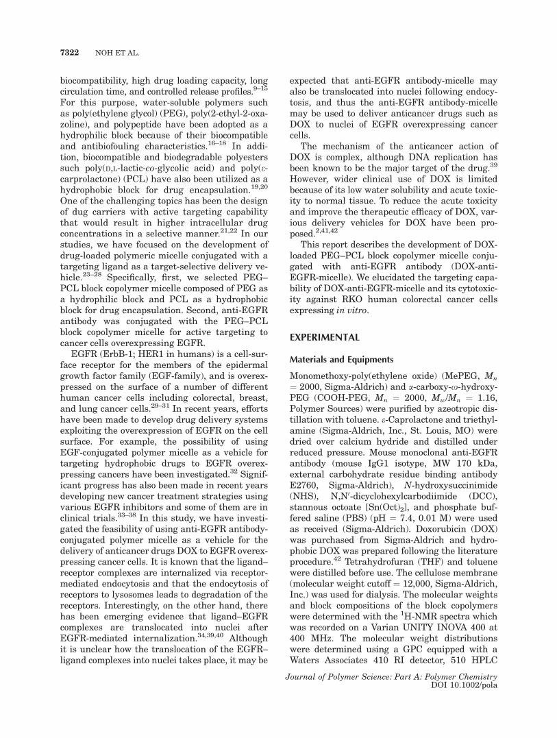
analysis. As summarized in Table 2, the mean di-ameter of the micelle derived from MePEG-b-PCL-micelle (PEG-PCL-micelle) was 25 nm (poly-dispersity factor, l2/C
2 ¼ 0.22). PEG-PCL-micelleand DOX-micelle exhibited similar mean diame-ter (25 nm, l2/C
2 ¼ 0.29).42,45 The micelles visual-ized by TEM were spherical as shown in Figure 2,and the size of DOX-micelle was about 25 nm.DOX-anti-EGFR-micelle was prepared by addinganti-EGFR-PEG-b-PCL into a DOX-micelle solu-tion. The mean diameter of DOX-anti-EGFR-micelle was 31 nm (l2/C
2 ¼ 0.31). As shown inFigure 2(c,d), the CONTIN analyses of theautocorrelation function for DOX-micelle andDOX-anti-EGFR-micelle respectively, showedmonomodal distributions. The quantities of encap-sulated DOX in DOX-micelle and DOX-anti-EGFR-micelle were 3.0 and 4.0 wt % of totalweight, respectively.
The conjugation of anti-EGFR antibody withthe block copolymer micelles was studied usingthe dot blot method. As shown in Figure 3, anti-EGFR antibody was detected only in DOX-anti-EGFR-micelles but not in the DOX-micelles or
Table 2. Properties of Block Copolymer Micelles
Sample
LoadingContent(wt %)
d(nm)a (l2/C
2)b
PEG-PCL-micelle – 25 0.22DOX-micelle 3.0 25 0.29DOX-anti-EGFR-micelle 4.0 31 0.31
aMean diameters of micelles in distilled water at 25 �C.bPolydispersity factor.
Figure 2. TEM images of (a) DOX-micelle and (b) DOX-anti-EGFR-micelle. Sizedistribution histograms of (c) DOX-micelle and (d) DOX-anti-EGFR-micelle at scat-tering angle of 90� (from CONTIN analysis of the autocorrelation function).
7326 NOH ET AL.
Journal of Polymer Science: Part A: Polymer ChemistryDOI 10.1002/pola

free DOX, indicating that anti-EGFR antibodywas conjugated onto DOX-micelles. The numberof anti-EGFR antibody conjugated to a micelle,quantified using the Micro BCA assay method(162 lg/mg), was 1.1 assuming that the aggrega-tion number of the micelle is � 200.
In vitro Release of DOX from Polymer Micelles
Figure 4 shows the rate of release of DOX fromDOX-micelle in phenol red-free DMEM. Approxi-mately 10% of DOX was released from DOX-micelle in DMEM in 4 h of incubation, and nofurther release occurred thereafter.
Cellular Uptake of DOX with FluorescenceMicroscopy
Figure 5(a) shows the DOX fluorescence distribu-tion, nuclei stained with 40-6-diamidino-2-phentli-nodole (DAPI), and merging of the DOX fluores-cence and DAPI staining in the RKO cells incu-bated with free DOX, DOX-micelle, and DOX-anti-EGFR-micelle for 1, 4, and 8 h. Note that theresults of only 1–8 h uptake are shown since con-siderable fractions of cells died of apoptosis after8 h treatment. Since DOX works as a topoisomer-ase inhibitor, nucleation of DOX is important forkilling of the target cancer cells. In the cells incu-bated with free DOX for 1 h, weak DOX fluores-
cence could be observed in the nuclei as well asoutside the nuclei. As the incubation time withfree DOX was increased to 4 and 8 h, the intensityof DOX fluorescence in the nuclei slightlyincreased while that around the nuclei dimin-ished. When the cells were incubated with DOX-micelle for 1 or 4 h, the nuclei exhibited littleDOX fluorescence whereas rather strong DOXfluorescence intensity could be observed aroundthe nuclei. Upon incubation with DOX-micelle for8 h, the DOX fluorescence intensity in the nucleislightly increased and that around the nuclei stillremained strong. It appeared that DOX-micellewas trapped outside the nuclei, probably in thenuclear membrane, endosomes, or lysosomes, asreported by other investigators.23,46 In the cellsincubated with DOX-anti-EGFR-micelle for 1 h,intense DOX fluorescence could be observed bothinside and outside the nuclei. As the incubationtime with DOX-anti-EGFR-micelle was increasedto 4 and 8 h, the nuclear DOX fluorescence inten-sity further increased while that in the cytoplasmappeared diminished slightly. The relative inten-sity of DOX fluorescence in the nuclei of the cellstreated with free DOX, DOX-micelle, and DOX-anti-EGFR-micelle for 1–8 h were quantitatedand shown in Figure 5(b). The intensity of nuclearDOX fluorescence in the cells treated with freeDOX and that of cells treated with DOX-micellewere similar, and it increased only slightly duringthe 8 h incubation. On the other hand, the nu-clear DOX fluorescence intensity in the cellstreated with DOX-anti-EGFR-micelle progres-sively increased during the 8 h incubation. More
Figure 3. Dot blot analysis for conjugation of anti-EGFR antibody with micelle. Extent of conjugation ofanti-EGFR antibody to micelle was monitored usingthe Dot blot method. Samples (2 lL) were loadedfrom each micelle fraction on the membrane and dry.The membrane was incubated in blocking solution of5% skim milk for 1 h. After washing, the membranewas immersed into the secondary antibody. Specificantigen–antibody complex was detected using theECL western blotting detection system.
Figure 4. Release of DOX from DOX-micelle inDMEM media. The percentage of DOX released isshown as a function of incubation time at 37 �C.
SYNTHESIS OF ANTI-EGFR-CONJUGATED PEG-B-PCL 7327
Journal of Polymer Science: Part A: Polymer ChemistryDOI 10.1002/pola

importantly, the DOX fluorescence intensity in thenuclei of the cells treated with DOX-anti-EGFR-micelle was about twofold greater (p\ 0.05) thanthat in the cells treated with free DOX or DOX-micelle throughout the 8 h incubation period. It
could be concluded that DOX was effectivelydelivered to nuclei when cells were treated withDOX-anti-EGFR-micelle. Pertinent to our con-clusion, there are compelling experimental evi-dence indicating that EGFR and its ligands are
Figure 5. (a) Fluorescence microscopy images of RKO cells incubated with freeDOX, DOX-micelle, and DOX-anti-EGFR-micelle for 1–8 h. The images show theDOX fluorescence (red), nuclear staining by DAPI (blue) and merged. (b) Relative in-tensity of DOX fluorescence in the nuclei of RKO cells incubated with free DOX,DOX-micelle, or DOX-anti-EGFR-micelle for 1–8 h. Means of more than 40 nucleiwith 1 S.E. are shown.
7328 NOH ET AL.
Journal of Polymer Science: Part A: Polymer ChemistryDOI 10.1002/pola

accumulated in the nuclei of various human tis-sues and cancer cell lines.34,40 It is known thatwhen ligands including EGF bind to cell surfaceEGFR, the ligand–receptor complexes are rap-idly internalized through endocytosis to lyso-somes and undergo proteolytic degradation. Onthe other hand, some of the ligand–EGFR com-plexes are translocated into nuclei possiblythrough the conventional nuclear importing sys-tem.34 Note that, in this study, we used an anti-EGFR antibody that binds to non-EGF-bindingsite, unlike other anti-EGFR antibody which isknown to block ligand-binding sites and undergodegradation without internalization.35
In vitro Cytotoxicity Assay
Figure 6 shows the flow cytometrically deter-mined apoptosis in RKO cells treated with freeDOX, DOX-micelle, or DOX-anti-EGFR-micelle.About 30 and 50% of cells died of apoptosis whencells were treated with free DOX or DOX-micellesfor 24 h, respectively. The DOX-anti-EGFR-mi-celle was most effective among the three drugpreparations causing apoptosis in about 70% ofthe cells in 24 h. The differences in % apoptosisamong the three different groups were statisti-cally significant at 16 h. However, after 24 h incu-bation, the % apoptosis caused by DOX-anti-EGFR-micelle was statistically greater than thatcaused by free DOX, but not that caused by DOX-micelle. Note that encouraging results have beenobserved in recent clinical trials to test the effec-tiveness of anti-EGFR antibodies alone or in com-bination with other anticancer agents for thetreatment of lung or breast cancer.47–50 We mayattribute the significantly greater apoptosis byDOX-anti-EGFR-micelle relative to that by DOXalone or DOX-micelle to the increased nuclearaccumulation of DOX and thus increased celldeath by DOX.
In using cell surface molecules as a target fordelivery of anticancer agents to the cells, impor-tant factors are specificity, stability, and availabil-ity of the target. Other factors that are also impor-tant for successful delivery of drugs to targetsinside the cells are the rate of internalization andthe extent and rate of the drugs to escape fromthe endosomes and/or lysosomes. As shown in Fig-ure 5(a), considerable amount of DOX remainedoutside of nuclei when cells were treated withDOX-micelle or DOX-anti-EGFR-micelle possiblybecause the vehicles were trapped in the endo-somes or lysosomes in the cytoplasm. Further
work to improve the anticancer effect of DOX-anti-EGFR-micelle in vitro as well as in vivo usingRKO xenografts grown in nude mice are in pro-gress in our laboratory.
CONCLUSIONS
A cancer targeting ligand, anti-EGFR antibody,was introduced to the PEG-PCL block copolymermicelle which contained a hydrophobic anticancerdrug, DOX, and the uptake of DOX and the drug-induced apoptosis were investigated using EGFR-positive RKO cells. The fluorescence microscopystudy indicated that the cellular and nuclei accu-mulation of DOX was greater in the cells treatedwith DOX-anti-EGFR-micelles as compared withthat in the cells treated with free DOX or DOX-micelles. Likewise, treating the cells with DOX-anti-EGFR-micelle was more effective to causeapoptosis than treating the cells with free DOX orDOX-micelles. Our results are in agreement withthe reports by others that ligand–EGFR com-plexes are internalized and translocated into cellnuclei.34,39,40 It is concluded that the anti-EGFRantibody conjugated with copolymer micelles is anefficient vehicle for the delivery of cytotoxic drugsto cancer cells overexpressing EGFR.
This work supported by National R and D Programs forCancer Control, Ministry of Health and Welfare, Korea
Figure 6. The induction of apoptotic RKO cellstreated with free DOX, DOX-micelle, and DOX-anti-EGFR-micelle. Changes in % apoptosis, as deter-mined by FACS-CaliburTM Flow cytometer as a func-tion of treatment times are shown.
SYNTHESIS OF ANTI-EGFR-CONJUGATED PEG-B-PCL 7329
Journal of Polymer Science: Part A: Polymer ChemistryDOI 10.1002/pola

(0620340-1), National Nuclear Technology Program(2007) from KOSEF, and the Korea Health 21 R and DProjects (A062254).
REFERENCES AND NOTES
1. Campbell, R. B.; Balasubramanian, S. V.; Strau-binger, R. M. J Pharm Sci 2001, 90, 1091–1105.
2. Shukla, R.; Thomas, T. P.; Peters, J. L.; Desai, A.M.; Kukowska-Latallo, J.; Patri, A. K.; Kotlyar,A.; Baker, J. R., Jr. Bioconjug Chem 2006, 17,1109–1115.
3. Hosokawa, S.; Tagawa, T.; Niki, H.; Hirakawa, Y.;Nohga, K.; Nagaike, K. Br J Cancer 2003, 89,1545–1551.
4. Park, C. W.; Lee, S. J.; Kim, D.; Lee, D. S.; Kim,S. C. J Polym Sci Part A: Polym Chem 2008, 46,1954–1963.
5. Shi, M.; Wosnick, J. H.; Ho, K.; Keating, A.; Shoi-chet, M. S. Angew Chem Int Ed Engl 2007, 46,6126–6131.
6. Lee, C. C.; Gillies, E. R.; Fox, M. E.; Guillandeu,S. J.; Frechet, J. M. J.; Dy, E. E.; Szoka, F. C.Proc Natl Acad Sci USA 2006, 103, 16649–16654.
7. Guillaudeu, S. J.; Fox, M. E.; Haidar, Y. M.; Dy,E. E.; Szoka, F. C.; Frechet, J. M. J. BioconjugChem 2008, 19, 461–469.
8. Hong, R.; Han, G.; Fernandez, J. M.; Kim, B. J.;Forbes, N. S.; Rotello, V. M. J Am Chem Soc 2006,128, 1078–1079.
9. Lee, S. C.; Kim, C. H.; Kwon, I. C.; Chung, H.;Jeong, S. Y. J Control Release 2003, 89, 437–446.
10. Allen, C.; Han, J.; Yu, Y.; Maysinger, D.; Eisen-berg, A. J Control Release 2000, 63, 275–286.
11. Otsuka, H.; Nagasaki, Y.; Kataoka, K. Adv DrugDeliv Rev 2003, 55, 403–4419.
12. Yokoyama, M.; Fukushima, S.; Uehara, R.; Oka-moto, K.; Kataoka, K.; Sakurai, Y.; Okano, T. JControl Release 1998, 50, 79–92.
13. Inoue, T.; Chen, G.; Nakamae, K.; Hoffman, A. S.J Control Release 1998, 51, 221–229.
14. Yoo, H. S.; Park, T. G. J Control Release 2001, 70,63–70.
15. Yamamoto, Y.; Nagasaki, Y.; Kato, Y.; Sugiyama,Y.; Kataoka, K. J Control Release 2001, 77, 27–38.
16. Kataoka, K.; Harada, A.; Nagasaki, Y. Adv DrugDeliv Rev 2001, 47, 113–131.
17. Adams, M. L.; Lavasanifar, A.; Kwon, G. J PharmSci 2003, 92, 1343–1355.
18. Savicdot, R.; Luo, L.; Eisenberg, A.; Maysinger, D.Science 2003, 300, 615–618.
19. Liggins, R. T.; Burt, H. M. Adv Drug Deliv Rev2002, 54, 191–202.
20. Burt, H. M.; Zhang, X.; Toleikis, P.; Embree, L.;Hunter, W. L. Colloids Surf B 1999, 16, 161–171.
21. Vega, J.; Ke, S.; Fan, Z.; Wallace, S.; Charsangavej,C.; Li, C. PharmRes 2003, 20, 826–832.
22. Torchilin, V. P.; Lukyanov, A. N.; Gao, Z.; Papa-hadjopoulos-Sternberg, B. Proc Natl Acad SciUSA 2003, 100, 6039–6044.
23. Yoo, H. S.; Park, T. G. J Control Release 2004, 96,273–283.
24. Nasongkla, N.; Bey, E.; Ren, J.; Ai, H.; Khemtong,C.; Guthi, J. S.; Chin, S. F.; Sherry, A. D.; Booth-man, D. A.; Gao, J. Nano Lett 2006, 6, 2427–2430.
25. Park, E. K.; Kim, S. Y.; Lee, S. B.; Lee, Y. M. JControl Release 2005, 109, 158–168.
26. Hood, J. D.; Bednarski, M.; Frausto, R.; Guccione,S.; Reisfeid, R. A.; Xiang, R.; Cheresh, D. A. Sci-ence 2002, 296, 2404–2407.
27. Nasongkla, N.; Shuai, X.; Ai, H.; Weinberg, B. D.;Pink, J.; Boothman, D. A.; Gao, J. Angew ChemInt Ed Engl 2004, 43, 6323–6327.
28. Lee, E. S.; Na, K.; Bae, Y. H. Nano Lett 2005, 5,325–329.
29. Herbst, R. S.; Shin, D. M. Cancer 2002, 94, 1593–1611.
30. Lynch, T. J.; Bell, D. W.; Sordella, R.; Gurubhaga-vatula, S.; Okimoto, R. A.; Brannigan, B.W.; Har-ris, P. L.; Haserlat, S. M.; Supko, J. G.; Haluska, F.G.; Louis, D. N.; Christiani, D. C.; Settleman, J.;Haber, D. A. N Engl J Med 2004, 350, 2129–2139.
31. Jhawer, M.; Goel, S.; Wilson, A. J.; Montagna, C.;Ling, Y.-H.; Byun, D.-S.; Nasser, S.; Arango, D.;Shin, J.; Klampfer, L.; Augenlicht, L. H.; Soler, R. P.;Mariadason, J. M. Cancer Res 2008, 68, 1953–1961.
32. Zeng, F.; Lee, H.; Allen, C. Bioconjug Chem 2006,17, 399–409.
33. Venook, A. P. Cancer 2005, 103, 2435–2446.34. Lo, H. W.; Hung, M.C. Br J Cancer 2006, 94, 184–
188.35. Rocha-Lima, C.M.; Soares, H. P.; Raez, L. E.; Sin-
gai, R. Cancer Control 2007, 14, 295–304.36. Vallbohmer, D.; Lenz, H. J. Clin Colorectal Cancer
2005(Suppl 1), S19–S27.37. Mendelsohn, J.; Baselga, J. Semin Oncol 2006,
33, 369–385.38. Vokes, E. E.; Chu, E. Oncology (Williston Park)
2006, 5(Suppl 2), 15–25.39. Fornari, F. A.; Randolph, J. K.; Yalowich, J. C.;
Ritke, M. K.; Gewirtz, D. A. Mol Pharmacol 1994,45, 649–656.
40. Szumiel, I. Cell Signal 2006, 18, 1537–1548.41. Kwon, G. S.; Yokoyama, M.; Okano, T.; Sakurai,
Y.; Kataoka, K. Pharm Res 1993, 10, 970–974.42. Shuai, X.; Ai, H.; Nasongkla, N.; Kim, S.; Gao, J.
J Control Release 2004, 98, 415–426.43. Lee, H.; Jang, I. H.; Ryu, S. H.; Park, T. G.
Pharm Res 2003, 20, 818–825.44. Kullberg, E. B.; Bergstrand, N.; Carlsson, J.;
Edwards, K.; Johnsson, M.; Sjoberg, S.; Gedda, L.Bioconjug Chem 2002, 13, 737–743.
45. Shuai, X.; Merdan, T.; Schaper, A. K.; Xi, F.; Kis-sel, T. Bioconjug Chem 2004, 15, 441–448.
46. Gilles, E. R.; Frechet, J. M. Bioconjug Chem 2005,16, 361–368.
7330 NOH ET AL.
Journal of Polymer Science: Part A: Polymer ChemistryDOI 10.1002/pola

47. Patnaik, A.; Beeram, M.; de Bono, J.S.; Mita, A.;Chu, S. C.; Rowinsky, E. K.; Schwartz, G.;O’Rourke, P.; Takimoto, C. H.; Tolcher, A. W. Pro-ceedings of American Society of Clinical OncologyAnnual Meeting 2005, Abstract No. 2000.
48. Polychronis, A.; Sinnett, H. D.; Hadjiminas, D.;Singhal, H.; Mansi, J. L.; Ali, S.; Slade, M. J.;Shousha, S.; Morrisson, K.; Coombes, R. C. Pro-ceedings of American Society of Clinical OncologyAnnual Meeting 2005, Abstract No. 552.
49. Ciardiello, F.; Troiani, T.; Caputo, F.; de Lauren-tiis, M.; Tortora, G.; Palmieri, G.; de Vita, F.;Colantuoni, G.; de Placido, S.; Bianco, A. Proceed-ings of American Society of Clinical Oncology An-nual Meeting 2005, Abstract No. 3080.
50. Graham, D. L.; Hillman, D. W.; Hobday, T. J.;Rousey, S. R.; Nair, S. G.; Soori, G. S.; Sabagh, T.M.; Perez, E. A. Proceedings of American Societyof Clinical Oncology Annual Meeting 2005, 23,Abstract No. 644.
SYNTHESIS OF ANTI-EGFR-CONJUGATED PEG-B-PCL 7331
Journal of Polymer Science: Part A: Polymer ChemistryDOI 10.1002/pola


![A novel diblock copolymer of (monomethoxy poly [ethylene glycol]-oleate) with a small hydrophobic fraction to make stable micelles/polymersomes for curcumin delivery to cancer cells](https://static.fdokumen.com/doc/165x107/6344914403a48733920af291/a-novel-diblock-copolymer-of-monomethoxy-poly-ethylene-glycol-oleate-with-a.jpg)