TiO2 Nanoparticles Dispersion in Block-Copolymer ... - MDPI
15
J. Funct. Biomater. 2022, 13, 39. https://doi.org/10.3390/jfb13020039 www.mdpi.com/journal/jfb Article TiO2 Nanoparticles Dispersion in Block-Copolymer Aqueous Solutions: Nanoarchitectonics for Self-Assembly and Aggregation Valeria Conti Nibali 1 , Giovanna D’Angelo 1 , Antonella Arena 2 , Carmine Ciofi 2 , Graziella Scandurra 2 and Caterina Branca 1, * 1 Department of Mathematical and Computer Science, Physical Sciences and Earth Sciences, University of Messina, Viale F. Stagno d’Alcontres, 98166 Messina, Italy; [email protected] (V.C.N.); [email protected] (G.D.) 2 Department of Engineering, University of Messina, Contrada di Dio, I-98166, 98166 Messina, Italy; [email protected] (A.A.); [email protected] (C.C.); [email protected] (G.S.) * Correspondence: [email protected]; Tel.: +39-090-6765017 Abstract: Achieving homogenous dispersion of nanoparticles inside a polymeric matrix is a great challenge for numerous applications. In the present study, we aim at understanding the role of dif- ferent factors on the dispersion properties of TiO2 in pluronic F-127 mixtures. The mixtures were prepared with different pH and guest/host ratios and investigated by UV-Vis spectroscopy, dy- namic light scattering, infrared spectroscopy and electrical conductivity. Depending on the prepa- ration conditions, different amounts of TiO2 were loaded within the copolymer as quantitatively determined by UV-Vis spectroscopy. The different content of nanoparticles has direct implications on the gelation and micellization of pluronic analyzed by dynamic light scattering. The information derived on the self-assembly behavior was interpreted in relation to the infrared and conductivity measurements results. Together, these results shed light on the most favorable conditions for im- proving the nanoparticle dispersion inside the copolymer matrix and suggest a possible strategy to design functional nanoparticle-polymer systems. Keywords: pluronic F127; TiO2; self-assembly; dynamic light scattering 1. Introduction Nanoparticles exhibit unique properties that make them suitable for multifunctional applications. For most of them, effectively dispersing nanoparticles in solution is often necessary but not always easily achievable, and it requires a correct processing strategy. In some cases, the incorporation of nanoparticles within a polymer host became a winning strategy for homogeneous dispersions whose properties are a unique combination of the guest and the host. Tailoring these properties is a great challenge in designing materials with advanced performances. Among transition metal oxide-based nanoparticles, titanium dioxide nanoparticles are the most extensively used in many different fields as gas sensors [1–5], solar cells [6], photocatalysts [7–10], and additives in food and cosmetics [11–13]. Moreover, thanks to their high biocompatibility and low toxicity, they have been extensively used in nano- medicine and nanobiotechnology for drug delivery, tissue engineering and others [14– 19]. Whatever their field of application, the dispersion and stability of TiO2 nanoparticles in aqueous or in a more complex medium play a key role in improving the efficiency and performance of the systems. It is widely reported that the dispersion of nanoparticles in aqueous systems generally modifies the physicochemical properties, e.g., agglomeration Citation: Conti Nibali, V.; D’Angelo, G.; Arena, A.; Ciofi, C.; Scandurra, G. TiO2 Nanoparticles Dispersion in Block-Copolymer Aqueous Solutions: Nanoarchitectonics for Self-Assembly and Aggregation. J. Funct. Biomater. 2022, 13, 39. https://doi.org/10.3390/jfb13020039 Academic Editors: Katsuhiko Ariga, Fabien Grasset and Yann Molard Received: 17 March 2022 Accepted: 7 April 2022 Published: 9 April 2022 Publisher’s Note: MDPI stays neu- tral with regard to jurisdictional claims in published maps and institu- tional affiliations. Copyright: © 2022 by the authors. Li- censee MDPI, Basel, Switzerland. This article is an open access article distributed under the terms and con- ditions of the Creative Commons At- tribution (CC BY) license (https://cre- ativecommons.org/licenses/by/4.0/).
-
Upload
khangminh22 -
Category
Documents
-
view
2 -
download
0
Transcript of TiO2 Nanoparticles Dispersion in Block-Copolymer ... - MDPI

J. Funct. Biomater. 2022, 13, 39. https://doi.org/10.3390/jfb13020039 www.mdpi.com/journal/jfb
Article
TiO2 Nanoparticles Dispersion in Block-Copolymer Aqueous
Solutions: Nanoarchitectonics for Self-Assembly
and Aggregation
Valeria Conti Nibali 1, Giovanna D’Angelo 1, Antonella Arena 2, Carmine Ciofi 2, Graziella Scandurra 2
and Caterina Branca 1,*
1 Department of Mathematical and Computer Science, Physical Sciences and Earth Sciences, University of
Messina, Viale F. Stagno d’Alcontres, 98166 Messina, Italy; [email protected] (V.C.N.);
[email protected] (G.D.) 2 Department of Engineering, University of Messina, Contrada di Dio, I-98166, 98166 Messina, Italy;
[email protected] (A.A.); [email protected] (C.C.); [email protected] (G.S.)
* Correspondence: [email protected]; Tel.: +39-090-6765017
Abstract: Achieving homogenous dispersion of nanoparticles inside a polymeric matrix is a great
challenge for numerous applications. In the present study, we aim at understanding the role of dif-
ferent factors on the dispersion properties of TiO2 in pluronic F-127 mixtures. The mixtures were
prepared with different pH and guest/host ratios and investigated by UV-Vis spectroscopy, dy-
namic light scattering, infrared spectroscopy and electrical conductivity. Depending on the prepa-
ration conditions, different amounts of TiO2 were loaded within the copolymer as quantitatively
determined by UV-Vis spectroscopy. The different content of nanoparticles has direct implications
on the gelation and micellization of pluronic analyzed by dynamic light scattering. The information
derived on the self-assembly behavior was interpreted in relation to the infrared and conductivity
measurements results. Together, these results shed light on the most favorable conditions for im-
proving the nanoparticle dispersion inside the copolymer matrix and suggest a possible strategy to
design functional nanoparticle-polymer systems.
Keywords: pluronic F127; TiO2; self-assembly; dynamic light scattering
1. Introduction
Nanoparticles exhibit unique properties that make them suitable for multifunctional
applications. For most of them, effectively dispersing nanoparticles in solution is often
necessary but not always easily achievable, and it requires a correct processing strategy.
In some cases, the incorporation of nanoparticles within a polymer host became a winning
strategy for homogeneous dispersions whose properties are a unique combination of the
guest and the host. Tailoring these properties is a great challenge in designing materials
with advanced performances.
Among transition metal oxide-based nanoparticles, titanium dioxide nanoparticles
are the most extensively used in many different fields as gas sensors [1–5], solar cells [6],
photocatalysts [7–10], and additives in food and cosmetics [11–13]. Moreover, thanks to
their high biocompatibility and low toxicity, they have been extensively used in nano-
medicine and nanobiotechnology for drug delivery, tissue engineering and others [14–
19].
Whatever their field of application, the dispersion and stability of TiO2 nanoparticles
in aqueous or in a more complex medium play a key role in improving the efficiency and
performance of the systems. It is widely reported that the dispersion of nanoparticles in
aqueous systems generally modifies the physicochemical properties, e.g., agglomeration
Citation: Conti Nibali, V.; D’Angelo,
G.; Arena, A.; Ciofi, C.; Scandurra,
G. TiO2 Nanoparticles Dispersion in
Block-Copolymer Aqueous
Solutions: Nanoarchitectonics for
Self-Assembly and Aggregation. J.
Funct. Biomater. 2022, 13, 39.
https://doi.org/10.3390/jfb13020039
Academic Editors: Katsuhiko Ariga,
Fabien Grasset and Yann Molard
Received: 17 March 2022
Accepted: 7 April 2022
Published: 9 April 2022
Publisher’s Note: MDPI stays neu-
tral with regard to jurisdictional
claims in published maps and institu-
tional affiliations.
Copyright: © 2022 by the authors. Li-
censee MDPI, Basel, Switzerland.
This article is an open access article
distributed under the terms and con-
ditions of the Creative Commons At-
tribution (CC BY) license (https://cre-
ativecommons.org/licenses/by/4.0/).

J. Funct. Biomater. 2022, 13, 39 2 of 15
state and surface charge variation [20]. Therefore characterizing the state of titania nano-
particles is of great significance, as they greatly affect many functionalities such as the
photocatalytic activity [21,22], the toxicity [23], the particle electronic structure, surface
defect density and the surface sorption sites [24].
Thus far, many studies have been performed to investigate the stability of TiO2
nanofluids with different concentrations and under various pH and ionic strength condi-
tions [20,25–30]. The final objective of such research works is to define the best conditions
for optimal performance of TiO2-based composites, whatever the field of application. In
particular, there is a strong applicative interest in realizing well-ordered porous titania
thin films. They have several advantages, such as a huge surface area and higher stability
that significantly affect the efficiency of the electronic properties. Among various possible
methods, the employment of structure-directing agents, including phosphates, ionic and
nonionic surfactants, amines, and block copolymers [31–42], was very promising.
From these studies, it emerged that the structural and morphological properties of
surfactant-based TiO2 systems greatly depend on the method of synthesis, the type and
concentration of surfactant used, and the thermal treatment temperature other than the
type and pH of copolymer solvent. Regardless of the procedure of synthesis used, as a
final step, a high-temperature thermal treatment was performed, which enabled efficient
removal of the copolymer template. Only after removal, the physical-chemical properties
of the TiO2 films were investigated.
Unlike these studies, in the present work, our object of interest is the copolymer-TiO2
water dispersion as a whole. TiO2 nanoparticles were dispersed in pluronic F127/water
suspensions at different copolymer concentrations and pH.
Pluronic F127 is a nonionic triblock copolymer consisting of hydrophilic poly (eth-
ylene oxide) and hydrophobic poly (propylene oxide) blocks (PEO–PPO–PEO) with a
PEO/PPO ratio of 2:1. Above a critical micelle concentration, the block copolymers assem-
ble into spherical micelles thanks to the difference in hydrophobicity between PEO and
PPO blocks. Reversible gelation can occur only above some concentration and tempera-
ture [43–51].
The choice of pluronic F127 stems from its thermosensitivity and capability to solu-
bilize and stabilize drugs inside the micelle core. This system is thus particularly interest-
ing given the possible applications in many fields. In particular, it is one of the most
widely used triblock copolymers in pharmaceutical formulations because of the good wa-
ter solubility through the high content of EO, the low toxicity in the body and the ability
to encapsulate any hydrophobic agents [52–59]. However, it is well known that the addi-
tion of co-solvents or solutes to pluronic water solutions can influence its properties, in-
ducing phase changes [60–68].
In the present study, we are interested in analyzing how the dispersion or aggrega-
tion of titania nanoparticles inside pluronic F127 can influence the structural arrange-
ments and, consequently, the systems' dynamics. As it is well known, when nanoparticles
are dispersed in solution, they undergo phenomena of agglomeration or aggregation, the
difference being in the strength of interaction. Agglomerates are weakly bound collections
of nanoparticles, whereas aggregates are tightly bound collections difficult to break up by
mechanical forces. The classical Derjaguin–Landau–Verwey–Overbeek (DLVO) theory
predicts the overall force between particles by combining Van der Waals attractive forces
and repulsive forces arising from the electrical double layer (EDL) around the particles
[69,70]. Other non-DLVO forces can influence nanoparticle dispersion, such as hydration,
hydrophobic, steric, electronic, and electrostatic forces [69,70]. Among these, steric effects
should be included in the case of nonionic polymer coatings; these are generally repulsive
interactions, but if the polymer chains can form bridges between particles, that can cause
aggregation. For all these reasons, it is clear that the dispersion properties are strongly
dependent on parameters such as pH and ionic strength as they directly affect the zeta
potential of the solution and the double layer thickness.
J. Funct. Biomater. 2022, 13, 39 3 of 15
Starting from these considerations, we have chosen to prepare the dispersions at pH
values far from the isoelectric point (IEP) of the TiO2 suspension, approximately 6.2 [29].
In such a way, the presence of particle surface charge should enhance the electrostatic
repulsion between metallic nanoparticles disfavoring, or suppressing, agglomeration. For
the same reason, we decided to prepare the samples at a low ionic strength since, accord-
ing to previous studies, the high ionic strength of the solution compresses the electrical
double layer [20,29]. In preparing the pluronic-nanoparticles dispersions, another im-
portant factor to be considered is the copolymer concentration since, depending on tem-
perature, it affects the phase behavior of the copolymer [43–50]. In the present case, the
prepared base-pluronic solutions have weight fractions of 14% and 20 wt%; in the tem-
perature range between 20 °C and 30 °C, the lowest concentration is always in a sol state,
whereas the highest one move from a sol state to a gel-like state [50,51].
All these aspects were taken into consideration while we conducted our research
with the final aim to define the best experimental conditions for optimal dispersion of
TiO2 in pluronic F-127 mixtures. The samples, prepared under different experimental con-
ditions, were investigated using UV-Vis spectroscopy, attenuated Fourier transform in-
frared spectroscopy (FTIR-ATR), dynamic light scattering (DLS) and electrical conductiv-
ity. The obtained results give valuable information on the dispersion state of nanoparti-
cles; this knowledge is potentially useful for developing and optimizing copolymer-based
nanosystems.
2. Materials and Methods
2.1. Materials
TiO2 nanoparticles dry powder anatase phase (average size 4–8 nm) were purchased
from PlasmaChem GmbH (Berlin, Germany). The powder is free of organic stabilizer. Plu-
ronic (F127, 12.600 Da) was obtained from Sigma-Aldrich (Dorset, UK). Hydrogen chlo-
ride (HCl), sodium hydroxide (NaOH), sodium chloride (NaCl) and deionized water were
purchased from Sigma-Aldrich. All the chemicals are of analytical grade.
2.2. Sample Preparation
TiO2 dry powder was suspended in 10 ml of solution at a 10.0 mg/mL concentration.
The solutions had pre-adjusted pH values of 4 and 10 and an ionic strength equal to 0.001
M. The dispersions were magnetically stirred for 24 h and then sonicated for 15 min using
a bath sonicator. After that, the suspensions were centrifuged for 30 min and 5 ml of each
supernatant was sampled and analyzed by DLS. The size distribution calculated by using
a CONTIN algorithm evidenced the presence of a bimodal distribution centered at 36 and
280 nm for the dispersion at pH 10 and 50 and 320 nm for the suspension at pH 4, respec-
tively (data not shown). To remove the large agglomerates, the supernatants were then
filtered and analyzed by UV-Vis spectroscopy to determine the effective TiO2 concentra-
tion. Absorption measurements were repeated in time for 24 h without observing any sig-
nificant variation, thereby excluding any change in the dispersion state of TiO2 in this time
range.
Stock solutions of Pluronic F-127 at weight fractions of 14 wt% and 20 wt% were pre-
pared, and aliquots of the TiO2 dispersions at the two pH values were then added. Atten-
tion was paid to adding the same TiO2 nanoparticle concentration in the final suspension
(0.1 wt% corresponding to 1 mg/mL).
Table 1 reports the sample labels as a function of the concentration of pluronic, TiO2
and pH conditions. The dispersions were gently stirred at 4 °C for 24 h and left overnight
in the refrigerator until a clear solution was obtained. No sol-gel transition was observed
for these systems up to 40 °C, as evidenced by the tube inversion method. The dispersions
were stirred again and centrifuged; then, the supernatants were removed and transferred
to fresh cuvettes for further analysis. No pH change from the stock TiO2 solutions was
J. Funct. Biomater. 2022, 13, 39 4 of 15
observed in the final dispersions. For comparison, two plain pluronic solutions with 14
wt% (PA) and 20 wt% (PB) concentrations were prepared.
Table 1. Samples labels with the corresponding initial composition and pH values. In the last col-
umn, the amount of TiO2 loaded into the samples as determined by UV-Vis measurements is re-
ported.
Sample Pluronic wt% TiO2 wt% pH TiO2 (Mean Value)
mg/mL
PAT-pH4 14 0.1 4 0.83 (0.083 wt%)
PAT-pH10 14 0.1 10 0.69 (0.069 wt%)
PBT-pH4 20 0.1 4 0.60 (0.060 wt%)
PBT-pH10 20 0.1 10 0.40 (0.040 wt%)
2.3. UV-Vis Spectroscopy
The UV-Vis measurements were made using a small-volume (100 μL) absorbance
cuvette (Hellma 105.201-QS) and a UV-1700 Shimadzu spectrophotometer in the 280–700
nm range. All measurements were carried out at room temperature and replicated three
times.
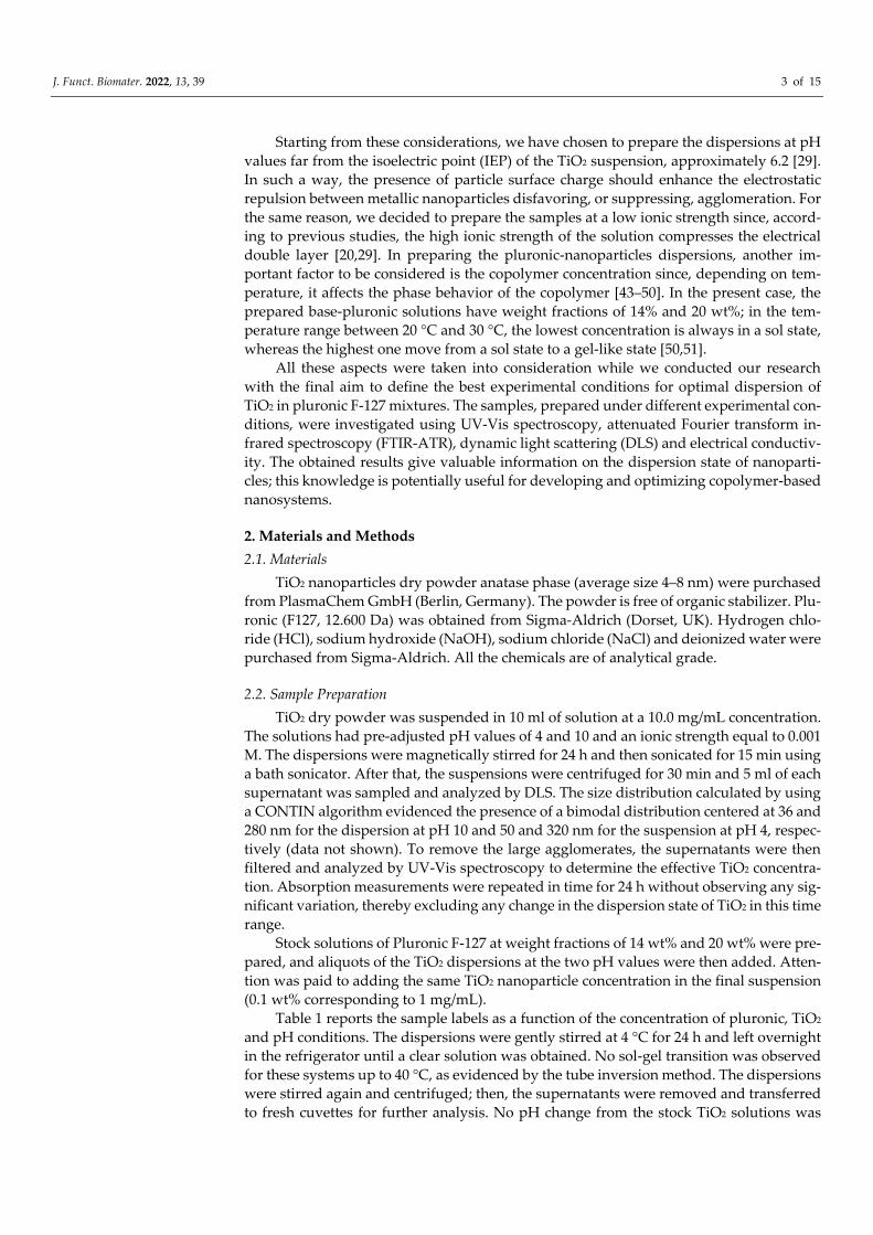
To quantitatively determine the concentration of TiO2 loaded in our samples, a cali-
bration plot was first made by recording the UV-Vis spectra for five TiO2 water solutions
of known concentration under the same pH and IS conditions. The intensity of the absorp-
tion peak at 330 nm was used to quantify TiO2. The linearity of the calibration curve was
evaluated by linear regression analysis evidencing a high value of fitting degree (R2>
0.999) (see the inset of Figure 1).
Figure 1. UV-Vis absorption spectra for the four pluronic-TiO2 dispersions studied in this work. For
comparison, the absorption spectrum of a pluronic-F127 water solution (PA) is also reported. In the
inset, the calibration plot for TiO2 is shown.
2.4. DLS Measurements
A laboratory-built goniometer equipped with single-mode fiber optics, two APD de-
tectors, and a multi-τ digital time correlator (LS Instrument AG) ALV single-photon de-
tector was used to perform DLS measurements. A He–Ne laser operating at 632.8 nm in
J. Funct. Biomater. 2022, 13, 39 5 of 15
linearly polarized single-frequency mode was used as an excitation source. The instru-
ment is equipped with a thermostated recirculating bath which allows us to maintain the
temperature with an accuracy of 0.1 °C. All measurements were performed at an angle of
90° to the incident beam at a temperature of 20 °C and 30 °C. Each sample was loaded in
a square low-volume cuvette and thermalized at each temperature for 30 min before meas-
uring. The repeatability of all measurements was verified with more than five measure-
ments. From the DLS experiment, we obtained the normalized intensity auto-correlation
function (ICF), g2 (q,t); details on the theoretical background of this technique are reported
elsewhere [68,71–73]. Decay times are estimated from the ICFs according to a fit model
reported in the results and discussion section. The CONTIN algorithm was also applied
to obtain the size distribution for the TiO2 solutions.
2.5. FTIR-ATR Spectroscopy
FTIR-ATR spectra covering the range 400–4000 cm−1 were recorded on a Bruker Ver-
tex 80V FTIR spectrometer equipped with a Bruker Platinum ATR accessory with a single
reflection diamond crystal. A background scan was recorded prior to the measurement
and subtracted from the sample spectra. All spectra are the average of three independent
measurements with 128 scans each, at a resolution of 2 cm−1. The ATR correction to each
spectrum was applied using the OPUS software (Bruker optics). The spectra were normal-
ized to the same area and compared to each other.
2.6. Electrical Conductivity
The electrical conductivity values were obtained from electrical resistance measure-
ments performed with an HP 4284a LCR meter. The instrument has been calibrated using
measurements on liquids of known conductivity, and it has been ascertained that con-
sistent measurements could be obtained in the 5 kHz–200 kHz frequency range. Measure-
ments were performed by transferring, using a pipette, a small volume of the samples in
ABS containers with a holding volume of 4.5 × 4.5 × 3 mm3 to limit the waste of material.
The electrodes immersed in the samples were two small gold connectors 3.18 mm long
and with a diameter of 0.5 mm, at a distance of 2.54 mm from each other. The conductivity
values were obtained by comparing the resistance curves obtained for the samples with
the calibration ones.
3. Results and Discussion
3.1. UV-Vis Spectroscopy
The UV-Vis spectra were first analyzed for the quantitative assessment of TiO2
loaded into the pluronic-based dispersions. Figure 1 shows the results of the absorbance
measurements for all the pluronic/TiO2 dispersions investigated and, as a reference, for a
pluronic water solution (PA sample). For this latter sample, the absorbance spectrum does
not reveal any significant absorption in the entire wavelength region investigated: the
peak at λ = 330 nm, observed for all the pluronic/TiO2 samples, can be attributed exclu-
sively to titanium dioxide. The observed changes in the intensity of this absorption peak
suggest the presence of a higher loading of TiO2 nanoparticles in the suspensions under
acidic conditions rather than in basic and, for the same pH, for the dispersions with the
lowest pluronic concentration.
Based on the calibration plot, see the inset of Figure 1, the effective concentration of
TiO2 in the pluronic-based dispersions was estimated; the obtained results are reported in
the last column of Table 1.
3.2. FTIR-ATR Measurements
The effect of a different loading of TiO2 inside the investigated samples was also ex-
amined by FTIR-ATR spectroscopy. Measurements were performed on samples purged
under dry nitrogen and left for equilibration for half an hour; nevertheless, as we will see,
J. Funct. Biomater. 2022, 13, 39 6 of 15
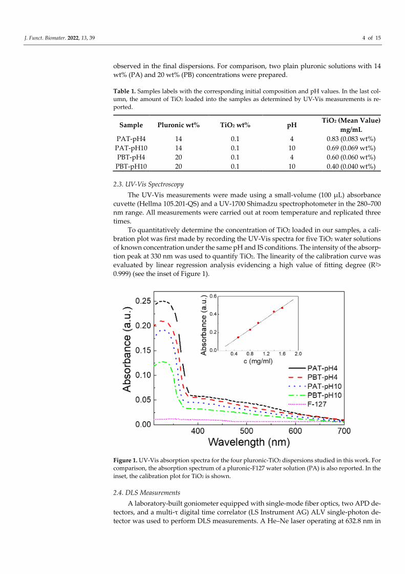
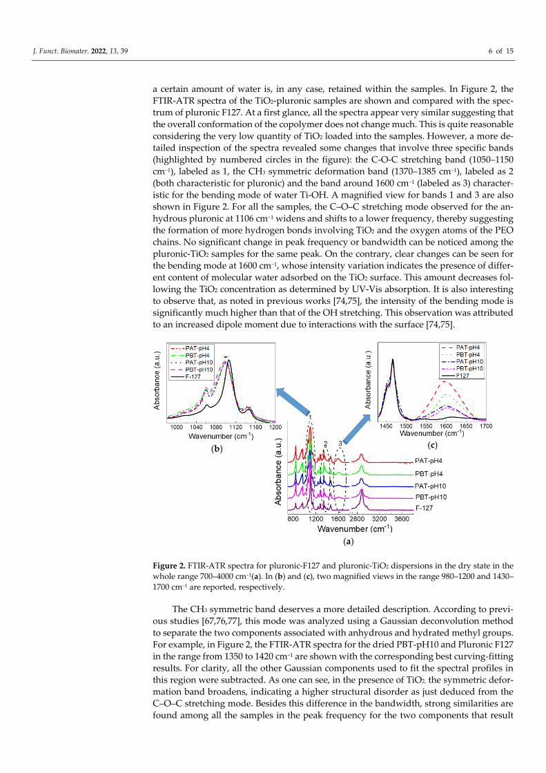
a certain amount of water is, in any case, retained within the samples. In Figure 2, the
FTIR-ATR spectra of the TiO2-pluronic samples are shown and compared with the spec-
trum of pluronic F127. At a first glance, all the spectra appear very similar suggesting that
the overall conformation of the copolymer does not change much. This is quite reasonable
considering the very low quantity of TiO2 loaded into the samples. However, a more de-
tailed inspection of the spectra revealed some changes that involve three specific bands
(highlighted by numbered circles in the figure): the C-O-C stretching band (1050–1150
cm−1), labeled as 1, the CH3 symmetric deformation band (1370–1385 cm−1), labeled as 2
(both characteristic for pluronic) and the band around 1600 cm−1 (labeled as 3) character-
istic for the bending mode of water Ti-OH. A magnified view for bands 1 and 3 are also
shown in Figure 2. For all the samples, the C–O–C stretching mode observed for the an-
hydrous pluronic at 1106 cm−1 widens and shifts to a lower frequency, thereby suggesting
the formation of more hydrogen bonds involving TiO2 and the oxygen atoms of the PEO
chains. No significant change in peak frequency or bandwidth can be noticed among the
pluronic-TiO2 samples for the same peak. On the contrary, clear changes can be seen for
the bending mode at 1600 cm−1, whose intensity variation indicates the presence of differ-
ent content of molecular water adsorbed on the TiO2 surface. This amount decreases fol-
lowing the TiO2 concentration as determined by UV-Vis absorption. It is also interesting
to observe that, as noted in previous works [74,75], the intensity of the bending mode is
significantly much higher than that of the OH stretching. This observation was attributed
to an increased dipole moment due to interactions with the surface [74,75].
Figure 2. FTIR-ATR spectra for pluronic-F127 and pluronic-TiO2 dispersions in the dry state in the
whole range 700–4000 cm−1(a). In (b) and (c), two magnified views in the range 980–1200 and 1430–
1700 cm−1 are reported, respectively.
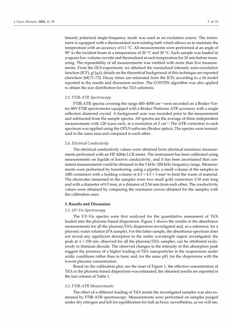
The CH3 symmetric band deserves a more detailed description. According to previ-
ous studies [67,76,77], this mode was analyzed using a Gaussian deconvolution method
to separate the two components associated with anhydrous and hydrated methyl groups.
For example, in Figure 2, the FTIR-ATR spectra for the dried PBT-pH10 and Pluronic F127
in the range from 1350 to 1420 cm−1 are shown with the corresponding best curving-fitting
results. For clarity, all the other Gaussian components used to fit the spectral profiles in
this region were subtracted. As one can see, in the presence of TiO2, the symmetric defor-
mation band broadens, indicating a higher structural disorder as just deduced from the
C–O–C stretching mode. Besides this difference in the bandwidth, strong similarities are
found among all the samples in the peak frequency for the two components that result
(a)
(b) (c)
J. Funct. Biomater. 2022, 13, 39 7 of 15
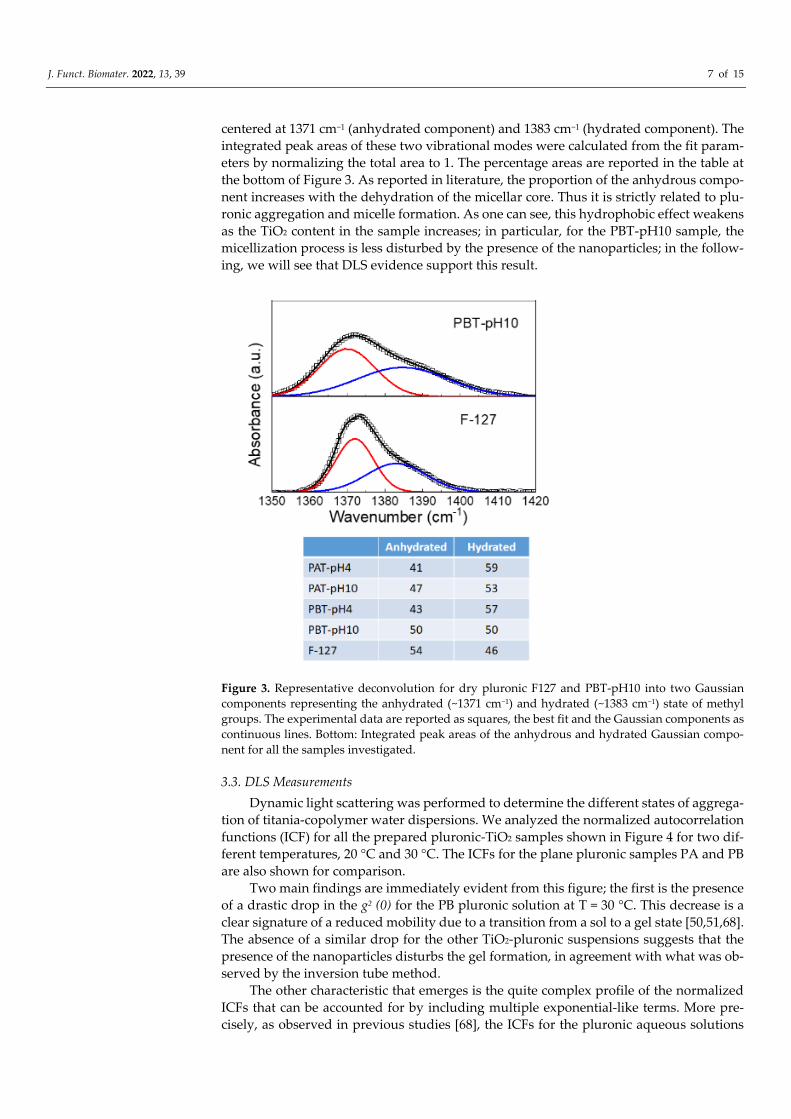
centered at 1371 cm−1 (anhydrated component) and 1383 cm−1 (hydrated component). The
integrated peak areas of these two vibrational modes were calculated from the fit param-
eters by normalizing the total area to 1. The percentage areas are reported in the table at
the bottom of Figure 3. As reported in literature, the proportion of the anhydrous compo-
nent increases with the dehydration of the micellar core. Thus it is strictly related to plu-
ronic aggregation and micelle formation. As one can see, this hydrophobic effect weakens
as the TiO2 content in the sample increases; in particular, for the PBT-pH10 sample, the
micellization process is less disturbed by the presence of the nanoparticles; in the follow-
ing, we will see that DLS evidence support this result.
Figure 3. Representative deconvolution for dry pluronic F127 and PBT-pH10 into two Gaussian
components representing the anhydrated (~1371 cm−1) and hydrated (~1383 cm−1) state of methyl
groups. The experimental data are reported as squares, the best fit and the Gaussian components as
continuous lines. Bottom: Integrated peak areas of the anhydrous and hydrated Gaussian compo-
nent for all the samples investigated.
3.3. DLS Measurements
Dynamic light scattering was performed to determine the different states of aggrega-
tion of titania-copolymer water dispersions. We analyzed the normalized autocorrelation
functions (ICF) for all the prepared pluronic-TiO2 samples shown in Figure 4 for two dif-
ferent temperatures, 20 °C and 30 °C. The ICFs for the plane pluronic samples PA and PB
are also shown for comparison.
Two main findings are immediately evident from this figure; the first is the presence
of a drastic drop in the g2 (0) for the PB pluronic solution at T = 30 °C. This decrease is a
clear signature of a reduced mobility due to a transition from a sol to a gel state [50,51,68].
The absence of a similar drop for the other TiO2-pluronic suspensions suggests that the
presence of the nanoparticles disturbs the gel formation, in agreement with what was ob-
served by the inversion tube method.
The other characteristic that emerges is the quite complex profile of the normalized
ICFs that can be accounted for by including multiple exponential-like terms. More pre-
cisely, as observed in previous studies [68], the ICFs for the pluronic aqueous solutions
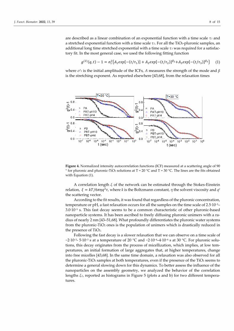
J. Funct. Biomater. 2022, 13, 39 8 of 15
are described as a linear combination of an exponential function with a time scale τ1 and
a stretched exponential function with a time scale τ2. For all the TiO2-pluronic samples, an
additional long time stretched exponential with a time scale τ3 was required for a satisfac-
tory fit. In the most general case, we used the following fitting function
𝑔(2)(𝑞, 𝑡) − 1 = 𝜎12{𝐴1𝑒𝑥𝑝[−(𝑡/𝜏1)] + 𝐴2𝑒𝑥𝑝[−(𝑡/𝜏2)]
𝛽2+𝐴3𝑒𝑥𝑝[−(𝑡/𝜏3)]𝛽3} (1)
where σ21 is the initial amplitude of the ICFs, A measures the strength of the mode and β
is the stretching exponent. As reported elsewhere [43,68], from the relaxation times
Figure 4. Normalized intensity autocorrelation functions (ICF) measured at a scattering angle of 90
° for pluronic and pluronic-TiO2 solutions at T = 20 °C and T = 30 °C. The lines are the fits obtained
with Equation (1).
A correlation length ξ of the network can be estimated through the Stokes-Einstein
relation, 𝜉 = 𝑘𝑇/6𝜋𝜂𝑞2𝜏, where k is the Boltzmann constant, η the solvent viscosity and q2
the scattering vector.
According to the fit results, it was found that regardless of the pluronic concentration,
temperature or pH, a fast relaxation occurs for all the samples on the time scale of 2.5·10−5–
3.0·10−5 s. This fast decay seems to be a common characteristic of other pluronic-based
nanoparticle systems. It has been ascribed to freely diffusing pluronic unimers with a ra-
dius of nearly 2 nm [43–51,68]. What profoundly differentiates the pluronic water systems
from the pluronic-TiO2 ones is the population of unimers which is drastically reduced in
the presence of TiO2.
Following the fast decay is a slower relaxation that we can observe on a time scale of
~2·10−3- 5·10−3 s at a temperature of 20 °C and ~2·10−4–4·10−4 s at 30 °C. For pluronic solu-
tions, this decay originates from the process of micellization, which implies, at low tem-
peratures, an initial formation of large aggregates that, at higher temperatures, change
into free micelles [43,68]. In the same time domain, a relaxation was also observed for all
the pluronic-TiO2 samples at both temperatures, even if the presence of the TiO2 seems to
determine a general slowing down for this dynamics. To better assess the influence of the
nanoparticles on the assembly geometry, we analyzed the behavior of the correlation
lengths ξ2, reported as histograms in Figure 5 (plots a and b) for two different tempera-
tures.

J. Funct. Biomater. 2022, 13, 39 9 of 15
Figure 5. Correlation lengths ξ2 for the samples investigated at T = 20 °C (a) and T = 30 °C (b). The
same data are reported in (c) as a function of the TiO2 concentration as determined by absorption
spectroscopy.
As cited above, the obtained data reproduce our previous findings for the pluronic
solutions. Independently of pluronic concentration, this result confirms the formation of
large aggregates at low T that decrease in size upon increasing temperature up to the typ-
ical size of micelles, 20–30 nm [68]. The same temperature trend in the size evolution can
also be recognized for the pluronic-TiO2 samples but with some differences. It is evident
that the presence of TiO2 hinders the formation of the large aggregates observed at the
lowest temperature. More precisely, it is possible to state that this effect is more pro-
nounced for those samples with the lowest TiO2 concentration. This is evident from Figure
5, plot c, where the same ξ2 data are reported as a function of the “real” TiO2 concentration,
as determined by absorption spectroscopy. This suggests that the lower the TiO2 content,
the better the metallic nanoparticles' capability of being dispersed inside the network of
PEO chains, hindering the formation of the large polymeric clusters.
This directly implies the slowest dynamic is occurring in the time range between 10−3
and 3·10−1 s. In fact, in this case, the corresponding correlation lengths ξ3, reported in Fig-
ure 6, evidence the presence of large micrometric aggregates that decrease in size with
both increasing temperature and decreasing TiO2 content.
The last comment concerns the weight fractions for the intermediate and the slowest
dynamics. From the evaluated fit parameters A2 and A3, it emerged that the presence of
TiO2 determines a generally strong decrease in the percentage of micelles for all samples
except for the PBT-pH10 one. This is the only one for which the percentage of micelles is
comparable to the counterpart PB sample, thus confirming the infrared evidence. Moreo-
ver, for the same PBT-pH10 sample, the population of the micrometric aggregates, A3, is
extremely low.

J. Funct. Biomater. 2022, 13, 39 10 of 15
Figure 6. Correlation lengths ξ3 for the samples investigated as a function of temperature. The arrow
shows the direction of the increment of TiO2 as determined by absorption spectroscopy.
At this point, it remains to be clarified the nature of the micrometric aggregates. As
stated in the introduction, at the pH values of this study, electrostatic repulsive interac-
tions between titania nanoparticles are predominant. Consequently, the agglomeration of
the nanoparticles due to a thickness reduction of the double layer should be unlikely. An-
yway, electrical conductivity measurements helped us shed some light on this point.
3.4. Electrical Conductivity
Thus far, many measurements of the electrical properties for water-based titania
nanofluids or titania-polymer systems have been carried out [78–80], but not for disper-
sions of TiO2 inside a micellar matrix. Here, the interest in conductivity measurements
arises from the possibility they offer to draw information on the nature of the micro-ag-
gregates observed by DLS.
The experimental data for the electrical conductivity of pluronic-TiO2 nanofluids are
summarized in Figure 7, where we reported the relative electrical conductivity, σr, defined
as the ratio between the electrical conductivities of pluronic-TiO2 dispersions, σn, and that
of the base fluid, σf, (pluronic solutions under the same conditions) σr = σn/σf.
As expected, the addition of TiO2 to pluronic solutions increases the electrical con-
ductivity of the systems. In principle, an increase in charge carriers determines an increase
in effective electrical conductivity. Anyway, an increase in conducting nanoparticles
could induce an increase in inter-particle collisions and consequently an enhancement of
the probability of aggregation of the nanoparticles. This could determine a reduction in
the effective number of charge carriers with a consequent deterioration of the effective
charge transport. Even if the establishment of short conducting paths through aggregate-
to-aggregate contact were assumed, the low intrinsic conductivity of TiO2 would exclude
the possibility. In conclusion, the behavior of the electrical conductivity observed for our
samples suggests that the presence of titania aggregates inside the polymeric matrix is
unlikely.
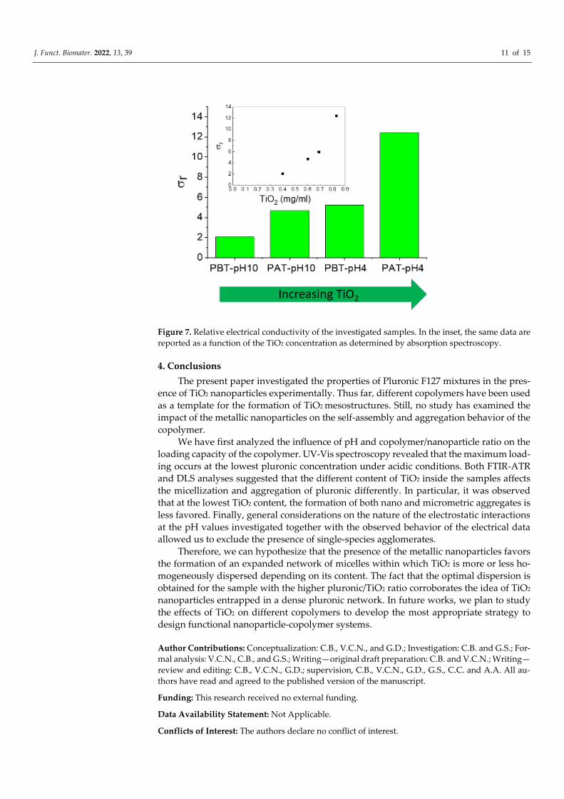
J. Funct. Biomater. 2022, 13, 39 11 of 15
Figure 7. Relative electrical conductivity of the investigated samples. In the inset, the same data are
reported as a function of the TiO2 concentration as determined by absorption spectroscopy.
4. Conclusions
The present paper investigated the properties of Pluronic F127 mixtures in the pres-
ence of TiO2 nanoparticles experimentally. Thus far, different copolymers have been used
as a template for the formation of TiO2 mesostructures. Still, no study has examined the
impact of the metallic nanoparticles on the self-assembly and aggregation behavior of the
copolymer.
We have first analyzed the influence of pH and copolymer/nanoparticle ratio on the
loading capacity of the copolymer. UV-Vis spectroscopy revealed that the maximum load-
ing occurs at the lowest pluronic concentration under acidic conditions. Both FTIR-ATR
and DLS analyses suggested that the different content of TiO2 inside the samples affects
the micellization and aggregation of pluronic differently. In particular, it was observed
that at the lowest TiO2 content, the formation of both nano and micrometric aggregates is
less favored. Finally, general considerations on the nature of the electrostatic interactions
at the pH values investigated together with the observed behavior of the electrical data
allowed us to exclude the presence of single-species agglomerates.
Therefore, we can hypothesize that the presence of the metallic nanoparticles favors
the formation of an expanded network of micelles within which TiO2 is more or less ho-
mogeneously dispersed depending on its content. The fact that the optimal dispersion is
obtained for the sample with the higher pluronic/TiO2 ratio corroborates the idea of TiO2
nanoparticles entrapped in a dense pluronic network. In future works, we plan to study
the effects of TiO2 on different copolymers to develop the most appropriate strategy to
design functional nanoparticle-copolymer systems.
Author Contributions: Conceptualization: C.B., V.C.N., and G.D.; Investigation: C.B. and G.S.; For-
mal analysis: V.C.N., C.B., and G.S.; Writing—original draft preparation: C.B. and V.C.N.; Writing—
review and editing: C.B., V.C.N., G.D.; supervision, C.B., V.C.N., G.D., G.S., C.C. and A.A. All au-
thors have read and agreed to the published version of the manuscript.
Funding: This research received no external funding.
Data Availability Statement: Not Applicable.
Conflicts of Interest: The authors declare no conflict of interest.

J. Funct. Biomater. 2022, 13, 39 12 of 15
References
1. Choi, H.; Sofranko, A.C.; Dionysiou, D.D. Nanocrystalline TiO2 photocatalytic membranes with a hierarchical mesoporous mul-
tilayer structure: Synthesis, characterization, and multifunction. Adv. Funct. Mater. 2006, 16, 1067–1074.
https://doi.org/10.1002/adfm.200500658.
2. Chuangchote, S.; Jitputti, J.; Sagawa, T.; Yoshikawa, S. Photocatalytic activity for hydrogen evolution of electrospun TiO2 nan-
ofibers. ACS Appl. Mater. Interfaces 2009, 1, 1140–1143. https://doi.org/10.1021/am9001474.
3. Kay, A.; Grätzel, M. Low cost photovoltaic modules based on dye sensitized nanocrystalline titanium dioxide and carbon pow-
der. Sol. Energy Mater. Sol. Cells 1996, 44, 99–117. https://doi.org/10.1016/0927-0248(96)00063-3.
4. Arango, A.C.; Johnson, L.R.; Bliznyuk, V.N.; Schlesinger, Z.; Carter, S.A.; Hörhold, H.-H. Efficient titanium oxide/conjugated
polymer photovoltaics for solar energy conversion. Adv. Mater. 2000, 12, 1689–1692.
5. Fujishima, A.; Rao, T.N.; Tryk, D.A. Titanium dioxide photocatalysis. J. Photochem. Photobiol. C 2000, 1, 1–21.
https://doi.org/10.1016/S1389-5567(00)00002-2.
6. Mor, G.K.; Shankar, K.; Paulose, M.; Varghese, O.K.; Grimes, C.A. Enhanced photocleavage of water using titania nanotube
arrays. Nano Lett. 2005, 5, 191–195. https://doi.org/10.1021/nl048301k.
7. Fujishima, A.; Honda, K. Electrochemical photolysis of water at a semiconductor electrode. Nature 1972, 238, 37–38.
https://doi.org/10.1038/238037a0.
8. Turchi, C.S.; Ollis, D.F. Photocatalytic degradation of organic water contaminants: Mechanisms involving hydroxyl radical at-
tack. J. Catal. 1990, 122, 178–192. https://doi.org/10.1016/0021-9517(90)90269-P.
9. Hoffmann, M.R.; Martin, S.T.; Choi, W.; Bahnemann, D.W. Environmental applications of semiconductor photocatalysis. Chem.
Rev. 1995, 95, 69–96. https://doi.org/10.1021/cr00033a004.
10. Pelizzetti, E. Concluding remarks on heterogeneous solar photocatalysis. Sol. Energy Mater. Sol. Cells 1995, 38, 453–457.
https://doi.org/10.1016/0927-0248(94)00237-1.
11. Berardinelli, A.; Parisi, F. TiO2 in the food industry and cosmetics. In Metal Oxides, Titanium Dioxide (Tio₂) and Its Applications;
Parrino, F., Palmisano, L., Eds.; Elsevier: Hoboken, NJ, USA, 2021; pp. 353–371. https://doi.org/10.1016/B978-0-12-819960-
2.00008-0.
12. Piccinno, F.; Gottschalk, F.; Seeger, S.; Nowack, B. Industrial production quantities and uses of ten engineered nanomaterials in
Europe and the world. J. Nanopart. Res. 2012, 14, 1109–1120. https://doi.org/10.1007/s11051-012-1109-9.
13. Weir, A.; Westerhoff, P.; Fabricius, L.; Hristovski, K; von Goetz, N. Titanium dioxide nanoparticles in food and personal care
products. Environ. Sci. Technol. 2012, 46, 2242–2250. https://doi.org/10.1021/es204168d.
14. Wang, Q.; Huang, J.-Y.; Li, H.-Q.; Chen, Z.; Zhao, A.Z.-J.; Wang, Y.; Zhang, K.-Q.; Sun, H.-T.; Al-Deyab, S.S.; Lai, Y.-K. TiO2
nanotube platforms for smart drug delivery: A review. Int. J. Nanomed. 2016, 11, 4819–4834. https://doi.org/10.2147/IJN.S108847.
15. Park, J.; Cimpean, A.; Tesler, A.B.; Mazare, A. Anodic TiO2 nanotubes: Tailoring Osteoinduction via Drug Delivery. Nanomateri-
als 2021, 11, 2359. https://doi.org/10.3390/nano11092359.
16. Babitha, S.; Korrapati, P.S. Biodegradable zein-polydopamine polymeric sca_old impregnated with TiO2 nanoparticles for skin
tissue engineering. Biomed. Mater. 2017, 12, 055008. https://doi.org/10.1088/1748-605X/aa7d5a.
17. Stan, M.S.; Nica, I.C.; Dinischiotu, A.; Varzaru, E.; Iordache, O.G.; Dumitrescu, I.; Popa, M.; Chifiriuc, M.C.; Pircalabioru, G.G.;
Lazar, V.; et al. Photocatalytic, antimicrobial and biocompatibility features of cotton knitcoated with Fe-N-Doped titanium di-
oxide nanoparticles. Materials 2016, 9, 789. https://doi.org/10.3390/ma9090789.
18. Seisenbaeva, G.A.; Fromell, K.; Vinogradov, V.V.; Terekhov, A.N.; Pakhomov, A.V.; Nilsson, B.; Ekdahl, K.N.; Vinogradov,
V.V.; Kessler, V.G. Dispersion of TiO2 nanoparticles improves burn wound healing and tissue regeneration through specific
interaction with blood serum proteins. Sci. Rep. 2017, 7, 15448. https://doi.org/15448 10.1038/s41598-017-15792-w.
19. Hasan, K.M.F.; Horváth, P.G.; Alpár, T. Potential Natural Fiber Polymeric Nanobiocomposites: A Review. Polymers 2020, 12,
1072. https://doi.org/10.3390/polym12051072.
20. Jiang, J.; Oberdörster, G.; Biswas, P. Characterization of size, surface charge, and agglomeration state of nanoparticle dispersions
for toxicological studies. J. Nanopart. Res. 2009, 11, 77–89. https://doi.org/10.1007/s11051-008-9446-4.
21. Almquist, C.B.; Biswas, P. Role of Synthesis Method and Particle Size of Nanostructured TiO2 on its Photoactivity. J. Catal. 2002,
212, 145–156. DOI:10.1006/jcat.2002.3783.
22. Sclafani, A.; Herrmann, J.M. Comparison of the Photoelectronic and Photocatalytic Activities of various anatase and rutile forms
of titania in pure liquid organic phases and in aqueous solutions. J. Phys. Chem. 1996, 100, 13655–13661.
https://doi.org/10.1021/jp9533584.
23. Jiang, J.; Oberdörster, G.; Elder, A.; Gelein, R.; Mercer, P.; Biswas, P. Does nanoparticle activity depend upon size and crystal
phase? Nanotoxicology 2008, 2, 33–42. https://doi.org/10.1080/17435390701882478.
24. Waychunas, G.A.; Kim, C.S.; Banfield, J.F. Nanoparticulate Iron Oxide Minerals in soils and sediments: Unique properties and
contaminant scavenging mechanisms. J. Nanopart. Res. 2005, 7, 409–433. https://doi.org/10.1007/s11051-005-6931-x.
25. Kaasalainen, M.; Aseyev, V.; von Haartman, E.; Şen Karaman, D.; Mäkilä, E.; Tenhu, H.; Rosenholm, J.; Salonen, J. Size, Stability,
and Porosity of Mesoporous Nanoparticles Characterized with Light Scattering. Nanoscale Res. Lett. 2017, 12, 74.
https://doi.org/10.1186/s11671-017-1853-y.

J. Funct. Biomater. 2022, 13, 39 13 of 15
26. Joo, N.Y.; Lee, J.; Kim, S.J.; Hong, S.H.; Park, H.M.; Yun, W.S.; Yoon, M.; Song, N.W. Preparation of an aqueous suspension of
stabilized TiO2 nanoparticles in primary particle form. J. Nanosci. Nanotechnol. 2013, 13, 6153–6159.
https://doi.org/10.1166/jnn.2013.7637.
27. Bielan, Z.; Dudziak, S.; Sulowska, A.; Pelczarski, D.; Ryl, J.; Zielińska-Jurek, A. Preparation and Characterization of Defective
TiO2. The Effect of the Reaction Environment on Titanium Vacancies Formation. Materials 2020, 13, 2763.
https://doi.org/10.3390/ma13122763.
28. Chakraborty, S. An investigation on the long-term stability of TiO2 nanofluid. Mater. Today Proc. 2019, 11, 714–718.
https://doi.org/10.1016/j.matpr.2019.03.032.
29. Suttiponparnit, K.; Jiang, J.; Sahu, M.; Suvachittanont, S.; Charinpanitkul, T.; Biswas, P. Role of Surface Area, Primary Particle
Size, and Crystal Phase on Titanium Dioxide Nanoparticle Dispersion Properties. Nanoscale Res Lett. 2011, 6, 27.
https://doi.org/10.1007/s11671-010-9772-1.
30. Zhang, X.; Yin, L.; Tang, M.; Pu, Y. Optimized method for preparation of TiO2 nanoparticles dispersion for biological study. J.
Nanosci. Nanotechnol. 2010, 10, 5213–5219. https://doi.org/10.1166/jnn.2010.2397.
31. Nilsson, E.; Furusho, H.; Terasaki, O.; Palmqvist, A.E.C. Synthesis of nanoparticulate anatase and rutile crystallites at low tem-
peratures in the Pluronic F127 microemulsion system. J. Mater. Res. 2011, 26, 288–295. https://doi.org/10.1557/jmr.2010.5.
32. Li, Y.Q.; Bastakoti, B.P.; Imura, M.; Hwang, S.M.; Sun, Z.Q.; Kim, J.H.; Dou, S.X.; Yamauchi, Y. Synthesis of mesoporous
TiO2/SiO2 hybrid films as an efficient photocatalyst by polymeric micelle assembly. Chem. Eur. J. 2014, 20, 6027–6032.
https://doi.org/10.1002/chem.201304689.
33. Luo, H.; Wang, C.; Yan, Y. Synthesis of mesostructured titania with controlled crystalline framework. Chem. Mater. 2003, 15,
3841–3846. https://doi.org/10.1021/cm0302882.
34. Peng, T.; Zhao, D.; Dai, K.; Shi, W.; Hirao, K. Synthesis of titanium dioxide nanoparticles with mesoporous anatase wall and
high photocatalytic activity. J. Phys. Chem. B 2005, 109, 4947–4952. https://doi.org/10.1021/jp044771r.
35. Smarsly, B.; Grosso, D.; Brezesinski, T.; Pinna, N.; Boissiere, C.; Antonietti, M.; Sanchez, C. Highly crystalline cubic mesoporous
TiO2 with 10-nm pore diameter made with a new block copolymer template. Chem. Mater. 2008, 16, 2948–2952.
https://doi.org/10.1021/cm0495966.
36. Kim, D.S.; Han, S.J.; Kwak, S.-Y. Synthesis and photocatalytic activity of mesoporous TiO2 with the surface area, crystallite size,
and pore size. J. Colloid Interface Sci. 2007, 316, 85–91. https://doi.org/10.1016/j.jcis.2007.07.037.
37. Agarwala, S.; Ho, G.W. Synthesis and tuning of ordering and crystallinity of mesoporous titanium dioxide film. Mater. Lett.
2009, 63, 1624–1627. https://doi.org/10.1016/j.matlet.2009.04.036.
38. Gajjela, S.R.; Ananthanarayanan, K.; Yap, C.; Gratzel, M.; Balaya, P. Synthesis of mesoporous titanium dioxide by soft template
based approach: Characterization and application in dye-sensitized solar cells. Energy Environ. Sci. 2010, 3, 838–845.
https://doi.org/10.1039/b921360k.
39. Deng, Y.; Cai, Y.; Sun, Z.; Liu, J.; Liu, C.; Wei, J.; Li, W.; Liu, C.; Wang, Y.; Zhao, D. Multifuntional mesoporous composite
microsphere with well-designed nanostructure: A highly integrated catalyst system. J. Am. Chem. Soc. 2010, 132, 8466–8473.
https://doi.org/10.1021/ja1025744.
40. Samsudin, E.M.; Abd Hamid, S.B.; Juan, J.C.; Basirun, W.J. Influence of triblock copolymer (pluronic F127) on enhancing the
physico-chemical properties and photocatalytic response of mesoporous TiO2. Appl. Surf. Sci. 2015, 355, 959–968.
https://doi.org/10.1016/j.apsusc.2015.07.178.
41. Suwanchawalit, C.; Wongnawa, S. Triblock copolymer-templated synthesis of porous TiO2 and its photocatalytic activity. J.
Nanopart. Res. 2010, 12, 2895–2906. https://doi.org/10.1007/s11051-010-9880-y.
42. Roy, S.; Ghosh, S.P.; Pradhan, D.; Sahu, P.K.; Kar, J.P. Morphological and electrical study of porous TiO2 films with various
concentrations of Pluronic F-127 additive. J. Porous Mater. 2021, 28, 231–238. https://doi.org/10.1007/s10934-020-00983-0.
43. Brown, W.; Schillén, K.; Almgren, M.; Hvidt, S.; Bahadur, P. Micelle and gel formation in a poly(ethylene oxide)/poly(propylene
oxide)/poly(ethylene oxide) triblock copolymer in water solution: Dynamic and static light scattering and oscillatory shear
measurements. J. Phys. Chem. 1991, 95, 1850–1858. https://doi.org/10.1021/j100157a064.
44. Wanka, G.; Hoffmann, H.; Ulbricht, W. Phase diagrams and aggregation behavior of poly(oxyethy1ene)-poly(oxypropylene)-
poly(oxyethylene) triblock copolymers in aqueous solutions. Macromolecules 1994, 27, 4145–4159.
https://doi.org/10.1021/ma00093a016.
45. Alexandridis, P.; Hatton, T.A. Poly(ethylene oxide)-poly(propylene oxide)-poly(ethylene oxide) block copolymer surfactants in
aqueous solutions and at interfaces: Thermodynamics, structure, dynamics, and modeling. Colloids Surf. A 1995, 96, 1–46.
https://doi.org/10.1016/0927-7757(94)03028-X.
46. Prudhomme, R.K.; Wu, G.; Schneider, D.K. Structure and rheology studies of poly(oxyethylene-oxypropylene-oxyethylene)
aqueous solution. Langmuir 1996, 12, 4651–4659. https://doi.org/10.1021/la951506b.
47. Malmsten, M.; Lindman, B. Self-Assembly in Aqueous Block Copolymer Solutions. Macromolecules 1992, 25, 5440–5445.
https://doi.org/10.1021/ma00046a049.
48. Mortensen, K. Structural studies of aqueous solutions of PEO–PPO–PEO triblock copolymers, their micellar aggregates and
mesophases; a small-angle neutron scattering study. J. Phys. Condens. Matter 1996, 8, A103–A124. https://doi.org/10.1088/0953-
8984/8/25A/008.

J. Funct. Biomater. 2022, 13, 39 14 of 15
49. Alexandridis, P.; Holzwarth, J.F.; Hatton, T.A. Micellization of Poly(ethylene oxide)-Poly(propylene oxide)-Poly(ethylene ox-
ide) Triblock Copolymers in Aqueous Solutions: Thermodynamics of Copolymer Association. Macromolecules 1994, 27, 2414–
2425. https://doi.org/10.1021/ma00087a009.
50. Shaikhullina, M.; Khaliullina, A.; Gimatdinov, R.; Butakov, A.; Chernov, V.; Filippov, A. NMR relaxation and self-diffusion in
aqueous micellar gels of pluronic F-127. J. Mol. Liq. 2020, 306, 112898. https://doi.org/10.1016/j.molliq.2020.112898.
51. Chaibundit, C.; Ricardo, N.M.P.S.; Costa, F.M.L.L.; Yeates, S.G.; Booth, C. Micellization and gelation of mixed copolymers P123
and F127 in aqueous solution. Langmuir 2007, 23, 9229–9236. https://doi.org/10.1021/la701157j.
52. Batrakova, E.V.; Kabanov, A.V. Pluronic block copolymers: Evolution of drug delivery concept from inert nanocarriers to bio-
logical response modifiers. J. Control. Release 2008, 130, 98–106. https://doi.org/10.1016/j.jconrel.2008.04.013.
53. Kulthe, S.S.; Inamdar, N.N.; Choudhari, Y.M.; Shirolikar, S.M.; Borde, L.C.; Mourya, V.K. Mixed micelle formation with hydro-
phobic and hydrophilic Pluronic block copolymers: Implications for controlled and targeted drug delivery. Colloids Surf. B 2011,
88, 691–696. https://doi.org/10.1016/j.colsurfb.2011.08.002.
54. Choi, W.I.; Lee, J.H.; Kim, J.-Y.; Kim, J.-C.; Kim, Y.H.; Tae, G. Efficient skin permeation of soluble proteins via flexible and
functional nano-carrier, J. Control. Release 2012, 157, 272–278. https://doi.org/10.1016/j.jconrel.2011.08.013.
55. Brunet-Maheu, J.M.; Fernandes, J.C.; De Lacerda, C.A.; Shi, Q.; Benderdour, M.; Lavigne, P. Pluronic F-127 as a Cell Carrier for
Bone Tissue Engineering. J. Biomater. Appl. 2009, 24, 275–287. https://doi.org/10.1177/0885328208096534.
56. Patel, H.S.; Shaikh, S.J.; Ray, D.; Aswal, V.K.; Vaidya, F.; Pathak, C.; Sharma, R.K. Formulation, solubilization, and in vitro
characterization of quercetin-incorporated mixed micelles of PEO-PPO-PEO block copolymers. Appl. Biochem. Biotechnol. 2022,
194, 445–463. https://doi.org/10.1007/s12010-021-03691-w.
57. Rahdar, A.; Hasanein, P.; Bilal, M.; Beyzaei, H.; Kyzas, G.Z. Quercetin-loaded F127 nanomicelles: Antioxidant activity and pro-
tection against renal injury induced by gentamicin in rats. Life Sci. 2021, 276, 119420. https://doi.org/10.1016/j.lfs.2021.119420.
58. Kassa, S.B.; Taslimi, P.; Ö zel, S.; Gür, B.; Gülçin, I.; Onganer, Y. Effects of some phenolic compounds on the inhibition of α-
glycosidase enzyme-immobilized on Pluronic® F127 micelles: An in vitro and in silico study. Colloid Surf. A 2022, 632, 127839.
https://doi.org/10.1016/j.colsurfa.2021.127839.
59. Rahdar, A.; Hajinezhad, M.R.; Barani, M.; Sargazi, S.; Zaboli, M.; Ghazy, E.; Baino, F.; Cucchiarini, M.; Bilal, M.; Pandey, S.
Pluronic F127/Doxorubicin microemulsions: Preparation, characterization, and toxicity evaluations. J. Mol. Liq. 2022, 345,
117028. https://doi.org/10.1016/j.molliq.2021.117028.
60. Jiang, J.; Li, C.; Lombardi, J.; Colby, R.H.; Rigas, B.; Rafailovich, M.H.; Sokolov, J.C. The effect of physiologically relevant addi-
tives on the rheological properties of concentrated Pluronic copolymer gels. Polymer 2008, 49, 3561–3567.
https://doi.org/10.1016/j.polymer.2008.05.038.
61. Pradines, B.; Djabourov, M.; Vauthier, C.; Loiseau, P.M.; Ponchel, G.; Bouchemal, K. Gelation and micellization behaviors of
pluronic® F127 hydrogel containing poly(isobutylcyanoacrylate) nanoparticles specifically designed for mucosal application.
Colloids Surf. B 2015, 135, 669–676. https://doi.org/10.1016/j.colsurfb.2015.08.021.
62. Dey, J.; Kumar, S.; Nath, S.; Ganguly, R.; Aswal, V.K.; Ismail, K. Additive induced core and corona specific dehydration and
ensuing growth and interaction of Pluronic F127 micelles. J. Colloid Interface Sci. 2014, 415, 95–102.
https://doi.org/10.1016/j.jcis.2013.10.019.
63. Nelson, A.; Cosgrove, T. Small-Angle Neutron Scattering Study of Adsorbed Pluronic Tri-Block Copolymers on Laponite. Lang-
muir 2005, 21, 9176–9182. https://doi.org/10.1021/la050680p.
64. Zhang, W.; Gilstrap, K.; Wu, L.; Bahadur, R.; Moss, M.A.; Wang, Q.; Lu, X.; He, X. Synthesis and characterization of thermally
responsive pluronic F127-chitosan nanocapsules for controlled release and intracellular delivery of small molecules. ACS Nano
2010, 4, 6747–6759. https://doi.org/10.1021/nn101617n.
65. Perry, C.; Hebraud, P.; Gernigon, V.; Brochon, C.; Lapp, A.; Lindner, P.; Schlatter, G. Pluronic and β-cyclodextrin in water: From
swollen micelles to self-assembled crystalline platelets. Soft Matter 2011, 7, 3502–3512. https://doi.org/10.1039/c0sm01092h.
66. Nambam, J.S.; Philip, J. Effects of Interaction of Ionic and Nonionic Surfactants on Self-Assembly of PEO−PPO−PEO Triblock
Copolymer in Aqueous Solution. J. Phys. Chem. B 2012, 116, 1499–1507. https://doi.org/10.1021/jp208902a.
67. Branca, C.; Khouzami, K.; Wanderlingh, U.; D’Angelo, G. Effect of intercalated chitosan/clay nanostructures on concentrated
pluronic F127 solution: A FTIR-ATR, DSC and rheological study. J. Colloid Interface Sci. 2018, 517, 221–229.
https://doi.org/10.1016/j.jcis.2018.02.004.
68. Branca, C.; D’Angelo, G. Aggregation behavior of Pluronic F127 solutions in presence of chitosan/clay nanocomposites exam-
ined by dynamic light scattering. J. Colloid Interface Sci. 2019, 542, 289–295. https://doi.org/10.1016/j.jcis.2019.02.031.
69. Derjaguin, B.V.; Landau, L.D. Theory of the stability of strongly charged lyophobic sols and of the adhesion of strongly charged
particles in solutions of electrolytes. Progr. Surf. Sci. 1941, 14, 733–762. https://doi.org/10.1016/0079-6816(93)90013-L.
70. Shrestha, S.; Wang, B.; Dutta, P. Nanoparticle processing: Understanding and controlling aggregation. Adv. Colloid Interface Sci.
2020, 279, 102162. https://doi.org/10.1016/j.cis.2020.102162.
71. Branca, C.; Wanderlingh, U.; D’Angelo, G.; Crupi, C.; Rifici, S. Study of the dynamical behavior of sodium alginate/myoglobin
aqueous solutions: A dynamic light scattering study. J. Mol. Liq. 2015, 209, 294–300. https://doi.org/10.1016/j.molliq.2015.06.002.
72. Hassan, P.A.; Rana, S.; Verma, G. Making sense of brownian motion: Colloid characterization by dynamic light scattering. Lang-
muir 2015, 31, 3–12. https://doi.org/10.1021/la501789z.
73. Berne, B.J.; Pecora, R. Dynamic Light Scattering: With Applications to Chemistry, Biology, and Physics; John Wiley & Sons: New York,
NY, USA, 1975.

J. Funct. Biomater. 2022, 13, 39 15 of 15
74. Finnie, K.M.; Cassidy, D.J.; Bartlett, J.R.; Woolfrey, J.L. IR Spectroscopy of Surface Water and Hydroxyl Species on Nanocrys-
talline TiO2 Films. Langmuir 2001, 17, 816–820. https://doi.org/10.1021/la0009240.
75. Bedurftig, K.; Volkening, S.; Wang, Y.; Wintterlin, J.; Jacobi, K.; Ertl, G. Vibrational and structural properties of OH adsorbed
on Pt(111). J. Chem. Phys. 1999, 111, 11147. https://doi.org/10.1063/1.480472.
76. Su, Y.L.; Wang, J.; Liu, H.Z. FTIR spectroscopic investigation of effects of temperature and concentration on PEO-PPO-PEO
block copolymer properties in aqueous solutions. Macromolecules 2002, 35, 6426–6431. https://doi.org/10.1021/ma0105284.
77. Su, Y.L.; Wang, J.; Liu, H.Z. Melt, hydration, and micellization of the PEO–PPO–PEO block copolymer studied by FTIR spec-
troscopy. J. Colloids Interface Sci. 2002, 251, 417–423. https://doi.org/10.1006/jcis.2002.8435.
78. Islam, M.R.; Shabani, B.; Rosengarten, G. Electrical and Thermal Conductivities of 50/50 Water-ethylene Glycol Based TiO2
Nanofluids to be Used as Coolants in PEM Fuel Cells. Energy Procedia 2017, 110, 101–108.
https://doi.org/10.1016/j.egypro.2017.03.113.
79. Chereches, E.I.; Minea, A.A. Electrical Conductivity of New Nanoparticle Enhanced Fluids: An Experimental Study. Nanomateri-
als 2019, 9, 1228. https://doi.org/10.3390/nano9091228.
80. Sikdar, S.; Basu, S.; Ganguly, S. Investigation of electrical conductivity of titanium dioxide nanofluids. Int. J. Nanopart. 2011, 4,
336–349. https://doi.org/10.1504/IJNP.2011.043496.