Nuclear EGFR modulation of DNA repair - UCL Discovery
269
Nuclear EGFR Modulation of DNA repair by Gianmaria Liccardi A thesis submitted to the University College London for the degree of Doctor of Philosophy April 2011 CRUK Drug-DNA Interaction Research Group Department of Oncology Cancer Institute University College London 72 Huntley street, London WC1E 6BT, UK
-
Upload
khangminh22 -
Category
Documents
-
view
2 -
download
0
Transcript of Nuclear EGFR modulation of DNA repair - UCL Discovery

Nuclear EGFR Modulation of
DNA repair
by
Gianmaria Liccardi
A thesis submitted to the University College London for
the degree of Doctor of Philosophy
April 2011
CRUK Drug-DNA Interaction Research Group
Department of Oncology
Cancer Institute
University College London
72 Huntley street, London WC1E 6BT, UK

I, Gianmaria Liccardi, confirm that the work presented in this thesis is my own.
Where information has been derived from other sources, I confirm that this has been
indicated in the thesis with appropriate referencing. The most significant part of this
work has been recently published in:
Gianmaria Liccardi, John A. Hartley, Daniel Hochhauser; EGFR Nuclear
Translocation Modulates DNA Repair following Cisplatin and Ionizing Radiation
Treatment. Cancer Research, February 1, 2011 71:1103-1114
Hence some figures of the result chapters have been taken from the published
manuscript of which I am the first Author. The article has been included at the end of
this thesis for further referencing.

In loving memory of my auntie Giulia

1
ABSTRACT
Overexpression of the epidermal growth factor receptor (EGFR) is associated with
resistance to chemotherapy and radiotherapy. EGFR involvement, in repair of radiation-
induced DNA damage, is mediated by association with the catalytic subunit of DNA
protein kinase (DNAPKcs). This study investigated the role of EGFR nuclear import,
and its association with DNAPKcs, on DNA repair following treatment either with
cisplatin or ionizing radiation (IR). EGFR- null murine NIH3T3 cells were transfected
with wild type or with mutated EGFR (mutations found in human cancers L858R,
EGFRvIII and mutations in the EGFR nuclear localization signal (NLS) sequence
NLS123, LNLS123). Comet assay analysis, which measures unhooking of cisplatin
crosslinks and repair of IR induced strand breaks, demonstrated that wtEGFR and
EGFRvIII completely repair cisplatin and IR induced DNA damage.
Immunoprecipitation studies show that repair is associated with the binding of both
wtEGFR and EGFRvIII to DNAPKcs, which increases by 2- fold 18 hours following
cisplatin treatment. Confocal analysis and proximity ligation assay indicated that this
association takes place both in the cytoplasm and in the nucleus resulting in a significant
increase of DNA-PK kinase activity. Intermediate levels of repair as shown by the
L858R construct with impaired nuclear localization demonstrated that EGFR kinase
activity is partially involved in repair but is not sufficient to determine EGFR nuclear
expression. EGFR-NLS mutants showed impaired nuclear localization and impaired
DNAPKcs association resulting in significant inhibition of DNA repair and down-
regulation of DNA-PK kinase activity.
Our data suggest that EGFR nuclear localization is required for the modulation of
cisplatin and IR induced DNA damage repair. The EGFR-DNAPKcs binding is
triggered by cisplatin or IR and not by EGFR nuclear translocation per se.
Understanding mechanisms regulating EGFR subcellular distribution in relation to DNA
repair kinetics will be a critical determinant of improved molecular targeting and
response to therapy.
2
ACKNOWLEDGEMENTS
My first and foremost thanks go to my supervisors: Prof. John Hartley, and Prof. Daniel
Hochhauser. I cannot express enough my gratitude for giving me the opportunity to
carry out my PhD under your insightful supervision. Thank you for giving me the
freedom to develop my own ideas, for the pressure, the support, for the critical
discussion and for making sure my work stayed focus… most of the time… I also would
like to thank all the members of the lab: those present at the moment and those that have
witnessed only a portion of this incredible experience: you all have contributed
somehow to my PhD and I can associate at least one unforgettable memory to each and
every one of you. Nevertheless there are some members in particular that deserve a
special thank. To Dr. Konstantinos Kiakos for being a friend, for the gossips, the fiffis,
for the fun and for being there when I needed a friend in the lab: thank you. To Dr.
Raisa Vuononvirta: thank you for your friendship, suggestions, honesty and for the
nights out with lab: those will be hard to forget. To Valeria Santoro: thank you for the
coffees, the lunches, for being a real friend and a great person. A great thanks also to
those that understand our code words (I shall not mention them for obvious reasons)…
you know who you are… some of the funniest moments of my PhD are linked to those
words and I really hope they will stay as a legacy… beside the science of course…
I also would like to thank Nicole Hartig, Berna Demiray and Anne Roehrig for their
support, friendship and for keeping me company during the latest (or earliest) time of
the day in the lab. A special thank goes to the Cancer Institute graduate tutor, Dr. Julie
Olszewski for her great work with the PhD students of the Cancer Institute and for being
always very helpful and ready to listen when most needed. A huge thanks goes to Lucia
Christodulides for her help in the most critical moments of my PhD, for her honesty and
support. You have been an icon during these years.
Special thanks go to my friends Mauro Proserpio and Antonio Gaglioti. Thank you for
your sincere friendship, for the nights out, for coming to the lab on the countless
Saturdays and Sundays waiting for my experiments to finish before going out. Thank
you.
Life would have most definitely been not the same without Francesco Piazza. I will
never be grateful enough for the inspiration, for your patience, support, for putting up

3
with me on the bad days and also… on the good days, but most importantly for keeping
me sane during this incredible journey. You have been a rock in my life and this has
helped me reaching this destination in one piece.
Most of all, I am deeply grateful to my parents: Maria Guida and Giovanni Liccardi and
to my sister Dr. Ilaria Liccardi, for their support, encouragement, love, guidance, for
giving me all the opportunities that some people only dream of, for being the best family
a son and brother can hope and for teaching me that no matter what comes I can always
find the strength to stand up and keep walking. Without you I would have never been
able to study abroad and for this I owe you everything I have accomplished so far.
Special thanks also go to the rest of my family for believing in me, for their love and
thoughts. I love you all very much.
Finally, thank you God for watching over me and bless me with such a wonderful life.
4
COMMUNICATION
PRESENTATIONS
Poster presentation. Liccardi G., Hartley, A. J., Hocchauser D., Studies of EGFR
nuclear expression. International PhD meeting, Amsterdam, Netherlands, April
2008
Poster presentation. Annual meeting, Cancer Institute, UCL, London UK July
2008
Poster presentation. Annual meeting, Cancer Institute, UCL, London UK July
2009.
Poster presentation. Graduate competition, UCL, London UK March 2009.
Poster presentation. AACR annual meeting, Washington DC, USA April 2010.
Oral presentation. Annual conference, Cancer Institute, London, UK. July 2010.
Poster presentation. 22nd
EORTC-NCI-AACR Symposium on Molecular Targets
and Cancer Therapeutics, Berlin, Germany. November 2010.
Poster presentation. EACR, Genes and Cancer, Warwick, UK, December 2010
PUBLICATIONS
Gianmaria Liccardi, John A. Hartley, Daniel Hochhauser; EGFR Nuclear
Translocation Modulates DNA Repair following Cisplatin and Ionizing Radiation
Treatment. Cancer Research, February 1, 2011 71:1103-1114

5
TABLE OF CONTENTS
ABSTRACT
1
ABSTRACT 1
ACKNOWLEDGEMENTS 2
COMMUNICATION 4
TABLE OF CONTENT 5
INDEX OF FIGURES 12
INDEX OF TABLES 15
ABBREVIATIONS 16
CHAPTER 1: GENERAL INTRODUCTION
CHAPTER 1: GENERAL INTRODUCTION 23
1.1 CANCER EPIDEMIOLOGY 24
1.2 CANCER AETIOLOGY 24
1.2.1 The molecular origin of Cancer 24
1.2.2 Cancer Heterogeneity confers resistance to treatments
25
1.3 CANCER TREATMENTS 25
1.3.1 Radiotherapy 26
1.3.1.1 Radiotherapy unspecificty 26
1.3.1.1.1 Oxygenation 27
1.3.1.1.2 Repopulation 27
1.3.2 Chemotherapy 27
1.3.2.1 Alkylating Agents 30
1.3.2.1.2 Cisplatin 30
1.3.3 Hormonal therapy 32
1.4 DNA DAMAGE AND REPAIR 32
1.4.1 Nucleotide excision repair (NER) 33
1.4.2 Base excision repair (BER) 34
1.4.3 Mismatch repair (MMR) 35
1.4.4 Double strand breaks repair pathways: Non-homologous end
joining and Homologous recombination. 39
1.4.4.1 Double strand breaks causes and consequences 39
1.4.4.2 Double strand breaks repair pathways 40
1.4.4.2.1 Non-homologous end joining (NHEJ) 40
1.4.4.2.2 Homologous Recombination (HR) 41

6
1.5 DNAPKcs 45
1.5.1 Structure 45
1.5.2 DNAPKcs involvement in repair
45
1.5.3 DNAPKcs phosphorylation
46
1.6 DNA REPAIR REGULATION THROUGH THE CELL CYCLE 46
1.6.1 Cell cycle checkpoints 47
1.6.1.1 Cell cycle arrest 48
1.6.2 Apoptosis
50
1.7 CELLULAR SIGNALLING AND CANCER 52
1.7.1 Oncogenic Receptor Tyrosine kinases (RTK) 52
1.7.1.1 The EGF receptor family 53
1.7.1.1.1 The evolution of the mammalian ERBB
network 53
1.7.1.2 ERBB ligands 54
1.7.1.3 ERBB2 55
1.7.1.4 ERBB3 55
1.7.1.5 ERBB4 55
1.7.1.6 Robustness of the ERBB signalling
56
1.8 EPIDERMAL GROWTH FACTOR RECEPTOR 57
1.8.1 Structure 57
1.8.1.1 EGFR ligand binding domain 58
1.8.1.2 Transmembrane domain and Juxtamembrane domain 60
1.8.1.3 Kinase domain 60
1.8.2 Autophosphorylation 61
1.8.3 EGFR and cellular signalling 62
1.8.3.1 EGFR activation of the MapK pathway 62
1.8.3.2 Src family of kinases 63
1.8.3.3 Stat and JAK 64
1.8.3.4 Phospholipids metabolism 64
1.8.4 EGFR biological downregulation: endocytosis mediated
degradation 66
1.8.4.1 CCps endocytosis 67
1.8.4.2 Cavoelae-mediated endocytosis (CavME) 70
1.8.5 ONCONGENIC EGFR 70
1.8.5.1 Autocrine mechanisms 71
1.8.5.2 EGFR overexpression 71
7
1.8.5.3 Evading degradative fate. 72
1.8.5.4 EGFR mutations 72
1.8.5.4.1 Extracellular mutations 72
1.8.5.4.2 Intracellular mutations: Kinase domain
mutations 73
1.8.5.4.3 Intracellular mutations: C- terminal mutations 74
1.8.5.5 EGFR nuclear expression 77
1.8.5.5.1 Mechanism of nuclear translocation 77
1.8.5.5.2 Nuclear localisation signals 78
1.8.5.5.3 EGFR Nuclear translocation 79
1.8.5.5.4 EGFR modulates transcriptional regulation 82
1.8.5.5.5 EGFR modulation of DNA repair 83
1.8.5.5.6 EGFR radio-protector function 84
1.8.5.6 EGFR targeted therapy 86
1.8.5.6.1 EGFR inhibitors 86
1.8.5.6.1.1 Tyrosine kinase inhibitors 87
1.8.5.6.1.2 Gefitinib IressaTM
, ZD1839 87
1.8.5.6.1.3 Irreversible TKI 89
1.8.5.6.2 Monoclonal antibodies
89
1.9 AIMS AND OBJECTIVES 89
CHAPTER 2: MATERIALS AND METHODS
91
2.1 MATERIALS 92
2.1.1 EGF 92
2.1.2 Cell lines and culture conditions 92
2.1.3 Chemotherapeutic drugs and other reagents 92
2.1.4 Plasmids 92
2.1.5 Primers 96
2.1.5.1 Mutagenic primers 96
2.1.5.2 Sequencing primers 96
2.1.5.3 Screening primers
97
2.2 METHODS 97
2.2.1 Tissue culture 97
2.2.1.1 Cell lines maintenance 97
2.2.1.2 Storage and retrieval from liquid nitrogen 97
2.2.1.3 Cell count 98

8
2.2.1.4 Cell doubling time 98
2.2.2 Drug treatments 98
2.2.3 Irradiation condition 99
2.2.4 The Comet assay 100
2.2.4.1 DNA damage level 100
2.2.4.2 DNA repair study 100
2.2.4.3 Assay methodology 100
2.2.4.4 Data analysis 101
2.2.5 Statistical Analysis 103
2.2.6 MTT 103
2.2.7 Western blotting analysis 104
2.2.7.1 Total protein extraction 104
2.2.7.2 Nuclear and cytoplasmic protein extraction 104
2.2.7.3 Protein quantification 105
2.2.7.4 Immunoblotting 105
2.2.8 Immunoprecipitation 107
2.2.9 Densitometric Analysis 108
2.2.8 Cloning 109
2.2.8.1 Site directed mutagenesis 109
2.2.8.2 Plasmids transformation and amplification 110
2.2.8.3 Plasmid digestion 110
2.2.8.4 Ligation 111
2.2.8.5 Colonies screening 112
2.2.8.6 Sequencing 112
2.2.8.7 Plasmids transfection 112
2.2.9 Cconfocal microscopy 113
2.2.9.1 Immunofluorescence Staining 113
2.2.9.2 Proximity Ligation assay 114
2.2.10 DNA-PK Functional Assay
114
CHAPTER 3:CHARACTERISATION OF THE EGFR NLS
MUTANTS
116
3.1 INTRODUCTION 117
3.1.1 EGFR endocytosis is required for receptor degradation, recycling
and nuclear translocation 117
3.1.1.1 EGFR phosphorylation mediates nuclear translocation 117
3.1.2 EGFR NLS sequence is a functional NLS sequence 118

9
3.1.2.1 The criteria for a functional NLS 118
3.1.3 The NLS sequence affinity for the importin molecule is highly
regulated 120
3.1.4 EGFR NLS sequence mutation 120
3.1.5 NIH3T3 cells as a cellular model 121
3.1.6 CHAPTER AIMS: 121
3.2 RESULTS 122
3.2.1 EGFR expression in NIH3T3 (EGFR null) cells 122
3.2.2 EGFR NLS site directed mutagenesis 122
3.2.3 Mutant characterisation 124
3.2.3.1 EGF stimulation induces EGFR activation 124
3.2.3.2 Dose-dependent cisplatin activation of EGFR 127
3.2.3.2.1 EGFR activation following cisplatin treatment 128
3.2.4 EGFR nuclear translocation is induced by ligand stimulation and
by IR treatment 129
3.2.4.1 EGF induction of EGFR nuclear translocation 130
3.2.4.2 EGFR nuclear translocation induced by IR
132
3.3. DISCUSSION 138
3.3.1 EGFR expression in NIH3T3 cells 138
3.3.2 NIH3T3 cells as the most suitable cellular model 138
3.3.3 EGFR expression mediated signalling dependence in NIH3T3
cells 139
3.3.4 Conformational change is required for EGFR activation 139
3.3.5 The Juxtamembrane domain regulates the cellular fate of the
receptor 141
3.3.6 The NLS sequence is a recognition sequence for the allosteric
activation of the receptor 142
3.3.7 The NLS third core of Arginine is required for dimer stabilisation 143
3.3.8 The L858R mutant does not require conformational change and
allosteric activation 144
3.3.9 EGFR nuclear localisation signal mutation impairs both nuclear
translocation and protein activation 145
3.3.10 NLS mutation impairs EGFR protein expression 147
3.3.11 CONCLUSION
147
CHAPTER 4: NUCLEAR EGFR MODULATION OF DNA REPAIR 148
4.1 INTRODUCTION 149
4.1.1 EGFR role in DNA repair 149
4.1.2 Cisplatin damage and repair 150
4.1.3 IR damage and repair 150
10
4.1.4 Constructs utilised in this study 151
4.1.5 CHAPTER AIMS: 151
4.2 RESULTS 152
4.2.1 EGFR constructs utilised for the DNA repair assays 152
4.2.2 EGFR modulation of cisplatin-induced DNA damage repair. 153
4.2.2.1 50 μM cisplatin forms sufficient ICLs to study repair
kinetics over time 153
4.2.2.1.1 Transfection of the EGFR mutants does not
affect the peak of crosslinks 155
4.2.2.1.2 wtEGFR and EGFRvIII completely repair
cisplatin crosslink 48 hours following treatment 155
4.2.3 Effects of gefitinib on cisplatin induced ICLs formation and repair 162
4.2.4 EGFR modulation of IR induced DNA damage repair 165
4.2.4.1 IR dose response 165
4.2.4.2 DNA repair kinetics following IR treatment 167
4.2.5 Cisplatin induced EGFR-DNAPKcs binding 167
4.2.6 EGFR-DNAPKcs binding following cisplatin treatment does not
correlate with EGFR activation
173
4.2.7 EGFR and DNAPKcs cellular localisation following IR or
cisplatin
173
4.3 DISCUSSION 180
4.3.1 The NLS sequence is required for nuclear translocation and
receptor activation 180
4.3.1.2 EGFR modulates the repair of cisplatin-induced ICLs 180
4.3.1.2.1 DNA repair of cisplatin lesions 180
4.3.1.2.2 Unhooking is the major determinant in
interstrand crosslink repair 182
4.3.1.2.3 Unhooking requires different protein complexes
often dependent on the type of bifunctional alkylator 183
4.3.1.2.4 EGFR involvement in Cisplatin repair 184
4.3.1.3 EGFR constructs that translocate to the nucleus repair
cisplatin lesions 185
4.3.2 DNAPKcs involvement in the repair of cisplatin lesions 185
4.3.3 EGFR modulation of DNA repair following IR 186
4.3.3.1 EGFR modulation of SSBs and DSBs is shown at
different time points 187
4.3.4 The window of molecular intervention to determine DNA repair 187
4.3.5 EGFR nuclear localisation expression does not quantitatively
determine modulation of DNA repair 187
4.3.6 Kinase activity does not determine nuclear expression and it is not
central to repair 188

11
4.3.7 Maximal gefitinib inhibition of repair is shown only when EGFR
translocates to the nucleus 189
4.3.8 Gefitinib binds to EGFR active conformation 190
4.3.9 The NLS sequence is a target for molecular intervention 190
4.3.10 DNAPKcs subcellular distribution 191
4.3.11 CONCLUSIONS 191
CHAPTER 5: THE MECHANISM OF EGFR MODULATION OF
DNA REPAIR
192
5.1 INTRODUCTION 193
5.1.1 The role of EGFR nuclear translocation in the binding to
DNAPKcs 194
5.1.2 EGFR nuclear localisation and DNAPKcs kinase activity 194
5.1.3 Stable expression of EGFR constructs 194
5.1.4 Cisplatin cytotoxicity and survival 194
5.1.4 Aims of this chapter: 195
5.2 RESULTS 196
5.2.1 wtEGFR and EGFRvIII are expressed in the nucleus 18 hours
following cisplatin treatment 196
5.2.2 Ionising radiation or cisplatin induce EGFR-DNAPKcs binding 196
5.2.3 Mutant EGFR associates with the heat shock protein 90 chaperone 199
5.2.4 DNAPKcs and EGFR localise in the same cellular compartment
following IR or cisplatin 200
5.2.5 EGFR and DNAPKcs association following cisplatin or IR
treatment 204
5.2.6 EGFR modulation of DNA-PK activity 208
5.2.7 EGFR modulation of cellular survival
210
5.3 DISCUSSION
5.3.1 IR or cisplatin induces EGFR-DNAPKcs association
215
5.3.2 wtEGFR and EGFRvIII associate to DNAPKcs 215
5.3.3 EGFR-Hsp90 binding 217
5.3.4 DNAPKcs subcellular localisation in response to IR or cisplatin is
influenced by EGFR subcellular distribution. 219
5.3.5 EGFR–DNAPKcs interaction takes place both in the nucleus and
in the cytoplasm 219
5.3.6 EGFR modulates DNAPKcs kinase activity 220
5.3.7 Cisplatin toxicity 221
5.3.7.1 Cisplatin-induced cell death 222
5.3.7.2 Pro-survival AKT signalling 222
5.3.7.3 p38MAPK/MAPK/JNK role in cisplatin resistance 223
5.3.7.4 EGFR is required for cisplatin resistance
223

12
CHAPTER 6: CONCLUSION 224
6.1 EGFR and DNAPKcs physical interaction 228
6.2 Novel partners involved in EGFR modulation of repair 229
6.3 EGFR mediated transcription 229
6.4 Conclusion 230
REFERENCES 231
PUBLICATION 265
INDEX OF THE FIGURES
Figure 1.1: Summary of the toxic lesions produced by Cancer
Treatment and the pathways engaged to repair them
29
Figure 1.2: Structure of cisplatin 31
Figure 1.3: Cellular response to DNA damage. 33
Figure 1.4: Nucleotide excision repair. 36
Figure 1.5: Base excision repair (BER) pathways. 37
Figure 1.6 Mismatch repair (MMR) pathway. 38
Figure 1.7: Non Homologous end joining (NHEJ) pathway. 43
Figure 1.8: Homologous recombination (HR) pathway. 44
Figure 1.9: Changes in cycle dependent kinase and cyclins during the
cell cycle.
48
Figure 1.10: Mammalian signalling activating cell cycle check points
following DSBs formation.
49
Figure 1.11: Apoptosis signalling pathway 51
Figure 1.12: Evolution of ERBB signalling. 54
Figure 1.13: ERBB family. 56
Figure 1.14: ERBB ligand perspective and network robustness 57
Figure 1.15: EGF receptor activation 59
Figure 1.16: Tyrosine phosphorylation site on EGFR. 62
Figure1.17: Topology of EGFR residues inducing cellular signalling. 63
Figure 1.18: Signalling pathways activated by EGFR. 66
Figure 1.19: Endocytosis pathways. 67
Figure1.20: EGFR life cycle. 69
Figure 1.21: Schematic representation of EGFR mutations. 75
Figure 1.22: Schematic representation of EGFR mutation in the kinase
domain.
76

13
Figure 1.23: Nuclear translocation mechanism. 79
Figure 1.24 Nuclear localisation mechanisms require masking of the
hydrophobic transmembrane domain.
81
Figure 1.25: Integral membrane protein translocation to the inner
nuclear membrane.
82
Figure 1.26: EGFR nuclear activity. 84
Figure 1.27: A model of EGFR radioprotection. 85
Figure 2.1: pUSEamp plasmid map. 93
Figure 2.2: pcDNA3 plasmid map. 93
Figure 2.3: Screen display of Komet analysis software 102
Figure 2.4: Repair of DNA damage caused by IR treatment. 102
Figure 2.5: DNA damage repair profile of irradiated cells treated with
cisplatin.
103
Figure 2.6: Densitometry programme 108
Figure 3.1: EGFR expression peak at 48 hours following plasmid
transfection.
122
Figure 3.2: Enzymatic digestion and sequencing confirmed the NLS
mutagenesis.
123
Figure 3.3 (A): EGF induces EGFR and downstream signalling
activation in EGFR transfected cells.
125
Figure 3.3 (B-D): Graphic representation of the densitometry analysis of
Fig. 3.4 A blots.
126
Figure 3.4: EGF treatment does not activate the EGF receptor bearing
the NLS123 mutation
127
Figure 3.5: EGFR activation following cisplatin treatment. 128
Figure 3.6: Cisplatin treatment does not activate the EGF receptor
bearing the NLS123 mutation.
129
Figure 3.7: EGFR cellular localisation following EGF treatment. 131
Figure 3.8: IR induced nuclear translocation in NIH3T3 cells transiently
transfected with wtEGFR.
133
Figure 3.9: IR does not induce EGFR nuclear translocation in NIH3T3
cells transiently transfected with NLS123.
134
Figure 3.10: IR does not induce EGFR nuclear translocation in NIH3T3
cells transiently transfected with L858R.
135
Figure 3.11: IR does not induce EGFR nuclear translocation in NIH3T3
cells transiently transfected with LNLS123.
136

14
Figure 3.12: EGFR asymmetrical activation. 140
Fig 3.13: The Juxtamembrane domain sequence of the EGF receptor. 141
Figure 3.14: Formation of the asymmetric dimer. 144
Figure 4.1: Graphic representation of the EGFR constructs employed in
the study.
152
Figure 4.2: 50 μM cisplatin induces optimal levels of detectable DNA
damage.
154
Figure 4.3: 50 μM cisplatin induces damage repairable over time. 154
Figure 4.4: Percentage decrease of tail moment, at the peak of ICLs
among the different constructs.
156
Figure 4.5: Effects of EGFR modulation in repair of ICLs. 157
Figure 4.6: Effects of EGFR modulation in repair of ICLs 161
Figure 4.7: Effects of gefitinib on EGFR modulation in repair of ICLs. 163
Figure 4.8: ICLs unhooking comparison between transfected cells
treated (dotted line) and untreated (complete line) NIH3T3
cells.
164
Figure 4.9: 15 Gy IR induces optimal levels of detectable DNA damage. 168
Figure 4.10: Effects of EGFR modulation on repair of Strand breaks. 168
Figure 4.11: Effects of EGFR modulation in repair of IR-induced SBs. 172
Figure 4.12: DNAPKcs-EGFR association over time. 174
Figure 4.13: DNAPKcs-EGFR association. 174
Figure 4.14: EGFR mutants’ activation. 175
Figure 4.15: EGFR and DNAPKcs cellular localisation following
cisplatin treatment.
176
Figure 4.16: EGFR and DNAPKcs cellular localisation following IR
treatment.
178
Figure 5.1: Cisplatin induces EGFR nuclear translocation. 198
Figure 5.2: Cisplatin induces EGFR nuclear translocation. 198
Figure 5.3: EGFR activation and binding to DNAPKcs and Hsp90. 200
Figure 5.4: EGFR and DNAPKcs cellular localisation following IR
treatment.
202
Figure 5.5: EGFR and DNAPKcs cellular localisation following cisplatin
treatment.
203
Figure 5.6: EGFR-DNAPKcs complex cellular localization. 206
Figure 5.7: EGFR-DNAPKcs complex cellular localization. 208
Figure 5.8: EGFR modulation of DNA-PK kinase activity. 210
Figure 5.9: Cisplatin effects on cellular survival. 211
Figure 5.10: Percentage decrease survival following cisplatin treatment. 214

15
Figure 5.11: Percenatge survival following cisplatin treatment 214
INDEX OF THE TABLES
Table 2.1: Compounds used in DNA repair studies
Table 2.1: Compounds used in DNA repair studies 92
Table2.2: List of plasmid used in this study 95
Table2.3: List of mutagenic primers 96
Table 2.4: List of sequencing primers 97
Table 2.5: List of screening primers 97
Table 2.6: Range of concentrations and lengths of exposure for drugs
used in the different experiments.
99
Table 2.7: List of antibody 107
Table 2.8: List of enzymes used for plasmids restriction. 111
Table 3.1: Experimental evidence for nuclear EGFR. 119
Table 3.2 A-D: Analysis of the densitometric values acquired via ImageJ
on the cytosolic - nuclear separation blots for (A)
wtEGFR, (B) NLS123, (C) L858R, (D) LNLS123.
137
Table 4.1: Values and statistical analysis of the peak of crosslink. 156
Table 4.2: Statistical analysis of the cisplatin induced damage repair
assay.
160
Table 4.3: Statistical analysis of cisplatin induced damage repair assay
in cells treated with 2μM gefitinib.
163
Table 4.4: Statistical analysis of cisplatin induced damage repair assay
in cells treated with2 μM gefitinib compared to cells
untreated with gefitinib.
165
Table 4.4: Statistical analysis of IR-induced DNA SB repair assay in
cells treated with 15 Gy IR.
171
Table 5.1: The table shows numerical values and statistical significance
from the DNAPK kinase assay.
210
Table 6.1: Statistical analysis of the cisplatin effects on cellular
survival.
213

16
ABBREVIATIONS
Abl Abelson
AP-2 adaptor protein 2
APTX Aprataxin
ATM Ataxia Telangiectasia Mutated homolog protein
ATP Adenosine TriPhosphate
ATR Ataxia Telangiectasia and Rad3 related protein
Bad Bcl-2-associated death promoter
Bax Bcl-2–associated X protein
Bcl-2 B-cell CLL/lymphoma 2
BER Base Excision Repair
bp base pair
BSA Bovine serum albumin
BRCT BRCA1 Carboxyl Terminus
cAMP Cyclic adenosine monophosphate
cavME cavoelae mediated endocytosis
Cbl Casitas B-lineage lymphoma protein
CCPs clathrin coated pits
Cdc Cell division cycle
CDDP cis-diamminedichloroplatinum(II), cisplatinum or cisplatin
Cdk Cyclin-dependent kinases

17
cDNA complementary DNA
CHK1/2 Choline Kinase
COMET Single-cell gel electrophoresis
COX-2 CycloOxygenase 2
CT Carboxy-terminal
DMEM Dulbecco Modified Eagle's Minimal Essential Medium
DMSO DiMethyl SulfOxide
DNA Deoxyribose Nucleic Acid
DNA-PK DNA-dependent Protein Kinase comples
DNAPKcs DNA-dependent Protein Kinase catalytic subunit
DSB Double strand break
dNTP deoxyNucleoside triphosphate
ECD Extracellular Domain
ECL Enhanced chemoilluminescence
EDTA EthyleneDiamine Tetraacetic Acid
EGF Epidermal Growth Factor
EGFR HER1 or ErbB1, Epidermal Growth Factor Receptor
EGFRvIII Epidermal Growth Factor Receptor variant III
ELISA Enzyme-Linked ImmunoSorbent Assay
ER Endoplasmic Reticulum
ErbB2 HER2 or ErbB2/neu, Epidermal Growth Factor Receptor 2
ErbB3 HER3, Epidermal Growth Factor Receptor 3

18
ErbB4 HER4, Epidermal Growth Factor Receptor 4
ERCC Excision Repair Cross-Complementing
ERK1/2 Extracellular signal-Regulated Kinase
FA Fanconi Anemia
FANCA Fanconi Anemia Complementation group A
FANCC Fanconi Anemia Complementation group C
FANCD Fanconi Anemia Complementation group D
FCS Foetal Calf Serum
GG-NER Global Genome Nucleotide Excision Repair
GIST GastroIntestinal Stromal Tumors
Grb2 Growth factor receptor-bound protein 2
GSK-3 Glycogen Synthase Kinase 3
Gy Gray (irradiation unit)
H2AX variant of the histone H2A
HR Homologous Recombination
HRP HorseRadish Peroxidase
ICL Interstrand CrossLinks
IF ImmunoFluorescence
IR Ionizing Radiation
i-NOS Inducible nitric oxide synthase
IP3 Inositol triphosphate
JAK Janus kinase
19
JNK C-jun N-terminal kinase
kDa kiloDaltons
Kb kilobase
L Leucine
L858R EGFR mutant bearing substitution of the Leucine 858 into Arginine
LNLS123 L858R bearing the NLS123 mutation
M1 EGFR mutant bearing the mutation of the first cluster of Arginine into
Alanine
M12 EGFR mutant bearing the mutation of the first and second cluster of
basic amino acid residues into Alanine
MAPK Mitogen Activated Protein Kinase
MDM2 Murine Double Minute 2 protein
MEK1/2 MAPK/ERK kinase
MRE11 Meiotic Recombination 11
MRN MRE11/NBS1/RAD51
mTOR mammalian Target Of Rapamycin
n degree of freedom
NBS1 Nijmegen Breakage Syndrome 1
NER Nucleotide Excision Repair
NFkB nuclear factor kappa-light-chain-enhancer of activated B cells
NHEJ Non Homologous End-Joining
NLS Nuclear Localisation Signal
NLS123 EGFR bearing the mutation of all the cluster of Arginine into Alanine

20
NRGs Neuregulins
NSCLC Non Small Cell Lung Cancer
OD Optical Density
OTK Oncogenic Tyrosine Kinase
p level of significance
PY Phospho tyrosine
P proline
PARP Poly (ADP-Ribose) Polymerase
PBS Phosphate Buffered Saline
PCNA Proliferating Cell Nuclear Antigen
PCR Polymerase Chain Reaction
PDGFR Platelet-derived Growth Factor Receptor
PDK1/2 Phosphoinositide Dependent Kinase
PI Propidium Iodide
PI3K PhosphatidylInositol-3 Kinase
PIKK Phosphatidylinositol-3 kinase-related kinases
PIP2 PhosphatidylInositol bisPhosphate
PIP3 PhosphatidylInositol (3,4,5)-trisPhosphate
PKB Protein Kinase B (Akt)
PLC-γ Phospholipase C subunit γ
PKC Protein Kinase C
PNK PolyNucleotide Kinase

21
PTB Phospho Tyrosine Binding domain
PTEN Phosphatase and TENsin homolog
mRNA messenger RiboNucleic Acid
R Arginine
ROS Reactive oxygen species
RT Room temperature
RTK Receptor tyrosine kinase
RPA Replication Protein A
RT-PCR Reverse Transcriptase PCR
S Serine
S473 Serine 473
SCID Severe combined immunodeficient
SCLC Small cell lung cancers
SDS Sodium dodecylsulphate
scFv single chain variable Fragment
SD Standard Deviation
SE Standard Error
SH2 Src homology 2
SOS Son of sevenless
SSB Single strand break
Src V-src sarcoma (Schmidt-Ruppin A-2) viral oncogene homolog
STAT Signal Transducer and Activator of Transcription
22
T308 Threonine 308
TBS Tris buffered saline
TBS-T Tris buffered saline Tween
TCR Transcription Coupled Repair
TGF Transforming Growth Factor
TK Tyrosine Kinase
TKD Tyrosine kinase domain
TKIs Tyrosine kinase inhibitors
TOPO2 Topoisomerase II
TP53 Tumour suppressor p53
UV UltraViolet
VEGF Vascular Endothelial Growth Factor
Y Turosine
XPA Xeroderma Pigmentosum A

23
CHAPTER 1:
GENERAL
INTRODUCTION
24
1.1 CANCER EPIDEMIOLOGY
According to the world health organization (WHO) more than 11 million people are
diagnosed with cancer every year. It is the leading cause of death worldwide accounting
for 7.4 million deaths in 2004 and 7.9 million deaths in 2007 (around 13% of all deaths).
The main types of Cancer leading to overall cancer mortality are lung (1.3 million
deaths/year), stomach (803000 deaths), colorectal (639000), liver (610000), breast
(519000). It has been estimated that deaths from cancer will rise with an estimated 12
million deaths in 2030.
1.2 CANCER AETIOLOGY
1.2.1 The molecular origin of Cancer
In contrast to genetic disorders where a single gene mutation is sufficient to cause
disease, cancer is a multi-step process (Hanahan & Weinberg, 2000). Tumorigenesis
stems from an acquired abnormal cellular growth and unregulated cellular replication
(Croce, 2008). Cumulative mutations within oncogenes, tumour suppressor genes and
stability genes arise from chromosomal translocation, gene amplifications or intragenic
mutations. These induce gene product over expression or constitutive activation leading
to cancer and its progression (Vogelstein & Kinzler, 2004). Missense mutations,
deletions, insertions or epigenetic silencing that reduce tumour suppressor activation
and/or expression lead to unregulated cellular replication (Martin, 2003).
Eukaryotic cells have evolved a complex system to respond to their environment and
needs. Correct cellular replication is indispensable to maintain integrity of the genome
within one cell and throughout its progeny (DePinho, 2000). Stability genes or
caretakers family of genes comprise DNA repair genes and genes involved in mitotic
recombination or chromosomal segregation. These genes minimise genetic alteration but
when inactive, mutations in other genes occur at a higher rate. Every gene can be a
target of increased mutation rate, but only mutations in oncogenes and tumour
suppressors can confer selective growth advantage over non-mutated cells (Vogelstein
& Kinzler, 2004).
Germline mutations of these genes result in hereditary predisposition to cancer (Merlo et
al, 2006; Shackleton et al, 2009). The mutations of these genes in somatic cells result in
tumours. The first somatic mutation in an oncogene or tumour suppressor that causes
25
clonal expansion initiates the neoplastic process. Following mutations result in
additional rounds of clonal expansion and therefore tumour progression (Hanahan &
Weinberg, 2000)
1.2.2 Cancer Heterogeneity confers resistance to treatments
According to the clonal evolution theory a tumour can grow, mutate and undergo
different selective pressures acquiring a fundamental diversity both between cancer
types and within an individual tumour. This model holds that such secondary or tertiary
genetic alterations confer a selective advantage that allows individual clones to out-
compete other clones and acquire differences in clinical behaviour, in the response to
therapy and in their resistance potential (Nowell, 1976). In contrast to this approach the
cancer stem cell model suggests that some cancers are driven by a small population
subset of cancer cells (Dick, 2008). As a normal stem cell differentiates into
phenotypically different cells, it has been shown that also cancer cells may undergo a
series of epigenetic changes that result in a progeny with a limited proliferative potential
and irreversible loss of tumorigenic capacity. These non-tumorigenic cells compose the
bulk of a tumour (Shackleton et al, 2009). While these two models are not mutually
exclusive in cancers that follow the stem cells model (cancer stem cell will generate a
progeny in a clonal evolution fashion) (Williams et al, 2007), the heterogeneity of
cancers that do not follow the stem cell model is only due to clonal evolution. The main
difference between the two models is that the heterogeneity proposed by the clonal
evolution model suggests that all cells within a cancer have the potential to contribute to
disease progression and therefore all cells must be eliminated to cure the disease
(Shackleton et al, 2009). In contrast the cancer stem cell origin suggests that only a
specific subset population of cancer stem cells needs to be eradicated (Dick, 2008).
Difficulty of eliminating all the tumorigenic cells before even one reaches growth
advantage (clonal evolution model) and the difficulty of reaching cancer stem cells and
distinguish them from non tumorigenic cells make clinical management difficult. This
and the clear understanding of which cancers follow which model are central to improve
successfulness of targeted therapy.
1.3 CANCER TREATMENTS
Surgery, radiation therapy (RT), hormonal therapy and chemotherapy are the major
treatments for cancer. Radiotherapy is based on the use of ionising radiation (IR) and
26
specifically targets a limited area of a tumour. Chemotherapy is a more systemic
approach, usually utilized to treat larger or spread tumours. The majority of patients
affected by cancer will receive some form of RT or chemotherapy as a single therapy or
in combination as a concomitant therapy, or adjuvant after surgical removal of a tumour.
It has been reported that the assessment of the contributions of different modalities to
cure rates, of those cured, 11% are cured by chemotherapy, 40% are cured by RT and
49% are cured by surgery (Tobias J & Hochhauser D.; 2010).
1.3.1 Radiotherapy
Standard radiotherapy is applied by the usage of high photons X-rays produced by linear
accelerators. Following the first observation that IR reduced the viscosity of DNA in
solution (Dikomey & Franzke, 1986a), IR has been shown to induce single and double
DNA strand breaks (SSBs, DSBs) and covalent bonds leading to DNA-protein
crosslinks (Dikomey & Franzke, 1986b) and DNA-DNA crosslinks (Jackson & Bartek,
2009; Szumiel, 2008; Ward, 1995). In a mammalian cell 1 Gray (Gy) of IR has been
estimated to produce 1000 SSBs, 30-40 DSBs and around 3000 base damages (being a
single strand break the consequence of 2.7 base damages) (Ward, 1995). DSBs are
known to be the most lethal type of damage and even 1 unresolved DSB could cause
cell death (Jackson & Bartek, 2009; Jeggo & Lavin, 2009). IR-induced DNA damage is
not randomly distributed throughout the genome of an exposed cell (Olive & Banath,
2006). Due to the high local energy deposition via a radiation particle track, the damage
produced (comprising 1 or more DSBs, associated SSBs, base damages and crosslinks)
is all clustered within about 10 base pairs (Glei et al, 2009). Because of its complexity,
this type of clustered damage is less repairable than sparse damage throughout the
genome.
1.3.1.1 Radiotherapy unspecificty
Therapeutic usage of X-rays requires high voltage varying from 50kVto 30 MeV. As the
voltage increases, X-rays of shortage wavelength are also produced (Collins, 2004).
Although these have a greater penetration within the tissues, the radiation energy
deposited at any depth of tissue falls off exponentially leading to irradiation of the
superficial area of the tumour and beyond it (the tumour free tissue surrounding it). This
represents the major limiting factor of Radiation therapy (McArt et al, 2009). The

27
effects of RT depend on two main factors: oxygenation of the tumour tissue and tumour
repopulation following treatment.
1.3.1.1.1 Oxygenation
Well oxygenated tissues are more radiosensitive than those that are anoxic. This is
because RT produces free oxygen radical adding a great deal of DNA damage to what
IR has already produced per se. Cancer cells often display increased glycolysis and CO2
production resulting in acidification and production of a hypoxic environment. Hypoxic
cancer cells require higher doses of radiation compared to anoxic cells because lack of
oxygen impedes formation of free radicals. Reduction of tumour bulk via RT results in
relieve of vascular obstruction allowing blood supply and greater sensitivity to
subsequent doses of irradiation due to the oxygenation effect (Azqueta et al, 2009; Fyles
et al, 1998; Masunaga et al, 2009).
1.3.1.1.2 Repopulation
Tumour repopulation during, or at the end of, the course of RT can take place via
regeneration of the radioresistant stem cells. Resistance to RT is usually acquired via: a)
DNA induced damage, b) the binding of free radicals by glutathione and other sulfryd
molecules, c) the increased glutathione S transferase and other enzymes that eliminate
free radicals and inhibit expression of anti apoptotic proteins (Bentzen, 2003; Gao et al,
2010; Schmidt-Ullrich et al, 1999).
1.3.2 Chemotherapy
The majority of current anticancer drugs exert their effects by targeting and reducing
enhanced cellular proliferation and division of cancer cells. Recently, the design of
drugs that target invasion, metastasis and vascularisation of tumour cells has been also a
major focus of cancer therapy (Helleday et al, 2008). Cell cycle and cellular division
can be targeted in several ways:
Targeting DNA directly with DNA damaging drugs
Direct targeting of the cell division with inhibitors of the mitotic spindles that
prevent equal division of DNA to the two daughter cells
Targeting the growth signals that promote entry of the cell into the cell cycle via
hormonal manipulation

28
Targeting the signalling pathway that control growth, proliferation and division
The majority of the drugs in use today fall in the first two categories. Highly replicating
cells, like cancer cells, can undergo increased cellular death by attempting to replicate
over damaged DNA. The capability of cancer cells to overcome/bypass this obstacle
suggests that DNA repair pathways efficacy can modulate the effects of
chemotherapeutic drugs. In some cancers the inherited inactivation of these pathways is
one of the contributing factors of carcinogenesis (Middleton & Margison, 2003).
Paradoxically, tumour progression has been associated in some instances with activation
and enhancement of the DNA repair pathways and in some other cases their inactivation
has also been associated with chemoresistance. These features make DNA repair
mechanisms and their modulators a promising target for cancer therapy (Darzynkiewicz
et al, 2009; Fojo, 2001; Helleday et al, 2008).
Anticancer drugs can be grouped according to type of DNA damage induced as
described in Fig 1.1. Only the most relevant drugs to this study have been reviewed.

29
Figure 1.1: Summary of the toxic lesions produced by Cancer Treatment and the
pathways engaged to repair them. The diagram shows the type of treatment together
with the chemical structure of the most representative molecules (left), the toxic lesion
correspondent to the treatment and the major DNA repair pathway involved (right). The
diagram was taken from (Helleday et al, 2008).

30
1.3.2.1 Alkylating Agents
These drugs induce a type of lesion that interferes with the replication fork progression
via the production of a chemical modification (adduct) of the DNA bases. Adducts are
generally created by the covalent binding of the drug alkyl group (R-CH2) to chemical
moieties in DNA after being metabolized in the body (McCune & Slattery, 2002).
Alkylating agents are categorized in Monofunctional alkylators with one active moiety
that modifies single bases and Bifuctional alkylating agents that have two reactive sites
and usually crosslink DNA with protein and/or crosslink two DNA bases within the
same DNA strand (intrastrand cross-links) or on opposite DNA strands (interstrand
cross-links). The cross-link is dependent on the chemical structure of the drug which
determines 1) the length of DNA that it affects, 2) the type of adduct on the opposite
strand and 3) the sequence of bases that is most favourable for the binding. Therefore,
alkylating agent‟s damage is highly chemical selective explaining the different
responses in different tumours (Lind & Ardiet, 1993).
Examples of monofunctional alkylators are: Alkysulphonates, Nitrosourea compounds,
Temozolomide. Nitrogen mustards, Mytomicin C and Cisplatin are bifunctional
alkylators (Chabner & Roberts, 2005).
1.3.2.1.2 Cisplatin
Cisplatin (cis-Diamine dichloroplatinum (II)) or cis-DPP (Fig 1.2) is one of the most
used antitumour drugs today. Its anticancer properties were only noticed in the middle
1960 by Rosenberg and co-workers when they realized that platinum electrodes released
platinum complexes by redox reactions that provoked complete inhibition of cellular
division in Escherichia coli (Cepeda et al, 2007). Cisplatin is a neutral, square planar
molecule of platinum (II) bound to two chloride and two ammonia groups, where the
chloride molecules are in the cis-geometry(Wang & Lippard, 2005). When
administrated intravenously, quickly diffuses into tissues binding primarily to plasma
proteins. Due to the strong reactivity of platinum against sulphur and thiol groups of
amino acids such as cysteine, nearly 90% of platinum in the blood is bound to albumin
and other proteins leading to the inactivation of the majority of cisplatin.

31
Figure 1.2: Structure of cisplatin. Cisplatin (cis-diamminedichloroplatinum(II),
CDDP) is an inorganic compound with a planar structure.
The loss of chloride groups is required for the binding to genomic DNA (gDNA). In the
blood, the chloride concentration is high (100mM) whereas upon entering cells, where it
is lower (30mM), the chloride groups are replaced by water molecules. This cationic
aquated mono, [Pt(H2O)Cl(NH3)2]+, and diaquo, [Pt(H2O)2(NH3)2]
+2, species of cisplatin
are very reactive with nucleophilic sites on macromolecules . The N7 atoms of guanine
(G) and adenine (A) located in the major groove of the double helix are the most
accessible and reactive sites for platinum binding to DNA due to their high
nucleophilicity and accessibility (Cepeda et al, 2007; Wang & Lippard, 2005).
Interestingly only 10% of the covalently bound cell-associated cisplatin is found in the
gDNA and about 75-85% of the drug binds to proteins, RNA, thiol-containing peptides,
and other cellular components rich in nucleophilic sites such as cytoskeletal
microfilaments (Akaboshi et al, 1992). The formation of the crosslinks inhibits DNA
replication and transcription, by stalling the replication machinery at the site of the
crosslink that can bend the double helix towards the major groove or even unwind it
(Martin et al, 2008). Recognition of the damage, failure to unhook the crosslink,
replication machinery stall, indirect further damage produced by the crosslinks and
activation of other signalling pathways contribute towards cisplatin cytotoxicity
(Jakupec et al, 2003; Kartalou & Essigmann, 2001a; Kartalou & Essigmann, 2001b).
Cisplatin is a widely used chemotherapeutic agent for a large number of cancers it is
effective in ovarian cancer, bladder cancer, lymphoma and non small-cell carcinoma of
the bronchis. It is active in osteosarcoma, oesophagogastric cancer, squamos cancer of
the head and neck. It has been used in combination with radiotherapy and other
chemotherapeutic agents in cancer that have shown resistance to treatment (Borst et al,
2008), (Tobias & Hochhauser 2010), (Savage et al, 2009).
Pt
H3N
ClH3N
Cl

32
1.3.3 Hormonal therapy
The drugs utilized for this approach aim at targeting the endocrine function by affecting
steroid hormones and their antagonists. This leads to a modulation of the tumour
growth. They are divided into two classes: antagonists or competitive antagonists of the
estrogen receptor (mainly used in breast cancer) and glucorticoids. The first group
induces a G-S transition arrest resulting in cell death the second group targets protein
synthesis leading to apoptosis (Prat & Baselga, 2008).
1.4 DNA DAMAGE AND REPAIR
Exogenous and endogenous damage agents that cause different types of DNA damage
constantly threaten the human genome (De Bont & van Larebeke, 2004; Shrivastav et al,
2008). These are:
Byproducts of normal cellular metabolism:
Hydrolysis is a cause of spontaneous DNA depurination, reactive oxygen species
that cause DNA breaks (Li et al, 2000)
Replication defects can cause mismatches and replication fork collapses
resulting in strand breaks (Paques & Haber, 1999).
Environmental agents:
ultraviolet light (UV) (De Bont & van Larebeke, 2004),
ionizing radiation (IR) (Azevedo et al)
Genotoxic chemicals (Branzei & Foiani, 2008).
As previously explained this constant induction of DNA damage and the inherited
mutation within the sequence of key genes can affect the integrity of the genome and
induce tumorigenesis. In order to protect themselves from the consequences of this
damage, cells have evolved DNA damage repair systems, which recognise the different
type of damage and activate specific repair pathways that will repair the DNA lesions
(Zhou & Elledge, 2000). Activation of DNA repair pathways often induces a cell cycle
arrest to allow sufficient time to completely repair the DNA lesions; however improper
repair can take place mostly due to the large extent of the damage. This causes
inheritable permanent mutations to the daughter cells and oncogenesis or sometimes
programmed cell death (Moynahan & Jasin, 2010). For example, recognition of
unrepaired DSBs can cause either activation of apoptosis or improper chromosome
33
segregation resulting in chromosome aberrations such as deletions, translocations, and
ultimately oncogenesis (Lobrich & Jeggo, 2007; Selvanayagam et al, 1995).
The next section will illustrate the different DNA repair mechanisms, their impact on
the cell cycle and the mechanism of cellular death triggered by DNA damage.
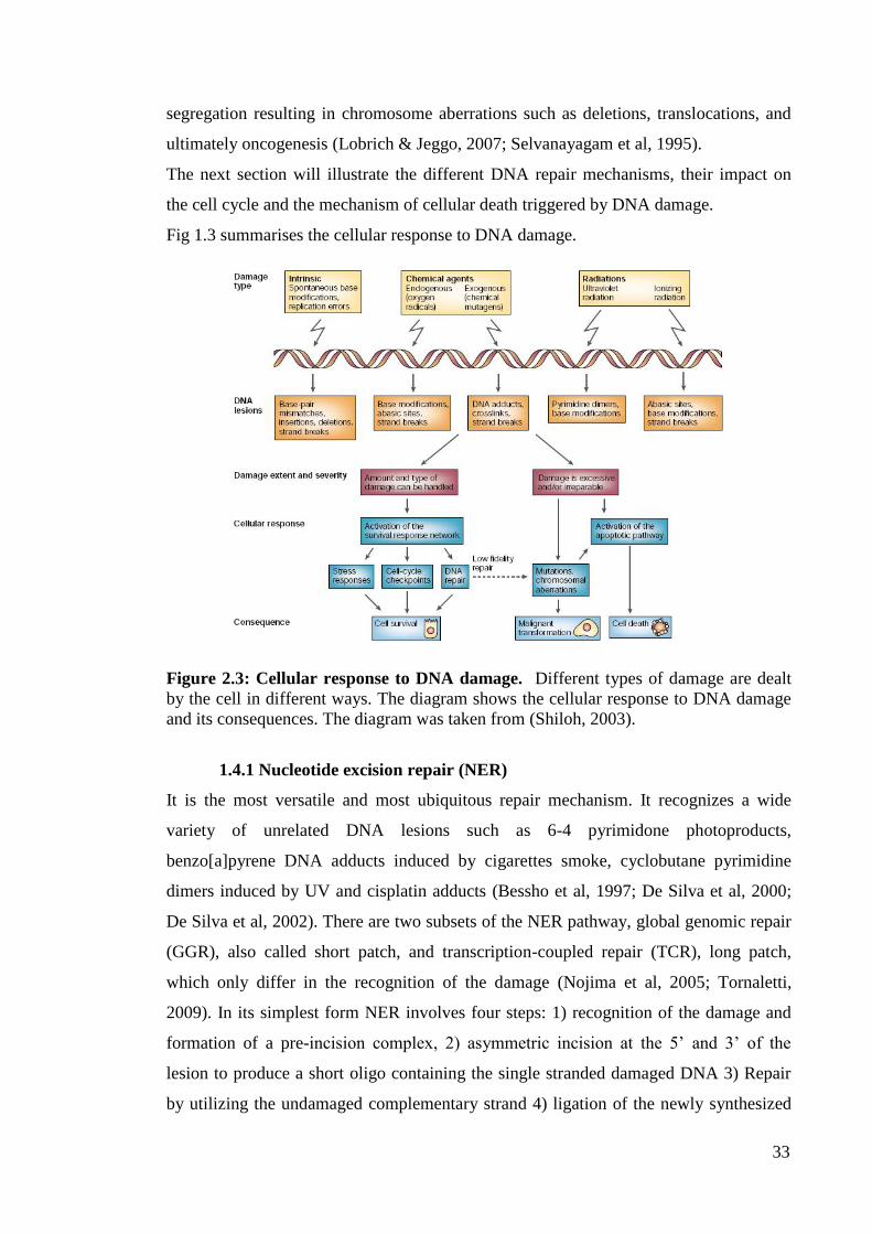
Fig 1.3 summarises the cellular response to DNA damage.
Figure 2.3: Cellular response to DNA damage. Different types of damage are dealt
by the cell in different ways. The diagram shows the cellular response to DNA damage
and its consequences. The diagram was taken from (Shiloh, 2003).
1.4.1 Nucleotide excision repair (NER)
It is the most versatile and most ubiquitous repair mechanism. It recognizes a wide
variety of unrelated DNA lesions such as 6-4 pyrimidone photoproducts,
benzo[a]pyrene DNA adducts induced by cigarettes smoke, cyclobutane pyrimidine
dimers induced by UV and cisplatin adducts (Bessho et al, 1997; De Silva et al, 2000;
De Silva et al, 2002). There are two subsets of the NER pathway, global genomic repair
(GGR), also called short patch, and transcription-coupled repair (TCR), long patch,
which only differ in the recognition of the damage (Nojima et al, 2005; Tornaletti,
2009). In its simplest form NER involves four steps: 1) recognition of the damage and
formation of a pre-incision complex, 2) asymmetric incision at the 5‟ and 3‟ of the
lesion to produce a short oligo containing the single stranded damaged DNA 3) Repair
by utilizing the undamaged complementary strand 4) ligation of the newly synthesized

34
strand to complete the repair of DNA (Hoeijmakers, 2001). While in GGR XPC/HR23B
is the major DNA lesion recognition complex, in TCR the damage is recognized by the
arrest of the elongating RNA polymerase (RNAP) at the site of the lesion. The
recognition of the lesion triggers the recruitment of the transcription factor II H (TFIIH).
TFIIH is a nine subunit complex two of which, XPB and XPD, are DNA helicases that
unwind the DNA damaged duplex. Once the strands are unwound and the damages site
inaccessible, XPF-ERCC1and XPG are recruited to make the 5‟ and 3‟ excision on each
site of the lesion creating a 22-30 base oligonucleotide. Using the opposite undamaged
strand replication factor C (RFC), PCNA, DNA polymerase or , DNA ligase I and
RPA, gap fill the excised strand and ligate the synthesized oligonucleotide to the DNA
strand (Andressoo et al, 2005; Andressoo et al, 2006; Hoeijmakers, 2001; Hoeijmakers,
2007; Hoeijmakers, 2009; Mitchell et al, 2003). Fig 1.4 summarise the pathway.
1.4.2 Base excision repair (BER)
It is mainly involved in the repair of DNA damage cause by cellular metabolism. BER
recognizes mainly DNA damage occurring during cellular metabolism as a result of
reactive oxygen species, hydrolysis, methylation and deamination. In addition BER is
employed by the formation of a SSB following IR (Swartzlander et al; Swartzlander et
al, 2010).
Normally glycosilases flip the damaged base out of the helix forming an abasic site
(hydrolysis per se forms and abasic site). At this stage the endonuclease APE1 makes an
incision at the damaged strand and DNA pol removes the 5‟ baseless sugar residue and
allows the XRCC1 protein to synthesize the damaged base. DNA Ligase 3 seals the
strand break completing the repair. If the long patch pathway is activated, DNA pol /
and PCNA produce an incision and re-synthesize 2-10 bases, FEN1 endonuclease
cleaves the displaced DNA fragment and DNA Ligase I seals it to the DNA strand.
Some BER lesions can cause block of transcription, which will be dealt by the TCR
(Hoeijmakers, 2007). Usually when BER is utilized for the repair of a SSB, the poly
ADP ribose polymerase-1 (PARP1) and PNK are recruited to the site of the damage to
prevent any unwanted recombination event and signal for BER initiation (Griffiths et al,
2009). Fig 1.5 shows the steps of this pathway.

35
1.4.3 Mismatch repair (MMR)
It is a highly conserved mechanism, strand specific which is initiated by the recognition
of mismatched or unmatched DNA base pairs or insertion-deletion of loops.
Mammalian MMR involves the family members of the E. coli MutS and MutL.
Heterodimer formation of the human MutS homolog 2 and homolog 6 (hMSH2/6) also
called hMutS recognizes mismatches and single base loops (Zdraveski et al, 2002).
Insertion or deletion loops are usually recognized by the hMSH2/3 (hMutS ) (Sugawara
et al, 1997). Formation of other heterodimeric complexes of the hMutL-like proteins
hMLH1/hPMS2 (hMutL ) and hMLH1/hPMS1 (hMutL ) interact with the MSH
complexes and the replication factors either to contact the replication machinery or to
stabilize the MSH complexes on the lesion. Following recognition of the damage strand
discrimination of the newly synthesized (containing the mismatch) strand is probably
based on physical contact with the nearby replication machinery (Martin et al, 2008).
Excision of a DNA fragment past the mismatch and its degradation are carried out by
RPA, PCNA, RFC, exonuclease1 and endonuclease FEN1(Hoeijmakers, 2001). Correct
synthesis is mediated by the activity of pol / . Fig 1.6 shows a diagram of the pathway.

36
Figure 1.4: Nucleotide excision repair. The diagram shows the different steps and the
molecules involved in this repair pathway (Hoeijmakers, 2001).

37
Figure 1.5: Base excision repair (BER) pathways. (Hoeijmakers, 2001)

38
Figure 1.6 Mismatch repair (MMR) pathway. Hoeijmakers J.H., Nature 2001

39
1.4.4 Double strand breaks repair pathways: Nonhomologous end joining
and Homologous recombination.
1.4.4.1 Double strand breaks causes and consequences
DSB are naturally generated in the cells when the replication fork encounters a DNA
nick or blocking lesion. DSBs can be classified in Physiological DSBs and Pathological
DSBs. Physiological DSBs are those that arise from programmed genome
rearrangement (yeast mating switch type, V(D)J recombination, antigen receptor gene
rearrangement and meiosis). Pathological double strand breaks are those produced by
oxidative free radicals, IR, replication across another lesion, inadvertent nuclear enzyme
action at a fragile site, topoisomerase failure, and physical stress imposed on the
chromosome during mitosis (Caldecott, 2008). Normally during oxidative respiration
mitochondria convert from 0.1-1% of the oxygen to superoxide (O2-). Superoxide
dismutase in the mitochondrion (SOD2) and in the cytosol (SOD1) converts this into
hydroxyl free radicals, which react with DNA causing single strand breaks. Two closely
spaced SSBs give rise to a DSB. Each hour around 1022
ROS are produced in the human
body (109 per hour per cell). Environmental IR, such as -rays and X-rays, is also a
cause of DSBs. Only at seas level around 300 million IR particles pass through each
person per hour. These create free radicals that cluster around DNA generating 1 DSB
for every 25 SSBs (De Bont & van Larebeke, 2004). Despite being the most mutagenic
form of DNA damage, mammals have evolved a way to exploit the formation of DSBs
to control biological processes and maintain heterogeneity. In addition to the action of
topoisomerases II that induce the formation of DSBs to decatenate the DNA strands,
DSBS occur also to initiate rearrangement during maturation of immunoglobulin genes
and are central in the recombination events between homologs during meiotic prophase
I (McKinnon & Caldecott, 2007).
The failure to repair or missrepair DSBs can result in cellular death or in large-scale
chromosome deletions, translocations, and fusions enhancing genome instability and
induce carcinogenesis. This is one of the main reasons why chemical agents such as
Topoisomerase poisons, other chemotherapeutic drugs and IR therapy while inducing
substantial damage to eradicate cancer cells may induce, at the same time, secondary
malignancies, by targeting the genome integrity of non cancer cells (Jeggo & Lobrich,
2007; Khanna & Jackson, 2001).

40
1.4.4.2 Double strand breaks repair pathways
Two major pathways in diploid cells repair DSBs. The most common form of repair is
the homology-directed repair or homologous recombination (HR), which requires long
sequence of homology to repair the damage. The second, but mostly developed in non-
dividing haploid organisms and diploid organism is the nonhomologous end joining
(NHEJ), which does not require the presence of a homologous strand to repair the DSB
(Shrivastav et al, 2008). Although still active area of investigation, it s generally
accepted that during S phase the physical vicinity of the sister chromatids provides the
homology required for activation of HR. During S/G2 transition if a homolog is not
present NHEJ will be used to repair the break and outside of this phase NHEJ is the only
possible option. The issue of homologue proximity and the possible competition among
the components of these pathways are yet to be fully understood (Allen et al, 2003).
Recent studies have suggested the DNA ligase IV complex may be responsible to
suppress the HR initiation step, leading to NHEJ activation (Shrivastav et al, 2008).
1.4.4.2.1 Non-homologous end joining (NHEJ)
Accurate and mostly precise on simple breaks such as blunt ends, this pathway rejoins
the two damaged ends of the DNA break in a sequence independent fashion (van Gent
& van der Burg, 2007). Although described as non homologous a small region of
homology (1-6bp) can facilitate the strand break rejoining (van Gent & van der Burg,
2007). It requires Ku heterodimer (Ku70 and Ku80) which binds to the reciprocal DNA
ends through is ring shape structure. This complex has mainly the function of a scaffold
to assemble the other components of the pathways at the DNA termini. One of the first
enzymes to be recruited is the DNA dependent kinase catalytic subunit (DNAPKcs).
This serine-threonine 460kDa protein forms a so-called synaptic complex that holds the
two ends of the broken DNA molecule together. Together with Ku70/80 it forms a
holeo-enzyme complex called DNAPK (Hammel et al, 2010). Usually in presence of
DNA blunt ends or complementary 5‟ phosphate and 3‟ hydroxyl group, the auto-
phosphorylation of DNAPKcs allows juxtapositioning of the DNA ends that become
available for ligation by the DNA ligase IV complex containing XRCC4 and XLF
cofactors (Weterings & Chen, 2007). In many cases, ends cannot be precisely rejoined
because of aberrant 3‟phoshate groups, 5‟ hydroxyl groups damaged backbone sugar
residues and damaged DNA bases. These ends require processing before ligation takes
41
place. This process can be mediated by different proteins according to the different type
of DNA ends fromed at the site of the break. Polynucleotide kinase (PNK) interacts with
DNA ligase IV in presence of 3‟ phosphate groups and 5‟ hydroxyl groups and recruits
ancillary components such aprataxin (APTX) and PNK-APTX like factor (PALF) for
additional 3‟ nuclease activity (van Gent & van der Burg, 2007). Artemis nuclease
activity is involved in the resolution of difficult DSBs. Following binding and
phosphorylation by DNAPKcs, Artemis exerts a diverse array of nuclease activity
including 5‟ and 3‟ endonuclease activity, hairpin opening activity in addition to its own
5‟ exonulease activity. In presence of aberrant DNA ends DNAPKcs phosphorylates
Werner helicase (WRN) whose nuclease activity is required for their processing
(Stracker et al, 2009). Despite the great flexibility shown by the XFL:XRCC4:DNA
ligase IV to ligate across gaps with incompatible ends there are 2 members of the
Polymerase X family Polymerase and that can fill the 5‟single stranded extensions.
Their activity is well suited in the NHEJ pathway as Pol is capable of both template
dependent and independent synthesis and Pol has more flexibility than replicative
polymerases (Lieber, 2010; Shrivastav et al, 2008).
There are several other factors shown to be required for the repair of difficult DSBs by
the NHEJ. These are: the MRE11/RAD50/NBS1 (MRN) complex, MDC1, 53BP1 and
ATM kinase. While the MRN complex may be implicated in keeping the DNA ends
together, or in close proximity, MDC1, 53BP1 and phosphorylation of the histone
variant H2AX seems to be required to the formation of this complex in foci fashion.
ATM is one of the major kinase of H2AX following formation of DSBs therefore it has
been suggested that ATM would induce the creation of chromatin microenvironment
that allows the formation of the MRN foci. Fig 1.7 shows the most significant steps of
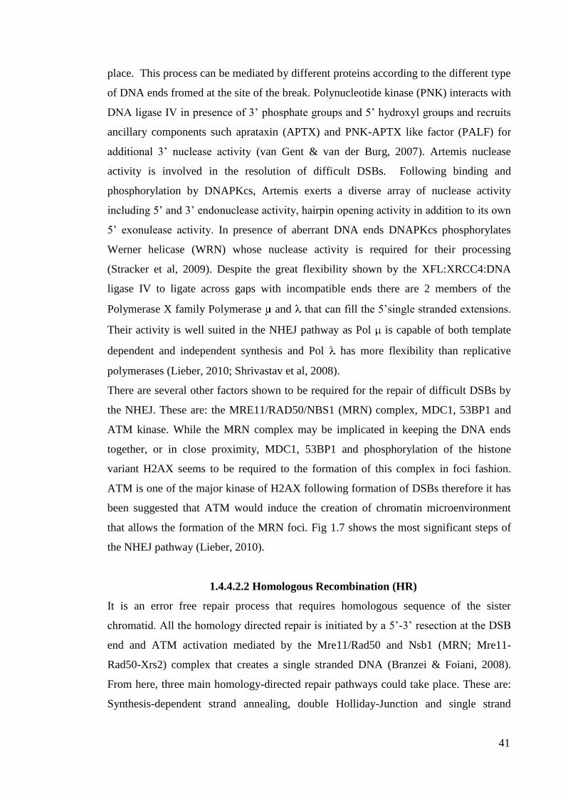
the NHEJ pathway (Lieber, 2010).
1.4.4.2.2 Homologous Recombination (HR)
It is an error free repair process that requires homologous sequence of the sister
chromatid. All the homology directed repair is initiated by a 5‟-3‟ resection at the DSB
end and ATM activation mediated by the Mre11/Rad50 and Nsb1 (MRN; Mre11-
Rad50-Xrs2) complex that creates a single stranded DNA (Branzei & Foiani, 2008).
From here, three main homology-directed repair pathways could take place. These are:
Synthesis-dependent strand annealing, double Holliday-Junction and single strand

42
annealing (Helleday et al, 2008). Following resection, in the synthesis-dependent strand-
annealing subpathway, Rad51 with help of a numerous group of proteins (BRAC2,
RAD52, RAD54, RAD54B and also RAD51 paralogues RAD51B, RAD51C, RAD51D,
XRCC2 and XRCC3) forms the nucleoprotein filament that searches for the homolog
and initiates strand invasion (Shrivastav et al, 2008). The base pairing of the homolog
and the invading strand creates a DNA heteroduplex that displaces one of the DNA
strands forming a D-loop that enlarges as the synthesis proceeds. The annealed 3‟ strand
is then elongated via repair synthesis mediated by polymerase (pol ). Several lines of
evidence have suggested that pol is involved in HR, however several other
polymerases can compensate in his absence. Synthesis must continue beyond the
original point of break to restore the missing information at the break point. On the other
side of the D-loop an X -like structure, called the Holliday Junction, is formed at the
border of the intersection between the invading strand and the homolog (Moynahan &
Jasin, 2010). In order to release the newly synthesized strand the Holliday junction has
to slide towards the same direction of the synthesis in a process referred as branch
migration. WRN, BLM, p53, RAD54, BLAP75, hMSH2, hMSH6 have all been shown
to be able to modulate the sliding and the direction of the junction. This is necessary to
allow release of the new strand however the sliding process and the regulating
mechanism are not fully understood (Helleday et al, 2007). Once free, the released
strand is bound by RPA and if the opposite end of the DSB is similarly fixed then
simple annealing will take place via the help of Rad52 or possibly p53. Presence of
damaged bases or the extent of the synthesis my lead to flaps, gaps. In this case
XPF/ERCC1 endonuclease activity is employed and the gaps are filled by the activity of
other proteins such as DNA ligase I, PCNA and pol / (Adair et al, 2000; Batty &
Wood, 2000).
Sometimes there is the possibility that a double Holliday junction forms at either side of
the D-loop. This normally happens when the opposite strand anneals to the D-loop and
also starts synthesis creating a double intersection. If the cleavage that release these
heteroduplexes is made at the holliday junctions then flanking sequences continuity of
the DSBs will be preserved otherwise it will result in a cross over event.
If at the sides of the DSBs two adjacent repeat sequences are present, single strand
annealing may be utilized to resolve the DSBs. This is a process facilitated by rad52
and RPA but Rad51-independent. If this HR subpathway is initiated then the sequence

43
between the repeats will be inevitably lost as well as one of the repeats. In the literature
this sub pathway is also referred as an unintentional consequence of the need to create a
single stranded DNA to initiate synthesis- dependent strand annealing. Fig 1.8 shows the
steps of this pathway (Evers et al, 2010; Helleday et al, 2007; Shrivastav et al, 2008).
Figure 1.7: Non Homologous end joining (NHEJ) pathway. (Chen & Nirodi, 2007)

44
Figure 1.8: Homologous recombination (HR) pathway. (Helleday, 2010)

45
1.5 DNAPKcs
1.5.1 Structure
The product of the PRDK gene is a 4128 amino acids polypeptide of 469 kDa. It is the
largest protein kinase in biology and a member of the phosphoinositide 3-kinase-
likefamily (PIKK) of protein kinases. DNAPKcs is specifically activated by double
stranded DNA ends of different configurations. DNAPKcs alone has a dissociation
constant of 3x10⁻⁹
M for blunt DNA ends. In presence of Ku at the DNA ends, the
affinity is higher with a dissociation constant of 3x10-11
M. DNAPKcs N-terminal
extends from amino acid 1-2908 (about 250 kDa) and contains a putative DNA-binding
domain, a leucine rich region and a series of HEAT repeats (huntingtin, elongation
factor 3, A subunit of protein phosphatase 2A and TOR1). The C- terminal region
contains a FAT [FRAP (FKBP12-rapamycin-associated-protein) ATM (ataxia-
talangiectasia mutated), TRRAP (transactivation/transformation-domain-associated-
protein)] domain (amino acids 2908-3539), the PIKK domain (3645-4029) and the
FATC domain (4906-4128). Cells that lack DNAPKcs expression have been shown to
be radiosensitive and also have defects in V(D)J recombination. In addition mammals
with DNAPKcs also showed severe combined immunodeficiency (SCID) (Lieber,
2010).
1.5.2 DNAPKcs involvement in repair
Despite its capability to bind DNA alone, DNAPKcs recruitment to damaged DNA is
Ku dependent. The binding of these two proteins is mediated by a conserved region in
the extreme C-terminus region of Ku and C-terminal region of DNAPKcs. By
translocating inwards, away from the DNA damaged ends, Ku allows DNAPKcs
interaction to the DNA, providing sufficient space for two DNAPKcs molecules to bind
to the DNA termini and interact in a synaptic complex that hold the ends tight together.
The DNAPKcs-Ku-DNA complex is referred as the DNAPK complex, known to be the
most central component of the NHEJ pathway (Collis et al, 2005; DiBiase et al, 2000).
Although DNAPK phosphorylates Ku70, Ku80, XRCC4, XLF, Artemis and DNA ligase
IV in vitro there is no evidence that these phosphorylation events are required in vivo for
NHEJ (Douglas et al, 2005; Sibanda et al, 2010). The only in vivo validated substrate of
DNAPK is DNAPKcs itself. Noteworthy is that the Ku component of DNAPK can only
load or unload DNA therefore the kinase activity of the complex is mediated by its only

46
kinase component, DNAPKcs (Chan et al, 2002). There are 16 phosphorylation sites
identified to contribute to DNAPKcs autophosphorylation in response to DNA damage.
1.5.3 DNAPKcs phosphorylation
The most in vivo characterized and IR inducible and DNAPK-dependent sites are the
ABCDE cluster (Ser2612
,Ser2624
, Thr2609
, Thr2620
, Thr2628
, Thr2647
) and Ser2056
, Thr3950
. In
addition IR-induced phosphorylation of Ser2612
Thr2628
and Thr2647
occurs following
inhibition of the related protein ATM. In contrast other studies have shown that both
ATM and ATR can phosphorylate Ser2612
, Thr2609
, Thr2628
and Thr2647
in response to IR
or UV. Cell type, cell cycle stage and the extent of the DNA damage may be the
determining factors that induce in vivo DNAPKcs phosphorylation by DNAPK or ATM
or ATR. Proteomic screen have also revealed additional in vivo phosphorylation site
(Ser2674
, Ser2675
, Ser2677
, Ser3205
) but the kinase responsible and the consequences of
these events have not been yet determined (Chan et al, 2002; Collis et al, 2005; Sibanda
et al, 2010). In vitro studies of DNAPKcs autophosphorylation have revealed that this
process induces loss of kinase activity and dissociation of the phosphorylated DNAPKcs
from KU-DNA. This suggests that the phosphorylation event may regulate the
assembly/disassembly of the DNAPK complex (Bailey et al, 2004; Chen et al, 2005).
Further studies have shown autophosphorylation of DNAPKcs is required for its release
from DSB in vivo since DNAPKcs kinase dead, and autophosphorylation defective
DNAPKcs are maintained longer at the site of DNA damage. DNAPKcs
autophosphorylation can be highly complex in vivo because although it may be required
for disassembly of the complex, phosphorylation at other regions, enhances the rate of
HR and can either positively or negatively regulate DNA processing (Gu et al, 2010).
1.6 DNA REPAIR REGULATION THROUGH THE CELL CYCLE
Although the activation of a specific DNA repair pathway depends on the type of DNA
lesion and its recognition, cell cycle is also a determining factor in this choice. Certain
repair mechanisms can only take place during one phase of the cells cycle. NER factors
will be usually required to repair SSB lesions in G1 cells (Branzei & Foiani, 2008). In
contrast, DSBs are repaired by HR during S phase because of the presence of an intact
and accessible sister chromatid but during the remaining phases of the cycle, being the
chromatin already highly condensed, repair will be undertaken by NHEJ (Shrivastav et
al, 2008). The recognition of the damage, the cell cycle and the DNA repair mechanisms

47
are highly linked and clearly dependent from each other. In order to maintain genome
stability and integrity cells have evolved two important checkpoints during the cell cycle
also called restriction points where DNA integrity is checked and damaged DNA is
repaired to assure correct transmission of the genetic information. In presence of DNA
damage, cells integrate DNA repair pathways with transcription, signalling, and
apoptosis in a tightly regulated network called the DNA damage response (DDR)
orchestrated by the checkpoint proteins. Recognition of damage usually becomes an
activating signal that checkpoint transducers amplify to downstream targets activating
DNA repair systems, cell cycle arrest, and apoptosis (Jackson, 2009). Central
components of the checkpoint machinery are the PI3K related proteins ATM, ATR and
DNAPKcs. ATM and DNAPKcs respond to the presence of DSBs whereas ATR is
activated by SSBs and stalled replication forks. These proteins are usually recruited by
interacting directly with their partners, NBS1, ATRIP and Ku respectively. ATM and
ATR target many substrates including the checkpoint kinase 2 (CHK2) and checkpoint
kinase 1 (CHK1) (Lobrich & Jeggo, 2007). While CHK2 activation is exclusively ATM
dependent, CHK1 is activated by ATR is mediated by the protein Claspin and also
indirectly by ATM that activates ATR. CHK1 and CHK2 target cyclin dependent
kinases (CDK) whose activation modulates the cell cycle progression by regulating the
cell cycle checkpoints (Branzei & Foiani, 2008). As mentioned above these restriction
points often induce cell cycle arrest to allow repair of damaged DNA before the
synthesis of the cell cycle and prior to enter mitosis. Thus they control the transition to
the most important phases of the cell cycle: G1/S and G2/M.
The next sections will briefly review the cell cycle checkpoints, their role in recognising
DNA damage and the consequence of unreparable damage
1.6.1 Cell cycle checkpoints
The interaction of three families of proteins regulates the checkpoints of the cell cycle:
CDKs, cyclins and the cyclin dependent kinase inhibitors (CKI). Each of these
components is made of a great array of molecules and sub family members that are
differentially activated during the cell cycle. Cyclins expression is mediated by
transcriptional control and destroyed by ubiquitin mediated proteolysis, CDKs requires
cyclin binding to relieve the obstruction of their catalytic site and CKI inhibits the
binding of the cyclin and CDKs rendering the CDKs inactive (Lee et al, 2008; Wojda,
2000).

48
As shown in Fig 1.9 each cyclin preferentially binds to a specific CDK. An important
substrate of the CDKs is the retinoblastoma protein pRb, which, in its
hypophosphorylated status, binds and inactivates the transcription factor E2F. RB
phosphorylation by the CDK-cyclin active complex, releases E2F which mediates
transcription of genes such as the DNA polymerase and thimidine kinase allowing
transition in S phase (Nevins, 2001). There are two main families of CKIs. The INK4
family (inhibitor of cyclin dependent kinase 4) family (p15INK4
, p16INK4
, p18INK4
,
p19/p14ARF
), which specifically binds to CDK4 and CDK6 and the Cip/Kip family
(p21Cip1/WAF1
, p57Kip2
, p27kip1
), which have a wider specificity and bind to the CDK-
cyclin complex rather than CDK alone (Shan et al, 1996).
Figure 1.9: Changes in cycle dependent kinase and cyclins during the cell cycle. The diagram outlines the production of cyclins during the different phases of the cell
cycle and the correspondent CDK binding partner. The picture is taken from: The
Biology of Cancer (© Garland Science 2007).
1.6.1.1 Cell cycle arrest
G1-S arrest is usually initiated by ATM and mediated by direct activation of CHK2 and
direct/indirect activation of p53 (Shiloh, 2003). While CHK2 induces activation and
consequent degradation of CDC25A, p53 induces transcription of p21Cip1/WAF1
. Both p21
and CDC25A inhibit the CDK2/cyclinE complex formation and its mediated pRb
phosphorylation(Lobrich & Jeggo, 2007; Meek, 2009). S phase arrest arises following
ATR recognition of SS DNA due to collapsed or stalled replication forks replication

49
blocks. Subsequent activation of CHK1 induces phosphorylation and degradation of
CDC25A. This prevents the initiation of a new replication origin and slows down
replication (Zhou & Elledge, 2000). G2 cell cycle arrest is regulated via the inactivation
of Cdk1 kinase (also known as Cdc2) (van Vugt et al, 2005). DSBs formation induces
direct recruitment of ATM and indirect recruitment of ATR by the ATM strand
resection. They activate CHK1 and CHK2 respectively inducing CD25A degradation
and promote stabilization of p53 and consequent induction of p21. The phosphorylation
of CDK1 and the inhibition of the complex formation with CyclinB induce the G2
arrest. Also, p27 and Wee1 have been shown to induce CDK1 inhibition. Fig 1.10
shows a summary of the above mentioned cell cycle arrest (Bassermann & Pagano,
2010).
Figure 1.10: Mammalian signalling activating cell cycle check points following
DSBs formation. The diagram shows the different response to DSBs according to the
different cell cycle phase. ATM responds mainly to DSBs whereas ATR to SSBs. The
recruitment of the MRN complex amplifies the signal and involves the transducer
kinases CHK1 and CHK2. In turn these transducers regulate inhibition of the cell cycle
by inhibiting the cyclin/CDK complex formation. The diagram was taken from (Lobrich
& Jeggo, 2007).
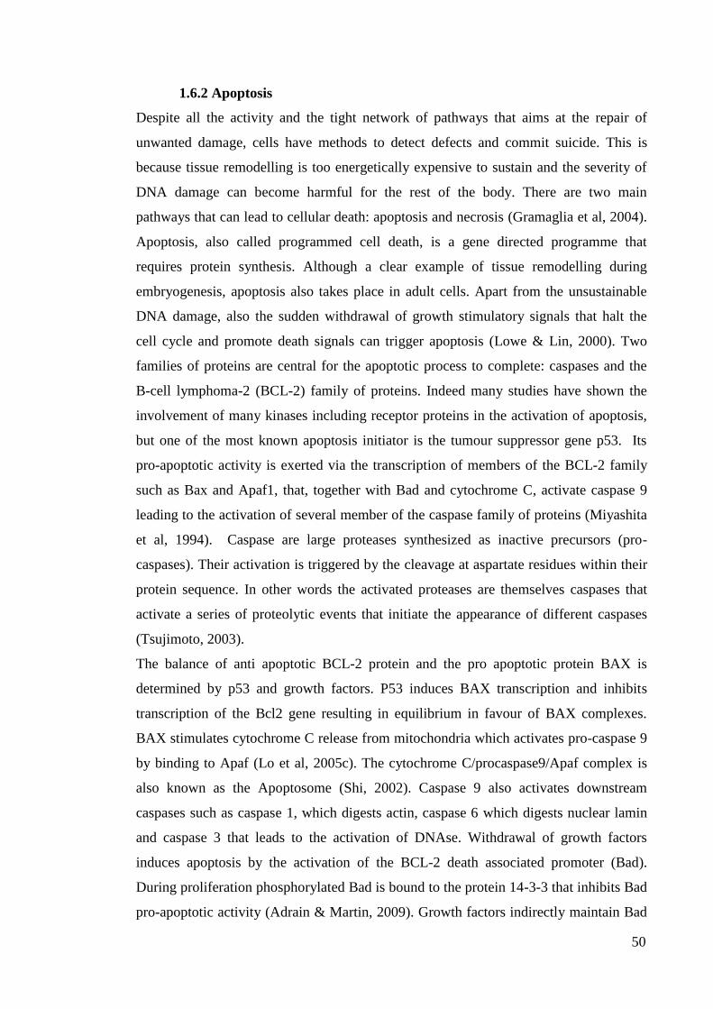
50
1.6.2 Apoptosis
Despite all the activity and the tight network of pathways that aims at the repair of
unwanted damage, cells have methods to detect defects and commit suicide. This is
because tissue remodelling is too energetically expensive to sustain and the severity of
DNA damage can become harmful for the rest of the body. There are two main
pathways that can lead to cellular death: apoptosis and necrosis (Gramaglia et al, 2004).
Apoptosis, also called programmed cell death, is a gene directed programme that
requires protein synthesis. Although a clear example of tissue remodelling during
embryogenesis, apoptosis also takes place in adult cells. Apart from the unsustainable
DNA damage, also the sudden withdrawal of growth stimulatory signals that halt the
cell cycle and promote death signals can trigger apoptosis (Lowe & Lin, 2000). Two
families of proteins are central for the apoptotic process to complete: caspases and the
B-cell lymphoma-2 (BCL-2) family of proteins. Indeed many studies have shown the
involvement of many kinases including receptor proteins in the activation of apoptosis,
but one of the most known apoptosis initiator is the tumour suppressor gene p53. Its
pro-apoptotic activity is exerted via the transcription of members of the BCL-2 family
such as Bax and Apaf1, that, together with Bad and cytochrome C, activate caspase 9
leading to the activation of several member of the caspase family of proteins (Miyashita
et al, 1994). Caspase are large proteases synthesized as inactive precursors (pro-
caspases). Their activation is triggered by the cleavage at aspartate residues within their
protein sequence. In other words the activated proteases are themselves caspases that
activate a series of proteolytic events that initiate the appearance of different caspases
(Tsujimoto, 2003).
The balance of anti apoptotic BCL-2 protein and the pro apoptotic protein BAX is
determined by p53 and growth factors. P53 induces BAX transcription and inhibits
transcription of the Bcl2 gene resulting in equilibrium in favour of BAX complexes.
BAX stimulates cytochrome C release from mitochondria which activates pro-caspase 9
by binding to Apaf (Lo et al, 2005c). The cytochrome C/procaspase9/Apaf complex is
also known as the Apoptosome (Shi, 2002). Caspase 9 also activates downstream
caspases such as caspase 1, which digests actin, caspase 6 which digests nuclear lamin
and caspase 3 that leads to the activation of DNAse. Withdrawal of growth factors
induces apoptosis by the activation of the BCL-2 death associated promoter (Bad).
During proliferation phosphorylated Bad is bound to the protein 14-3-3 that inhibits Bad
pro-apoptotic activity (Adrain & Martin, 2009). Growth factors indirectly maintain Bad
51
phosphorylation via the activation of a member of the PI3K pathway, protein kinase B
(Akt). In absence of growth factors Bad is dephosphorylated and following dissociation
from 14-3-3 it binds BCL2 blocking its inhibitory effect on cell death. P53 seems to be
involved in the activation of the Akt pathway responsible for Bad inactivation (Breen et
al, 2007). Necrosis is commonly seen in the centre of solid tumours. Usually low or
poor nutrient supply determines disruption of the energy dependent ion channels. The
consequent increase in cell volume and loss of membrane integrity induces the
activation of proteases and nucleases. This type of cellular lysis is often associated with
an inflammatory response. Fig 1.11 shows the complexity of the apoptotic pathway
(Weisz et al, 2007).
Figure 1.11: Apoptosis signalling pathway (www.cellsignal.com).

52
1.7 CELLULAR SIGNALLING AND CANCER
Despite the presence of an extensive array of molecules and mechanisms, evolved to
protect cells from DNA damage and prevent its incorporation and transmission, cells
still manage to acquire somatic mutations developing into malignancies. Interestingly,
not even the induction of further DNA damage within these same cells via the usage of
genotoxic agents and RT is, most of the time, sufficient to kill cancer cells (Savage et al,
2009). One of the main aims of cancer therapy is to achieve tumour control with less
toxicity. Targeting aberrant oncogenic factors or pathways that are specific to cancer
cells has shown improvement in the outcome of cancer therapies without increasing
toxicity (Nyati et al, 2006). The concept of resistance to treatments and to the innate
cellular defence mechanisms is incredibly vast and the attempt to give a comprehensive
picture, fair to all the molecules involved in these processes, would require volumes.
This section is going to review the role of receptor tyrosine kinases and in particular the
role of the epidermal growth factor receptor (EGFR) in tumorigenesis, cancer signalling
and resistance to therapy. This is necessary to understand the role of molecular targeted
therapy within cancer management and the importance of a better understanding of
cellular mechanism following radiotherapy or chemotherapy to design more efficient
molecular target therapies.
1.7.1 Oncogenic Receptor Tyrosine kinases (RTK)
Growth factor and RTKs coordinate, through a complex system of signalling network,
cellular survival, apoptosis, proliferation, differentiation and migration.
There are 58 known RTKs in mammals distributed in 20 subfamilies and despite sharing
a lot of structural homology and downstream signalling pathways each RTK associates
uniquely to these pathways contributing to an enormous combinatorial complexity
(Lemmon & Schlessinger, 2010). All RTKs have a ligand binding domain in the
extracellular region, a single transmemebrane helix, and a cytoplasmic domain
containing the kinase domain and an additional carboxyl (C-) terminal and
juxtamembrane regions. Their mechanism of activation, topology and downstream
signalling pathways are conserved from the nematode Caenorhabditis elegans to
humans (Amit et al, 2007b). Mutation in RTKs and aberrant activation of their
signalling pathways have been linked to diseases such as diabetes, inflammation, bone
disorders angiogenesis and arterosclerosis. Nearly 30% of the RTKs are involved in
cancer or tumorigenesis via exerting their oncogenic potential (Bublil & Yarden, 2007;

53
Katz et al, 2007; Seger et al, 2008). Their aberrant activation is mediated by four main
molecular mechanisms: autocrine activation, chromosomal activation, overexpression,
or gain of functional mutations (Amit et al, 2007b). One of the main characteristics of
RTKs and also one of the properties that renders them central in understanding and
designing new molecular targets is their functional robustness, shown by their capability
to maintain output reproducibility despite input variation (Citri & Yarden, 2006).
1.7.1.1 The EGF receptor family
The ERBB family of protein takes its name from their homology to the erythroblastoma
viral gene product v-erb. It comprises four receptors: ERRB1-4, also known as HER1-4,
and 13 polypeptide extracellular ligands which all contain a conserved epidermal growth
factor (EGF) domain. All four receptor are transmembrane glycoproteins whose
activation takes place via ligand binding and/or homo or heterodimerisation (Bublil &
Yarden, 2007). The vertebrate family of receptors evolved from the single ligand-
receptor pair expressed in nematodes. While in worms there is only one single EGF-like
ligand (LIN-3), drosophila expresses four stimulatory ligands and one inhibitory ligand
called ARGOS. The function of this family of receptor is also very well conserved
across the species. In worms, EGFR controls vulva development, differentiation of the
male tail and posterior-ectoderm development, the drosophila. Likewise, the Drosophila
EGF receptor (DER) controls the development of several organs during embryogenesis
and the mammalian ERBBs control organogenesis in multiple epithelial tissues (Amit et
al, 2007b).
1.7.1.1.1 The evolution of the mammalian ERBB network
The origin of the first ERBB receptor must have been the result of a gene fusion event
that linked a cytoplasmic tyrosine kinase to a cell surface receptor. Amino acid sequence
analysis of the four ERBB has revealed that a gene duplication event must have
occurred to generate two ancestral receptors, the ERBB1/ERBB2 precursor and the
ERBB3/ERBB4 precursor. Following subsequent gene duplication these precursors
generated the 4 ERBB proteins. In parallel to the evolutionary events that lead to the
segregation of 4 receptors, the vertebrate ligands also segregated into the ERBB1
ligands and the ERBB3/ ERBB4 ligands (Fig 1.12) (Amit et al, 2007b).

54
Figure 1.12: Evolution of ERBB signalling. From a single vertical cascade of
signalling events, mediated by one receptor in invertebrate primordial specie (left), three
scenarios may have evolved. Option A describes two events of RTK duplication leading
to 4 cascades contributing to gene expression remaining isolated from each other. In
option B the four cascades interact and establish a complex bow tie of layered signalling
network. Evolution from invertebrates may have preferred directly option B or the
transit from option A (Option C). There is no evidence of evolutionary stability of
option A. Fig rearranged from (Amit et al, 2007b).
1.7.1.2 ERBB ligands
The epidermal growth factor (EGF), transforming growth factor alpha (TGF- ) and
amphiregulin (AR) bind selectively to EGFR. Betacellulin (BTC), heparin-binding EGF
(HB-EGF) and epiregulin (EPR) bind to ERBB1 and ERBB4. The third classes of
ligands, the neuregulins (NRG1-4) bind and activate ERBB3 and ERBB4. ERBB2 lacks
ligand association but it is known to be the most preferred partner for other ERBB
interactions. The majority of the ERBB ligands are synthesized as type-1
transmembrane proteins. The mature and soluble form of the ligand is released into the
cellular matrix following protease cleavage by members of the ADAM family (A
disintegrin And Metalloproteinases) of metalloproteinases (Zhou et al, 2006). Each
member of this family recognizes at least one feature of the EGF like motif of a ligand.
This motif is made up of 6 spatially conserved cysteine residues (CX7 CX4-5 CX10-13
CXCX8 C) that forms three peptide loops through the formation of disulfide bonds by
the interaction of C1-C3, C2-C4, and C5-C6 (Nakamura et al, 2000). In some cases

55
(HB-EGF, TGF- , AR and BTC) the ligand binding molecule is not cleaved from the
membrane and can function even if tethered to the plasma membrane through adjacent
receptor bearing cells (juxtacrine signalling) (Klein et al, 2004; Lemmon, 2009;
Lemmon & Schlessinger; Lemmon & Schlessinger, 2010; Ozcan et al, 2006).
1.7.1.3 ERBB2
Heterodimers containing HER2 have stronger signal output because of the larger subset
of phosphoproteins that HER2 can bind, and the higher affinity and broader specificity
that these heterodimers have with different ligands (Lazzara & Lauffenburger, 2009).
Also the endocytosis rate of the HER2 heterodimers is slower compared to the other
ERBBs and usually, rather than degraded; they are reshuffled back to the plasma
membrane (Madshus & Stang, 2009). The analysis of the HER2 structure has also
shown that differently from the other ERBBs the dimerisation loop of ERBB2 is
constitutively extended even in a monomeric state (i.e. resembles the same conformation
adopted by ligand bound EGFR)(Alvarado et al, 2009; Bose & Zhang, 2009). The very
strong interaction between the extracellular domains I and III render the ligand binding
domain inaccessible by ligands. For these reasons ERBB2 is mostly recognized as a non
autonomous amplifier (Fig 1.13) (Seger et al, 2008).
1.7.1.4 ERBB3
ERBB3 is a kinase defective, non-autonomous receptor forming three functional
heterodimers. It evades ligand induced degradation and in association with ERBB2
activates strongly PI3K (Fig 1.13) (Sergina et al, 2007; Shi et al).
1.7.1.5 ERBB4
ERBB4 is an autonomous receptor that shares many features with EGFR. In addition to
sharing most of their ligands ERBB4 recruits some of the most characterised interacting
partner of EGFR (Shc, Grb2 and STAT5) (Qiu et al, 2008) (Fig1.13). The incapability
of recruiting a ubiquitin ligase, induces a slow receptor down-regulation. Proteolytic
cleavage of ERBB4 cytoplasmic domain translocates the truncated receptor (discussed
below) to the nucleus and might exert transcriptional activity (Zaczek et al, 2005).
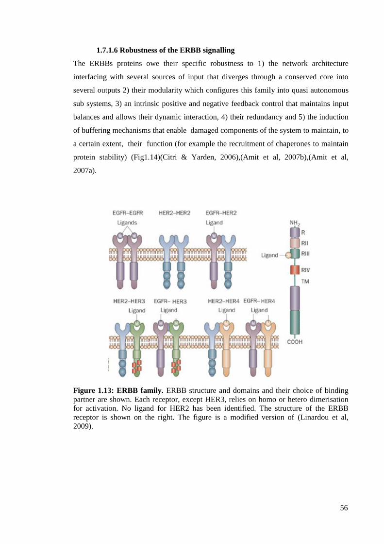
56
1.7.1.6 Robustness of the ERBB signalling
The ERBBs proteins owe their specific robustness to 1) the network architecture
interfacing with several sources of input that diverges through a conserved core into
several outputs 2) their modularity which configures this family into quasi autonomous
sub systems, 3) an intrinsic positive and negative feedback control that maintains input
balances and allows their dynamic interaction, 4) their redundancy and 5) the induction
of buffering mechanisms that enable damaged components of the system to maintain, to
a certain extent, their function (for example the recruitment of chaperones to maintain
protein stability) (Fig1.14)(Citri & Yarden, 2006),(Amit et al, 2007b),(Amit et al,
2007a).
Figure 1.13: ERBB family. ERBB structure and domains and their choice of binding
partner are shown. Each receptor, except HER3, relies on homo or hetero dimerisation
for activation. No ligand for HER2 has been identified. The structure of the ERBB
receptor is shown on the right. The figure is a modified version of (Linardou et al,
2009).
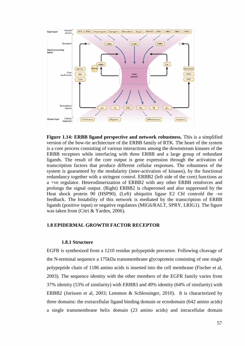
57
Figure 1.14: ERBB ligand perspective and network robustness. This is a simplified
version of the bow-tie architecture of the ERBB family of RTK. The heart of the system
is a core process consisting of various interactions among the downstream kinases of the
ERBB receptors while interfacing with three ERBB and a large group of redundant
ligands. The result of the core output is gene expression through the activation of
transcription factors that produce different cellular responses. The robustness of the
system is guaranteed by the modularity (inter-activation of kinases), by the functional
redundancy together with a stringent control. ERBB2 (left side of the core) functions as
a +ve regulator. Heterodimerization of ERBB2 with any other ERBB reinforces and
prolongs the signal output. (Right) ERBB2 is chaperoned and also suppressed by the
Heat shock protein 90 (HSP90). (Left) ubiquitin ligase E2 Cbl controld the -ve
feedback. The bistability of this network is mediated by the transcription of ERBB
ligands (positive input) or negative regulators (MIG6/RALT, SPRY, LRIG1). The figure
was taken from (Citri & Yarden, 2006).
1.8 EPIDERMAL GROWTH FACTOR RECEPTOR
1.8.1 Structure
EGFR is synthesized from a 1210 residue polypeptide precursor. Following cleavage of
the N-terminal sequence a 175kDa transmembrane glycoprotein consisting of one single
polypeptide chain of 1186 amino acids is inserted into the cell membrane (Fischer et al,
2003). The sequence identity with the other members of the EGFR family varies from
37% identity (53% of similarity) with ERBB3 and 49% identity (64% of similarity) with
ERBB2 (Jorissen et al, 2003; Lemmon & Schlessinger, 2010). It is characterized by
three domains: the extracellular ligand binding domain or ectodomain (642 amino acids)
a single transmembrane helix domain (23 amino acids) and intracellular domain

58
subdivided into three regions 1) the juxtamembrane domain (50 amino acids) 2) the
kinase domain and 3) the carboxyl tail (542 amino acids). While the kinase domain has
the highest sequence identity with other tyrosine kinases (average of 59-81%) the
carboxyl terminal domains share the lowest identity (average of 12-30%) (Fiske et al,
2009; Klein et al, 2004).
1.8.1.1 EGFR ligand binding domain
The extracellular region of EGFR contains four domains (I-IV). Domains I and III are
each about 160 amino acids in length comprising a B-helix LRR solenoid like structure
and together they bind to activating ligands. Domains II and IV are cysteine rich domain
both consisting of around 150 amino acids, held together by one or two disulfide bonds
(Lemmon, 2009). The x ray structure (Fig 1.15) of the extracellular domain revealed
that a protruding structure form the domain II makes contact with domain II of the other
dimerising receptor suggesting that the dimerisation is entirely receptor dependent
(Bublil & Yarden, 2007). Differently from all the other receptor tyrosine kinases, the
EGFR ligand contacts two distinct sites within one single dimerising molecule (domains
I and III) rather than crosslinking two separate molecules into a single receptor molecule
(Lemmon & Schlessinger, 2010) Following the ligand binding EGFR extracellular
domain undergoes a conformational change which releases the dimerisation arm in
domain II (Chang et al, 2009; Hubbard, 2009; Jura et al, 2009). Before ligand binding,
this arm is completely buried by intramolecular interaction with domain IV (Lemmon,
2009). This tethered conformation stabilizes the receptor and autoinhibits ligand binding
and dimerisation (Dawson et al, 2007; Mattoon et al, 2004). Ligand binding disrupts the
tether (Mattoon et al, 2004) allowing the dimerisation arm II to interact with a second
ligand bound receptor molecule (Dawson et al, 2007).
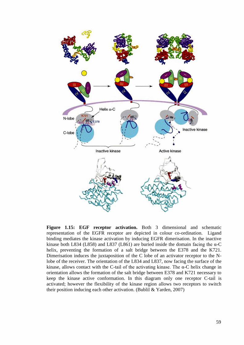
59
Figure 1.15: EGF receptor activation. Both 3 dimensional and schematic
representation of the EGFR receptor are depicted in colour co-ordination. Ligand
binding mediates the kinase activation by inducing EGFR dimerisation. In the inactive
kinase both L834 (L858) and L837 (L861) are buried inside the domain facing the α-C
helix, preventing the formation of a salt bridge between the E378 and the K721.
Dimerisation induces the juxtaposition of the C lobe of an activator receptor to the N-
lobe of the receiver. The orientation of the L834 and L837, now facing the surface of the
kinase, allows contact with the C-tail of the activating kinase. The α-C helix change in
orientation allows the formation of the salt bridge between E378 and K721 necessary to
keep the kinase active conformation. In this diagram only one receptor C-tail is
activated; however the flexibility of the kinase region allows two receptors to switch
their position inducing each other activation. (Bublil & Yarden, 2007)

60
1.8.1.2 Transmembrane domain and Juxtamembrane domain
EGFR transmembrane domain is made up of one single a-helix that continues to the
juxtamembrane domain. The juxtamembrane domain has a number of different
regulatory functions, such as downregulation and ligand dependent internalization,
basolateral sorting in polarized cells and association as a docking site for several other
molecules (eps38, calmodulin and PKC) (Hubbard, 2001; Hubbard, 2004; Hubbard,
2009; Kil et al, 1999; Macdonald-Obermann & Pike, 2009; Red Brewer et al, 2009).
The versatility of this domain will be further discussed in the result chapter.
1.8.1.3 Kinase domain
Each tyrosine kinase domain is uniquely cis-autoinhibited via intramolecular
interactions. The phosphorylation of the activation loop is a key regulatory element in
most kinases and is necessary to destabilize the cis- autoinhibitory interaction, and to
stabilize the receptor into an active configuration (Bublil & Yarden, 2007; Morandell et
al, 2008; Morgan & Grandis, 2009). In addition to the activation loop other kinases are
relived from the autoinhibition by elements outside of the TKD(Schlessinger, 2003).
One of the most common and known example is the juxtamembrane domain
autoinhibition. The juxtamembrane region can make several contacts with the kinase
domain, including the activation loop acting as a stabilizer of the autoinhibited
conformation (example KIT) (Bose & Zhang, 2009; Hubbard, 2004; Mattoon et al,
2004). Receptor dimerisation promotes the trans-phosphorylation of key tyrosine within
the juxtamembrane domain that disrupts the autoinhibitory conformation and lead to
receptor activation (Lemmon, 2009). In addition to the juxtamembrane also the c-
terminal domain has been shown to play a role in the auto-inhibition mechanism (Bose
& Zhang, 2009). The region of the c-terminal domain containing tyrosine residues that
undergoes autophosphorylation impedes access of ATP substrate to the active site
within the kinase domain. Only following autophosphorylation the physical obstruction
is released, and the receptor is stabilized into an active conformation (Mosesson et al,
2008). The activation loop, juxtamembrane domain and carboxyl tail are determinant for
the kinase domain activation of RTKs and EGFR is not an exception. However,
differently from all the other allosteric models where one autoinhibited kinase domain
trans-phosphorylate the other receptor kinase domain within a dimer, EGFR adopts an
allosteric model that resemble the activation of CDKs by cyclins (Lemmon &
Schlessinger, 2010). The EGFR tyrosine kinas domain forms an asymmetric dimer

61
whereby the c lobe of one TKD (activator) contact the N lobe of the other TKD
(receiver) inducing a conformational change that releases the autoinhibitory effect of the
receiver (Jura et al, 2009). In this way the receiver kinase domain adopts an active
conformation without the necessity to phosphorylate its activation loop. The
juxtamembrane domain has been shown to play a key part in EGFR allosteric
conformational change by „exposing‟ the C lobe of the activator kinase in the dimer
formation (Bublil & Yarden, 2007; Macdonald-Obermann & Pike, 2009; Red Brewer et
al, 2009). In an inactive receptor the C-terminal region is found to obstruct the site on
the Activator where the receiver juxtamembrane region docks to achieve receptor
activation supporting earlier studies that have previously indentified autoinhibitory role
of EGFR C-terminus (Fig 1.15) (Zhang et al, 2006).
1.8.2 Autophosphorylation
Following kinase activation, autophosphorylation of tyrosine residue within the
carboxyl tails create the phosphotyrosine biding site necessary to recruit signalling
molecules containing Src homology 2 (SH2) and phosphotyrosine binding (PTB)
domains. The EGFR phosphotyrosine may directly recruit these proteins or indirectly
via the binding to other docking proteins such as Grb2. In addition to
autophosphorylation EGFR is also trans-phosphorylated by other tyrosine kinases (such
as Src) providing the required docking site for the adaptors and modifiers (Gandhi et al,
2009; Gazdar & Minna, 2008; Morandell et al, 2008; Shtiegman et al, 2007; Zandi et al,
2007). Autophosphorylation is not a prerequisite for EGFR signalling as several studies
have shown that signalling is stimulated also by c-terminally truncated EGFR expressed
alone or in combination with other ERBB. This is because other trans-phosphorylating
proteins may mediate the phosphorylation of the docking sites (Wang et al, 2007; Zandi
et al, 2007). Fig 1.16 shows the different phosphorylation sites together with the kinase
responsible for the phosphorylation event and the interacting protein.

62
Figure 1.16: Tyrosine phosphorylation site on EGFR. Diagram shows amino acid
kinase inducing the phosphorylation and the interacting protein. Bold amino acids
residues are numbered according to gi-29725609 and gi-2811086; in brackets according
to gi-4885199 sequence. From (Morandell et al, 2008).
1.8.3 EGFR and cellular signalling
There are four main pathways activated by EGFR: the mitogen activated protein kinase
(MapK) pathway, the activation of the Sarcoma (Src) family of kinases, such as the
Janus Kinase and the signal transducer and activator of transcription (JAKs – STATs)
pathway and the phospholipase D (PLD), the subunit of the Phosphoinositide
phospholipase C (PLC- ) and phosphatidylinositol-3-kinase (PI3K) lipid metabolism
pathways. Each of these pathways can be activated by more than one phosphorylated
tyrosine contributing to the high level of redundancy and robustness regulated by EGFR.
Fig 1.17 highlights the major EGFR-phosphorylation events and their correspondent
downstream signalling pathways.
1.8.3.1 EGFR activation of the MapK pathway
The key event for the activation of the downstream MapK kinase is the EGFR induced
phosphorylation of the proto-oncogene Rat sarcoma (Ras). Usually EGFR
autophosphorylation and kinase activation promotes the Src Homology 2 domain (SH2)
mediated binding of the growth factor receptor-bound protein 2 (Grb2). Alternative the
EGFR-Grb2 binding is indirectly mediated by the binding of EGFR to Sh2 that then
recruits Grb2 (Grant et al, 2002; Katz et al, 2007). This adaptor protein is constitutively
bound to the Ras exchange factor called Son of sevenless (Sos). The relocation of the

63
cytosolic Grb2/SOS complex facilitates the Ras association with SOS resulting in the
exchange of Ras- bound Guanosine diphosphate (GDP) for Guanosine triphosphate
(GTP), hence inducing Ras activation (Katz et al, 2007). Ras in turn activates Raf1
though a series of intermediate kinases leading to the phosphorylation of the
extracellular signal-regulated kinases (Erks) Erk1 and Erk2 that catalyse the
phosphorylation of transcription factor promoting proliferation (Fig1.18) (Huang et al,
2007b).
Figure1.17: Topology of EGFR residues inducing cellular signalling. The diagram
enlarges a portion of the kinase domain and C-tail highlighting all the activating
Tyrosine residues. Src-dependent phosphorylated tyrosines are highlighted in blue, EGF
dependent tyrosine phosphorylation in orange. K721 residue, critical for the ATP
binding, and phospho-groups, are shown in light yellow. Arrows point the activating
kinase of the residue followed by consequent function or the activated signalling
pathway. Where not indicated the activating kinase is EGFR itself. The picture was
taken from (Wheeler et al, 2010).
1.8.3.2 Src family of kinases
It is still not clear whether the Src family of proteins is a signal transducer downstream
of EGFR or a kinase for EGFR. The direct association of these proteins takes place via
the Src SH2 domain although the EGFR binding site is not yet been recognized. Src
does not bind to the major autophosphorylation sites but instead it phosphorylates Y891
64
and Y920 via its SH2 domain providing docking site for p85 subunit of the PI3K. The
Src dependent Y845 phosphorylation has been linked to STAT5b activation however the
role of this site in mitogenic signalling has been quite controversial (Fig 1.18) (Benhar
et al, 2002; Yang et al, 2008).
1.8.3.3 Stat and JAK
STATs proteins are usually recruited at the intracellular domain of RTKs via the binding
of STAT SH2 domain and the receptor phosphotyrosine residues. Tyrosine
phosphorylation of key STAT phosphotyrosine (Y669 STAT5b, Y694 in STAT5a and
Y701 in STAT1) and hetero and homodimerisation is required for STAT activation and
nuclear translocation (Jorissen et al, 2003). STAT1, 3, 5 have been implicated in EGFR
signalling. Differently from the other cytokine receptor EGF induced, EGFR-dependent
STAT activation does not require JAK kinases. In addition STAT proteins do not bind
the EGFR carboxyl tail but indeed it appears that EGFR-STATs are constitutively
associated however the STAT transcriptional activity is strictly dependent on the EGFR
kinase activity (Fig 1.18) (Morgan & Grandis, 2009).
1.8.3.4 Phospholipids metabolism
EGF stimulation in cells induces the production of arachinoid acid (AA), phosphatidic
acid (PA) and phosphotidylinositol turnover. EGFR activates at least three of the
enzymes involved in this process: phospholipase C- (PLC- ), phospholipase D (PLD)
and PI3-K. Despite being still obscure the mechanisms of EGFR- PLD mediated
activation seems to require the physical interaction of the two proteins but tyrosine
phosphorylation does not seem to be an absolute requirement. PLC- instead binds
directly to the EGFR autophosphorylation Y1173 and Y992 residues and is
consequently phosphorylated by EGFR on Y771 and Y1254 (Kamat & Carpenter,
1997). Once activated it catalyses the hydrolysis of PtdIns(4,5)-P2 which accumulates
the second messenger 1.2 diacylglycerol (DAG) and inositol 1, 3, 5, triphosphate (IP3).
The cellular role of IP3 is to release calcium while DAG acts as a cofactor in the
activation of the protein kinase C (PKC) kinase. Therefore EGFR can activate Ca2+
-
dependent pathways such as the nuclear factor kappa-light-chain-enhancer of activated
B cells (NF B) and through the PKC indirect activation other signalling pathways such
as MAPK, JUNK and possibly Na+/H
+ exchange. PI3-Ks are members of one of the
65
most important family of kinases in the cell regulating proliferation, survival, adhesion,
and migration (Liaw et al, 1998; Marais et al, 1998). Some members of this family are
also involved in DNA repair pathways and DNA damage response. They normally
catalyse the phosphorylation of the 3‟ positon of phosphatydilinositols and are further
classified into three classes according to their substrate and lipid structure. Among
these, only class I is regulated by RTKs. While the interaction of ERBB3 with PI3-K is
direct and this receptor is the main activator of the pathway, EGFR requires the binding
of p85 subunit that then interacts with the PI3-K-SH2 domain (Carpenter, 1993;
Djordjevic & Driscoll, 2002). EGFR/ERBB3 heterodimers have been shown to activate
PI3-K following EGFR ligands stimulation as well as Src induced EGFR
phosphorylation (Kim et al, 1994). PI3-K produces phosphatidylinositol-3-,4,5
triphosphate (PIP3). The most characterised target of this secondary message is the
serine threonine kinase AKT (PKB) (Fig 1.18). This protein normally binds to lipids but
is translocated to the plasma membrane and activated by phosphoinositide-dependent
kinase 1 (PDK1) on the threonine 308 (T308) (Nicholson & Anderson, 2002). Full AKT
activation is mediated by PDK-2 that phosphorylates the serine residue 473 (S473).
Once fully activated, AKT localizes both in the cytosol and the nucleus where it
mediates survival, evasion of apoptosis, sustained angiogenesis and replicative potential.
More specifically AKT is involved in the regulation of the cell cycle by the
phosphorylation of the CKI p21 and p27 that leads to their inhibition and prevents their
nuclear translocation (Liang et al, 2002). Akt kinase activity has also been shown in the
inactivation of the glycogen synthase and activation of glycolisis as well as glucose
transport, hence playing a central role in energy metabolism. The most recognized and
characterized function of this EGFR downstream target is its regulation of the anti-
apoptotic signal via the NFkB and the inactivation of BAD, CASP9 and the forkhead
transcription factor (Luo et al, 2003). Both EGFR and ERBB3 activation of AKT
signalling, induced by ligand or IR, is a major mechanism that leads to tumour
resistance against IR treatment (Engelman, 2009; Li et al, 2009; Toulany et al, 2008b).

66
Figure 1.18: Signalling pathways activated by EGFR. The pathways do not include
all the components but only the first and last member of the pathway. Cross talk is not
shown for clarity. The function of each pathway is highlighted in the colour boxes. Two
inhibitors (PTEN and MKP1) are shown to inhibit Pi3K and MAPK pathway
respectively. The mediated transcriptional gene activation is also shown. Lawrence et
al., Nature reviews Cancer 2006.
1.8.4 EGFR biological downregulation: endocytosis mediated degradation
Following activation EGFR undergoes a desensitization phase called endocytosis where
the active complex of ligand-receptor is internalized and sorted either for degradation, or
recycling or nuclear translocation (the latter will be further discussed later on) (Madshus
& Stang, 2009). Just as important as activating cellular signalling is switching them off
and although EGFR (as well as many RTKs) may spend hours or even days at the cell
surface, its removal can happen within seconds after messenger stimulation (Oved &
Yarden, 2002; Zwang & Yarden, 2009). In resting cells EGFR resides in lipid rafts of
the cell membrane enriched in caveolins and cholesterol. Following ligand binding,
EGFR exits these domains in Src dependent manner. Already within the lipid rafts the
ligand bound receptor associate to coat adaptors such as adaptor protein 2 (AP2) and
Eps15 involved in the endocytic machinery. EGFR endocytosis takes place through
clathrin coated pits (CCPs), via cavoelae mediated endocytosis (CavME), or via
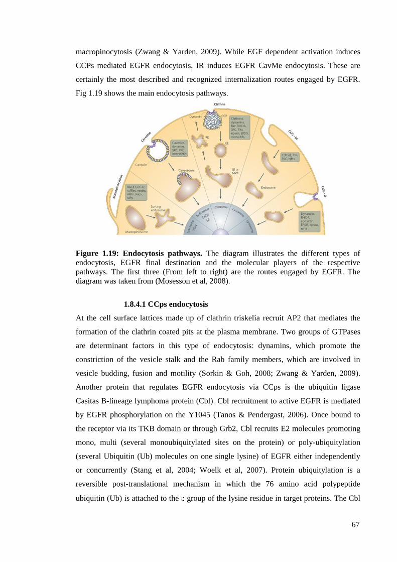
67
macropinocytosis (Zwang & Yarden, 2009). While EGF dependent activation induces
CCPs mediated EGFR endocytosis, IR induces EGFR CavMe endocytosis. These are
certainly the most described and recognized internalization routes engaged by EGFR.
Fig 1.19 shows the main endocytosis pathways.
Figure 1.19: Endocytosis pathways. The diagram illustrates the different types of
endocytosis, EGFR final destination and the molecular players of the respective
pathways. The first three (From left to right) are the routes engaged by EGFR. The
diagram was taken from (Mosesson et al, 2008).
1.8.4.1 CCps endocytosis
At the cell surface lattices made up of clathrin triskelia recruit AP2 that mediates the
formation of the clathrin coated pits at the plasma membrane. Two groups of GTPases
are determinant factors in this type of endocytosis: dynamins, which promote the
constriction of the vesicle stalk and the Rab family members, which are involved in
vesicle budding, fusion and motility (Sorkin & Goh, 2008; Zwang & Yarden, 2009).
Another protein that regulates EGFR endocytosis via CCps is the ubiquitin ligase
Casitas B-lineage lymphoma protein (Cbl). Cbl recruitment to active EGFR is mediated
by EGFR phosphorylation on the Y1045 (Tanos & Pendergast, 2006). Once bound to
the receptor via its TKB domain or through Grb2, Cbl recruits E2 molecules promoting
mono, multi (several monoubiquitylated sites on the protein) or poly-ubiquitylation
(several Ubiquitin (Ub) molecules on one single lysine) of EGFR either independently
or concurrently (Stang et al, 2004; Woelk et al, 2007). Protein ubiquitylation is a
reversible post-translational mechanism in which the 76 amino acid polypeptide
ubiquitin (Ub) is attached to the group of the lysine residue in target proteins. The Cbl

68
mediated ubiquitylation is required for receptor sorting in the endosomes and
subsequent sorting into the lysosomal degradative compartment (Huang et al, 2007a).
Epsin-1 and Cin85 facilitate EGFR translocation from the clathrin-coated pits to the
plasma membrane. Cin-85 has been found to be associated to Cbl suggesting that
together they regulate EGFR endocytosis (Sigismund et al, 2008).
Following the release of internalised plasma proteins, endocytic vesicles undergo
uncoating, a process mediated by the auxiliary protein Hsc70. This allows EGFR
induced activation of Rab5 and membrane translocation of the early endosomes
antigen1 (EEA1) determining the fusion of the vesicle with early endosomes (Massie &
Mills, 2006; Mosesson et al, 2008). The endocytic process then terminates with the
formation of the mutivesicular bodies (MVBs) from late endosomes by invagination of
the endosomes membrane to produce intraluminal vesicles (Puri et al, 2005). MVBs
then fuse with lysosome or mature into lysosomes leading to the lysis of their protein
content. Three multiprotein complexes mediate the MVBs formation of EGFR-
concentrated-limiting endosomal membranes: Endosomal sorting complexes required
for transport I, II, III (ESCRT-I, II, III). Alternatively, EGFR can also escape the
endosomal compartments via receptor recycling. While EGF-induced EGFR activation
leads to endocytosis and receptor degradation, TGF- induced-endocytosis is followed
by receptor recycling (Yu et al, 2002). Recycling endosomes contain RAB4 or Rab1
GTPases, which are involved in fast and slow recycling respectively (Mosesson et al,
2003).
Protein kinase C (PKC) has been shown to be involved in recycling by inducing EGFR
threonine phosphorylation. Kinsin family member 16B (KIF16B) and the cytoskeleton
associated recycling or transport (CART) complex also regulate recycling by
modulating the actin filaments and facilitating the trafficking to the recycling
endosomes (Massie & Mills, 2006; Sorkin & Goh, 2008). Fig 1.20 shows a diagram of
these events. Along with their degradative function endosomes have also been
associated with signalling pathways. Endosomes contain many cellular kinases and
EGFR (in particular in its oncogenic status) has been shown to stimulate survival and
proliferation by interacting with internalised Ras, Shc, and Grb2, and induce
oncogenesis by coupling with internalized c-Met to the STAT3 pathway (Oved &
Yarden, 2002; Warren & Landgraf, 2006; Woelk et al, 2007).
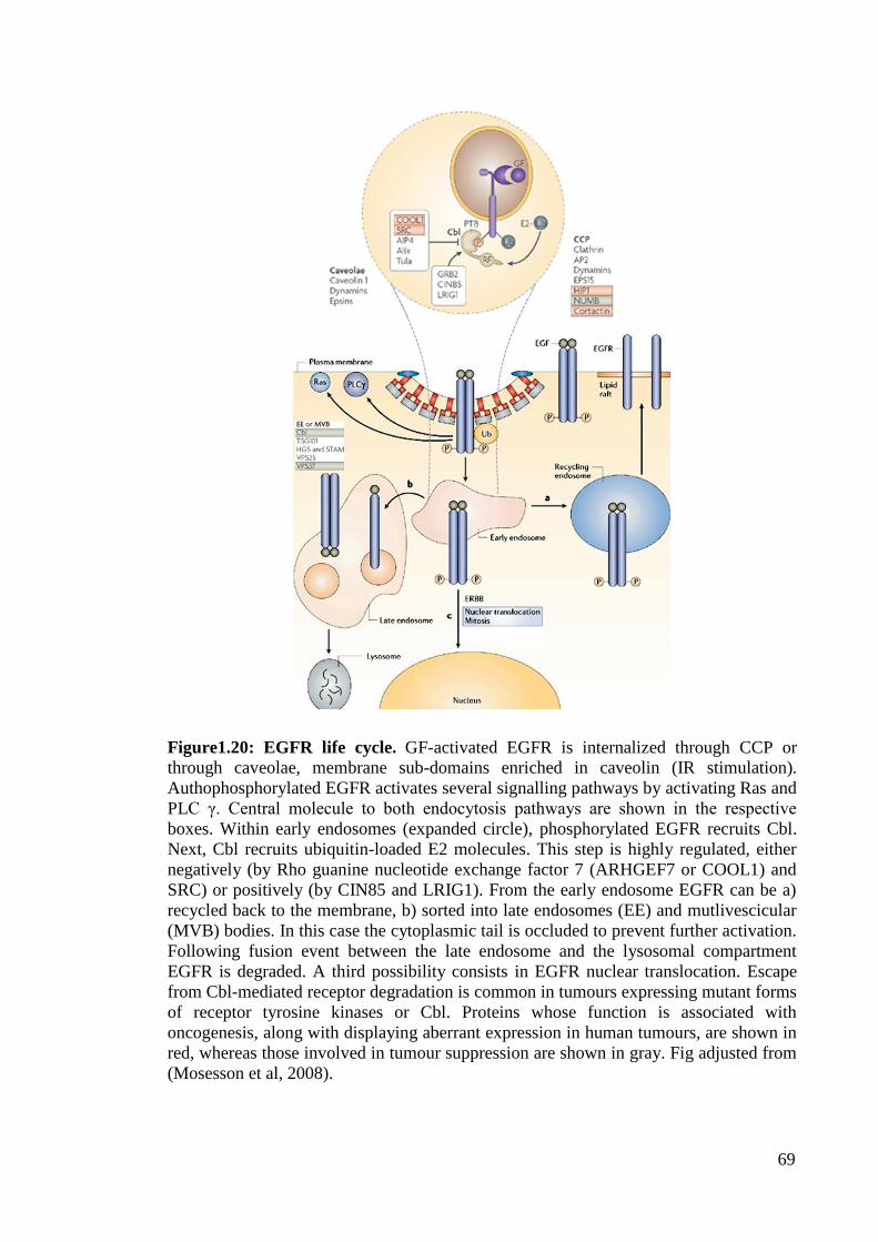
69
Figure1.20: EGFR life cycle. GF-activated EGFR is internalized through CCP or
through caveolae, membrane sub-domains enriched in caveolin (IR stimulation).
Authophosphorylated EGFR activates several signalling pathways by activating Ras and
PLC γ. Central molecule to both endocytosis pathways are shown in the respective
boxes. Within early endosomes (expanded circle), phosphorylated EGFR recruits Cbl.
Next, Cbl recruits ubiquitin-loaded E2 molecules. This step is highly regulated, either
negatively (by Rho guanine nucleotide exchange factor 7 (ARHGEF7 or COOL1) and
SRC) or positively (by CIN85 and LRIG1). From the early endosome EGFR can be a)
recycled back to the membrane, b) sorted into late endosomes (EE) and mutlivescicular
(MVB) bodies. In this case the cytoplasmic tail is occluded to prevent further activation.
Following fusion event between the late endosome and the lysosomal compartment
EGFR is degraded. A third possibility consists in EGFR nuclear translocation. Escape
from Cbl-mediated receptor degradation is common in tumours expressing mutant forms
of receptor tyrosine kinases or Cbl. Proteins whose function is associated with
oncogenesis, along with displaying aberrant expression in human tumours, are shown in
red, whereas those involved in tumour suppression are shown in gray. Fig adjusted from
(Mosesson et al, 2008).
70
1.8.4.2 Cavoelae-mediated endocytosis (CavME)
Cavoelae are specialized lipid rafts generated by the oligomerisation of the protein
caveolin. The first step of this mechanism consists in the inward budding of caveolin
vesicles into larger caveolin enriched organelles called caveosomes. The vesicle release
is mediated by dynamins, PKC, and Src kinases(Sigismund et al, 2005; Zwang &
Yarden, 2009).
1.8.5 ONCONGENIC EGFR
Despite the overwhelming network of regulatory mechanism that are employed in
EGFR mediated cell signalling activation and inhibition, which increase the robustness
of this system, the ERBB family and in particular EGFR represent a major player in
many types of malignancies. As EGFR is involved in cell proliferation, motility and
survival, an imbalanced EGFR system can easily lead to neoplastic transformation
(Fischer et al, 2003). In 1980 it was found that addition of EGF to culture medium of
human epidermoid carcinoma (A431) led to high yields of tyrosine phosphorylation,
similarly to cells infected with oncogenic viruses. EGFR has been the first RTK to be
associated with a human cancer and indeed deregulated EGFR is now associated with
many human cancers. There are two main mechanisms by which the tight control of the
EGFR system can be by passed: modulation of the EGFR-ligand system and by passing
some of EGFR inherent controls. The first mechanism includes production of ligands in
an autocrine fashion, overexpression of EGFR via gene amplification, defective
downregulation of EGFR (defective endocytosis) and cross talk with heterologous
receptor systems (Bublil & Yarden, 2007). The second mechanism includes the
acquisition of somatic mutations, which give rise to constitutive active EGFR
variants.(Gazdar, 2009a; Nyati et al, 2006)
These mechanisms of aberrant EGFR expression and activation induce an indirect
deregulation of the cellular machinery (EGFR regulates its downstream kinases which
then promote uncontrolled growth, proliferation, migration etc). They represent the
EGFR mediated contribution towards oncogenic transformation (Fry et al, 2009;
Grandal & Madshus, 2008). In addition, the activation of EGFR signalling pathways
following radiotherapy and chemotherapy also play an important part in acquiring
resistance and tolerance to treatment. EGFR can also directly modulate cellular survival
and proliferation by translocating into the nucleus and associate with nuclear proteins
regulating cell cycle, transcription and DNA repair. This section will review the

71
mechanisms that lead to EGFR deregulation and the molecular therapeutic attempts to
inhibit EGFR in cancer cells.
1.8.5.1 Autocrine mechanisms
Both EGF and TGF- are usually found co-expressed with EGFR in a variety of human
cancers. Overexpression of TGF- in transgenic mice induces tumour formation and
also addition of EGF or TGF- in mouse fibroblast expressing EGFR induces cellular
transformation. However the effect of the ligand activation on cellular transformation is
only detected when EGFR is overexpressed, as medium levels of expression do not lead
to transformation (Zandi et al, 2007). Autocrine production of these ligands in EGFR
overexpressed tumours (colorectal cancer, head and neck cancer squamous cell
carcinoma) has shown to lead to high proliferation and worse clinical outcome. As
explained above TGF- induced EGFR activation evades CCPs mediated endocytosis
and degradation maintaining therefore recycling and signalling activation (Gazdar &
Minna, 2008).
1.8.5.2 EGFR overexpression
Elevated levels of EGFR expression have been correlated with increased survival in
many cancer types including head and neck, bladder, ovarian, cervical and esophageal.
Spontaneous receptor dimerisation, due to the numerous receptors at the membrane,
induces constitutive activation resulting in ligand independent increased motility, and
continuous activation of downstream signalling contributing to a more malignant
phenotype (Arteaga, 2002). Increased number of EGFR molecule by gene amplification
has been observed in breast carcinomas, non small-cell lung cancer (NSCLC) and
particularly glioblastoma multiforme (GBM). Other mechanisms that lead to EGFR
overexpression are: increased activity of the EGFR promoter or deregulation at the
translational and post translational level (Gazdar & Minna, 2008). P53 has been shown
to bind to the promoter of EGFR activating EGFR transcription. Therefore, as the levels
of p53 are usually high in cancer cells, so the levels of the receptor will be. It has also
been reported that a specific region of the EGFR gene has enhancer ability in some
breast cell cancer lines over-expressing EGFR. These cell lines have also been shown to
express more DNAase H1 sites in the intron 1 of the EGFR gene suggesting that
chromatin structure of the EGFR regulatory regions may have a role in its

72
expression(Kersting et al, 2008). Again in intron1 the number of polymorphic CA
dinucleotide seems to correlate with the EGFR expression (Huang et al, 2009). The
higher number of polymorphic sites the lower the EGFR expression (Kang et al, 2005).
1.8.5.3 Evading degradative fate.
EGFR overexpression and over activation due to autocrine and paracrine ligand
activation and by spontaneous dimerisation may saturate the capacity of the CCps and
endosomal adaptors (Stang et al, 2004). A single amino acid mutation within the Cbl
zinc finger domain renders the protein ligase defective therefore impairing EGFR
ubiquitylation (Shtiegman et al, 2007). Indeed ERBB2 has been often associated with
Hsp90, which stabilise the protein rendering it a poor substrate for lysosomal
degradation; hence EGFR/ERBB2 dimer may undergo the same by pass (Yang et al,
2006). Several reports have implicated the Src family of kinases in control of Cbl half-
life. Interaction with Src redirects the Cbl ubiquitin ligase activity towards itself
resulting in Cbl proteosomal degradation. Therefore the uncoupling of the Cbl
mechanism may be one of the facets of the EGFR-Src interaction during
oncogenesis(Huang & Sorkin, 2005; Zwang & Yarden, 2009). Several ligands including
EGF induce transcription of Sprouty proteins, which displace RTKs from Cbl. Ligands
increase Sprouty tyrosine phosphorylation thereby sequestering Cbl from activated
EGFRs (Sorkin & Goh, 2008).
1.8.5.4 EGFR mutations
The somatically acquired mutations in the EGFR gene are not randomly distributed but
rather clustered in specific areas, which assumes the role of mutational hot spots. There
are 1380 mutations of the EGFR gene sequenced from tumour samples. 30% (413 of
1380) are found in adenocarcinoma, 2% in NSCLC (16 of 1380). The number of
sequenced EGFR mutations is incredibly high and will be categorized according to their
location within the EGFR domains (Gazdar, 2009a). All the mutations are shown in Fig
1.21 and 1.22. The most relevant to this study will be discussed.
1.8.5.4.1 Extracellular mutations
Mutations of the extracellular domain are particularly found in Glioblastoma. EGFR
type I, II, III variant (EGFRvI, EGFRvII, EGFRvIII) are the most known and contain
deletions of exons encoding parts or the entire extracellular domain(Zandi et al, 2007).

73
These truncated mutants, despite having impaired ligand binding, are found
constitutively active and highly tumorigenic. EGFRvI lacks most of the extracellular
domain, is ligand independent and constitutively active (Pines et al). EGFRvII has an in
frame deletions of 83 amino acids (exon14-15) and therefore still a capable of ligand
binding. The oncogenic role of this truncated receptor is still unclear (Grandal et al,
2007). The most frequent and also most characterized mutation is the EGFRvIII also
described as de2-7 EGFR or 2-7 EGFR which is the result of an in frame deletion of
exons 2-7. These exons encode domain I and two thirds of domain II of the extracellular
domain and the deletions of amino acid 2-273 results in the loss of most of the ligand
binding area. Like EGFR, EGFRvIII activates the RAS/RAF/MEK/ERK pathway and
Jun N-terminal kinase via PI3K (Grandal et al, 2007; Modjtahedi et al, 2003). EGFRvIII
is trapped in a partially activated state which is sufficient for oncogenic signalling but
not for degradation (Zandi et al, 2007). In fact despite its internalization this truncated
receptor fails to be ubiquitylated promoting also intracellular oncogenic signalling.
Another truncated receptor is the EGFRvIII/D12-13, which in addition to the deletion of
exon 2-7, this mutant also lacks part of domain III (exon 12- 13). Besides these deletion
mutants there is also the report of a mutant that contains duplication of exon 2-7: EGFR
TDM/2-7. This mutant contains an extra cysteine reach domain I and part of domain II
which do not impair its ligand binding dependency. Genomic analysis of the EGFR gene
has revealed that both exon 1 and exon 7 contain an Alu element where DNA breaks are
centred (Fenstermaker & Ciesielski, 2000). The recombination of these Alu elements
have been implicated in the mechanism of deletion/duplication of the region they are
flanking explaining why the extracellular domain is subject to these types of mutations
(Wong et al, 1992). Fig 1.21 show a diagram of the mutation above mentioned.
1.8.5.4.2 Intracellular mutations: Kinase domain mutations
All the somatic mutation falling in this category target the residues around the adenosine
triphosphate (ATP) binding pocket leading to ligand independent activation of TK
activity (Gazdar, 2009a). They are divided into three classes. Class I mutation are in
frame deletions in exon 19 (various sets of deletions from amino acid 746 to 753)
which almost always include deletion from Leucine (L) 747 to Glutamic acid (E) 749
accounting for about 44% of all the EGFR mutations. Class II mutations comprises
single point mutation which cause a missense mutation. The most characterised is the
single point mutation in exon 21 of Leucine (L) 858 into Arginine (R). L858R has the

74
highest prevalence of any other single point mutation of the EGFR TK domain (41% of
all activating mutations). To classII belongs the mutation of Glycine (G) 719 into Serine
(S), Alanine (A) or (C) Cysteine (4% of all the activating mutations) (Sharma &
Settleman, 2009). Other missense mutations account for another 6 % of EGFR
mutations (Gazdar, 2009a). Class III mutations are in frame duplications or insertions in
exon 20. This class accounts for the remaining 5% of all the activating mutations. There
is a remaining portion of low frequency (<1%) mutations in exon 20 such as the
Threonine (T) 783 into Alanine (A) and the Valine (V) 765 into Alanine (A) (Gandhi et
al, 2009). Mutations in exon 19 and the L858R mutation represent about 90% of all the
activating mutations and for this reason they are also named classical mutations(Gazdar
& Minna, 2008). The L858R mutation is located next to the conserved aspartic acid-
phenyalanine-glycine sequence (DFG) that has the function to stabilize the activation
loop. This mutation and also the G719X (X indicates S, A or C) result in similar
configuration changes which destabilizes the helical axis narrowing the ATP binding
site and conferring kinase activation and higher protein stability (Gazdar et al, 2004;
Murray et al, 2008; Sharma & Settleman, 2009). Fig 1.22 shows a diagram o f all the
kinase mutations
1.8.5.4.3 Intracellular mutations: C- terminal mutations
These mutations target the C-terminal domain and are best described in GBM models.
While EGFRvIV lacks exon 25-27, EGFRvV is truncated from amino acid 958 and
lacks the rest of the C-tail (exon 25-28) (Frederick et al, 2000). EGFRTDM 18- 25 and
EGFRTDM 18-26, which contain duplication of exons 18-25 and 18-26 respectively,
also belong to this category. Of these four mutants, only EGFRvIV has been shown to
have oncogenic properties. The removal of exon 25-27 removes the autoinhibitory
regulation of the carboxyl on the TK domain resulting in constitutive kinase activity
(Zandi et al, 2007). Fig 1.21 shows all the mentioned mutations.

75
Figure 1.21: Schematic representation of EGFR mutations. From left to right:
wtEGFR, deletions or duplication of EGFR (EGFRvI-EGFRvV, EGFRvIIID12-13,
EGFRTDM/2-7, and EGFRTDM18-25/26). Modified from(Zandi et al, 2007).
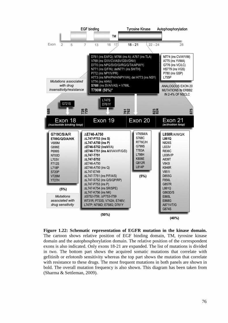
76
Figure 1.22: Schematic representation of EGFR mutation in the kinase domain.
The cartoon shows relative position of EGF binding domain, TM, tyrosine kinase
domain and the autophosphorylation domain. The relative position of the correspondent
exons is also indicated. Only exons 18-21 are expanded. The list of mutations is divided
in two. The bottom part shows the acquired somatic mutations that correlate with
gefitinib or erlotonib sensitivity whereas the top part shows the mutation that correlate
with resistance to these drugs. The most frequent mutations in both panels are shown in
bold. The overall mutation frequency is also shown. This diagram has been taken from
(Sharma & Settleman, 2009).

77
1.8.5.5 EGFR nuclear expression
In addition to the canonical role of EGFR as the initiator of the signalling events that
originate at the plasma membrane in response to ligand binding, accumulating evidence
from the research of several group in the past 10 years, shows that EGFR and other
members of the EGFR family shuttle from the cell surface to the nucleus transducing
growth, proliferation and modulation of DNA repair response. This section will
illustrate the molecular mechanism that mediates nuclear translocation, the recent
findings on EGFR nuclear expression and their significance in terms of tumorigenesis
and resistance to chemo and radiotherapy.
1.8.5.5.1 Mechanism of nuclear translocation
The design of eukaryotic cells separates the cytosol and also its components from the
nucleus via the nuclear envelope. The regulation of the transport across this cellular
barrier facilitates the control of important cellular process such as gene expression, cell
cycle progression, signal transduction and mediates the recognition and recruitment of a
specific cytosolic protein (cargo), its translocation through the nuclear envelope an the
release of the cargo in the nucleus (Lange et al, 2007). While small macromolecules
<40-50 kDa can freely move across the nuclear pore complexes by diffusion, large
molecules must contain a specific recognition signal (Kaffman & O'Shea, 1999;
Moroianu, 1999). The carrier proteins that transport macromolecules across the nucleus
are collectively referred as karyopherins ( - karyopherins or Importin is the most
representative superfamily) with those involved in the import called importins and those
involved in the export called exportins (Corbett & Krebber, 2004; Lange et al, 2007).
Although the majority of the cargo proteins bind directly to - karyopherins, the
interaction between them can also be mediated by the adaptor molecule importin . The
energy required for the transport is provided by the small Ras GTPases, Ran that binds
and exchanges both GDP and GTP (Kaffman & O'Shea, 1999; Powers & Dasso, 2004).
The constant cycling between these two bound states is determined by the Ran guanine
nucleotide exchange factor (Ran GEF), and the Ran GTPase-activating protein
(RanGAP). Because these modulators are segregated in different cellular compartment
the different forms of Ran are also differently distributed with RanGTP in the nucleus
and RanGDP in the cytosol (Kaffman & O'Shea, 1999; Madrid & Weis, 2006;
Moroianu, 1999). Importin recognizes and binds the cargo protein in the cytoplasm

78
linking it with the importin . Importin mediates the transport of the formed trimeric
complex through the nuclear pore into the nucleus and dissociates by the binding of the
RanGTP. This association causes a conformational change in the importin resulting in
the release of the cargo- importin (Wente & Rout). The release of the cargo is then
mediated by importin IBB (importin binding) domain, the nucleoporin NP2 and the
export receptor Cse1/Ran GTP. The latter then recycles the importin in the cytoplasm
for another round of import (Lange et al, 2007). Fig 1.23 shows the mechanism of
nuclear translocation.
1.8.5.5.2 Nuclear localisation signals
In order to discriminate among all the molecules in the cytosol, proteins destined for
transport into the nucleus contain specific recognition target amino acid sequence called
nuclear localisation signal (NLS). The best characterised are the classical signals:
monopartite consisting of one and bipartite consisting of two stretches of basic amino
acids sequences spaced by 3 or 10 stretches of amino acids. The most known examples
are the monopartite signal is the SV40 large T antigen NLS (126PKKKRRV) and
bipartite nucleoplasmin NLS (155KRPAATKKAGQAKKKK170) (Chuderland et al,
2008; Kaffman & O'Shea, 1999; Lange et al, 2007). The nuclear import and
accumulation of the cargo is dependent on the affinity of the NLS cargo to the
importin , and on the concentration of the importin (Zaidi et al, 2004). Importin is
composed of 10 armadillo motifs each made up of three helices (ARM domain) and a
N-terminal domain required for binding to the importin and for cargo dissociation.
The 10 armadillo motifs generate a curved molecule in which the two NLS binding sites
are located within the concave faces of the molecule. Both pockets are made up of
conserved tryptophan and 4 downstream asparagines. The N-terminus major pocket
binds the monopartite NLS, the minor pocket binds the smaller stretch of basic amino
acid residues of the bipartite NLS (Cyert, 2001; Lange et al, 2007). The cargo NLS runs
anti-parallel within the curved importin , the NLS lysine or arginine (basic) amino
acids side chain lie between the conserved side chains of the tryptophans forming salt
bridges with the negatively charged residues present in the binding groove. The
downstream asparagines make key chain contact. The N-terminal region of the importin
contains an autoinhibitory region consisting of basic amino acids (KRR) that
competes with the cargo NLS for binding (Chuderland et al, 2008; Lange et al, 2007;

79
Weis, 2007; Wente & Rout; Zaidi et al, 2004). During import the binding of importin
inhibits this region but once in the nucleus the two molecules dissociate allowing the
autoinhibitory region to fold over and interact with the major NLS binding pocket of the
importin . Because both mono and bipartite signals bind the major pocket, the binding
of the autoinhibitory domain affects the affinity of importin to the NLS facilitating the
release of the cargo in the nucleus preventing the NLS cargo to re-bind to the importin
(Lange et al, 2007). Fig 1.23 shows the dynamics of the nuclear import cycle.
The import is further regulated at different levels. Cargo post translational modification
may affect the affinity for the importin, both the cargo and the importin may be bound
by an insoluble cellular component compromising the nuclear transfer; the nuclear pore
could undergo deregulation affecting the transport (Corbett & Krebber, 2004; Lange et
al, 2007; Rout & Aitchison, 2001; Strambio-De-Castillia et al; Weis, 1998; Weis, 2007;
Wente & Rout).
Figure 1.23: Nuclear translocation mechanism. The NLS containing cargo is bound
by the heterodimeric complex importin /importin in the cytoplasm. Following nuclear
localisation, RanGTP induces importin dissociation. Nup2 and Cse1 then mediate NLS
cargo release. Importin autoinhibitory domain folds back preventing re-binding to the
NLS cargo and the new Cse1/RanGTP/importin translocates back to the cytosol.
(Lange et al, 2007).
1.8.5.5.3 EGFR Nuclear translocation
Nuclear EGFR was firstly observed in hepatocytes during liver regeneration (Kamio et
al, 1990; Marti et al, 1991). Many studies have now reported nuclear EGFR in the form

80
of uncleaved receptor in the nuclei of various tissues such as thyroid cells, ovarian
epithelial cells, immortalized renal cells and also primary tumours including those of the
bladder, breast, oesophagus and thyroid (Dittmann et al; Hoshino et al, 2007; Kamio et
al, 1990; Lo et al, 2005b; Marti et al, 2001; Xia et al, 2009; Xu et al, 2009b). Hsu et al.,
that characterized the EGFR tripartite NLS 645RRRHIVRKRLLRRR657, provided the
initial analysis of the mechanism of EGFR nuclear translocation in 2007. Despite not
being the only characterised tripartite NLS, this type of NLS lacks a specific model of
binding with importin and the EGFR mechanism of nuclear import is still not
completely understood. Sequence analysis reveals that the EGFR NLS sequence is well
conserved among different species and also among the other ERBB family members
with the exception of ERBB3 (Hsu & Hung, 2007). While the ERBB4 nuclear
localisation takes place via sequential receptor cleavage, the mechanisms for EGFR and
ERBB2 is still not clear. Ligand binding to ERBB4 or activation of PKC by phorbol
esters stimulates an initial metalloproteinase mediated cleavage of ERBB4 that release
the extracellular 120 kDa part of the receptor, leaving behind an 80kDA membrane
bound intracellular domain that includes the kinase domain . There is now sufficient
evidence showing that ADAM17 (a disentigrin and metallopeptidase 17) is responsible
for this event. The 80 kDa fragment has two possible fates: it can be ubiquitylated and
subsequently degraded, or further cleaved by presenilin-dependendent secretase that
release the soluble intracellular domain within the cytoplasm which is then translocated
to the nucleus (Fig 1.24) (Wells & Marti, 2002).
ERBB2 colocalises with importin in the endosomes and form a tricomplex with it and
the nucleoporin Nup358 (usually found at the cytoplasmic filaments of the nuclear pore
complex) that translocates near the nuclear envelope (Wang et al, 2010b).
EGFR ligand stimulation and radiation treatment have been shown to stimulate EGFR
nuclear localisation. Following activation and receptor internalization EGFR can have
three possible fates: recycling, degradation or routed from the early endosomes to the
Golgi and the ER by retrograde transport. This retrotranslocation would then allow
binding with the importin leading to receptor nuclear translocation (Dittmann et al,
2008a; Dittmann et al, 2010b; Hsu et al, 2009; Lo, 2010; Wang et al, 2010a; Wang et al,
2010b; Wang et al). Despite this being the only published mechanism (discussed further
in chapter 3) it still unclear how EGFR (having a transmembrane domain) can pass
through the aqueous channels of the NPC. One possibility, yet to be confirmed, is that it
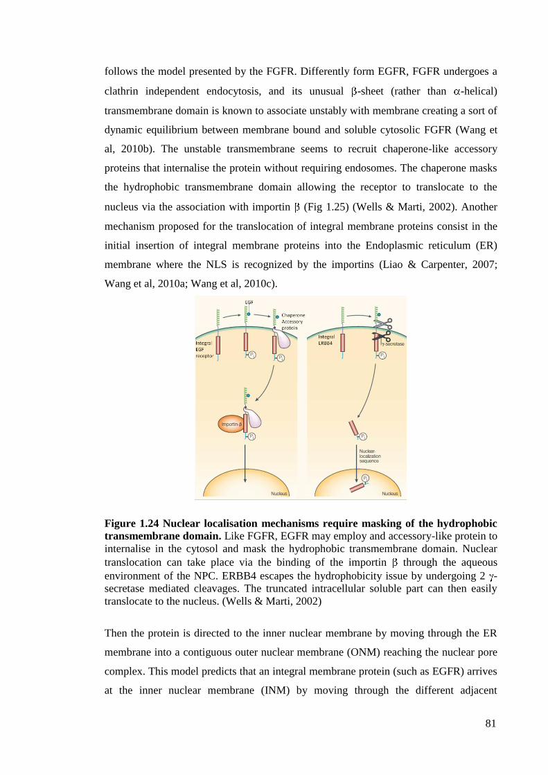
81
follows the model presented by the FGFR. Differently form EGFR, FGFR undergoes a
clathrin independent endocytosis, and its unusual -sheet (rather than -helical)
transmembrane domain is known to associate unstably with membrane creating a sort of
dynamic equilibrium between membrane bound and soluble cytosolic FGFR (Wang et
al, 2010b). The unstable transmembrane seems to recruit chaperone-like accessory
proteins that internalise the protein without requiring endosomes. The chaperone masks
the hydrophobic transmembrane domain allowing the receptor to translocate to the
nucleus via the association with importin (Fig 1.25) (Wells & Marti, 2002). Another
mechanism proposed for the translocation of integral membrane proteins consist in the
initial insertion of integral membrane proteins into the Endoplasmic reticulum (ER)
membrane where the NLS is recognized by the importins (Liao & Carpenter, 2007;
Wang et al, 2010a; Wang et al, 2010c).
Figure 1.24 Nuclear localisation mechanisms require masking of the hydrophobic
transmembrane domain. Like FGFR, EGFR may employ and accessory-like protein to
internalise in the cytosol and mask the hydrophobic transmembrane domain. Nuclear
translocation can take place via the binding of the importin through the aqueous
environment of the NPC. ERBB4 escapes the hydrophobicity issue by undergoing 2 -
secretase mediated cleavages. The truncated intracellular soluble part can then easily
translocate to the nucleus. (Wells & Marti, 2002)
Then the protein is directed to the inner nuclear membrane by moving through the ER
membrane into a contiguous outer nuclear membrane (ONM) reaching the nuclear pore
complex. This model predicts that an integral membrane protein (such as EGFR) arrives
at the inner nuclear membrane (INM) by moving through the different adjacent

82
membranes (ER-ONM-INM) still membrane bound. This would allow its
transmembrane hydrophobic domain to hide from the aqueous nuclear pore complex
environment and trick its way to the nucleus. The receptor is then released from the
INM by an INM sorting motif that is recognized by importin isoform and released in
the nucleoplasm (Wang et al, 2010a; Wang et al, 2010b) (Fig 1.25).
Figure 1.25: Integral membrane protein translocation to the inner nuclear
membrane. Integral membrane protein are initially inserted into the ER and then via
membrane interaction to the outer nuclear membrane (ONM) and the trough the nuclear
pore complex (NPC) into the nucleus via the inner nuclear membrane (INM). In this
way the embedded transmembrane hydrophobic domain does not have to pass through
the soluble NPC environment. Inner nuclear membrane proteins (INM pr.) utilize this
mechanism with the help of ribosomes. (Wang et al, 2010a)
1.8.5.5.4 EGFR modulates transcriptional regulation
During the past 10 years accumulating evidence has shown that EGFR escapes receptor
internalization and degradation and that it translocates into the nucleus not only in
response to ligand binding, but also following ionizing radiation heat shock, H2O2 and
chemotherapy. Upon EGF stimulation EGFR translocates to the nucleus and binds to A-
T- reach regions (ATRS) working as a transcription co-factor via its functional trans-
activation domain (Lin et al, 2001). Nuclear EGFR binds the proximal region of the
promoters of several important genes: cyclinD1 (Lo et al, 2006a), involved in cell cycle
progression and inducible nitric oxide synthase (iNOS) (Lo et al, 2005a; Lo & Hung,

83
2006), TWIST and cyclooxygenase-2 (COX-2) (Husvik et al, 2009; Lo et al, 2010; Xu
et al, 2009a). The binding of EGFR to STAT proteins has been shown to compensate its
inability to directly bind DNA (due to the lack of a DNA binding domain). Nuclear
EGFR cooperates with STAT3 to upregulate expression of COX-2 and iNOS genes,
with E2F1 to activate transcription of the B-MYB gene (an important regulator of
cellular proliferation) (Hanada et al, 2006). In addition the physical association of EGFR
with STAT5 has been shown to enhance the expression of the AURORA-A gene (Hung
et al, 2008) (Fig 1.26). Increased expression of Aurora-A is involved in centrosome
amplification and microtubule disorder. Another mechanism that shows EGFR mediated
gene regulation is its recently shown interaction with the Mucin1 (MUC1) (Bitler et al,
2010). This interaction promotes chromatin-bound EGFR and colocalisation of EGFR
with phosphorylated RNA polymerase II (Huo et al).
1.8.5.5.5 EGFR modulation of DNA repair
In addition to the transcriptional regulator function nuclear EGFR has been involved in
DNA repair. Recent studies have shown that EGFR is normally present in the
perinuclear region of the cells and that following IR treatment it is internalized in the
nuclear compartments and binds DNAPKcs and the regulatory subunit ku70 (Wheeler et
al, 2010). This interaction has been shown to correlate with DNAPKcs T2609
phosphorylation and enhanced DNAPK kinase activity (Dittmann et al, 2010b). The
resulting modulation in both formation of -H2AX foci, which are involved in the
recognition of damaged DNA, and the slower DNA repair kinetics suggested that EGFR
modulates the repair kinetics of DNA strand breaks following IR via the association
with DNAPKcs (Dittmann et al, 2008a; Dittmann et al, 2008b; Rodemann et al, 2007;
Wang & Hung, 2009). In addition the role of EGFR in DNA repair was also shown by
the activated nuclear EGFR dependent Y211 phosphorylation of the proliferation cell
nuclear antigen (PCNA) that is essential for the DNA sliding clamp involved in both
DNA replication and DNA repair (Wheeler et al, 2010). This showed that nuclear EGFR
tyrosine kinase activity could also regulate proliferation and DNA repair by regulating
PCNA function.

84
Figure 1.26: EGFR nuclear activity. The diagram shows the binding partner utilised
by EGFR to modulate transcription of iNOS, B-Myb, Aurora A. Cyclin D1 is the only
gene for which an EGFR binding partner is still unidentified. The arrow also shows
EGFR modulation of DNA repair by association with DNAPKcs and PCNA. (Wheeler
et al, 2010)
1.8.5.5.6 EGFR radio-protector function
Indeed, EGFR involvement in response to IR treatment is the most understood (Gazdar,
2009a; Nyati et al, 2006). EGFR mediates three important stages to overcome IR
induced DNA damage. Following IR the very early response (1-4 h) is mediated by
EGFR nuclear translocation, binding with DNAPKcs, and enhanced DNA repair
(Rodemann et al, 2007). Exposure to radiation induces a first round of rapid cell death in
a caspase3 mediated apoptosis fashion that precedes cell division. Then a second cell
death phase occurs due the unrepaired DSBs that emerge from the radiation induced cell
cycle arrest (Rodemann et al, 2007). Experimental evidence has shown that radiation
induced EGFR activation protects cells from apoptosis by the activation of the AKT anti
apoptotic signalling pathway (second phase 4-24 h) (Chen & Nirodi, 2007; Feng et al,
2007; Toulany et al, 2007; Toulany et al, 2008a; Toulany et al, 2006; Toulany et al,
2008b; Wang & Greene, 2005). The third phase of EGFR mediated radioprotection is
shown by EGFR ligand-independent phosphorylation and activation of the
Ras/Raf/Mek/Erk and STAT signalling that offer a critical survival advantage by

85
promoting repopulation and tumour progression (>24 h) (Baumann et al, 2007; Russo et
al, 2009; Ruzzo et al; Toulany et al, 2006). Fig 1.27 shows the EGFR radioprotective
model.
Figure 1.27: A model of EGFR radioprotection. EGFR radioprotection can be
didvided into three phases. 1) Immediate early: EGFR translocates to the nucleus and
enhance DNA response. 2) Early: EGFR mediates antiapoptotic signalling via activation
of AKT. 3) Post DSB repair: EGFR mediates tumour progression and repopulation by
activation of the MAPK pathway. Chen D. J. et al, Clinical cancer research 2007
Expression of nuclear EGFR correlates with overall survival in breast cancer, with
significant increase if local recurrence and decrease in disease free survival in
oropharyngeal squamos cell carcinoma. Interestingly in a study of small cohort of
esophageal cancer patients nuclear located phosphorylated EGFR was correlated with
higher tumour node metastasis, nodal metastasis and poor treatment outcome (Gazdar,
2009a) (Epperly et al, 2006; Ibrahim et al, 1999; Psyrri et al, 2008; Reiter et al). Nuclear
expression of EGFRvIII has been correlated with invasive breast cancer, brain tumour
and prostate cancer (Andersen et al, 2009; Golding et al, 2009; Lo et al, 2010;
Mukherjee et al, 2009; Viana-Pereira et al, 2008). In addition in hormone refractory
prostate cancer EGFRvIII nuclear expression was associated with decreased time to
death from biochemical relapse and decreased overall survival (Wheeler et al, 2010). In
86
glioblastoma the nuclear association of EGFRvIII with STAT3 upregulates iNOS and
also found to mediate the EGFRvIII induce glial transformation (de la Iglesia et al,
2008; Lo; Lo et al, 2010; Wheeler et al). The inhibition of EGFR nuclear trafficking by
means of molecular inhibitors (discussed below) has shown suppression of cyclinD1
expression and also tumour growth and has also shown induced radiosensitivity
(Agelaki et al, 2009).
1.8.5.6 EGFR targeted therapy
Despite the great variety of anticancer treatments (listed above) the overall prognosis for
cancers such as lung cancers still remains 5 years survival rate of 15% of all stages. In
addition the high toxicity of the drugs together with poor tissue conservation also has
been for many years the big disadvantage of conventional therapy(Hynes & Lane,
2005). The development of EGFR targeted therapy stems from two important
observations. Tumour cells harbouring oncogenic EGFR alleles acquire dependency on
the survival signals transduced by the hyperactive, over-expressed EGFR such that acute
disruption leads to rapid death of tumour cells (Arteaga, 2002). Many cancer are not
only accompanied by over expressed EGFR and over activation of its down stream
signalling pathways but the majority of the most aggressive forms of cancer show EGFR
somatically acquired mutations which express a gain of function and also nuclear EGFR
(Gazdar & Minna, 2008; Wheeler et al). The design of EGFR targeting molecules has
generated a great deal of interest following the observation that the acquired mutations
may predict the sensitivity for tyrosine kinase inhibitors and therefore allow a more
specific design of cancer therapy and that the abrogation of EGFR nuclear translocation
has been shown to correlate with better therapy response.
There are two main approaches utilized to inhibit EGFR activation: 1) targeting the
ligand binding domain by use of monoclonal antibodies which inhibit dimer formation
and kinase activation leading to subsequent receptor degradation and 2) targeting kinase
domain by using Tyrosine kinases inhibitors which compete with ATP for the binding of
the activating ATP pocket domain leading to inhibition of kinase activity and
consequent inhibition of downstream signalling pathways (Debucquoy et al).
1.8.5.6.1 EGFR inhibitors
These types of inhibitors are mainly reversible inhibitors although irreversible kinase
inhibitors have also been synthesized. Some act by mimicking the tyrosine moiety and
87
attaching to the binding region a non-phosphorilable peptide, other instead target
downstream signals by targeting the binding site of intracellular adaptor proteins.
1.8.5.6.1.1 Tyrosine kinase inhibitors
Reversible TKI are: Gefitinib, Erlotonib, Lapatinib, Imatinib, Dasanitib, BMS-599626
and AEE788 (Gazdar, 2009a; Gazdar, 2009b). Erlotonib is a selective low molecular
weight inhibitor that (like gefitinib) competes with ATP for binding in the TK domain.
In addition to the inhibitory activity exerted on EGFR, it can also target ERBB2,
causing inhibition of the AKT pathway and MAPK signalling, and target the formation
of ERBB2/ERBB3 dimers (Chang et al, 2007; Xu et al; Yeo et al). Lapatinib is a dual
inhibitor for both EGFR and ERBB2 causing inhibition of receptor phosphorylation and
of downstream signalling pathways (Carter et al, 2009; Kim et al, 2009a; Kondo et al;
Wang et al, 2006). Clinical studies have shown its activity in advanced breast cancer
and in advanced metastatic cancers. Both BMS-599626 and AEE788 target EGFR,
ERBB2 and the vascular endothelial growth factor receptor (VEGFR). They mainly
target the receptor kinase activity and inhibit cellular proliferation by heterodimerisation
and downstream signalling pathways. Imatinib is a competitive inhibitor of multiple
tyrosine kinases (EGFR, c-kit, platelet derived growth factor receptor (PDGFR).
Dasanitib is a dual inhibitor of Src-Abl used in patients that show imatinib resistance in
chronic myelogenous leukaemia (Carter et al, 2009).
1.8.5.6.1.2 Gefitinib IressaTM
, ZD1839
Gefitinib is a low molecular weight anilinoquinazoline acting as a reversible ATP-
competitor. Both in vitro and in vivo studies have demonstrated the efficacy of this
inhibitor in blocking EGFR autophosphorylation and kinase activity (Gelibter et al,
2003; Golsteyn, 2004; Yano et al, 2003). Gefitinib has also been shown to modulate cell
cycle progression by disrupting cdk2 regulation possibly by upregulation of the CKI
p27kip1 (Yano et al, 2003). The antitumour activity of gefitinib has been shown in a
subset of patients with NSCLC: 10% of European patients and 30% of patients from
East Asia. Sequencing of the EGFR gene has shown that in those patients whose
tumours exhibited EGFR mutations, the response rate to gefitinib is about 75%. Some
studies also showed that EGFR activating mutations (class I and II) not only impact
response rate but also progression free survival (PFS) in patients with NSCLC treated
with gefitinib. The INTEREST study showed 7.0 vs 4.1 months PFS in patients with
88
mutation treated with gefitinib compared to those treated with docetaxel alone whereas
those patients expressing wild type EGFR trended in favour of docetaxel (1.7 vs 2.6
months) (Gazdar, 2009b). A similar association was also found in the I-PASS trial,
which compared first line gefitinib with carboplatin/paclitaxel in Asian patients with
advanced NSCLC and with no history of smoking. In these studies those patients with
mutation had a significantly longer PFS whereas those expressing wtEGFR had better
PFS with chemotherapy (Armour & Watkins). The EGFR activating mutations have
become independent predictors for time to treatment failure with the deletion of exon 19
and the L858R mutation being the best predictor for longer time to treatment failure
(Giaccone, 2005; Morita et al, 2009; Paez et al, 2004; Pallis et al, 2007; Sequist et al,
2008; Soh et al, 2007; Sorscher, 2004; Tam et al, 2009; Ushiki et al, 2009; Zhu et al,
2008). In addition to the improvement in the regimen of NSCLC, Gefitinib has been
also shown to improve survival of patients with advanced pancreatic cancer, head and
neck cancer and GBM (Pedersen et al, 2005; Rossler et al, 2009; Sundberg et al, 2003).
Two clinical studies of gefitinib in combination with gemcitabine and cisplatin and in
combination with paclitaxel and carboplatin have not confirmed the previously obtained
result in vitro (Choong et al, 2006; Giaccone et al, 2004b; Maemondo et al). Moreover,
gefitinib treatment of early NSCLC has been also unsuccessful suggesting that EGFR
activating mutations may be utilized as a prognostic factor rather that predictive factor
for TKI efficacy (Wu et al). The controversial issues on the gefitinib efficacy can be
partially resolved by two important facts. First, not all the activating mutations lead to
all TKI sensitivity in fact tumours expressing the L858R mutation are significantly more
sensitive to gefitinib than those expressing the G719S mutation (Chen et al, 2006).
Second, tumours might have additional genetic lesions that relieve the tumour
dependency on the EGFR signalling pathway. Indeed, it is largely recognized now that
the efficacy of gefitinib is of limited duration due to the emergence of another somatic
mutation in the tyrosine kinase domain. The Threonine 790 to Methionine (T790M) is
found in about 50% of the patients that show resistance to TKI. The mechanism and the
structural reconfiguration that this mutation confers to EGFR are unclear. It has been
proposed that as the L858R mutations shows higher Km for ATP and a lower Ki for
gefitinib (i.e. the affinity for ATP is lower and for gefitinib is higher) the secondary
mutation T790M re-establishes the affinity for the ATP resulting in a reduced potency
of an ATP competitive agent (Bean et al, 2007; Costa et al, 2008; Engelman et al, 2007;
Kwak et al, 2005; Tam et al, 2009). The Aspartic acid 761 to Threonine (D761T)
89
mutation has also been implicated in conferring TKI resistance and it has been
suggesting that these mutations may weaken the interaction of the TKI with EGFR
(Gazdar, 2009a; Gazdar, 2009b; Ushiki et al, 2009).
1.8.5.6.1.3 Irreversible TKI
The attempt to overcome the resistance to the T790M mutation has lead to the
production of irreversible TKI. These kinases inhibitors form irreversible covalent
bonds to the kinase active site by reacting with a nucleophilic cysteine residue. This
irreversibly blocks the binding of ATP to the kinase domain rendering EGFR inactive.
The clinically most advanced irreversible inhibitors of EGFR are HKI-272, BIBW2992
and PF00299804 (Citri et al, 2002; Engelman et al, 2007; Gazdar, 2009a; Harada et al;
Kwak et al, 2005; Sebastian et al, 2005; Yu et al, 2007).
1.8.5.6.2 Monoclonal antibodies
While the TKI target EGFR intracellular domain, mAB target the extracellular domain
and modulate the intracellular cascade of signalling events by interfering with the
receptor homo and hetero dimerisation (Rivera et al, 2008; Schmiedel et al, 2008). The
most widely used in the clinic is Cetuximab (Erbitux®
, IMC-C225) (Reade & Ganti,
2009). Cetuximab is a chimeric/human antibody containing approximately 30% of
murine sequences with proven second or third line efficacy in colorectal and head and
neck cancer. It competes with ligand for the ligand binding site on EGFR domain III for
which it has a five times greater affinity. It inhibits EGFR not only by impeding ligand
binding but also by sterically prohibiting EGFR to adopt the extended active
conformation (Benavente et al, 2009; Li et al, 2008; Morgan & Grandis, 2009;
Vallbohmer et al, 2005).
1.9 AIMS AND OBJECTIVES
EGFR has a major role in oncogenesis, cancer progression and cancer therapy. The aim
of this investigation is to study the role of nuclear EGFR in the modulation of DNA
repair following IR and cisplatin treatment. To this end, the following questions will be
addressed:
90
Does the mutation of the EGFR NLS sequence determine impaired nuclear
translocation (Chapter 3)?
How does the NLS mutation affect EGFR? (Chapter 3)
Does EGFR impaired nuclear translocation inhibit repair of cisplatin induced
ICLs or IR induced SBs (Chapter 4)?
How does EGFR impaired nuclear translocation inhibits repair of IR or cisplatin
induced damage (Chapter 4-5)?
What are the consequences of cisplatin damage in terms of survival (Chapter 5)?

91
CHAPTER 2:
MATERIALS AND METHODS
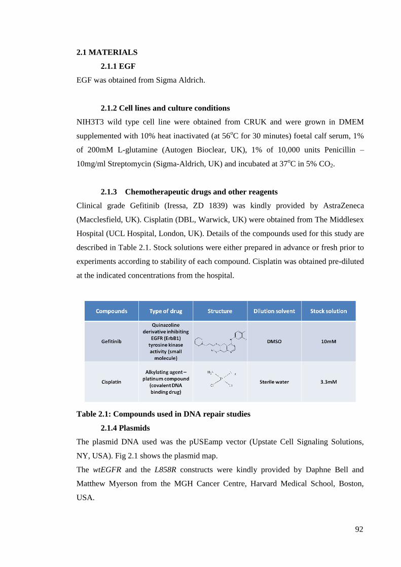
92
2.1 MATERIALS
2.1.1 EGF
EGF was obtained from Sigma Aldrich.
2.1.2 Cell lines and culture conditions
NIH3T3 wild type cell line were obtained from CRUK and were grown in DMEM
supplemented with 10% heat inactivated (at 56oC for 30 minutes) foetal calf serum, 1%
of 200mM L-glutamine (Autogen Bioclear, UK), 1% of 10,000 units Penicillin –
10mg/ml Streptomycin (Sigma-Aldrich, UK) and incubated at 37oC in 5% CO2.
2.1.3 Chemotherapeutic drugs and other reagents
Clinical grade Gefitinib (Iressa, ZD 1839) was kindly provided by AstraZeneca
(Macclesfield, UK). Cisplatin (DBL, Warwick, UK) were obtained from The Middlesex
Hospital (UCL Hospital, London, UK). Details of the compounds used for this study are
described in Table 2.1. Stock solutions were either prepared in advance or fresh prior to
experiments according to stability of each compound. Cisplatin was obtained pre-diluted
at the indicated concentrations from the hospital.
Table 2.1: Compounds used in DNA repair studies
2.1.4 Plasmids
The plasmid DNA used was the pUSEamp vector (Upstate Cell Signaling Solutions,
NY, USA). Fig 2.1 shows the plasmid map.
The wtEGFR and the L858R constructs were kindly provided by Daphne Bell and
Matthew Myerson from the MGH Cancer Centre, Harvard Medical School, Boston,
USA.
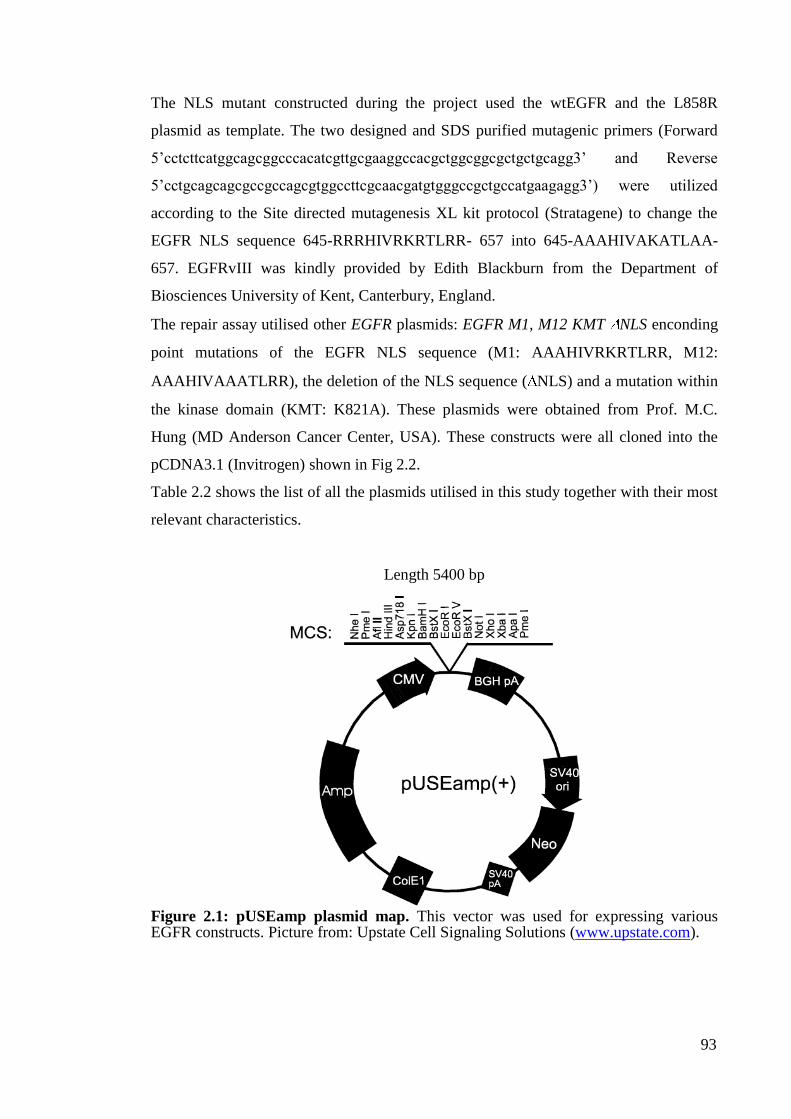
93
The NLS mutant constructed during the project used the wtEGFR and the L858R
plasmid as template. The two designed and SDS purified mutagenic primers (Forward
5‟cctcttcatggcagcggcccacatcgttgcgaaggccacgctggcggcgctgctgcagg3‟ and Reverse
5‟cctgcagcagcgccgccagcgtggccttcgcaacgatgtgggccgctgccatgaagagg3‟) were utilized
according to the Site directed mutagenesis XL kit protocol (Stratagene) to change the
EGFR NLS sequence 645-RRRHIVRKRTLRR- 657 into 645-AAAHIVAKATLAA-
657. EGFRvIII was kindly provided by Edith Blackburn from the Department of
Biosciences University of Kent, Canterbury, England.
The repair assay utilised other EGFR plasmids: EGFR M1, M12 KMT NLS enconding
point mutations of the EGFR NLS sequence (M1: AAAHIVRKRTLRR, M12:
AAAHIVAAATLRR), the deletion of the NLS sequence ( NLS) and a mutation within
the kinase domain (KMT: K821A). These plasmids were obtained from Prof. M.C.
Hung (MD Anderson Cancer Center, USA). These constructs were all cloned into the
pCDNA3.1 (Invitrogen) shown in Fig 2.2.
Table 2.2 shows the list of all the plasmids utilised in this study together with their most
relevant characteristics.
Length 5400 bp
Figure 2.1: pUSEamp plasmid map. This vector was used for expressing various EGFR constructs. Picture from: Upstate Cell Signaling Solutions (www.upstate.com).

94
Figure 2.2: pcDNA3 plasmid map. This plasmid carries the resistance gene ampicillin
and neomycin. Unique restriction sites are indicated on the plasmid.
(www.invitrogen.com).

95
Table2.2: List of plasmid used in this study

96
2.1.5 Primers
All the primers were self designed and ordered from MWG
2.1.5.1 Mutagenic primers
Table2.3: List of mutagenic primers
2.1.5.2 Sequencing primers
Table 2.4: List of sequencing primers
2.1.5.3 Screening primers
Table 2.5: List of screening primers

97
2.2 METHODS
2.2.1 Tissue culture
2.2.1.1 Cell lines maintenance
All cell lines were grown in 75cm2 flasks (T75), in humidity-saturated (95%)
incubators (Forma Scientific, UK), at 37oC with 5% CO2. All procedures were
carried out in Class II MDH biological safety cabinet (Intermed MDH, UK) using
aseptic techniques (cabinet was cleaned with 70% industrial methylated spirit – IMS
– as well as the equipment used inside the cabinet). Cells were routinely passage
twice a week, at 80-90% confluence. As all cell lines were adherent, media was
removed and cells were washed with 3ml of sterile 0.01M phosphate-buffered saline
solution (PBS, Sigma-Aldrich, UK) to remove the residual serum. Cells were
subsequently detached using 5ml of 1xTrypsin/EDTA (Autogen Bioclear, UK) at
37oC. 5ml of complete medium was then added to inactivate the trypsin and cells
were pelleted by centrifugation at 1200rpm for 5 minutes at room temperature. The
supernatant was discarded and cells were resuspended in 10ml of complete medium.
Cells were finally seeded into new flasks at a ratio of 1/2 to 1/5 according to the cell
line. All cell lines were passaged to a maximum of 30 times, after which point fresh
cells were retrieved from the liquid nitrogen stock.
2.2.1.2 Storage and retrieval from liquid nitrogen
In order to store cell lines, frozen cell stocks were prepared. Cells were grown in
175cm2 flasks (T175) to 80% confluence. Cells were trypsinised and resuspended to
a concentration of 1x10⁶cells/ml in freezing medium (FCS containing 10%
dimethylsulphoxide – DMSO – Sigma-Aldrich, UK). The cell suspension was
aliquoted in 1ml cryotubes and frozen at -80oC for one day. Tubes were then
transferred to a liquid nitrogen tank. Cells were recovered from liquid nitrogen by
thawing the cryotubes rapidly in a 37oC water bath. The cell solution was added to a
25cm2 flask (T25) containing 9ml of complete growth medium. Flasks were
incubated at 37oC with 5% CO2. The medium was changed after 24 hours (once cells
have re-attached to the bottom of the flask) to avoid toxicity due to the DMSO from
the freezing mixture.
98
2.2.1.3 Cell count
Cells were counted, once resuspended in 10ml of complete growth medium (see cell
line maintenance), using a haemocytometer. A haemocytometer has two chambers
and each chamber has a microscopic grid etched on the glass surface (4 squares each
containing 16 smaller square all connected by a 25 square grid). The chambers are
overlaid with a glass coverslip that rests on pillars exactly 0.1mm above the chamber
floor. 15µl of cell suspension was mixed with 15µl of trypan blue (Sigma-Aldrich,
UK) in order to exclude dead cells (1 in 2 dilution). 15 µl of this Dilution was loaded
into one chamber. The number of cells present in the original solution was
determined by counting the number of cells within the 4 large squares. The obtained
numbered was then divided by 4 (number of squares) and then multiplied by 2
(dilution 1:2). The total number of cells obtained was multiplied by 1x10⁴, giving the
number of cells per ml of suspension.
2.2.1.4 Cell doubling time
NIH3T3 cells were plated to an initial cell number of 1x 10⁵ per 25cm
2 flask
containing 8 ml of growing media. Single flasks were utilised for each time point
until confluence. Every 24 hours cells were trypsinised, centrifuged, resupended and
counted. The doubling time was calculating by plotting the number of counted cells
(Y) against the number of hours (X). This resulted in 1 doubling time every 20-24
hours.
2.2.2 Drug treatments
Prior to drug treatment, cells were seeded in flasks, 96 or 6 well plates and incubated
at 37oC in 5% CO2 for 24 hours. Cells were then treated at a range of concentrations
and lengths of exposure (Table 2.6). Optimal concentration was determined by the
DNA repair response curve. Appropriate schedules were used for the different
experiments, as outlined in the methods detailed previously.
99
Table 2.6: Range of concentrations and lengths of exposure for drugs used in
the different experiments. The ranges of concentration indicated in the table reflect
the minimum and maximum concentrations used.
2.2.3 Irradiation condition
In radiotherapy, the potency of IR is referred to the measurement of charge deposited
within a tissues and usually corresponds to exposure (coulomb/kg). For living tissue
the energy deposited is expressed in Gray or Gy, being 1 Gy the amount of radiation
required to deposit 1 Joule of energy in 1 kg. This represents the absorbed dose.
During this thesis there will be a general reference to IR, however alpha or beta,
gamma rays and X-rays are all different types of ionising radiation. The biological
damage produced by each of these different types is measured in Sievert. This
corresponds to the absorbed dose multiplied by the quality factor which, for alpha
particles is 20 while for X-rays is 1. In this study it was chosen to study the effect of
X-rays. These are measured in Gy.
For irradiation conditions cells were plated at a concentration of 1 x 10⁵/ml.
Following 48 hours transfection, cells were serum starved for 24 hours and irradiated
with 4 Gy (for the interaction and nuclear translocation studies) or 12.5 - 15Gy (for
the repair studies) using the A.G.O. HS 321kV X-ray system. The x-ray machine was
calibrated by a nuclear engineer to a dose rate of 4.39 Gy/min; 250 KV/12.5mA at a
30cm distance using a filter made of 0.25mm Cu and 1.00 mm Al.
2.2.4 The Comet assay
2.2.4.1 DNA damage level
Cells were plated at 10x104cells/ml in 25cm
2 flasks (Nunclon, VWR, UK) and
incubated for 24 hours at 37oC in 5% CO2. In order to analyse DNA damage and
repair, cells were treated with the appropriate chemotherapeutic drug at a range of
concentrations. Drugs were left in contact with the cells for a short period of time (1
to 2 hours according to the drug) and left to incubate in drug free media (if
necessary), for DNA damage to reach its peak (e.g. 9 hours for cisplatin). From this,

100
a fixed drug concentration was determined to treat the cell and observe the repair of
drug-induced DNA damage. For IR induced strand breaks the dose producing a tail
moment (see section 2.10.2) of 9-10 was used. For interstrand crosslink agents
(cisplatin), the drug concentration giving about 70% decrease in tail moment (see
section 2.10.2) was useed.
2.2.4.2 DNA repair study
Once the concentration was selected, fresh cells were plated at 1x10⁵
cells/ml in 6
well plates and incubated at 37oC in 5% CO2 for 24 hours. Cells were then treated for
the appropriate time and the drug was subsequently replaced by drug free media.
Cells were incubated in drug free media for various period of time in order to
measure DNA damage repair. Cells were collected by trypsinisation, pelleted and
resuspended in 1ml of foetal calf serum containing 10% DMSO, before to be stored
at -80oC.
2.2.4.3 Assay methodology
When studying damage caused by a crosslinking agent, cells were thawed and
resuspended in ice cold media to a concentration of 2.5x10⁴cells/ml. Those cell
suspensions were divided into two samples, one of which being irradiated (12.5Gy)
using the A.G.O. HS 321 kV X-ray system to deliver a fixed number of random
DNA strand breaks (procedure carried out on ice), immediately before analysis. For
strand breaking agents, cells were thawed and diluted to a concentration of
2.5x10⁴cells/ml without any irradiation.
In both cases, the following methodology was the same. All procedures were carried
out on ice and in subdued light. Cells were embedded in 1% type VII agarose
(Sigma-Aldrich, UK) (1ml of agarose + 0.5ml of cell suspension) on duplicate 1%
type 1A agarose (Sigma-Aldrich, UK) pre-coated microscope slides. Cells were
lysed, in the dark on ice, for one hour in ice cold lysis buffer (100mM disodium
EDTA, 2.5M NaCl, 10mM Tris-HCl pH 10.5) containing 1% Triton X-100 (Sigma-
Aldrich, UK) added fresh. Slides were subsequently washed every 15 minutes in ice
cold distilled water for 1 hour. Slides were then incubated in ice cold alkali buffer
(50mM NaOH, 1mM disodium EDTA, pH 12.5) for 45minutes followed by
electrophoresis in the same buffer for 25 minutes at 18 V (0.6V/cm), 250mA. The
slides were finally rinsed in neutralising buffer (0.5M Tris-HCl, pH 7.5) then saline

101
(PBS). Slides left to dry overnight were re-hydrated for 20 minutes with distilled
water and stained with propidium iodide (2.5µg/mL) for 30 minutes (in the dark)
then rinsed in distilled water. Finally, slides were left to dry in a drying oven and
stored.
2.2.4.4 Data analysis
Images (illustrated in Fig 2.3) were visualised using a NIKON inverted microscope
with high-pressure mercury light source, 510-560nm excitation filter and 590nm
barrier filter at x20 magnification. Images were captured using an on-line charge-
couple device (CCD) camera and analysed using Komet Analysis software (Kinetic
Imaging, Liverpool, U.K.). For each duplicate slide 25 individual cells were
analysed. Tail moment for each cell was calculated using the Komet Analysis
software as the product of the percentage DNA in the comet tail and the distance
between the means of the head and tail distributions, based on the definition of Olive
et al. (2002).
For strand breaks, tail moment data were analysed as a function of time post-
incubation or drug concentration. Fig 2.4 illustrates the effect of a drug inducing
strand breaks. Data shown in this study were the result of three independent
experiments and included corresponding standard deviation bars.

102
1 - TMdi - TMcu
TMci - TMcu( )[ ]x 100% decrease in tail moment =
Background area
Comet head
Comet tail
Figure 2.3: Screen display of Komet analysis software. The Fig show untreated
irradiated cells. Red arrows are showing the head and the tail of comet. The
background is also taken into consideration by the software for calculation of the tail
moment.
Figure 2.4: Repair of DNA damage caused by IR treatment. Comet tail appears to
be reduced over time
For crosslinking agents, results were expressed as percentage decrease in tail
moment compared to untreated controls calculated by the following formula (tail
moment was calculated from the same software as previously described):
where TMdi = tail moment of drug-treated irradiated sample
TMcu = tail moment of untreated, unirradiated control
TMci = tail moment of untreated, irradiated control
Time post-
treatment (hours)
0 1 4 24

103
Percentage decrease in tail moment data were the result of three independent
experiments, with corresponding standard error bars. Fig 2.5 represents DNA
damage repair profile of irradiated cell treated with cisplatin. The short comet tail
after 9 hours is due to the presence of interstrand crosslinks, delaying the DNA
migration. Hence, as crosslinks are unhooked (repaired), the comet tail in irradiated
cells is restored. For cisplatin the peak of crosslinking is observed after 9 hours post-
incubation.
Figure 2.5: DNA damage repair profile of irradiated cells treated with cisplatin. Cells were treated with cisplatin for 1 hour and subsequently incubated in drug-free
media for various times. After 9 hours crosslinking reached its maximum (60-70%
crosslinks) as the tail appears much shorter (crosslinks are causing the delay in DNA
migration). At 24 and 48 hours, the unhooking (repair) of cisplatin crosslinks causes
the comet tail to re-elongate.
2.2.5 Statistical Analysis
The two way ANOVA and Bonferroni post tests were used for calculating the
significance of the differences in unhooking crosslinks and in the repair of the DNA
SBs. All the cell lines were compared to the wtEGFR expressing cell line, statistical
values of P < 0.01 were considered significant.
2.2.6 MTT
Cells were diluted 2.5 X 10⁴/ml and 500 (100 l) or 1000 (200 l) cells were plated in
each well of a 96 well plate and incubated overnight in full serum media. Cells were
incubated with 50 M cisplatin for one hour in serum free media and then left in full
serum media over a period of 72 hours. Following 24, 48 and 72 hours cells were
incubated with 20 l of 5mg/ml Thiazolyl Blue Tetrazolium Bromide (MTT
solution) for 3 hours at 37 C. Next, the media was replaced with 200 l Dymethil
sulfoxide (DMSO). Each plate was left on the shaker for 5 minutes. Cell density was
Time post-treatment
(hours) 0 9 24 48

104
read at 560 nm, corrected with background reading and correlated to untreated
control to calculate % survival.
2.2.7 Western blotting analysis
2.2.7.1 Total protein extraction
Cells were washed twice with ice-cold PBS buffer. Sigma lysis buffer containing
fresh 1.5 X protease inhibitor and 1.5 X phosphatase inhibitor (Roche, UK), was
added to the cells (kept on ice) for few minutes. Then, cells were scraped using cell
scrapers (VWR, UK) and left rotating for 15 minutes at 4oC. Samples were then
pelletted by centrifugation for 15 minutes at 4oC (13,000rpm). The total cell lysate
(supernatant) was placed in a fresh eppendorf tube and stored at -80°C.
2.2.7.2 Nuclear and cytoplasmic protein extraction
Nuclear and cytoplasmic separation was achieved using the Nuclear extract kit from
Perbio (Cramlington, UK). Cells were grown and treated in 25cm2 flasks (T25).
Briefly, cells were washed gently twice with cold PBS and scraped in 2ml of cold
PBS. Cell solutions obtained were transferred to a pre-chilled 15ml falcon tube
(VWR, UK) and centrifuged 5 minutes at 500g at 4oC (supernatant was discarded).
Cell pellets were resuspended in 300µl of cold PBS and transferred to a cold
eppendorf tube and centrifuged at 500g, 4oC for 5 minutes (supernatant was
discarded – all the PBS must be removed). Pellets were finally resuspended in 100 l
of ice-cold CERI (Cytoplasmic Extraction Reagent I) buffer, containing protease
inhibitor, and vortexed at high speed for 15 seconds. The cell suspensions were
incubated 10 minutes on ice before adding 5.5 l of CERII (Cytoplasmic Extraction
Reagent II) buffer. Cell solutions were vortexed for 5 seconds and incubated for 1
minute on ice. Solutions were vortexed once more for 5 seconds and centrifuged for
5 minutes at 16,000g at 4oC. The supernatant corresponds to the cytoplasmic fraction
and was stored at -80oC (the last few microliters of supernatant were discarded to
avoid contamination with the nuclear pellet).
The nuclear pellets were resuspended in 50 l of NER (Nuclear Extraction Reagent)
buffer containing protease inhibitor and vortexed 15 seconds at high speed. Samples
were vortexed for 15 seconds every 10 minutes for 40 minutes. Lysed nuclei were

105
centrifuged for 10 minutes at 16,000g at 4oC and the nuclear fractions (supernatant)
were stored at -80oC.
2.2.7.3 Protein quantification
Once extracted, proteins were quantified using the RC DC protein assay from Bio-
Rad Laboratories. This assay is based on the Lowry protocol (Lowry et al., 1951),
using reagents A, S and B. Briefly, 2µl of each lysate was mixed with 18µl of
distilled water, then 100µl of the mix of reagent A and S (20µl of reagent S with 1ml
of reagent A) was added. Finally 800µl of reagent B is added and solutions are
incubated for 15 minutes at room temperature. Absorbance (OD) was measured,
against a blank containing only distilled water at 750nm on a Philips
spectrophotometer (Beam PU8620 Series UV/Vis single) and total protein
concentration was determined with the following formula:
Concentration in µg/µl = OD*25
Once the concentration was determined, loading dye (250mM Tris HCl, pH 6.8,
500mM DTT, 10% SDS, 0.5% Bromophenol Blue, 50% glycerol and distilled water
were used to make a 5 times stock) was added to 50µg of protein (volume
equivalent). Samples were boiled at 70°C for 10 minutes and then stored at -20oC
until immunoblotting.
2.2.7.4 Immunoblotting
Samples prepared as in section 2.7.1 and 2.7.2 were centrifuged for 5 seconds and
loaded onto 3-8% Tris acetate gels (Novex pre-cast gels, Invitrogen, UK), using the
XCell SureLock™ Mini-Cell module (Invitrogen, UK) with NuPAGE tri-acetate
SDS running buffer (60.5g Tris Base, 89.5 Tricine, 10.0g SDS and distilled water
added to a volume of 1 litre were used to make a 20 times stock) in order to separate
proteins at 150V, at room temperature. Proteins smaller than 40kDa were separated
on 4-12% Bis-Tris gels (Novex pre-cast gels, Invitrogen, UK) using the same module
as above but with MES running buffer (97.6g MES, 60.6g Tris Base, 10g SDS, 3g
EDTA and distilled water added to a volume of 500ml was used to make a 20 times
stock). Kaleidoscope marker (Bio-Rad, UK) was used as a size marker.

106
Proteins were transferred electrophoretically (35 V at RT for 2.5 hours) onto
activated immobilon P membranes (Sigma-Aldrich, UK) (membranes are activated
by immersion in 100% MeOH for 30 seconds, followed by 2 minute in distilled
water and 5 minutes in transblot buffer), using the XCell II Blot module (Invitrogen,
UK) with transblot buffer (100ml of running buffer stock x10 (30.3g Tris Base,
144.1 Glycine and distilled water to 1 litre – pH 8.3), 200ml MetOH and distilled
water to 2 litre). Unbound sites on membranes were subsequently blocked using
blocking buffer: 5% BSA (Bovine Serum Albumin – Sigma-Aldrich, UK) in TBS
with 0.1% Tween 20 for phosphorylated proteins. Proteins were probed using the
appropriate antibody (see dilutions and preparation in Table 2.7). Finally, the
primary antibody was probed with HRP-conjugated polyclonal antibodies (Mouse or
Rabbit – 1/2000, cell signalling – see Table 2.7) for chemiluminescence detection
(ECL system, Amersham Biosciences, UK, Millipore GeHealthcare). To this end,
blots were dried and incubated 5 minute shaking vigorously with ECL reagents
before covering them with cling film and exposing them to Kodak X-OMAT™LS
film for various times (2 seconds to 1 hour).
For all experiments using total protein lysate, -tubulin or calnexin were used as a
loading control. However, for the nuclear and cytoplasmic separation, lamin was
used for the nuclear fraction and calnexin for the cytoplasmic fraction.

107
Table 2.7: List of antibody
2.2.8 Immunoprecipitation
Protein Sepharose G/ATM
4 Fast Flow (Amersham Biosciences, Sweden) beads were
washed as follows: 1 ml of Protein Sepharose was added to 14 ml of sterile PBS into
a 15 ml falcon. The sample was mixed and then centrifuged at 5000 rpm for 5
minutes in a cold centrifuge. The supernatant was discarded at the pelleted beads
were cleaned twice as described above. The beads were then resupsended in PBS
with a ratio 1:4.
Stably expressing NIH3T3 cell lines were plated at 2 X 10⁵ /mL or 4 X 10
5/mL in a
10 cm dish (Nunc) and left overnight before treatment. Exponentially growing cells
were then left in growing medium, or treated with 50μM cisplatin for one hour in
serum free media and then left in drug free growing medium for 18 hours, or serum
starved for 24-36 hours or serum starved for 24-36 hours and treated with EGF
(Sigma) 100ng/ml or treated with 4 gy IR and left 20 minutes in serum free media.
Following appropriate treatments, approximately 5x10⁶ cells were lysed on ice in
500μl of CelLyticTM
M Cell lysis reagent (Sigma) supplemented with Protease and

108
Phospahatases inhibitor (Roche) and Benzonase (Merck) according to
manufacturer‟s protocol. Following lysis, 1.5 mg of protein sample was incubated
with 2μg of anti-EGFR antibody (clone R19/48 Invitrogen) and left rotating at 4 C
for 2.5 hours. Then, 10 μl (1:4 PBS) of packed A Sepharose beads (Amersham) were
added and the eppendorfs were left rotating for an additional 30 minutes.
Immunocomplexes were pulled down from cleared lysates with the A sepharose
beads and washed 1%Triton X-100 PBS. Pulse spin centrifugation, washing and
mixing was repeated three times. Following the final wash the beads were drained by
vacuum suction using a 25G x5/8‟‟ needle (Terumo) and resuspended in sample
buffer for Western blot analysis.
2.2.9 Densitometric Analysis
To compare the intensity of bands produced by immunoblotting the ImageJ
densitometric analysis tool was used. Briefly, blots were scanned and bands of
interest were selected. Intensity was measured by calculating the plotting area of
each of individual bands. Background values and control values were then detracted
to determine the fold increase. Fig illustrates the methodology
Figure 2.6: Densitometry programme

109
2.2.8 Cloning
2.2.8.1 Site directed mutagenesis
Primers were designed according to manufacturer protocol
Forward 5‟cctcttcatggcagcggcccacatcgttgcgaaggccacgctggcggcgctgctgcagg3‟
Reverse 5‟cctgcagcagcgccgccagcgtggccttcgcaacgatgtgggccgctgccatgaagagg3‟
Primers were designed to contain all the desired mutation flanked by 10-15 bases of
correct sequence and SDS purified (MWG).
The melting temperature of the mutagenic primers was calculated according to the
equation:
Tm= 81.5+0.41(%GC)-675/N
=81.5+ 0.41(57)-675/22
=81.5+23.37-30.68=
= 75
Mutagenesis reaction:
5 μl of 10x reaction Buffer
X μl (5, 10, 20 or 50 ng) double stranded DNA template
X μl (125ng) Forward Primer
X μl (125ng) Reverse Primer
1 μl of dNTP mix
5 μl of QuikSolution
1 μl PfuUltra HF DNA polymerase (2.5U/ μl)
Cycles:
1 95 oC 2minutes
95 oC 1 minute
2 60 oC 50 seconds 18 times
68 oC 9 minutes
3 68 oC 7 minutes
4 4 o
C 2 minutes or more
Following the mutagenesis/ amplification, the reactions were incubated with 2 μl of
DpnI (10U/μl) for 2 hours at 37 oC to digest the maternal strand. Reactions were then
transformed using the XL10-Gold Ultracompetent Cells (Stratagene).
2.2.8.2 Plasmids transformation and amplification

110
In order to transform the plasmids into E. coli, 100µl of TOP10F chemically
competent E. coli (Invitrogen, UK) was mixed with 5µl of plasmid and incubated for
15 minutes on ice. Cells were heat shocked for 90 seconds at 42oC before incubation
for 2 minutes on ice. 800µl of SOC medium was added and cells were pelleted for
6.5 minutes at 4,000rpm, at room temperature. Finally, 750µl of the supernatant was
removed and the pellet was resuspended in the rest of the supernatant. Each
transformed plasmid was plated on a pre-warmed LB (Luria-Bertani) agar plate
(Invitrogen, UK), containing either 50μg/ml of ampicillin (Sigma-Aldrich, UK) or
30μg/ml of kanamycin (Sigma-Aldrich, UK) selective agent. Plates were incubated
overnight at 37oC. After selection, colonies were picked and grown overnight in
10ml of LB broth base (Lennox L Broth Base) medium (Invitrogen, UK) containing
50μg/ml of ampicillin (Sigma-Aldrich, UK) or 30μg/ml of kanamycin (Sigma-
Aldrich, UK), in an incubator shaker (37oC, 300rpm). Cells were harvested and
plasmids were extracted using the QIAprep Spin Miniprep, Highspeed Maxiprep kit
(Qiagen, UK) and quantified at 260nm using a NanoDrop® ND-1000 UV-Vis
Spectrophotometer (NanoDrop, UK)
Following site directed mutagenesis plasmids were digested and ligated into a newly
linearised backbone plasmid.
2.2.8.3 Plasmid digestion
After extraction, integrity and purity of the plasmids was checked. For the restriction
reactions, 2 l of plasmid DNA was incubated with 0.5 l of the appropriate enzyme
(see Table 2.8), 1.5 l of the corresponding 10x buffer (supplied with the enzyme),
1 l of 30.3mg/ml RNAseA (Sigma-Aldrich, UK) and distilled water to 15 l.
Reactions were incubated for 2 hours at 37 oC and run on a 1% agarose ethidium
bromide gel. The gel was electrophoresed at 100V and photographed using a dual
intensity ultraviolet transilluminator coupled with camera (UVP, UK). A 1Kb DNA
ladder was used to determine the size of each fragment obtained. Bands were
excised and purified using the gel extraction kit from Qiagen according to
manufacturer protocol.
The expected sizes were as follow: EGFR 4.0Kb; pcDNA3 5.4Kb; pUSEamp
5.4Kb; NLS123 4.0Kb; L858R 4.0Kb; LNLS123 4.0Kb; M1 3.9Kb; M12
3.9Kb; ∆NLS 3.9Kb; KMT 3.9Kb, EGFRvIII 3.1Kb

111
Table 2.8: List of enzymes used for plasmids restriction. Enzymes were chosen
according to the position of the insert on the plasmid.
2.2.8.4 Ligation
Excised and purified bands were quantified at 260 nm using the nanodrop.
Ligation was carried out by using the DNA ligation Kit Ver 2.1 (Takara).
30-50 ng of linearised Vector were used per reaction. The correct amount of the
insert was calculated by the equation:
(ng Vector x Kb Insert)/Kb Vector=ng Vector
This correspond to the ng of the insert necessary for a 1:1 ratio (Vector:Insert). In
order to maximise the chances of successfulness Ligation reactions were always
prepared using a 1:3, 1:5 and 1:8 ratio (Vector:Insert).
To the Vector:Insert initial mix was then added the correspondent volume of
Solution I from the DNA ligation kit. The reaction was incubated at 16 oC for 30
minutes. Prior to transformation, 1 l of Solution III was added to 9 l of ligation
reaction to increase the number of transformants. When the initial ligation reaction
over-exceeded 10 l, ligated DNA was precipitated with ethanol and dissolved in a
volume of 10 l TE Buffer.
112
2.2.8.5 Colonies screening
Bacterial colonies were screened by PCR using EGFR screening primers.
Briefly, PCR reaction was prepared according to the Promega GOtaq protocol.
Individual colonies were picked and gently submerged into individual tubes
containing a single reaction mix to provide sufficient template DNA for PCR. The
same colony was then incubated in a 1.5 ml Eppendorf (labelled accordingly)
containing LB media at 37 oC over the period of the PCR amplification and gel
electrophoresis. Positive colonies were then further incubated overnight at 37 oC by
pouring the content of the Eppendorf in 250 ml of LB media.
2.2.8.6 Sequencing
Samples were sent for sequencing to MWG Company (Germany)
2.2.8.7 Plasmids transfection
Transfection was achieved using GeneJuice® transfection reagent (Novagen
® EMD
Biosciences Darmstadt, Germany). Exponentially growing NIH3T3 cells were plated
in 6 well plates (Nunclon™, VWR, UK) at a concentration of 1 x10⁵cells/ml (1.5 ml
per well), without any antibiotics and incubated for 24 hours at 37oC (5% CO2). 3 l
was vortexed with 100 l of DMEM FCS free and incubated for 5 minutes in an
eppendorf. 1 g of plasmid was added to the solution and incubated at room
temperature for 15 minutes. Media from the plates was removed and replaced by
fresh complete media with no antibiotics. Each plasmid/GeneJuice mixture was
added dropwise to one well. Cells were incubated at 37oC (5% CO2) until
confluence. Cells were subsequently trypsinised (0.5ml of trypsin) and pelleted
before being plated in T25 flasks with media containing antibiotics and selective
agent G418 (Sigma-Aldrich, UK). Transfected cells were incubated for 3 to 4 weeks
to allow clones to grow (media was regularly changed to keep selective pressure).
All the clones used were grown as stable transfected cell lines, maintaining selective
pressure in the media.

113
2.2.9 Confocal Microscopy
2.2.9.1 Immunofluorescence Staining
2 X 10⁴
Stable and transfected NIH3T3 were plated on 13mm cover glass (VWR) in
a 24 well plate overnight before treatment. Cells were then treated with 50μM
cisplatin for one hour in serum free media and/or left growing in drug free growing
medium; serum starved for 24-36 hours or/and then treated with 4 Gy IR.18 hours
following the treatment with cisplatin and 20 minutes following the IR treatment
cells were washed twice in ice cold PBS followed by fixing with 4% PFA for 15
minutes at room at RT. Cells were then permeabilised with PBS containing 0.5%
Triton X-100 for 10 minutes washed 3X with PBS and blocked in PBS 5% BSA
(Sigma) for 1 hour at room temperature. Respective primary antibodies were added
as follows: anti-rabbit EGFR (1:50, clone 15F8 cell signaling) and anti-mouse
DNAPKcs (1:50 AbCam) in PBS 1% BSA and incubated in a humid chamber at 4ºC
overnight. Slides were then washed 3X with PBS 0.1% Triton X-100 for 10 minutes
and protein expression was visualized by incubating the cells with corresponding
secondary fluorescent conjugated Abs, as follows: 1:100 Alexa Fluor 647 goat anti-
rabbit and 1:100 Alexa Fluor 488 goat anti-mouse (Molecular probes and Invitrogen
Life Technologies) for 2 hours at RT. Fluorescent label- antibodies were added alone
as negative controls. Following 3 washes with PBS 0.1% Triton X-100 Nuclei were
stained using Dapi (Sigma) and mounted onto glass slides with fluorescent mounting
medium (Dako). Cells were then visualised by confocal microscopy (objective X40,
Leica TCS SP2). Nuclear slices images were acquired by sequential scanning using
the LAS AF Lite programme.
2.2.9.2 Proximity Ligation assay
Stables NIH3T3 grown on 13 mm cover glass (VWR) were left in growing medium,
or treated with 50μM cisplatin for one hour in serum free media and then left in drug
free growing medium for 18 hours, or serum starved for 24-36 hours, or serum
starved for 24-36 hours and treated with 4 gy IR and left 20 minutes in serum free
media. Cells were washed with chilled PBS and fixed with 4% paraformaldehyde for
15 min, blocked, permeabilized, and incubated overnight with anti-rabbit EGFR
(clone 15F8 cell signalling 1:50), anti-mouse DNAPKcs (AbCam 1:50) as described
above. Cells were then incubated with secondary antibodies conjugated with

114
oligonucleotides (PLA probe minus: anti-mouse and plus: anti-rabbit) for 90 minutes
at 37 C in a humidity chamber. Following 2 washes for 5 minutes with 1X PBS
0.1% Triton X-100, the hybridization solution, consisting of two oligonucleotides,
was diluted in high purity water (1:5) and added to the cells for 15 minutes at 37 C.
Cells were then washed once for 3 minutes to remove the hybridization solution.
Next, the ligation solution was diluted in high purity water (1:5) and 1.5 l of ligase
to a final volume of 60 l (per cover glass). The ligation reaction was then added to
the cells for 15 minutes at 37 C. Following 2 washes for 2 minutes, the amplification
solution was diluted in high purity water (1:5) and 0.75 l of polymerase to a final
volume of 60 l per cover glass. The slides were then incubated for 90 minutes at
37 C. Next the cells were washed 2 times for 3 minutes and the detection solution
was diluted in hi purity water (1:5) and added to the cells for 60 minutes at 37 C.
Finally cells were washed 2 times with 1X PBS 0.1% Triton X-100 for 2 minutes, 1
time with 0.5 X PBS 0.05% Triton X-100 for 2 minutes, 2 time with high purity
water for 2 minutes and 1 time with 70% ETOH for 1 minutes. The slides were left
to dry for 3 minutes at RT and then mounted onto a glass slide with fluorescent
medium (Dako). Cy3 signal amplification was utilised for the assay. Cells were then
examined with a confocal microscope (objective X40, Leica TCS SP2).
2.2.10 DNA-PK Functional Assay
DNA-PK activity was detected using the Promega SignaTECT DNA-PK assay
system, according to the manufacturer‟s protocol. The SignaTECT utilises the unique
SAM2TM
Biotin Capture Membrane and results in high density of streptadavidin on
the membrane matrix. This provides rapid, quantitative capture of biotinylated
substrate molecules, based on the strong affinity of biotin for streptadavidin (Kd=10-
15M). This system overcomes the problem of non-specific substrate binding by using
a biotinylated DNA-PK p53 derived peptide substrate. The high binding capacity of
the SAM2TM
Biotin capture membrane for the DNA-PK biotinylated peptide
substrate and the low background observed with this system, maximises the signal-
to-noise ratio. Briefly, 50μg of Benzonase treated whole-cell extract, was incubated
with DNA-PK, biotinylated peptide substrate, [ -32P] ATP, and either DNA-PK
activation buffer or DNA-PK control buffer for 5 minutes at 30 ºC. Termination

115
buffer was added, and 10 μl of each reaction sample were spotted onto a SAM2TM
biotin capture membrane. The SAM2TM membrane squares were washed and dried
before analysis by scintillation counting. The enzymatic activity of DNA-PK was
expressed as a percentage decrease of control DNA-PK activity.
116
CHAPTER 3
CHARACTERISATION OF THE
EGFR NLS MUTANTS

117
3.1 INTRODUCTION
3.1.1 EGFR endocytosis is required for receptor degradation, recycling
and nuclear translocation
In resting cells the epidermal growth factor receptor (EGFR) resides at the lipid rafts
of the cell membrane rich in cavoelae and cholesterol (Zwang & Yarden, 2009).
Upon ligand binding EGFR exits these domains in a Src-dependent manner and
releases the auto-inhibition of its catalytic function via homo or hetero-dimerisation
with members of the ERBB family (Sorkin & Goh, 2008; Wang et al, 1999). The
dimerised receptors trans-phosphorylate a plethora of effector molecules including
MAPK and AKT (Grandal & Madshus, 2008; Puri et al, 2005). Following the
stimulation of these intracellular signalling pathways EGFR is bound by the SH2
domain of Cbl leading to EGFR polyubiquitination and internalisation in the early
endosomes (Mosesson et al, 2008; Tvorogov & Carpenter, 2002). The receptor
endocytosis takes place through the formation of clathrin-coated pits (CCPs) or, in
the presence of high doses of ligand, via cavoelae (Sigismund et al, 2005; Sorkin &
Goh, 2008). Clathrin-mediated endocytosis (CME) is the major receptor
internalisation pathway (Sigismund et al, 2008; Zwang & Yarden, 2009). Membrane
receptors can spend limteless time at the membrane, however following activation,
they will be internalised within seconds to assure immediate inactivation of the
downstream signalling events (Mosesson et al, 2008; Oved & Yarden, 2002).
3.1.1.1 EGFR phosphorylation mediates nuclear translocation
Internalised EGFR can undertake three different routes: 1) recycling to the
membrane in an inactive form, 2) lysosomal degradation into the late endosomes or
3) nuclear translocation (Mosesson et al, 2008; Waterman & Yarden, 2001). The
decision is made according to the expression level and the activation status (Zwang
& Yarden, 2009). EGFR high expression saturates the degradation machinery
leading to the recycling of the receptor at the transmembrane. In addition,
internalised active EGFR represents a very poor substrate for lysosomal degradation
inducing EGFR sorting into the Golgi and ER leading to nuclear translocation (Liao
& Carpenter, 2007). A large body of evidence has shown that EGFR, either alone or
118
associated to ERBB2, can be further phosphorylated once internalised into the early
endosomes (Mosesson et al, 2008). This is suggested to prevent degradation and
contribute further to nuclear translocation. Following sorting through the Golgi and
ER, EGFR association with sec61 results in a retro-translocation to the cytosol where
EGFR is stabilised following association with HSP70. Binding of importin β,
mediated through the NLS sequence, translocates the receptor to the nucleus
(Dittmann et al, 2008a; Hsu & Hung, 2007; Liao & Carpenter, 2007; Lo et al,
2006a).
3.1.2 EGFR NLS sequence is a functional NLS sequence
Nuclear translocation requires energy, physiological temperature, a NLS and a
soluble transporter. The study of this very complex mechanism has revealed that the
last two components are known to be the “conditio sine qua non” for nuclear
translocation. The import takes place following three important steps: docking at the
nuclear pore complex, translocation and deposition. During the docking phase the
molecule that needs to be imported (the cargo molecule) binds to a soluble molecule
known as importin via the recognition of the NLS sequence. Therefore in absence of
one of these two components nuclear localisation is impaired.
3.1.2.1 The criteria for a functional NLS
The recognition of a NLS sequence via a bioinformatic approach is only valuable for
predicting putative NLS motifs. The functionality of a NLS sequence in any protein
is determined by four criteria:
1. The putative NLS sequence must be necessary for import. The transport of the
cargo protein into the nucleus is therefore hindered when the sequence is
deleted or altered. The most common approach to verify whether a sequence is
necessary for import is to mutate the consensus residues (basic amino acids –
R/K) into Alanine and verify that the nuclear translocation of the protein
decreases (Hsu & Hung, 2007).
2. The sequence must be sufficient to target an unrelated protein into the nucleus.
Usually this is demonstrated by fusing the putative NLS sequence to the N-
terminus of the Green Fluorescent Protein (GFP) and the location of the protein
is assessed visually (Hsu & Hung, 2007).

119
3. The protein of interest (containing the NLS) must interact with the import
receptor (importins) and the interaction must be mediated via the putative NLS
sequence. This is addressed by performing an in vitro binding experiment with
the purified proteins and with RanGDP and RanGTP that, as explained in the
introduction chapter, provide the energy for association and dissociation (Liao
& Carpenter, 2007; Lo et al, 2006a; Lo et al, 2006b).
4. The transport machinery must be disabled to show that nuclear import of the
protein of interest is mediated by a nuclear transport mechanism that can only
take place via the NLS sequence (Liao & Carpenter, 2007; Lo et al, 2006a).
Table 3.1 describes the criteria met by EGFR.
Table 3.1: Experimental evidence for nuclear EGFR. The table shows individual
criterion, the experimental design utilised to validate the criterion requirement and
the published research article where the evidence is shown.
3.1.3 The NLS sequence affinity for the importin molecule is highly
regulated
The NLS sequence is pivotal during the entire nuclear translocation mechanism
because it is the only known biological signature that can successfully obtain binding
with importin molecules and its success is dictated by the affinity of the NLS
sequence with the importin molecule. In many cases such affinity is further

120
positively or negatively regulated via phosphorylation and/or via intramolecular
association (Moroianu, 1999). For example, phosphorylation of the serine residue
contained in the NLS of the transcription factor Pho4 reduces affinity with the
importin molecule (Kaffman & O'Shea, 1999). Unphosphorylated pho4 localises to
the nucleus whereas phosphorylation lowers affinity with its importin molecule
triggering nuclear export and impeding re-import. Another example is the
transcription factor NF-AT. Unphosphorylated NF-AT localises to the nucleus and
when phosphorylated undergoes a conformational change that masks its NLS,
impeding binding with the importin molecule (Zaidi et al, 2004) SWI5 is
phosphorylated on three Serines, two of which lie in proximity of its NLS.
Unphosphorylated SWI5 is nuclear localised whereas when phosphorylated the
transcription factor is restrained in the cytoplasm (Kaffman & O'Shea, 1999; Wells &
Marti, 2002). From this last study it was suggested that phosphorylation of the sites
in the vicinity of the basic NLS prevents binding of the import machinery and
therefore cytoplasmic localisation.
3.1.4 EGFR NLS sequence mutation
The principal aim of this study is to investigate the role of EGFR nuclear
translocation in the modulation of DNA repair. In the introduction chapter the role of
the EGFR acquired mutations in therapy and their importance in predicting
sensitivity have been outlined. The EGFR NLS sequence is a tripartite sequence
made up of three clusters of basic amino acid residues RRRHIVRKRTLRR and
published data have demonstrated that mutation of the first two clusters of basic
amino acids into Alanines leads to a 78% decrease in nuclear EGFR following ligand
binding (Hsu & Hung, 2007). There is no published evidence investigating whether
the remaining 22% of nuclear EGFR is sufficient to mediate the transcriptional-
mediated EGFR nuclear function. In addition, there is no published study
investigating the nuclear translocation of EGFR NLS mutants following IR (the
second most studied inducer of EGFR nuclear translocation (Das et al, 2007;
Dittmann et al, 2005a). For these reasons wtEGFR and EGFR L858R (L858R)
mutant expressing plasmids were utilised as a background to construct two NLS-
EGFR mutants bearing a mutation of all the Arginine residues into Alanine
(RRRHIVRKRTLRR into AAAHIVAKATLAA): EGFRNLS123 (NLS123) and
L858RNLS123 (LNLS123).
121
3.1.5 NIH3T3 cells as a cellular model
Stable transfection of the human EGFR into the murine fibroblast NIH3T3 cells and
EGF supplementation into the growth medium were shown to be necessary and
sufficient to achieve cellular transformation of this cell line (Di Fiore et al, 1987),
making this system an unbiased and optimal cellular model to study the modulation
of EGFR on DNA damage repair.
3.1.6 CHAPTER AIMS:
Validation of the NIH3T3 cellular system
wtEGFR and L858R expression following transient transfection
Validation of the constructed mutants
Sequence analysis and restriction digest to prove site directed
mutagenesis
Characterisation of the expressing EGFR constructs
Phosphorylation of wtEGFR and mutants following ligand binding, IR
or chemotherapy
Activation of downstream signalling pathways following ligand
induction
Nuclear localisation following EGF or IR stimulation

122
3.2 RESULTS
3.2.1 EGFR expression in NIH3T3 (EGFR null) cells
Following transient transfection of NIH3T3 cells, wtEGFR and L858R expression
was confirmed via western blotting analysis over a period of 72 hours. This was
necessary to determine the maximal expression of the EGFR constructs in the
NIH3T3 cells. Fig 3.1 shows that EGFR expression peaks at 48 hours following
transient transfection while no expression was detected in the vector control (VC)
transfected cell line (Fig 3.1).
Figure 3.1: EGFR expression peak at 48 hours following plasmid transfection.
NIH3T3 cells were transiently transfected with 2 μg of plasmid DNA encoding for
wtEGFR or L858R or with the backbone plasmid (V.C.). Cells were seeded and
lysed over a period of 72 hours. Whole cell lysates were resolved on a denaturing gel
and blotted for total EGFR and α-tubulin.
3.2.2 EGFR NLS site directed mutagenesis
To examine the modulation of EGFR nuclear translocation, NLS sequence mutants
(NLS123, LNLS123) were generated as described in the Materials and Methods
section. The expected mutagenesis was validated via enzymatic digestion of the
obtained plasmids and by sequencing. Fig 3.2A shows the result of the EcoRV-XhoI
double digestion and of the EcoRI single digestion whereas Fig 3.2B and C show the
nucleotide and amino acid alignment of the obtained sequences. The mutated
constructs were sequenced three times to confirm correct mutagenesis and absence of
any other mutation in the remaining DNA sequence.

123
Figure 3.2: Enzymatic digestion and sequencing confirmed the NLS
mutagenesis. A) In the left panel 2μg of plasmid DNA was digested with EcorV-
XhoI to generate the expected fragments of 3.994 KB (EGFR cDNA) and 5.426 KB
(pUSEamp) and with EcorI to generate the expected fragments of 0.02, 0.5, 0.8, 1.8
and 6.3 KB (right panel). Digested DNA was then resolved on a 1% Agarose gel and
samples were visualised using a UV transilluminator. B) NLS123 nucleotide
sequence obtained by MWG sequencing service was blasted against the Human
genome database. The Fig represents a snapshot from the whole sequence alignment.
C) The cDNA sequences obtained by the MWG sequencing service were translated
into an amino acids sequence using the Expasy tool and then were aligned using the
clustalW 2.0.5 multiple sequence alignment. The alignment file was then processed
with BOXSHADE 3.21. The Fig represents a snapshot from the whole sequence
alignment.
A
B
C
124
3.2.3 Mutant characterisation
3.2.3.1 EGF stimulation induces EGFR activation
The activation of EGFR upon ligand binding has been fully described in the
introduction chapter. Being the EGFR specific ligand, EGF has a strong and
immediate effect on EGFR dimerisation and phosphorylation. EGF is known to
promote kinase autophosphorylation leading to the activation of many important
cellular responses (Oved & Yarden, 2002; Toulany et al, 2007; Tvorogov &
Carpenter, 2002). Physiological EGF levels typically range from 10 to 100 ng/ml
(Sigismund et al, 2005) and in fact as little as 10 ng/ml can activate EGFR within
minutes. It has been shown that 100 ng/ml of EGF is the dose necessary to induce
EGFR nuclear translocation (Hsu & Hung, 2007). Therefore the effects of this dose
on the tyrosine phosphorylation over a period of 1 hour were investigated and the
activation of the downstream MAPK and AKT pathways and the timing of the
nuclear translocation examined. This is necessary to elucidate the correlation of these
events and the significance of their timing.
Fig 3.3A shows the western blots of NIH3T3 cells transiently transfected with
wtEGFR, NLS123, L858R, LNLS123 or VC treated with 100 ng EGF over a period
of 1 hour. This illustrates EGF-dependent EGFR phosphorylation and activation of
MAPK and AKT downstream signalling pathways. Fig 3.3 B-D show the fold
increase of Y1068 (B), AKT (C) and MAPK (D) activation.
Cells transfected with wtEGFR showed a time dependent Y1068 phosphorylation
increasing 3.13-fold by 30 minutes, remaining at this level at 60 minutes. Similarly,
phosphorylation of both AKT and MAPK increased 3.20 and 2.06-fold by 60
minutes. L858R transfected cells showed constitutive receptor activation and MAPK
phosphorylation. In contrast, there was a 2.2-fold increase in AKT phosphorylation
by 60 minutes. NLS123 transfected cells showed lack of Y1068 phosphorylation and
no upregulation of either AKT or MAPK. LNLS123 expressing cells showed
constitutive receptor activation, however this was reduced compared to Y1068
phosphorylation shown in wtEGFR cells. LNLS123 cells showed no upregulation of
either AKT or MAPK.
In light of the lack of EGFR-Y1068 phosphorylation in the NLS123 transfected cells
following EGF induction, the overall receptor phosphorylation was assessed together
with the ligand dependent and independent phosphorylation in wtEGFR and EGFR
mutant-expressing cell lines. Fig 3.4 shows the western blots of cells transfected with
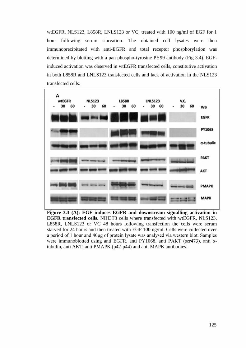
125
wtEGFR, NLS123, L858R, LNLS123 or VC, treated with 100 ng/ml of EGF for 1
hour following serum starvation. The obtained cell lysates were then
immunoprecipitated with anti-EGFR and total receptor phosphorylation was
determined by blotting with a pan phospho-tyrosine PY99 antibody (Fig 3.4). EGF-
induced activation was observed in wtEGFR transfected cells, constitutive activation
in both L858R and LNLS123 transfected cells and lack of activation in the NLS123
transfected cells.
Figure 3.3 (A): EGF induces EGFR and downstream signalling activation in
EGFR transfected cells. NIH3T3 cells where transfected with wtEGFR, NLS123,
L858R, LNLS123 or VC 48 hours following transfection the cells were serum
starved for 24 hours and then treated with EGF 100 ng/ml. Cells were collected over
a period of 1 hour and 40μg of protein lysate was analysed via western blot. Samples
were immunoblotted using anti EGFR, anti PY1068, anti PAKT (ser473), anti α-
tubulin, anti AKT, anti PMAPK (p42-p44) and anti MAPK antibodies.
A

126
D
Figure 3.3 (B-D): Graphic representation of the densitometry analysis of Figure
3.4 A blots. In each lane, left graphs show the fold increase compared to their
individual negative control (EGF untreated – serum starved) and right graphs show
the comparison when densitometry was done compared to the untreated wtEGFR
sample. In order the graphs show: the fold increase of Y1068 phosphorylation (B),
AKT phosphorylation (C) and MAPK phosphorylation (D).
B
C
D

127
Figure 3.4: EGF treatment does not activate the EGF receptor bearing the
NLS123 mutation. 60 minutes following EGF treatment 500 µg of protein lysate
was used to immunoprecipitate EGFR. Samples were then analysed via western blot
and immunoblotted with PY99 and for total EGFR. + indicates treated sample –
indicates non-treated sample. Numbers represent fold increase calculated as a mean
density of the first two exposures.
3.2.3.2 Dose-dependent cisplatin activation of EGFR
To extend our knowledge of the nature of the NLS123 mutation and its impact on the
receptor phosphorylation, this study investigated the receptor activation in transiently
transfected cells treated with different doses of cisplatin (Fig 3.5). This is because
cisplatin has been shown to induce ligand-independent EGFR phosphorylation on the
Y845 residue (Benhar et al, 2002). Since both the L858R and LNLS123 mutant
receptor have shown that EGF treatment induced EGFR activation, this assay was
performed only on the wtEGFR and the NLS123.
Fig 3.5 shows the western blot results of cells transfected with wtEGFR and
NLS123. Following 48 hours transfection, cells were treated for one hour with
cisplatin with a dose range of 10-150 µM in serum-free media. Cell lysates were then
blotted with PY845 antibody that recognises ligand-independent kinase domain
activation. In wtEGFR transfected cells, phosphorylation increased in a dose
dependent manner peaking at 50µM cisplatin, whereas NLS123 cells showed no
Y845 phosphorylation.
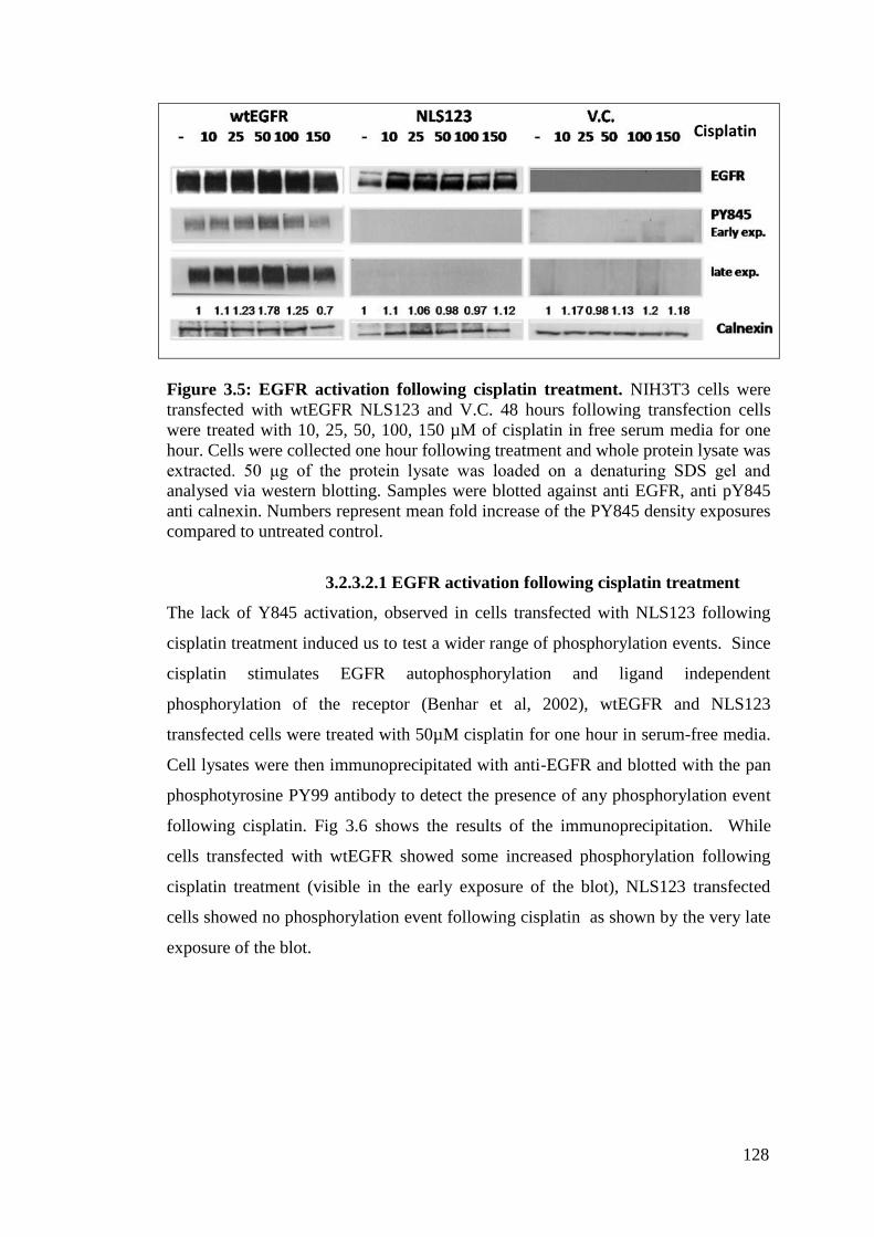
128
Figure 3.5: EGFR activation following cisplatin treatment. NIH3T3 cells were
transfected with wtEGFR NLS123 and V.C. 48 hours following transfection cells
were treated with 10, 25, 50, 100, 150 µM of cisplatin in free serum media for one
hour. Cells were collected one hour following treatment and whole protein lysate was
extracted. 50 μg of the protein lysate was loaded on a denaturing SDS gel and
analysed via western blotting. Samples were blotted against anti EGFR, anti pY845
anti calnexin. Numbers represent mean fold increase of the PY845 density exposures
compared to untreated control.
3.2.3.2.1 EGFR activation following cisplatin treatment
The lack of Y845 activation, observed in cells transfected with NLS123 following
cisplatin treatment induced us to test a wider range of phosphorylation events. Since
cisplatin stimulates EGFR autophosphorylation and ligand independent
phosphorylation of the receptor (Benhar et al, 2002), wtEGFR and NLS123
transfected cells were treated with 50µM cisplatin for one hour in serum-free media.
Cell lysates were then immunoprecipitated with anti-EGFR and blotted with the pan
phosphotyrosine PY99 antibody to detect the presence of any phosphorylation event
following cisplatin. Fig 3.6 shows the results of the immunoprecipitation. While
cells transfected with wtEGFR showed some increased phosphorylation following
cisplatin treatment (visible in the early exposure of the blot), NLS123 transfected
cells showed no phosphorylation event following cisplatin as shown by the very late
exposure of the blot.
Cisplatin
M

129
Figure 3.6: Cisplatin treatment does not activate the EGF receptor bearing the
NLS123 mutation. wtEGFR, NLS123, or VC transfected NIH3T3 cells were treated
with 50 μM cisplatin in serum free media, 48 hours after transfection. Cells were
seeded 60 minutes following cisplatin treatment and 500 µg of the protein lysate was
used to immunoprecipitate EGFR. Immunoprecipitated samples were then analysed
via western blot and immunoblotted with anti PY99 or anti EGFR. + indicates treated
sample – indicates non treated sample.
3.2.4 EGFR nuclear translocation is induced by ligand stimulation and
by IR treatment
Several reports have shown that EGF induction or IR treatment induce EGFR nuclear
translocation (Dittmann et al; Dittmann et al, 2005a; Dittmann et al, 2008a; Dittmann
et al; Hsu & Hung, 2007; Lo et al, 2006a). 100 ng/ml is the EGF dose shown to
induce nuclear translocation in CHO cells transfected with wtEGFR (Hsu & Hung,
2007). However, reports suggest that the EGF concentration is not strictly the
determining factor for EGFR translocation (Liao & Carpenter, 2009). In a cancer cell
the overexpression of EGFR is often accompanied by the autocrine production of
several ligands such as EGF. Therefore 100 ng/ml is the concentration of EGF that
can mostly mimic this and show the response of an EGFR over-expressing cell line
(Gazdar & Minna, 2008; Zandi et al, 2007). Published evidence has also shown that
4 Gy is the IR dose sufficient to induce EGFR nuclear translocation in some of the
most widely characterised EGFR over-expressing cancer cell lines including A431
and A549 (Rodemann et al, 2007). In this study the same dose has been utilised to
130
test wtEGFR nuclear translocation within the NIH3T3 cells and determine whether
the NLS123 mutation impairs EGFR nuclear translocation.
3.2.4.1 EGF induction of EGFR nuclear translocation
NIH3T3 cells transfected with wtEGFR, NLS123, L858R and LNLS123 were used
in this study. Cells were serum starved for 24 hours and then 100 ng/ml of EGF was
added to the serum-free medium. Cells were then fixed with 4% PFA, 30 and 60
minutes following addition of EGF and then probed using anti-rabbit EGFR and
stained using DAPI and goat anti-rabbit alexa fluor 568. Fig 3.7 shows the result of
the staining of the fixed cells visualised via confocal microscopy. Cells expressing
wtEGFR showed EGFR time-dependent nuclear translocation. 30 minutes following
EGF stimulation wtEGFR showed cytosolic internalisation and initial nuclear
expression followed by marked nuclear translocation at 60 minutes confirming
published reports (Hsu & Hung, 2007; Liao & Carpenter, 2007; Liao & Carpenter,
2009; Lo et al, 2006a). In contrast, NLS123, L858R and LNLS123 transfected cells
showed no EGFR nuclear translocation at either time point with clear membrane and
cytosolic accumulation.
3.2.4.2 EGFR nuclear translocation induced by IR
Having established EGFR nuclear translocation following EGF stimulation in
wtEGFR expressing cells and the effects of the NLS123 mutation, EGFR nuclear
translocation was investigated in wtEGFR, NLS123, L858R or LNLS123 transfected
cells following treatment with 4 Gy IR. Prior to irradiation, cells were starved for 24
hours.

131
Fig
ure
3.7
: E
GF
R ce
llu
lar
loca
lisa
tion
fo
llow
ing E
GF
tr
eatm
ent.
N
IH3T
3 ce
lls
tran
sien
tly tr
ansf
ecte
d ex
pre
ssin
g
wtE
GF
R,
NL
123,
L858R
, L
NL
S123 o
r V
C w
ere
seru
m s
tarv
ed f
or
24 h
ours
, tr
eate
d w
ith 1
00ng/m
l of
EG
F a
nd t
hen
fix
ed
wit
h 4
% P
FA
at
30 o
r 6
0 m
inute
s fo
llow
ing t
reat
men
t. C
ells
wer
e st
ained
wit
h G
oat
anti
-Rab
bit
Ale
xa
fluor
568 (
EG
FR
),
and D
api
(nucl
eus)
.

132
Cells were collected over a period of 20 minutes from the time of irradiation and the
cytosolic fraction was separated from the nuclear compartment. Densitometric
analysis was used to quantify receptor nuclear accumulation. To normalise the
densitometric data and take into account cytosolic contamination of the nuclear
fraction, the ratio between the densities of cytosolic EGFR/calnexin was subtracted
from the ratio between the densities of nuclear EGFR/calnexin (contamination). The
ratio of cytosolic untreated EGFR/calnexin represents the physiological amount of
EGFR in the cytosol in relation to the amount of cytosolic marker calnexin present in
the blotted cellular extracts. By subtracting this amount from all the ratios of nuclear
EGFR/calnexin it is possible to determine, and exclude, if EGFR nuclear
accumulation is due to any cytosolic contamination (shown by the calnexin bands in
the nucleus) and therefore the real fold increase of EGFR nuclear accumulation over
time can be determined.
Fig 3.8 shows the results of samples obtained from wtEGFR transfected cells. These
cells showed a time-dependent EGFR nuclear accumulation with a corresponding
time-dependent cytosolic reduction. Densitometric analysis shows a 2.16-fold
increase in EGFR nuclear accumulation by 20 minutes. NLS123 (Fig 3.9) and L858R
(Fig 3.10) transfected cells show no EGFR nuclear accumulation over time as
indicated by the densitometric values which fall below 1 following IR treatment. In
both these transfected cell lines there was a time dependent accumulation of EGFR in
the cytosolic fraction. LNLS123 transfected cell lines (Fig 3.11) showed lack of
EGFR nuclear accumulation as indicated by the negative value. Considering the
potent effect of IR on EGFR phosphorylation, the phosphorylation of the Y1068 and
Y845 residues in both cellular compartments following IR was investigated in all the
transfected cell lines. Cytosolic fraction from wtEGFR (Fig 3.8), L858R (Fig 3.10),
LNLS123 (Fig 3.11) showed activation at both Y845 and Y1068 phosphorylation
residues following IR treatment. Only the wtEGFR nuclear fraction (Fig 3.9) showed
increased Y845 phosphorylation following IR treatment. NLS123 transfected cells
(Fig 3.10) showed no receptor activation either in the cytosol fraction or in the
nucleus.
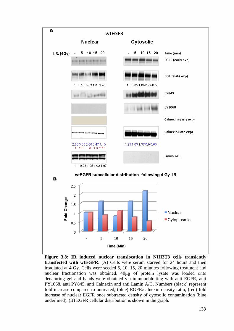
133
B
A
Figure 3.8: IR induced nuclear translocation in NIH3T3 cells transiently
transfected with wtEGFR. (A) Cells were serum starved for 24 hours and then
irradiated at 4 Gy. Cells were seeded 5, 10, 15, 20 minutes following treatment and
nuclear fractionation was obtained. 40μg of protein lysate was loaded onto
denaturing gel and bands were obtained via immunoblotting with anti EGFR, anti
PY1068, anti PY845, anti Calnexin and anti Lamin A/C. Numbers (black) represent
fold increase compared to untreated, (blue) EGFR/calnexin density ratio, (red) fold
increase of nuclear EGFR once subtracted density of cytosolic contamination (blue
underlined). (B) EGFR cellular distribution is shown in the graph.
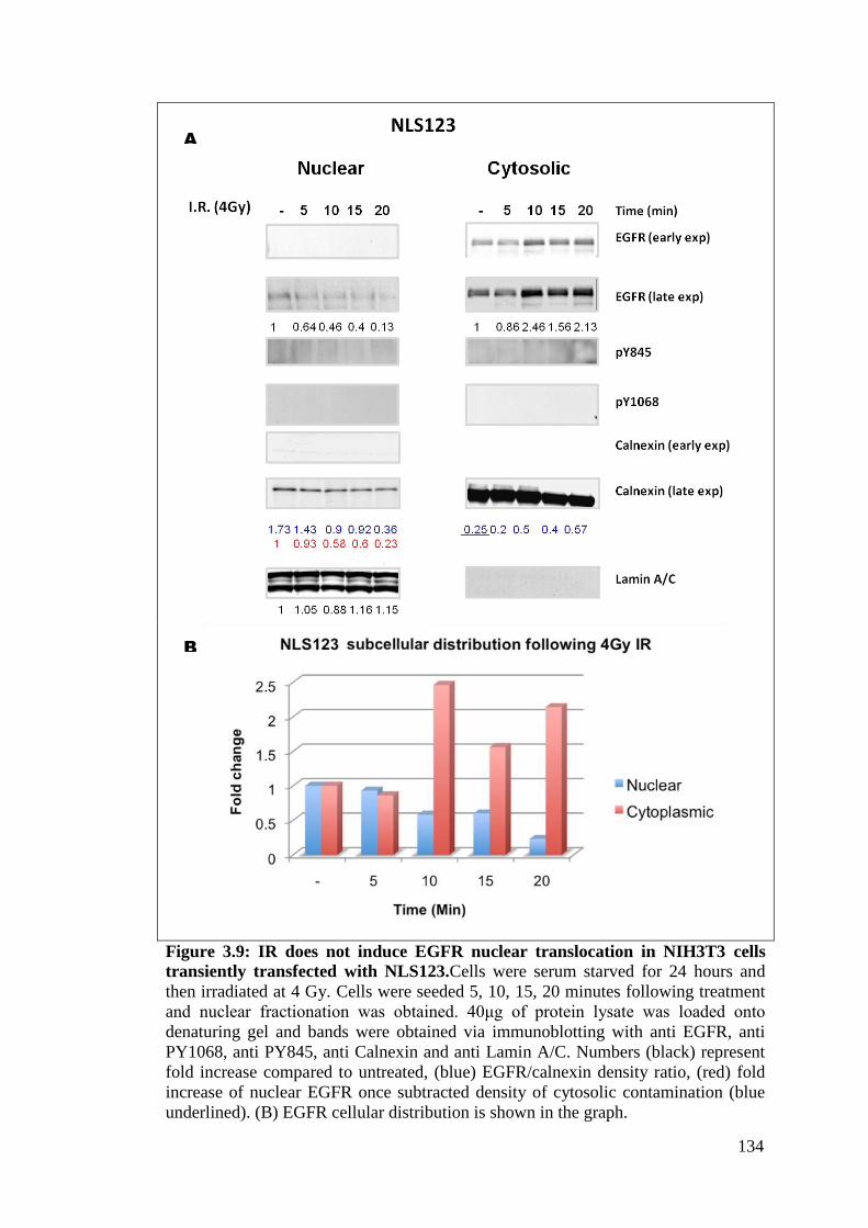
134
A
B
Figure 3.9: IR does not induce EGFR nuclear translocation in NIH3T3 cells
transiently transfected with NLS123.Cells were serum starved for 24 hours and
then irradiated at 4 Gy. Cells were seeded 5, 10, 15, 20 minutes following treatment
and nuclear fractionation was obtained. 40μg of protein lysate was loaded onto
denaturing gel and bands were obtained via immunoblotting with anti EGFR, anti
PY1068, anti PY845, anti Calnexin and anti Lamin A/C. Numbers (black) represent
fold increase compared to untreated, (blue) EGFR/calnexin density ratio, (red) fold
increase of nuclear EGFR once subtracted density of cytosolic contamination (blue
underlined). (B) EGFR cellular distribution is shown in the graph.

135
B
A
Figure 3.10: IR does not induce EGFR nuclear translocation in NIH3T3 cells
transiently transfected with L858R. (A) Cells were serum starved for 24 hours and
then irradiated at 4 Gy. Cells were seeded 5, 10, 15, 20 minutes following treatment
and nuclear fractionation was obtained. 40μg of protein lysate was loaded onto
denaturing gel and bands were obtained via immunoblotting with anti EGFR, anti
PY845, anti Calnexin and anti Lamin A/C. Numbers (black) represent fold increase
compared to untreated, (blue). EGFR/calnexin density ratio, (red) fold increase of
nuclear EGFR once subtracted density of cytosolic contamination (blue underlined).
(B) EGFR cellular distribution is shown in the graph.
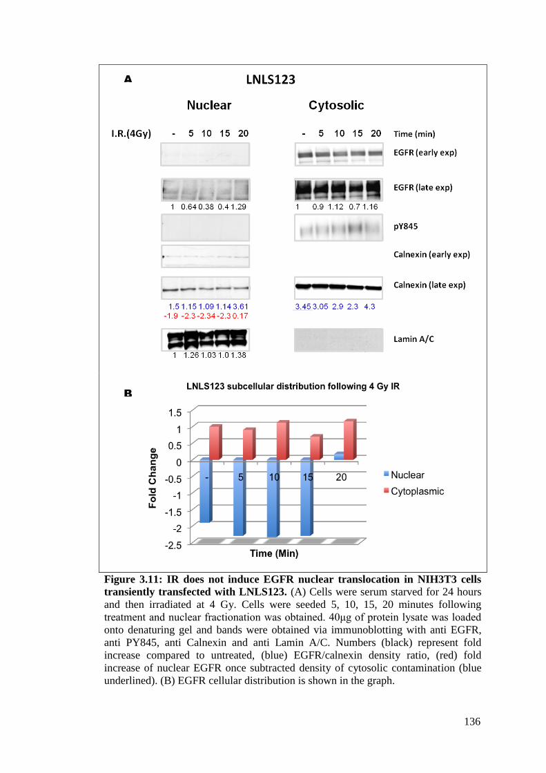
136
B
A
Figure 3.11: IR does not induce EGFR nuclear translocation in NIH3T3 cells
transiently transfected with LNLS123. (A) Cells were serum starved for 24 hours
and then irradiated at 4 Gy. Cells were seeded 5, 10, 15, 20 minutes following
treatment and nuclear fractionation was obtained. 40μg of protein lysate was loaded
onto denaturing gel and bands were obtained via immunoblotting with anti EGFR,
anti PY845, anti Calnexin and anti Lamin A/C. Numbers (black) represent fold
increase compared to untreated, (blue) EGFR/calnexin density ratio, (red) fold
increase of nuclear EGFR once subtracted density of cytosolic contamination (blue
underlined). (B) EGFR cellular distribution is shown in the graph.
LNLS123
I.R. 4gy

137
Table 3.2 A-D: Analysis of the densitometric values acquired via ImageJ on the
cytosolic - nuclear separation blots for (A) wtEGFR, (B) NLS123, (C) L858R,
(D) LNLS123.
A
B
C
D
138
3.3. DISCUSSION
3.3.1 EGFR expression in NIH3T3 cells
Over-expression of the EGF receptor is considered to be one of the principal
characteristics of many types of cancer cells (Moasser et al, 2001). Our data showed
that following transient transfection, the EGF receptor expression peaks at 48 hours
remaining constant for the next 24 hours (Fig 3.1). Although sufficient receptor
expression was already detectable from 12 hours following transfection, all the
treatments were made 48 hours following transfection to mimic the EGFR over-
expression of other cancer cells.
3.3.2 NIH3T3 cells as the most suitable cellular model
Although the plasmids utilised express human EGFR, NIH3T3 mouse fibroblast cell
line was employed for this study. These cells do not express EGFR and they have
been shown to become tumorigenic if cultured with EGF and overexpressing the
EGF receptor (Di Fiore et al, 1987). Moreover, in addition to the intrinsic identity of
the two genomes (human and mouse genome share 97% identity of their translated
sequence) that have conserved the same cellular mechanisms; the necessity of
employing an EGFR negative system is clearly primary. Among the other organisms
whose genome has high homology to humans, the Green monkey COS7 and the
hamster CHO cell lines are also EGFR negative. However, the CHO cell lines have a
great practical disadvantage since there are not many commercially available
antibodies or assays that guarantee cross reactivity with the hamster species
rendering our molecular approach, potentially, difficult to interpret. The COS7 cells,
although more closely related to humans, have not been shown to undergo
transformation following over-expression of EGFR and therefore, among these three
cellular systems, negative in the expression of EGFR, the mouse fibroblast NIH3T3
cell line was chosen. Despite the fact that a mouse-derived cell line cannot fully
recapitulate the cellular circuits and the mechanisms of EGFR induced DNA repair
regulation of human cancer cell lines, there are other biological disadvantages that
have led to the NIH3T3 choice. Among the published cancer cell lines there are only
two lines that do not express EGFR: ZR75 and DU4475 (Moasser et al, 2001). These
cancer cell lines express HER2 and therefore the modulation of DNA repair by

139
another member of the ERBB family could have not been excluded. The final
category, which was examined as a possible candidate for our model, includes those
cancer cell lines that express EGFR and are also EGFR dependent. There are
disadvantages of knocking out the existing receptor and knocking in our mutants and
also the high expression of other members of the ERBB family of receptors.
Moreover there are several reports showing that silencing the EGF receptor in an
EGFR-dependent cell line results in spontaneous cell death (Park et al, 2009).
3.3.3 EGFR expression mediated signalling dependence in NIH3T3 cells
This chapter has confirmed that the NIH3T3 cells are EGFR negative (Fig 3.1) and
also shown that upon EGFR transient expression, two main signalling pathways are
activated in response to the ligand-induced upstream activation of the receptor (Fig
3.3). This demonstrates the adaptability of our cellular system and at the same time
the influence of the EGF receptor on the NIH3T3 endogenous AKT and MAPK
pathway (Klein & Levitzki, 2009). This was important to confirm the validity of this
system to examine the EGFR modulation of DNA repair in this cell line and to
compare the cellular responses in the presence of the somatic or the constructed
EGFR mutants. Interestingly, despite the constitutive activation of the L858R, the
densitometric analysis revealed a lower AKT and MAPK pathway compared to the
activation measured in cells transfected with wtEGFR. This is a novel finding which
has not been reported elsewhere. The downstream signalling activation of the L858R
receptor has not been extensively analysed since this somatic mutant is often
expressed together with the wtEGFR and discerning the actual activator of the
signalling pathway is virtually impossible. As outlined in the Chapter1, the activation
of one EGFR molecule relies on dimerisation and on the activation of the receiver
kinase by the activator kinase. L858R may exert its oncogenic potential by playing
the part of the activator with the wtEGFR molecule that will induce the activation of
the downstream pathways. In contrast, the NLS123 showed lack of receptor
phosphorylation and there was no detectable upregulation of the AKT and MAPK
signalling pathways.
3.3.4 Conformational change is required for EGFR activation
The results so far reported indicate that the mutation of the NLS sequence of the
EGFR receptor compromises receptor activation. The classical model of activation of
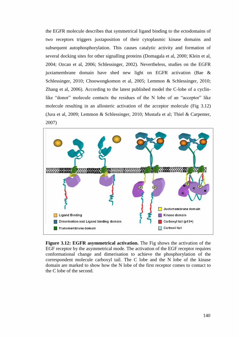
140
the EGFR molecule describes that symmetrical ligand binding to the ectodomains of
two receptors triggers juxtaposition of their cytoplasmic kinase domains and
subsequent autophosphorylation. This causes catalytic activity and formation of
several docking sites for other signalling proteins (Domagala et al, 2000; Klein et al,
2004; Ozcan et al, 2006; Schlessinger, 2002). Nevertheless, studies on the EGFR
juxtamembrane domain have shed new light on EGFR activation (Bae &
Schlessinger, 2010; Choowongkomon et al, 2005; Lemmon & Schlessinger, 2010;
Zhang et al, 2006). According to the latest published model the C-lobe of a cyclin-
like “donor” molecule contacts the residues of the N lobe of an “acceptor” like
molecule resulting in an allosteric activation of the acceptor molecule (Fig 3.12)
(Jura et al, 2009; Lemmon & Schlessinger, 2010; Mustafa et al; Thiel & Carpenter,
2007)
Figure 3.12: EGFR asymmetrical activation. The Fig shows the activation of the
EGF receptor by the asymmetrical mode. The activation of the EGF receptor requires
conformational change and dimerisation to achieve the phosphorylation of the
correspondent molecule carboxyl tail. The C lobe and the N lobe of the kinase
domain are marked to show how the N lobe of the first receptor comes to contact to
the C lobe of the second.

141
3.3.5 The Juxtamembrane domain regulates the cellular fate of the
receptor
The majority of the literature reports have portrayed the ligand binding domain, the
kinase domain and the carboxyl tail as the most important domains. This is
confirmed further by the design of EGFR-specific inhibitors. Nevertheless, despite
its short length, also the Juxtamembrane domain (JX segment) contains many
regulatory motives that dictate the fate of the entire receptor (Aifa et al, 2006a; Jura
et al, 2009) (Fig 3.13).
Figure 3.13: The Juxtamembrane domain sequence of the EGF receptor. The
coloured amino acids highlight the four amino-acid signals present in this domain
The two basolateral signals: the dominant, proline rich core 667PXXP670, and the
recessive, leucine rich 658LL659 and the lysosomal signal, 679LL680 are the residue
and the motif that dictate the distribution of the molecule across the membrane and
its recycling (Choowongkomon et al, 2005; Red Brewer et al, 2009). Essentially,
when EGFR is inactive the only signal accessible is the basolateral dominant which
recycles EGFR into an inactive form to the basolateral surface. The other signals are
sterically obstructed and therefore inaccessible. This signal seems to be sufficient for
targeting the receptor released at the trans Golgi to the basolateral plasma membrane
and redirect the endosomal recycled receptor to the basolateral membrane
(Choowongkomon et al, 2005). Upon ligand binding the lysosomal import becomes
the only alternative because the homo or heterodimerisation of the molecules
removes the inhibition of the lysosomal signal. The JX segments of the dimerising
molecules come closer together exposing the lysosomal signal for recognition. The

142
active molecule following its activation is therefore degraded and recycled back to
the surface membrane into an inactive state (Thiel & Carpenter, 2007).
The characterisation of the NLS mutation herein reported suggests that the NLS123
mutant is kinase dead. EGF, cisplatin or IR do not stimulate the kinase activity of the
receptor, down-regulating the signalling pathways that are normally controlled by
EGFR. This pattern suggests that the impairment of kinase activity observed in this
mutant receptor may be the result of an obstructed conformational change that
impedes the allosteric activation and the EGFR kinase-kinase juxtaposition. The NLS
mutation seems therefore to be responsible for this dramatic kinase impairment.
3.3.6 The NLS sequence is a recognition sequence for the allosteric
activation of the receptor
It has been shown that the 13 amino acids 645-657 (NLS sequence) containing the
Threonine 654 (T654) not only represent the NLS sequence of the receptor but they
are indispensable to achieve the conformational change that allows receptor
dimerisation and subsequent activation. Upon ligand binding the receptor undergoes
a conformational change in the intracellular domain at the site of the JX segment
whereby the tyrosine kinase domain presents its carboxyl tail to the correspondent
dimerised sister molecule (and vice versa) allowing a reciprocal phosphorylation of
their residues (Aifa et al, 2006a; Aifa et al, 2006b). Some studies have shown that the
phosphorylation of the T654 by protein kinase C (PKC) regulates the allosteric
activation of the receptor. The phosphorylation of the T654 not only seems to
suppress the kinase activity but also seems to re-route the receptor to the basolateral
surface. The proposed model infers that this phosphorylation event may disrupt the
helical structure causing the recessive basolateral signal to become accessible and to
dominate over the lysosomal sorting signal (Choowongkomon et al, 2004; Dittmann
et al; Macdonald-Obermann & Pike, 2009).
However, contrasting published evidence has shown that full length EGFR lacking
these NLS 13 amino acids does not dimerise or autophosphorylate suggesting that
the phosphorylation of T654 (residue contained in the NLS sequence) is neither
necessary nor indispensable to switch off the kinase activity of the receptor and that
the phosphorylation may be a consequence (rather than the reason) of the lack of the
conformational change that favours this post-translational modification. The

143
undetected phosphorylation of the NLS123 mutant suggests that the phosphorylation
of the T654 is not necessary to determine or signal the phosphorylation inhibition.
However, the undetected phosphorylation of the T654 (located in the middle of the
NLS sequence) could be the consequences of an obstructed protein structure which
does not allow addition of a phosphate group (the juxtamembrane stretch is not
accessible) or, if accessible, to the neutral charge of the NLS caused by the alanine
substitution which does not attract negatively charged groups.
Truncated mutants, deprived of the extracellular region but with an intact p13 (NLS
sequence is a peptide of 13 amino acids), dimerise and achieve a carboxyl tail
autophosphorylation (Aifa et al, 2006b). This suggests that ligand binding is not the
key component for the receptor activation. Collectively, these findings have
proposed a model whereby the thirteen amino acid peptide corresponding to the NLS
sequence, p13+ (mainly because the threonine residue is surrounded by basic amino
acids and therefore positively charged) of the JX segment will electrostatically
interact with another 13 amino acids peptide of exact but opposite charge, p13-,
where the carboxyl tail faces the kinase domain of the other molecule allowing the
reciprocal autophosphorylation (Aifa et al, 2006a).
3.3.7 The NLS third core of Arginine is required for dimer stabilisation
It has been recently shown that the last two arginines of the NLS sequence 656-RR-
657 are responsible for adopting an α helical conformation that is indispensable for
EGFR activity (Jura et al, 2009). The authors of this work compared the differential
activation of these two arginines mutated into alanines (which would basically
represent a charge removal) to the replacement by glycine (which would weaken the
helix). The effect of the alanine substitution is smaller compared to the glycines
substitution however the impairment on kinase activity of the receptor following
EGF activation is significant for both.
In the model presented by Jura et al. 2009; the juxtamembrane segment JM-A forms
an antiparallel helical dimer to allow the binding of the asymmetric kinase dimer.
The JM-B allows stabilising contact between the acceptor and donor lobe; however,
there is no functional involvement of this part of the Juxtamembrane domain (Fig
3.14). The model showed clearly the importance of the last core of the Arginine
residues within the NLS sequence, whose mutation is probably the main responsible
for the kinase inactivation of the NLS123 mutant. In the original study that has
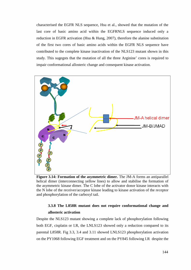
144
characterised the EGFR NLS sequence, Hsu et al., showed that the mutation of the
last core of basic amino acid within the EGFRNLS sequence induced only a
reduction in EGFR activation (Hsu & Hung, 2007), therefore the alanine substitution
of the first two cores of basic amino acids within the EGFR NLS sequence have
contributed to the complete kinase inactivation of the NLS123 mutant shown in this
study. This sugegsts that the mutation of all the three Arginine‟ cores is required to
impair conformational allosteric change and consequent kinase activation.
Figure 3.14: Formation of the asymmetric dimer. The JM-A forms an antiparallel
helical dimer (interconnecting yellow lines) to allow and stabilise the formation of
the asymmetric kinase dimer. The C lobe of the activator donor kinase interacts with
the N lobe of the receiver/acceptor kinase leading to kinase activation of the receptor
and phosphorylation of the carboxyl tail.
3.3.8 The L858R mutant does not require conformational change and
allosteric activation
Despite the NLS123 mutant showing a complete lack of phosphorylation following
both EGF, cisplatin or I.R, the LNLS123 showed only a reduction compared to its
parental L858R. Fig 3.3, 3.4 and 3.11 showed LNLS123 phosphorylation activation
on the PY1068 following EGF treatment and on the PY845 following I.R despite the

145
presence of the same NLS mutation as in the NLS123 responsible for its kinase
inactivation. The LNLS123, and its parental construct (L858R), also bear a kinase
point mutation (Leucine 858 into Arginine- L858R) that enhances the affinity of the
ATP binding site to ATP like molecules rendering the receptor more prone to auto-
phosphorylation. This mutation allows a thermodynamically stable conformation of
the protein that enables autophosphorylation of the Y845 residue that resides in the
same activation loop of the residue in position 858. (Yang et al, 2008) The reduced,
rather than abolished, phosphorylation confirms that the L858R mutation of the
receptor in non small cell lung cancer (NSCLC) confers to the EGF receptor a
constitutive activation (Gazdar, 2009a). This study suggests that this mutation
permits a mode of activation that escapes the necessity of undergoing a
conformational change required by WT receptor. This would explain why, despite
the mutation on its NLS sequence/p13+ peptide, the LNLS123 mutant still shows
activation following ligand or IR treatment. The reduced activation requires further
explanation. It has been reported that even minute amounts of EGFR receptor
activation are sufficient to activate downstream signalling pathways and therefore
also low levels must be taken into account (Arteaga, 2002; Bublil & Yarden, 2007).
Moreover, although a clear reduction compared to its parental is observed, the
phosphorylation levels presented by the LNLS123 are not negligible. Therefore, the
activation potential of this construct will be categorised in this study as constitutive
as with its parental L858R.
Structurally, the mutation to the nuclear localisation signal changed the arginine
residues into alanine residues. As a result, the affinity to the importin molecule is lost
and the receptor nuclear translocation is impaired. In addition, the mutation itself
seems to be sufficient to disturb the conformational change necessary for the receptor
activation as shown in the results.
3.3.9 EGFR nuclear localisation signal mutation impairs both nuclear
translocation and protein activation
Our data have shown the successful mutation of the wtEGFR and L858R constructs
NLS sequence. The mutated tripartite NLS signal of the NLS123 and LNLS123
constructs has resulted in marked reduction of EGFR nuclear localisation following
EGF or IR treatment.

146
The western blot analysis on the protein lysate of the separated cellular
compartments showed increase of wtEGFR nuclear translocation over time whereas
in the other mutants only a significant impairment of nuclear translocation. On the
other hand the results shown by the confocal microscopy following EGF treatment
show clear lack of nuclear accumulation in NLS123, L858R, LNLS123 both at 30
and 60 minutes following EGF treatment. Therefore the residual EGFR expression
detectable on the western blots of the nuclear compartments of NLS123, L858R and
LNLS123 expressing cells, is most probably due to the reported cytoplasmic fraction
contamination rather than the actual receptor translocation as confirmed by the
densitometry analysis. This study also indicates that only phosphorylated wtEGFR
on Y845 residue was found in the nucleus following IR treatment. However, both
L858R and LNLS123 also undergo PY845 autophosphorylation, and the lack of
PY845 signal within the nuclear fraction suggests that nuclear EGFR expression
found in the blots from cells transfected with the other mutants is due to cytosolic
contamination. This suggests that Y845 phosphorylation is not the post-translational
modification causing the nuclear translocation but that it is a consequence of an a
priori event that allows nuclear translocation. Taken together, the constitutive
activation of the L858R and its lack of nuclear accumulation suggest that kinase
activation per se is not sufficient for EGFR nuclear expression and that the
mechanism is more complex.
The confocal microscopy analysis following EGF treatment shows a cytosolic and
membrane accumulation of the receptor in the NLS mutants and in the L858R
expressing cells as opposed to the gradual EGFR nuclear accumulation shown by the
wtEGFR transfected cells. The EGFR staining pattern of the wtEGFR expressing
cells shows EGFR localised primarily to endocytic vesicles in the perinuclear region
at 30 minutes following treatment which is shown by the circle-like structure around
the membrane and the nucleus. Differently from the other mutants, the cell
membrane of wtEGFR expressing cells shows a punctuated staining since the
receptor undergoes endocytosis and retrotranslocation. The nuclear distribution of
the wild type receptor at 60 minutes shows again the circle-like internalisation
pattern whereas the remaining mutants show a cytosolic accumulation. Considering
the lack of kinase activation in NLS123 expressing cells, the cytosolic accumulation
is most probably indicating receptor degradation in the lysosomal compartment

147
whereas the constitutive activation shown by the L858R and LNLS123 suggests that
the receptors are reshuffled back to the transmembrane.
The impairment of nuclear localisation following EGF and IR could be explained by
the reduction of affinity between the receptor molecule and the importin machinery
due to a lack of conformational change and asymmetric dimer stabilisation due to the
NLS123 mutation or to an alternative mode of activation.
3.3.10 NLS mutation impairs EGFR protein expression
Despite efforts to achieve equal protein expression (i.e. transfecting cells with 2-8 μg
of plasmid DNA) the levels of the NLS123 are always different to its parental,
wtEGFR, levels. This may reflect :
a) An intrinsic incapability of the cells to cope with this kinase mutant
b) The fact that the stable inactive (immature) conformation of the receptor renders
the molecule more susceptible to degradation and/or reshuffling producing a reduced
accumulation of the receptor both at the membrane and in the cytosol or
c) The inactivity of the receptor does not stimulate the degradation and synthesis of
new receptor molecules.
3.3.11 CONCLUSION
This chapter has validated the NIH3T3 cellular system as a good model to study the
effect of EGFR expression. The kinase activity of the constructs as well as their
capability of translocating to the nucleus has been shown. Differently from the
published mutants, the NLS123 mutation shows completely abolished nuclear
translocation. Interestingly, the somatic mutant L858R and the LNLS123 mutant,
have also shown inhibition of nuclear translocation suggesting that kinase activation
does not determine nuclear import and that a specific receptor structural
conformation is required to mediate the binding to the NLS sequence. The
understanding of the different behaviour of these constructs is necessary to critically
analyse their influence on DNA repair following IR or cisplatin (discussed in the
next chapter).
148
CHAPTER 4:
NUCLEAR EGFR MODULATION
OF DNA REPAIR
149
4.1 INTRODUCTION
EGFR nuclear localisation requires receptor allosteric conformational change. This is
central to expose the NLS sequence for recognition and binding (Jura et al, 2009;
Macdonald-Obermann & Pike, 2009; Red Brewer et al, 2009; Thiel & Carpenter,
2007). Although receptors, regardless of their active or inactive conformation will be
internalised in the cytosol (Madshus & Stang, 2009), their nuclear translocation is
dependent on the retrograde translocation through the sec61 translocon in the ER and
the binding of importin to the NLS sequence (Liao & Carpenter, 2007). The NLS123
mutant not only showed lack of nuclear translocation but also ligand dependent or
independent activation. This is because the EGFR NLS sequence is involved in
dimer stabilisation and allosteric conformational change (Jura et al, 2009). The
constitutive activation showed by the LNLS123 mutant suggested that the L858R
mutation per se, within the kinase domain, is sufficient to determine a receptor mode
of activation that escapes the allosteric conformational change requirement. This
explains why the LNLS123 is active despite harbouring the NLS mutation that
rendered the NLS123 kinase dead.
4.1.1 EGFR role in DNA repair
Increased EGFR activation has been strongly associated with tumorigenesis and
cancer progression (Bublil & Yarden, 2007; Fischer et al, 2003; Lemmon &
Schlessinger, 2010). Although EGFR already promotes the activation of survival,
growth and proliferation signalling, EGFR inhibition by TKI (e.g. gefitinib and
erlotonib) and monoclonal antibodies (e.g. cetuximab) have indicated a role for
EGFR in the modulation of DNA repair following chemotherapy and radiotherapy
(Gazdar, 2009a; Wheeler et al). Several studies have demonstrated the association of
EGFR with DNAPKcs, the central component of the NHEJ pathway involved in the
repair of DNA strand breaks (Dittmann et al, 2008a; Dittmann et al, 2007; Friedmann
et al, 2006; Hsu et al, 2009; Mahaney et al, 2009; Mukherjee et al; Rodemann et al,
2007). Nuclear EGFR is found to correlate with worse prognosis in a variety of
human cancers such as breast, head and neck and ovarian (Galleges Ruiz et al, 2009;
Hoshino et al, 2007; Lo; Lo, 2010). Therefore, understanding the relationship
between sensitivity to cancer therapy and EGFR sub-cellular distribution is necessary
to design novel therapeutic combination strategies. While the interaction between
EGFR and DNAPKcs has been demonstrated in response to IR (Rodemann et al,

150
2007), little is known about the role of EGFR in the modulation of repair following
drug (e.g. cisplatin) treatment (Hsu et al, 2009). Having characterised the nuclear
import and the activation potential of the different constructs the relationship
between nuclear translocation and the kinetics of DNA repair following treatment
with cisplatin or IR were characterised. This was achieved by the use of the alkaline
single cell gel electrophoresis (comet) assay which determined the formation and
repair of DNA strand breaks (both single and double) and interstrand crosslinks
(ICLs) (Hartley et al, 1999).
4.1.2 Cisplatin damage and repair
Cisplatin can form intrastrand crosslinks, interstrand crosslinks (ICLs), DNA-protein
crosslinks and monoadducts with DNA (Nojima et al, 2005) and inhibition of
ribosomal RNA synthesis (Jordan & Carmo-Fonseca, 1998). Among all these lesions
ICLs, although only 6-8% of the total lesions (Zdraveski et al, 2000), are considered
to be the main determinant of toxicity of crosslinking agents. This is due to the
difficulty to resolve the ICLs that can also result in blocked transcription and DNA
replication and initiate apoptosis (Wang & Lippard, 2005).
The comet assay is designed as a method to measure DNA strand breaks in single
cells (Olive, 1999) and can be modified for the purpose of measuring interstrand
crosslink formation and repair (Spanswick et al) as described in the Materials and
Methods section. Cisplatin repair in this study refers to the unhooking of the ICLs as
measured using the comet assay.
4.1.3 IR damage and repair
IR exposure gives rise to multiple forms of direct DNA lesions including damage to
the bases, cleavage of the DNA backbone to form single strand breaks (SSBs) and
double strand breaks (DSBs) which can occur when two SSBs are formed at opposite
strands or up to 10-20 bases apart (De Bont & van Larebeke, 2004; Jeggo & Lavin,
2009; Olive, 2009; Szumiel, 2008). Moreover, indirect damage due to ionisation,
hydrated electrons and hydroxyl radicals, results in sugar derived product, additional
single and double strand breaks and DNA-protein crosslinking (Ward, 1995).
Exposure to one Gray of IR is known to produce 1000 SSBs and about 30 DSBs per
diploid cell independently from the cell type (Jeggo, 2009). Unrepaired SSBs lead to
blockage of DNA replication forks during S phase and possibly to the formation of
151
DSBs (Mahaney et al, 2009) Although cells have two non-competing repair
pathways, NHEJ and HR, responsible for the repair of DSBs, acute levels of SSBs
may saturate these pathways leading to genetic instability or cell death (van Gent &
van der Burg, 2007). Alternatively, high levels of SSBs may induce cell death by
excessive activation of the SSB sensor protein poly (ADP-ribose) polymerase1
(PARP1) whose prolonged activation leads to depletion of NAD and ATP and
release of the apoptosis-inducing factor AIF from mitochondria (Hefferin &
Tomkinson, 2005). DNA DSBs are considered the most lethal type of lesion. It has
been shown that even 1 unrepaired DSB is sufficient to cause cell death (Jackson &
Bartek, 2009).
4.1.4 Constructs utilised in this study
The investigation of the involvement of EGFR in the modulation of DNA repair and
the mechanism that mediates this modulation requires a careful and detailed analysis
of the different aspects of EGFR biology. For this reason five additional constructs
were added to this study in addition to those described in the previous chapter. These
are three EGFR NLS sequence mutants, 1 kinase mutant and a somatic mutant found
in Glioma (EGFRvIII). While the addition of the three NLS mutants aims at mapping
the extent of EGFR nuclear translocation required for repair, both the kinase mutant
and EGFRvIII are needed to decipher the role of kinase activation in both nuclear
translocation and repair.
4.1.5 CHAPTER AIMS:
Test the hypothesis that nuclear localisation is required for the repair of
cisplatin or IR induced damage and investigate the differential repair
modulation of all the mutants.
Investigate the relation between DNA repair modulation and intrinsic
phosphorylation activity of the different transfected constructs.
Determine the involvement of DNAPKcs in the repair of cisplatin-induced
damage

152
4.2 RESULTS
4.2.1 EGFR constructs utilised for the DNA repair assays
The effect of both cisplatin and IR on EGFR expressing cell lines has been well
described. However, the role and consequences of EGFR nuclear translocation and
kinase activation in the repair of both ICLs and strand breaks have not been fully
examined. To probe the differential EGFR modulation on DNA repair, the EGFR
negative cell line, NIH3T3, was transfected with 10 different plasmids (wtEGFR,
NLS123, L858R, LNLS123, EGFRvIII, VC, M1, M12, KMT or ∆NLS) and DNA
repair was assessed via the alkaline comet assay. These contain mutations within the
NLS sequence (NLS123, LNLS123, M1, M12, ∆NLS), the kinase domain (L858R,
KMT) and the extracellular domain (EGFRvIII) (Fig 4.1). The M1 receptor bears the
mutation of the first cluster of basic amino acids of the NLS sequence
(RRRHIVRKRTLRR into AAAHIVRKRTLRR), the M12 receptor bearing the
mutation of the first two clusters (RRRHIVRKRTLRR into AAAHIVAAATLRR),
the NLS having the NLS sequence deleted. KMT bears the mutation of the Lysine
721 into an Alanine (K721/A) and the EGFRvIII bears the deletion of exons 2-7.
These affect the receptor kinase activity rendering the mutants: kinase dead (KMT)
or constitutively active (EGFRvIII) respectively.
Figure 4.1: Graphic representation of the EGFR constructs employed in the
study.

153
4.2.2 EGFR modulation of cisplatin-induced DNA damage repair.
4.2.2.1 50 μM cisplatin forms sufficient ICLs to study repair
kinetics over time
Prior to investigating the repair of cisplatin adducts, the dose of cisplatin required to
induce detectable DNA damage was calculated using a modification of the comet
assay as previously described (Spanswick et al, 2002; Spanswick et al). Cells
transfected with wtEGFR or VC were incubated with cisplatin at a dose up to 150
μM for 1 hour in serum-free media. It has been previously shown that cisplatin ICL
formation peaks 9 hours after treatment with cisplatin. For this reason the cells were
examined 9 hours following the treatment to determine the level of ICLs formed
following drug treatment (Fig 4.2). The analysis of the comet assay showed that there
was no difference in the formation of the crosslinks between cells transfected with
wtEGFR or VC at all the time points. Levels of ICL formation, sufficient to
investigate repair over time, were already detectable following 50 μM cisplatin.
Higher doses (100-150 μM) showed a plateau effect for the formation of ICLs,
suggesting that 50 μM was the optimal dose to determine repair over time.
Next, it was established whether the amount of damage inflicted by 50 μM cisplatin
was reparable over a period of 48 hours in cell transfected with wtEGFR. Fig 4.3
shows a representative result of the % decrease in tail moment over a period of 48
hours in NIH3T3 cells transfected with wtEGFR following treatment with 50 μM
cisplatin. The graph shows that ICLs start forming already at 1 hour and that the
peak of ICLs is at 9 hours following treatment (71.7 +/-2.01%). The majority of the
lesions are then resolved by 36 hours in wtEGFR expressing cells suggesting that the
amount of ICLs formed is sufficient to investigate repair kinetics over a period of 48
hours.

154
Figure 4.2: 50 μM cisplatin induces optimal levels of detectable DNA damage.
Wild type and vector control expressing NIH3T3 were treated with 10, 25, 50, 100,
150 μM cisplatin 48 hour following transient transfection. Cells were then examined
9 hours following the treatment to determine the percentage of ICLs a major
cytotoxic lesion produced following drug treatment. The graph shows the mean
values from three independent experiments. Error bars represent standard deviation
(SD).
Figure 4.3: 50 μM cisplatin induces damage repairable over time. Cells
expressing wtEGFR were treated with 50 μM cisplatin over 48 hour following
transient transfection. Cells were then examined at different time points s following
the treatment to determine the percentage decrease in tail moment as a measurement
of ICLS unhooking. The graph shows the mean values from three independent
experiments. Error bars represent standard deviation (SD).

155
4.2.2.1.1 Transfection of the EGFR mutants does not affect
the peak of crosslinks
At this point, before evaluating the kinetics of repair of cisplatin induced ICLs, it was
investigated whether the transfection of all the mutants in NIH3T3 cells could have
an effect on the formation of ICLs following cisplatin treatment. Cells transfected
with wtEGFR, NLS123, L858R, LNLS123, M1, M12, KMT, NLS, EGFRvIII or
VC were treated with 50 μM cisplatin for 1 hour and samples were obtained 9 hours
following treatment. Fig 4.4 and table 4.1 show the peak of crosslink across all the
transfected cell lines. There was no significant difference among the peaks of ICLs
suggesting that the transfection of different EGFR mutants does not affect the
amounts ICLs formed at 9 hours.
4.2.2.1.2 wtEGFR and EGFRvIII completely repair
cisplatin crosslink 48 hours following treatment
Having determined the optimal dose of cisplatin to use during the comet assay, and
after establishing that the transfection of different mutants in NIH3T3 cells was not
affecting the formation of the crosslinks, NIH3T3 cells were transfected with all the
mutants described in Fig 4.1 and cells were treated with 50μM cisplatin, for 1 hour in
serum free media. Samples were then collected over a period of 48 hours to assess
repair of ICLs. Since there was no alteration in the formation of ICL by cisplatin in
cells expressing all constructs and no significant difference among the peak of
crosslinks, the data collected over 48 hours were plotted as percentage of peak of
crosslink. Fig 4.5 shows the graphical comparison of the repair kinetics between each
individual construct and wtEGFR transfected cells. Only cells transfected with
wtEGFR and EGFRvIII showed complete repair by 48 hours following cisplatin
treatment. The repair kinetics of these transfected cells showed significant
differences already by 18 hours following treatment establishing three different
repair behaviours: greatly impaired repair (NLS123, LNLS123), intermediate repair
(L858R, KMT, NLS) and complete repair (wtEGFR, EGFRvIII, M1 and M12),
shown together in Fig 4.6.
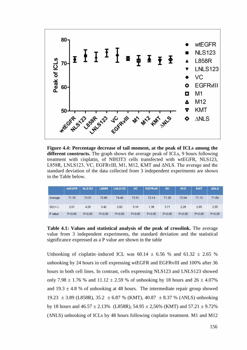
156
Figure 4.4: Percentage decrease of tail moment, at the peak of ICLs among the
different constructs. The graph shows the average peak of ICLs, 9 hours following
treatment with cisplatin, of NIH3T3 cells transfected with wtEGFR, NLS123,
L858R, LNLS123, VC, EGFRvIII, M1, M12, KMT and ΔNLS. The average and the
standard deviation of the data collected from 3 independent experiments are shown
in the Table below.
Table 4.1: Values and statistical analysis of the peak of crosslink. The average
value from 3 independent experiments, the standard deviation and the statistical
significance expressed as a P value are shown in the table
Unhooking of cisplatin–induced ICL was 60.14 ± 6.56 % and 61.32 ± 2.65 %
unhooking by 24 hours in cell expressing wtEGFR and EGFRvIII and 100% after 36
hours in both cell lines. In contrast, cells expressing NLS123 and LNLS123 showed
only 7.98 ± 1.76 % and 11.12 ± 2.59 % of unhooking by 18 hours and 26 ± 4.07%
and 19.3 ± 4.8 % of unhooking at 48 hours. The intermediate repair group showed
19.23 ± 3.89 (L858R), 35.2 ± 6.87 % (KMT), 40.87 ± 8.37 % ( NLS) unhooking
by 18 hours and 46.57 ± 2.13% (L858R), 54.95 ± 2,56% (KMT) and 57.21 ± 9.72%
( NLS) unhooking of ICLs by 48 hours following cisplatin treatment. M1 and M12

157
expressing cell lines showed 84.09 ± 4.87% and 97.41 ±2.73% unhooking of ICLs
respectively at 48 hours following cisplatin treatment. All the numerical data are
reported in Table 4.2, where the Statistical analysis by the 2 way ANOVA shows the
average values of every single time point, the standard deviation and its statistical
significance expressed as P value. Statistical significance (P value < 0.01/0.001) was
found at 48 hours time points and/or at earlier time points when the kinetics of cells
expressing wtEGFR were compared to the kinetics of cells expressing NLS123,
L858R, LNLS123, M1, M12, KMT, NLS or VC.
Figure 4.5: Effects of EGFR modulation in repair of ICLs. The modulation in
repair of single mutants is compared to the modulation in repair by wtEGFR.

158
Figure 4.5: cont.

159
Figure 4.5: cont.

160
Fig 4.5: cont.
Table 4.2: Statistical analysis of the cisplatin induced damage repair assay. Tables
show the average value of three independent experiments. P values obtained with the 2-way
Anova statistical analysis represent the comparison between all the mutants and to wtEGFR
transfected cells

161
Fig
ure
4.6
: E
ffec
ts of
EG
FR
m
od
ula
tion
in
re
pair
of
ICL
s.
Mea
sure
men
t of
dru
g-i
nduce
d D
NA
IC
Ls
in N
IH3T
3 ce
lls
tran
sfec
ted w
ith w
tEG
FR
, N
LS
123,
L858R
, L
NL
S123,
EG
FR
vII
I, V
ecto
r co
ntr
ol,
M1,
M12,
KM
T,
∆N
LS
tre
ated
wit
h 5
0μ
M
cisp
lati
n a
lone.
Err
or
bar
s re
pre
sent
stan
dar
d d
evia
tion.
The
gra
ph s
how
s th
e av
erag
e val
ue
of
thre
e in
dep
enden
t ex
per
imen
ts.
The
level
s of
ICL
is
norm
alis
ed t
o t
he
pea
k o
f IC
L a
t 9 h
ours
.

162
4.2.3 Effects of gefitinib on cisplatin induced ICLs formation and repair
The use of gefitinib in combination with cisplatin has shown to have a poor clinical
outcome in tumours presenting EGFR overexpression and/or secondary acquired
somatic mutations (Giaccone et al, 2004a; Normanno, 2005; Oliveras-Ferraros et al,
2008). The effect of the single agent gefitinib on the EGFR modulation of cisplatin
DNA damage repair was therefore investigated. NIH3T3 cells transfected with
wtEGFR, NLS123, L858R, LNLS123 or VC were pre-treated with 2μM gefitinib for
1hour prior to treatment with cisplatin as previously reported (Moasser et al, 2001).
Following treatment of 50 μM cisplatin for 1 hour in serum free media containing 2
μM gefitinib, cells were further incubated in media containing 2 μM gefitinib and
samples were collected over a period of 48 hours. Fig 4.7 shows the percentage of
peak of crosslinks results obtained from the analysis of the comet assay. There was
no difference in the peak formation of the ICLs in all the mutants. Despite treatment
with gefitinib showed inhibition of cisplatin unhooking in cells expressing wtEGFR
(66.18 ± 8.88 % unhooking by 48 hours), there was a significant difference when
compared to unhooking in NLS123 (35.88 ± 1.74 %), L858R (41.74 ± 9.38%) and
LNLS123 (25.48 ± 6.92%) expressing cells (Table 4.3). Statistical significance was
also found when the unhooking of cisplatin ICLs in wtEGFR expressing cells was
compared to VC expressing cells with wtEGFR expressing cells showing 22.57 %
less unhooking (Table 4.3). Next, the results of the cisplatin unhooking in cells
treated with gefitinib were compared to the unhooking of cells treated with cisplatin
alone (Fig 4.8). Interestingly, only cells expressing wtEGFR treated with gefitinib
showed significant differences at all the time points following peak of crosslinks
(Table 4.4). L858R expressing cells showed statistical significance at 18 hours
(19.43 % less unhooking in gefitinib treated cells) and at 24 hours (20.4% less
unhooking in gefitinib treated cells) following cisplatin treatment. There was no
statistical significant difference when treatment with gefitinib was compared to
cisplatin alone in NLS123 or LNLS123 expressing cells (Table 4.4). Only the repair
kinetics of wtEGFR transfected cells were impaired when cells were treated with
cisplatin in combination with gefitinib.
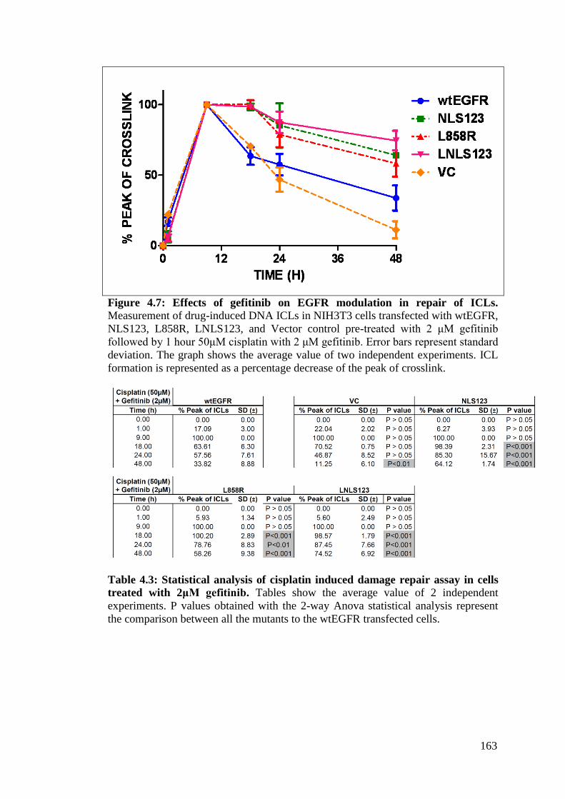
163
Figure 4.7: Effects of gefitinib on EGFR modulation in repair of ICLs.
Measurement of drug-induced DNA ICLs in NIH3T3 cells transfected with wtEGFR,
NLS123, L858R, LNLS123, and Vector control pre-treated with 2 μM gefitinib
followed by 1 hour 50μM cisplatin with 2 μM gefitinib. Error bars represent standard
deviation. The graph shows the average value of two independent experiments. ICL
formation is represented as a percentage decrease of the peak of crosslink.
Table 4.3: Statistical analysis of cisplatin induced damage repair assay in cells
treated with 2μM gefitinib. Tables show the average value of 2 independent
experiments. P values obtained with the 2-way Anova statistical analysis represent
the comparison between all the mutants to the wtEGFR transfected cells.
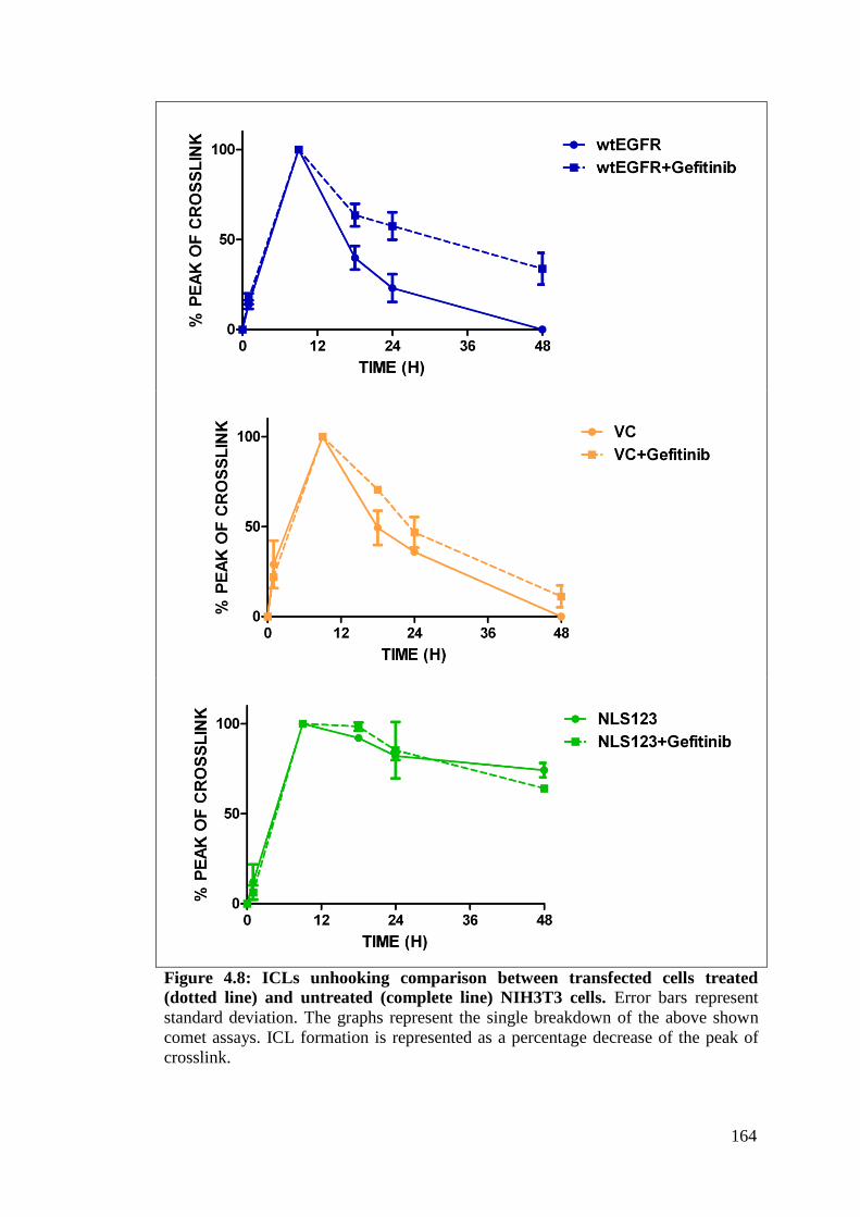
164
Figure 4.8: ICLs unhooking comparison between transfected cells treated
(dotted line) and untreated (complete line) NIH3T3 cells. Error bars represent
standard deviation. The graphs represent the single breakdown of the above shown
comet assays. ICL formation is represented as a percentage decrease of the peak of
crosslink.

165
Figure 4.8: cont.
4.2.4 EGFR modulation of IR induced DNA damage repair
4.2.4.1 IR dose response
The dose of IR required to induce detectable DNA damage and sufficient to
investigate its repair over time was determined by using the comet assay. NIH3T3
cells transfected with wtEGFR or VC were treated with IR at a dose up to 30 Gy.
Since IR produces an immediate physical lesion to the DNA, treated cells were
examined after the termination of the exposure. Fig 4.9 shows the results of the dose
response obtained from the comet assay. The amount of strand breaks produced is
shown as the mean tail moment value of three independent experiments. The data
suggest that 15 Gy is the dose necessary to produce sufficient DNA strand breaks to
asses repair over time and that EGFR expression does not influence the quantity of
detectable DNA damage produced. For this reason the kinetics of repair will be
shown as a percentage decrease of tail moment.

166
Table 4.4: Statistical analysis of cisplatin induced damage repair assay in cells
treated with2 μM gefitinib compared to cells untreated with gefitinib. Tables
show P values obtained with the 2 way Anova statistical analysis represent the
comparison between cell treated vs untreated with gefitinib but transfected with the
same construct.

167
4.2.4.2 DNA repair kinetics following IR treatment
Next, the effect of all the EGFR constructs (reported in Fig 4.1) on repair of stand
breaks induced by IR was investigated. NIH3T3 cells were transfected with
wtEGFR, NLS123, L858R, LNLS123, M1, M12, KMT, ΔNLS, EGFRvIII or VC.
Following 48 hours transfection, cells were treated with 15 Gy IR and repair of IR-
induced DNA-SB was examined. This is shown as percentage of tail moment
calculated from the tail moment detected immediately following irradiation, over a
period of 4 hours. Fig 4.10 shows the difference in repair kinetics between wtEGFR
and single construct transfected cells. Following treatment with IR alone, repair of
strand breaks was rapid in all the cell lines. Table 4.5 shows the statistical
comparison between single constructs and wtEGFR transfected cells. Significant
differences (P value <0.001) in repair kinetics between wtEGFR and NLS123,
L858R, LNLS123, M1, M12, KMT or NLS were found already at 30 minutes
following radiation (Table 4.4). The decrease of tail moment was 100% in both
wtEGFR and EGFRvIII-expressing cell lines at 4 hours following treatment,
indicating repair of SB. In contrast, at 4 hours, cells expressing NLS123 and
LNLS123 showed significant delay in repair of SBs with 22.48 ± 3.72 % and 24.94 ±
1.45 % of unrepaired SBs. Intermediate levels of repair were observed for cells
expressing L858R (12.33 ±1.00 %), KMT (18.86±3.45%), NLS (17.38±5.06 %),
M1 (8.51±1.12%) and M12 (9.28±2.26%). The results are graphically grouped
together in Fig 4.11 where the kinetics of all mutants are shown on the same graph.
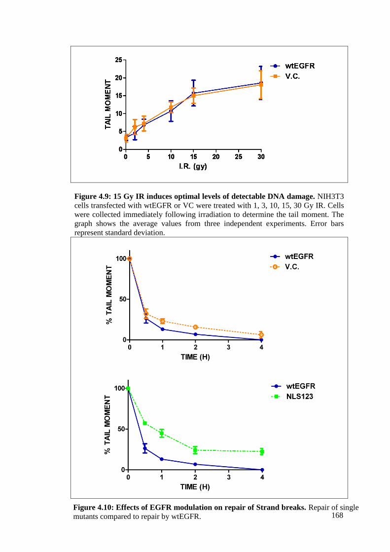
168
Figure 4.9: 15 Gy IR induces optimal levels of detectable DNA damage. NIH3T3
cells transfected with wtEGFR or VC were treated with 1, 3, 10, 15, 30 Gy IR. Cells
were collected immediately following irradiation to determine the tail moment. The
graph shows the average values from three independent experiments. Error bars
represent standard deviation.
Figure 4.10: Effects of EGFR modulation on repair of Strand breaks. Repair of single
mutants compared to repair by wtEGFR.

169
Figure 4.10: cont.
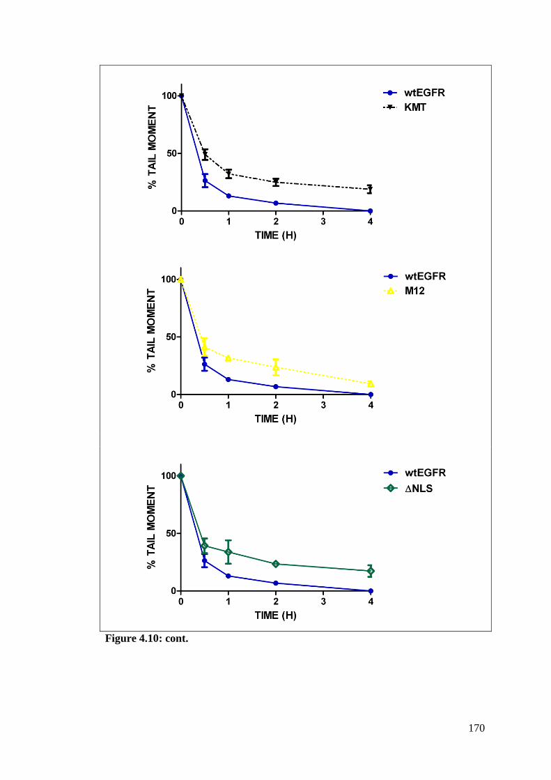
170
Figure 4.10: cont.
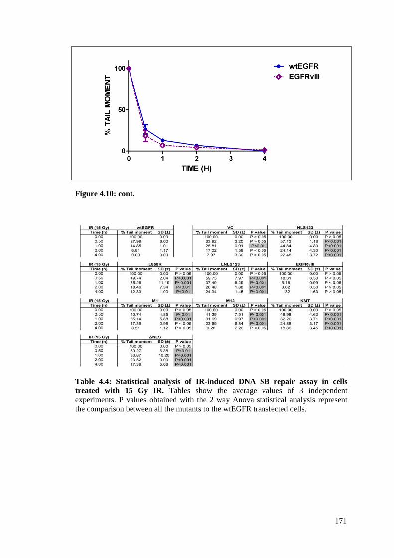
171
Figure 4.10: cont.
Table 4.4: Statistical analysis of IR-induced DNA SB repair assay in cells
treated with 15 Gy IR. Tables show the average values of 3 independent
experiments. P values obtained with the 2 way Anova statistical analysis represent
the comparison between all the mutants to the wtEGFR transfected cells.

172
Fig
ure
4.1
1:
Eff
ects
of
EG
FR
mod
ula
tion
in
rep
air
of
IR-i
nd
uce
d S
Bs.
Mea
sure
men
t of
IR D
NA
SB
s in
NIH
3T
3 c
ells
tra
nsf
ecte
d
wit
h w
tEG
FR
, N
LS
123,
L85
8R
, L
NL
S123,
EG
FR
vII
I, V
ecto
r co
ntr
ol,
M1,
M12,
KM
T,
∆N
LS
tre
ated
wit
h 4
Gy I
R.
Err
or
bar
s
repre
sent
stan
dar
d d
evia
tion.
The
gra
ph s
how
s th
e av
erag
e v
alue
of
thre
e in
dep
end
ent
exp
erim
ents
. S
B r
epai
r is
rep
rese
nte
d a
s a
per
centa
ge
dec
reas
e o
f ta
il m
om
ent.

173
4.2.5 Cisplatin induced EGFR-DNAPKcs binding
Previous studies have demonstrated the association of EGFR and DNAPKcs
following IR suggesting an intrinsic link between EGFR nuclear localisation and
DNAPKcs association and activation (Dittmann et al, 2005a; Dittmann et al, 2008a;
Golding et al, 2009; Rodemann et al, 2007). The timing of this association and its
significance have not been described following cisplatin treatment, although already
reported following cisplatin treatment (Hsu et al, 2009). EGFR-DNAPKcs
association and its involvement in the mechanism of cisplatin repair was
investigated. Since the modulation in repair kinetics in wtEGFR expressing cells was
already detectable at 18 hours following cisplatin treatment the association between
EGFR and DNAPKcs was investigated over a period of 24 hours following cisplatin
treatment. NIH3T3 cells were transfected with wtEGFR and 48 hour following
transfection they were treated with 50µM cisplatin. Cells were then collected at
various time points up to 24 hours following treatment. Protein extracts were
immunoprecipitated using an anti-DNAPKcs monoclonal antibody and blotted with
an anti-EGFR antibody. Fig 4.12 shows a time dependent association of EGFR and
DNAPKcs with a 2.7-fold increase at 18 hours following cisplatin treatment.
4.2.6 EGFR-DNAPKcs binding following cisplatin treatment does not
correlate with EGFR activation
Next the cisplatin-induced activation in all the mutants and their binding with
DNAPKcs was examined. NIH3T3 cells transfected with wtEGFR, M1, M12,
NLS123, L858R, LNLS123, KMT, ΔNLS, EGFRvIII, or VC were treated with
50µM cisplatin. Cells were then collected 18 hours following treatment and protein
lysates were immunoprecipitated with anti DNAPKcs and blotted for EGFR and
DNAPKcs. Fig 4.13 shows EGFR and DNAPKcs association in wtEGFR, M1, M12
or EGFRvIII transfected cells. In contrast, L858R and LNLS123 transfected cells
showed reduced EGFR-DNAPKcs binding whereas NLS123, KMT or ΔNLS
transfected cells showed no binding. The previous chapter showed that EGFR
nuclear localisation was not dependent on EGFR phosphorylation. Therefore the
correlation between EGFR phosphorylation and EGFR-DNAPKcs binding was
ivestigated. Cells transfected with wtEGFR, M1, M12, NLS123, L858R, LNLS123,
KMT or ΔNLS were treated with cisplatin for one hour and cell lysates were
immunoprecipitated with EGFR and blotted with PY20 and EGFR. In Fig 4.14,

174
wtEGFR, M1, M12, EGFRvIII showed phosphorylation activation following
cisplatin treatment, L858R and LNLS123 transfected cells showed constitutive
tyrosine phosphorylation. NLS123, KMT, ΔNLS transfected cells showed lack of
kinase activation. There was no correlation between EGFR activation and EGFR-
DNAPKcs binding as different constitutively active mutants showed pronounced
(EGFRvIII) or reduced (L858R, LNLS123) association with DNAPKcs.
Figure 4.12: DNAPKcs-EGFR association over time. wtEGFR Transfected
NIH3T3 cells were treated with 50μM cisplatin for 1 hour in serum free media. Cells
were then lysed at 1, 9, 12, 15, 18, 24, hours following treatment. 750μg of protein
lysate were immunoprecipitated using anti DNAPKcs and blotted with anti EGFR
and anti DNAPKcs. EGFR pull down was quantified by 2D densitometry analysis
and shown as a fold binding compared to the untreated control. Mouse unrelated
antibody (M IGg) was used as negative control.
Figure 4.13: DNAPKcs-EGFR association. NIH3T3 cells were transfected with all
the mutants reported in Fig 4.1. 48 hours following transfection cells were treated
with 50μM cisplatin for 1 hour in serum free media. Cells were then lysed 18 hours
following treatment. 750μg of protein lysate were immunoprecipitated using anti
DNAPKcs and blotted with anti EGFR and anti DNAPKcs.

175
Figure 4.14: EGFR mutants’ activation. NIH3T3 cells were transfected with all
the mutants reported in Fig 4.1. 48 hours following transfection cells were treated
with 50μM cisplatin for 1 hour in serum free media. Cells were then lysed
immediately following treatment. 750μg of protein lysate were immunoprecipitated
using anti EGFR and blotted with anti PY20 and anti EGFR.
4.2.7 EGFR and DNAPKcs cellular localisation following IR or cisplatin
Next, the cellular localisation of both EGFR and DNAPKcs following cisplatin (Fig
4.15 A-B) or IR (Fig 4.16 A-B) was investigated to determine whether the role of
EGFR nuclear translocation in the repair of SB or ICL DNA damage was related also
to and the EGFR-DNAPKcs binding. Cells transfected with wtEGFR, M1, M12,
NLS123, L858R, LNLS123, KMT, ΔNLS, EGFRvIII, or VC were either fixed 18
hours following treatment with 50 µM cisplatin or serum starved for 24 hours and
then fixed 20 minutes following 4 Gy IR. Fig 4.15 and 4.16 show the
immunofluorescence results obtained via confocal analysis. Cells transfected with
wtEGFR, M1, M12 and EGFRvIII showed EGFR nuclear expression following
cisplatin or IR. DNAPKcs showed similar pattern of expression in cells transfected
with these constructs. In wtEGFR and EGFRvIII transfected cells, EGFR was largely
expressed in the nucleus following cisplatin or IR. In contrast M1 and M12
transfected cells showed reduced levels of EGFR nuclear expression.
L858R, KMT and ΔNLS transfected NIH3T3 showed only membrane/cytosolic
EGFR localisation following cisplatin or IR. DNAPKcs nuclear expression in L858R
cells was reduced compared to wtEGFR and EGFRvIII transfected cells following
cisplatin or IR. In contrast, NLS123 and LNLS123 transfected cells showed lack of
both DNAPKcs and EGFR nuclear expression.

176
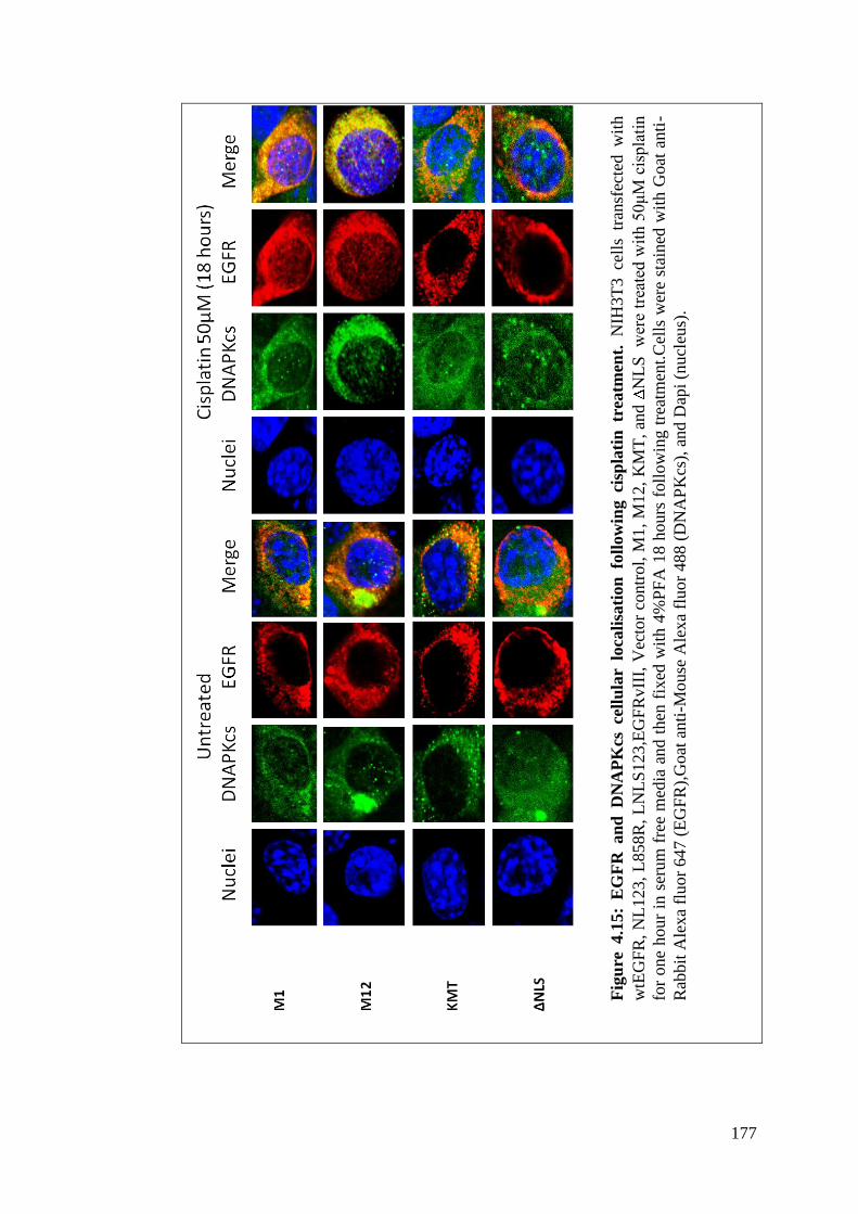
177
Fig
ure
4.1
5:
EG
FR
an
d D
NA
PK
cs ce
llu
lar
loca
lisa
tion
fo
llow
ing ci
spla
tin
tr
eatm
ent.
N
IH3T
3 ce
lls
tran
sfec
ted w
ith
wtE
GF
R,
NL
123,
L85
8R
, L
NL
S123,E
GF
RvII
I, V
ecto
r co
ntr
ol,
M1
, M
12,
KM
T,
and
NL
S
wer
e tr
eate
d w
ith 5
0μ
M c
ispla
tin
for
one
hour
in s
erum
fre
e m
edia
and t
hen
fix
ed w
ith 4
%P
FA
18 h
ours
foll
ow
ing t
reat
men
t.C
ells
wer
e st
ained
wit
h G
oat
anti
-
Rab
bit
Ale
xa
fluor
647 (
EG
FR
),G
oat
anti
-Mouse
Ale
xa
fluor
488 (
DN
AP
Kcs
), a
nd D
api
(nucl
eus)
.

178

179
Fig
ure
4.1
6:
EG
FR
an
d D
NA
PK
cs c
ellu
lar
loca
lisa
tion
foll
ow
ing I
R t
reatm
ent.
NIH
3T
3 c
ells
tra
nsf
ecte
d w
ith w
tEG
FR
,
NL
123,
L858R
, L
NL
S123,E
GF
RvII
I, V
ecto
r co
ntr
ol,
M1,
M12,
KM
T,
and
NL
S
wer
e se
rum
sta
rved
for
24 h
ours
and t
hen
trea
ted w
ith 4
Gy a
nd
then
fix
ed w
ith 4
%P
FA
20 m
in f
oll
ow
ing t
reat
men
t.C
ells
wer
e st
ained
wit
h G
oat
anti
-Rab
bit
Ale
xa
fluor
647 (
EG
FR
),G
oat
anti
-Mouse
Ale
xa
fluo
r 488 (
DN
AP
Kcs
), a
nd D
api
(nucl
eus)
.

180
4.3 DISCUSSION
4.3.1 The NLS sequence is required for nuclear translocation and
receptor activation
The data presented in Chapter 3 have confirmed the previously reported EGF and IR
induced EGFR nuclear translocation and investigated the NLS123 mutation of the
EGFR NLS sequence. The basic Arginine amino acids contained in the NLS/p13+
peptide sequence of the EGFR JX domain are necessary to achieve a conformational
change that seems to be a prerequisite to achieve receptor activation. Upon any
activating stimulus, the mutant receptors fail to achieve a conformational change that
not only prevents activation but also stops the protein from binding to the importin
machinery and achieve nuclear translocation. The NLS123 mutation has shown two
effects in a wtEGFR background. Firstly it inhibits the protein translocation to the
nucleus and secondly it interferes with EGFR phosphorylation, negatively affecting
its downstream signalling pathways. This chapter provides evidence for an EGFR
role in the modulation of DNA repair following cisplatin or IR induced DNA
damage. The data show DNA repair kinetics modulated by the expression of EGFR
and also by its capability to translocate to the nucleus. Receptor activation and kinase
activity do not seem to be central in this modulation. It appears that binding and the
similar localisation pattern with DNAPKcs within the nuclear compartment may be
responsible for the enhanced repair kinetics.
4.3.1.2 EGFR modulates the repair of cisplatin-induced ICLs
4.3.1.2.1 DNA repair of cisplatin lesions
The effects of cisplatin and the repair pathways that may be involved in the repair of
cisplatin damage are still debated and the literature emphasises that there is more
than one pathway at work (Hlavin et al, 2010a; Martin et al, 2008; Wang & Lippard,
2005). It is, in fact, established that cisplatin not only binds to DNA but also to many
cellular components that have soft nucleophilic sites such as cytoskeletal
microfilaments, RNA, proteins. Only 5-10% of covalently bound associated cisplatin
is found in the genomic DNA whereas 75-85% of the drug binds to proteins
suggesting that the toxicity of this drug is also mediated by activation of specific
signalling pathways that ultimately leads to cell death (Zdraveski et al, 2000).
Although there is evidence suggesting a strong effect on DNA replication it is not
completely clear whether replication may be one of the consequences rather than the

181
reason of the lethality of the treatment (Zorbas and Keppler 2005).This is because in
cycling cells the persistence of ICLs may also induce single and double DNA strand
breaks (Smeaton et al, 2008; Smeaton et al, 2009). Therefore as a result, the cell
machinery blocks replication and transcription allowing sufficient time for DNA
repair mechanisms to resolve the crosslinks (Nojima et al, 2005). The pathway
responsible for this cellular arrest is not completely characterised but EGFR seems to
be involved in the mechanism (Ceppi et al, 2006; Chen et al, 2000; Hiraishi et al,
2008; Hsu et al, 2009; Kim et al, 2009b; Klass et al, 2009; Michaelis et al, 2008;
Oliveras-Ferraros et al, 2008; Yoshida et al, 2008).
Because of the variety of DNA damage that arises from cross-linking agents, there
are many pathways that are activated to repair these lesions. Their concerted activity
is a key element to achieve complete DNA repair. Cross-links are well known to be
repaired by Nucleotide excision repair (NER) and post replication repair (PRR)
(Clingen et al, 2007; Clingen et al, 2005; De Silva et al, 2002; Hartley et al, 2004;
McHugh et al, 2001). Cisplatin adducts are detected by the NER components
Xpx/Hr23B and XPA. These then recruit the endonucleases Ercc1-Xpf and Xpg
which are the catalytic NER proteins responsible for the dual incision process. The
strand affected by the crosslink is incised 5‟ to the lesion by ERCC1-Xpf and 3‟to the
lesion by Xpg. Once the lesion is removed the gap is filled by repair synthesis.
In presence of a stalled replication machinery at the site of the lesion, both
Translesion DNA synthesis (TLS) and HR are required to help restart replication.
TLS fills the gap via RAD6/ RAD18 and specialised polymerases such as Polε, Polκ
and Polδ complete repair. HR fills the gap left by the excised lesion by copying the
other intact sister DNA as a template. HR could also be required to repair the
induced DSB created as an indirect consequence of crosslinked DNA during
replication. NHEJ can also be employed to relegate the strands broken as a result of
the stalled replication machinery (Nojima et al. 2005c; Kartalou and Essigmann
2001; Cepeda et al. 2007). Some studies have shown that upon defective HR and
TLS, mammalian cells seemed to be hypersensitive to crosslinking agents suggesting
a minor role of NER (Nojima et al. 2005b), on the other hand there are some other
studies that have clearly shown that upon inefficiency of NER components, cells are
hypersensitive to cisplatin adducts (Nojima et al. 2005a; De, I et al. 2002; De, I et al.
2000).
182
4.3.1.2.2 Unhooking is the major determinant in
interstrand crosslink repair
The ability of cisplatin to form covalent linkages between two strands of the double
helix is without any doubt the basis for its high cellular cytotoxicity (Malinge et al,
1999). If left unrepaired, interstrand crosslinks prevent, virtually, all aspect of DNA
metabolism, such as replication, transcription and recombination (Hlavin et al,
2010b). The capability of linking both the DNA strands renders bifunctional
crosslinking agents the most genotoxic and mutagenic of compounds. This is,
perhaps, the reason why their removal is the most complicated of DNA repair
processes (Wang & Lippard, 2005). ICLs repair takes place in two ways. During the
first, also called recombination-independent, the lesion is excised (unhooked) on one
strand of the helix and the remaining gap is filled by TLS. This process converts the
crosslink into a mono-adduct which is, subsequently, resolved by a second cycle of
excision repair. In bacteria this process is carried out by NER and Pol II, however it
remains unclear in mammalian cells. Although NER has been shown to be a major
component, this repair mechanism seems to be clearly minor in cycling mammalian
cells; however, in differentiated non-replicating cells it may represent the only way
for the removal of ICLs. The second repair mode begins with the excision of the
crosslinks and is followed by homologous recombination. This second pathway takes
place during the S phase and it is the predominant pathway in cycling mammalian
cells. Despite the different modality of DNA synthesis of these two systems, the
central step in the repair of crosslinks is still the initial processing /unhooking
(Hlavin et al, 2010a; Hlavin et al, 2010b; McCabe et al, 2009; Muniandy et al, 2010;
Shrivastav et al, 2009; Smeaton et al, 2009; Tornaletti, 2009; Wang & Lippard,
2005). The context in which the ICL is recognised induces the recruitment of
different repair proteins rendering this step incredibly complex.
4.3.1.2.3 Unhooking requires different protein complexes
often dependent on the type of bifunctional alkylator
Although many proteins have been implicated the precise mechanism remains to be
elucidated. Cycling cells treated with crosslinking reagents accumulate replication
fork induced-breaks consisting in a DSB on one end, and a double stranded DNA
cross-linked with a single strand in three stranded structure on the other. Different

183
from E. coli and S. cervisiae, ICL processing in mammalian cells only requires some
of the protein involved in NER. XPF-ERCC1 are structure specific endonucleases
capable of making 5‟ incision at the junction between double stranded DNA and a 3‟
single stranded region (Clingen et al, 2005). Cells deficient in XPF-ERCC1 show
only 15% unhooking of nitrogen mustards (NH2) induced ICLs (assessed by the
Comet assay). Other NER components do not seem to be required in the unhooking
of NH2 ICLs. In contrast, little or no unhooking of cisplatin induced ICLs has been
shown not only in cells deficient in XPF-ERCC1 but also in XPD, XPB or XPG
deficient cells implicating all these NER proteins in the unhooking step of cisplatin
ICLs (De Silva et al, 2000; McCabe et al, 2009). Interestingly, although all impaired
in the unhooking, only cells deficient in XPF-ERCC1 have higher cisplatin
sensitivity (37-40 fold more sensitive than their isogenic) compared to the other NER
mutant cells (1.3-3.1 fold more sensitive than their parental line) (Hlavin et al,
2010a). In addition the observation that XRCC2 and XRCC3 deficient cells are also
highly sensitive to cisplatin treatment, suggests that, as these two proteins, also XPF-
ERCC1 may be (additionally) involved in recombinational repair (or DNA re-
sysnthesis to fill the gap left by the DNA incision) (De Silva et al, 2002; Smeaton et
al, 2009). It is therefore unclear whether the inhibition of unhooking observed in
cells deficient in XPF-ERCC1 via the comet assay is actually the product of failed
recombination intermediates that result in DNA migrating with the similar retarded
mobility as cross-linked DNA (Fisher et al, 2008). Smeaton et al., have also shown
NER independent 5‟ incision, unaffected by the absence of ERCC1-XPF suggesting
alternative contributions of these proteins to the unhooking step. This was further
confirmed by the finding that the ERCC1-XPF does not function alone and it has
been found associated with the SLX4 protein that acts as a scaffolding for this
complex as well as for two other nucleases: MUS81-EME1 and SLX1.
Cell free extracts and purified protein experiments have also shown contrasting
results (Hlavin et al, 2010a; Smeaton et al, 2008; Smeaton et al, 2009). Bessho et al.
have shown that the NER factors XPA, RPA, TFIIH XPC, XPG, ERCC1-XPF, were
necessary and sufficient for the incision step and also that cells employ the incision
step to relieve the DNA duplex from the distortion at either sides of the ICLs rather
than removing the ICLs following its recognition (Bessho et al, 1997). Another study
showed the requirement of RPA for both incision (unhooking) and re-synthesis and
the essential role of PCNA for DNA synthesis and incision. The same group has also

184
shown the involvement of the complex containing the Pso4 pre mRNA splicing
factor and the Werner Syndorme (WS) helicase activity (stimulated by BRAC1) in
the initial ICL processing (Cheng et al, 2006). Recently there has been some
evidence of the involvement of Fanconi Anemia (FA) complex in the unhooking of
cisplatin ICLs. Although still not fully understood, it seems that FA complex
recognises stalled replication forks created by ICLs, recruits protein complexes to
this site and induces remodelling of the replication fork allowing repair to proceed.
The role of FA proteins in unhooking is controversial. Although cells deficient in
FANCA, B, C, D2, F and G or treated with antibodies against FANCA showed
reduced incision, the suppression is generally not complete with the 3‟ incision being
more resistant. Many other reports have shown partial or no requirement of these
proteins (McCabe et al, 2009; McHugh et al, 2001; Muniandy et al, 2010; Nojima et
al, 2005).
4.3.1.2.4 EGFR involvement in Cisplatin repair
The role of EGFR in the response to cisplatin treatment was firstly reported by
Benhar M et al. Genotoxic stress induced by cisplatin activates DNA damage signals
that culminate in EGFR autophosphorylation in a ligand independent mechanism.
While initially it was proposed that EGFR activation induces downstream signalling
responsible for cellular death, different groups have shown the contribution of EGFR
in the repair of cisplatin lesions (Dittmann et al, 2005a; Friedmann et al, 2004;
Friedmann et al, 2006; Oliveras-Ferraros et al, 2008). Recently, a study by Hsu et
al., has suggested that nuclear EGFR is involved in the repair of cisplatin DNA
damage (Hsu et al, 2009). The study looked at the phosphorylation of H2AX as a
marker of DNA damage and of the repair of DSBs. Although their results pointed
towards an involvement of nuclear EGFR in cisplatin toxicity, the phosphorylation of
H2AX was determined only 12 hours following treatment (cisplatin crosslinks peak
at nine hours following treatment) and repair of DSBs is not a good marker to asses
the repair kinetics of cisplatin induced damage as DSBs are not directly caused by
cisplatin.

185
4.3.1.3 EGFR constructs that translocate to the nucleus repair
cisplatin lesions
This study confirms cisplatin-induced EGFR phosphorylation, shows enhanced
repair kinetics in cells expressing EGFR constructs undergoing nuclear translocation
and suggests that this mechanism is mediated by EGFR binding with DNAPKcs.
Three distinctive repair behaviours: complete repair, intermediate inhibition and
highest inhibition of repair have been observed. Cells transfected with wtEGFR or
EGFRvIII show complete repair both by 36 hours following cisplatin. Our data have
demonstrated that both wtEGFR and EGFRvIII not only undergo nuclear
translocation but that they also bind DNAPKcs following cisplatin. In addition the
confocal analysis of wtEGFR and EGFRvIII expressing cells shows similar EGFR
and DNAPKcs localisation pattern in the nucleus following cisplatin treatment.
Cells that showed intermediate repair inhibition (L858R, KMT, NLS) and highest
inhibition of repair (NLS123, LNLS123) correlated to impaired EGFR nuclear
translocation.
4.3.2 DNAPKcs involvement in the repair of cisplatin lesions
The matter of whether DNAPKcs promotes, amplifies, transmits stress signals, has
been largely debated. The initial studies that looked at the involvement of DNAPKcs
in the repair of cisplatin damage showed that cells deficient in DNAPKcs were
hypersensitive to cisplatin treatment and that in the parental cell lines although
showing quicker repair kinetics, DNAPKcs was not found at the site of the DNA
lesion (Boeckman et al, 2005; Turchi et al, 1999; Turchi et al, 2000; Turchi et al,
1996). Subsequently it was reported that cisplatin adducts reduce the rate at which
Ku, the subunit of the DNA-PK heterotrimeric complex, slides along the DNA
duplex blocking its translocation from the DNA terminus (Pawelczak et al, 2005;
Turchi et al, 2000). This resulted in a reduced association of Ku to DNAPKcs and
also in the formation of inactive kinase complexes that are unable to resolve DNA
breaks suggesting that cisplatin could be used to pre-sensitise cells to radiotherapy
treatments (Turchi et al, 2000). However, in 2004 Glazer et al. showed that cells
deficient in Ku80 and DNAPKcs were actually resistant to cisplatin treatment
compared to the wild type but that such difference is only marked when cells were
treated at high confluence (Jensen & Glazer, 2004). Moreover the authors showed
that following cisplatin treatment DNA-PK trimeric complexes mediate death signals

186
within damaged cells via cell to cell contact through gap junction (Jensen & Glazer,
2004; Zorbas & Keppler, 2005). Subsequently, Shao et al. 2008 showed that high
levels of DNAPKcs/Ku80 kinase activity in gliomas correlates with cisplatin
resistance suggesting that activation of this trimeric complex could be utilised to
screen for patients that would benefit from cisplatin treatment (Shao et al, 2008).
4.3.3 EGFR modulation of DNA repair following IR
The involvement of EGFR in the repair of IR-induced damage has been extensively
reviewed in the introduction section. The characterised nuclear interaction of EGFR
with DNAPKcs following IR is responsible for the DNA repair and resistance to IR
treatment. Similarly, this study shows that cells expressing wtEGFR and EGFRvIII
have completely repaired IR-induced SBs by 4 hours following treatment and this
also correlates with EGFR nuclear translocation and similar DNAPKcs nuclear
localisation pattern. EGFRvIII expressing cell line shows the highest rate of DNA SB
repair (significantly higher to wtEGFR) suggesting that cancer cell expressing this
mutant may present a more efficient DNA repair mechanism and therefore a higher
resistance potential to IR.
4.3.3.1 EGFR modulation of SSBs and DSBs is shown at different
time points
The differences in percentage tail moment among the different transfectants are
smaller compared to the distinct patterns observed following cisplatin treatment. The
type of damage conferred and the different repair pathways involved explain the
significance of these differences. DNA SBs are formed immediately following IR
treatment and therefore repair will act promptly upon their recognition. While SSBs
are quickly repaired by the DNA repair machinery, the more complex double strand
breaks will require a longer time. Therefore the differences observed at 4 hours
following IR treatment represent the impairment of DSBs repair whereas the very
early time points show the different kinetics in the repair of single strand breaks. The
alkaline comet assay does not distinguish between the single and double strand
breaks therefore it is unknown whether the percentage decrease in tail moment is due
uniquely to inhibition of DSBs or to the accumulation of unrepaired SSBs and DSBs.
This suggests that another DNA repair component, rather than DNAPKcs, involved
in the EGFR modulation of IR induced repair cannot be excluded. Considering the

187
high biological significance and the great lethality of even one DSB the differences
in percentage decrease of tail moment following IR among the mutants, although
rather small numerically, have been still classified using the three categories
(complete repair, intermediate inhibition and highest inhibition of repair) as for the
cisplatin comet assay result.
4.3.4 The window of molecular intervention to determine DNA repair
The cisplatin comet assay also shows that the % unhooking obtained at the 18-hour
time point could be very important to predict the overall repair response. Less than
50% of the unhooking at 18 hours will indicate that it is unlikely the ICLs will be
successfully resolved within the 48 hours. This suggests that the binding between
DNAPKcs and EGFR is only involved in the resolution of 50% of the unhooking
however the immunoprecipitation analysis shows that the EGFR-DNAPKcs binding
is progressive overtime therefore the earlier unhooking may still be due to this
complex. Similarly, the reduction of tail moment at 30 minutes following IR
treatment predicts clearly the rate of repair. More than 60% reduction of tail moment
will result in complete repair within the following 4 hours. These data suggest that
the initial 18 hours following cisplatin treatment and 30 minutes following IR
treatment could be utilised to predict the overall repair kinetics following therapy
4.3.5 EGFR nuclear localisation expression does not quantitatively
determine modulation of DNA repair
The IF analysis shows that despite the mutation in the NLS sequence, the nuclear
translocation of both M1 and M12 show is only decreased. This, together with their
ability to repair (shown in the comet assays), suggests that the amount of EGFR
nuclear expression is not in linear relation with the repair kinetics. Even a small
percentage of EGFR nuclear translocation results in sufficient repair. The difference
in nuclear translocation as compared with wtEGFR and EGFRvIII, interestingly,
suggests that the induction of EGFR nuclear expression following IR and cisplatin
may not only be required for DNA purposes but also to exert some other biological
functions. In addition, the comparison between the levels of M1/DNAPKcs,
M12/DNAPKcs binding in relation to the levels of M1 and M12 nuclear expression
and the levels of wtEGFR/DNAPKcs binding in relation to its nuclear expression,
suggests a role for EGFR independent of DNAPKcs.
188
4.3.6 Kinase activity does not determine nuclear expression and it is not
central to repair
KMT, L858R and ΔNLS show very similar intermediate levels of repair however
their structural characteristics are quite different. The kinase dead mutant KMT bears
the mutation K721A which is known to modify the kinase domain conformation
hindering both dimerisation and receptor activation. The ΔNLS mutant lacks entirely
the NLS sequence and in accordance to the allosteric model, (an EGFR receptor
molecule lacking the p13+ peptide is unable of dimerise and activate) this receptor is
kinase dead. Both these two mutants show impaired EGFR nuclear localisation. On
the other hand, L858R bears the somatically acquired mutation of the Leucine 858
into an Arginine which renders the mutant 50 fold more active; 10-100 fold more
sensitive to gefitinib and binds to gefitinib 20 times more tightly. In the inactive
conformation Leucine 858 is exposed in the helical turn of the activation segment.
Here it makes non-polar contact with the nearby Phenialanine 723 and the glycine-
rich loop. Substitution with the positively charged amino acid Arginine is not
compatible with the inactive conformation but is with the active conformation. This
produces a lower affinity for ATP but higher affinity for TKI (Dixit & Verkhivker,
2009; Fassina et al, 2009). Moreover it has been shown that due to this mutation the
residue Tyrosine 845 does not require SRC phosphorylation but undergoes
constitutive autophosphorylation (Fu et al, 2008; Yang et al, 2008). Expression of the
L858R mutant also resulted in impaired nuclear expression despite constitutive
kinase activity (Das et al, 2007). This resulted in reduction of DNA strand break
repair which is consistent with the observation that non-small cell lung cancer lines
expressing L858R show increased sensitivity to IR and reduced nuclear expression.
Moreover, the difference in repair between cells expressing L858R (kinase active but
with impaired nuclear localization) and EGFRvIII (kinase active and expressed in the
nucleus) suggests that, as a consequence of the lack of allosteric activation, the
impaired nuclear EGFR accumulation and not kinase activity per se determines the
reduced DNA repair in these models.
The confocal imaging shows that L858R, KMT or NLS transfected cells still induce
nuclear DNAPKcs following cisplatin and IR suggesting the presence of a backup
signalling pathway that triggers some DNAPKcs nuclear accumulation however
insufficient to determine complete repair. It is unknown whether the KMT mutant

189
undergoes a conformational change and therefore whether its lack of nuclear
localisation is due to a sterically hindered NLS sequence or to an impaired kinase
activation. Previous data have shown that inhibition of EGFR kinase activity is
sufficient to inhibit the receptor nuclear translocation suggesting that despite an
intact NLS sequence the kinase activity is also a requirement for nuclear
translocation. Our data have shown that nuclear accumulation is the absolute
requirement for efficient repair as kinase activity without nuclear accumulation can
only account for intermediate levels of repair.
4.3.7 Maximal gefitinib inhibition of repair is shown only when EGFR
translocates to the nucleus
The results obtained by the gefitinib treated cells following cisplatin suggest that
nuclear localisation is central for the modulation of cisplatin repair modulation.
Gefitinib showed maximal inhibition only in wtEGFR transfected cells whereas it
had no significant effect on cells expressing NLS123 or LNLS123. Although
wtEGFR cell treated with gefitinib showed significant inhibition compared to VC
transfected cells (which do not express EGFR), the inhibition of repair was still
significantly lower compared to cells with impaired EGFR nuclear translocation.
Importantly, the intermediate levels of repair shown by wtEGFR expressing cells,
treated with gefitinib, suggests that whilst not sufficient to determine a complete
inhibition of repair, inhibition of EGFR kinase activity has a role in repair, however
different from nuclear translocation.
The increased sensitivity shown in L858R transfected cells reflects the characteristic
of this mutant to have a higher affinity to 4-anilinoquinazoline compounds such as
gefitinib. Significant inhibition of repair was found only between 18 and 24 hours
following cisplatin treatment. There was no significant difference in gefitinib treated
cells at 48 hours. The effect of the inhibitor on the receptor activation was not
determined therefore it cannot be concluded that the higher inhibition is directly
determined by the peak of gefitinib-induced impairment of kinase activation.
Interestingly, the time frame coincides with EGFR-DNAPKcs nuclear association
shown in cells where EGFR translocates to the nucleus. It has been reported that
gefitinib treatment induces cytosolic accumulation of DNAPKcs (Friedmann et al,
2004). The possibility that repair inhibition observed in L858R cells is due to other

190
factors rather than to an impaired kinase activity cannot be excluded. Future
experiments will answer these important questions.
4.3.8 Gefitinib binds to EGFR active conformation
The necessity of a conformational change to allow recognition of the NLS sequence
for nuclear translocation has been previously discussed. The impaired EGFR nuclear
localisation has been explained as a result of an inactive conformation (NLS123) due
to the lack of a dimer-dimer interface that initiates the allosteric mechanism and to a
kinase mutation that achieves active conformation without requiring the
conformational change (L858R). It has been shown that gefitinib binds only EGFR
molecules in an active conformation. In line with this evidence, L858R and wtEGFR
show gefitinib-induced inhibition of DNA repair. The lack of gefitinib-induced
inhibition, in NLS123 cells, adds more evidence towards the argument that the
NLS123 mutation impedes the allosteric conformational change necessary to achieve
an active conformation. The fact that gefitinib treatment has shown no effect in
LNS123 cells suggests that the mutation of all the arginines within the NLS sequence
may impair the active conformation that the L858R receptor obtains via the arginine
substitution at position 858. This underlines the importance of this stretch of amino
acid and the necessity for further experiments to determine the correlation between
the induction of impaired active conformation and DNA repair.
4.3.9 The NLS sequence is a target for molecular intervention
The NLS mutation (AAAHIVAKATLAA) contributed to an even greater delay of
repair. The NLS123 mutant showed not only inhibition of nuclear translocation,
abolished kinase activation and impaired DNAPKcs binding but also lack of
DNAPKcs nuclear accumulation. The LNLS123 showed impaired EGFR nuclear
accumulation and DNAPKcs binding, mirrored by the inhibition of DNAPKcs
nuclear translocation maintaining, however, the receptor constitutive activation. The
mutation of the three Arginine cores of the NLS sequence may have caused a
structural change to the receptor obstructing the binding of other proteins and their
trans-phosphorylation which results in the lack of DNAPKcs nuclear accumulation.
A recent study has shown that targeting EGFR NLS sequence by a peptide
corresponding to phosphorylated NLS resulted in abolished nuclear EGFR, reduced
radiation-induced activation of DNAPK and reduced survival following IR
(Dittmann et al, 2010a).
191
4.3.10 DNAPKcs subcellular distribution
These data clearly show that DNAPKcs is not only localised into the nucleus
supporting other published data on the cytosolic functions of DNAPKcs. Moreover,
in our EGFR overexpressing cellular system, DNAPKcs mirrors the EGFR cellular
localisation suggesting that the behaviour of these two proteins is highly linked. In
fact the inhibition of the nuclear translocation of the receptor is sufficient to mediate
decreased DNAPKcs nuclear accumulation following cisplatin and IR. Previous data
have shown that EGFR inhibition of gefitinib induce DNAPKcs accumulation in the
cytosol (Friedmann et al, 2006). Although the gefitinib dose used in that study was
greater the effects on EGFR kinase activity are comparable to those obtained with the
NLS123 mutant. The statement that EGFR cellular localisation influences DNAPKcs
subcellular distribution can be justified if one accepts the idea of EGFR being a
kinase for DNAPKcs.
4.3.11 Conclusions
This chapter shows that nuclear EGFR plays an important role in modulating the
repair of DNA damage caused by cisplatin and IR. Transfecting cells with wtEGFR
and EGFRvIII showed an increased efficiency of the repair of DNA lesions, whereas
the transfection of EGFR-NLS mutants demonstrated an impairment of DNA repair.
Following cisplatin treatment there was a time dependent association of EGFR and
DNAPKcs as assessed by immunoprecipitation assays. Confocal microscopy in cells
transfected with wtEGFR or EGFRvIII showed similar localisation pattern of EGFR
and DNAPKcs in the nucleus whereas the two proteins were exclusively detectable
in the cytoplasm in cells transfected with EGFR NLS123 mutants. These results are
in keeping with the observation that inhibitors of EGFR can inhibit DNA repair and
demonstrates the importance of EGFR nuclear expression in this process.
It now needs to be demonstrated that both cisplatin and IR stimulate the formation of
EGFR and DNAPKcs complex and therefore as a consequence of their physical
interaction DNAPKcs undergoes a kinase modulation which is mediated only by
nuclear EGFR.

192
CHAPTER 5:
THE MECHANISM OF EGFR
MODULATION OF DNA REPAIR
193
5.1 INTRODUCTION
5.1.1 The role of EGFR nuclear translocation in the binding to DNAPKcs
EGFR modulation of DNA repair has been described in many reports with regards to
the binding with DNAPKcs following IR. Although this interaction has been greatly
discussed and characterised in the literature, there is no clear understanding as to
whether EGFR nuclear expression is sufficient and necessary to determine this
binding. The previous chapter has shown how EGFR can modulate ICL and SB
repair. EGFR kinase activity and nuclear localization, despite appearing to rely both
on the receptor active conformation, have distinct, non-mutually exclusive, roles in
the repair of DNA damage.
Inhibition of EGFR (or other ERBB family receptors) kinase activity can either be
achieved by TKI or by antibodies that impair the ligand binding to the receptor.
When wtEGFR transfected cells were treated with gefitinib in combination with
cisplatin the resulting effect on the DNA repair kinetics were similar to the behaviour
of the KMT mutant. Cell expressing this kinase dead mutant showed impaired EGFR
nuclear translocation and intermediate levels of ICL or SB repair. Our data have also
shown that induced inhibition of EGFR nuclear translocation via the NLS123
mutation resulted in a consequent impaired kinase activity. However cells transfected
with the NLS123 showed complete inhibition of repair suggesting that different
targeting clearly has a more dramatic effect.
Confocal microscopy analysis suggested that mutation in the EGFR NLS sequence
inhibits DNAPKcs nuclear localization without establishing a physical interaction.
The question as to whether EGFR-DNAPKcs association takes place exclusively in
the nucleus and whether EGFR kinase activity or nuclear translocation per se can
induce this association remain unclear. The suggestion that EGFR acts as a kinase
for DNAPKcs would explain DNAPKcs cytosolic localisation in presence of an
EGFR NLS mutant. EGFR-DNAPKcs binding inhibition could interfere with
DNAPKcs activation resulting in a DNAPKcs accumulation in the cytosol.
5.1.2 EGFR nuclear localisation and DNAPKcs kinase activity

194
Previous reports have shown a correlation between direct inhibition of EGFR kinase
activity and consequent inhibition of DNA-PK kinase activity suggesting a relation
between EGFR kinase activity and DNAPKcs. EGFR kinase inhibition by TKI
(gefitinib) or mAB (cetuximab) have shown inhibition of DNA-PK kinase activity,
DNAPKcs cytosolic accumulation and delayed repair (Dittmann et al, 2008a;
Dittmann et al, 2005b; Friedmann et al, 2006; Golding et al, 2009). Recent reports
have also shown a function for cytosolic DNAPKcs in apoptosis, activation of AKT
and in cell-cell communication (Achanta et al, 2001; Gurley et al, 2009; Lu et al,
2006; Shi et al, 2009; Toulany et al, 2008b). Here it has been shown that inhibition of
EGFR nuclear translocation via the NLS123 mutation also indirectly inhibits
DNAPKcs nuclear accumulation following cisplatin and IR induced damage. This
raised many questions on the induction of EGFR-DNAPKcs association and its
consequences including whether this binding takes place exclusively in the nucleus,
whether it is EGFR nuclear translocation, the treatments or EGFR allosteric
conformational change that induce EGFR-DNAPKcs association and whether the
complex can modulate DNAPK kinase activity. These are all central to decipher the
mechanism that links EGFR with DNAPKcs and DNA repair, to provide substantial
evidence to establish that nuclear EGFR is required for repair of cisplatin and IR
induced DNA damage and that repair is mediated via binding to DNAPKcs.
5.1.3 Stable expression of EGFR constructs
The transient over-expression of the wtEGFR or other EGFR mutants has been
utilised to determine the effect of EGFR on repair. In this chapter wtEGFR, NLS123,
L858R, LNLS123, EGFRvIII were stably expressed in NIH3T3 cells and the
mechanism of EGFR modulation of DNA repair was investigated. This allowed
homogeneous EGFR- expressing populations of cells to be examined. The constructs
within the stable transfectants represent, more significantly, the different DNA
damage repair behaviours discussed in chapter 3 while presenting different EGFR
characteristics in terms of nuclear translocation, kinase activation, and DNAPKcs
binding.
5.1.4 Cisplatin cytotoxicity and survival
As detailed in the introduction chapter, cisplatin has been used in the clinic for over
30 years in the treatment of testicular, head and neck, cervix, ovary and lung cancer.

195
However, clinical responses to cisplatin are variable and there is substantial evidence
suggesting that EGFR over-expression, as well as its nuclear expression, are major
determinants of cisplatin-acquired resistance (Benhar et al, 2002; Eckstein et al,
2008; Wernyj & Morin, 2004; Winograd-Katz & Levitzki, 2006; Wynne et al, 2007).
The general consensus is that the variety of platinum adducts underlie most of the
cytotoxic effects of this drug (Malinge et al, 1999; Wang & Lippard, 2005). Besides
EGFR, cisplatin activates a variety of cellular protein that exerts many crucial
cellular functions such as p53, p73, c-abl, hence predicting signalling responses to
cisplatin has been a challenge for many years (Boeckman et al, 2005; Laachi et al,
2009; Nojima et al, 2005; Patrick et al, 2008; Turchi, 2006; Wang et al, 2005;
Winograd-Katz & Levitzki, 2006). It remains to elucidate the consequences of such
high inhibition of repair in terms of survival. This is essential to assess the potential
benefits of targeting EGFR nuclear localisation for therapeutic purposes.
5.1.4 Aims of this chapter:
Investigate the induction of EGFR-DNAPKcs binding
Determine the role of EGFR kinase activity in the binding with DNAPKcs.
Determine the role of EGFR cellular localisation in relation to DNAPKcs
cellular distribution.
Identify the cellular compartment where EGFR-DNAPKcs association takes
place.
Study the consequence of EGFR-DNAPKcs association in terms of DNAPK
kinase activity.
Determine the survival of exponentially growing wtEGFR, NLS123, L858R,
LNLS123, EGFRvIII or VC expressing cells following cisplatin treatment.

196
5.2 RESULTS
5.2.1 wtEGFR and EGFRvIII are expressed in the nucleus 18 hours
following cisplatin treatment
In the previous chapter, confocal microscopy showed EGFR nuclear expression 18
hours following cisplatin treatment. Although immunofluorescence is the most
reliable technique to determine the cellular localisation of any protein, wtEGFR and
EGFRvIII nuclear translocation was confirmed by cellular fractionation. Cells
expressing wtEGFR, EGFRvIII, NLS123 or VC were treated with 50μM cisplatin for
one hour in serum-free media and cytosolic and nuclear compartments were
separated 18 hours following cisplatin treatment (Fig 5.1). Densitometric analysis
showed a 2.35-fold increase in EGFR nuclear expression in wtEGFR expressing cells
and 3.28-fold increase in EGFRvIII expressing cells. EGFR nuclear translocation
was impaired in NLS123 expressing cells. Fig 5.2 shows the graphical representation
of the densitometric analysis
5.2.2 Ionising radiation or cisplatin induce EGFR-DNAPKcs binding
The timing of EGFR nuclear translocation following cisplatin was chosen according
to the peak of EGFR-DNAPKcs binding following cisplatin. Interestingly, it has
been shown that IR-induced EGFR-DNAPKcs binding also coincides with the time
of IR-induced EGFR nuclear translocation (Das et al, 2007; Dittmann et al, 2008a).
Although the formation of the EGFR- DNAPKcs complex and EGFR nuclear
expression have always been related in the literature there is no clear understanding
as to whether EGFR-DNAPKcs binding is a consequence of EGFR nuclear
expression. Therefore the induction of this complex formation was invetsigated.
Cells stably expressing wtEGFR, NLS123, L858R, LNLS123 or EGFRvIII were
treated with 4 Gy IR or 50µM cisplatin or 100 ng EGF and samples were collected at
the appropriate timing showing EGFR nuclear translocation. Cellular extracts were
then immunoprecipitated using an anti-EGFR antibody and blotted with anti-
DNAPKcs. The addition of the EGF treatment was necessary to determine whether
the EGFR-DNAPKcs association is induced by nuclear translocation per se, by the
induction of the allosteric conformational change that exposes the NLS sequence, or
by the treatment. Fig 5.3 shows the western blot analysis of the EGFR pull-down

197
lysates blotted with anti-DNAPKcs. Cells expressing wtEGFR and EGFRvIII
showed EGFR association with DNAPKcs only following treatment with IR or
cisplatin. Cells expressing NLS123, L858R and LNLS123 showed no interaction
between the mutant EGFR and DNAPKcs. This confirmed the results of the
reciprocal DNAPKcs pull-down following cisplatin treatment shown in the previous
chapter. EGF induction of EGFR nuclear translocation did not induce EGFR-
DNAPKcs binding suggesting that the type of treatment induces EGFR-DNAPKcs
binding. Since cisplatin, IR and EGF are potent activators of EGFR kinase activity,
EGFR phosphorylation activation in relation to EGFR-DNAPKcs complex
formationwas investigated. Fig 5.3 shows the analysis of the EGFR- pull down
lysates blotted with the pan-phosphotyrosine PY20 antibody. Cells expressing
wtEGFR showed maximal activation of the receptor following EGF treatment.
Intermediate levels of activation were detected following IR or cisplatin. L858R,
LNLS123 and EGFRvIII expressing cells showed a constitutive activation of the
receptor whereas NLS123 showed no receptor activation. Despite maximal EGFR
activation being achieved in wtEGFR expressing cells following EGF treatment,
there was no correspondent EGFR-DNAPKcs binding. Kinase activity is therefore
not sufficient to determine EGFR-DNAPKcs binding. This association is triggered
by cisplatin or IR and not by the EGFR nuclear translocation per se.
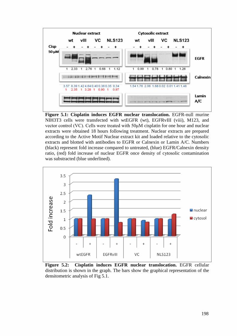
198
Figure 5.1: Cisplatin induces EGFR nuclear translocation. EGFR-null murine
NIH3T3 cells were transfected with wtEGFR (wt), EGFRvIII (viii), M123, and
vector control (VC). Cells were treated with 50μM cisplatin for one hour and nuclear
extracts were obtained 18 hours following treatment. Nuclear extracts are prepared
according to the Active Motif Nuclear extract kit and loaded relative to the cytosolic
extracts and blotted with antibodies to EGFR or Calnexin or Lamin A/C. Numbers
(black) represent fold increase compared to untreated, (blue) EGFR/Calnexin density
ratio, (red) fold increase of nuclear EGFR once density of cytosolic contamination
was substracted (blue underlined).
Figure 5.2: Cisplatin induces EGFR nuclear translocation. EGFR cellular
distribution is shown in the graph. The bars show the graphical representation of the
densitometric analysis of Fig 5.1.
199
5.2.3 Mutant EGFR associates with the heat shock protein 90 chaperone
The association of heat shock proteins (Hsps) to protein kinases has been shown in
several reports (Citri et al, 2006; Pashtan et al, 2008; Sawai et al, 2008). Hsps are
molecular chaperones whose expression increases in response to metabolic insults.
Their most acknowledge function is to assist protein synthesis and folding (Young et
al, 2001). In addition they have been shown to participate in protein assembly,
secretion and trafficking and regulation of transcription factors (Citri et al, 2006).
While Hsp70 have been shown to be involved in the nuclear translocation of EGFR
(Liao & Carpenter, 2007), Hsp90 has been often shown to associate with EGFR
somatic mutants. Previous reports have demonstrated that EGFRs harbouring
mutation within the kinase domain require the Hsp90 chaperone for conformational
maturation and stability. In fact, the inhibition of Hsp90 has been often associated
with reduced expression of EGFR mutants and consequent sensitivity to treatment
(Sawai et al, 2008). The cellular requirement for EGFR mutants to achieve post-
translational maturity and proper folding induces HSp90 association with EGFR
kinase mutants (Lavictoire et al, 2003). Since the NLS123 mutation interferes with
EGFR allosteric conformation, the Hsp90-EGFR association in wtEGFR, NLS123,
L858R, LNLS123, EGFRvIII or VC expressing cells was inevstigated to understand
the relationship between HsP90, EGFR nuclear localisation and EGFR-DNAPKcs
binding. Therefore, the EGFR pull-down samples were also blotted with anti-HSP90
antibody. Fig 5.3 shows the results of the western blot analysis. While, wtEGFR
showed association with HSP90 only following cisplatin treatment, in contrast
EGFRvIII expressing cell lines showed impaired association between EGFR and
Hsp90. L858R, LNLS123 and NLS123expressing cells showed constitutive levels of
association between EGFR and HSP90 following IR, cisplatin or EGF treatment,
although weaker following IR treatment in L858R and NLS123 expressing cells (Fig
5.3).

200
Figure 5.3: EGFR activation and binding to DNAPKcs and Hsp90. Stable
NIH3T3 cells expressing wtEGFR, NLS123, L858R, LNLS123, EGFRvIII, and
Vector control were treated with 50μM cisplatin or 4 G IR, or treated with 100ng/ml
EGF as described in the materials and methods. 1.5mg of protein lysate was then
immunoprecipitated using anti-EGFR monoclonal antibody and blotted with anti
DNAPKcs, anti-PY20, anti-EGFR and anti-HSP90.
5.2.4 DNAPKcs and EGFR localise in the same cellular compartment
following IR or cisplatin
Having established the levels of association between EGFR and DNAPKcs following
either cisplatin or IR treatment, their cellular localisation by confocal microscopy
following IR (Fig 5.4) or cisplatin (Fig 5.5) treatment was also validated in the stably
transfected lines. Cells expressing wtEGFR, NLS123, L858R, LNLS123, EGFRvIII
or VC were either fixed 18 hours following treatment with 50 µM cisplatin or serum

201
starved for 24 hours and then fixed 20 minutes following 4 Gy IR. Fig 5.4 and 5.5
show the immunofluorescence results obtained via confocal analysis. Cells
expressing wtEGFR and EGFRvIII showed nuclear expression of both EGFR and
DNAPKcs following IR or cisplatin treatment. In contrast, L858R-expressing cells
showed impaired EGFR nuclear localisation following either treatment. This was
associated with diminished expression of DNAPKcs within the nucleus. Expression
of EGFR and DNAPKcs was exclusively cytosolic in NLS123 and LNLS123
expressing cell lines following either IR or cisplatin treatment (Fig 5.4 and 5.5).
Inhibition of EGFR nuclear translocation impaired DNAPKcs nuclear translocation,
confirming the results obtained in Chapter 4 with the transiently transfected cells.
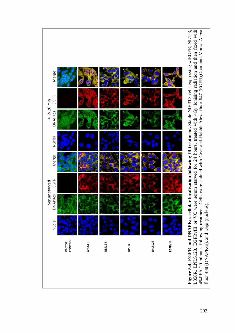
202
Fig
ure
5.4
: E
GF
R a
nd
DN
AP
Kcs
cel
lula
r lo
cali
sati
on
foll
ow
ing I
R t
reatm
ent.
Sta
ble
NIH
3T
3 c
ells
expre
ssin
g w
tEG
FR
, N
L123,
L858R
, L
NL
S123,
EG
FR
vII
I or
VC
wer
e se
rum
sta
rved
for
24 h
ours
, tr
eate
d w
ith 4
Gy I
onis
ing r
adia
tion a
nd t
hen
fix
ed w
ith
4%
PF
A 2
0 m
inute
s fo
llow
ing t
reat
men
t. C
ells
wer
e st
ained
wit
h G
oat
anti
-Rab
bit
Ale
xa
fluor
647 (
EG
FR
),G
oat
anti
-Mouse
Ale
xa
fluor
488 (
DN
AP
Kcs
), a
nd D
api
(nucl
eus)
.
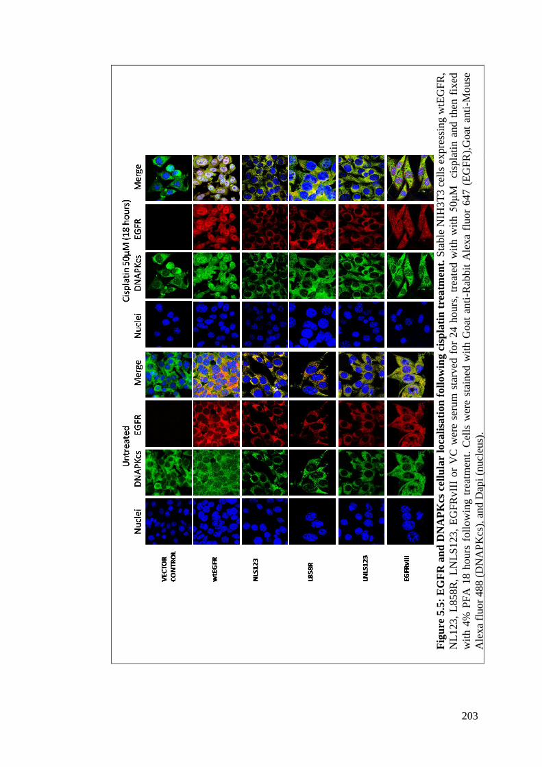
203
Fig
ure
5.5
: E
GF
R a
nd
DN
AP
Kcs
cell
ula
r lo
cali
sati
on
foll
ow
ing c
isp
lati
n t
reatm
ent.
Sta
ble
NIH
3T
3 c
ells
expre
ssin
g w
tEG
FR
,
NL
123,
L858R
, L
NL
S1
23,
EG
FR
vII
I o
r V
C w
ere
seru
m s
tarv
ed f
or
24 h
ours
, tr
eate
d w
ith w
ith 5
0μ
M
cisp
lati
n a
nd t
hen
fix
ed
wit
h 4
% P
FA
18 h
ours
foll
ow
ing t
reat
men
t. C
ells
wer
e st
ain
ed w
ith G
oat
anti
-Rab
bit
Ale
xa
fluor
647 (
EG
FR
),G
oat
anti
-Mouse
Ale
xa
fluor
488 (
DN
AP
Kcs
), a
nd D
api
(nucl
eus)
.

204
5.2.5 EGFR and DNAPKcs association following cisplatin or IR
treatment
The time dependent association between EGFR and DNAPKcs showed a progressive
formation of this complex following cisplatin treatment. EGFR is mainly a
membrane/cytosolic protein, while DNAPKcs is principally found in the nucleus.
Despite nuclear translocation and protein function, the most abundantly expressed
nuclear protein will localise into the cytosol to be degraded. Although previous
reports have shown DNAPKcs cytosolic accumulation following EGFR inhibition by
gefitinib(Friedmann et al, 2006), the majority of the published data has investigated
this complex formation almost exclusively in nuclear extracts (Dittmann et al, 2005a;
Dittmann et al, 2008a; Dittmann et al, 2005b; Dittmann et al, 2007; Golding et al,
2009; Hsu et al, 2009; Mahaney et al, 2009). DNAPKcs has been shown to have
diverse roles in the cytosol (discussed later). Our cellular fractionations have never
achieved a nuclear fraction free from any cytosolic contamination, limiting the
possibility to investigate EGFR-DNAPKcs association using fractionated cellular
extracts. For this reason the cellular distribution of the EGFR-DNAPKcs complex
formation via the proximity ligation assay was investigated. This assay allows
fluorescent visualisation of the interaction between two proteins in fixed cells by
labelling the secondary antibodies, used for the recognition of the protein involved in
a complex, with two complementary oligos. Oligos from one secondary antibody will
ligate only with oligos labelling the secondary antibody from different species (since
they have been purposely made complementary) and within the proximity of 30nm.
Ligation between two oligoes labelling the secondary antibody from the same species
is therefore impossible. Therefore the formation of circular DNA molecule will be
achievable only when two proteins are labelled by two secondary antibodies from
different species and within the distance of 30nm. This is the distance recognised to
be sufficient to determine physical interaction between two molecules. Before
assessing the EGFR-DNAPKcs complex distribution in the NIH3T3 cells stably
expressing wtEGFR or EGFR mutants, the assay and the antibody concentration
required to prevent saturation were tested on A549 cells over expressing EGFR and
DNAPKcs. Primary antibody concentration titration (1:50 - 1:150) was used to
determine the most sensitive antibody concentration in cells treated with IR. Fig 5.6
shows the results of the confocal analysis of A549 cells blocked with anti-Rabbit
EGFR antibody and anti-Mouse DNAPKcs and labelled using the proximity ligation

205
assay. Each interaction is represented via a single red fluorescent dot. There was a
clear accumulation of red dots in the nuclei of A549 cells following treatment with
IR at all the antibody dilutions. This confirmed the previously described EGFR-
DNAPKcs association and established the optimal concentration of primary
antibodies to use in wtEGFR, NLS123, EGFRvIII or VC expressing cells. Fig 5.6
shows that higher antibody dilutions (1:75, 1:100 and 1:150) produced less
sensitivity in detecting nuclear complexes maintaining a discreet level of background
complexes (serum starved) while the lower antibody dilution (1:50) showed a clear
increase in nuclear fluorescent dots following IR treatment.
Next cells stably expressing wtEGFR, NLS123, EGFRvIII or VC were examined.
Cells were treated with 50µM cisplatin or 4 Gy IR and then fixed 18 hours following
treatment with cisplatin or 20 minutes following IR. Fig 5.7 shows the confocal
analysis of the stable cells blocked with 1:50 anti-Rabbit EGFR antibody and 1:50
anti-Mouse DNAPKcs and labelled using the proximity ligation assay. In cells
expressing vector or NLS123 constructs no interaction was detectable. In contrast,
cells expressing wtEGFR or EGFRvIII showed interaction between EGFR and
DNAPKcs following either IR or cisplatin. The pattern of interaction suggests that
EGFR-DNAPKcs complex formation takes place both in the nucleus and cytoplasm.

206
Figure 5.6: EGFR-DNAPKcs complex cellular localization. A549 cells were
treated with 4 Gy IR and then fixed with 4%PFA 20 minutes follwowing radiation.
Cells were then immuno blocked with anti-rabbit EGFR and anti-mouse DNAPKcs
Dilution of the two antibodies are indicated (1:50-1:150). Interacting complexes were
then visulised via the duo link proximity assay. Each red spot represent a single
interaction.

207
Figure 5.6 cont.

208
Figure 5.7: EGFR-DNAPKcs complex cellular localization. Stable NIH3T3 cells
expressing wtEGFR, NLS123, EGFRvIII and Vector control were treated with 50μM
cisplatin for one hour in serum free media and then fixed with 4%PFA 18 hours
following treatment or 4 Gy and then fixed with 4%PFA 20 minutes follwowing
radiation. Cells were then immuno blocked with anti-rabbit EGFR and anti-mouse
DNAPKcs. Interacting complexes were then visulised via the duo link proximity
assay. Each red spot represent a single interaction.
5.2.6 EGFR modulation of DNA-PK activity
Previous reports have shown that the association of EGFR and DNAPKcs stimulates
DNA-PK kinase activity. The experiments detailed above demonstrated that EGFR
kinase activity alone is not sufficient to determine EGFR-DNAPKcs association and
that cisplatin or IR treatment was required to induce this complex formation. This is
suggesting that cisplatin and IR may be responsible for DNAPKcs activation which
would then allow binding. The modulation of repair observed during the study,
however, suggests that treatment alone and putative activation of DNAPK is not
sufficient to achieve complete repair (shown by the VC-expressing cells). Therefore

209
the effect of EGFR-DNAPKcs binding on the alteration of functional enzyme
activity was investigated via the SignaTECT DNA-Dependent Protein Kinase Assay
System. This assay utilises SAM2TM
Biotin Capture Membrane and overcomes the
problem of non specific substrate binding by using a byotinylated DNA-PK p53-
derived peptide substrate. The dose and the time to investigate the kinase activity
were chosen according to the EGFR-DNAPKcs time of binding following IR (4 Gy,
20 minutes) or cisplatin (50 μM cisplatin, 18 hours). EGF induction of EGFR (100
ng/ml, 1 hour) was utilised to determine whether EGFR kinase activation per se were
responsible for the effects on DNA-PK kinase activity.
Fig 5.8 shows the results of the DNA-PK kinase assay performed on extracts from
cells expressing wtEGFR, NLS123, L858R, LNLS123, EGFRvIII or VC. Single
treatment controls were used to determine the percentage change in DNA-PK kinase
activity. As compared with EGFR-negative cells transfected with VC, cells
expressing wtEGFR showed a 27.5 ± 7.22% increase in DNA-PK activity following
IR (4Gy) and 37.52 ± 4.01 % increase following 50µM cisplatin treatment for 1 hour
(Fig 5.7). Similarly, in cells expressing EGFRvIII there was 32.42 ± 16.58 %
increase of DNAPK activity following IR and 26.6 ± 8.49% increase following the
cisplatin treatment. In contrast, no significant change in DNA-PK activity compared
to controls was found in L858R and LNLS123 expressing cell lines following IR (-
5.29 ± 8.27% and 2.72 ± 7.06%) or cisplatin treatment (9.04 ±3.46% and 2.25
±6.07%). Results are summarised in Table 5.1. Only cells expressing NLS123
showed a clear decrease in DNA-PK kinase activity compared to control. EGF
treatment showed no statistical change in kinase activity suggesting that EGFR
binding to DNAPKcs has a more significant role in determining DNA-PK kinase
activity following cisplatin or IR.
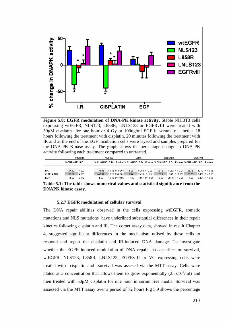
210
Figure 5.8: EGFR modulation of DNA-PK kinase activity. Stable NIH3T3 cells
expressing wtEGFR, NLS123, L858R, LNLS123 or EGFRvIII were treated with
50μM cisplatin for one hour or 4 Gy or 100ng/ml EGF in serum free media. 18
hours following the treatment with cisplatin, 20 minutes following the treatment with
IR and at the end of the EGF incubation cells were lsysed and samples prepared for
the DNA-PK Kinase assay. The graph shows the percentage change in DNA-PK
activity following each treatment compared to untreated.
Table 5.1: The table shows numerical values and statistical significance from the
DNAPK kinase assay.
5.2.7 EGFR modulation of cellular survival
The DNA repair abilities observed in the cells expressing wtEGFR, somatic
mutations and NLS mutations have underlined substantial differences in their repair
kinetics following cisplatin and IR. The comet assay data, showed in result Chapter
4, suggested significant differences in the mechanism utlised by these cells to
respond and repair the cisplatin and IR-induced DNA damage. To investigate
whether the EGFR induced modulation of DNA repair has an effect on survival,
wtEGFR, NLS123, L858R, LNLS123, EGFRvIII or VC expressing cells were
treated with cisplatin and survival was assesed via the MTT assay. Cells were
plated at a concentration that allows them to grow exponentially (2.5x10⁴/ml) and
then treated with 50μM cisplatin for one hour in serum free media. Survival was
assessed via the MTT assay over a period of 72 hours Fig 5.9 shows the percentage

211
decrease in survival of each stable cell line as compared to their daily untreated
control. The graph shows wtEGFR expressing cells comapred to single each cell line
expressing different constructs. Cells expressing wtEGFR and EGFRvIII showed
61.99 ± 5.02 and 62.33 ± 6.82 % survival. L858R expressing cells showed 42.26 ±
1 % survival, statistically significant when compared to the wtEGFR % survival
(P<0.01). NLS123 and LNLS123 showed 37.44 ± 2.26 and 33.76 ± 6.49 % survival,
with high statistical significance when compared to wtEGFR % survival (P value <
0.001). Details of statistical analysis is reported in Table 5.2. These data are
graphically grouped in Fig 5.10. Fig 5.11 shows only the survival 72 hours following
cisplatin expressed as % survival.
Figure 5.9: Cisplatin effects on cellular survival. Cells expressing wtEGFR,
NLS123, L858R, LNLS123, EGFRvIII or VC were plated at 2.5 x10⁴/ml. Then cells
were treated with 50μM cisplatin for one hour in serum free media and then
incubated over a period of 72 hours. Survival was assessed at 24 hours, 48 hours and
72 hours following cisplatin treatment. % decrease survival was calculated by
comparing individually untreated cell lines with corresponding cisplatin treated. The
graph shows mean % decrase survival of 3 independent experiments. The Fig shows
comparison with wtEGFR expressing cells.

212
Figure 5.9: cont.
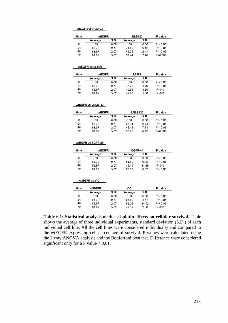
213
Table 6.1: Statistical analysis of the cisplatin effects on cellular survival. Table
shows the average of three individual experiments, standard deviation (S.D.) of each
individual cell line. All the cell lines were considered individually and compared to
the wtEGFR expressing cell percentage of survival. P values were calculated using
the 2 way ANOVA analysis and the Bonferroni post test. Difference were considered
significant only for a P value < 0.01.
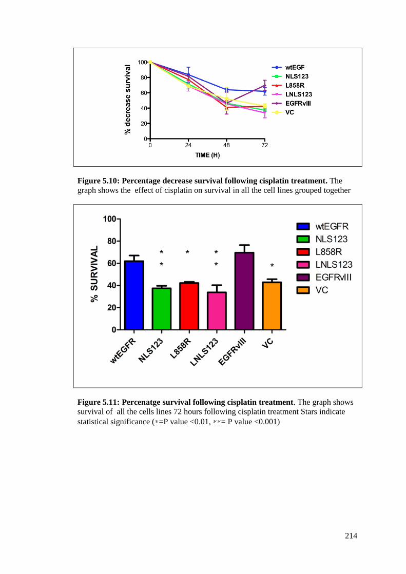
214
Figure 5.10: Percentage decrease survival following cisplatin treatment. The
graph shows the effect of cisplatin on survival in all the cell lines grouped together
Figure 5.11: Percenatge survival following cisplatin treatment. The graph shows
survival of all the cells lines 72 hours following cisplatin treatment Stars indicate
statistical significance ( =P value <0.01, = P value <0.001)

215
5.3 DISCUSSION
5.3.1 IR or cisplatin induces EGFR-DNAPKcs association
The association between EGFR and DNAPKcs has been shown in several reports
however its induction has been controversial. The first study that shed light on this
interaction demonstrated in fact that cetuximab inhibition of EGFR induced EGFR
and DNAPKcs association resulting in a down-regulation of the DNA-PK kinase
activity that impaired DNA repair (Bandyopadhyay et al, 1998). In contrast, multiple
lines of evidence have since shown that EGFR is a major determinant of radiation-
response and that this radio-protective function is exerted via its ability to bind
DNAPKcs and mediate DNA repair (Dittmann et al, 2005a; Dittmann et al, 2007;
Rodemann et al, 2007). These studies also demonstrated that EGFR inhibition via
mAB or TKI treatment inhibits EGFR nuclear translocation and also association with
DNAPKcs rendering cells sensitive to radiotherapy (Baumann et al, 2007; Das et al,
2007; Dittmann et al, 2008a; Nyati et al, 2006; Rodemann & Blaese, 2007; Toulany
et al, 2005; Wanner et al, 2008). This study has shown that EGF, IR and cisplatin
mediated EGFR nuclear translocation however only treatment with IR and cisplatin
induce EGFR-DNAPKcs binding. Nuclear expression per se is therefore not
sufficient to mediate this association suggesting that EGFR-DNAPKcs binding is
specifically induced in response to DNA damage confirming the involvement of
EGFR in modulation of DNA repair. As highlighted in the Introduction Chapter the
role of EGFR nuclear expression is not uniquely related to DNA repair. EGFR
stimulation by ligand has been shown to modulate gene expression and contribute to
a variety of important transcription related cellular mechanisms such as proliferation
(cyclinD1 expression) or oxidative stress (iNos expression) (Lo et al, 2005a; Wang &
Hung, 2009). This highlights the complexity of EGFR nuclear localisation
mechanism and its modularity to different inputs. Although, the molecular targeting
of the pathway involved in EGFR nuclear translocation can still represent a good
candidate for molecular intervention, these results have shown that EGFR nuclear
translocation is not directly responsible for the binding with DNAPKcs.
5.3.2 wtEGFR and EGFRvIII associate to DNAPKcs
Despite the great number of reports discussing EGFR-DNAPKcs association and the
lack of this complex formation in cells expressing L858R, the criteria necessary for

216
this binding and the EGFR characteristics required to allow this association have not
been described. In this study there is not sufficient data to outline the mechanism that
leads to this complex formation, however, the analysis of these results have lead to a
few hypotheses.
While the nuclear expression of wtEGFR has been shown in several reports, nuclear
expression of EGFRvIII has been controversial in the literature. EGFRvIII and
STAT3 localisation within the nucleus was demonstrated in some studies (de la
Iglesia et al, 2008), whereas other studies have reported lack of nuclear expression in
glioma models (Mukherjee et al, 2009). The understanding that EGFRvIII, following
internalisation, is recycled rather then sent to the lysosomal compartment supports
the EGFR nuclear localisation mechanism. As previously discussed the nuclear
translocation can take place only if the receptor escapes degradation. This study
confirms that wtEGFR and EGFRvIII are expressed in the nucleus and also shows
that they bind to DNAPKcs following cisplatin or IR treatment. If originally the
constitutive activation may have been accounted as a determinant of EGFR-
DNAPKcs binding, the hypothesis is now excluded since EGFR-DNAPKcs binding
takes place only following cisplatin or IR treatment, despite constitutive activation
throughout the conditions (EGFRvIII, L858R, LNLS123) and EGF-induced EGFR
phosphorylation. Another hypothesis was that cisplatin or IR may induce EGFR
phosphorylation on specific residues that favour EGFR-DNAPKcs interaction.
Although this could be valid for wtEGFR receptor, the EGFRvIII constitutive
activation suggests that EGFR-DNAPKcs binding is not determined by a specific
phosphorylation event or by the up-regulation of the kinase activity of the receptor.
Moreover, the lack of EGFR-DNAPKcs binding shown in L858R expressing cells
only confirms that EGFR kinase activation is not central to determine EGFR-
DNAPKcs binding. The previous chapters has already suggested that the impaired
EGFR nuclear translocation in L858R-expressing cells is probably due to a lack of
conformational change that does not expose the NLS for recognition. EGFRvIII is
always in a dimerised active conformation suggesting that, differently from the
L858R mutant, EGFRvIII constitutive activation is a consequence of a constitutive,
structural, and active allosteric conformation. Recently, it has been shown that
induced phosphorylation by peptide targeting of the residue T654 within the EGFR
NLS sequence results in EGFR phosphorylated NLS which abolishes EGFR nuclear
localisation and DNAPK activation inhibiting repair of IR induced-DNA damage
217
(Huang et al, 1997; Li et al, 2004). These results suggest that IR or cisplatin
treatment may either induce the activation of a secondary molecule that prevents
T654 EGFR phosphorylation or induce an EGFR conformational change that
prevents this phosphorylation event and favours DNAPKcs-EGFR association.
5.3.3 EGFR-Hsp90 binding
HSP90 is one of the most abundant proteins in the cell. It is widely involved in
biogenesis, stability and activity of its binding partners also called client proteins. It
participates in the maturation of nascent proteins and in the regulation of signalling
events as it is often found bound to a protein effector domain (e.g.: hormone binding
domain or kinase domain) (Young et al, 2001). In many cases Hsp90 binding is
characterised by repeated cycles of low affinity binding and release (Lavictoire et al,
2003). The literature suggests that Hsp90, rather than recognising specific motifs,
binds to features that are common to unstable proteins, such as unveiled hydrophobic
patches, gaining a higher level of affinity to the client protein via co-chaperones such
as CDC37 (Prince & Matts, 2005). Pharmacological inhibition of Hsp90, via
ansamycin antibiotics such as Geldanamycin, results in the degradation of client
proteins showing that this chaperone is involved in protein stability (Sawai et al,
2008). As with other Hsps, it responds to stressful conditions (mainly metabolic
insults) by assuring stability of key proteins and preventing their degradation.
Hsp90 functions have been exploited by molecular intervention to target the over-
expression of proteins that are involved in tumorigenesis such as EGFR and Her2
(Citri et al, 2004b; Yang et al, 2006). While EGFR requires Hsp90 for the maturation
of nascent protein, but becomes independent of it once matured, Her2 stability
depends on Hsp90 binding (Citri et al, 2004a). Wild type EGFR is quickly
internalised following ligand activation and ubiquitinated by Cbl. In addition Cbl
also promotes EGFR internalisation via its interaction with CIN85, which interacts
with endophilins, the regulatory component of the clathrin coated pits. Recognition
and internalisation of active EGFR by the Cin85/Cbl complex is necessary to
regulate the distribution of the receptor and also its signal input. EGFR somatic
mutations in NSCLC such as L858R have been shown to escape the ligand induced
receptor down-regulation and present a strong association with Hsp90 and
unphosphorylated Cbl. This impairs the Cbl mediated recruitment of CIN85 to the
endocytic complex preventing the receptor degradation (Sawai et al, 2008;

218
Shimamura et al, 2005; Yang et al, 2006). The data showed in Fig 5.3 confirmed the
binding between Hsp90 and L858R however IR treatment seemed to weaken this
interaction. Similarly, NLS123 expressing cells showed the same pattern of
interaction. This confirms the previously discussed data suggesting that the NLS123
mutation destabilises the receptor conformation. This semi-constitutive association
with Hsp90 seems to be a signature for EGFR conformational instability. It is
difficult to determine whether the weaker association shown following IR is due to
timing or indicates receptor degradation. Future experiments will be able to shed
more light on this open question.
Interestingly, LNLS123 expressing cells do not show weakening of the association
following IR treatment; in contrast the Hsp90 binding does not seem to be modulated
by any particular treatment suggesting a mechanism of interaction that may involve
both conformational change and receptor activation. It has been shown that
mutations that occur within or near the M5 loop region ( C- 4 region) of the EGFR
protein, are likely to favour the binding with Hsp90 by affecting the conformation of
this loop or altering the conformation of other parts of the kinase domain that
interacts with Hsp90. All the mutations that expose the kinase domain to structural
stresses require the chaperone stabilisation and, as a consequence, Hsp90 clients will
boast an overall positive or neutral charge within the binding region. In contrast,
proteins that do not require chaperone regulation will normally bear an overall
negative charge (Citri et al, 2006). The change of all the arginine residues within the
NLS sequence into alanines changes the positive signal of that stretch of amino acid
into a neutral signal; therefore the change in charge is not the determinant factor.
However, as shown in the previous chapter, the NLS123 mutation is altering the
conformational change of the receptor impeding the allosteric activation and the
dimer interface formation. Therefore, the structural stress imposed on the receptor
may be responsible for the Hsp90 binding to the NLS123 mutant. The impact of the
NLS123 mutation on protein stability is also shown by the LNLS123 mutant that
shows higher binding affinity with Hsp90 as compared to the L858R.
It is difficult to comment on the interaction observed between Hsp90 and wtEGFR
following cisplatin treatment. Although, the literature suggests mainly that Hsp90
binding confers stability to the client protein, the main role of heat shock proteins is
to respond to cellular shocks favouring survival. Cisplatin-induced binding of
wtEGFR to Hsp90 could prevent EGFR degradation and maintain a constitutive

219
function required in the response to cisplatin treatment. A very recent study showed
dependence of wtEGFR and EGFRvIII on Hsp90 binding (Pines et al) . It is clear
from Fig 5.3 that, although greatly weaker compared to the other mutants, both
wtEGFR and EGFRvIII show association with Hsp90. Future studies will determine
the relevance of this dependence and its degree in the context of DNA repair.
5.3.4 DNAPKcs subcellular localisation in response to IR or cisplatin is
influenced by EGFR subcellular distribution.
In line with previous published evidence, this study shows EGFR and DNAPKcs
association. The confocal analysis of the stable cells expressing constructs confirms
the results shown in the previous chapter. Only wtEGFR and EGFRvIII are
expressed in the nucleus following cisplatin or IR and this is induced by the
treatments and not by a transient over-expression of the wtEGFR and EGFRvIII
expressing plasmids. Interestingly, not only does DNAPKcs co-localise in the
nucleus with wtEGFR and EGFRvIII but the impaired nuclear localisation shown by
the remaining mutants seems to indirectly determine DNAPKcs cytosolic
accumulation. Moreover, in L858R expressing cells DNAPKcs seems to be capable
of localising within the nucleus despite the lack of L858R nuclear localisation. This
explains why L858R cells showed intermediate repair following IR and cisplatin. In
addition, this suggests that the NLS123 mutation may further, sterically, obstruct the
receptor direct or indirect mediation of DNAPKcs nuclear localisation. EGFR may
indirectly signal for DNAPKcs cellular distribution by activation of a secondary
messenger or by physical interaction with another protein. The NLS123 mutation
within the wtEGFR (NLS123) and the L858R (LNLS123) may interfere with this
mechanism supporting the evidence that targeting the NLS inhibits DNAPKcs-EGFR
binding and its function in repair (Dittmann et al)
5.3.5 EGFR–DNAPKcs interaction takes place both in the nucleus and in
the cytoplasm
It was clearly demonstrated that EGFR binds to DNAPKcs and that they both co-
localise within the same cellular compartments following cisplatin and IR. Since
EGFR-DNAPKcs association has been reported this binding has only been shown by
co-immunoprecipitating these two proteins from nuclear extracts. Although this
answer the question relative to modulation in DNA repair, it does not characterise
220
this association with regards to whether these two proteins come into contact before
entering the nucleus, whether this association is exclusively nuclear and therefore
dependent of EGFR nuclear translocation. This is central to fully understand how
molecular intervention can effectively target this interaction and modulate repair
response and kinetics. The precise distance between two proteins sufficient to allow a
physical interaction has not yet been defined. The proximity ligation assay
determines whether the physical space between two proteins is smaller than 40 nm,
which is acknowledged to be sufficient to allow protein-protein interaction. Fig 5.7
confirms the result of the immunoprecipitation by showing that only in wtEGFR and
EGFRvIII expressing cells EGFR and DNAPKcs are in the proximity sufficient to
interact. Moreover, it also shows that this interaction takes place both in the nucleus
and in the cytoplasm suggesting that DNAPKcs recruitment by EGFR is most
probably a cytosolic event that precedes the nuclear translocation. The time course
immunoprecipitation discussed in the previous chapter showed that the binding
between these two proteins increases over time and peaks at 18 following cisplatin
treatment. The time course experiment also shows interaction already at 1 hour
following cisplatin treatment, suggesting a recruitment that anticipates the DNA
damage response. The possibility that EGFR-DNAPKcs complex forms in the
cytosol and then migrates to the nucleus cannot be excluded. Similarly, the
possibility that the cytosolic portion of this complex may have another secondary
function still involved in response to therapy cannot be excluded.
5.3.6 EGFR modulates DNAPKcs kinase activity
EGFR modulation of DNAPK kinase activity has been shown in several reports.
Cetuximab-induced reduction in DNA-PK kinase has been correlated not only with
inhibited EGFR kinase activation but also with EGFR nuclear localisation (Dittmann
et al, 2005b; Li et al, 2008; Perez-Torres et al, 2006). This has supported the
experimental evidence that IR induces EGFR-DNAPKcs association leading to
enhanced kinase activity of the NHEJ DNA-PK complex (Chen & Nirodi, 2007).
This study showed that expression of EGFR modulates DNAPK kinase activity only
in cells expressing wtEGFR or EGFRvIII. This did not occur in cells expressing
EGFR constructs deficient in EGFR nuclear translocation and DNAPKcs binding.
These data show that EGFR modulation of DNA repair is mediated via DNAPK
pathways and requires EGFR-DNAPKcs binding and EGFR nuclear localisation.

221
The decrease of DNAPK activity in NLS123 expressing cells confirms the
previously published data where EGFR inhibition by cetuximab or via Src
knockdown also induced inhibition of DNAPK kinase activity (Andersen et al, 2009;
Dittmann et al, 2008a; Dittmann et al, 2009; Gueven et al, 1998; Wheeler et al,
2009). Interestingly these down-regulations were highly statistically significant when
compared to cells not expressing EGFR. The possibility that the NLS123 mutant
indirectly inhibits the activation of signalling pathway involved in DNAPK kinase
activity cannot be excluded. Future experiments utilising EGFR inhibitors and/or a
kinase dead mutant will address this issue. There was no change in the kinase activity
of cells expressing L858R or LNLS123 expressing cells. This suggests that, although
nuclear translocation and the binding are determining the increment of DNAPK
kinase activity, receptor kinase activation may also influence, indirectly, DNAPK. It
has been shown that activation of AKT may lead to DNAPKcs activation as AKT
has been shown to act as a kinase of DNAPKcs (Bozulic et al, 2008; Shi et al, 2009;
Toulany et al, 2008b). The role of EGFR as a direct kinase activator of DNAPKcs
via their physical interaction or as an indirect activator needs to be elucidated. It is
arguable that EGFR recruits cytosolic DNAPKcs inducing DNAPKcs nuclear
accumulation. This would then enhance the DNAPKcs-DNA interaction and
therefore the kinase activity of the DNAPK complex. In EGFRvIII expressing cells
both the binding and/or the nuclear translocation and the possible activation of a
putative AKT back-up activation pathway may trigger the enhanced DNAPK kinase
activity significantly higher than in wtEGFR expressing cells allowing a faster rate of
DNA SB repair following IR.
5.3.7 Cisplatin toxicity
Cisplatin is one of the most widely used anticancer drugs. Although it is generally
accepted that the binding of cisplatin to genomic DNA is the main event responsible
for its anti-tumor property, nearly 90% of platinum is bound to plasma proteins
(Turchi et al, 1999; Wang & Lippard, 2005; Wernyj & Morin, 2004). This leads to
the activation of many signalling pathways which many line of evidence have shown
to either confer sensitivity or resistance to the treatment. The activation of DNA
repair mechanisms in response to cisplatin treatment has already been reviewed both
in the introduction and in result chapter 2. Therefore the transduction of DNA
damage signals on cellular signalling and survival will be briefly reviewed with
222
regards to the cisplatin induction of cell death and the involvement of AKT,
MAPK/JNK/ERK in the response to cisplatin and how EGFR has been shown to
modulate these pathways.
5.3.7.1 Cisplatin-induced cell death
Several lines of evidence have shown that cisplatin induces two different modes of
cellular death: necrosis and apoptosis. A cytosolic swelling and loss of plasma
membrane integrity characterize necrosis. It is initialized by the DNA damage-
induced activation of PARP-1 that cleaves the glycolytic coenzyme NAD+. Loss of
NAD+ inhibits the glycolytic production of ATP with subsequent depletion of
cellular ATP leading to cell death via necrosis. In contrast, cells undergoing
apoptosis show shrinkage, chromatin condensation and DNA fragmentation.
Apoptotic death results from the activation of the caspases (aspartate specific family
of proteases). The activation of the upstream CASP8 or CASP9 leads to the
activation of downstream caspases such as CASP3 and CASP7. This induces the
cleavage and subsequent degradation of many cellular proteins leading to apoptosis.
Although in resistant cell lines cisplatin seems to produce characteristic features of
necrosis there is evidence suggesting that within the same population of cells,
platinum can induce both necrosis and apoptosis. Moreover the cooperation of these
two pathways has been shown by the caspase activation and induction of apoptosis in
presence of PARP1 induced NAD+ cleavage inhibition (Fishel et al, 2005; Lee et al,
2005; Wernyj & Morin, 2004).
5.3.7.2 Pro-survival AKT signalling
AKT promotes cell survival via the down regulation of apoptotic pathways. It
phosphorylates the X-linked inhibitor of apoptosis XIAP and inhibits the auto-
ubiquitylation of this protein and the cisplatin induced ubiquitylation activities.
Increased levels of XIAP have been shown to correlate with decreased levels of
CASP3 and down regulation of apoptosis(Fraser et al, 2003a; Fraser et al, 2003b). In
addition AKT has been shown to induce activation of NFkB (Asselin et al, 2001;
Dan et al, 2004). Increased activity of NFkB correlates with decreased apoptosis
whereas its inhibition has been shown to augment cisplatin cytoxicity. It has been
demonstrated that cisplatin-induced T308 phosphorylation of AKT in MDA-MB-468

223
is dependent on Src and EGFR with EGFR being the initiator of the signal
transduction that leads to the activation of this pro-survival signal (Yang et al, 2004).
5.3.7.3 p38MAPK/MAPK/JNK role in cisplatin resistance
There is a growing body of evidence suggesting that p38MAPK has a central role in
the response to cisplatin treatment. Cisplatin activates p38MAPK for 8-12 hours in
sensitive cells and for 1-3 hours in resistant cells suggesting that the different kinetics
may explain the differential cytotoxicity (Mansouri et al, 2003). There is instead
conflicting evidence on the role of ERK in influencing cellular survival following
cisplatin treatment. Some studies indicate that ERK activation is associated with
enhanced survival (Hayakawa et al, 1999). However the use of the MEK1 inhibitor
has been shown to be effective only in certain cell line suggesting a more complex
and cell line dependent response to cisplatin (Mandic et al, 2001). In addition
elevated activation of RAS, an upstream component of the ERK signaling pathway
has been shown to confer sensitivity to cisplatin (Wang & Lippard, 2005).
Far from being clear is also the JNK mediated response to cisplatin treatment.
Several studied have shown evidence that activation of the JNK pathway contributes
to cisplatin induced apoptosis (Mansouri et al, 2003) whereas some other have shown
a protective response necessary for cell survival (Davis, 2000). Interestingly one
study has shown that duration of the signalling pathway activation might be central
for the decision between cellular proliferation or death (Chen et al, 1996).
It has been shown that phosphorylation of the T669 by p38MAPK leads to cisplatin
induced-EGFR internalization. This event seems to be mediated by ERK inducing
the abrogation of EGFR signalling to JNK (Winograd-Katz & Levitzki, 2006). At the
same time, this study has suggested that EGFR signalling to JNK is dependent on
receptor internalization. Since internalization defective EGFR was shown to increase
cell proliferation and lead to ERK down- regulation it has been suggested that
internalized EGFR may form signalling complexes within the endosomes triggering
a switch in signalling from proliferation to survival (Grandal & Madshus, 2008).
5.3.7.4 EGFR is required for cisplatin resistance
In line with these studies the MTT assay have shown that wtEGFR or EGFRvIII
expression confers survival to cells following cisplatin treatment. Our analysis does
not verify whether the cisplatin induced EGFR activation (wtEGFR) or the
224
constitutive kinase activation (EGFRvIII) may be responsible for induction of pro-
survival signalling. These data suggest that by 36 hours following cisplatin treatment,
cells expressing wtEGFR and EGFRvIII have already repaired all the crosslinks
induced by cisplatin treatment (Result Chapter 4) and also that by 18 hours both the
wt receptor and the EGFRvIII mutant are expressed in the nucleus and associate with
DNAPKcs resulting in up-regulation of DNAPK activity. The EGFR kinase
activation alone does not seem to be responsible for the survival response. Cells
expressing the constitutive kinase active L858R mutant, which does not translocates
to the nucleus, show less than 50% survival, suggesting that both EGFR nuclear
expression and binding to DNAPKcs modulation of DNA repair could correlate with
the pro-survival response. This is confirmed also by the results obtained in cell
expressing NLS123 and LNLS123 where cisplatin toxicity is clearly compromising
cellular viability. Further experiments will show whether it is a direct effect of DNA
repair inhibition or whether this is the result of signalling events in response to DNA
damage or both.

225
CHAPTER 6: CONCLUSION
226
EGFR is commonly expressed in human tumours and has been an important target
for therapy. Although several small molecules inhibitors and antibodies are currently
utilised in the clinic in combination with chemotherapy and IR, the successfulness of
these treatments has been limited. EGFR nuclear expression has been a source of
important insights in the biology of this oncogene. This study investigated the role of
nuclear EGFR in the modulation of DNA repair following treatment with cisplatin
and IR.
Chapter 3 focused on the construction of two NLS mutants, one in the background
of wtEGFR and the other one in the background of the somatic mutation L858R
often found in lung cancer. Then nuclear expression of EGFR following IR treatment
and EGF induction was confirmed. Evidence was provided showing that the
mutation of all arginine residues of EGFR NLS sequence induced inhibition of
nuclear translocation in the NLS123 and LNLS123 mutant. In line with published
data, also L858R showed impaired nuclear translocation. The characterisation of the
constructs within the NIH3T3 murine fibroblast (negative for the expression of
EGFR) showed that the NLS123 mutation impairs EGFR kinase activity. Recent
studies have shown the importance of the NLS sequence in EGFR activation and
binding to the importin machinery. This suggested that the inhibition of nuclear
translocation was due to the lack of an allosteric change required by EGFR to adopt
an active conformation. Only EGFR molecules undergoing this conformational
change expose the NLS sequence for recognition. Interestingly LNLS123 expressing
cells showed EGFR constitutive activation suggesting that the impaired EGFR
nuclear translocation observed in L858R is due to the receptor conformation. The
kinase mutation at position 858 induces a modification within the active loop that
allows constitutive kinase activation without requiring kinase domain juxtaposition.
This explains why the single amino acid mutation is sufficient to impair EGFR
nuclear translocation in L858R and LNLS123 expressing cells. Chapter 4 showed
EGFR modulation of DNA repair kinetics following cisplatin and IR using the
alkaline single-cell gel electrophoresis (comet assay). Constructs expressing point
mutation within the NLS sequence, a deletion of the NLS sequence, an EGFR kinase
death mutant and the somatic EGFRvIII were utilised to compare and contrast the
different modulation of the unhooking of cisplatin interstrand crosslinks and the
repair of DNA strand breaks. Three different repair behaviours were observed.
Whilst wtEGFR and EGFRvIII showed complete repair by 36 hours following

227
cisplatin and 4 hour following IR treatment, intermediate levels of repair were
observed in cells expressing L858R, KMT and NLS. Complete inhibition of repair
was observed in cell expressing NLS123 and LNLS123. The result obtained by the
comet assays suggested that EGFR kinase activity is not central to the modulation of
repair and that the inhibition of nuclear translocation via the NLS mutation was
central to the repair inhibition. The impaired receptor dimerisation and the lack of an
active conformation seem to be critical for this mechanism. The confocal analysis on
the transient transfectants showed that the impaired nuclear localisation of EGFR and
DNAPKcs (NLS123 and LNLS123 expressing cells) correlated with complete
inhibition of repair. Interestingly, while low levels of EGFR nuclear accumulation
(M1 and M12) corresponded to DNA repair, low levels of DNAPKcs nuclear
accumulation but complete inhibition of EGFR nuclear expression (L858R, KMT
and NLS expressing cells) resulted only in intermediate levels of repair. The
difference in modulation shown with the comet assay and the nuclear expression of
the different constructs shown in the confocal analysis confirms that the repair is
mediated by EGFR and that is modulated by EGFR nuclear translocation.
Chapter 5 investigated the mechanism involved in the EGFR mediated modulation
of DNA repair. The association between EGFR and DNAPKcs, induced by IR and
cisplatin, correlated with DNA repair. EGFR interaction with DNAPKcs was shown
only in wtEGFR and EGFRvIII expressing cells. Although EGFR nuclear
translocation per se was not sufficient to mediate binding with DNAPKcs, nuclear
expression was required for repair. The confocal analysis showed that the nls123
mutation not only inhibited EGFR nuclear translocation (NLS123 and LNLS123
expressing cells) but also binding to DNAPKcs and, indirectly, DNAPKcs nuclear
accumulation. The formation of this complex was shown to take place both in the
nucleus and in the cytoplasm and seemed to require EGFR allosteric active
conformational change. The association between EGFR and DNAPKcs is central to
repair as shown by the DNAPK kinase assay. While, in cells expressing wtEGFR and
EGFRvIII there was a clear upregulation in DNAPK kinase activity, L858R and
LNLS123 expressing cells showed no upregulation. In line with previous study that
showed that Gefitinib-induced EGFR inhibition resulted in down regulation of
DNAPK kinase activity and also in DNAPKcs cytosolic accumulation, the impaired
binding to DNAPKcs and impaired kinase activity shown in NLS123 expressing
cells correlated with a significant downregulation in DNAPK kinase activity. These
228
data suggest that EGFR inhibition by targeting EGFR NLS sequence inhibits EGFR
nuclear accumulation and EGFR kinase activity via the impairment of its allosteric
activation. As a consequence, the binding to DNAPKcs and DNAPK kinase activity
are significantly impaired leading to inhibition of DNA repair following cisplatin and
IR. Our data have also shown that the impairment of EGFR nuclear translocation
and the consequent modulation of DNA repair via the inhibition of DNAPK kinase
activity correlate with cellular survival following cisplatin. The mechanism of
nuclear translocation, following treatment with cisplatin or IR can therefore be
accounted as a cellular response that cells employ to overcome DNA damage and
survive treatment toxicity.
6.1 EGFR and DNAPKcs physical interaction
This study validates the importance of EGFR-DNAPKcs interaction following
treatment with cisplatin and IR. Deletion constructs expressing either protein can be
generated and used to determine the domain involved in this interaction. This could
have potential implication in the design of novel drugs with relevance to the EGFR-
DNAPKcs association. Our initial work on the construction of deletion mutants has
led to the conclusion that EGFR subcellular distribution and allosteric conformation
are determinant to the binding. Although, deletion mutants would be ideal to
underpin the exact stretch of amino acid responsible for EGFR-DNAPKcs binding,
the impaired association observed in the NLS mutants suggests that other regions
within EGFR may indirectly impair this association. DNAPKcs deletion mutants are
therefore the only viable option.
Despite the in vitro success of many EGFR inhibitors clinical outcomes are still poor.
This study points out that dimerisation and conformational change, being events
upstream of kinase activation, may be better targets for EGFR directed therapy. The
validation of EGFR allosteric conformational change as an absolute requirement for
DNAPKcs binding can be evaluated by analysing EGFR dimer formations following
treatment in wtEGFR or EGFR mutants expressing cells. This together with a
pharmacological inhibition of EGFR dimer formation will shed some light on other
modalities to inhibit EGFR and DNAPKcs that may result in better therapeutic
outcome.

229
6.2 Novel partners involved in EGFR modulation of repair
The repair of SBs shown by the comet assay suggest that the most significant
difference in repair kinetics among all the mutants is within the first 30 minutes
following IR. Many studies have discussed the type of breaks detected by the comet
assay and although it primarily detects singles strand breaks, double strand breaks are
not to be excluded. Despite being difficult to detect via the comet assay, the
clustering formation of DNA damage following IR implies that statistically the
formation of DSBs is inevitable. Considering their complexity as opposed to SSBs,
DSBs are regarded as being the subject of „late repair‟. While the early and
immediate repair shows the kinetics of SSBs, the difference in kinetics of the late
repair highlights the repair inhibition of DSBs. DNAPKcs is the central component
of NHEJ the pathway involved in the repair DSBs, therefore the observed differences
in the early repair kinetics may be the result of EGFR interaction with other DNA
repair proteins involved in SSB repair. Our preliminary data on the gliomas model
MO59J and M059K suggest that in absence of DNAPKcs, Gefitinib-induced EGFR
inhibition results in slower repair kinetic following IR treatment. This suggests a role
for EGFR in repair independent of DNAPKcs. The study of alternative interactions
by immunoprecipitation and confocal imaging in cells expressing wtEGFR and
EGFR mutants in the NLS sequence may reveal novel mechanisms of EGFR
modulation of DNA repair.
6.3 EGFR mediated transcription
EGFR nuclear expression following ligand activation has been shown to induce
iNOS, cyclin D1, Aurora A, and RNA helicase A expression. Binding with the STAT
family of transcription factors is central to the modulation of gene expression.
Although here and in previous studies cisplatin and IR treatments have been shown
to induce EGFR nuclear translocation, little is known about the modulation of gene
expression mediated by EGFR following these treatments. Further studies should be
designed to address this point and to determine a read out of all the genes whose
expression is mediated by EGFR. The Hsp90 binding to wtEGFR following cisplatin
treatment (chapter 5) is indicative of a stress response mechanism that may involve
transcriptional regulation. The system validated in this study will allow the
investigation of the EGFR characteristics involved in transcriptional activation and
unveil novel mechanisms involved in resistance to cisplatin and IR.

230
6.4 Conclusion
These results highlight the importance of understanding the events that lead to EGFR
subcellular distribution and its involvement in mediating DNA response following
cisplatin and IR. The molecular mechanisms of EGFR nuclear translocation and
binding with DNAPKcs will form the design of future molecular targeted therapy
and point the way for further studies to investigate the role of EGFR in cancer
therapy.
231
References
Achanta G, Pelicano H, Feng L, Plunkett W, Huang P (2001) Interaction of p53 and
DNA-PK in response to nucleoside analogues: potential role as a sensor complex for
DNA damage. Cancer Res 61: 8723-8729
Adair GM, Rolig RL, Moore-Faver D, Zabelshansky M, Wilson JH, Nairn RS
(2000) Role of ERCC1 in removal of long non-homologous tails during targeted
homologous recombination. EMBO J 19: 5552-5561
Adrain C, Martin SJ (2009) Apoptosis: calling time on apoptosome activity. Sci
Signal 2: pe62
Agelaki S, Spiliotaki M, Markomanolaki H, Kallergi G, Mavroudis D, Georgoulias
V, Stournaras C (2009) Caveolin-1 regulates EGFR signaling in MCF-7 breast
cancer cells and enhances gefitinib-induced tumor cell inhibition. Cancer Biol Ther
8: 1470-1477
Aifa S, Frikha F, Miled N, Johansen K, Lundstrom I, Svensson SP (2006a)
Phosphorylation of Thr654 but not Thr669 within the juxtamembrane domain of the
EGF receptor inhibits calmodulin binding. Biochem Biophys Res Commun 347: 381-
387
Aifa S, Miled N, Frikha F, Aniba MR, Svensson SP, Rebai A (2006b) Electrostatic
interactions of peptides flanking the tyrosine kinase domain in the epidermal growth
factor receptor provides a model for intracellular dimerization and
autophosphorylation. Proteins 62: 1036-1043
Akaboshi M, Kawai K, Maki H, Akuta K, Ujeno Y, Miyahara T (1992) The number
of platinum atoms binding to DNA, RNA and protein molecules of HeLa cells
treated with cisplatin at its mean lethal concentration. Jpn J Cancer Res 83: 522-526
Allen C, Halbrook J, Nickoloff JA (2003) Interactive competition between
homologous recombination and non-homologous end joining. Mol Cancer Res 1:
913-920
Alvarado D, Klein DE, Lemmon MA (2009) ErbB2 resembles an autoinhibited
invertebrate epidermal growth factor receptor. Nature 461: 287-291
232
Amit I, Citri A, Shay T, Lu Y, Katz M, Zhang F, Tarcic G, Siwak D, Lahad J, Jacob-
Hirsch J, Amariglio N, Vaisman N, Segal E, Rechavi G, Alon U, Mills GB, Domany
E, Yarden Y (2007a) A module of negative feedback regulators defines growth
factor signaling. Nat Genet 39: 503-512
Amit I, Wides R, Yarden Y (2007b) Evolvable signaling networks of receptor
tyrosine kinases: relevance of robustness to malignancy and to cancer therapy. Mol
Syst Biol 3: 151
Andersen P, Villingshoj M, Poulsen HS, Stockhausen MT (2009) Improved response
by co-targeting EGFR/EGFRvIII and Src family kinases in human cancer cells.
Cancer Invest 27: 178-183
Andressoo JO, Hoeijmakers JH, de Waard H (2005) Nucleotide excision repair and
its connection with cancer and ageing. Adv Exp Med Biol 570: 45-83
Andressoo JO, Hoeijmakers JH, Mitchell JR (2006) Nucleotide excision repair
disorders and the balance between cancer and aging. Cell Cycle 5: 2886-2888
Armour AA, Watkins CL The challenge of targeting EGFR: experience with
gefitinib in nonsmall cell lung cancer. Eur Respir Rev 19: 186-196
Arteaga CL (2002) Epidermal growth factor receptor dependence in human tumors:
more than just expression? Oncologist 7 Suppl 4: 31-39
Asselin E, Mills GB, Tsang BK (2001) XIAP regulates Akt activity and caspase-3-
dependent cleavage during cisplatin-induced apoptosis in human ovarian epithelial
cancer cells. Cancer Res 61: 1862-1868
Azevedo F, Marques F, Fokt H, Oliveira R, Johansson B Measuring oxidative DNA
damage and DNA repair using the yeast comet assay. Yeast 28: 55-61
Azqueta A, Shaposhnikov S, Collins AR (2009) DNA oxidation: investigating its
key role in environmental mutagenesis with the comet assay. Mutat Res 674: 101-
108
Bae JH, Schlessinger J (2010) Asymmetric tyrosine kinase arrangements in
activation or autophosphorylation of receptor tyrosine kinases. Mol Cells 29: 443-
448
Bailey SM, Brenneman MA, Halbrook J, Nickoloff JA, Ullrich RL, Goodwin EH
(2004) The kinase activity of DNA-PK is required to protect mammalian telomeres.
DNA Repair (Amst) 3: 225-233
Bandyopadhyay D, Mandal M, Adam L, Mendelsohn J, Kumar R (1998) Physical
interaction between epidermal growth factor receptor and DNA-dependent protein
kinase in mammalian cells. J Biol Chem 273: 1568-1573

233
Bassermann F, Pagano M (2010) Dissecting the role of ubiquitylation in the DNA
damage response checkpoint in G2. Cell Death Differ 17: 78-85
Batty DP, Wood RD (2000) Damage recognition in nucleotide excision repair of
DNA. Gene 241: 193-204
Baumann M, Krause M, Dikomey E, Dittmann K, Dorr W, Kasten-Pisula U,
Rodemann HP (2007) EGFR-targeted anti-cancer drugs in radiotherapy: preclinical
evaluation of mechanisms. Radiother Oncol 83: 238-248
Bean J, Brennan C, Shih JY, Riely G, Viale A, Wang L, Chitale D, Motoi N, Szoke
J, Broderick S, Balak M, Chang WC, Yu CJ, Gazdar A, Pass H, Rusch V, Gerald W,
Huang SF, Yang PC, Miller V, Ladanyi M, Yang CH, Pao W (2007) MET
amplification occurs with or without T790M mutations in EGFR mutant lung tumors
with acquired resistance to gefitinib or erlotinib. Proc Natl Acad Sci U S A 104:
20932-20937
Benavente S, Huang S, Armstrong EA, Chi A, Hsu KT, Wheeler DL, Harari PM
(2009) Establishment and characterization of a model of acquired resistance to
epidermal growth factor receptor targeting agents in human cancer cells. Clin Cancer
Res 15: 1585-1592
Benhar M, Engelberg D, Levitzki A (2002) Cisplatin-induced activation of the EGF
receptor. Oncogene 21: 8723-8731
Bentzen SM (2003) Repopulation in radiation oncology: perspectives of clinical
research. Int J Radiat Biol 79: 581-585
Bessho T, Sancar A, Thompson LH, Thelen MP (1997) Reconstitution of human
excision nuclease with recombinant XPF-ERCC1 complex. J Biol Chem 272: 3833-
3837
Bitler BG, Goverdhan A, Schroeder JA (2010) MUC1 regulates nuclear localization
and function of the epidermal growth factor receptor. J Cell Sci 123: 1716-1723
Boeckman HJ, Trego KS, Turchi JJ (2005) Cisplatin sensitizes cancer cells to
ionizing radiation via inhibition of nonhomologous end joining. Mol Cancer Res 3:
277-285
Borst P, Rottenberg S, Jonkers J (2008) How do real tumors become resistant to
cisplatin? Cell Cycle 7: 1353-1359
Bose R, Zhang X (2009) The ErbB kinase domain: structural perspectives into
kinase activation and inhibition. Exp Cell Res 315: 649-658
Bozulic L, Surucu B, Hynx D, Hemmings BA (2008) PKBalpha/Akt1 acts
downstream of DNA-PK in the DNA double-strand break response and promotes
survival. Mol Cell 30: 203-213

234
Branzei D, Foiani M (2008) Regulation of DNA repair throughout the cell cycle. Nat
Rev Mol Cell Biol 9: 297-308
Breen L, Heenan M, Amberger-Murphy V, Clynes M (2007) Investigation of the
role of p53 in chemotherapy resistance of lung cancer cell lines. Anticancer Res 27:
1361-1364
Bublil EM, Yarden Y (2007) The EGF receptor family: spearheading a merger of
signaling and therapeutics. Curr Opin Cell Biol 19: 124-134
Caldecott KW (2008) Single-strand break repair and genetic disease. Nat Rev Genet
9: 619-631
Carpenter G (1993) EGF: new tricks for an old growth factor. Curr Opin Cell Biol 5:
261-264
Carter CA, Kelly RJ, Giaccone G (2009) Small-molecule inhibitors of the human
epidermal receptor family. Expert Opin Investig Drugs 18: 1829-1842
Cepeda V, Fuertes MA, Castilla J, Alonso C, Quevedo C, Perez JM (2007)
Biochemical mechanisms of cisplatin cytotoxicity. Anticancer Agents Med Chem 7:
3-18
Ceppi P, Volante M, Novello S, Rapa I, Danenberg KD, Danenberg PV, Cambieri A,
Selvaggi G, Saviozzi S, Calogero R, Papotti M, Scagliotti GV (2006) ERCC1 and
RRM1 gene expressions but not EGFR are predictive of shorter survival in advanced
non-small-cell lung cancer treated with cisplatin and gemcitabine. Ann Oncol 17:
1818-1825
Chabner BA, Roberts TG, Jr. (2005) Timeline: Chemotherapy and the war on cancer.
Nat Rev Cancer 5: 65-72
Chan DW, Chen BP, Prithivirajsingh S, Kurimasa A, Story MD, Qin J, Chen DJ
(2002) Autophosphorylation of the DNA-dependent protein kinase catalytic subunit
is required for rejoining of DNA double-strand breaks. Genes Dev 16: 2333-2338
Chang JW, Chou CL, Huang SF, Wang HM, Hsieh JJ, Hsu T, Cheung YC (2007)
Erlotinib response of EGFR-mutant gefitinib-resistant non-small-cell lung cancer.
Lung Cancer 58: 414-417
Chang YC, Wu W, Zhang JL, Huang Y (2009) Allosteric activation mechanism of
the cys-loop receptors. Acta Pharmacol Sin 30: 663-672
Chen BP, Chan DW, Kobayashi J, Burma S, Asaithamby A, Morotomi-Yano K,
Botvinick E, Qin J, Chen DJ (2005) Cell cycle dependence of DNA-dependent
protein kinase phosphorylation in response to DNA double strand breaks. J Biol
Chem 280: 14709-14715
Chen DJ, Nirodi CS (2007) The epidermal growth factor receptor: a role in repair of
radiation-induced DNA damage. Clin Cancer Res 13: 6555-6560

235
Chen YR, Fu YN, Lin CH, Yang ST, Hu SF, Chen YT, Tsai SF, Huang SF (2006)
Distinctive activation patterns in constitutively active and gefitinib-sensitive EGFR
mutants. Oncogene 25: 1205-1215
Chen YR, Wang X, Templeton D, Davis RJ, Tan TH (1996) The role of c-Jun N-
terminal kinase (JNK) in apoptosis induced by ultraviolet C and gamma radiation.
Duration of JNK activation may determine cell death and proliferation. J Biol Chem
271: 31929-31936
Chen Z, Ke LD, Yuan XH, Adler-Storthz K (2000) Correlation of cisplatin
sensitivity with differential alteration of EGFR expression in head and neck cancer
cells. Anticancer Res 20: 899-902
Cheng WH, Kusumoto R, Opresko PL, Sui X, Huang S, Nicolette ML, Paull TT,
Campisi J, Seidman M, Bohr VA (2006) Collaboration of Werner syndrome protein
and BRCA1 in cellular responses to DNA interstrand cross-links. Nucleic Acids Res
34: 2751-2760
Choong NW, Dietrich S, Seiwert TY, Tretiakova MS, Nallasura V, Davies GC,
Lipkowitz S, Husain AN, Salgia R, Ma PC (2006) Gefitinib response of erlotinib-
refractory lung cancer involving meninges--role of EGFR mutation. Nat Clin Pract
Oncol 3: 50-57; quiz 51 p following 57
Choowongkomon K, Carlin CR, Sonnichsen FD (2005) A structural model for the
membrane-bound form of the juxtamembrane domain of the epidermal growth factor
receptor. J Biol Chem 280: 24043-24052
Choowongkomon K, Hobert ME, He C, Carlin CR, Sonnichsen FD (2004) Aqueous
and micelle-bound structural characterization of the epidermal growth factor receptor
juxtamembrane domain containing basolateral sorting motifs. J Biomol Struct Dyn
21: 813-826
Chuderland D, Konson A, Seger R (2008) Identification and characterization of a
general nuclear translocation signal in signaling proteins. Mol Cell 31: 850-861
Citri A, Alroy I, Lavi S, Rubin C, Xu W, Grammatikakis N, Patterson C, Neckers L,
Fry DW, Yarden Y (2002) Drug-induced ubiquitylation and degradation of ErbB
receptor tyrosine kinases: implications for cancer therapy. EMBO J 21: 2407-2417
Citri A, Gan J, Mosesson Y, Vereb G, Szollosi J, Yarden Y (2004a) Hsp90 restrains
ErbB-2/HER2 signalling by limiting heterodimer formation. EMBO Rep 5: 1165-
1170
Citri A, Harari D, Shohat G, Ramakrishnan P, Gan J, Lavi S, Eisenstein M, Kimchi
A, Wallach D, Pietrokovski S, Yarden Y (2006) Hsp90 recognizes a common
surface on client kinases. J Biol Chem 281: 14361-14369

236
Citri A, Kochupurakkal BS, Yarden Y (2004b) The achilles heel of ErbB-2/HER2:
regulation by the Hsp90 chaperone machine and potential for pharmacological
intervention. Cell Cycle 3: 51-60
Citri A, Yarden Y (2006) EGF-ERBB signalling: towards the systems level. Nat Rev
Mol Cell Biol 7: 505-516
Clingen PH, Arlett CF, Hartley JA, Parris CN (2007) Chemosensitivity of primary
human fibroblasts with defective unhooking of DNA interstrand cross-links. Exp
Cell Res 313: 753-760
Clingen PH, De Silva IU, McHugh PJ, Ghadessy FJ, Tilby MJ, Thurston DE,
Hartley JA (2005) The XPF-ERCC1 endonuclease and homologous recombination
contribute to the repair of minor groove DNA interstrand crosslinks in mammalian
cells produced by the pyrrolo[2,1-c][1,4]benzodiazepine dimer SJG-136. Nucleic
Acids Res 33: 3283-3291
Collins AR (2004) The comet assay for DNA damage and repair: principles,
applications, and limitations. Mol Biotechnol 26: 249-261
Collis SJ, DeWeese TL, Jeggo PA, Parker AR (2005) The life and death of DNA-
PK. Oncogene 24: 949-961
Corbett AH, Krebber H (2004) Hot trends erupting in the nuclear transport field.
Workshop on mechanisms of nuclear transport. EMBO Rep 5: 453-458
Costa DB, Nguyen KS, Cho BC, Sequist LV, Jackman DM, Riely GJ, Yeap BY,
Halmos B, Kim JH, Janne PA, Huberman MS, Pao W, Tenen DG, Kobayashi S
(2008) Effects of erlotinib in EGFR mutated non-small cell lung cancers with
resistance to gefitinib. Clin Cancer Res 14: 7060-7067
Croce CM (2008) Oncogenes and cancer. N Engl J Med 358: 502-511
Cyert MS (2001) Regulation of nuclear localization during signaling. J Biol Chem
276: 20805-20808
Dan HC, Sun M, Kaneko S, Feldman RI, Nicosia SV, Wang HG, Tsang BK, Cheng
JQ (2004) Akt phosphorylation and stabilization of X-linked inhibitor of apoptosis
protein (XIAP). J Biol Chem 279: 5405-5412
Darzynkiewicz Z, Traganos F, Wlodkowic D (2009) Impaired DNA damage
response--an Achilles' heel sensitizing cancer to chemotherapy and radiotherapy. Eur
J Pharmacol 625: 143-150
Das AK, Chen BP, Story MD, Sato M, Minna JD, Chen DJ, Nirodi CS (2007)
Somatic mutations in the tyrosine kinase domain of epidermal growth factor receptor
(EGFR) abrogate EGFR-mediated radioprotection in non-small cell lung carcinoma.
Cancer Res 67: 5267-5274
237
Davis RJ (2000) Signal transduction by the JNK group of MAP kinases. Cell 103:
239-252
Dawson JP, Bu Z, Lemmon MA (2007) Ligand-induced structural transitions in
ErbB receptor extracellular domains. Structure 15: 942-954
De Bont R, van Larebeke N (2004) Endogenous DNA damage in humans: a review
of quantitative data. Mutagenesis 19: 169-185
de la Iglesia N, Konopka G, Puram SV, Chan JA, Bachoo RM, You MJ, Levy DE,
Depinho RA, Bonni A (2008) Identification of a PTEN-regulated STAT3 brain
tumor suppressor pathway. Genes Dev 22: 449-462
De Silva IU, McHugh PJ, Clingen PH, Hartley JA (2000) Defining the roles of
nucleotide excision repair and recombination in the repair of DNA interstrand cross-
links in mammalian cells. Mol Cell Biol 20: 7980-7990
De Silva IU, McHugh PJ, Clingen PH, Hartley JA (2002) Defects in interstrand
cross-link uncoupling do not account for the extreme sensitivity of ERCC1 and XPF
cells to cisplatin. Nucleic Acids Res 30: 3848-3856
Debucquoy A, Machiels JP, McBride WH, Haustermans K Integration of epidermal
growth factor receptor inhibitors with preoperative chemoradiation. Clin Cancer Res
16: 2709-2714
DePinho RA (2000) The age of cancer. Nature 408: 248-254
Di Fiore PP, Pierce JH, Fleming TP, Hazan R, Ullrich A, King CR, Schlessinger J,
Aaronson SA (1987) Overexpression of the human EGF receptor confers an EGF-
dependent transformed phenotype to NIH 3T3 cells. Cell 51: 1063-1070
DiBiase SJ, Zeng ZC, Chen R, Hyslop T, Curran WJ, Jr., Iliakis G (2000) DNA-
dependent protein kinase stimulates an independently active, nonhomologous, end-
joining apparatus. Cancer Res 60: 1245-1253
Dick JE (2008) Stem cell concepts renew cancer research. Blood 112: 4793-4807
Dikomey E, Franzke J (1986a) DNA repair kinetics after exposure to X-irradiation
and to internal beta-rays in CHO cells. Radiat Environ Biophys 25: 189-194
Dikomey E, Franzke J (1986b) Three classes of DNA strand breaks induced by X-
irradiation and internal beta-rays. Int J Radiat Biol Relat Stud Phys Chem Med 50:
893-908
Dittmann K, Mayer C, Fehrenbacher B, Schaller M, Kehlbach R, Rodemann HP
Nuclear EGFR shuttling induced by ionizing radiation is regulated by
phosphorylation at residue Thr654. FEBS Lett

238
Dittmann K, Mayer C, Fehrenbacher B, Schaller M, Kehlbach R, Rodemann HP
(2010a) Nuclear EGFR shuttling induced by ionizing radiation is regulated by
phosphorylation at residue Thr654. FEBS Lett 584: 3878-3884
Dittmann K, Mayer C, Fehrenbacher B, Schaller M, Raju U, Milas L, Chen DJ,
Kehlbach R, Rodemann HP (2005a) Radiation-induced epidermal growth factor
receptor nuclear import is linked to activation of DNA-dependent protein kinase. J
Biol Chem 280: 31182-31189
Dittmann K, Mayer C, Kehlbach R, Rodemann HP (2008a) Radiation-induced
caveolin-1 associated EGFR internalization is linked with nuclear EGFR transport
and activation of DNA-PK. Mol Cancer 7: 69
Dittmann K, Mayer C, Kehlbach R, Rothmund MC, Peter Rodemann H (2009)
Radiation-induced lipid peroxidation activates src kinase and triggers nuclear EGFR
transport. Radiother Oncol 92: 379-382
Dittmann K, Mayer C, Rodemann HP Nuclear EGFR as novel therapeutic target:
insights into nuclear translocation and function. Strahlenther Onkol 186: 1-6
Dittmann K, Mayer C, Rodemann HP (2005b) Inhibition of radiation-induced EGFR
nuclear import by C225 (Cetuximab) suppresses DNA-PK activity. Radiother Oncol
76: 157-161
Dittmann K, Mayer C, Rodemann HP (2010b) Nuclear EGFR as novel therapeutic
target: insights into nuclear translocation and function. Strahlenther Onkol 186: 1-6
Dittmann K, Mayer C, Wanner G, Kehlbach R, Rodemann HP (2007) The
radioprotector O-phospho-tyrosine stimulates DNA-repair via epidermal growth
factor receptor- and DNA-dependent kinase phosphorylation. Radiother Oncol 84:
328-334
Dittmann KH, Mayer C, Ohneseit PA, Raju U, Andratschke NH, Milas L, Rodemann
HP (2008b) Celecoxib induced tumor cell radiosensitization by inhibiting radiation
induced nuclear EGFR transport and DNA-repair: a COX-2 independent mechanism.
Int J Radiat Oncol Biol Phys 70: 203-212
Dixit A, Verkhivker GM (2009) Hierarchical modeling of activation mechanisms in
the ABL and EGFR kinase domains: thermodynamic and mechanistic catalysts of
kinase activation by cancer mutations. PLoS Comput Biol 5: e1000487
Djordjevic S, Driscoll PC (2002) Structural insight into substrate specificity and
regulatory mechanisms of phosphoinositide 3-kinases. Trends Biochem Sci 27: 426-
432
Domagala T, Konstantopoulos N, Smyth F, Jorissen RN, Fabri L, Geleick D, Lax I,
Schlessinger J, Sawyer W, Howlett GJ, Burgess AW, Nice EC (2000) Stoichiometry,
kinetic and binding analysis of the interaction between epidermal growth factor
(EGF) and the extracellular domain of the EGF receptor. Growth Factors 18: 11-29

239
Douglas P, Gupta S, Morrice N, Meek K, Lees-Miller SP (2005) DNA-PK-
dependent phosphorylation of Ku70/80 is not required for non-homologous end
joining. DNA Repair (Amst) 4: 1006-1018
Eckstein N, Servan K, Girard L, Cai D, von Jonquieres G, Jaehde U, Kassack MU,
Gazdar AF, Minna JD, Royer HD (2008) Epidermal growth factor receptor pathway
analysis identifies amphiregulin as a key factor for cisplatin resistance of human
breast cancer cells. J Biol Chem 283: 739-750
Engelman JA (2009) Targeting PI3K signalling in cancer: opportunities, challenges
and limitations. Nat Rev Cancer 9: 550-562
Engelman JA, Zejnullahu K, Gale CM, Lifshits E, Gonzales AJ, Shimamura T, Zhao
F, Vincent PW, Naumov GN, Bradner JE, Althaus IW, Gandhi L, Shapiro GI,
Nelson JM, Heymach JV, Meyerson M, Wong KK, Janne PA (2007) PF00299804,
an irreversible pan-ERBB inhibitor, is effective in lung cancer models with EGFR
and ERBB2 mutations that are resistant to gefitinib. Cancer Res 67: 11924-11932
Epperly MW, Franicola D, Zhang X, Nie S, Greenberger JS (2006) Effect of EGFR
antagonists gefitinib (Iressa) and C225 (Cetuximab) on MnSOD-plasmid liposome
transgene radiosensitization of a murine squamous cell carcinoma cell line. In Vivo
20: 791-796
Evers B, Helleday T, Jonkers J (2010) Targeting homologous recombination repair
defects in cancer. Trends Pharmacol Sci 31: 372-380
Fassina A, Gazziero A, Zardo D, Corradin M, Aldighieri E, Rossi GP (2009)
Detection of EGFR and KRAS mutations on trans-thoracic needle aspiration of lung
nodules by high resolution melting analysis. J Clin Pathol 62: 1096-1102
Feng FY, Lopez CA, Normolle DP, Varambally S, Li X, Chun PY, Davis MA,
Lawrence TS, Nyati MK (2007) Effect of epidermal growth factor receptor inhibitor
class in the treatment of head and neck cancer with concurrent radiochemotherapy in
vivo. Clin Cancer Res 13: 2512-2518
Fenstermaker RA, Ciesielski MJ (2000) Deletion and tandem duplication of exons 2
- 7 in the epidermal growth factor receptor gene of a human malignant glioma.
Oncogene 19: 4542-4548
Fischer OM, Hart S, Gschwind A, Ullrich A (2003) EGFR signal transactivation in
cancer cells. Biochem Soc Trans 31: 1203-1208
Fishel ML, Newell DR, Griffin RJ, Davison R, Wang LZ, Curtin NJ, Zuhowski EG,
Kasza K, Egorin MJ, Moschel RC, Dolan ME (2005) Effect of cell cycle inhibition
on Cisplatin-induced cytotoxicity. J Pharmacol Exp Ther 312: 206-213
Fisher LA, Bessho M, Bessho T (2008) Processing of a psoralen DNA interstrand
cross-link by XPF-ERCC1 complex in vitro. J Biol Chem 283: 1275-1281

240
Fiske WH, Threadgill D, Coffey RJ (2009) ERBBs in the gastrointestinal tract:
recent progress and new perspectives. Exp Cell Res 315: 583-601
Fojo T (2001) Cancer, DNA repair mechanisms, and resistance to chemotherapy. J
Natl Cancer Inst 93: 1434-1436
Fraser M, Leung B, Jahani-Asl A, Yan X, Thompson WE, Tsang BK (2003a)
Chemoresistance in human ovarian cancer: the role of apoptotic regulators. Reprod
Biol Endocrinol 1: 66
Fraser M, Leung BM, Yan X, Dan HC, Cheng JQ, Tsang BK (2003b) p53 is a
determinant of X-linked inhibitor of apoptosis protein/Akt-mediated
chemoresistance in human ovarian cancer cells. Cancer Res 63: 7081-7088
Frederick L, Wang XY, Eley G, James CD (2000) Diversity and frequency of
epidermal growth factor receptor mutations in human glioblastomas. Cancer Res 60:
1383-1387
Friedmann B, Caplin M, Hartley JA, Hochhauser D (2004) Modulation of DNA
repair in vitro after treatment with chemotherapeutic agents by the epidermal growth
factor receptor inhibitor gefitinib (ZD1839). Clin Cancer Res 10: 6476-6486
Friedmann BJ, Caplin M, Savic B, Shah T, Lord CJ, Ashworth A, Hartley JA,
Hochhauser D (2006) Interaction of the epidermal growth factor receptor and the
DNA-dependent protein kinase pathway following gefitinib treatment. Mol Cancer
Ther 5: 209-218
Fry WH, Kotelawala L, Sweeney C, Carraway KL, 3rd (2009) Mechanisms of ErbB
receptor negative regulation and relevance in cancer. Exp Cell Res 315: 697-706
Fu YN, Yeh CL, Cheng HH, Yang CH, Tsai SF, Huang SF, Chen YR (2008) EGFR
mutants found in non-small cell lung cancer show different levels of sensitivity to
suppression of Src: implications in targeting therapy. Oncogene 27: 957-965
Fyles AW, Milosevic M, Pintilie M, Hill RP (1998) Cervix cancer oxygenation
measured following external radiation therapy. Int J Radiat Oncol Biol Phys 42:
751-753
Galleges Ruiz MI, Floor K, Steinberg SM, Grunberg K, Thunnissen FB, Belien JA,
Meijer GA, Peters GJ, Smit EF, Rodriguez JA, Giaccone G (2009) Combined
assessment of EGFR pathway-related molecular markers and prognosis of NSCLC
patients. Br J Cancer 100: 145-152
Gandhi J, Zhang J, Xie Y, Soh J, Shigematsu H, Zhang W, Yamamoto H, Peyton M,
Girard L, Lockwood WW, Lam WL, Varella-Garcia M, Minna JD, Gazdar AF
(2009) Alterations in genes of the EGFR signaling pathway and their relationship to
EGFR tyrosine kinase inhibitor sensitivity in lung cancer cell lines. PLoS One 4:
e4576

241
Gao M, Mayr NA, Huang Z, Zhang H, Wang JZ (2010) When tumor repopulation
starts? The onset time of prostate cancer during radiation therapy. Acta Oncol 49:
1269-1275
Gazdar AF (2009a) Activating and resistance mutations of EGFR in non-small-cell
lung cancer: role in clinical response to EGFR tyrosine kinase inhibitors. Oncogene
28 Suppl 1: S24-31
Gazdar AF (2009b) Personalized medicine and inhibition of EGFR signaling in lung
cancer. N Engl J Med 361: 1018-1020
Gazdar AF, Minna JD (2008) Deregulated EGFR signaling during lung cancer
progression: mutations, amplicons, and autocrine loops. Cancer Prev Res (Phila Pa)
1: 156-160
Gazdar AF, Shigematsu H, Herz J, Minna JD (2004) Mutations and addiction to
EGFR: the Achilles 'heal' of lung cancers? Trends Mol Med 10: 481-486
Gelibter A, Ceribelli A, Milella M, Mottolese M, Vocaturo A, Cognetti F (2003)
Clinically meaningful response to the EGFR tyrosine kinase inhibitor gefitinib
('Iressa', ZD1839) in non small cell lung cancer. J Exp Clin Cancer Res 22: 481-485
Giaccone G (2005) EGFR point mutation confers resistance to gefitinib in a patient
with non-small-cell lung cancer. Nat Clin Pract Oncol 2: 296-297
Giaccone G, Gonzalez-Larriba JL, van Oosterom AT, Alfonso R, Smit EF, Martens
M, Peters GJ, van der Vijgh WJ, Smith R, Averbuch S, Fandi A (2004a)
Combination therapy with gefitinib, an epidermal growth factor receptor tyrosine
kinase inhibitor, gemcitabine and cisplatin in patients with advanced solid tumors.
Ann Oncol 15: 831-838
Giaccone G, Herbst RS, Manegold C, Scagliotti G, Rosell R, Miller V, Natale RB,
Schiller JH, Von Pawel J, Pluzanska A, Gatzemeier U, Grous J, Ochs JS, Averbuch
SD, Wolf MK, Rennie P, Fandi A, Johnson DH (2004b) Gefitinib in combination
with gemcitabine and cisplatin in advanced non-small-cell lung cancer: a phase III
trial--INTACT 1. J Clin Oncol 22: 777-784
Glei M, Hovhannisyan G, Pool-Zobel BL (2009) Use of Comet-FISH in the study of
DNA damage and repair: review. Mutat Res 681: 33-43
Golding SE, Morgan RN, Adams BR, Hawkins AJ, Povirk LF, Valerie K (2009)
Pro-survival AKT and ERK signaling from EGFR and mutant EGFRvIII enhances
DNA double-strand break repair in human glioma cells. Cancer Biol Ther 8: 730-
738
Golsteyn RM (2004) The story of gefitinib, an EGFR kinase that works in lung
cancer. Drug Discov Today 9: 587
Gramaglia D, Gentile A, Battaglia M, Ranzato L, Petronilli V, Fassetta M, Bernardi
P, Rasola A (2004) Apoptosis to necrosis switching downstream of apoptosome

242
formation requires inhibition of both glycolysis and oxidative phosphorylation in a
BCL-X(L)- and PKB/AKT-independent fashion. Cell Death Differ 11: 342-353
Grandal MV, Madshus IH (2008) Epidermal growth factor receptor and cancer:
control of oncogenic signalling by endocytosis. J Cell Mol Med 12: 1527-1534
Grandal MV, Zandi R, Pedersen MW, Willumsen BM, van Deurs B, Poulsen HS
(2007) EGFRvIII escapes down-regulation due to impaired internalization and
sorting to lysosomes. Carcinogenesis 28: 1408-1417
Grant S, Qiao L, Dent P (2002) Roles of ERBB family receptor tyrosine kinases, and
downstream signaling pathways, in the control of cell growth and survival. Front
Biosci 7: d376-389
Griffiths LM, Swartzlander D, Meadows KL, Wilkinson KD, Corbett AH, Doetsch
PW (2009) Dynamic compartmentalization of base excision repair proteins in
response to nuclear and mitochondrial oxidative stress. Mol Cell Biol 29: 794-807
Gu J, Li S, Zhang X, Wang LC, Niewolik D, Schwarz K, Legerski RJ, Zandi E,
Lieber MR (2010) DNA-PKcs regulates a single-stranded DNA endonuclease
activity of Artemis. DNA Repair (Amst) 9: 429-437
Gueven N, Dittmann K, Mayer C, Rodemann HP (1998) Bowman-Birk protease
inhibitor reduces the radiation-induced activation of the EGF receptor and induces
tyrosine phosphatase activity. Int J Radiat Biol 73: 157-162
Gurley KE, Moser R, Gu Y, Hasty P, Kemp CJ (2009) DNA-PK suppresses a p53-
independent apoptotic response to DNA damage. EMBO Rep 10: 87-93
Hammel M, Yu Y, Mahaney BL, Cai B, Ye R, Phipps BM, Rambo RP, Hura GL,
Pelikan M, So S, Abolfath RM, Chen DJ, Lees-Miller SP, Tainer JA (2010) Ku and
DNA-dependent protein kinase dynamic conformations and assembly regulate DNA
binding and the initial non-homologous end joining complex. J Biol Chem 285:
1414-1423
Hanada N, Lo HW, Day CP, Pan Y, Nakajima Y, Hung MC (2006) Co-regulation of
B-Myb expression by E2F1 and EGF receptor. Mol Carcinog 45: 10-17
Hanahan D, Weinberg RA (2000) The hallmarks of cancer. Cell 100: 57-70
Harada T, Lopez-Chavez A, Xi L, Raffeld M, Wang Y, Giaccone G Characterization
of epidermal growth factor receptor mutations in non-small-cell lung cancer patients
of African-American ancestry. Oncogene
Hartley JA, Spanswick VJ, Brooks N, Clingen PH, McHugh PJ, Hochhauser D,
Pedley RB, Kelland LR, Alley MC, Schultz R, Hollingshead MG, Schweikart KM,
Tomaszewski JE, Sausville EA, Gregson SJ, Howard PW, Thurston DE (2004) SJG-
136 (NSC 694501), a novel rationally designed DNA minor groove interstrand cross-
linking agent with potent and broad spectrum antitumor activity: part 1: cellular
243
pharmacology, in vitro and initial in vivo antitumor activity. Cancer Res 64: 6693-
6699
Hartley JM, Spanswick VJ, Gander M, Giacomini G, Whelan J, Souhami RL,
Hartley JA (1999) Measurement of DNA cross-linking in patients on ifosfamide
therapy using the single cell gel electrophoresis (comet) assay. Clin Cancer Res 5:
507-512
Hayakawa J, Ohmichi M, Kurachi H, Ikegami H, Kimura A, Matsuoka T, Jikihara H,
Mercola D, Murata Y (1999) Inhibition of extracellular signal-regulated protein
kinase or c-Jun N-terminal protein kinase cascade, differentially activated by
cisplatin, sensitizes human ovarian cancer cell line. J Biol Chem 274: 31648-31654
Hefferin ML, Tomkinson AE (2005) Mechanism of DNA double-strand break repair
by non-homologous end joining. DNA Repair (Amst) 4: 639-648
Helleday T (2010) Homologous recombination in cancer development, treatment and
development of drug resistance. Carcinogenesis 31: 955-960
Helleday T, Lo J, van Gent DC, Engelward BP (2007) DNA double-strand break
repair: from mechanistic understanding to cancer treatment. DNA Repair (Amst) 6:
923-935
Helleday T, Petermann E, Lundin C, Hodgson B, Sharma RA (2008) DNA repair
pathways as targets for cancer therapy. Nat Rev Cancer 8: 193-204
Hiraishi Y, Wada T, Nakatani K, Tojyo I, Matsumoto T, Kiga N, Negoro K, Fujita S
(2008) EGFR inhibitor enhances cisplatin sensitivity of oral squamous cell
carcinoma cell lines. Pathol Oncol Res 14: 39-43
Hlavin EM, Smeaton MB, Miller PS (2010a) Initiation of DNA interstrand cross-link
repair in mammalian cells. Environ Mol Mutagen 51: 604-624
Hlavin EM, Smeaton MB, Noronha AM, Wilds CJ, Miller PS (2010b) Cross-link
structure affects replication-independent DNA interstrand cross-link repair in
mammalian cells. Biochemistry 49: 3977-3988
Hoeijmakers JH (2001) Genome maintenance mechanisms for preventing cancer.
Nature 411: 366-374
Hoeijmakers JH (2007) Genome maintenance mechanisms are critical for preventing
cancer as well as other aging-associated diseases. Mech Ageing Dev 128: 460-462
Hoeijmakers JH (2009) DNA damage, aging, and cancer. N Engl J Med 361: 1475-
1485
Hoshino M, Fukui H, Ono Y, Sekikawa A, Ichikawa K, Tomita S, Imai Y, Imura J,
Hiraishi H, Fujimori T (2007) Nuclear expression of phosphorylated EGFR is
associated with poor prognosis of patients with esophageal squamous cell carcinoma.
Pathobiology 74: 15-21

244
Hsu SC, Hung MC (2007) Characterization of a novel tripartite nuclear localization
sequence in the EGFR family. J Biol Chem 282: 10432-10440
Hsu SC, Miller SA, Wang Y, Hung MC (2009) Nuclear EGFR is required for
cisplatin resistance and DNA repair. Am J Transl Res 1: 249-258
Huang CL, Yang CH, Yeh KH, Hu FC, Chen KY, Shih JY, Lin ZZ, Yu CJ, Cheng
AL, Yang PC (2009) EGFR intron 1 dinucleotide repeat polymorphism is associated
with the occurrence of skin rash with gefitinib treatment. Lung Cancer 64: 346-351
Huang F, Goh LK, Sorkin A (2007a) EGF receptor ubiquitination is not necessary
for its internalization. Proc Natl Acad Sci U S A 104: 16904-16909
Huang F, Sorkin A (2005) Growth factor receptor binding protein 2-mediated
recruitment of the RING domain of Cbl to the epidermal growth factor receptor is
essential and sufficient to support receptor endocytosis. Mol Biol Cell 16: 1268-1281
Huang HS, Nagane M, Klingbeil CK, Lin H, Nishikawa R, Ji XD, Huang CM, Gill
GN, Wiley HS, Cavenee WK (1997) The enhanced tumorigenic activity of a mutant
epidermal growth factor receptor common in human cancers is mediated by
threshold levels of constitutive tyrosine phosphorylation and unattenuated signaling.
J Biol Chem 272: 2927-2935
Huang WC, Hsu RM, Chi LM, Leu YL, Chang YS, Yu JS (2007b) Selective
downregulation of EGF receptor and downstream MAPK pathway in human cancer
cell lines by active components partially purified from the seeds of Livistona
chinensis R. Brown. Cancer Lett 248: 137-146
Hubbard SR (2001) Theme and variations: juxtamembrane regulation of receptor
protein kinases. Mol Cell 8: 481-482
Hubbard SR (2004) Juxtamembrane autoinhibition in receptor tyrosine kinases. Nat
Rev Mol Cell Biol 5: 464-471
Hubbard SR (2009) The juxtamembrane region of EGFR takes center stage. Cell
137: 1181-1183
Hung LY, Tseng JT, Lee YC, Xia W, Wang YN, Wu ML, Chuang YH, Lai CH,
Chang WC (2008) Nuclear epidermal growth factor receptor (EGFR) interacts with
signal transducer and activator of transcription 5 (STAT5) in activating Aurora-A
gene expression. Nucleic Acids Res 36: 4337-4351
Huo L, Wang YN, Xia W, Hsu SC, Lai CC, Li LY, Chang WC, Wang Y, Hsu MC,
Yu YL, Huang TH, Ding Q, Chen CH, Tsai CH, Hung MC RNA helicase A is a
DNA-binding partner for EGFR-mediated transcriptional activation in the nucleus.
Proc Natl Acad Sci U S A 107: 16125-16130
Husvik C, Bryne M, Halstensen TS (2009) Epidermal growth factor-induced
cyclooxygenase-2 expression in oral squamous cell carcinoma cell lines is mediated

245
through extracellular signal-regulated kinase 1/2 and p38 but is Src and nuclear
factor-kappa B independent. Eur J Oral Sci 117: 528-535
Hynes NE, Lane HA (2005) ERBB receptors and cancer: the complexity of targeted
inhibitors. Nat Rev Cancer 5: 341-354
Ibrahim SO, Lillehaug JR, Johannessen AC, Liavaag PG, Nilsen R, Vasstrand EN
(1999) Expression of biomarkers (p53, transforming growth factor alpha, epidermal
growth factor receptor, c-erbB-2/neu and the proliferative cell nuclear antigen) in
oropharyngeal squamous cell carcinomas. Oral Oncol 35: 302-313
Jackson SP (2009) The DNA-damage response: new molecular insights and new
approaches to cancer therapy. Biochem Soc Trans 37: 483-494
Jackson SP, Bartek J (2009) The DNA-damage response in human biology and
disease. Nature 461: 1071-1078
Jakupec MA, Galanski M, Keppler BK (2003) Tumour-inhibiting platinum
complexes--state of the art and future perspectives. Rev Physiol Biochem Pharmacol
146: 1-54
Jeggo P, Lavin MF (2009) Cellular radiosensitivity: how much better do we
understand it? Int J Radiat Biol 85: 1061-1081
Jeggo PA (2009) Risks from low dose/dose rate radiation: what an understanding of
DNA damage response mechanisms can tell us. Health Phys 97: 416-425
Jeggo PA, Lobrich M (2007) DNA double-strand breaks: their cellular and clinical
impact? Oncogene 26: 7717-7719
Jensen R, Glazer PM (2004) Cell-interdependent cisplatin killing by Ku/DNA-
dependent protein kinase signaling transduced through gap junctions. Proc Natl
Acad Sci U S A 101: 6134-6139
Jordan P, Carmo-Fonseca M (1998) Cisplatin inhibits synthesis of ribosomal RNA in
vivo. Nucleic Acids Res 26: 2831-2836
Jorissen RN, Walker F, Pouliot N, Garrett TP, Ward CW, Burgess AW (2003)
Epidermal growth factor receptor: mechanisms of activation and signalling. Exp Cell
Res 284: 31-53
Jura N, Endres NF, Engel K, Deindl S, Das R, Lamers MH, Wemmer DE, Zhang X,
Kuriyan J (2009) Mechanism for activation of the EGF receptor catalytic domain by
the juxtamembrane segment. Cell 137: 1293-1307
Kaffman A, O'Shea EK (1999) Regulation of nuclear localization: a key to a door.
Annu Rev Cell Dev Biol 15: 291-339
246
Kamat A, Carpenter G (1997) Phospholipase C-gamma1: regulation of enzyme
function and role in growth factor-dependent signal transduction. Cytokine Growth
Factor Rev 8: 109-117
Kamio T, Shigematsu K, Sou H, Kawai K, Tsuchiyama H (1990)
Immunohistochemical expression of epidermal growth factor receptors in human
adrenocortical carcinoma. Hum Pathol 21: 277-282
Kang D, Gridley G, Huang WY, Engel LS, Winn DM, Brown LM, Bravo-Otero E,
Wu T, Diehl SR, Hayes RB (2005) Microsatellite polymorphisms in the epidermal
growth factor receptor (EGFR) gene and the transforming growth factor-alpha
(TGFA) gene and risk of oral cancer in Puerto Rico. Pharmacogenet Genomics 15:
343-347
Kartalou M, Essigmann JM (2001a) Mechanisms of resistance to cisplatin. Mutat
Res 478: 23-43
Kartalou M, Essigmann JM (2001b) Recognition of cisplatin adducts by cellular
proteins. Mutat Res 478: 1-21
Katz M, Amit I, Yarden Y (2007) Regulation of MAPKs by growth factors and
receptor tyrosine kinases. Biochim Biophys Acta 1773: 1161-1176
Kersting C, Agelopoulos K, Schmidt H, Korsching E, August C, Gosheger G,
Dirksen U, Juergens H, Winkelmann W, Brandt B, Bielack S, Buerger H, Gebert C
(2008) Biological importance of a polymorphic CA sequence within intron 1 of the
epidermal growth factor receptor gene (EGFR) in high grade central osteosarcomas.
Genes Chromosomes Cancer 47: 657-664
Khanna KK, Jackson SP (2001) DNA double-strand breaks: signaling, repair and the
cancer connection. Nat Genet 27: 247-254
Kil SJ, Hobert M, Carlin C (1999) A leucine-based determinant in the epidermal
growth factor receptor juxtamembrane domain is required for the efficient transport
of ligand-receptor complexes to lysosomes. J Biol Chem 274: 3141-3150
Kim HH, Sierke SL, Koland JG (1994) Epidermal growth factor-dependent
association of phosphatidylinositol 3-kinase with the erbB3 gene product. J Biol
Chem 269: 24747-24755
Kim HP, Yoon YK, Kim JW, Han SW, Hur HS, Park J, Lee JH, Oh DY, Im SA,
Bang YJ, Kim TY (2009a) Lapatinib, a dual EGFR and HER2 tyrosine kinase
inhibitor, downregulates thymidylate synthase by inhibiting the nuclear translocation
of EGFR and HER2. PLoS One 4: e5933
Kim JS, Kim MA, Kim TM, Lee SH, Kim DW, Im SA, Kim TY, Kim WH, Yang
HK, Heo DS, Bang YJ, Lee KU, Choe KJ, Kim NK (2009b) Biomarker analysis in
stage III-IV (M0) gastric cancer patients who received curative surgery followed by
adjuvant 5-fluorouracil and cisplatin chemotherapy: epidermal growth factor
receptor (EGFR) associated with favourable survival. Br J Cancer 100: 732-738

247
Klass CM, Choe MS, Hurwitz SJ, Tighiouart M, Zhang X, Chen ZG, Shin DM
(2009) Sequence dependence of cell growth inhibition by EGFR-tyrosine kinase
inhibitor ZD1839, docetaxel, and cisplatin in head and neck cancer. Head Neck 31:
1263-1273
Klein P, Mattoon D, Lemmon MA, Schlessinger J (2004) A structure-based model
for ligand binding and dimerization of EGF receptors. Proc Natl Acad Sci U S A
101: 929-934
Klein S, Levitzki A (2009) Targeting the EGFR and the PKB pathway in cancer.
Curr Opin Cell Biol 21: 185-193
Kondo N, Tsukuda M, Ishiguro Y, Kimura M, Fujita K, Sakakibara A, Takahashi H,
Toth G, Matsuda H Antitumor effects of lapatinib (GW572016), a dual inhibitor of
EGFR and HER-2, in combination with cisplatin or paclitaxel on head and neck
squamous cell carcinoma. Oncol Rep 23: 957-963
Kwak EL, Sordella R, Bell DW, Godin-Heymann N, Okimoto RA, Brannigan BW,
Harris PL, Driscoll DR, Fidias P, Lynch TJ, Rabindran SK, McGinnis JP, Wissner
A, Sharma SV, Isselbacher KJ, Settleman J, Haber DA (2005) Irreversible inhibitors
of the EGF receptor may circumvent acquired resistance to gefitinib. Proc Natl Acad
Sci U S A 102: 7665-7670
Laachi N, Cho J, Dorfman KD (2009) DNA unhooking from a single post as a
deterministic process: insights from translocation modeling. Phys Rev E Stat Nonlin
Soft Matter Phys 79: 031928
Lange A, Mills RE, Lange CJ, Stewart M, Devine SE, Corbett AH (2007) Classical
nuclear localization signals: definition, function, and interaction with importin alpha.
J Biol Chem 282: 5101-5105
Lavictoire SJ, Parolin DA, Klimowicz AC, Kelly JF, Lorimer IA (2003) Interaction
of Hsp90 with the nascent form of the mutant epidermal growth factor receptor
EGFRvIII. J Biol Chem 278: 5292-5299
Lazzara MJ, Lauffenburger DA (2009) Quantitative modeling perspectives on the
ErbB system of cell regulatory processes. Exp Cell Res 315: 717-725
Lee BJ, Chon KM, Kim YS, An WG, Roh HJ, Goh EK, Wang SG (2005) Effects of
cisplatin, 5-fluorouracil, and radiation on cell cycle regulation and apoptosis in the
hypopharyngeal carcinoma cell line. Chemotherapy 51: 103-110
Lee YS, Huang K, Quiocho FA, O'Shea EK (2008) Molecular basis of cyclin-CDK-
CKI regulation by reversible binding of an inositol pyrophosphate. Nat Chem Biol 4:
25-32
Lemmon MA (2009) Ligand-induced ErbB receptor dimerization. Exp Cell Res 315:
638-648
248
Lemmon MA, Schlessinger J Cell signaling by receptor tyrosine kinases. Cell 141:
1117-1134
Lemmon MA, Schlessinger J (2010) Cell signaling by receptor tyrosine kinases. Cell
141: 1117-1134
Li B, Yuan M, Kim IA, Chang CM, Bernhard EJ, Shu HK (2004) Mutant epidermal
growth factor receptor displays increased signaling through the phosphatidylinositol-
3 kinase/AKT pathway and promotes radioresistance in cells of astrocytic origin.
Oncogene 23: 4594-4602
Li J, Li Y, Feng ZQ, Chen XG (2009) Anti-tumor activity of a novel EGFR tyrosine
kinase inhibitor against human NSCLC in vitro and in vivo. Cancer Lett 279: 213-
220
Li JZ, Zheng JW, Zhang ZY (2008) Evaluation of Cetuximab in combination with
radiotherapy or chemotherapy against advanced squamous cell carcinoma of the
head and neck. Shanghai Kou Qiang Yi Xue 17: 1-5
Li W, Hesabi B, Babbo A, Pacione C, Liu J, Chen DJ, Nickoloff JA, Shen Z (2000)
Regulation of double-strand break-induced mammalian homologous recombination
by UBL1, a RAD51-interacting protein. Nucleic Acids Res 28: 1145-1153
Liang J, Zubovitz J, Petrocelli T, Kotchetkov R, Connor MK, Han K, Lee JH,
Ciarallo S, Catzavelos C, Beniston R, Franssen E, Slingerland JM (2002) PKB/Akt
phosphorylates p27, impairs nuclear import of p27 and opposes p27-mediated G1
arrest. Nat Med 8: 1153-1160
Liao HJ, Carpenter G (2007) Role of the Sec61 translocon in EGF receptor
trafficking to the nucleus and gene expression. Mol Biol Cell 18: 1064-1072
Liao HJ, Carpenter G (2009) Cetuximab/C225-induced intracellular trafficking of
epidermal growth factor receptor. Cancer Res 69: 6179-6183
Liaw YS, Yang PC, Yu CJ, Kuo SH, Luh KT, Lin YJ, Wu ML (1998) PKC
activation is required by EGF-stimulated Na(+)-H+ exchanger in human pleural
mesothelial cells. Am J Physiol 274: L665-672
Lieber MR (2010) The mechanism of double-strand DNA break repair by the
nonhomologous DNA end-joining pathway. Annu Rev Biochem 79: 181-211
Lin SY, Makino K, Xia W, Matin A, Wen Y, Kwong KY, Bourguignon L, Hung
MC (2001) Nuclear localization of EGF receptor and its potential new role as a
transcription factor. Nat Cell Biol 3: 802-808
Linardou H, Dahabreh IJ, Bafaloukos D, Kosmidis P, Murray S (2009) Somatic
EGFR mutations and efficacy of tyrosine kinase inhibitors in NSCLC. Nat Rev Clin
Oncol 6: 352-366

249
Lind MJ, Ardiet C (1993) Pharmacokinetics of alkylating agents. Cancer Surv 17:
157-188
Lo HW EGFR-targeted therapy in malignant glioma: novel aspects and mechanisms
of drug resistance. Curr Mol Pharmacol 3: 37-52
Lo HW Nuclear mode of the EGFR signaling network: biology, prognostic value,
and therapeutic implications. Discov Med 10: 44-51
Lo HW (2010) Nuclear mode of the EGFR signaling network: biology, prognostic
value, and therapeutic implications. Discov Med 10: 44-51
Lo HW, Ali-Seyed M, Wu Y, Bartholomeusz G, Hsu SC, Hung MC (2006a)
Nuclear-cytoplasmic transport of EGFR involves receptor endocytosis, importin
beta1 and CRM1. J Cell Biochem 98: 1570-1583
Lo HW, Cao X, Zhu H, Ali-Osman F (2010) Cyclooxygenase-2 is a novel
transcriptional target of the nuclear EGFR-STAT3 and EGFRvIII-STAT3 signaling
axes. Mol Cancer Res 8: 232-245
Lo HW, Hsu SC, Ali-Seyed M, Gunduz M, Xia W, Wei Y, Bartholomeusz G, Shih
JY, Hung MC (2005a) Nuclear interaction of EGFR and STAT3 in the activation of
the iNOS/NO pathway. Cancer Cell 7: 575-589
Lo HW, Hsu SC, Hung MC (2006b) EGFR signaling pathway in breast cancers:
from traditional signal transduction to direct nuclear translocalization. Breast Cancer
Res Treat 95: 211-218
Lo HW, Hung MC (2006) Nuclear EGFR signalling network in cancers: linking
EGFR pathway to cell cycle progression, nitric oxide pathway and patient survival.
Br J Cancer 94: 184-188
Lo HW, Xia W, Wei Y, Ali-Seyed M, Huang SF, Hung MC (2005b) Novel
prognostic value of nuclear epidermal growth factor receptor in breast cancer.
Cancer Res 65: 338-348
Lo S, Tolner B, Taanman JW, Cooper JM, Gu M, Hartley JA, Schapira AH,
Hochhauser D (2005c) Assessment of the significance of mitochondrial DNA
damage by chemotherapeutic agents. Int J Oncol 27: 337-344
Lobrich M, Jeggo PA (2007) The impact of a negligent G2/M checkpoint on
genomic instability and cancer induction. Nat Rev Cancer 7: 861-869
Lowe SW, Lin AW (2000) Apoptosis in cancer. Carcinogenesis 21: 485-495
Lu D, Huang J, Basu A (2006) Protein kinase Cepsilon activates protein kinase
B/Akt via DNA-PK to protect against tumor necrosis factor-alpha-induced cell
death. J Biol Chem 281: 22799-22807

250
Luo KQ, Yu VC, Pu Y, Chang DC (2003) Measuring dynamics of caspase-8
activation in a single living HeLa cell during TNFalpha-induced apoptosis. Biochem
Biophys Res Commun 304: 217-222
Macdonald-Obermann JL, Pike LJ (2009) The intracellular juxtamembrane domain
of the epidermal growth factor (EGF) receptor is responsible for the allosteric
regulation of EGF binding. J Biol Chem 284: 13570-13576
Madrid AS, Weis K (2006) Nuclear transport is becoming crystal clear.
Chromosoma 115: 98-109
Madshus IH, Stang E (2009) Internalization and intracellular sorting of the EGF
receptor: a model for understanding the mechanisms of receptor trafficking. J Cell
Sci 122: 3433-3439
Maemondo M, Inoue A, Kobayashi K, Sugawara S, Oizumi S, Isobe H, Gemma A,
Harada M, Yoshizawa H, Kinoshita I, Fujita Y, Okinaga S, Hirano H, Yoshimori K,
Harada T, Ogura T, Ando M, Miyazawa H, Tanaka T, Saijo Y, Hagiwara K, Morita
S, Nukiwa T Gefitinib or chemotherapy for non-small-cell lung cancer with mutated
EGFR. N Engl J Med 362: 2380-2388
Mahaney BL, Meek K, Lees-Miller SP (2009) Repair of ionizing radiation-induced
DNA double-strand breaks by non-homologous end-joining. Biochem J 417: 639-
650
Malinge JM, Giraud-Panis MJ, Leng M (1999) Interstrand cross-links of cisplatin
induce striking distortions in DNA. J Inorg Biochem 77: 23-29
Mandic A, Viktorsson K, Molin M, Akusjarvi G, Eguchi H, Hayashi SI, Toi M,
Hansson J, Linder S, Shoshan MC (2001) Cisplatin induces the proapoptotic
conformation of Bak in a deltaMEKK1-dependent manner. Mol Cell Biol 21: 3684-
3691
Mansouri A, Ridgway LD, Korapati AL, Zhang Q, Tian L, Wang Y, Siddik ZH,
Mills GB, Claret FX (2003) Sustained activation of JNK/p38 MAPK pathways in
response to cisplatin leads to Fas ligand induction and cell death in ovarian
carcinoma cells. J Biol Chem 278: 19245-19256
Marais R, Light Y, Mason C, Paterson H, Olson MF, Marshall CJ (1998)
Requirement of Ras-GTP-Raf complexes for activation of Raf-1 by protein kinase C.
Science 280: 109-112
Marti U, Burwen SJ, Wells A, Barker ME, Huling S, Feren AM, Jones AL (1991)
Localization of epidermal growth factor receptor in hepatocyte nuclei. Hepatology
13: 15-20
Marti U, Ruchti C, Kampf J, Thomas GA, Williams ED, Peter HJ, Gerber H, Burgi
U (2001) Nuclear localization of epidermal growth factor and epidermal growth
factor receptors in human thyroid tissues. Thyroid 11: 137-145

251
Martin GS (2003) Cell signaling and cancer. Cancer Cell 4: 167-174
Martin LP, Hamilton TC, Schilder RJ (2008) Platinum resistance: the role of DNA
repair pathways. Clin Cancer Res 14: 1291-1295
Massie C, Mills IG (2006) The developing role of receptors and adaptors. Nat Rev
Cancer 6: 403-409
Masunaga S, Hirayama R, Uzawa A, Kashino G, Suzuki M, Kinashi Y, Liu Y, Koike
S, Ando K, Ono K (2009) The effect of post-irradiation tumor oxygenation status on
recovery from radiation-induced damage in vivo: with reference to that in quiescent
cell populations. J Cancer Res Clin Oncol 135: 1109-1116
Mattoon D, Klein P, Lemmon MA, Lax I, Schlessinger J (2004) The tethered
configuration of the EGF receptor extracellular domain exerts only a limited control
of receptor function. Proc Natl Acad Sci U S A 101: 923-928
McArt DG, Wasson GR, McKerr G, Saetzler K, Reed M, Howard CV (2009)
Systematic random sampling of the comet assay. Mutagenesis 24: 373-378
McCabe KM, Olson SB, Moses RE (2009) DNA interstrand crosslink repair in
mammalian cells. J Cell Physiol 220: 569-573
McCune JS, Slattery JT (2002) Pharmacological considerations of primary
alkylators. Cancer Treat Res 112: 323-345
McHugh PJ, Spanswick VJ, Hartley JA (2001) Repair of DNA interstrand
crosslinks: molecular mechanisms and clinical relevance. Lancet Oncol 2: 483-490
McKinnon PJ, Caldecott KW (2007) DNA strand break repair and human genetic
disease. Annu Rev Genomics Hum Genet 8: 37-55
Meek DW (2009) Tumour suppression by p53: a role for the DNA damage
response? Nat Rev Cancer 9: 714-723
Merlo LM, Pepper JW, Reid BJ, Maley CC (2006) Cancer as an evolutionary and
ecological process. Nat Rev Cancer 6: 924-935
Michaelis M, Bliss J, Arnold SC, Hinsch N, Rothweiler F, Deubzer HE, Witt O,
Langer K, Doerr HW, Wels WS, Cinatl J, Jr. (2008) Cisplatin-resistant
neuroblastoma cells express enhanced levels of epidermal growth factor receptor
(EGFR) and are sensitive to treatment with EGFR-specific toxins. Clin Cancer Res
14: 6531-6537
Middleton MR, Margison GP (2003) Improvement of chemotherapy efficacy by
inactivation of a DNA-repair pathway. Lancet Oncol 4: 37-44
Mitchell JR, Hoeijmakers JH, Niedernhofer LJ (2003) Divide and conquer:
nucleotide excision repair battles cancer and ageing. Curr Opin Cell Biol 15: 232-
240

252
Miyashita T, Krajewski S, Krajewska M, Wang HG, Lin HK, Liebermann DA,
Hoffman B, Reed JC (1994) Tumor suppressor p53 is a regulator of bcl-2 and bax
gene expression in vitro and in vivo. Oncogene 9: 1799-1805
Moasser MM, Basso A, Averbuch SD, Rosen N (2001) The tyrosine kinase inhibitor
ZD1839 ("Iressa") inhibits HER2-driven signaling and suppresses the growth of
HER2-overexpressing tumor cells. Cancer Res 61: 7184-7188
Modjtahedi H, Moscatello DK, Box G, Green M, Shotton C, Lamb DJ, Reynolds LJ,
Wong AJ, Dean C, Thomas H, Eccles S (2003) Targeting of cells expressing wild-
type EGFR and type-III mutant EGFR (EGFRvIII) by anti-EGFR MAb ICR62: a
two-pronged attack for tumour therapy. Int J Cancer 105: 273-280
Morandell S, Stasyk T, Skvortsov S, Ascher S, Huber LA (2008) Quantitative
proteomics and phosphoproteomics reveal novel insights into complexity and
dynamics of the EGFR signaling network. Proteomics 8: 4383-4401
Morgan S, Grandis JR (2009) ErbB receptors in the biology and pathology of the
aerodigestive tract. Exp Cell Res 315: 572-582
Morita S, Okamoto I, Kobayashi K, Yamazaki K, Asahina H, Inoue A, Hagiwara K,
Sunaga N, Yanagitani N, Hida T, Yoshida K, Hirashima T, Yasumoto K, Sugio K,
Mitsudomi T, Fukuoka M, Nukiwa T (2009) Combined survival analysis of
prospective clinical trials of gefitinib for non-small cell lung cancer with EGFR
mutations. Clin Cancer Res 15: 4493-4498
Moroianu J (1999) Nuclear import and export pathways. J Cell Biochem Suppl 32-
33: 76-83
Mosesson Y, Mills GB, Yarden Y (2008) Derailed endocytosis: an emerging feature
of cancer. Nat Rev Cancer 8: 835-850
Mosesson Y, Shtiegman K, Katz M, Zwang Y, Vereb G, Szollosi J, Yarden Y (2003)
Endocytosis of receptor tyrosine kinases is driven by monoubiquitylation, not
polyubiquitylation. J Biol Chem 278: 21323-21326
Moynahan ME, Jasin M (2010) Mitotic homologous recombination maintains
genomic stability and suppresses tumorigenesis. Nat Rev Mol Cell Biol 11: 196-207
Mukherjee B, Choy H, Nirodi C, Burma S Targeting nonhomologous end-joining
through epidermal growth factor receptor inhibition: rationale and strategies for
radiosensitization. Semin Radiat Oncol 20: 250-257
Mukherjee B, McEllin B, Camacho CV, Tomimatsu N, Sirasanagandala S,
Nannepaga S, Hatanpaa KJ, Mickey B, Madden C, Maher E, Boothman DA, Furnari
F, Cavenee WK, Bachoo RM, Burma S (2009) EGFRvIII and DNA double-strand
break repair: a molecular mechanism for radioresistance in glioblastoma. Cancer Res
69: 4252-4259

253
Muniandy PA, Liu J, Majumdar A, Liu ST, Seidman MM (2010) DNA interstrand
crosslink repair in mammalian cells: step by step. Crit Rev Biochem Mol Biol 45: 23-
49
Murray S, Dahabreh IJ, Linardou H, Manoloukos M, Bafaloukos D, Kosmidis P
(2008) Somatic mutations of the tyrosine kinase domain of epidermal growth factor
receptor and tyrosine kinase inhibitor response to TKIs in non-small cell lung cancer:
an analytical database. J Thorac Oncol 3: 832-839
Mustafa M, Mirza A, Kannan N (2011) Conformational regulation of the EGFR
kinase core by the juxtamembrane and C-terminal tail: a molecular dynamics study.
Proteins 79: 99-114
Nakamura K, Mitamura T, Takahashi T, Kobayashi T, Mekada E (2000) Importance
of the major extracellular domain of CD9 and the epidermal growth factor (EGF)-
like domain of heparin-binding EGF-like growth factor for up-regulation of binding
and activity. J Biol Chem 275: 18284-18290
Nevins JR (2001) The Rb/E2F pathway and cancer. Hum Mol Genet 10: 699-703
Nicholson KM, Anderson NG (2002) The protein kinase B/Akt signalling pathway
in human malignancy. Cell Signal 14: 381-395
Nojima K, Hochegger H, Saberi A, Fukushima T, Kikuchi K, Yoshimura M, Orelli
BJ, Bishop DK, Hirano S, Ohzeki M, Ishiai M, Yamamoto K, Takata M, Arakawa H,
Buerstedde JM, Yamazoe M, Kawamoto T, Araki K, Takahashi JA, Hashimoto N,
Takeda S, Sonoda E (2005) Multiple repair pathways mediate tolerance to
chemotherapeutic cross-linking agents in vertebrate cells. Cancer Res 65: 11704-
11711
Normanno N (2005) Gefitinib and cisplatin-based chemotherapy in non-small-cell
lung cancer: simply a bad combination? J Clin Oncol 23: 928-930; author reply 930-
921
Nowell PC (1976) The clonal evolution of tumor cell populations. Science 194: 23-
28
Nyati MK, Morgan MA, Feng FY, Lawrence TS (2006) Integration of EGFR
inhibitors with radiochemotherapy. Nat Rev Cancer 6: 876-885
Olive PL (1999) DNA damage and repair in individual cells: applications of the
comet assay in radiobiology. Int J Radiat Biol 75: 395-405
Olive PL (2009) Impact of the comet assay in radiobiology. Mutat Res 681: 13-23
Olive PL, Banath JP (2006) The comet assay: a method to measure DNA damage in
individual cells. Nat Protoc 1: 23-29
Oliveras-Ferraros C, Vazquez-Martin A, Lopez-Bonet E, Martin-Castillo B, Del
Barco S, Brunet J, Menendez JA (2008) Growth and molecular interactions of the

254
anti-EGFR antibody cetuximab and the DNA cross-linking agent cisplatin in
gefitinib-resistant MDA-MB-468 cells: new prospects in the treatment of triple-
negative/basal-like breast cancer. Int J Oncol 33: 1165-1176
Oved S, Yarden Y (2002) Signal transduction: molecular ticket to enter cells. Nature
416: 133-136
Ozcan F, Klein P, Lemmon MA, Lax I, Schlessinger J (2006) On the nature of low-
and high-affinity EGF receptors on living cells. Proc Natl Acad Sci U S A 103: 5735-
5740
Paez JG, Janne PA, Lee JC, Tracy S, Greulich H, Gabriel S, Herman P, Kaye FJ,
Lindeman N, Boggon TJ, Naoki K, Sasaki H, Fujii Y, Eck MJ, Sellers WR, Johnson
BE, Meyerson M (2004) EGFR mutations in lung cancer: correlation with clinical
response to gefitinib therapy. Science 304: 1497-1500
Pallis AG, Voutsina A, Kalikaki A, Souglakos J, Briasoulis E, Murray S,
Koutsopoulos A, Tripaki M, Stathopoulos E, Mavroudis D, Georgoulias V (2007)
'Classical' but not 'other' mutations of EGFR kinase domain are associated with
clinical outcome in gefitinib-treated patients with non-small cell lung cancer. Br J
Cancer 97: 1560-1566
Paques F, Haber JE (1999) Multiple pathways of recombination induced by double-
strand breaks in Saccharomyces cerevisiae. Microbiol Mol Biol Rev 63: 349-404
Park EJ, Min HY, Chung HJ, Hong JY, Kang YJ, Hung TM, Youn UJ, Kim YS, Bae
K, Kang SS, Lee SK (2009) Down-regulation of c-Src/EGFR-mediated signaling
activation is involved in the honokiol-induced cell cycle arrest and apoptosis in
MDA-MB-231 human breast cancer cells. Cancer Lett 277: 133-140
Pashtan I, Tsutsumi S, Wang S, Xu W, Neckers L (2008) Targeting Hsp90 prevents
escape of breast cancer cells from tyrosine kinase inhibition. Cell Cycle 7: 2936-
2941
Patrick SM, Tillison K, Horn JM (2008) Recognition of cisplatin-DNA interstrand
cross-links by replication protein A. Biochemistry 47: 10188-10196
Pawelczak KS, Andrews BJ, Turchi JJ (2005) Differential activation of DNA-PK
based on DNA strand orientation and sequence bias. Nucleic Acids Res 33: 152-161
Pedersen MW, Pedersen N, Ottesen LH, Poulsen HS (2005) Differential response to
gefitinib of cells expressing normal EGFR and the mutant EGFRvIII. Br J Cancer
93: 915-923
Perez-Torres M, Guix M, Gonzalez A, Arteaga CL (2006) Epidermal growth factor
receptor (EGFR) antibody down-regulates mutant receptors and inhibits tumors
expressing EGFR mutations. J Biol Chem 281: 40183-40192

255
Pines G, Huang PH, Zwang Y, White FM, Yarden Y EGFRvIV: a previously
uncharacterized oncogenic mutant reveals a kinase autoinhibitory mechanism.
Oncogene 29: 5850-5860
Powers MA, Dasso M (2004) Nuclear transport erupts on the slopes of Mount Etna.
Nat Cell Biol 6: 82-86
Prat A, Baselga J (2008) The role of hormonal therapy in the management of
hormonal-receptor-positive breast cancer with co-expression of HER2. Nat Clin
Pract Oncol 5: 531-542
Prince T, Matts RL (2005) Exposure of protein kinase motifs that trigger binding of
Hsp90 and Cdc37. Biochem Biophys Res Commun 338: 1447-1454
Psyrri A, Egleston B, Weinberger P, Yu Z, Kowalski D, Sasaki C, Haffty B, Rimm
D, Burtness B (2008) Correlates and determinants of nuclear epidermal growth
factor receptor content in an oropharyngeal cancer tissue microarray. Cancer
Epidemiol Biomarkers Prev 17: 1486-1492
Puri C, Tosoni D, Comai R, Rabellino A, Segat D, Caneva F, Luzzi P, Di Fiore PP,
Tacchetti C (2005) Relationships between EGFR signaling-competent and
endocytosis-competent membrane microdomains. Mol Biol Cell 16: 2704-2718
Qiu C, Tarrant MK, Choi SH, Sathyamurthy A, Bose R, Banjade S, Pal A,
Bornmann WG, Lemmon MA, Cole PA, Leahy DJ (2008) Mechanism of activation
and inhibition of the HER4/ErbB4 kinase. Structure 16: 460-467
Reade CA, Ganti AK (2009) EGFR targeted therapy in non-small cell lung cancer:
potential role of cetuximab. Biologics 3: 215-224
Red Brewer M, Choi SH, Alvarado D, Moravcevic K, Pozzi A, Lemmon MA,
Carpenter G (2009) The juxtamembrane region of the EGF receptor functions as an
activation domain. Mol Cell 34: 641-651
Reiter M, Welz C, Baumeister P, Schwenk-Zieger S, Harreus U Mutagen sensitivity
and DNA repair of the EGFR gene in oropharyngeal cancer. Oral Oncol 46: 519-524
Rivera F, Vega-Villegas ME, Lopez-Brea MF, Marquez R (2008) Current situation
of Panitumumab, Matuzumab, Nimotuzumab and Zalutumumab. Acta Oncol 47: 9-
19
Rodemann HP, Blaese MA (2007) Responses of normal cells to ionizing radiation.
Semin Radiat Oncol 17: 81-88
Rodemann HP, Dittmann K, Toulany M (2007) Radiation-induced EGFR-signaling
and control of DNA-damage repair. Int J Radiat Biol 83: 781-791
Rossler J, Odenthal E, Geoerger B, Gerstenmeyer A, Lagodny J, Niemeyer CM,
Vassal G (2009) EGFR inhibition using gefitinib is not active in neuroblastoma cell
lines. Anticancer Res 29: 1327-1333

256
Rout MP, Aitchison JD (2001) The nuclear pore complex as a transport machine. J
Biol Chem 276: 16593-16596
Russo A, Rizzo S, Bronte G, Silvestris N, Colucci G, Gebbia N, Bazan V, Fulfaro F
(2009) The long and winding road to useful predictive factors for anti-EGFR therapy
in metastatic colorectal carcinoma: the KRAS/BRAF pathway. Oncology 77 Suppl
1: 57-68
Ruzzo A, Graziano F, Canestrari E, Magnani M Molecular predictors of efficacy to
anti-EGFR agents in colorectal cancer patients. Curr Cancer Drug Targets 10: 68-79
Savage P, Stebbing J, Bower M, Crook T (2009) Why does cytotoxic chemotherapy
cure only some cancers? Nat Clin Pract Oncol 6: 43-52
Sawai A, Chandarlapaty S, Greulich H, Gonen M, Ye Q, Arteaga CL, Sellers W,
Rosen N, Solit DB (2008) Inhibition of Hsp90 down-regulates mutant epidermal
growth factor receptor (EGFR) expression and sensitizes EGFR mutant tumors to
paclitaxel. Cancer Res 68: 589-596
Schlessinger J (2002) Ligand-induced, receptor-mediated dimerization and activation
of EGF receptor. Cell 110: 669-672
Schlessinger J (2003) Signal transduction. Autoinhibition control. Science 300: 750-
752
Schmidt-Ullrich RK, Contessa JN, Dent P, Mikkelsen RB, Valerie K, Reardon DB,
Bowers G, Lin PS (1999) Molecular mechanisms of radiation-induced accelerated
repopulation. Radiat Oncol Investig 7: 321-330
Schmiedel J, Blaukat A, Li S, Knochel T, Ferguson KM (2008) Matuzumab binding
to EGFR prevents the conformational rearrangement required for dimerization.
Cancer Cell 13: 365-373
Sebastian T, Malik R, Thomas S, Sage J, Johnson PF (2005) C/EBPbeta cooperates
with RB:E2F to implement Ras(V12)-induced cellular senescence. EMBO J 24:
3301-3312
Seger R, Rodeck U, Yarden Y (2008) Receptor tyrosine kinases: the emerging tip of
systems control. IET Syst Biol 2: 1-4
Selvanayagam CS, Davis CM, Cornforth MN, Ullrich RL (1995) Latent expression
of p53 mutations and radiation-induced mammary cancer. Cancer Res 55: 3310-
3317
Sequist LV, Martins RG, Spigel D, Grunberg SM, Spira A, Janne PA, Joshi VA,
McCollum D, Evans TL, Muzikansky A, Kuhlmann GL, Han M, Goldberg JS,
Settleman J, Iafrate AJ, Engelman JA, Haber DA, Johnson BE, Lynch TJ (2008)
First-line gefitinib in patients with advanced non-small-cell lung cancer harboring
somatic EGFR mutations. J Clin Oncol 26: 2442-2449

257
Sergina NV, Rausch M, Wang D, Blair J, Hann B, Shokat KM, Moasser MM (2007)
Escape from HER-family tyrosine kinase inhibitor therapy by the kinase-inactive
HER3. Nature 445: 437-441
Shackleton M, Quintana E, Fearon ER, Morrison SJ (2009) Heterogeneity in cancer:
cancer stem cells versus clonal evolution. Cell 138: 822-829
Shan B, Durfee T, Lee WH (1996) Disruption of RB/E2F-1 interaction by single
point mutations in E2F-1 enhances S-phase entry and apoptosis. Proc Natl Acad Sci
U S A 93: 679-684
Shao CJ, Fu J, Shi HL, Mu YG, Chen ZP (2008) Activities of DNA-PK and Ku86,
but not Ku70, may predict sensitivity to cisplatin in human gliomas. J Neurooncol
89: 27-35
Sharma SV, Settleman J (2009) ErbBs in lung cancer. Exp Cell Res 315: 557-571
Shi F, Telesco SE, Liu Y, Radhakrishnan R, Lemmon MA ErbB3/HER3 intracellular
domain is competent to bind ATP and catalyze autophosphorylation. Proc Natl Acad
Sci U S A 107: 7692-7697
Shi M, Vivian CJ, Lee KJ, Ge C, Morotomi-Yano K, Manzl C, Bock F, Sato S,
Tomomori-Sato C, Zhu R, Haug JS, Swanson SK, Washburn MP, Chen DJ, Chen
BP, Villunger A, Florens L, Du C (2009) DNA-PKcs-PIDDosome: a nuclear
caspase-2-activating complex with role in G2/M checkpoint maintenance. Cell 136:
508-520
Shi Y (2002) Apoptosome: the cellular engine for the activation of caspase-9.
Structure 10: 285-288
Shiloh Y (2003) ATM and related protein kinases: safeguarding genome integrity.
Nat Rev Cancer 3: 155-168
Shimamura T, Lowell AM, Engelman JA, Shapiro GI (2005) Epidermal growth
factor receptors harboring kinase domain mutations associate with the heat shock
protein 90 chaperone and are destabilized following exposure to geldanamycins.
Cancer Res 65: 6401-6408
Shrivastav M, De Haro LP, Nickoloff JA (2008) Regulation of DNA double-strand
break repair pathway choice. Cell Res 18: 134-147
Shrivastav M, Miller CA, De Haro LP, Durant ST, Chen BP, Chen DJ, Nickoloff JA
(2009) DNA-PKcs and ATM co-regulate DNA double-strand break repair. DNA
Repair (Amst) 8: 920-929
Shtiegman K, Kochupurakkal BS, Zwang Y, Pines G, Starr A, Vexler A, Citri A,
Katz M, Lavi S, Ben-Basat Y, Benjamin S, Corso S, Gan J, Yosef RB, Giordano S,
Yarden Y (2007) Defective ubiquitinylation of EGFR mutants of lung cancer confers
prolonged signaling. Oncogene 26: 6968-6978

258
Sibanda BL, Chirgadze DY, Blundell TL (2010) Crystal structure of DNA-PKcs
reveals a large open-ring cradle comprised of HEAT repeats. Nature 463: 118-121
Sigismund S, Argenzio E, Tosoni D, Cavallaro E, Polo S, Di Fiore PP (2008)
Clathrin-mediated internalization is essential for sustained EGFR signaling but
dispensable for degradation. Dev Cell 15: 209-219
Sigismund S, Woelk T, Puri C, Maspero E, Tacchetti C, Transidico P, Di Fiore PP,
Polo S (2005) Clathrin-independent endocytosis of ubiquitinated cargos. Proc Natl
Acad Sci U S A 102: 2760-2765
Smeaton MB, Hlavin EM, McGregor Mason T, Noronha AM, Wilds CJ, Miller PS
(2008) Distortion-dependent unhooking of interstrand cross-links in mammalian cell
extracts. Biochemistry 47: 9920-9930
Smeaton MB, Hlavin EM, Noronha AM, Murphy SP, Wilds CJ, Miller PS (2009)
Effect of cross-link structure on DNA interstrand cross-link repair synthesis. Chem
Res Toxicol 22: 1285-1297
Soh J, Toyooka S, Ichihara S, Suehisa H, Kobayashi N, Ito S, Yamane M, Aoe M,
Sano Y, Kiura K, Date H (2007) EGFR mutation status in pleural fluid predicts
tumor responsiveness and resistance to gefitinib. Lung Cancer 56: 445-448
Sorkin A, Goh LK (2008) Endocytosis and intracellular trafficking of ErbBs. Exp
Cell Res 314: 3093-3106
Sorscher SM (2004) EGFR mutations and sensitivity to gefitinib. N Engl J Med 351:
1260-1261; author reply 1260-1261
Spanswick VJ, Craddock C, Sekhar M, Mahendra P, Shankaranarayana P, Hughes
RG, Hochhauser D, Hartley JA (2002) Repair of DNA interstrand crosslinks as a
mechanism of clinical resistance to melphalan in multiple myeloma. Blood 100: 224-
229
Spanswick VJ, Hartley JM, Hartley JA Measurement of DNA interstrand
crosslinking in individual cells using the Single Cell Gel Electrophoresis (Comet)
assay. Methods Mol Biol 613: 267-282
Stang E, Blystad FD, Kazazic M, Bertelsen V, Brodahl T, Raiborg C, Stenmark H,
Madshus IH (2004) Cbl-dependent ubiquitination is required for progression of EGF
receptors into clathrin-coated pits. Mol Biol Cell 15: 3591-3604
Stracker TH, Williams BR, Deriano L, Theunissen JW, Adelman CA, Roth DB,
Petrini JH (2009) Artemis and nonhomologous end joining-independent influence of
DNA-dependent protein kinase catalytic subunit on chromosome stability. Mol Cell
Biol 29: 503-514
Strambio-De-Castillia C, Niepel M, Rout MP The nuclear pore complex: bridging
nuclear transport and gene regulation. Nat Rev Mol Cell Biol 11: 490-501

259
Sugawara N, Paques F, Colaiacovo M, Haber JE (1997) Role of Saccharomyces
cerevisiae Msh2 and Msh3 repair proteins in double-strand break-induced
recombination. Proc Natl Acad Sci U S A 94: 9214-9219
Sundberg AL, Almqvist Y, Tolmachev V, Carlsson J (2003) Treatment of cultured
glioma cells with the EGFR-TKI gefitinib ("Iressa", ZD1839) increases the uptake of
astatinated EGF despite the absence of gefitinib-mediated growth inhibition. Eur J
Nucl Med Mol Imaging 30: 727-729
Swartzlander DB, Griffiths LM, Lee J, Degtyareva NP, Doetsch PW, Corbett AH
Regulation of base excision repair: Ntg1 nuclear and mitochondrial dynamic
localization in response to genotoxic stress. Nucleic Acids Res 38: 3963-3974
Swartzlander DB, Griffiths LM, Lee J, Degtyareva NP, Doetsch PW, Corbett AH
(2010) Regulation of base excision repair: Ntg1 nuclear and mitochondrial dynamic
localization in response to genotoxic stress. Nucleic Acids Res 38: 3963-3974
Szumiel I (2008) Intrinsic radiation sensitivity: cellular signaling is the key. Radiat
Res 169: 249-258
Tam IY, Leung EL, Tin VP, Chua DT, Sihoe AD, Cheng LC, Chung LP, Wong MP
(2009) Double EGFR mutants containing rare EGFR mutant types show reduced in
vitro response to gefitinib compared with common activating missense mutations.
Mol Cancer Ther 8: 2142-2151
Tanos B, Pendergast AM (2006) Abl tyrosine kinase regulates endocytosis of the
epidermal growth factor receptor. J Biol Chem 281: 32714-32723
Thiel KW, Carpenter G (2007) Epidermal growth factor receptor juxtamembrane
region regulates allosteric tyrosine kinase activation. Proc Natl Acad Sci U S A 104:
19238-19243
Tornaletti S (2009) DNA repair in mammalian cells: Transcription-coupled DNA
repair: directing your effort where it's most needed. Cell Mol Life Sci 66: 1010-1020
Toulany M, Baumann M, Rodemann HP (2007) Stimulated PI3K-AKT signaling
mediated through ligand or radiation-induced EGFR depends indirectly, but not
directly, on constitutive K-Ras activity. Mol Cancer Res 5: 863-872
Toulany M, Dittmann K, Baumann M, Rodemann HP (2005) Radiosensitization of
Ras-mutated human tumor cells in vitro by the specific EGF receptor antagonist
BIBX1382BS. Radiother Oncol 74: 117-129
Toulany M, Dittmann K, Fehrenbacher B, Schaller M, Baumann M, Rodemann HP
(2008a) PI3K-Akt signaling regulates basal, but MAP-kinase signaling regulates
radiation-induced XRCC1 expression in human tumor cells in vitro. DNA Repair
(Amst) 7: 1746-1756

260
Toulany M, Kasten-Pisula U, Brammer I, Wang S, Chen J, Dittmann K, Baumann
M, Dikomey E, Rodemann HP (2006) Blockage of epidermal growth factor receptor-
phosphatidylinositol 3-kinase-AKT signaling increases radiosensitivity of K-RAS
mutated human tumor cells in vitro by affecting DNA repair. Clin Cancer Res 12:
4119-4126
Toulany M, Kehlbach R, Florczak U, Sak A, Wang S, Chen J, Lobrich M,
Rodemann HP (2008b) Targeting of AKT1 enhances radiation toxicity of human
tumor cells by inhibiting DNA-PKcs-dependent DNA double-strand break repair.
Mol Cancer Ther 7: 1772-1781
Tsujimoto Y (2003) Cell death regulation by the Bcl-2 protein family in the
mitochondria. J Cell Physiol 195: 158-167
Turchi JJ (2006) Nitric oxide and cisplatin resistance: NO easy answers. Proc Natl
Acad Sci U S A 103: 4337-4338
Turchi JJ, Henkels KM, Hermanson IL, Patrick SM (1999) Interactions of
mammalian proteins with cisplatin-damaged DNA. J Inorg Biochem 77: 83-87
Turchi JJ, Henkels KM, Zhou Y (2000) Cisplatin-DNA adducts inhibit translocation
of the Ku subunits of DNA-PK. Nucleic Acids Res 28: 4634-4641
Turchi JJ, Li M, Henkels KM (1996) Cisplatin-DNA binding specificity of calf high-
mobility group 1 protein. Biochemistry 35: 2992-3000
Tvorogov D, Carpenter G (2002) EGF-dependent association of phospholipase C-
gamma1 with c-Cbl. Exp Cell Res 277: 86-94
Ushiki A, Koizumi T, Kobayashi N, Kanda S, Yasuo M, Yamamoto H, Kubo K,
Aoyagi D, Nakayama J (2009) Genetic heterogeneity of EGFR mutation in
pleomorphic carcinoma of the lung: response to gefitinib and clinical outcome. Jpn J
Clin Oncol 39: 267-270
Vallbohmer D, Zhang W, Gordon M, Yang DY, Yun J, Press OA, Rhodes KE,
Sherrod AE, Iqbal S, Danenberg KD, Groshen S, Lenz HJ (2005) Molecular
determinants of cetuximab efficacy. J Clin Oncol 23: 3536-3544
van Gent DC, van der Burg M (2007) Non-homologous end-joining, a sticky affair.
Oncogene 26: 7731-7740
van Vugt MA, Bras A, Medema RH (2005) Restarting the cell cycle when the
checkpoint comes to a halt. Cancer Res 65: 7037-7040
Viana-Pereira M, Lopes JM, Little S, Milanezi F, Basto D, Pardal F, Jones C, Reis
RM (2008) Analysis of EGFR overexpression, EGFR gene amplification and the
EGFRvIII mutation in Portuguese high-grade gliomas. Anticancer Res 28: 913-920
Vogelstein B, Kinzler KW (2004) Cancer genes and the pathways they control. Nat
Med 10: 789-799

261
Wang D, Lippard SJ (2005) Cellular processing of platinum anticancer drugs. Nat
Rev Drug Discov 4: 307-320
Wang Q, Greene MI (2005) EGFR enhances Survivin expression through the
phosphoinositide 3 (PI-3) kinase signaling pathway. Exp Mol Pathol 79: 100-107
Wang Q, Zhu F, Wang Z (2007) Identification of EGF receptor C-terminal
sequences 1005-1017 and di-leucine motif 1010LL1011 as essential in EGF receptor
endocytosis. Exp Cell Res 313: 3349-3363
Wang SC, Hung MC (2009) Nuclear translocation of the epidermal growth factor
receptor family membrane tyrosine kinase receptors. Clin Cancer Res 15: 6484-6489
Wang SE, Narasanna A, Perez-Torres M, Xiang B, Wu FY, Yang S, Carpenter G,
Gazdar AF, Muthuswamy SK, Arteaga CL (2006) HER2 kinase domain mutation
results in constitutive phosphorylation and activation of HER2 and EGFR and
resistance to EGFR tyrosine kinase inhibitors. Cancer Cell 10: 25-38
Wang WD, Li R, Chen ZT, Li DZ, Duan YZ, Cao ZH (2005) Cisplatin-controlled
p53 gene therapy for human non-small cell lung cancer xenografts in athymic nude
mice via the CArG elements. Cancer Sci 96: 706-712
Wang YN, Wang H, Yamaguchi H, Lee HJ, Lee HH, Hung MC (2010a) COPI-
mediated retrograde trafficking from the Golgi to the ER regulates EGFR nuclear
transport. Biochem Biophys Res Commun 399: 498-504
Wang YN, Yamaguchi H, Hsu JM, Hung MC (2010b) Nuclear trafficking of the
epidermal growth factor receptor family membrane proteins. Oncogene 29: 3997-
4006
Wang YN, Yamaguchi H, Huo L, Du Y, Lee HJ, Lee HH, Wang H, Hsu JM, Hung
MC The translocon Sec61beta localized in the inner nuclear membrane transports
membrane-embedded EGF receptor to the nucleus. J Biol Chem 285: 38720-38729
Wang YN, Yamaguchi H, Huo L, Du Y, Lee HJ, Lee HH, Wang H, Hsu JM, Hung
MC (2010c) The translocon Sec61beta localized in the inner nuclear membrane
transports membrane-embedded EGF receptor to the nucleus. J Biol Chem 285:
38720-38729
Wang Z, Zhang L, Yeung TK, Chen X (1999) Endocytosis deficiency of epidermal
growth factor (EGF) receptor-ErbB2 heterodimers in response to EGF stimulation.
Mol Biol Cell 10: 1621-1636
Wanner G, Mayer C, Kehlbach R, Rodemann HP, Dittmann K (2008) Activation of
protein kinase Cepsilon stimulates DNA-repair via epidermal growth factor receptor
nuclear accumulation. Radiother Oncol 86: 383-390
Ward JF (1995) Radiation mutagenesis: the initial DNA lesions responsible. Radiat
Res 142: 362-368

262
Warren CM, Landgraf R (2006) Signaling through ERBB receptors: multiple layers
of diversity and control. Cell Signal 18: 923-933
Waterman H, Yarden Y (2001) Molecular mechanisms underlying endocytosis and
sorting of ErbB receptor tyrosine kinases. FEBS Lett 490: 142-152
Weis K (1998) Importins and exportins: how to get in and out of the nucleus. Trends
Biochem Sci 23: 185-189
Weis K (2007) The nuclear pore complex: oily spaghetti or gummy bear? Cell 130:
405-407
Weisz L, Damalas A, Liontos M, Karakaidos P, Fontemaggi G, Maor-Aloni R, Kalis
M, Levrero M, Strano S, Gorgoulis VG, Rotter V, Blandino G, Oren M (2007)
Mutant p53 enhances nuclear factor kappaB activation by tumor necrosis factor
alpha in cancer cells. Cancer Res 67: 2396-2401
Wells A, Marti U (2002) Signalling shortcuts: cell-surface receptors in the nucleus?
Nat Rev Mol Cell Biol 3: 697-702
Wente SR, Rout MP The Nuclear Pore Complex and Nuclear Transport. Cold Spring
Harb Perspect Biol
Wernyj RP, Morin PJ (2004) Molecular mechanisms of platinum resistance: still
searching for the Achilles' heel. Drug Resist Updat 7: 227-232
Weterings E, Chen DJ (2007) DNA-dependent protein kinase in nonhomologous end
joining: a lock with multiple keys? J Cell Biol 179: 183-186
Wheeler DL, Dunn EF, Harari PM Understanding resistance to EGFR inhibitors-
impact on future treatment strategies. Nat Rev Clin Oncol 7: 493-507
Wheeler DL, Dunn EF, Harari PM (2010) Understanding resistance to EGFR
inhibitors-impact on future treatment strategies. Nat Rev Clin Oncol 7: 493-507
Wheeler DL, Iida M, Kruser TJ, Nechrebecki MM, Dunn EF, Armstrong EA, Huang
S, Harari PM (2009) Epidermal growth factor receptor cooperates with Src family
kinases in acquired resistance to cetuximab. Cancer Biol Ther 8: 696-703
Wheeler SE, Suzuki S, Thomas SM, Sen M, Leeman-Neill RJ, Chiosea SI, Kuan CT,
Bigner DD, Gooding WE, Lai SY, Grandis JR Epidermal growth factor receptor
variant III mediates head and neck cancer cell invasion via STAT3 activation.
Oncogene 29: 5135-5145
Williams RT, den Besten W, Sherr CJ (2007) Cytokine-dependent imatinib
resistance in mouse BCR-ABL+, Arf-null lymphoblastic leukemia. Genes Dev 21:
2283-2287

263
Winograd-Katz SE, Levitzki A (2006) Cisplatin induces PKB/Akt activation and
p38(MAPK) phosphorylation of the EGF receptor. Oncogene 25: 7381-7390
Woelk T, Sigismund S, Penengo L, Polo S (2007) The ubiquitination code: a
signalling problem. Cell Div 2: 11
Wojda I (2000) [The group of protein kinases CKI]. Postepy Biochem 46: 140-147
Wong AJ, Ruppert JM, Bigner SH, Grzeschik CH, Humphrey PA, Bigner DS,
Vogelstein B (1992) Structural alterations of the epidermal growth factor receptor
gene in human gliomas. Proc Natl Acad Sci U S A 89: 2965-2969
Wu JY, Wu SG, Yang CH, Chang YL, Chang YC, Hsu YC, Shih JY, Yang PC
Comparison of gefitinib and erlotinib in advanced NSCLC and the effect of EGFR
mutations. Lung Cancer
Wynne P, Newton C, Ledermann JA, Olaitan A, Mould TA, Hartley JA (2007)
Enhanced repair of DNA interstrand crosslinking in ovarian cancer cells from
patients following treatment with platinum-based chemotherapy. Br J Cancer 97:
927-933
Xia W, Wei Y, Du Y, Liu J, Chang B, Yu YL, Huo LF, Miller S, Hung MC (2009)
Nuclear expression of epidermal growth factor receptor is a novel prognostic value
in patients with ovarian cancer. Mol Carcinog 48: 610-617
Xu K, Chang CM, Gao H, Shu HK (2009a) Epidermal growth factor-dependent
cyclooxygenase-2 induction in gliomas requires protein kinase C-delta. Oncogene
28: 1410-1420
Xu Y, Liu H, Chen J, Zhou Q Acquired resistance of lung adenocarcinoma to EGFR-
tyrosine kinase inhibitors gefitinib and erlotinib. Cancer Biol Ther 9: 572-582
Xu Y, Shao Y, Zhou J, Voorhees JJ, Fisher GJ (2009b) Ultraviolet irradiation-
induces epidermal growth factor receptor (EGFR) nuclear translocation in human
keratinocytes. J Cell Biochem 107: 873-880
Yang L, Dan HC, Sun M, Liu Q, Sun XM, Feldman RI, Hamilton AD, Polokoff M,
Nicosia SV, Herlyn M, Sebti SM, Cheng JQ (2004) Akt/protein kinase B signaling
inhibitor-2, a selective small molecule inhibitor of Akt signaling with antitumor
activity in cancer cells overexpressing Akt. Cancer Res 64: 4394-4399
Yang S, Park K, Turkson J, Arteaga CL (2008) Ligand-independent phosphorylation
of Y869 (Y845) links mutant EGFR signaling to stat-mediated gene expression. Exp
Cell Res 314: 413-419
Yang S, Qu S, Perez-Tores M, Sawai A, Rosen N, Solit DB, Arteaga CL (2006)
Association with HSP90 inhibits Cbl-mediated down-regulation of mutant epidermal
growth factor receptors. Cancer Res 66: 6990-6997

264
Yano S, Yamaguchi M, Dong RP (2003) EGFR tyrosine kinase inhibitor "gefitinib
(Iressa)" for cancer therapy. Nippon Yakurigaku Zasshi 122: 491-497
Yeo WL, Riely GJ, Yeap BY, Lau MW, Warner JL, Bodio K, Huberman MS, Kris
MG, Tenen DG, Pao W, Kobayashi S, Costa DB Erlotinib at a dose of 25 mg daily
for non-small cell lung cancers with EGFR mutations. J Thorac Oncol 5: 1048-1053
Yoshida T, Okamoto I, Iwasa T, Fukuoka M, Nakagawa K (2008) The anti-EGFR
monoclonal antibody blocks cisplatin-induced activation of EGFR signaling
mediated by HB-EGF. FEBS Lett 582: 4125-4130
Young JC, Moarefi I, Hartl FU (2001) Hsp90: a specialized but essential protein-
folding tool. J Cell Biol 154: 267-273
Yu X, Sharma KD, Takahashi T, Iwamoto R, Mekada E (2002) Ligand-independent
dimer formation of epidermal growth factor receptor (EGFR) is a step separable
from ligand-induced EGFR signaling. Mol Biol Cell 13: 2547-2557
Yu Z, Boggon TJ, Kobayashi S, Jin C, Ma PC, Dowlati A, Kern JA, Tenen DG,
Halmos B (2007) Resistance to an irreversible epidermal growth factor receptor
(EGFR) inhibitor in EGFR-mutant lung cancer reveals novel treatment strategies.
Cancer Res 67: 10417-10427
Zaczek A, Brandt B, Bielawski KP (2005) The diverse signaling network of EGFR,
HER2, HER3 and HER4 tyrosine kinase receptors and the consequences for
therapeutic approaches. Histol Histopathol 20: 1005-1015
Zaidi SK, Young DW, Choi JY, Pratap J, Javed A, Montecino M, Stein JL, Lian JB,
van Wijnen AJ, Stein GS (2004) Intranuclear trafficking: organization and assembly
of regulatory machinery for combinatorial biological control. J Biol Chem 279:
43363-43366
Zandi R, Larsen AB, Andersen P, Stockhausen MT, Poulsen HS (2007) Mechanisms
for oncogenic activation of the epidermal growth factor receptor. Cell Signal 19:
2013-2023
Zdraveski ZZ, Mello JA, Farinelli CK, Essigmann JM, Marinus MG (2002) MutS
preferentially recognizes cisplatin- over oxaliplatin-modified DNA. J Biol Chem
277: 1255-1260
Zdraveski ZZ, Mello JA, Marinus MG, Essigmann JM (2000) Multiple pathways of
recombination define cellular responses to cisplatin. Chem Biol 7: 39-50
Zhang X, Gureasko J, Shen K, Cole PA, Kuriyan J (2006) An allosteric mechanism
for activation of the kinase domain of epidermal growth factor receptor. Cell 125:
1137-1149
Zhou BB, Elledge SJ (2000) The DNA damage response: putting checkpoints in
perspective. Nature 408: 433-439

265
Zhou BB, Peyton M, He B, Liu C, Girard L, Caudler E, Lo Y, Baribaud F, Mikami I,
Reguart N, Yang G, Li Y, Yao W, Vaddi K, Gazdar AF, Friedman SM, Jablons DM,
Newton RC, Fridman JS, Minna JD, Scherle PA (2006) Targeting ADAM-mediated
ligand cleavage to inhibit HER3 and EGFR pathways in non-small cell lung cancer.
Cancer Cell 10: 39-50
Zhu JQ, Zhong WZ, Zhang GC, Li R, Zhang XC, Guo AL, Zhang YF, An SJ, Mok
TS, Wu YL (2008) Better survival with EGFR exon 19 than exon 21 mutations in
gefitinib-treated non-small cell lung cancer patients is due to differential inhibition
of downstream signals. Cancer Lett 265: 307-317
Zorbas H, Keppler BK (2005) Cisplatin damage: are DNA repair proteins saviors or
traitors to the cell? Chembiochem 6: 1157-1166
Zwang Y, Yarden Y (2009) Systems biology of growth factor-induced receptor
endocytosis. Traffic 10: 349-363
Reference List
Cepeda V, Fuertes MA, Castilla J, Alonso C, Quevedo C and Perez JM. (2007).
Anticancer Agents Med Chem, 7, 3-18.

266
De S, I, McHugh PJ, Clingen PH and Hartley JA. (2000). Mol Cell Biol, 20, 7980-
7990.
De S, I, McHugh PJ, Clingen PH and Hartley JA. (2002). Nucleic Acids Res, 30,
3848-3856.
Kartalou M and Essigmann JM. (2001). Mutat Res, 478, 1-21.
Nojima K, Hochegger H, Saberi A, Fukushima T, Kikuchi K, Yoshimura M, Orelli
BJ, Bishop DK, Hirano S, Ohzeki M, Ishiai M, Yamamoto K, Takata M, Arakawa H,
Buerstedde JM, Yamazoe M, Kawamoto T, Araki K, Takahashi JA, Hashimoto N,
Takeda S and Sonoda E. (2005a). Cancer Res, 65, 11704-11711.
Nojima K, Hochegger H, Saberi A, Fukushima T, Kikuchi K, Yoshimura M, Orelli
BJ, Bishop DK, Hirano S, Ohzeki M, Ishiai M, Yamamoto K, Takata M, Arakawa H,
Buerstedde JM, Yamazoe M, Kawamoto T, Araki K, Takahashi JA, Hashimoto N,
Takeda S and Sonoda E. (2005b). Cancer Res, 65, 11704-11711.
Nojima K, Hochegger H, Saberi A, Fukushima T, Kikuchi K, Yoshimura M, Orelli
BJ, Bishop DK, Hirano S, Ohzeki M, Ishiai M, Yamamoto K, Takata M, Arakawa H,
Buerstedde JM, Yamazoe M, Kawamoto T, Araki K, Takahashi JA, Hashimoto N,
Takeda S and Sonoda E. (2005c). Cancer Res, 65, 11704-11711.
Zorbas H and Keppler BK. (2005). Chembiochem, 6, 1157-1166.