The_clinical_and_genetic_heter.pdf - UCL Discovery
184
The Clinical and Genetic Heterogeneity of Autosomal Dominant Cataract To be submitted to the University of London for the degree of MD September 2000 Alexander C W lonides Department of Molecular Genetics Institute of Ophthalmology University College London Bath Street London EClV 9EL f
-
Upload
khangminh22 -
Category
Documents
-
view
4 -
download
0
Transcript of The_clinical_and_genetic_heter.pdf - UCL Discovery
The Clinical and Genetic Heterogeneity of Autosomal Dominant Cataract
To be submitted to the University of London for the degree of MDSeptember 2000
Alexander C W lonides
Department of Molecular Genetics Institute of Ophthalmology University College London
Bath Street London EC lV 9EL
f

ProQuest Number: 10010096
All rights reserved
INFORMATION TO ALL USERS The quality of this reproduction is dependent upon the quality of the copy submitted.
In the unlikely event that the author did not send a complete manuscript and there are missing pages, these will be noted. Also, if material had to be removed,
a note will indicate the deletion.
uest.
ProQuest 10010096
Published by ProQuest LLC(2016). Copyright of the Dissertation is held by the Author.
All rights reserved.This work is protected against unauthorized copying under Title 17, United States Code.
Microform Edition © ProQuest LLC.
ProQuest LLC 789 East Eisenhower Parkway
P.O. Box 1346 Ann Arbor, Ml 48106-1346

Abstract
AbstractAim: The aims of this study are to assess the clinical heterogeneity of
autosomal dominant cataract (ADC) between and within different pedigrees
and find the loci for genes causing ADC.
Methods: Patients with ADC were recruited from the genetic clinic data base
at Moorfields Eye Hospital and invited to attend for an eye examination where
particular attention was given to the lens, including anterior segment
photography. Blood was extracted for DNA analysis and a linkage study
undertaken using microsatellite markers.
Results: Three hundred and thirty-six individuals were assessed (including
members of 16 large pedigrees) and 180 patients found to be affected. In all
pedigrees the cataract morphology could be classified as one o f the eight
following phenotypes; anterior polar, posterior polar, blue-dot (cerulean),
lamellar, cortical, nuclear, coralliform and pulverulent. The visual outcomes
were found to be related to the phenotypes and the age at time of surgery.
Linkage studies identified the gene locus for six pedigrees. A pedigree
affected by posterior polar cataract demonstrated linkage to chromosome Ip, a
pedigree affected by nuclear cataract to 2q, linkage was also demonstrated for
three pedigrees affected by pulverulent cataract, one to Iq and two to 13q and
a pedigree affected by anterior polar cataract to 17p. In one of the pedigrees
affected by pulverulent cataract a mutation in the connexinSO gene on
chromosome Iq was found to give rise to the cataract. Two separate mutations
in the gene encoding connexin46 v^s found to be the cause of the cataract in /
the two pulverulent pedigrees that demonstrated linkage to 13q.
Conclusion: ADC is clinically and genetically heterogeneous. Ten loci have
now been shown to contain genes causing ADC and the underlying mutation
has been identified in seven genes. There is wide clinical and genetic
heterogeneity, but a close correlation exists between the phenotype and the
underlying genetic aetiology.
Contents
ContentsPage no.
Title page 1
Abstract 2
Table of contents 3
Table of figures 4
List of tables 7
Acknowledgements 8
Abbreviations 9
Conjoint statement 10
Introduction 11
Patients and Methods 45
Results 54
Discussion 125
References 142
Appendices 155
Contents: Table o f figures
Figures
Figure Illustration Page No.
1 Zones o f discontinuity 14
2 Anterior polar cataract: Pedigree A. Pyramidal 57
3 Anterior polar cataract: Pyramidal in Retroillumination 57
4 Anterior polar cataract: Pedigree A.Plaque 58
5 Anterior polar cataract: Plaque in Retroillumination 58
6 Anterior polar cataract: Pedigree A. Retroillumination o f capsule irregularity
59
7 Stationary post polar cataract. Stationary cataract. Ped B, ind. 2.8
61
8 Posterior polar cataract: Retroillumination 61
9 Posterior polar cataract: Pedigree B. Stationary cataract 62
10 Posterior polar cataract: Retroillumination. 62
11 Posterior polar cataract: Small Stationary cataract. 63
12 Posterior polar cataract:Small stationary cataract. 63
13 Progressive Posterior polar cataract. 64
14 Progressive Posterior polar cataract: Retroillumination. 65
15 Posterior polar cataract: Pedigree C. Progressive. 65
16 Nuclear cataract: Pedigree D. Dense central opacities 67
17 Nuclear cataract: Retroillumination 67
18 Nuclear cataract: Pedigree D. Fine central opacities including opacification o f the Y-sutures.
68
19 Nuclear cataract: Retroillumination. 68
20 Nuclear cataract: Pedigree D. Fine opacities with opacification o f the Y-sutures.
69
21 Nuclear cataract: Retroillumination. 69
22 Nuclear cataract: CCL. Pedigree E.Fine central opacities.
70
23 Nuclear cataract: CCL. Retroillumination. 70
24 Nuclear cataract: CCL. Pedigree E. Fine opacities 71
25 Nuclear cataract: CCL. Retroillumination. 71
26 Nuclear cataract: CCL Pedigree E. Dense central opacities.
A
72

Contents: Table o f figures
Figure Illustration Page No.
27 Nuclear cataract: CCL. Retroillumination. 72
28 Nuclear cataract. Coarse central opacities. 73
29 Nuclear cataract. Retroillumination. 73
30 Lamellar cataract. 75
31 Lamellar cataract. 76
32 Lamellar cataract: Retroillumination. 76
33 Lamellar cataract: 77
34 Lamellar cataract: Retroillumination 77
35 Coralliform cataract. 78
36 Blue-dot cataract. 80
37 Blue-dot cataract: Retroillumination 80
38 Cortical cataract. Pedigree K. 82
39 Cortical cataract: Retroillumination 82
40 Cortical cataract. Pedigree K. 83
41 Cortical cataract: Retroillumination 83
42 Pulverulent cataract:Pedigree L. Fine opacities. 86
43 Pulverulent cataract: Retroillumination 86
44 Pulverulent cataract:Pedigree L. Sutural opacities. 87
45 Pulverulent cataract: Retroillumination 87
46 Pulverulent cataract:Pedigree L. Fine opacities. 88
47 Pulverulent cataract .Pedigree L. Retroillumination 88
48 Pulverulent cataract: Posterior cortical opacities. 89
49 Pulverulent cataract: Retroillumination 89
50 Pulverulent cataract: Pedigree 0. Dense central 90
51opacities.Pulverulent cataract: Retroillumination 90
52 Pulverulent cataract: Pedigree O. Dense central 91
53opacities.Pulverulent cataract: Retroillumination 91
54 Pulverulent cataract: Pedigree O. Fine opacities. 92
55 Pulverulent cataract:Pedigree 0. Fine opacities. 92
Contents: Table o f figures
Figure Illustration Page No.
56 Pulverulent cataract: Pedigree M. Coarse opacities. 93
57 Pulverulent cataract: Retroillumination 93
58 Age at diagnosis o f inherited cataract 96
59 Visual outcome for those undergoing surgery under 2 years o f age.
100
60 Visual outcome fo r those undergoing surgery aged 3-5 years.
100
61 Visual outcome for those undergoing surgery between the ages o f 6-10 years.
101
62 Visual outcome fo r those undergoing surgery between the ages o f 11-20 years.
101
63 Visual outcome fo r those undergoing surgery after the age o f 20 years old.
102
64 Final visual acuity and mean age at time o f surgery. 103
65 Visual outcome fo r those patients aged under 16 years who underwent single or multiple surgical procedures.
104
66 Visual outcome for those patients aged under 16 years who underwent multiple surgical procedures.
105
67 Visual outcome fo r those patients aged under 16 years who underwent a single surgical procedure.
105
68 Anterior polar cataract. Visual outcome in operated and unoperated eyes.
107
69 Posterior polar (stationary) cataract. Visual outcome in operated and unoperated eyes.
107
70 Posterior polar (progressive) cataract. Visual outcome in operated and unoperated eyes.
108
71 Nuclear cataract. Visual outcome in operated and unoperated eyes.
108
72 Lamellar cataract. Visual outcome in operated and unoperated eyes.
109
73 Coralliform cataract. Visual outcome in operated and unoperated eyes.
109
74 Blue-dot (cerulean) actaract. Visual outcome in operated and unoperated eyes.
110
75 Cortical cataract. Visual outcome in operated and unoperated eyes.
110
76 Pulverulent cataract. Visual outcome in operated and unoperated eyes.
111
Contents: List o f tables
T ables
Table Title Page no.
1 Autosomal dominant cataract loci. 38
2 Loci o f mouse cataracts 43
3 Major candidate genes fo r autosomal dominant cataract. 53
4 Pedigree details and phenotypes. 55
5 Age at diagnosis fo r different phenotypes. 97
6 Final visual acuity and age at time o f surgery fo r 164 99eyes.
I Visual outcome fo r those patients aged under 16 yrs who 104underwent single or multiple procedures.
8 Visual outcome fo r the different phenotypes. 106
9 Age at time o f surgery fo r individual eyes o f different 112phenotypes.
10 Complications; Glaucoma 113
I I Complications; Retinal detachment. 114
12 Mapped Phenotypes 115
13 Two point lod scores (Z) fo r linkage between the CTPA 116locus and chromosome Ip markers.
14 Two point Z values fo r linkage between CZPl and 118chromosome Iq markers.
15 Two point LOD scores (Z) fo r linkage between CCL and 120chromosome 2q markers.
16 Two point LOD scores (Z) fo r linkage between 121pulverulent cataract (pedigreeL) and chromosome 13qmarkers.
17 Two point LOD scores (Z) fo r linkage between 122pulverulent cataract(pedigree M) and chromosome 13qmarkers.
18 Two-point lod scores (Z) fo r linkage between the anterior 124polar cataract locus and chromosome 17 markers.
19 Mutations giving rise to ADC 139
Acknowledgements
AcknowledgementsI would like to thank my two supervisors: Tony Moore, Consultant
Ophthalmologist at Moorfields Eye Hospital and Addenbrooks Hospital,
Cambridge and Shomi Bhattacharya, Professor o f the Molecular Genetics
Department at The Institute o f Ophthalmology, London. Tony Moore’s help with
grant applications, the clinical aspects throughout the project and preparation for
presentations at scientific meetings has been enormous. Shomi has created an
efficient and hard working lab with people who are extremely helpful and very
knowledgeable. His enthusiasm is legendary. I would mainly like to thank the
'cataract team' consisting of Alan Shiels ('team leader'), Vanita Berry (‘post-doc’)
and Donna Mackay ('prize-winning PhD student'). Their help with my lab work
has been crucial, their patience more than long suffering and their own work
fantastic. The results of the molecular genetic research is largely their work. I
would also like to thank Neil Ebenezer and Chris Ingleheame for answering
questions and discussing lab techniques.
The Medical Illustration Department at Moorfields Eye Hospital has been
wonderful and their ability to capture the smallest detail of lens opacity has
resulted in this collection of photographs. In particular Kulwat Sehmi and Tony
Sullivan. The genetic database at Moorfields Eye Hospital set up by Marcelle Jay
and currently managed by Catherine Plant made the collection of families
affected by cataract easier and it allows projects such as this to happen. David
Taylor and Isabel Russell-Eggitt at Great Ormond Street Hospital for Sick
Children gave me access to their patients which increased the clinical resources
o f the project and allowed the completion of pedigree P.
I am grateful to The Friends of Moorfields who generously funded my salary for
the project and the Special Trustees of Moorfields who funded the running costs
in the lab as well as the travelling expenses of the patients. I would especially
like to thank Noel Rice who was behind the original idea for the project and for
his help in obtaining the funding.
Finally I would like to thank the individual patients who took part in this study
for their enthusiasm and willingness to participate.
8
Table o f abbreviations
Abbreviations
a
PY
Ô
8
T
Alpha
Beta
Gamma
Delta
Epsilon
Tau
Zeta
ADC
APC
CAE
CAM
CCL
CCV
cM
Cx
CRYA
CRYB
CRYG
CRYZ
DNA
EDTA
E N O l
GJA
kD
NADPH
LOD
Autosomal dominant cataract
Anterior polar cataract
Zonular pulverulent cataract (Ev family cataract).
Cataract Mamer
Coppock-like cataract
Volkman cataract
Centi-Morgan
Connexin
Alpha (a-) crystallin gene locus
Beta (p-) crystallin gene locus
Gamma (y-) crystallin gene locus
Zeta (Ç-) crystallin gene locus
Deoxyribonucleic acid
Ethylenediaminetetraacetate
Enolase gene
Gap junction
Kilodaltons
Nicotinamide dehydrogenase phosphate
Logarithm of the odds (that two loci are linked)
Table o f abbreviations
MP Membrane Protein
M IP "Major Intrinsic Protein (polypeptide) o f the lens
PCR Polymerase Chain Reaction
PPG Posterior polar cataract
RFLP Restriction fragment length polymorphisms
RNA Ribonucleic acid
V/A Visual acuity
Statement regarding conjoint work.
All the clinical work has been conducted solely by myself. This involved
researching the family trees from the probands seen in the genetic clinic,
contacting the families and arranging to see them at Moorfields Eye Hospital, or
a local eye department, or in their homes. The ophthalmological examination,
blood samples and clinical assessment were carried out by myself.
The laboratory work was then carried out by a team of people that formed the
cataract group at the Institute of Ophthalmology; Alan Shiels, Vanita Berry and
Donna Mackay. I extracted all the DNA and was competent at each stage of the
linkage process, including; PCR amplification of DNA, radio-labelling o f the
markers, gel electrophoresis and radiographic imaging, ‘reading’ the gel and the
computer analysis of results. A total of approximately 200 ‘markers’ were run by
myself.
10

Introduction
Introduction
The lens
Anatomy & Embryology 12
Molecular Biology 21
Autosomal dominant cataract
Clinical Background 31
Molecular Genetics 37
11

Introduction: The Lens; anatomy & embryology
THE LENS
The lens is one of the most specialised tissues of the human. A variety of
mechanisms combine to create this transparent tissue capable of altering its shape
to focus images on the retina. The lens must withstand the potentially damaging
effects of optical radiation to last the entire human lifespan.
The study of lens molecular genetics offers the chance to analyse lens function
and cataract formation. Cataract is by far the commonest cause of blindness
worldwide and cataract extraction is one o f the commonest surgical procedures
performed on the National Health Service. An improved understanding of the
pathogenesis of inherited cataract may lead to new treatments for age-related
cataract.
(See figure 27, page 72)
19
Introduction: The Lens; anatomy & embryology
Anatomy of the lens
V .
Introduction: The adult lens is a transparent biconvex structure with an
equatorial diameter of 9-10 mm and a thickness of 3.5-4mm (Donaldson 1976). It
is suspended in the eye between the aqueous humour and the vitreous body by the
zonular ligaments. The aqueous humour is the source o f nutrients for the lens and
is responsible for the removal of its waste products. The lens is contained within
a basement membrane called the lens capsule and immediately behind the
anterior lens capsule is a single layer of epithelial cells. These cells divide and
move towards the equator where they elongate to form the lens fibre cells. The
lens continues to grow throughout life and the slit lamp appearance o f the lens
anatomy is continually changing.
The function of the crystalline lens is to focus light onto the retina. The lens must
therefore be transparent and this is achieved by the uniform arrangement o f the
lens fibres (Kuszak JR et al, 1986) and the organisation of the major cytoplasmic
proteins (Vérétout F et al, 1989; Tardieu A et al, 1988). To understand lens
architecture and to appreciate the formation of some morphologies of inherited
cataract it is important to understand the anatomy and development o f the lens.
O f particular importance are the zones o f discontinuity, the formation o f the
primary and secondary lens fibres, the structure of the lens sutures and the
embryology of the lens.
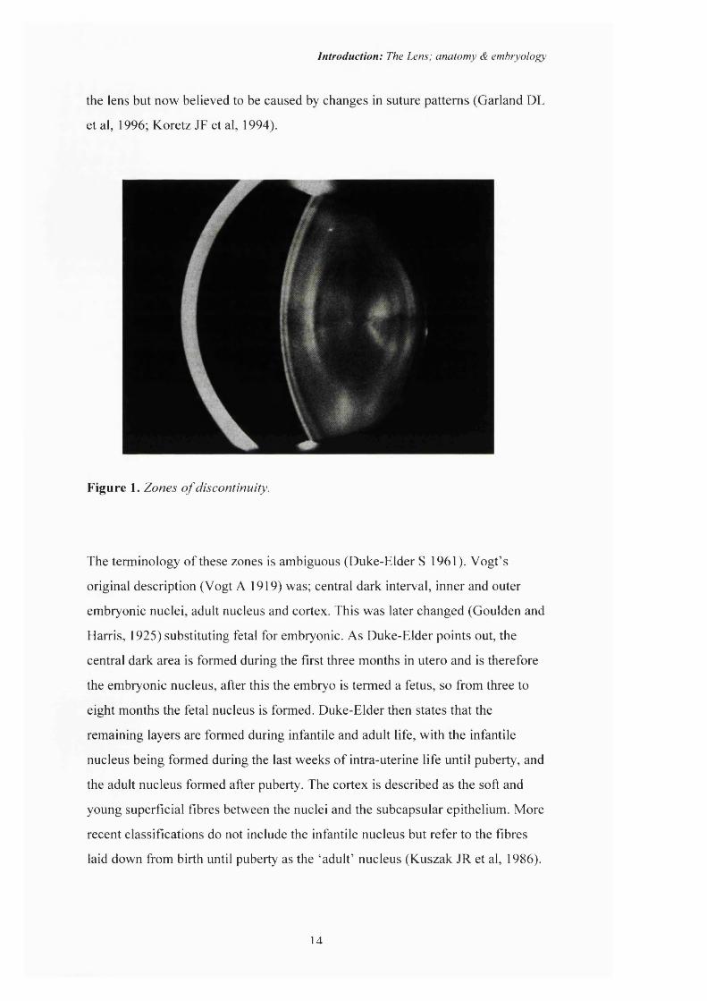
Zones of discontinuity: Slit lamp examination o f the lens reveals layers of
optical discontinuity (Vogt A, 1919) that correspond to different ages o f lens
development (Figure 1). Understanding these boundaries and the ages o f the
different optical zones allows for an estimate as to the timing of the insult
associated with the site of opacification in congenital and inherited cataract.
Although the secondary lens fibres are continually laid down throughout life they
do not form an optically homogenous lens but create sharp bands with different
optical qualities. These bands are formed by abrupt changes in the refractive
index previously thought to be caused by a temporary slowing in the growth of
13
Introduction: The Lens; anatomy & emhryology
the lens but now believed to be caused by changes in suture patterns (Garland DL
et al, 1996; Koretz JF et al, 1994).
Figure 1. Zones o f discontinuity.
The terminology of these zones is ambiguous (Duke-Elder S 1961). Vogt’s
original description (Vogt A 1919) was; central dark interval, inner and outer
embryonic nuclei, adult nucleus and cortex. This was later changed (Goulden and
Harris, 1925) substituting fetal for embryonic. As Duke-Elder points out, the
central dark area is formed during the first three months in utero and is therefore
the embryonic nucleus, after this the embryo is termed a fetus, so from three to
eight months the fetal nucleus is formed. Duke-Elder then states that the
remaining layers are formed during infantile and adult life, with the infantile
nucleus being formed during the last weeks of intra-uterine life until puberty, and
the adult nucleus formed after puberty. The cortex is described as the soft and
young superficial fibres between the nuclei and the subcapsular epithelium. More
recent classifications do not include the infantile nucleus but refer to the fibres
laid down from birth until puberty as the ‘adult’ nucleus (Kuszak JR et al, 1986).
la
Introduction: The Lens; anatomy & embryology
Phelps Brown and Bron reject this nomenclature on the grounds that the same
sets of lens fibres are regarded as being cortex at one period and nucleus at a later
date (Phelps Brown N, 1996). They regard the nucleus as being the total lens at
birth comprising the embryonic and fetal nuclei and the cortical fibres are those
laid down after birth (Sparrow JM, et al. 1986). This nomenclature creates two
broad zones of the adult lens; nucleus (formed in utero) and cortex (which forms
after birth). As will be seen later the phenotypes of autosomal dominant cataract
(ADC) appear to reflect this nomenclature in that the boundary between the outer
fetal nucleus and the cortex is a watershed in the development of lens opacities.
However, this also dictates that all congenital cataracts are therefore nuclear by
definition. The embryonic nucleus measures about 0.3mm in diameter whilst the
fetal nucleus measures 6-8mm in equatorial diameter and is the size of the lens at
birth (Phelps Brown, 1996). This is supported by the findings of Garland et al ^ ^
( 1996) who describe a characteristic protein pattern seen on electrophoresis that
distinguishes the lens nucleus (7mm in diameter) from the lens cortex (outside
the 7mm core).
t hroughout life the lens fibres are subjected to ‘compaction’ such that as more
lens fibres are laid down, the inner fibres become compressed and the size of
central opacities therefore reduces throughout life. Nuclear compaction takes
place during the first 20-30 years of life and thereafter the ‘compaction’ affects
only the cortical fibres (Brown NAP et al, 1988).
The lens fibres: The lens fibre cells develop from the lens epithelium lining the
anterior capsule. The anterior epithelium is divided into different regions
according to the stages of cellular development. The central zone consists of
flattened cuboidal cells surrounded by a concentric region referred to as the pre-
germinative zone which consists of cuboidal cells that are less flattened with
some of the cells entering mitosis. The next concentric region is the germinative
zone and consists of many cells undergoing mitosis. This continued cell division
pushes the fibre-precursor cells into the transitional zone (Beebe DC et al, 1980),
where they take on a more columnar shape. The uniform shape and ordered
15
9^ ■
\j*

Introduction: The Lens; anatomy & embryology
alignment characteristic o f the fibre cells is established in this region. When the
cells reach the lens bow at the equator, they rotate by 90 degrees, lose their high
columnar shape and elongate inta the slender lens fibre cells (Kuszak JR et al,
1983).
The morphology of the lens fibre cells is different according to the site they
occupy within the lens. The outer fibres are flatter having a smaller cross-
sectional diameter, whilst the inner fibres have more abundant membranous
inter-digitations and a greater cross-sectional diameter. Transmission electron
microscopy (EM) and scanning EM of the different regions of the lens show all
the fibre cells to contain homogeneous cytoplasm (Taylor VL et al 1996).
Further maturation of the lens fibre cell is accompanied by the loss o f the cell
nucleus and the organelles, and an increase in the concentration of crystallin
protein and the number of gap junctions (Bassnett S et al, 1992). The loss of the
organelles results in the fibre cell becoming metabolically inert with metabolites
and ions passing to the inner fibre cells from the outer cells of the lens via the
intercellular junctions. The crystallins that are laid down at this time must
therefore survive the entire life o f the individual, as they cannot be exchanged nor
removed.
Lens sutures: The lens sutures are formed by the interlocking ends o f the
secondary lens fibres and are opacified in many of the phenotypes o f ADC. As
the secondary fibres form around the embryonic nucleus o f primary lens fibre
cells, they meet at the anterior and posterior poles. There are different suture
patterns formed by the interlocking of the secondary fibres across different
species (Duke-Elder S et al, 1970). The simplest pattern is called umbilical and is
when all the fibres pass from one pole to the other, converging on the same point.
There is considerable tapering at the distal ends of the fibres to enable them all to
converge on the same spot. The remaining three patterns are called line sutures,
Y-sutures (Marshall J et al, 1982) and star sutures (Kuszak JR, et al, 1986). All
occur in the human lens at different stages of development although they are
16
h
Introduction: The Lens; anatomy & embryology
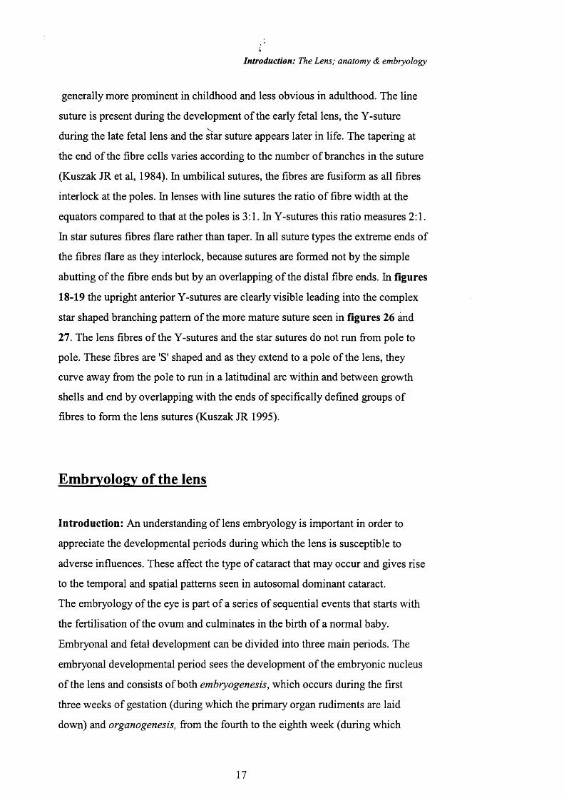
generally more prominent in childhood and less obvious in adulthood. The line
suture is present during the development of the early fetal lens, the Y-suture
during the late fetal lens and the ^ar suture appears later in life. The tapering at
the end of the fibre cells varies according to the number of branches in the suture
(Kuszak JR et al, 1984). In umbilical sutures, the fibres are fusiform as all fibres
interlock at the poles. In lenses with line sutures the ratio o f fibre width at the
equators compared to that at the poles is 3:1. In Y-sutures this ratio measures 2:1.
In star sutures fibres flare rather than taper. In all suture types the extreme ends of
the fibres flare as they interlock, because sutures are formed not by the simple
abutting of the fibre ends but by an overlapping of the distal fibre ends. In figures
18-19 the upright anterior Y-sutures are clearly visible leading into the complex
star shaped branching pattern of the more mature suture seen in figures 26 and
27. The lens fibres o f the Y-sutures and the star sutures do not run from pole to
pole. These fibres are 'S' shaped and as they extend to a pole of the lens, they
curve away from the pole to run in a latitudinal arc within and between growth
shells and end by overlapping with the ends of specifically defined groups of
fibres to form the lens sutures (Kuszak JR 1995).
Embryology of the lens
Introduction: An understanding of lens embryology is important in order to
appreciate the developmental periods during which the lens is susceptible to
adverse influences. These affect the type of cataract that may occur and gives rise
to the temporal and spatial patterns seen in autosomal dominant cataract.
The embryology of the eye is part of a series of sequential events that starts with
the fertilisation of the ovum and culminates in the birth of a normal baby.
Embryonal and fetal development can be divided into three main periods. The
embryonal developmental period sees the development of the embryonic nucleus
of the lens and consists of both embryogenesis, which occurs during the first
three weeks of gestation (during which the primary organ rudiments are laid
down) and organogenesis, from the fourth to the eighth week (during which
17
Introduction: The Lens; anatomy & embryology
these primary organ rudiments -including the embryonic lens, are developed).
The fetal development is dominated by differentiation, in which the organs
develop into fully or partially active organs (Y. Robert Barishak, 1992). It is
during this period that the fetal lens develops.
Embryogenesis covers the first three weeks o f development and results in the
formation of the three germ layers; ectoderm, mesoderm and endoderm and the
establishment of the primary organ rudiments. Although the eye is composed of
three primordial tissues; neural ectoderm, surface ectoderm and mesoderm, the
lens is derived from only the surface ectoderm. There is no evidence o f the lens at
this stage.
The first evidence of the future lens appears at the 4mm stage, during the fourth
week, with the formation of the lens plate or placode (Ida C Mann, 1928) at
which point the ectodermal cells become epithelial cells (Zigman S, 1985). The
lens placode is a thickening of the surface ectoderm overlying the optic vesicle.
The ectodermal cells become epithelial cells and failure of this stage of
development results in a primary aphakia, a rare developmental abnormality of
the eye. The lens plate gives rise to the lens pit during the fifth week of gestation
and the formation o f the lens vesicle which remains temporarily attached to the
surface ectoderm by the lens stalk.
The lens vesicle is covered by a basal membrane, which develops into the lens
capsule and surrounds the lens vesicle completely as the lens stalk degenerates.
Persistence of the optic stalk as a comeolenticular connection (Coulombre AJ,
1979) may be associated with anterior polar cataract (see figures 2-3 p57). The
formation of the lens vesicle, enclosed within the capsule, seals off the lens cells
from the rest of the body. This semi-permeable lens capsule gives the lens
immunological privilege, allows for the development of osmotic cataract as seen
in diabetes, galactosaemia (Chylack LT Jr et al, 1981) and the Nakano mouse
(Nakano T et al, 1960; Kinoshita JH et al, 1974). As the lens increases in size
18
Introduction: The Lens; anatomy & embryology
throughout development, so does the capsule, and abnormal capsular
development may result in capsular irregularities and polar cataract.V,
The sixth week sees the initial development of the primary lens fibres from the
posterior cells of the lens vesicle and during the seventh week these elongate to
obliterate the lumen o f the lens vesicle and form the embryonic nucleus which
measures about 0.3mm in diameter. This elongation is accompanied by the
formation of intra-cellular microtubules parallel to the axis o f elongation and if
these fail to develop the cells enlarge with the accumulation o f crystallins but do
not develop their strap-like shape resulting in cataract (Byers B et al, 1964).
Influences from outside the lens at this stage may result in cataract. It has been
demonstrated that in chick embryos the vitreous produces lentropin that
stimulates crystallin synthesis (Piatigorsky J, 1981) and the neural retina provides
growth factors that stimulate lens development (Simmoneau L et al, 1983). It has
been shown experimentally that removal of the neural retina results in abnormal
lens development and cataract formation, whilst replacement of the neural retina
allows normal lens development (Coulombre J et al, 1965). Even in cases where
cataract is the only abnormality, as in ADC, extra-lenticular factors may still be
involved in the pathogenesis.
With the addition of successive layers o f lens fibres, the primary lens fibres lose
contact with the capsule and the nuclei o f these cells as well as some o f the cell
organelles degenerate (Kuwabara T, 1975). This degeneration limits the time
period during which genes that may result in cataract formation can be expressed,
offering some protection to the embryonic nucleus. The transition of the
epithelial cells to fibre cells is completed with elongation and the loss o f the
organelles.
There is rapid development of the lens during the eighth week as the secondary
lens fibres form. The anterior epithelial cells divide and the newly formed cells
are pushed posteriorly and become elongated. Each successive layer of cells
grows over the previous one and results in the formation of the two following
19
Introduction: The Lens; anatomy & embryology
structures: The lens bow is formed by the nuclei o f the secondary lens fibres as
they move forward and form an arc with the convex anterior; the lens sutures
develop because all fibres of the same age have similar lengths and meet
anteriorly and posteriorly.
Differentiation: At the end o f the third month an embryonic and an inner fetal
nucleus can be identified (Ozanics Vet al, 1982). The outer fetal nucleus
develops during the fourth month and new suture lines appear as bifurcations in
the arms o f the sutures. Lens growth occurs in the same manner in an adult lens
as in an embryonic lens by the addition of the secondary lens fibres. The
changing shape o f the lens from a sphere to an elliptical form takes place from
the fourth to the sixth month as the lens equatorial cells continue to proliferate
(Sivak JG et al, 1987) and at eight months of development the lens is
approximately 6-8mm in diameter.
Summary: The anatomy of the lens as seen through the slit lamp is composed of
different regions each o f which are associated with a particular period of
development. The small central embryonic nucleus measures approximately
0.3mm and is formed by the primary lens fibres during the first two months of
development. Thereafter the secondary lens fibres continue to be laid down
throughout life and form the remainder of the lens. The fetal nucleus is formed
up until birth and contains the Y-sutures and measures approximately 6-8mm in
equatorial diameter at birth. The secondary lens fibres laid down after birth make
up the cortex of the lens.
Autosomal dominant cataract can therefore be pre-natal, affecting the lens
nucleus, post-natal, affecting the lens cortex, or have opacities throughout the
lens reflecting abnormal development both before and after birth.
20
Introduction. The Lens; molecular biology
Molecular Biology of the lens
Introduction
In this section the molecular biology of the lens is discussed. Many of the genes
encoding major lens proteins such as the crystallin proteins, the membrane
proteins and the cytoskeletal proteins have been identified and are good
candidate genes for inherited cataract in man.
Crystallin Proteins
The mature lens fibre cell has no nucleus nor cell organelles and there is
therefore little turnover nor renewal of protein in the inner fibres of the lens
(Kuwabara T, 1975). For the lens to maintain its transparent and refractive
properties it requires a high concentration of regularly packed proteins that are
very stable and can resist dénaturation from optical radiation, free radicals and
heat. These proteins also need to last the entire life of the lens as they cannot be
degraded nor replaced. The crystallin proteins fulfil these requirements.
Previously thought to be structural proteins with a unique function in a highly
specialised tissue, it is now realised that the crystallins are expressed in other
tissues of the body in very different guises (Piatigorsky J et al, 1983).
The crystallin proteins make up over 90% of the water-soluble proteins o f the
lens and are responsible for conferring and maintaining lens transparency (Cartier
M et al, 1994; Delaye M et al, 1983; Wistow GJ et al, 1988). Crystallins can be
divided into two categories; the ubiquitous crystallins that are present in all
vertebrate lenses (a, p and y) (Bloemendal H, 1989) and the taxon specific
crystallins, only present in certain species (Piatigorsky J et al, 1989). The three
major classes of human crystallins are a , P and y, the high molecular weight
fraction being the a-crystallins, the intermediate fraction the p-crystallins and the
low molecular weight fraction the y-crystallins (Bloemendal H et al, 1989). The
a-crystallin proteins are thought to have evolved from one gene and the P- and y-
crystallins are so closely related to each other that they are also thought to have
evolved from a single gene (WW de Jong et al 1989). The P- and y-crystallins are
21
Introduction. The Lens; molecular biology
now often grouped together and referred to as the Py-crystallins. A Ç-(zeta)
crystallin gene has recently been described in humans, although it has a very low
level of expression (Heinzmann et al 1994).
The order o f appearance of the crystallins in development as well as their
position within the lens differs according to different species (Piatigorsky J,
1981). a-crystallin is found in the lens placode and is synthesised in the epithelial
cells whilst the p- and y-crystallins appear after or during fibre cell elongation
(Harding J, 1991, McAvoy 1978). The heavier molecular weight proteins
predominate in the epithelium and outer cortex while y-crystallins and the lower
molecular weight proteins predominate in the protein-rich inner nucleus
(McAvoy JW, 1981). This creates a protein dense nucleus resulting in a higher
refractive index of the nucleus as compared to the cortex. The optical benefit is a
decrease in both the spherical and chromatic abberations that affect lenses of
uniform refractive index.
The crystallins have traditionally been regarded as soluble, structural proteins
restricted to the lens although it is now apparent that they have close homology
to other proteins outside the lens and in some cases are coded by the same or
similar genes as these other proteins. It is probable that these proteins were
recruited by the lens to form the crystallins because of their stable structure
enabling them to survive within the lens fibre cells and maintain lens
transparency (Wistow G et al, 1987). The crystallins may have arisen by gene
duplication and divergence, specialising for the lens environment. This is
particularly illustrated by the study of the taxon specific crystallins that show
close homology with enzymes. These taxon (or species) specific crystallins will
be discussed later.
a-crystallins: The a-crystallins comprise 30-40% of the soluble protein and are
polypeptides (20 kDa) that aggregate to form larger complexes (approximately
800 kDa). They are coded by two genes that share nearly 55% sequence
homology, aA (chromosome 21;22.3) and aB (chromosome 11; 22.3-23.3), that
22

Introduction. The Lem; molecular biology
give rise to one acidic and one basic polypeptide. Post-translational modifications
to the protein by phosphorylation, deamidation, acétylation, degradation and
racemization lead to a variety of a-aggregations in the lens (Spector A et al,
1985). These modifications may have a role in altering the interactions of a-
crystallins with each other and with other lens components.
The aB-crystallins show sequence homology to a number of proteins found
elsewhere in nature. These include heat shock proteins (Ingolia TD et al, 1982)
and a schistosoma mansoni egg antigen (Nene V et al, 1986). Similar proteins
have been found in skeletal muscle, heart, kidney, retina and lung where they
may have a function as heat-shock proteins. Proteins similar to aB-crystallin
have also been found in the brains of humans affected by neurological diseases
including multiple sclerosis (van Noort JM et al, 1995), Creutzfeld-Jakob disease
(Jahal C et al, 1992), Alexander’s disease (Iwaki T et al, 1989), Huntingdon’s
disease, Parkinson’s, Pick’s and Lewy body diseases. They gave also been found
in the brains of hamsters with scrapie (Duguid JR et al, 1988). More recently the
aB-crystallins have been found to have chaparone activity which may stabilise
other crystallins in the lens preventing uncontrolled aggregation (Rao PV et al,
1995). The similarités of the aB crystallin to heat shock proteins suggests a role
in the reaction of the lens to stress. aB-crystallin can be induced by heat-shock
and other insults including osmotic stress, ischaemia and expression of some
oncogenes (Wistow G, 1995). The function and mechanism of action of the heat
shock proteins is not known but they are present in chloroplasts of plants where
they may protect against damage from light and heat. Unlike aB crystallin, aA
crystallin is not expressed so widely outside the lens although it has been found
in spleen and thymus (Kato K et al, 1991) and may also function as a heat shock
protein (Van den Ijssel et al, 1996). A mutation in the human aA crystallin has
been shown to cause a range of eye disorders including microphthalmia and
cataract (Litt et al, 1998).
Little is known about the regulation of the genes for the a-crystallins in the
human lens, although aA-crystallin mRNA has been detected in fetal lenses
23

Introduction. The Lens; molecular biology
(Brakenhoff RH et al, 1990). The a-crystallins are the first crystallins to appear
during the development of the lens in rodents and are present in the lens vesicle
before the development of the primary lens fibre cells and are synthesized mainly
in the lens epithelium (van Leen RW et al, 1987). The mRNA for both aA - and
aB-crystallin can be detected at all stages of lens development although as
already stated the outer layers contain a greater proportion of the a-crystallins.
P-crystallins: The p-crystallins also form aggregates and are also divided into
basic (PBl-3) and acidic (PAl-4) sub-types. Like the a-crystallins they undergo
various modifications including phosphorylation, oxidation, cross-linking and de
amidation. The P-crystallins are mainly encoded by genes on chromosome 22
(22q 11.2-13.1), although PAI and pA3 are on chromosome 17 (1 7 q ll.l-q l2 )
(Sparkes RS et al, 1986). The developmental regulation o f the p-crystallin genes
has been studied in the chicken and the rat but little is known about the regulation
in humans, although PB3 is known to be expressed early and pB2 in the later
stages (Brackenhoff RH et al, 1992) of lens development in humans. The
chromosome 22 locus for pAl and pA3 has been shown to be linked to a
cerulean cataract in humans (Kramer P et al, 1996) and a chain termination
mutation has been identified in CRYBB2 as causing the cataract. In the Philly
mouse a dominant nuclear and subcapsular cataract (Kador PF et al, 1980) is
caused by a deletion in the PB2 cDNA (Chambers C et al, 1991) resulting in a
mutant pB2 protein being formed that is thought to give rise to the cataract.
y-crystallins: The y-crystallins unlike the other crystallins are monomers and can
be divided into two groups; one containing the ys present in all vertebrate
species, and the other made up of six closely related proteins known as yA, yB,
yC, yD, yE and yF which are absent in fish and some reptiles. The y-crystallins
have a high internal symmetry and are very stable enabling them to withstand
dehydrated environments. The ys gene is located on chromosome 3 and the yA-yF
genes are clustered together on chromosome 2 (Shilloh Y et al, 1986) region q33-
q35. In the embryonic nucleus of humans the yC and the yD account for 82% of
24
Introduction. The Lens; molecular biology
the y-crystallin transcripts, the yA gene contributes 14% and the yB gene 5%
(Brackenhoff RH et al, 1992). The yE and yF are considered pseudogenes
although a low level o f yE transcript has been detected. There is also a segment
o f the yF gene that is duplicated within the gene cluster, called yG but it is non
functional. The y-crystallins are located primarily in the lens nucleus and most of
the genes are expressed early in development and have reduced activity after
birth. At 22 months only the yD can be detected and remains present until 10
years of age.
The y-crystallin gene complex is important regarding cataract formation and
linkage has been demonstrated for two cataract phenotypes to this locus in
humans; the Coppock-like cataract (Lubsen et al 1987) in which the opacities are
confined to the nucleus, and the polymorphic cataract (Rogaev El et al, 1996)
which has more widespread and variable opacities. The Coppock-like cataract
(figures 20 - 25) has been shown to be caused by an over-expression o f the yE
pseudogene (Brakenhoff RH et al, 1994). In the Elo (eye lens obsolescence)
mouse, a single nucleotide deletion in the yE-crystallin gene is responsible for the
dominant cataract (Cartier M et al, 1992).
Zeta crystallin: The Ç-crystallin lens protein has been associated with an
autosomal dominant cataract in the guinea pig (Rodriguez IR et al, 1992), and is
a major protein in the lenses of guinea pigs and camels where it comprises about
10% of the total soluble protein (Garland D et al, 1991). The Ç-crystallin gene
(CRYZ) has recently been mapped in humans to chromosome lp22-
p3 l(Heinzmann C et al, 1994) and although it has a low level o f expression it
provides an important candidate gene to be included in linkage studies for
autosomal dominant cataract.
Taxon specific crystallins: Amongst the taxon specific (or species specific)
crystallins some are active enzymes and others are related to enzymes but have
no activity. In the lenses of crocodiles and birds, e-crystallin was found to be
essentially identical to lactate dehydrogenase (Wistow GJ, 1987) and T-crystallin
in the lens o f some fish, birds and lampreys is closely related to enolase in
25
"A

Introduction. The Lens; molecular biology
humans and in yeast (Wistow GJ et al, 1988). The major protein o f the
embryonic lens in birds and reptiles is ô-crystallin which has significant sequence
homology to the urea cycle enzyme argininosuccinate lysase in humans (O’Brien
W et al, 1986) and Ç-crystallin in guinea pigs (Huang QL et al, 1987) has
recently been found to have NADPH-dependent quinone reductase activity. The
^-crystallin gene is unusual in that the high expression in the lens o f guinea pigs
is mediated by an alternative lens-specific promoter distinct from that responsible
for expression of the enzyme in other tissues (Gonzalez P et al, 1994). These
taxon specific crystallins are not only similar to these enzymes but are products
of the same genes as the enzymes. The crystallins that resemble enzymes but
have no activity are p-crystallin in frogs (Gause GG et al, 1985) and X-crystallin
in rabbits and hares (Mulders JWM et al, 1988).
Most o f the enzymes that are associated with the crystallins are related to
enzymes involved in carbohydrate metabolism and these enzymes are in turn
induced by stress (Nickells RW et al, 1988). This is relevent as the metabolic
energy required by the lens is largely obtained from anaerobic glycolysis
(Harding JJ et al, 1984). It is not yet clear why the crystallins have been recruited
from these particular enzymes, it may be the stable structure, the individual
enzyme function or even the propensity for readily inducible gene expression
explaining their presence in such large amounts within the lens fibre cells (de
Jong WW et al, 1989).
The relevance of the taxon specific crystallins to a human linkage study is
illustrated by the linkage demonstrated for the Volkmann cataract (Eiberg H et al,
1995) and the stationary posterior polar cataract (lonides et al, 1997) in humans
to chromosome lp36. Within this region lies the gene ENOl (enolase 1 alpha
that also codes for T-crystallin, a crystallin not found in humans). Although this
has not yet been shown to be involved in the formation of the Volkmann cataract
nor the posterior polar cataract, this has yet to be fully assessed.
26
Introduction. The Lens; molecular biology
Lens membranes
The embryonic and fetal nuclei of the lens contain some of the longest lived cell
membranes in biological systems. Although the crystallins are the major water
soluble proteins accounting for 80-90% of the dry weight of the lens, the
remaining 10-20% is made up of the cytoskeletal and membrane proteins and is
collectively referred to as the water insoluble fraction. The standard
nomenclature for the plasma membrane proteins is ‘M P’ standing for ‘membrane
protein’ followed by the molecular weight in kilodaltons (Bloemendal H et al,
1977). Major intrinsic protein (MIP) is therefore called MP-26, having a
molecular weight of 26kD and is the the principle constituent o f this fraction
(Zigman S, 1985).
The histological differentiation of the lens epithelial cell into a fibre cell is
associated with the synthesis o f large amounts of two classes of lens proteins; the
crystallins and the membrane proteins (Bloemendal H, 1979). The membrane
proteins are known to be involved in the formation o f intercellular junctions
called 'gap junctions' and the percentage of the membrane involved in these
junctions is as high as 64% (Kuszak J et al, 1978). The loss o f the organelles
reduces light scatter and the increased number of intercellular junctions is
thought to permit the passive diffusion of ions and metabolites between the cells
(Rae J et al, 1983. Bassnett S et al, 1994). The outer epithelial cells therefore
fulfil the metabolic requirements of the inner fibre cells via this large network of
fibre cell gap junctions (Kinsey et al 1964, Mathias et al 1997, Goodenough et al
1992). There are no blood vessels in the lens. Another important role o f the
membranes is the control of the ionic balance which is achieved by the Na, K-
ATPase and the Ca-ATPase. Sodium is pumped out of the cells followed by
chloride and water, preventing swelling and clouding of the lens. The gap
junction also maintains a very narrow intercellular space which is critical for lens
transparency and the disruption of this may result in cataract.
Connexins: Gap junctions are channels that connect neighbouring cells and
allow the passive diffusion of inorganic ions and small molecules (Elfgang et al
27
Introduction. The Lem; molecular biology
1995). The trans-membrane proteins that make up the gap junctions are called
connexins (Cx) and are encoded by family of genes that have recently been
identified (Kumar & Gilula 1996). Connexons are classified according to their
molecular weight. Six connexin molecules aggregate to form a ring called a
connexon, within the plasma membrane (Cascio et al 1995). Two connexons
from neighbouring cells then align with each other and 'dock' together to form the
gap junction channel. Gap junctions then cluster together in the plane of the
membrane to form gap junction plaques. The permeability of the gap junctions
varies according to the constituent connexins (Elfgang et al 1995). Cataracts have
been shown to occur in mice lacking Cx43 (Gao et al 1998) which is
predominantly expressed in lens epithelial cells. Two connexins are known to be <
present in the human lens, Cx46 and Cx50.
MP-70 (Cx 50): The gene for MP70 has been mapped to chromosome lq21.1
(Church RL et al, 1994) in humans and a murine homologue protein called
connexin 50 has been shown to be a gap junction protein. The CAE pedigree ^
studied first by Nettleship, later by Renwick and Lawler and more recently as
part of this study maps to this region on Iq. As will be discussed later we have '
demonstrated that a mutation in this gene is responsible for the cataract in the
CAE family (Shiels et al 1998) Pedigree P in appendix 1. Connexin 46 (GJA3)
has been assigned to chromosome 13ql l-q l2 (Willecke et al 1990, Mignon et al
1996) we have shown that two mutations in this gene give rise to the cataract in
two different pedigrees affected by pulverulent cataract (Mackay et al 1999). The
pedigrees are pedigrees L and M in appendix 1.
MP-26 (MIP): MP-26 (Main Intrinsic Polypeptide) also called aquaporin, is the
major protein of the cell junctions (Sas DF et al, 1985), forms 40% of the fibre
membrane and the increase in inter-cellular junctions during the maturation of
the fibre cells is accompanied by a large increase of MP-26. The increased
protein content of the ageing cells (Horwitz JN et al, 1979) has however been
associated with cataract in later life (Zigman S, 1985; Spector A, 1979). MP26 is
present in both the lens nucleus and cortex and is a member of a large family of
28
If
Introduction. The Lens; molecular biology
integral membrane channel proteins involved in the regulation of water
movement (Reizer J et al, 1993). There are several types of inter cellular
junctions between the lens f i b r e s M P 2 6 is organised differently with each
type (Zampighi GA et al, 1989), it is also present in non-junctional membrane.
The MP-26 gene has been mapped to the distal end of chromosome 10 in the
mouse (Griffen CS et al, date) and to chromosome 12q in humans (Sparkes RS et
al, 1986) and a mutation o f MP-26 has been implicated as the cause of the
cataracts in both the Cat ^'^and the Lop mouse cataracts (Shiels A et al, 1993;
Shiels A et al, 1991; Shiels A et al, 1996). It is therefore an important candidate
gene for inherited cataract in mice and humans.
MP-19: The second most abundant membrane protein is another intrinsic protein
(Mulders JWM et al, 1988), MP-19 (also referred to as MP-17 and MP-18) and is
again thought to be involved in the formation of these osmoregulatory trans
membrane channels. Like MP26 (MIP) it is distributed throughout the lens
(TenBroek E et al, 1992) and has been shown to bind calmodulin (Galvan A et al,
1989) although its role is still uncertain (Zampighi GA et al, 1992). The gene has
been mapped to chromosome 19 in humans and mutations in the mouse gene for
M PI9 has been shown to cause inherited cataract.
Cytoskeletal elements
As the fibre cells elongate and become internalised into the lens nucleus, the
alteration in shape is reflected by an alteration in the cytoskeletal architecture.
Initially, cytokeratins, vimentin and two lens specific intermediate filaments,
phakinin (CP49) and filensin are expressed in the embryonic lens (Sandilands A
et al, 1995). In the mature lens fibre cell keratin and vimentin are not expressed
leaving CP49 and filensin as the only remaining intermediate filaments. These
two cytoskeletal proteins are unique to the lens and are the major components of
the beaded filament that combine with a-crystallin (Carter JM et al, 1995).
Filensin and CP49 have now been cloned and the genes localised to
chromosomes 20 and 3 (Hess JF et al, 1996).
29

h
Introduction. The Lens; molecular biology
Molecular Biology and Cataract
General features o f cataract formation include the presence o f vesicles, lens fibre
swelling, disordered orientation of fibre cells and loss of membrane integrity
(Berman ER, 1991). At the molecular level there are aggregates of high
molecular weight proteins caused by the unfolding of crystallins, products of
proteolysis, abnormal levels o f sodium, potassium and calcium and decreased
amounts of glutathione and soluble crystallins. These changes have been studied
mainly in senile cataract but they point to potential mutation sites that may give
rise to inherited cataract such as the crystallin genes, the genes for membrane
proteins and genes known to play a role in lens development.
Whilst biochemical studies have implicated these molecules in cataract
formation, it has not been possible to determine whether their involvement was a
primary or a secondary event. Molecular genetic studies can screen the genes for
these proteins and the finding of pathological mutations will provide evidence
that defective crystallins and other lens proteins can be a primary cause o f
cataract.
30
Introduction. ADC: Clinical background
ADC: Clinical background
Historical background: Congenital cataract is a major cause of childhood
blindness (Lambert SL et al, 1996; Evans J et al, 1996; Lloyd IC et al, 1994) and
has a genetic basis in over 25% of cases (Merin S et al, 1971). Inherited cataract
has been recognised for many centuries and can be inherited as an autosomal
dominant, autosomal recessive or X-linked trait. Doyne (Doyne RW, 1889),
Nettleship (Nettleship E, 1905; Nettleship E, 1909; Nettleship E, 1909), Harman
(Harman N, 1909; Harman N, 1910)and others (Sæbo J , 1949; Priestly Smith,
1910) have documented enormous numbers of pedigrees affected with inherited
cataract and have clearly illustrated the patterns of inheritance. The great
majority of inherited cataract is autosomal dominant inkfip mode of transmission.
The phenotypes of inherited cataract have not been systematically investigated.
In 1910 Bishop Harman described the appearance of the lens opacities seen in
families with inherited cataract and listed five groups; lamellar, coralliform,
stellate, anterior and posterior polar and finally an undefined group (Harman N,
1910). Clapp divided congenital cataract of all aetiologies into two broad groups;
complete and partial. He then subdivided the partial cataracts into zonular,
coralliform, punctate, discoid, pyramidal, Y opacities, Roriform, fusiform, disc
shaped and Morgagnian (Clyde A Clapp, 1934). Duke Elder (Duke-Elder S,
1964) François (François J, 1963) and Vogt (Vogt A, 1979) describe in detail the
different kinds of cataract that can be inherited in individuals but no study has
been undertaken examining the different phenotypes of pedigrees.
Mapped Phenotypes
At the start of this study, linkage had been demonstrated for three phenotypes and
the gene had been isolated at one of these loci (at the y-crystallin gene complex,
Lubsen et al 1987). Ten loci have now been shown to contain genes for human
cataract and the mutations have been identified in seven of these genes. This
study demonstrated linkage at five loci and has identified three mutations in two
novel genes that give rise to cataracts (Table 1, page 38). Mutations in the genes
31
Introduction. ADC: Clinical background
encoding a-, p- and y- crystallins as well as in the genes for connexins have now
been shown to give rise to inherited cataract. There is no generally agreed
classification for the phenotypes o f ADC. Initially the names o f the phenotypes
were taken from the surname of an affected ancestor (Coppock, Volkmann), the
resemblance to a previously described phenotype (Coppock-like) or even the
ophthalmologist involved in their care (Mamer). More recently the phenotypes
have been named according to a description of the opacity (anterior or posterior
polar etc) but a classification has never been accepted into general
ophthalmological use. A clinical classification would not be able to successfully
encompass the enormous heterogeneity of ADC. There has yet to be a common
terminology amongst researchers for describing the lens opacities and the
phenotypes.
Below is a description of the phenotypes from the pedigrees that had been
mapped to specific loci at the onset and during the initial years of this thesis.
Zonular pulverulent Cataract (CAE): Renwick and Lawler studied a family
affected by zonular pulverulent cataract that was described in their original paper
to be “family Ev. from Southern England” (Renwick and Lawler 1963). This
family was first documented by Nettleship (Nettleship E, 1909). The opacity is
described as being in the centre of the lens, ‘about 4mm in apparent diameter and
sometimes, within this zone, (contains) a distinct denser part in the nuclear
region. Each cataract was composed of innumerable powdery opacities... The
cataract...was bounded by a smooth envelope consisting of a thin layer of
particularly fine opacities and, outside this, was clear in about five eyes out of
six. In the remaining eyes, this outer zone had one or more shells also consisting
o f these powdery opacities.’ This zonular pulverulent cataract is often mistakenly
called the Coppock cataract. The Coppock cataract has never been included in
any published linkage study, has opacities confined to the embryonic nucleus and
is called the ‘central pulverulent cataract’ (as opposed to the zonular pulverulent
cataract described here). It was at one time also called the Doyne’s discoid
cataract (Adams PH, 1942) after the author of its first description. Although the
32
Introduction. ADC: Clinical background
Coppock pedigree was studied by Nettleship (Nettleship E, 1906) no kinship
between the Coppock and the Everett families (family Ev) could be found.
Coppock-like C ataract (CCL): The ‘Coppock-like cataract’ family were
originally described by Priestly Smith (Smith P, 1910) and Bishop Harman
(Harman NB, 1910) in 1910. Smith’s paper is titled ‘A pedigree o f Doyne’s
discoid cataract’ and states that the Forman pedigree ‘bears much resemblance to
the Coppock pedigree.’ The cataract is described as ‘discoid in form...its size
varies in various individuals, producing various degrees o f visual impairment.’
Bishop Harman describes the phenotype as being ‘circular, about 3mm in
diameter, and o f a granular appearance.’ Duke-Elder classifies the Coppock and
Coppock-like cataracts as central (nuclear) cataracts (Duke-Elder S, 1964). No
connecting link between the Coppocks and the Formans suggesting kinship could
be detected at this time. The descendants o f the Forman family have participated
in this study and Figures 22-27 (pp70-72) are slit lamp photographs of the
phenotype o f the Coppock-like cataract from two different individuals showing
the central, nuclear position of the opacity. There have never been any previously
published illustrations o f the cataract from the Coppock-like pedigree.
M arner C ataract: The Olsen family from Denmark affected with this cataract
were first described by Giersing in 1878 (Giersing CM, 1878) updated by Norrie
in 1896 (Norrie G, 1896) and then studied by Mamer from whom the phenotype
received its name. Mamer describes the cataract as having ‘fine, dispersed,
pulvemlent opacities in the embryonic nucleus. In the outer zone of this nucleus,
equatorially orientated, linear condensations are found...opposite the extremities
o f the anterior Y-suture. These condensations may be slightly arched with the
concavity directed centrally making the circumference of the embryonic nucleus
look triangular...Outside the embryonal nucleus one or more zones with
pulvemlent opacities may be seen, and peripherally to this some ‘riders’ may be
located. Sometimes, star shaped opacities located in the posterior cortical layer of
the lens are observed.’ There is also considerable inter-individual and intra-
familial variation of the phenotype (Mamer E, 1989). In an earlier paper the
33
Introduction. ADC: Clinical background
different morphologies were described as being zonular, central, stellate or with
an anterior polar opacity (Mamer E, 1949).
Cerulean cataract: Linkage has been demonstrated for two pedigrees affected
by cemlean (blue-dot) cataract to two different loci. The cerulean cataract was
first described by Vogt in 1922 (Vogt A, 1922) and Armitage et al demonstrated
linkage o f a pedigree affected by this phenotype to chromosome 17q (Armitage
MM et al, 1995). The pedigree they studied had an early onset bilateral cataract
characterised by tiny blue or white opacities that formed in the superficial layers
of the fetal lens nucleus and could be detected by slit lamp examination from the
age o f 18-24 months. There was no sign of the cataract at birth. The opacities
progressed to involve the adult nucleus and cortex ‘forming concentric layers,
with central lesions orientated radially.’ The cataracts were slowly progressive
with the affected individuals requiring cataract surgery between the ages o f 16
and 35 years.
A second locus for cemlean cataract was then reported a year later by Kramer et
al mapping to the p-crystallin region on chromosome 22q (Kramer P et al, 1996)
and a mutation of the p crystallin gene detected (Litt M et al 1997). The cataract
was described in younger members as having ‘peripheral blue flakes and
occasional spoke-like central opacifications’ with minimal effect on visual
acuity. ‘In most cases cataract extraction became necessary between 20 and 40
years o f age.’(Bodker FS et al, 1992). The slit lamp photographs o f an affected
individual from a family affected by blue-dot (cemlean) cataract included in this
study are shown in figures 36 and 37, page 80.
Zonular-sutural cataract: A three-generation family with isolated autosomal
dominant cataract was studied by Padma et al (Padma T et al, 1995) in which the
phenotype consisted of zonular and sutural opacities. They state that all ‘affected
family members showed a zonular cataract that measured 3.5-4mm in diameter,
an erect Y-shaped anterior sutural cataract, and an inverted Y-shaped posterior
sutural cataract. The fetal and embryonic nuclei within the zonular cataract were
34
Introduction. ADC: Clinical background
clear except for a few dotlike white opacities.’ This description is very similar to
lamellar pedigrees examined as part of this study and illustrated in figures 30-34
pp75-78. As in the zonular pulverulent and Mamer cataract pedigrees, the
zonular sutural cataract pedigree shows intra-familial phenotypic heterogeneity in
that the cataract may vary from a uniform dense opacity to a collection o f fine
dots forming a hazy cloud.
Volkmann cataract: This phenotype is named after the earliest known ancestor
o f the affected Danish family. It has been followed for ten generations and
consists of 426 people. The cataract is ‘characterised by a progressive, central
and zonular cataract, with opacities both in the embryonic, fetal and juvenile
nucleus and around the anterior and posterior Y-suture.’ It has a ‘highly variable
expression ranging from hardly recognisable opacities in the lens to dense
cataracts.’ (Eiberg H et al, 1995).
Polymorphic: A study of the Turkmen population in the former Soviet Union
found a high incidence of autosomal dominant cataract. A seven-generation
pedigree consisting of 254 members of whom 103 were affected by a
polymorphic cataract underwent linkage analysis. The phenotype ‘is
characterised by partial opacity of the lens, which has a variable location on the
periphery between the fetal nucleus of the lens and the equator. The form of the
opacity is irregular and looks similar to a bunch of grapes or a lump of cotton.
The opacities can be located simultaneously in the different lens layers. The
colour of the opacities varies from shining crystal-like to snow-white. This
cataract is different from nuclear cataracts and more similar to zonular or
lamellar types o f cataracts.’ (Rogaev El et al, 1996). The detailed description is to
illustrate the difference of this polymorphic phenotype to that of the Coppock-
like cataract as they both link to the y-crystallin cluster on 2q.
Pedigrees affected by anterior polar cataract, posterior polar cataract and two
pedigrees affected by pulverulent cataract have been linked to different loci as
part of this project and these phenotypes will be discussed later.
35
Introduction. ADC: Clinical background
O ther linkage studies:
There have been many other linkage studies with families affected by ADC and
whilst these have excluded loci they have not demonstrated positive linkage.
Many of the phenotypes in these linkage studies resemble mapped phenotypes,
which suggests considerable genetic heterogeneity. Hammerstein and Scholz
(Hammerstein W et al 1974), conducted linkage analyses on a family with
cataracts in the embryonal nucleus similar to the central pulverulent cataract,
Huntzinger et al (Huntzinger RS et al, 1977) studied a family with dense nuclear
cataract, Conneally et al (Conneally PM et al, 1978) studied a cataract resembling
the zonular pulverulent cataract which suggested linkage to the same locus on
chromosome 1 but was inconclusive and Stabile et al (Stabile M et al, 1983)
performed linkage on a family with a pulverulent cataract. Bateman et al
(Batemen JB et al, 1986) studied a family with a nuclear cataract, Beaumont et al
(Beaumont C et al, 1989) and Bird wood et al (Bird wood SC et al, 1968)
excluded loci an a family with lamellar cataract and Barrett et al (Barrett D et al,
1988) showed exclusion for a family with an embryonal cataract. More recently,
negative linkage studies were still being published and Basti et al reported the
negative findings from a linkage study on a family affected by zonular cataracts
and sutural opacities (Basti et al, 1996).
Summary:
These phenotypes that have been mapped to genetic loci represent a fraction of
the broad clinical spectrum seen in ADC. Clinical heterogeneity exists not just
between different pedigrees but also within certain pedigrees. Similar phenotypes
map to widely separated loci, as seen with the blue-dot phenotype, and very
different phenotypes appear to be allelic, as with the Volkmann and posterior
polar cataract phenotypes. However, the phenotypes that have demonstrated
linkage and have had the underlying gene mutation analysed show startling
correlation to the molecular biology.
36

Introduction. ADC: Molecular genetics
Molecular Genetics
Introduction
In recent years there have been considerable advances in the field of molecular
genetics (Antonarakis SE, 1994. Yates JRW, 1996) and there are now ten loci
known to be involved in the development of ADC. At the outset o f this study in
1994 there were only three loci identified as being involved in cataract formatiôn;
the early linkage in 1963 by Renwick and Lawler (Renwick and Lawler 1963),
the linkage of the Coppock-like cataract to the y-crystallin gene complex on 2q in
1987 (Lubsen et al 1987) with the subsequent mutation in the y-E crystallin gene.
And finally the linkage o f the Mamer cataract to 16q in 1988 (Eiberg et al 1988).
The linkage by Renwick and Lawler was performed using protein polymorphisms
such as blood group and serum protein markers. The small numbers of such
proteins and the low heterozygosity (leading to uninformative results) restricted
such linkage studies to small isolated areas of the genome. With the development
of the more numerous restriction fragment length polymorphisms (RFLPs) it
became possible to exclude a greater proportion of the genome. In 1987 the first
human genetic map was published using mainly RFLPs (Donis-Keller et
al.,1987). The average spacing between markers was greater than lOcM and the
markers were not very informative and were difficult to type. Microsatellites are
now the accepted and most advanced method to perform linkage studies (Heame
CM et al, 1992) and a IcM microsatellite map is now available (Dib C et al,
1996). Microsatellite markers are also described as short tandem repeat
polymorphism (STRPs) and are abundant, dispersed throughout the genome and
are highly informative. They are also much easier to type then their predecessors.
The advances in linkage technology have been accompanied by an explosion in
linkage studies. With the onset of microsatellite marker systems linkage studies
became more numerous and in 1995 alone, after the start o f this research, three
new loci were demonstrated (Eiberg et al 1995, Padma et al 1995, Armitage et al
1995). There are now ten loci and the underlying mutations have been isolated at
37
Introduction. ADC: Molecular genetics
seven of these. Animal models and chromosome abnormalities in individuals
affected with cataract have also been useful in suggesting candidate loci for
cataract genes.
Linkage Studies; The term linkage refers to the simultaneous transmission of
two chromosomal loci from one parent to an offspring at a higher frequency than
that expected by chance. This means that they are not only on the same
chromosome, but are very close to each other. Many linkage studies into the
molecular genetics of cataract have been undertaken, most have excluded loci
and some have demonstrated linkage. There are now ten loci for dominant
cataracts discovered so far and the muàtation in the underlying gene has been
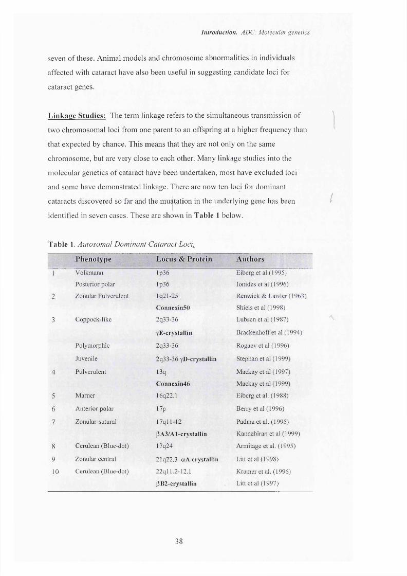
identified in seven cases. These are shown in Table 1 below.
Table 1. Autosomal Dominant Cataract Loci.
Phenotype Locus & Protein Authors
1 Volkmann lp36 Eiberg et al.(1995)
Posterior polar lp36 lonides et al (1996)
2 Zonular Pulverulent lq21-25 Renwick & Lawler (1963)
ConnexinSO Shiels et al(1998)
3 Coppock-like 2q33-36 Lubsen et al (1987)
yE-crystallin Brackenhoff et al (1994)
Polymoiphic 2q33-36 Rogaev et al ( 1996)
Juvenile 2q33-36 yD-crystallin Stephan et al (1999)
4 Pulverulent 13q Mackay et al (1997)
Connexin46 Mackay et al (1999)
5 Mamer 16q22.1 Eiberg et al. (1988)
6 Anterior polar* 17p BeiTy et al (1996)
7 Zonular-sutural 17qll-12 Padma et al. (1995)
PA3/Al-crystalliii Kannabiran et al (1999)
8 Cerulean (Blue-dot) 17q24 Armitage et al. (1995)
9 Zonular central 21q22.3 aA crystallin Litt et al(1998)
10 Cerulean (Blue-dot) 22ql 1.2-12.1 Kramer et al. (1996)
PB2-crystalIin Litt et al(1997)
38

Introduction. ADC: Molecular genetics
The details of the successful linkage studies with families affected by ADC are
given below. Although Table 1 includes all the known loci and mutations
published up to the end of 1999, only those published up 1997 will be discussed
as part of the introduction to this thesis. It was these publications that made a
significant impact on our linkage approach at that time and were therefore ofV \
great interest as they appeared in the scientific press.r
Zonular pulverulent C ataract: In 1963 Renwick and Lawler demonstrated
linkage of a large family with congenital zonular pulverulent cataract to the
Duffy blood group locus (Renwick JH et al, 1963) and so described the first
human disease to be mapped to a specific autosome (Maumenee IH, 1979). The
Duffy locus was later mapped to chromosome Iq (Donahue RP et al, 1968). The
linkage was achieved using blood group systems and a maximum LOD score of
3.78 was obtained at 0 = 0. Work related to this MD has now described the
mutation in a gene encoding connexinSO to be the cause of the cataract in this
family.
Coppock-like Cataract: In 1987 linkage was demonstrated for the Coppock-like
cataract to chromosome 2q using restriction enzyme fragment length
polymorphisms, demonstrating a maximum LOD score of 7.58 at 0 = 0 (Lubsen
NH et al, 1987). Most of the crystallin genes had been cloned by this time
(Bloemendal H, 1985) allowing a candidate gene approach to successfully
demonstrate linkage to the y-crystallin gene complex on chromosome 2q.
Although abnormalities in the crystallin proteins had been shown to give rise to
hereditary cataract in mice (Carper D et al, 1982; Garber AT et al, date) this was
the first such example in human ADC. This is the only linked phenotype in
which the underlying genetic aetiology has been further assessed. There is an
over expression of the yE pseudogene (Brakenhoff RH et al, 1994) owing to a
cluster of sequence changes within and around its TATA box which is thought to
give rise to an improperly folded y-crystallin fragment. More recently the original
authors have expressed doubt as to whether this mutation is indeed the cause of
the cataract but have yet to publish any further details.
39
A
Introduction. ADC: Molecular genetics
Marner Cataract (CAM): Linkage of this phenotype using polymorphic marker
systems was demonstrated in 1988 to haptoglobin with a maximum lod score of
8.31 at recombination fraction 0 = 0.05. This places the gene causing the cataract
within band 16q22 (McGill JR et al, 1984) although no candidate gene has been
identified in this region (Eiberg H et al, 1988). DNA had been collected from 104
affected and 85 unaffected family members and there was only a single
recombination. A pedigree affected by a different phenotype, a posterior polar
cataract has been described by Maumenee (Maumenee IH, 1979) and may also be
linked to this same locus. In this second pedigree 60 family members were
examined including 21 affected individuals and the LOD score calculated was
2.11 (Richards J et al, 1984). This falls below the current criteria for linkage
(demanding a minimum LOD score of 3.0). If this posterior polar cataract and the
Mamer cataract do both link to this locus, it may be that the two different
phenotypes are alleles of the same gene.
Cerulean cataract: Linkage has been demonstrated for two pedigrees affected
by cemlean (blue-dot) cataract, to two different loci. Armitage et al demonstrated
linkage of this phenotype to a 6cM interval on chromosome 17q in 1995
(Armitage MM et al, 1995) during a genome wide search using microsatellite
markers. The highest LOD score was 9.46 at 0 = 0.085 for marker D17S802,
with multipoint analysis giving a LOD score o f 13.71 for the region between
D17S802-D17S836. The 17q24 region contains four genes including the
galactokinase gene. This gene is known to cause autosomal recessive cataracts
but has not yet been cloned. The other three genes were excluded as being the
causative gene by linkage analysis to intragenic DNA repeat polymorphisms.
A second locus for cemlean cataract was then reported a year later by Kramer et
al mapping to the P-crystallin region on chromosome 22q (Kramer P et al, 1996).
This region contains three P-crystallin genes; CRYBB2, CRYBB3 and
CRYBB2P1. An initial linkage study demonstrating exclusion had been
published six years earlier (Bodker FS et al, 1992) but with the development of
microsatellite markers a more detailed analysis of the candidate regions was
40
Introduction. ADC: Molecular genetics
undertaken. A LOD score of 6.80 was reached with the marker D22S258 and
multipoint analysis showed a maximum multipoint location score o f 7.59 for this
marker. A chain-termination mutation in the P-crystallin gene (CRYBB2) has
since been detected and is associated with the cataract in this family (Litt M et al,
1997).
Zonular-sutural cataract: A three generation family with this phenotype was
studied by Padma et al (Padma T et al, 1995). After exclusion o f the regions on
the genome known to contain cataract loci, other genes were considered as
candidates and close to the PA3/A1 crystallin gene locus on chromosome 17q
they demonstrated linkage with a maximum lod score of 3.9 at D17S805 with a
17cM interval. A mutation in this gene has now been shown to give rise to the
cataract (Kannabiran C et al 1999).
Volkman cataract: The linkage study into this family excluded approximately
95% of the human autosomes by typing 300 polymorphic loci in 91 family
members (Eiberg H et al, 1995). The closest linkage was reached at marker
D1S243 with a lod score oiZm ax = 14.04 at 0^= 0.025 0p= 0.000. The enolase 1
alpha gene (ENO 1) is close to this region and codes for both an enzyme and t -
crystallin, which is present in lower species. This has yet to be excluded as
causing the Volkmann cataract.
Polymorphic: A seven generation pedigree consisting of 254 members of which
103 were affected by a polymorphic cataract underwent linkage analysis (Rogaev
El et al, 1996). The polymorphic cataract gene was found to be linked to a 15cM
region with a maximum lod score of 10.62 for the CRYG 1 locus marker.
Multipoint analysis increased the LOD score to >17.0 at the CRYG 1 locus and
the authors have started to screen for mutations.
Other linkage studies:
Many linkage studies have been performed that have resulted in exclusion of
certain candidate loci, but have failed to demonstrate linkage. Most of the studies
41
Introduction. ADC: Molecular genetics
were performed before the availability of microsatellite markers (Dib C et al,
1996) and so genome wide searches were not possible. Other polymorphic
systems were used (Jay M , 1982) and only the major candidate loci were able to
be excluded. Many of the phenotypes in these linkage exclusion studies resemble
mapped phenotypes which suggests considerable genetic heterogeneity.
Animal models:
Many animal models exist for inherited cataract including mice, rats, guinea pigs
and dogs (Zigler JS, 1990; Tripathi BJ et al, 1991; Sidjanin et al 1998). The
advantages of animal models include the comparative ease of breeding large
litters that provide statistically significant data, as well as the availability of lens
material allowing the course of the disease to be followed histologically and
biochemically from the earliest stages. Another advantage in mice is that the
mouse genome is well characterised with many mouse-to-human homologies A
(Peters J et al 1996). Extensive linkage studies can be carried out and transgenic
animal models can test the role of different mutations on cataract formation. The
identification of loci for eye diseases in mice has in turn led to the identification
of the corresponding human genes (Travis GH et al, 1991) owing to the strong
evolutionary conservation of genomic regions between mice and men.
In many animal models the clinical phenotype and age of onset has been
documented and in many cases the underlying abnormality has been detected. Of
particular importance are the Philly mouse in which a 12 nucleotide deletion in
the pB2 cDNA (Chambers C et al, 1991) results in a mutant pB2 protein that is
thought to give rise to the cataract. A chain termination mutation in the human
pB2-crystallin is associated with autosomal dominant cerulean cataract (Litt et al
1997). In the Elo mouse the mutation consists of a single base pair deletion in the
yE-crystallin gene (Cartier M et al, 1992) resulting in a failure of the lens fibre
cells to elongate. A mutation in the human yE-crystallin gene is associated with
the nuclear CCL cataract and a splice site mutation in the y-crystallin gene causes
an autosomal dominant cataract in the guinea pig (Roderiguez IR et al 1992). The
Cat^" and the Cat'®*’ mice have cataracts caused by mutations in the gene for MIP-
42

Introduction. ADC: Molecular genetics
particularly mouse cataract, provides useful information that is relevant to the
human genome (Kerscher S et al, 1996). Over 60 autosomal dominant cataract
mouse mutants have been described (Sidjanin et al 1997) indicating the extent of
the genetic heterogeneity in both mice and humans (Doolittle DP et al, 1996).
Chromosomal Abnormalities associated with cataract:
In humans, translocations and deletions have been noted to be associated with :
cataract and although they have not led to the discovery of new loci for ADC,
they have contributed candidate regions to linkage studies concerning inherited
cataract.
A reciprocal translocation between the short arms of chromosome 3 and 4 in a
father and son with cataract but no other ocular nor systemic abnormalities has
been reported (Reese PD et al, 1987) and although this does not prove a causal
relationship it suggests candidate loci at the break points involved in the
translocation at 3p26.2 and 4pl5. A translocation between chromosomes 2 and
14 associated with anterior polar cataract (Moross T et al, 1984) similarly
suggests that two loci at 2p25 and 14q24 may be involved in cataract formation.
Cataract has also been described in association with other chromosomal disorders
but has included more generalised ocular and systemic abnormalities (Rubin SE
et al, 1994; Miller BA et al, 1992; Yokoyama Y et al, 1992; Bonneau D et al,
1995). The causative genes in these cases may well have a wider role than that
limited to cataract formation and have not been included as candidate regions for
ADC.
Summary: As knowledge concerning the molecular biology and genetics of lens
development and cataracatogenesis increases, a longer list o f candidate regions
emerges for those undertaking a linkage study in ADC. In the remaining chapters
of this thesis a description is given of how the families were collected and
clinically assessed, and the approach taken to the linkage study and how, from
the 15 pedigrees, linkage was demonstrated for five pedigrees and mutations
isolated in three of these pedigrees.
45

Introduction. ADC: Molecular genetics
aquaporin (Griffen et al 1992, Muggleton-Harris et al 1987) which maps to 12q
in humans and is an important candidate gene for human cataract (Francis et al
submitted).
The remainder of the mouse mutants and the coiTesponding human regions are
shown in table 2 below.
Table 2. Loci of the mouse cataracts with the corresponding human regions.
Mouse _9- Chromosome
HumanSynteny
1 Czyge CRYG/ CCL
-
C/yga
Car/"'
y-E crystallin mutation
2 Filensin on Ch 20
4 Cur
5 fAzV/y PBl-crystallin P-crystallin 22q
Npp 4pl6-21q
7 Lim2, MPIO
10 C a f ' MZP MIP, 12q
Cal°"
To2 or Cat5 6q21-q27
12q
16 <%/ 16p or 2q t(2;16)
break
19 ak
X Xcat
Note: This table is adapted from Francis et al 1999.
The wide genetic heterogeneity of inherited cataract in humans is a major
obstacle in the isolation of the causative genes and the study of animal models,
43

Patients & Methods
Patients & Methods
Patients
Clinical Assessment 46
Laboratory techniques
Genotyping & Linkage Analysis 49
Candidate genes 52
45
Patients & Methods: Clinical assessment
PATIENTS & METHODS
Clinical assessment;
The patients were recruited from the genetic clinic data-base at Moorfields Eye
Hospital and invited to attend for a single visit to participate in the study. A
medical and ophthalmological history was taken along with family details to
construct a family tree. The patients underwent visual acuity testing, tonometry,
anterior segment examination both before and after pupil dilatation including
lens photography where possible, and finally examination of the retina. The
findings were recorded on the sheet below. A blood sample was taken for DNA
analysis. The parents, siblings and children of all affected individuals in any
single pedigree were then contacted and invited to participate in the study.
46
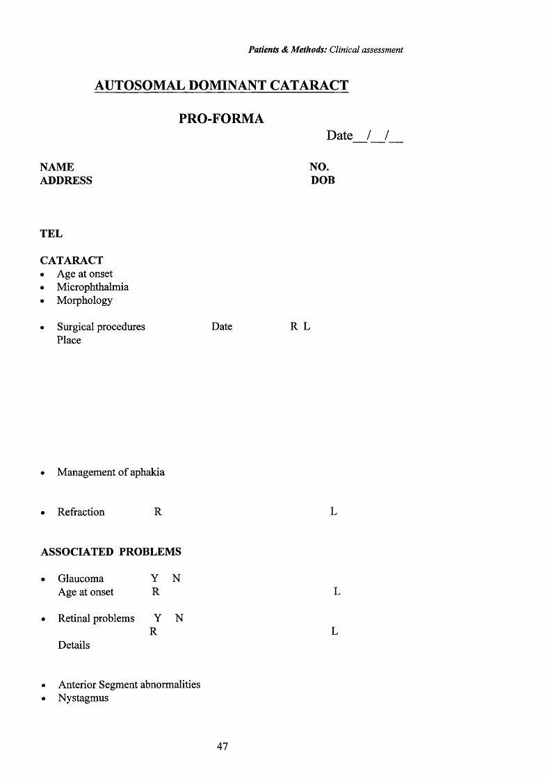
Patients & Methods: Clinical assessment
AUTOSOMAL DOMINANT CATARACT
PRO-FORMADate / /
NAME NO.ADDRESS DOB
TEL
CATARACT• Age at onset• Microphthalmia• Morphology
• Surgical procedures Date R LPlace
Management of aphakia
• Refraction R L
ASSOCIATED PROBLEMS
• Glaucoma Y NAge at onset R L
• Retinal problems Y N
DetailsR L
Anterior Segment abnormalities Nystagmus
47

Patients & Methods: Clinical assessment
Strabismus
Registration
Education
Family Tree
Nystagmus
Medication ^
EXAMINATION
V/A
RAPD
lidsconj
cornea
Gonioscopy
O O
Disc
48
Patients & Methods: Clinical assessment
LABORATORY TECHNIQUES.
Genotyping
Genomic DNA was extracted from EDTA-sequestered blood samples using the
Nucleon II DNA extraction kit (Scotlab Bioscience) as described below.
Preparation of DNA from whole blood.
Eighteen millilitres of whole blood was collected from each subject in two
separate sterile tubes containing 0.5M ethylenediaminetetraacetate (EDTA). They
were stored at -80°C in separate freezers until the pedigree was large enough to
begin the linkage analysis. The blood was then thawed at 37°C to allow for DNA
extraction performed using the Nucleon II DNA Extraction kit. This method
entails the following steps.
1) Cell Preparation. The blood sample was poured into a 50ml falcon tube and
reagent A (see below) was added to a total of SOmls. This was carefully mixed
and then centrifuged at 1400 rpm for ten minutes. The supernatant was discarded
leaving the leucocyte pellet.
2) Cell lysis. Two millilitres of reagent B (see below) were then added to the
pellet which was broken up by gentle pipetting and the cell lysate transferred to a
5ml tube.
3) Deproteination. 500pl of 5M sodium perchlorate were added to the lysate and
rotary mixed for 15 minutes at room temperature. They were then incubated at
65°C for 25 minutes whilst rotary mixing.
4) DNA extraction. After removal from the oven the samples were cooled on ice
and
2ml o f chloroform and 300pl of nucleon silica suspension were added. The
contents
49
Patients & Methods: Clinical assessment
were then rotary mixed for five minutes at room temperature. Separation of the
aqueous and organic phases was achieved by centrifuging at 1400rpm for 6
minutes. ^
5) DNA precipitation. The top aqueous layer containing the DNA was
transferred to a universal tube using a pipette and two parts o f ethanol equal to
the amount o f aqueous were added to precipitate the DNA.
6) DNA resuspension. The precipitated DNA was picked out of the universal
using sterile needles and transferred to a sterile 1.5ml Eppendorf tube and washed
with 70% alcohol. The pellet of DNA was then resuspended in 400pl of sterile
distilled water and left for 24 hours. Twenty microlitre aliquots were then
removed and diluted in preparation for linkage analysis and the remaining stock
kept at -20°C.
Nucleon II DNA Extraction Kit Solutions.
Reagent A: lOmM Tris-HCl
320mM sucrose
5mM MgCl]
1% Triton X-100
pH 8 with 2M NaOH
Reagent B: 400mMTris-HCl
pH 8 with mM NaOH
60mM EDTA
ISOmMNaCl
1% SDS
Genotvping
Genethon microsatellite markers (Gyapay G et al, 1994) were amplified using the
polymerase chain reaction (PGR). The forward PGR primer was 5’-end labelled
using [y-^^P]dATP (-110 Tbq/mmol) and T4 polynucleotide kinase prior to
amplification. The standard PGR mix (lOpl) contained 200ng DNA, 2.5pmol of
each primer, 50 mM KGL, lOmM Tris-HGl (pH 9.0), 0.15% Triton-X 100, 1.5
50

Patients & Methods: Clinical assessment
mM MgCl], 250 p.M od each dNTP and 0.6 U of Taq DNA polymerase. Standard
PCR conditions were: 1 cycle at 94°C for 3 min, followed by 30 cycles at 94°C
for 1 min, 50 or 55 °C for 1 min, 72°C for 1 min, then 1 cycle at 72°C for 5 min.
Radiolabelled PCR products were separated on denaturing 6% polyacrylamide
gels and detected by autoradiography.
Linkage analysis.Data was collated using the LD4RSYS (version 3.1) data management package
(Attwood J et al, 1988). Two-point LOD scores were calculated using the MLINK
and ILINK sub-programmes of the LINKAGE (version 5.1) package (Lathrop
GM et al, 1984) Allele frequencies calculated from the spouses in the families
were similar to those quoted by Genethon. Multipoint analysis was computed
using LINKMAP run on the computing facilities o f UK Human Genome
Mapping Project Resource Centre computing facilities. Full penetrance and a
gene frequency of 0.0001 were assumed for the disease locus.
51

Patients & M eth od: Candidate genes
Candidate Gene Approach
Candidate genes: A candidate gene approach involves creating a genetic map of
loci known to be relevent to the formation of cataract. These loci are then the first
areas of the genome to be screened for the disease-causing gene in the affected
families, prior to undertaking a genome wide search. This approach is the
accepted method for linkage studies and has been successfully used in locating
the genes for the Coppock-like cataract, the Volkmann, polymorphic, Mamer,
zonular-sutural and cerulean cataract.
The different candidate loci are placed in an order of priority. In this study the
first loci to be assessed were those that had already been demonstrated to be
linked to inherited cataract phenotypes (see Table 3, p53). The crystallin genes
were then excluded followed by the membrane proteins, cytoskeletal proteins and
finally the genes for relevent enzymes and the taxon specific crystallins. A
genome wide search was then undertaken.
For each locus microsatellite markers were picked at 5cM intervals where
possible. The initial temperature for the PCR was 55° C, and if this failed to give
a meaningful result, the temperature would be altered to either 51° C or 58° C. If
this failed or the marker was uninformative for the family, then a neighbouring
marker would be picked as an alternative.
52

Patients <& Methods: Candidate genes
Table 3. Major Candidate Genes for Autosomal Dominant Cataract. (Excluding the taxon specific genes, translocations and mouse loci).
Cataract genes " , Gene Location
Volkmann (CCV), Posterior polar (CTPA) lp36Zonular Pulverulent (CZP1 ) 1 q21 -25Coppock-like (CCL), Polymorphic 2q33-36(CCP),aculeiform, juvenile.Pulverulent 13qMamer 16q22.1Anterior polar (CTAA2) 17p 12-13Zonular-sutural (CCZS) 17q 11 -12Cerulean (CCA 1 ) 17q24Zonular central 2 Iq l 1.2-12.1Cerulean (CCA2) 22q
Human Crystallin genes ; L ■ v « n U* - -,
oA (CRYAl) 21q22.3aB(CRYA2) Hq22.3-23.3PA 1 /A3 (CRYBA1/3) 17q 11.1 -q 12PA2 (CRYBA2) 2q34-36PA4 (CRYBA4) 22q 11.2-13.1PB2 (CRYBB2) 22q ll.2 -q l2pB3 (CRYBB3) 22q ll.2 -q l2yA-F (CRYGA-F) 2q32-36ysO^RYGS) 3p(CRYM) 16pl3.11-pl2.3Ç(CRYZ) lp22-31
Membrane proteins . ' r \ 7““..
MIP (MP-26), Aquqporin. 12ql3-14 ^ k j f -JMP70 / Cx50 (GJA8) lq21.1M PI9 (LIM2) 19Cx 46 (GJA3) 13q
Cytoskeletal proteins
Phakinin (CP49 kD) 3q21-25Filensin (CP94 kD) 20
53

Results
Results
Clinical assessment 55
Visual outcome & complications of surgery 95
Age at diagnosis
Age at surgery and visual outcome
Number of surgical interventions and visual outcome
Visual outcome for the different phenotypes
Complications
Linkage Analysis 115
Positive Linkage:
Posterior polar (CTPA) cataract IpNuclear (CAE) cataract IqNuclear (CCL) cataract 2qPulverulent (CZP2) cataract, two 13qpedigreesAnterior polar (CTAA2) cataract 17p
54
Results: Clinical Assessment
Clinical Assessment
180 patients affected by autosomal dominant cataract were examined. 163 of
these came from 16 pedigrees that were large enough for linkage analysis. The
remaining 17 patients were from small pedigrees. The families had no other
ocular nor systemic abnormalities. Families were included for linkage analysis if
there were three generations of affected individuals with at least four affected
members. Male to male transmission was seen in 12 families and in the
remaining families the severity of affected members was equal in both males and
females suggesting autosomal dominant inheritance. The number of family
members examined (excluding partners-although partners were examined and
genotyped) and the number of affected individuals is shown in Table 4. In cases
where surgery was performed early, a description of the phenotype was taken
from the notes.
Eight phenotypes could be recognised that included all the individuals seen in the
study. These phenotypes were; anterior polar, posterior polar, nuclear,
coralliform, cortical, lamellar, blue-dot (cerulean) and pulverulent.
Table 4. Pedigree details and phenotypes.
Phenotype No. ofaffectedpatients
No. of pedigrees(large enough for a linkage study)
Anterior polar 27 1Posterior polar 23 2Nuclear 20 2Lamellar 38 3Coralliform 8 1Blue-dot (cerulean) 7 1Cortical 6 1Pulverulent 51 5Total 180 16
Results: Clinical Assessment
In any single pedigree the phenotypes o f all the affected individuals was the
same, varying only in the severity and density o f the opacity. For example, in
pedigrees affected by anterior polar cataract the opacity was always at the
anterior pole o f the lens, never in the nucleus nor elsewhere in the lens. The size
o f the opacity varied between affected individuals however, as will be seen in the
subsequent illustrations. In pedigrees with the pulverulent phenotype there was
considerable variation in the cataract phenotype amongst individuals from the
same family.
Anterior Polar Cataract
In a single family with anterior polar cataract (pedigree A, Appx 1, p i22) 21
members were found to be affected with the cataract. 7 patients had undergone
surgery and the phenotype was examined in the remaining 14 cases. The opacity
was always in the same position of the lens, the anterior pole, but varied in
severity. The larger opacities (figures 2 & 3) were pyramidal with a base o f 4mm
and extended into the anterior chamber, whilst the smaller opacities consisted of
a flat plaque measuring under 1mm in diameter (figures 4 & 5). In one case the
only sign o f an anterior polar abnormality was an irregularity o f the anterior
capsule (figure 6). Anterior polar cataract has traditionally been regarded as
having a minimal effect on visual acuity but 11 o f the affected patients who had
not undergone surgery in the pedigree were amblyopic in one eye; the amblyopia
occuring in the eye with the denser opacity.
56
Results: Clinical Assessment
Figure 2. Anterior polar cataract: Pedigree A. Pyramidal Individ 4.2
%
Figure 3. Anterior polar cataract: Pyramidal in retroillumination. Individ 4.2
S7
Results: Clinical Assessment
Figure 4. Anterior polar cataract. Ped A. Plaque. Individ 4.3
Figure 5. Anterior polar cataract: Plaque in retroillumination. Individ 4.3
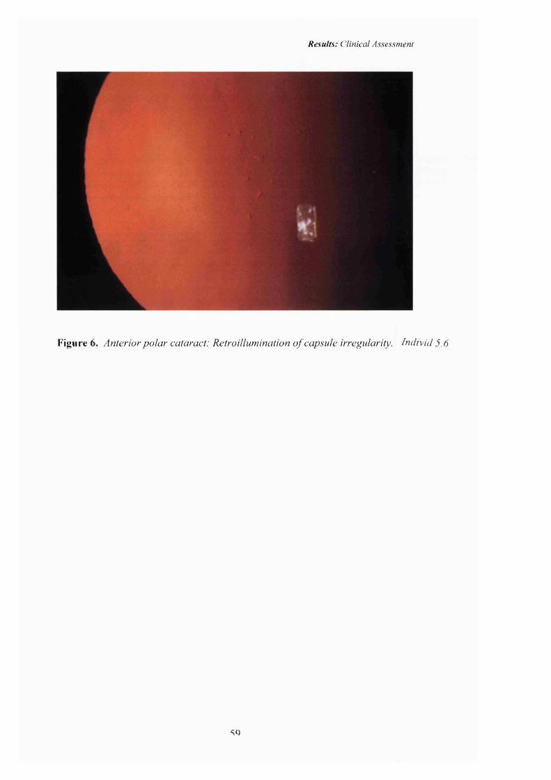
Results: Clinical Assessment
Figure 6. Anterior polar cataract: Retroillumination of capsule irregularity. Individ 5.6
SQ

Results: Clinical Assessment
Posterior polar cataract.
Two families affected by posterior polar cataract were studied. In one pedigree
(Pedigree B) ten peopole were affected and o f these seven patients had
undergone surgery. Details o f the cataract were obtained from the notes in four o f
the cases, all o f whom were described as having posterior polar or posterior
subcapsular cataract. Three patients had not had surgery and the cataract
consisted o f a single well defined posterior polar plaque (Figures 7 & 8, right
eye) that varied in size both between individuals (Figures 9 & 10), as well as
between the eyes o f an individual (Figures 11 & 12, left eye). Although we have
not studied the family over a prolonged period o f time, the hospital and
optometrists notes suggest that the cataracts did not progress. The operated eyes
underwent surgery at a mean age o f 22 months (range 2-72 months, median o f 3
months, mode 2 months) with a final V/A o f 6/6 to NFL ( median 6/12, mode
6/9). The unoperated eyes had a V/A of 6/5 to 6/36.
In the second family with posterior polar cataract (Pedigree C), 11 were affected
and all but one unilateral case had undergone surgery. Clinical records were
available in nine cases and the opacity consisted o f a posterior plaque that
developed posterior cortical extensions (Figure 13) with punctate opacities
causing a deterioration o f the visual acuity requiring cataract extraction. No
records were available in one case. The progressive nature o f the cataract is also
suggested in that the average age o f diagnosis was 5 years but the surgery was
performed at a mean age o f 20 years (range 3-40, median 19, mode 16). The final
V/A ranged from 6/5 to NPL (mode and median o f 6/9). The age at surgery was
older in this second family affected by posterior polar cataract and the visual
outcome better.
One patient from a small pedigree affected by progressive posterior polar cataract
is illustrated in Figures 14 and 15. This 13 year old developed a deterioration in
visual acuity associated with the appearance o f the early cortical extensions and
punctate opacities.
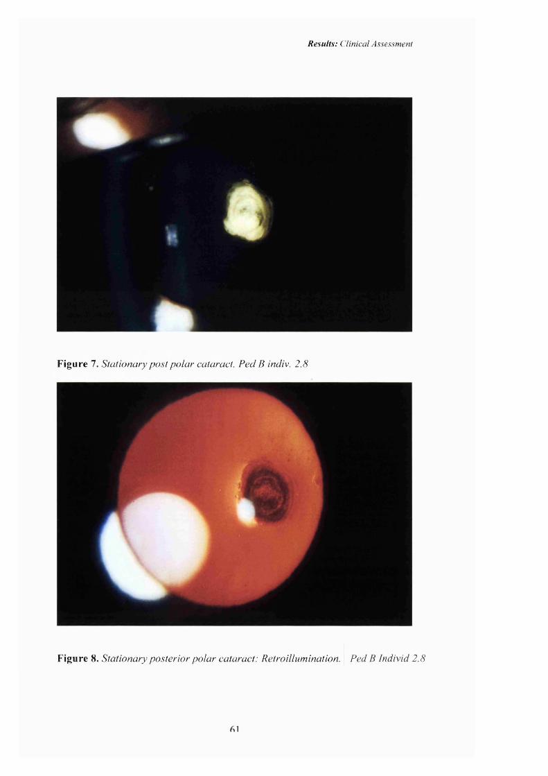
Results: Clinical Assessment
Figure 7. Stationary post polar cataract. Ped B indiv. 2.8
Figure 8. Stationary posterior polar cataract: Retroillumination. Ped B Individ 2.8

Results: Clinical Assessment
Figure 9. Stationary posterior polar cataract. Pedigree B Indiv. 2.1.
Figure 10. Posterior polar cataract: Retroillumination. Ped B Individ 2 .1
69
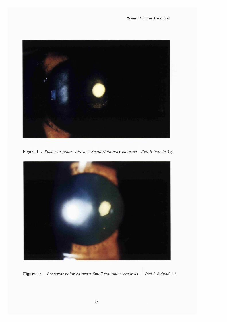
Results: Clinical Assessment
Figure 11. Posterior polar cataract: Small stationary cataract. Ped B Individ 3.6
Figure 12. Posterior polar cataract:Small stationary cataract. Ped B Individ 2.1
61
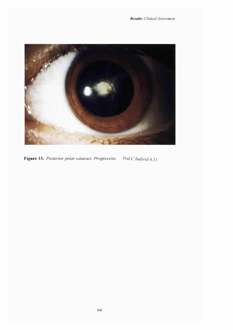
Results: Clinical Assessment
Figure 13. Posterior polar cataract. Progressive. Ped C Individ 4.11
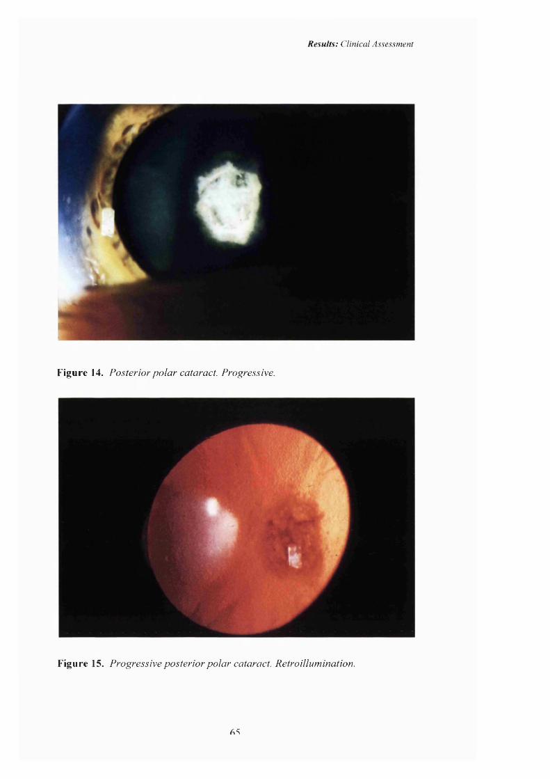
Results: Clinical Assessment
Figure 14. Posterior polar cataract. Progressive.
Figure 15. Progressive posterior polar cataract. Retroillumination.
AS
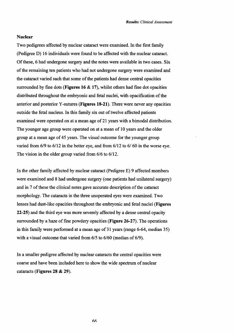
Results: Clinical Assessment
N uclear
Two pedigrees affected by nuclear cataract were examined. In the first family
(Pedigree D) 16 individuals were found to be affected with the nuclear cataract.
O f these, 6 had undergone surgery and the notes were available in two cases. Six
o f the remaining ten patients who had not undergone surgery were examined and
the cataract varied such that some o f the patients had dense central opacities
surrounded by fine dots (Figures 16 & 17), whilst others had fine dot opacities
distributed throughout the embryonic and fetal nuclei, with opacification o f the
anterior and posterior Y-sutures (Figures 18-21). There were never any opacities
outside the fetal nucleus. In this family six out o f twelve affected patients
examined were operated on at a mean age of 21 years with a bimodal distribution.
The younger age group were operated on at a mean o f 10 years and the older
group at a mean age o f 45 years. The visual outcome for the younger group
varied from 6/9 to 6/12 in the better eye, and from 6/12 to 6/ 60 in the worse eye.
The vision in the older group varied from 6/6 to 6/12.
In the other family affected by nuclear cataract (Pedigree E) 9 affected members
were examined and 8 had undergone surgery (one patients had unilateral surgery)
and in 7 o f these the clinical notes gave accurate description o f the cataract
morphology. The cataracts in the three unoperated eyes were examined. Two
lenses had dust-like opacities throughout the embryonic and fetal nuclei (Figures
22-25) and the third eye was more severely affected by a dense central opacity
surrounded by a haze of fine powdery opacities (Figure 26-27). The operations
in this family were performed at a mean age o f 31 years (range 6-64, median 35)
with a visual outcome that varied from 6/5 to 6/60 (median o f 6/9).
In a smaller pedigree affected by nuclear cataracts the central opacities were
coarse and have been included here to show the wide spectrum o f nuclear
cataracts (Figures 28 & 29).
f%f\

Results: Clinical Assessment
Figure 16 Nuclear cataract, dense central opacities. Ped D Individ 5.2
Figure 17. Nuclear cataract: Retroillumination. Ped D Individ 5.2
67
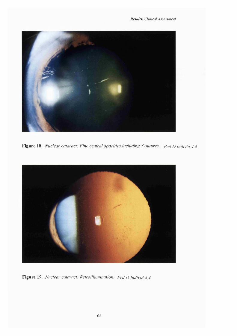
Results: Clinical Assessment
Figure 18. Nuclear cataract: Fine central opacities,including Y-sutures. Ped D Individ 4.4
Figure 19. Nuclear cataract: Retroillumination. P edD Individ 4.4
AX
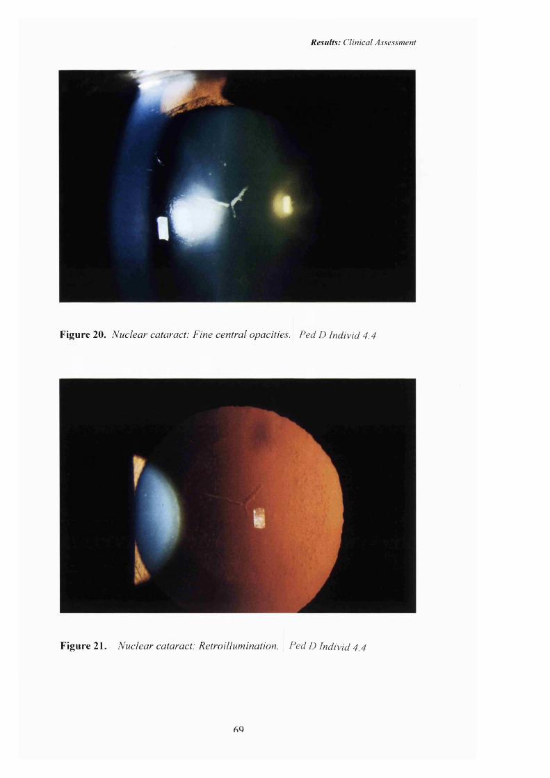
Results: Clinical Assessment
< #
Figure 20. Nuclear cataract: Fine central opacities. Ped D Individ 4.4
Figure 21. Nuclear cataract: Retroillumination. Ped D Individ 4.4
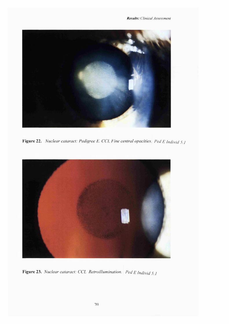
Results: Clinical Assessment
Figure 22. Nuclear cataract: Pedigree E. CCL Fine central opacities. Ped E Individ 5.1
Figure 23. Nuclear cataract: CCL Retroillumination. Ped E Individ 5
70
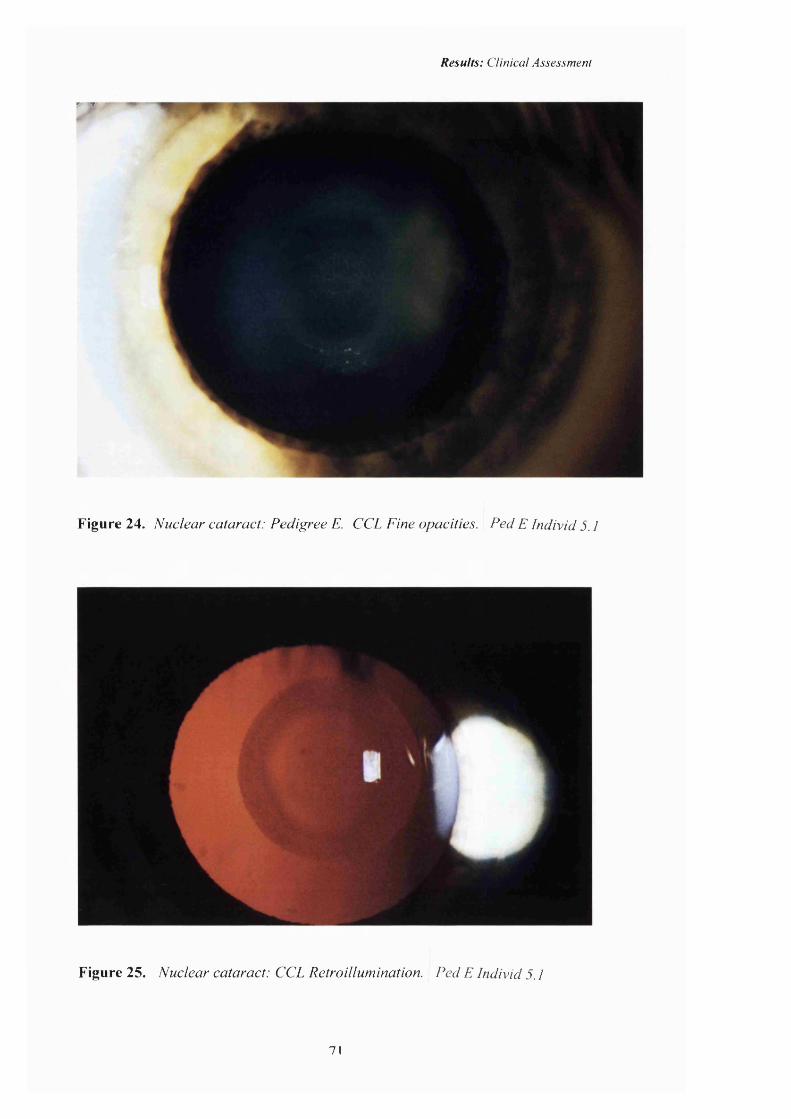
Results: Clinical Assessment
- T
Figure 24. Nuclear cataract: Pedigree E. CCL Fine opacities. Ped E Individ 5.1
Figure 25. Nuclear cataract: CCL Retroillumination. Ped E Individ 5.1
71
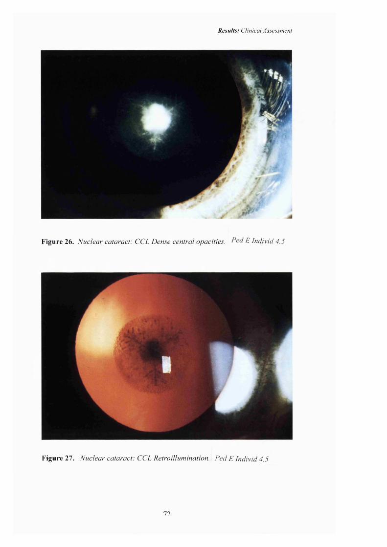
Results: Clinical Assessment
Figure 26. Nuclear cataract: CCL Dense central opacities. E Individ 4.5
Figure 27. Nuclear cataract: CCL Retroillumination. Ped E Individ 4.5
77

Results: Clinical Assessment
Figure 28. Nuclear cataract. Coarse central opacities.
Figure 29. Nuclear cataract. Coarse central opacities. Retroillumination.
71
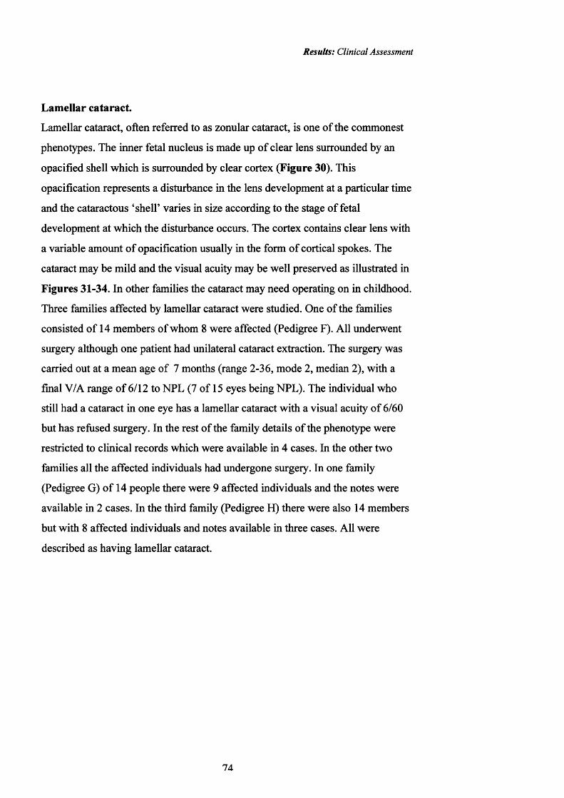
Results: Clinical Assessment
Lamellar cataract.
Lamellar cataract, often referred to as zonular cataract, is one of the commonest
phenotypes. The inner fetal nucleus is made up o f clear lens surrounded by an
opacified shell which is surrounded by clear cortex (Figure 30). This
opacification represents a disturbance in the lens development at a particular time
and the cataractous ‘shell’ varies in size according to the stage o f fetal
development at which the disturbance occurs. The cortex contains clear lens with
a variable amount o f opacification usually in the form of cortical spokes. The
cataract may be mild and the visual acuity may be well preserved as illustrated in
Figures 31-34. In other families the cataract may need operating on in childhood.
Three families affected by lamellar cataract were studied. One of the families
consisted o f 14 members of whom 8 were affected (Pedigree F). All underwent
surgery although one patient had unilateral cataract extraction. The surgery was
carried out at a mean age o f 7 months (range 2-36, mode 2, median 2), with a
final V/A range o f 6/12 to NPL (7 of 15 eyes being NPL). The individual who
still had a cataract in one eye has a lamellar cataract with a visual acuity o f 6/60
but has refused surgery. In the rest o f the family details of the phenotype were
restricted to clinical records which were available in 4 cases. In the other two
families all the affected individuals had undergone surgery. In one family
(Pedigree G) o f 14 people there were 9 affected individuals and the notes were
available in 2 cases. In the third family (Pedigree H) there were also 14 members
but with 8 affected individuals and notes available in three cases. All were
described as having lamellar cataract.
74

Results: Clinical Assessment
Figure 30. Lamellar cataract.
7S
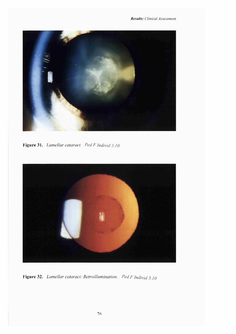
Results: Clinical Assessment
Figu re 31. Lamellar cataract. Fed F Individ 3.10
r
Figure 32. Lamellar cataract: Retroillumination. Fed F Individ 3 IQ
7A
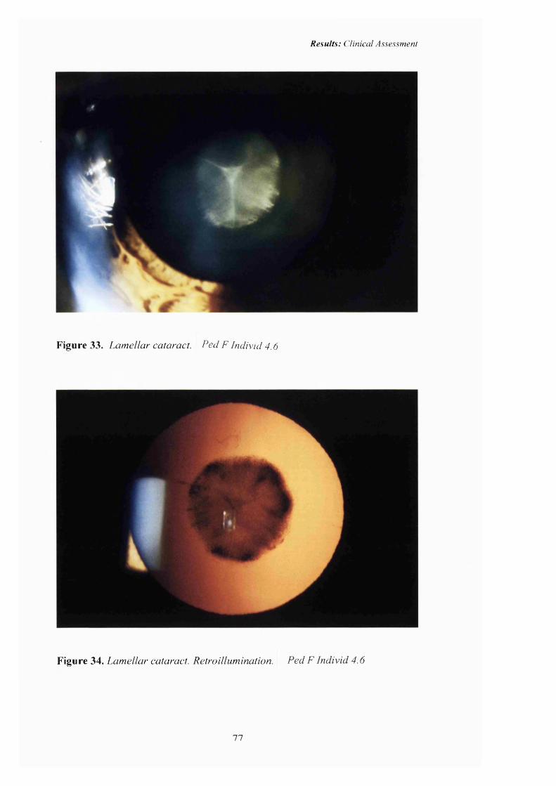
Results: Clinical Assessment
Figure 33. Lamellar cataract. Ped F Individ 4.6
Figure 34. Lamellar cataract. Retroillumination. Ped F Individ 4.6
77

Results: Clinical Assessment
Coralliform
The single pedigree of this phenotype (Pedigree I) from the Moorfields genetic
data base is descended from a family originally described by Nettleship
(Nettleship E, 1906) and illustrated in Figure 35. The name is derived from the
resemblance o f the cataract to sea coral, having finger-like tubular protuberances
extending from the nucleus. All affected members of this pedigree had been
operated on and none were available for slit lamp photography. The description
of the cataract was taken from the clinical notes. The mean age at operation was 5
years (range 1-12 years, median 5, mode 7 years). The visual outcome was 6/6 to
6/36 (median 6/9, mode 6/9).
Figure 35. Coralliform cataract.
78
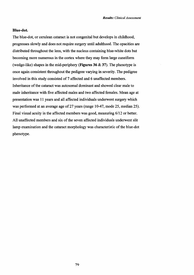
Results: Clinical Assessment
Blue-dot.
The blue-dot, or cerulean cataract is not congenital but develops in childhood,
progresses slowly and does not require surgery until adulthood. The opacities are
distributed throughout the lens, with the nucleus containing blue-white dots but
becoming more numerous in the cortex where they may form large cuneiform
(wedge-like) shapes in the mid-peripheiy (Figures 36 & 37). The phenotype is
once again consistent throughout the pedigree varying in severity. The pedigree
involved in this study consisted o f 7 affected and 6 unaffected members.
Inheritance o f the cataract was autosomal dominant and showed clear male to
male inheritance with five affected males and two affected females. Mean age at
presentation was 11 years and all affected individuals underwent surgery which
was performed at an average age o f 27 years (range 10-47, mode 25, median 25).
Final visual acuity in the affected members was good, measuring 6/12 or better.
All unaffected members and six o f the seven affected individuals underwent slit
lamp examination and the cataract morphology was characteristic o f the blue-dot
phenotype.
7Q
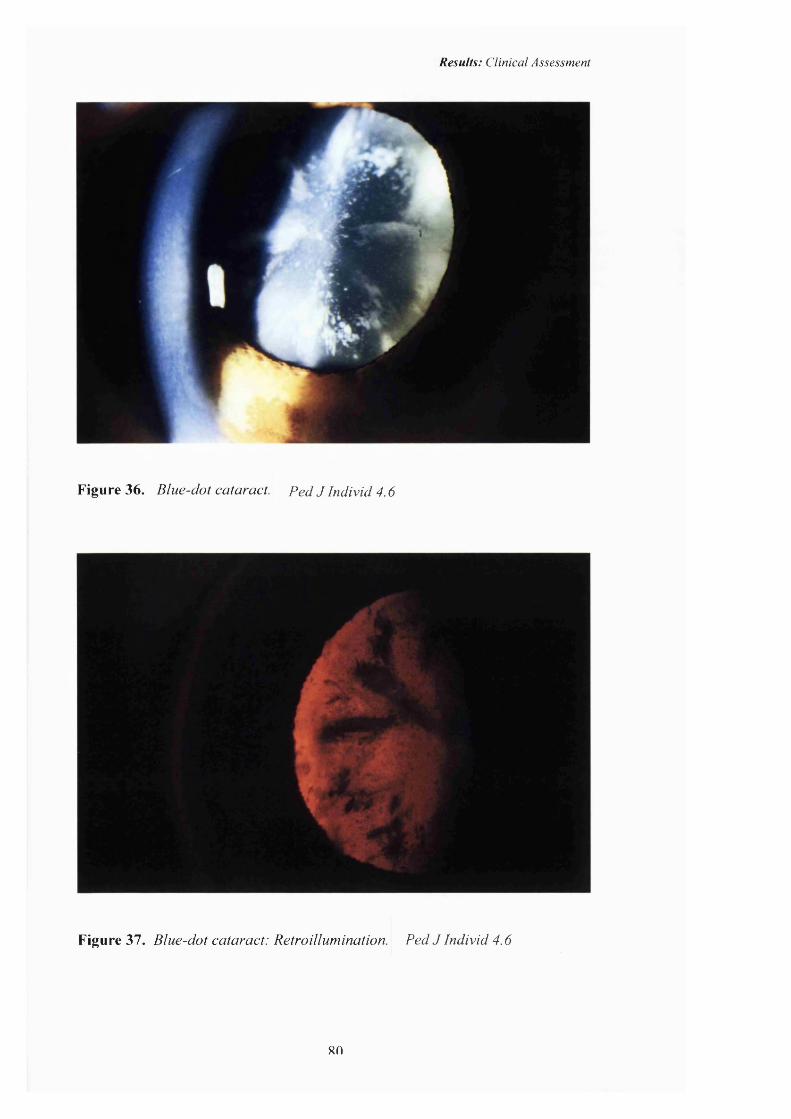
Results: Clinical Assessment
Figu re 36. Blue-dot cataract. Ped J Individ 4.6
Figure 37. Bluc-dot cataract: Retroillumination. Ped J Individ 4.6
80
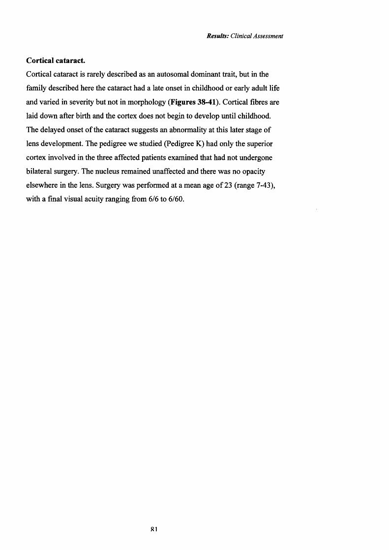
Results: Clinical Assessment
Cortical cataract.
Cortical cataract is rarely described as an autosomal dominant trait, but in the
family described here the cataract had a late onset in childhood or early adult life
and varied in severity but not in morphology (Figures 38-41). Cortical fibres are
laid down after birth and the cortex does not begin to develop until childhood.
The delayed onset o f the cataract suggests an abnormality at this later stage of
lens development. The pedigree we studied (Pedigree K) had only the superior
cortex involved in the three affected patients examined that had not undergone
bilateral surgery. The nucleus remained unaffected and there was no opacity
elsewhere in the lens. Surgery was performed at a mean age o f 23 (range 7-43),
with a final visual acuity ranging from 6/6 to 6/60.
81

Results: Clinical Assessment
Figure 38. Cortical cataract. Ped K Individ 3.7
Figure 39. Cortical cataract. Retroillumination. Ped K Individ 3.7
89

Results: Clinical Assessment
Figure 40. Cortical cataract. ^ Individ 4.7
>Figure 41. Cortical cataract. Retroillumination. Ped K Individ 4.7
Results: Clinical Assessment
Pulverulent.
Pulverulent cataracts are powdery ('pulverised') opacities and although there is
great intra-familial variety in pulverulent pedigrees, most o f the affected patients
had these fine, powdery opacities. Some individuals had sparse, fine opacities
throughout the lens (Figure 42 & 43), others have dot-like sutural cataract
(Figures 44-47), some have pulverised opacities in the posterior cortex (Figures
48 & 49) and others can have a dense central cataract (Figures 50-53). It is
important to examine as many affected members o f a family with pulverulent
cataract as possible, as it can be difficult to diagnose this phenotype in a pedigree
after examining only a few members. The main feature is the pulverulent nature
o f the opacities in several affected family members and the features that
distinguish a pulverulent from a nuclear pedigree include the wide variation in
morphology within a pulverulent pedigree as well as the involvement o f the lens
outside the embryonic and fetal nuclei.
Four families involved in this study had pulverulent cataract. In one o f the
families (Pedigree L) affected by pulverulent cataract 18 patients were found to
be affected. The cataract varied between affected individuals. In some the fine
opacities formed a lamellar distribution with a clear cortex (Figure 44) and
minimal inner fetal nuclear involvement. Others had more widely spread
pulverised opacities (Figures 42 & 43) extending into the cortex with no
demarcation o f the fetal nucleus (Figure 46). In some the cataract was mild and
the fine dust-like opacities were clustered only around the anterior and posterior
Y-sutures (Figure 47). Nine had undergone surgery at a mean age o f 17 years (3-
82, median 8, mode 11) with a final visual acuity that varied fi'om 6/4 to NFL. If
the operation was performed under 8 years o f age, the visual acuity was 6/9 to
NFL. If performed after the age o f 20 years the range improved to 6/4 - 6/12.
In a second family (Pedigree M) with pulverulent cataract 15 members were
examined and 10 were affected. Four had undergone bilateral cataract surgery,
two unilateral and four had not had surgery. The mean age at surgery was 14
years (range 5-22 years, median 17) with a final visual acuity of 6/6 - 6/18. The
R4
Results: Clinical Assessment
cataract had powdery opacities throughout the lens although they were more
abundent in the fetal nucleus. In some the opacities were large and coarse (figure
56 & 57) and in others finer and dust-like.
A third family affected by pulverulent cataract (Pedigree N) consisted o f 15
family members o f whom 8 were affected by the cataract. Only two patients had
undergone surgery and examination o f the remaining six revealed fine dust-like
opacities predominately in the fetal nucleus but also in the cortex, with moderate
opacification of the Y-sutures.
The fourth family affected by pulverulent cataract (Pedigree O) consisted of 21
family members with 8 affected individuals. The phenotype in this family
showed great intra-familial variation, some had the typical fine dots throughout
the lens (Figures 54 & 55), others had posterior opacities with the rest o f the lens
unaffected and one individual had dense bilateral nuclear cataracts (Figures 50-
53).
The fifth pedigree affected by a pulverulent cataract (Pedrigree P) was
previously studied by Nettleship at the end o f the 19* century and had been the
subject o f a linkage study by Renwick published in 1963. All the affected
individuals had been operated on and it was not possible to obtain any
photographs o f the cataract but descriptions o f the phenotype (a 'zonular
pulverulent cataract) have been published by Nettleship and then later by
Renwick.
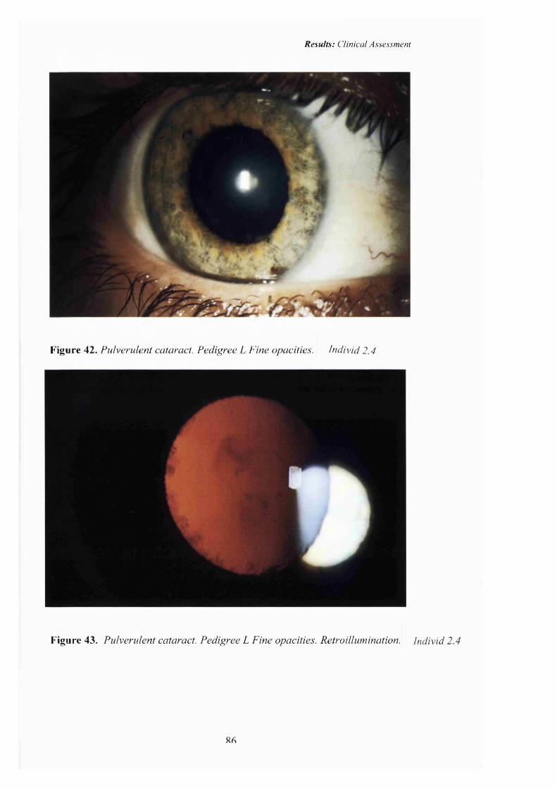
Results: Clinical Assessment
Figure 42. Pulverulent cataract. Pedigree L Fine opacities. Individ 2.4
Figure 43. Pulverulent cataract. Pedigree L Fine opacities. Retroillumination. Individ 2.4

Results: Clinical Assessment
1
Figure 44. Pulverulent cataract. Pedigree L. Sutural opacities. Individ 2.4
Figure 45. Pulverulent cataract.Pedigree L. Retroillumination. Individ 2.4
87
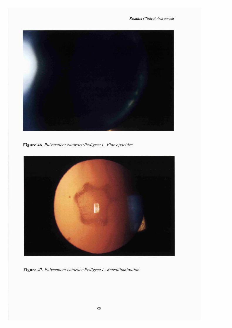
Results: Clinical Assessment
Figure 46. Pulverulent cataract:Pedigree L. Fine opacities.
4
Figure 47. Pulverulent cataract:Pedigree L. Retroillumination
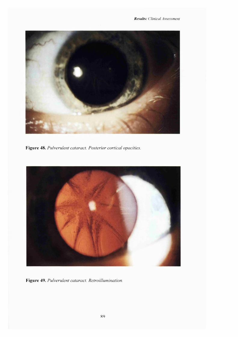
Results: Clinical Assessment
Figure 48. Pulverulent cataract. Posterior cortical opacities.
\
Figure 49. Pulverulent cataract. Retroillumination.
8Q
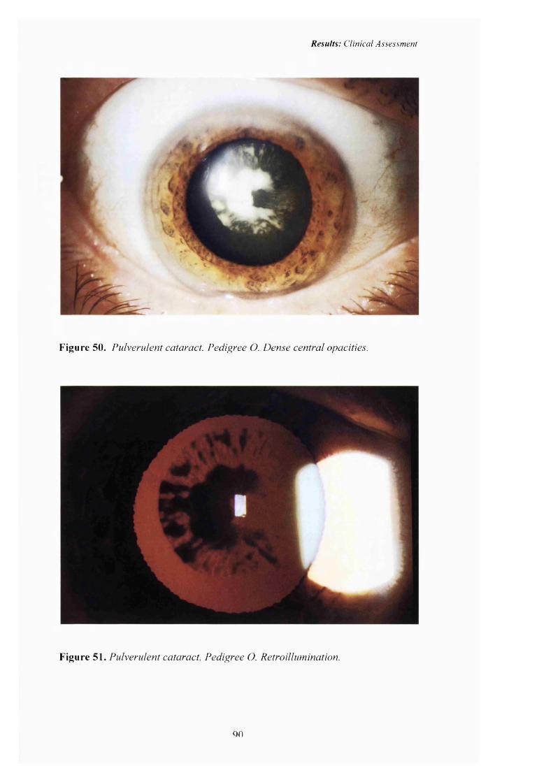
Results: Clinical Assessment
Figure 50. Pulverulent cataract. Pedigree O. Dense central opacities.
Figure 51. Pulverulent cataract. Pedigree O. Retroillumination.
QO
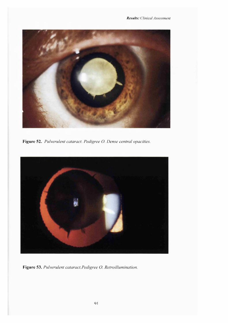
Results: Clinical Assessment
Figure 52. Pulverulent cataract. Pedigree O. Dense central opacities.
Figure 53. Pulverulent cataract.Pedigree O. Retroillumination.
Q1
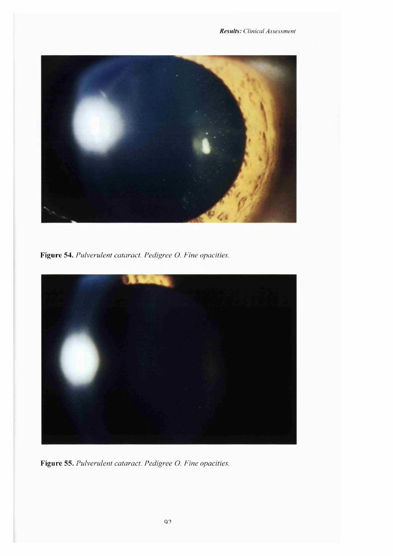
Results: Clinical Assessment
Figure 54. Pulverulent cataract. Pedigree O. Fine opacities.
Figure 55. Pulverulent cataract. Pedigree O. Fine opacities.
Q9
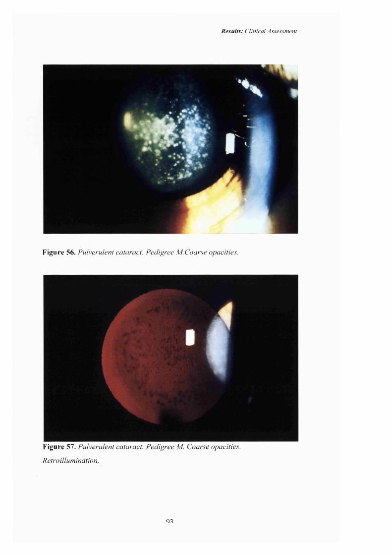
Results: Clinical Assessment
Figure 56. Pulverulent cataract. Pedigree M.Coarse opacities.
Figure 57. Pulverulent cataract. Pedigree M. Coarse opacities.
Retroillumination.
Q1
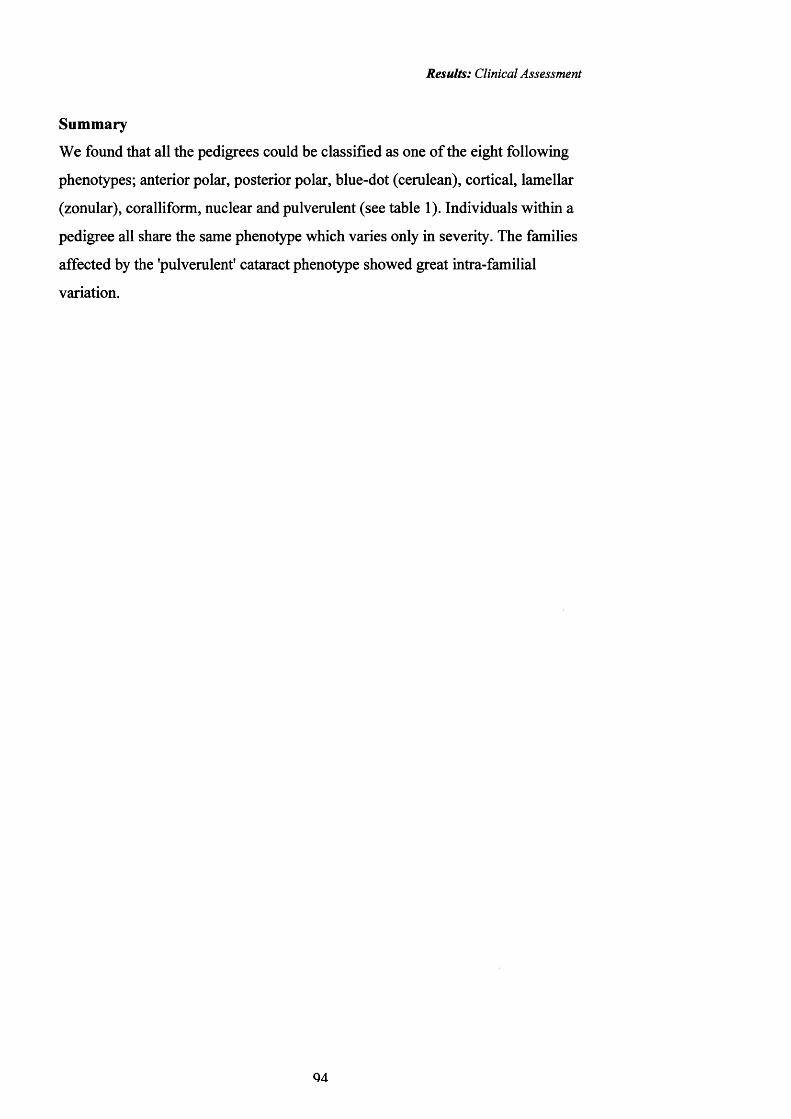
Results: Clinical Assessment
Summary
We found that all the pedigrees could be classified as one o f the eight following
phenotypes; anterior polar, posterior polar, blue-dot (cerulean), cortical, lamellar
(zonular), coralliform, nuclear and pulverulent (see table 1). Individuals within a
pedigree all share the same phenotype which varies only in severity. The families
affected by the ’pulverulent' cataract phenotype showed great intra-familial
variation.
Q4

Results: Visual outcome & Complications
Visual Outcome & complications of surgery
Page no.
Age at diagnosis 96
Age at surgery and visual outcome. 99
Number of surgical interventions and visual 104
outcome.
Visual outcome for the different phenotypes. 106
Complications. 112

Results: Visual outcome & Complications
Age at diagnosis180 affected patients were examined of whom 96 were female and 84 male. The
age at diagnosis was available on 136/180 affected patients of whom 101 had
undergone surgery (figure 58). The mean age at diagnosis of cataract in all
affected individuals was 5.2 years ranging from birth to 71 years. The age at
diagnosis of the cataract in those who required surgery was 4.5 years, ranging
from birth to 41 years. As part of the study asymptomatic relatives of affected
Figure 58 Age at diagnosis of inherited cataract.
All cases Those requiring surgery
a, 25
' X 20
11-20 21-40 40+0-1 1 2 3 4 5 6-10
Age (years)
Table to accompany figure 58. Age at diagnosis o f inherited cataract.
Age X<1 1 2 3 4 5 6-10 11-20 21-40 40+
39 26 12 2 10 14 14 10 6 3
23 21 11 1 10 8 10 6 4 1
patients were examined and some found to have cataracts. The age at diagnosis in
these individuals was therefore not directly related to the clinical course of the
disease as they remained asymptomatic and did not require clinical intervention.
06

Results: Visual outcome & Complications
As can be seen in figure 58 the age at diagnosis follows a bimodal pattern. Most
cases were diagnosed either at birth or in late childhood.
The age at diagnosis of the different phenotypes of cataract in those patients who
underwent surgery is shown in table 5.
Table 5. Age at diagnosis for different phenotypes in those patients who had
undergone surgery.
Phenotype Average age at diagnosis
(years)
Age Range (years)
n(total 101)
Anterior polar <1 0-1 8
Posterior polar- Stationary 1.25 0-8 8
Lamellar 2.53 1-8 15
Nuclear 3.0 0-30 22
Coralliform 3.1 1-6 8
Pulverulent 5.05 0-22 19
Posterior polar- Progressive 5.9 0-25 8
Blue-dot 11.6 1-16 7
Cortical 18.8 5-41 6
The phenotypes that are diagnosed early are the anterior polar and stationary
posterior cataracts.
The progressive posterior polar cataract was picked up later in life. The centrally
placed lamellar, nuclear and coralliform cataract were generally diagnosed before
the age of four although the milder opacities were not diagnosed until adulthood.
The affected lens fibres were laid down in utero and were therefore present at
birth, but the delayed diagnosis may suggest that these phenotypes are not always
congenital.
07
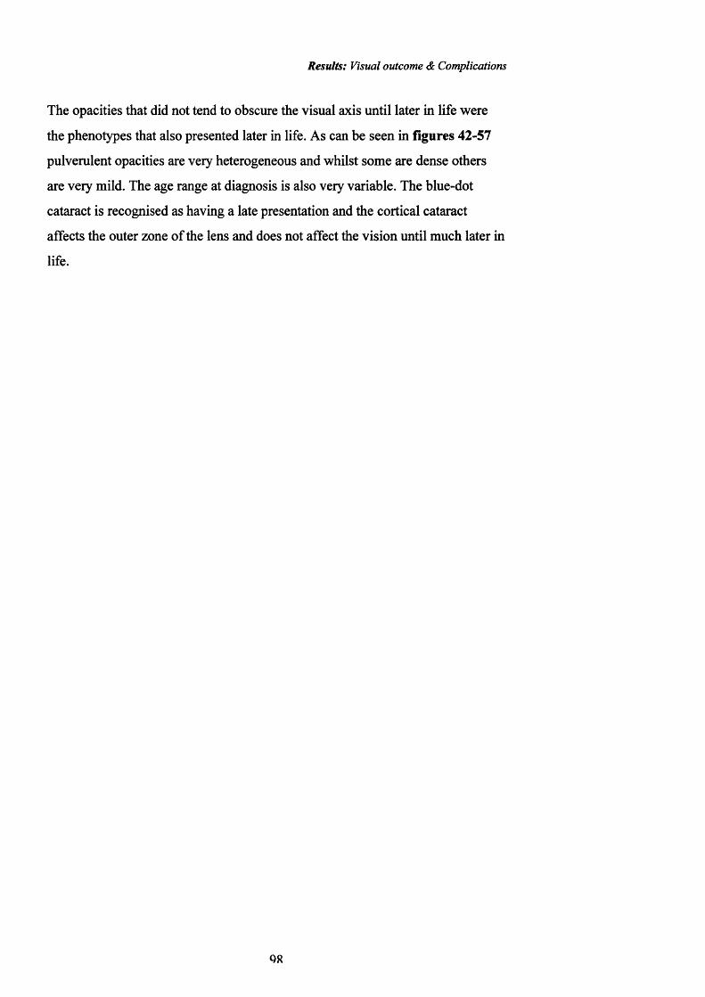
Results: Visual outcome & Complications
The opacities that did not tend to obscure the visual axis until later in life were
the phenotypes that also presented later in life. As can be seen in figures 42-57
pulverulent opacities are very heterogeneous and whilst some are dense others
are very mild. The age range at diagnosis is also very variable. The blue-dot
cataract is recognised as having a late presentation and the cortical cataract
affects the outer zone o f the lens and does not affect the vision until much later in
life.
Q8
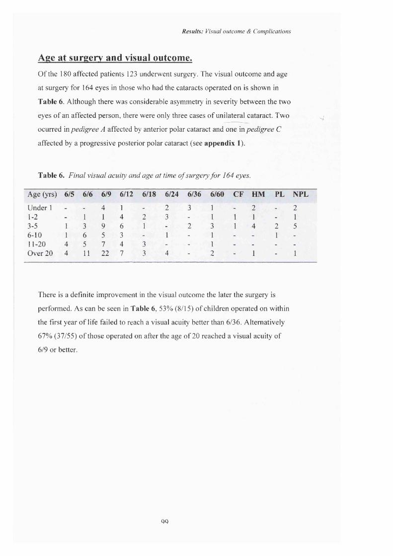
Results: Visual outcome & Complications
Age at surgery and visual outcome.Of the 180 affected patients 123 underwent surgery. The visual outcome and age
at surgery for 164 eyes in those who had the cataracts operated on is shown in
Table 6. Although there was considerable asymmetry in severity between the two
eyes of an affected person, there were only three cases of unilateral cataract. Two
ocurred in pedigree A affected by anterior polar cataract and one in pedigree C
affected by a progressive posterior polar cataract (see appendix 1).
Table 6. Final visual acuity and age at time o f surgery for 164 eyes.
4.
Age (yrs) 6/5 6/6 6/9 6/12 6/18 6/24 6/36 6/60 CF HM PL NFL
Under 1 - - 4 1 - 2 3 1 - 2 - 21-2 - 1 1 4 2 3 - 1 1 1 - 13-5 1 3 9 6 1 - 2 3 1 4 2 56-10 1 6 5 3 - 1 - 1 - - 1 -
11-20 4 5 7 4 3 - - 1 - - - -
Over 20 4 11 22 7 3 4 - 2 - 1 - 1
There is a definite improvement in the visual outcome the later the surgery is
performed. As can be seen in Table 6, 53% (8/15) of children operated on within
the first year of life failed to reach a visual acuity better than 6/36. Alternatively
67% (37/55) o f those operated on after the age of 20 reached a visual acuity of
6/9 or better.
QO

Results: Visual outcome & Complications
The visual outcome for those patients undergoing surgery under 2 years of age is
shown in Figure 59 below.
Figure 59. Visual outcome for those undergoing surgery under 2years o f age.
HMS-NPL20%
6Z5-6/920%
6/60-CFS10%
6/12-6/1823%
6/24-6/3627%
06/5-6/9 06/12-6/18 06/24-6/36 ■6/60-CFS ■ HMS-NPL
The majority of patients had a visual acuity of 6/24 or worse. As the age of
Figure 60. Visual outcome for those undergoing surgery aged 3-5years o f age.
HMS-NPL 30% 6/5-Ô/9
35%
6/60-CFS11% 6/24-6/36
5%6/12-6/18
19%
06/5-6/9 06/12-6/18 06/24-6/36 ■6/60-CFS ■HMS-NPL
surgery increases, the final visual acuity improves as shown in Figure 60
showing the visual acuity for those operated on in the age group 3-5 years old. In
this age group 54% of patients achieved a visual acuity better than 6/24.
100
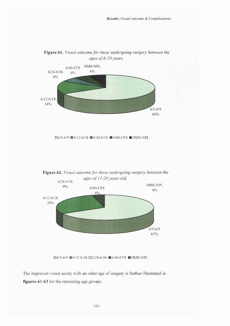
Results: Visual outcome & Complications
Figure 61. Visual outcome fo r those undergoing surgery between theages o f 6-10 years.
6/60-CFS HMS-NPL 6/24-6/36 0/. 6%6%
6%
6/12-6/1816%
6/S-6/966%
06/5-6/9 06/12-6/18 06/24-6/36 ■6/60-CFS ■ HMS-NPL
Figure 62. Visual outcome fo r those undergoing surgery between the ages o f 11-20 years old.
6/24-6/360% 6/60-CFS
4%
HMS-NPL0%
6/12-6/18 29% ^
6/S-6/967%
06/5-6/9 06/12-6/18 06/24-6/36 ■ 6/60-CFS ■ HMS-NPL
The improved visual acuity with an older age of surgery is further illustrated in
figures 61-63 for the remaining age groups.
101
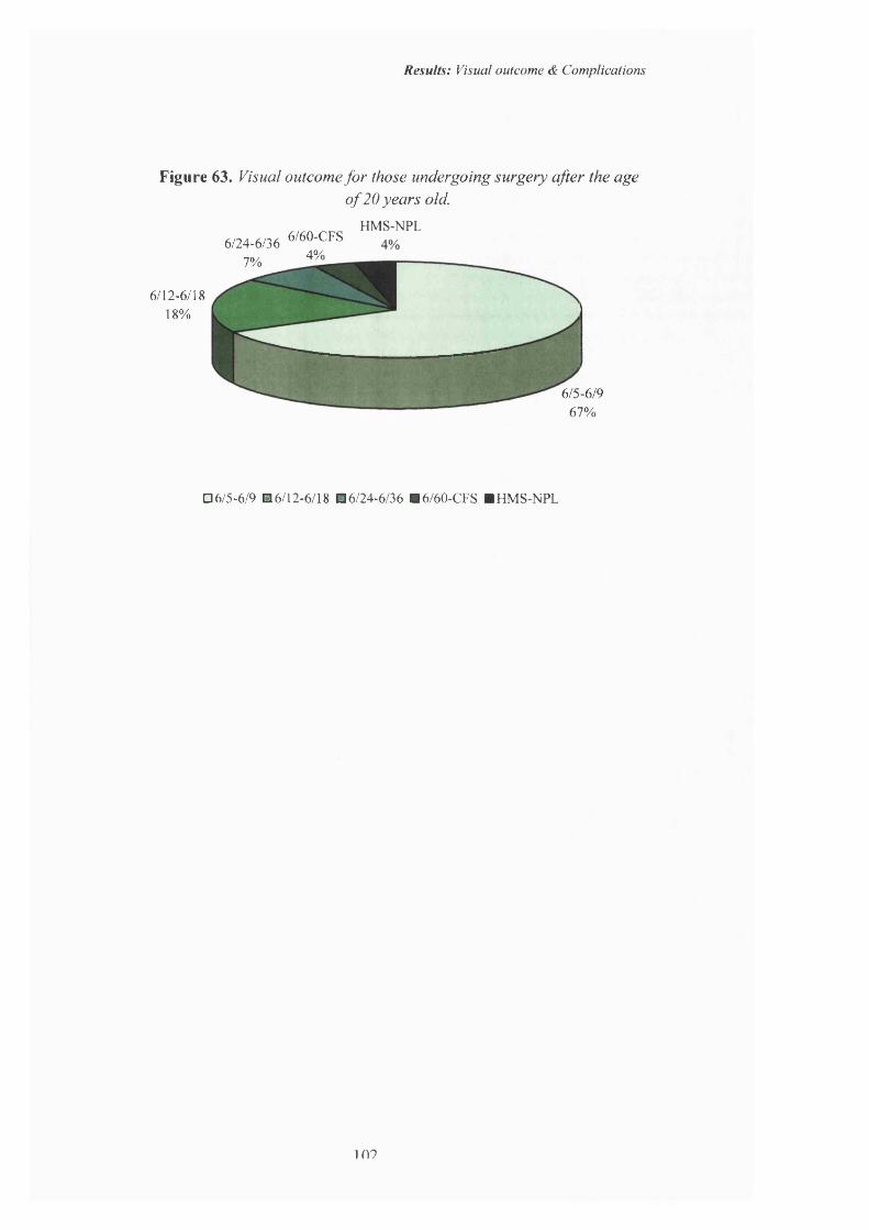
Results: Visual outcome & Complications
Figure 63. Visual outcome for those undergoing surgery after the ageo f 20 years old.
HMS-NPL6/24-6/36 6/60-CFS
4%7%
6/12-6/1818%
6/S-6/967%
06/5-6/9 06/12-6/18 06/24-6/36 ■6/60-CFS ■ HMS-NPL
109
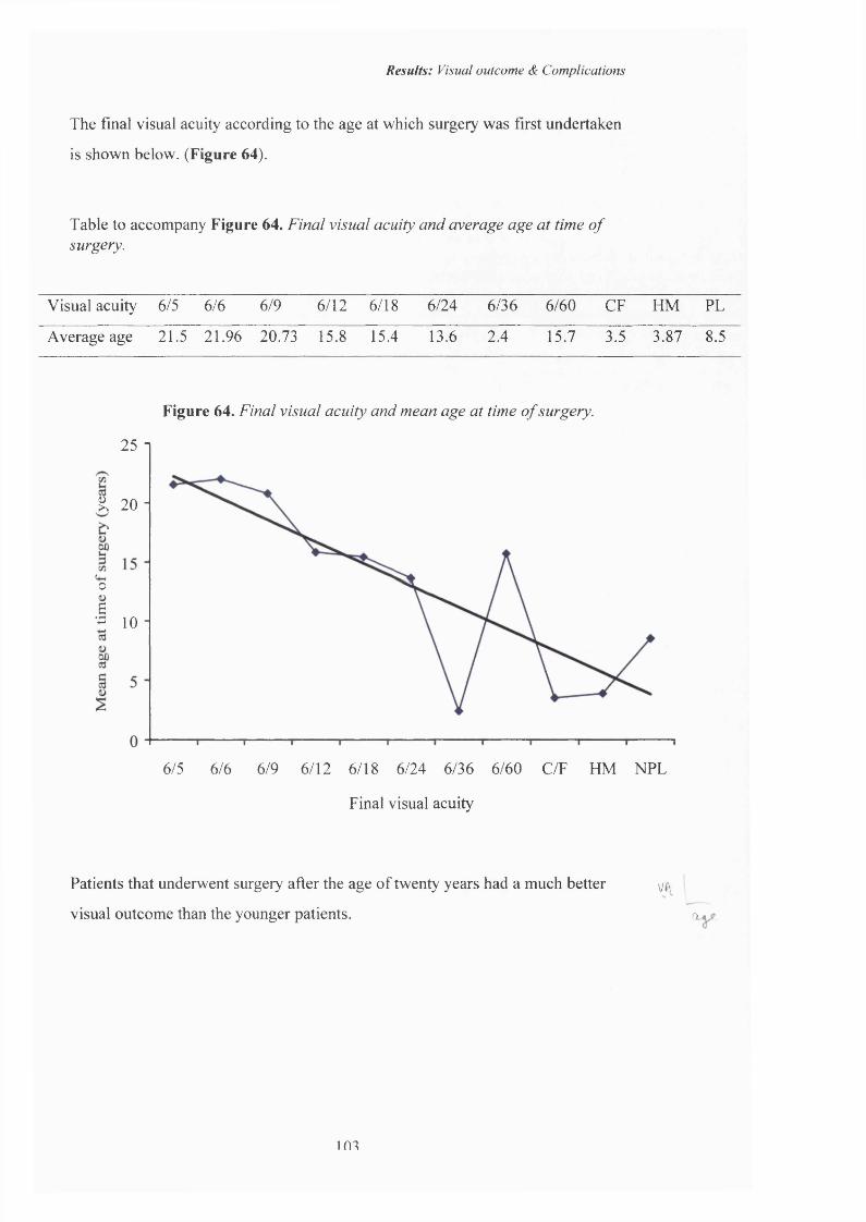
Results: Visual outcome <Sc Complications
The final visual acuity according to the age at which surgery was first undertaken
is shown below. (Figure 64).
Table to accompany Figure 64. Final visual acuity and average age at time o f surgery.
Visual acuity 6/5 6/6 6/9 6/12 6/18 6/24 6/36 6/60 CF HM PL
Average age 21.5 21.96 20.73 15.8 15.4 13.6 2.4 15.7 3.5 3.87 8.5
Figure 64. Final visual acuity and mean age at time o f surgery.
25 1
K 2 0 -
oS- 10 ■
6/5 6/6 6/9 6/12 6/18 6/24 6/36 6/60 C/F HM NPL
Final visual acuity
Patients that underwent surgery after the age of twenty years had a much better
visual outcome than the younger patients.
VA
a
103
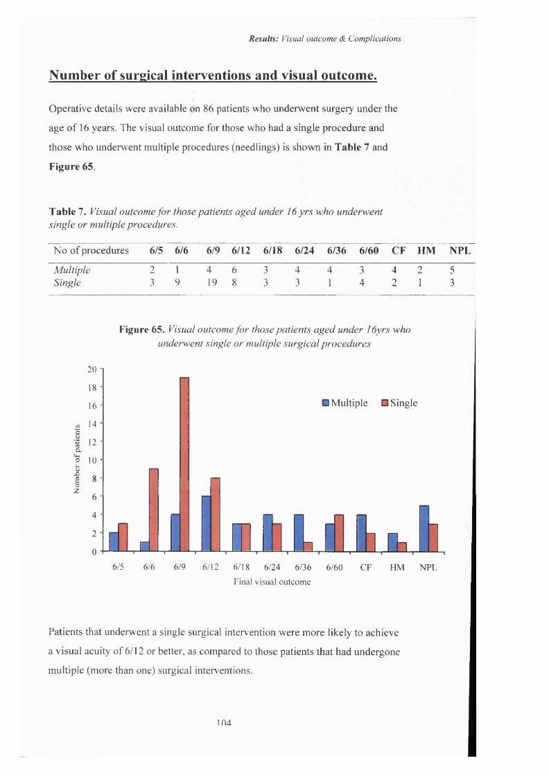
Results: Visual outcome & Complications
Number of surgical interventions and visual outcome.
Operative details were available on 86 patients who underwent surgery under the
age of 16 years. The visual outcome for those who had a single procedure and
those who underwent multiple procedures (needlings) is shown in Table 7 and
Figure 65.
Table 7. Visual outcome for those patients aged under 16 yrs who underwent single or multiple procedures.
No of procedures 6/5 6/6 6/9 6/12 6/18 6/24 6/36 6/60 CF HM NPL
Multiple 2 1 4 6 3 4 4 3 4 2 5Single 3 9 19 8 3 3 1 4 2 1 3
20
18
16
14
12
10
Figure 65. Visual outcome for those patients aged under lôyrs who underwent single or multiple surgical procedures
I101 IZ
n r
□ Multiple □ Single
Q . i m6/5 6/6 6/9 6/12 6/18 6/24 6/36 6/60 CF HM NPL
Final visual outcome
Patients that underwent a single surgical intervention were more likely to achieve
a visual acuity of 6/12 or better, as compared to those patients that had undergone
multiple (more than one) surgical interventions.
104
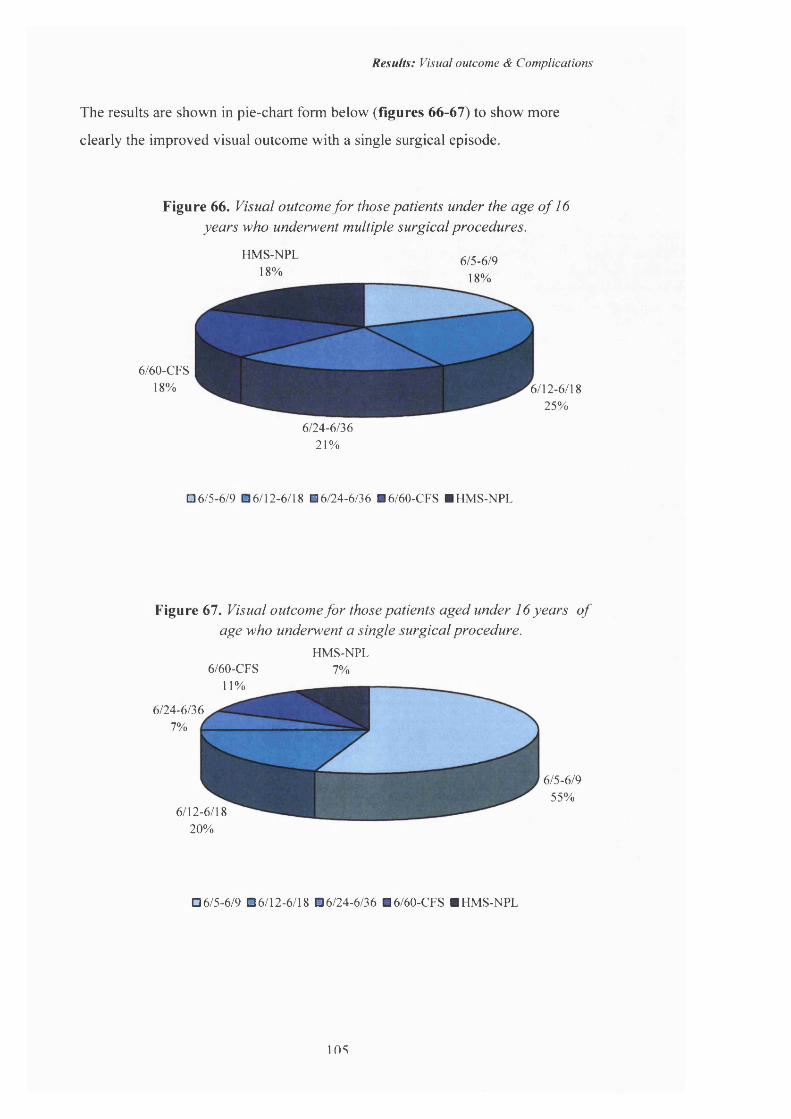
Results: Visual outcome & Complications
The results are shown in pie-chart form below (figures 66-67) to show more
clearly the improved visual outcome with a single surgical episode.
Figure 66. Visual outcome fo r those patients under the age o f 16 years who underwent multiple surgical procedures.
HMS-NPL18%
675-6/918%
6/60-CFS18% 6/12-6/18
25%
6/24-6/3621%
06/5-6/9 06/12-6/18 ■ 6/24-6/36 ■6/60-CFS ■ HMS-NPL
Figure 67. Visual outcome fo r those patients aged under 16 years o fage who underwent a single surgical procedure.
HMS-NPL 6/60-CFS 7%
11%
6/24-6/36 7%
6/5-67955%
6/12-6/1820%
06/5-6/9 06/12-6/18 06/24-6/36 ■ 6/60-CFS ■ HMS-NPL
105
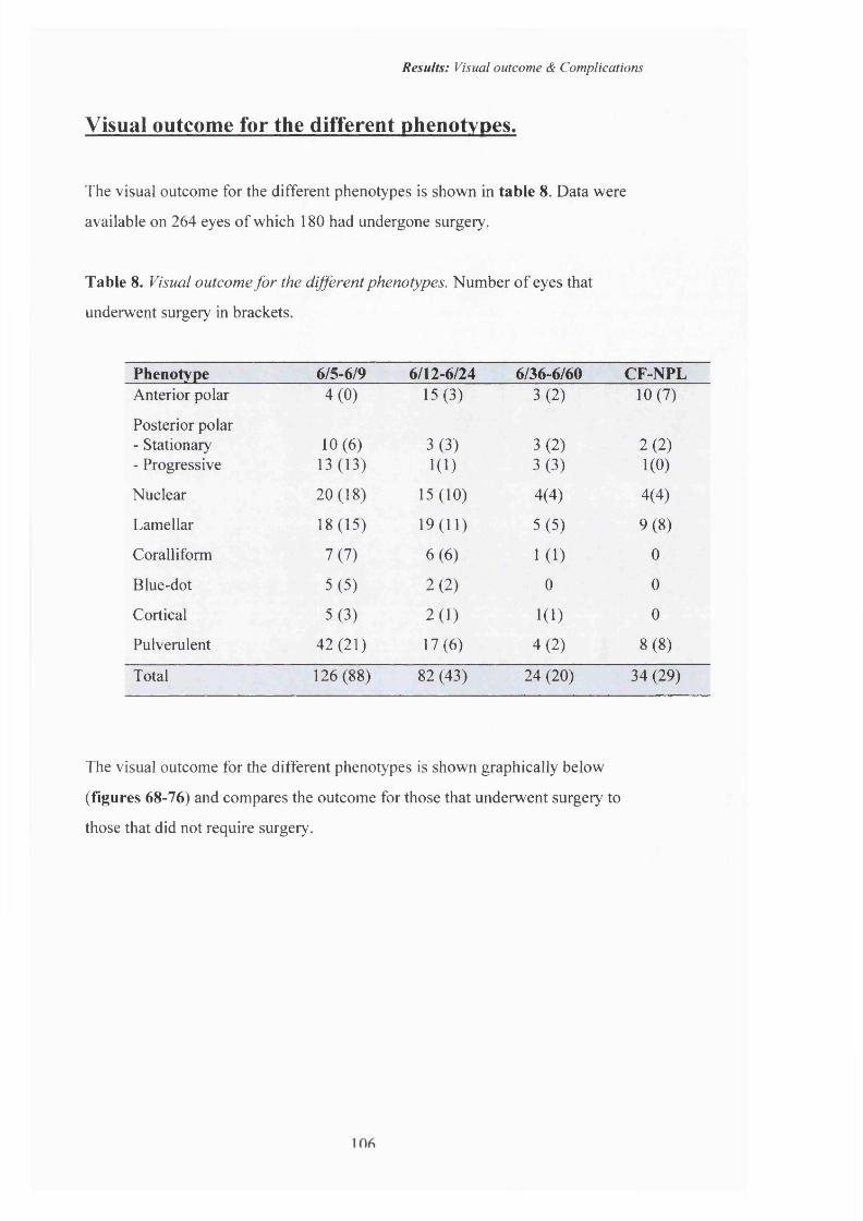
Results: Visual outcome & Complications
Visual outcome for the different phenotypes.
The visual outcome for the different phenotypes is shown in table 8. Data were
available on 264 eyes of which 180 had undergone surgery.
Table 8. Visual outcome for the different phenotypes. Number o f eyes that
underwent surgery in brackets.
Phenotype 6/S-6/9 6/12-6/24 6/36-6/60 CF-NPLAnterior polar 4(0) 15(3) 3(2) 10(7)
Posterior polar - Stationary 10(6) 3(3) 3(2) 2(2)- Progressive 13(13) 1(1) 3(3) 1(0)
Nuclear 20(18) 15(10) 4(4) 4(4)
Lamellar 18(15) 19(11) 5(5) 9(8)
Corail iform 7(7) 6(6) 1(1) 0
Blue-dot 5(5) 2(2) 0 0
Cortical 5(3) 2(1) 1(1) 0
Pulverulent 42(21) 17(6) 4(2) 8(8)
Total 126(88) 82 (43) 24 (20) 34 (29)
The visual outcome for the different phenotypes is shown graphically below
(figures 68-76) and compares the outcome for those that underwent surgery to
those that did not require surgery.

Results: Visual outcome & Complications
Figure 68. Anterior polar cataract. Visual outcome inoperated and unoperated eyes.
S1'o
IIZ
12i
10
8
6
4
2'
06/S-6/9 6/12-6/24 6/36-6/60 CFS-NPL
Final visual acuity
□ unoperated ■ surgery
Figure 69. Posterior polar (stationary) cataract. Visual outcome in operated and unoperated eyes.
615- 6/9 6/12-6/24 6/36-6/60 CFS-NPL
Final visual acuity
□ unoperated □ surgery
107

Results: Visual outcome & Complications
Figure 70. Posterior polar (progressive) cataract. Visualoutcome in operated and unoperated eyes.
§1(4-101
6/5-6Z9 6/12-6/24 6/36-6/60
Final visual acuity
CFS-NPL
□ unoperated □ surgery
Figure 71. Nuclear cataract. Visual outcome in operated and unoperated eyes.
T—*---- - I6/5-6Z9 6/12-6/24 6/36-6/60 CFS-NPL
Final visual acuity
□ unoperated □ surgery
108
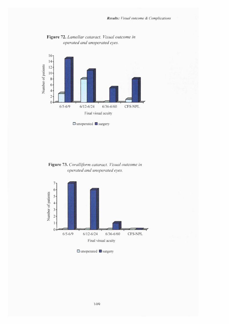
Results: Visual outcome & Complications
Figure 72. Lamellar cataract. Visual outcome inoperated and unoperated eyes.
5 10 o.
6Z5-6/9 6/12-6/24 6/36-6/60 CFS-NPL
Final visual acuity
□ unoperated ■ surgery
Figure 73. Coralliform cataract. Visual outcome in operated and unoperated eyes.
a 4-
6/5-6/9 6/12-6/24 6/36-6/60 CFS-NPL
Final visual acuity
□ unoperated ■ surgery
ino
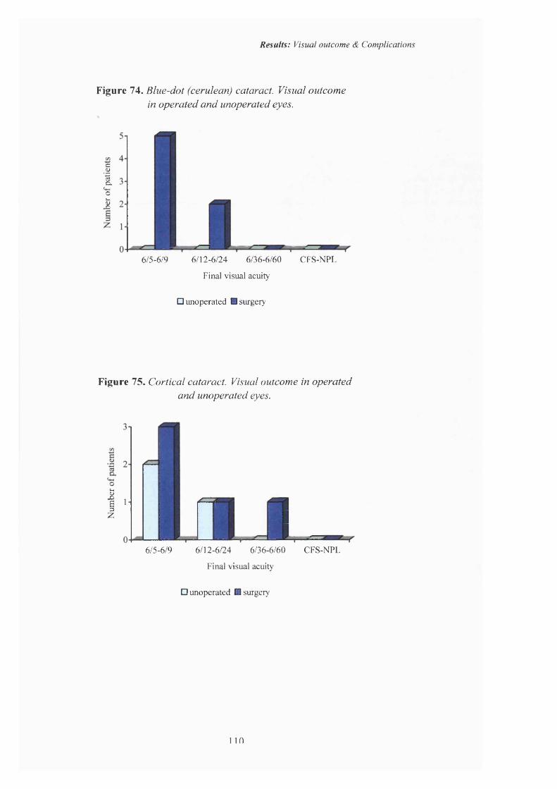
Results: Visual outcome & Complications
Figure 74. Blue-dot (cerulean) cataract. Visual outcomein operated and unoperated eyes.
g1(w01
6/5-6Z9 6/12-6/24 6/36-6/60
Final visual acuity
CFS-NPL
□ unoperated □ surgery
Figure 75. Cortical cataract. Visual outcome in operated and unoperated eyes.
I'z 33 00
6/S-6/9 6/12-6/24 6/36-6/60 CFS-NPL
Final visual acuity
□ unoperated □ surgery
110
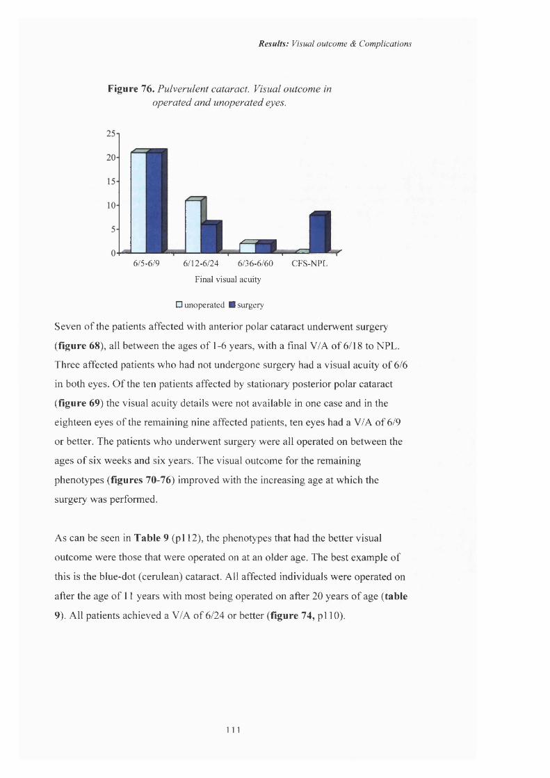
Results: Visual outcome & Complications
Figure 76. Pulverulent cataract. Visual outcome inoperated and unoperated eyes.
u Xf=m6/5-679 6/12-6/24 6/36-6/60 CFS-NPL
Final visual acuity
□ unoperated ■ surgery
Seven of the patients affected with anterior polar cataract underwent surgery
(figure 68), all between the ages of 1-6 years, with a final V/A of 6/18 to NPL.
Three affected patients who had not undergone surgery had a visual acuity of 6/6
in both eyes. O f the ten patients affected by stationary posterior polar cataract
(figure 69) the visual acuity details were not available in one case and in the
eighteen eyes of the remaining nine affected patients, ten eyes had a V/A of 6/9
or better. The patients who underwent surgery were all operated on between the
ages of six weeks and six years. The visual outcome for the remaining
phenotypes (figures 70-76) improved with the increasing age at which the
surgery was performed.
As can be seen in Table 9 (pi 12), the phenotypes that had the better visual
outcome were those that were operated on at an older age. The best example of
this is the blue-dot (cerulean) cataract. All affected individuals were operated on
after the age of 11 years with most being operated on after 20 years of age (table
9). All patients achieved a V/A of 6/24 or better (figure 74, pi 10).
111

Results: Visual outcome & Complications
Table 9. Age at time o f surgery fo r individual eyes o f different phenotypes.
Phenotype
Total no. of eyes that underwent surgery
x<l
Age at surgery (years)
1-5 6-10 11-20 x>20Anterior polar 17 4 6 5 2 -
Posterior polar - Stationary 15 6 9- Progressive 18 - 2 3 5 8
Nuclear 39 - 8 8 3 20
Lamellar 44 8 19 7 2 8
Coralliform 18 - 8 8 2 -
Blue-dot 14 - - - 6 8
Cortical 10 - - 3 4 3
Pulverulent 37 - 8 6 7 16
Total 212 18 60 40 31 63
11?

Results: Visual outcome & Complications
Complications.The complications of glaucoma and retinal detachment are shown below. Out of
the 180 patients affected with ADC, 8 developed glaucoma and 9 had retinal
detachments.
Glaucoma.
Table 10. Complications; glaucoma.No. of
proceduresPhenotype Affected Age at surgery
eye (years)Visual acuity Right Left
Left1. Posterior polar L 1 3 NPL 6/12 Multiple
2. Lamellar R&L 1 1 NPL NPL Multiple
3. Lamellar R&L 3 5 NPL 6/12 Single
4. Lamellar R&L 1 1 6/24 HM Single
5. Coralliform R 7 7 6/24 6/9 Multiple
6. Coralliform R 3 3 6/12 6/9 Multiple
7. Anterior polar R&L 6 6 NPL NPL Multiple
8. Nuclear R&L 8 8 6/9 6/24 Single
Out o f the 180 affected patients o f whom 123 underwent surgery only 8
developed glaucoma. All the patients who developed glaucoma were aphakic and
had been operated on under the age of nine. O f the eight patients with glaucoma,
five had undergone multiple surgical procedures and three had only had one
operation.
i n
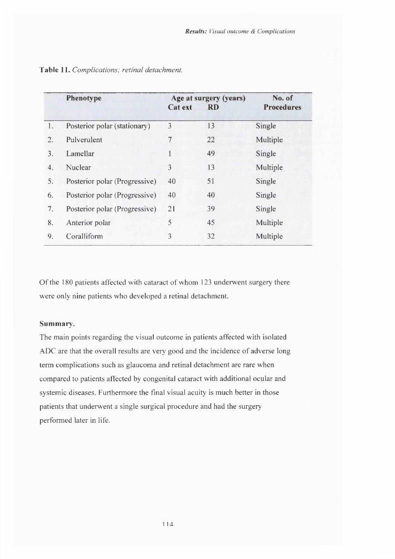
Results: Visual outcome & Complications
Table 11. Complications; retinal detachment.
Phenotype Age at surgery (years) Cat ext RD
No. of Procedures
1. Posterior polar (stationary) 3 13 Single
2. Pulverulent 7 22 Multiple
3. Lamellar 1 49 Single
4. Nuclear 3 13 Multiple
5. Posterior polar (Progressive) 40 51 Single
6. Posterior polar (Progressive) 40 40 Single
7. Posterior polar (Progressive) 21 39 Single
8. Anterior polar 5 45 Multiple
9. Coralliform 3 32 Multiple
O f the 180 patients affected with cataract of whom 123 underwent surgery there
were only nine patients who developed a retinal detachment.
Summary.
The main points regarding the visual outcome in patients affected with isolated
ADC are that the overall results are very good and the incidence of adverse long
term complications such as glaucoma and retinal detachment are rare when
compared to patients affected by congenital cataract with additional ocular and
systemic diseases. Furthermore the final visual acuity is much better in those
patients that underwent a single surgical procedure and had the surgery
performed later in life.
1 U
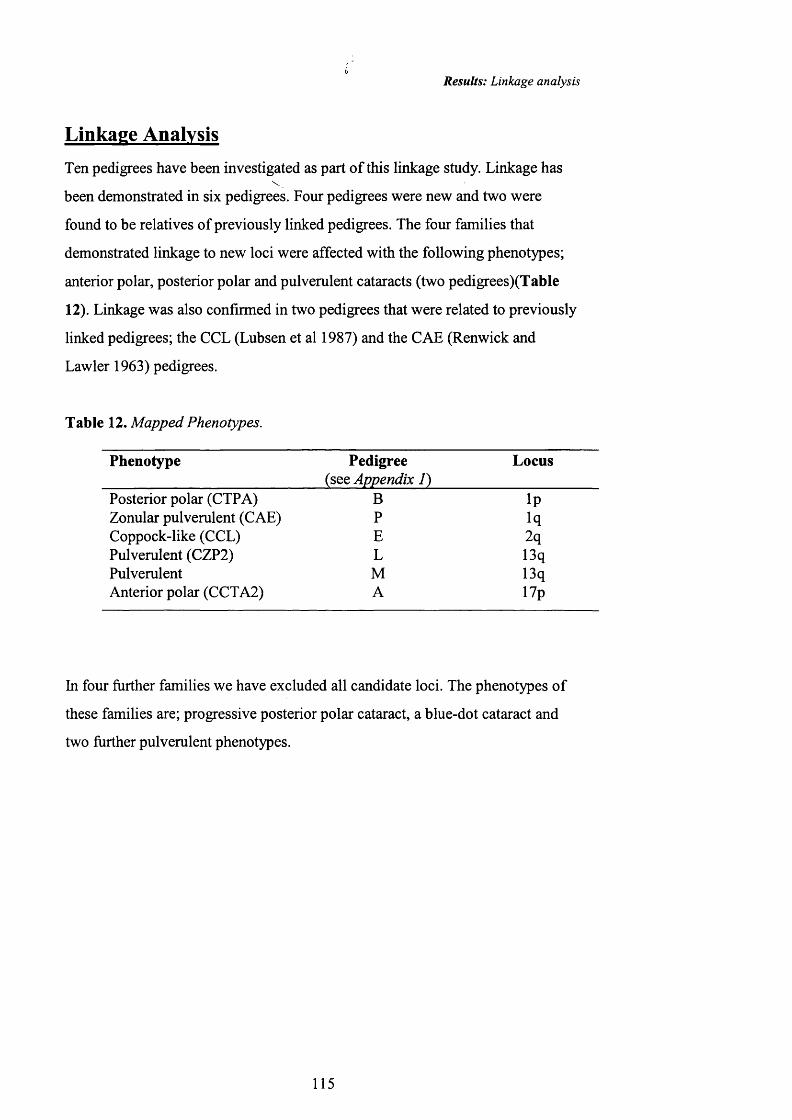
Results: Linkage analysis
Linkage AnalysisTen pedigrees have been investigated as part of this linkage study. Linkage has
been demonstrated in six pedigrees. Four pedigrees were new and two were
found to be relatives of previously linked pedigrees. The four families that
demonstrated linkage to new loci were affected with the following phenotypes;
anterior polar, posterior polar and pulverulent cataracts (two pedigrees)(Table
12). Linkage was also confirmed in two pedigrees that were related to previously
linked pedigrees; the CCL (Lubsen et al 1987) and the CAE (Renwick and
Lawler 1963) pedigrees.
Table 12. Mapped Phenotypes.
Phenotype Pedigree(see Appendix 1)
Locus
Posterior polar (CTPA) B IpZonular pulverulent (CAE) P iqCoppock-like (CCL) E 2qPulverulent (CZP2) L 13qPulverulent M 13qAnterior polar (CCTA2) A 17p
In four further families we have excluded all candidate loci. The phenotypes of
these families are; progressive posterior polar cataract, a blue-dot cataract and
two further pulverulent phenotypes.
115
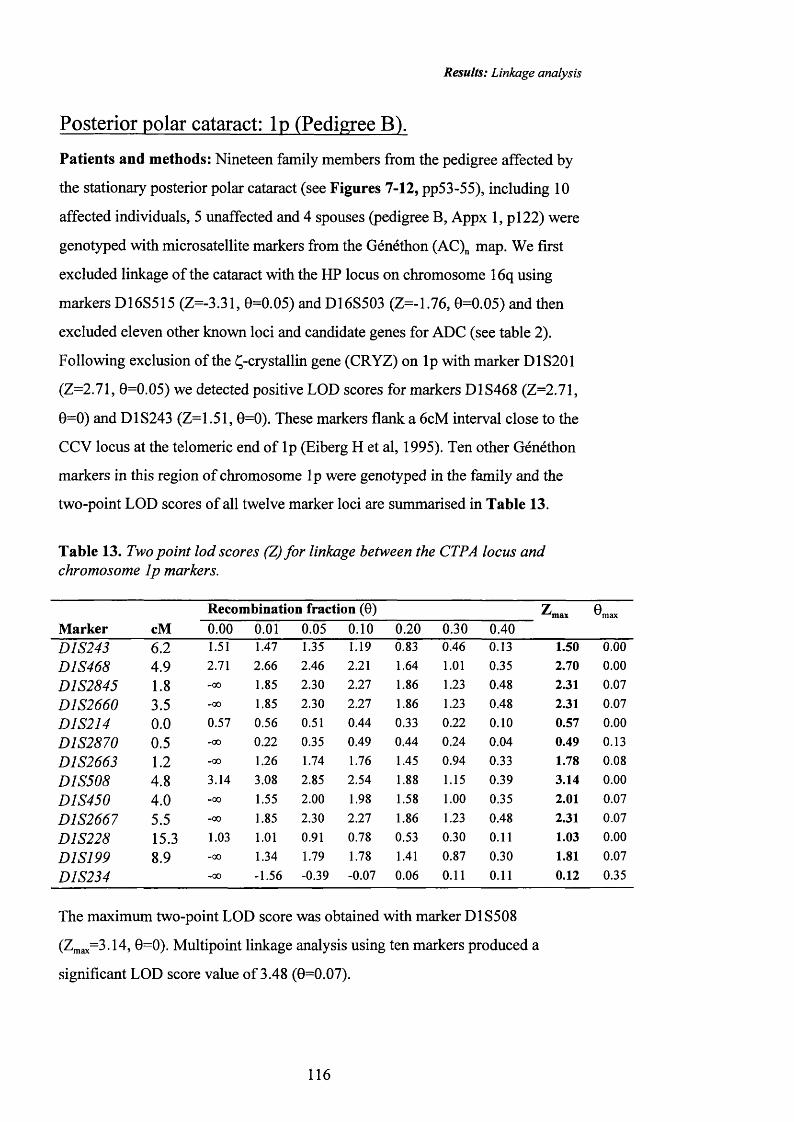
Results: Linkage analysis
Posterior polar cataract: Ip (Pedigree B).
Patients and methods: Nineteen family members from the pedigree affected by
the stationary posterior polar cataract (see Figures 7-12, pp53-55), including 10
affected individuals, 5 unaffected and 4 spouses (pedigree B, Appx 1, p i22) were
genotyped with microsatellite markers from the Généthon (AC)„ map. We first
excluded linkage of the cataract with the HP locus on chromosome 16q using
markers D16S515 (Z=-3.31, 0=0.05) and D16S503 (Z=-1.76, 0=0.05) and then
excluded eleven other known loci and candidate genes for ADC (see table 2).
Following exclusion of the Ç-crystallin gene (CRYZ) on Ip with marker D1S201
(Z=2.71, 0=0.05) we detected positive LOD scores for markers D1S468 (Z=2.71,
0=0) and D1S243 (Z=1.51, 0=0). These markers flank a 6cM interval close to the
CCV locus at the telomeric end of Ip (Eiberg H et al, 1995). Ten other Généthon
markers in this region of chromosome Ip were genotyped in the family and the
two-point LOD scores of all twelve marker loci are summarised in Table 13.
Table 13. Two point lod scores (Z) for linkage between the CTPA locus and chromosome Ip markers.
M ark er cM
R ecom bination fraction (0) ^max ®max0.00 0.01 0.05 0.10 0.20 0.30 0.40
D1S243 6 .2 1.51 1.47 1.35 1.19 0.83 0.46 0.13 1.50 0.00
D1S468 4.9 2.71 2.66 2.46 2.21 1.64 1.01 0.35 2.70 0.00
D1S2845 1.8 -00 1.85 2.30 2.27 1.86 1.23 0.48 2.31 0.07
D1S2660 3.5 -00 1.85 2.30 2.27 1.86 1.23 0.48 2.31 0.07
D1S214 0 .0 0.57 0.56 0.51 0.44 0.33 0.22 0.10 0.57 0.00
D1S2870 0.5 -00 0.22 0.35 0.49 0.44 0.24 0.04 0.49 0.13
D1S2663 1.2 -00 1.26 1.74 1.76 1.45 0.94 0.33 1.78 0.08
D1S508 4.8 3.14 3.08 2.85 2.54 1.88 1.15 0.39 3.14 0.00
D1S450 4.0 -00 1.55 2.00 1.98 1.58 1.00 0.35 2.01 0.07
D1S2667 5.5 -00 1.85 2.30 2.27 1.86 1.23 0.48 2.31 0.07
D1S228 15.3 1.03 1.01 0.91 0.78 0.53 0.30 0.11 1.03 0.00
D1S199 8.9 -00 1.34 1.79 1.78 1.41 0.87 0.30 1.81 0.07
D1S234 -00 -1.56 -0.39 -0.07 0.06 0.11 0.11 0.12 0.35
The maximum two-point LOD score was obtained with marker D1S508
(Z^ax^3.14, 0=0). Multipoint linkage analysis using ten markers produced a
significant LOD score value of 3.48 (0=0.07).
116

Results: Linkage analysis
R esu lts: Haplotype analysis (see Appx 2, publications) detected three affected
individuals (3-6, 3-7, 3-8) that were recombinant with the most centromeric
marker used in the region, D1S234. Only one unaffected individual (4-1) was
recombinant for markers D1S2667, D1S450, D1S2663, D1S468 and D1S243.
The informative haplotype data for individual 4-1 indicated that the cataract
locus probably lies in the interval between D1S2845 and the telomere on
chromosome Ip. For a copy of the published results see Appendix 2; |
publications (lonides et al 1997).
117

Results: Linkage analysis
Zonular pulverulent cataract (CAE) Iq (pedigree P)
Patients and methods: Descendents of the family Ev (see Introduction page )
originally described by Nettleship in 1909 and later investigated by Renwick and
Lawler in 1963 were traced using the records at Great Ormond Street Hospital for
Sick Children and those at Moorfields Eye Hospital, both in London. The
pedigree is illustrated in appendix 1, pedigree X. All affected individuals had
been operated on and it was not possible to get a good description of the
phenotype. It had previously been described as 'lamellar' (Nettleship 1909) and
'zonular pulverulent' (Renwick and Lawler 1963). Although assumed to be
identical to the Coppock cataract and often called the Coppock cataract, there is
no kinship between the Everett and Coppock pedigrees and the Coppock cataract
has never been the subject of a published linkage study.
Eighteen members of the family were genotyped using microsatellite markers
from the Généthon map (Dibb et al 1996), including 10 affected individuals, 6
unaffected and 2 spouses. The two-point maximum lod scores for the markers are
shown in Table 14 below.
Table 14. Two point Z values fo r linkage between CZPl and chromosome Iq markers.
M arker cM
Recombination fraction (0)
0.00 0.01 0.05 0.10 0.20 0.30 0.40 ^max 8max
D1S2746 1.1 -00 1.19 1.66 1.65 1.31 0.84 0.34 1.68 .07
D1S250 0 .6 3.52 3.46 3.19 2.84 2.10 1.31 0.52 3.52 .00
D1S189 2 .2 3.34 3.27 3.02 2.69 2.00 1.27 0.52 3.34 .00
D1S2669 1.3 4.52 4.43 4.09 3.65 2.72 2.24 0.76 4.52 .00
D1S514 1.1 4.48 4.40 4.07 3.64 2.73 1.77 0.77 4.48 .00
D1S2696 1.1 3.49 3.44 3.22 2.91 2.21 1.44 0.65 3.49 .00
D1S442 1.2 1.07 1.04 0.95 0.84 0.62 0.41 0.20 1.07 .00
D1S2345 3.3 2.23 2.16 1.90 1.57 0.90 0.30 0.00 2.23 .00
D1S305 1.6 2.34 2.28 2.05 1.76 1.17 0.62 0.20 2.34 .00
D1S2721 1.7 3.83 3.75 3.42 2.99 2.08 1.15 0.35 3.83 .00
D1S506 2.7 1.11 1.09 0.99 0.86 0.59 0.33 0.11 1.11 .00
D1S2635 2.7 0.93 0.92 0.86 0.76 0.53 0.31 0.13 0.93 .00
D1S2771 -00 -1.84 -0.57 -0.14 0.11 0.12 0.07 0.13 .25
118
Results: Linkage analysis
Results. The maximum two point lod score was obtained with marker D1S2669.
Multipoint analysis using markers D1S2746, D1S2669, D1S442 and D1S2771
gave a maximum lod score of 5.22 (0 = 0) at D1S2669 (see Shiels et al 1998,
Appendix 2\ publications) and this information combined with haplotype
analysis suggested that the gene for the cataract lies in the genetic distance
D1S2746-D1S2771. A missense mutation in the gene that codes for connexin 50
{GJA8) has been found to be the cause of the cataract (Shiels et al 1998).
119
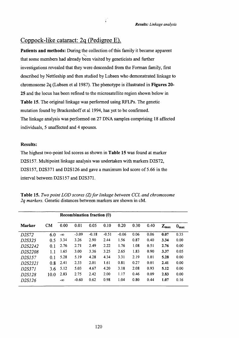
Results: Linkage analysis
Coppock-like cataract: 2q (Pedigree E lPatients and methods: During the collection of this family it became apparent
that some members had already been visited by geneticists and further
investigations revealed that they were descended from the Forman family, first
described by Nettleship and then studied by Lubsen who demonstrated linkage to
chromosome 2q (Lubsen et al 1987). The phenotype is illustrated in Figures 20-
25 and the locus has been refined to the microsatellite region shown below in
Table 15. The original linkage was performed using RFLPs. The genetic
mutation found by Brackenhoff et al 1994, has yet to be confirmed.
The linkage analysis was performed on 27 DNA samples comprising 18 affected
individuals, 5 unaffected and 4 spouses.
Results:
The highest two-point lod scores as shown in Table 15 was found at marker
D2S157. Multipoint linkage analysis was undertaken with markers D2S72,
D2S157, D2S371 and D2S126 and gave a maximum lod score of 5.66 in the
interval between D2S157 and D2S371.
Table 15. Two point LOD scores (Z) fo r linkage between CCL and chromosome 2q markers. Genetic distances between markers are shown in cM.
M arker CM
R ecom bination fraction (6)
0.00 0.01 0.05 0.10 0.20 0.30 0.40 Zrmax ®max
D25"72 6.0 -00 -3.09 -0.18 -0.51 -0.06 0.06 0.06 0.07 0.35
D2S325 0.5 3.34 3.26 2.90 2.44 1.56 0.87 0.40 3.34 0.00D2S2242 0.1 2.76 2.71 2.49 2.22 1.76 1.08 0.51 2.76 0.00
1.1 1.65 3.00 3.36 3.25 2.65 1.83 0.90 3.37 0.05
D2S157 0.1 5.28 5.19 4.28 4.34 3.31 2.19 1.01 5.28 0.00D2S232J 0 .8 2.41 2.33 2.01 1.61 0.81 0.27 0.01 2.41 0.00D2S371 3.6 5.12 5.03 4.67 4.20 3.18 2.08 0.93 5.12 0.00
10.0 2.83 2.75 2.42 2.00 1.17 0.46 0.09 2.83 0.00D2S126 -00 -0.60 0.62 0.98 1.04 0.80 0.44 1.07 0.16
120
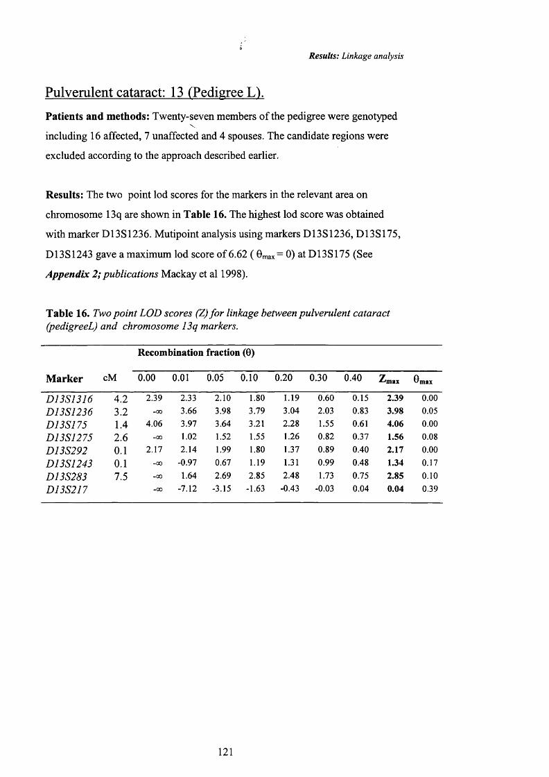
Results: Linkage analysis
Pulverulent cataract: 13 (Pedigree L).
Patients and methods: Twenty-seven members o f the pedigree were genotyped
including 16 affected, 7 unaffected and 4 spouses. The candidate regions were
excluded according to the approach described earlier.
Results: The two point lod scores for the markers in the relevant area on
chromosome 13q are shown in Table 16. The highest lod score was obtained
with marker D13S1236. Mutipoint analysis using markers D13S1236, D13S175,
D13S1243 gave a maximum lod score of 6.62 ( 0max= 0) at D13S175 (See
Appendix 2; publications Mackay et al 1998).
Table 16. Two point LOD scores (Z) fo r linkage between pulverulent cataract (pedigreeL) and chromosome 13q markers.
M arker cM
R ecom bination fraction (6)
0.00 0.01 0.05 0.10 0.20 0.30 0.40 Zmax Omax
D13S1316 4.2 2.39 2.33 2.10 1.80 1.19 0.60 0.15 2.39 0.00
D13S1236 3.2 -00 3.66 3.98 3.79 3.04 2.03 0.83 3.98 0.05
D13S175 1.4 4.06 3.97 3.64 3.21 2.28 1.55 0.61 4.06 0.00
D13S1275 2.6 -00 1.02 1.52 1.55 1.26 0.82 0.37 1.56 0.08
D13S292 0.1 2.17 2.14 1.99 1.80 1.37 0.89 0.40 2.17 0.00
D13S1243 0.1 -00 -0.97 0.67 1.19 1.31 0.99 0.48 1.34 0.17
D13S283 7.5 -00 1.64 2.69 2.85 2.48 1.73 0.75 2.85 0.10
D13S217 -00 -7.12 -3.15 -1.63 -0.43 -0.03 0.04 0.04 0.39
121

Results: Linkage analysis
Pulverulent Cataract: 13q (Pedigree M).
Patients and methods: Eighteen family members were genotyped including 10
affected, 5 unaffected and 3 spouses {sqq Appendix 1; pedigree M). The genome
was ranked into the candidate regions described above, although the candidate
cataract loci now included the 13q site.
Results: A maximum lod score of 3.91 was obtained with markers D13S175,
D13S1275 and D13S292 as shown in Table 17.
Table 17. Two point LOD scores (Z) for linkage between pulverulent cataract (pedigree M) and chromosome 13q markers.
M arker cM
R ecom bination fraction (0)
0.00 0.01 0.05 0.10 0.20 0.30 0.40Z m ax 8 m . .
4.2 3.37 3.31 3.06 2.73 2.02 1.23 0.49 3.37 0.00
3.2 0.90 0.89 0.84 0.77 0.61 0.44 0.24 0.90 0.00
1.4 3.91 3.85 3.60 3.27 2.56 1.76 0.89 3.91 0.00
2.6 3.91 3.85 3.60 3.27 2.56 1.76 0.89 3.91 0.00
DyJ5'2P2 0.1 3.91 3.85 3.60 3.27 2.56 1.76 0.89 3.91 0.00
Dyj^y243 0.1 2.91 2.07 1.93 1.74 1.33 0.88 0.41 2.11 0.00
7.5 3.61 3.55 3.30 2.97 2.26 1.47 0.65 3.61 0.00-00 -1.62 -0.33 0.13 0.43 0.43 0.27 0.45 0.25
Both these two families affected by pulverulent cataract map to the same region
on chromosome on 13q and a candidate gene that codes for connexin46 lies in
this region. Further studies have now isolated I different mictions in this gene in
the two families that gives rise to the cataract.
122
Results: Linkage analysis
Anterior polar cataract: 17p (Pedigree A).
Patients and methods: Twenty-eight family members, o f whom 16 were
affected (see Figures 2-6), 8 unaffected and 4 spouses were genotyped using
microsatellite markers from the Genethon poly-CA map (Gyapay G et al, 1994).
Candidate loci for anterior polar cataract were excluded before excluding the
known loci for ADC and the crystallin genes. Aniridia is associated with anterior
polar cataract and is caused by a mutation on the PAX6 gene on chromosome
1 lp l3 (Riccardi VM et al, 1978) a translocation (2;14)(p25;q24) (Moross T et al,
1984) has also been associated with anterior polar cataract. Both these regions
were excluded by linkage analysis to contain the relevant gene in this pedigree.
Results: Positive lod scores were obtained for markers D17S799 (Z= 0.74 @
0=0.30) and D17S798 (Z= 0.45 @ 0= 0.30). These markers flank the zonular
sutural cataract locus and the pA3/Al-crystallin gene. Linkage to these loci were
excluded with marker D17S805 (Z= -3.93 @ 0= 0.05). The cerulean cataract
locus on 17q24 was also excluded with marker D17S802 (Z=-3.27 @ 0=0.05),
thus excluding the known cataract and crystallin loci on 17q. Chromosome 17 is
rich in genes and contains many loci important in eye diseases so it was felt that
the entire chromosome should be excluded. During this exclusion significant lod
scores were obtained for the following markers on 17p (see Table 18, below),
marker D17S786 (Z=3.61 @0=0.10), D17S796 (Z=4.17 @ 0=0.05) and
D17S849 (Z=4.01 @ 0=0.05).
123

Results: Linkage analysis
Table 18. Two-point lod scores (Z) for linkage between the anterior polar
cataract locus and chromosome 17 markers.
M arker
R ecom bination fraction (0) 7max ®max0.00 0.01 0.05 0.10 0.20 0.30 0.40-00 3.68 4.01 3.83 3.10 2.14 1.05 4.01 0.05-00 3.84 4.17 3.99 3.26 2.30 1.17 4.17 0.05-00 2.51 3.52 3.61 3.11 2.24 1.16 3.61 0.08
Centromere-00 -3.39 -0.81 0.11 0.71 0.74 0.49 - --00 -8.01 -3.93 -2.27 -0.82 -0.19 0.05 - --00 -2.73 -0.78 -0.80 0.39 0.45 0.30 - --00 -7.19 -3.27 -1.83 -0.73 -0.28 -0.04 - -
Maximum lod scores and corresponding recombination fractions are shown (in bold) only for the 17p markers.
The maximum lod score of 4.17 was obtained with D17S796. Multipoint analysis
using markers D17S849, D17S796 and D17S786 gave a maximum lod score of
5 2 @ =0.06. This placed the anterior polar cataract locus in a 13cM interval
between the markers D17S849 and D17S796 at the telomeric end of chromosome
17p. Assignment to this genetic locus is supported by fully informative haplotype
analysis (see Appx 2, publications) which detected four recombinant individuals
with D17S799; 4-2, 4-6, 4-9 and 4-11, two recombinant individuals with
D17S786; 4-6 and 4-9, and one recombinant individual with D17S796; 4-6. This
means that the cataract locus cannot lie centromeric to D17S796. Work is
continuing on this family to try and find the underlying mutation.
Exclusion data:
In five further pedigrees exclusion of various loci has been undertaken. All the
studies were subject to a candidate gene approach to exclude the known relevant
loci for the particular phenotype and positive linkage has yet to be demonstrated. \
124

Discussion
Discussion
Clinical heterogeneity 126
Visual Outcome & Complications of surgery 130
Age at diagnosis 130
Age at surgery and visual outcome 131
Number of surgical interventions and visual 132outcome
Visual outcome for the different phenotypes 132
Complications 136
Genetic heterogeneity 139
125
Discussion: Clinical heterogeneity
Clinical Heterogeneity.
In this study of ADC we have demonstrated that all the phenotypes o f the
pedigrees encountered could be classified as one of the eight following types;
anterior polar, posterior polar, nuclear, lamellar, coralliform, blue-dot (cerulean),
cortical and pulverulent. This study was limited to large families that were
suitable for linkage studies but this was thought not to affect the distribution of
the affected phenotypes. Other phenotypes not included in this study have been
reported but they have either been described in individual cases or are very
uncommon. Although an attempt was made to examine the phenotypes o f all
family members at risk of inheriting the affected gene, the description o f intra-
familial variation in the phenotypes was limited by the number of patients who
had already undergone surgery. This is a limitation inherent in all studies o f early
onset inherited cataract and we attempted to obtain as detailed a description as
possible from the hospital records in such cases.
The eight phenotypes described here are all recognised and previously described.
Cortical cataract however, is the least well recognised of the eight phenotypes.
Jaafar and Robb (1984) list the types of cataract seen in 463 patients diagnosed as
having congenital cataract o f all aetiologies in a paediatric eye department, 37
patients were described as having 'cortical' cataracts. There is no description nor
photograph of the cortical cataracts and the paper concentrates on the clinical
course and visual outcome of those affected by anterior polar cataract, not
cortical cataract. Lamellar cataracts are also listed amongst the phenotypes by
Jaafar and Robb indicating that cortical and lamellar are distinct phenotypes in
their classification. Cortical cataract is a description more usually applied to adult
'age-related' cataract. There is evidence for a single gene to be involved in a large
proportion of adult cortical cataract (Heiba et al 1995) and the identification of
the gene for the cortical cataract in pedigree K will allow us to test the
hypothesis that mutations in this gene may influence cortical cataract in the
elderly.
126
collection of fine dots forming a hazy cloud. This description suggests that this ?
pedigree may be affected by the pulverulent phenotype. For these reasons we
have avoided the term 'zonular' cataract and used 'lamellar' to describe a cataract
that forms a shell around a clear inner nuclear area as described and illustrataed
in Figures 30-34 pp75-77.
127
V
Discussion: Clinical heterogeneity
There is also evidence for a single gene for age related nuclear cataract (Heiba et
al 1993). In a study analysing data from the Beaver Dam Eye Study Heiba et al
found that a single major gene can account for 35% of the total variability of age-
sex-adjusted measures of nuclear sclerosis. Although it is unlikely that a
mutation in a gene for autosomal dominant nuclear cataract may also give rise to
the nuclear sclerosis seen in the elderly population, there may be other mutations
or a series o f mutations that may have a role to play in age-related cataract ^
(Kantorow et al 1998).
Lamellar cataract is occasionally referred to as 'zonular', although this term is not
used here as zonular describes a variety of different morphologies. Duke-Elder
uses the term zonular for an opacity limited to a specific zone of the lens due to a
temporary disturbance of development at a particular period. This definition
includes almost every form of inherited cataract. Posterior polar cataract only
affects the posterior zone of the lens, anterior cataracts affect the anterior zone of
the lens and so on. Vogt defines zonular cataract as having a layer o f opacities
surrounding a clear nucleus and describes an infinite variety of zonular cataracts.
It is this wide clinical spectrum that makes ‘zonular’ a confusing term. Duke-
Elder includes central pulverulent, nuclear and lamellar cataracts as being
zonular. The zonular pulverulent cataract linked to Iq by Renwick and Lawler is
probably a nuclear cataract, as the opacity is ‘always in the centre of the lens.’
The term zonular in this instance is not used according to Vogt’s definition but
follows Duke-Elder’s interpretation of the opacity being in a specific zone of the
lens, which in this case is the nucleus. Padma et al demonstrated linkage of a
zonular-sutural cataract. Although most affected individuals had a clear
embryonic and fetal nucleus, fulfilling Vogt’s description of a zonular cataract,
some opacities were described as varying from a uniform dense opacity to a
? ?
Discussion: Clinical heterogeneity
The distinction between stationary and progressive posterior polar cataract has
been previously documented. Tulloh (1955) describes the progressive posterior
polar cataract in a five generatioi^family that bad "a peculiar discoid posterior
polar cataract, with fluffy white extensions through the posterior cortex towards
the equator. There were a few anterior cortical punctate opacities." Tulloh goes
on to state that the initial opacity was the circular posterior polar cataract, but the
progression of the cortical opacities occurred later. In the family he describes
subsequent generations developed the extensions at progressively earlier ages.
This phenomenon of 'anticipation' was not seen in the family examined in this
study (Pedigree C). However the phenotype is very similar in the description and
the progression as that seen in this study and illustrated in Figure 13, page 62.
In all the pedigrees apart from the pulverulent pedigrees the morphology was the
same but varied in severity. In the pulverulent pedigrees the phenotype varied
amongst affected individuals from the same family. Intra-familial clinical
heterogeneity in ADC has been described by many authors including interocular
variability in individuals affected by ADC (Scott et al 1994). The pedigrees
described as having this wide variation in morphology are characteristic of the
pulverulent type described in this study and the cataract pedigree described by
Scott et al that shows inter-individual variability is referred to in that paper as the
'pulverulent' type of cataract.
There was no evidence o f a gradual worsening nor improvement in the severity
of the cataract with subsequent generations. Only the phenotype could be
predicted, in all but the families affected by pulverulent cataract.
The phenotype of the cataract remains consistent throughout the affected
individuals in all but the families affected by pulverulent cataract. For all the
other families the cataract does not alter according to the part of the lens that is
affected by cataract, although it will vary in severity. A parent affected by a
posterior polar cataract has a 50% chance o f passing the gene on to their
128
Discussion: Clinical heterogeneity
offspring, and any affected children will therefore be affected by a posterior polar
cataract. The only way the cataract may vary, is in its severity, as clearly shown
in the photographs in the illustrations of the opacities earlier in this thesis. This is
true for all the phenotypes except those pedigrees affected by the pulverulent
phenotype and Figures 42-55 show the wide scope for phenotypic variability in
these pedigrees.
This study of the lens morphology in ADC has demonstrated wide clinical
heterogeneity and had initially set out to suggest a clinical classification. Most of
the phenotypes can be classified according to a few o f the different types. Almost
25% of all affected patients had lamellar cataracts and over 28% had pulverulent
cataracts. This means that over 50% of affected individuals could be classified as
one o f two phenotypes. For this reason it is tempting to try and create a clinical
classification but no clinical clj^^ication can include the rare and unusual
phenotypes that make up a significant number of those affected by ADC.
Inherited cataract has been a topic o f study by many ophthalmologists in the past
including Doyne, Nettleship, Vogt, Duke-Elder and François. None have
suggested a clinical classification for the opacities. This thesis has attempted to
classify the opacities according to the position in the lens and the cataract
morphology and has therefore suggested the eight phenotypes as a basic
approach to creating some form of unified approach in dealing with the clinical
aspects o f ADC.
There is clearly considerable genetic heterogeneity and further advances in the
genetic aetiology will allow for a more detailed comparison between the genetic
mutation and the clinical phenotype as well as offering the potential for a genetic
classification of ADC. Already it is possible to see a correlation between the
phenotype and the underlying genetic mutation and further evaluation o f the
molecular genetics of ADC will undoubtedly provide a genetic classification.
129
Discussion: Visual Outcome and Complications o f Surgery
Visual Outcome and Complications of surgery
Age at diagnosis.
The age at diagnosis of those affected hy ADC is illustrated in Figure 58 on page
96 and shows a himodal distribution. The peaks are at birth and then during late
infancy (aged 5-19 years). These ages probably reflect the ages at which visual
screening is most likely to take place. Following birth all babies are given a
general examination hy a paediatrician prior to discharge from the hospital that
includes examining the eyes and the 'red reflex' with a direct ophthalmoscope.
Whilst it is unlikely that they will diagnose the mild and more diffuse cataracts,
this technique will pick up the central dense opacities. The second peak of the
bimodal distribution, which occurs in late infancy, is during early school age
when visual problems are more likely to be recognised either by routine
screening or by teachers noticing children with visual problems.
The phenotypes that are diagnosed early (Table 5, page 97) are the anterior polar
(mean age at presentation under 1 year old) and stationary posterior polar
cataracts (mean age at presentation 1.25 years). These are more clearly visible as
a white dot in the pupil and will also he more visible as a central shadow against
the 'red reflex' on direct ophthalmoscopy. Table 5 only shows the age at
diagnosis for the cataracts that underwent surgery. The milder polar opacities that
never required surgery have not been included.
The other phenotypes that involve the visual axis early in their development;
lamellar, nuclear and coralliform, were the next to present at a mean age of; 2.5,
3.0 and 3.1 years respectively. The opacities that predominately affect the lens
cortex presented last. The blue-dot cataract presented at a mean age o f 11.6 years
and the cortical cataract at a mean age of 18.8 years. The wide clinical
heterogeneity o f the pulverulent cataracts is reflected in the age at presentation.
Whilst some are dense and present early others are mild and require surgery at a
much later date. The mean age at presentation is therefore later than the polar
cataracts but earlier than the hlue-dot and cortical cataracts. The late age at
presentation of the progressive posterior polar cataract is surprising. It may be
that this phenotype is not congenital but develops later in life.
130
Discussion: Visual Outcome and Complications o f Surgery
Very few of the affected patients from all the phenotypes had received regular
examinations by an ophthalmologist or optician in early childhood, despite the
strong family history of cataracts.
Age at surgery and visual outcome.
The age at surgery had a significant effect on the final visual outcome. Table 6
(page 99) shows the final visual outcome and time of surgery for 164 eyes.
Figures 59-63 (pp 100-102) show in pie-chart form the final visual outcome for
the different age groups. The percentage of eyes achieving a visual acuity of 6/9
or better increases as the age at time of surgery gets older. For those operated on
under the age of 2 years, 20% achieve a V/A better than 6/9, whilst 67% of those
operated on after the age of 20 years achieve a V/A of 6/9 or better. There is a
gradual increase in the percentage of patients achieving a visual acuity o f 6/9 or
better. In the intervening years, 35% of the 3-5 year group, 66% if the 6-10 year
olds and 67% of the 11-20 have a final visual acuity of 6/9 or better.
On page 103 Figure 64 shows the mean age at time of surgery for those
achieving a V/A of 6/5 through to non-perception of light (NPL). The 'trend line'
clearly shows that those achieving a better V/A were operated on later in life.
What this cannot take into account is the severity of the opacities and the age at
presentation to an ophthalmologist. The issue of when to operate on bilateral
infantile cataracts has received considerable attention over the years and early
intervention is recommended (Rice & Taylor 1982). This is supported by recent
improvements in visual acuity testing, surgical techniques, instruments and
contact lens management. Although this study shows the benefits o f late surgery,
it is only possible to delay surgery for the milder cataracts. The dense opacities
have to be operated on earlier in life and there are many guidelines to help in the
decision of when to operate. These include the ability to visualise the fundus
(Stark et al 1979), whether the opacity is greater than 3mm in diameter (Hiles
DA 191983), or how dense the cataract is (Arkin et al 1992). In the individuals
with more dense cataracts that require earlier surgery there may well be a more
advanced degree of amblyopia. Whilst there are reports of good visual results in
patients with dense cataracts who have had surgery late or not at all (Freeman &
131
Discussion: Visual Outcome and Complications o f Surgery
Rovick 1994), these are the exceptions and many studies have shown the benefits
of early surgery. What this study shows is that with the partial cataracts, where
possible, delayed surgery is associated with a better final visual acuity. This
study has not investigated the effect of amblyopia on the final visual outcome.
Number of surgical interventions and visual outcome.
In order to assess the number of surgical interventions and the effect this may
have had on the final visual acuity, only patients operated on under the age o f 16
years were included. The overall results are shown graphically in Figure 65
(page 104) and also in the pie charts in Figures 66 and 67 (page 105).
The visual results are better for those patients who underwent a single surgical
procedure. 55% of patients who underwent a single procedure achieved a V/A of
6/9 or better compared to only 18% of those who had multiple surgical
procedures. A number of factors probably underlie these figures. The patients
who had the multiple procedures may well have had earlier surgery for dense
opacities in an era before the advent of improved methods of visual acuity
testing, surgical techniques and contact lens management. These patients are also
more likely to have had periods of poor vision prior to repeated surgery owing to
the visual axis being obscured by the re-growth of lens fibres. This would leave
them at a greater risk of developing amblyopia.
Visual outcome for different phenotypes.
The visual outcome for the different phenotypes is shown in Table 8 on page
104. This information is shown graphically for the different phenotypes in
Figures 68-76 (ppl07-l 11). Data was not available on all the operated eyes.
There is a close association between those phenotypes that have a good visual
outcome and those that are operated on at an older age (ie cortical and blue-dot).
Alternatively, those with a poorer visual outcome are the phenotypes that
underwent surgery in early infancy (the polar cataracts). Table 9 on page 112
illustrates this by showing the age at time of surgery for the different phenotypes.
Anterior polar cataract.
Those patients affected by anterior polar cataract that underwent surgery (Table
7 [page 104] & Figure 66 [page 105]) had a poorer final visual outcome than
those that did not have surgery (lonides et al 2000).Most of the un-operated cases
132
Discussion: Visual Outcome and Complications o f Surgery
had mild opacities but the patient illustrated in Figures 2 and 3 (p57) with
pyramidal anterior cataracts had refused surgery and had 6/18 vision in each eye.
It is not possible to assess the density of the cataracts in the patients who had
already been operated on but it is likely that they had large and dense cataracts
that completely occluded the visual axis. The better visual results for those that
did not have surgery may suggest that in some cases there is an argument for
postponing surgery to an older age so as to increase the chances o f obtaining an
improved final visual acuity. The three unoperated eyes with visual acuities of
CFS-NPL were in two elderly patients who had refused surgery. In Table 9
(page 112) it can be seen that patients affected by the anterior polar cataract
tended to be operated on in early infancy and this may account for the poor
visual results. Only one patient developed glaucoma and one patient had a retinal
detachment that occurred forty years following the original cataract surgery. This
had been performed at the age of three years.
Stationary posterior polar cataract.
The visual results for this phenotype are illustrated in Figure 69 (page 107). The
affected patients were operated on early in life, as shown in Table 9(112) with
all o f the operations occurring within the first five years o f life. The visual results
are better than was the case with the anterior polar cataracts that also tended to be
operated on early in life. A final visual acuity of 6/9 or better was achieved in
10/18 affected patients and six of these ten patients had undergone surgery for
their cataracts. Posterior polar cataracts affect the optical nodal point o f the lens
and therefore even small opacities can theoretically have a profound impact on
visual symptoms. However in some cases large, dense opacities as illustrated in
Figure 7 and 8 on page 61 resulted in a relatively small drop in visual acuity.
This patient had a V/A of 6/24. Although individual cases cannot create
guidelines for the management of cataract, it may again suggest that some cases
o f moderately severe opacities may benefit from delayed surgery. The two
patients with V/A of CFS-NPL consisted of one eye that had a retinal detachment
and the other eye was densely amblyopic in a patient who had undergone
unilateral surgery. The patient who developed a retinal detachment had
undergone surgery on the affected eye at the age of 3 years and had been kicked
in that eye at the age of 13 years. This caused a complete retinal detachment for
133
Discussion: Visual Outcome and Complications o f Surgery
which he underwent vitreo-retinal surgery but this failed to re-attach the retina
and the eye is now phthisical. This patient has developed glaucoma in his other
eye.
Progressive posterior polar cataract.
All but one case underwent surgery in the single pedigree affected with
progressive posterior polar cataract (Figure 70 page 108). The single unoperated
eye was in a ten year old girl with a dense unilateral cataract and profound
amblyopia, illustrated in Figure 13 page 64. Of the 18 affected patients 13
underwent surgery after the age of 11 years and 8 after the age of 20 years. The
good visual outcome, with 13 out of 18 eyes seeing 6/9 or better is largely due to
the late stage at which their cataracts caused visual symptoms, allowing for the
later surgery and the good final visual outcome. However, three patients
developed retinal detachments as will be discussed under the complications
section, but all had good outcomes from the retinal surgery.
Nuclear cataract.
Nuclear cataract affects the visual axis early in its development and would be
expected to have a poor visual outcome with surgery performed early in life. The
visual outcome was surprisingly good. Despite the relatively early age at
diagnosis of the cataracts (Table 5, p97) they were operated on relatively late in
life (Table 9, p i 12) suggesting slow progression. As can be seen in Figure 71
page 108, the majority of patients (18/43; 41.8%) achieved a V/A of 6/S-6/9.
Although previously associated with glaucoma (Barkhash et al 1975) this study
found only one patient to develop glaucoma.
Lamellar cataract.
Lamellar cataract was one of the commonest phenotypes seen in this study and
most affected patients (39/51; 76%, Figure 72, p i09) underwent surgery. The
majority of patients had the surgery performed before the age of 5 years old
(27/44; 61%, Table 9, p i 12). Despite the young age at the time of surgery, 35%
(18/51) had a final visual acuity of 6/9 of better. Seven of the eight patients with
a final visual acuity of CFS-NPL underwent surgery within the first year of life.
Previous studies have linked lamellar cataract to the development of glaucoma
134
Discussion: Visual Outcome and Complications o f Surgery
and of the eight patients found to have developed glaucoma in this study three of
them had lamellar cataract. However, lamellar cataract was the commonest
phenotype encountered (38/180 patients) in this study and the incomplete clinical
data as well as the small number of patients developing glaucoma make any
interpretation unreliable.
Coralliform cataract.
In the single family affected by coralliform cataract all affected individuals had
undergone surgery. The majority of eyes had a good visual outcome with 7/14
patients seeing 6/9 or better. All affected patients had surgery performed by the
age of 20 years with 16/18 eyes having been operated on by the age o f 10 years.
Two patients developed glaucoma, both in their right eye and one patient
developed the raised intra-ocular pressure following repeated retinal detachment
surgery involving silcone oil.
Blue-dot cataract.
The blue-dot cataract presented later than the other phenotypes and once again all
the affected patients in this study were operated on. The surgery was performed
after the age of 11 years old in all cases with 8/14 patients being operated on
after 20 years old. The visual results were very good with 5/7 patients on whom
it was possible to get visual details having a final visual outcome of 6/9 or better.
No patient developed glaucoma nor retinal detachment.
Cortical cataract.
The cortical phenotype had not been described before as an autosomal dominant
trait and is therefore rare. There were visual details on only eight affected eyes
and therefore it is difficult to draw any real conclusions. However, as would be
expected with a phenotype that does not affect the visual axis until late in its
pathogenesis the affected eyes underwent surgery late in life, with 7/10 eyes
being operated on after the age of 11 years. 5/8 eyes had a final visual acuity of
6/9 or better with only one patient having a V/A worse than 6/12.
135

Discussion: Visual Outcome and Complications o f Surgery
Pulverulent cataract.
Despite the wide clinical heterogeneity of this phenotype no patients were
operated on under the age of lyear old. The majority of patients were operated
on after the age of 11 years with 43% (16/37) having surgery after the age of 20
years. 42/71 of all affected eyes had a V/A of 6/9 or better.
Complications
Glaucoma.
The incidence of glaucoma following surgery for congenital cataract (Russell-
Eggitt et al 1997) varies from 2.9% (Barkhash et al 1975) to 32% (Parks et al
1993). This study found that eight patients developed glaucoma, from a total of
180 affected patients of whom 123 had undergone surgery. This is an incidence
of 6.5%. Previous studies have reviewed cases affected by cataracts of all
aetiologies including microphthalmia and persistent primary hyperplastic
vitreous (PHPV). The most important factor affecting the onset o f glaucoma is a
comeal diameter less than 10mm (Parks et al 1993, Robb and Petersen 1992,
Wallace and Plager 1996). Other factors such as the surgical technique at time of
surgery (Chrousos et al 1984), surgery performed in early infancy (Phelps and
Arafat 1977) and cataract phenotype have not been shown to be as important.
However, Robb and Petersen (1992) found glaucoma to be more common in
lamellar cataract, and Barkhash et al (1975) et al found glaucoma in eyes affected
by nuclear or lamellar cataract.
In this study the patients who had poorer vision were less likely to attend the
hospital for an eye examination and therefore the incomplete clinical data may
favour a low pick-up rate for the patients affected by complications such as
glaucoma. This study only recruited patients affected by ADC, therefore patients
affected by microphthalmia and small comeal diameter were not encountered
although the comeal diameters were not measured. This would also help to
explain the low incidence of glaucoma in this study. The average age at surgery
136
Discussion: Visual Outcome and Complications o f Surgery
for those patients that went on to develop glaucoma was 4 years old and this is
not that different from the average age of all the patients that underwent surgery
for their cataracts which was 4.5 years.
Retinal detachment.
This study found 9/164 (5.49%) patients to have a retinal detachment. Parks et al
(1993) had one retinal detachment out of 174 paediatric cataract extractions and
notes that the type of surgery employed in cataract extraction affects the
incidence. Prior to aspiration techniques 3% to 10% of cases had retinal
detachment, but following its introduction this reduced to under 3% (Francois
1979, Ryan et al 1965). Retinal detachments can occur up to 30 years following
surgery (Lloyd et al 1992) and this makes analysis of the true incidence difficult.
In Table 11 (page 114) it can be seen that the age at surgery was not related to
the development of a retinal detachment, nor was the number of surgical
procedures. The posterior polar cataracts have a much higher rate o f detachment
than would be expected, despite the older age at time of cataract surgery in three
of the four patients with posterior polar cataract who developed a retinal
detachment. These three cases occurred in patients with progressive posterior
polar cataracts and all retinas underwent successful retinal surgery with a visual
outcome of 6/9 in two cases and 6/18 in the last. The case of retinal detachment
in the patient with a stationary posterior polar cataract followed a kick in the
affected eye resulting in a total retinal detachment. Retinal surgery failed and the
eye is now phthisical.
Conclusion
This study was designed primarily to examine the molecular genetics of
autosomal dominant cataract and insufficient data were available on the clinical
aspects of the affected patients to draw strong conclusions regarding the visual
outcomes and complications o f surgery. The patients of any one pedigree tended
to have received their eye care at hospitals across the United Kingdom and in a
few cases in Europe and the United States. The medical histories spanned the
past 70 years so that full medical notes were not usually available. The more
severely affected patients were often unwilling to attend the hospital for a clinical
137
Discussion: Visual Outcome and Complications o f Surgery
examination and also preferred not to receive a 'home visit'. They would usually
allow a blood sample to be taken at their local health centre or General
Practitioners surgery and have it sent in for molecular analysis. It may be that if
all patients had undergone a full ophthalmological examination with complete
eye histories, the incidence of complications would be higher and the visual
outcome for ADC in this study would not be as good. The incomplete clinical
data meant that it was sometimes possible to get the age at surgery but not the
final visual outcome, and without examining the patients the incidence of
nystagmus, strabismus, amblyopia and additional problems could not be properly
addressed.
However, on the results obtained in this study, it appears that the visual outcome
is largely dictated by the phenotype. Although the complications of glaucoma
and retinal detachment are probably not phenotype related, the overall visual
results are better for those that underwent delayed surgery. The phenotypes that
had surgery performed in early infancy (anterior and posterior polar opacities)
tended to have a poor visual outcome, whilst those that were milder could be left
until late childhood before having surgery with an improved visual outcome. The
phenotypes that presented the latest and had surgery performed in adulthood
(blue-dot cataract) had the best visual outcome.
138
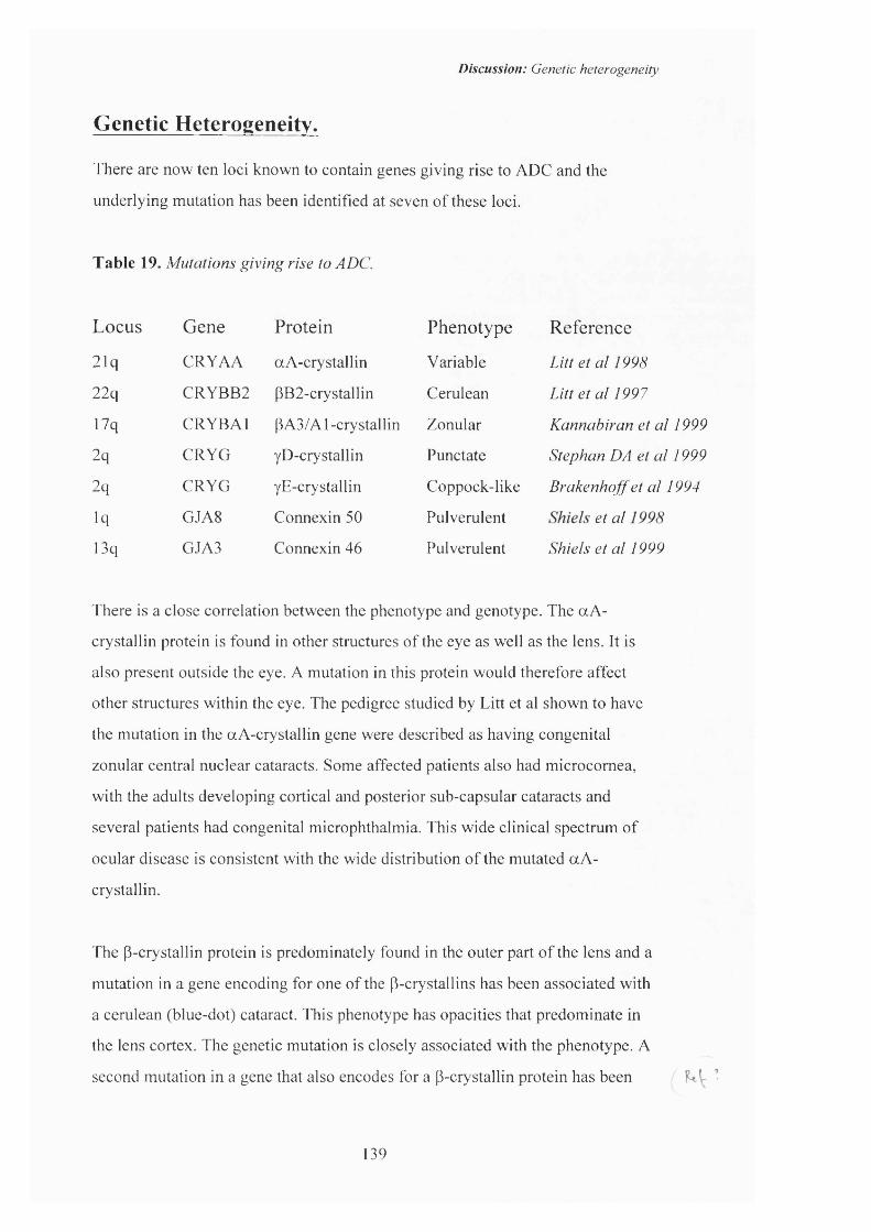
Discussion: Genetic heterogeneity
Genetic Heterogeneity.
There are now ten loci known to contain genes giving rise to ADC and the
underlying mutation has been identified at seven of these loci.
Table 19. Mutations giving rise to ADC.
L ocus G ene P ro tein P heno type R eference
21q CRYAA aA-crystallin Variable Tüfgfo /yppa
22q CRYBB2 PB2-crystallin Cerulean Litt et al 1997
17q CRYBAl PA3/Al-crystallin Zonular Kannabiran et al 1999
2q CRYG yD-crystallin Punctate Stephan DA et al 1999
2q CRYG yE-crystallin Coppock-like Brakenhoff et al 1994
iq GJA8 Connexin 50 Pulverulent gr a/
13q GJA3 Connexin 46 Pulverulent Shiels et al 1999
There is a close correlation between the phenotype and genotype. The aA-
crystallin protein is found in other structures of the eye as well as the lens. It is
also present outside the eye. A mutation in this protein would therefore affect
other structures within the eye. The pedigree studied by Litt et al shown to have
the mutation in the aA-crystallin gene were described as having congenital
zonular central nuclear cataracts. Some affected patients also had microcomea,
with the adults developing cortical and posterior sub-capsular cataracts and
several patients had congenital microphthalmia. This wide clinical spectrum of
ocular disease is consistent with the wide distribution of the mutated aA-
crystallin.
The p-crystallin protein is predominately found in the outer part of the lens and a
mutation in a gene encoding for one of the p-crystallins has been associated with
a cerulean (blue-dot) cataract. This phenotype has opacities that predominate in
the lens cortex. The genetic mutation is closely associated with the phenotype. A
second mutation in a gene that also encodes for a P-crystallin protein has been
139
Discussion: Genetic heterogeneity
more recently described. The phenotype is described as a zonular cataract with
sutural opacities. Although there are no published photographs nor detailed
descriptions of this phenotype it may that the opacities are located outside the
nucleus and the cortical location of the opacities is consistent with the current
theories on the location of the P-crystallin proteins.
The y-crystallin mutation in the Coppock-like cataract pedigree also parallels the
molecular biology of the lens. The cataract (Figures 22-27 pages 70-72) is a
nuclear cataract and the y-crystallin protein is found in greatest quantity in the
lens nucleus. The mutation has recently been questioned as to whether it is the
causative factor in the formation of cataract but no further details have been
published to date. Another more recently described mutation in the y-crystallin
gene complex is in the y-D-erystallin gene. This has been found in a family _
where the affected members have a progressive juvenile onset punctate cataract. 1 j ^
Protein modelling has suggested that the effect of this mutation is subtle, vUL.. IL Uv.,
affecting only the surface properties of the crystallin moleeule which the authors
suggest is consistent with the normal lens at birth (Stephan DA et el 1999).
The CAE cataract, a 'zonular pulverulent' cataract has been found to be caused by
a mutation in a gap-j unction protein. The gap junetions are cell-to-cell
communication pores and are made up of various proteins that dictate the
characteristics of the pores and the class of molecules that can pass through the
channel. Connexin 50 is one of these gap junction proteins and a mutation would
expect to give rise to widely distributed and discreetly localised opacities. The
pulverulent cataract phenotype can therefore be explained by understanding the
molecular biology of the lens, the gene mutation and the role of the connexin
proteins in the formation of gap junctions. A mutation in the gene encoding
connexin 46 has also been shown to cause a pulverulent cataract. Mutations at
two different points in this gene causes two different phenotypes of pulverulent
cataract (Pedigrees L and M, Figures 42-47, 56-57, pp86-88 and p93).
140
Discussion: Genetic heterogeneity
When this study began only three loci had been identified as containing genes for
ADC. The mutation had been isolated at one of these loci. There are now ten loci
and the underlying gene mutation has been identified in seven of these cases.
This study has contributed three new loci and two new mutations to the
molecular genetics of ADC.
The study of genes for autosomal dominant cataract will shed new light on lens
development. Sequence changes in the genes for autosomal dominant cataract
could also offer information on the risks for age related cataract and would have
far wider implications.
141
References
ReferencesAdams PH. Doyne’s discoid cataract (Coppock). Trans Ophthalmol Sac UK 1942;62:152-153.
Amsbaugh DF, Stone SH. Autosomal dominant congenital nuclear cataracts in strain 13/N guinea pigs. JH ered 1984;75: 55-8.
Antonarakis SE. Genome linkage scanning: Systematic or intelligent? Nature Genet 1994;8:211-212.
Arkin M, Azar D, Fraioli A. Infantile cataracts. Int Ophthalmol din 1992;32:107-120.
Armitage MM, Kivlin JD & Ferrell RE. A progressive early onset cataract gene maps to human chromosome 17q24 Nature Genet 1995;9:37-40.
Attwood,J. and Bryant,SA. A computer program to make analysis with LIPED and LINKAGE easier to perform and less prone to input errors. Am. J. Hum. Genet., 1988;52, 259.
Barishak YR. Embryology o f the eye and its adnexae. Dev Ophthalmol. 1992;24:
Barkhash SA, Pikalova LD, Grechko AS. Secondary glaucoma after extraction o f congenital cataract in children. Oftalmol Zh 1975; 30:204-206.
Barrett DJ, Sparkes RS, Gorin MB. Genetic linkage analysis o f autosomal dominant congenital cataracts with lens-specific DNA probes and polymorphic phenotypic markers. Ophthalmol 1988;95: 538-544.
Bassnett S, Beebe DC, Coincident loss of mitochondria and nuclei during lens fibre cell differentiation. Dev Dyn 1992;19: 421-5.
Bassnett S, Kuszak JR, Reinisch L, et al. Intercellular communication between epithelial and fiber cells o f the eye lens. 7 o / Cell Science 1994;107:799-811.
Basti S, Hejtmancik JF, Padma T, et al. Autosomal dominant zonular cataract with sutural opacities in a four-generation family. Am J Ophthalmol 1996; 121 (2): 162-8.
Batemen JB, Spence MA, Marazita ML, et al. Genetic linkage analysis o f autosomal dominant cataracts. Am J Ophthalmol 1986;101: 218-225.
Beaumont C, Garrett C, Gamer C, et al. Linkage data on dominant inherited congenital cataracts in families from the South West of England. Cytogenet Cell Genet 1989;51: 959.
Beebe DC, Johnson MC, Feagans DE, et al. In: Hilfer SR and Sheffield JB eds. Ocular size and shape regulation during development. The mechanism o f cell elongation during lens fibre cell differentiation.l9S0:pp 79-98.
Berman ER. Biochemistry o f the eye. New York: Plenium Press. 199 l:pp201-290.
Berry V, lonides ACW, Moore AT, et al. A locus for anterior polar cataract on chromosome 17p. Hum Molec Genet 1996;5:415-419.
Birdwood SC, Garrett C, Gamer C, et al. The locus causing a form of dominantly inherited congenital lamellar cataracts is not linked to the y-crystallin gene cluster on chromosome 2q33-35. Cytogenet Cell Genet 1992;58:1968.
Bloemendal H. Lens research; From protein to gene. Exp Eye Res 1985;4I: 429-448.
142

References
Bloemendal H, Vermorken AJM, Kibbelaar M. Nomenclature for the polypeptide chains o f lens plasma membranes (letter). Exp Eye Res 1977;24: 413.
Bloemendal H. Lens proteins as markers o f terminal differentiation. Ophthalmic Res 1979;! 1: 243- 253.
Bloemendal H, Piatigorsky J, Spector A. Recommendations for crystallin nomenclature. Exp Eye Res 1989;48; 465-466.
Bodker FS, Lavery MA, Mitchell TN, et al. Microphthalmos in the presumed homozygous offspring of a first cousin and linkage analysis of a locus in a large family with autosomal dominant cerulean congenital cataracts. A J Med Genet 1992;37: 54-59.
Bonneau D, Winter-Fuseau I, Loiseau M-N, et al. Bilateral cataract and high serum ferritin: a new dominant genetic disorder? J Med Genet 1995;32: 778-779.
Brakenhoff RH, Henskens HAM, van Rossum MWPC, et al. Activation o f the yE-crystallin pseudogene in the human hereditary Coppock-like cataract. Hum Mol Genet 1994;3: 270-283.
Brakenhoff RH, Aarts HIM, Reek FH, et al. The human y-crystallin genes: a gene family on its way to extinction. J Mol Biol 1990;216: 519-532.
Brackenhoff RH, Aarts HIM, Schuren F, et al. The second human aB2-crystallin is a psuedogene. Exp Eye Res 1992;54: 803-806.
Brown NAP, Bron AJ, Sparrow JM. Central con^action in the process o f lens growth as indicated by lamellar cataract. Br J Ophthalmol 1988;72: 538-544.
Byers B, Porter K. Orientated microtubules in elongating cells o f the developing lens rudiments after induction. Proc Acad Natl Sci USA 1964;52: 1091.
Carper D, Shinohara T, Piatigorsky J, et al. Deficiency o f functional messenger RNA for a developmentally regulated beta-crystallin polypeptide in a hereditary cataract. Science 1982;217(4558): 463-465.
Carter JM, Hutcheson AM, Quinlan RA. In vitro studies on the assembly properties o f the lens proteins CP49, CPI 15. Coassembly with a-crystallin but not with vimentin. Exp Eye Res 1995;60: 181-192.
Cartier M, Breitman ML, Tsui L-C. A frameshift mutation in the yE-crystallin gene o f the Elo mouse. Nature Genet 1992;2:42-45.
Cartier M, Tsui L, Ball SP, et al. Crystallin genes and cataract. In; Molecular genetics of inherited eye disorders, eds. Wright AF, Jay B. Vol 2 Modem genetics Series ed. Evans HJ. Publ Harwood Acad Press. Switzerland. 1994
Cascio M, Kumar NM, Safarik R, et al. Physical characterisation o f gap junction membrane connexons (hemi-channels) isolated from rat liver. J Biol Chem. 1995;270: 18643-18648.
Chambers C, Russell P. Deletion mutation in an eye lens P-crystallin. J Biol Chem 1991 ;266: 6742- 6746.
Chrousos GA, Parks MM, O'Neil JF. Incidence of chronic glaucoma, retinal detachment and secondary membrane surgery in pediatric aphakic patients. Ophthalmol 1984;91:1238-1241.
Church RL, Wang J, Steele E. The human lens intrinsic membrane protein MP70 (Cx50) gene: clinal analysis and chromosome mapping. Curr Eye Res 1994;14: 215-221.
143
References
Chylack LT Jr, Sugar Cataracts- possibly the beginning of medical anti-cataract therapy. Mechanisms o f cataract formation in the human lens, G Duncan, ed. Academic press, London. pp237-252. (1981)
Clyde A Clapp, Cataract Its etiolo^and treatment. Henry Kington, London. 1934. pp86-90.
Conneally PM, Wison AF, Merritt AD, et al. Confirmation o f genetic heterogeneity in autosomal dominant forms of congenital cataracts from linkage studies. Cytogenet Cell Genet 1978;22: 295-297.
Coulombre AJ. Cataractogensis: Developmental inputs and constraints. Ophthalmol 1979;86: 1559- 1570.
Coulombre J, Coulombre A. Regeneration of neural retina ffrom the pigmented epithelium in the chick embryo. Dev Biol 1965;12:79.
de Jong WW, Hendriks W, Mulders JWM, et al. Evolution of eye lens crystallms: the stress connection. TIBS 1989;14: 365-368.
Delaye M, Tardieu A. Short-range order of crystallin proteins accounts for eye lens transparency. Nature 1983;302: 415-417.
Dib C. et al. A comprhensive genetic map of the human genome based on 5,264 microsatellites. Nature 1996;380:152-154.
Donahue RP. Probable assignment of the Duffy blood group locus to chromosome 1 in man. Proc Natl Acad Sci USA 1968;61: 949-955.
Donaldson The Crystalline Lens.
Donis-Keller H, Green P, Helms C et al.A genetic linkage map of the human genome. Cell 1987;51:319-337.
Doolittle DP, Davisson MT, Guidi JN, et al. (1996). In Genetic variants and strains o f the laboratory mouse (MF Lyon, S Rastan, SDM Brown, eds). 3rd ed pp 17-854, Oxford Univ Press. London.
Doyne RW. A peculiar form of lens degeneration. Trans Ophth Soc UK. 1889;9: 113.
Dubin RA, Wawrousek EF, Piatigorsky J. Epression o f the murine alpha B-crystallin gene is not restricted to the lens. Mol Cell Biol. 1989;9(3): 1083-1091.
Duguid JR, Rohwer RG, Seed B. Isolation of cDNAs of scrapie modulated RNA by subtractive hybridisation of a cDNA library. Proc Natl Acad Sci USA 1988;85(15):5738-5742.
Duke-Elder S, Abrams D. 1970 The refraction of light. In ‘Systems of Ophthalmology’ Vol V. (Duke-Elder S. ed) pp74-78. CV Mosby Co. St Louis.
Duke-Elder S. In: A System o f Ophthalmology. Henry Kimpton 1964: Vol III Part 2, The Lens, 715- 759.
Duke-Elder S. In: A System o f Ophthalmology. Henry Kimpton 1964: Vol III Part 2, The Lens, 732
Duke-Elder S. System of Ophthalmology, Voume II pp320-321. The anatomy o f the visual system. London Henry Kimpton. 1961
Eiberg H, Marner E, Rosenberg T, et al. Mamer’s cataract (CAM) assigned to chromosome 16: Linkage to Haptoglobin. Clin Genet 1988;34: 272-275.
Eiberg H, Lund AM, Warburg M, et al. Assignment of congenital cataract Volkmann type (CCV) to chromosome lp36. Hum Genet 1995;96: 33-38.
144
References
Elfgang C, Eckert R, Lichenberg-Fraté H, et al. Specific permeability and selective formation of gap junction channels in connexin-transfected HeLa cells. J Cell Biol 1995;129: 805-817.
Evans J, Rooney C, Ashwood F, et a/. Blindness and partial sight in England and Wales: April 1990 - March 1991. Health Trends 1996; 28: 5-12.
Francis PJ, Berry V, Moore AT, et al. Lens biology, development and human cataractogenesis.Trends Genet 1999;15:191-6.
Francois J, Congenital Cataracts. Springfield, IE. Charles C Thomas 1963.
Francois J. Late results of congenital cataract surgery. Ophthalmology 1979;86:1586-1598.
Francois J. Genetics of cataract. Ophthalmologica 1982; 184:61-71.
Fraser FC, Schabtach G. ‘Shrivelled’: a hereditary degeneration of the lens in the house mouse. Genet Res 1962;3:383-7.
Freeman RS, Rovick LP. Cloudy lenses and Issues: A pedigree of unoperated congenital cataracts. J Pediatr Ophthalmol Strabismus 1994;31:318-322.
Fukui HN, Merola LO, Kinoshita JH. A possible cataractogenic factor in the Nakano mouse lens. Exp Eye Res 1978;26: 477-485.
Galvan A, Lampe PD, Hur KC, et al. Structural organisation of the lens fiber cell plasma membrane protein MIP18. Chem 1989;264:19974-19978.
Gao Y, Spray DC. Structural changes in lenses of mice lacking the gap j une ion protein connexin43. Invest Ophthalmol Vis Sci 1998;39:1198-1209.
Garber AT, Winkler C, Shinohara T, et al. Selective loss of a family of gene transcripts in a hereditary murine cataract. Science 1985;227(4682):74-77.
Garland DL, Duglas-Tabor Y, Jimenez-Assensio J, et al. The nucleus of the human lens: Demonstration of a highly characteristic protein pattern by two-dimensional electrophoresis and introduction of a new method of lens dissection. Exp Eye Res 1996;62: 285-291.
Garland D, Rao PV, Del Corso A, et al. d -crystallin is a major protein in the lens of Camelus dromedarius. Arch Biochem Biophys. 1991;285: 134-136.
Gause GG, Tomarwv SI, Zinovieva RD, et al. (1985). in The Lens: Transparency and Cataract, (ed G Duncan). 171-179. Eurage.
Giersing OM. En kataraktos Bondefamilie. Ugeskr Læger 1878;3.rk.XXXVI: 273-277.
Goldman, v Graefes Arc Ophthalmol. 1929;122:198.
Gonzalez P, Rao V, Zigler JS. Organisation of the human (! -crystallin/Quinone reductase gene (CRYZ). Genomics 1994;21: 317-324.
Goodenough DA. The crystalline lens: A system networked by gap junctional intercellular communication. Semin Cell Biol. 1992;3: 49-58.
Goulden and Harris. Koby’s Slit lamp micros of the living eye, London 1925.
Griffen CS, Shiels A. Localisation of the gene for the major intrinsic protein of eye- ^ ® membranes to mouse chromosome 10 by in situ hybridisation. Cytogenet Cell Genet1992;59(4):300-302.
145

References
Gyapay G, Morisette J, Vignal, A, et al. The 1993-1994 Genethon human genetic linkage map.Nature Genet.,\99A\1'. 246-339.
Hammerstein W, Scholz W. Familière form einer “cataracta centralis”.- Klinisch-genetische studie mit koppelungsdaten. Arch Klin Exp Ophthalmol 1974;189: 9-19.
Harding JJ, Crabbe MJC. (1984). in The Eye (Davson H ed) pp207-492, Academic Press.
Harding J. The Normal Lens. Pp22-25. In Cataract: Biochemistry, epidemiology, and pharmacology.{\99\) Pub. Chapman and Hall. London.
Harman NB. Ten pedigrees of congenital and infantile cataract; lamellar, coralliform, discbid, and posterior polar with microphthalmia. Trans Ophthalmol Soc UK 1910;XXX: 251-274.
Harman NB. Congenital cataract; a pedigree o f five generations. Trans Ophthalmol Soc UK 1909;29:101-108.
Harman NB. Treasury of Human Inheritance. Part 4. Section Xllla, Congenital cataract. Eugenics Library Memoirs XI. London 1910.
Heame CM, Ghosh S, Todd JA. Microsatellites for linkage of genetic traits. TIG 1992;8: 288-294.
Heiba IM, Elston RC, Klein BE, et al. Evidence for a major gene for cortical cataract. lOVS 1995;36(l):227-235.
Heinzmarm C, Kojis TL, Gonzalez P, et al. Assignment of the zeta crystallin gene (CRYZ) to human chromosome Ip22-lp31 and identification of restriction fragment length polymorphisms. Genomics. 1994;23: 403-407.
Hess JF, Casselman IT, FitzGerald PG. Chromosomal localisation of the genes for the beaded filaments. Curr Eye Res 1996;14: 11-18.
Hiles DA. Infantile cataracts. Pediatr Ann 1983;12:556-573.
Horwitz JN, Robertson NP, Wong MM, et al. Some properties o f lens plasma membrane polypeptides isolated from normal human lenses. Exp Eye Res 1979;28:359-365.
Huang Q-L, Du XY, Stone SH, et al. Association of hereditary cataracts in strain 13/N guinea pigs with mutation of the gene for (î -crystallin. Exp Eye Res 1990;50:317-325.
Huang QL, Russell P, Stone SH, et al. Zeta crystallin, a novel lens protein from the guinea pig. Curr Eye Res 1987;6:725-732.
Hammerstein W, Scholz W. Familiare form einer “cataracta centralis”.- Klinisch-genetische studie mit koppelungsdaten. Arch Klin Exp Ophthalmol 1974; 189: 9-19.
Huntzinger RS, Weitkamp LR, Roca PD. Linkage relationships o f a locus for congenital total nuclear cataract, y AfeiJ Genet 1977;15:113-115.
lonides ACW, Berry V, Mackay D, et al. Phenotypic variation in a single pedigree with anterior polar cataract. Eye 1998;12:234-236.
lonides ACW, Berry V, Mackay D, et al. A posterior polar cataract maps to chromosome Ip. Hum Mol Genet 1997;6: 47-51.
Ingolia TD, Craig EA. Four small heat drosophila heat shock proteins are related to each other and to mammalian alpha-crystallin. Proc Natl Acad Sci USA 1982;79:2360-2364.
Iwaki T, Kume-Iwaki A, Liem RKH, et al. Alpha B-crystalin is expressed in non-lenticular tissues
146
References
and accumulates in Alexander's disease brain. Cell 1989;57: 71-78.
Jahal C, Renkawek K, de Jong WW, et al. aB-crystallin is present in reactive glia in Creutzfeld-Jakob disease. Acta Neuropathol (Berl) J^2;83:324-7.
Jay M. Linkage and chromosomal studies on congenital cataract. Trans Ophthalmol Soc UK 1982;102:350-354.
Kannabiran C, Rogan PK, Olmos L, et al. Autosomal dominant zonular cataract with sutural opacities is associated with a splice mutation in the betaA3/A 1 -crystallin gene. Molecular Vision 1999;4:21
Kantorow M, Kays T, Horwitz J, et al. Differential display detects altered gene expression between cataractous and normal human lenses. Invest Ophthlamol Vis Sci 1998;39:2344-2354.
Kato K, Shinohara H, Kurobe N, et al. Immunoreactive aA-crystallin in rat non-lenticular tissues detected with a sensitive immunoassay method. Biochem Biophys Acta. 1991;1080:173-180.
Kador PF, Fukui HN, Fukushi S, et al. Philly mouse; a new model of hereditary cataract. Exp Eye Res 1980;30:59-68.
Kerscher S, Glenister PH, Favor J, et al. Two new cataract loci, Ccw and To3, and further mapping of the Npp and Opj cataracts in the mouse. Genomics 1996;36: 17-21.
Kinsey VE, Reddy DVN. (1964) Studies on the crystalline lens, XI; The relative role of the epithelium and capsule in transport. Invest Opthalmol. 4: 104-116.
Kinoshita JH. Mechanisms initiating cataract formation. Proctor Lecture. Invest Ophthalmol 13: 713- 737(1974)
Kramer P, Yount J, Mitchell T, et al. A second gene for cerulean cataracts maps to the (3 crystallin region on chromosome 22. Genomics 1996;35: 539-542.
Koretz JF, Cook CA, Kusak JR. The zones of discintinuity in the human lens: development and distribution with age. Vision Res 1994;34: 2955-62.
Kuliszewski M, Rupert J, Golod R. The ontogeny of y-crystallin mRNAs in Cat*"* * mice. Genet Res Camb 1988;52: 45-9.
Kumar NM, Gilula NB. The gap junction communication channel. Cell 1997;84: 381-388.
Kumar NM, Gilula NB. Molecular biology and genetics of gap junction channels. Sem Cell Biol 1992;3: 3-16.
Kuszak J, Maisel H, Harding C. Gap junctions of chick lens fibre cells. Exp Eye Res 1978;27:495- 498.
Kuszak JR, Bertram BA, Macsai MS, et al. 1984. Sutures of the crystalline lens: A review. Scan Elec M icrlW : 1369-1378.
Kuszak JR, Bertram BA, Rae JL. The ordered structure o f the crystalline lens. In Development of order in the visual system, eds SR Hilfer, JB Sheffield. Cell and developmental biology o f the eye. Springer-Verlag New York. 1986.
Kuszak JR, Macsai MS, Rae JL. Stereo scaning electron microscopy of the crystalline lens. Scan Elec Micr 1983;111: 1415-1426.
Kuwabara T. (1975) The maturation of the lens cell: A morphologic study. Exp Eye Res 20:427-443.
147
References
Lambert SL, Drack AV. Infantile Cataracts. Surv Ophthalmol 1996;40:427-458.
Lathrop G.M, Lalouel JM, Julier C, et al. Strategies for multipoint linkage analysis in humans. Proc. Natl. Acad. Sci. USA, 1984;81:3443-3446.
Litt M, Carrero-Valenzuela R, LaMorticella DM, et al. Autosomal dominant cemlean cataract is associated with a chain mutation in the haman beta-crystallin gene CRYBB2. Hum Mol Genet 1997;6(5): 665-8.
Litt M, Kramer P, LaMorticella DM, et al. Autosomal dominant congenital cataract associated with a missense mutation in the human alpha crystallin gene CRYAA. Hum Mol Genet 1998;&:471-474.
Lloyd IC, Goss-Sampson M, Jeffrey BG, et al. Neonatal cataract; Aetiology, pathogenesis and management. 1992;6:184-196.
Lund AM,Eiberg H, Rosenberg T, et al. Autosomal dominant congenital cataract; linkage relations; clinical and genetic heterogeneity. C/in. Genet. 1992;41:65-69.
Lubsen NH, Renwick JH, Tsui L-C, et al. A locus for a human hereditary locus is closely linked to the y-crystallin gene family Proc Natl Acad Sci USA 1987;84;489-492.
McAvoy JW. Developmental biology of the lens. In: Mechanisms of cataract formation in the human lens. G Duncan, ed. Academic press, London. pp7-46 (1981)
McAvoy JW. Cell division, cell elongation and distribution of a-, (3-, y-crystallins in the rat lens. J Embryol Exp Morph. 1978;44:140-165.
Mackay D. lonides A. Kibar Z, et al. Connexin46 mutations in autosomal dominant congenital catàract. American Journal o f Human Genetics. 64(5): 1357-64, 1999 May.
Mackay DM, lonides ACW, Berry V, et al. Autosomal dominant 'zonular pulverulent' cataract linked to human chromosome 13. Am J Hum Genet. 1997;60:1474-1478.
McGill JR, Yang F, Baldwin WD. Localisation of the haptoglobin alpha and beta genes (HPA and HPB) to human chromosome 16q22 by in situ hybridisation. Cytogenet Cell Genet 1984;38:155-157.
Maisel H. The lens Marcel Dekker, Publ New York. Now published. Find ref from library.
Mann IC. The development of the human eye. Ill The Lens 1928 Cambridge Univ Press, London.
Mamer E. A family with eight generations of hereditary cataract. Acta Ophthalmol 1949;27: 537-551.
Mamer E, Rosenberg T, Eiberg H. Autosomal dominant congenital cataract. Morphology and mappmg. Acta Opthalmologica 1989;67: 151-158.
Marshall J, Beaconsfield M, Rottery S. The anatomy and development of the humen lens and zonules. Trans Ophthalmol Soc UK 1982;102: 423-439.
Mathias RT, Rae JL, Baldo GJ. Physiological properties of the normal lens. Physiol Rev 1997;77: 21- 50.
Maumenee IH. Classification of hereditary cataracts in children by linkage analysis. Ophthalmol 1979;86: 1554-1558.
Merin S, Crawford JS. The etiology o f congenital cataracts. Can J Ophthalmol 1971;1:1.
Mignon C, Fromaget C, Mattel M-G, et al. Assignment of connexin 26 (GJB20 and 46 (GJA3) genes to human chromosome 12ql l-q l2 and mouse chromosome 14D1-E1 by in situ hybridisation. Cytogenet Cell Genet 1996;72: 185-186.
148

References
Miller BA, Jaafar MS, Washington DC, et al. Chromosome 14- Terminal deletion and cataracts. Arch Ophthalmol 1992;110:1053.
Moross T, Vaithilingam SS, Styles^, et al. Autosomal dominant anterior polar cataracts associated with a familial 2;14 translocation. J Med Genet 1984;21: 52-53.
Muggleton-Harris A, Festing M, Hall M. A gene Ication for the ingeritance of the cataract Fraser (CatFr) mouse congenital cataract. Gen Res Camb 1987;49:235-239.
Mulders JWM, Hendriks W, Blankesteijn WM, et al. Lambda crystallin, a major rabbit lens protein is related to hydroxyacyl-coenzyme A dehydrogenases. JB iol Chem 1988;263:15462-15466.
Mulders JWM, Voorter CEM, Lamers C, et al. MPI7, a fiber-specific intrinsic membrane protein from mammalian eye lens. Curr Eye Res 1988;7:207-219.
Nakano T, Yamamoto S, Kutsukade G, et al. Hereditary cataract in mice. Jpn J Clin Ophthalmol 1960;14:196.
Nene V, Dunne DW, Johnson KS, et al. Sequence and expression o f a major egg antigen from Schistosoma mansoni: Homologies to heat shock proteins and a-crystallins. Mol Biochem Parasitol 1986;21:179-88.
Nettleship E. On heredity in the various forms o f cataract. Royal Lon Ophthalmol Hosp Reports. 1905;16:179.
Nettleship E. On some hereditary diseases of the eye. The Bowman Lecture (1909). Trans Ophthalmol Soc UK XXIX: LVII- CL
Nettleship E. A peculiar form of hereditary congenital cataract. Trans Ophthalmol Soc UK. 1906;XXVI: 191-207.
Nettleship E. Seven new pedigrees of hereditary cataract. Trans Ophthalmol Soc UK 1909;XXIX: 188-211.
Nickells RW, Browder LW. A role for glyceraldehyde-3 -phsophate dehydrogenase in the development of thermotolerance in xenopus laevis embryos. J Cell Biol 1988;107: 1901-1909.
Norrie G. Arvelighed af graa staer. Ugeskr Læger 1896;5.rk III: 937-944.
O’Brien W, Mclnnes R, Kalumuck K, et al. Cloning and sequence analysis o f cDNA for human argininosuccinate lysase. Proc Natl Acad Sci 1986;83:7211-7215.
Oda S-I, Watanabe K, Fujisawa H, et al. Impaired development o f lens fibres in genetic microphthalmia. Eye lens obsolescence, Elo, o f the mouse. Exp Eye Res 1980;31:673-681.
Ozanics V, Jakobiec F: Prenatal development o f the eye and its adnexae; in Jakobiec, Ocular anatomy, embryology and teratology, chapter 2,pp 11-96 (Harper & Row, Philadelphia 1982).
Padma T, Ayyagari R, Murty JS, et al. Autosomal dominant zonular cataract with sutural opacities localized to chromosome 17ql 1-12. Am J Hum Genet 1995;57:840-845
Parks MM, Johnson DA, Reed GW. Long term visual results and complications in children with aphakia; a function of cataract type. Ophthalmology 1993;100:826-840.
Peters J, Searle AG. Linkage and synteny homologies in mouse and man. In: Lyon MF, Rastan S, Brown SDM, eds. Genetic Variants and Strains o f the Laboratory Mouse. New York: Oxford University Press; 1996:1256-1312.
149
References
Phelps Brown N, Bron AJ. (1996) Lens Disorders A Clinical manual o f cataract diagnosis. Butterworth Heinemann, Oxford. pp23-28.
Phelps CD, Arafat NI. Open-anglev^aucoma following surgery for congenital cataracts. Arch Ophthalmol 1977;95:1985-1987.
Piatigorsky J. Lens differentiation in vertebrates; A review o f cellular and molecular features. Differentiation 1981 ;19: 134-153.
Piatigorsky J. Treton JA, King CR, et al. A molecular genetic approach to vision research: Crystallin gene expression in the lens. Ophthalmic Paediatr Genet 1983;3:6-72.
!
Piatigorsky J. Wistow GJ. Enzyme/crystallins: Gene sharing as an evolutionary strategy. Cell 1989;57:197-199.
Rae J, Kuszak J. The electrical coupling of epithelium and fibres in the frog lens. Exp Eye Res 1983;36:317-326.
Rao PV, Huang Q-L, Horwitz J, et al. Evidence that a-crystallin prevents non-specific protein aggregation in the intact eye lens. Biochem Biophys Acta 1995;1245: 439-447.
Reese PD, Tuck-Miller CM, Maumenee IH. Autosomal dominant congenital cataract associated with chromosomal tranclocation [t(3;4)(p26.2;pl5)] Arch Ophthalmol 1987;105:1382-1384.
Reizer J, Reizer A, Saier MH. The MIP family of integral membrane channel proteins. Curr Eye Biochem Mol Biol. 1993;28:235-257.
Renwick JH, Lawler SD. Probable linkage between a congenital cataract locus and the Dufiy blood group locus. Ann Hum Genet 1963;27:67-84.
Riccardi VM. Chromosome imbalance in the aniridia-Wilms’ tumor association: l ip interstitial deletion. Paediatrics 1978;61:604.
Rice NSC, Taylor D. Congenital cataract; A cause of preventable blindness in children. BMJ 1982;285(6342):581-582.
Richards J, Maumenee IH, Rowe S, et al. Congenital cataract possibly linked to haptoglobin. Cytogenet Cell Genet 1984;37: 570.
Robb RM, Petersen RA. Outcome of treatment for bilateral congenital cataracts. Ophthalmic Surg 1992;23:650-656.
Rodriguez IR, Gonzalez P, Zigler JS, et al. A splice-site deletion that causes a defective ^-crystallin is found in a congenital guinea pig cataract. Invest Ophthalmol Vis Sci 1991;32 (suppl), 782.
Rodriguez IR, Gonzalez P, Zigler JS, et al. A guinea pig hereditary cataract contains a splice site deletion in a crystallin gene. Biochem Biophys Acta 1992;1180: 44-52.
Rogaev El, Rogaeva EA, Korovaitseva GI, et al. Linkage of polymorphic congenital cataract to the y- crystallin gene locus on human chromosome 2q33-35. Hum Molec Genet 1996; 5;5:699-703.
Rubin SE, Nelson LB, Pletcher EA. Anterior polar cataract in two sisters with an unbalanced 3; 18 chromosomal translocation. Am J Ophthalmol 1994;117:512-515.
Runge PE, Hawes NL, Heckenlively JR, et al. Autosomal dominant mouse' cataract (Lop-10). Invest Ophthalmol Vis Sci 1991;33:3202-3208.
Russell-Eggitt I, Zamiri P. Review of aphakic glaucoma after surgery for congenital cataract. J Cataract Refract Surg 1997;23:664-668.
150

References
Ryan SJ, Blanton FM,von Noorden GK. Surgery of congenital cataract. Am J Ophthalmol 1965;60:583-587.
Sæbo J. An investigation into the mode of heredity of congenital and juvenile cataracts. B J Ophthalmol 1949: 601-629.
Sandilands A, Prescott AR, Carter JM, et al. Vimentin and CP49/Filensin form distinct networks in the lens which are independently modulated during lens fibre cill differentiation. J Cell Sci 1995;108(4): 1397-1406.
Sas DF, Sas MJ, Johnson KR et al Junctions between lens fibre cells are labelled with a monoclonal antibody shown to be specific for MP26. J Cell Biol 1985;100: 216-225. i
Scott MH, Hejtmancik JF, Wozencraft LA, et al. Autosomal dominant congenital cataract. Interocular phenotypic variability. Ophthalmology 1994;101:866-871.
Segal S. Disorders of galactose metabolism. In The Metabolic basis o f inherited disease. Vol 1. (eds Scriver, CR et al) 453-480. (McGraw-Hill, New York 1989).
Sidjanin DJ, Grimes PA, Pretsch W, et al. Mapping of the autosomal dominant cataract mutation (Coc) on mouse chromosome 16. lOVS 38: 2502-2507.
Shiels A, Griffen CS, Muggleton-Harris AL. Restriction fragment length polymorphisms associated with the gene for the major intrinsic protein o f eye-lens fibre cell membranes in mice with hereditary cataracts, Biochem Biophys Acta 1991;1097: 81-85.
Shiels A, Griffen CS. Aberrant expression o f the gene for lens major intrinsic protein in the CAT mouse. Curr Eye Res 1993;12:913-921.
Shiels A, Bassnett S. Mutations in the founder of the MIP gene family underlie cataract development in the mouse. Nature Genet 1996;12:212-215.
Shiels A, Mackay D, lonides A, et al. A missense mutation in the human connexin50 gene (GJA8) underlies autosomal dominant 'zonular pulvemlent' cataract on chromosome \(\. Am J Hum Genet 1998;62:526-532.
Shiels A, Mackay DM, lonides A, et al. Connexin46 mutations in autosomal dominant congenital cataract. Am J Hum Genet 1999; In press (May 1999).
Shilloh Y, Donlon T, Bmns G, et al. Assignment of the human y-crystallin gene cluster (CRYG) to the long arm of chromosome 2, region q33-36. Hum Genet 1986;73:17-19.
Siezen RJ, Anello RD, Thomson JA. Interactions o f lens proteins. Concentration dependence of (3- crystallin aggregation. Exp Eye Res 1986;43:293-303.
Simmoneau L, Herve B, Jacquiemin E, et al. State of differentiation o f bovine epithelial cells in vitro. Cell Differ. 1983;13:185-190.
Sivak JG, Dovrat A. Embryonic lens of the human eye as an optical stmcture. Am J Optom physiol. Opt. 1987;64:599-603.
Skalka HW, Prchal JT. Presenile cataract formation and decreased activity of galactosémie enzymes. Arch Ophthalmol 98: 269-273.
Smith P. A pedigree o f Doyne’s discoid cataract. Trans Ophthalmol Soc UK 1910;XXX:37-42.
Sparkes RS, Mohandas T , Heinzmann C et al. The gene for the major intrinsic protein (MIP) of the ocular lens is assigned to human chromosome 12cen-ql4. Invest Ophthalmol Vis Sci 1986;27:1351- 1354.
151

References
Sparrow JM, Bron AJ, Brown NAP, et al. Oxford Clinical cataract classification and grading system. Int Ophthalmol 1986;9:207-25.
Sparkes RS, Mohandas T, Gorin NÎB, et al. Assignmaent o f a human beta-crystallin to 17cen-q23. Hum Genet 1986;74:133-136.
Spector A. An extrinsic membrane polypeptide associated with high molecular weight protein aggregates inhuman cataract. Science 1979;204:1323-1324.
Spector A, Chiesa R, Sredy J, et al. c AMP-dependent phosphorylation of bovine lens a-crystallin. Proc Nat Acad Sci USA 1985;82:4712-4716. ■
Stabile M, Amoriello A, Capobianco ML, et al. Study of a form o f pulverulent cataract in a large kindred. J Med Genet 1983;20: 419-421.
Stambolian D. Galactose and cataract. Survey o f Ophthalmol 1988;32:333-349.
Stambolian D, Ai Y, Sidjanin D, Nesbum K, et al. Cloning of the galactokinase cDNA and identification o f mutations in two families with cataracts. Nature Genet 1995;10: 307-312.
Stark WJ, Taylor HR, Michels RG, et al. Management of congenital cataracts. Ophthalmology 1979;86:1571-1578.
Stephan DA, Gillanders E, Vanderveen D, et al. Progressive juvenile-onset punctate cataracts caused by mutation of the gammaD-crystallin gene. Proc Natl Acad Sci USA 1999;96:1008-12.
Tardieu A, Delaye M. Eye lens proteins and transparency: from light transmission theory to solution X-ray structural analysis. Ann Rev Biophys Biophys Chem 1988;17:47-70.
Tayor VL, al Ghoul KJ, Lane CW, et el. Morphology of the normal human lens. lOVS 1996;37:1396- 410.
TenBroek E, Ameson M, Jarvis M, et al. The relationship between the fibre cell intrinsic membrane proteins MP20 and MP 70 in the mammalian lens. J Cell Sci 1992;103: 245-257.
Thomson J A, Augusteyn R C. Ontogeny o f the human lens crystallins. Exp Eye Res. 1985;40:393- 410.
Travis GH, Christerson L, Danielson PE, et al. The human retinal degeneration slow (RDS) gene: chromosome assignment and structure of the mRNA. Genomics 1991;10:733-739.
Tripathi BJ, Tripathi RC, Borisuth NSC, et al. Rodent models o f congenital and hereditary cataract in man. Lens Eye Tox Res 1991;8:373-413.
Tulloh CG. Hereditary posterior polar cataract with report o f a pedigree. Brit J Ophthalmol 1955;39:374.
Uga S, Kador PF, Kuwabara T. Cytological study of Philly mouse cataract. Exp Eye Res 1980;30:79- 92.
Van den Ijssel, Smulders RHPH, de Jong WW, et al. a-crystallin: Molecular chaperone and heat shock protein. Opthalmic Res\996\2^:{supp\ 1): 39-43.
Van Heyningen R. Flourescent compounds in the human lens. CIBA Found Symp 19: 151-164 (1973)
Van Leen RW, Van Roozenddaal KEP, Lubsen NH, et al. Differential expression o f crystallin genes during development o f the rat eye lens. Dev Biol 1987;120:457-464.
152

References
Van Noort JM, Van Sechel AC, Bajramovic JJ, et al. The small heat-shock protein aB-crystallin as candidate autoantigen in multiple sclerosis. Nature 1995;375: 798-801.
Vérétout F, Delaye M, Tardieu A. Molecular basis o f eye lens transparency. Osmotic pressure and X- ray analysis o f a-crystallin solution^ J Mol Biol 1989;205: 713-728.
Vogt A. Die spezifitat ander borener und erworbener starformer fur die einzelnen linsezonene. Albrecht Von Graefes Arch Klin Ophthalmol 1922;108: 219-228.
Vogt. Klin Mbl Augenheilk 62: 582 (1919). Atlas, Berlin (1921).
Vogt A. Lens and zonule. In Vol II Textbook and atlas o f the living eye.Pub IP Weyenborgh. Bonn 1979.
Wallace DK, Plager DA. Comeal diameter in childhood aphakic glaucoma. J Pediatr Ophthalmol Strabismus 1996;33:230-234.
Willecke K, Jungbluth S, Dahl E, et al. Six genes of the human connexin gene family coding for gap junctional proteins are assigned to four different human chromosomes. E u rJ Cell Biol 1990;53:275- 280.
Wistow G. Piatigorsky J. Recruitment of enzymes as lens stmcural proteins. Science 1987;236:1554- 1556.
Wistow GJ, Piatigorsky J. Lens crystallins: The evolution and expression o f proteins for a highly specialised tissue. Ann Rev Biochem 1988;57:479.
Wistow GJ, Piatigorsky J. Lens crystallins: The evolution and expression o f proteins for a highly specialised tissue. Ann Rev Biochem 1988;57:479-504.
Wistow G. The Ubiquitous crystallins: Stress protein recmites to the lens. In Molecular biology and evolution o f crystallins: Gene recruitment and multifunctional proteins in the eye lens. Pub Springer- Verlag. Heidelberg, Germany. 1995.
Yates JRW. Recent advances. Medical genetics. RA/y 312:1021-1025.
Yokoyama Y, Narahara K, Tsuji K, et al. Autosomal dominant congenital cataract and microphthalmia associated with a familial t(2;16) translocation. Hum Genet 1992;90:177-178.
Zampighi GA, Hall JE, Ehring GR, et al. The stmctural organisation and protein composition of lens fibre junctions, y Ce/Z Biol. 1989;108:2255-2275.
Zampighi GA, Simon SA, Hall JE. The specialised junctions o f the Xcns.Int Rev Cytol 1992;136:185- 225.
Zigler JS. Animal models for the study of maturity onset and hereditary cataract. Exp Eye Res 1990;50: 651-657.
Zigman S. Selected aspects o f lens differentiation. Biol Bull 1985;168:189-213.
153

References
154
Appendices
Appendices
Appendix 1. Pedigrees 156
Appendix 2. Publications arising from this work 164
Appendix 3. Presentations arising from this work 165
155
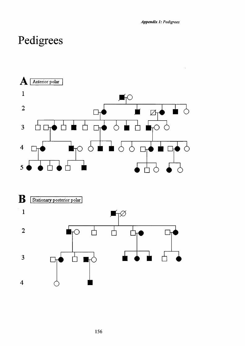
Appendix 1: Pedigrees
Pedigrees
I A nterior polar
1
2
3
mohi i Ehi i i
[hi 6 i 6 Chi 6 i 6 to 6
Ori t o 6 i i 6 i Oii i o i 6
m i i )
I Stationary posterior polar
1
156
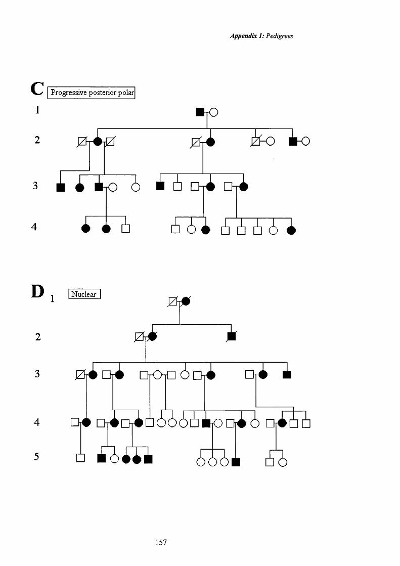
Appendix 1: Pedigrees
I Progre ssive posterior polar1 #|02 / j i jZ 0À ^ imo
i t o 6 A 6 Chi D ri
D Nuclear
i o i a i I
157
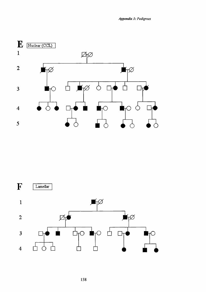
Appendix I: Pedigrees
E [Nuclear (CCL)
1
2
# r O
i Orim to àjO &
F1
2
3
4
Lamellar
0J0
I tO
Ô
t r O
158

Appendix 1: Pedigrees
Lamellar
HLamellar ÉT0
6 ^ 6 ^ T
A ^ 6 I tO T#
□ 4 ^ :
159
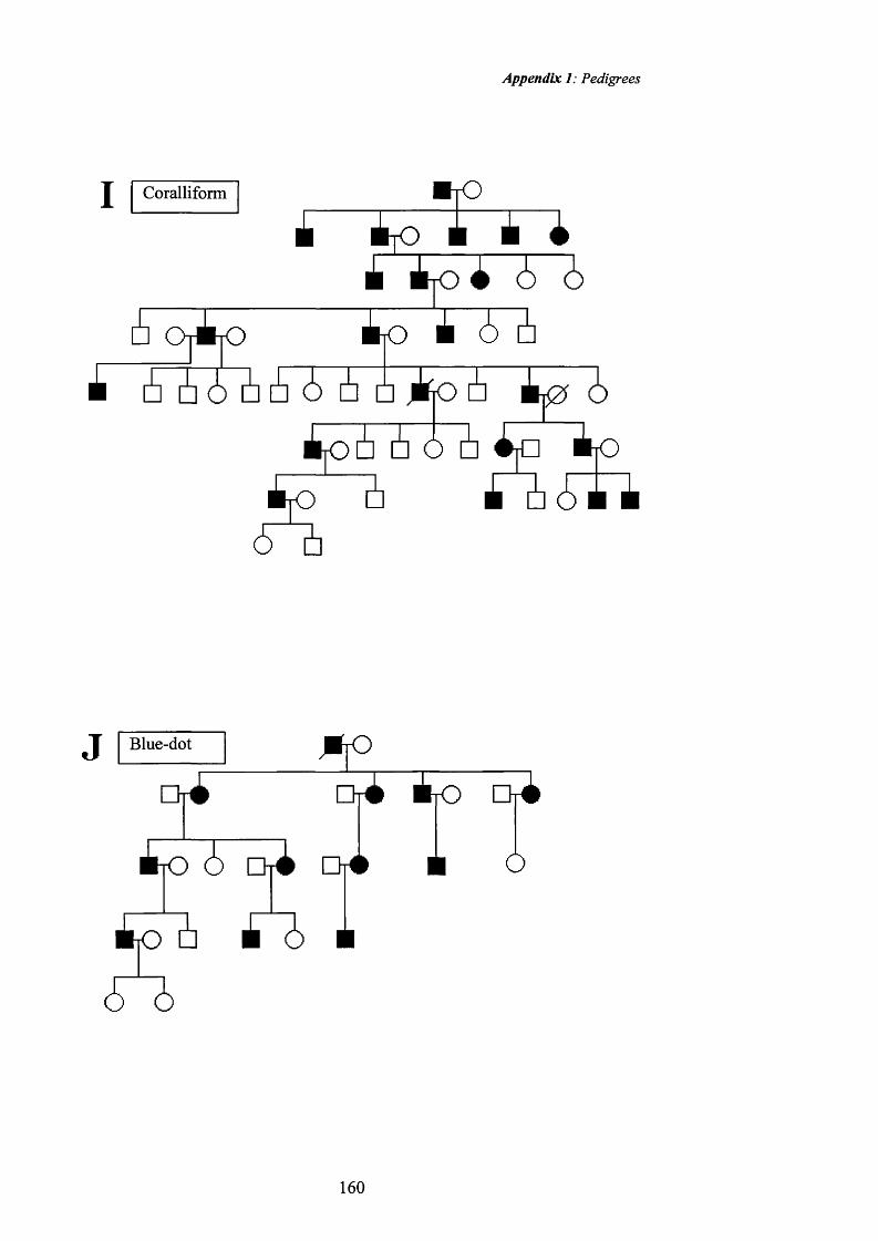
Appendix 1: Pedigrees
I Coralliform * r O
■ t o A *à èyO i 6 6
o io tp Ji 6, 5 6 6 6 6 6 tjo^
►t tot 5~ii
Blue-dot
to 6 o t
to5o
t o
* o
a to o tÔ
160
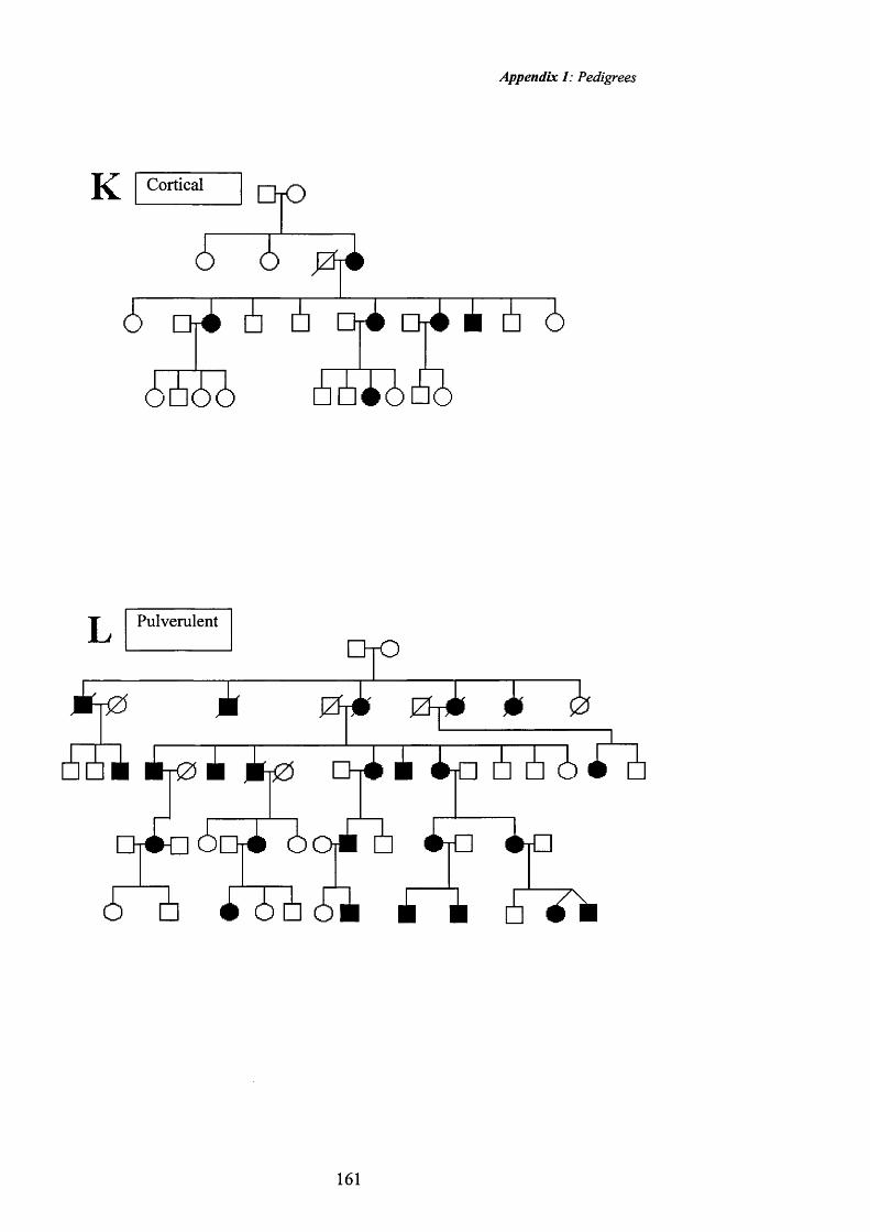
Appendix 1: Pedigrees
K CorticalQ K D
6666
c h ^ dt^ A 6 6
1666
Pulverulent
1t0 1 /T1 Z rl 1 0 1Ü61&
T
i2) 1 6 - 4
tfi» 6cJ
- l i Lin iy□ i
o #
161

Appendix 1: Pedigrees
M Pulvemlent
U À q 3 l i O
a
Pulvemlent
162
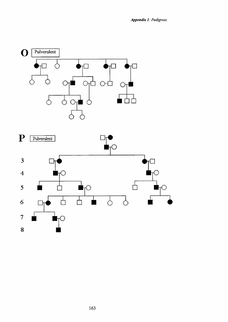
Appendix 1: Pedigrees
o Pulverulent
O tH O Û O r#
o o
P I PulverulenT
3 C
4 ÈyD
5 j □
6 nÀ 5t o
■ tO
É tO
t|0 fi t|0
5"b 5 i à £
163
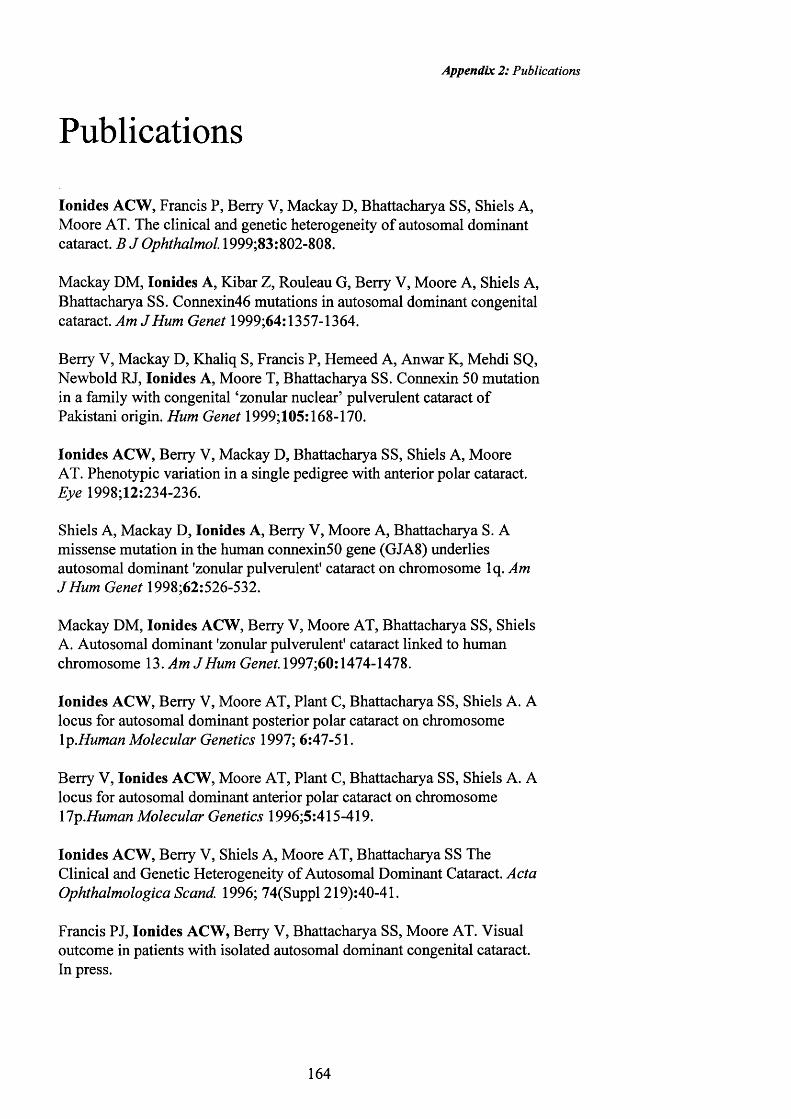
Appendix 2: Publications
Publications
lonides ACW, Francis P, Berry V, Mackay D, Bhattacharya SS, Shiels A, Moore AT. The clinical and genetic heterogeneity o f autosomal dominant cataract. B J Ophthalmol. 1999;83:802-808.
Mackay DM, lonides A, Kibar Z, Rouleau G, Berry V, Moore A, Shiels A, Bhattacharya SS. Connexin46 mutations in autosomal dominant congenital cataract. Am J Hum Genet 1999;64:1357-1364.
Berry V, Mackay D, Khaliq S, Francis P, Hemeed A, Anwar K, Mehdi SQ, Newbold RJ, lonides A, Moore T, Bhattacharya SS. Connexin 50 mutation in a family with congenital ‘zonular nuclear’ pulverulent cataract of Pakistani origin. Hum Genet 1999;105:168-170.
lonides ACW, Berry V, Mackay D, Bhattacharya SS, Shiels A, Moore AT. Phenotypic variation in a single pedigree with anterior polar cataract. Eye 1998;12:234-236.
Shiels A, Mackay D, lonides A, Berry V, Moore A, Bhattacharya S. A missense mutation in the human connexin50 gene (GJA8) underlies autosomal dominant 'zonular pulverulent' cataract on chromosome Iq. Am J Hum Genet 1998;62:526-532.
Mackay DM, lonides ACW, Berry V, Moore AT, Bhattacharya SS, Shiels A. Autosomal dominant 'zonular pulverulent' cataract linked to human chromosome \ 3 .A m J H u m 1997;60:1474-1478.
lonides ACW, Berry V, Moore AT, Plant C, Bhattacharya SS, Shiels A. A locus for autosomal dominant posterior polar cataract on chromosome \p.Human Molecular Genetics 1997; 6:47-51.
Berry V, lonides ACW, Moore AT, Plant C, Bhattacharya SS, Shiels A. A locus for autosomal dominant anterior polar cataract on chromosome Xl^.Human Molecular Genetics 1996;5:415-419.
lonides ACW, Berry V, Shiels A, Moore AT, Bhattacharya SS The Clinical and Genetic Heterogeneity of Autosomal Dominant Cataract. Acta Ophthalmologica Scand 1996; 74(Suppl 219):40-41.
Francis PJ, lonides ACW, Berry V, Bhattacharya SS, Moore AT. Visual outcome in patients with isolated autosomal dominant congenital cataract. In press.
164

Appendix 3: Presentations
Presentations
Various aspects o f this work have been presented at the following meetings.
Autosomal Dominant Cataract. lonides ACW, Moore AT. Sep 1995European Paediatric Ophthalmology Group. Cambridge.UK.
Molecular Genetics of Autosomal Dominant Cataract. lonides Jan 1996ACW, Bhattacharya SS, Moore AT. Moorfields Alumni.London. UK.
An Autosomal Dominant Anterior Polar Cataract is linked to the Apr 1996 short arm o f human chromosome 17. Shiels A, Berry V, lonides ACW, Moore AT, Bhattacharya SS. The Association for Research in Vision and Ophthalmology. Florida, USA.
A Clinical Classification for autosomal dominant cataract. Apr 1996lonides ACW, Berry V, Mackay D, Shiels A, Bhattacharya SS,Moore AT. The Association fo r Research in Vision and Ophthalmology. Florida, USA.
Autosomal Dominant Cataract. lonides ACW, Berry V, May 1996Mackay D, Shiels A, Bhattacharya SS, Moore AT. Royal College o f Ophthalmologists Annual Congress. Edinburgh,Scotland.
Morphological classification of autosomal dominant cataract. Jun 1996ACW lonides, SS Bhattacharya, AT Moore. International Infantile Cataract Symposium. Great Ormond Street, London.
Phenotypic variation in a single pedigree of anterior polar July 1996cataract. lonides ACW, Berry V, Mackay D, Shiels A,Bhattacharya SS, Moore AT Oxford Congress. UK.
Genes for human cataract. lonides ACW, Berry V, Mackay D, Jan 1997Shiels A, Bhattacharya SS, Moore AT. Moorfields Alumni.London.
Mapping genes for human cataract. lonides ACW, Berry V, April 1997 Mackay D, Shiels A, Bhattacharya SS, Moore AT. Royal College o f Ophthalmologists Annual Congress. Birmingham,England.
An autosomal dominant posterior polar cataract is linked to the May 1997 short arm o f chromosome 1. lonides ACW, Berry V, Mackay
165
Appendix 3: Presentations
D, Shiels A, Bhattacharya SS, Moore AT The Association fo r Research in Vision and Ophthalmology. Florida, USA.
Autosomal dominant 'zonular pulverulent' cataract is linked to May 1997human chromosome 13. Mackay D, lonides ACW, Berry V,Shiels A, Bhattacharya SS, Moore AT. The Association fo r Research in Vision and Ophthalmology. Florida, USA.
Phenotypic variation and linkage analysis in a single pedigree April 1998affected by autosomal dominant stationary posterior polar cataract. lonides ACW, Berry V, Mackay D, Shiels A,Bhattacharya SS, Moore AT. Royal College o f Ophthalmolgists Annual Congress. Glasgow.
A connexin gene mutation causes human cataract. lonides April 1998ACW, Berry V, Mackay D, Shiels A, Bhattacharya SS, Moore AT. Royal College o f Ophthalmolgists Annual Congress.Glasgow. The Association fo r Research in Vision and Ophthalmology. Florida, USA.
Visual Outcome in patients with dominantly inherited cataract. May 1999lonides ACW, Francis P, Berry V, Mackay D, Shiels A,Bhattacharya SS, Moore AT. ARVO Royal College o f Ophthalmolgists annual congress. Cardiff, UK.
Genes for human cataract. lonides ACW, Berry V, Mackay D, Oct 1999Shiels A, Bhattacharya SS, Moore AT. EVER, Palma,Mallorca.
166
A cta O phthalm ologica Scandinavica 1996
The clinical and genetic heterogeneity of autosomal dominant cataractAlexander lonides\ Vanita Berry\ Tony Moore-, Shomi Bhattacharya and Alan Shiels^
Institute of Ophthalmology*, University College London and Moorfields Eye Hospital^, London, UK
ABSTRACT. As part o f a linkage study we have reviewed over 150 patients w ith autosom al dom inant cataract and suggest a clinical classification o f the different phenotypes consisting o f anterior and posterior polar, nuclear, cortical, b lue-dot, lam ellar, coralliform and pulverulent cataracts.
Key words: autosomal dominant cataract - clinical phenotypes.
Acta Ophthalmol. Scand. 1996: 74 (Suppl 219): 40-41
There are now six loci known to be important in the development of
autosomal dominant cataract. The first was discovered in 1963 by Renwick & Lawler amd in 1995 the team including Mette Warburg showed linkage of the Voikmann cataract to the short arm of chromosome 1. During this time the precise genetic mutation has been identified at only one locus. There are many different phenotypes and it is likely that more loci will be shown to be involved in the formation of cataract.
Clinical heterogeneityAutosonnal dominant cataract is a collection o f disorders that vary in morphology, in the effect on visual acuity, the age at presentation and the need for surgery. There is no agreed classification for autosomal dominant cataract and this has resulted in confusion as 10 the terminology used in the descriptions of the different phenotypes.
Cataract phenotypes have largely been named after the affected family, as in the Coppock and Voikmann cataracts, or after the ophthalmologist in
volved in their care, as in the Marner cataract.
In a study of over 150 affected patients with autosomal dominant cataract we have identified several distinct phenotypes; anterior polar, posterior polar, nuclear, cortical, blue-dot (cerulean), lamellar (zonular), coralliform and pulverulent. This clinical classification covers all the phenotypes encountered so far in this study. The phenotypes run true throughout the pedigrees in all the phenotypes except the pulverulent cataract. Families affected by pulverulent (dust-like) cataract show great heterogeneity between individuals and even between the eyes of an individual. This was also found to be the case in the family published by Scott et al. (1994). The dust like opacities may be confined to the Y-sutures. the nucleus or may be more widely spread to different regions of the lens. The dust-like, powdery opacities may even be so numerous as to create a total cataract. The zonular pulverulent, Coppock-like, Marner and perhaps the Voikmann cataract, could all be referred to as pulverulent under our classification.
Genetic heterogeneityOne of the first human diseases to be linked to a chromosomal site was the zonular pulverulent cataract (see Table 1). This is an autosomal dominant cataract ocurring in a family originally studied at the turn of the century by Nettleship (1909). Linkage with the Duffy blood group was discovered by Renwick & Lawler (1963), and the Duffy blood group was later assigned to chromosome Iq (Donahue et al. 1968). This cataract is often incorrectly called the Coppock cataract. However, the Coppock cataract, described by Nettleship & Ogilvie (1906) is a different phenotype and was later referred to as a central pulverulent cataract owing to the powdery opacities being central and not “zonular'’. The Coppock cataract is named after the affected family that originally came from Oxford (UK) and has not been included in any published linkage study.
The Coppock-like cataract (central pulverulent cataract) was the next phenotype to be linked (Lubsen et al. 1986) and is different from the zonular pulverulent cataract studied by Renwick and Lawler. In the Coppock-like cataract, first described by Harman (1910), the powdery opacities are restricted to the embryonic nucleus only, as in the central pulverulent (Coppock) cataract. The chromosome Iq cataract of Renwick & Lawler (1963) differed from this in that it had powdery opacities in the central part of the lens bounded by a thin layer of fine opacities with opacities occasionally in the outer zones, hence the name, zonu-
40
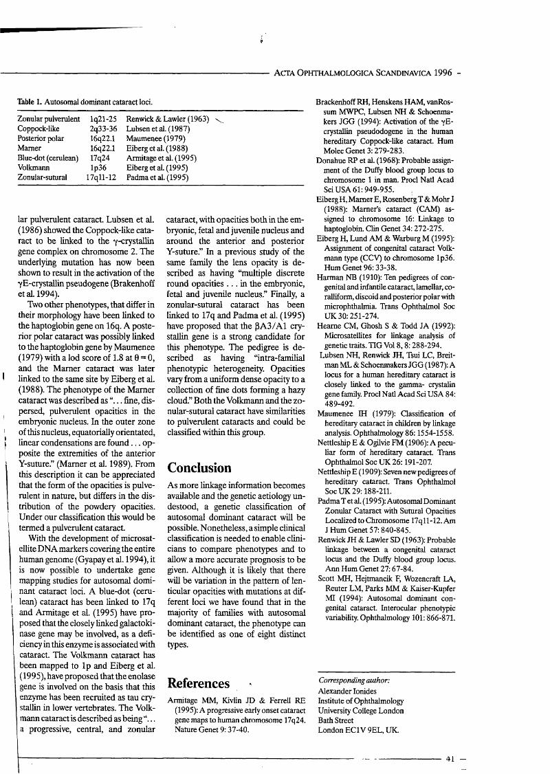
A c ta O p h th a lm o lo g ic a S can d in av ica 1996 -
Table 1. Autosomal dominant cataract loci.
Zonular pulverulent lq21-25Coppock-like 2q33-36Posterior polar 16q22.1Mamer 16q22.1Blue-dot (cerulean) I7q24Voikmann lp36
Renwick & Lawler (1963) Lubsen et al. (1987) Maumenee (1979)Eiberg et al. (1988) Armitage et al. (1995) Eiberg et al. (1995)
Zonular-sutural 17qll-12 Padma et al. (1995)
iar pulverulent cataract. Lubsen et al. (1986) showed the Coppock-like cataract to be linked to the y-crystallin gene complex on chromosome 2. The underlying mutation has now been shown to result in the activation of the yE-crystallin pseudogene (Brakenhoff et al. 1994).
Two other phenotypes, that differ in their morphology have been linked to the haptoglobin gene on 16q. A posterior polar cataract was possibly linked to the haptoglobin gene by Maumenee (1979) with a lod score of 1.8 at 0 = 0, and the Mamer cataract was later linked to the same site by Eiberg et al. (1988). The phenotype of the Mamer cataract was described as “. . . fine, dispersed, pulverulent opacities in the embryonic nucleus. In the outer zone of this nucleus, equatorially orientated, linear condensations are found. . . opposite the extremities of the anterior Y-suture.” (Mamer et al. 1989). From this description it can be appreciated that the form of the opacities is pulvemlent in nature, but differs in the distribution of the powdery opacities. Under our classification this would be termed a pulvemlent cataract.
With the development of microsatellite DNA markers covering the entire human genome (Gyapay et al. 1994), it is now possible to undertake gene mapping studies for autosomal dominant cataract loci. A blue-dot (cem- lean) cataract has been linked to 17q and Armitage et al. (1995) have proposed that the closely linked galactokinase gene may be involved, as a deficiency in this enzyme is associated with cataract. The Voikmann cataract has been mapped to Ip and Eiberg et al. (1995), have proposed that the enolase gene is involved on the basis that this enzyme has been recmited as tau crystallin in lower vertebrates. The Voikmann cataract is described as being “... a progressive, central, and zonular
cataract, with opacities both in the embryonic, fetal and juvenile nucleus and around the anterior and posterior Y-suture.” In a previous study of the same family the lens opacity is described as having “multiple discrete round opacities . . . in the embryonic, fetal and juvenile nucleus.” Finally, a zonular-sutural cataract has been linked to 17q and Padma et al. (1995) have proposed that the |3A3/A1 crystallin gene is a strong candidate for this phenotype. The pedigree is described as having “intra-famüial phenotypic heterogeneity. Opacities vary from a uniform dense opacity to a collection of fine dots forming a hazy cloud.” Both the Voikmann and the zonular-sutural cataract have similarities to pulverulent cataracts and could be classified within this group.
ConclusionAs more linkage information becomes available and the genetic aetiology undestood, a genetic classification of autosomal dominant cataract will be possible. Nonetheless, a simple clinical classification is needed to enable clinicians to compare phenotypes and to allow a more accurate prognosis to be given. Although it is likely that there will be variation in the pattern of lenticular opacities with mutations at different loci we have found that in the majority of families with autosomal dominant cataract, the phenotype can be identified as one of eight distinct types.
ReferencesArmitage MM, Kivlin JD & Ferrell RE
(1995): A progressive early onset cataract gene maps to human chromosome 17q24. Nature Genet 9:37-40.
Brackenhoff RH, Henskens HAM, vanRos- sum MWPC, Lubsen NH & Schoenma- kers JGG (1994): Activation of the yE- crystallin pseudodogene in the human hereditary Coppock-like cataract. Hum Molec Genet 3:279-283.
Donahue RP et al. (1968): Probable assignment of the Duffy blood group locus to chromosome 1 in man. Prod Natl Acad Sci USA 61:949-955.
Eiberg H, Mamer E, Rosenberg T & Mohr J (1988): Mamer’s cataract (CAM) assigned to chromosome 16: Linkage to haptoglobin. Clin Genet 34:272-275.
Eiberg H, Lund AM & Warburg M (1995): Assignment of congenital cataract Voikmann type (CCV) to chromosome lp36. Hum Genet 96:33-38.
Harman NB (1910): Ten pedigrees of congenital and infantile cataract, lamellar, coralliform, discoid and posterior polar with microphthalmia. Trans Ophthalmol Soc UK 30:251-274.
Heame CM, Ghosh S & Todd JA (1992): Microsatellites for linkage analysis of genetic traits. TIG Vol 8,8:288-294.
Lubsen NH, Renwick JH, Tsui LC, Breit- man ML & Schoenmakers JGG (1987): A locus for a human hereditary cataract is closely linked to the gamma- crystalin gene family. Prod Natl Acad Sci USA 84: 489-492.
Maumenee IH (1979): Classification of hereditary cataract in children by linkage analysis. Ophthalmology 86:1554-1558.
Nettleship E & Ogilvie FM (1906): A peculiar form of hereditary cataract. Trans Ophthalmol Soc UK 26:191-207.
Nettleship E (1909): Seven new pedigrees of hereditary cataract. Trans Ophthalmol Soc UK 29:188-211.
Padma T et al. (1995): Autosomal Dominant Zonular Cataract with Sutural Opacities Localized to Chromosome 17qll-12. Am J Hum Genet 57:840-845.
Renwick JH & Lawler SD (1963): Probable linkage between a congenital cataract locus and the Duffy blood group locus. Ann Hum Genet 27:67-84.
Scott MH, Hejtmancik F, Wozencraft LA, Reuter LM, Parks MM & Kaiser-Kupfer MI (1994): Autosomal dominant congenital cataract. Interocular phenotypic variability. Ophthalmology 101:866-871.
Corresponding author: Alexander lonides Institute of Ophthalmology University College London Bath StreetLondon EClV 9EL, UK.
41 -
Anterior polar cataract: clinical spectrum and gen etic linkage in a single family
AL E XA N D E R l ONI DES, V A N I T A BERRY, D O N N A M A C K A Y , AL A N SHIELS, S H O M I B H A T T A C H A R Y A ,A N T H O N Y M O O R E
A. lonidesV. BerryD. MackayA. ShielsS. BhattacharyaDepartment o f MolecularGeneticsInstitute o f Ophthalmology London EC IV 9EL, UK
A. lonides I S A. MooreMoorfields Eye Hospital City RoadLondon EC1V2PD, UK
Tel: + 4 4 (0 )1 7 1 2 53 3411 Fax: + 4 4 (0 )1 7 1 253 4 6 9 6
A. M ooreAddenbrooke's HospitalCambridgeUK
Presented at the Oxford Congress, July 1996
Abstract
Anterior polar cataract can occur as a sporadic finding, in association with other ocular abnormalities or as an inherited, autosomal dominant disorder. We have demonstrated linkage in a family with autosomal dominant anterior polar cataract to the short arm of chromosome 17, locating the gene to the region 17pl2-13. All affected members of this large family had an opacity at the anterior pole of the lens that varied only in size and the effect on visual acuity. Anterior polar cataract is thought to have a minimal effect on visual acuity although in the affected members of this family there was a high incidence of unilateral amblyopia.
Key words Anterior polar, Cataract, Autosomal dominant. Linkage, Phenotype
Inherited cataract is a significant cause of visual impairment in childhood and is most often transmitted as a congenital autosomal dominant (AD) trait. Eight different loci have now been mapped for AD cataract " but the underlying genetic abnormality is known in only one case: the Coppock-like cataract has been shown to be related to the overexpression of a y-crystallin pseudogene. We have demonstrated linkage of an AD anterior polar cataract (AFC) to the short arm of chromosome 17 in a single family and in this paper describe the clinical spectrum of the disease.
Patients and methods
The patients were from a large family with AD anterior polar cataract (Fig. 1) identified from the genetic data base at Moorfields Eye Hospital. Family members were invited to attend the hospital for an eye examination. The lens was examined by slit lamp and anterior segment photography was taken where possible. Blood was taken for DNA extraction to be used for the linkage analysis.
Results
The results of the molecular genetic study demonstrating linkage of the anterior polar cataract in this family to chromosome 17p have already been published.^ Anterior polar cataracts were seen in all the affected family members. Thirty family members were examined and 19 found to be affected. Seven patients had undergone surgery and the phenotype was examined in the remaining 12 cases. The opacity was always in the same position of the lens, the anterior pole, but varied in severity. The larger opacities were pyramidal with a base of 4 mm and extended into the anterior chamber (Figs. 2, 3), whilst the smaller opacities consisted of a flat plaque measuring less than 1 mm in diameter (Figs. 4, 5). In one case the only sign of an anterior pole abnormahty was an irregularity of the anterior capsule (Fig. 6). Seven of the affected patients underwent surgery, aU between the age of 1 and 6 years, with a final visual acuity of 6/18 to NPL. Two patients who had a visual acuity of NFL had both developed glaucoma and retinal detachment. One affected patient who had not undergone surgery but had minimal opacities had a visual acuity of 6/6 in both eyes.
Ten of the 12 affected patients who had not undergone surgery were amblyopic in one eye with a visual acuity of 6/12 to 6/36 in the worse eye. The amblyopic eye was always the eye with the larger opacity.
Discussion
Anterior polar cataract has been reported to have a minimal effect on visual acuity.’ '’ In this family unilateral amblyopia was common, suggesting that children with anterior polar cataracts should be offered regular follow-up.'' The unoperated patients had a better visual outcome than those who had surgery for their anterior polar cataracts. Although the visual outcome may reflect the severity of the cataract it suggests that better long-term results may be achieved with conservative treatment.
224 Eye (1998) 12, 224-226 © 1998 Royal College of Ophthalmologists

____________ D |i à \3 ji à 6
6 o | i 6 A 6 o i 6 & 6 &|0 6
Chi i |0 6 i i 6 6 Ori A o i 6
m i nFig. 1. Pedigree affected by anterior polar cataract.
AD cataract may show considerable phenotypic variability between family members.^^ In this family, however, all affected individuals had lens opacities confined to the anterior pole of the lens. We have demonstrated that the anterior polar cataract in this family is linked to the short arm of chromosome 17. The mechanism by which the lens opacity is confined to the anterior pole is unclear but identification of the precise genetic mutation will help in understanding the pathogenesis. There are at present no candidate genes in this region on chromosome 17p, but research to identify the causative gene continues.
Fig. 2. A n ter io r polar cataract. S li t lam p photograph o f a pyram ida l cataract fro m in d iv id u a l 4:2.
%
«Fig. 4. A n ter io r polar cataract. S li t lam p photograph o f a plaque-like opacity fr o m in d iv id u a l 4:3.
Fig. 5. A n te r io r polar cataract. R e tro illum ina tion o f the p laque-like opacity fro m ind iv id u a l 4:3.
Fig. 3. A n ter io r polar cataract. R e tro illu m in a tio n o f the pyram ida l cataract fr o m ind iv id u a l 4:2.
Fig. 6. A n te r io r polar cataract. R e tro illum ina tion o f an anterior capsular irregu la rity fr o m ind iv id u a l 5:6.
References
1. François J. Genetics of cataract. Ophthalmologica 1982;184:61-71.
2. Eiberg H, Lund AM, Warburg M, Rosenberg T. Assignment of congenital cataract Volkmarm type (CCV) to chromosome lp36. Hum Cenet 1995;96:33-8.
225
3. lonides ACW, Berry V, Mackay DM, Moore AT, Bhattacharya SS, Shiels A. A posterior polar cataract maps to chromosome Ip. Hum Mol Genet 1997;6:47-51.
4. Renwick JH, Lawler SD. Probable linkage between a congenital cataract locus and the Duffy blood group locus. Ann Hum Genet 1963;27:67-84.
5. Lubsen NH, Renwick JH, Tsui L-C, Breitman ML, Schoenmakers JGG. A locus for a human hereditary locus is closely linked to the y-crystallin gene family. Proc Natl Acad Sci USA 1987;84:489-92.
6. Rogaev El, Rogaeva EA, Korovaitseva GI, Farrer LA, Petrin AN, Keryanov SA, e t al. Linkage of polymorphic congenital cataract to the y-crystallin gene locus on human chromosome 2q33-35. Hum Mol Genet 1996;5:699-703.
7. Eiberg H, Mamer E, Rosenberg T, Mohr J. Marner's cataract (CAM) assigned to chromosome 16: linkage to haptoglobin. Clin Genet 1988;34:272-5.
8. Padma T, Ayyagari R, Murty JS, Basti S, Fletcher T, Rao GN, e t al. Autosomal dominant zonular cataract with sutural opacities localised to chromosome 17qll-12. Am J Hum Genet 1995;57:840-5.
9. Armitage MM, Kivlin JD, Ferrell RE. A progressive early onset cataract gene maps to human chromosome 17q24. Nature Genet 1995;9:37-40.
10. Brackenhoff RH, Henskens HAM, van Rossum MWPC, Lubsen NH, Schoenmakers JGG. Activation of the yE- crystallin pseudogene in the human hereditary Coppock-like cataract. Hum Mol Genet 1994;3:279-83.
11. Berry V, lonides ACW, Moore AT, Plant C, Bhattacharya SS, Shiels A. A locus for anterior polar cataract on chromosome 17p. Hum Mol Genet 1996;5:415-9.
12. Duke-Elder S, editor. System of ophthalmology, vol HI, part 2: Normal and abnormal development: congenital deformities. St Louis: CV Mosby, 1964:717-23.
13. Bouzas AG. Anterior polar congenital cataract and comeal astigmatism. J Pediatr Ophthalmol Strabismus 1992;29:210-2.
14. Jaafar MS, Robb RM. Congenital anterior polar cataract: a review of 63 cases. Ophthalmology 1984;91:249-52.
15. Scott MH, Hejtmancik MD, Wozencraft MS, Reuter LM, Parks MM, Keiser-Kupfer MD. Autosomal dominant congenital cataract: interocular phenotypic variability. Ophthalmology 1994;101:866-71.
© 1997 Oxford University Press Human Molecular Genetics, 1997, Vol. 6, No. 1 47-51
A locus for autosomal dominant posterior polar cataract on chromosome IpAlexander C. W. lonides, Vanita Berry, Donna S. Mackay, Anthony T. Moore\ Shomi 8. Bhattacharya and Alan Shiels*
Department of Molecular Genetics, Institute of Ophthaimology, University College London, 11-43 Bath Street, London EC1V 9EL, UK and ‘'Moorfields Eye Hospital, City Road, London EC1V 2PD, UK
Received July 16,1996; Revised and Accepted October 10,1996
Autosomal dominant congenital cataract is a clinically and genetically heterogeneous lens disease. Here we report the linkage of a locus for autosomal dominant posterior polar cataract {CPPi to the distal short arm of chromosome 1. To map the C/V’ locus we performed molecular genetic linkage analysis using microsatellite markers In a three-generation pedigree. After exclusion of 13 known ioci and candidate lens genes for autosomal dominant cataract, we obtained significantly positive LOD scores for markers D1S508 (2 = 3.14,8 = 0) and D1S468{Z = 2.71,0 = 0). Multipoint analysis gave a maximum LOD score of 3.48 (8 = 0.07) between markers DlSSOSsn^ D1S468. From hapiotype data, however, C/Vprobabiy lies inthetelomeric interval D1S284&Aplev, which includes the locus for the clinically distinct Volkman congenital cataract (CCL). This study provides the first evidence for genetic heterogeneity of autosomal dominant posterior polar cataract for which a locus had been linked previously to chromosome 16q.
INTRODUCTION
Congenital cataract is a significant cause of visual impairment and blindness in childhood (1). Approximately 25% of all congenital cataract is inherited, most often as an autosomal dominant Mendelian trait (2) showing considerable inter- and intra-familial phenotypic variation (1-4). Currently, eight independent loci for autosomal dominant cataract have been linked to chromosomes lp36 (5), Iq21-q25 (6), 2q33-q35 (7,8), 16q22.1 (9-11), 17pl3(12), 17qll-12(13), 17q24(14)and22q (15). The Coppock-like ‘nuclear’ cataract and a polymorphic ‘lamellar’ cataract map to the y-crystallin gene cluster on 2q and the former is associated with novel activation of the "^-crystallin pseudogene (7). Causative genes at the other seven human cataract loci have yet to be identified, however, the PA3/A1 crystallin gene is a strong candidate for the ‘zonular sutural’ cataract on 17q (13) and the ‘cerulean’ cataract on 22q maps provocatively close to the p-crystallin gene cluster (15). In the
mouse, autosomal dominant cataract has been associated with mutations in the genes for'^-crystallin on chromosome 1 (16), PB2-crystallin on chromosome 5 (17) and lens major intrinsic protein on chromosome 18 (18). Mutation of the P-ciystallin gene has also been shown to underlie autosomal dominant congenital cataract in the guinea-pig (19).
Posterior polar cataract is a clinically distinctive opacity located at the back of the lens and, because of its proximity to the optical centre of the eye, can have a marked effect on visual acuity. Posterior lens opacities are commonly acquired in age-related cataract (20) and may also present in neurofibromatosis type 2 (21) and genetically heterogeneous retinal degenerations (22). Ultrastructural studies of such opacities have identified abnormal migration of ‘dysplastic’ swollen fibre cells and accumulation of granular extracellular material at the posterior pole of the lens (23-25). However, the underlying pathogenesis is poorly understood.
DominanÜy inherited posterior polar congenital cataract (9,10) has previously been linkoi with the haptoglobin locus (HP) on 16q, along with the dominant Mamer congenital cataract (11). Bo these cataracts are described as progressive, however, in the Mamer family opacities initiate at the nucleus rather than at the posterior pole of tiie lens suggesting that either allelic heterogeneity or two distinct cataract genes exist at the 16q locus. To obtain further insight into the genetic aetiology of congenital cataract we performed linkage analysis on a three-generation family of English descent with autosomal dominant posterior polar cataract
RESULTS
Ten affected and three unaffected members of the family, including the ‘critical recombinant’ female 4.1 aged 17 years (see below), underwent a full ophthalmological examination by one of us (A.C.W.I.). The opacity (Fig. 1), which was bilateral in all cases, consisted of a single well defined plaque confined to the posterior pole of the lens and varied from 0.5-3 mm in diameter. Hospital records indicated that the opacity was usually present at birth or developed within the first few months of life but did not progress with age to other regions of the lens. There was no evidence of posterior lenticonus or high myopia and no family history of other ocular or systemic abnormalities.
"To whom correspondence should be addressed

48 Human Molecular Genetics, 1997, Vol. 6, No. I
Figure 1. Slit-iamp photograph of the right eye of affected female 2.8 from the autosomal dominant cataract pedigree (Fig. 2) showing a single well defined opacity located at the posterior pole of the lens.
Table 1. Two-point LOD scores (Z) for linkage between the CPP locus and chromosome Ip markers
Marker cM Recombination fraction (0)
0.00 0.01 0.05 0.10 0.20 0.30 0.40
Zmax 0max
Ipter
D IS243 6.2 1.51 1.47 1.35 1.19 0.83 0.46 0.13 1.50 0.00
D1S468 4.9 2.71 2.66 2.46 2.21 1.64 1.01 0.35 2.70 0.00
D 1S2845 1.8 -CO 1.85 2.30 2.27 1.86 1.23 0.48 2.31 0.07
D 1S2660 3.5 -oo 1.85 2.30 2.27 1.86 1.23 0.48 2.31 0.07
D 1S214 0.0 0.57 0.56 0.51 0.44 0.33 0.22 0.10 0.57 0.00
D 1S2870 0.5 -0.22 0.35 0.49 0.44 0.24 0.04 0.49 0.13
D JS2663 1.2 — 1.26 1.74 1.76 1.45 0.94 0.33 1.78 0.08
D 1S508 4.8 3.14 3.08 2.85 2.54 1.88 1.15 0.39 3.14 0.00
D 1S450 4.0 -oo 1.55 2.00 1.98 1.58 1.00 0.35 2.01 0.07
D IS2667 5.5 1.85 2.30 2.27 1.86 1.23 0.48 2.31 0.07
D 1S228 15.3 1.03 1.01 0.91 0.78 0.53 0.30 0.11 1.03 0.00
D 1S199 8.9 1.34 1.79 1.78 1.41 0.87 0.30 1.81 0.07
D1S234 — -1.56 -0.39 -0.07 0.06 0.11 0.11 0.12 0.35
Sex-averaged genetic distances between the markers (26) are shown in centi-Morgans (cM).

Human Molecular Genetics, 1997, Vol. 6, No. I 49
4 6 2 2 6 8 6 3 4 34 15 6 5 3 2 6 4 4
4 4 3 6 1 3
5 ^6 6 3 4 3 4 6 6 5 23 2 I 2 1 2 3 2 3 23 2 4 5 4 5 3 2 1 54 2 4 6 4 6 4 2 5 66 4 3 4 3 4 6 4 5 54 2 1 2 1 2 4 3 3 27 4 I 4 1 4 7 2 7 54 4 3 4 3 4 4 3 3 43 5 5 6 5 6 3 4 2 71 3 I 3 1 3 1 5 2 4
6 6 2 1 2 6 2 3 4 I2 3 4 54 45 63 3
6 6 3 33 54 3 6 6 4 2 7 3 4 5 3 5 1 5
3 7 2 2
B6 I 3 53 54 26 4 4 37 7 4 I 3 7 1 2
[Dré S ilr® É O Ë É4 53 33 14 56 54 37 7 4 3 3 25 2
7 54 43 75 4
6 23 23 54 56 54 37 7 4 3 3 25 2
D u eu e Haploiyp*D1S243 6D1S468 3D1S2845 3D1S2663 4D1S508 6D1S450 4D1S2667 7D1S228 4D1S199 3D1S234 1
5 62 5 9 9 1 6 2 6 1 5 4 53 2 1 74 5
6 4 5 2 9 51 42 3 5 2 4 23 37 44 1
4 62 35 3 4 43 6 2 42 73 44 35 I
5 62 3 9 31 42 6 1 4 4 73 4 7 44 1
Figure 2. Abridged pedigree of the posterior polar cataract family used in this study showing the segregation of nine chromosome Ip markers listed in descending order from the telomere. Squares and circles symbolise males and females, respectively. Open and filled symbols indicate unaffected and affected individuals, respectively. *The unaffected ‘critical recombinant’ female 4.1 was uninformative for these markers.
D1S243D1S508D1S468
0 1 S 2 3 4
,3.5
2.4cM 7.7cM lOcM,10cM,
- 0.2-0 .4 0 2 0.4RECOMBINATION FRACTION
Figure 3. Multipoint linkage analysis between the CPP locus and chromosome Ip markers, D lS 234-{39 cM )-D lS 508 -{\2 cM )-D lS468-{6 cM )-D lS243 (26).
For linkage analysis, 19 members of the pedigree (Fig. 2) including 10 affected individuals, five unaffected individuals and four spouses were genotyped with microsatellite markers from the Généthon (AC)n map (26). We first excluded linkage of the cataract with HP on 16q using markers D16S515 (Z = -3.31, 0 = 0.05) and D16S503 (Z = -1.76,0 = 0.05). We then proceeded to exclude 12 other known loci and candidate lens genes for autosomal dominant cataract on 22q (cerulean cataract type 2 and P-crystallin), 21q (al-crystallin), 19q (lens integral membrane
protein 2), 17q24 (cerulean cataract type 1), 17ql l-ql2 (zonular sutural cataract and PA3/À1-crystalin), 17p (anterior polar cataract), 16p ( .-crystallin), 12q (lens major intrinsic protein), llq (o2-crystallin), 2q (y-crystallin), Iq (zonular pulverulent cataract), Ip ( -crystallin) (Human Genome Database, 1996). Following exclusion of the -crystallin gene (CRYZ) on lp3 l-p22 with marker D1S201 (Z = -l .09,0 = 0.05) we detected positive LOD scores for markers D1S468 (Z = 2.71, 0 = 0) and DIS243 (Z= 1.51,0 = 0), which flank a 6 cM interval close to the
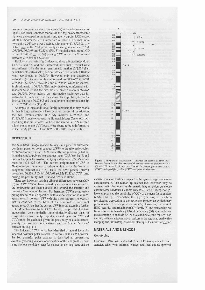
50 Human Molecular Genetics, 1997, Vol. 6, No. I
Volkman congenital cataract locus (CCV) at the telomeric end of Ip (5). Ten other Généthon markers in this region of chromosome Ip were genotyped in the family and the two-point LOD scores of all 12 marker loci are summarised in Table 1. The maximum two-point LOD score was obtained with marker D/S505 (Z^ax = 3.14, 0max = 0)- Multipoint analysis using markers DIS234, DIS508, DIS468 and D1S243 (Fig. 3) yielded a maximum LOD score of 3.48 (9max = 0.07) placing CPP in the 12 cM interval between D1S508 and DIS468.
Haplotype analysis (Fig. 2) detected three affected individuals (3.6, 3.7 and 3.8) and one unaffected individual (3.9) that were recombinant with the most centromeric marker D1S234 (i.e., which lies closest to CRYZ) and one affected individual (3.10) that was recombinant at D1S199. However, only one unaffected individual (4.1 ) was recombinant for markersD752667, D1S450, D1S2663, D1S2870, D1S2660 and D1S2845, which lie increasingly telomeric to D1S234. This individual was uninformative for markers DIS508 and the two most telomeric markers DIS468 and DIS243. Nevertheless, the informative haplotype data for individual 4.1 indicated that the cataract locus probably lies in the interval between D1S2845 and the telomere on chromosome Ip, i.e., D/52545-1 pter (Fig. 4).
Attempts to trace additional family members that may enable further linkage refinement have been unsuccessful. In addition, the two tetranucleotide (GATAX, markers {D1SI605 and DIS2I33) from the Cooperative Human Linkage Center (CHLC) map (27) that are reported to lie in the interval D/5245-1 pter, which contains the CCV locus, were found to be uninformative in the family (Z = -0.14 and 0.25 at 0 = 0.05, respectively).
C C V
cM - D 1 S 2 4 3
6
- D 1S468
\ 5\ - D 1S2845
C P P
C R Y Z
C A E 1
DISCUSSION
We have used linkage analysis to localise a gene for autosomal dominant posterior polar cataract (CPP) to the telomeric region of chromosome Ip. CPP is both clinically and genetically distinct from the zonular pulverulent cataract locus (CAEI) on Iq (6) and does not appear to involve the Ç-crystallin gene (CRYZ) which maps to Ip31-p22 (28). The current assignmnent of CPP to D/52545-lpter, however, overlaps with that for the Volkman congenital cataract (CCV, 5). Thus, the CPP genetic interval comprises D/52545-(5cM)-D/54d5-(6cM)-D/5245-CC7V-lpter, raising the possibility that CCV and CPP are allelic.
There are, however, striking clinical differences between CCV (5) and CPP. CCV is characterised by central opacities located in the embryonic and fetal nucleus and around the anterior and posterior Y-sutures of the lens. Furthermore, CCV is progressive giving rise to zonular opacities with a wide variation in clinical appearance. In contrast, CPP exhibits a non-progressive opacity that is confined to the back of the lens with a consistent appearance. Given that the current CPP interval extends a further -11 cM centromeric to the CCV interval, it is possible that two independent genes underlie these clinically distinct types of congenital cataract on Ip. Equally, a single gene for CPP and CCV cannot be excluded given the possibility of allelic heterogeneity for posterior polar cataract and the Mamer ‘nuclear’ cataract on 16q (11).
The linkage of CPP to Ip has identified a second locus for inherited posterior polar cataract. In contrast with CPP, however, the 16q posterior polar cataract is described as progressive, eventually leading to a total opacification of the lens (9-11). There is no obvious candidate gene for cataract at the 16q locus and no
Figure 4. Idiogram of chromosome 1 showing the genetic distances (cM) between three microsatellite markers (26) and the calculated positions of CCV (5) and CPP on the distal short arm. The loci for zonular pulverulent cataract (CAEI) on Iq and p-crystallin (CRYZ) on Ip are also indicated.
cataract mutation has been mapped to the syntenic region of mouse chromosome 8. The human Ip cataract loci, however, may be syntenic with the recessive dysgenetic lens mutation on mouse chromosome 4 (Mouse Genome Database, 1996). Eiberg et al. (5) have emphasised the proximity of CCV to the gene for a-enolase (EN01) on Ip. Remarkably, this glycolytic enzyme has been recruited as x-crystallin in the turtle lens through an evolutionary process referred to as gene-sharing (29). However, the red-cell ENO1 activity is normal in the CCV family (5) and cataract has not been reported in hereditary ENOl deficiency (30). Currently, we are attempting to exclude ENOl as a candidate gene for CPP and identify additional informative markers in the region to enable fine mapping and, ultimately, positional cloning of the underlying gene.
MATERIALS AND METHODS
Genotyping
(jenomic DNA was extracted from EDTA-sequestered blood samples, taken with informed consent and local ethical approval.
Human Molecular Genetics, 1997, Vol. 6, No. 1 51
using the Nucleon H DNA extraction kit (Scotlab Bioscience). PCR-based genotyping using Généthon microsatellite markers (26) at 5-10 cM intervals was performed as described previously (12).
Linkage analysis
Data was collated using the LINKSYS (version 3.1) data management package (31). Two-point LOD scores were calculated using the MLINK and BLINK sub-programs of the LINKAGE (version 5.1) package (32). Allele frequencies calculated from the four spouses in the family were similar to those quoted by Généthon (26). Multipoint analysis was computed using LINKMAP (32) run on the UK Human Genome Mapping Project (HGMP) Resource Centre computing facilities (Cambridge, UK). A full penetrance and a gene frequency of 0.0001 were assumed for the cataract locus.
ACKNOWLEDGEMENTS
We thank the family for their cooperation in this study and the UK HGMP Resource Centre for microsatellite primer synthesis and use of computing facilities. This work is supported by a grant from The Wellcome Trust (043073/Z/94/Z) to A.S. and S.S.B. A.C.W.L is supported by a grant from the Friends of Moorfields Eye Hospital and D.S.M. is a Wellcome Prize Student.
REFERENCES
1. LundA-M., Eiberg,H., Rosenberg,T. and Warburg,M. (1992) Autosomal dominant congenital cataract; linkage relations; clinical and genetic heterogeneity. Clin. Genet., 41,65-69.
2. Francoisjf. (1982) Genetics of cataract O phthalm oligica, 184, 61-71.3. ScottM.H., HejtmancikJ.F., Wozencraft L.A., Reuter,L.M., Parks,M.M. and
Kaiser-Kupfer, M.I. (1994) Autosomal dominant congenital cataract—inter- ocular phenotypic variability. Ophthalmology, 101,866-871.
4. Ionides,A.C.W., Berry,V., Moore,A.T., Bhattacharya,S.S. and Shiels,A. (1996) Clinical and genetic heterogeneity of autosomal dominant cataract. Acta. OphthalmoL, 74, S219,40-41.
5. Eiberg,H., Lund,A.M., Warburg,M. and Rosenberg,?. (1995) Assignment of congenital cataract Volkmann type (CCV) to chromosome lp36. Hum. Genet., 96,33-38.
6. RcnwickJ.H. and Lawler,S.D. (1963) Probable linkage between a congenital cataract locus and the Duffy blood group locus. Ann. Hum. Genet., 27,67-84.
7. Brakenhoff,R.H., Henskens,H.A.M., van Rossum,M.W.P.C., Lubsen,N.H. and Schoenmakos J.G.G. (1994) Activation of theT^-crystallin pseudogene in the human hoeditary Coppock-like cataract Hum. M ol. Genet., 3, 279-283.
8. RogaevJE.1., Rogaeva,E.A., Korovaitseva,G.L, FarrarL-A., Petrin,A.N., Kayanov,S.A., Tluaeva.S., Chumakov,!., St. Geotge-HyslopJ*. and Ginter,E.K. (1996) Linkage of polymorphic congenital cataract to the ycrystallin gene locus on human chromosome 2q33-35.//um. M ol. Genet., 5,699-703.
9. Maumenee,I.R (1979) Classification of hereditary cataracts in children by linkage analysis. Ophthalmol., 86, 1554-1558.
10. Richards,!., Maumenee,I.H., Rowe,S. and Lovrien,E.W. (1984) Congenital cataract possibly linked to haptoglobin. Cytogenet. C ell Genet., 37,570.
11. Mamer,E., Rosenberg,?, and Eiberg,H. (1989) Autosomal dominant congenital cataract; morphology and genetic mapping. Acta. O phthalm ol, 67, 151-158.
12. Berry,V., lonides.A.C.W., Moore,A.?., PlantC., Bhattacharya,S.S. and Shiels,A. (1996) A locus for autosomal dominant anterior polar cataract on chromosome 17p. Hum. M o l Genet., 5,415-419.
13. Padma,?., Ayyagari,R. MurtyJ.S., Basti,S., Fletcher,?., Rao,G.N., Kaiser- KupferM- and HejtmancikJF. (1995) Autosomal dominant zonular cataract with sutural opacities localized to chromosome 17qll-12. Am. J. Hum. Genet., 57,840-845.
14. Armitage,M.M., Kivlin J.D. and Ferell,R.E. (1995) A progressive early onset cataract gene maps to human chromosome 17q24. Nature Genet., 9, 37-40.
15. Kramer,P., Yount, !., Mitchell,?., LaMorticella,D., Carrero-Valenzuela, R., Lovrien, E., Maumenee,!. and Litt,M. (1996) A second locus for cerulean cataract maps to the P crystallin region on chromosome 22. G enomics 35, 539-542.
16. Cartier,M., Breitman,M.L. and ?sui,L-C. (1992) A fraraeshift mutation in the l^crystallin gene of the Elo mouse. Nature Genet., 2, 42-45.
17. Chambers,C. and Russell,P. (1991) Deletion mutation in an eye lens P-crystallin. J B iol Chem., 266,6742-6746.
18. Shiels,A. and Bassnett,S. (1996) Mutations in the founder of the MIP gene family underlie cataract development in the mouse. Nature Genet., 12, 212-215.
19. Rodriguez,I.R., Gonzalez,R, Zigler,J.S. and Borras,?. (1992) A guinea-pig hereditary cataract contains a splice-site deletion in a crystallin gene. Biochim. Biophys. Acta., 1180,44-52.
20. KleinJB.E.K., Klein,R. and Linton,K.L.P. (1992) Prevalence of age-related lens opacities in a population. Ophthalmology, 99,546-552.
21. Ragge, N.K., Baser,M.E., KleinJ., NechiporukA-, SainzJ., Pulst,S.M. and Riccardi,V.M. (1995) Ocular abnormalities in neurofibromatosis 2. Am. J. O phthalm ol, 120,634-641.
22. HeckenlivelyJ.R. (1982) ?he frequency of posterior subcapsular cataract in hereditary retinal degenerations. Am. J. O pluhalm ol, 93,733-738.
23. Eshaghian,!. and Streeten,B.W. (1980) Human posterior subcapsular cataract. Arch. O phthalm ol, 98, 134-143.
24. EshagianJ., Rafferty,N.S. and Goossens,W. (1980) Ultrastructure of human cataract in retinitis pigmentosa. Arch. O phthalm ol, 98,2227-2230.
25. Nagata,M., Matsuura,H. and Fujinaga,Y. (1986) Ultrastructure of posterior subcapsular cataract in human lens. Ophthalmic Res., 18,180-184.
26. Dib, C., Fame, S., Fizames, C , Samson?)., Drouot,N., Vignal,A., Millas- seau,R, Marc,S., Kazan?., Seboun,E., Lathrop,M., Gyapay,G., Morissett?. and Weissenbach?. (1996) A comprehensive genetic map of the human genome based on 5,264 microsatellites. Nature, 380,152-154.
27. Murray?.C, Buetow,K.H., Weber?.L., Ludwigsen,S., Scherpbier-Heddc- ma,?., Manion,F., Quillen,J.,Sheffield,V.C., Sunden.S., Duyk,G.M., Weissenbach?., Gyapay,G., Dib, C , Morissett?., Lathrop,M., Vignal,A., White?!., Matsunami,N., Gerken,S., Melis,R., Albertsen?!., Plaetke,R., Odelberg,S., Ward,D., DausseL, Cohen?), and Gann?!. (1994) A comprehensive human linkage map with centimorgan density. Science, 265, 2049-2054.
28. Heinzmann,C., Kojis,?.L., Gonzalez,R, R ao fV , Zigla-?.S., Polymeropou- los?d.H., Klisak,!., Sparkes,R.S., Mohandas,?, and Bateman?.B. (1994) Assignment of the Ç-oystalIin gene (Œ YZ) to human chromosome lp22-31 and identificadon of restriction firagment length polymor^Aisms. Genomics, 23,403-407.
29. Wistow,G., Lietman, ?. and Williams?.A. (1988) t-Crystallin/a-enolase: One gene encodes both an enzyme and a lens structural protein. J Cell B io l, 107,2729-2736.
30. Lachant,N.A., Jennings,M.A. and ?anaka,K.R. (1986) Partial erythrocyte enolase deficiency: a hereditary disorder with variable clinical expression. Blood, 68 ,55a.
31. Attwood,!. and Bryant,S.A. (1988) A computer program to make analysis with LIPED and LINKAGE easier to perform and less prone to input errors. Am. J. Hum. Genet., 52,259.
32. Lathrop,G.M., Lalouel,J.M., Julier,C. and Ott?. (1984) Strategies for multipoint linkage analysis in humans. Proc. N a tl Acad. S c l USA, 81, 3443-3446.
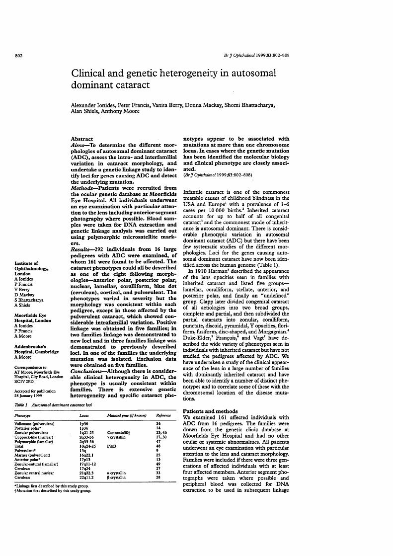
802 B rJ Ophthalmol 1999;83:802-808
Clinical and genetic heterogeneity in autosomal dominant cataract
Alexander lonides, Peter Francis, Vanita Berry, Donna Mackay, Shomi Bhattacharya, Alan Shiels, Anthony Moore
Institute ofOphthalmology,LondonA lonides P Francis V Berry D Mackay S Bhattacharya A Shiels
Moorfields Eye Hospital, LondonA lonides P Francis A Moore
Addenbrooke’s Hospital, Cambridge A Moore
C o r r e s p o n d e a c e to :A T M o o r e , M o o r f ie ld s E y e H o s p ita l , C ity R o a d , L o n d o n E C l V 2 F D .
A c c e p te d fo r p u b l ic a t io n 2 8 J a n u a r y 1 9 9 9
AbstractAims—To determine the different morphologies of autosomal dominant cataract (ADC), assess the intra- and interfamilial variation in cataract morphology, and imdertake a genetic linkage study to identify loci for genes causing ADC and detect the underlying mutation.Methods—Patients were recruited from the ocular genetic database at Moorfields Eye Hospital. All individuals underwent an eye examination with particular attention to the lens including anterior segment photography where possible. Blood samples were taken for DNA extraction and genetic linkage analysis was carried out using polymorphic microsatellite markers.Results—292 individuals from 16 large pedigrees with ADC were examined, of whom 161 were found to be affected. The cataract phenotypes could all be described as one of the eight following morphologies—anterior polar, posterior polar, nuclear, lamellar, coralliform, blue dot (cerulean), cortical, and pulverulent. The phenotypes varied in severity but the morphology was consistent within each pedigree, except in those affected by the pulverulent cataract, which showed considerable intrafamilial variation. Positive linkage was obtained in five families; in two families linkage was demonstrated to new loci and in three families linkage was demonstrated to previously described loci. In one of the families the underlying mutation was isolated. Exclusion data were obtained on five families. Conclusions—Although there is considerable clinical heterogeneity in ADC, the phenotype is usually consistent within families. There is extensive genetic heterogeneity and specific cataract phe
Tabk I Autosomal dominant cataraa loci
Phenotype Locus Mutated getu (if knoion) Reference
V o lk m a n n ( p u lv e r u le n t ) l p 3 6 2 4P o s te r io r p o la r * l p 3 6 1 4Z o n u la r p u lv e r u le n t l q 2 1 - 2 5 C o n n e x in S O f 2 3 , 4 6C o p p o c k - l ik e (n u c le a r ) 2 q 3 3 - 3 6 Y c r y s ta ll in 1 7 , 3 0P o ly m o r p h ic ( la m e lla r ) 2 q 3 3 - 3 6 4 7T o ta l 1 0 q 2 4 - 2 5 P it x 3 4 8P u lv e r u le n t* 1 3 q 9M a m e r ( p u lv e r u le n t ) 1 6 q 2 2 .1 2 5A n te r io r p o la r * 1 7 p l 3 1 3Z o n u la r - su tu r a l ( la m e lla r ) 1 7 q l l - 1 2 4 9C e r u le a n 1 7 q 2 4 2 7Z o n u la r c e n tr a l n u c le a r 2 1 q 2 2 .3 a c r y s ta ll in 3 3C e r u le a n 2 2 q l l . 2 P c r y s ta ll in 2 8
« L in k a g e f ir s t d e s c r ib e d b y t h is s t u d y g r o u p . f M u t a t i c n f ir s t d e s c r ib e d b y t h is s t u d y g r o u p .
notypes appear to be associated with mutations at more than one chromosome locus. In cases where the genetic mutation has been identified the molecular biology and clinical phenotype are closely associated.(B rJ Ophthalmol 1999;83:802-808)
Infantile cataract is one of the commonest treatable causes of childhood blindness in the USA and Europe' with a prevalence of 1-6 cases per 10 000 births.* Inherited cataract accounts for up to half of all congenital cataract’ and the commonest mode of inheritance is autosomal dominant. There is considerable phenotypic variation in autosomal dominant cataract (ADC) but there have been few systematic studies of the different morphologies. Loci for the genes causing autosomal dominant cataract have now been identified across the human genome (Table 1).
In 1910 Harman’ described the appearance of the lens opacities seen in families with inherited cataract and listed five groups— lamellar, coralliform, stellate, anterior, and posterior polar, and finally an “undefined” group. Clapp later divided congenital cataract of all aetiologies into two broad groups, complete and partial, and then subdivided die partial cataracts into zonular, coralliform, punctate, discoid, pyramidal, Y opacities, fiori- form, fusiform, disc-shaped, and Morgagnian.^ Duke-Elder,’ François,* and Vogt’ have described the wide variety of phenotypes seen in individuals with inherited cataract but have not studied the pedigrees affected by ADC. We have undertaken a study of the clinical appearance of the lens in a large number of families with dominantly inherited cataract and have been able to identify a number of distinct phenotypes and to correlate some of these with the chromosomal location of the disease mutations.
Patients and methodsWe examined 161 affected individuals with ADC from 16 pedigrees. The families were drawn from the genetic clinic database at Moorfields Eye Hospital and had no other ocular or systemic abnormalities. All patients underwent an eye examination with particular attention to the lens and cataract morphology. Families were included if there were three generations of affected individuals with at least four affected members. Anterior segment photographs were taken where possible and peripheral blood was collected for DNA extraction to be used in subsequent linkage

Clinical and genetic heterogeneity in autosomal dominant cataract 803
Table 2 Pedigree details
PhenotypeNo of family members
No ofaffectedpatients
No of pedigrees
A n te r io r p o la r 3 0 19 1P o s te r io r p o la r 3 6 2 1 2N u c le a r 4 0 2 3 2L a m e l la r 4 2 2 5 3C o r a l l if o r m 1 9 8 1B lu e d o t 1 3 7 1C o r t ic a l 2 0 6 1P u lv e r u le n t 9 2 5 2 5T o t a l 2 9 2 1 6 1 1 6
studies. Ethical approval for the study was given by the hospital ethics committee and informed, signed consent was obtained from the patients. In subjects where surgery had been performed early, a description of the phenotype was taken from the hospital notes.
Genomic DNA was extracted from EDTA sequestered blood samples and linkage analysis using Généthon microsatellite markers' was performed using a previously described protocol.’
ResultsEight phenotypes could be recognised that included all the pedigrees seen in the study. These phenotypes were anterior polar, posterior polar, nuclear, lamellar, coralliform, blue dot (cerulean), cortical, and pulverulent (Table 2). The morphology of the cataract in affected individuals within any single pedigree was the same for all except the pulverulent pedigrees, varying only in the severity and density of the opacity. In pedigrees with the pulverulent phenotype there was considerable variation in the cataract morphology among affected individuals from the same pedigree. Male to male transmission was seen in 12 families. In the remaining families, the severity of affected members was equal in both males and females suggesting autosomal dominant inheritance.
A N T E R I O R P O L A R C A T A R A C TAnterior polar cataracts arc situated at the anterior pole of the lens and have been reported to have a minimal effect on visual ac u ity ." We studied a single family with anterior polar cataract. Thirty family members (excluding partners) were examined and 19 found to be affected. Seven patients had undergone surgery and the phenotype was examined in the remaining 12 cases. ITie opacity was always in the same position of the lens, the anterior pole, but varied in severity." The larger opacities were pyramidal with a base of 4 mm and extended into the anterior chamber (Fig la), while the smaller opacities consisted of a flat plaque measuring under 1 mm in diameter. In one affected individual who showed the disease haplotype the only sign of an anterior polar abnormality was an irregularity of the anterior capsule. We have demonstrated linkage of anterior polar cataract to chromosome 17p in this family" showing significantly positive lod scores for markers D17S849 (Z ^= 4.01, 0 ^= 0.05) and D17S796 (Z ^= 4.17, 6 ^ = 0.05). Multipoint analysis gave a maximum lod score of 5.2 between these two markers.
P O S T E R I O R P O L A R C A T A R A C T In the pedigrees studied the posterior polar opacity was always at the posterior pole of the lens. Two types of posterior polar cataract were identified in this study—a stationary posterior polar cataract and a progressive posterior polar cataract. The stationary posterior polar cataract consisted of a single well defined posterior polar plaque (Fig lb), which varied in size among individuals as well as between the eyes of an individual. No other part of the lens was affected and although we have not studied the family over a prolonged period, the clinical records suggest that the cataract did not increase significantly in size. In a pedigree affected by this stationary posterior polar cataract 15 members of the family were examined and 10 found to be affected. Seven patients had undergone surgery and details of the cataract were obtained from the notes in four of these cases, all of whom were described as having posterior polar cataract.
We have demonstrated linkage of this phenotype to chromosome Ip" with significantly positive lod scores for markers D1S508 (Z^= 3.14, e„„= 0) and D1S468 (Z^= 2.71, 0n„= 0). Multipoint analysis gave a m axim um lod score of 3.48 (0^= 0.07) between markers D1S508 and DlS468.The second family with posterior polar cataract was affected by a progressive posterior polar cataract that initially consisted of a posterior plaque but later developed posterior cortical extensions (Fig Ic) with punctate cortical opacities causing a progressive deterioration of visual acuity requiring cataract extraction. Twenty one family members were exam ined, 11 were affected and all but one unilateral case had undergone surgery. Clinical records available in nine of the 10 operated cases documented the progressive nature of the cataract; the average age of diagnosis was 5 years but surgery was performed at a mean age of 20 years (range 3-40 years, median 19 years, mode 16 years). We have excluded linkage of this phenotype to the loci known to be associated with ADC.
N U C L E A R C A T A R A C TNuclear cataract consists of opacities confined to the embryonic and fetal nuclei of the lens. The cataract varies such that some patients have dense central opacities surroimded by fine dots (Fig Id), while others have fine dot opacities distributed throughout the embryonic and fetal nuclei (Fig le), with opacification of the anterior and posterior Y sutures. The opacities are confined to the fetal nucleus. Two pedigrees affected by nuclear cataract were examined. In the first family 21 individuals were examined and 12 found to be affected. Of these, six had undergone surgery and the clinical notes were available in two cases. Preliminary linkage results in this family have excluded the loci known to be associated with ADC.
In the second family affected by nuclear cataract 19 family members were examined and 11 found to be affected. Eight patients had undergone surgery and in seven of these the clinical notes gave an accurate description of

804 lonides, Francis, Berry, et al
Figure 1 Autosomal dominant congenital cataract phenotypes, (a) Slit lamp and retroillumination vietv of anterior polar cataract (47 year old tvoman). (b) Slit lamp and retroillumination vietu of a stationary posurior polar cataract (49 year old tooman). (c) Slit lamp view of a progressive posterior polar cataract (9 year old female) , (d) Slit lamp and retroillumination view of a dense nuclear cataract (14 year old male), (e) Slit lamp and retroillumination view of nuclear opacities (50 year old woman), (f) Slit lamp and retroillumination view of the Coppock-like cataract with fine nuclear opacities (24 year old tvoman). (g) Slit lamp arui retroillumination view of the Coppock-like cataract with dense central opacities (6 year old female). (h)Slit lamp view of lamellar cataract, (i) Slit lamp view of a blue dot (cerulean) cataract (32year old man), (j) Slit lamp and retroillumination view of a cortical cataract (45 year old tvoman). (k) Slit lamp •view of a fine pml'verulent cataract (32year old man). (I) Slit lamp view of large pulverulent opacities (12 year old female).

Clinked and genetk heterogeneity in autosomcd dominant cataract 805
the cataract morphology. The cataracts in three unoperated patients were examined. The mildly affected individuals had dust-like opacities throughout the embryonic and fetal nuclei (Fig If) while the more severely affected patients had a dense central opacity surrounded by a haze of fine powdery opacities (Fig Ig). We have demonstrated linage of this pedigree to the y crystallin locus on chromosome 2q (D2S157 Z„„= 5.28, G^= 0.00) but have discovered that this pedigree is related to the Coppock-like pedigree initially studied by Smith” and Harman” and previously linked to the y crystallin gene.” The Coppock-like cataract phenotype is well described in the literature but no photographs of the opacity have ever been published (Fig If and g).
L A M E L L A R C A T A R A C TLamellar cataract is also referred to by some authors as zonular cataract and is one of the commonest phenotypes of ADC. The iimer fetal nucleus is made up of clear lens surrounded by an opacified shell which is in turn surrounded by clear cortex (Fig Ih) which may contain opacities referred to as “riders” or “cortical spokes”. Lamellar cataract represents a disturbance in the lens development at a particular time and the cataractous “shell” varies in size according to the stage of fetal development at which the disturbance occurs. The cataract may be mild and the visual acuity may be well preserved or it may be dense enough to need surgery in childhood. Three families affected by lamellar cataract were studied. One of the families consisted of 14 members of whom eight were affected. All underwent surgery although one patient had unilateral cataract extraction. The surgery was carried out at a mean age of 7 months (range 2-36, mode 2, median 2). The individual who still had a cataract in one eye has a lamellar cataract with a visual acuity of 6/60 but has refused surgery. In the rest of the family details of the phenotype were restricted to clinical records that were available in four cases. In the other two families all the affected individuals had undergone surgery. In one family of 14 people there were nine affected individuals and the notes were available in two cases. In the third family there were also 14 members but with eight affected individuals and notes available in three cases. All were described as having lamellar cataract. These pedigrees have yet to be the subject of linkage studies.
5 years (range 1-12 years, median 5, mode 7 years). This pedigree has not yet been the subject of a linkage study.
B L L IE D O T ( c e r u l e a n ) C A T A R A C T The blue dot cataract first described by Vogt ® is not believed to be congenital but develops in childhood, progresses slowly, and does not require surgery until adulthood.^' “ The opacities are distributed throughout the lens, with the outer nucleus containing blue-white dots that become more numerous in the cortex where they may form large cuneiform (wedgelike) shapes in the mid-periphery (Fig li). In this pedigree seven of 13 patients were diagnosed as having the cataract and five of the affected seven were examined. The cataract was diagnosed at a mean age of 17 years and patients underwent surgery at a mean age of 27 years (range 10-47, mode 25, median 25). The phenotype was consistent within the family. This pedigree has been excluded from the two lod already known to be assodated with blue dot (cerulean) cataract. The blue dot locus on chromosome 17 was excluded with the markers D17S1790 (-2.46 at 0„„= 0.05), D17S802 (-3.40 at 0 ^ = 0.05), D17S1847 (-2.66 at 0 ^ = 0.05), D17S836 (-3.04 at 0„„= 0.05), and D17S784 (-1.27 at 0 ^= 0.05). The blue dot locus on chromosome 22 was excluded with the single marker D22S315 (-2.63 at 0 ^ = 0.05).
C O R T I C A L C A T A R A C TCortical cataract is rarely described as an autosomal dominant trait. In the family studied as part of this project the cataract presented in childhood or early adult life and varied in severity but not in morphology. The nucleus was unaffected and the opacified fibres were restricted to the outer cortex in contact with the lens capsule affecting a sector of lens (Fig Ij) and being different in this respect firom lamellar cataract. The pedigree we studied consisted of 20 family members of whom only six were affected with the cataract and four of these were available for examination. In three of the affected members the cataract affected only the superior cortex, the nucleus remained unaffected, and there was no opacity elsewhere in the lens. Nine eyes of the six affected patients had undergone surgery which was performed at a mean age of 23 (range 7-43). This pedigree has yet to be the subject of a linkage study.
C O R A L L I F O R M C A T A R A C TThe single pedigree of this phenotype from the Moorfields genetic data base is descended from a family originally described by Nettleship.'* The name of this phenotype is derived firom the resemblance of the cataract to sea coral,” having finger-like tubular protuberances extending from an opacified nucleus. Nineteen family members (excluding partners) were examined and eight found to be affected. All affected members of this pedigree had had surgery and the description of the cataract was taken from the clinical notes that were available in seven cases. The mean age at operation was
P U L V E R U L E N T C A T A R A C T Pulverulent cataracts are characterised by powdery (pulverised) opacities that may be found in any part of the lens. Pedigrees affected by pulverulent cataract showed a wide intrafamilial variation in the cataract phenotype and, although some affected individuals from these families had cataracts similar to lamellar or nuclear cataract, pedigrees were classified as pulverulent if the majority of affected individuals had fine, powdery opacities (Fig Ik). Five families had pulverulent cataract. In one of the families 23 members were examined and 16 found to be affected. The cataract varied

806 lonides, Francis, Berry, et al
between affeaed individuals. In some the fine opacities formed a lamellar distribution with a clear cortex and minimal inner fetal nuclear involvement. Others had more widely spread pulverised opacities extending into the cortex with no demarcation of the fetal nucleus. In some the cataract was mild and the fine dust-like opacities were clustered only around the anterior and posterior Y sutures. In this first pedigree nine individuals had undergone surgery at a mean age of 17 years (range 3-82, median 8, mode 11). This pedigree has been linked to chromosome 13 with a maximum two point lod score of 4.06 (8^= 0.00) with marker D13S175.’ In a second family with pulverulent cataract 15 members were examined and 10 were affected. Four had undergone bilateral cataract surgery, two unilateral, and four had not had surgery. The mean age at surgery was 14 years (range 5-22 years, median 17). The cataract had powdery opacities throughout the lens although they were more abundant in the fetal nucleus. In some the opacities were large and coarse (Fig 11) and in others finer and dust-like.
A third family affected by pulverulent cataract consisted of 15 family members of whom eight were affected by the cataract. Only two patients had undergone surgery and examination of the remaining six revealed fine dustlike opacities predominately in the fetal nucleus but also in the cortex, with moderate opacification of the Y sutures.
The fourth family affected by pulverulent cataract consisted of 21 family members with eight affected individuals. The phenotype in this family showed great intrafamilial variation, some had the typical fine dots throughout the lens, others had posterior opacities wiffi the rest of the lens unaffected, and one individual had dense bilateral nuclear opacification.
A fifth family, previously shown in 1963 to cosegregatc with the Duffy blood group locus on chromosome Iq was recontacted and 18 members of the pedigree (10 affected, six unaffected, and two spouses) examined. All affected individuals had previously undergone cataract surgery, their hospital records indicating that dense opacification was present at birth. linkage to Iq has been confirmed (maximum multipoint lod score 5.22 at 6 ^= 0) and a missense mutation identified in the gap junction a8 gene that encodes connexin 50.:"
DiscussionIn this study of ADC we have demonstrated that the phenotypes of the pedigrees encountered could be classified as one of the eight following types—anterior polar, posterior polar, nuclear, lamellar, coralliform, blue dot (cerulean), cortical, and pulverulent. This study was limited to large families that were suitable for linkage studies but this was thought not to affect the distribution of the affected phenotypes. Other phenotypes not encountered in this study have been reported but they have either been described in individual cases and not in families or are very uncommon. Although an attempt was made to examine the
phenotypes of all family members at risk of inheriting the affected gene, the description of intrafamilial variation in the phenotypes was limited by the number of patients who had already undergone surgery. This is a limitation inherent in all studies of early onset inherited cataract and we attempted to obtain as detailed a description as possible &om the hospital records in such cases.
In all the pedigrees apart firom the pulverulent pedigrees the morphology was the same in all affected family members but varied in severity. In the pulverulent pedigrees the phenotype varied among affected individuals from the same family. Intrafamilial clinical heterogeneity in ADC has been described by many authors:* including interocular variability in individuals affected by ADC.:* The pedigrees described as having this wide variation in morphology are characteristic of the pulverulent type described here. A total of 14 pedigrees have now been mapped to 13 different loci identified as containing genes that give rise to cataract (Table 1). In this study we have assigned a chromosomal locus in four pedigrees and 11 remain to be linked. An anterior polar pedigree maps to chromosome 17p," a posterior polar pedigree to Ip,'* a nuclear pedigree now known to be related to the Coppock-like pedigree maps to the y crystallin region on chromosome 2q, and a pulverulent pedigree maps to chromosome 13.’ We have excluded linkage of a pedigree affected by the blue dot cataract to the two loci known to give rise to this phenotype and have also excluded linkage of a posterior polar pedigree and a pulverulent pedigree to the known cataract loci. Further linkage studies are under way.
Clinically similar phenotypes of autosomal dominant cataract map to different regions of the genome and different phenotypes map to the same locus: two uiu-elated pedigrees affected by blue dot (cerulean) cataract have been mapped to different chromosomes:’ “ and two unrelated pedigrees affected by posterior polar cataract have also been mapped to different chromosomes.'* ” Two different phenotypes, a posterior polar'* and the Volkmann cataract,:* both map to what may prove to be the same locus on chromosome Ip. In contrast, in families where the underlying genetic mutation has been identified the resultant phenotype correlates with the underlying molecular biology of the lens: genes for human crystallin proteins have now been mapped and are important candidate genes for ADC formation. The Coppock-like cataract is a nuclear cataract caused by a mutation in the y crystallin gene complex on chromosome 2q” and the Y crystallins are predominantly found in the lens nucleus."' The blue dot (cerulean) cataract consists of multiple blue-white dots that predominate in the lens cortex and p crystallins are more abundant in the outer (cortical) layers of the lens. A pedigree affected by blue dot (cerulean) cataract has been demonstrated to have a mutation in the P crystallin region on chromosome 22q“ with the affected members having a chain termination mutation in the human P crystallin gene CRYBB2.": A mis-
Clinical and genetic heterogeneity in autosomal dominant cataract 807
sense mutation in the gene for connexin 50, a gap junction membrane protein, on chromosome Iq has been shown to give rise to pulverulent cataract in humans/^
A missense mutation in the human a crystallin gene CRYAA has been shown to give rise to a series of eye abnormalities including zonular central nuclear opacities, cortical and posterior subcapsular cataract, microcornea, and congenital nticrophthalmia.” The a crystallin protein is found in many tissues in the eye and throughout the body and a mutation in this protein may be expected to have a considerable effect on ocular development.
In mouse models for ADC, mutations in the crystallin and membrane bound proteins have also been shown to give rise to cataract, although the relation between genotype and phenotype is not as clear. The PhiUy mouse has a nuclear and subcapsular cataract^ caused by a mutation in one of the P crystallin genes” and the Eh mouse, which is a model for microphthalmia secondary to abnormal lens formation” is caused by a mutation in one of the y crystallin genes.” A mutation in the gene for the major intrinsic protein (MIP) of the lens fibre cell membranes has been implicated as causing tiie cataracts in both the Cat[Fr] mouse and the Lop mouse.” ” The human gene for MIP has been mapped to chromosome 12q“ but has not yet been implicated in human cataract.
Although in humans the major crystallins are the a, P, and y crystallins, contributing 90% of the water soluble proteins of the lens, there are many other species specific crystallins that may be relevant to human cataract formation. These crystallins are enzymes that in some cases have been shown to have a different function outside the lens but have been recruited during evolution to perform as lens crystallins owing to their stable structures and/or in some cases their original enzyme activity.” ^ The Volkmann and the posterior polar cataract both map to a region on chromosome Ip within which Ues the x crystallin/a enolase gene. This crystallin is not known to be expressed in the human lens. Indeed, no mutation has been found in the gene in these families and the single family described with red cell enolase deficiency does not have cataract. However, it is tempting to speculate that an abnormality in the gene maybe associated with cataract in humans.”
This study of lens morphology in ADC has demonstrated wide clinical heterogeneity. There is clearly considerable genetic heterogeneity and the identification of the causative mutations will allow for a more detailed comparison between the precise mutations and the clinical phenotype as well as offering further insight into the mechanism of human cataractogenesis.
W e th a n k t h e fa m il ie s fo r a g r e e in g t o p a r t ic ip a te in th is s t u d y a n d t h e U K H G M P R e s o u r c e C e n tr e f o r m ic r o s a te l l i te p r im e r s y n t h e s is a n d t h e u s e o f c o m p u t in g fa c il i t ie s . T h e w o r k w a s s u p p o r te d b y a g r a n ts fr o m t h e W e l lc o m e T r u s t ( 0 4 3 0 7 3 t o A S a n d S S B ; 0 5 3 4 1 6 t o A T M a n d S S B ) . A C W I w a s s u p p o r te d b y a g r a n t f r o m T h e F r ie n d s o f M o o r f ie ld s E y e H o s p ita l a n d D S M is a W e l lc o m e P r iz e s tu d e n t .
1 E v a n s J , R o o n e y C , A s h w o o d V,etaL B l in d n e s s a n d p a r tia l s ig h t in E n g la n d a n d W a le s: A p r il 1 9 9 0 - M a r c h 1 9 9 1 . Health Trends 1 9 9 6 ; 2 8 : 5 - 1 2 .
2 L a m b e r t S , D r a c k A . I n fa n t i le C a ta r a c ts . Surv Ophthalmol1 9 9 6 ; 4 0 : 4 2 7 - 5 8 .
3 H a r m a n N . T r e a s u r y o f h u m a n in h e r i t a n c e . P a r t 4 . S e c t io n X U I a . C o n g e n i t a l c a ta r a c t . Eugenia Library Memoirs X I 1 9 1 0 .
4 C la p p C . Cataracv its aetiology and treatment. L o n d o n : H e n r y K im p to n , 1 9 3 4 .
5 D u k e - E ld e r S . T h e l e n s . In : D u k e - E ld e r S , e d . A system oj ophthalmology. L o n d o n : H e n r y K im p to n , 1 9 6 4 : 7 1 5 - 5 9 .
6 F r a n ç o is J. Congenital cataracts. S p r in g f ie ld , IL : C h a r le s C T h o m a s , 1 9 6 3 .
7 V o g t A . Lem and zonule. B o n n : J P W e y e n b o r g h , 1 9 7 9 .8 G y a p a y G , M o r i s e t t e J , V ig n a l A , et aL T h e 1 9 9 3 - 1 9 9 4
G e n e t h o n h u m a n l in k a g e m a p . M o t Genet 1 9 9 4 ; 7 : 2 4 6 - 3 3 9 .9 M a c k a y D , l o n i d e s A , B e r r y W, et al A u to s o m a l d o m in a n t
c o n g e n it a l c a ta r a c t l in k e d t o c h r o m o s o m e 1 3 . Am J Hum G e n e t 1 9 9 7 ; 6 0 : 1 4 7 4 - 8 .
1 0 J a a fa r M , R o b b R . C o n g e n it a l a n te r io r p o la r c a ta r a c t: ar e v ie w o f 6 3 c a s e s . Ophthalmology 1 9 8 4 ; 9 1 : 2 4 9 - 5 2 .
11 B o u z a s A . A n te r io r p o la r c o n g e n it a l c a ta r a c t a n d c o r n e a l a s t ig m a t is m . J POediatr Ophdiahnol Strabismus 1 9 9 2 ;2 9 : 210- 12.
1 2 l o n i d e s A , B e r r y V , M a c k a y D , et aL A n te r io r p o la r ca ta r a c t:c l in ic a l s p e c t r u m a n d g e n e t i c l in k a g e in a s in g le fa m ily . Eye 1 9 9 8 ; 1 2 : 2 2 4 - 6 .
1 3 B e r r y V , l o n i d e s A , M o o r e A , et al. A lo c u s f o r a u to s o m a l d o m in a n t a n te r io r p o la r c a ta r a c t o n c h r o m o s o m e 1 7 p . Mol G e n e t 1 9 9 6 ; 5 : 4 1 5 - 1 9 .
1 4 lo n i d e s A , B e r r y V , M a c k a y D, et al A lo c u s fo r a u to s o m a ld o m in a n t p o s t e r io r p o la r c a ta r a c t o n c h r o m o s o m e I p . Hum Mol G e n e t 1 9 9 7 ; 6 : 4 7 - 5 1 .
1 5 S m it h P . A p e d ig r e e o f D o y n e ’s d is c o id c a ta r a c t . Trans Ophthalmol Soc UK 1 9 1 0 ; 3 0 : 3 7 - 4 2 .
1 6 H a r m a n N . T e n p e d ig r e e s o f c o n g e n it a l a n d in fa n t i lec a ta r a c t; la m e lla r , c o r a l l if o r m , d is c o id a n d p o s t e r io r p o la r w ith m ic r o p h th a lm ia . Tram Ophthalmol Soc UK 1 9 1 0 ;3 0 : 2 5 1 - 7 4 .
1 7 L u b s e n N , R e n w ic k J , T s u i L - C , et al A lo c u s fo r h u m a n h e r e d ita r y c a ta r a c t i s c lo s e ly l in k e d t o t h e g a m m a -c r y s ta l l in g e n e fa m ily . Proc Nad Acad Sci USA 1 9 8 7 ; 8 4 : 4 8 9 - 9 2 .
1 8 N e t t l e s h ip E . O n h e r e d ity in t h e f o r m s o f c a ta r a c t . The RoyalLand Ophth Hosp Rep 1 9 0 6 ; 1 7 : 2 1 8 - 2 2 .
1 9 G u im R M . P e c u lia r c o r a l l if o r m c a ta r a c t w ith c r y s ta ls in t h ele n s . T m nr Ophthalmol Soc UK 1 8 9 5 ;X V : 1 1 9 .
2 0 V o g t A . D i e s p e z i f i ta t a u d e r b o r e n e r u n d e r w o r b e n e rs ta r fo r m e r fu r d ie e i n z e ln e n l in s e z o u e n e . Albrecht Km Graefa Arch Clin Exp Ophth 1 9 2 2 ; 1 0 8 : 2 1 9 - 2 8 .
2 1 K iv l in J , L o v r ie n E , G e o r g e C, et a l L in k a g e b e tw e e nc e r u le a n c a ta r a c t a n d P G P . Cytogerut Cell Genet 1 9 8 5 ;4 0 : 6 6 9 .
2 2 B o d k e r F , L a v e r y M , M itc h e l l T, et al M ic r o p h t h a lm o s int h e p r e s u m e d h o m o z y g o u s o f f s p r in g o f a f ir s t c o u s in m a r r ia g e a n d l in k a g e a n a ly s is o f a l o c u s in a f iu n ily w i th a u to s o m a l d o m in a n t c e r u le a n c o n g e n it a l c a ta r a c ts . Am J Med Genet 1 9 9 0 ; 3 7 : 5 4 - 9 .
2 3 S h ie l s A , M a c k a y D , l o n i d e s A ,eta l.A m is s e n s e m u t a t io n int h e h u m a n c o n n e x i n 5 0 g e n e (G J A 8 ) t m d e r lie s a u to s o m a l d o m in a n t " z o n td a r p u lv e r u le n t ” c a ta r a c t , o n c h r o m o s o m e Iq .A m J Hum Genet 1 9 9 8 ; 6 2 : 5 2 6 - 3 2 .
2 4 E ib e r g H , L u n d A , W a r b u r g M., et a l A s s ig n m e n t o fc o n g e n it a l c a ta r a c t V o lk m a im - ty p e ( C C V ) t o c h r o m o s o m e l p 3 6 . Hum Genet 1 9 9 5 ; 9 6 : 3 3 - 8 .
2 5 E ib e r g E , M a m e r E , R o s e n b e r g T ,e ta l M a m e r ’s c a ta r a c t( C A M ) a s s ig n e d t o c h r o m o s o m e 1 6 : l in k a g e t o h a p t o g lo b in . Clin Genet 1 9 8 8 ; 3 4 : 2 7 2 - 5 .
2 6 S c o t t M , H e j t m a n c ik J , W o z e n c r a ft "L, et a l A u to s o m a ld o m in a n t c o n g e n i t a l c a ta r a c t . I n te r o c u la r p h e n o t y p ic v a r ia b ility . Ophthalmology 1 9 9 4 ; 1 0 1 : 8 6 6 - 7 1 .
2 7 A n n i t a g e M , K iv l in J , F e r r e ) R . A p r o g r e s s iv e e a r ly o n s e tc a ta r a c t g e n e m a p s t o h u m a n c h r o m o s o m e 1 7 q 2 4 . Nat Genet 1 9 9 5 ; 9 : 3 7 - 4 0 .
2 8 K r a m e r P , Y o u n t J , M ic h e l l T , et a l A s e c o n d g e n e fo rc e r u le a n c a ta r a c t m a p s t o t h e b e ta - c r y s t a l l in r e g io n o n c h r o m o s o m e 2 2 . Genomics 1 9 9 6 ; 3 5 : 5 3 9 ^ 2 .
2 9 R ic h a r d s J , M a u m e n e e I , R o w e S , et al C o n g e n it a l c a ta r a c tp o s s ib ly l in k e d t o h a p t o g lo b in . Cytogenet Cell Genet 1 9 9 4 ; 3 7 : 5 7 0 .
3 0 B r a c k e n h o f f R , H e s k e n s H , R o s s u m M V , et a l A c t iv a t io n o ft h e g a m m a -E -c r y s ta ll in p s e u d o g e n e in th e h u m a n h e r d ita r y C o p p o c k - lik e c a ta r a c t . Hum Mol Genet 1 9 9 4 0 : 2 7 9 - 8 3 .
3 1 M c A v o y J. D e v e lo p m e n t a l b io lo g y o f d ie l e n s . In : D u n c a nG , e d . Mechanisrm of cataract formation in the human lem. L o n d o n : A c a d e m ic P r e s s , 1 9 8 1 : 7 - 4 6 .
3 2 L it t M , C a r r e r o -V a le n z u e la R , L a M o r t ic e l la D , et a l A u t o s o m a l d o m in a n t c o n g e n i t a l c a ta r a c t is a s s o c ia t e d w i t h a c l u i n t e r m in a d o n m u t a t io n in t h e h u m a n b e ta -c r y s ta l lin g e n e C R Y B B 2 . Hum Mol Genet 1 9 9 7 ; 6 : 6 6 5 - 8 .
3 3 L h t M , K r a m e r P , L a M o r t ic e l la D, et a l A u to s o m a ld o m in a n t c o n g e n i t a l c a ta r a c t a s s o c ia t e d w i t h a m is s e n s e m u t a t io n in t h e h u m a n a lp h a c r y s ta ll in g e n e C R Y A A . Hitm Mol Genet 1998;7:471-4.
3 4 K a d o r P , F u k u i H , F u k u s h i S ,e ta l A n e w m o d e l o f h e r e d ita r y c a ta r a c t . Exp Eye Res 1 9 8 0 ;3 0 : 5 9 .
3 5 C h a m b e r s C , R u s s e ll P . D e l e t io n m u t a t io n in a n e y e le n sb e ta - c r y s t a l l in . J B W CA em 1 9 9 1 ; 2 6 6 : 6 7 4 2 - 6 .
3 6 O d a S - I , W a ta n a b e K , F u j isa w a H, et al Im p a ir e d d e v e lo p m e n t o f le n s f ib r e s in g e n e t ic m ic r o p h th a lm ia , e y e le n s o b s o le s c e n c e , e lo , o f th e m o u s e . Exp Eye Res 1 9 8 0 0 1 : 6 7 3 - 8 1 .
3 7 C a r t ie r M , B r e i t m a n M , T s u i L . A fr a m e sh if t m u t a t io n int h e g a n u n a E - c r y s t a ll in g e n e o f th e E L O m o u s e . Nat Genet 1 9 9 2 ; 2 : 4 2 - 5 .

808 lonides, Francis, Berry, et al
3 8 M u g g le t o n -H a r r is A , F e s t in g M , H a ll M . A g e n e lo c a t io n 4 4 W is t o w G , P ia t ig o r s k y J. R e c r u itm e n t o f e n z y m e s a s le n sf o r t h e in h e r i t a n c e o f t h e C a ta r a c t F r a se r ( C a tF r ) m o u s e s t r u c tu r a l p r o te in s . Science 1 9 8 7 ; 2 3 6 : 1 5 5 4 - 5 .c o n g e n it a l c a ta r a c t . Gen Res Comb 1 9 8 7 ; 4 9 : 2 3 5 - 9 . 4 5 W is t o w G , L e i t m a n T , W ill ia m s h ,e t aL t -C r y s ta l l in /a lp h a
3 9 S h ie ls A , B a s s n e t t S . M u ta t io n s in th e f o u n d e r o f t h e M I P e n o la s e : o n e g e n e e n c o d e s b o th a n e n z y m e a n d a le n sg e n e fa m ily t m d e r lie c a ta r a c t d e v e lo p m e n t in t h e m o u s e . s t r u c tu r a l protein. J Cell Biol 1 9 8 8 ; 1 0 7 : 2 7 2 9 - 3 6 .Nat Genet 1 9 9 6 ; 1 2 : 2 1 2 - 1 5 . 4 6 R e n w ic k J , L a w le r S . P r o b a b le l in k a g e b e tw e e n a c o n g e n it a l
4 0 N o o r t Jv, S e c h e l A v , B a jr a m o v ic J , et aL T h e sm a l l h e a t c a ta r a c t lo c u s a n d t h e D u A y b l o o d g r o u p l o c u s . Ann Hums h o c k p r o te in a lp h a B - c r y s t a l lin a s c a n d id a te a u to a n t ig e n in Genet 1 9 6 3 ; 2 7 : 6 7 - 8 4 .m u lt ip le s c le r o s is . Nature 1 9 9 5 ; 3 7 5 : 7 9 8 - 8 0 1 . 4 7 R o g a e v E , R o g a e v a E , K o r o v a it s e v a G ,F et aL L in k a g e o f
4 1 S p a r k e s R , M o h a n d a s T , H e in z m a n C . T h e g e n e fo r t h e p o ly m o r p h ic c o n g e n it a l c a ta r a c t t o t h e g a m m a -c r y s ta l l inm a jo r in tr in s ic p r o te in ( M I P ) o f t h e o c u la r le n s is a s s ig n e d lo c u s o n h tu n a n c h r o m o s o m e 2 q 3 3 - 3 5 . Hum Mol Genett o h u m a n c h r o m o s o m e 1 2 c e n - q l 4 . Invest Ophthalmol Vis 1 9 9 6 ; 5 : 6 9 9 - 7 0 3 .S c i 1 9 8 6 ; 2 7 : 1 3 5 1 - 4 . 4 8 S e m in a E , F e r r e ll R , M in tz - H it tn e r H , c t o f A n o v e l h o m e o -
4 2 C a r t ie r M , T s u i L , B a ll S,etaL Crystallin genes and cataraa. b o x g e n e P I T X 3 is m u t a te d in fa in i l ie s w i t h a u to s o m a lS w itz e r la n d : M o d e m G e n e t ic s S e r ie s H a r w o o d A c a d e m ic d o m in a n t c a ta r a c ts a n d A S M O . Nat Genet 1 9 9 8 ; 1 9 : 1 6 7 -P r e s s , 1 9 9 4 . 7 0 .
4 3 P ia t ig o r s k y J. M u lt i iu n c t io n a l le n s c r y s ta ll in s a n d c o m e a l 4 9 P a d m a T , A y y a g a r i R , M u r ty J , a al. A u to s o m a l d o m in a n te n z y m e s . M o r e t h a n m e e t s t h e e y e . Arm N Y Acad Sci 1 9 9 8 ; z o n u la r c a ta r a c t w i th su tu r a l o p a c it ie s l o c a l is e d t o c h r o m o -8 4 2 : 7 - 1 5 . s o m e 1 7 q l l - 1 2 . / l m J ' / f u m Getut 1 9 9 5 ; 5 7 : 8 5 0 - 5 .