Chitosan-alginate composites blended with cloisite 30B as a novel drug delivery system for...
14
RESEARCH ARTICLE Chitosan-alginate composites blended with cloisite 30B as a novel drug delivery system for anticancer drug paclitaxel P. L. Nayak & Debasish Sahoo Received: 21 March 2010 /Accepted: 18 January 2011 / Published online: 24 February 2011 # Central Institute of Plastics Engineering & Technology 2011 Abstract In the present research program, polymer composites have been used as the drug carrier for delivery system of anticancer drug Paclitaxel. Chitosan (CS) and sodium alginate (ALG) with different ratios were blended with different wt% of montmorillonite (MMT) (Cloisite 30B) solution by solvent evaporation method. MMT was incorporated in the formulation as a matrix material component which also plays the role of a co-emulsifier in the nanocomposite preparation. Paclitaxel (PTX) with different concentrations were loaded with CS-ALG/MMT nanocompo- sites for studying the in-vitro drug delivery systems. Morphology and structure characterization of nanocomposites were investigated by Fourier Transmission Infra Red Spectroscopy(FTIR),Scanning Electron Microscope (SEM), X-Ray Diffraction (XRD) respectively. The drug release was studied by changing time, pH and drug concentrations. The kinetics of the drug release was studied in order to ascertain the type of release mechanism. Based on the diffusion as well as the kinetics, the mechanism of the drug release from the composite matrix has been reported. Keywords Chitosan . Alginate . Paclitaxel Introduction In recent years, biodegradable polymers have attracted much attention to be used as biomaterials particularly, for tissue engineering, gene therapy, wound healing and controlled drug delivery systems [1]. The most important advantage of biodegradable polymers is the disappearance of implanted foreign materials from the body as a result of their biodegradation. Most important biodegradable polymers used in biomedical applications are poly(lactic acid)(PLA), poly(glycolic acid)(PGA), poly(e-aprolactone) Int J Plast Technol (June 2011) 15(1):68–81 DOI 10.1007/s12588-011-9000-6 P. L. Nayak (*) : D. Sahoo P.L.Nayak Research Foundation, Cuttack, Bhubaneswar, India e-mail: [email protected]
-
Upload
independent -
Category
Documents
-
view
3 -
download
0
Transcript of Chitosan-alginate composites blended with cloisite 30B as a novel drug delivery system for...
RESEARCH ARTICLE
Chitosan-alginate composites blended with cloisite 30Bas a novel drug delivery system for anticancerdrug paclitaxel
P. L. Nayak & Debasish Sahoo
Received: 21 March 2010 /Accepted: 18 January 2011 /Published online: 24 February 2011# Central Institute of Plastics Engineering & Technology 2011
Abstract In the present research program, polymer composites have been used asthe drug carrier for delivery system of anticancer drug Paclitaxel. Chitosan (CS) andsodium alginate (ALG) with different ratios were blended with different wt% ofmontmorillonite (MMT) (Cloisite 30B) solution by solvent evaporation method.MMT was incorporated in the formulation as a matrix material component whichalso plays the role of a co-emulsifier in the nanocomposite preparation. Paclitaxel(PTX) with different concentrations were loaded with CS-ALG/MMT nanocompo-sites for studying the in-vitro drug delivery systems. Morphology and structurecharacterization of nanocomposites were investigated by Fourier Transmission InfraRed Spectroscopy(FTIR),Scanning Electron Microscope (SEM), X-Ray Diffraction(XRD) respectively. The drug release was studied by changing time, pH and drugconcentrations. The kinetics of the drug release was studied in order to ascertain thetype of release mechanism. Based on the diffusion as well as the kinetics, themechanism of the drug release from the composite matrix has been reported.
Keywords Chitosan . Alginate . Paclitaxel
Introduction
In recent years, biodegradable polymers have attracted much attention to be used asbiomaterials particularly, for tissue engineering, gene therapy, wound healing andcontrolled drug delivery systems [1]. The most important advantage of biodegradablepolymers is the disappearance of implanted foreign materials from the body as a resultof their biodegradation. Most important biodegradable polymers used in biomedicalapplications are poly(lactic acid)(PLA), poly(glycolic acid)(PGA), poly(e-aprolactone)
Int J Plast Technol (June 2011) 15(1):68–81DOI 10.1007/s12588-011-9000-6
P. L. Nayak (*) : D. SahooP.L.Nayak Research Foundation, Cuttack, Bhubaneswar, Indiae-mail: [email protected]

(PCL), poly (3-hydroxybutyrate) (PHB), copolymers of polyglycolide, chitosan,alginate and soy protein [2–5].
Chitosan( CS) is a biodegradable polymer obtained by the deacetylation of chitin,which is present in shells of insects and marine crustacean [6, 7]. This is a marine–based polymer.The unique properties of chitosan, among others, are biodegradability,bioactivity, nontoxicity as well as good adhesion and sorption, which largely contributeto its multiple applications [8]. Chitosan is also a valuable component of polymerblends and composites [9, 10]. Using an appropriate technological process one mayobtain films, fibers, gels and foams as well as chitosan beads of different sizes andmorphology. Numerous in vitro studies have analyzed the response to chitosan bysmooth muscle cells, macrophages, osteoblasts, chondrocytes, erythrocytes andwhole blood. In addition, many studies have been conducted with mouse, rat, rabbit,and canine animal models in order to describe in vivo biocompatibility,biodegradability, drug delivery, DNA delivery, and wound healing using chitosanas a carrier [11, 12].
Alginate (ALG) is a water soluble linear polysaccharide extracted from brown seaweed and is composed of alternating blocks of 1–4 linked α-L-guluronic and β-dmannuronic acid residues. ALG has been reported to be mucoadhesive,biodegradable, and biocompatible and has potential for numerous pharmaceuticaland biomedical applications such as drug delivery system and cell encapsulation[13]. Alginate micro and nanoparticles can be obtained easily by inducing gelationwith calcium ions [14]. Such easy-gelling property can be used to produce a pre-gelconsisting of very small aggregates of gel particles, followed by the addition of anaqueous polycationic solution to make a polyelectrolyte complex coating [15].Chitosan-alginate blended with cloisite 30 B is a novel composite to be used as acarrier for controlled drug delivery system.The addition of CS to ALG not onlyendow nanoparticles positive surface charge, but also prolong the time that the activeingredients contact with the epithelium and enhance absorption via the para-cellulartransport pathway through the tight junctions [16, 17]. MMTwas incorporated in theformulation as a matrix material component which also plays the role of a co-emulsifierin the nanocomposite preparation.
Paclitaxel a poly-oxygenated naturally occurring diterpenoid isolated from thebark of the Pacific yew tree, Taxus brevifolia, is a microtubule-stabilizing agent [16].It binds to the β-subunit of the tubulin heterodimer ultimately resulting in the arrestof the cell division cycle between the prophase and anaphase stages, eventuallyleading to apoptosis of the cancer cells [17]. It has been used against a widespectrum of cancers, including breast cancer, ovarian cancer, lung cancer, head andneck carcinomas, and acute leukemia. However, the success of its clinicalapplication is limited by its low therapeutic index and low solubility in mostpharmaceutical solvents. More effective chemotherapy using paciltaxel is relying ondevelopment of its new dosage forms, among which nanoparticles of biodegradablepolymers and lipid belayed vesicles (liposome) seem the most prospective.Currently, the only available dosage form of paciltaxel is Taxol® for intravenous(ivy.) infusion, which is a solution of paciltaxel in an adjuvant called Cremophor EL,which causes serious side effects such as hypersensitivity reactions, nephrotoxicity,neurotoxicity and cardiotoxicity [18, 19]. In recent years, various controlled deliveryforms, such as polymeric micro/nanospheres, liposome’s, micelles, potential
Int J Plast Technol (June 2011) 15(1):68–81 6969

emulsion, and prod rugs have been investigated to increase its solubility, to minimizethe side effects as well as to avoid the use of toxic adjuvant. [20–24].
One of the most attractive areas of research in drug delivery today is the design ofnanosystems that are able to deliver drugs to the right place, at appropriate times andat the right dosage. Nanoparticulate delivery systems have the potential power toimprove drug stability, increase the duration of the therapeutic effect and permitadministration through enteral or parenteral administration, which may prevent orminimize the drug degradation and metabolism as well as cellular efflux [25–30].
In the present research program, we wish to report the preparation of a novelnanocomposites formulation, i.e. biodegradable chitosan-alginate (CS-ALG) nano-composites incorporated with medical clay, montmorillonite (MMT) called CSALG/MMT nanocomposites, for in vitro drug delivery by using paciltaxel as a prototypedrug due to its excellent therapeutic effects against a wide spectrum of cancers andits great commercial success as the best seller among various anticancer agents. Thecomposites have been characterized using FTIR, SEM and XRD techniques. Thekinetics of the drug delivery system has been reported.
Experimental
Materials
Chitosan (CS) (Degree of Deacetylation=95%) was purchased from India Sea Foods,Kerela, India. Sodium alginate of low viscosity (0.02 Pa·s) for a 1% solution at 20°Cwaspurchased from Shanghai Chemical Co. Ltd (China). Paclitaxel was a generous gift fromVINS Bioproducts, Medak, Andhra Pradesh. Dichloromethane (DCM, Product NumberDS1432, HPLC/Spectro Grade) was purchased from Tedia (Tritech Scientific Pt. Ltd,Singapore), acetic acid, NaH2PO4, NaOH, and other chemicals were used as analyticalgrade and purchased from Sigma Aldrich Company.
Preparation of chitosan-alginate nanocomposites
Both the sodium alginate and chitosan solutions were prepared by dissolving thechemicals in distilled water and 1% acetic acid respectively. Blend solution ofdifferent compositions (i.e. the weight ratios between chitosan and alginate of 100/0,80/20, 70/30, 60/40, 0/100(w/w) respectively) were then prepared by casting amixture of the solutions in a respective weight ratio on a Teflon dish. To this blendsolution of CS-ALG , MMT (Cloisite 30 B) of different compositions (2 wt% and5 wt %) were added with constant stirring for 4 hours at room temperature to get ahomogenous solution. It should be noted that stirring was used to homogenize themixture prior to pouring onto the dish. The casting was let dry at room temperaturefor 3 days and was collected for characterization.
Drug loading
Paclitaxel-loaded CS-ALG/MMT( 80/20/ 2 wt % ) nanocomposites were preparedby emulsion/solvent evaporation method. In short, paciltaxel of different loadings,
70 Int J Plast Technol (June 2011) 15(1):68–81

i.e., 5 wt%, 10 wt%, 15 wt%, 20 wt% and 25 wt% were dissolved in ethanol withCS-ALG/MMT( 80–20/2 wt%). The formed solution was poured into a labeled Petridish and allowed to evaporate the solvent overnight at room temperature. Thiscompound was used for drug delivery purposes.
Dissolution experiments
Dissolution experiments were performed at 37°C using the dissolution tester (Dissotest, Lab India, Mumbai, India) equipped with six paddles at a paddle speed of100 rpm. About 900 ml of phosphate buffer solution (pH 1.2 and 7.4) was used asthe dissolution media to stimulate gastrointestinal tract (GIT) conditions. A 5 mlaliquot was used each time for analyzing the Paclitaxel content at a fixed timeinterval. The dissolution media was replenished with a fresh stock solution. Theamount of Paclitaxel released was analyzed using a UV spectrophotometer(Systronics, India) at the λ max value of 230 nm.
Characterization
Fourier Transmission Infra Red spectroscopy (FTIR)
The FTIR spectrum of the chitosan, alginate, and chitosan-alginate blend wasobtained using a BIORAD-FTS-7PC type FTIR spectrophotometer.
X-Ray Diffraction (XRD)
The change in gallery height of the blend was investigated by WAXD experiments,which were carried out using an X-ray diffractometer (BEDE D-3 system) with CuKα radiation at a generator voltage of 40 kV and a generator current of 100 mA.Samples were scanned from 2θ=1–100° at a scanning rate of 2°/min.
Scanning Electron Microscopy (SEM)
The blending of the Chitosan-Alginate composites containing different concen-trations were characterized using SEM (440, Leica Cambridge Ltd., Cambridge,UK). The powdered specimens were placed on the Cambridge standard aluminiumspecimen mounts (pin type) with double-sided adhesive electrically conductivecarbon tape (SPI Supplies, West Chester, PA). The specimen mounts were thencoated with 60% Gold and 40% Palladium for 30 s with 45 mA current in a sputtercoater (Desk II, Denton Vacuum, Moorestown, NJ). The coated specimens were thenobserved on the SEM using an accelerating voltage of 20 kV at a tilt angle of 30° toobserve the microstructure of the chitosan-alginate composite blends.
Swelling studies
Water absorption of the polymer-drug conjugates was measured following ASTM D570–81. The samples were preconditioned at 50°C for 24 h and then cooled in a
Int J Plast Technol (June 2011) 15(1):68–81 7171

desiccator before being weighed. The preconditioned samples were submerged indistilled water at 25°C for 24 h.The samples were removed and dried with a papertowel before weighing. Water absorption was calculated as a percentage of initialweight. The soluble material loss was checked by weighting the specimens afterdrying them in an oven at 50°C for another 24 h. The total water absorption for 24 hwas calculated including the soluble material loss
%Swelling ¼ W1 �W2
W2� 100
Where, W1=Weight of Swollen composite after 24 hr., W2=Weight of DryComposite.
Results and discussion
Fourier Transmission Infra Red spectroscopy (FTIR)
FTIR was adopted to characterize the potential interactions in the nanocomposites.FTIR spectra of alginate, chitosan, CS/alginate nanocomposites are shown in Fig. 1.In the spectra of CS, the broad band at 3424 cm−1 corresponded to the amine andhydroxyl groups; the peak at 2876 cm−1 was due to −OH stretching; the absorptionband of the carbonyl (C=O) stretching of the secondary amide (amide I band) at1655 cm−1, and the bending vibrations of the N–H (N acetylated residues, amide IIband) at 1599 cm−1 [31]. The peaks at 1423 and 1381 cm−1 belong to the N–Hstretching of the amide and ether bonds and N–H stretching (amide III band),respectively. The peaks observed at 1081 and 1033 cm−1 were the secondaryhydroxyl group (characteristic peak of −CH-OH in cyclic alcohols, C-O stretch) andthe primary hydroxyl group (characteristic peak of −CH2-OH in primary alcohols,C-O stretch) [32]. The bands around 1030 cm−1 (C-O-C stretching) presenting in theIR spectrum of sodium alginate are attributed to its saccharide structure. In addition,
Fig. 1 FTIR-Spectra of pure alginate (i.e. the topmost curve), pure chitosan (i.e. the middle curve) and(i.e. the bottommost curve) chitosan-alginate/MMT (80/20/2 wt%)
72 Int J Plast Technol (June 2011) 15(1):68–81
the bands at 1617 and 1417 cm−1 are assigned to asymmetric and symmetricstretching peaks of carboxylate salt groups [33]. So in the IR spectrum of CS/ALGnanopcomposites, we can observe the asymmetrical stretching of -COO- groupsshifted to 1637 cm−1 and the symmetrical stretching of - COO- groups shifted to1415 cm−1 [34]. In addition, the absorption band at 1599 cm−1 of chitosan shifts to1559 cm−1 after the reaction with alginate, the stretching vibration of −OH and−NH2 at 3424 cm−1 shifts to 3448 cm−1 and becomes broad. Hence the aminogroups of chitosan may interact with the carboxyl groups of the alginate. Theseresults indicate that the carboxylic groups of ALG associate with ammonium groupsof CS through electrostatic interactions to form the polyelectrolyte complex.
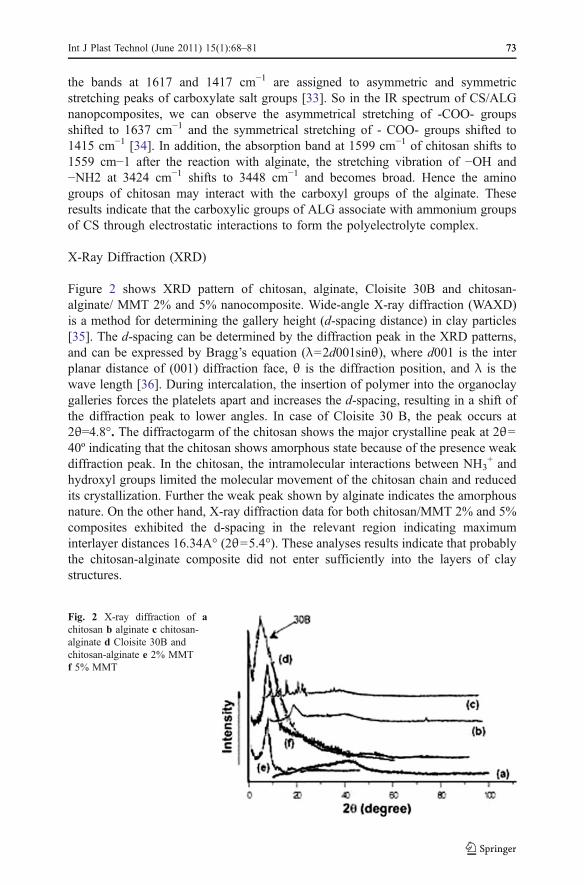
X-Ray Diffraction (XRD)
Figure 2 shows XRD pattern of chitosan, alginate, Cloisite 30B and chitosan-alginate/ MMT 2% and 5% nanocomposite. Wide-angle X-ray diffraction (WAXD)is a method for determining the gallery height (d-spacing distance) in clay particles[35]. The d-spacing can be determined by the diffraction peak in the XRD patterns,and can be expressed by Bragg’s equation (λ=2d001sinθ), where d001 is the interplanar distance of (001) diffraction face, θ is the diffraction position, and λ is thewave length [36]. During intercalation, the insertion of polymer into the organoclaygalleries forces the platelets apart and increases the d-spacing, resulting in a shift ofthe diffraction peak to lower angles. In case of Cloisite 30 B, the peak occurs at2θ=4.8°. The diffractogarm of the chitosan shows the major crystalline peak at 2θ=40º indicating that the chitosan shows amorphous state because of the presence weakdiffraction peak. In the chitosan, the intramolecular interactions between NH3
+ andhydroxyl groups limited the molecular movement of the chitosan chain and reducedits crystallization. Further the weak peak shown by alginate indicates the amorphousnature. On the other hand, X-ray diffraction data for both chitosan/MMT 2% and 5%composites exhibited the d-spacing in the relevant region indicating maximuminterlayer distances 16.34A° (2θ=5.4°). These analyses results indicate that probablythe chitosan-alginate composite did not enter sufficiently into the layers of claystructures.
Fig. 2 X-ray diffraction of achitosan b alginate c chitosan-alginate d Cloisite 30B andchitosan-alginate e 2% MMTf 5% MMT
Int J Plast Technol (June 2011) 15(1):68–81 7373

Scanning Electron Microscopy (SEM)
The scanning electron micrograph of CS/ALG and CS/ALG (80:20) 25% drug-loaded is shown in Fig. 3. The upper surface of CS/ALG composites (a) was verysmooth, and its cross section (b) was very integrated and dense. However,incorporation of model drugs in the CS/ALG 25% resulted in a significant changeof the surface and cross section morphologies. For example, large pores wereobserved on the upper surface of CS/ALG 25% drug loaded composite (c), the crosssection was very rough, and many deficiencies were observed (d). The compositeshowed surface cracks probably caused by partial collapsing of the polymer networkduring drying.
Fig. 3 SEM of CS/ALG (80:20) (a and b) and CS/ALG (80:20) 25% drug-loaded (c and d)
74 Int J Plast Technol (June 2011) 15(1):68–81

Equilibrium swelling studies
The swelling behaviors of the composites are depicted in Fig. 4. It is generallyknown that the swelling behavior of the polymer network depends upon the natureof the polymer, polymer solvent compatibility and degree of cross-linking. However,in the case of ionic networks, swelling behavior depends upon mass transferlimitations, ion exchange and ionic interaction [37].
The CS-ALG-MMT nanocomposite matrix with different drug loadings weredirectly immersed in pH 7.4 at room temperature and it showed that the swellingincreases with time up to a certain level, and then levels off Fig. 4 All the polymerdrug nanocomposites have shown the maximum swelling equilibrium within10 hours Hydrophilicity of different drug loading becomes greater with an increaseof drug loading, so the swelling of CS-ALG-MMT nanocomposites increases withincreasing amount of drug loading.
In vitro drug release
6 mg of paclitaxel was weighed and transferred into a 50 ml volumetric flaskcontaining a small volume of DCM, this mixture was stirred at 25°C at 50 rpm for10 min to dissolve the drug. This stock solution was made up to volume with DCMand was ultrasonicated for 10 min. The solutions were spectrophotometricallyanalyzed and absorbance values were recorded.
Effect of pH
Figure 5 shows the cumulative release curves of paciltaxel from CS-ALGnanocomposite matrix at various pH at 7.4 and 1.2 with constant time gap. It can
Fig. 4 Swelling equilibrium of CS-ALG nanocomposites with different drug loadings
Int J Plast Technol (June 2011) 15(1):68–81 7575

be seen that paciltaxel released from CS-ALG nanocomposite matrixes are 70.93,75.58, 78.58, 83.55, 84.94% at 5, 10, 15, 20, 25% drug loadings respectively atpH 7.4 and 49.4, 60.71, 63.93, 68.57, 71.19% at 5, 10, 15, 20, 25% drug loadingsrespectively at pH 1.2 within 10 hour. The release profile was characterized by aninitial burst effect in two media at first 2 hours, followed by a continuous andcontrolled release phase within 3 hours. As shown, within the first 2 hours, where amaximum (18.42) is evident in the release curve at pH 1.2 and a maximum (32.55%)is released at pH 7.4. This suggests that the drug release properties of CS-ALG/MMT nanocomposites are pH sensitive. Hence the drug release depends upon thenature of the polymer matrix as well as pH of the media.
Effect of time
More than 25% paciltaxel is released from all composites at pH 7.4 within 3 hours,whereas less than 44 wt% of the drug is released at pH 1.2 within 4 h. It seems thatthe release profile shows the initial burst release which indicates that a significantamount of paciltaxel initially associated with nanocopmosites remained on theirsurfaces by weak interactions forces between CS-ALG/MMT and paciltaxel. Therelease half times t50 (time required for releasing 50 wt% of drug) for 5, 10, 15, 20and 25 wt % drug loading are 4.16 h, 5 h, 7 h, 7.5 h, 8 h at pH 7.4 and 1.2 h , 2 h ,3 h , 3.5 h and 5 h at pH 1.2 respectively.
Effect of drug loading
Figure 6(a) and (b) displays the release profiles of paclitaxel-loaded CS-PLA/MMTnanocomposites at different amounts of drug loadings. Release data showed that
Fig. 5 Standard curve of Paclitaxel
76 Int J Plast Technol (June 2011) 15(1):68–81

formulations containing highest amount of drug (25%) displays fast and higherrelease rates than the formulations containing a small amount of paciltaxel. Theprolonged drug release was observed for formulation containing lower amount ofpaciltaxel. The release rate becomes quite slower at the lower amount of drug in thenanocomposites, due to the availability of more free void spaces through which alesser number of drug molecules could transport. For all the PTX loadedformulations, 40% of PTX release has occurred in about 6 hour, but 65% of thedrug release was observed around 8 hours.
Drug release kinetics
The drug delivery system was developed for the purpose of bringing, up taking,retaining, releasing, activating, localizing and targeting the drugs at the right timeperiod, dose and place [38–41].Biodegradable polymers are now-a-days used ascarriers of drugs either in vitro or in vivo. Through precise control of the drug carrierarchitecture, the release of the drug can be tuned to achieve a desired kinetic profileFrom time to time; various authors have proposed different types of drug releasekinetics and mechanisms based on the polymer matrices. It has been proposed thatdrug release from matrices usually implies water penetration in the matrix,hydration, swelling, diffusion of the dissolved drug (polymer hydro fusion), and/orthe erosion of the gelatinous layer.
Fig. 6 % Cumulative release ofpaclitaxel from CS-ALG nano-composite (80:20) with differentdrug loadings in pH 7.4 (a) and1.2 (b)
Int J Plast Technol (June 2011) 15(1):68–81 7777

In many experimental situations, including the case of drug release from swellablepolymeric systems, the mechanism of drug release diffusion deviates from theFickian equation and follows a non-Fickian(anomalous) behavior. In these cases thefollowing general equation or its logarithmic form can be used [42, 43].
Mt=M1 ¼ K5 tn ð1Þ
where Mt/M∞, is the fractional release of the drug at time t, k is the constant relatedto the structural and geometric characteristic of the device, and n is the swellingexponent, indicative of the drug release mechanism.
The term “n” is the diffusional constant that characterizes the drug releasetransport mechanism. When n=0.5, the drug diffuses through and is released fromthe polymeric matrix with a quasi-Fickian diffusion mechanism. For n>0.5, ananomalous, non-Fickian drug diffusion occurs. When n=1, a non-Fickian, case II orZero-order release kinetics could be observed.
In the present research program, the drug release kinetics was monitored byplotting the cumulative release data vs. time by fitting to an exponential equation ofthe type [44] as shown in Eq. (1).Using the least squares procedure, we haveestimated the values of and k for all the five formulations and these data arepresented in Table 1. The values of k and n have shown a dependence on the, % drugloading and polymer content of the matrix. Values of n for composites prepared byvarying the amount of drug containing 5, 10, 15, 20 and 25 wt% and keepingconstant CS-ALG (80:20) matrix, ranged from 0.48 to 1.88 suggesting shifting ofdrug transport from non-Fickian to anomalous type.
The values of n between 0.48 and 1.88 are an indication of both diffusioncontrolled drug release and swelling controlled drug release (anomalous transport).Values above 1 indicate case-II transport which relate to polymer relaxation duringpolymer blend swelling. Values around 0.48 indicate that drug release from polymerwas due to Fickian diffusion [45, 46]. The results are shown in Table 1.
Further, the value of n more than 1 may be due to a reduction in the regions oflow micro viscosity inside the matrix and closure of micro cavities during theswollen state of the polymer. Similar findings have also been reported wherein the
Sample Code k n Co-ordination-coefficient, R2
7.4 pH
5% 0.08 0.50 0.9700
10% 0.04 0.55 0.9653
15% 0.07 1.68 0.9628
20% 0.25 1.55 0.9656
25% 0.29 1.66 0.9632
1.2 pH
5% 0.24 0.48 0.9002
10% 0.02 0.54 0.9153
15% 0.19 1.55 0.9502
20% 0.04 1.67 0.9472
25% 0.20 1.88 0.9483
Table 1 Release kineticsparameters of different formulationsat pH 7.4 and pH 1.2
78 Int J Plast Technol (June 2011) 15(1):68–81

effect of different polymer ratios and other factors on dissolution kinetics wereinvestigated [47–50].
Conclusion
Controlled drug delivery devices that utilize biodegradable polymers have asignificant advantage over competing delivery systems in that there is no needfor surgical removal of the device. Further, if the polymer degrades only at thesurface, the drug release process is simplified in water diffusion into the bulk isminimized and drug release rate is governed by polymer degradation rate. Novelnanocomposites of chitosan and alginate blended with MMT (Cloisite 30B) ( CS/ALG/MMT)were prepared and characterized by FTIR spectroscopy, X-raydiffractometry and scanning electron microscopy. From the XRD analysis it isevident that probably the chitosan-alginate composite did not enter sufficientlyinto the layers of clay structures. This blend was loaded with different amountsof anticancer drug paciltaxel to study the drug release behavior. The swellingstudies of the nanocomposites have been reported. The drug release wasmonitored by changing time, % drug loading and pH of the medium. It wasobserved that the release was much more pronounced in the basic medium thanthe acidic medium. The kinetics of the drug release was investigated and basedon the kinetic parameters such as “k”and “n” values the probable drug releasemechanism has been postulated.
References
1. Ikada Y, Tsuji H (2000) Biodegradable polyesters for medical and ecological applications. MacromolRapid Commun 21:117–132
2. Edlund U, Albertsson AC (2002) Degradable polymer microspheres for controlled drug delivery. AdvPolym Sci 157:67–112
3. Kamath KR, Park K (1993) Biodegradable hydrogels in drug delivery. Adv Drug Deliv Rev 11:59–844. Mikos AG, Lyman MD, Freed LE, Langer R (1994) Wetting of poly(L-lactic acid) and poly(DL-
lactic-co-glycolic acid) foams for tissue culture. Biomaterials 15(1):55–85. Taylor MS, Daniels AU, Andriano K, Heller J (1994) Six bioabsorbable polymers: In vitro acute
toxicity of accumulated degradation products. J Appl Biomater 5(2):151–1576. Sahoo D, Sahoo S, Mohanty P, Sasmal S, Nayak PL (2009) Chitosan: a new versatile bio-polymer for
various applications. Designed monomers and polymersm, 1–287. Roberts GAF (1992) Chitin chemistry. Mac Millian Press, Houndmills, pp 1–508. Ravi Kumar MNV (2000) A review of chitin and chitosan applications. React Funct Polym 46(1):1–279. Mucha M, Miśkiewicz D (2000) Chitosan blends as fillers for paper. J Appl Polym Sci 77(14):3210–1510. Mucha M, Wańkowicz K, Balcerzak J (2007) Analysis of water adsorption on chitosan and its blends
with hydroxypropylcellulose. e-Polymers, 01611. Chandy T, Sharma CP (1990) Chitosan as a biomaterial, artificial cells, blood substitutes, and
biotechnology. Int J 18:1–2412. Demarger-Andre S, Karnicki ZS, Wojtaso PA, Breziski MM, Bylowski PJ Domard A (eds) (1994). New
properties of chitosan in lipid dispersions. In: Chitin World, Bremerhauser, Germany, pp 153–15813. Gombotz WR, Wee SF (1998) Protein release from alginate matrices. Adv Drug Deliv Rev 31:267–28514. Pan Y, Li YJ, Zhao HY, Zheng JM et al (2002) Bioadhesive polysaccharide in protein delivery system:
chitosan nanoparticles improve the intestinal absorption of insulin in vivo. Int J Pharm 249:139–14715. De S, Robinson D (2003) Polymer relationships during preparation of chitosan–alginate and poly-L-
lysine–alginate nanospheres. J Control Rel 89:101–112
Int J Plast Technol (June 2011) 15(1):68–81 7979

16. Miller M, Ojima I (2001) Chemistry and chemical biology of taxane anticancer agents. Chem Rec 1(3):195–211
17. Kuhn WC (2003) Therapy for recurrent ovarian cancer. Curr Womens Health Rep 3(1):33–3818. Kongshaug M, Cheng LS, Moan J, Rimington C (1991) Interaction of cremophor EL with human
plasma. Int J Biochem Cell Biol 23(4):473–47819. Mankad P, Spatenka J, Slavik Z, Oneil G, Chester A, Yacoub M (1992) Acute effects of cyclosporine
and Cr EL on endothelial function and vascular smooth muscle in the isolated rat-heart. CardiovascDrugs Ther 6(1):77–83
20. Jordan MA, Wilson L (2004) Microtubules as a target for anticancer drugs. Nat Rev Cancer 4:253–265
21. Markman M (2000) Weekly paclitaxel in the management of ovarian cancer. Semin Oncol 27:37–4022. Perez EA (1998) Paclitaxel in breast cancer. Oncologist 3(6):373–38923. Seiden MV (2001) Ovarian cancer. Oncologist 6:327–33224. Singla AK, Garg A, Aggarwal D (2002) Paclitaxel and its formulations. Int J Pharm 235(1):179–19225. Dobrozsi JD (2000) Oral liquid mucoadhesive compositions. WO 001053026. Kotze AF, Thanou MM, Luebetaen HL, de Boer AG (1999) Enhancement of paracellular drug
transport with highly quaternized N-trimethyl chitosan chloride in neutral environments: in vitroevaluation in intestinal epithelial cells (Caco-2). J Pharm Sci 88:253–257
27. Sarmento B, Ribeiro A, Veiga F, Sampaio P (2007) Alginate/chitosan nanoparticles are effective fororal insulin delivery. Pharm Res 24(12):2198–2206
28. Cypes SH, Saltzman WM, Giannelis EP (2003) Organosillicate-polymer drug delivery systems:controlled release and enhanced mechanical properties. J Control Release 90:163–9
29. Fejer I, Kata M, Eros I, Berkesi O, Dekany I (2001) Release of cationic drugs from loaded clayminerals. Colloid Polym Sci 279(12):1177–1182
30. Forni F, Iannuccelli V, Coppi G, Bernabei MT (1982) Effect of montmorillonite on drug release frompolymeric matrices. Arch Pharm 322(11):789–793
31. Lee WF, Chen YC (2004) Effect of bentonite 434 on the physical properties and drugrelease behaviorof poly (AA-co-PEGMEA)/bentonite nanocomposite hydrogels for mucoadhesive. J Appl Polym Sci91(5):2934–41
32. Lin FH, Lee YH, Jian CH, Wong JM, Shieh MJ, Wang CY (2002) A study of purifiedmontmorillonite intercalated with 5-fluorouracil as drug carrier. Biomaterials 23(9):1981–7
33. Sankalia MG, Mashru RC, Sankalia JM, Sutariya VB (2007) Reversed chitosan–alginatepolyelectrolyte complex for stability improvement of alpha-amylase: optimization and physicochemicalcharacterization. Eur J Pharm Biopharm 65:215–232
34. Chen SC, Wu YC, Mi FL, Lin YH et al (2004) A novel pH-sensitive hydrogel composed of N, O-carboxymethyl chitosan and alginate crosslinked by genipin for protein drug delivery. J Control Rel96:285–300
35. Sartori C, Finch DS, Ralph B (1997) Determination of the cation content of alginate thin films byFTIR spectroscopy. Polymer 38:43–51
36. Li T, Shi XW, Du YM, Tang YF (2007) Quaternized chitosan/alginate nanoparticles for proteindelivery. J Biomed Mater Res A 83(2):383–390
37. Di YW, Iannace S, Maio ED, Nicolais L (2003) J Polym Sci, B: Polym Phys 41:67038. Choi WM, Kim TW, Park OO, Chang YK, Lee JW (2003) J Appl Polym Sci 90:52539. Frank S, Lauterbur PC (1993) Voltage-sensitive magnetic gels as magnetic resonance monitoring
agents. Nature 363:334–33640. Langer R (1990) New methods of drug delivery. Science 249:1527–153341. Langer R (1998) Drug delivery and targeting. Nature 392:5–1042. Lewis DH, Chasin M, Langer R (eds) (1990) Biodegradable polymers as drug delivery systems.
Marcel Dekker, New York, 45:1–843. Rathbone MJ, Witchey-Lakshmanan L, Ciftci K (1999) Veterinary application. In: Mathiowitz E (ed)
Encyclopedia of controlled drug delivery. Wiley, New York, pp 1007–103744. Xu G, Sunada H (1995) Influence of Formation changes on drug release kinetics. Chem Pharm Bull
43:483–48745. Singla AK, Medirata DK (1988) Influence of sodium lauryl sulfate on indomethacin release patterns.
Drug Dev Ind Pharm 14:1883–188846. Higuchi T (1963) Mechanism of sustained action medication. Theoretical analysis of rate of release of
solid drugs dispersed in solid matrices. J Pharm Sci 52:1145–114947. Kulkarni AR, Soppimath KS, Aminabhavi TM (1999) Controlled release of diclofenac sodium from
sodium alginate beads crosslinked with glutaraldehyde. Phramaceutica Acta Helvitae 74:29–36
80 Int J Plast Technol (June 2011) 15(1):68–81

48. Ritger RL, Peppas NA (1987) A simple equation for disposition of solute release-II. J Control Release5:37–42
49. Aminabhavi TM, Naik HG (1998) Chemical compatibility study of geomembranessorption/desorption, diffusion and swelling phenomena. J Hazard Mater 60:175–203
50. Lyu SP, Sparer R, Hobot C, Dang K (2005) Adjusting drug diffusivity using miscible polymer blends.J Control Release 102:679–687
Int J Plast Technol (June 2011) 15(1):68–81 8181