Journey of anthraquinones as anticancer agents - RSC ...
22
Journey of anthraquinones as anticancer agents – a systematic review of recent literature M. Shaheer Malik, * a Reem I. Alsantali, b Rabab S. Jassas, c Abdulrahman A. Alsimaree, d Riyaz Syed, e Meshari A. Alsharif, a Kulkarni Kalpana, f Moataz Morad, a Ismail I. Althagafi a and Saleh A. Ahmed * ag Anthraquinones are privileged chemical scaffolds that have been used for centuries in various therapeutic applications. The anthraquinone moiety forms the core of various anticancer agents. However, the emergence of drug-resistant cancers warrants the development of new anticancer agents. The research endeavours towards new anthraquinone-based compounds are increasing rapidly in recent years. They are used as a core chemical template to achieve structural modi fications, resulting in the development of new anthraquinone-based compounds as promising anticancer agents. Mechanistically, most of the anthraquinone-based compounds inhibit cancer progression by targeting essential cellular proteins. Herein, we review new anthraquinone analogues that have been developed in recent years as anticancer agents. This includes a systematic review of the recent literature (2005–2021) on anthraquinone-based compounds in cell-based models and key target proteins such as kinases, topoisomerases, telomerases, matrix metalloproteinases and G-quadruplexes involved in the viability of cancer cells. In addition to this, the developments in PEG-based delivery of anthraquinones and the toxicity aspects of anthraquinone derivatives are also discussed. The review dispenses a compact background knowledge to understanding anthraquinones for future research on the expansion of anticancer therapeutics. M. Shaheer Malik is currently Assistant Professor in chemistry at Umm Al-Qura University, Makkah (Saudi Arabia). He carried out his doctoral studies in organic chemistry at the Indian Institute of Chemical Technology, Hyderabad, and was awarded a PhD from Osmania University, India. Subsequently, he moved for a postdoctoral assignment to Yonsei University, Seoul (Korea), working in biocatalysis. His research interest deals with the design and synthesis of novel chemical compounds as pharmaceutical agents and the application of enzymes in organic transformations. He regularly contributes and reviews research ndings towards the advancement of science in his area of interest. Prof. Dr Saleh A. Ahmed was born in Assiut, Egypt in 1968 where he undertook undergraduate and post- graduate studies. He received his Bachler and MSc from Assiut University, Egypt and PhD in photochemistry (photochromism) under the supervision of Prof. Heinz D¨ urr at Saarland University, Saarbr¨ ucken, Germany. He worked as postdoctoral fellow, senior researcher and visiting professor in France (CNRS fellow), Japan (JSPS fellow), Germany (AvH, DFG and DAAD fellows), Italy (TEMPUS fellow) and USA (Arab fund fellow). His current research interests include synthesis and photophysical properties of novel organic compounds, electronic devices and solar energy conversion, photocatalysis, nanomaterials and their applications, uorophores and biologically active molecules. a Department of Chemistry, Faculty of Applied Sciences, Umm Al-Qura University, Makkah 21955, Saudi Arabia. E-mail: [email protected]; [email protected] b Department of Pharmaceutical Chemistry, College of Pharmacy, Taif University, P. O. Box 11099, Taif 21944, Saudi Arabia c Department of Chemistry, Jamoum University College, Umm Al-Qura University, 21955 Makkah, Saudi Arabia d Department of Basic Science (Chemistry), College of Science and Humanities, Shaqra University, Af, Saudi Arabia e Centalla Discovery, JHUB, Jawaharlal Nehru Technological University Hyderabad, Kukatpally, Hyderabad 500085 , India f Department of Humanities and Sciences (Chemistry), Gokaraju Rangaraju Institute of Engineering and Technology, Bachupally, Hyderabad 500090, India g Department of Chemistry, Faculty of Science, Assiut University, 71516 Assiut, Egypt Cite this: RSC Adv. , 2021, 11, 35806 Received 27th July 2021 Accepted 6th October 2021 DOI: 10.1039/d1ra05686g rsc.li/rsc-advances 35806 | RSC Adv. , 2021, 11, 35806–35827 © 2021 The Author(s). Published by the Royal Society of Chemistry RSC Advances REVIEW Open Access Article. Published on 05 November 2021. Downloaded on 5/31/2022 9:02:15 PM. This article is licensed under a Creative Commons Attribution-NonCommercial 3.0 Unported Licence. View Article Online View Journal | View Issue
-
Upload
khangminh22 -
Category
Documents
-
view
1 -
download
0
Transcript of Journey of anthraquinones as anticancer agents - RSC ...

RSC Advances
REVIEW
Ope
n A
cces
s A
rtic
le. P
ublis
hed
on 0
5 N
ovem
ber
2021
. Dow
nloa
ded
on 5
/31/
2022
9:0
2:15
PM
. T
his
artic
le is
lice
nsed
und
er a
Cre
ativ
e C
omm
ons
Attr
ibut
ion-
Non
Com
mer
cial
3.0
Unp
orte
d L
icen
ce.
View Article OnlineView Journal | View Issue
Journey of anthr
MAaMciITwOSaY
working in biocatalysis. His researcand synthesis of novel chemicalagents and the application of enzymHe regularly contributes and reviewadvancement of science in his area
aDepartment of Chemistry, Faculty of Appl
Makkah 21955, Saudi Arabia. E-mail: msmabDepartment of Pharmaceutical Chemistry, C
O. Box 11099, Taif 21944, Saudi ArabiacDepartment of Chemistry, Jamoum Univer
21955 Makkah, Saudi ArabiadDepartment of Basic Science (Chemistry)
Shaqra University, Af, Saudi Arabia
Cite this: RSC Adv., 2021, 11, 35806
Received 27th July 2021Accepted 6th October 2021
DOI: 10.1039/d1ra05686g
rsc.li/rsc-advances
35806 | RSC Adv., 2021, 11, 35806–3
aquinones as anticancer agents –a systematic review of recent literature
M. Shaheer Malik, *a Reem I. Alsantali,b Rabab S. Jassas,c
Abdulrahman A. Alsimaree,d Riyaz Syed,e Meshari A. Alsharif,a Kulkarni Kalpana,f
Moataz Morad,a Ismail I. Althagafia and Saleh A. Ahmed *ag
Anthraquinones are privileged chemical scaffolds that have been used for centuries in various therapeutic
applications. The anthraquinone moiety forms the core of various anticancer agents. However, the emergence of
drug-resistant cancers warrants the development of new anticancer agents. The research endeavours towards
new anthraquinone-based compounds are increasing rapidly in recent years. They are used as a core chemical
template to achieve structural modifications, resulting in the development of new anthraquinone-based
compounds as promising anticancer agents. Mechanistically, most of the anthraquinone-based compounds inhibit
cancer progression by targeting essential cellular proteins. Herein, we review new anthraquinone analogues that
have been developed in recent years as anticancer agents. This includes a systematic review of the recent
literature (2005–2021) on anthraquinone-based compounds in cell-based models and key target proteins such as
kinases, topoisomerases, telomerases, matrix metalloproteinases and G-quadruplexes involved in the viability of
cancer cells. In addition to this, the developments in PEG-based delivery of anthraquinones and the toxicity
aspects of anthraquinone derivatives are also discussed. The review dispenses a compact background knowledge
to understanding anthraquinones for future research on the expansion of anticancer therapeutics.
. Shaheer Malik is currentlyssistant Professor in chemistryt Umm Al-Qura University,akkah (Saudi Arabia). Hearried out his doctoral studiesn organic chemistry at thendian Institute of Chemicalechnology, Hyderabad, andas awarded a PhD fromsmania University, India.ubsequently, he moved forpostdoctoral assignment to
onsei University, Seoul (Korea),h interest deals with the designcompounds as pharmaceuticales in organic transformations.s research ndings towards theof interest.
Prof. Dr Saleh A. Ahmedwas born inAssiut, Egypt in 1968 where heundertook undergraduate and post-graduate studies. He received hisBachler and MSc from AssiutUniversity, Egypt and PhD inphotochemistry (photochromism)under the supervision of Prof. HeinzDurr at Saarland University,Saarbrucken, Germany. He workedas postdoctoral fellow, seniorresearcher and visiting professor inFrance (CNRS fellow), Japan (JSPS
fellow), Germany (AvH, DFG and DAAD fellows), Italy (TEMPUS fellow)andUSA (Arab fund fellow). His current research interests include synthesisand photophysical properties of novel organic compounds, electronicdevices and solar energy conversion, photocatalysis, nanomaterials andtheir applications, uorophores and biologically active molecules.
ied Sciences, Umm Al-Qura University,
[email protected]; [email protected]
ollege of Pharmacy, Taif University, P.
sity College, Umm Al-Qura University,
, College of Science and Humanities,
eCentalla Discovery, JHUB, Jawaharlal Nehru Technological University Hyderabad,
Kukatpally, Hyderabad 500085 , IndiafDepartment of Humanities and Sciences (Chemistry), Gokaraju Rangaraju Institute
of Engineering and Technology, Bachupally, Hyderabad 500090, IndiagDepartment of Chemistry, Faculty of Science, Assiut University, 71516 Assiut, Egypt
5827 © 2021 The Author(s). Published by the Royal Society of Chemistry

Review RSC Advances
Ope
n A
cces
s A
rtic
le. P
ublis
hed
on 0
5 N
ovem
ber
2021
. Dow
nloa
ded
on 5
/31/
2022
9:0
2:15
PM
. T
his
artic
le is
lice
nsed
und
er a
Cre
ativ
e C
omm
ons
Attr
ibut
ion-
Non
Com
mer
cial
3.0
Unp
orte
d L
icen
ce.
View Article Online
1. Introduction
Anthraquinone (1), a quinone-containing chemical compound,is enriched with a myriad of interesting biological proles thatcan be harnessed for multiple therapeutic applications withstepwise iterations. Quinones, a cyclic diketone structuralcompound, form the basis for the subgroup 9,10-anthraqui-nones (a.k.a 9,10-dioxoanthracenes, anthracene-9,10-diones,anthradiones, dioxoanthracenes, 9,10-anthrachinons andanthracene-9,10-quinones).1 The carbonyl function is presenton the 9th and 10th carbon positions of the quinone moiety. Therigidity, planarity, and aromaticity of the anthraquinone systemhave been studied widely with respect to its pharmacologicalproperties. In particular, the planarity of this molecule providesthe advantage of embedding in the DNA double helix, thusacting as a DNA intercalator. An interesting historical journey ofanthraquinones in anticancer drug development is depicted inFig. 1. The anthraquinone moiety can be found in nature, forexample in emodin (2), aloe-emodin (3), rhein (4), and chrys-ophanol (5) or utilized as a starting material in the developmentof many anticancer agents (Fig. 2).2
A brief historical account of anthraquinones as a chemical classis provided here. The earliest recorded discovery of anthraquinonewas in 1840 when Laurent oxidized anthracene to synthesizeanthraquinone. He named the chemical “anthracenuse”, while itwas termed “oxanthracen” by Anderson independently.3 Interest-ingly, the presently used name “anthraquinone” was proposed byGraebe and Libermann in the year 1868, almost three decades laterto earlier nomenclature.4 Similar to Laurent, Fritsche reported thesynthesis of anthraquinone via oxidation of anthracene withchromic acid.5 In a stimulating development, Graebe and Lie-bermann proposed the structural formula of anthracene andsuccessfully established the relationship between anthraquinoneand alizarin (6) by synthesizing alizarin from anthracene.4,6 Finally,in 1873, Fittig proposed the correct diketone structure of
Fig. 1 Milestones in anthraquinone journey as an anticancer agent.
© 2021 The Author(s). Published by the Royal Society of Chemistry
anthraquinone, which is widely used today,7 and more than 75natural anthraquinones were identied from various sources likelichens, marine sources, fungi, and medicinal plants of variousfamilies.8,9
Anthraquinones have drawn the interest of chemists toaccess diversely substituted derivatives via different syntheticprotocols. In recent years, most of the anthraquinone deriva-tives synthesized as potential anticancer agents utilizecommercially available anthraquinones as starting material.However, several synthetic approaches are available to synthe-size substituted anthraquinones, and a few of them are sum-marised here (Table 1). A classical approach to accessanthraquinone ring is the intramolecular cyclization of 2-(4-alkyllbenzoyl)benzoic acid derivatives in pyrophosphoryl chlo-ride to afford 7-substituted anthraquinone analogs (entry 1).Another classical strategy of Diels–Alder cycloaddition wasimproved by treating napthaquinone with cyclohexadiene to giveanthraquinone moiety in the presence of ionic liquid (entry 2). Inanother approach to synthesize 2-substituted 1,3-dihydroxyanthraquinones, phthalic anhydride was treated with 2-methylresorcinol to afford damnacanthal and nordamnacanthal (entry 3).Similarly, the 7-substituted anthraquinones were reported to beobtained from reacting appropriate N,N-diethyl benzamide withortho-bromo benzaldehyde (entry 4). In an iridium catalysed route,the 1,4-dibutyl anthraquinones were synthesized from 1,10-(1,2-phenylene)bis(hept-2-yn-1-one) and ethyne derivatives in thepresence of organophosphorus compounds under reuxingtoluene (entry 5). In addition to this, anthracene and anthracene-based compounds were converted to anthraquinones by utilizingdifferent catalytic strategies.10–13
Because of their plethora of biological properties, anthra-quinones have become an important class of compounds indrug discovery. In traditional medicines, anthraquinonecompounds have been used for many centuries, and aloe is oneof the classic examples.19 Many anthraquinone derivatives have
RSC Adv., 2021, 11, 35806–35827 | 35807

Fig. 2 Naturally occurring anthraquinone molecules from different sources.
RSC Advances Review
Ope
n A
cces
s A
rtic
le. P
ublis
hed
on 0
5 N
ovem
ber
2021
. Dow
nloa
ded
on 5
/31/
2022
9:0
2:15
PM
. T
his
artic
le is
lice
nsed
und
er a
Cre
ativ
e C
omm
ons
Attr
ibut
ion-
Non
Com
mer
cial
3.0
Unp
orte
d L
icen
ce.
View Article Online
also been identied in bacteria, fungi, and insects.20 In additionto their biological properties, many natural and syntheticanthraquinones are well known for their uses in the textileindustry, paints, imaging photocleavable protecting groups,devices and biochips, foods, cosmetics, and pharmaceuti-cals.21–26 Moreover, they are useful catalysts in several chemicaland biogeochemical processes like the degradation of
Table 1 Synthetic protocols leading to anthraquinone derivatives
S. no. Synthetic approach
1
2
3
4
5
35808 | RSC Adv., 2021, 11, 35806–35827
contaminants exploiting the redox potential.27,28 More promi-nently, the scaffold is widely recognized for its diverse phar-macological proles. A quick glance at the interesting activities ofanthraquinone reveals its applications in antifungal,29 antiviral,30–32
antimalarial,33 antimicrobial,34,35 antiplatelet,36 antidiabetic,37,38
neuroprotective,39 laxative,40 and many more therapeutic settings.To discuss a few, antibacterial anthraquinone, emodin from the
Reference
14
15
16
17
18
© 2021 The Author(s). Published by the Royal Society of Chemistry

Review RSC Advances
Ope
n A
cces
s A
rtic
le. P
ublis
hed
on 0
5 N
ovem
ber
2021
. Dow
nloa
ded
on 5
/31/
2022
9:0
2:15
PM
. T
his
artic
le is
lice
nsed
und
er a
Cre
ativ
e C
omm
ons
Attr
ibut
ion-
Non
Com
mer
cial
3.0
Unp
orte
d L
icen
ce.
View Article Online
roots of Rheum ribes, displayed a MIC value of 39 mg mL�1 againstStaphylococcus aureus.41 Antithrombotic compound PSB0702,exhibited potent activity in binding studies with Ki value of21 nM,42 and the dehydration of gallic acid yielded a potent anti-malarial agent rugallol, active against Plasmodium falciparumwith an IC50 value of 35 nM.43 The planarity and rigidity ofanthraquinones, discussed at the start of this review, has attractedmany medicinal chemists to explore their anticancer potential.The 1,4-anthraquinone (7) exhibits anticancer activity by inhibitingcrucial proteins and nucleic acid synthesis in cellular machinery.44
There are a plethora ofmolecules that have been derived from coreanthraquinones scaffold aimed at various cancer targets. Thisimmense interest stems from the marketed anticancer drugs suchas mitoxantrone, doxorubicin and epirubicin with anthraquinonering structure. The new-age drug delivery techniques have furtherenhanced the target and site-specic delivery of these derivatives.In addition, the research towards the design and development ofnew anthraquinone derivatives is rising day by day owing to theirprofound biological activity.
\In specic, anthraquinone-based compounds plays a signi-cant role in the treatment of cancer by chemotherapeutics agents.Some of the anthraquinone scaffold containing drugs such asdaunorubicin (8), idarubicin (9), doxorubicin (10), epirubicin (11),valrubicin (12), pixantrone (13), mitoxantrone (14) are currently inclinical use for various types of cancer treatments (Fig. 3).45
In the past, several research groups reported the anticancerpotential of anthraquinones and their derivatives againstdifferent cancer cell lines as well as cancer targets. Cancer,a complex disease, is characterized by the uncontrolled growthof abnormal cells that can be invasive or metastatic and is thesecond leading cause of human deaths worldwide.46 Themajority of the anticancer drugs have failed at the clinical leveldue to non-selectivity, toxicity, low therapeutic window, anddrug resistance.47 Hence, the design and development of novel
Fig. 3 FDA approved drugs containing anthraquinone nucleus.
© 2021 The Author(s). Published by the Royal Society of Chemistry
anticancer drugs with fewer side effects are the primary focus ofcancer drug discovery. Anthraquinones are potential anticanceragents which are easily metabolized and excreted renally aerconversion to more hydrophilic glucuronides by uridinediphosphate glucuronosyl transferase (UGT) enzymes in thehuman body.48 The amount and percentage of each glucuronideformed from each anthraquinone in the liver and intestinalmicrosomes differ and is not always equal to the total glucu-ronides formed from each anthraquinone.49 The historicalevidence of the therapeutic application of anthraquinones canbe seen in the plant herb Compendium of Materia Medica, whichis frequently used in Chinese traditional medicine.50 Later, thelaxative effect of rhubarb roots rich in aloe-emodin, emodin,rhein, chrysophanol, and subsequent glucopyranosides wasreported. Not only the phyto-based anthraquinones, thesynthetic derivatives of anthraquinones were also found to bepromising therapeutic agents against a wide array of diseases.The antitumour activities of anthraquinones include inhibitionof cancer cell proliferation, invasion, migration, metastasis,induction of cellular apoptosis and tumour angiogenesis,regulation of the host immune response, antioxidant, anti-inammatory, reversing tumour cell multidrug resistance, andso on. Furthermore, different research groups described theanticancer potential of anthraquinones by the inhibition ofvarious targets like protein kinases topoisomerases, telomerase,ecto-nucleotidases, matrix metalloproteinases (MMPs), DNAquadruplex and many more. The developments in the eld ofanthraquinone-based anticancer agents is reviewed basedprimarily based on the biological targets.2,20,51
Herein, based on the abundance of literature from 2005 todate, we reviewed and recapitulated the developments inanthraquinones research in the context of anticancer agents toserve as a source and guiding tool for further investigation. Thearticle provides insights into the systematic improvements todevelop anthraquinone compounds towards anticancer thera-peutics from 2005 onwards. This is followed by a criticaldiscussion on the target protein-specic anthraquinone deriv-atives. The studies on the selective delivery of anthraquinonesat specic sites exploiting the PEG-based approach and thetoxicity prole of the anthraquinones are highlighted. Finally,a structure–activity relationship of the anthraquinone moietyon the antitumour potency is also discussed. This approach willallow readers to get a diverse and holistic perspective on thepotential of anthraquinones and can guide them in designingnovel anthraquinone-derived anticancer agents.
2. Development of non-specificcytotoxic anthraquinone derivatives
In recent decades, extensive research has been carried out toexplore the anticancer potency of new anthraquinone deriva-tives. However, most of these reports deal with cell-based assaysthat assess the cytotoxicity of new anthraquinone derivativesagainst selected cancer cell lines. In these studies, thebiochemical assays to determine the mechanism of action arenot generally undertaken. For the purpose of better
RSC Adv., 2021, 11, 35806–35827 | 35809

RSC Advances Review
Ope
n A
cces
s A
rtic
le. P
ublis
hed
on 0
5 N
ovem
ber
2021
. Dow
nloa
ded
on 5
/31/
2022
9:0
2:15
PM
. T
his
artic
le is
lice
nsed
und
er a
Cre
ativ
e C
omm
ons
Attr
ibut
ion-
Non
Com
mer
cial
3.0
Unp
orte
d L
icen
ce.
View Article Online
understanding, the research from 2005 to date are reviewed ina year wise manner.
2.1 2005–2008
In general, anthraquinones and their derivatives show a uniqueantiproliferative activity. However, several researchers havemade signicant modications in the structure from theirinitial discovery, resulting in the development of new anthra-quinone derivatives with promising anticancer activity againstvarious cancer types. Teng et al. reported the cytotoxic potencyof 1,3-dihydroxy-9,10-anthraquinone compounds againstdifferent human cancer cell lines like HepG2, Hep3B, HT-29,and MCF-7. Among all the compounds synthesized, theanthraquinone derivative (15) showed selective cytotoxicitytowards HepG2 cells with an ED50 value of 1.23 mM. Also,another derivative, 16, exhibited good activity against MCF-7cells (Fig. 4). Further, the mechanistic studies revealed thatcompound 16 induces cell cycle arrest in G2/M phase andcauses cell death via apoptosis.52 In another investigation,Dzieduszycka et al. described the anticancer potential of tetra-cyclic anthraquinone fused pyridine conjugates. The cytotoxicpotential of the synthesized derivatives was examined onhuman cell lines such as human promyelocytic leukaemia (HL-60) and vincristine resistant (HL-60/VINC) and doxorubicin-resistant (HL-60/DX) cell lines. The anthraquinone analogue17 exhibited decent activity against sensitive as well as resistantcell lines. It showed cytotoxicity activity of 311 nM, 1012 nM,and 667 nM against HL-60 cells, HL-60/VINC, and HL-60/DX. Inaddition, other derivatives of the series 18 and 19 displayedgood activity towards HL-60 with IC50 values of 146 nM and327 nM, respectively. The same compounds showed good tomoderate activity against drug-resistant cell lines as well.53
Fig. 4 Structures of anthraquinone derivatives reported in the year 200
35810 | RSC Adv., 2021, 11, 35806–35827
Similarly, Siwy et al. synthesized a series of 1,3-(oxy-tetraethylenoxy)cyclotriphosphazene derivatives and examinedtheir antileukemic activity against MOLT4, L 1210, HL-60, andP388 cell lines. The derivatives 20 and 21 unveiled promisingantiproliferative activity against MOLT4 cells with ID50 values of2.1 and 1.14 mg mL�1, respectively.53 Valderrama et al. designedand examined the biological activity of anthraquinone epoxidesand their isomerization products. The biological activity wasinvestigated against human cancer gastric epithelial cells (AGS).Further, the non-toxic nature of the synthesized derivatives wasstudied on normal human lung cells (MRC-5). Among all thederivatives, 22 and 23 showed potential anticancer activity againstAGS cells with IC50 values of 4.1 and 4.9 mM, respectively (Fig. 4).54
Likewise, Tietze et al. reported the synthesis of anthraquinonesanalogues that are derived from the natural productsmensacarcin,islandicin, and chrysophanol. Later, the cytotoxicity of synthesizedderivatives was studied against human lung carcinoma cell lines(A549). The compoundmensacarcin and its analogue 24 displayedpromising antitumour activity with ED50 values of 1.6 and 11.6 mM,respectively (Fig. 5).55
In another study, Huang et al. synthesized a series of 34analogues of 2,7-bis-substituted amido-anthraquinone (25) andevaluated their effects on cancer cell proliferation and telome-rase activity. Most of the derivatives showed promising anti-cancer and telomerase inhibitory activity.56 Click chemistryapproach was used by Wang et al. to design and synthesizewater-soluble anthraquinone derivatives. The anticanceractivity evaluation of the same was performed on BGC gastriccancer cells along with mechanistic studies such as generationof reactive oxygen species, loss of mitochondrial membranepotential, transition of mitochondrial permeability, cell cyclearrest, and the release of cytochrome C. The derivative 26exhibited promising antiproliferative activity against BGC cellswith an IC50 of 4.02 mM. Further, the same derivative induced
5–2006.
© 2021 The Author(s). Published by the Royal Society of Chemistry

Fig. 5 Structures of anthraquinone analogs reported in the year 2007–2012.
Review RSC Advances
Ope
n A
cces
s A
rtic
le. P
ublis
hed
on 0
5 N
ovem
ber
2021
. Dow
nloa
ded
on 5
/31/
2022
9:0
2:15
PM
. T
his
artic
le is
lice
nsed
und
er a
Cre
ativ
e C
omm
ons
Attr
ibut
ion-
Non
Com
mer
cial
3.0
Unp
orte
d L
icen
ce.
View Article Online
cancer cell death via apoptosis. At a concentration of 25 mM, themajority of the cells (39.4%) entered into the apoptotic phase.57
2.2 2009–2012
Yang et al. synthesized and reported the anticancer activity of oxir-anyl and thiiranyl phenolic compounds. They investigated thecytotoxic potential of synthesized derivatives against a panel ofdifferent human cancer cells such as MDA-MB-231, LNCaP, DU145,and PC3 cells. The derivatives 27 and 28 demonstrated good activitytowards PC3 cells with IC50 values of 2.5 and 4.0 mM, respectively. Inaddition, derivative 27 showed signicant topoisomerase activity at10 mM.58 In yet another study, Jin et al. reported the synthesis of –1,4-bis(dimethylamino)-9,10-anthraquinone derivatives andassessed their biological evaluation againstmouse leukemic tumourcells (p388). Most of the synthesized derivatives exhibited good tomoderate activity against p388 cells. The analogue 29 showed anED50 value of 1.3 mg mL�1 while analogue 30 exhibited 1.5 mg mL�1
against the tested tumour cells.59
Tu et al. synthesized different anthraquinones derivativesthat include 3-(3-alkylaminopropoxy)-9,10-anthraquinone and1-hydroxy-3-(3-alkylaminopropoxy)-9,10-anthraquinone andevaluated their cytotoxicity towards different human cancer celllines such as human urothelial carcinoma cells (NTUB1) andhuman prostate cancer cells (PC3). The derivatives 31 and 32showed good anticancer activity against PC3 cells with IC50
values of 7.64 and 8.89 mM, respectively. Further, various
© 2021 The Author(s). Published by the Royal Society of Chemistry
mechanistic studies like increased ROS production, cell cyclearrest, immuno-uorescent staining, and gene expression ofp21, p53, Bax, and cyclin B1 were investigated. The studiesrevealed that compound 31 induces apoptotic cell death byarresting the cell growth in G2/M phase with up-regulation ofp21, p53, Bax, and cyclin B1.60
2.3 2012–2014
Feng et al. reported the antiproliferative activity ofphytochemical-based anthraquinones such as 3-hydroxy-1,5,6-trimethoxy-2-methyl-9,10- anthraquinone, also referred to asPCON6 (33) (Fig. 5). The authors studied the anticancer potentialof PCON6 against a panel of 15 different cancer cell lines thatincludes a group of 11 non-lung cancer cell lines and four NSCLCcell lines. The most active compound arrested the cell growth inalmost all the tested cell lines. However, non-small cell lung cancer(H520) and breast cancer (MCF7) were more sensitive to PCON6treatment. The compound exhibited IC50 values of 7.8 and 10.2 mMagainst H520 and MCF7 cells, respectively. Other mechanisticstudies revealed that the compound arrested the cell growth in theS-phase of the cell cycle by inducing apoptosis-mediated cell deathin tested cell lines.61
In another study, Markovic et al. synthesized anthraquinone-thiosemicarbazone derivatives and tested their anticancerpotency against different human cancer cells such as HeLa,A549, K562, MDA-MB-453, MDA-MB-361. Almost all the
RSC Adv., 2021, 11, 35806–35827 | 35811

RSC Advances Review
Ope
n A
cces
s A
rtic
le. P
ublis
hed
on 0
5 N
ovem
ber
2021
. Dow
nloa
ded
on 5
/31/
2022
9:0
2:15
PM
. T
his
artic
le is
lice
nsed
und
er a
Cre
ativ
e C
omm
ons
Attr
ibut
ion-
Non
Com
mer
cial
3.0
Unp
orte
d L
icen
ce.
View Article Online
compounds exhibited good cytotoxicity against the tested celllines. Most of the derivatives showed good specic activitytowards K562 cells. The derivatives 34 and 35 showed promisinganticancer activity against K562 cells with IC50 values of 2.17and 2.35 mM, respectively (Fig. 6). Notably, derivative 36 dis-played good activity against HeLa cells with an IC50 value of 7.66mM. Further, 36 induced cell cycle arrest in the sub-G1 phaseand promoted apoptosis in HeLa cells.62
Bhasin et al. reported the synthesis of a series of substitutedanthraquinones as well as 1,4-naphthoquinones and examinedtheir biological activity against human prostate cancer cells(DU-145) and colon cancer (HT-29). Among the synthesized,anthraquinone 37 showed good antiproliferative activity againstDU145 and HT-29 cells with IC50 values of 10.2 mM and 8.5 mM,respectively, while its analogue 38 exhibited 11.5 mM IC50 valuetowards DU-145 and 10.4 mM IC50 value against HT-29 cells.63
Castro et al. synthesized a series of 1-azabenzanthroneanalogues, their 2,3-dihydro derivatives, and substituted 9,10-anthracenediones. Later, the authors examined their anticancerpotential towards four different human cancer cells such asgastric adenocarcinoma (AGS), lung cancer cells (SK-MES-1),bladder cancer cells (J82), and myelocytic leukaemia cells (HL-60). Among the synthesized, compounds 39 and 40 exhibitedpromising antiproliferative activity against gastric adenocarci-noma cells with IC50 values of 1.5 and 3.3 mM, respectively.64
Similarly, Lee et al. examined the anticancer potential ofseven anthraquinones derived small molecules which arepreviously synthesized in their laboratory and screened againstthe NCI's 60 panel of human cancer cells comprising coloncancer, NSCLC, ovarian cancer, breast cancer, leukaemia, renalcancer, prostate cancer, CNS, and melanoma cancer. Amongst
Fig. 6 Structures of anthraquinone derivatives reported in the year 2013
35812 | RSC Adv., 2021, 11, 35806–35827
the series, seven derivatives showed promising anticanceractivity against all the tested cell lines. However, compound 41exhibited promising activity and inhibited PARP enzyme (65%)at 10 mM in a dose-dependent manner.65 In another investiga-tion, Liang et al. reported the synthesis and biological exami-nation of new anthraquinone analogues. Aerward, theyexamined the c-Met kinase inhibition activity in A549 cells.Derivatives such 42 and 43 elicited good c-Met kinase inhibitoryactivity with IC50 values of 1.2 and 4.0 mM, respectively.66
Taher et al. synthesized two series of bis-anthraquinonederivatives and evaluated their biological potential againstdifferent human cancer cell lines. Among the synthesized, vederivatives were selected for studying the anticancer potentialtowards a panel of 60 NCI human cancer cell lines. The deriv-ative 44 showed potent activity against all the tested cell lines(Fig. 6). It also showed very good anticancer activity againstcolon cancer cells (HCT-116) with a GI50 value of 0.3 mM.67
Kolundzija et al. designed and synthesized a class of iminederivatives of hybrid chalcones containing anthraquinonederivatives and examined their in vitro cytotoxicity againstHeLa, LS174, and A549 cancer cell lines. The derivatives 45 and46 inhibited the proliferation of HeLa cells at concentrations of1.45 mM and 1.82 mM, respectively (Fig. 7). Further, thecompounds in this series elevated the levels of caspase-3 and -8and showed strong anti-angiogenic activity.68
Almutairi et al. reported the synthesis of hybrids of celecoxiband 2-amino anthraquinone derivatives and carried out cyto-toxicity prole against hepatic carcinoma cells (HepG2). Thehybrid molecules 47 and 48 displayed good activity againstHepG2 cells with IC50 values of 3.74 and 3.92 mg mL�1,respectively.69 In another investigation, Chen et al. studied the
.
© 2021 The Author(s). Published by the Royal Society of Chemistry

Fig. 7 Structures of anthraquinone derivatives reported in the year of 2014.
Review RSC Advances
Ope
n A
cces
s A
rtic
le. P
ublis
hed
on 0
5 N
ovem
ber
2021
. Dow
nloa
ded
on 5
/31/
2022
9:0
2:15
PM
. T
his
artic
le is
lice
nsed
und
er a
Cre
ativ
e C
omm
ons
Attr
ibut
ion-
Non
Com
mer
cial
3.0
Unp
orte
d L
icen
ce.
View Article Online
anticancer potential of NSC745885 (49) in oral squamouscarcinoma cells. The mechanistic studies such as annexin Vstaining, caspase expression, and other xenogramouse modelstudies revealed that compound 49 is a potential therapeuticdrug for treating oral squamous cell carcinoma.70
Sangthong et al. reported the synthesis of anthracene-9,10-dione derivatives, and their anticancer potential againsthuman papillomavirus (HPV) positive cancer cell line, CaSki.The derivative 50 showed good activity against the tested cellline with an IC50 value of 0.3 mM. Further studies demonstratedthat the derivative arrested the cell growth in G2/M phase of thecell cycle and up-regulated the expression of p53 while down-regulating Bcl-2 gene.71 Similarly, Zhang et al. synthesizeda series of azasugar-modied 2-mono substituted, 2,6- and 2,7-
Fig. 8 Structures of anthraquinone derivatives reported in the year 2015
© 2021 The Author(s). Published by the Royal Society of Chemistry
bis substituted anthraquinone analogs and examined the anti-cancer activity against human breast cancer cells (MCF-7). Theazasugar-anthraquinone derivative 51 showed superior activityagainst MCF-7 cells with an IC50 of 17.3 mM.72
2.4 2015–2016
From the literature, it was observed that most anticancer drugseither interact with DNA or inhibit DNA synthesis.73 In thiscontext, Zuravka et al. synthesized bis-3-chloropiperidine teth-ered anthraquinone (52) nucleus (Fig. 8).74 The compound 52was tested for its reactivity towards DNA using chlorambucil asa positive control. Results indicated that the compound binds atthe guanine sites of supercoiled plasmid and duplex oligonu-cleotide and causes DNA cleavage. It was further concluded that
–2017.
RSC Adv., 2021, 11, 35806–35827 | 35813

RSC Advances Review
Ope
n A
cces
s A
rtic
le. P
ublis
hed
on 0
5 N
ovem
ber
2021
. Dow
nloa
ded
on 5
/31/
2022
9:0
2:15
PM
. T
his
artic
le is
lice
nsed
und
er a
Cre
ativ
e C
omm
ons
Attr
ibut
ion-
Non
Com
mer
cial
3.0
Unp
orte
d L
icen
ce.
View Article Online
the anthraquinone nucleus of the compound was more effectivein driving DNA intercalation than its naphthalene derivative.
Similarly, Prati et al. synthesized 2-phenoxy-1,4-naphthoquinones derivatives and tested their activity againstHT-29 and IGROV-1 cancer cell lines along with HDF non-cancerous cell line. The most active compound 53 was foundto inhibit the tested cell lines at an IC50 of 2.70 and 1.43 mM,respectively. Further, the compounds that showed promising anti-tumour activity were analysed for their reactive oxygen consumptionin bovine heart mitochondria.75 The main focus of a study reportedby Lin et al. was to explore the binding mechanism of compoundNSC749235 (54) to human telomeric G-quadruplex DNA, one of thevital targets in cancer progression. The enzymatic assay was evalu-ated by measuring the thermodynamic stability and affinity oftelomeric G-quadruplex DNA via FRET melting assay. The resultsindicated that the ligands selectively stabilized the potassium formof human telomeric G-quadruplex DNA compared to the otherforms. Further, the cytotoxic activity of the compounds was evalu-ated in HeLa and A549 cell lines using daunorubicin as a referencecompound. The derivative 54 exhibited effective inhibitory activityagainst HeLa cell line. The stabilization of potassium-containingtelomeric G-quadruplex DNA by 54 led to DTm values rangingfrom 3.66 to 8.04 �C. Compound 54 exhibits inhibitory activityagainst the HeLa and A549, with IC50 values ranging from 5.54 to14.54 mM. The results indicated that HeLa cell line was much moresensitive to the compound 54 than the A549 cell line. All the studiescollectively showed that NSC794235 (54) serves as a scaffold fordesigning new anticancer chemical entities.76
2.5 2017–2019
Zheng et al. synthesized quaternary ammonium salts ofanthraquinone and tested their antiproliferative activitiesagainst A375, BGC-823, HepG2, and 8-HELF cancer cells.Among the tested compounds, the derivative 55 inducedapoptosis and signicantly increased ROS levels in A375 cells(Fig. 8). The derivative 55 exhibited IC50 values of 1.39 mM, 2.79
Fig. 9 Structures of anthraquinone analogs reported in the year 2019.
35814 | RSC Adv., 2021, 11, 35806–35827
mM and 4.12 mM concentrations on A375, BGC-823 and HepG2cell lines, respectively. Furthermore, the apoptotic capabilitywas validated by tracing the indicator proteins caspase-3 andP53 using the western blot technique.77 Okumura et al.synthesized compounds intending to establish and understandthe relation between redox properties and antitumour activity ofanthraquinones with a hydroxyl andmethoxy group (56) (Fig. 9).The synthesized derivatives with different substitutions likehydroxy and methoxy at the 4th, 5th, and 8th positions ofanthraquinone were studied for their redox behaviour usingcyclic voltammograms (CVs). The redox studies indicated thatthe oxidative behaviours are different for each derivative. Tofurther understand the pattern behind it, the authors per-formed molecular orbital energy calculations. It was found thatthe LUMO energies of the compounds were identical, while theHOMO energies varied depending on the position of thesubstituent. Cytotoxic studies against HL-60 and HP100 cells(using LDH activity assay) indicated that oxidized radicalsplayed a signicant role in inducing cell death.78 The toxicitytowards HL-60 increased with the increase in the concentrationof 56. However, the toxicity in relation to HP100 was less thanhalf the toxicity to HL-60, indicating that H2O2 is involved in theprocess leading to cell death. Specically, the cytotoxicityobserved against HL-60 could be ascribed to reactive oxygenspecies (ROS) originating from electron transfer to oxygenaccompanying the formation of reduced or oxidized compound56 radicals.78 In another investigation, Roa-Linares et al.synthesized 27 terphenyl-1,4-naphthoquinone (NQ), 1,4-anthraquinone (AQ), and heterocycle-fused quinone (HetQ)derivatives to evaluate their cytotoxicity against HeLa and Jurkattumour cell lines. Compound 57was found to be themost activeagainst the tested cell lines, and the IC50 values of 57 observedto be cell lines 0.010 mM, 1.4 mM, and 231.9 mM against HeLaATCC CRL-1958, Jurkat ATCC TIB-152, and Vero ATCC CCL-81,respectively.79
Phosphoglycerate mutase 1 is one of the critical enzymesthat supports cancer cell proliferation. Since it regulates
© 2021 The Author(s). Published by the Royal Society of Chemistry

Review RSC Advances
Ope
n A
cces
s A
rtic
le. P
ublis
hed
on 0
5 N
ovem
ber
2021
. Dow
nloa
ded
on 5
/31/
2022
9:0
2:15
PM
. T
his
artic
le is
lice
nsed
und
er a
Cre
ativ
e C
omm
ons
Attr
ibut
ion-
Non
Com
mer
cial
3.0
Unp
orte
d L
icen
ce.
View Article Online
glycolysis and biosynthesis, developing an inhibitor that regu-lates this enzyme is of therapeutic importance. In this study,Huang et al. synthesized anthraquinone derivatives and estab-lished the structure–activity relationship (SAR) of the 31compounds synthesized. Compound 58 was the most effective,with an IC50 value of 0.27 mM. It also exhibited antiproliferativeactivity in different cancer cell lines such as H1299, A549, andPC9 with IC50 values 6.9 � 1.2 mM; 12.7 � 2.7 mM and 13.8 � 1.0mM, respectively. A deep look at SAR evaluation suggested that 3-sulfonamide substituents of the anthraquinone scaffold playeda crucial role in determining the potency of the compounds.80
Celik et al. synthesized anthraquinone derivates for evalua-tion of cytotoxicity potential. Further, the density functionaltheory (DFT) B3LYP method was used to determine the moststable molecular structure. The stable piperazinyl anthraqui-none derivative 59 exhibited potential cytotoxicity against theA549 cell line. However, it was found that high doses of thecompound were lethal to healthy human cells, while low dosewas ineffective in cancer cells.81 The existing body of research onPGAM1 inhibitor PGMI-004A suggests that anthraquinoneregulates the key pathways like glycolysis and serine synthesis,essential for tumour growth. Based on this information, novelanthraquinone derivatives were synthesized to evaluate theirPGAM1 inhibiting activity. Of all the compounds synthesized,compound 60 exhibited good PGAM1 inhibiting activity with anIC50 value of 0.25 � 0.07 mM. It was further tested to evaluate itsin vitro cytotoxic activity against H1299, A549, and PC9 cell linesand in vivo activity in H1299 xenogras models. The experi-mental results suggested that the efficacy of the compoundscontaining phenyl substituents was more active than thecompound with dimethylamino and morpholine substituents.To further understand the site of binding, the crystal structureof the 60 and PGAM1 complex was evaluated.82
Literature studies suggest that structurally novel sulfon-amide derivatives show pronounced antitumour activity. Takingthis into account, Awasthi et al. synthesized 1-substitutedanthraquinone sulfonamide derivatives and tested their cyto-toxic, antibacterial, and antifungal properties. Of all thecompounds synthesized, compound (61) displayed better cyto-toxic activity in HeLa cell lines than the reference compoundmitoxantrone. Compound 61 arrested the cell cycle progressionat G1 and G2 phases. Docking studies between the synthesizedcompounds and telomeric sequence revealed that all thesynthesized compounds could be suitable i-motif inhibitors.83
Another study involved the synthesis of 9,10-anthraquinonehooked piperidine units to evaluate their antiproliferativeactivity. The synthesized compounds were tested in drug-sensitive human cancer lines HL-60, LoVo, and drug-resistantcancer cell line HL60/MX2, LoVo/Dx. Later, the compoundswere also evaluated in BALB/3T3 normal mouse broblasts celllines (selectivity) using cisplatin, mitoxantrone and doxorubicinas reference compounds. Results suggested that all thecompounds were effective against drug-resistant cell lines, with1-(piperidin-1-yl)-9,10-anthraquinone (62) being the mostpotent of all (Fig. 9). Since all the compounds showed strongpotency against drug-resistant HL60/MX2 cell line, it was
© 2021 The Author(s). Published by the Royal Society of Chemistry
concluded that piperidine substituted anthraquinone deriva-tives can be developed as anticancer agents.
2.6 2020–2021
In the last couple of years, several anthraquinone-basedcompounds were synthesized and investigated for anticancerproperties. The specic objective of the study carried out by Liet al. was to synthesize emodin anthraquinone derivatives usingmicrowave-assisted one-step process. The synthesizedcompounds were examined for their antiproliferative activity incancer cells. Among all the tested compounds, compound 63exhibited antitumour effect in HCT116 cells, with an IC50 value of108.1 mM (Fig. 10). Moreover, it displayed good apoptosis induc-tion by G0/G1 cell cycle arrest and increased the reaction oxygenspecies at an intracellular level.84 In another study, Li et al. evalu-ated S. lycopersici. Associated with D. gemmacea and isolated twonew anthraquinone derivatives, alterporriol Y and macrosporin 2-O-a-D-glucopyranoside. Apart from this, few other knowncompounds like altersolanol B and altersolanol A were also iso-lated. All the isolated compounds were evaluated for their anti-tumour activity in various cancer cells. Altersolanol A (64) exhibitedsignicant inhibitory activity with IC50 values of 9.0 and 7.2 mM inHCT-116 and MCF-7 cells, respectively. Altersolanol A also showedgrowth inhibitory activity in Huh7 cancer stem cell-like cellsmaking it a promising candidate for anticancer agents.85
Although mitoxantrone is an established anthraquinoneanalog to treat cancer, its cardiotoxicity and other serious sideeffects are less desirable. Hence, Oliveira et al. synthesized N-alkylated and O-alkylated anthraquinone derivatives to overcomethis limitation. The compounds synthesized were evaluated fortheir antiproliferative activity inMCF-7, HeLa, M059J tumour cells,and GM07492A non-cancerous human cells. Among all thesynthesized, compound (65) showed the highest cytotoxic activitywith IC50 values of 13.6, 14.1, and 14.8 mM on MCF-7, HeLa, andMO59J cells, respectively.86 Another compound from the series alsoexhibited antitumour activity with a selectivity index of 1.66 inHeLa and 1.87 in MCF-7 cells. Upon evaluating the structure–activity relationship, it was concluded that the cancer cell selec-tivity was dependant on the lipophilic nature of the substituents atthe 1st and 4th positions of anthraquinone.
Anifowose et al. synthesized structural analogues of AQ-101to evaluate the structural activity relationship of the compoundsin acute lymphoblastic leukemia (ALL) cells. Among the synthe-sized compounds, 66 displayed signicant cytotoxic activityagainst the leukemia cell line with an IC50 value of 0.74 mM. Thebiological activity of these compounds was evaluated using WST-8assay. It was deciphered that the active compound 66 exhibitedcytotoxicity through a different mechanism than its referencecompound. Unlike the reference compound AQ-101, it up-regulated p53 expression but did not induced MDM2 degrada-tion. The structural evaluation suggested that adding a methylenemoiety by replacing the –NH in chloroacetamide group decreasedcytotoxic activity but did not subdue the activity.87
Since chemotherapy causes several undesirable side effects,the research for new anticancer drugs continues to be a lucra-tive area of research. In a recent study, Li et al. focused on
RSC Adv., 2021, 11, 35806–35827 | 35815

Fig. 10 Structures of anthraquinone derivatives reported in the year 2020–2021.
RSC Advances Review
Ope
n A
cces
s A
rtic
le. P
ublis
hed
on 0
5 N
ovem
ber
2021
. Dow
nloa
ded
on 5
/31/
2022
9:0
2:15
PM
. T
his
artic
le is
lice
nsed
und
er a
Cre
ativ
e C
omm
ons
Attr
ibut
ion-
Non
Com
mer
cial
3.0
Unp
orte
d L
icen
ce.
View Article Online
synthesizing amide anthraquinone derivatives to target theproteins in cancer cells specically. Among the compoundssynthesized, 67 showed signicant efficacy with an IC50 value of17.80 mg mL�1 in HCT116 cells. Upon evaluating the biologicalactivity, it is found that the synthesized compounds inducetumour cell apoptosis by activation of ROS-JNK, which in turnreleases cytochrome C into the cytoplasm. This reaction furthersets off the e-cysteine protease pathway.88 Comparative MolecularField Analysis (CoMFA) and Comparative Molecular SimilarityIndex Analysis (CoMSIA) models were used to analyze the struc-ture–activity relationship of the compounds. From the studies, itwas interpreted that the activity of the compounds greatly depen-ded on the electron-withdrawing capacity of the nitro group at theC-1 position; the higher the electron-withdrawing capacity, themore the inhibitory activity of the compound.
A considerable amount of research has been published onthe potential of heteroarene-fused anthraquinones as anti-tumour drugs. In this direction, Tikhomirov et al. examined therole of heterocyclic moiety tethered anthraquinones in regu-lating cancer. They synthesized a series of naphtho[2,3-f]indole-3-and anthra[2,3-b]thiophene-3-carboxamides that showed anti-cancer activity similar to the reference compound doxorubicin.Among all the compounds, naphtho[2,3-f]indole-3-carboxamide(68) exhibited anti-proliferative activity with IC50 values of 0.5 �0.2 mM; 0.9 � 0.1 mM; 0.9 � 0.2 mM; 0.9 � 0.1 mM; 0.8 � 0.1 mMagainst Capan-1, HCT116, NCIeH460, HL60 and K562 cancer celllines respectively. The compound 68 also showed better DNAaffinity as compared to its furan and thiophene counterparts.From in vivo studies, it was concluded that the compoundenhanced the lifespan of mice carrying P388 lymphoma trans-plants hinting at tumour inhibition.89 Selaginella tamariscina isa traditional Chinese herb used to treat diseases like cancer, dia-betes, and hepatitis. Rui Liu et al. isolated four new anthraquinonecompounds selaginones A, selaginones B, triarylbenzophenone
35816 | RSC Adv., 2021, 11, 35806–35827
analogue, selagibenzophenone B from S. tamariscina herb. Amongthe compounds isolated, compound 69 showed antiproliferativeactivity against SMMC-7721 and MHCC97-H cell lines with IC50
values of 39.8 and 51.5 mM, respectively. The antiproliferativeactivity was tested using the CCK-8method.90 Lin et al. synthesized13 anthraquinone derivatives and tested them against a knownreference compound cisplatin. Among the synthesized derivatives,70 showed signicant cytotoxicity in NTUB1 and PC3 cells withIC50 values of 1.51 � 0.31 mM, and 12.78 � 1.46 mM, respectively(Fig. 10). They further established the efficacy of the compoundsusing autophagy and MTT assays. Compound 70 at 1 and 3 mMconcentrations induced DNA damage and triggered apoptosis inNTUB1 cells. The structural evaluation of 70 suggested that thehydroxy group at C-1 signicantly enhanced the antiproliferativeactivity. Simultaneously, replacing the bromo atom in the sidechain of C-3 signicantly reduced the cytotoxicity.91
3. Development of target-specificcytotoxic anthraquinone derivatives
Apart from the above discussed year-wise non-target specicliterature, the following section highlights the specic enzymetargeting ability of anthraquinone. Targets such as top-oisomerases,92 telomerase,56 protein kinases,93 MMPs,94 andDNA95 are the major enzymes with which anthraquinones areknown to exert their action.
3.1 Topoisomerase inhibitors
Topoisomerases remain an attractive chemotherapeutic drugtarget for the discovery and development of novel anticanceragents. Different types of topoisomerase enzymes play a crucialrole in DNA replication and transcription within the cells. Inaddition, the enzymes are involved in the relaxation of positiveor negatively supercoiled DNA, the introduction of positive or
© 2021 The Author(s). Published by the Royal Society of Chemistry

Review RSC Advances
Ope
n A
cces
s A
rtic
le. P
ublis
hed
on 0
5 N
ovem
ber
2021
. Dow
nloa
ded
on 5
/31/
2022
9:0
2:15
PM
. T
his
artic
le is
lice
nsed
und
er a
Cre
ativ
e C
omm
ons
Attr
ibut
ion-
Non
Com
mer
cial
3.0
Unp
orte
d L
icen
ce.
View Article Online
negative supercoils into the DNA, and catenation or decatena-tion of circular and linear DNA, which are vital for cell survival.The enzymes are also accountable for regulating cellularprocesses other than replication and transcription, DNA repair,chromosomal condensation/segregation, and so on.96 Type Iand type II topoisomerases are the subfamilies of DNA top-oisomerases. In general, Type I topoisomerases interrupt DNAtopology by creating a transient single-strand DNA break fol-lowed by passage of the opposing single strand in duplex DNAusing tyrosine residue in the active site to cleave the DNA strandand form a phosphodiester bond. On the other hand, double-stranded breaks are generated by type II topoisomerase usingtyrosine residues in the active site.97 Functionally, type I top-oisomerases are non-ATP-dependent proteins; hence theydepend on the intrinsic strain energy of the supercoiled DNA. Incontrast, type II topoisomerases are ATP-dependent proteinsand possess a DNA-binding domain and ATP-binding domain.Two biochemically and genetically different topoisomerase II(topo II) forms exist in mammals and are named topo IIa andtopo IIb. Topo IIa plays a signicant role in mitotic processesand is present in only proliferating cells. At the same time, topoIIb is present in all the tissues and expressed abundantly inpost-mitotic neuronal cells.98 Topoisomerase I, IIa, and IIb arethe principal targets for several marketed cancer drugs.Anthracyclines are the derivatives of anthraquinones, which are
Fig. 11 Anthraquinone derivatives effect the activity of topoisomerases.
© 2021 The Author(s). Published by the Royal Society of Chemistry
the rst recognized class of topoisomerase inhibitors used incancer chemotherapy.99 Doxorubicin (9), epirubicin (10), valru-bicin (11), daunorubicin (7), and idarubicin (8) are clinicallymarketed anthracycline derivatives (Fig. 2). Further, emodin (2),a naturally occurring anthraquinone obtained from plants andfungi, inhibits DNA topoisomerase II. It generates DNA double-strand breaks through stabilization of topoisomerase II–DNAcleavage complex, thereby inhibiting ATP hydrolysis.97
McKeown et al. and Smith et al. reported the topoisomeraseactivity of the alkylamino anthraquinones with their mono-N-oxide structures. Almost all the compounds exhibited goodanticancer activity and also elevated the levels of topo IIa.Compound 71 displayed greater cytotoxicity and inhibited DNAsynthesis in the S-phase of the cell cycle and was more activethan the marketed drug mitoxantrone (Fig. 11). Though IC50
value of compound 71 is not reported, the molecular modellinginvestigations demonstrated that the compound could formstable, intercalated complexes with DNA. Other derivatives suchas 72 showed similar cytotoxicity as that of mitoxantrone. Thecompound 72, when bound to DNA, inhibits topoisomerase II.It was found that any diffusion of compound 72 would result intoxicity to cells irrespective of the level of oxygenation. Further,the analogues like 73 (mono-N-oxide) and 74 (di-N-oxide) arepresently in in vivo preclinical evaluation to establish theirpotential role as bio reductive agents in radiotherapy.100,101
RSC Adv., 2021, 11, 35806–35827 | 35817
RSC Advances Review
Ope
n A
cces
s A
rtic
le. P
ublis
hed
on 0
5 N
ovem
ber
2021
. Dow
nloa
ded
on 5
/31/
2022
9:0
2:15
PM
. T
his
artic
le is
lice
nsed
und
er a
Cre
ativ
e C
omm
ons
Attr
ibut
ion-
Non
Com
mer
cial
3.0
Unp
orte
d L
icen
ce.
View Article Online
In addition, two Ru(II) chiral anthraquinone complexes 75 and76 showed dual inhibition against topo I and II enzymes. Thecomplexes were intercalated with DNA nucleotide base pairs bystrong binding affinity.102 The propylamine oligopyrrole carbox-amides linked with anthraquinones revealed promising anticanceractivity by inhibiting topoisomerase I to enhance the biologicalactivity of combilexins (77).103 Alchemix (78), a novel alkylatinganthraquinone, displayed effective anticancer activity in both invitro and in vivo in drug-resistant (doxorubicin and cisplatin)ovarian cancer cells. Themolecule specically inhibited topo IIa ascompared to topo IIb.104 The anthraquinone derivatives extractedfrom the roots of Rubia cordifolia, 79 and 80 exhibited maximuminhibition of topoisomerase I at 100 mM concentration.105 Further,a series of proline derivatives of anthraquinone-2-carboxylic aciddisplayed good cytotoxic activity against MCF-7 cells. The analogue81 inhibited the catalytic activity of both topoisomerase I and II at30 and 60 mM concentrations, respectively (Fig. 11).106
3.2 Matrix metalloproteinase (MMP) inhibitors
Matrixmetalloproteinases (MMPs) are Zn andCa-dependent neutralendopeptidases that play a crucial role in the physiological andpathological remodelling of the extracellular matrix.107 Based on thesubstrate specicity, MMP enzymes are categorized into ve maingroups: gelatinases, stromelysins, collagenases, membrane typeenzymes, and others. The activity of the MMPs was evaluated indifferent disease areas like cancer, cardiovascular diseases, athero-sclerosis, and arthritis. In cancer, the MMPs are mainly involved ininvasion and metastasis.108 Among different types of MMPs, gelati-nase B (MMP-9) has been associated with the invasive stage ofcarcinomas. MMP-9 destroys extracellular matrix components suchas type I and IV collagen, a major component of the membrane.109
Naturally occurring aloe-emodin (3) acts as a potent anti-tumour agent which inhibits MMP9 enzyme. The treatment of 3with B16-F10 melanoma cell decreased proliferation in a time-dependent manner, with negligible cell toxicity. Anti-metastatic capability of 3 was reported to be involved ininduction of cell differentiation, increase in homotypic aggre-gation, reduction of both cell motility, and shape ckleness.The gelatin-zymographic analysis showed that aloe-emodininhibits the secretory MMPs in B16-F10 cells. Compared tothe untreated sample, the compound reduced 33% of the MMP-9 activity aer 48 h, while a 29% reduction in the enzymaticactivity occurred aer 72 h. In contrast, MMP-2 activity wasslightly increased aer 48 h and was back to the control valueaer 72 h of aloe-emodin treatment.110 Further, aloe-emodininhibited the nuclear translocation and DNA binding of NF-kB, a crucial transcription factor that controls MMP-2/9 andVEGF gene expression. Aloe-emodin successfully inhibitedMMP-2/9 expression at both mRNA as well as protein levels.111
3.3 Telomerase inhibitors
Telomerase, a reverse transcriptase enzyme that stabilizes thetelomere length and maintains the chromosome integrity, playsa vital role in cellular immortalization. In general, telomerase isexpressed in germline cells and highly expressed in cancer cells(85%), whereas in normal somatic cells, the enzyme expression
35818 | RSC Adv., 2021, 11, 35806–35827
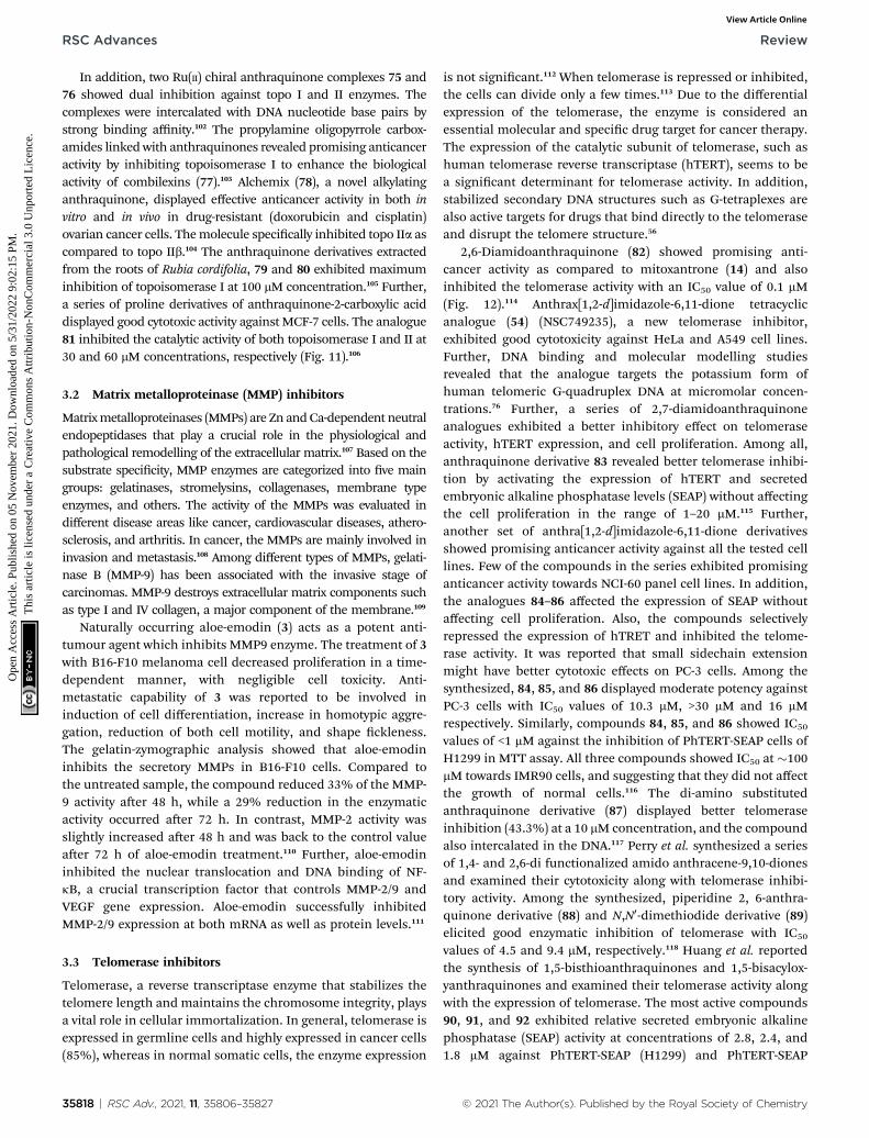
is not signicant.112 When telomerase is repressed or inhibited,the cells can divide only a few times.113 Due to the differentialexpression of the telomerase, the enzyme is considered anessential molecular and specic drug target for cancer therapy.The expression of the catalytic subunit of telomerase, such ashuman telomerase reverse transcriptase (hTERT), seems to bea signicant determinant for telomerase activity. In addition,stabilized secondary DNA structures such as G-tetraplexes arealso active targets for drugs that bind directly to the telomeraseand disrupt the telomere structure.56
2,6-Diamidoanthraquinone (82) showed promising anti-cancer activity as compared to mitoxantrone (14) and alsoinhibited the telomerase activity with an IC50 value of 0.1 mM(Fig. 12).114 Anthrax[1,2-d]imidazole-6,11-dione tetracyclicanalogue (54) (NSC749235), a new telomerase inhibitor,exhibited good cytotoxicity against HeLa and A549 cell lines.Further, DNA binding and molecular modelling studiesrevealed that the analogue targets the potassium form ofhuman telomeric G-quadruplex DNA at micromolar concen-trations.76 Further, a series of 2,7-diamidoanthraquinoneanalogues exhibited a better inhibitory effect on telomeraseactivity, hTERT expression, and cell proliferation. Among all,anthraquinone derivative 83 revealed better telomerase inhibi-tion by activating the expression of hTERT and secretedembryonic alkaline phosphatase levels (SEAP) without affectingthe cell proliferation in the range of 1–20 mM.115 Further,another set of anthra[1,2-d]imidazole-6,11-dione derivativesshowed promising anticancer activity against all the tested celllines. Few of the compounds in the series exhibited promisinganticancer activity towards NCI-60 panel cell lines. In addition,the analogues 84–86 affected the expression of SEAP withoutaffecting cell proliferation. Also, the compounds selectivelyrepressed the expression of hTRET and inhibited the telome-rase activity. It was reported that small sidechain extensionmight have better cytotoxic effects on PC-3 cells. Among thesynthesized, 84, 85, and 86 displayed moderate potency againstPC-3 cells with IC50 values of 10.3 mM, >30 mM and 16 mMrespectively. Similarly, compounds 84, 85, and 86 showed IC50
values of <1 mM against the inhibition of PhTERT-SEAP cells ofH1299 in MTT assay. All three compounds showed IC50 at �100mM towards IMR90 cells, and suggesting that they did not affectthe growth of normal cells.116 The di-amino substitutedanthraquinone derivative (87) displayed better telomeraseinhibition (43.3%) at a 10 mM concentration, and the compoundalso intercalated in the DNA.117 Perry et al. synthesized a seriesof 1,4- and 2,6-di functionalized amido anthracene-9,10-dionesand examined their cytotoxicity along with telomerase inhibi-tory activity. Among the synthesized, piperidine 2, 6-anthra-quinone derivative (88) and N,N0-dimethiodide derivative (89)elicited good enzymatic inhibition of telomerase with IC50
values of 4.5 and 9.4 mM, respectively.118 Huang et al. reportedthe synthesis of 1,5-bisthioanthraquinones and 1,5-bisacylox-yanthraquinones and examined their telomerase activity alongwith the expression of telomerase. The most active compounds90, 91, and 92 exhibited relative secreted embryonic alkalinephosphatase (SEAP) activity at concentrations of 2.8, 2.4, and1.8 mM against PhTERT-SEAP (H1299) and PhTERT-SEAP
© 2021 The Author(s). Published by the Royal Society of Chemistry

Fig. 12 Structures of anthraquinone derivatives that target telomerases.
Review RSC Advances
Ope
n A
cces
s A
rtic
le. P
ublis
hed
on 0
5 N
ovem
ber
2021
. Dow
nloa
ded
on 5
/31/
2022
9:0
2:15
PM
. T
his
artic
le is
lice
nsed
und
er a
Cre
ativ
e C
omm
ons
Attr
ibut
ion-
Non
Com
mer
cial
3.0
Unp
orte
d L
icen
ce.
View Article Online
(hTERT-BJ1) cell lines. The 1,5-bisacyloxyanthraquinones (90,91, 92) demonstrated good telomerase inhibitory activity andactivated hTERT expression without affecting the cell viability(Fig. 12).119
DNA can acquire a range of alternative conformations basedon specic sequence motifs and interactions with severalproteins. Among these conformations, G-quadruplex structuresare a form of non-canonical nucleic acid structures that canform within specic repetitive G-rich DNA or RNA regions.120
These G-quadruplex structures are unique and extensivelyinvolved in the regulation of several biological processes. Ingeneral, G-rich repeat sequences with the capability to form G-quadruplex structures are present and overrepresented in telo-meres, transcriptional start sites, and double-strand breaksites.121 The presence of G-quadruplex structures in telomeres iscapable of inhibiting the activity of telomerase, an enzyme thatis overexpressed in cancer cells.122 Hence, targeting the G-quadruplex structure is one of the promising strategies indeveloping anticancer therapeutics.
Due to the structural diversity and promising therapeuticactivity, some of the anthraquinones derivatives are signi-cantly bound and involved in stabilizing G-quadruplex struc-tures. Das and Dutta reported the promising anticancer activityof anthraquinone-based natural compounds like aloe emodin,aloe emodin-8-glucoside, and aloin. Further, the authors
© 2021 The Author(s). Published by the Royal Society of Chemistry
investigated the binding affinity of these compounds againsta set of six different quadruplex structures like c-KIT, c-MYC,HUMTEL, BCL-2, KRAS, and VEGF. Among all the examinedstructures, aloe emodin (3) exhibited signicant bindingaffinity, i.e. (2.11 � 0.33) � 105 M�1 towards c-KIT as comparedto aloe emodin-8-glucoside ((9.70 � 0.50) � 105 M�1). Incontrast, aloin was not capable of targeting the quadruplexstructures.123 In addition, Wang et al. reported the G-quadruplexstructure stabilization activity of aloe emodin. Aloe emodinreduced the transcription of hTERT gene in the three differentbreast cancer cell lines such as MDA-MB-453, MDA-MB-231 andMCF-7. The results unveiled that aloe emodin binds andstabilizes the G-quadruplex DNA with a binding affinity of 2.55� 106 M�1 and subsequently inhibits the enzymatic activity ofthe telomerase.124
In another study, Mei et al. reported the synthesis of a ruth-enium(II) complex of emodin and the biological activity of thecompound against c-myc G4 DNA. The compound showed goodbinding affinity with c-myc G-quadruplex DNA with bindingaffinity of 6.7 � 0.19 � 104 mol L�1.125 Similarly, Elvira et al.synthesized nitrogen substituted 1-(3-aminoprop-1-ynyl)-4-hydroxyanthraquinone derivatives and studied their anti-cancer potential in a panel of cancer cells like MCF-7, U-87 MG,DU-145, SNB-19, and hTERT lung broblasts. Further, themolecular binding studies of the synthesized compounds
RSC Adv., 2021, 11, 35806–35827 | 35819

RSC Advances Review
Ope
n A
cces
s A
rtic
le. P
ublis
hed
on 0
5 N
ovem
ber
2021
. Dow
nloa
ded
on 5
/31/
2022
9:0
2:15
PM
. T
his
artic
le is
lice
nsed
und
er a
Cre
ativ
e C
omm
ons
Attr
ibut
ion-
Non
Com
mer
cial
3.0
Unp
orte
d L
icen
ce.
View Article Online
towards DNA G-quadruplex revealed that almost all the deriva-tives showed good binding affinities towards DNA motifs.126 Inyet another research study, 2,6-disubstituted amido anthracene-9,10-dione dimeric distamycin derivatives were designed andsynthesized. Among all the synthesized derivatives, the disub-stituted anthraquinone with tri-N-methylpyrrole side chain(ANTP) was found to be more promising and exhibited goodactivity towards c-Myc G-quadruplex DNA with a binding affinityof 3.8 � 0.01 � 106 M�1.127
3.4 Kinase inhibitors
Protein kinases are the enzymes that phosphorylate protein bytransferring g-phosphate group to the protein, whereas phos-phatase removes the phosphate group from protein. Phos-phorylation is the most common form of reversible post-translational modications of the protein.128 Approximately50% of all proteins undergo phosphorylation, and specickinases, as well as phosphatases tightly, control this process.Almost 538 known kinases are identied in the human genome.These kinases maintain cellular functions by switching theprotein function on most protein kinases involved in signallingnetworks that employ phosphorylation, which modulate targetprotein activities. The kinases are critically involved in almostall cellular processes that promote cell survival, proliferation,metabolism, and migration.129 The abnormal expression ofkinases leads to oncogenesis and other diseases. Several kinasesare identied that are involved in cancer cell signalling path-ways, angiogenesis, proliferation, and metastases of varioustypes of cancer.130 Due to widespread clinical applications,kinases are considered promising drug targets for anticancertherapeutics.
3-(Azidomethyl)-1,8-dihydroxy-6-methoxy anthracene-9,10-dione (93) is a phyto-based emodin derivative isolated fromgiant knotweed. The compound exhibited potent anticanceractivity in both in vitro and in vivo models (Fig. 13). Furtherstudies revealed that the compound inhibits the overexpressionof Her2/neu in lung cancer and breast cancer through
Fig. 13 Anthraquinone derivatives targeting kinases involved in cancer p
35820 | RSC Adv., 2021, 11, 35806–35827
proteasomal degradation of Her2/neu.115 Another studydemonstrated that the anthraquinone derivative 94 was moreeffective compared to emodin. It inhibited cell proliferation andtransformation of HER-2/neu, which is overexpressed in humanbreast cancer cells via blocking the tyrosine phosphorylation ofp185neu. The IC50 of 94was found to be 17 mMand 1 mMagainsttyrosine phosphorylation of HER-2/neu and MDA-MB-453 cells,respectively.131 Further, the combination of emodin and pacli-taxel synergistically inhibited the anchorage-dependent and-independent growth of HER-2/neu overexpressing breastcancer cells (MDA-MB-361) by 70% in in vitro assay along withinhibition of the tumour growth.93 Muto et al. reported thatemodin extracted from the root and rhizome of Rheumpalmatum L., induced apoptosis in myeloma cells. In addition,emodin down-regulated the Mcl-1(induced myeloid leukaemiacell differentiation protein), leading to the apoptotic cell deathof cancer cells.132 Damnacanthal (95) is a potent naturalanthraquinone molecule that selectively inhibits p56lck tyro-sine kinase with an IC50 value of 17 nM. Further, the compoundalso has therapeutic efficacy in treating T-cell malignancies andautoimmune diseases.133 In a separate study, Shi et al. isolatedseveral antiproliferative anthraquinone derivatives fromHedyotis diffusa. Among the isolated compounds, 2-hydroxy-3-methylanthraquinone (96) induced apoptotic mediated celldeath in malignant cells via mitochondrial pathway by inhib-iting receptor Src tyrosine kinase. Compound 96 displayed IC50
values of 33 mM and 67 mM against protein tyrosine kinasesactivities of pp60-src (3 U mL�1), active GST-v-src protein(0.1 mg mL�1) and natural SPCA-1 cell lysate (0.5 mg mL�1)prepared as target proteins. Further, compound 96 exhibited anIC50 value of 51 mM against HepG-2 cell lines.134 1-Deoxy-rhodoptilometrin (97), another anthraquinone derivative, isa marine metabolite described to act as a potential lead foranticancer activity by inhibiting various distinct protein kinasessuch as EGFR, ERBB-2,4 and IGF-1. Compound 97 showedinhibitory activity against 23 protein kinases and was found tobe the most potent inhibitor of Aurora-A, Aurora-B, EGF-R, SRC,
rogression.
© 2021 The Author(s). Published by the Royal Society of Chemistry

Review RSC Advances
Ope
n A
cces
s A
rtic
le. P
ublis
hed
on 0
5 N
ovem
ber
2021
. Dow
nloa
ded
on 5
/31/
2022
9:0
2:15
PM
. T
his
artic
le is
lice
nsed
und
er a
Cre
ativ
e C
omm
ons
Attr
ibut
ion-
Non
Com
mer
cial
3.0
Unp
orte
d L
icen
ce.
View Article Online
and VEGF-R2 at IC50 of 3, 1.8, 4, 3.7, and 1.8 mM, respectively.135
Similarly, quinalizarin (98), a potent kinase inhibitor, possessedthe ability to selectively inhibit CK2 (Ser/Thr protein kinase)comparable to emodin. It induced apoptosis more effectivelythan other CK2 inhibitors, which are commonly used like4,5,6,7-tetra bromo-1H-benzotriazole and 2-dimethylamino-4,5,6,7-tetra bromo-1H-benzimidazole (Fig. 13). The IC50 valueof quinalizarin was found to be 0.11 mM inhibiting HEK-293Tcells. Jurkat cells upon treatment with compound 98 (5 mM)for 4 h treatment showed a 48% fall in CK2 activity in the celllysates. Compound 98 (quinalizarin) is structurally very similarto emodin, a quite promiscuous inhibitor of CK2 and of severalother protein kinases as well. It exhibited potency toward PIM3(IC50 of 0.08 mM) that is 30-fold higher than that of CK2 (IC50 of2.50 mM).136
3.5 Miscellaneous cancer targets
Ecto-nucleotidases are the enzymes that hydrolyse the extra-cellular nucleotides to nucleosides and control nucleoside (P1)and nucleotide (P2) receptor-mediated signaling.137 The enzymealters the adenosine level that in turn increases or decreases P1and P2 receptor activity. Hence, the inhibition of adenosineproduction in the tumour cell environment, through inhibitingthe enzyme activity, might be a promising and novel strategy foranticancer therapy.138,139 Baqi Y reported that some of theanthraquinone derivatives 99 and 100 were found to be potentinhibitors of ecto-nucleotidase with inhibitory constant (Ki)values of 150 and 260 nM, respectively (Fig. 14).140 Similarly,physcion (101), a naturally occurring anthraquinone derivative,is a promising anticancer agent primarily used to treat humannasopharyngeal cancer. It induced apoptosis and autophagy inhuman nasopharyngeal cancer cells by the downregulation oftranscription factor Sp1. Compound 101, also known as parie-tin, upon treatment with physcion (5, 10, and 20 mmol L�1) ina dose-dependent manner suppressed the cell viability andcolony formation in CNE2 cells. Physcion (10 and 20 mmol L�1)dose-dependently blocked cell cycle progression at G1 phase
Fig. 14 Anthraquinone derivatives that target other miscellaneous prote
© 2021 The Author(s). Published by the Royal Society of Chemistry
and induced both caspase-dependent apoptosis and autophagyin CNE2 cells. Similarly, 101 induced apoptosis and autophagyin human nasopharyngeal carcinoma cells by targeting Sp1,which was mediated by ROS/miR-27a/ZBTB10 signaling.141
Apart from the mentioned targets, emodin exhibited selec-tive activity towards human nasopharyngeal cancer cells (CNE-2Z). It reduced cell viability and induced cell cycle arrest andapoptotic cell death by targeting the chloride channels in CNE-2Z cells compared to positive control tamoxifen.142 In addition,emodin exhibited an anti-metastatic effect by the down-regulation of CXC chemokine receptor type 4. The CXC che-mokine receptor type 4 plays a crucial role in cancer invasionand metastasis.143 Furthermore, emodin also inhibited VascularEndothelial Growth Factor Receptor (VEGFR) and MMPs inconnotation with downregulation of runt-related transcriptionfactor 2 (Runx2), which controls both VEGF signalling andtranscriptional activity.144 Further, emodin has a structuralsimilarity with ATP Citrate Lyase (ACL) inhibitors. ACL playsa signicant role in de novo fatty acid and cholesterol biosyn-thesis. Moreover, the enzyme is highly expressed in some of thecancer cells. Hence it can act as a promising anticancer agent byinhibiting the ACL enzyme.145
Another naturally occurring anthraquinone 102, isolatedfrom endophytic fungi, was reported as an anticancer agent thatinduced caspase-mediated apoptosis and also suppressedphosphorylation of Akt kinase. Compound 102 displayed acti-vation of caspases (8, 9 and 3) and poly (ADP-ribose) polymerase(PARP) in MCF-7 and MDA-MB-435 breast cancer cells signi-cantly at concentrations of 3.75 mM and 3 mM, respectively.146
Similarly, Kamiya et al. isolated ten anthraquinones from Mor-inda citrifolia roots and examined their anticancer potentialagainst human colon cancer (HCT116) cells and DNA poly-merase activity. One of the compounds, morindone (103),showed signicant polymerase inhibition properties, therebysuppressing the growth of HCT116 cells. It induced cell growthsuppression with LD50 value 32.2 mM on human DNA poly-merase g.147 Finally, the sulphonamide-anthraquinone
ins involved in cancer growth.
RSC Adv., 2021, 11, 35806–35827 | 35821

RSC Advances Review
Ope
n A
cces
s A
rtic
le. P
ublis
hed
on 0
5 N
ovem
ber
2021
. Dow
nloa
ded
on 5
/31/
2022
9:0
2:15
PM
. T
his
artic
le is
lice
nsed
und
er a
Cre
ativ
e C
omm
ons
Attr
ibut
ion-
Non
Com
mer
cial
3.0
Unp
orte
d L
icen
ce.
View Article Online
derivative 104 was reported to inhibit cancer cell progression inseveral types of cancer cells. It signicantly inhibited theproliferation of HCT116, HCT116 p53—/— and p53 mutantHT29 (R273H) cells at a level comparable to that of the positivecontrol 5-FU (10 mM) in MTT assay. The derivative 104 inducedcell cycle arrest at the S and G2/M phases (38.5, 42.2, and 33.4;28.1, 29.5, and 49.0%, respectively) compared to that induced bythe vehicle control (30.2, 36.3 and 21.2; 23.4, 27.1, and 20.5%,respectively). Treatment with 10 mM of 104 increased caspase-3cleavage in all three cell lines (HCT116, HCT116 P53—/— andHT29). These results indicated that 104 induced both apoptoticand necrotic cell death in a p53-independent manner(Fig. 14).148
4. Toxicity of anthraquinones
Toxicity or safety assessment of a potential drug molecule is theprimary area of concern before clinical usage.149 Apart from thefavourable pharmaceutical applications, some anthraquinonederivatives may induce potential damage to cells due to theirclose resemblance to anthracene, a toxic analogue.150 A few invitro studies describe the toxicity behavior of anthraquinonederivatives with limited information on in vivo studies ofanthraquinones. Liu et al. investigated the in vitro and in silicohepatotoxicity of different anthraquinones and their deriva-tives. Among all the studied, rhein was identied as a potentialliver toxicant against HuH-7 cells with an EC50 value of 93.9 mMupon repeated treatment.151 Chen et al. examined the photoin-duced acute toxicity of a series of fourteen anthraquinonederivatives against Daphnia magna, an important modelorganism for toxicity assessment. Almost all the compoundsexhibited no observable toxicity in the presence of visible lightat the maximum concentration of the compounds used in thestudy. However, chloro and dihydroxy substituted anthraqui-nones exhibited apparent toxicity in presence of visible lightwith EC50 values of 837.0 and 959.3 nmol L�1, respectively.152
Further, Viljoen et al. summarized the toxicity of emodinagainst mouse and rat foetuses. The study revealed that emodindisplayed adverse effects against mice and rats at 17 and 60 mgkg�1 or higher doses. In addition, emodin was noticed to betoxic towards brine shrimp and exhibited lethality with an LC50
value of 0.19 mM.153 Oshida et al. examined the toxicologicaleffects of emodin on differential gene expression proles of thetestis in mice models. The compound producing testiculartoxicity in male mouse models via IGF-1 receptor signallingpathway.154 He et al. investigated the potential toxicity effects ofemodin on zebrash embryos. The compound displayedadverse effects on embryo survival as well as hatching success inzebrash at a concentration of 0.93 mM and higher. In additionto this, it induced many abnormalities in zebrash embryos,including oedema, abnormal morphogenesis, and crookedtrunk.155
Anthraquinones present in plant species such as Rhamnusspecies (aloe-emodin, physcion, rhein, chrysophanol, emodin)were suspected to be involved in diseases such as renal failure,rhabdomyolysis, nephrotoxicity (regular intake), dehydration,anorexia (long-term exposure), genotoxicity in mammalian
35822 | RSC Adv., 2021, 11, 35806–35827
cells, mutation, tumour promotion, chromosomal aberrationand liver enlargement. In addition to this, the long-term use ofanthranoid laxatives is reported to exhibit symptoms associatedwith melanosis coli which is characterized by dark pigmenta-tion of the colonic mucosa, and in few cases, it is also believedto result in morphological changes of the colonic myentericsystem.156 This is because anthraquinone-based drugs containchromophores which impart a bright yellow colour to colonicepithelial cells and is reversible in most of cases. However, theyare also suspected to cause permanent physical change tocolonic tissue as well as permanent damage of renal tubularcells resulting in more serious conditions such as Chron'sdisease or ulcerative colitis.
The anthraquinone derivatives, especially those derivedfrom Rhubarb have shown neuroprotective effects. Emodinexerted a neuroprotective effect in cerebral ischemic stroke(CIS) by maintaining the integrity of blood–brain barrier (BBB),ameliorating inammation, and controlling the apoptosisprocess. Li et al. attributed this effect of emodin to the inhibi-tion of connexin 43 (Cx43) and aquaporin 4 (AQP4).157 Theneuroprotective effect of chrysophanol (CHR) was reported to beassociated with oxidative/antonitrosative, anti-inammatoryand by inhibiting apoptosis. Similarly, Zhao et al. reportedneuroprotective effect of rhein is by oxidative stress andapoptosis.158
5. Polyethylene glycol (PEG) basedanthraquinone drug delivery
Anthraquinones are one of the key chemotherapeutic agentsused in the early management of cancer. Anthraquinones arepotent anti-cancer agents with many benecial effects; however,few unwanted properties such as low polarity and structuralinstability in in vivo conditions are also observed in some cases.Current research studies mainly focus on enhancing the bio-logical efficacy of naturally occurring and chemically synthe-sized anthraquinone derivatives. However, few drug deliveryapproaches are reported to address the undesired properties oflow polarity and stability. It is imperative to develop drugdelivery vehicles for anthraquinone or build multi-drug deliverysystems to administer two or three anti-cancer agents in onedose. Polymer based control release formulations are gainingimportance as they are very efficient in loading multiple drugsin layers for attaining programmed release kinetics under invivo conditions.159 One such example was a report where a DNA-damaging anthracycline agent doxorubicin (dox) (10) anda phosphatidylinositol-3 kinase inhibitor wortmannin (wor)were conjugated. These drugs were used alone or in combina-tion with poly(ethylene glycol)-poly(aspartate hydrazide) blockcopolymers through a hydrazone bond.160 The polymer formedunimodal micelle structure encapsulating the conjugated anti-cancer drugs (dox and wor). The polymer-drug conjugatedsystem developed in this way has particle size of <100 nm. Thedrug mixing ratios between dox/wor were precisely controlled inthis delivery system. This polymer-drug conjugated system re-ported better drug release properties and also reduced the
© 2021 The Author(s). Published by the Royal Society of Chemistry

Review RSC Advances
Ope
n A
cces
s A
rtic
le. P
ublis
hed
on 0
5 N
ovem
ber
2021
. Dow
nloa
ded
on 5
/31/
2022
9:0
2:15
PM
. T
his
artic
le is
lice
nsed
und
er a
Cre
ativ
e C
omm
ons
Attr
ibut
ion-
Non
Com
mer
cial
3.0
Unp
orte
d L
icen
ce.
View Article Online
amount of drug (wor) required for cytotoxicity while maintain-ing the biological activity of the independent polymericmicelles.
Other PEG conjugated systems reported in recent literatureinclude poly(ethylene glycol) PEG-doxorubicin (dox) conjugateswith polymers of linear or branched architecture (molecularweight 5000–20 000 g mol�1), and different peptidyl linkers(GFLG, GLFG, GLG, GGRR, and RGLG). GFLG linker showed�30% release of dox at 5 h irrespective of PEGmolecular weightor architecture. All PEG conjugates prepared by this methodwere more than 10-fold less toxic (IC50 values >2 mg mL�1) thanfree Dox (IC50 value of 0.24 mg mL�1). PEG-dox showed greatertumour-targeting than free Dox in radiolabelled studies. Theradio iodinated PEGs showed a clear relationship betweenmolecular weight, blood clearance, and tumouraccumulation.161
Other more advanced conjugate systems of anthraquinonesreported in the literature are polymer enzyme liposome therapy(PELT),162 triple block nanocarrier (TBN) platforms,163 andamphiphilic core cross-linked star (CCS) polymers164 thatshowed excellent pharmacokinetic prole in in vivo models. Ina TBN platform, hydrophilic polyethylene glycol (PEG) was usedas an outer shell, and a hydrophobic biodegradable poly-caprolactone (PCL) block for encapsulating anthracycline anti-cancer drug was utilized as the middle layer. The carboxylic-functionalized polycaprolactone (CPCL) based inner shell waslled with non-anthracycline anti-cancer drug for inducingsynergistic effect. The dual drug-loaded TBN exhibited superiorsynergistic cell death at much lower drug concentrations.163
These ndings represent the critical role of PEG-conjugatedanthracycline-based effective anti-cancer drug delivery meth-odology that might reduce the effective dose and toxicity in vivocompared to the conventional drug formulations.
6. Structure–activity relationship ofanthraquinones
Structure–activity relationship (SAR) studies of the anthraqui-none ring structure provide an idea about the primary structuralconsiderations that result in maximum biological effect.Moreover, it is essential to assess the substituents whichundergo enzymatic degradation due to endogenous enzymes,thereby eliminating the molecule before it exerts its activity.SAR studies help understand the groups that improve thesynthesized molecule's pharmacokinetics and substituentsresistant to enzymatic degradation. Few of the SAR observationsare discussed here.
Studies showed that the sulphonamide and methyl ketonesubstituent at 1st position of anthraquinone would be advan-tageous in exerting superior anticancer activity. Similarly,carboxylic acid substitution at 1st position would result inactivity loss.63 Another report suggested that when anthraqui-none monomers are considered, the position of hydroxyl (C-5and C-8) is very crucial for antitumour efficacy. The hydroxyl-ation at the C-1 position was reported to drastically enhance itscytotoxic activity, which indicates that the phenolic hydroxyl
© 2021 The Author(s). Published by the Royal Society of Chemistry
groups are essential for the antitumour activity of anthraqui-nones.165 Emodin is identied as the most abundantly existinganthraquinone in plant species having a wide range of anti-cancer properties on various cancers. Dong et al. identied thatC-1 and C-3 are the most important functional sites for anti-tumour activities of anthraquinone molecules.50 Zhou et al. re-ported six anthraquinones from Xanthophytum attopvensis pierreand studies revealed that a large steric hindrance due to glycosylsubstituents was the reason for the weakening of the bondbetween drug and target cells. The substituents such ashydroxyl or hydroxymethyl groups present at only the C-1position, then the anthraquinones exhibit similar cytotoxicactivity. In one of the comparative studies between emodin(anthraquinone) and cassiamin (bianthraquinones), bian-throquinones showed lower anti-cancer activities than anthra-quinones, and the reason ascribed to this observation was thesteric hindrance of bianthroquinones with a distorted conr-mation.166 Additionally, the atomic charge at C-10 position andthe number of hydroxyl groups present on the benzene ring ofmonomer increased its anti-cancer potency compared to itsdimer. Similarly, chlorination was found to decrease the efficacyof these compounds.167
7. Future perspectives & conclusion
Cancer, characterized by unbridled cell proliferation, isa leading cause of death because of high incidence andmortality, which is exacerbated by the emergence of drugresistance. Therefore, there is a need for the development ofnew anticancer agents. Anthraquinones have attracted themedicinal chemist's attention for their diverse biologicalpotential. They are typical anticancer fragments that havea broad scope for chemical modications to be exploited todevelop new anticancer agents. A handful of anticancer drugsbased on anthraquinone moiety are marketed and used inchemotherapy in stand-alone regimens or widely used incombination therapy alongside radiation treatment to mitigatethe metastasis of cancer cells. However, the emergence ofresistance is increasingly being reported, and new explorationswere undertaken to develop new anthraquinone-based anti-cancer agents with some success.
Unfortunately, the majority of the research being pursued inacademia, leading to the generation of a library of anthraqui-nones, is not intensively explored towards understanding theirmechanistic biological pathways and target-specic activity inin vitro and in vivo models. Moreover, the toxicity and safetyassessment of the anthraquinone derivatives to be used aspotential drug molecules needs to be thoroughly investigated atthe academic research level as limited research is available inthe public domain. The toxicity and efficacy studies such aspharmacokinetics and pharmacodynamics could help betterunderstand the prole of new anthraquinones. Hence, it ishighly desirable to explore the potential of these anthraquinonelibraries to identify hit/lead-like molecules that can be taken upfor further development.
Several opportunities are ready to be fully exploited, whichinclude selective drug delivery, combination with other
RSC Adv., 2021, 11, 35806–35827 | 35823

RSC Advances Review
Ope
n A
cces
s A
rtic
le. P
ublis
hed
on 0
5 N
ovem
ber
2021
. Dow
nloa
ded
on 5
/31/
2022
9:0
2:15
PM
. T
his
artic
le is
lice
nsed
und
er a
Cre
ativ
e C
omm
ons
Attr
ibut
ion-
Non
Com
mer
cial
3.0
Unp
orte
d L
icen
ce.
View Article Online
anticancer drugs, computational techniques (e.g., ligand-baseddrug designing), drug repurposing, etc. Some of the issuescritical to few anthraquinone derivatives, such as low polarityand structural instability in in vivo conditions, could besuccessfully addressed by employing polyethylene glycol-baseddrug delivery vehicles. There is ample scope for utilizinganthraquinone-based anticancer drugs in combination thera-pies with newly approved anticancer drugs targeting differentbiological receptors. This can be achieved through a detailedunderstanding of the mechanistic pathways of the intendedcombination. The development of modern age tools andcomputational techniques such as ligand-based drug designing(LBDD) and scaffold hopping led to newer molecules withimproved efficacies. The in silico drug designing tools tremen-dously boosted the eld of anthraquinone-anticancer drugdiscovery in the diversication of targets, from kinases to genesand immune responses. By leveraging the new age bio/cheminformatics tools, one can design anthraquinone-basednew chemical entities with much accuracy in terms of safety &efficacy. Further, the emergence of drug repurposing tech-niques could be utilized to explore this scaffold for new thera-peutic applications, and there is development in this direction,as can be construed from literature.
In view of the ongoing challenge of drug resistance of theexisting marketed drugs, concerted efforts are required toaddress the shortcomings in academia research (limitedadvanced studies) and utilize the aforementioned approaches.This could lead to the development of new anthraquinone-based anticancer agents that can eventually be translated intosafer and effective chemotherapeutics.
Conflicts of interest
There are no conicts to declare.
Acknowledgements
The authors are grateful to the Deanship of the ScienticResearch (DSR), Umm Al-Qura University, for the nancialsupport through the project code: 19-SCI-1-01-0019.
References
1 H. Hussain, A. Al-Harrasi, A. Al-Rawahi, I. R. Green, R. Csuk,I. Ahmed, A. Shah, G. Abbas, N. U. Rehman and R. Ullah,Expert Opin. Ther. Pat., 2015, 25, 1053–1064.
2 W. Tian, C. Wang, D. Li and H. Hou, Future Med. Chem.,2020, 12, 627–644.
3 Demselben, Justus Liebigs Ann. Chem., 1840, 34, 287–296.4 C. Graebe, Ber. Dtsch. Chem. Ges., 1890, 23, 3739–3740.5 M. Phillips, Chem. Rev., 1929, 6, 157–174.6 C. Graebe and C. Liebermann, Ber. Dtsch. Chem. Ges., 1869,2, 14.
7 R. Fittig, Ber. Dtsch. Chem. Ges., 1873, 6, 167–169.8 A. R. Burnett and R. H. Thomson, J. Chem. Soc. C, 1967,2100–2104, DOI: 10.1039/J39670002100.
35824 | RSC Adv., 2021, 11, 35806–35827
9 R. Singh and S. M. Chauhan, Chem. Biodiversity, 2004, 1,1241–1264.
10 M. Klaper, P. Wessig and T. Linker, Chem. Commun., 2016,52, 1210–1213.
11 J. J. Dong, D. Unjaroen, F. Mecozzi, E. C. Harvey, P. Saisaha,D. Pijper, J. W. deBoer, P. Alsters, B. L. Feringa andW. R. Browne, ChemSusChem, 2013, 6, 1774–1778.
12 Y. Xu, Z. Yang, J. Hu and J. Yan, Synthesis, 2013, 45, 370–374.
13 R. Kumar, N. Sharma, N. Sharma, A. Sharma andA. K. Sinha, Mol. Diversity, 2011, 15, 687–695.
14 S. El-Sayra and S. Rayyan, Molecules, 2001, 6, 279–286.15 H. Kaper, M. Antonietti and F. Goettmann, Tetrahedron
Lett., 2008, 49, 4546–4549.16 M. N. Akhtar, S. Zareen, S. K. Yeap, W. Y. Ho, K. M. Lo,
A. Hasan and N. B. Alitheen, Molecules, 2013, 18, 10042–10055.
17 X. Wang and V. Snieckus, Synlett, 1990, 1990, 313–316.18 T. Sawano, Y. Toyoshima and R. Takeuchi, Inorganics, 2019,
7(11), 138.19 T. J. Monks, R. P. Hanzlik, G. M. Cohen, D. Ross and
D. G. Graham, Toxicol. Appl. Pharmacol., 1992, 112, 2–16.20 E. M. Malik and C. E. Muller, Med. Res. Rev., 2016, 36, 705–
748.21 Y. Caro, L. Anamale, M. Fouillaud, P. Laurent, T. Petit and
L. Dufosse, Nat. Prod. Bioprospect., 2012, 2, 174–193.22 D. W. Shaw, Dermatitis, 2009, 20, 292–295.23 L. Dufosse, Food Res. Int., 2014, 65, 132–136.24 E. M. Malik, Y. Baqi and C. E. Muller, Beilstein J. Org. Chem.,
2015, 11, 2326–2333.25 T. Furuta, Y. Hirayama and M. Iwamura, Org. Lett., 2001, 3,
1809–1812.26 S. Patnaik, A. Swami, D. Sethi, A. Pathak, B. S. Garg,
K. C. Gupta and P. Kumar, Bioconjugate Chem., 2007, 18,8–12.
27 J. C. Forti, R. S. Rocha, M. R. V. Lanza and R. Bertazzoli, J.Electroanal. Chem., 2007, 601, 63–67.
28 M. Uchimiya and A. T. Stone, Chemosphere, 2009, 77, 451–458.
29 G. Rath, M. Ndonzao and K. Hostettmann, Int. J.Pharmacogn., 1995, 33, 107–114.
30 D. L. Barnard, D. W. Fairbairn, K. L. O'Neill, T. L. Gage andR. W. Sidwell, Antiviral Res., 1995, 28, 317–329.
31 R. F. Schinazi, C. K. Chu, J. R. Babu, B. J. Oswald,V. Saalmann, D. L. Cannon, B. F. Eriksson and M. Nasr,Antiviral Res., 1990, 13, 265–272.
32 D. O. Andersen, N. D. Weber, S. G. Wood, B. G. Hughes,B. K. Murray and J. A. North, Antiviral Res., 1991, 16, 185–196.
33 C. P. Osman, N. H. Ismail, R. Ahmad, N. Ahmat, K. Awangand F. M. Jaafar, Molecules, 2010, 15, 7218–7226.
34 M. Y. Fosso, K. Y. Chan, R. Gregory and C. W. Chang, ACSComb. Sci., 2012, 14, 231–235.
35 W. Wang, R. Chen, Z. Luo, W. Wang and J. Chen, Nat. Prod.Res., 2018, 32, 558–563.
© 2021 The Author(s). Published by the Royal Society of Chemistry

Review RSC Advances
Ope
n A
cces
s A
rtic
le. P
ublis
hed
on 0
5 N
ovem
ber
2021
. Dow
nloa
ded
on 5
/31/
2022
9:0
2:15
PM
. T
his
artic
le is
lice
nsed
und
er a
Cre
ativ
e C
omm
ons
Attr
ibut
ion-
Non
Com
mer
cial
3.0
Unp
orte
d L
icen
ce.
View Article Online
36 K. H. Gan, C. H. Teng, H. C. Lin, K. T. Chen, Y. C. Chen,M. F. Hsu, J. P. Wang, C. M. Teng and C. N. Lin, Biol.Pharm. Bull., 2008, 31, 1547–1551.
37 S. Z. Choi, S. O. Lee, K. U. Jang, S. H. Chung, S. H. Park,H. C. Kang, E. Y. Yang, H. J. Cho and K. R. Lee, Arch.Pharmacal Res., 2005, 28, 1027–1030.
38 A. Arvindekar, T. More, P. V. Payghan, K. Laddha,N. Ghoshal and A. Arvindekar, Food Funct., 2015, 6, 2693–2700.
39 T. C. Jackson, J. D. Verrier and P. M. Kochanek, Cell DeathDis., 2013, 4, e451.
40 B. A. van Gorkom, E. G. de Vries, A. Karrenbeld andJ. H. Kleibeuker, Aliment. Pharmacol. Ther., 1999, 13, 443–452.
41 A. M. Alaadin, E. H. Al-Khateeb and A. K. Jager, Pharm. Biol.,2007, 45, 688–690.
42 Y. Baqi, K. Atzler, M. Kose, M. Glanzel and C. E. Muller, J.Med. Chem., 2009, 52, 3784–3793.
43 R. W. Winter, K. A. Cornell, L. L. Johnson, L. M. Isabelle,D. J. Hinrichs and M. K. Riscoe, Bioorg. Med. Chem. Lett.,1995, 5, 1927–1932.
44 E. M. Perchellet, M. J. Magill, X. Huang, D. M. Dalke,D. H. Hua and J. P. Perchellet, Anti-Cancer Drugs, 2000,11, 339–352.
45 M. B. Martins-Teixeira and I. Carvalho, ChemMedChem,2020, 15, 933–948.
46 Y. Lazebnik, Nat. Rev. Cancer, 2010, 10, 232–233.47 L. A. Torre, R. L. Siegel, E. M. Ward and A. Jemal, Cancer
Epidemiol., Biomarkers Prev., 2016, 25, 16–27.48 D. Wang, X.-H. Wang, X. Yu, F. Cao, X. Cai, P. Chen, M. Li,
Y. Feng, H. Li and X. Wang, Front. Pharmacol., 2021, 12,638993.
49 W. Wu, N. Hu, Q. Zhang, Y. Li, P. Li, R. Yan and Y. Wang,Chem.-Biol. Interact., 2014, 219, 18–27.
50 X. Dong, J. Fu, X. Yin, S. Cao, X. Li, L. Lin and J. Ni,Phytother. Res., 2016, 30, 1207–1218.
51 S. Siddamurthi, G. Gutti, S. Jana, A. Kumar and S. K. Singh,Future Med. Chem., 2020, 12, 1037–1069.
52 C. H. Teng, S. J. Won and C. N. Lin, Bioorg. Med. Chem.,2005, 13, 3439–3445.
53 M. Siwy, D. Sek, B. Kaczmarczyk, I. Jaroszewicz,A. Nasulewicz, M. Pelczynska, D. Nevozhay andA. Opolski, J. Med. Chem., 2006, 49, 806–810.
54 J. A. Valderrama, O. Espinoza, M. F. Gonzalez, R. A. Tapia,J. A. Rodrıguez, C. Theoduloz and G. Schmeda-Hirschmann, Tetrahedron, 2006, 62, 2631–2638.
55 L. F. Tietze, K. M. Gericke and I. Schuberth, Eur. J. Org.Chem., 2007, 2007, 4563–4577.
56 H. S. Huang, K. F. Huang, C. L. Li, Y. Y. Huang,Y. H. Chiang, F. C. Huang and J. J. Lin, Bioorg. Med.Chem., 2008, 16, 6976–6986.
57 S. Wang, Q. Wang, Y. Wang, L. Liu, X. Weng, G. Li, X. Zhangand X. Zhou, Bioorg. Med. Chem. Lett., 2008, 18, 6505–6508.
58 Z. Yang, J.-A. Kang, W.-H. Kim, A.-Y. Park, H.-S. Kim,J.-S. Kim, J.-A. Kim, P. Gong, L.-S. Jeong and H.-R. Moon,Bull. Korean Chem. Soc., 2009, 30, 1463–1469.
© 2021 The Author(s). Published by the Royal Society of Chemistry
59 G. Z. Jin, H. S. Jin and L. L. Jin, Arch. Pharmacal Res., 2011,34, 1071–1076.
60 H. Y. Tu, A. M. Huang, C. H. Teng, T. C. Hour, S. C. Yang,Y. S. Pu and C. N. Lin, Bioorg. Med. Chem., 2011, 19,5670–5678.
61 S. X. Feng, Q. Guan, T. Chen and C. Du, Arch. PharmacalRes., 2012, 35, 1251–1258.
62 V. Markovic, A. Janicijevic, T. Stanojkovic, B. Kolundzija,D. Sladic, M. Vujcic, B. Janovic, L. Joksovic,P. T. Djurdjevic, N. Todorovic, S. Trifunovic andM. D. Joksovic, Eur. J. Med. Chem., 2013, 64, 228–238.
63 D. Bhasin, J. P. Etter, S. N. Chettiar, M. Mok and P. K. Li,Bioorg. Med. Chem. Lett., 2013, 23, 6864–6867.
64 V. Castro-Castillo, C. Suarez-Rozas, N. Castro-Loiza,C. Theoduloz and B. K. Cassels, Eur. J. Med. Chem., 2013,62, 688–692.
65 Y. R. Lee, D. S. Yu, Y. C. Liang, K. F. Huang, S. J. Chou,T. C. Chen, C. C. Lee, C. L. Chen, S. H. Chiou andH. S. Huang, PLoS One, 2013, 8, e56284.
66 Z. Liang, J. Ai, X. Ding, X. Peng, D. Zhang, R. Zhang,Y. Wang, F. Liu, M. Zheng, H. Jiang, H. Liu, M. Geng andC. Luo, ACS Med. Chem. Lett., 2013, 4, 408–413.
67 A. T. Taher and G. H. Hegazy, Arch. Pharmacal Res., 2013, 36,573–578.
68 B. Kolundzija, V. Markovic, T. Stanojkovic, L. Joksovic,I. Matic, N. Todorovic, M. Nikolic and M. D. Joksovic,Bioorg. Med. Chem. Lett., 2014, 24, 65–71.
69 M. S. Almutairi, G. H. Hegazy, M. E. Haiba, H. I. Ali,N. M. Khalifa and M. Soliman Ael, Int. J. Mol. Sci., 2014,15, 22580–22603.
70 Y. W. Chen, H. S. Huang, Y. S. Shieh, K. H. Ma, S. H. Huang,D. Y. Hueng, H. K. Sytwu and G. J. Lin, PLoS One, 2014, 9,e104703.
71 S. Sangthong, N. Sangphech, T. Palaga,N. Ngamrojanavanich, S. Puthong, T. Vilaivan andN. Muangsin, Eur. J. Med. Chem., 2014, 77, 334–342.
72 P.-Z. Zhang, H.-L. Yang, C.-C. Li, Z.-C. Xia, X.-M. Wang,H. Wei, R.-X. Rong, Z.-R. Cao, K.-R. Wang, H. Chen andX.-L. Li, Chin. Chem. Lett., 2014, 25, 1057–1059.
73 L. Kelland, Idrugs, 1998, 1, 397–398.74 I. Zuravka, A. Sosic, B. Gatto and R. Gottlich, Bioorg. Med.
Chem. Lett., 2015, 25, 4606–4609.75 F. Prati, C. Bergamini, M. T. Molina, F. Falchi, A. Cavalli,
M. Kaiser, R. Brun, R. Fato and M. L. Bolognesi, J. Med.Chem., 2015, 58, 6422–6434.
76 Y. H. Lin, S. M. Chuang, P. C. Wu, C. L. Chen,S. Jeyachandran, S. C. Lo, H. S. Huang and M. H. Hou,Sci. Rep., 2016, 6, 31019.
77 Y. Zheng, L. Zhu, L. Fan, W. Zhao, J. Wang, X. Hao, Y. Zhu,X. Hu, Y. Yuan, J. Shao and W. Wang, Eur. J. Med. Chem.,2017, 125, 902–913.
78 N. Okumura, H. Mizutani, T. Ishihama, M. Ito, A. Hashibe,T. Nakayama and B. Uno, Chem. Pharm. Bull., 2019, 67, 717–720.
79 V. C. Roa-Linares, Y. Miranda-Brand, V. Tangarife-Castano,R. Ochoa, P. A. Garcıa, M. Castro, L. Betancur-Galvis andA. San Feliciano, Molecules, 2019, 24, 1279.
RSC Adv., 2021, 11, 35806–35827 | 35825

RSC Advances Review
Ope
n A
cces
s A
rtic
le. P
ublis
hed
on 0
5 N
ovem
ber
2021
. Dow
nloa
ded
on 5
/31/
2022
9:0
2:15
PM
. T
his
artic
le is
lice
nsed
und
er a
Cre
ativ
e C
omm
ons
Attr
ibut
ion-
Non
Com
mer
cial
3.0
Unp
orte
d L
icen
ce.
View Article Online
80 K. Huang, L. Jiang, H. Li, D. Ye and L. Zhou, Molecules,2019, 24, 806.
81 S. Celik, F. Ozkok, A. E. Ozel, Y. Muge Sahin, S. Akyuz,B. D. Sigirci, B. B. Kahraman, H. Darici and E. Karaoz, J.Biomol. Struct. Dyn., 2020, 38, 756–770.
82 K. Huang, L. Jiang, R. Liang, H. Li, X. Ruan, C. Shan, D. Yeand L. Zhou, Eur. J. Med. Chem., 2019, 168, 45–57.
83 P. Awasthi, M. Vatsal and A. Sharma, J. Biomol. Struct. Dyn.,2019, 37, 4465–4480.
84 Y. Li, F. Guo, T. Chen, L. Zhang, Z. Wang, Q. Su and L. Feng,Chem. Biodiversity, 2020, 17, e2000328.
85 J. Li, Y. B. Zheng, T. Kurtan, M. X. Liu, H. Tang, C. L. Zhuangand W. Zhang, Nat. Prod. Res., 2020, 34, 2116–2123.
86 L. A. Oliveira, H. D. Nicolella, R. A. Furtado, N. M. Lima,D. C. Tavares, T. A. Correa and M. V. Almeida, Med. Chem.Res., 2020, 29, 1611–1620.
87 A. Anifowose, A. A. Agbowuro, R. Tripathi, W. Lu, C. Tan,X. Yang and B. Wang,Med. Chem. Res., 2020, 29, 1199–1210.
88 Y. Li, F. Guo, Y. Guan, T. Chen, K. Ma, L. Zhang, Z. Wang,Q. Su, L. Feng, Y. Liu and Y. Zhou,Molecules, 2020, 25, 1672.
89 A. S. Tikhomirov, V. A. Litvinova, D. V. Andreeva,V. B. Tsvetkov, L. G. Dezhenkova, Y. L. Volodina,D. N. Kaluzhny, I. D. Treshalin, D. Schols,A. A. Ramonova, M. M. Moisenovich, A. A. Shtil andA. E. Shchekotikhin, Eur. J. Med. Chem., 2020, 199, 112294.
90 R. Liu, H. Zou, Z. X. Zou, F. Cheng, X. Yu, P. S. Xu, X. M. Li,D. Li, K. P. Xu and G. S. Tan, Nat. Prod. Res., 2020, 34, 2709–2714.
91 K. W. Lin, W. H. Lin, C. L. Su, H. Y. Hsu and C. N. Lin,Bioorg. Chem., 2021, 107, 104395.
92 Y. Li, Y. Luan, X. Qi, M. Li, L. Gong, X. Xue, X. Wu, Y. Wu,M. Chen, G. Xing, J. Yao and J. Ren, Toxicol. Sci., 2010, 118,435–443.
93 L. Zhang, Y. K. Lau, W. Xia, G. N. Hortobagyi andM. C. Hung, Clin. Cancer Res., 1999, 5, 343–353.
94 Y. Y. Chen, S. Y. Chiang, J. G. Lin, Y. S. Ma, C. L. Liao,S. W. Weng, T. Y. Lai and J. G. Chung, Int. J. Oncol., 2010,36, 1113–1120.
95 Y. Y. Chen, S. Y. Chiang, J. G. Lin, J. S. Yang, Y. S. Ma,C. L. Liao, T. Y. Lai, N. Y. Tang and J. G. Chung,Anticancer Res., 2010, 30, 945–951.
96 J. J. Champoux, Annu. Rev. Biochem., 2001, 70, 369–413.97 K. Hevener, T. A. Verstak, K. E. Lutat, D. L. Riggsbee and
J. W. Mooney, Acta Pharm. Sin. B, 2018, 8, 844–861.98 G. Capranico, J. Marinello and G. Chillemi, J. Med. Chem.,
2017, 60, 2169–2192.99 M. N. Drwal, K. Agama, L. P. Wakelin, Y. Pommier and
R. Griffith, PLoS One, 2011, 6, e25150.100 P. J. Smith, N. J. Blunt, R. Desnoyers, Y. Giles and
L. H. Patterson, Cancer Chemother. Pharmacol., 1997, 39,455–461.
101 S. R. McKeown, M. V. Hejmadi, I. A. McIntyre, J. J. McAleerand L. H. Patterson, Br. J. Cancer, 1995, 72, 76–81.
102 J. F. Kou, C. Qian, J. Q. Wang, X. Chen, L. L. Wang, H. Chaoand L. N. Ji, J. Biol. Inorg. Chem., 2012, 17, 81–96.
103 C. Hotzel, A. Marotto and U. Pindur, Eur. J. Med. Chem.,2003, 38, 189–197.
35826 | RSC Adv., 2021, 11, 35806–35827
104 K. Pors, Z. Paniwnyk, P. Teesdale-Spittle, J. A. Plumb,E. Willmore, C. A. Austin and L. H. Patterson, Mol. CancerTher., 2003, 2, 607–610.
105 J. K. Son, S. J. Jung, J. H. Jung, Z. Fang, C. S. Lee, C. S. Seo,D. C. Moon, B. S. Min, M. R. Kim and M. H. Woo, Chem.Pharm. Bull., 2008, 56, 213–216.
106 A. Bielawska, K. Kosk and K. Bielawski, Pol. J. Pharmacol.,2001, 53, 283–287.
107 S. M. Wojtowicz-Praga, R. B. Dickson and M. J. Hawkins,Invest. New Drugs, 1997, 15, 61–75.
108 B. E. Bachmeier, C. M. Iancu, M. Jochum and A. G. Nerlich,Expert Rev. Anticancer Ther., 2005, 5, 149–163.
109 A. R. Farina and A. R. Mackay, Cancers, 2014, 6, 240–296.110 C. Tabolacci, A. Lentini, P. Mattioli, B. Provenzano,
S. Oliverio, F. Carlomosti and S. Beninati, Life Sci., 2010,87, 316–324.
111 P. Suboj, S. Babykutty, D. R. Valiyaparambil Gopi, R. S. Nair,P. Srinivas and S. Gopala, Eur. J. Pharm. Sci., 2012, 45, 581–591.
112 E. H. Blackburn, Cell, 2001, 106, 661–673.113 J. W. Shay and S. Bacchetti, Eur. J. Cancer, 1997, 33, 787–
791.114 H. S. Huang, I. B. Chen, K. F. Huang, W. C. Lu, F. Y. Shieh,
Y. Y. Huang, F. C. Huang and J. J. Lin, Chem. Pharm. Bull.,2007, 55, 284–292.
115 Y. Y. Yan, L. S. Zheng, X. Zhang, L. K. Chen, S. Singh,F. Wang, J. Y. Zhang, Y. J. Liang, C. L. Dai, L. Q. Gu,M. S. Zeng, T. T. Talele, Z. S. Chen and L. W. Fu, Mol.Pharmaceutics, 2011, 8, 1687–1697.
116 C. L. Chen, D. M. Chang, T. C. Chen, C. C. Lee, H. H. Hsieh,F. C. Huang, K. F. Huang, J. H. Guh, J. J. Lin andH. S. Huang, Eur. J. Med. Chem., 2013, 60, 29–41.
117 D. Cairns, E. Michalitsi, T. C. Jenkins and S. P. Mackay,Bioorg. Med. Chem., 2002, 10, 803–807.
118 P. J. Perry, S. M. Gowan, A. P. Reszka, P. Polucci,T. C. Jenkins, L. R. Kelland and S. Neidle, J. Med. Chem.,1998, 41, 3253–3260.
119 H. S. Huang, J. F. Chiou, Y. Fong, C. C. Hou, Y. C. Lu,J. Y. Wang, J. W. Shih, Y. R. Pan and J. J. Lin, J. Med.Chem., 2003, 46, 3300–3307.
120 M. L. Bochman, K. Paeschke and V. A. Zakian, Nat. Rev.Genet., 2012, 13, 770–780.
121 N. Kosiol, S. Juranek, P. Brossart, A. Heine and K. Paeschke,Mol. Cancer, 2021, 20, 40.
122 J. Blasiak, Curr. Med. Chem., 2017, 24, 1488–1503.123 A. Das and S. Dutta, ACS Omega, 2021, 6, 18344–18351.124 S. Wang, W. W. Yan, M. He, D. Wei, Z. J. Long and
Y. M. Tao, Pharmacol. Rep., 2020, 72, 1383–1396.125 Z. Zhang, X.-H. Wu, F.-Q. Sun, F. Shan, J.-C. Chen,
L.-M. Chen, Y.-S. Zhou and W.-J. Mei, Inorg. Chim. Acta,2014, 418, 23–29.
126 N. S. Sirazhetdinova, V. A. Savelyev, D. S. Baev,T. S. Golubeva, L. S. Klimenko, T. G. Tolstikova,J. Ganbaatar and E. E. Shults, Med. Chem. Res., 2021, 30,1541–1556.
127 S. Roy, A. Ali, M. Kamra, K. Muniyappa and S. Bhattacharya,Eur. J. Med. Chem., 2020, 195, 112202.
© 2021 The Author(s). Published by the Royal Society of Chemistry

Review RSC Advances
Ope
n A
cces
s A
rtic
le. P
ublis
hed
on 0
5 N
ovem
ber
2021
. Dow
nloa
ded
on 5
/31/
2022
9:0
2:15
PM
. T
his
artic
le is
lice
nsed
und
er a
Cre
ativ
e C
omm
ons
Attr
ibut
ion-
Non
Com
mer
cial
3.0
Unp
orte
d L
icen
ce.
View Article Online
128 E. De Moliner, S. Moro, S. Sarno, G. Zagotto, G. Zanotti,L. A. Pinna and R. Battistutta, J. Biol. Chem., 2003, 278,1831–1836.
129 G. Maurer, B. Tarkowski and M. Baccarini, Oncogene, 2011,30, 3477–3488.
130 J. Zhang, P. L. Yang and N. S. Gray, Nat. Rev. Cancer, 2009, 9,28–39.
131 L. Zhang, Y. K. Lau, L. Xi, R. L. Hong, D. S. Kim, C. F. Chen,G. N. Hortobagyi, C. Chang and M. C. Hung, Oncogene,1998, 16, 2855–2863.
132 A. Muto, M. Hori, Y. Sasaki, A. Saitoh, I. Yasuda,T. Maekawa, T. Uchida, K. Asakura, T. Nakazato,T. Kaneda, M. Kizaki, Y. Ikeda and T. Yoshida, Mol.Cancer Ther., 2007, 6, 987–994.
133 C. R. Faltynek, J. Schroeder, P. Mauvais, D. Miller, S. Wang,D. Murphy, R. Lehr, M. Kelley, A. Maycock, W. Michne,et al., Biochemistry, 1995, 34, 12404–12410.
134 Y. Shi, C. H. Wang and X. G. Gong, Biol. Pharm. Bull., 2008,31, 1075–1078.
135 W. Watjen, S. S. Ebada, A. Bergermann, Y. Chovolou,F. Totzke, M. H. G. Kubbutat, W. Lin and P. Proksch,Arch. Toxicol., 2017, 91, 1485–1495.
136 G. Cozza, M. Mazzorana, E. Papinutto, J. Bain, M. Elliott,G. di Maira, A. Gianoncelli, M. A. Pagano, S. Sarno,M. Ruzzene, R. Battistutta, F. Meggio, S. Moro, G. Zagottoand L. A. Pinna, Biochem. J., 2009, 421, 387–395.
137 S. C. Robson, J. Sevigny and H. Zimmermann, PurinergicSignalling, 2006, 2, 409–430.
138 V. Umansky, I. Shevchenko, A. V. Bazhin and J. Utikal,Cancer Immunol. Immunother., 2014, 63, 1073–1080.
139 S. S. Virtanen, A. Kukkonen-Macchi, M. Vainio, K. Elima,P. L. Harkonen, S. Jalkanen and G. G. Yegutkin, Mol.Cancer Res., 2014, 12, 1863–1874.
140 Y. Baqi, Drug discovery today, 2016, 21, 1571–1577.141 M. J. Pang, Z. Yang, X. L. Zhang, Z. F. Liu, J. Fan and
H. Y. Zhang, Acta Pharmacol. Sin., 2016, 37, 1623–1640.142 L. Ma, Y. Yang, Z. Yin, M. Liu, L. Wang, L. Chen, L. Zhu and
H. Yang, Biomed. Pharmacother., 2017, 90, 615–625.143 K. A. Manu, M. K. Shanmugam, T. H. Ong,
A. Subramaniam, K. S. Siveen, E. Perumal, R. P. Samy,P. Bist, L. H. Lim, A. P. Kumar, K. M. Hui and G. Sethi,PLoS One, 2013, 8, e57015.
144 J. Ma, H. Lu, S. Wang, B. Chen, Z. Liu, X. Ke, T. Liu andJ. Fu, Int. J. Oncol., 2015, 46, 1619–1628.
145 S. K. Koerner, J. I. Hanai, S. Bai, F. E. Jernigan, M. Oki,C. Komaba, E. Shuto, V. P. Sukhatme and L. Sun, Eur. J.Med. Chem., 2017, 126, 920–928.
© 2021 The Author(s). Published by the Royal Society of Chemistry
146 G. Xie, X. Zhu, Q. Li, M. Gu, Z. He, J. Wu, J. Li, Y. Lin, M. Li,Z. She and J. Yuan, Br. J. Pharmacol., 2010, 159, 689–697.
147 K. Kamiya, W. Hamabe, S. Tokuyama, K. Hirano, T. Satake,Y. Kumamoto-Yonezawa, H. Yoshida and Y. Mizushina,Food Chem., 2010, 118, 725–730.
148 H. K. Choi, H. Ryu, A. R. Son, B. Seo, S. G. Hwang, J. Y. Songand J. Ahn, Biomed. Pharmacother., 2016, 79, 308–314.
149 A. P. Li, Chem.-Biol. Interact., 2004, 150, 27–33.150 L. E. Sendelbach, Toxicology, 1989, 57, 227–240.151 Y. Liu, M. S. T. Mapa and R. L. Sprando, Food Chem.
Toxicol., 2020, 140, 111313.152 Y. Wang, J. Chen, L. Ge, D. Wang, X. Cai, L. Huang and
C. Hao, Dyes Pigm., 2009, 83, 276–280.153 R. B. Semwal, D. K. Semwal, S. Combrinck and A. Viljoen,
Phytochemistry, 2021, 190, 112854.154 K. Oshida, M. Hirakata, A. Maeda, T. Miyoshi and
Y. Miyamoto, J. Appl. Toxicol., 2011, 31, 790–800.155 Q. He, K. Liu, S. Wang, H. Hou, Y. Yuan and X. Wang, Drug
Chem. Toxicol., 2012, 35, 149–154.156 N. Yang, M. Ruan and S. Jin, Gastroenterol. Hepatol., 2020,
43, 266–272.157 Y. Li, Q. Q. Xu, C. S. Shan, Y. H. Shi, Y. Wang and
G. Q. Zheng, Front. Pharmacol., 2018, 9, 943.158 Q. Zhao, X. Wang, A. Chen, X. Cheng, G. Zhang, J. Sun,
Y. Zhao, Y. Huang and Y. Zhu, Int. J. Mol. Med., 2018, 41,2802–2812.
159 F. Danhier, E. Ansorena, J. M. Silva, R. Coco, A. Le Bretonand V. Preat, J. Controlled Release, 2012, 161, 505–522.
160 Y. Bae, T. A. Diezi, A. Zhao and G. S. Kwon, J. ControlledRelease, 2007, 122, 324–330.
161 F. M. Veronese, O. Schiavon, G. Pasut, R. Mendichi,L. Andersson, A. Tsirk, J. Ford, G. Wu, S. Kneller,J. Davies and R. Duncan, Bioconjugate Chem., 2005, 16,775–784.
162 E. L. Ferguson, A. Scomparin, H. Hailu and R. Satchi-Fainaro, J. Drug Targeting, 2017, 25, 818–828.
163 B. Surnar and M. Jayakannan, Biomacromolecules, 2016, 17,4075–4085.
164 D. Gu, K. Ladewig, M. Klimak, D. Haylock, K. M. McLean,A. J. O'Connor and G. G. Qiao, Polym. Chem., 2015, 6,6475–6487.
165 S.-X. Feng, J. Hao, T. Chen and S. X. Qiu, Helv. Chim. Acta,2011, 94, 1843–1849.
166 J. Koyama, I. Morita, K. Tagahara, Y. Nobukuni,T. Mukainaka, M. Kuchide, H. Tokuda and H. Nishino,Cancer Lett., 2002, 182, 135–139.
167 J. Koyama, I. Morita, K. Tagahara, M. Ogata, T. Mukainaka,H. Tokuda and H. Nishino, Cancer Lett., 2001, 170, 15–18.
RSC Adv., 2021, 11, 35806–35827 | 35827