Chicken egg white: Hatching of a new old biomaterial
53
HAL Id: hal-03041639 https://hal.archives-ouvertes.fr/hal-03041639 Submitted on 3 Oct 2021 HAL is a multi-disciplinary open access archive for the deposit and dissemination of sci- entific research documents, whether they are pub- lished or not. The documents may come from teaching and research institutions in France or abroad, or from public or private research centers. L’archive ouverte pluridisciplinaire HAL, est destinée au dépôt et à la diffusion de documents scientifiques de niveau recherche, publiés ou non, émanant des établissements d’enseignement et de recherche français ou étrangers, des laboratoires publics ou privés. Chicken egg white: Hatching of a new old biomaterial Sasan Jalili-Firoozinezhad, Miriam Filippi, Fatemeh Mohabatpour, Didier Letourneur, Arnaud Scherberich To cite this version: Sasan Jalili-Firoozinezhad, Miriam Filippi, Fatemeh Mohabatpour, Didier Letourneur, Arnaud Scher- berich. Chicken egg white: Hatching of a new old biomaterial. Materials Today, Elsevier, 2020, 40, pp.193 - 214. 10.1016/j.mattod.2020.05.022. hal-03041639
-
Upload
khangminh22 -
Category
Documents
-
view
0 -
download
0
Transcript of Chicken egg white: Hatching of a new old biomaterial

HAL Id: hal-03041639https://hal.archives-ouvertes.fr/hal-03041639
Submitted on 3 Oct 2021
HAL is a multi-disciplinary open accessarchive for the deposit and dissemination of sci-entific research documents, whether they are pub-lished or not. The documents may come fromteaching and research institutions in France orabroad, or from public or private research centers.
L’archive ouverte pluridisciplinaire HAL, estdestinée au dépôt et à la diffusion de documentsscientifiques de niveau recherche, publiés ou non,émanant des établissements d’enseignement et derecherche français ou étrangers, des laboratoirespublics ou privés.
Chicken egg white: Hatching of a new old biomaterialSasan Jalili-Firoozinezhad, Miriam Filippi, Fatemeh Mohabatpour, Didier
Letourneur, Arnaud Scherberich
To cite this version:Sasan Jalili-Firoozinezhad, Miriam Filippi, Fatemeh Mohabatpour, Didier Letourneur, Arnaud Scher-berich. Chicken egg white: Hatching of a new old biomaterial. Materials Today, Elsevier, 2020, 40,pp.193 - 214. �10.1016/j.mattod.2020.05.022�. �hal-03041639�

1
Chicken Egg White: Hatching of a new old biomaterial
Sasan Jalili-Firoozinezhad1,,, Miriam Filippi1,, Fatemeh Mohabatpour2, Didier
Letourneur3, Arnaud Scherberich1*
1Department of Biomedicine, University of Basel, University Hospital of Basel,
Hebelstrasse 20, 4031, Basel, Switzerland
2Division of Biomedical Engineering, University of Saskatchewan, Saskatoon,
S7N5A9 Canada
3INSERM 1148, Laboratory for Vascular Translational Science, X. Bichat Hospital,
Université de Paris, Paris, France
Current address: Koch Institute for Integrative Cancer Research, Massachusetts
Institute of Technology, Cambridge, MA 02139
These authors contributed equally to this work.
*Corresponding author:
Arnaud Scherberich, [email protected]
2
Chicken egg white is an abundant, inexpensive and natural source of
important proteins such as ovalbumin and lysozyme. Thanks to its bioactivity,
easy handling, anti-bacterial activity and biodegradability, egg white is being
used since centuries as excipient of poultices for the treatment of various
disorders. Owing to unique thermal and electrical features, egg white is
currently used in bioplastic development and in fabrication of field-effect
transistors, but it could also contribute to various biomedical applications in
the future. Indeed, egg white and some of its byproducts were shown to
improve tissue engraftment and to stimulate angiogenesis, making it
particularly attractive in wound healing and tissue engineering applications.
Moreover, egg white can be manipulated to obtain versatile platforms for
tridimensional in vitro tissue models or drug delivery systems. This review
describes the structure and physicochemical properties of egg white as well
as its biological features. It also summarizes fabrication methods from egg
white for the generation of functional platforms, and provides a
comprehensive overview of the role and performance of egg white in various
biomedical applications. Finally, new perspectives for future studies in health
with this ancient material are critically discussed.
1. Introduction
Although ceramic and metallic biomaterials are widely used in biomedical
applications, polymeric materials attract great attention due to the versatility of their
chemistry, manufacturability, tunable physico-mechanical properties and degradation
rates. Among them, natural polymers, which are classified as proteins (e.g.
collagen), polysaccharides (e.g. alginate) or polynucleotides (e.g. DNA), owing to
3
their intrinsic bioactivity and biodegradability have been preferred over synthetic
polymers in various biomedical studies [1]. Natural biomaterials mimic extracellular
matrix (ECM) elements and present peptide sequences, protease cleavage or
integrin cell-binding sites which in turn, results in enhanced cellular behavior both in
vitro and in vivo [2–4]. When enhanced mechanical properties are required, natural
polymers are often used in combination with synthetic polymers [5,6].
Among different natural polymers available for biomedical applications, those with
high bioactivity and availability, easy handling and low production costs are of great
interest. Although polysaccharides are used in various applications as injectable cell
carriers or 3D printing bioinks, they lack certain cell-binding sites, which limits their
biological activities [7,8]. Protein-based biomaterials are bioactive and support
cellular responses such as attachment, proliferation and migration, but they often
either have limited sources or require complex extraction and purification
procedures, which makes them costly or inaccessible [9]. Interestingly, egg white is a
low cost and easily available protein-based material directly usable in its raw form for
different applications. Moreover, thanks to its intrinsic liquid phase, no additional
extraction or resuspension into a solution are needed and its viscosity can also be
modulated by temperature. In addition, high transparency of egg white renders it
suitable for 3D cell culture systems through which cell growth can be easily
monitored [10,11]. Besides, owing to its bioactive components, egg white offers a
wide variety of biological activities such as wound healing, cell growth promotion and
anti-bacterial properties [12].
Chicken egg white, an excellent natural source of proteins, is an overlooked
native biomaterial with eminent physico-chemical, structural and biological
properties. The history of egg white dates back to the ancient times (before 1000

4
AD), when egg white has been used by Egyptian, Roman and Persian physicians to
treat multiple disorders [13–15]. Based on what is available today from ancient
handwritten and manuscripts, egg white has been implemented as poultice,
cataplasm or ointment for wound healing purposes, particularly as burn dressing, as
well as cancer treatment. Egg white alone or mixed with honey, cabbage, or herbs
has been prescribed to soothe inflamed eyes and remedy the sprains and swellings,
and to cover the fractured limbs [16–18]. While these evidences indicate the
medicinal benefits of egg white, the underlying molecular mechanisms are yet to be
better understood. Although egg white has been overlooked during the past
decades, an increasing amount of work has been recently reported on biological
applications of egg white, thanks to its antibacterial, healing-enhancing,
antihypertensive, anti-inflammatory, and cell growth stimulatory features. In light of
its growing involvement in new areas of biomedical sciences, a detailed review on
the structure, biological properties and biomedical applications of egg white is still
required. This review consolidates and discusses the general properties, different
morphologies and current applications of egg white-based materials, as well as the
future prospects for their further developments in biomedical science and
engineering.
2. Egg white structure and physicochemical properties
The major structural components of chicken egg are shell, shell membrane, yolk
and white (Fig.1a). Egg shell, which is mainly composed of calcium and phosphate,
has a porous structure (~17000 tiny pores) that allows air permeation to the interior.
Eggshell membrane, with protein-based fibrous structure, resides between the egg
shell and white, and supports the formation of enzymes and proteins. Eggshell
membrane that mimics the ECM in human tissues, has three morphologically distinct

5
layers; outer, inner and limiting membranes that together, protect the egg contents
from bacteria (Fig. 1a) [19–21]. Egg yolk suspended in the egg white via two
connection tissues, named chalazae, feeds the developing embryo as such, yolk is a
great source of vitamins and nutrients. Despite having high amount of cholesterol
(~11 mg/g of edible portion) and lipids, serum yolk works as a reservoir for large
quantities of hen’s immunoglobulin (IgY), which could be used as an alternative
source of antibodies for prevention and treatment of infectious diseases [22–24].
Egg white, also known as “albumen”, is mainly a mixture of water (~85%),
proteins (~10%) and carbohydrates (~5%), and acts as a second protection layer to
prevent penetration of bacteria to the yolk. The composition of egg white as
compared to whole egg has been reviewed in Table 1. Egg white is composed of
four layers that differ in viscosities, and named based on their viscosity and position
in respect to yolk: outer thin (next to the shell membrane), outer thick, inner thin and
inner thick (chalaziferous) layers. Presence of high content of ovomucin in thick
portions results in their high viscosity (40 times greater than thin portions).
Rheological behavior of the whole egg white is most similar to that of the thin portion
and it shows pseudoplastic properties, where its apparent viscosity decreases with
increasing temperature until the fluidity is lost at around 60 C [25]. Moreover, during
shearing, the filamentous super aggregates of thick parts break down, leading to
lower viscosities observed in thin portions [26].
Egg white’s multifunctional features (such as gelling, foaming, water-binding and
emulsifying) make it a great material for the food industry as well as biomedical
applications. Some of the most relevant egg white’s physical and structural
properties have been summarized in Table 2. The emulsifying activity and emulsion
stability of egg white proteins, which affects their functionality, are dependent on pH,
6
protein concentration and presence of salts. The surface hydrophobicity of
ovalbumin, main protein of egg white, is greatest at pH 3. Thus, reducing the pH of
the egg white solution to 3 maximizes its emulsifying activity without changing the
secondary structure and globular conformation of proteins [27]. By exploiting the
hydrophobic-hydrophobic interactions between egg white protein chains at pH 3,
hydrogels can be fabricated and used as bioactive materials for tissue engineering
applications and beyond [28]. Since residues of hydrophobic amino acids are mainly
located within the globular protein molecules, extra processing steps, such as
liquification, thermal treatment or pH change, are often prerequisite for establishing
intermolecular interactions. Otherwise, the internal electrostatic and hydrophobic
interactions, and covalent disulfide intramolecular bonds restrain the molecular
flexibility of egg white proteins [29].
3. Egg white protein composition
As principal elements of egg white, proteins importantly contribute to its physical
and biological properties [30]. Egg white proteins are globular and categorized into
two groups: main proteins (more abundant, >83% of total proteins; Fig. 1b) and
minor proteins (less abundant, <17 %) [31,32], as described below. In the following
paragraphs, some of the proteins more often involved in bioengineering are
introduced and ordered based on their abundance.
Main proteins
Ovalbumin. Ovalbumin (OVA) is a phosphoglycoprotein which comprises
54% of total egg white proteins [32]. The full-length sequence of OVA contains 385
amino acids with a molecular weight of 44.5 kDa and an isoelectric point (pI) of 4.5
[33]. The OVA structure is predominantly formed from 41% of -helix, 34% of -

7
sheet, 12% of -turns as well as 13% of random coils [34]. The protein chain of OVA
ends with acetylated glycine and proline amino acids in the N- and C-terminals,
respectively [35]. One OVA molecule contains 4 free sulfhydryl groups, 1 single
disulfide bond, 0-2 phosphoryl groups and one carbohydrate chain in an OVA
molecule [36,37]. Owing to its unique surface and thermal properties, OVA plays a
pivotal role in heat-induced gelation, foaming and emulsifying properties of egg white
[38–40]. However, it is noteworthy that the structure and properties of OVA can
change over storage time. For instance, the percentages of -helix and -sheet are
diminished, while the percentage of -turns and random coils are increased [40].
Moreover, a proportion of OVA is irreversibly converted into a more heat-stable form,
S-ovalbumin, which is contingent upon temperature, time and pH [41,42].
Conalbumin. Conalbumin, also known as ovotransferrin, is a glycoprotein
which accounts for 13% of total protein content in egg white [32]. This protein is
composed of 686 amino acids with a molecular weight of 77.9 kDa and a pI of 6 [43].
The polypeptide chain of conalbumin is folded into two lobes (N- and C-lobes) that
are interconnected by an -helix [44]. Each lobe includes two domains linked by
antiparallel -strands and owes a strong binding site for iron and other transition
metals such as copper, zinc and aluminum [32,33,45]. Conalbumin has 15 disulfide
bonds (6 in N-lobe and 9 in C-lobe) [46] and one glycan chain linked to C-terminal
domain [47]. These disulfide bonds provide the structural stability [46] and the iron
binding capability endows conalbumin with antimicrobial properties [42,48].
Conalbumin is the most heat-sensitive protein, undergoing denaturation and
aggregation at 53-65 C and leading to alteration of the egg white viscosity and initial
gelation [49–51]. However, OVA is able to inhibit the thermal aggregation of

8
conalbumin at a temperature lower than its own denaturation temperature of OVA
[51].
Ovomucoid. Ovomucoid (OM) is a glycoprotein forming 11% of total proteins
in egg white [32]. With 186 amino acid residues in its chain, OM has a molecular
weight of 28 kDa and a pI of 4.1 [52]. Its structure consists of 46% of -sheet, 10%
of -turns and 26% -helix along with 18% of random coils [32]. Moreover, the
molecule of OM is divided into three domains, each of which contains 60 amino
acids and is crosslinked by three intradomain disulfide bonds, however, no disulfide
bridge exists between domains [53]. OM functions as a trypsin inhibitor [54].
Furthermore, it is also recognized as a prominent allergen in egg white because of
its strong resistance to heat and enzymatic digestion and its allergic reactivity
[55,56].
Ovomucin. Ovomucin is another glycoprotein which is found in egg white,
contributing to 3.5% of proteins [32]. It is arranged in two subunits: -subunit and -
subunit linked by disulfide bonds [57]. -subunit contains a lower level of
carbohydrates (15%) and has a molecular weight of 210 kDa, while -subunit with a
molecular weight of 5500-8300 kDa is rich in carbohydrate (60%) [58,59]. As a highly
viscose protein that confers the gel-like structure to the egg white, ovomucin acts as
a mechanical barrier for egg yolk against pathogens [60]. It is also thermally stable
and has the tendency to interact with other proteins [61].
Lysozyme. Lysozyme is a secretory enzyme which constitutes 3.4% of total
proteins in egg white [32]. The single polypeptide chain of lysozyme contains 129
amino acids with a molecular weight of 14.4 kDa and a pI of 10.9 [62]. The three-
dimensional structure of lysozyme is made up of two domains: N-domain consisting

9
of antiparallel -sheets and C-domain containing four -helices [63–65]. The two
domains are segregated by a helix-loop-helix motif which is situated at the upper
side of the enzyme’s active site [66,67]. Lysosome is cross-linked by 4 disulfide
bonds which results in thermal stability as well as cohesion [35,61]. Being competent
to catalyze the hydrolysis of peptidoglycan in cell walls of bacteria, lysozyme exhibits
strong antimicrobial properties [68,69]. Furthermore, unlike other egg white proteins
which possess negative charges at physiological pH, lysozyme is positively charged
at this pH and therefore able to interact with negatively charged molecules [70,71].
Other proteins (minor proteins)
In addition to the main proteins, a number of other proteins are influential for the
physiochemical and biological characteristics of the egg white, regardless of their
limited abundance [72]. Nevertheless, not all the minor egg white proteins are fully
identified and characterized, given that many of them are present with an extensive
range of concentrations and molecular weights [11,73,74]. Ovoinhibitor (1.5%),
ovoglycoprotein (1%), flavoprotein (0.8%), ovomacroglobulin (0.5%), cystatin
(0.05%) and avidin (0.05%) are the most eminent among the minor proteins found in
egg white [75]. Ovoinhibitor is a heat stable glycoprotein with 21 disulfide bonds, and
it is competent to inhibit serine proteinase including trypsin, chymotrypsin, elastase
and fungal proteinase [76–78]. Flavoprotein, also called ovoflavoprotein, is an acidic
phosphoglycoprotein which has 8 disulfide bonds and can link to riboflavin (Vitamin
B2) [33,79]. Cystatin, a small-sized protein with 2 disulfide bonds, displays
thermostability, antibacterial properties as well as the ability to inhibit cysteine
proteases such as papain and ficin [61,72,80]. Avidin is an alkaline, tetrameric
glycoprotein which acts as an antibacterial agent [77,81] and possesses a very
strong affinity for biotin (Vitamin B7) [82]. The avidin-biotin binding has been

10
extensively employed for various applications such as immunoassays, gene probes,
drug delivery, affinity chromatography and diagnostic assays [31,73]. Additionally,
the Arg-Tyr-Asp-Ser (RYDS) sequence present in the avidin molecule is able to
mimic the function of Arg-Gly-Asp-Ser (RGDS) sequence regulating the cell
attachment [11].
4. Biological properties of egg white
Being rich in components with high biological activity, egg white exhibits some
unique properties appealing for biomedical, pharmaceutical, nutraceutical, cosmetic
and fodder purposes. Some biological activity of egg white includes anti-bacterial,
cell attachment and growth, and growth factor binding properties [83].
Egg white proteins exert antibacterial effect via different mechanisms such as
bacterial cell lysis, metal binding, or vitamin binding. Lysozyme, for instance,
possesses inherent antibacterial features via hydrolyzing bacteria cell walls by
breaking down the β-1,4 linkage between the N-acetylglucosamine and the N-
acetylmuramic acid of peptidoglycan barrier in bacteria [84]. Lysozyme inhibits the
activity and growth of different strains of bacteria including Gram-positives (e.g. C.
glutamicum) and Gram-negatives (e.g. E. coli and S. flexneri). Although changes in
viscosity and pH values modulate the antimicrobial activity of other egg white
proteins, lysozyme retains its activity at both native and denatured states through
enzymatic and nonenzymatic fashions, respectively [85]. Other proteins, such as
Ovomucin, inhibit the spread of microorganisms by maintaining the structure and
viscosity of egg white, thus constituting a physical barrier. Interestingly, besides its
physical function, ovomucin possesses also owes in vitro antiviral activity against
different viruses such as influenza [86,87]. Ovotransferrin, an indispensable
antimicrobial component of egg’s defense system, contains a distinct 92-amino acid

11
domain within its N-lobe which permeates the outer membrane of Gram-negative
bacteria, allows selective transfer of ions and eventually diminishes their cytoplasmic
membrane [88]. In addition to their role in the intracellular catabolism of peptides and
proteins, cystatins exert bactericidal activity and thus, are a potential candidate for
antibacterial drug development [89].
Egg white proteins mediate cell adhesion and growth, and enhance their
ability to establish interactions with biomaterials. Similar to glycoproteins, such as
glycosidases, lectins and fibronectin, ovalbumin manifests high adhesion-mediating
functions [90]. This adhesion-promoting feature is apparently cell-type specific, for
instance, egg white was reported to significantly enhances the attachment and
spreading of hamster fibroblasts (e.g. NIL and BHK cells) but not of liver cells. This
property might be attributable to a certain recognition-adhesion reaction resulting
from a surface activity peculiar to specific cell types [90,91], although the growth-
promoting activity of egg white depends on dose and cell seeding density.
Interestingly, egg white simulates the nerve growth factor (NGF) effects, by inducing
the neurite outgrowth in mammalian cells (e.g. PC12 cells), suggesting the presence
of NGF-like bioactive components in egg white [91]. In light of its various favorable
biological properties, egg white has been used as a storage medium for keeping
avulsed teeth viable for periodontal ligament (PDL) healing. Compared to other
natural storage solutions, such as milk or human saliva, egg white resulted in higher
PDL healing and lower amount of inflammatory, surface and replacement resorptions
[92,93].
Thanks to their protein composition offering anchoring sites for bioactive
molecules, egg white sponges demonstrate increased cell attachment and
proliferation in vitro, and enhance angiogenesis in vivo compared to other

12
conventional sponges (Fig. 2A) [28]. Subcutaneous implantation of egg white
macroporous sponges in nude mice revealed efficient cell colonization and
engraftment of the construct with surrounding tissue with no evident inflammatory
responses like fistula, infection or fibrous capsule. Immunohistochemical staining for
CD31, a marker of endothelial cells, revealed that compared to collagen sponges
with similar architecture and mechanical properties, egg white sponges displayed a
substantial increase in the number, size and ingrowth depth of vessels (Fig. 2B).
These features were associated with efficient cytokine adsorption and by enhanced
polarization of macrophages into a regenerative, M2-like phenotype. M2
macrophages are known to improve tissue repair through elimination of interfering
particles and cellular remnants, and by secreting cytokines and growth factors [28].
As a natural ECM, egg white is capable of adsorbing soluble growth factors, a
property that not only increases their local concentration, but also modulates their
diffusion, fine-tunes their concentration gradients, localizes their morphogenetic
activity, enhances their biological activity and protects them from enzymatic
degradation [28,94,95]. Angiogenesis is of great importance in clinical applicability of
tissue engineering scaffolds, as it works to provide oxygen and nutrients within the
tissues and prevent suboptimal tissue growth or cell death. The vascular endothelial
growth factor (VEGF) is regarded as the major proangiogenic factor that regulates
endothelial cells proliferation, migration and differentiation [96]. To adsorb and
deliver VEGF, scaffolds like collagen sponges sometimes require chemical
treatments for its covalent anchoring. Egg white, in contrast, intrinsically adsorbs
VEGF upon in vitro incubation in VEGF-supplemented cell culture medium or
following in vivo implantation in mouse and rat (Fig. 3A) [28,97]. When adipose
tissue-derived stromal cells (ASCs) expressing aberrant VEGF levels were cultured

13
on egg white-based sponges and implanted subcutaneously in rats, enlarged blood
vessels with multiple lumens, covered with smooth muscle cells, were observed (Fig.
3B). In contrast, the same cell population on collagen sponges or ASCs expressing
controlled levels of VEGF on egg white scaffold did not reveal enhanced
angiogenesis [97]. This observation corroborates the superior capacity of egg white
to adsorb large amounts of VEGF within its structure and improve angiogenesis in
tissue engineering applications.
5. Morphological diversification of egg white-based materials
Due to easy supply and facile processing, egg white can generate a wide array of
biomaterials, in which biotechnologies allow for control of mechanical properties and
modulation of biological responses from seeded cells. In fact, the egg white proteins
display versatile techno-functional features, such as foaming capability, emulsifying
activity, and gel formation upon heating [98,99]. As a result of these processes, they
can be shaped into porous scaffolds, hydrogels, films, fibers, particles, and nanogels
(Fig. 4). Below, we review different morphologies of egg white biomaterials and
summarize in Table 3 their advantages and limitations.
Macroporous sponges
With a configuration based on interconnected pores, sponges are ideal
supporting materials for hosting cells and modelling vascularization processes
[100,101]. Egg white-based macroporous sponges have exhibited excellent potential
for soft tissue regeneration [28,102]. Importantly, the type and concentration of
crosslinkers used in sponge fabrication are decisive factors in determining the
overall scaffold characteristics, such as morphology, pore size, matrix degradation
rate, denaturation temperature, and other mechanical features, without affecting the

14
egg white bioactive properties [28,103]. For OVA sponge, crosslinking is often
necessary to enhance its physico-mechanical attributes for both hard (e.g. bone) and
soft (e.g. skin) tissue engineering applications [103,104], where cell proliferation and
differentiation play pivotal roles to obtain optimal tissue regeneration.
Interestingly, the egg white itself can serve as foaming agent to produce
desired pore size and porosity in polymeric and ceramic-based (e.g. calcium
phosphate glass) constructs [105]. Benefiting from the foaming property of egg
white, its individual proteins could also be mixed with other natural or synthetic
polymers to exhibit enhanced bioactive behaviors. For example, ovomucin-based
porous scaffolds were prepared by adding gelatin as a co-polymer to enhance the
structure stability and protein foaming ability (Fig. 5A). When implanted
subcutaneously into rats, the open porous design allowed cells to infiltrate into the
matrix and more easily degrade the scaffold, and thus support tissue remodeling
over time. The implanted sponge did not induce persistent fibrosis and only limited
inflammatory and allergic responses appeared. Formation of blood vessels was also
observed along the fibrous tissue formed at the periphery and within the central
regions of ovomucin-gelatin sponge [102].
Under certain approaches, such as non-aqueous precipitation method and
ultrasonication, sub-micrometric sponges can be developed. Recently, titanium/egg
white composites were fabricated, which presented adsorption capability of organic
pollutants such as methyl orange, high Brunauer–Emmett–Teller (BET) surface area,
highly nanoporous structure, and exceptional photocatalytic activity [106].
Films
Films are defined as stand-alone thin layers of materials, that could be used
as barriers, wraps or covers, in contrast to coatings which are layers formed in situ

15
on a surface or substrate [107,108]. Amorphous films have been developed since
decades from both whole egg white or its purified components [109–111]. Beyond
their initial applicative purpose in food industry [107], novel scopes have been
envisaged for these films, especially in the medical field. Egg white-based films
treated with chemical crosslinkers, like 1-ethyl-3-3-dimethyl aminopropyl
carbodiimide hydrochloride (EDC), for enhanced biodegradation and mechanical
properties, have been designed as the base material for fabrication of wound
dressing and skin care products. These films showed no sign of cytotoxicity,
facilitated the attachment of human dermal fibroblasts and allowed the cells to
spread and form filopodia [112]. Blending of silk-fibroin and egg white at various
ratios produces composite films, in which the silk fibroin fraction augmented the
breaking strength of the films and egg white component contributed in increasing the
elasticity and water absorption of the composite, resulting in the adhesion of
endothelial cells and their long-term proliferation [113].
Different chemical crosslinkers can be used to fabricated egg white films;
however, those that are more biocompatible and induce enhanced biophysical
properties are favorable. Primary amine-based molecules like diethylenetriamine
(DETA) can polymerize the egg white protein chains and create what is called poly-
albumen polymer (Fig. 5B). Egg white films fabricated using this approach show
good thermal stability, and enhanced strength and toughness. Moreover, similar to
human bone tissues, this poly-albumen films exhibit linear increase in stiffness with
time. In vitro studies using breast cancer cell lines confirmed that poly-albumen films
do not affect the cell proliferation. This construct can be harnessed for different
biomedical applications such as implantable electronics [114]. By adding
hydroxyapatite crystals to the polymeric matrices prepared using this approach,

16
composite scaffolds can be fabricated and used for bone tissue engineering
purposes [115].
Fibers
Egg white can be modelled into fiber-shaped structures. For instance, blend
fibers are produced by sulphuric acid-induced gelation of egg white proteins and
cellulose fiber formation at the same time [116]. In this protocol, the addition of egg
white resulted in rougher fiber surface as well as increased tendency to form β-
sheet-type structures and micro-sized fibers. Among the various procedures to
manufacture composite fibers from the micro- to nano-scale (1µm to 100 nm
diameter), electrospinning is an advanced technique allowing for tailoring important
properties, such as the porosity degree, macropore’ size, interconnectivity and
interphase tension. Thus far, a plethora of biopolymers have been already shaped
into electrospun fibers [117,118]. However, the poor molecular entanglement,
caused by the globular molecular structure of pure egg white proteins, reduces their
electrospinnability and impedes fiber development [119]. The incorporation of
synthetic polymers, such as PVA and poly-ethyleneoxide (PEO), improves
performance and results in homogenous composite fibers [119–121]. Addition of the
egg white proteins into PEO solutions before electrospinning greatly influenced the
polymer thermal behavior, decreasing its melting point and crystallite formation,
while keeping the homogeneity of PEO/egg white fiber diameter (Fig. 5C) [122].
These composite fibers exhibit faster water absorption compared to PEO alone and
enable fine-tuning of the fiber diameter.
Besides being used as basal material for fabrication of electrospun fibers, egg
white proteins sometimes are also loaded onto electrospun fibers of different natures
for controlled release studies or microbiocidal purposes. For instance, lysozyme, as

17
a water-soluble bioactive reagent can form non-woven, biodegradable nanomeshes
in combination with Poly(ɛ-caprolactone) as shell and Poly(ethylene glycol) as core
(Fig. 5C) [123]. When lysozyme is immobilized onto electrospun chitosan nanofibers,
it retained its initial antibacterial activity for longer time and through repeated
application cycles, as compared to free lysozyme, demonstrating potential for
enhanced and continuous bactericidal use [124]. Although, lysozyme itself is capable
of forming fibrils of amyloid type, which further assemble into bigger nanofibers
[125,126]. In this process, they arrange in planar orientation in a direction
perpendicular to the main fiber axis, while beta-strands build the hydrogen bonds to
neighboring molecules [125,126].
Due to the increasing attention towards nanofiber-based materials in medicine
and soft matter nanotechnology, novel and faster methods to produce and
manipulate fibrils are currently under investigation [127]. Through wet-spinning
process and in presence of a polyanionic polysaccharide as cross-linker, lysozyme
amyloid fibers are processed into macroscopic fibers. These fibers are capable of
releasing small molecules in response to pH variations and mimicking the
fibrolamellar structure of bone tissue through oriented calcium phosphate
mineralization [128]. Furthermore, the combination of lysozyme fibrils with magnetic
materials like nanoparticles (NPs) and their ordering into a liquid crystal phase under
external magnetic fields are also of scientific interest in optoelectronics, photonics
and biosensing areas [129–131].
Hydrogels
Thermal treatment above protein denaturation temperature leads to proteins’
structure unfolding, aggregation, and exposure of their functional groups (non-polar
and sulfhydryl-containing amino acids). The consequent establishment of

18
hydrophobic and disulfide interactions results in a tridimensional (3D) network
formation, eventually producing a strong viscoelastic hydrogel [132]. Depending on
the salt concentration and pH, two gelation mechanisms are observed in most of the
globular proteins, forming either stranded and transparent gels, or opaque and
particulate gels [133,134]. Also, through thermal gelation, the egg white could form a
hydrogel, whose microstructure differentially affected the swelling behavior [135]. In
addition, by adding alkaline solutions (e.g. NaOH) at optimal concentrations, the egg
white solution forms a solid hydrogel with uniform porous network structures in about
5 minutes [10]. The resulted egg white hydrogel exhibits autonomous self-healing
properties without external stimulation due to non-covalent hydrogen bonding
interactions within the hydrogel (Fig. 5D). Since the formation of alkaline-based
hydrogels is based on physical crosslinking due to electrostatic repulsion and
intramolecular hydrogen bonding, injectable and shear-thinning hydrogels (desirable
for 3D printing technology) can be obtained. Egg white hydrogels could be printed
into complex human tissue structures (e.g. ear). Multilayer printed egg white
hydrogels are elastic and recover to their original length quickly after removing the
applied force [10].
Moreover, the introduction of certain additives, such as polysaccharides,
modulates the hydrogel functional and structural features, and its degradation
kinetics. The characteristics of these biomaterials strongly depend on the nature of
the biopolymers, the protein-to-polysaccharide ratio, and the environmental
conditions (e.g. pH and ionic strength). For instance, the addition of glucomannan,
gellan gum, and soy isolates allowed for such control on egg white-hydrogel
properties [136–138]. A more open structure with wider external surface area is
typical of hydrogels with augmented porosity, which can be obtained by various
19
methods, such as solvent-casting, freeze-drying, gas foaming, phase separation,
porogen leaching [139], as well as inclusion of porogen agents. In fact, porogen
compounds at solid, liquid, and gas phases could be used as templates to create the
porous architecture [140]. The addition of gelatin, at various concentrations during
egg white heat-coagulation and its subsequent depletion via leaching into 40 °C
water yielded to porous hydrogels with different swelling degrees, water-holding
capacity, in vitro gastric degradation, and textural properties [141]. Bioactive
hydrogels could not only be fabricated from whole egg white, but also from its single
components. For instance, the gelation of lysozyme at low and high pH results in
hydrogels with fibrillar morphology and particulate texture respectively, able to
promote cell attachment, spreading and proliferation [142,143]. The amyloid fibrils
extracted by lysozyme fragmentation can be lyophilized, stored and then used to
produce injectable hydrogels on demand [144].
Hydrogels are ideal systems for creating moist wound dressings to regenerate
and repair dermal and epidermal tissues. Egg white hydrogels can be enriched with
nanosized particles to create nanocomposites with enhanced properties for some
biomedical applications. Adding chemicals or minerals can further boost the physical,
mechanical and biological activity of the egg white-based composites. For instance,
addition of Na-montmorillonite (MMT) clay to egg white/PVA matrix works as
reinforcing agent, improves thermal stability, and emulates the water content and
vapour exchange rates of human skin [145,146]. Given the presence of egg white as
a source of proteins and the creation of a moister environment, this nanocomposite
elicited faster migration of dermal cells and enhanced the collagen formation in vivo,
as compared to conventional wound dressings [145]. PVA/MMT/egg white
nanocomposites with gel content ranging from 79% to 85% and elastic moduli higher
20
than 3 MPa demonstrated to be suitable for practical wound dressing applications
[147]. Another example is in situ incorporation of gold nanoclusters in luminescent
hydrogel matrices derived from egg white. Such platforms could be harnessed for
bioprinting, 3D cell culture and diagnostic applications [148].
Particles
Studies on egg white protein-protein aggregation under pulsed electric fields
(PEFs) and heat revealed that the dominant binding forces during aggregation are
disulfide and weakly noncovalent bonds, respectively [149]. Thermal aggregation of
egg white components allows for the production of particles helpful in different
applicative areas, ranging from food technology to biochemistry. When OVA
aggregates and shapes into larger particles, its allergenicity decreases, making it an
interesting case in nutrition science [150]. In biomedical science, egg white/sodium
alginate NPs prepared by electronic spray method and loaded with paclitaxel
efficiently inhibited colorectal cancer cells in vitro [151]. As tested in simulated gastric
and intestinal fluids, ovomucin NPs can function as effective pharmaceutical carriers
enabling the encapsulation of drugs and their sustained release in intestinal mucosal
tissues thanks to favorable mucoadhesive properties and loading profile of both
positively and negatively charged substances [152]. More importantly, adding egg
white proteins to particle-platforms of different nature enhances the properties and
functions of these proteins [153]. The OVA peptide, known to be involved in immune
response and investigated for cancer immunotherapy, inhibited the tumor
progression more efficiently when loaded onto alginate particles, which served as
both carriers and adjuvants [154]. Similarly, the immobilization of lysozyme onto
magnetic NPs was found to augment the enzyme stability, with optimal biocatalytic
performance with expected utility in the food industry (e.g. for winemaking) [155].

21
Nanogels
With a low density and open network structure, the egg white hydrogels are
interesting systems for drug loading and delivery. To this purpose, hydrogels can be
generated in nano or micro size in order to promptly react to the environmental
stimulations [156,157]. Either physically synthesized or prepared by chemical cross-
linking reactions, nanogels combine features of particles and hydrogels in one single
nanoplatform [158]. Because of its negative charges, the OVA prevents protein
coagulation and stabilizes aqueous colloidal suspensions. These properties have
been exploited for manufacturing stable nanogel formulations in combination with
other egg white components (such as lysozyme) via thermally-induced protein
denaturation and subsequent gelation. In such approach, lysozyme tended to
distribute within the internal gel core, whereas OVA remained at the external surface,
generating a spherical core-shell architecture with hydrodynamic radius of about 100
nm [159]. By mixing OVA with conalbumin, different, yet stable, spherical nanogels
with amphoteric nature were manufactured. A drug model, the benzoic acid, unable
to bound to the single proteins in their native states, could be instead loaded in the
nanogels by exploiting hydrophobic and electrostatic interactions [160].
Nanogel deformability affects the drug loading and target adhesion affinity in
mixed nanogel formulations, like lysozyme-dextran. By introducing various homo-bi-
functional cross-linkers, the Young's moduli of nanogels can be tuned, regulating
their vascular targeting behavior [161,162]. The overall mechanical properties of the
nanogels varied in relation to the structure and hydrophilicity of the crosslinkers,
affecting the ability to bind endothelial markers via antibody-mediated recognition
[162]. Lysozyme nanogels, in combination with chitosan and amorphous calcium,
have found extended applications as dental materials. These heterogeneously sized
22
(50-500 nm) nanogels penetrate the dentinal tubules and enhance dentinal
remineralization, thus functioning as mineralized occluding substance by virtue of
their dentin-like nature [163]. Finally, lipid droplet-encapsulating egg white microgels
generated via injection-gelation process, demonstrated low digestibility rate in
simulated gastro-intestinal tract as compared to free lipid droplets, that makes them
a good candidate for encapsulation and sustained release of hydrophobic
compounds [164].
6. Biomedical applications of egg white-based biomaterials
Shapeable in a variety of different morphologies, egg white emerged as an
extremely attractive material for easy manipulation and use in several fields of
human life. Due to their bioactivity, availability and easy manipulation, egg white
proteins have found application in diverse areas of biomedicine, including cell
culture, wound healing, tissue engineering, 3D cell culture models, pharmaceutics,
nutrition and food technology, and biosensing (Fig. 6). Below, we provide an
overview of both well-established and innovative applications of various egg white
biomaterials thus far described.
In vitro cell culture models
Egg white proteins affect cell behavior in different ways. These proteins better
preserve osmolality and viability in human skin fibroblast cultures with respect to
human saliva [92], in good accordance with previous discovery that lysozyme can
stimulate both their anchorage to substrate and spreading after trypsinization [165].
Ovomacroglobulin promotes fibroblast migration by enhancing cell adhesion to
extracellular matrix, strengthening the cytoskeleton and reducing intercellular
aggregation [166]. OVA and OM promote myoblast cell proliferation, whereas

23
conalbumin inhibits it and lysozyme has no effect [167]. Some of these protein
fractions separated by tandem ion exchange chromatography possess proliferative
bioactivity in cells of different origin [168]. For example, egg white components
smaller than three kDa stimulate cell proliferation and survival in 293T cell line,
derived from human embryonic kidney cells [169]. Moreover, egg white maintains the
cell survival and sustains the differentiation of spleen cells [170]. Given the positive
effects on cell behaviors, egg white hydrolysates have been therefore used to enrich
the cell culture medium of multiple cell lines for years [171].
Due to a number of favorable features, such as temperature-tunable viscosity,
optical transparency to monitor cells, availability and low cost, egg white has
emerged in the search for reliable and economical substitute supplements for
commercial 3D cell culture medium. For instance, certain egg white-based culture
supplements improved 3D organotypic models of immortalized human breast
epithelial cells (MCF10A) [172], salivary gland cells [173] and endothelial and
smooth muscle cells from human and rodent origin [174]. Similar to reconstituted
basement membrane preparations such as Matrigel, MCF10A cells cultured in egg
white forms organized acinar structures with apico-basal polarity. However, the 3D
cell growth in egg white is not tissue or species restricted. Other established cell
lines from different tissues, such as breast cancer (MCF7), human embryonic kidney
(HEK293), human cervical cancer (HeLa), human prostate adenocarcinoma
(LNCaP) and human osteosarcoma (Saos-2) cell lines, show comparable
morphology in both egg white and Matrigel culture systems [172] (Fig. 7A).
Wound healing, tissue regeneration and angiogenesis
The presence of proteins with cell-stimulatory properties and the abundance of
antimicrobial compounds make the egg white an interesting material in designing
24
and optimizing biological engineering strategies for wound healing and tissue
engineering. The treatment of chronic wounds often combines wound healing,
antibiotic therapy, and sometimes surgical removal of damaged tissues. When
topically applied, the tryptophan, a vital substance and essential amino acid found in
egg white, augmented the re-epithelialization, cell proliferation and
neovascularization, helping the healing process of murine and human burn skin
wounds [175,176]. Egg white-based ointment improved recovery rate in second-
degree chronic burn wounds in humans, where wound depth, necrotic tissue,
exudate discharge, peripheral edema and granulation were considerably decreased
versus the egg white-free dressings [177]. Furthermore, one of the potent antioxidant
peptides identified in egg white hydrolysates, called DG-10, exhibited promising
wound-healing activity closing more than 90% of 1 cm-wide full thickness wounds in
adult rats [178]. Thanks to these remarkable wound healing properties, number of
film and hydrogel platforms have been fabricated from egg white to dress acute and
chronic wounds [112,113,146].
Not in wound healing alone, but also in tissue engineering facets, egg white is
harnessed as a supporting material to constitute scaffolds for tissue regeneration
and vascular ingrowth [28,102,113,179]. Various egg white-derived formulations
satisfy some of the contingent criteria for implantation into living tissue, such as
tunable physical characteristics. As mentioned above, angiogenesis plays a crucial
role in cell survival and integration of the newly formed tissue into the host. For this
reason, egg white constructs find extensive application in tissue engineering. In fact,
their capability to interact with growth factors, the density and architecture of their
internal matrix, and other features sustain and strongly prompt the vessel ingrowth
[28]. Similar to Matrigel and collagen matrices, egg white could be also used as an

25
ECM substitute for in vitro 3D angiogenesis assays, where the formation of the
vascular tubule networks can be easily observed upon fluorescent labeling of cells
[174]
The combination of a polymeric matrix with solid particles is a common strategy
to fabricate scaffolds for hard tissue regeneration. Egg white has previously been
incorporated in bone tissue engineering multi-layered apatite-based constructs,
where polymerized egg white acted as a load transfer entity to induce toughness to
hydroxyapatite crystals [115]. At the interface between the egg white and the
crystals, hydrogen and ionic bonds are established which contribute to the high
toughness and stiffness of the scaffold, respectively. In another report, egg white
ovalbumin was combined to a synthetic polymer (poly-methyl methacrylate, PMMA)
and a solid phase composed of Ce-Cu substituted apatite to manufacture a novel
bone scaffold with enhanced antibacterial property, cytocompatibility and
degradation rates [179].
3D printing, as one of the strategies currently used in tissue engineering, is
capable of generating anatomical structures with highly tunable compositions,
reproducible architectures and patient-specific requirements. In this system, bioinks
provide temporary environment for the cells to attach, proliferate and differentiate
into tissue-specific lineages. To develop a new product with high printability and
biocompatibility, overcoming the limitations of natural-based bioinks, a composite
bioink consisting of sodium alginate (NaAlg) and egg white was developed. In vitro
experiments indicated that human umbilical vein endothelial cells cultured on a
bioprinted NaAlg/egg white scaffold can maintain high viability and develop vascular
sprouting and neovascular networks in between fibers of printed scaffold [12]. This

26
composite 3D printed scaffold has great potential in tissue/organ engineering and
induction of vascularization within the bioengineered tissues (Fig. 7B).
Pharmaceuticals
Egg white-derived bioactive peptides are also used in the pharmaceutical
industry, either in their native state or after modification with enzymes. In this area,
the main functions are: (i) transportation of drugs and metal chelation, (ii)
antimicrobial activity, (iii) anticancer activity, (iv) antioxidants, (v) antiviral, (vi)
immune-modulatory, and (vii) antihypertensive activities [180,181]. Below we
introduce the potential of a few egg white-derived substances towards
pharmaceuticals applications. Entire cell organelles extracted from the egg white,
like the lysosomes, displayed prolonged antimicrobial activity towards E. coli with
neither bacterial resistance nor cytotoxicity against mammalian cells [85]. Increasing
the permeability of the bacterial outer membrane by forming large size pores,
lysozyme displays well-recognized bactericide properties especially against Gram-
positive bacteria [182]. Self-assembled mixed β-lactoglobulin-lysozyme
microspheres were developed as bioactive vehicles in the formulation of food
products and pharmaceuticals (i.e. nutraceuticals) [183]. Conalbumin, as a well-
recognized iron binder and transporter, could serve as an iron supplementing agent.
Moreover, being a superoxide dismutase (SOD)-mimicking protein with a potent
superoxide anion (O2−)-scavenging activity, it also exhibits notable O2− dismutation
capacity [184]. OVA demonstrates substantially improved antioxidant activity after
conjugation with polysaccharides [185], selenite [186] and buckwheat polyphenol
rutin [187]. Ovomucin exhibits antibacterial, antiviral, and antitumor activities, and
suppresses cholesterol absorption, while its derived peptides present metal
chelating, antioxidant and angiotensin-converting enzyme inhibitory actions [188–

27
190]. Recently, intensive research has been carried out to produce and characterize
enzymatic hydrolysates of OVA, ovomucoid, and ovomucin [190–193]. The
regulation of immune responses triggered by egg white hydrolysates has been
studied in human peripheral blood cells [194]. Besides reducing the synthesis of
angiotensin II, endothelial cell inflammation and endothelial dysfunction, a
conalbumin-derived tripeptide was found to attenuate the angiotensin-induced over-
proliferation, superoxide production, and inflammation in vascular smooth muscular
cells [195], reducing blood pressure in in vivo models of hypertension [196].
Apart from individual egg white proteins, whole egg white could be incorporated
in formulations of drug delivery constructs. Egg white has previously been used in
microparticle-based drug delivery systems, which are highly controllable, capable of
loading sufficient amounts of drugs and allow monitoring the process of release. In
this dual filling delivery system, the egg yolk inverse opal particles were initially
fabricated by the negative template replication of silica colloidal crystal beads, and
then an egg white pre-gel solution was added to achieve the secondary filling of the
hydrogel into the inverse opal particles. Different hydrophobic (e.g. camptothecin)
and hydrophilic (e.g. doxorubicin) drugs were encapsulated into these microparticles
at different stages and enable a synergistic drug delivery (Fig. 7C). The sustained
release of the two drugs significantly reduced the viability of human liver cancer cell
lines (HepG2) [197]. Nevertheless, the actual clinical applicability of this egg-based
delivery system in the treatment of tumors remains to be demonstrated.
Nutrition technologies
As a source of high quality proteins and functional lipids as well as valuable
minerals, carbohydrates, and vitamins, the egg is one of the most versatile foods,
providing complete nutrient bioavailability [182]. In the form of dried powder

28
(advantageous in relation to storage, microbiological safety, and transportation),
eggs are extensively used in the food industry for the preparation of bakery and
convenience food, sauces and confectionaries [198]. Specifically, the egg white
components are of particular interest for their antimicrobial and antioxidant activity,
protease function, storage stability, and reduction of protein allergenicity by
enzymatic hydrolysis [180,199–201]. Importantly, as functional food ingredient with
high water solubility, digestibility, absorption in gastrointestinal tract, and defined
hypolipidemic properties, the egg white hydrolysates have prospects for their
inclusion into dietary products for prevention and treatment of the metabolic
syndrome [202]. The combination of egg white with other materials has proven
effective in enhancing the properties of edible egg white-based materials [203]. For
instance, the addition of polysaccharides (e.g. galactomannan) enhances the
antioxidant effect of ovalbumin and the antimicrobial activity of lysozyme [204].
Carrageenans, as another example of polysaccharides family, improve
characteristics, such as mechanical stability, consistency, texture and taste [205].
In most foods with foaming characteristics, proteins stabilize the dispersed
gaseous phase. Due to its excellent aeration capacity, the egg white protein is used
as a surface-active ingredient for aerated confectionery. Pectin, a carboxylated
anionic polysaccharide, serves instead as food gelling and thickening agent [206].
Studies have shown that the electrostatic interactions between pectin and egg white
proteins increase the foam stability. This polysaccharide provides the egg white
foams with electrical neutrality and a viscoelastic interfacial network at the air–water
interface, which reduces the gas permeability and enhances the resistance to
disproportionation (air diffusion from a small bubble to a big bubble or to the
atmosphere) [207].
29
Food packaging
Food safety is an important topic affecting public health. Active and smart
packaging are recently gaining strong attention as they enable real-time monitoring
of the food quality [208]. Increasing efforts have been dedicated to incorporate
multifunctional sensing systems to manufacture smart packaging biomaterials with
tunable or responsive properties that can monitor gas production, humidity,
temperature and growth of microorganisms [209–211]. Food packaging materials
can be produced by mixing biopolymer formulations with plasticizers and disrupting
agents (like water or glycerol), which, by weakening the intermolecular forces,
increase the mobility of the polymeric chains, lower the material glass transition
temperature, and reduce its brittleness [212]. Massively produced, soy and egg white
proteins are appealing renewable candidates for food packaging preparations
[213,214]. Multiple reports have, in fact, already demonstrated the feasibility of
production of egg white-plastics by thermo-mechanical methods [214–216].
Subsequent characterization of thermal, optical, mechanical, and moisture
absorption properties revealed that these materials are good candidates for
applications in food packaging and material moulding processes [84,214]. However,
to further enhance the physical properties, other additives, such as polysaccharides
(e.g. tragacanth gum, cellulose and alginate), are often added to increase the water
uptake or soften the structure by lowering the values of flexural moduli and tensile
properties [137,203,217,218]. Edible egg white/cellulose nanofibrous composites
have been proposed for textile applications and food packaging [116,219]. In this
system, the egg white component increases the storage modulus and surface
roughness of the composite fibers, while maintaining the native monoclinic crystalline
structure of the cellulose.

30
In light of the progress achieved in applying the egg white-based products in
other biomedical areas (such as sustained drug delivery systems, tissue engineering
scaffolds or antimicrobial surfaces), we expect that these biomaterials will
experience tremendous growth also in food packaging technology in the future.
Moreover, having proper physico-mechanical characteristics on one side and
antibacterial features on the other, egg white could serve not only for packaging of
food, but also for production of smart drug capsules in pharmaceutical industry [84].
Biosensors and bioelectronics
As natural source biomass working as chelating and reducing agents, egg
white has also been involved in the development of biosensing systems, usually with
the function of template and stabilizer. When hybridized with targeting food proteins,
the gold nanoclusters (AuNCs) are excellent biosensors to detect food contaminants
and bioactive nutrients [220]. Through an egg white-assisted one-step green
synthetic approach, AuNCs were synthesized to build a fluorometric platform for
highly sensitive detection of organophosphorus pesticides and mercury ions
[221,222]. Here, the egg white was directly employed without purifying any protein
fraction. Furthermore, the egg white proteins exerted reducing and protecting
functions in AuNC-based fluorogenic biosensors developed to measure the Cu(II)-
induced prooxidant activity of natural antioxidants abundant in biological samples
and foods [223]. Egg white in the form of a hydrogel incorporating conductive carbon
nanotubes were printed by direct ink writing to manufacture wearable electronic
sensors to capture various body motion signals [10].
Given the remarkable flexibility and facile manipulation, the egg white-based
hydrogels with incorporation of conductive carbon nanotubes (CNTs) have been 3D
printed to generate electronic sensors [10]. These sensors are able to capture and

31
analyze various motion signals in the human body, including vigorous finger bending
and delicate motions like wrist reflection of index finger flexion and pulse.
Interestingly, discrete activities (like respiration frequencies) can also be monitored in
real-time through these composites (Fig. 5D). Combining the attributes of direct 3D
printing and high flexibility, 3D printed egg white-CNT sensing hydrogels offer a
promising platform for the next generation of wearable electronics and stimulus-
responsive actuators.
Thanks to the biodegradability, biocompatibility, and environmentally friendly
features, the application of egg white-biomaterials in organic electronics has gained
considerable attention in recent years. Bioorganic field-effect transistors employ
biomaterials as semiconductors, dielectrics, and substrates to simplify the fabrication
process while decreasing the production costs in organic electronics. Egg white has
been previously used as a gate dielectric in organic field-effect transistors (Fig. 7D).
Interestingly, the output currents of transistors made with egg white dielectrics
doubled those generated in common polymeric dielectrics, such as poly(methyl
methacrylate) and polystyrene dielectrics. The high dielectric constant of albumen is
one of its advantages in organic field-effect transistor applications [224]. As a
versatile inexpensive material, easily manipulated through green processing, the egg
white easily enters innovative applications such as bio-actuators, wearable devices,
and biosensors.
7. Conclusions and future remarks
Egg white has shown great utility and potential as a biomaterial for various
biomedical applications, particularly in the areas of wound healing, tissue
engineering, in vitro cell culture, drug delivery, nutrition, biosensor and bioplastic
technologies. This new old material demonstrates attractive features such as high
32
bioactivity and availability, biocompatibility, easy handling and low production costs.
Moreover, since egg white proteins exhibit versatile physical properties, like foaming,
gelling and emulsifying activities, they can be shaped into various morphologies such
as sponges, hydrogels, films, fibers, nanogels and particles. Where physico-
mechanical properties and stability of these constructs are unsatisfying, whole egg
white, its proteins or their derivatives are sometimes chemically crosslinked or mixed
with other biocompatible synthetic polymers. Egg white-based composites find
numerous applications from green synthesis of electrocatalysts [225] to smart
sensing hydrogels [10].
Egg white proteins show favorable biological activities, such as antimicrobial,
growth factor binding and cell growth and attachment stimulatory properties. In light
of these advantageous biological properties, egg white scaffolds have shown great
promise in tissue engineering applications, particularly in inducing new blood vessel
formation and promoting the host tissue integration. While in vitro and in vivo studies
confirm the inherent VEGF binding potential in egg white sponges and their
subsequent angiogenic modulatory response [28,97], future studies will help us
understand the interaction between other growth factors or chemokines with egg
white biomaterials. Moreover, the successful application of egg white-based
materials in tissue engineering requires a deeper comprehension of the long-term
biocompatibility as well as a better ability to precisely tailor their morphologies and
adapt their characteristics to tissue specific requirements. Strategies to generate
hybrid materials or adding native ECM components to egg white matrices could
address the aforementioned concerns.
Although egg white in its native form is known to induce allergic responses in
some people [226], no adverse immune reactions have been reported upon

33
implantation of egg white biomaterials in animal species [28,97,102]. That said,
further investigations are still required to understand the underlying complex immune
responses occurring in animals in order to properly translate these materials towards
clinical applications. Egg white biomaterials display efficient engraftment,
vascularization and targeted drug delivery, which are critical towards clinical
translation. Moreover, they are fabricated via facile, inexpensive approaches and find
utilization for myriad of applications, such as wearable devices and biosensors.
Although preclinical models can provide important information on biocompatibility
and efficacy of these biomaterials, many regulatory hurdles must be overcome in
order to demonstrate safety and efficacy in clinical translation. Regulatory
certification standards and manufacturing tactics throughout the product
development process, as well as the compliance to the current good manufacturing
practice (GMP) process are among the important parameters in their clinical
applications [227,228]. Batch-to-batch variations, adverse immune responses and
allergies, long-term host-tissue receptivity and cell-tissue integration, as well as
mutagenic effects are some of the factors that could potentially hamper this
translation process. One approach is to study individual egg white proteins in a one-
at-a-time approach to potentially target those that might induce unfavorable
behaviors. The other is to devise multidisciplinary approaches to bring chemical,
physical, biological and manufacturing fields together and introduce de novo design
techniques to tackle the aforementioned challenges. Lastly, extensive preclinical
experiments using advanced ex vivo or in vivo models that could recapitulate human
responses will complete the assessment of the clinical translatability. In light of the
recent progress in the field and the growing attention to egg white biomaterials, we
envision more translational developments to come in the next decade.

34
Acknowledgements
None
Competing financial interests
There are no conflicts to declare.
Data availability
The authors declare no data availability.

35
References
[1] B. Dhandayuthapani, Y. Yoshida, T. Maekawa, D.S. Kumar, Int. J. Polym. Sci. 2011 (2011) 1–19.
[2] S. Rajabi-Zeleti, S. Jalili-Firoozinezhad, M. Azarnia, F. Khayyatan, S. Vahdat, S. Nikeghbalian, A. Khademhosseini, H. Baharvand, N. Aghdami, Biomaterials 35 (2014) 970–982.
[3] P. Baei, S. Jalili-Firoozinezhad, S. Rajabi-Zeleti, M. Tafazzoli-Shadpour, H. Baharvand, N. Aghdami, Mater. Sci. Eng. C 63 (2016) 131–141.
[4] H.-Y. Cheung, K.-T. Lau, T.-P. Lu, D. Hui, Compos. Part B Eng. 38 (2007) 291–300. [5] B.-S. Kim, I.-K. Park, T. Hoshiba, H.-L. Jiang, Y.-J. Choi, T. Akaike, C.-S. Cho, Prog.
Polym. Sci. 36 (2011) 238–268. [6] R.V. Shevchenko, S.L. James, S.E. James, J. R. Soc. Interface 7 (2010) 229–258. [7] K.Y. Lee, D.J. Mooney, Prog. Polym. Sci. 37 (2012) 106–126. [8] F. Mohabatpour, A. Karkhaneh, A.M. Sharifi, RSC Adv. 6 (2016) 83135–83145. [9] S.M. Choi, P. Chaudhry, S.M. Zo, S.S. Han, in: H.J. Chun, C.H. Park, I.K. Kwon, G. Khang
(Eds.), Cut.-Edge Enabling Technol. Regen. Med., Springer Singapore, Singapore, 2018, pp. 161–210.
[10] Q. Chang, M.A. Darabi, Y. Liu, Y. He, W. Zhong, K. Mequanin, B. Li, F. Lu, M.M.Q. Xing, J. Mater. Chem. A 7 (2019) 24626–24640.
[11] B.A. Kaipparettu, I. Kuiatse, B. Tak-Yee Chan, M. Benny Kaipparettu, A. V Lee, S. Oesterreich, Biotechniques 45 (2008) 165–171.
[12] S. Liu, H. Zhang, Q. Hu, Z. Shen, D. Rana, M. Ramalingam, J. Mech. Behav. Biomed. Mater. 104 (2020) 103642.
[13] S.I. Hajdu, Cancer 117 (2011) 1097–1102. [14] R.D. Forrest, J. R. Soc. Med. 75 (1982) 198–205. [15] L. Cuttle, J. Pearn, J.R. McMillan, R.M. Kimble, Burns J. Int. Soc. Burn Inj. 35 (2009)
768–775. [16] H.H. El-Kamali, J. Ethnopharmacol. 72 (2000) 279–282. [17] H. Selin, ed., Medicine Across Cultures, Kluwer Academic Publishers, Dordrecht,
2003. [18] A. Lathrop, West. Folk. 20 (1961) 1. [19] M. Baláž, Acta Biomater. 10 (2014) 3827–3843. [20] S. Park, K.S. Choi, D. Lee, D. Kim, K.T. Lim, K.-H. Lee, H. Seonwoo, J. Kim, Biosyst.
Eng. 151 (2016) 446–463. [21] M.K. Sah, S.N. Rath, Mater. Sci. Eng. C Mater. Biol. Appl. 67 (2016) 807–821. [22] G. Puertas, M. Vázquez, Crit. Rev. Food Sci. Nutr. 59 (2019) 2276–2286. [23] Y. Mine, J. Kovacs-Nolan, J. Med. Food 5 (2002) 159–169. [24] A. Larsson, R.-M. Bålöw, T.L. Lindahl, P.-O. Forsberg, Poult. Sci. 72 (1993) 1807–
1812. [25] T. Croguennec, F. Nau, G. Brule, J. Food Sci. 67 (2002) 608–614. [26] E.R. Lang, C. Rha, Int. J. Food Sci. Technol. 17 (2007) 595–606. [27] Y. Mine, T. Noutomi, N. Haga, J. Agric. Food Chem. 39 (1991) 443–446. [28] S. Jalili-Firoozinezhad, S. Rajabi-Zeleti, P. Mohammadi, E. Gaudiello, S. Bonakdar,
M. Solati-Hashjin, A. Marsano, N. Aghdami, A. Scherberich, H. Baharvand, I. Martin, Adv. Healthc. Mater. 4 (2015) 2281–2290.
[29] A.C.C. Alleoni, Sci. Agric. 63 (2006) 291–298.
36
[30] E. Nolasco, S. Guha, K. Majumder, in: Eggs Funct. Foods Nutraceuticals Hum. Health, 2019, pp. 223–258.
[31] L. Stevens, Comp. Biochem. Physiol. Part B Comp. Biochem. 100 (1991) 1–9. [32] H.H. Sunwoo, N. Gujral, Handb. Food Chem. (2015) 331–363. [33] A.M. Abdou, M. Kim, K. Sato, Bioact. Food Pept. Health Dis. (2013) 115–143. [34] S. Savadkoohi, A. Bannikova, N. Mantri, S. Kasapis, Food Hydrocoll. 53 (2016)
104–114. [35] E. Abeyrathne, H.Y. Lee, D.U. Ahn, Poult. Sci. 92 (2013) 3292–3299. [36] L. Lv, Y. Chi, C. Chen, W. Xu, Int. J. Food Prop. 18 (2015) 1326–1333. [37] J.A. Huntington, P.E. Stein, J. Chromatogr. B. Biomed. Sci. App. 756 (2001) 189–
198. [38] Y. Zhao, Z. Chen, J. Li, M. Xu, Y. Shao, Y. Tu, Food Hydrocoll. 61 (2016) 390–398. [39] K. Iwashita, A. Handa, K. Shiraki, Food Hydrocoll. 67 (2017) 206–215. [40] L. Sheng, M. Huang, J. Wang, Q. Xu, H.H.M. Hammad, M. Ma, J. Food Eng. 219
(2018) 1–7. [41] Q. Huang, N. Qiu, M.H. Ma, Y.G. Jin, H. Yang, F. Geng, S.H. Sun, Poult. Sci. 91 (2012)
739–743. [42] A.C.C. Alleoni, Sci. Agric. 63 (2006) 291–298. [43] F. Giansanti, L. Leboffe, F. Angelucci, G. Antonini, Nutrients 7 (2015) 9105–9115. [44] J. Wu, A. Acero-Lopez, Food Res. Int. 46 (2012) 480–487. [45] H. Kurokawa, B. Mikami, M. Hirose, J. Mol. Biol. 254 (1995) 196–207. [46] G. Rabbani, E. Ahmad, N. Zaidi, R.H. Khan, Cell Biochem. Biophys. 61 (2011) 551–
560. [47] B. Lei, K. Majumder, S. Shen, J. Wu, Food Chem. 124 (2011) 808–815. [48] J.H. Lee, D.U. Ahn, H.-D. Paik, Korean J. Food Sci. Anim. Resour. 38 (2018) 1226. [49] Y. Liu, I. Oey, P. Bremer, A. Carne, P. Silcock, Compr. Rev. Food Sci. Food Saf. 18
(2019) 986–1002. [50] W. Chaiyasit, R.G. Brannan, D. Chareonsuk, W. Chanasattru, Braz. J. Poult. Sci. 21
(2019). [51] K. Iwashita, A. Handa, K. Shiraki, Food Hydrocoll. 89 (2019) 416–424. [52] S. Benedé, R. López-Fandiño, M. Reche, E. Molina, I. López-Expósito, PloS One 8
(2013) e80810. [53] J. Kovacs-Nolan, J.W. Zhang, S. Hayakawa, Y. Mine, J. Agric. Food Chem. 48 (2000)
6261–6266. [54] E. Abeyrathne, H.Y. Lee, C. Jo, J.W. Suh, D.U. Ahn, Poult. Sci. 94 (2015) 2280–2287. [55] R. Jiménez-Saiz, P. Rupa, Y. Mine, J. Agric. Food Chem. 59 (2011) 13195–13202. [56] Y. Mine, E. Sasaki, J.W. Zhang, Biochem. Biophys. Res. Commun. 302 (2003) 133–
137. [57] X. Sun, J. Huang, H. Zeng, J. Wu, J. Agric. Food Chem. 66 (2018) 11034–11042. [58] D.A. Omana, J. Wu, J. Agric. Food Chem. 57 (2009) 3596–3603. [59] Y. Shan, D. Tang, R. Wang, A. Tu, Y. Yi, X. Wang, B. Liu, Y. Zhou, Q. Huang, X. Lü,
Food Hydrocoll. 100 (2020) 105393. [60] M. Offengenden, M.A. Fentabil, J. Wu, Glycoconj. J. 28 (2011) 113–123. [61] G. Lesnierowski, J. Stangierski, Trends Food Sci. Technol. 71 (2018) 46–51. [62] E. Abeyrathne, H.Y. Lee, D.U. Ahn, Poult. Sci. 93 (2014) 1001–1009. [63] G. Lesnierowski, J. Kijowski, in: Bioact. Egg Compd., Springer, 2007, pp. 33–42. [64] A.C.M. Young, R.F. Tilton, J.C. Dewan, J. Mol. Biol. 235 (1994) 302–317. [65] Y. Tokunaga, Y. Sakakibara, Y. Kamada, K. Watanabe, Y. Sugimoto, Int. J. Biol. Sci.
9 (2013) 219.
37
[66] H.R. Ibrahim, U. Thomas, A. Pellegrini, J. Biol. Chem. 276 (2001) 43767–43774. [67] Y. Mine, F. Ma, S. Lauriau, J. Agric. Food Chem. 52 (2004) 1088–1094. [68] R. Cegielska-Radziejewska, G. Lesnierowski, T. Szablewski, J. Kijowski, Eur. Food
Res. Technol. 231 (2010) 959–964. [69] T. Yang, G. Leśnierowski, PloS One 14 (2019) e0213021. [70] W. Carrillo, J.A. Gómez-Ruiz, B. Miralles, M. Ramos, D. Barrio, I. Recio, Eur. Food
Res. Technol. 242 (2016) 1777–1785. [71] K. Stroobants, D. Saadallah, G. Bruylants, T.N. Parac-Vogt, Phys. Chem. Chem.
Phys. 16 (2014) 21778–21787. [72] P. Varelis, L. Melton, F. Shahidi, Encyclopedia of Food Chemistry, Elsevier, 2018. [73] Y. Mine, Appl. Food Protein Chem. (2014) 459–490. [74] T. He, H. Zhang, J. Wang, S. Wu, H. Yue, G. Qi, PloS One 12 (2017) e0182886. [75] C. Chang, T. Lahti, T. Tanaka, M.T. Nickerson, J. Sci. Food Agric. 98 (2018) 5547–
5558. [76] M. Słowińska, E. Liszewska, J. Nynca, J. Bukowska, A. Hejmej, B. Bilińska, J.
Szubstarski, K. Kozłowski, J. Jankowski, A. Ciereszko, Biol. Reprod. 91 (2014) 101–108.
[77] E. Abeyrathne, X. Huang, D.U. Ahn, Poult. Sci. 97 (2018) 1462–1468. [78] J. Gautron, N. Guyot, A. Brionne, S. Réhault-Godbert, in: Eggs Funct. Foods
Nutraceuticals Hum. Health, 2019, pp. 259–284. [79] K. Maehashi, M. Matano, M. Nonaka, S. Udaka, Y. Yamamoto, Chem. Senses 33
(2007) 57–63. [80] F. Baron, F. Nau, C. Guérin-Dubiard, S. Bonnassie, M. Gautier, S.C. Andrews, S. Jan,
Food Microbiol. 53 (2016) 82–93. [81] E. Krkavcová, J. Kreisinger, L. Hyánková, P. Hyršl, V. Javůrková, Biol. Open 7
(2018) bio031518. [82] R.S. Carling, C. Turner, in: Lab. Assess. Vitam. Status, Elsevier, 2019, pp. 193–217. [83] Tadeusz Trziszka, Henryk Różański, Antoni Polanowski, J. Life Sci. 8 (2013). [84] A. Jones, M.A. Zeller, S. Sharma, Prog. Biomater. 2 (2013) 12. [85] J. Yoon, J.-M. Park, K.-J. Kim, Y.-H. Kim, J. Min, J. Microbiol. Biotechnol. 19 (2009)
1364–1368. [86] Q. Xu, Y. Shan, N. Wang, Y. Liu, M. Zhang, M. Ma, Int. J. Biol. Macromol. 119 (2018)
533–539. [87] A. Gottschalk, P.E. Lind, Nature 164 (1949) 232–233. [88] H.R. Ibrahim, Y. Sugimoto, T. Aoki, Biochim. Biophys. Acta 1523 (2000) 196–205. [89] E. Wesierska, Y. Saleh, T. Trziszka, W. Kopec, M. Siewinski, K. Korzekwa, World J.
Microbiol. Biotechnol. 21 (2005) 59–64. [90] H. Rauvala, S. Hakomori, J. Cell Biol. 88 (1981) 149–159. [91] C. Zou, K. Kobayashi, A. Kato, J. Agric. Food Chem. 39 (1991) 2137–2141. [92] N. Rozenfarb, A. Kupietzky, Z. Shey, Pediatr. Dent. 19 (1997) 347–348. [93] A.A. Khademi, A. Atbaee, S.-M. Razavi, M. Shabanian, Dent. Traumatol. 24 (2008)
510–514. [94] L.G. Griffith, M.A. Swartz, Nat. Rev. Mol. Cell Biol. 7 (2006) 211–224. [95] M.P. Lutolf, J.A. Hubbell, Nat. Biotechnol. 23 (2005) 47–55. [96] J. Rouwkema, A. Khademhosseini, Trends Biotechnol. 34 (2016) 733–745. [97] E. Gaudiello, L. Melly, G. Cerino, S. Boccardo, S. Jalili-Firoozinezhad, L. Xu, F.
Eckstein, I. Martin, B.A. Kaufmann, A. Banfi, A. Marsano, Adv. Healthc. Mater. 6 (2017) 1700600.

38
[98] M. Nasabi, M. Labbafi, M.E. Mousavi, A. Madadlou, Int. J. Biol. Macromol. 102 (2017) 970–976.
[99] M. van den Berg, F.L. Jara, A.M.R. Pilosof, Food Hydrocoll. 48 (2015) 282–291. [100] J.A. Hubbell, Biotechnol. Nat. Publ. Co. 13 (1995) 565–576. [101] S. Pina, V.P. Ribeiro, C.F. Marques, F.R. Maia, T.H. Silva, R.L. Reis, J.M. Oliveira,
Mater. Basel Switz. 12 (2019). [102] N.T. Carpena, C.D.G. Abueva, A.R. Padalhin, B.-T. Lee, J. Biomed. Mater. Res. B
Appl. Biomater. 105 (2017) 2107–2117. [103] B. Luo, C. Choong, J. Biomater. Appl. 29 (2015) 903–911. [104] G. Farrar, J. Barone, A. Morgan, J. Tissue Eng. 1 (2010) 209860. [105] E.S. Sanzana, M. Navarro, M.-P. Ginebra, J.A. Planell, A.C. Ojeda, H.A. Montecinos, J.
Biomed. Mater. Res. A 102 (2014) 1767–1773. [106] G. Feng, F. Jiang, Z. Hu, W. Jiang, J. Liu, Q. Zhang, Q. Wu, Q. Hu, L. Miao, S. Cheng,
RSC Adv. 10 (2020) 8525–8529. [107] S.A. Valencia-Chamorro, L. Palou, M.A. Del Río, M.B. Pérez-Gago, Crit. Rev. Food
Sci. Nutr. 51 (2011) 872–900. [108] A. Gennadios, in: Protein-Based Films Coat., CRC Press, 2002, pp. 21–62. [109] D. Mazia, T. Hayashi, Arch. Biochem. Biophys. 43 (1953) 424–442. [110] T. Yamashita, H.B. Bull, J. Colloid Interface Sci. 27 (1968) 19–24. [111] A.V. Gorelov, V.N. Morozov, Biofizika 33 (1988) 216–219. [112] M. Shojaee, F. Navaee, S. Jalili–Firoozinezhad, R. Faturechi, M. Majidi, S. Bonakdar,
Mater. Sci. Eng. C 48 (2015) 158–164. [113] R. You, J. Zhang, S. Gu, Y. Zhou, X. Li, D. Ye, W. Xu, Mater. Sci. Eng. C Mater. Biol.
Appl. 79 (2017) 430–435. [114] P.S. Owuor, T. Tsafack, H. Agrawal, H.Y. Hwang, M. Zelisko, T. Li, S.
Radhakrishnan, J.H. Park, Y. Yang, A.S. Stender, S. Ozden, J. Joyner, R. Vajtai, B.A. Kaipparettu, B. Wei, J. Lou, P. Sharma, C.S. Tiwary, P.M. Ajayan, Mater. Today Chem. 9 (2018) 73–79.
[115] P.S. Owuor, T. Tsafack, H.Y. Hwang, M. Sajadi, S. Jung, T. Li, S. Susarla, B. Wei, R. Vajtai, J. Lou, S. Bhowmick, C.S. Tiwary, P.M. Ajayan, ACS Appl. Mater. Interfaces 10 (2018) 41757–41762.
[116] M. Tomczyńska-Mleko, K. Terpiłowski, S. Mleko, Carbohydr. Polym. 126 (2015) 168–174.
[117] A. Greiner, J.H. Wendorff, Angew. Chem. Int. Ed Engl. 46 (2007) 5670–5703. [118] S. Tavakol, S. Jalili-Firoozinezhad, O. Mashinchian, M. Mahmoudi, in: M.R.
Hamblin, P. Avci, T.W. Prow (Eds.), Nanosci. Dermatol., Academic Press, Boston, 2016, pp. 337–352.
[119] S. Wongsasulak, K.M. Kit, D.J. McClements, T. Yoovidhya, J. Weiss, Polymer 48 (2007) 448–457.
[120] D. Cho, O. Nnadi, A. Netravali, Y.L. Joo, Macromol. Mater. Eng. 295 (2010) 763–773.
[121] A.-C. Vega-Lugo, L.-T. Lim, J. Polym. Sci. Part B Polym. Phys. 50 (2012) 1188–1197.
[122] J.E. Martín-Alfonso, A.A. Cuadri, A. Greiner, Int. J. Biol. Macromol. 112 (2018) 996–1004.
[123] H. Jiang, Y. Hu, Y. Li, P. Zhao, K. Zhu, W. Chen, J. Control. Release Off. J. Control. Release Soc. 108 (2005) 237–243.
[124] J.-M. Park, M. Kim, H.-S. Park, A. Jang, J. Min, Y.-H. Kim, Int. J. Biol. Macromol. 54 (2013) 37–43.

39
[125] N. Yagi, N. Ohta, T. Matsuo, Int. J. Biol. Macromol. 45 (2009) 86–90. [126] A. Cao, D. Hu, L. Lai, Protein Sci. Publ. Protein Soc. 13 (2004) 319–324. [127] N.H.C.S. Silva, R.J.B. Pinto, C.S.R. Freire, I.M. Marrucho, Colloids Surf. B
Biointerfaces 147 (2016) 36–44. [128] C. Meier, M.E. Welland, Biomacromolecules 12 (2011) 3453–3459. [129] J. Majorosova, V.I. Petrenko, K. Siposova, M. Timko, N. Tomasovicova, V.M.
Garamus, M. Koralewski, M.V. Avdeev, B. Leszczynski, S. Jurga, Z. Gazova, S. Hayryan, C.-K. Hu, P. Kopcansky, Colloids Surf. B Biointerfaces 146 (2016) 794–800.
[130] N. Tomašovičová, P.-S. Hu, C.-L. Zeng, J. Majorošová, K. Zakutanská, P. Kopčanský, Nanomater. Basel Switz. 9 (2018).
[131] N. Tomašovičová, P.-S. Hu, C.-L. Zeng, M. Huráková, K. Csach, J. Majorošová, M. Kubovčíková, P. Kopčanský, Colloids Surf. B Biointerfaces 161 (2018) 457–463.
[132] T.V. Nieto-Nieto, Y.X. Wang, L. Ozimek, L. Chen, Food Res. Int. 55 (2014) 418–425. [133] M.R.H. Krebs, G.L. Devlin, A.M. Donald, Biophys. J. 92 (2007) 1336–1342. [134] T. Nicolai, M. Britten, C. Schmitt, Food Hydrocoll. 25 (2011) 1945–1962. [135] J.-H. Lee, H.-W. Kim, J. Tissue Eng. 9 (2018) 2041731418768285. [136] J. Liu, K. Zhu, T. Ye, S. Wan, Y. Wang, D. Wang, B. Li, C. Wang, Food Res. Int. 51
(2013) 437–443. [137] J. Babaei, F. Khodaiyan, M. Mohammadian, Int. J. Biol. Macromol. 128 (2019) 94–
100. [138] Y. Su, Y. Dong, F. Niu, C. Wang, Y. Liu, Y. Yang, Eur. Food Res. Technol. 240 (2015)
367–378. [139] G. Tang, H. Zhang, Y. Zhao, Y. Zhang, X. Li, X. Yuan, J. Biomater. Sci. Polym. Ed. 23
(2012) 2241–2257. [140] Z. Guo, C.A. Higgins, B.M. Gillette, M. Itoh, N. Umegaki, K. Gledhill, S.K. Sia, A.M.
Christiano, Stem Cell Res. Ther. 4 Suppl 1 (2013) S2. [141] J. Babaei, M. Mohammadian, A. Madadlou, Food Chem. 270 (2019) 189–195. [142] H. Yan, A. Nykanen, J. Ruokolainen, D. Farrar, J.E. Gough, A. Saiani, A.F. Miller,
Faraday Discuss. 139 (2008) 71–84; discussion 105-128, 419–420. [143] H. Yan, A. Saiani, J.E. Gough, A.F. Miller, Biomacromolecules 7 (2006) 2776–2782. [144] L. Yang, H. Li, L. Yao, Y. Yu, G. Ma, ACS Omega 4 (2019) 8071–8080. [145] M. Sirousazar, A. Jahani-Javanmardi, F. Kheiri, Z.M. Hassan, J. Biomater. Sci.
Polym. Ed. 27 (2016) 1569–1583. [146] A. Jahani-Javanmardi, M. Sirousazar, Y. Shaabani, F. Kheiri, J. Biomater. Sci.
Polym. Ed. 27 (2016) 1262–1276. [147] M. Sirousazar, M. Kokabi, Z.M. Hassan, J. Appl. Polym. Sci. 123 (2012) 50–58. [148] S. Patra, S. Ravulapalli, M.G. Hahm, K.K. Tadi, T.N. Narayanan, Nanotechnology 27
(2016) 415603. [149] W. Zhao, R. Yang, Y. Tang, W. Zhang, X. Hua, J. Agric. Food Chem. 57 (2009) 3571–
3577. [150] M. Claude, R. Lupi, G. Bouchaud, M. Bodinier, C. Brossard, S. Denery-Papini, Food
Chem. 203 (2016) 136–144. [151] J. Zhang, J. Huang, K. Huang, J. Zhang, Z. Li, T. Zhao, J. Wu, Int. J. Pharm. 564
(2019) 188–196. [152] A. Akbari, J. Wu, Drug Deliv. Transl. Res. 7 (2017) 598–607. [153] S.A. Ansari, Q. Husain, Biotechnol. Adv. 30 (2012) 512–523. [154] L. Zhu, F. Ge, L. Yang, W. Li, S. Wei, Y. Tao, G. Du, Med. Sci. Monit. Basic Res. 23
(2017) 166–172.

40
[155] K. Liburdi, R. Straniero, I. Benucci, A.M. Vittoria Garzillo, M. Esti, Appl. Biochem. Biotechnol. 166 (2012) 1736–1746.
[156] P.F. Kiser, G. Wilson, D. Needham, Nature 394 (1998) 459–462. [157] T.K. Bronich, P.A. Keifer, L.S. Shlyakhtenko, A.V. Kabanov, J. Am. Chem. Soc. 127
(2005) 8236–8237. [158] H. Zhang, Y. Zhai, J. Wang, G. Zhai, Mater. Sci. Eng. C Mater. Biol. Appl. 60 (2016)
560–568. [159] S. Yu, P. Yao, M. Jiang, G. Zhang, Biopolymers 83 (2006) 148–158. [160] J. Hu, S. Yu, P. Yao, Langmuir ACS J. Surf. Colloids 23 (2007) 6358–6364. [161] J.W. Myerson, B. Braender, O. Mcpherson, P.M. Glassman, R.Y. Kiseleva, V.V.
Shuvaev, O. Marcos-Contreras, M.E. Grady, H.-S. Lee, C.F. Greineder, R.V. Stan, R.J. Composto, D.M. Eckmann, V.R. Muzykantov, Adv. Mater. Deerfield Beach Fla 30 (2018) e1802373.
[162] J.W. Myerson, O. McPherson, K.G. DeFrates, J.H. Towslee, O.A. Marcos-Contreras, V.V. Shuvaev, B. Braender, R.J. Composto, V.R. Muzykantov, D.M. Eckmann, ACS Nano 13 (2019) 11409–11421.
[163] J. Song, H. Wang, Y. Yang, Z. Xiao, H. Lin, L. Jin, Y. Xue, M. Lin, F. Chen, M. Zhu, Y. Zhao, Z. Qiu, Y. Li, X. Zhang, J. Mater. Sci. Mater. Med. 29 (2018) 84.
[164] L. Gu, Y. Su, Z. Zhang, B. Zheng, R. Zhang, D.J. McClements, Y. Yang, J. Agric. Food Chem. 65 (2017) 6919–6928.
[165] G. Satta, B. Azzarone, P.E. Varaldo, R. Fontana, S. Valisena, In Vitro 16 (1980) 738–750.
[166] F. Geng, X. Huang, M. Ma, J. Sci. Food Agric. 96 (2016) 3188–3194. [167] W. Mizunoya, A. Tashima, Y. Sato, R. Tatsumi, Y. Ikeuchi, Anim. Sci. J. Nihon
Chikusan Gakkaiho 86 (2015) 194–199. [168] A. Lee, M.P. Molloy, M.S. Baker, A. Kapur, J. Agric. Food Chem. 61 (2013) 4079–
4088. [169] G.-P. Ruan, X. Yao, J.-X. Wang, J.-F. Liu, F. Shu, Z.-A. Li, R.-Q. Pang, X.-H. Pan,
Cytotechnology 68 (2016) 1115–1122. [170] G.-P. Ruan, J.-X. Wang, R.-Q. Pang, X. Yao, X.-M. Cai, Q. Wang, L.-H. Ma, X.-Q. Zhu,
X.-H. Pan, Cytotechnology 64 (2012) 541–551. [171] E. Veleva, I. Genov, L. Stefanova, P. Tekerlekov, G. Tivchev, Vet.-Meditsinski Nauki
17 (1980) 37–42. [172] B.A. Kaipparettu, I. Kuiatse, B. Tak-Yee Chan, M. Benny Kaipparettu, A.V. Lee, S.
Oesterreich, BioTechniques 45 (2008) 165–168, 170–171. [173] A.M. Charbonneau, J.M. Kinsella, S.D. Tran, Mater. Basel Switz. 12 (2019). [174] Y. Mousseau, S. Mollard, H. Qiu, L. Richard, R. Cazal, A. Nizou, N. Vedrenne, S.
Rémi, Y. Baaj, L. Fourcade, B. Funalot, F.G. Sturtz, Lab. Investig. J. Tech. Methods Pathol. 94 (2014) 340–349.
[175] A. Sadiq, M. Qasim Hayat, G. Ara Trali, A. Javed, Int. J. Biosci. IJB 12 (2018) 147–153.
[176] N. Barouti, C. Mainetti, L. Fontao, O. Sorg, Dermatology 230 (2015) 332–339. [177] S. Jahani, H. Ashrafizadeh, K. Babai, A. Siahpoosh, B. Cheraghian, Avicenna J.
Phytomedicine 9 (2019) 260–270. [178] M. Homayouni-Tabrizi, A. Asoodeh, M.-R. Abbaszadegan, K. Shahrokhabadi, M.
Nakhaie Moghaddam, Pharm. Biol. 53 (2015) 1155–1162. [179] R. Sangeetha, D. Madheswari, G. Priya, J. Photochem. Photobiol. B 187 (2018)
162–169. [180] E.D.N.S. Abeyrathne, H.Y. Lee, D.U. Ahn, Poult. Sci. 92 (2013) 3292–3299.

41
[181] E.D.N.S. Abeyrathne, X. Huang, D.U. Ahn, Poult. Sci. 97 (2018) 1462–1468. [182] R. Huopalahti, R. López-Fandiño, M. Anton, R. Schade, eds., Bioactive Egg
Compounds, Springer Berlin Heidelberg, Berlin, Heidelberg, 2007. [183] F. Diarrassouba, G. Remondetto, G. Garrait, P. Alvarez, E. Beyssac, M. Subirade,
Food Chem. 173 (2015) 203–209. [184] H.R. Ibrahim, A. Hozono, M. Fukami, M.A. Shaban, T. Miyata, J. Agric. Food Chem.
61 (2013) 6358–6365. [185] Soichiro. Nakamura, Akio. Kato, Kunihiko. Kobayashi, J. Agric. Food Chem. 40
(1992) 2033–2037. [186] C.-P. Li, Z. He, X. Wang, L. Yang, C. Yin, N. Zhang, J. Lin, H. Zhao, Food Chem. 148
(2014) 209–217. [187] R. Awatsuhara, K. Harada, T. Maeda, T. Nomura, K. Nagao, Mol. Med. Rep. 3
(2010) 121–125. [188] Y. Shan, Q. Xu, M. Ma, Int. J. Biol. Macromol. 70 (2014) 230–235. [189] S. Nagaoka, M. Masaoka, Q. Zhang, M. Hasegawa, K. Watanabe, Lipids 37 (2002)
267–272. [190] E.D.N.S. Abeyrathne, H.Y. Lee, C. Jo, J.W. Suh, D.U. Ahn, Food Chem. 192 (2016)
107–113. [191] E.D.N.S. Abeyrathne, H.Y. Lee, C. Jo, K.C. Nam, D.U. Ahn, Poult. Sci. 93 (2014)
2678–2686. [192] E.D.N.S. Abeyrathne, H.Y. Lee, C. Jo, J.W. Suh, D.U. Ahn, Poult. Sci. 94 (2015) 2280–
2287. [193] Y. Horimoto, L.-T. Lim, Food Res. Int. Ott. Ont 95 (2017) 108–116. [194] D. Lozano-Ojalvo, E. Molina, R. López-Fandiño, PloS One 11 (2016) e0151813. [195] W. Liao, S. Chakrabarti, S.T. Davidge, J. Wu, J. Agric. Food Chem. 64 (2016) 7342–
7347. [196] W. Liao, H. Fan, S.T. Davidge, J. Wu, Mol. Nutr. Food Res. 63 (2019) e1900063. [197] Y. Liu, C. Shao, F. Bian, Y. Yu, H. Wang, Y. Zhao, ACS Appl. Mater. Interfaces 10
(2018) 17058–17064. [198] W.J. Stadelman, O.J. Cotterill, eds., Egg Science and Technology, 4th ed, Food
Products Press, New York, 1995. [199] D.-Y. Cho, K. Jo, S.Y. Cho, J.M. Kim, K. Lim, H.J. Suh, S. Oh, Korean J. Food Sci. Anim.
Resour. 34 (2014) 362–371. [200] Q. Rao, T.P. Labuza, Food Chem. 132 (2012) 373–384. [201] J. Kovacs-Nolan, M. Phillips, Y. Mine, J. Agric. Food Chem. 53 (2005) 8421–8431. [202] B. Shirouchi, R. Matsuoka, J. Oleo Sci. 68 (2019) 517–524. [203] K. Han, Y. Liu, Y. Liu, X. Huang, L. Sheng, Food Chem. 315 (2020) 126201. [204] S. Nakamura, A. Kato, Nahr. 44 (2000) 201–206. [205] C.J.F. Souza, C.S.F. Souza, L.P. Heckert Bastos, E.E. Garcia-Rojas, Int. J. Biol.
Macromol. 109 (2018) 467–475. [206] E. Dickinson, Food Hydrocoll. 17 (2003) 25–39. [207] M.S. Sadahira, F.C.R. Lopes, M.I. Rodrigues, A.T. Yamada, R.L. Cunha, F.M. Netto,
Carbohydr. Polym. 125 (2015) 26–34. [208] H. Yousefi, H.-M. Su, S.M. Imani, K. Alkhaldi, C.D. M. Filipe, T.F. Didar, ACS Sens. 4
(2019) 808–821. [209] R.R. Koepsel, A.J. Russell, Biomacromolecules 4 (2003) 850–855. [210] J.A. Heredia-Guerrero, L. Goldoni, J.J. Benítez, A. Davis, L. Ceseracciu, R. Cingolani,
I.S. Bayer, T. Heinze, A. Koschella, A. Heredia, A. Athanassiou, Carbohydr. Polym. 173 (2017) 312–320.

42
[211] H. Yousefi, M.M. Ali, H.-M. Su, C.D.M. Filipe, T.F. Didar, ACS Nano 12 (2018) 3287–3294.
[212] M. Pommet, A. Redl, S. Guilbert, M.-H. Morel, J. Cereal Sci. 42 (2005) 81–91. [213] A.K. Mohanty, P. Tummala, W. Liu, M. Misra, P.V. Mulukutla, L.T. Drzal, J. Polym.
Environ. 13 (2005) 279–285. [214] L. Fernández-Espada, C. Bengoechea, F. Cordobés, A. Guerrero, Food Bioprod.
Process. 91 (2013) 319–326. [215] J. Gonzalez-Gutierrez, P. Partal, M. Garcia-Morales, C. Gallegos, Bioresour.
Technol. 101 (2010) 2007–2013. [216] J. González-Gutiérrez, P. Partal, M. García-Morales, C. Gallegos, Carbohydr. Polym.
84 (2011) 308–315. [217] M.L. López-Castejón, C. Bengoechea, M. García-Morales, I. Martínez, Carbohydr.
Polym. 152 (2016) 62–69. [218] E.A. Erçelebi, E. Ibanoğlu, J. Food Sci. 74 (2009) C506–C512. [219] S. Wongsasulak, M. Patapeejumruswong, J. Weiss, P. Supaphol, T. Yoovidhya, J.
Food Eng. 98 (2010) 370–376. [220] C. Li, H. Chen, B. Chen, G. Zhao, Crit. Rev. Food Sci. Nutr. 58 (2018) 689–699. [221] X. Yan, H. Li, T. Hu, X. Su, Biosens. Bioelectron. 91 (2017) 232–237. [222] X.-J. Li, J. Ling, C.-L. Han, L.-Q. Chen, Q.-E. Cao, Z.-T. Ding, Anal. Sci. Int. J. Jpn. Soc.
Anal. Chem. 33 (2017) 671–675. [223] E. Akyüz, F.B. Şen, M. Bener, K.S. Başkan, R. Apak, Talanta 208 (2020) 120425. [224] J.-W. Chang, C.-G. Wang, C.-Y. Huang, T.-D. Tsai, T.-F. Guo, T.-C. Wen, Adv. Mater.
23 (2011) 4077–4081. [225] Q. He, D. He, Y. Zhang, X. Luo, L. Hu, P. Jiang, ChemistrySelect 3 (2018) 4683–
4686. [226] Y. Mine, J.W. Zhang, J. Agric. Food Chem. 50 (2002) 2679–2683. [227] K. Sadtler, A. Singh, M.T. Wolf, X. Wang, D.M. Pardoll, J.H. Elisseeff, Nat. Rev.
Mater. 1 (2016) 16040. [228] K. Xue, X. Wang, P.W. Yong, D.J. Young, Y.-L. Wu, Z. Li, X.J. Loh, Adv. Ther. 2 (2019)
1800088. [229] S. Jalili-Firoozinezhad, I. Martin, A. Scherberich, Mater. Sci. Eng. C 76 (2017) 543–
550. [230] R. Huopalahti, M. Anton, R. López-Fandiño, R. Schade, Bioactive Egg Compounds,
Springer, 2007. [231] J.L. Slavin, M.-L. Fernandez, G. Handelman, C. Richard, N. Burd, J. Wu, C. Andersen,
H. Ibrahim, K. Majumder, J. Gautron, Eggs as Functional Foods and Nutraceuticals for Human Health, Royal Society of Chemistry, 2019.
[232] Q. Rao, A. Klaassen Kamdar, T.P. Labuza, Crit. Rev. Food Sci. Nutr. 56 (2016) 1169–1192.
[233] H. Oku, Y. Ogawa, E. Iwaoka, M. Kunitomo, H. Ueda, H. Okamura, K. Ishiguro, Biol. Pharm. Bull. 30 (2007) 1324–1328.
[234] M. Miguel, M. Manso, A. Aleixandre, M.J. Alonso, M. Salaices, R. López-Fandiño, J. Agric. Food Chem. 55 (2007) 10615–10621.
[235] K. Majumder, S. Chakrabarti, J.S. Morton, S. Panahi, S. Kaufman, S.T. Davidge, J. Wu, PLoS ONE 8 (2013) e82829.
[236] K. Majumder, G. Liang, Y. Chen, L. Guan, S.T. Davidge, J. Wu, Mol. Nutr. Food Res. 59 (2015) 1735–1744.
[237] Z. Yu, W. Zhao, L. Ding, Y. Yu, J. Liu, Food Funct. 7 (2016) 491–497.
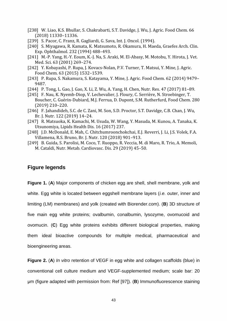
43
[238] W. Liao, K.S. Bhullar, S. Chakrabarti, S.T. Davidge, J. Wu, J. Agric. Food Chem. 66 (2018) 11330–11336.
[239] S. Pacor, C. Franz, R. Gagliardi, G. Sava, Int. J. Oncol. (1994). [240] S. Miyagawa, R. Kamata, K. Matsumoto, R. Okamura, H. Maeda, Graefes Arch. Clin.
Exp. Ophthalmol. 232 (1994) 488–493. [241] M.-P. Yang, H.-Y. Eoum, K.-J. Na, S. Araki, M. El-Abasy, M. Motobu, Y. Hirota, J. Vet.
Med. Sci. 63 (2001) 269–274. [242] Y. Kobayashi, P. Rupa, J. Kovacs-Nolan, P.V. Turner, T. Matsui, Y. Mine, J. Agric.
Food Chem. 63 (2015) 1532–1539. [243] P. Rupa, S. Nakamura, S. Katayama, Y. Mine, J. Agric. Food Chem. 62 (2014) 9479–
9487. [244] P. Tong, L. Gao, J. Gao, X. Li, Z. Wu, A. Yang, H. Chen, Nutr. Res. 47 (2017) 81–89. [245] F. Nau, K. Nyemb-Diop, V. Lechevalier, J. Floury, C. Serrière, N. Stroebinger, T.
Boucher, C. Guérin-Dubiard, M.J. Ferrua, D. Dupont, S.M. Rutherfurd, Food Chem. 280 (2019) 210–220.
[246] F. Jahandideh, S.C. de C. Zani, M. Son, S.D. Proctor, S.T. Davidge, C.B. Chan, J. Wu, Br. J. Nutr. 122 (2019) 14–24.
[247] R. Matsuoka, K. Kamachi, M. Usuda, W. Wang, Y. Masuda, M. Kunou, A. Tanaka, K. Utsunomiya, Lipids Health Dis. 16 (2017) 237.
[248] J.D. McDonald, E. Mah, C. Chitchumroonchokchai, E.J. Reverri, J. Li, J.S. Volek, F.A. Villamena, R.S. Bruno, Br. J. Nutr. 120 (2018) 901–913.
[249] B. Guida, S. Parolisi, M. Coco, T. Ruoppo, R. Veccia, M. di Maro, R. Trio, A. Memoli, M. Cataldi, Nutr. Metab. Cardiovasc. Dis. 29 (2019) 45–50.
Figure legends
Figure 1. (A) Major components of chicken egg are shell, shell membrane, yolk and
white. Egg white is located between eggshell membrane layers (i.e. outer, inner and
limiting (LM) membranes) and yolk (created with Biorender.com). (B) 3D structure of
five main egg white proteins; ovalbumin, conalbumin, lysozyme, ovomucoid and
ovomucin. (C) Egg white proteins exhibits different biological properties, making
them ideal bioactive compounds for multiple medical, pharmaceutical and
bioengineering areas.
Figure 2. (A) In vitro retention of VEGF in egg white and collagen scaffolds (blue) in
conventional cell culture medium and VEGF-supplemented medium; scale bar: 20
μm (figure adapted with permission from: Ref [97]). (B) Immunofluorescence staining
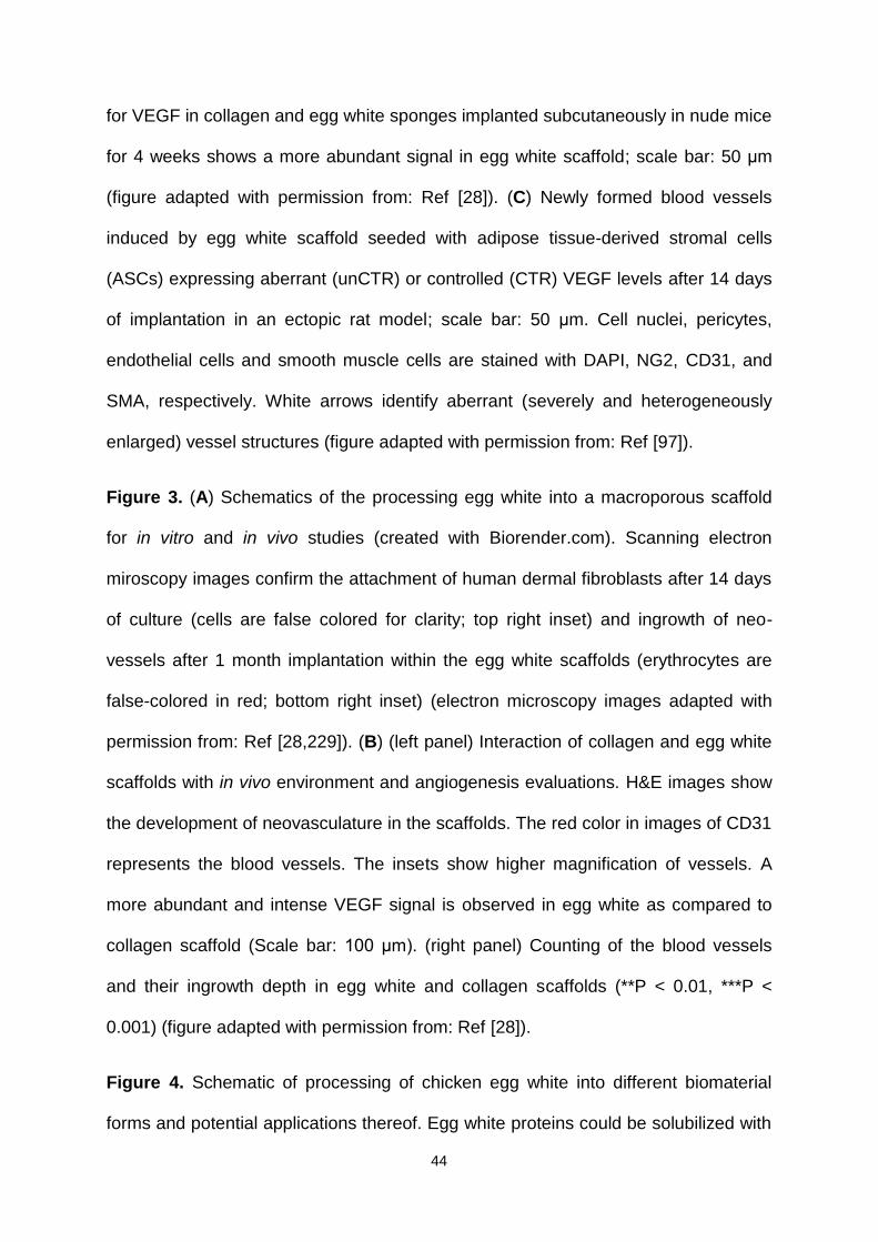
44
for VEGF in collagen and egg white sponges implanted subcutaneously in nude mice
for 4 weeks shows a more abundant signal in egg white scaffold; scale bar: 50 μm
(figure adapted with permission from: Ref [28]). (C) Newly formed blood vessels
induced by egg white scaffold seeded with adipose tissue-derived stromal cells
(ASCs) expressing aberrant (unCTR) or controlled (CTR) VEGF levels after 14 days
of implantation in an ectopic rat model; scale bar: 50 μm. Cell nuclei, pericytes,
endothelial cells and smooth muscle cells are stained with DAPI, NG2, CD31, and
SMA, respectively. White arrows identify aberrant (severely and heterogeneously
enlarged) vessel structures (figure adapted with permission from: Ref [97]).
Figure 3. (A) Schematics of the processing egg white into a macroporous scaffold
for in vitro and in vivo studies (created with Biorender.com). Scanning electron
miroscopy images confirm the attachment of human dermal fibroblasts after 14 days
of culture (cells are false colored for clarity; top right inset) and ingrowth of neo-
vessels after 1 month implantation within the egg white scaffolds (erythrocytes are
false-colored in red; bottom right inset) (electron microscopy images adapted with
permission from: Ref [28,229]). (B) (left panel) Interaction of collagen and egg white
scaffolds with in vivo environment and angiogenesis evaluations. H&E images show
the development of neovasculature in the scaffolds. The red color in images of CD31
represents the blood vessels. The insets show higher magnification of vessels. A
more abundant and intense VEGF signal is observed in egg white as compared to
collagen scaffold (Scale bar: 100 μm). (right panel) Counting of the blood vessels
and their ingrowth depth in egg white and collagen scaffolds (**P < 0.01, ***P <
0.001) (figure adapted with permission from: Ref [28]).
Figure 4. Schematic of processing of chicken egg white into different biomaterial
forms and potential applications thereof. Egg white proteins could be solubilized with
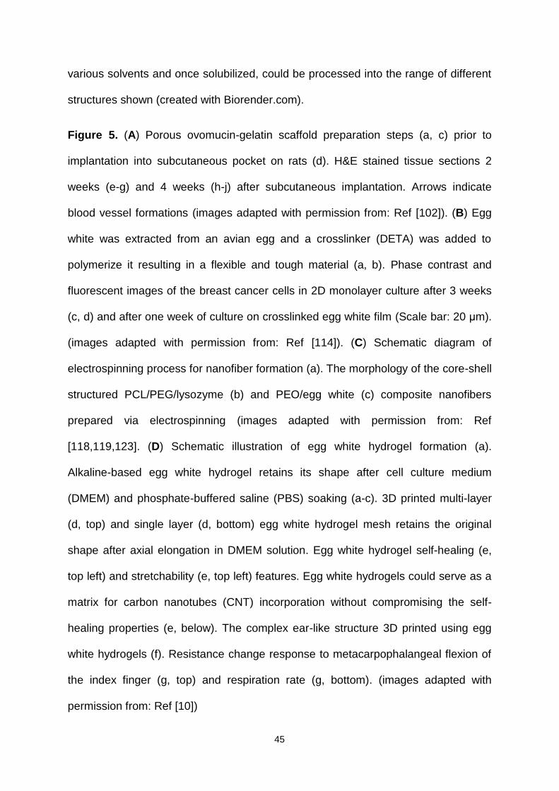
45
various solvents and once solubilized, could be processed into the range of different
structures shown (created with Biorender.com).
Figure 5. (A) Porous ovomucin-gelatin scaffold preparation steps (a, c) prior to
implantation into subcutaneous pocket on rats (d). H&E stained tissue sections 2
weeks (e-g) and 4 weeks (h-j) after subcutaneous implantation. Arrows indicate
blood vessel formations (images adapted with permission from: Ref [102]). (B) Egg
white was extracted from an avian egg and a crosslinker (DETA) was added to
polymerize it resulting in a flexible and tough material (a, b). Phase contrast and
fluorescent images of the breast cancer cells in 2D monolayer culture after 3 weeks
(c, d) and after one week of culture on crosslinked egg white film (Scale bar: 20 μm).
(images adapted with permission from: Ref [114]). (C) Schematic diagram of
electrospinning process for nanofiber formation (a). The morphology of the core-shell
structured PCL/PEG/lysozyme (b) and PEO/egg white (c) composite nanofibers
prepared via electrospinning (images adapted with permission from: Ref
[118,119,123]. (D) Schematic illustration of egg white hydrogel formation (a).
Alkaline-based egg white hydrogel retains its shape after cell culture medium
(DMEM) and phosphate-buffered saline (PBS) soaking (a-c). 3D printed multi-layer
(d, top) and single layer (d, bottom) egg white hydrogel mesh retains the original
shape after axial elongation in DMEM solution. Egg white hydrogel self-healing (e,
top left) and stretchability (e, top left) features. Egg white hydrogels could serve as a
matrix for carbon nanotubes (CNT) incorporation without compromising the self-
healing properties (e, below). The complex ear-like structure 3D printed using egg
white hydrogels (f). Resistance change response to metacarpophalangeal flexion of
the index finger (g, top) and respiration rate (g, bottom). (images adapted with
permission from: Ref [10])
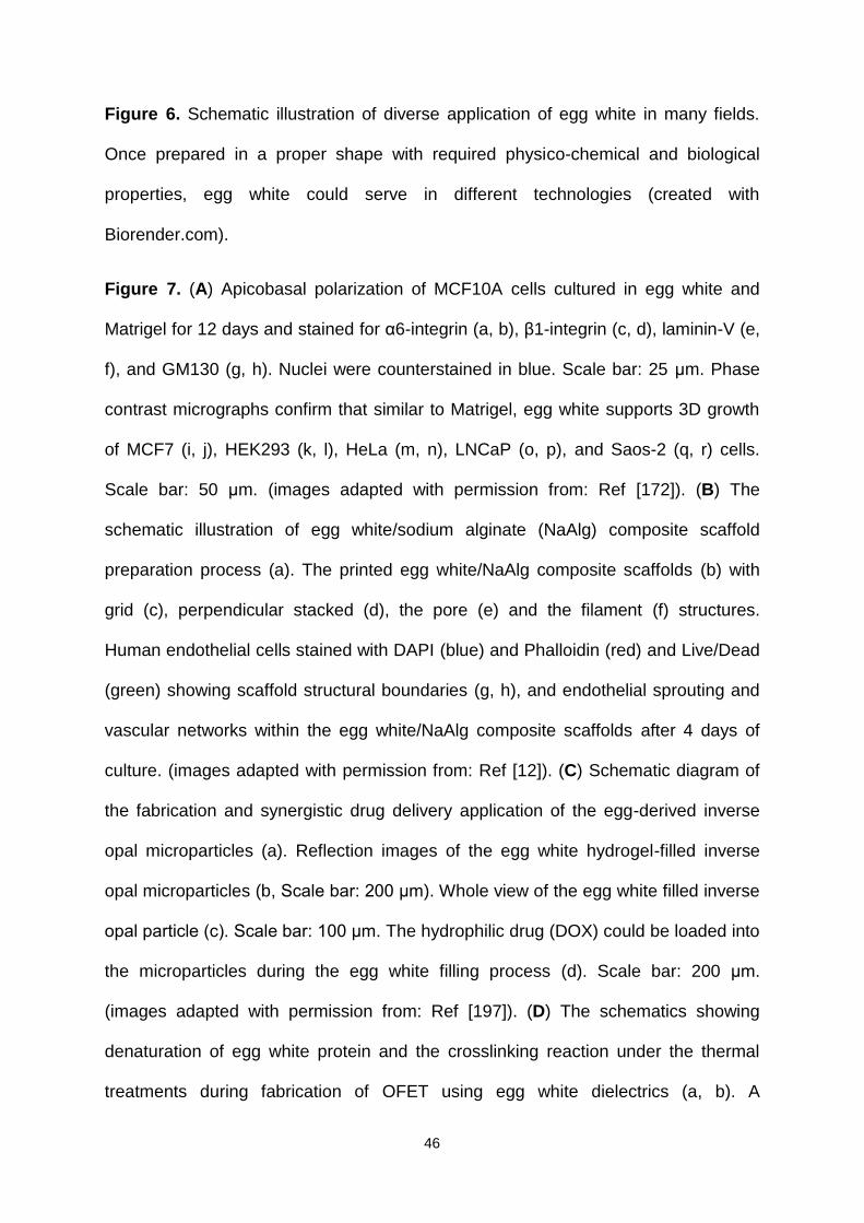
46
Figure 6. Schematic illustration of diverse application of egg white in many fields.
Once prepared in a proper shape with required physico-chemical and biological
properties, egg white could serve in different technologies (created with
Biorender.com).
Figure 7. (A) Apicobasal polarization of MCF10A cells cultured in egg white and
Matrigel for 12 days and stained for α6-integrin (a, b), β1-integrin (c, d), laminin-V (e,
f), and GM130 (g, h). Nuclei were counterstained in blue. Scale bar: 25 μm. Phase
contrast micrographs confirm that similar to Matrigel, egg white supports 3D growth
of MCF7 (i, j), HEK293 (k, l), HeLa (m, n), LNCaP (o, p), and Saos-2 (q, r) cells.
Scale bar: 50 μm. (images adapted with permission from: Ref [172]). (B) The
schematic illustration of egg white/sodium alginate (NaAlg) composite scaffold
preparation process (a). The printed egg white/NaAlg composite scaffolds (b) with
grid (c), perpendicular stacked (d), the pore (e) and the filament (f) structures.
Human endothelial cells stained with DAPI (blue) and Phalloidin (red) and Live/Dead
(green) showing scaffold structural boundaries (g, h), and endothelial sprouting and
vascular networks within the egg white/NaAlg composite scaffolds after 4 days of
culture. (images adapted with permission from: Ref [12]). (C) Schematic diagram of
the fabrication and synergistic drug delivery application of the egg-derived inverse
opal microparticles (a). Reflection images of the egg white hydrogel-filled inverse
opal microparticles (b, Scale bar: 200 μm). Whole view of the egg white filled inverse
opal particle (c). Scale bar: 100 μm. The hydrophilic drug (DOX) could be loaded into
the microparticles during the egg white filling process (d). Scale bar: 200 μm.
(images adapted with permission from: Ref [197]). (D) The schematics showing
denaturation of egg white protein and the crosslinking reaction under the thermal
treatments during fabrication of OFET using egg white dielectrics (a, b). A

47
photograph of the device configuration (c). The capacitances of egg white, PMMA,
and PS, corresponding to driving frequencies of 200 Hz to 1 M Hz at 25 V (d).
(images adapted with permission from: Ref [224]).
Tables:
Table 1. Composition of whole egg and egg white [230,231].
Component (per 100 g) Whole egg (100%) Egg white (58%) Water (g) 74.4 88.6
Proteins (g) 12.3 10.6
lipids (g) 11.9 0.1
Carbohydrates (g) 0.7 0.8
Minerals (mg)
Na 120 155
Cl 172 175
K 125 140
Ca 50 8
P 193 18
Fe 1.7 0.1
Mg 12 10
S 164 163
Zn 1.4 0.12
Cu 0.06 0.02
Mn 0.04 0.007
I 0.05 0.003
Vitamins (mg)
Vitamin A 150 0
Vitamin B1 913 10
Vitamin B2 447 430
Vitamin B6 133 10
Vitamin B12 1 0.1
Vitamin D 1.5 0
Vitamin E 1,200 0
Folic acid 56 12
Niacin 79 90
Biotin 25 7
Pantothenic acid 1,700 250
Essential amino acids (mg)
Isoleucine 290 240
Leucine 660 560
Lysine 1,040 880
Methionine + Cystine
820 660
Phenylalanine + Tyrosine
640 670
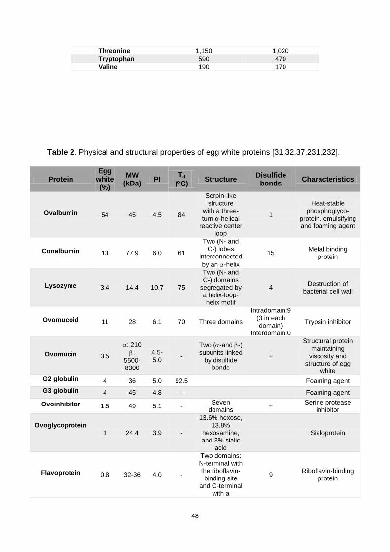
48
Threonine 1,150 1,020
Tryptophan 590 470
Valine 190 170
Table 2. Physical and structural properties of egg white proteins [31,32,37,231,232].
Protein Egg
white (%)
MW (kDa)
PI Td
(C) Structure
Disulfide bonds
Characteristics
Ovalbumin 54 45 4.5 84
Serpin-like structure
with a three-turn α-helical
reactive center loop
1
Heat-stable phosphoglyco-
protein, emulsifying and foaming agent
Conalbumin 13 77.9 6.0 61
Two (N- and C-) lobes
interconnected
by an -helix
15 Metal binding
protein
Lysozyme 3.4 14.4 10.7 75
Two (N- and C-) domains
segregated by a helix-loop-helix motif
4 Destruction of
bacterial cell wall
Ovomucoid 11 28 6.1 70 Three domains
Intradomain:9 (3 in each domain)
Interdomain:0
Trypsin inhibitor
Ovomucin 3.5
: 210
: 5500-8300
4.5-5.0
-
Two (-and -) subunits linked
by disulfide bonds
+
Structural protein maintaining
viscosity and structure of egg
white
G2 globulin 4 36 5.0 92.5 Foaming agent
G3 globulin 4 45 4.8 - Foaming agent
Ovoinhibitor 1.5 49 5.1 - Seven
domains +
Serine protease inhibitor
Ovoglycoprotein
1 24.4 3.9 -
13.6% hexose, 13.8%
hexosamine, and 3% sialic
acid
Sialoprotein
Flavoprotein 0.8 32-36 4.0 -
Two domains: N-terminal with the riboflavin-binding site
and C-terminal with a
9 Riboflavin-binding
protein

49
negatively charged amino
acid portion
Ovomacroglobuli
n 0.5 650 4.5 -
Four subunits linked by
disulfide bonds +
Strongly antigenic protein, protease
inhibitor
Cystatin 0.05 12.7 5.6 and 6.5
-
An
-helical region/a five
stranded -pleated sheet
2 Thiol protease
inhibitor
Avidin 0.05 68 10 85 Four identical
subunits 4 (1 in each
subunit) Biotin-binding
protein
MW: Molecular weight PI: Isoelectric point Td: Denaturation temperature +: Disulfide groups exist in protein Table 3. Main advantages and disadvantages of egg white biomaterials shaped in different morphologies.
Morphologies Strengths Limitations
Sponge
High porosity enables cell-cell interactions and ECM deposition; Interconnective structure
allows for nutrients/wastes transport and promotes tissue development; Prevent
clustering of the cells, avoiding necrotic center formation
May shrivel; Different pore sizes are required for the specific cell types and are therefore time consuming;
Film Enables multilayer construction; Transparent
(allows wound healing monitoring); Facile fabrication processing
Not suitable for fabricating 3D complex structures; limited mechanical (compression)
properties; No cell infiltration
Hydrogel
Soft and flexible; Multiple forms (transparent, opaque or particulate); Injectable and shear-
thinning; Self-healing properties; Printable into complex 3D constructs
Not stable (uncontrolled dissolution may occur); Low mechanical
resistance; Pore size difficult to control
Nanofiber
Their nanometric scale matches that of ECM fibers; Relatively large surface area beneficial
for cell attachment and bioactive factor loading
Low mechanical properties; Fabrication process is challenging
(bead formation); Limited cell infiltration
Microparticle
Easily fabricated with controlled physical characteristics suitable for fast or sustained
drug delivery; Targeted drug delivery;
Batch-to-batch variation hinders the scaling-up process;
Nanogel
Prompt reaction to environmental stimulations; Reversible sites to bind and
release drugs; Thermoreversible hydrophobicity and size change
Limited control over the nanogel size
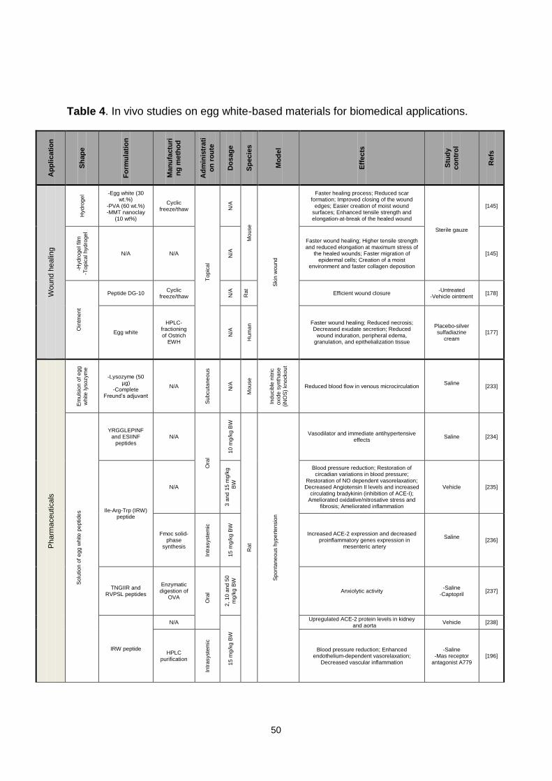
50
Table 4. In vivo studies on egg white-based materials for biomedical applications.
Ap
plic
ati
on
Sh
ap
e
Fo
rmu
lati
on
Man
ufa
ctu
ri
ng
me
tho
d
Ad
min
istr
ati
on
ro
ute
Do
sag
e
Sp
ec
ies
Mo
de
l
Eff
ec
ts
Stu
dy
co
ntr
ol
Re
fs
Wound h
ealin
g
Hydro
ge
l -Egg white (30 wt.%)
-PVA (60 wt.%) -MMT nanoclay
(10 wt%)
Cyclic freeze/thaw
To
pic
al
N/A
Mo
use
Skin
wou
nd
Faster healing process; Reduced scar formation; Improved closing of the wound
edges; Easier creation of moist wound surfaces; Enhanced tensile strength and elongation-at-break of the healed wound
Sterile gauze
[145]
-Hydro
ge
l film
-To
pic
al h
yd
roge
l
N/A N/A
N/A
Faster wound healing; Higher tensile strength and reduced elongation at maximum stress of
the healed wounds; Faster migration of epidermal cells; Creation of a moist
environment and faster collagen deposition
[145]
Oin
tme
nt
Peptide DG-10 Cyclic
freeze/thaw N/A
Rat
Efficient wound closure -Untreated
-Vehicle ointment [178]
Egg white
HPLC-fractioning of Ostrich
EWH
N/A
Hum
an Faster wound healing; Reduced necrosis;
Decreased exudate secretion; Reduced wound induration, peripheral edema,
granulation, and epithelialization tissue
Placebo-silver sulfadiazine
cream [177]
Pharm
aceuticals
Em
uls
ion
of
eg
g
white
lysozym
e
-Lysozyme (50 µg)
-Complete Freund’s adjuvant
N/A
Su
bcu
tan
eo
us
N/A
Mo
use
Ind
ucib
le n
itri
c
oxid
e s
yn
tha
se
(iN
OS
) kn
ocko
ut
Reduced blood flow in venous microcirculation Saline
[233]
So
lution
of
eg
g w
hite
pe
ptid
es
YRGGLEPINF and ESIINF
peptides N/A
Ora
l
10
mg
/kg
BW
Rat
Sp
on
tan
eo
us h
yp
ert
en
sio
n
Vasodilator and immediate antihypertensive effects
Saline [234]
Ile-Arg-Trp (IRW) peptide
N/A
3 a
nd
15
mg
/kg
BW
Blood pressure reduction; Restoration of circadian variations in blood pressure;
Restoration of NO dependent vasorelaxation; Decreased Angiotensin II levels and increased
circulating bradykinin (inhibition of ACE-I); Ameliorated oxidative/nitrosative stress and
fibrosis; Ameliorated inflammation
Vehicle [235]
Fmoc solid-phase
synthesis
Intr
asyste
mic
15
mg
/kg
BW
Increased ACE-2 expression and decreased proinflammatory genes expression in
mesenteric artery
Saline
[236]
TNGIIR and RVPSL peptides
Enzymatic digestion of
OVA
Ora
l
2,
10
an
d 5
0
mg
/kg
BW
Anxiolytic activity -Saline
-Captopril [237]
IRW peptide
N/A
15
mg
/kg
BW
Upregulated ACE-2 protein levels in kidney and aorta
Vehicle [238]
HPLC purification
Intr
asyste
mic
Blood pressure reduction; Enhanced endothelium-dependent vasorelaxation;
Decreased vascular inflammation
-Saline -Mas receptor
antagonist A779 [196]
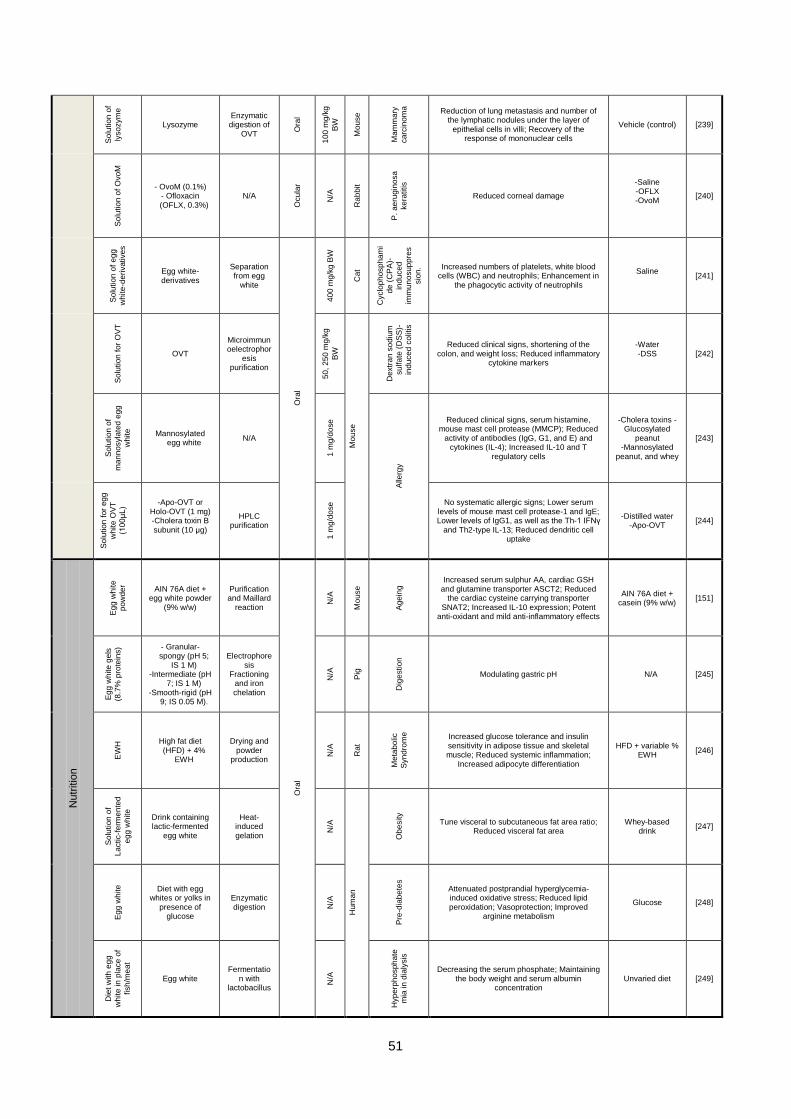
51
So
lution
of
lyso
zym
e
Lysozyme Enzymatic digestion of
OVT
Ora
l
10
0 m
g/k
g
BW
Mo
use
Ma
mm
ary
ca
rcin
om
a
Reduction of lung metastasis and number of the lymphatic nodules under the layer of
epithelial cells in villi; Recovery of the response of mononuclear cells
Vehicle (control) [239] S
olu
tion
of
Ovo
M
- OvoM (0.1%) - Ofloxacin (OFLX, 0.3%)
N/A
Ocu
lar
N/A
Rab
bit
P.
ae
rug
ino
sa
ke
ratitis
Reduced corneal damage
-Saline -OFLX -OvoM
[240]
So
lution
of
eg
g
white
-de
rivative
s
Egg white-derivatives
Separation from egg
white
Ora
l
40
0 m
g/k
g B
W
Cat
Cyclo
ph
osp
ha
mi
de
(C
PA
)-
ind
uce
d
imm
un
osup
pre
s
sio
n. Increased numbers of platelets, white blood
cells (WBC) and neutrophils; Enhancement in the phagocytic activity of neutrophils
Saline
[241]
So
lution
fo
r O
VT
OVT
Microimmunoelectrophor
esis purification
50
, 2
50
mg
/kg
BW
Mo
use
Dextr
an
so
diu
m
su
lfa
te (
DS
S)-
ind
uce
d c
olit
is
Reduced clinical signs, shortening of the colon, and weight loss; Reduced inflammatory
cytokine markers
-Water -DSS
[242]
So
lution
of
ma
nn
osyla
ted
eg
g
white
Mannosylated egg white
N/A
1 m
g/d
ose
Alle
rgy
Reduced clinical signs, serum histamine, mouse mast cell protease (MMCP); Reduced
activity of antibodies (IgG, G1, and E) and cytokines (IL-4); Increased IL-10 and T
regulatory cells
-Cholera toxins -Glucosylated
peanut -Mannosylated
peanut, and whey
[243]
So
lution
fo
r eg
g
white
OV
T
(10
0μ
L)
-Apo-OVT or Holo-OVT (1 mg) -Cholera toxin B subunit (10 μg)
HPLC purification
1 m
g/d
ose No systematic allergic signs; Lower serum
levels of mouse mast cell protease-1 and IgE; Lower levels of IgG1, as well as the Th-1 IFNγ
and Th2-type IL-13; Reduced dendritic cell uptake
-Distilled water -Apo-OVT
[244]
Nutr
itio
n
Eg
g w
hite
po
wde
r
AIN 76A diet + egg white powder
(9% w/w)
Purification and Maillard
reaction
Ora
l
N/A
Mo
use
Ag
ein
g Increased serum sulphur AA, cardiac GSH
and glutamine transporter ASCT2; Reduced the cardiac cysteine carrying transporter
SNAT2; Increased IL-10 expression; Potent anti-oxidant and mild anti-inflammatory effects
AIN 76A diet + casein (9% w/w)
[151]
Eg
g w
hite
ge
ls
(8.7
% p
rote
ins)
- Granular-spongy (pH 5;
IS 1 M) -Intermediate (pH
7; IS 1 M) -Smooth-rigid (pH
9; IS 0.05 M).
Electrophoresis
Fractioning and iron chelation
N/A
Pig
Dig
estio
n
Modulating gastric pH N/A [245]
EW
H High fat diet
(HFD) + 4% EWH
Drying and powder
production
N/A
Rat
Me
tab
olic
Syn
dro
me
Increased glucose tolerance and insulin sensitivity in adipose tissue and skeletal muscle; Reduced systemic inflammation;
Increased adipocyte differentiation
HFD + variable % EWH
[246]
So
lution
of
La
ctic-f
erm
en
ted
eg
g w
hite
Drink containing lactic-fermented
egg white
Heat-induced gelation
N/A
Hum
an
Ob
esity
Tune visceral to subcutaneous fat area ratio; Reduced visceral fat area
Whey-based drink
[247]
Eg
g w
hite
Diet with egg whites or yolks in
presence of glucose
Enzymatic digestion N
/A
Pre
-dia
be
tes
Attenuated postprandial hyperglycemia-induced oxidative stress; Reduced lipid peroxidation; Vasoprotection; Improved
arginine metabolism
Glucose [248]
Die
t w
ith
eg
g
white
in
pla
ce
of
fish
/me
at
Egg white Fermentatio
n with lactobacillus
N/A
Hype
rph
osph
ate
mia
in
dia
lysis
Decreasing the serum phosphate; Maintaining the body weight and serum albumin
concentration Unvaried diet [249]

52
Tis
sue e
ngin
eerin
g
Ma
cro
poro
us
sp
on
ge
Egg white
Lyophilization
Su
bcu
tan
eo
us
So
ft/v
ascu
lar
tissu
e e
ng
inee
ring
Mo
use
Su
bcu
tan
eo
us im
pla
nta
tio
n
Low immune reaction; Enhanced angiogenesis; Effective tissue ingrowth
Collagen sponges
[28]
Rat
Aberrantly enlarged vascular structures Collagen sponges
[97]
Po
rou
s s
ca
ffo
ld
-Ovomucin (50%) -Gelatin (50%)
EDC crosslinking
Bo
ne
or
derm
is
tissu
e e
ng
inee
ring
Increased vascularization over time; Limited fibrosis; Minimal allergic responses; Activating
macrophages favorable for tissue repair N/A [102]
BW: Body weight OVT: Ovotransferrin EWH: Egg white hydrolysates OvoM: Ovomacroglobulin N/A: Not available