Do amphibious crabs have amphibious eggs? A case study of Armases miersii
Upload
khangminh22Category
view
3download
0
Import risk analysis: Passerine hatching eggs from the European Union
1 February 2006


Import risk analysis: Passerine hatching eggs from the European Union
Biosecurity New Zealand Ministry of Agriculture and Forestry
Wellington New Zealand
1 February 2006

This page is intentionally blank

Ministry of Agriculture and Forestry Te Manatu Ahuwhenua, Ngaherehere
ASB Bank House 101-103 The Terrace
P O Box 2526 Wellington
New Zealand
Telephone: +64 4 474 4100 Facsimile: +64 4 474 4133
Internet: http://www.maf.govt.nz
Animal Biosecurity Biosecurity Authority
Import risk analysis: Passerine hatching eggs from the European Union
1 February 2006
Approved for general release
Debbie Pearson Director Preclearance
Biosecurity New Zealand

This page is intentionally blank

- i -
TABLE OF CONTENTS
1 EXECUTIVE SUMMARY ...................................................................................... 1
2 INTRODUCTION..................................................................................................... 3
2.1 COMMODITY DEFINITION ..................................................................................... 3 2.2 BACKGROUND...................................................................................................... 3 2.3 METHODOLOGY ................................................................................................... 4
3 ORGANISM RISK ANALYSES........................................................................... 12
3.1 ORTHOMYXOVIRIDAE ........................................................................................ 12 3.1.1 Avian influenza.............................................................................................. 12
3.2 PARAMYXOVIRIDAE ........................................................................................... 21 3.2.1 Avian paramyxoviruses................................................................................. 21 3.2.2 Pneumovirus ................................................................................................. 31
3.3 HERPESVIRIDAE ................................................................................................. 35 3.3.1 Duck virus enteritis ....................................................................................... 35 3.3.2 Infectious laryngotracheitis .......................................................................... 36 3.3.3 Marek’s disease ............................................................................................ 37 3.3.4 Other avian herpesviruses ............................................................................ 38
3.4 CORONAVIRIDAE...................................................................................................................46 3.5 ADENOVIRIDAE.................................................................................................. 48
3.5.1 Group I adenoviruses.................................................................................... 48 3.5.2 Group II adenoviruses – HE, MSD, AAS...................................................... 53 3.5.3 Group III adenoviruses – EDS...................................................................... 55
3.6 POXVIRIDAE....................................................................................................... 57 3.6.1 Avipoxvirus ................................................................................................... 57
3.7 CIRCOVIRIDAE ................................................................................................... 62 3.7.1 General ......................................................................................................... 62 3.7.2 Chicken infectious anaemia .......................................................................... 63 3.7.3 Psittacine beak and feather disease.............................................................. 64 3.7.4 Circovirus-like viruses .................................................................................. 65
3.8 BIRNAVIRIDAE ................................................................................................... 68 3.8.1 Infectious bursal disease............................................................................... 68
3.9 PAPOVAVIRIDAE ................................................................................................ 71 3.9.1 Polyomavirus ................................................................................................ 71 3.9.2 Papillomavirus.............................................................................................. 73
3.10 PARVOVIRIDAE AND OTHER NON-SPECIFIC ENTERITIS-ASSOCIATED AGENTS...... 76 3.10.1 Parvoviruses ............................................................................................. 76 3.10.2 Enteritis associated viruses s)................................................................... 78
3.11 TOGAVIRIDAE .................................................................................................... 79 3.11.1 Equine encephalitis (Eastern, Western & Venezuelan) ............................ 79 3.11.2 Other Alphaviruses ................................................................................... 80
3.12 FLAVIVIRIDAE.................................................................................................... 85 3.12.1 West Nile virus .......................................................................................... 85 3.12.2 Japanese encephalitis virus ...................................................................... 88

- ii -
3.12.3 Louping Ill................................................................................................. 90 3.12.4 Other flaviviruses...................................................................................... 92
3.13 REOVIRIDAE....................................................................................................... 94 3.13.1 Rotavirus ................................................................................................... 94 3.13.2 Orbivirus ................................................................................................... 97 3.13.3 Other reoviruses........................................................................................ 99
3.14 BUNYAVIRIDAE................................................................................................ 100 3.14.1 Nairoviruses............................................................................................ 100 3.14.2 Other Bunyaviruses................................................................................. 103
3.15 BORNAVIRIDAE................................................................................................ 107 3.15.1 Borna virus.............................................................................................. 107
3.16 PICORNAVIRIDAE ............................................................................................. 110 3.16.1 Avian encephalomyelitis ......................................................................... 110 3.16.2 Duck hepatitis (DHV 1 & 3) ................................................................... 112
3.17 ASTROVIRIDAE................................................................................................. 113 3.17.1 Astroviruses - duck hepatitis, turkey astrovirus, Avian nephritis .......... 113
3.18 HEPADNAVIRIDAE............................................................................................ 116 3.18.1 Hepadnavirus (duck virus hepatitis) ....................................................... 116
3.19 RETROVIRIDAE................................................................................................. 118 3.19.1 Avian leucosis-sarcoma group................................................................ 118 3.19.2 Lymphoproliferative disease virus.......................................................... 122 3.19.3 Avian reticuloendotheliosis virus group ................................................. 123 3.19.4 Other retroviruses................................................................................... 125
3.20 CHLAMYDOPHILA ............................................................................................ 126 3.20.1 Chlamydophila spp. (ornithosis)............................................................. 126
3.21 BACTERIA ASSOCIATED WITH ENTERIC & GENERALIZED INFECTION IN BIRDS. . 130 3.21.1 Salmonellae - general ............................................................................. 130 3.21.2 Salmonella enterica subsp. enterica serovar Gallinarum-Pullorum...... 131 3.21.3 Salmonella Abortusovis, S. arizonae and S. Dublin. .............................. 133 3.21.4 Salmonella Typhimurium DT 104 (antibiotic resistant). ........................ 136 3.21.5 Salmonella Enteritis phage type 4 .......................................................... 140 3.21.6 Salmonella Typhimurium phage type 44................................................. 143 3.21.7 Other Salmonellae .................................................................................. 145 3.21.8 Escherichia coli ...................................................................................... 151 3.21.9 Campylobacter spp. ................................................................................ 155 3.21.10 Other Enterobacteriaceae....................................................................... 161
3.22 BACTERIA COMMONLY ASSOCIATED WITH RESPIRATORY DISEASE IN BIRDS .... 168 3.22.1 Pasteurella multocida ............................................................................. 168 3.22.2 Riemerella anatipestifer.......................................................................... 170 3.22.3 Other Pasteurellae .................................................................................. 172 3.22.4 Ornithobacterium rhinotracheale........................................................... 172 3.22.5 Bordetella spp. ........................................................................................ 176 3.22.6 Haemophilus paragallinarum................................................................. 178 3.22.7 Mycoplasma spp...................................................................................... 179

- iii -
3.23 INTRACELLULAR BACTERIA ............................................................................. 187 3.23.1 Mycobacterium tuberculosis ................................................................... 187 3.23.2 Mycobacterium avium............................................................................. 189 3.23.3 Other Mycobacteria ................................................................................ 191
3.24 OTHER BACTERIA............................................................................................. 193 3.24.1 Francisella tularensis. ............................................................................ 193 3.24.2 Megabacteria .......................................................................................... 195 3.24.3 Gram positive contaminants (Streptococci/Staphylococci) .................... 198
3.25 SPIROCHETES ................................................................................................... 200 3.25.1 Borrelia anserina (avian spirocaetosis) ................................................. 200 3.25.2 Borrelia burgdorferi (Lyme disease) ...................................................... 203 3.25.3 Brachyspira spp. ..................................................................................... 205
3.26 RICKETTSIAL AGENTS ...................................................................................... 208 3.26.1 Coxiella burnetii ..................................................................................... 208 3.26.2 Cowdria ruminantium............................................................................. 211 3.26.3 Aegyptianella pullorum........................................................................... 213 3.26.4 Other Rickettsia ...................................................................................... 216
3.27 FUNGI AND YEASTS. ......................................................................................... 218 3.27.1 Hazard identification .............................................................................. 218
3.28 INTERNAL PARASITES....................................................................................... 226 3.28.1 Nematodes............................................................................................... 226 3.28.2 Trematodes.............................................................................................. 228 3.28.3 Cestodes .................................................................................................. 229 3.28.4 Acanthocephala....................................................................................... 231 3.28.5 Protozoa.................................................................................................. 233
3.29 EXTERNAL PARASITES...................................................................................... 235

- iv -
CONTRIBUTORS TO THIS RISK ANALYSIS 1. Author Bruce Simpson Biosecurity Ltd, Palmerston North, New Zealand 2. Internal Peer Review Susan Cork Biosecurity New Zealand, Wellington Rachel Gordon Biosecurity New Zealand, Wellington Howard Pharo Biosecurity New Zealand, Wellington 3. External Scientific Review Neil Christensen Avivet Ltd, Palmerston North, New Zealand Brett Gartrell Massey University, Palmerston North, New Zealand
MINISTRY OF AGRICULTURE AND FORESTRY PASSERINE HATCHING EGGS ● 1
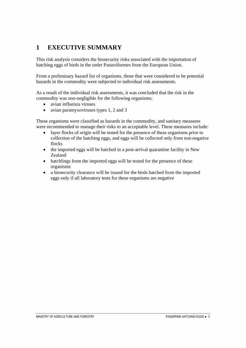
1 EXECUTIVE SUMMARY This risk analysis considers the biosecurity risks associated with the importation of hatching eggs of birds in the order Passeriformes from the European Union. From a preliminary hazard list of organisms, those that were considered to be potential hazards in the commodity were subjected to individual risk assessments. As a result of the individual risk assessments, it was concluded that the risk in the commodity was non-negligible for the following organisms:
• avian influenza viruses • avian paramyxoviruses types 1, 2 and 3
These organisms were classified as hazards in the commodity, and sanitary measures were recommended to manage their risks to an acceptable level. These measures include:
• layer flocks of origin will be tested for the presence of these organisms prior to collection of the hatching eggs, and eggs will be collected only from test-negative flocks
• the imported eggs will be hatched in a post-arrival quarantine facility in New Zealand
• hatchlings from the imported eggs will be tested for the presence of these organisms
• a biosecurity clearance will be issued for the birds hatched from the imported eggs only if all laboratory tests for these organisms are negative

2 ● PASSERINE HATCHING EGGS MINISTRY OF AGRICULTURE AND FORESTRY
This page is intentionally blank
MINISTRY OF AGRICULTURE AND FORESTRY PASSERINE HATCHING EGGS ● 3
2 INTRODUCTION This risk analysis examines the biosecurity risks posed by the importation of hatching eggs of birds in the taxonomic order Passeriformes from the European Union into New Zealand. 2.1 Commodity definition The commodity is hatching eggs of any species of the order Passeriformes from the EU. The eggs must be clean (free of faeces) when collected, unwashed and have intact shells (uncracked). Following collection the eggs must be disinfected in accordance with Appendix 3.4.1 of the OIE Terrestrial Animal Health Code. 2.2 Background Zoological gardens and aviary owners wish to import passerine eggs for the purposes of hatching to produce birds for inclusion in their collections. Over 50% of the world’s 9,600 avian species fall within the Order Passeriformes. These are the song birds or perching birds and include such well known birds as house sparrows, starlings, thrushes, magpies, crows, swallows and the many species of finches. There are 53 families that fall directly within the order Passeriformes, while another 29 families are contained in the suborder Oscines and 5 families are contained in the suborder Tyranni (1). There have been very few evaluations of the diseases or potentially pathogenic organisms carried by bird eggs, other than those of poultry, in New Zealand. Most relevant avian disease information comes from poultry species and/or sporadic case reports and/or from local and regional surveys. Many of the organisms considered in this risk analysis commonly infect birds without causing disease. On occasions, however, they may be associated with incidents of disease. Examples of this include avian influenza viruses, paramyxoviruses, adenoviruses, alphaviruses, bunyaviruses and Salmonella spp. Surveillance for many of these organisms in New Zealand is relatively insensitive so that their lack of recognition in this country does not provide a basis for confidence that they are not present. In New Zealand surveillance information on diseases in passerine species comes mainly from passive surveillance (i.e. reports of incidents of disease sufficiently pronounced to attract attention and to encourage investment in professional examinations and laboratory investigations) and it is likely that organisms causing sub-clinical disease or only occasional clinical disease may remain undiagnosed. More information is available on organisms present in passerines in Great Britain, continental Europe and the United States. This arises, in part, from the potential for these birds to carry zoonotic organisms and it also results from the interest of the public and

4 ● PASSERINE HATCHING EGGS MINISTRY OF AGRICULTURE AND FORESTRY
scientists in ensuring the well being of their native and introduced avifauna. The recognition that passerine birds are contributing to the global phenomenon of “emerging diseases” originating in wildlife and causing disease in humans and animals has contributed to recent increases in interest in the organisms (particularly arthropod-borne viruses) carried by these birds. Prominent amongst these is West Nile Virus which had been recognised in Africa, with incursions into southern Europe, for many years before it emerged in North America in 1999 causing extensive mortalities in passerine birds and numerous deaths in humans. Large numbers of birds, previously exotic to New Zealand, have been imported with little or no evaluation of their carriage of organisms that might, in today’s terms, be classified as hazards. Importations of passerine birds identified by Heather and Robertson (2) are house sparrows (>100, source not stated, 1866 – 1871), chaffinches (several hundred, from South Africa, 1862 – 1880), redpolls (500, source not stated, 1862 – 1875), goldfinches (500, source not stated, 1862), greenfinches (<100, source not stated, 1862 – 1880), yellow hammers, starlings (1,000, 1862 – 1883), mynahs (several hundred, source not stated, 1870 – 1880), Australian magpies (>1,000, Australia, 1864 – 1874), black birds (1,000, source not stated, 1862 – 1875) and song thrushes (several hundred, source not stated, 1862 – 1878). All of these birds were imported prior to the recognition of most of the diseases covered in this risk analysis and prior to the recognition of the aetiology of any of them. During the 20th century, importations of large numbers of birds, including passerine species, continued from Europe and the United States into the 1960s and from Australia until 1997. An unknown number of the birds imported from Australia had originated in Europe and entered Australia under entry conditions directed at protecting Australia from the major epidemic diseases of poultry, particularly Newcastle disease and avian influenza.(3) It is likely that a high proportion of potential hazards that could reasonably be expected to have been imported with passerine species from Europe and Australia entered New Zealand with the importations that have taken place over the past 143 years. 2.3 Methodology The methodology used in this risk analysis follows the guidelines in Section 1.3 of the OIE Terrestrial Animal Health Code (4). In New Zealand, the OIE risk analysis framework is applied as described in Import Risk Analysis Animals and Animal Products (5), the key elements of which are shown in Figure 1. The hazard identification process begins with the collation of a list of organisms potentially associated with the commodity. Table 1 shows these organisms, together with some of the key information considered for each organism in determining whether or not it must be classified as a potential hazard in the commodity. This list was compiled from those contagious diseases of passerine birds identified from the several standard textbooks (6, 7) and from searches of the international scientific literature. Additional diseases were included on the basis of initial uncertainty as to whether they might infect passerines species.

MINISTRY OF AGRICULTURE AND FORESTRY PASSERINE HATCHING EGGS ● 5
Figure 1. The risk analysis process.
Organisms of potential concern:* OIE listed* organisms affecting the economy, the people, the environment of New Zealand
Not of concern in this risk analysis
Not considered to be a potential hazard in this commodity
No
Yes
Is the organism associated with
the animal species?
concerned?
Is the organism likely to be
associated with the commodity?
Are strain differences reported in
other countries?
Is the organism exotic to New
Zealand?
RISK ASSESSMENT
Release assessmentHow likely is the agent to be
introduced in the commodity?
Exposure assessmentHow likely are susceptible animals to be exposed?
Consequence assessmentWhat are the likely
consequences of exposure?
Risk estimationIs the organism considered
to be a hazard in the commodity?Is there a
control programme in New Zealand?
What is the acceptable level
of risk?
How does the assessed risk
compare to the acceptable level
of risk?
What is the effect of each safeguard
on the level of risk?
RISK MANAGEMENT
HAZARD IDENTIFICATION
Apply safeguards that reduce risk from assessed level to acceptable level
No
No
No
No
Yes
Yes
Yes
Yes
What safeguards are
available?
Potential hazard in the
commodity
Yes
No safeguards necessary
No

6 ● PASSERINE HATCHING EGGS MINISTRY OF AGRICULTURE AND FORESTRY
Table 1. Organisms considered in this risk analysis Organism / Disease Present in
New Zealand?
OIE listed? 1 Under official control or unwanted ? 2
More virulent strains overseas ? 3
Orthomyxoviridae Avian influenza Yes Yes
(H5 or H7 strains or any highly pathogenic strains)
Unwanted (H5 and H7 strains)
Yes
Paramyxoviridae Avian paramyxovirus 1 (APMV-1 to 9)
Yes (Some serogroups and strains)
Yes (Newcastle disease)
Notifiable (Exotic strains - Newcastle disease)
Yes
Pneumoviruses No? No None N.A. Herpesviridae Duck virus enteritis No Yes Notifiable N.A. Laryngotracheitis Yes Yes None Yes Marek’s disease Yes Yes Other exotic
organism (Exotic strains)
Yes
Other avian Herpes viruses
Yes / No No Pacheco’s disease (Other exotic organism)
Yes
Coronaviridae Infectious bronchitis Yes Yes Unwanted
(exotic strains) Yes
Adenoviridae Group I avian adenoviruses
Yes
No None Yes
Group II avian adenoviruses
No No None N.A.
Group III avian adenoviruses (Egg Drop Syndrome)
Yes No None No
Poxviridae Avipoxvirus Yes Yes
(Fowl pox) None Yes
Circoviridae Gyrovirus (Chicken Infectious Anaemia)
Yes No None No
Avian Circovirus Yes No None Yes Birnaviridae Infectious Bursal Disease No Yes Unwanted
(Exotic strains) N.A.

MINISTRY OF AGRICULTURE AND FORESTRY PASSERINE HATCHING EGGS ● 7
Papovaviridae Polyomavirus Yes No None No Papillomavirus No No None N.A. Parvoviridae Parvoviruses No No Unwanted N.A. Togaviridae Equine encephalitides No No Notifiable N.A. Other Alphaviruses Yes No None Yes Flaviviridae West Nile Virus No No None N.A. Japanese encephalitis virus
No No Notifiable N.A.
Louping ill No No Unwanted N.A. Other Flaviviruses No No None N.A. Reoviridae Rotavirus Yes No None Yes Orbivirus Yes No None No Other reoviruses Yes No None Yes Bunyaviridae Nairoviruses No No None N.A. Other Bunyaviruses No No None N.A. Bornaviridae Bornavirus No No Unwanted N.A. Picornaviridae Avian encephalomyelitis Yes No None No Duck hepatitis (DHV 1 & 3)
No No Unwanted N.A.
Astroviridae Astroviruses Uncertain No None N.A. Hepadaviridae Duck virus hepatitis No Yes Unwanted N.A. Retroviridae Leucosis/sarcoma complex viruses
Yes No None No
Lymphoproliferative disease virus
No No Unwanted N.A.
Reticuloendotheliosis Yes No None No Other retroviruses No No None No Chlamydophila Chlamydia sp Yes Yes None Yes Bacteria associated with enteric and generalised infections in birds Salmonella spp Yes / No Yes
(Fowl typhoid, Pullorum disease)
Unwanted (Some serovars and variants within serovars)
Yes
Escherichia coli Yes No None No

8 ● PASSERINE HATCHING EGGS MINISTRY OF AGRICULTURE AND FORESTRY
Campylobacter spp. Yes No None No Other Enterobacteriaceae Yes / No No Not species
infecting birds Not species infecting birds
Bacteria commonly associated with respiratory disease in birds Pasteurella multocida Yes Yes
(Fowl cholera)
None Yes
Riemerella anatipestifer Yes No None No Pasteurella gallinarum No No None N.A. Ornithobacterium rhinotracheale
No No Other exotic organism
N.A.
Bordetella avium No No Other exotic organism
N.A.
Haemophilus paragallinarum
No No Other exotic organism
N.A.
Mycoplasma gallisepticum Yes Yes None No Mycoplasma synoviae Yes No None No Mycoplasma iowae No No Other exotic
organism N.A.
Intracellular bacteria Mycobacterium tuberculosis
Yes No Unwanted No
Mycobacterium avium Yes Yes None No Other Mycobacteria Yes (Some) No Other exotic
organism (exotic strains)
Yes
Other bacteria Francisella tularensis No No Other exotic
organism N.A.
Megabacteria Yes No None No Gram positive contaminants (e.g. Staphylococci / Streptococci)
Yes No None No
Spirochetes Borrelia anserina (Avian spirochaetosis)
No No Other exotic organism
N.A.
Borrelia burgdorferi (Lyme Disease)
No No Other exotic organism
N.A.
Brachyspira spp Yes No None No Rickettsial agents Coxiella burnetii No No Notifiable N.A. Cowdria ruminantium No No Unwanted N.A. Aegyptianella pullorum No No None N.A. Other Rickettsia Yes / No No Some general in
register Yes

MINISTRY OF AGRICULTURE AND FORESTRY PASSERINE HATCHING EGGS ● 9
Fungi and yeasts Yes / No No Yes
(Histoplasma farciminosum)
Yes
Internal parasites (Nematodes, cestodes, protozoa)
Yes / No No None Yes
External parasites (ticks, mites, lice)
Yes / No No Unwanted (Some genera)
Yes
1 Based on information on the OIE website at
www.oie.int/eng/maladies/en_classification.htm 2 Based on the information from the register of unwanted organism at
http://www.biosecurity.govt.nz/pests-diseases/registers-lists/unwanted organisms/
3 More virulent exotic strains are recognised where either strain typing of New Zealand isolates allows differentiation from more pathogenic types recognised in other countries or where descriptions of the disease in New Zealand allow it to be recognised as less virulent than disease episodes in other countries. Where host specific strains are recognised overseas but not in NZ, these are treated as “more virulent” in the compilation of this table.
N.A. Not applicable because assessment of strain variations is not relevant to this process when the organism is not recognised as present in New Zealand.
In this analysis, for each organism listed, the epidemiology is discussed, including a consideration of the following questions:
1) whether eggs from passerine birds could act as a vehicle for the introduction of the organism,
2) if the organism requires a vector, whether competent vectors might be in New Zealand,
3) whether the organism is exotic to New Zealand but likely to be present in exporting countries and
4) if it is present in New Zealand, a. whether it is "under official control", which could be by government
departments, by national or regional pest management strategies or by a small-scale programme, or
b. whether more virulent strains are known to exist in other countries. For any organism, if the answers to question one is “yes” (and the answer to question 2 is “yes” in the cases of organisms requiring a vector) and the answers to either questions three or four are ‘yes’, it is classified as a potential hazard. Under this framework, which is based on international agreements on trade in agricultural products, organisms that are present in New Zealand cannot be considered as as hazards unless there is evidence that strains with higher pathogenicity are likely to be present in the commodity to be imported. Therefore, although there may be potential for organisms to be present in the imported commodity, the risks to human or animal health are no different than risks resulting from the presence of the organism in this country already. In

10 ● PASSERINE HATCHING EGGS MINISTRY OF AGRICULTURE AND FORESTRY
such situations, measures to limit negative impacts on the health of humans or animals in contact with the imported commodity, or subsequent progeny, should be those appropriate to good practice irrespective of the importation. In line with the OIE risk analysis methodology, for each potential hazard the following analysis is carried out: Risk Assessment
a) Release assessment - the likelihood of the organism being imported in the commodity.
b) Exposure assessment - the likelihood of animals or humans in New Zealand being exposed to the potential hazard.
c) Consequence assessment - the consequences of entry, establishment or spread of the organism.
d) Risk estimation - a conclusion on the risk posed by the organism based on the release, exposure and consequence assessments. If the risk estimate is non-negligible, then the organism is classified as a hazard.
In assessing the likelihood of exposure to wild birds, caged or aviary birds or poultry in New Zealand, an assumption is made that there is potential for contact between caged or aviary birds and those outside that environment. Such contact might be direct through the walls of enclosures, indirect through transfer of fomites, movement of rodents, insects or other animals or through escape or release of the imported birds. It is important to note that all of the above steps may be necessary in all risk assessments. The OIE methodology makes it clear that if the likelihood of release is negligible for a certain potential hazard, then the risk estimate is automatically negligible and the remaining steps of the risk assessment need not be carried out. The same situation arises where the likelihood of release is non-negligible but the exposure assessment concludes that the likelihood of exposure to susceptible species in the importing country is negligible, or where both release and exposure are non-negligible but the consequences of introduction are concluded to be negligible. For all organisms that are classified as a hazard, the risk management step is carried out, comprising the following three sub-steps: a) Risk evaluation - a determination is made as to whether sanitary
measures are necessary.
b) Option evaluation - identify the options available for managing the risk, and consider risk reduction effects.

MINISTRY OF AGRICULTURE AND FORESTRY PASSERINE HATCHING EGGS ● 11
c) Recommended measures - the recommendation of the appropriate option or combination of options that achieve a negligible likelihood of entry, spread or establishment, while minimising negative trade effects.
Further details, including the full hazard identification, and where appropriate the risk assessment and the recommended risk management measures, can be found in the chapters on the individual agents. References
1. Integrated Taxonomic Information System retrieved 30 September 2004. (http://www.itis.usda.gov).
2. Heather, B.; Robertson, H. The field guide to the birds of New Zealand. Penguin Books (NZ) Ltd.). 2000.
3. Mulqueen, K. 2005. Personal communication 7 September 2005.
4. Anonymous. Terrestrial Animal Health Code. World Organisation for Animal Health 2004.
5. Murray, N. Import Risk Analysis. Animals and Animal Products. Animal Biosecurity, Ministry of Agriculture and Fisheries, 2002.
6. Rosskopf W, Woerpel R (Eds) Diseases of Cage and Aviary Birds. 3rd Edition. Williams and Wilkins, Baltimore. 1996.
7. Saif YM Diseases of Poultry. 11th Edition. Iowa State Press 2003.

12 ● PASSERINE HATCHING EGGS MINISTRY OF AGRICULTURE AND FORESTRY
3 ORGANISM RISK ANALYSES 3.1 Orthomyxoviridae 3.1.1 Avian influenza
3.1.1.1 Hazard identification Aetiological agent Avian Influenza (AI) viruses are Influenza A viruses within the family Orthomyxoviridae. These viruses are characterised by antigenic surface glycoprotein haemagglutinin (types H1 – 15) and neuraminidase (N1 – 9) (1). H and N antigens may be present in any combination (Hx, Ny). Strains of AI are commonly separated into highly pathogenic strains (HPAI) and low pathogenic strains (LPAI) on the basis of their pathogenicity in poultry. All HPAI virus isolates have been subtypes H5 or H7 but not all H5 or H7 isolates have been highly pathogenic. For statutory purposes, the main basis for differentiation on HPAI and LPAI strains has been pathogenicity in susceptible chickens (1, 2). OIE List Notifiable Avian Influenza (NAI) viruses are on the OIE List. NAI refers to any avian influenza virus of H5 or H7 subtypes or any AI virus with pathogenicity above limits set in the Terrestrial Animal Health Code 2005 (3). New Zealand Status Avian influenza H5 and H7 are listed as notifiable organisms in the unwanted organisms register. AI viruses have been isolated from healthy wild mallard ducks in New Zealand (4, 5, 6, 7). Subtypes identified have included H4N6, H1N3 and H5N2 (4, 7). The H5N2 isolates were shown to be non-pathogenic (7). A survey in the 1990s found no evidence of AI virus infection in 54 pigeons trapped at three locations in NZ nor in samples from 55 native birds (8). This negative finding received support from a 2003 serological survey of 560 pigeons (domestic and feral) sampled from Auckland, Wellington, Christchurch and Dunedin (9). Epidemiology The epidemiology of disease attributable to HPAI viruses has been reviewed and summarized by Alexander et al. (2). In the years from 1959 to the time of writing of Alexander’s report (2003), 19 epidemics of disease in poultry had been attributed to
MINISTRY OF AGRICULTURE AND FORESTRY PASSERINE HATCHING EGGS ● 13
HPAI. All were caused by H5 or H7 viral sub-types (2) and all had the characteristics of high morbidity and high mortality affecting farmed flocks of either turkeys or chickens. Flocks reared outdoors, and, therefore, more vulnerable to exposure to wild birds, are affected more frequently than flocks maintained indoors. AI virus is introduced into an area by infected wild birds, mainly waterfowl, with gulls and sea birds playing a lesser role. The main means of spread is through large quantities of virus passed in faeces or respiratory secretions. Spread may be directly into areas occupied by poultry flocks, through contamination of water or through carriage on fomites. Because of the relatively high prevalence of AI viruses in migratory waterfowl, commercial flocks located outdoors and within migratory pathways appear to be at highest risk. Secondary spread is through transfer of infection from faeces, most commonly with people moving between farms (2). More recently, spread of AI (HPAI and LPAI) within live-bird markets has been found to be an important means of dissemination between poultry farms in both Asia (10, 11, 12) (HPAI) and the north eastern United States (13, 14) (LPAI). Market hygiene, (including control of interspecies contact) has been found to be a critical factor in controlling spread within these environments. It appears that virulent H5 and H7 strains derive, by mutation, from low pathogenic H5 and H7 strains. These mutations arise following transfer of infection from the wild host to poultry and the ability of these mutated viruses to infect multiple tissues results in their high pathogenicity. Infectivity of HPAI in poultry appears to be lower than the infectivity of the strains from which they are derived (2). The propensity for AI strains to mutate is further illustrated by the genetic diversity of isolates within local epidemics (12). Reports of identification of avian influenza virus from birds in the order Passeriformes include birds in quarantine in Great Britain arriving from India, Ghana, Taiwan and Holland (15); isolations from birds in Canada (16, 17); isolations from 42 of 134 passerine birds in Canada from birds sampled during spring 1980 and summer 1981 (18); isolations from starlings and serological evidence of infection of sparrows in the vicinity of an epidemic of AI in Australia (19); isolations from one dead and one sick passerine birds in Malaysia (20) and isolations from two house sparrows from the vicinity of an epidemic of HPAI in Italy (21). The pathogenicity of AI virus strains varies depending upon the species infected. This has been illustrated by differences in responses of different species to experimental infections with an H5N1 strain of AI (22) and differences in clinical and pathological presentation of natural infections with LPAI and HPAI H7N1 in different species (23). Horimoto and Kawaoka (24) reviewed cross-species infection with AI, particularly cross infections between ducks, pigs and humans, and the mechanisms (adaptation and genetic re-assortment) for the development of strains with high levels of virulence in humans. These processes are aided by the intensive mix of humans, pigs and ducks found in Asia.

14 ● PASSERINE HATCHING EGGS MINISTRY OF AGRICULTURE AND FORESTRY
Conclusion
1. Any H5 or H7 strain of AI virus is considered to be potentially able to mutate to HPAI and is, therefore, a potential hazard in this commodity.
2. LPAI viruses may cause disease with varying morbidity and mortality. The
determinants of pathogenicity of LPAI strains for different species of birds are not known. Any strain of AI virus is therefore considered to be a potential hazard in this commodity.
3.1.1.2 Risk assessment Release assessment A number of strains of AI, including H5 and H7 strains, have been isolated from both live and dead birds imported to the United Kingdom (25, 26). Infections identified in farmed chickens and ducks have, commonly, coincided with periods of migration of wild birds (27). Waterfowl and sea birds in northern Europe have been shown to harbour LPAI viruses including types H5 and H7. The majority of the samples were collected from the Netherlands (28). There is extensive movement of birds between Great Britain and continental Europe and migration of birds from the Arctic to areas of the UK, continental Europe and countries further south. These movements include species of waterfowl, sea birds and passerines (29, 30, 31, 32). On this basis the continuing presence, or repeated introductions of LPAI H5, H7 and other strains of AI into Great Britain and continental Europe is considered likely. Although infection rates in healthy passerine birds may be relatively low, and reports indicating that passerines play an important part in the epidemiology of AI have not been located, the likelihood of infection with AI can not be excluded. HPAI strains are believed to develop in farmed poultry and most records of isolation of HPAI strains from other birds appear to have been as the result of overflow infections from affected poultry flocks. AI virus has been isolated from both the internal contents (yolk and yolk plus albumen) of eggs and from egg shells from both broiler breeder flocks both in the presence of clinical disease and in infected flocks with no clinical signs (33). Although reports of transmission of infection to chicks via infected eggs have not been identified, movement of egg trays and associated fomites was a significant risk factor in the spread of AI infection during an epidemic in the Netherlands in 2003 (34). No reports of studies of infection of passerine eggs have been located. In the absence of evidence to the contrary, it is accepted that there is a non-negligible likelihood that passerine eggs can be infected with AI virus. Therefore the release assessment for LPAI virus (including H5 and H7 strains) in passerine eggs is non-negligible. The release assessment for HPAI virus is negligible

MINISTRY OF AGRICULTURE AND FORESTRY PASSERINE HATCHING EGGS ● 15
except at times when HPAI infection is present in the area from which birds are to be imported. In that latter situation the release assessment is non-negligible. Exposure assessment Separation of imported birds from wild and other captive birds in NZ will be minimal. AI virus is contagious being spread through faeces, on fomites and with people. Therefore the exposure assessment for AI viruses is non-negligible. Consequence assessment There is a high likelihood that consequences of the importation of HPAI could include epidemic disease in poultry in New Zealand with high mortalities, disruption of the poultry industries and of export trade in poultry products. Both low susceptibility of birds in the wild and the imposition of control measures in the event of an incident of HPAI infection would limit the consequences of HPAI introduction on birds in the wild. However, as the current Asian H5N1 epidemic has demonstrated, spillover from poultry to wild birds can occur in some circumstances it is possible that the consequences may be severe. Most isolations of LPAI virus have been from healthy birds. In 1961 an outbreak of serious disease South African terns was attributed to an influenza virus that would be classified as LPAI using current criteria (35) and deaths of cage birds due to influenza virus infections during transport (15, 20, 25, 26) have been reported. The factors contributing to the differences in development of disease in birds infected with LPAI strains are not known and the potential for LPAI strains to cause disease in birds (native or otherwise) in NZ can not be excluded. Establishment of additional strains of H5 and/or H7 AI virus in NZ may increase the risk of the development of HPAI strains in NZ poultry. This could result in heavy direct losses to the poultry industry and would result in loss of access to most international markets currently supplied by the NZ poultry industry. Impacts of HPAI on native species are unknown but, based on experience with wild birds elsewhere, the likelihood of significant disease in native species is low but not negligible. The intensive mixing of humans, pigs and ducks, seen in Asia and considered to contribute strongly to the development of new human strains of influenza virus pathogenic to humans, is very uncommon in New Zealand. The likelihood of adaptation or genetic re-assortment of AI viruses leading to the development of new strains capable of causing serious disease in humans is very small. The epidemiology of the development of strains of AI virus pathogenic to humans is such that the likelihood of AI viruses in this commodity resulting in a hazard to human health is very low. However, as with all influenza viruses, if genetic reassortment were to occur and result in a human pandemic strain, the human health consequences would be severe.

16 ● PASSERINE HATCHING EGGS MINISTRY OF AGRICULTURE AND FORESTRY
Avian influenza viruses in this commodity are considered to be a potential hazard to New Zealand native birds, wild birds and poultry and the economy. The consequence assessment for AI virus is non-negligible. Risk estimation Since the release assessment, exposure assessment and consequence assessments for avian influenza viruses in passerine hatching eggs are all non-negligible, the release estimation is non-negligible and avian influenza viruses are classified as a hazard in the commodity. 3.1.1.3 Risk management Risk evaluation Since avian influenza viruses are considered to be hazards in the commodity, sanitary measures will need to be employed to effectively manage the risks to reduce them to negligible. Risk management objective To ensure that importation of the commodity does not result in release of AI virus to bird populations in New Zealand. Risk management options
1. High to moderate levels of confidence that birds from which eggs will be collected are not infected with AI can be achieved by
a. Ensuring that the birds are from flocks in area(s) recognised as free from notifiable avian influenza as defined in the OIE Terrestrial Animal Health Code 2005 (3). This will provide a high to moderate level of assurance that the birds are not carrying highly pathogenic notifiable avian influenza (HPNAI) and a moderate to low level of assurance that they are free of low pathogenicity notifiable AI (LPNAI).
b. Moving birds intended for production of eggs into an enclosure that isolates them from other birds and testing these birds for AI. See comments in 3 below on test procedures.
c. Maintaining birds intended for production of eggs in isolation from other birds from the time of testing through the period of egg production.
2. Additional confidence that hatchlings to be given biosecurity clearance in New Zealand are not infected with AI can be attained by hatching the eggs and maintaining the hatchlings in quarantine and
a. testing material from all embryos/chicks dead-in-shell and from any hatchlings dying.

MINISTRY OF AGRICULTURE AND FORESTRY PASSERINE HATCHING EGGS ● 17
b. testing a sample of hatchlings prior to clearance.
3. Test procedures available a. Cloacal or choanal swabs from live birds or hatchlings may be tested by
i. using PCR methods that detect Group A influenza viruses (36) ii. Culturing the swabs for AI virus using methods described in the
OIE Manual of Diagnostic Tests and Vaccines for Terrestrial Animals (36).
b. For blood samples – Commercial ELISA tests are available. c. Keeping the birds and/or hatchlings in contact with specific pathogen free
chickens during the quarantine period and then testing the chickens for infection with AI using methods identified above.
Note: Neither reports of the magnitude of serological responses to AI in passerines, nor reports of the validation of serological tests in passerine birds have been located. Testing using PCR or viral culture is recommended.
Recommended sanitary measures
4. birds from which eggs will be collected should come from flocks a. in area(s) recognised as free from notifiable avian influenza as defined in
the OIE Terrestrial Animal Health Code 2005 AND
b. with negative test results for AI on a sample of birds.
5. prior to the period of egg collection, birds from which eggs will be collected should be isolated from other birds and tested for AI.
6. eggs should be hatched in quarantine and a. samples from all embryos/chicks dead-in-shell and any hatchlings that die
should be tested for AI AND
b. a sample of hatchlings should be tested for AI prior to biosecurity clearance in New Zealand.
7. samples from both laying birds and hatchlings are cloacal or choanal swabs.
8. test procedures for laying birds, hatchlings and embryos/chick dead-in-shell or dead hatchlings are EITHER
a. PCR methods that detect Group A influenza viruses OR
b. Culturing samples or swabs for AI virus using methods described in the OIE Manual of Diagnostic Tests and Vaccines for Terrestrial Animals

18 ● PASSERINE HATCHING EGGS MINISTRY OF AGRICULTURE AND FORESTRY
References
1. The Definition of Avian Influenza. The use of Vaccination against Avian Influenza. (June 2000) Scientific Committee on Animal Health and Animal Welfare, European Commission. Health and Consumer Protection Directorate-General.
2. Alexander, D,; Capua, H.; Swayne, D.; Pittman, M.; Olivarria, H.R.; Caporale, V.; Vallat, B.; Wilson, D. 2003. Draft Report of the meeting of the Ad hoc Group on Avian Influenza. OIE. Paris, 12-14 November 2003.
3. Anonymous. 2004. Chapter 2.7.12. Avian Influenza. OIE Terrestrial Animal Health Code. 2005.
4. Austin, F.J.; Hinshaw, V.S. 1984. The isolation of influenza A viruses and paramyxoviruses from feral ducks in New Zealand. Australian Journal of Experimental Biology and Medical Science. 62 (Pt 3): 355-360.
5. Stanislawek, W.L. 1990. Avian influenza survey of New Zealand wild ducks. Surveillance 17 (2): 13-14.
6. Stanislawek, W.L. 1992. Survey of wild ducks for evidence of avian influenza viruses, 1989 and 1990. Surveillance 19 (1): 21-22.
7. Stanislawek, W.L.; Wilks, C.R.; Meers, J.; Horner, G.W.; Alexander, D.J.; Manvell, R.J.; Kattenbelt, J.A.; Gould, A.R. 2002. Avian paramyxoviruses and influenza viruses isolated from mallard ducks (Anas platyrhynchos) in New Zealand. Archives of Virology. 147: 1287-1302.
8. Motha, J.; Gibbons, A.M.; Reed, C.E.M. 1997. A survey of paramyxoviruses and influenza viruses in feral pigeons and native birds in New Zealand. N.Z. Veterinary Journal. 45: 215-216.
9. Black, H.; Stanislaweck, W.; Cooper, C.; Saunders, W. 2004. Avian virus survey in pigeons. Surveillance. 31 (4): 20-21.
10. Sims, L.D.; Ellis, T.M.; Liu, K.K.; Dyrting, K.; Wong, H.; Peiris, M.; Guan, Y.; Shortridge, K.F. 2003. Avian influenza in Hong Kong 1997-2002. Avian Diseases 47 (Special issue): 832-838.
11. Kung, N.Y.; Guan, Y.; Perkins, N.R.; Bissett, L.; Ellis, T.; Sims, L.; Morris, R.S.; Shortridge, K.F.; Peiris, J.S.M. 2003. The impact of a monthly rest day on avian influenza virus isolation rates in retail live poultry markets in Hong Kong. Avian Diseases 47 (Special issue): 1037-1041.
12. Sims, L.D.; Guan, Y.; Ellis, T.M.; Liu, K.K.; Dyrting, K.; Wong, H.; hung, N.Y.H.; Shortridge, K.F.; Peiris, M. 2003. An update on avian influenza in Hong Kong 2002. Avian Diseases. 47 (Special issue): 1083-1086.
13. Bulaga, L.L.; Garber, L.; Senne, D.A.; Myers, T.J.; Good, R.; Wainwright, S.; Trock, S.; Suarez, D.L. 2003. Epidemiologic and surveillance studies on avian influenza in live-bird markets in New York and New Jersey, 2001. Avian Diseases. 47 (Special issue): 996-1001.
14. Mullaney, R. 2003. Live-bird market closure activities in the North-eastern United States. Avian Diseases. 47 (Special issue): 1096-1098.
15. Ashton, W.L.G.; Alexander, D.J. 1980. A two year survey on the control of the importation of captive birds into Great Britain. Veterinary Record 106 (4): 80-83.

MINISTRY OF AGRICULTURE AND FORESTRY PASSERINE HATCHING EGGS ● 19
16. Boudreault, A.; Lecomte, J.; Hinshaw, V.S. 1980. Antigenic characterisation of influenza A viruses isolated from avian species in Ontario Quebec and maritime Canada during the 1977 season. Revue Canadienne de Biologie. 40 (2): 107-114. (Abstracted in Biological Abstracts. Accession number BACD198171025126.)
17. Boudreault, A.; Lecomte, J. 1981. Isolation of influenzavirus from different avian species in Canada in 1978. Revue Canadienne de Biologie 40 (1): 139-145. (Abstracted in CAB Abstracts. Accession number 19822287801.)
18. Roy, G.; Burton, J.; Lecomte, J.; Boudreault, A. 1983. Role of passerine birds in the ecology of influenza virus. Revue Canadienne de Biologie Experimentale. 42 (1): 73-81. (Abstracted in CAB Abstracts. Accession number 19842245859)
19. Nestorowicz, A.; Kawaoka, Y.; Bean, W.J.; Webster, R.G. 1987. Molecular analysis of the hemagglutinin genes or Australian H7N7 influenza viruses: role of passerine birds in maintenance or transmission. Virology. 160 (2): 411-418.
20. Ibrahim, H.M.; Awang, I.P.R.; Alexander, D.J.; Manvell, R.J.; Aini, I.; Ibrahim, A.L. 1990. Isolations of influenza A viruses from passerine birds in Malaysia. Veterinary Record 127 (21): 528.
21. Capua, I.; Grossele, B.; Bertoli, E.; Cordioli, P. 2000. Monitoring for highly pathogenic avian influenza in wild birds in Italy. Veterinary Record 147 (22): 640.
22. Perkins, L.E.L.; Swayne, D.E. 2003. Varied pathogenicity of a Hong Kong-origin H5N1 avian influenza virus in four passerine species and budgerigars. Veterinary Pathology. 40: 14-24.
23. Mutinelli, F.; Capua, I.; Terregino, C.; Cattoli, G. 2003. Clinical, gross, and microscopic findings in different avian species naturally infected during the H7N1 low- and high-pathogenicity avian influenza epidemics in Italy during 1999 and 2000. Avian Disease 47 (Special issue): 844-848.
24. Horimoto, T.; Kawaoka, Y. 2001. Pandemic threat posed by avian influenza A viruses. Clinical Microbiology Reviews. 2001 (1): 129-149.
25. Alexander, D.J. 1980. Isolation of influenza viruses from avian species in Great Britain. Comparative Immunology, Microbiology and Infectious Diseases 3 (1/2): 165-170. (Abstracted in CAB Abstracts. Accession number 19812263170.)
26. Alexander, D.J. (1988) Influenza A isolations from exotic caged birds. Veterinary record. 123 (17): 442.
27. Alexander, D.J.; Gough, R.E. (1986) Isolations of avian influenza virus from birds in Great Britain. Veterinary Record 118 (19): 537-538.
28. Fouchier, R.A.M.; Olsen, B.; Bestebroer, T.M.; Herfst, S.; van der Kemp, L.; Rimmelzwaan, G.F.; Osterhaus, A.D.M.E. (2003) Avian Diseases. 47: 857-860.
29. Robel, D.; Koenigstedt, D. 1979. The migration passage of the dotterel Eudromias-morinellus in southeastern Europe. Beitraege zur Vogelkunde 25 (6): 356-358. (Abstracted in Biological Abstracts. Accession number BACD198070021889.)
30. Koopman, K. 1987. Differences in wintering area of Frisian Oystercatchers Haematopus-ostralegus. Limosa 60 (4): 179-184. (Abstracted in Biological Abstracts. Accession number BACD198886012829.)

20 ● PASSERINE HATCHING EGGS MINISTRY OF AGRICULTURE AND FORESTRY
31. Scheiffarth, G. 2001. Bar-tailed godwits (Limosa lapponica) in the Sylt-Romo Wadden Sea: Which birds, when, from where, and where to. Volgelwarte 41 (1): 53-69. (Abstracted in Biological Abstracts. Accession number BACD200200036044.)
32. Easton, J. 2003. SSSI, SAC, SPOA & Ramsar, What’s it all about? The Nature Conservation Interest of the Sefton Coast and the Ribble Estuary. Coastlines. Summer 2003 (at www.seftoncoats.org.uk/articles/03summer_designations.html )
33. Cappucci, D.T.; Johnson, D.T., Jr.; Brugh, M.; Smith, T.M.; Jackson, C.F.; Pearson, J.E.; Senne, D.A. 1985. Isolation of avian influenza virus (subtype H5N2) from chicken eggs during a natural outbreak. Avian Diseases 29 (4): 1195-1200.
34. Thomas, M.E.; Bouma, A.; Ekker, H.M.; Fonken, A.J.M.; Stegeman, J.A.; Nielen, M. 2005. Risk factors for the introduction of high pathogenicity Avian Influenza virus into poultry farms during the epidemic in the Netherlands in 2003. Preventative Veterinary Medicine 69: 1-11.
35. Becker, W.B. 1966 The isolation and classification of tern virus: influenza virus A/tern/South Africa/1961. Journal of Hygiene. 64: 309-320.
36. Anonymous 2005 Chapter 2.1.14 Avian Influenza in Manual of Diagnostic Tests and Vaccines for Terrestrial Animals. Updated: 8.07.2005 (http://www.oie.int/eng/normes/mmanual/A_00037.htm )

MINISTRY OF AGRICULTURE AND FORESTRY PASSERINE HATCHING EGGS ● 21
3.2 Paramyxoviridae 3.2.1 Avian paramyxoviruses 3.2.1.1 Hazard identification Aetiological agent Nine “prototype” virus strains of paramyxovirus are recognised in birds. They are in the genus Rubulavirus, are differentiated on serological grounds and identified as Avian paramyxoviruses 1 to 9 (APMV-1 to 9) (1). Pathogenic strains of APMV-1 cause Newcastle disease (ND) and strains have, in the past, been differentiated on the basis of their ability to cause chick embryo mortality (2) which provides a guide to the severity of disease caused by the virus strains. The OIE criteria for reporting an outbreak of ND provide for differentiation of isolates of APMV-1 on the basis of either intra-cerebral pathogenicity in day-old chicks or demonstration of specific amino acids at specific locations on F1 and F2 proteins in the virus (3). APMVs 1 to 9 show varying degrees of host specificity (see “epidemiology” section below). OIE List Newcastle disease is included in the OIE list of notifiable diseases. New Zealand Status APMV - 1 (exotic strains) (Newcastle disease) is listed as notifiable in the unwanted organisms register. APMVs - 2, 3 and 5 are listed as “other exotic organisms” in the unwanted organisms register. Newcastle disease has never been diagnosed in New Zealand. A non-pathogenic strain of APMV-1 is present (4). Pharo (5) reviewed New Zealand’s status with respect to Newcastle disease. APMV-1 has been isolated from mallard ducks, chickens and one parrot. All New Zealand APMV-1 isolates have been demonstrated to be avirulent. In addition to the APMV-1 isolations, APMV-4 was identified in samples from 17 ducks. Serological tests in ducks were positive for APMV-1, 2, 3, 4, 6, 7, 8 and 9 but, because of the cross reactivity that occurs between the prototype strains (1), only APMV-1, 4 and 6 could be concluded to be present. Testing did not include APMV-5 (6). Stanislawek et al. (7) interpreted serological results from caged birds, wild birds and poultry as indicative of the presence of APMV-1 in all categories and suggestive (but not

22 ● PASSERINE HATCHING EGGS MINISTRY OF AGRICULTURE AND FORESTRY
confirmatory) of the presence of APMV-2 in wild birds. These interpretations applied to both caged and wild passerines. Because of cross reactivity between APMVs, the presence of other APMVs in caged or wild birds could not be excluded. Epidemiology Newcastle disease virus (NDV) is distributed in poultry throughout the world with clinical disease being largely controlled in more developed areas through the widespread use of vaccines (1). Transmission between birds may be through either inhalation or ingestion. Geographic spread may be aided by movement of live birds, contact between animal groups, movement of people and/or fomites and spread in aerosols. Contamination of waterways, ponds and surface water have also been proposed as means of spread of NDV (8, 9). Infection in groups of animals may present with signs varying from high morbidity and high mortality to inapparent infections depending upon viral strain and host strain or species. There is anecdotal evidence that ND causes mild transient conjunctivitis and, occasionally, fever in humans. Reports of human to human transmission have not been identified (1). Mutation of a NDV of low virulence was proposed as the most likely source of high virulence virus that caused a disease outbreak in Australia (10, 11). APMV-1 infection has been reported from 241 species of birds from 27 orders with differences in clinical presentation even between species within the same genus (12). Further identifications have taken place since that time and Alexander (1) proposed that the majority, if not all, birds are susceptible to infection. APMV-2 (also called Yucaipa virus) is widespread in poultry, particularly chickens and turkeys, around the world (1). In these species it, most commonly, causes mild respiratory disease although more severe disease has been reported in turkeys (13). In wild and caged birds APMV-2 has been recorded from Europe, Asia, Africa and America with most isolations being from passerine birds (1). APMV-3 was first identified in turkeys in the United States and, subsequently in other countries. In turkeys it causes egg production problems. There have been no reports of natural infections of chickens. APMV-3 has been isolated relatively frequently from caged and quarantined birds, mainly psittacines but also passerines. Based on both structural polypeptide analyses (14) and serotyping using monoclonal antibodies (15), APMV-3 strains infecting caged birds differ from those infecting turkeys. APMV-4 viruses have been isolated only from ducks and geese and have not been associated with disease (1, 6). APMV-5 has been reported only from budgerigars in a single unique epizootic in pet budgerigars in Japan between 1974 and 1976 (16)

MINISTRY OF AGRICULTURE AND FORESTRY PASSERINE HATCHING EGGS ● 23
APMV-6 has been isolated from turkeys, in which it may cause mild disease and from ducks and geese from which disease association has not been reported (1). APMV-7 has been reported from pigeons, doves, turkeys and ostriches. It has been associated with mild respiratory disease in turkeys (1) but searches for reports of pathogenicity in other species have not been successful. APMV-8 and 9 have been reported from ducks and geese (APMV-8) (1) but reports suggesting pathogenicity have not been located. Conclusion APMV-1 is considered to be a potential hazard in this commodity. APMV-2, 3, 5, 7, 8 and 9 are considered to be potential hazards in this commodity. APMV-4 and 6 are present in New Zealand and are not considered to be potential hazards in this commodity. 3.2.1.2 Risk assessment Release assessment Passerine birds are common amongst the wild and caged birds identified as infected by APMV-1. Reports include
1. 25% mortality in Gouldian finches in Germany (17), 2. 50% mortality in canaries in Germany (17), 3. Isolation from a dyspnoeic small cubafinch from a flock that had sporadic
mortalities in Austria (18), 4. Isolation from a tree sparrow in Germany (19), 5. Isolation of a lentogenic strain from a migrating starling with neurological disease
in Israel (20), 6. Isolation from a White spotted Munia (21) and 7. Isolations from starlings and sparrows in Pakistan (22).
Reports of isolation of APMV-2 from passerine species include
1. From tracheal swabs of captured birds in Germany (23), 2. From faeces of wild birds of passerine species in Senegal destined for export to
Europe (24), 3. From cloacal swabs of common wrens in Czechoslovakia (25), 4. From cloacal swabs from one caged finch and one caged wren in Costa Rica (26).
Both birds were in good health.
Serological evidence of APMV-2 was found in Passerine species (sparrows) during surveys of wild and domestic birds in southern Spain (27).

24 ● PASSERINE HATCHING EGGS MINISTRY OF AGRICULTURE AND FORESTRY
Reports of disease associated with APMV-2 in passerine birds have not been located and the virus is, generally considered to be non-pathogenic in non-poultry species. Goodman et al. (28), however, found decreased activity in recently experimentally infected finches and suggested that the behavioural changes could result in increased susceptibility of wild birds to disease. Alexander (16) considered that passerine birds were the primary hosts for APMV-2 and that psittacines became infected when in close proximity. APMV-3 has been isolated from caged, but not feral, birds. Isolations have been from imported psittacine and passerine birds (1) and passerine species may carry infection sub-clinically for months (29). Reports of infection have included those
1. From two exotic finches in breeder aviaries, both from the same importation, in Germany (30),
2. From red headed tits that died in a quarantine facility in Hong Kong after being captured in China (31),
3. From imported caged passerine birds in Israel. The birds had become ill following importation (32).
4. From an incident of high mortality in a breeding colony of ornamental finches in Sweden that had recently received new birds from a breeder in Denmark (33).
Alexander (16) suggested that psittacine birds were the primary hosts for APMV-3 (caged bird strains) and that passerines became infected when in close proximity. No reports of APMV-4 or 6 to 9 in wild or caged passerine birds have been located. There is only one report of APMV-5 and that was from a single episode of disease in caged budgerigars in Japan. Alexander (1) considered that whether true vertical transmission of NDV occurred was controversial, at least in part because birds infected with pathogenic strains commonly cease laying and, also because infection of eggs commonly results in death of the embryo. Lethality of NDV in embryonated eggs, is however, used as measure of virulence of virus isolates (3), so the comments by Alexander are interpreted as referring to transmission of virulent NDV during outbreaks of disease. Chen and Wang (34), on the basis of epidemiological evidence and results from experimental infection of chicken embryos, concluded that egg borne transmission of NDV was possible. McFerran et al. (35) found that Yucaipa virus and Bangor virus strains (both members of APMV-2) isolated from finches grew in eggs and that some embryos survived. While there may be doubt whether true transovarial vertical transmission of APMV occurs, there are fewer doubts that APMV can penetrate eggs shells, either cracked or intact, after laying. While there is no information available on the role of the egg in transmission of APMV in passerines, it is concluded that the likelihood of such a means of spread is non-negligible. The release assessment for APMV-1, 2 and 3 (cage bird strains) is non-negligible. The release assessment for APMV- 3 (turkey strains), and 4 to 9 is negligible.

MINISTRY OF AGRICULTURE AND FORESTRY PASSERINE HATCHING EGGS ● 25
Exposure assessment Although little is known of the epidemiology of APMVs other than APMV-1, all can be expected to behave as contagious organisms. Should infected eggs be imported and lead to infected birds that are given biosecurity clearance, spread to other susceptible species to which they are exposed is likely. Exposure assessments for APMV-1, 2 and 3 (cage bird strains) in passerine eggs are non-negligible.. Consequence assessment The potential consequences of introduction of new strains of APMV-1 to New Zealand vary greatly. The current lentogenic strain is reported to spread relatively slowly in poultry and introduction of a strain that spread rapidly could disrupt current sero-surveillance (Christensen, N.H. 2005. Review of Import Risk Analysis: Birds of the Order Passeriformes from the European Union, Draft 14 May 2005). Otherwise such an introduction would be of no consequence unless it subsequently mutated to a more pathogenic form. The introduction of a velogenic strain would have serious consequences for the poultry industry and it could result in substantial mortalities in wild and/or caged birds. Although there are anecdotal reports of APMV-1 causing disease in humans, these reports have notbeen confined to velogenic strains. Given the presence of a lentogenic strain of APMV-1 in New Zealand and the mild and transient nature of the disease reported anecdotally as being caused by APMV-1 in humans (1), any consequence to human health is considered negligible. The introduction of APMV-2 could have negative consequences for the poultry industry, especially the turkey industry, with mild respiratory disease and some decreases in egg production (1). Clinical disease in passerine, or other wild or caged birds is unlikely, however, negative behavioural effects in stressed birds can not be excluded. The introduction of APMV-3 strains with imported passerine eggs could result in disease in both passerines and psittacines. Although reports in the literature are from caged birds and birds in quarantine, the possibility of infection and disease in native birds, especially birds under stress, cannot be excluded. The consequences of APMV-1 and 2 in the commodity would be restricted to the New Zealand poultry industry. The consequences of APMV-3 (cage bird strains) in the commodity would be expected in both passerine and psittacine birds. These organisms would not affect the environment or industries other than poultry. Any consequences to human health are considered minor, if not negligible. Therefore, the consequence assessments for APMV-1 and 2 and 3 are non-negligible.

26 ● PASSERINE HATCHING EGGS MINISTRY OF AGRICULTURE AND FORESTRY
Risk estimation Since the release assessment, exposure assessment and consequence assessments for APMV- 1, 2 and 3 (cage bird strains) in passerine hatching eggs are all non-negligible, the release estimation for these viruses is non-negligible and they are classified as hazards in the commodity. 3.2.1.3 Risk management Risk evaluation Since APMV- 1, 2 and 3 (cage bird strains) are are considered to be hazards in the commodity, sanitary measures will need to be employed to effectively manage the risk to reduce them to negligible. Risk management objective To ensure that importation of the commodity does not result in release of APMV- 1, 2 or 3 (cage bird strains) to bird populations in New Zealand. Risk management options
1. High levels of confidence that birds from which eggs will be collected are not infected with APMV-1, -2 or –3 (cage bird strain) can be achieved by
a. Ensuring that the birds are from flocks in area(s) recognised as free from notifiable Newcastle disease as defined in the OIE Terrestrial Animal Health Code 2005 (36). This will provide a high level of assurance that the birds are not carrying velogenic APMV-1.
b. Testing a sample of birds from each source flock for APMV. See comments below on test procedures.
c. Maintaining birds in pre-export quarantine prior to, and during, pre-export testing.
2. Additional confidence that hatchlings to be given biosecurity clearance in New Zealand are not infected with APMV-1, -2 or –3 (cage bird strain) can be attained by
a. Testing material from all eggs failing to hatch and from any hatchlings dying.
b. Testing a sample of hatchlings prior to clearance.
3. Test procedures available a. Culture for APMV using methods described in the OIE Manual of
Diagnostic Tests and Vaccines for Terrestrial Animals (3). Alternatives for the identification of any isolates of APMV are

MINISTRY OF AGRICULTURE AND FORESTRY PASSERINE HATCHING EGGS ● 27
i. Test any APMV isolate for serotype, particularly for APMVs -1, -2 and -3 (cage bird strains), paying particular attention to the antigens and antisera used to avoid erroneous identification. See reference 3. OR
ii. Assume that any APMV isolated is either APMV -1, -2 or -3 (cage bird strain). This assumption is justified on the basis of the epidemiology and releases assessments in this risk analysis.
b. Serological tests – haemagglutination, haemagglutination inhibition tests and ELISAs are used in the diagnosis of Newcastle disease (3). Validation of tests has, mainly, focussed on APMV-1 in poultry and Alexander (1) comments on the need for care in reagent selection.
Note: Use of sentinel specific pathogen free chickens in contact with passerines may allow detection of APMV-1 and 2. This procedure may not allow the detection of APMV-3 as, although one-day-old chickens have been shown to be susceptible to experimental infection with one isolate (37) this was not a cage-bird strain and there are no reports of natural infection of chickens with APMV-3.
Recommended sanitary measures
1. birds from which eggs will be collected should come from flocks a. in area(s) recognised as free from Newcastle disease as defined in the OIE
Terrestrial Animal Health Code 2005 AND
b. with negative test results for APMV on a sample of birds.
2. prior to the period of egg collection, birds from which eggs will be collected should be isolated from other birds and tested for APMV.
3. eggs should be hatched in quarantine and a. samples from all embryos/chicks dead-in-shell and all hatchlings that die
should be tested for APMV AND
b. a sample of hatchlings should be tested for APMV prior to biosecurity clearance in New Zealand.
4. samples from both laying birds and hatchlings are cloacal or choanal swabs.
5. test procedure for laying birds, hatchlings, embryos/chicks dead-in shell and dead
hatchlings is culture for APMV using methods described in the OIE Manual of Diagnostic Tests and Vaccines for Terrestrial Animals (3). It should be assumed that any APMV isolated is either APMV -1, -2 or -3 (cage bird strain).

28 ● PASSERINE HATCHING EGGS MINISTRY OF AGRICULTURE AND FORESTRY
References
1. Alexander, D.J. 2003. Newcastle disease, other avian paramyxoviruses, and pneumovirus infections. In Diseases of poultry. 11th Edition. Pp 63-99. Editor Saif, Y.M. Iowa State Press.
2. Hanson, R.P.; Spalatin, J. 1955. Identification of vaccine strains of Newcastle disease virus. Science. 122: 156-157.
3. Anonymous. 2004. Chapter 2.1.15 Newcastle Disease in OIE Manual of Diagnostic Tests and Vaccines for Terrestrial Animals. Updated: 23.07.2004 (http://www.oie.int/eng/normes/MMANUAL/A_00038.htm )
4. Durham, P.J.K.; Poole, W.S.H.; Gow, A.; Watters, C.B. 1980. Characteristics of lentogenic strains of Newcastle disease virus isolated in New Zealand. NZ Veterinary Journal. 28: 108-112.
5. Pharo, H. 2000. New Zealand Newcastle disease status. Surveillance. 27 (4): 8-13.
6. Stanislawek, W.L.; Wilks, C.R.; Meers, J.; Horner, G.W.; Alexander, D.J.; Manvell, R.J.; Kettenbelt, J.A.; Gould, A.R. 2002. Avian paramyxoviruses and influenza viruses isolated from mallard ducks (Anas platyrhynchos) in New Zealand. Archives of Virology. 147: 1287-1302.
7. Stanislawek, W.L.; Meers, J.; Wilks, C.; Horner, G.W.; Morgan, C.; Alexander, D.J. 2001. A survey of paramyxoviruses in caged birds, wild birds, and poultry in New Zealand. NZ Veterinary Journal 49 (1): 18-23.
8. McFerran, J.B.; Gordon, W.A.M.; Finlay, J.T.T. 1968 An outbreak of subclinical Newcastle disease in N. Ireland. Veterinary Record 82: 589-592.
9. Alexander, D.J. 1988 Newcastle Disease: Methods of Spread in Newcastle Disease. Ed Alexander D.J. Kluwer Academic, Boston.
10. Kirkland, P.D. 2000 Virulent Newcastle disease virus in Australia: in through the “back door”. Australian Veterinary Journal 78 (5): 331-333.
11. Gould, A.R.; Kattenbelt, J.A.; Selleck, P.; Hansson, E.; Della-Porta, A.; Westbury, H.A. 2001. Virulent Newcastle disease in Australia: Molecular epidemiological analysis of viruses isolated prior to and during the outbreaks of 1998-2000. Virus research 77 (1): 51-60. Abstracted in CAB Abstracts. Accession number 20013111698.)
12. Kaleta, E.F.; Baldauf, C. 1988. Newcastle disease in free-living and pet birds. N D.J. Alexander (ed). Newcastle disease. Kluwer Academic Publishers. Boston, MA, 197-246. (Cited by Alexander, D.J. 2003. Ref 1 above).
13. Bankowski, R.A.; Almquist, J.; Dombrucki, J. 1981. Effect of paramyxovirus Yucaipa on fertility, hatchability and poult yield of turkeys. Avian Diseases. 25: 517-520.
14. Alexander, D.J.; Collins, M.S. 1984. Characterisation of avian paramyxoviruses of serotype PMV-3 isolated from commercial turkeys in Great Britain. Avian Pathology 13 (2): 215-221.
15. Anderson, C.; Kearsley, R.; Alexander, D.J.; Russell, P.H. 1987. Antigenic variation in avian paramyxovirus type 3 isolates detected by mouse monoclonal antibodies. Avian Pathology. 16 (4): 691-698.

MINISTRY OF AGRICULTURE AND FORESTRY PASSERINE HATCHING EGGS ● 29
16. Alexander, D.J. 1986. The classification, host range and distribution of avian paramyxoviruses. Pp. 52-66 in Acute Virus Infections of Poultry. Eds McFerran, J.B.; McNulty, M.S. Martinus Nijhoff; Dordrecht; Netherlands. 1986.
17. Luthgen, W. 1972 Newcastle disease in the Gouldian finch (Poephilia gouldiae) and the canary (Serinus canarius). Tierarztliche Umschau 27 (No. 1):29-30, 32-33. (Abstracted in CAB Abstracts. Accession number 19722269056.)
18. Schondauer, M.; Kolbi, S. 1981 Newcastle disease in a large flock of finches. Wiener Tierarztliche Monatsschrift. 68 (12): 441-442. (Abstracted in CAB Abstracts. Accession number 19822299712.)
19. Telbis, C. 1986 Comparison of paramyxovirus isolates from various species of wild bird. Vergleichende Untersuchungen an Paramyxovirus-Isolaten aus verschiedenen Wildvogelspexies 89pp. (Abstracted in CAB Abstracts. Accession number 19872299264.)
20. Lipkind, M.; Rivetz, B.; Shihmanter, E. 1987. Comparative Immunology, Microbiology and Infectious Diseases 10 (1): 65-70.
21. Schneeganss, D.; Korbel, R. 1988. Occurrence of avian paramyxoviruses in wild and imported birds. VI. Tagung der Fachgruppe “Geflugelkrankheiten”, Munchen, 3. und 4. Marz 1988. Thema: Volgelkrankheiten. (Abstracted in CAB Abstracts. Accession number 19902222044.)
22. Arshad, M. et al. 1988. Isolation of Newcastle disease virus from pigeons, starlings and sparrows from Faisalabad and Lahore district, Pakistan. Pakistan Journal of Zoology. 20 (4): 367-371. (Abstracted in CAB Abstracts. Accession number 19922275528.)
23. Nymadava, P.; Konstantinow-Siebelist, I.; Schulze, P.; Starke, G. 1977. Isolation of paramyxoviruses from free-flying birds of the order Passeriformes in the German Democratic Republic. Acta Virologica. 21 (5): 443.
24. Fleury, H.J.A.; Alexander, D.J. 1979. Isolation of twenty three Yucaipa-like viruses from 616 wild birds in Senegal, West Africa. Avian Diseases 23 (3): 742-744.
25. Tumova, B. et al. 1979. A further member of the Yucaipa group isolated from the common wren (Troglodytes troglodytes). Acta Virologica. 23 (6): 504-507. (Abstracted in CAB Abstracts. Accession number 19802244184.)
26. Goodman, B.B.; Hanson, R.P. 1988. Isolation of avian paramyxovirus-2 from domestic and wild birds in Costa Rica. Avian Diseases. 32 (4): 713-717.
27. Malonado, A.; Arenas, A.; Tarradas, M.C.; Carranza, J.; Loque, I.; Miranda, A.; Perea, A. 1994. Prevalence of antibodies to avian paramyxoviruses 1, 2 and 3 in wild and domestic birds in southern Spain. Avian Pathology. 23 (1): 145-152.
28. Goodman, B.B.; Hanson, R.P.; Moermond, T.C.; Christensen, B.M. 1990. Experimental avian PMV-2 infection in a domesticated wild host: daily behaviour and effect on activity levels. Journal of Wildlife Diseases. 26 (1): 22-27.
29. Dorrestein, G.M. 1996 Medicine and surgery of canaries and finches. Pp 915-927 In Diseases of cage and aviary birds. Eds Rosskopf, W.; Woerpel, R. Williams and Wilkins, 1996.
30. Schemera, B.; Toro, H.; Kaleta, E.F.; Herbst, W. 1987. A paramyxovirus of serotype 3 isolated from African and Australian finches. Avian Diseases 31 (4): 921-925.

30 ● PASSERINE HATCHING EGGS MINISTRY OF AGRICULTURE AND FORESTRY
31. Shortridge, K.F.; Burrows, D.; Erdei, J. 1991. Potential danger of avian paramyxovirus type 3 to ornithological collections. Veterinary Record. 129 (16): 363-364.
32. Shihmanter, E.; Weisman, Y.; Lublin, A.; Mahani, S.; Panshin, A.; Lipkind, M. 1998. Isolated of avian serotype 3 paramyxoviruses from imported birds in Israel. Avian Diseases. 42 (4): 829-831.
33. Jansson, D.S.; Engstrom, B. 1999. Isolation of avian paramyxovirus type 3 in caged finches in Sweden. Svensk Veterinartidning 51 (13): 637-641. (Abstracted in CAB Abstracts. Accession number 19992217136.)
34. Chen, J-P.; Wang, C-H. 2002 Clinical epidemiological and Experimental evidence for the transmission of Newcastle Disease Virus through eggs. Avian Diseases 46: 461-465.
35. McFerran, J.B.; Connor, T.J.; Allan, G.M.; Adair, B. 1974 Studies of a paramyxovirus from a finch. Arch fur die Gesamte Virusforshung 46 (no. ¾): 281-290.
36. Anonymous 2005 Chapter 2.7.13 Newcastle disease in OIE Terrestrial Animal Health Code. Updated: 26.07.05. (http://www.oie.int/eng/normes/mcode/en_chapitre_2.7.13.htm)
37. Alexander, D.J.; Collins, M.S. 1982 Pathogenicity of PMV (avian paramyxovirus)-3/parakeet/Netherlands/449/75 for chickens. Avian Pathology 11 (1): 179-185.
MINISTRY OF AGRICULTURE AND FORESTRY PASSERINE HATCHING EGGS ● 31
3.2.2 Pneumovirus
3.2.2.1 Hazard identification Aetiological agent Family Paramyxoviridae, subfamily Pneumovirinae, genus Metapneumovirus. Avian pneumovirus is a virus within the genus Metapneumovirus. Avian pneumovirus has proved difficult to culture and, for that reason, much of the research involving identification of infected birds has used PCR technology. Serological tests are also available. OIE List Not listed. New Zealand Status Turkey rhinotracheitis virus is listed in the unwanted organisms register. No evidence of turkey rhinotracheitis or of swollen head syndrome has been reported in New Zealand. Epidemiology Four types of avian pneumovirus (APV) have been identified. Subgroups A and B are the common types in Europe. United States isolates have been shown to have significantly different genetic make-up from the European subgroups and have been classified as subgroup C. Two strains isolated in France were classified as subgroup D (1). APV is the cause of Turkey rhinotracheitis and a cause of (or precursor to) swollen head syndrome of chickens and guinea fowl. The major importance of the virus is as a cause of disease in turkeys. Direct contact is believed to be the main means of transmission between birds (2) In Minnesota, Shin et al. (2000) (3) detected APV viral RNA in sparrows, geese, swallows and starlings that had been captured in the vicinity of infected turkey farms. Attempted virus isolation was unsuccessful. Two groups ( a and b) of sentinel ducks caged in ponds neighbouring infected turkey farms were PCR positive at weeks 1 and 2 and weeks 8 and 9 respectively. Virus isolation was successful from one duck in group a. Serological testing of group b ducks provided positive titres from week 4. These findings were consistent with the two groups of birds having become infected during the first and seventh weeks of the trial. Using PCR technology RNA of Avian pneumovirus has been detected in Canadian geese (3, 4), Blue-winged teal (4), mallard ducks, English sparrows, barn swallows and European starlings (3). In these studies, virus was isolated from only wild Canadian

32 ● PASSERINE HATCHING EGGS MINISTRY OF AGRICULTURE AND FORESTRY
geese(4) and sentinel ducks confined in the vicinity of an infected turkey flock (3). No attempt was made to culture the virus from the passerine species. Antibody to APV was demonstrated in sentinel ducks but passerine species were not tested (3). In chickens experimentally infected with APV, Shin et al. (2000) (5) were able to isolate virus for up to 6 days; APV RNA remained detectable for up to 15 days by which time birds had established positive titres to the virus. The authors proposed that RNA detectable by PRC beyond the time that virus could be isolated was probably due to the immune response resulting in a lack of viable virus. This explanation is consistent with the demonstration of the ability to detect APV RNA by PCR following the destruction of virus infectivity using autoclave or microwave treatments (6). Spread of APV within turkey establishments is thought to be, largely, through direct contact. It is suspected that fomites and movement of people may contribute to local spread but the means of dissemination over longer distances are not known (2). Geographic spread within continents has been slow (3, 7, 8) even though Minnesota (the centre of infection in turkey flocks) is in the centre of major bird migratory routes. Despites the demonstration of infection of ducks held in the vicinity of infected turkey establishments (3) and evidence of APV RNA in other wild birds (3,8) the role of birds in the dissemination of infection remains speculative. Although APV infection in turkeys has been shown to persist for several weeks in turkeys (9), Shin et al. (5) found that active infection could only be demonstrated for a short time after infection of chickens. The inability to identify infectious virus in other wild birds (including passerines) with APV RNA and APV antibodies is also consistent with an hypothesis that many infections are terminated shortly after the development of an immune response. In Europe, APV has been isolated from farm-reared pheasants (10), and an APV with greatest similarity to the United States group C virus has been identified from Muscovy ducks (11). Although there is some speculation that APV may be transmitted vertically, supporting evidence is scarce and inconclusive. Jones et al. (12) found that when immunologically naïve turkeys were infected with APV intra-nasally pathology developed in the oviduct and eggs with abnormal shells developed. There was widespread replication of virus in the epithelium of the reproductive tract for only a short time (detected on days 7 and 9). Virus was not identified in the ovary. They commented that if egg transmission does occur it is temporary, at a very low rate and of little importance in the spread of infection. They further concluded that there was no evidence to-date (1988) that the virus is transmitted through eggs. However, Shin et al. (13) suggested, on the basis of the detection of APV RNA in very young turkeys and their later sero-conversion, that vertical transmission may have led to infection of the poults. Experimental infections of chickens with APV have resulted in oviduct pathology and egg abnormalities (14, 15) but neither infection of eggs nor vertical transmission have been demonstrated. No reports of disease of passerine birds associated with APV have been located.

MINISTRY OF AGRICULTURE AND FORESTRY PASSERINE HATCHING EGGS ● 33
Conclusion Based on the known presence of APV in Europe and evidence that APV genetic material can be identified in passerine birds in the United States, APV is considered to be a potential hazard in the commodity. 3.2.2.2 Risk assessment. Release assessment There is no evidence that APV subgroups A or B infect passerine birds. Nor are there reports of APV infecting passerine species in Europe. Whether APV genetic material detected in passerine birds in the United States (5) represented viable virus infection is unknown. If passerine species do become infected with APV, it is most likely that infection will be transitory. The paucity of evidence for vertical transmission in turkeys and chickens, the species in which APV is most common, causes most disease and has been most intensively researched, raises doubts over the likelihood that vertical transmission will occur in passerine species. Based on the evidence of Jones et al. (12) from turkeys, any infection of eggs is likely to be restricted to a very short period soon after infection. The release assessment for APV in passerine eggs imported from the EU is negligible. Risk estimation Since the release assessment for APV in passerine eggs is negligible, it is not considered to be a hazard in the commodity. References
1. Njenga, M.K.; Lwanba, H.M.; Seal, B.S. 2003 Metapneumoviruses in birds and humans. Virus Research. 91: 163-169.
2. Gough, R.E. 1993 Avian pneumoviruses in Diseases of poultry. 11th Edition. Pp 92-99. Editor Saif, Y.M. Iowa State Press.
3. Shin, H-J.; Njenga, K.; McComb, B.; Halvorson, D.A.; Nagaraja, K.V. 2000 Avian Pneumovirus (APV) RNA from Wild and Sentinel Birds in the United States Has Genetic Homology with RNA from APV Isolates from Domestic Turkeys. Journal of Clinical Microbiology 38 (11): 4282-4284.
4. Bennett, R.S.; McComb, B.; Shin, H-J.; Njenga, M.K.; Nagaraja, K.V.; Halvorson, D.A. 2002. Detection of avian pneumovirus in wild Canada geese (Branta canadensis) and Blue-winged teal (Anas discors). Avian diseases 46 (4): 1025-1029.
5. Shin, H.J.; McComb, B.; Back, A.; Shaw, P.; Halvorson, D.; Nagaraja, K.V. 2000. Susceptibility of broiler chicks to infection by avian pneumovirus of turkey origin. Avian Diseases. 44 (4): 797-802.

34 ● PASSERINE HATCHING EGGS MINISTRY OF AGRICULTURE AND FORESTRY
6. Elhafi, G.; Naylor, C.J.; Savage, C.E.; Jones, R.C. 2004 Microwave and autoclave treatment destroy the infectivity of infectious bronchitis virus and avian pneumovirus but allow detection by reverse transcriptase-polymerase chain reaction. Avian Pathology 33 (3): 303-306.
7. Baxter-Jones, C.; Grant, M.; wilding, P. Serological observations on viral turkey rhinotracheitis (TRT). In Proc. 40th North Central Avian Disease Conference and Poultry respiratory Disease Symposium, Athens, OH. Pp. 99-103. 1989. (Cited by Bennett et al. (2004) Ref 8 below)
8. Bennett, R.S.; Nezworski, J.; Velayudhan, B.T.; Nagaraja, K.V.; Zeman, D.H.; Dyer, N.; Graham, T.; Lauer, D.C.; Njenga, M.K.; Halvorson, D.A. 2004 Evidence of avian pneumovirus spread beyond Minnesota among wild and domestic birds in central north America. Avian Diseases 48: 902-908.
9. Shin, H.J. 2001 Persistent infection of avian pneumovirus (APV) in turkeys. Korean Journal of Veterinary Research 41 (4): 523-528. (Abstracted in CAB Abstracts, Accession Number 20023061088.)
10. Gough, R.E.; Drury, S.E.; Aldous, E.; Laing, P.W. 2001 Isolation and identification of avian pneumovirus from pheasants. Veterinary Record 149 (1): 312.
11. Toquin, D.; Bayon-Auboyer, M.H.; Eterradossi, N.; Jestin, V.; Morin, H. 1999 Isolation of pneumovirus from a Muscovy duck Veterinary Record 145 (23): 680.
12. Jones, R.C.; Williams, R.A.; Baxter-Jones, C.; Savage, C.E.; Wilding, G. 1988 Experimental infection of laying turkeys with rhinotracheitis virus: distribution of virus in the tissues and serological response. Avian Pathology 17 (4): 941-950.
13. Khehra, R.S.; Jones, R.C. 1999 In vitro and in vivo studies on the pathogenicity of avian pneumovirus for the chicken oviduct. Avian Pathology 28: 257-262.
14. Khehra, R.S.; Jones, R.C. 1999 In vitro and in vivo studies on the pathogenicity of avian pneumovirus from the chicken oviduct. Avian Pathology 28: 257-262.
15. Cook, J.K.A.; Chesher, J.; Orthel, F.; Woods, M.A.; Orbell, S.J.; Baxendale, W.; Huggins, M.B. 2000 Avian pneumovirus infection of laying hens: experimental studies. Avian Pathology 29: 545-556.
MINISTRY OF AGRICULTURE AND FORESTRY PASSERINE HATCHING EGGS ● 35
3.3 Herpesviridae 3.3.1 Duck virus enteritis
3.3.1.1 Hazard identification Aetiological agent Duck virus enteritis (DVE) is caused by a herpes virus. OIE List DVE is included in the OIE list of notifiable diseases. New Zealand Status DVE is listed in the unwanted organisms register as notifiable. DVE has not been diagnosed in NZ. Epidemiology DVE is a contagious disease of ducks and other waterfowl. One means of transmission of DVE is vertically from hen to egg and hence to hatchlings. No records of duck virus enteritis in Passeriformes species have been found. Conclusion Based on the restricted host range and the absence of reports of DVE in passserines, DVE is not considered to be a potential hazard in the commodity.

36 ● PASSERINE HATCHING EGGS MINISTRY OF AGRICULTURE AND FORESTRY
3.3.2 Infectious laryngotracheitis
3.3.2.1 Hazard identification Aetiological agent Infectious laryngotracheitis (ILT) is caused by Gallid herpesvirus 1, a virus within the subfamily Alphaherpesviridae, family Herpesviridae. OIE List ILT is included in the OIE list of notifiable diseases. New Zealand Status ILT is endemic in NZ and is not listed in the register of unwanted organisms. Epidemiology ILT is primarily a disease of chickens and there have been reports of the disease in pheasants (1) Experimental challenge of peafowl (Pavo cristatus), various species of pheasant (Phasianus colchicus, Lophura swinhoeii, Lophophorus impejanus), guinea-fowl (Numida meleagris), canaries (Serinus canaria), budgerigars (Melopsittacus undulatus) and Japanese quail (Coturnix coturni japonica) found only the peafowl and pheasants to be susceptible. Canaries, the only passerine species included in this trial, proved refractory (2). No reports of vertical transmission of ILT have been located. No reports of natural infections of passerine species have been located. Conclusion Based on the narrow host range and lack or reports of ILT in passerines, ILT (Gallid herpesvirus 1) is not considered to be a potential hazard in the commodity. References
1. Guy, J.S.; Bagust, T.J. 2003. Laryngotracheitis in Diseases of poultry. 11th Edition. Pp 121-134. Editor Saif, Y.M. Iowa State Press
2. Hilbink, F.W. 1985 Susceptibility of some avian species other than chickens to Infectious Laryngotracheitis virus. Tijdschrift voor Diergeneeskunde 110 (11): 437-439. (Abstracted in CAB Abstracts. Accession number 19852264667.)

MINISTRY OF AGRICULTURE AND FORESTRY PASSERINE HATCHING EGGS ● 37
3.3.3 Marek’s disease 3.3.3.1 Hazard identification Aetiological agent Marek’s disease (MD) is caused by a cell associated herpes virus (MDV). OIE List MD is included in the OIE list of notifiable diseases. New Zealand Status MD is endemic in New Zealand. “Avian herpesvirus (exotic strains) – Marek’s disease exotic strains” is listed in the register of unwanted organisms. Strains of Marek’s disease virus and of Turkey herpesvirus are used in vaccines to provide protection against Marek’s disease. Epidemiology MD is a transmissible lymphoproliferative disease of chickens present in poultry producing areas around the world. Natural infections have been diagnosed in Japanese quail and turkeys. Sparrows appear to be refractory to MD virus (1). Literature searches have failed to identify reports of MD in passerine birds. No reports of vertical transmission of Marek’s disease virus have been located. Conclusion Based on the narrow host range, the evidence that sparrows are refractory to MD, the lack of reports of MD from passerines, and the absence of reports of vertical transmission, the organism is not considered to be a potential hazard in the commodity References
1. Witter,R.L.; Schat, 2003. Marek’s Disease in Diseases of Poultry. (Ed Saif, Y.M.) pp. 407-465. Iowa State Press.
38 ● PASSERINE HATCHING EGGS MINISTRY OF AGRICULTURE AND FORESTRY
3.3.4 Other avian herpesviruses 3.3.4.1 Hazard identification Aetiological agent Herpesviruses are recognised in a number of avian species. , and are known to cause a range of diseases including Pacheco’s disease and Amazon tracheitis in psittacines, hepatitis in pigeons and others. All avian herpesviruses that have been characterised using REA or PCR methods have been alphaherpesviruses. The viruses tend to be relatively host specific but cross-species and cross-order infections do occur. OIE List Other avian herpesviruses are not included in the OIE list of notifiable diseases.. New Zealand Status A probable case of herpesvirus infection of pigeons, characterised by focal hepatocellular necrosis and intranuclear inclusion bodies, has been reported in NZ (1) and antibodies to pigeon herpesvirus are widespread (2). Two incidents of Pacheco’s disease were diagnosed in 1977 on the basis of pathology and viral isolation and characterisation (3). In 1997 cases were encountered in birds in quarantine with some birds from the consignment being illegally removed and not recovered (4). A serological survey, carried out in 2002, resulted in one positive virus Pacheco’s virus neutralisation test result in testing carried out in the United States (5) . In 1997, material from formalin fixed, paraffin embedded tissues from the 1977 cases was tested for evidence of Pacheco’s disease virus using in situ hybridation with negative results (Personal communication Loth, L. 7 October 2005). Several months after the initial sampling, the bird that returned the positive serological test in the 2002 survey was resampled (blood and cloacal swab) (6) and both serological and cultural tests carried out at the Ministry of Agriculture and Forestry laboratory were negative (5). The negative results from these repeat tests led to suggestions that New Zealand should be considered free of Pacheco’s disease (4, 5). This suggestion, however, has not been universally accepted. Epidemiology Herpesviruses are generally host-specific and spread from host to host by direct contact. Once an animal is infected it will commonly remain infected for life. The virus genome of alphaherpesviruses may become incorporated into neurones of cranial and spinal ganglia. Subsequently the virus can become reactivated, particularly under periods of stress, and be excreted. At times of reactivation clinical signs may reappear (7, 8).

MINISTRY OF AGRICULTURE AND FORESTRY PASSERINE HATCHING EGGS ● 39
Pacheco’s disease (9), is recognised as a cause of disease and death in psittacine species in many countries. Other reported diseases with herpesvirus involved in their aetiology include internal papillomas of parrots in Germany (10), , mortalities in cranes in China (11) and Austria (12), haemorrhagic enteritis in storks in Spain (13), oesophagitis in psittacines in Canada (14), coagulative necrosis and death in eagles in Spain (15) and pneumonic disease in parakeets in north America (16) and Europe (17) (termed “Amazon tracheitis” because of the apparent susceptibility of birds in the Amazona genus). Pigeon herpesvirus-1 (PHV-1) causes a disease in pigeons which is widely distributed and commonly presents with signs of respiratory distress and with pathology characterized by multifocal-heptatocellular necrosis with intra-nuclear inclusion bodies. This disease has been reported from Europe (18, 19, 20, 21, 22, 23, 24) the United Kingdom (25, 26), North America (27), Australia (28) and New Zealand (1). Another disease of pigeons presenting with signs of central nervous system disturbance and pathology of encephalomyelitis was first reported in 1979 from Iraq (29) and subsequently from Egypt (30), Poland (31), Saudi Arabia (32) and the Canary Islands (33). This disease was, initially, attributed to a herpesvirus which was named pigeon herpes encephalomyelitis virus (PHEV). Subsequently cultures of the virus have been shown to contain a pigeon paramyxovirus-1 and the disease attributed to either pigeon PMV-1 (34) or, alternatively, a combination of pigeon PMV-1 and PHV-1 (35). A herpesvirus has been identified as the cause of mortalities in Gouldian finches in Europe (36, 37) and North America (38). Characterisation of the virus from the North American incident, using PCR, indicated that it was an alphaherpesvirus (38). Differences in species susceptibility to the virus causing disease in Gouldian finches were illustrated by the 100% mortality of the finches while no birds of other species in the same room showed any signs of ill-health (37). An epidemic of conjunctivitis and respiratory disease affecting Gouldian finches, and other finch species of Australian origin, in Belgium was attributed to a herpesvirus which, on the basis of histological and ultrastructural characteristics was considered to be a cytomegalovirus (39) however reports of genetic characterisation of the virus have not been identified. The clinical and pathological findings in these cases, together with the host range being restricted to finches of Australian origin leaves open the possibility that all of these incidents had a common aetiology. Other reports of herpesvirus infection in passeriformes include an inclusion body conjunctivitis in a Red-cheeked Cordon-blue (Uraeginthus bengalus) (40), and virus isolations from Bengalese finch (Lonchura striata), Northern cardinal (Cardinalis cardinalis), canaries (Serinus canaria), Zebra finch (Taeniopygia guttata), Bronze mannikin (Spermestes cucullatus) (41) and a Superb starling (Lamprotornis superbus) (42) some of which had central nervous disorders or respiratory disease. Genetic typing of avian herpesviruses has shown close relationships between passerine and psittacine isolates. Gunther et al (43) compared 15 avian isolates, including five from passerines and two from psittacine on the bases of serotype and REA patterns. The passerine and psittacine isolates were distinct from isolates from other avian orders and

40 ● PASSERINE HATCHING EGGS MINISTRY OF AGRICULTURE AND FORESTRY
were categorised within serotype 4 (4.1 or 4.2) and restriction pattern group IV (IV a, b, c or d). The authors were able to differentiate the four passerine isolates into types (each from different sources) and the two psittacine isolates into two (again from different sources). They considered that the divergence in REA patterns was greater than that between strains of ILTV tested by other authors and proposed names for four species of Herpesviridae on that basis. Tomaszewski et al. (44), on the other hand, characterised a herpes isolated from a superb starling using PCR and concluded that virus was a psittacine virus PsHV genotype 1. These authors also examined the data from Gunther et al (43) and considered that the viruses of passerine origin examined by those authors also fell within the PsHV genotype 1 category. Following later standards for the classification and phylogenetic assessment of herpesviruses, Wellehan et al. (45) described the characterisation of a herpesvirus from Gouldian finches in Canada without reference to the work by Gunther et al. The virus of Gouldian finch origin was considered to be an alphaherpesvirus (Passerid HV1) distinct from other herpesviruses but most closely related to Gallid HV1 and Psittacid HV1. Based on the above, there is a reasonable likelihood that passerine specific herpesviruses exist and that some psittacine herpesviruses can infect passerines. In the absence of genetic characterisation, whether the viruses infecting Gouldian finches in Europe are cytomegalovirus or alphaherpesviruses similar to that identified from Australian finches in Canada is unknown. There have been relatively few studies on the epidemiology of avian herpesviruses other than in poultry. Serological evidence of infection in clinically normal birds has been found in 47 % of carrier pigeons sampled in Belgium (46) and 60% of pigeons sampled in Germany (47). Vindevogel and others (48, 49, 50, 51) have reported that up to 100% of adult pigeons may be infected with PHV. Infection within a group may persist for over 12 months with some birds continuing to excrete virus. Birds with latent infection excrete virus during the reproductive period and infect their young prior to weaning although the young appear partially protected by passive immunity acquired from the yolk sac. Seven of ten squabs were asymptomatic carriers of the virus after weaning with no detectable antibody. Immune suppression, with cyclophasphamide, of birds with latent infections resulted in re-excretion of virus. Literature searches revealed one report of vertical transmission of an avian herpesvirus. Burgess and Yuill (52) demonstrated that clinically healthy ducks infected with strains of duck virus enteritis virus (DVEV) laid eggs with decreased hatchability and that this was attributable to DVEV. Some hatchlings died within two weeks while survivors beyond that time carried infections of DVEV and excreted virus. The significance of these findings in the epidemiology of the disease remained unknown because the quantity of virus shed by surviving hatchlings was low and the authors were uncertain whether exposure to such levels of virus would result in infection of other birds. No other reports of vertical transmission of avian herpesviruses have been located.

MINISTRY OF AGRICULTURE AND FORESTRY PASSERINE HATCHING EGGS ● 41
The observations on the epidemiology of PHV in pigeons are consistent with the behaviour of herpesviruses in other species. In passerine birds, infection with herpesviruses will be more common than clinical disease, latent infections must be expected, some birds with latent infections will be seronegative, recrudescence of infections with excretion of virus will occur at times of stress or when immunity is suppressed and transfer of infection to chicks will take place. Conclusion Based on their restricted host ranges neither Pacheco’s disease virus nor Pigeon herpesvirus are considered to be potential hazards in the commodity. Based on the presence of herpesviruses in passerine birds in Europe and the evidence that at least one avian herpesvirus can be transmitted through eggs, passerine herpesviruses are considered to be a potential hazard in the commodity. 3.3.4.2 Risk assessment Release assessment Herpesvirus infections of birds from the order Passeriformes have been diagnosed in Europe. Information on the strains of herpesvirus that may infect passerine birds is incomplete as is information on the prevalence of latent infection. Evidence indicates that at least one psittacine herpes virus can infect passerine species. Based on one report of vertical transmission of an avian Herpesvirus (DVEV) (Burgess and Yuill (52), the likelihood that infection of eggs of passerine species is considered non-negligible. The release assessment is non-negligible. Exposure assessment Burgess and Yuill (52), the only authors who have reported vertical transmission of avian Herpesvirus, considered that the quantities of virus excreted by the hatchlings from infected eggs were so small that transmission to other birds was doubful. This observation, from an experimental infection, is consistent with the absence of reports of suggesting that vertical transmission of Herpesvirus plays a role in the epidemiology of the organism. The exposure assessment is negligible.

42 ● PASSERINE HATCHING EGGS MINISTRY OF AGRICULTURE AND FORESTRY
Risk estimation Based on the the scarcity of reports on vertical transmission and the negligible exposure asssessment, Herpesviruses are not considered a hazard in the commodity. References
1. Thompson, E.J.; Gumbrell, R.C.; Watson, P.R. 1977 Herpes infection of pigeons. New Zealand Veterinary Journal. 25: 74.
2. Black, H.; Stanislawek, W.; Cooper, C.; Saunders, W. 2004. Avian virus survey in pigeons. Surveillance. 31 (4): 20-21.
3. Durham, P,J,K.; Gumbrell, R.C.; Clark, R.G. 1977 Herpesvirus hepatitis resembling Pacheco’s disease in New Zealand parrots. New Zealand Veterinary Journal 25: 168.
4. Thornton, R,; Stanislawek, W. 2003 Pacheco’s disease ruled out in at-risk smuggled parrots. Surveillance 30 (4): 10-12.
5. Loth, L. 2003 Pacheco’s disease ruled out in a Goffin cockatoo. Surveillance 30 (4): 13-14.
6. Loth, L 2003 personal communication cited in Jakob-Hoff, R. 2003. Report to the Ministry of Agriculture and Forestry Biosecurity Authority on the avian animal health surveillance project (Contract Number BAH/51/2001). Wildlife Health and Research Centre, Auckland Zoo Private Bag, Grey Lynn, Auckland.
7. Davison, A.J.; Clements, J.B. Chapter 17. Herpesviruses: General properties. pp 309-323 in Topley and Wilson’s microbiology and microbial infections. Vol.1. Virology Editors Mahy, B.W.J. and Collier, L. Oxford University Press, London. 1998.
8. Fenner, F.J.; Gibbs, E.P.J.; Murphy, F.A.; Rott, R.; Studdert, M.J.; White, D.O. Chapter 10. Persistent infections. In Veterinary Virology Eds Fenner et al. Academic Press, San Diego. 1993.
9. Cho, B.; McDonald, T.L. 1980 Isolation and characterisation of a herpesvirus of Pacheco’s parrot disease. Avian Diseases 24 (1): 268-277.
10. Johne, R.; Konrath, A.; Krautwald-Junghanns, M.-E.; Kaleta, E.F.; Gerlach, H.; Mueller, H. 2002 Herpesviral, but no papoviral sequences, are detected in cloacal papillomas of parrots. Archives of Virology. 147 (10): 1869-1880.
11. Yu, R.; Gao, J.; Lei, Z.; Wang, Y. 1989 Morphological observations by electron microscopy of a viral agent isolated from Japanese cranes (Grus-japonensis). Chinese journal of virology. 5 (1): 83-85. (Abstracted in Biological Abstracts. Accession number BACD198988080569.)
12. Burtscher, H.; Gruenberg, W. 1979 Herpes virus hepatitis in cranes a ves Gruidae 1. Patho morphological studies. Zentralblatt Fuer Veterinaermedizin Reihe B. 26 (7): 561-569. (Abstracted in Biological Abstracts. Accession number BACD198070017384.)
13. Gomex-Villamandos, J.C.; Hervas, J.; Salguero, F.J.; Quevedo, M.A.; Aguilar, J.M.; Moxos, E. 1998 Haemorrhagic enteritis associated with herpesvirus in storks. Avian Pathology 27 (3): 229-236.
14. Cheeseman, M.T.; Riddell, C. 1995 Esophagitis due to a herpesvirus associated with mortality in a psittacine aviary. Avian Diseases 39 (3): 658-660.

MINISTRY OF AGRICULTURE AND FORESTRY PASSERINE HATCHING EGGS ● 43
15. Ramis, A.; Majo, N.; Pumarola, J.; Fondevila, D.; Ferrer, L. 1994 Herpesvirus in two eagles in Spain. Avian Diseases 38 (1): 197-200.
16. Helfer, D.H.; Schmitz, J.A.; Seefeldt, S.L.; Lowenstine, L. 1980 A new viral respiratory infection of parakeets. Avian Diseases 24 (3): 781-783.
17. Gerlach, H.; Enders, F.; Casares, M.; Truyen, U. 1998 Amazon tracheitis in Australian parakeets: A case report. At www.vet.uga.edu/IVCVM/gerlach1/gerlach1.htm
18. Vetesi, F.; Tanyi, J. 1975 Occurrence of a pigeon disease in Hungary caused by a herpesvirus. Magaya Allatorvosok Lapja 30 (3): 193-187. (Abstracted in CAB Abstracts. Accession number 19752285978.)
19. Vindevogel, H.; Pastoret, P.P.; Burtonboy, G.; Gouffaux, M.; Duchalet, J.P. 1975 Isolation of a herpes virus from pigeons fattened for meat. Annales de Recherches Veterinaires 6 (4): 431-436. (Abstracted in CAB Abstracts. Accession number 19762276818.)
20. Vindevogel, H.; Pastoret, P.-P.; Thiry, E.; Peeters, N. 1982. Reappearance of severe forms of New Castle disease in the pigeon. Annales de Medicine Veterinaire 126 (1): 5-7. (Abstracted in CAB Abstracts. Accession number 19822204865.)
21. Landre, F.; Vindevogel, H.; Pastoret, P.P.; Schwers, A.; Thiry, E.; Espinasse, J. 1982 Frequency of pigeon herpesvirus 1 and New Castle disease virus infections in pigeons in northern France. Recueil de Medecine Veterinaire 158 (6): 523-525. (Abstracted in CAB Abstracts. Accession number 19822213918.)
22. Schraishuhun, P.; Kolberg, M. 1990 Results of virological examination of 1012 racing pigeons. VII. Tagung uber Volgelkrankheiten, Mnchen, 1. und 2. Marz 1990.: 114-119. (Abstracted in CAB Abstracts. Accession number 19912226162.)
23. Bermanbe, A.; Gomez, M.A.; Navarro, J.A.; Gomez, S.; Sanchez, J. 1994 Herpesvirus hepatitis in a pigeon in Spain. Anales de Veterinaria de Murcia 9/10: 57-60. (Abstracted in CAB Abstracts. Accession number 19952218207.)
24. Weissenbock, H.; Fuchs, A. 1995 Histological and ultrastructural characterisation of hepatic intranuclear inclusion bodies in psittacine birds and pigeons. Avian Pathology24 (3): 507-521.
25. McCracken, R.M.; McFerran, J.B.; Evans, R.T.; Connor, T.J. 1976 Experimental studies on the aetiology of inclusion body hepatitis. Avian Pathology 5 (4): 325-339.
26. Gough, R.E.; Drury, S.E.N. 1996 Circovirus-like particles in the bursae of young racing pigeons. Veterinary Record 138 (7): 167.
27. Saik, J.E.; Weintraub, E.R.; Diters, R.W.; Egy, M.A.E. 1986 Pigeon herpesvirus: inclusion body hepatitis in a free ranging pigeon. Avian Diseases 30 (2): 426-429.
28. Surman, P.G.; Purcell, D.A.; Tham, V.L.; Wilson, A.J.; Schultz, D.J. 1975 The isolation of a herpesvirus from a pigeon and experimental infection in psittacine birds. Australian Veterinary Journal 51 (11): 537-538.
29. Falluji, M.M.Al; Sheikhly, F.Al.; Tantawi, H.H. 1979 Viral encephalomyelitis of pigeons: Pathology and virus isolation. Avian Diseases 23 (4): 777-784.
30. Tantawi, H.H.; Hassan, F.K. 1982 Pigeon herpes encephalomyelitis virus in Egypt. Tropical Animal Health and Production. 14 (1): 20-22. (Abstracted in CAB Abstracts. Accession number 19822299648.)

44 ● PASSERINE HATCHING EGGS MINISTRY OF AGRICULTURE AND FORESTRY
31. Szeleszczuk, P.; Borzemska, W.; Darmos, K.; Bielecki, W. 1983 Outbreak of viral encephalomyelitis in pigeons in the Warsaw district. Medycyna Weterynaryja. 39 (12): 722-724. (Abstracted in CAB Abstracts. Accession number 19842245687.)
32. Shalaby, M.A.; El-sisi, M.A.; Ismail, O.E.; Afaleque, A.I. 1985 Isolation of pigeon herpes encephalomyelitis virus in Saudi Arabia. Veterinary Research Communications 9 (3): 239-244.
33. Carranza, J.; Poveda, J.B.; Fernandez, A. 1986 An outbreak of encephalitis in pigeons (Columba livia) in the Canary Islands. Avian Diseases 30 (2): 416-420.
34. Kaleta, E.F.; Alexander, D.J.; Russell, P.H. 1985 The first isolation of the avian PMV-1 virus responsible for the current panzootic in pigeons. Avian Pathology 14 (4): 553-557.
35. Vindevogel, H.; Pastort, P.P. Herpesvirus infections of pigeons and wild birds. In Virus Infections of Birds. Eds McFerran, J.B.; McNulty, M.S. Elsevier Science Publications. Amsterdam. 1993.
36. Schonbauer, M.; Kohler, H. 1982 A virus infection of finches (Estrildidae). Kleintierpraxis 27 (3): 149-152. (Abstracted in CAB Abstracts. Accession number 19822208692.)
37. Rotz, A. von; Rubel, A.; Mettler, F.; Hoop, R. 1984 Fatal herpesvirus infection in gouldian finches (Chloebia gouldiae). Schweizer Archiv fur Tierheilkunde 126 (12): 651-658. (Abstracted in CAB Abstracts. Accession number 19852256283.)
38. Wellehan, J.F.X.; Gagea, M.; Smith, D.A.; Taylor, W.M.; Berhane, Y.; Bienzle, D. 2003 Characterization of a herpesvirus associated with tracheitis in Gouldian finches (Erythrura (Chloebia) gouldiae). Journal of Clinical Microbiology 41 (9): 4054-4057.
39. Desmidt, M.; Ducatelle, R.; Uyttebroek, E.; Wyffels, E.; Charlier, G.; Hoorens, J.K. 1991 Cytomegalovirus-like conjunctivitis in Australian finches. Journal of the Association of Avian Veterinarians. 5 (3): 132-136. (Abstracted in CAB Abstracts. Accession Number 19922264281.)
40. Mueller, M.E. 1990 Respiratory herpesvirus infection with inclusion body conjunctivitis in Red-cheeked Cordon-blue (Uraeginthus bengalus). Avian Pathology 19 (3): 595-599
41. Blumenstein, V. 1993 Isolation and biological properties of six new herpesviruses from various passerine birds. Isolierung und biologische Eigenschaften von sechs neuen Herpesviren aus vershiedenen Sperlingsvoglen (Passeriformes). 135 pp. 1993. (Abstracted in CAB Abstracts. Accession number 19942212010.)
42. Gravendyck, M. 1996 Isolation and biological properties of a new avian herpesvirus from a superb starling. Attempts to differentiate herpesviruses from passerine birds by means of restriction endonuclease. Isolierung und biologische Eigenschaften eines neuen Herpesvirus aus einem Dreifarbenglanastar (Lamprotornis superbus Ruppell 1845) sowie Versuche zur Differenzierung Herpesviren aus Passeriformes durch Restriktionsendonuklease. 115 pp. 1996. (Abstracted in CAB Abstracts. Accession number 19972210568.)
43. Guenther, B.M.; Klupp, B.G.; Gravendyk, M.; Lohr, J.E.; Mettenleiter, T.C.; Kaleta, E.F. 1997 Comparison of the genomes of 15 avian herpes-virus isolates by restriction endonuclease analysis. Avian Pathology 26 (2): 305-316.
44. Tomaszewski, E.K.; Gravendyk, M.; Kaleta, E.F.; Phalen, D.N. 2004 Genetic characteristics of a herpesvirus isolate from a superb starling (Lamprotornis superbus) as a psittacid herpesvirus genotype 1. Avian Diseases 48 (1): 212-214.

MINISTRY OF AGRICULTURE AND FORESTRY PASSERINE HATCHING EGGS ● 45
45. Wellehan, J.F.X.; Gagea, M.; Smith, D.A.; Taylor, W.M.; Berhane, Y.; Bienzle, D. 2003 Characterisation of a herpesvirus associated with tracheitis in Gouldian finches (Erythrura (Chloebia) gouldiae). Journal of Clinical Microbiology 41 (9): 4054-4057.
46. Vindevogel, H.; Dagenais, L.; Lansival, B.; Pastoret, P.P. 1981 Incidence of rotavirus, adenovirus and herpesvirus infection in pigeons. Veterinary Record. 109 (13): 285-286.
47. Steinmetz, D.; Frost, J.W. 1997 Occurrence of herpesvirus among racing pigeons in southern Hesse, Germany. Tieraztliche Umschau 52 (3): 143-146. (Abstracted in CAB Abstracts. Accession number 19972206059.)
48. Vindevogel, H.; Pastoret, P.P. 1981 Pathogenesis of pigeon herpesvirus infection. Journal of Comparative Pathology 91 (3): 415-426.
49. Vindevogel, H.; Debruyne, H.; Pastoret, P.P. 1985 Observation of pigeon herpesvirus 1 re-excretion during the reproductive period in conventionally reared homing pigeons. Journal of Comparative Pathology 95 (1): 105-112.
50. Vindevogel, H.; Pastoret, P.P. 1980 Pigeon herpes infection natural transmission of the disease. Journal of Comparative Pathology 90 (3): 409-414.
51. Vindevogel, H.; Pastoret, P.P.; Burtonboy, G. 1980 Pigeon herpes infection : excretion and re-excretion of virus after experimental infection. Journal of Comparative Pathology 90 (3): 401-408.
52. Burgess, E.C.; Yuill, T.M. 1981. Vertical transmission of Duck plague virus (DPV) by apparently healthy DPV carrier waterfowl. Avian diseases 25 )4): 795-800.

46 ● PASSERINE HATCHING EGGS MINISTRY OF AGRICULTURE AND FORESTRY
3.4 Coronaviridae 3.4.1.1 Hazard identification Aetiological agent Coronaviridae are large enveloped RNA viruses. Recognised avian diseases caused by Coronaviruses are
1. Infectious bronchitis of chickens, 2. Turkey coronavirus enteritis, 3. Poult enteritis-mortality syndrome and 4. a respiratory / renal disease of pheasants.
OIE List Infectious bronchitis is included in the OIE list of notifiable diseases. New Zealand Status Infectious bronchitis virus (IBV) is endemic in New Zealand. Infectious bronchitis (exotic strains) is listed on the register of unwanted organisms. Coronavirus infections of turkeys or pheasants have not been reported in NZ. Epidemiology Infectious Bronchitis is a coronavirus disease of chickens. Chickens are the only species recognised as being naturally infected with IBV and in which it causes disease. Very similar viruses have been isolated from pheasants. IBV did not cause disease in pheasants, turkeys or starlings when administered experimentally (1). Turkey coronavirus enteritis is an acute highly contagious disease of turkeys. It has been recognised in the United States, Canada and Australia. Turkeys are the only species recognised as being naturally infected with this virus (2). Poult enteritis-mortality syndrome is a contagious disease of young turkeys in which it is thought that coronavirus plays a role. It is thought that it is multifactorial disease which may involve a combination of viral, bacterial and protozoal agents (3). Coronavirus isolates from pheasants are genetically similar to IBV but appear to have a different host range (1). Literature searches have failed to identify reports of coronavirus infection in passerine birds.

MINISTRY OF AGRICULTURE AND FORESTRY PASSERINE HATCHING EGGS ● 47
Conclusion Based on the host specificity of recognised strains of avian coronaviruses and the lack of reports of coronaviruses in passerine species, corona viruses are not considered to be potential hazards in the commodity. References
1. Cavanagh, D. 2003. Infectious bronchitis in Diseases of poultry. 11th Edition. Pp 101-119. Editor Saif, Y.M. Iowa State Press.
2. Guy, J.S. 2003. Turkey coronavirus enteritis in Diseases of poultry. 11th Edition. Pp 300-307. Editor Saif, Y.M. Iowa State Press.
3. Barnes, H.J.; Guy, J.S. 2003. Poult enteritis-mortality syndrome in Diseases of poultry. 11th Edition. Pp 1171-1180. Editor Saif, Y.M. Iowa State Press.
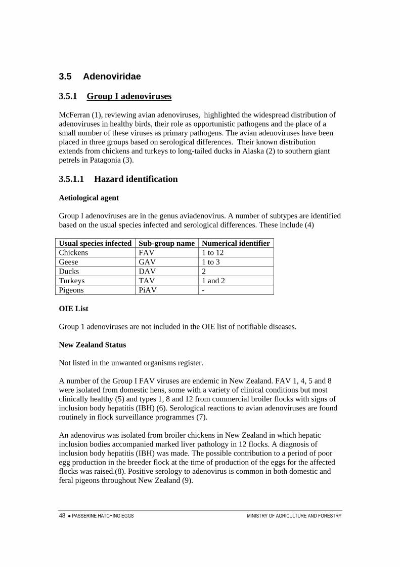
48 ● PASSERINE HATCHING EGGS MINISTRY OF AGRICULTURE AND FORESTRY
3.5 Adenoviridae 3.5.1 Group I adenoviruses McFerran (1), reviewing avian adenoviruses, highlighted the widespread distribution of adenoviruses in healthy birds, their role as opportunistic pathogens and the place of a small number of these viruses as primary pathogens. The avian adenoviruses have been placed in three groups based on serological differences. Their known distribution extends from chickens and turkeys to long-tailed ducks in Alaska (2) to southern giant petrels in Patagonia (3). 3.5.1.1 Hazard identification Aetiological agent Group I adenoviruses are in the genus aviadenovirus. A number of subtypes are identified based on the usual species infected and serological differences. These include (4) Usual species infected Sub-group name Numerical identifierChickens FAV 1 to 12 Geese GAV 1 to 3 Ducks DAV 2 Turkeys TAV 1 and 2 Pigeons PiAV - OIE List Group 1 adenoviruses are not included in the OIE list of notifiable diseases. New Zealand Status Not listed in the unwanted organisms register. A number of the Group I FAV viruses are endemic in New Zealand. FAV 1, 4, 5 and 8 were isolated from domestic hens, some with a variety of clinical conditions but most clinically healthy (5) and types 1, 8 and 12 from commercial broiler flocks with signs of inclusion body hepatitis (IBH) (6). Serological reactions to avian adenoviruses are found routinely in flock surveillance programmes (7). An adenovirus was isolated from broiler chickens in New Zealand in which hepatic inclusion bodies accompanied marked liver pathology in 12 flocks. A diagnosis of inclusion body hepatitis (IBH) was made. The possible contribution to a period of poor egg production in the breeder flock at the time of production of the eggs for the affected flocks was raised.(8). Positive serology to adenovirus is common in both domestic and feral pigeons throughout New Zealand (9).

MINISTRY OF AGRICULTURE AND FORESTRY PASSERINE HATCHING EGGS ● 49
Epidemiology Adenoviruses are widespread in chicken populations and adenoviruses of the chicken serotypes have been recovered from a range of other species (10). Antibodies to adenoviruses are sufficiently common to have lead Hess (11) to comment that serology is of little value as a diagnostic tool. In chickens, vertical transmission through eggs is common. Horizontal transmission is also important. Spread by fomites can take place and venereal transmission in semen is also possible, associated with renal infection and excretion.(10) Latent infections are known to establish in chickens and be transmitted between generations (12). A syndrome of IBH in chickens is commonly attributed to Group I adenovirus. McFerran et al. (13) reported the isolation of adenoviruses types 2, 3, 4, 5 and 8 from outbreaks of IBH in chickens but cautioned the interpretation of these findings pointing out that adenoviruses could be isolated from virtually all broiler flocks at the time that maternal antibody had diminished. Similar reservations over the role of adenoviruses in incidents in which hepatic inclusion bodies are found in chickens have been expressed by others (8, 14). Hepatic inclusion bodies associated with adenoviruses (or adenovirus-like particles) have also been reported from psittacines(15, 16, 17), American kestrels (18), pigeons (19) and geese(20). Other reported sites of inclusion bodies associated with adenoviruses include the intestines (21) and renal tubules of psittacines (22). McFerran (23) reported the isolation of adenoviruses (types 2, 5 and 8) from a range of avian species with a variety of clinical diseases and Pennycott (24) reported the isolation of an adenovirus, along with a reovirus-like organism, Escherichia coli and Clostridium perfringens from episodes of high mortality in budgerigars in Scotland. Authors commonly expressed reservations over the role of the adenoviruses as primary pathogens. One report of the finding of an adenovirus in passerine birds was located. That infection, of Gouldian finches in a single aviary in California, was considered likely to be consequent to infection of the birds with a circovirus. The circovirus infections were associated with lymphoid depletion in both the bursa of Fabricius and the thymus and concurrent infections with E. coli and Klebsiella oxytoca were also present. The adenovirus infection was located in the renal tubules and was associated with a mild nephritis (25). For the purpose of this risk analysis, this infection will be considered as a Group I adenovirus. Conclusion Group I avian adenoviruses are considered to be potential hazards in the commodity.

50 ● PASSERINE HATCHING EGGS MINISTRY OF AGRICULTURE AND FORESTRY
3.5.1.2 Risk assessment Release assessment The release assessment for Group I adenoviruses in passerine eggs from Europe is based on the general ubiquitous nature of these viruses and the evidence that transmission through eggs take place. The release assessment is non-negligible. Exposure assessment Hatchlings from imported eggs may be infected. Both horizontal and vertical transmission may lead to spread of the virus. The exposure assessment is non-negligible. Consequence assessment There is only one report of an adenovirus associated with disease in Passeriformes. That case was in California, was as a co-infection with a circovirus and was associated with only a mild nephritis. The consequence assessment for adenoviruses in passerine eggs imported to New Zealand from Europe is negligible. Risk estimation Based on the absence of reports of Group I adenoviruses causing ill health in passerine species in Europe, these viruses do not present a hazard to New Zealand bird species (endemic, native or introduced), the New Zealand poultry industry agriculture, the economy or the environment in the importation of birds within the commodity definition addressed by the risk analysis. References
1. McFerran, J.B. 2003. Adenovirus infections in Diseases of Poultry. 11th Edition. Pp 213-214. Editor Saif, Y.M. Iowa State Press.
2. Hollmen, T.E.; Franson, J.C.; Flint, P.L.; Grand, J.B.; Lanctot, R.B.; Docherty, D.E.; Wilson, H.M. 2003. An adenovirus linked to mortality and disease in long-tailed ducks (Clangula hyemalis) in Alaska. Avian Diseases 47 (4): 1434-1440.
3. Uhart, M.M.; Quintana, F.; Karesh, W.B.; Braselton, W.E. 2003. Hematology, plasma biochemistry, and serosurvey for selected infectious agents in southern petrels from Patagonia, Argentina. Journal of Wildlife Diseases. 39 (2): 359-365.

MINISTRY OF AGRICULTURE AND FORESTRY PASSERINE HATCHING EGGS ● 51
4. Russell, W.C., Adrian, T., Bartha, A., Fujinaga, K., Ginsberg, H.S., Hierholzer, J.C., DeJong, J.C., Li, Q.G., Mautner, V., N´asz, I. & Wadell, G. (1995). In T.A. Murphy, C.M. Fauquet, D.H.L. Bishop, S.A. Ghabrial, A.W. Jarvis, G.P. Martelli, M.A. Mayo, & M.D. Summers (Eds.), Virus Taxonomy, Classification and Nomenclature of Viruses Sixth Report of the International Committee on “Taxonomy of Viruses” (pp. 128–133). Vienna: Springer Verlag. (Cited by Hess, M. 2000 Detection and differentiation of avian adenoviruses: A review. Avian Pathology. 29 (3): 195-206.
5. Green, A.F.; Clarke, J.K.; Lohr, J.E. 1976. Detection of four serotypes of avian adenovirus in New Zealand. Avian Diseases 20 (2): 236-241.
6. Saifuddin, Md.; Wilks, C.R.; Murray, A. 1992. Characterisation of avian adenoviruses associated with inclusion body hepatitis. NZ Veterinary Journal. 40: 52-55.
7. Poland, R. 2004 Poultry health surveillance. Surveillance. 31 (2): 27.
8. Christensen, N.H.; Saifuddin, Md. 1989. A primary epidemic of inclusion body hepatitis in broilers. Avian Diseases. 33: 622-630.
9. Black, H.; Stanislawek, W.; cooper, C.; Saunders, W. 2004. Avian virus survey in pigeons. Surveillance 31 (4): 20-21.
10. McFerran, J.B.; Adair, B.M. 2003. Group I adenovirus infections. in Diseases of poultry. 11th Edition. Pp 214-227. Editor Saif, Y.M. Iowa State Press.
11. Hess, M. (1990) Detection and differentiation of avian adenoviruses: A review. Avian Pathology. 29 (3): 195-206.
12. Fadly, A.M.; Riegle, B.J.; Nazerian, K.; Stephens, E.A. 1980 Some observations on an adenovirus isolated from specific pathogen-free chickens. Poultry Science. 59: 21-27.
13. McFerran, J.B.; McCracken, R.M.; Connor, T.J.; Evans, R.T. 1976. Isolation of viruses from clinical outbreaks of inclusion body hepatitis. Avian Pathology 5: 315-324.
14. Bains, B.S.; Watson, A.R.A. 1977. Inclusion body hepatitis of chickens. NZ Veterinary Journal 25: 352.
15. Scott, P.C.; Condron, R.J.; Reece, R.L. 1986. Inclusion body hepatitis associated with adenovirus-like particles in a cockateil (Psittaciformes: Nymphicus hollandicus). Australian Veterinary Journal. 63: 337-338.
16. Pass, D.A. 1987. Inclusion bodies and hepatopathies in psittacines. Avian pathology 16:581-597.
17. Capua, I.; Liberti, L.; Gough, R.E.; Casaccia, C.; Asdrubali, G. 1995. Isolation and characterization of an adenovirus associated with inclusion body hepatitis in psittacine birds. Avian Pathology. 24 (4): 717-722.
18. Sileo, L.; Franson, J.C.; Graham, D.L.; Domermuth, C.H.; Rattner, B.A.; Pattee, O.H. 1983. Hemorrhagic enteritis in captive American kestrels (Falco sparverius). Journal of wildlife diseases. 19: 244-247.
19. Coussement, W.; Ducatelle, R.; Lemahieu, P.; Froyman, R.; Devriese, L.; Hoorens, J. 1984. Pathology of adenovirus infections in pigeons. Vlaamse Diergeneeskunde Tijdschrift. 53: 277-283. (Abstracted in CAB Abstracts. Accession number 19842248842.)
20. Riddell, C. 1984. Viral hepatitis in domestic geese in Saskatchewan. Avian Diseases. 28: 774-782.

52 ● PASSERINE HATCHING EGGS MINISTRY OF AGRICULTURE AND FORESTRY
21. Gomez-Villamandos, J.J.; Mozos, E.; Sierra, M.A.; Perez, J.; Mendez, A. 1992 Inclusion bodies containing adenovirus-like particles in the intestine of a psittacine bird affected by inclusion body hepatitis. Journal of Wildlife Diseases. 28 (2): 319-322.
22. Mori, F.; Touchi, A.; Suwa, T.; Itakura, C.; Hashimoto, A.; Hirai, K. 1989 Inclusion bodies containing adenovirus-like particles in the kidneys of psittacine birds. Avian pathology. 18 (1): 197-202.
23. McFerran, J.B.; Connor, T.J.; McCracken, R.M. 1976. Isolation of adenoviruses and reoviruses from avian species other than domestic fowl . Avian Diseases 20: 519-524.
24. Pennycott, T. 2004. Mortality in budgerigars in Scotland: pathological findings. Veterinary record. 154(17): 538-539.
25. Shivaprasad, H.L.; Hill, D.; Todd, D.; Smyth, J.A. 2004. Circovirus infection in a Gouldian finch (Chloebia gouldiae). Avian Pathology 33 (5): 525-529.

MINISTRY OF AGRICULTURE AND FORESTRY PASSERINE HATCHING EGGS ● 53
3.5.2 Group II adenoviruses – HE, MSD, AAS 3.5.2.1 Hazard identification Aetiological agent Group II adenoviruses fall within the proposed genus of Siadenoviruses. The unique feature of the members of this group is their genetic coding for sialidase (1). The viruses in this group cause haemorrhagic enteritis (HE) of turkeys, marble spleen disease (MSD) of pheasants, and avian adenovirus splenomegaly (AAS) of chickens. OIE List Group II adenoviruses are not included in the OIE list of notifiable diseases. New Zealand Status Not listed on the unwanted organisms register. Literature searches have not revealed any New Zealand record of the diagnosis of any of the diseases caused by Group II adenoviruses. Epidemiology The host range of each of the diseases associated with Group II adenoviruses is limited. Haemorrhagic enteritis is a disease of turkeys from four weeks old. Marble spleen disease affects pheasants between three and eight months old raised in captivity and AAS occurs in broiler chickens. Disease has been diagnosed in guinea fowl and there is one report of a suspect infection in psittacine birds (2). A survey of 618 wild birds of 42 species in the south eastern United States for antibodies to the Haemorrhagic enteritis / Marble spleen disease group of viruses produced negative results. This survey included 207 passerines from eight families with all passerines sampled being from Florida (3). Conclusion As group II adenoviruses are recognised as avian pathogens and are not present in New Zealand they are considered to be potential hazards in the commodity. 3.5.2.2 Risk assessment Release assessment The host range of Group II adenoviruses is limited. No reports of Group II adenoviruses in non-captive birds have been located, reports of natural infections in species outside the gallinaceous group are restricted to one, from psittacines (4). Searches for reports of infection in passerine species have been unsuccessful.
54 ● PASSERINE HATCHING EGGS MINISTRY OF AGRICULTURE AND FORESTRY
On the basis of the restricted range of hosts from which Group II adenovirus have been reported and the lack of reports from passerine species, the likelihood of Group II adenoviruses being present in passerine eggs from Europe is negligible. The release assessment is negligible. Risk estimation With the proposed importation of passerine eggs from Europe, Group II adenoviruses do not present a hazard to New Zealand native, endemic or other wild birds. Nor do they present a hazard to poultry, agriculture, human health or the environment. References
1. McFerran, J.B. 2003. Adenovirus infections in Diseases of Poultry. 11th Edition. Pp 213-214. Editor Saif, Y.M. Iowa State Press.
2. Pierson, F.W.; Fitzgerald, S.D. 2003. Hemorrhagic enteritis and related infections. In Diseases of poultry. 11th Edition. Pp 237-247. Editor Saif, Y.M. Iowa State Press.
3. Domermuth, C.H.; Forrester, D.J.; Trainer, D.O.; Bigler, W.J. 1977. Serologic examination of wild birds for hemorrhagic enteritis of turkeys and marble spleen disease of pheasants. Journal of Wildlife Diseases. 13: 405-408.
4. Gomez-Villamandos, J.C.; Mulas, J.M.M.delas; Hervas, J.; Lara, F.C.M.de; Perez, J.; Mozos, E. 1995. Spleno-enteritis caused by adenovirus in psittacine birds: a pathological study. Avian Pathology 24 (3): 553-563.

MINISTRY OF AGRICULTURE AND FORESTRY PASSERINE HATCHING EGGS ● 55
3.5.3 Group III adenoviruses – EDS 3.5.3.1 Hazard identification Aetiological agent The aetiological agent for egg drop syndrome (EDS) is the sole member of Group III adenovirus. OIE List EDS is not included in the OIE list of notifiable diseases. New Zealand Status EDS is not included in the register of unwanted organisms. Epidemiology Egg drop syndrome is a contagious disease of chickens, spreading through both lateral and vertical transmission and causing decreased egg production. The virus is also recognised in ducks, geese and quail (1). EDS was first diagnosed in New Zealand in 1981 (2). Although serological prevalence remained low through the 1980s and Howell (3, 4) predicted that the virus was not likely to persist in commercial flocks, infection, as evidenced by serological titres, has remained (5, 6). No reports of natural infections of passerines with EDS have been located. Conclusion Based on its restricted host range, and its presence in New Zealand, EDS is not considered to be a potential hazard in the commodity. References
1. McFerran, J.B.; Adair, B.M. 2003. Egg drop syndrome in Diseases of Poultry. 11th Edition. Pp 227-237. Editor Saif, Y.M. Iowa State Press.
2. Howell, J. 1981. Egg drop syndrome: A new disease in New Zealand. Surveillance 8 (4): 2-4.
3. Howell, J. 1983. An update on egg drop syndrome 76 (EDS 76) in New Zealand. Surveillance 10 (3): 24.
4. Howell, J. 1992. Viral diseases and the New Zealand poultry industry. Surveillance 19 (2): 15-17.

56 ● PASSERINE HATCHING EGGS MINISTRY OF AGRICULTURE AND FORESTRY
5. Christensen, N.H.; Stanislawek, W.L. 1994 EDS 76 antibodies in a flock of free-range layers. NZ Veterinary Journal 42: 70-72.
6. Poland, R. 2004 Poultry health surveillance. Surveillance. 31 (2): 27.

MINISTRY OF AGRICULTURE AND FORESTRY PASSERINE HATCHING EGGS ● 57
3.6 Poxviridae 3.6.1 Avipoxvirus
3.6.1.1 Hazard identification Aetiological agent Order – mononegavirales; Family – Poxviridae; Genus – Avipoxvirus. Poxviruses are enveloped DNA viruses. Recognised species within the Avipoxvirus genus are (1):
1. Fowlpox virus (three species), 2. Pigeonpox virus 3. Psittacinepox virus 4. Quailpox virus 5. Turkeypox virus 6. Starlingpox virus - * 7. Sparrowpox virus - * 8. Canarypox virus - * 9. Juncopox virus - * 10. Mynahpox virus - *
Tentative species are (1):
11. Crowpox virus 12. Peacockpox virus 13. Penguinpox virus
Note: in the above list, the viruses marked * have passerine species as their primary host. OIE List Fowlpox is included in the OIE list of notifiable diseases. New Zealand Status Not listed in the unwanted organisms register. Fowlpox is widespread, especially in the northern parts of New Zealand. Pigeonpox and fowlpox vaccines have been used routinely in endemic areas in the past. Both cutaneous and “wet” (laryngeal) pox are seen in chickens (2). Poxvirus infections have also been diagnosed in turkeys, pigeons and canaries+ (3). Gartrell et al. (4) listed pipit (Anthus novaeseelandiae+), shore plover (Thinornis novaeseelandiae), variable oyster catcher (Haematopus unicolor), weka (Gallirallus australis), NZ wood pigeon (Hemiphaga novaeseelandiae), thrush (Turdus philomelos+), silver eye (Zosterops lateralis+), black robin (Petroica traverse+) and North Island robin (Petroica Australia+) as species in

58 ● PASSERINE HATCHING EGGS MINISTRY OF AGRICULTURE AND FORESTRY
which avian pox had been diagnosed. Pox infection of an Oyster catcher (Haematopus leucocephalus) has also been reported (5). The status of New Zealand with respect to psittacinepox virus is uncertain following the finding of psittacinepox in a quarantine aviary and illegal removal of birds from it (4). The six species marked + are in the Order Passeriformes. Epidemiology Poxviruses are stable in dry environments and can be transmitted by aerosols, direct contacts, fomites or by biting insects. Latent infections occur and these may be reactivated during times of stress (6). Bolte et al. (7) reviewed the avian species reported as infected with avipoxviruses and included 232 species from 23 orders in their list. That list included 99 species within the Passeriformes Order but only three of the passerine species identified with avian pox in NZ. The true number of species that are vulnerable to infection with avipoxviruses must be very large. As with other poxviruses, avipoxviruses are generally considered to be host specific or to have a narrow host range. Reports in the literature, however, provide examples where avipoxviruses appear to have host ranges of varying scope. Some examples that illustrate this mixed picture include: • Infection of caged Rothschild’s mynahs (Leucospar rothchildii) by virus from
starlings (Sturnus vulgaris) gaining access to the aviary while over a hundred birds of different species remained unaffected (8) (infection of birds in different genera within the Family Sturnidae but not birds in other families – this incident was consistent with observations by Williamson (9) that Starlingpox is specific to the Sturnidae family),
• Failures of attempts to infect chickens and canaries (Phylloscopus canariensis) with poxvirus from diseased Amazon parrots (Amazona aestiva) (10) (failure to infect birds in another genus or Family) ,
• Non-pathogenicity, to fowl, turkey, duck and pigeon, of poxvirus from diseased Red Siskins (Carduelis cucullatus) while a minor lesion developed in a bird of another Siskin species (11) (infection of a bird in another species within the same genus but not of birds in other orders) and
• An outbreak of pox in an aviary restricted to canaries (Serinus canaria) and house sparrows (Passer domesticus) although 10 other passerine species were present (12) (restriction of infection to two species and genera with the order Passeriformes.)
• Avipoxvirus infection affecting five species of pheasants and free-ranging Indian red junglefowl (Gallus gallus murghi) in a zoo at the same time (13) (infection of birds from several genera within the family Phasianidae),

MINISTRY OF AGRICULTURE AND FORESTRY PASSERINE HATCHING EGGS ● 59
• Infection of chickens and bobwhite quail with poxvirus from mynahs (Gracula religiosa) imported from Malaysia (14) (Note – Infection was dependent upon route of inoculation. Cutaneous inoculation resulted in lesions whereas swabbing of virus onto conjunctivae and oral mucosa did not.) (Infection of birds from the Order Galliformes with virus from a passerine bird),
• Infection establishing in Amazona ochrocephala (Yellow crowned parrot), Aratinga holochlora (Perico mexicano) and chickens following inoculation with a virus isolated from Amazona albifrons (White fronted parrot) (15). (Infection of birds of different species within the same genus and of birds in a different genus.) and
• Simultaneous cases of pox infection in house finches, (Carpodacus mexicanus), mourning doves (Zenaida macroura), house sparrows (Passer domesticus), robin (Turdus migratorius) and a golden eagle (Aquila chrysaetos) (16). (Infection of birds in different families with the same order and of a bird in a different Order.)
These observations are consistent with those of Kirmse (17), who recognised examples of cross-species and cross-Order infections and also commented that cross immunity was highly variable. With over 100 species in the Order Passeriformes recorded as having been infected with Avipoxvirus, and five Avipoxvirus species recognised as being sufficiently host related to passerine birds to be named after them, there remains a great deal to learn about the range of avipoxviruses that infect passerines and their host ranges. Conclusion Avipoxviruses are considered to be a potential hazard in the commodity. 3.6.1.2 Risk assessment Release assessment No reports suggesting vertical transmission of avian poxviruses have been located. The release assessment for avipoxviruses in passerine eggs imported from Europe is negligible. Risk estimation It is concluded that Avipoxviruses are not a hazard in the commodity.

60 ● PASSERINE HATCHING EGGS MINISTRY OF AGRICULTURE AND FORESTRY
References
1. Murphy, F.A. et al. 2002. ICTV approved virus orders family and genera. Compiled from Virus Taxonomy Reports of the international committee on taxonomy of viruses. At www.ictvbd.iacr.ac.uk/Ictv/fr-fst-h.htm
2. Howell, J. 1992 Viral disease and the New Zealand poultry industry. Surveillance 19 (2): 15-17.
3. Smits, B. 1995 Viral diseases of psittacines. Surveillance 22 (4): 16-19.
4. Gartrell, B.; Stone, M.; King, C.; Wang, J. 2003 An outbreak of disease caused by psittacinepoxvirus in rosellas. Surveillance 30 (3): 11-13.
5. Johnstone, A,C,; Cork, S.C. 1993 Diseases of aviary and native birds of New Zealand. Surveillance 20 (3): 35-36.
6. Qinn, P.J.; Markey, B.K.; Carter, M.E.; Donnelly, W.J.; Leonard, F.C. 2002 Poxviridae in Veterinary Microbiology and Microbial Disease. Blackwell Publishing Company. Oxford, UK.
7. Bolte, A.L.; Meurer, J.; Kaleta, E.F. 1999 Avian host spectrum of avipoxviruses. Avian Pathology 28: 415-432.
8. Landbolt, M.; Kocan, R.M. 1976 Transmission of avian pox from starlings to Rothschild’s Mynahs. Journal of Wildlife Diseases 12 (3): 353-356.
9. Williamson, F.S.L. 1968 The ecology of pox disease in the starling Sturnus vulgaris L. PhD Thesis. School of Hygiene and Public Health, John Hopkins University, Baltimore, Maryland. (cited by Landbolt, M.; Kocan, R.M. 1976 Transmission of avian pox from starlings to Rothschild’s Mynahs. Journal of Wildlife Diseases 12 (3): 353-356. (Ref 8 above))
10. Hitchner, S.B.; Clubb, S.L. 1980 Relationship between poxvirus of parrots and other birds. Proceedings of 29th Western Poultry Disease Conference, Acapulco, Mexico, 22-25 April 1980. (Abstracted in CAB Abstracts. Accession number 19812285713.)
11. Kaleta, E.F.; Marschall. H.-J. 1982 Pox in Red Siskins (Carduelis cullata). Zentralblat fur Veterinarmedizin, B 29 (10): 776-781. (Abstracted in CAB Abstracts. Accession number 1932222653.)
12. Donnelly, F.M.; Crane, L.A. 1984 An epornitic of avian pox in a research aviary. Avian Diseases 28 (2): 517-525.
13. Ensley, P.K.; Anderson, M.P.; Costello, M.L.; Powell, H.C.; Cooper, R. 1978 Epinortic of avian pox in a zoo. Journal of the American Veterinary Medical Association. 173 (9): 1111-1114.
14. Reed, W.W.; Schrader, D.L. 1989 Pathogenicity and immunogenicity of mynah pox virus in chickens and bobwhite quail. Poultry Science 68 (5): 631-638.
15. Boosinger, T.R.; Winterfield, R.W.; Feldman, D.S.; Dhillion, A.S. 1982 Psittacine pox virus: virus isolation and identification, transmission, and cross-challenge studies in parrots and chickens. Avian diseases 26 (2): 437-444.
16. Hill, J.R.; Bogue, G. 1977 Epinortic of pox in a wild bird population. Journal of the American Veterinary Medical Association 171 (9): 993-994.

MINISTRY OF AGRICULTURE AND FORESTRY PASSERINE HATCHING EGGS ● 61
17. Kirmse. P. 1969 Host specificity and pathogenicity of pox virus from wild birds. Bulletin of the Wildlife Diseases Association. 5: 376-386.

62 ● PASSERINE HATCHING EGGS MINISTRY OF AGRICULTURE AND FORESTRY
3.7 Circoviridae 3.7.1 General 3.7.1.1 Hazard identification Aetiological agent Circoviruses are small, non-enveloped viruses that replicate in the nucleus of cells. They are stable in the environment and maintain infectivity through temperatures up to 60 degrees for 30 minutes (1). The family Circoviridae contains two recognised genera;
• Gryovirus - the only member of which is the Chicken anaemia virus and • Circovirus – which includes Porcine circovirus and Beak and Feather disease
virus (2). In addition, unidentified Circovirus-like viruses have been identified in birds> OIE List No members of the Circoviridae are included in the OIE list of notifiable diseases. NZ status No avian members of the Circoviridae are included in the register of unwanted organisms.

MINISTRY OF AGRICULTURE AND FORESTRY PASSERINE HATCHING EGGS ● 63
3.7.2 Chicken infectious anaemia 3.7.2.1 Hazard identification Epidemiology Chicken infectious anaemia virus (CIAV) is established and widespread in New Zealand (3, 4). It infects only chickens. Conclusion On the basis of its presence in New Zealand and its restricted host range, CIAV is not considered to be a potential hazard in the commodity.

64 ● PASSERINE HATCHING EGGS MINISTRY OF AGRICULTURE AND FORESTRY
3.7.3 Psittacine beak and feather disease 3.7.3.1 Hazard identification New Zealand Status Ritchie et al. (5) identified psittacine beak and feather disease virus (PBFDV) in 21 of 25 captive psittacine birds sampled in New Zealand. These positive results came from eight of the ten species included in the study. Genotype clustering of viruses within related psittacine species was found and these clustering patterns were similar to those seen in similar species in Australia. This has led to the suggestion that PBFDV was introduced into New Zealand in psittacines imported from Australia. In 2003, 5 of 71 captive psittacines sampled by Jakob-Hoff (6) were positive for PBFDV, while all 76 wild psittacines sampled were negative by PCR. Conclusion On the basis that PBFDV is present in New Zealand it is concluded that PBFDV is not a potential hazard in the commodity.

MINISTRY OF AGRICULTURE AND FORESTRY PASSERINE HATCHING EGGS ● 65
3.7.4 Circovirus-like viruses 3.7.4.1 Hazard identification Aetiological agent Pigeon circovirus (PiCV) has been accepted as a tentative member of the Circovirus genus (7) and Todd (8) listed reports of circovirus-like viruses being identified in Senegal doves, canaries, finches, ostrich, goose and a gull between 1994 and 1999. Since Todd’s review Circovirus-like agents have been identified in pheasants (9) and mulard ducks (10). New Zealand Status A circovirus-like virus was identified in a southern black-backed gull that was found terminally ill with Aspergillus spp. infection and severe airsacculitis in the Manawatu region (11). Epidemiology Available evidence indicates that specific genotypes of circoviruses have restricted host ranges. Ritchie et al. (5) identified clustering of genotypes of PBFD viruses from taxonomically related psittacine species. Different viral groupings were associated with lorikeets, cockatoos and a budgerigar. The genome of PiCV is sufficiently distinct from that of PBFDV for it to have been given tentative status as a virus in the Circovirus genus (7). Reports of Circovirus in Senegal doves (12), canaries (13, 14), mulard ducks (10) and geese (15) have each claimed that these viruses are novel. Shivaprasad (16) reported circovirus infection associated with lesions of lymphoid depletion of the bursa of Fabricius and the thymus in a Gouldian finch from an aviary in which 50% of the birds of that species died while other species of finch in the aviary remained healthy. Todd (8) expressed the opinion that “other mammalian and avian species are likely to be infected with novel circoviruses that are presently unrecognized” and went on to suggest that “now that the characteristic lesions are becoming better known to avian pathologists, it is likely that reports describing circovirus infections of additional avian species will be forthcoming”. In the five years since Todd’s review was published, these forecasts have proven to be correct. Consistent pathological findings in Circovirus-associated disease in birds include lesions of lymphoid tissue including the bursa of Fabricius. The pathogenesis of disease appears to include immune suppression allowing other pathogens or opportunist pathogens to infect the bird(s) and cause disease. Diagnoses of Circovirus-like infections are largely dependent upon recognition of inclusion bodies in lymphoid tissues and investigations using electron microscopy. Investigations of the prevalence of PBFDV and PiCV, for which PCR tests are available, have revealed that infections of psittacine and columbid species, respectively, are mainly in healthy birds.

66 ● PASSERINE HATCHING EGGS MINISTRY OF AGRICULTURE AND FORESTRY
Studies of the epidemiology of Circovirus and Circovirus-like viruses has been hampered by difficulties in growing the virus in tissue culture. PBFDV, however, is thought to be transmitted both horizontally and vertically (1) but specific information on the means of transmission of the Circovirus-like viruses is not available. It is assumed that circovirus-like viruses can be transmitted by the same routes as those proposed for PBFDV. Conclusion Based on the following factors
• Both recognised avian Circoviridae (CIAV and PBFDV) are established in New Zealand,
• The finding of a Circovirus-like infection in a southern black-backed gull in New Zealand raises the possibility that other birds may also be infected,
• Thousand of passerine birds have entered New Zealand without biosecurity measures that would have restricted the entry of Circovirus-like viruses,
• In species in which investigations have taken place, Circovirus-like viruses are common in healthy birds,
• Very few passerine birds are subject to laboratory diagnostic procedures in New Zealand and procedures likely to identify infection with Circovirus-like viruses will rarely be used
it is likely that Circovirus-like infections are present in birds in New Zealand, either as a result of long-term presence, or as the result of the introduction with imported birds.
It is concluded that Circovirus-like viruses are not a potential hazard in the commodity. References
1. Quinn, P.J.; Markey, B.K.; Carter, M.E.; Donnelly, W.J.; Leonard, F.C. 2002 Circoviridae in Veterinary Microbiology and Microbial Disease. Blackwell Publishing Company. Oxford, UK.
2. Buchen-Osmond, C. 2005 The Universal Virus database of the International Committee on Taxonomy of Viruses. http://www.ncbi.nlm.nih.gov/ICTVdb/
3. Stanislawek, W.L.; Howell, J. 1994. Isolation of chicken anaemia virus from broiler chickens in New Zealand. NZ Veterinary Journal. 42: 58-62.
4. Poland, R. 2004. Poultry health surveillance. Surveillance 31 (2): 27.
5. Ritchie, P.A.; Anderson, I.L.; Lambert, D.M. 2003 Evidence of specificity of psittacine beak and feather disease viruses among avian hosts. Virology 306: 109-115.
6. Jakob-Hoff, R. 2003. Report to the Ministry of Agriculture and Forestry Biosecurity Authority on the Avian Health Surveillance Project (Contract Number BAH/51/2001. Wildlife Health and Research Centre, Auckland Zoo Private Bag, Grey Lynn, Auckland.
7. Seventh Report of the International Committee on Taxonomy of Viruses Eds van Regenmortel, M.H. V.; Fauquet, C.M.; Bishop, D.H.L.; Carstens, E.B.; Estes, M.K.; Lemon, S.M.; Maniloff, J.; Mayon, M.A.; McGeoch, D.J.; Pringle, C.R.; Wickner, R.B.

MINISTRY OF AGRICULTURE AND FORESTRY PASSERINE HATCHING EGGS ● 67
Academic Press. 2000 http://www.virustaxonomyonline.com/virtax/lpext.dll?f=templates&fn=main-h.htm
8. Todd, D. 2000 Circoviruses: immunosuppressive threats to avian species: a review. Avian Pathology 29: 373-394.
9. Terregino, C.; Montesi, F.; Mutinelli, F.; Capua, I.; Pandolfo, A. 2001. Detection of a circovirus-like agent in farmed pheasants in Italy. Veterinary Record 149 (11): 340.
10. Soike, D.; Albrecht, K.; Hattermann, K.; Schmitt, C.; Mankerz, A. 2004 Novel circovirus in mulard ducks and developmental and feathering disorders. Veterinary Record 154: 792-793.
11. Twentyman, C.M.; Alley, M.R.; Cooke, M.M.; Duigan, P.J. 1999 Circovirus-like infection in a southern black-backed gull (Larus dominicanus). Avian Pathology 28: 513-516.
12. Raidal, S.R.; Riddoch, P.A. 1997. A feather disease in Senegal doves (Streptopelia sengalensis) morphologically similar to psittacine beak and feather disease. Avian Pathology 26 (4): 11-18.
13. Todd, D.; Weston, J.; Ball, N.W.; Borghmans, B.J.; Smyth, J.A.; Gelmini, L.; Lavazza, A. 2001 Nucleotide sequence-based identification of a novel circovirus of canaries. Avian Pathology 30: 321-325.
14. Phenix, K.V.; Weston, J.H.; Ypelaar, I.; Lavazza, A.; Smyth, J.A.; Todd, D.; Wilcox, G.E.; Raidal, S.R. 2001. Nucleotide sequence analysis of a novel circovirus of canaries and its relationship to other members of the genus circovirus of the family Circoviridae. Journal of General Virology 82: 2805-2809.
15. Ball, N.W.; Smyth, J.A.; Weston, J.H.; Borghmans, B.J.; Palya, V.; Glavitas, R.; Ivanics, E.; Dan, A.; Todd, D. 2004 Diagnosis of goose circovirus infection in Hungarian geese samples using polymerase chain reaction and dot hybridisation tests. Avian Pathology. 33 (1): 51-58.
16. Shivaprasad, H.L.; Hill, D.; Todd, D.; Smyth, J.A. 2004. Circovirus infection in a Gouldian finch (Chloebia gouldiae). Avian Pathology 33 (5): 525-529.
68 ● PASSERINE HATCHING EGGS MINISTRY OF AGRICULTURE AND FORESTRY
3.8 Birnaviridae 3.8.1 Infectious bursal disease
3.8.1.1 Hazard identification Aetiological agent Infectious bursal disease virus (IBDV) is the only species within the Avibirnavirus genus within the Birnaviridae. Sixteen strains are recognised by the International Committee on Taxonomy of Viruses (http://www.ncbi.nlm.nih.gov/ICTVdb/Ictv/index.htm ). Strains are commonly differentiate into two based on serological grouping. OIE List IBDV is included in the OIE list of notifiable diseases. New Zealand Status IBDV (Exotic strains) is listed in the unwanted organisms register as a notifiable organism. Serological surveys had indicated that NZ poultry were free of IBDV until its presence was detected in 1993 (1). The virus was less contagious and less persistent in the environment than classical strains (2). Subsequently, IBDV isolated in NZ were characterised as type 1 and, in challenge trials in SPF chickens, did not cause clinical disease (3). Implementation of a national control programme by the poultry industry has resulted in a situation where commercial poultry flocks have been sero-negative since 1999 and national freedom has been viewed as a realistic goal (4). The serological status of non-commercial poultry or of caged or wild birds is unknown. Epidemiology IBDV is, generally, considered to be a disease of chickens in which species it is highly contagious and a cause of significant disease. The virus persists for several weeks in vacated chicken houses. In susceptible chicken flocks morbidity may be 100% while mortality varies between nil and 30%. Following establishment of infection in a flock, disease is less marked and may be non-clinical. Apart from its role as a primary cause of disease, IBDV infection results in immunosuppression and increased susceptibility to other infectious diseases (5). IBDV has been isolated from chickens, turkeys, ducks, ostriches (5) and penguins (6). Serological evidence of infection has been found in sparrows, ducks and geese in China (7), from rooks, wild pheasants and several rare species of birds in Ireland (8), several species of wild birds including carrion crow and jungle crow (9), and accipitrid birds (the family including hawks, eagles, buzzards and Old World vultures, harriers and kites)(10).

MINISTRY OF AGRICULTURE AND FORESTRY PASSERINE HATCHING EGGS ● 69
In Japan, 4 of 212 carrion crow were positive to IBDV serotype 1 and 10 to serotype 2. Reports of isolation and characterisation of IBDV from passerines have not been located. Attempts to infect Guinea fowl (11, 12), pheasants, partridges and quail (12) have not resulted in clinical signs of disease or lesions discoverable post-mortem. Conclusion IBDV is considered to be a potential hazard in the commodity. 3.8.1.2 Risk assessment Release assessment Isolation of IBDV from passerine birds has not been reported but positive serology from sparrows (7), rooks (8) and crows (9) suggests that infections of passerines may occur. There is no information on the persistence of infection in non-poultry species. There is no evidence that IBDV can be transmitted vertically through eggs. The release assessment is negligible. Risk estimation IBDV is not a hazard in the commodity. References
1. Anonymous 1994 Disease surveillance in poultry. Surveillance 21 (1): 13.
2. Christensen, N.H. 1994 Persistence of infectious bursal disease virus in New Zealand commercial egg layer flocks. NZ Veterinary Journal 43: 43.
3. Motha, J. 1996 Characterisation of infectious bursal disease viruses isolated in New Zealand. Surveillance 23 (4): 26-27.
4. Ryan, T.; Diprose, B.; Leong, R. 2000 Country-freedom plan for infectious bursal disease: a producer-led national disease control programme. Surveillance 27 (4): 3-5.
5. Lukert, P.D.; Saif, Y.M. 2003 Infectious bursal disease in Diseases of Poultry. 11th Edition. Pp 161-179. Editor Saif, Y.M. Iowa State Press.
6. Jackwood, D.J.; Gough, R.E.; Sommer, S.E. 2005 Nucleotide and amino acid sequence analysis of a Birnavirus isolated from penguins. Veterinary Record 156 (17): 550-552.
7. YongShan, W.; ZongAn, Z,.; ChungSheng, Z.; HanLu, L.; Chan, D.; YuAn, F. 1997 Studies on the ecology of infectious bursal disease virus (IBDV): serological surveys of non-chicken avian species naturally infected with IBDV. Chinese journal of veterinary science and technology 27 (8): 15-16. (Abstracted in CAB Abstracts. Accession number 19982217744.)

70 ● PASSERINE HATCHING EGGS MINISTRY OF AGRICULTURE AND FORESTRY
8. Woolcock, P.R.; Chin, R.P.; Saif, Y.M. 1995. Personal communication. Cited by Luckert and Saif (reference 5 above).
9. Ogawa, M.; Wakuda, T.; Tamaguchi, T.; Murata, K.; Setiyono, A.; Fukushi, H.; Hirai, K. 1998. Seroprevalence of Infectious Bursal Disease in Free-Living Wild Birds in Japan. Journal of Veterinary Medical Science. 60 (11): 1277-1279.
10. Ursula, H.; Blanco, J.M.; Kaleta, 2001. Neutralising antibodies against infectious bursal disease virus in sera from free-living and captive birds of prey from central Spain (Preliminary results). Proceedings II International Symposium on infectious bursal disease and chicken infectious anaemia. Rauischholzhausen, 247-251. Cited by Luckert and Saif (reference 5 above).
11. Okoye, J.O.A.; Okpe, G.C. 1989. The pathogenicity of an isolate of infectious bursal disease virus in guinea fowl. Acta Veterinaria Brno. 58: 91-96. Cited by Luckert and Saif (reference 5 above).
12. Van den Berg, T.B.; Ona, A.; Morales, D.; Rodriguez, J.F. 2001. experimental inoculation of game/ornamental birds with a virulent strain of IBDV. Proceedings II International Symposium on infectious bursal disease and chicken infectious anaemia. Rauischholzhausen, 247-251. Cited by Luckert and Saif (reference 5 above).

MINISTRY OF AGRICULTURE AND FORESTRY PASSERINE HATCHING EGGS ● 71
3.9 Papovaviridae 3.9.1 Polyomavirus
3.9.1.1 Hazard identification Aetiological agent The genus Polyomavirus is within the Family Polyomaviridae. The only Polyomavirus of birds recognised by the International Committee on Taxonomy of Viruses is Budgerigar fledgling disease virus (BFDV). OIE List Avian polyomavirus is not included in the OIE list of notifiable diseases. New Zealand Status Not listed in the unwanted organisms register. A survey by Jakob-Hoff (1) provided positive serological evidence of the presence of avian polyomavirus in two umbrella cockatoos, one sun conure, and one sulphur crested cockatoo. These findings confirm the speculation by epidemiologists (2) and the suspicions of veterinary pathologists (3) and an avian microbiologist (4) that avian polyomavirus is present in New Zealand. Epidemiology Avian Polyomavirus has been reported from many parts of the world, including Europe, and is considered by many to be ubiquitous within avian populations. BFD affects a wide range of psittacine species with young birds being most susceptible. Morbidity and mortality in affected groups can be up to 100%. Horizontal transmission is the main means of spread but vertical transmission may also take place (5). Disease and mortality attributed to Polyomavirus, has also been reported from a number of passerine species (6, 7, 8, 9, 10). Lafferty et al. (8), using PCR technology found that the virus in a Green aracaris (Pteroglossus viridis – Order Piciformes (Woodpeckers)) was a variant of previously characterised Polyomavirus(es). Serological testing of remaining birds in the aviary produced positive results from birds in four orders including both psittacine and passerine species. Analysis of the genotypes of 20 isolates of Polyomavirus from various locations, times and bird species lead Phalen et al. (11) to conclude that species-specific types had not developed.

72 ● PASSERINE HATCHING EGGS MINISTRY OF AGRICULTURE AND FORESTRY
Conclusion Based on the evidence for the presence of Polyomavirus in New Zealand and the apparent lack of species specificity, avian Polyomavirus is not considered to be a potential hazard in the commodity. References
1. Jakob-Hoff, R. 2003. Report to the Ministry of Agriculture and Forestry Biosecurity Authority on the avian animal health surveillance project (Contract Number BAH/51/2001). Wildlife Health and Research Centre, Auckland Zoo Private Bag, Grey Lynn, Auckland.
2. Jackson, R.; Morris, R.S.; Boardmand, W. 1999. Biosecurity Risk Assessment: Assessment of the Risk to New Zealand’s Indigenous Wildlife Populations from the Introduction of Exotic Birds. Internal Report for the Department of Conservation. (Cited by Jakob-Hoff Ref 1 above)
3. Smits, B.; Ellison, R.; Black, A.; Johnstone, A.; Thompson, K. 1999 Alpha Scientific Ltd in Quarterly review of diagnostic cases – January to March 1999. Surveillance 26 (2): 15-16.
4. Stanislawek, W. 2004. personal communication (Phone 6 September 2004).
5. Cross, G. 1996 Avian Viral Diseases in Diseases of Cage and Aviary Birds. 3rd Edition. Eds Rosskopf, W.; Woerpel, R. Williams and Wilkins, Pennsylvania, USA.
6. Garcia, A.P.; Latimer, K.S.; Niagro, F.D.; Norton, T.M.; Harmon, B.G.; Campagnoli, R.P.; Steffens, W.L. 1993 Avian polyomavirus infection in three black-bellied seed crackers (Pyrenestes ostrinus). Journal of the association of avian veterinarians. 7 (2): 79-82. (Abstracted in CAB Abstracts. Accession number 19942202070.)
7. Garcia, A.; Latimer, K.S.; Niagro, F.D.; Norton, T.M.; Campagnoli, R.P.; Harmon, B.G.; Howerth, E.W.; Ritchie, B.W. 1994 Diagnosis of polyomavirus infection in seed crackers (Pyrenestes spp.) and blue bills (Spermophaga haematina) using DNA in situ hybridisation. Avian Pathology 23 (3): 525-537.
8. Lafferty, S.L.; Fudge, A.M.; Schmidt, R.E.; Wilson, VG.van; Phalen, D.N. 1999 Avian polyoma infection and disease in a green aracaris (Pteroglossus viridis). Avian Diseases 43 (3): 577-585.
9. Sandmeier, P.; Gerlach, H.; Johne, R.; Muller, H. 1999 Polyoma infections in exotic birds in Switzerland. Schweizer Archiv fur Tierheilkunde 141 (5): 223-229. . (Abstracted in CAB Abstracts. Accession number 19992209179.)
10. Verkeecken, M.; Herdt, P.; de Charlier, G.; Raue, R.; Ducatelle, R. 2001 A clinical outbreak of polyomavirus infection in shamas (Copsychus malabaricus). Vlaams Diergeneeskundig Tijdschrift 70 (3): 216-220 . (Abstracted in CAB Abstracts. Accession number 20013098687.)
11. Phalen, D.N.; Wilson, V.G.; Gaskin, J.M.; Derr, J.N.; Graham, D.L. 1999 Genetic diversity in twenty variants of the avian polyomavirus. Avian Diseases 43 (2): 207-218.

MINISTRY OF AGRICULTURE AND FORESTRY PASSERINE HATCHING EGGS ● 73
3.9.2 Papillomavirus 3.9.2.1 Hazard identification Aetiological agent Papillomavirus is a genus within the family Papillomaviridae. OIE List Avian papillomavirus is not included in the OIE list of notifiable diseases. New Zealand Status Not listed in the unwanted organisms register. No reports of Papillomavirus in birds in New Zealand have been located. Epidemiology Papillomaviruses are, generally, host specific and tissue specific. Lesions in different tissues of the same species are caused by different viral types (1, 2). Cloacal papillomatosis is a condition of “new world” psittacines (3), proventricular papillomas are seen in Amazona spp. and macaws (3) and cutaneous papillomas are most common in African grey parrots (4). There are doubts that cloacal papillomatosis is caused by a Papillomavirus (5, 6, 7). Papillomaviruses may be latent with no signs of infection until activated during a period of stress. Virus is shed with cells desquamating from the surface of papillomas (1). Amongst birds of the Order Passeriformes, papillomatosis has been reported most frequently from chaffinches (Fringilla coelebs). Literak et al. (8) identified 12 reports from this species. Countries of origin were Great Britain, Germany, Netherlands, Sweden and the Czech Republic and all reports involved lesions on the feet and/or legs and/or claws. Most reports were of single cases although one was of an incident in which 10 of 20 birds in an aviary were affected. Lesions can persist and grow for at least two years. Papillomatosis has also been reported from canaries (Serinus canaries) (2, 9) and greenfinches (Carduelis chloris) (10). Lina et al. (11) reported infection of both chaffinches (Fringilla coelebs) and bramblings (F. moutifringilla) in one incident whereas all other reports of apparent Papillomavirus infection in passerines traced have involved only one species although other birds (including passerines) may have been sharing the aviary. Very few reports of characterization of Papillomaviruses from birds has been located. Moreno-Lopez et al. (12) found that individual viruses from chaffinches in Sweden and Holland were closely related but that there was little genetic homology with a bovine Papillomavirus. A Papillomavirus isolated from an African grey parrot (Psittacus

74 ● PASSERINE HATCHING EGGS MINISTRY OF AGRICULTURE AND FORESTRY
erithacus timneh) was found to be distinct from 17 mammalian and one chaffinch viruses tested (13). Conclusion Papillomaviruses of passerines are considered to be a potential hazard in the commodity. 3.9.2.2 Risk assessment Release assessment Papillomaviruses have been reported from only very few passerine species and then, with the exception of chaffinches, on only very few occasions and with low prevalence of affected birds. Although the existence of Papillomaviruses in other species can not be excluded, it seems that such viruses must be either infrequent or of very low pathogenicity. No reports suggesting or demonstrating vertical transmission of avian Papillomaviruses through eggs have been discovered. The release assessment for Papillomaviruses in passerine eggs is negligible. Risk estimation Based on a negligible release assessment it is concluded that avian Papillomaviruses are not a hazard in the commodity. References
1. Quinn, P.J.; Markey, B.K.; Carter, M.E.; Donnelly, W.J.; Leonard, F.C. 2002. Papillomaviridae pp 327-330 in Veterinary Microbiology and Microbial Disease. Blackwell Science Ltd.
2. Dom, P.; Ducatelle, R.; Charlier, G.; Groot, P.de. 1993 Papillomavirus-like infections in canaries (Serinus canarius). Avian Pathology. 22 (4): 797-803.
3. Rosskopf, W.J. 1996. Digestive system disorders. pp. 436-448 in Diseases of Cage and Aviary Birds. Rosskopf, W.; Woerpel, R. 3rd Edition. Williams & Wilkin. Baltimore.
4. Schmidt, R.E. 1996. Pathologic Disorders of the Skin and Feathers. pp. 387-396 in Diseases of Cage and Aviary Birds. Rosskopf, W.; Woerpel, R. 3rd Edition. Williams & Wilkin. Baltimore.
5. Sundberg, J.P.; Junge, R.E.; O’Banion, M.K.; Basgall, E.J.; Harrison, G.; Herron, A.J.; Shivaprasad, H.L. 1986 Cloacal papillomas in psittacines. American journal of Veterinary Research 47 (4): 928-932.
6. Goodwin, M.; McGee, E.D. 1993 Herpes-like virus associated with a cloacal papilloma in an Orange-fronted Conure (Aratinga canicularis). Journal of the Association of Avian Veterinarians 7 (1): 23-25. (Abstracted in CAB Abstracts. Accession number 19932286657.)

MINISTRY OF AGRICULTURE AND FORESTRY PASSERINE HATCHING EGGS ● 75
7. Latimer, K.S.; Niagro, F.D.; Rackich, P.M.; Campagnoli, R.P.; Richie, B.W.; McGee, E.D. 1997 Investigation of parrot Papillomavirus in cloacal and oral papilloma of psittacine birds. Veterinary Clinical Pathology. 26 (4): 158-163.
8. Literak, I.; Smid, B.; Valicek, L. 2003. Papillomatosis in chaffinches (Fringilla coelebs) in the Czech Republic and Germany. Veterinarni Medicina 48 (6): 169-173.
9. Maich, R.R.; Brandan Recalde, E,; Don, L.E. 1988. Viral papillomatosis in canaries. Pet’s Ciencia 4 (23): 352-357. (Abstracted in CAB Abstracts. Accession number 19892288293.)
10. Sironi, G.; Gallazzi, D. 1992. Papillomavirus infection in greenfinches (Carduelis chloris). Journal of Veterinary Medicine. Series B. 39 (6): 454-458.
11. Lina, P.H.C.; Noord, M.J.van; Groot, F.G.de. 1973. Detection of virus in squamous papillomas of the wild bird species Fringilla coelebs (chaffinch). Journal of the National Cancer Institute. 50 (No.2): 567-571. (Cited by Sironi and Gallazzi 1992 (Ref. 10 above))
12. Moreno-Lopez, J.; Ahola, H.; Stenlund, A.; Osterhaus, A.; Petterson, U. 1984 Genome of an avian Papillomavirus. Journal of virology 51 (3): 872-875.
13. O’Banion, M.K. et al. 1992 Molecular cloning and partial characterization of a parrot Papillomavirus. Intervirology 33 (2): 91-96.

76 ● PASSERINE HATCHING EGGS MINISTRY OF AGRICULTURE AND FORESTRY
3.10 Parvoviridae and other non-specific enteritis-associated agents
3.10.1 Parvoviruses
3.10.1.1 Hazard identification Aetiological agent Parvovirus is a genus within the Parvoviridae. They are DNA viruses replicating within the nuclei of dividing cells. OIE List Goose parvovirus is not included in the OIE list of notifiable diseases. New Zealand Status Goose parvovirus is listed in the unwanted organisms register. No reports of parvovirus infections of birds in New Zealand have been located.. Epidemiology Goose parvovirus infection (Derzsy’s disease) is not recognised as infecting species other than geese, Muscovy ducks and some hybrid breeds (1). Both chicken anaemia and psittacine beak and feather disease have been associated with parvovirus-like organisms but their causative organisms are now classified as circoviruses. (See section 3.7) The only reference located suggesting the presence of a Parvovirus-like organism in passerine birds is that by Helfer et al (2) who reported an incident of myocarditis-encephalopathy in canaries accompanied by parvo-virus like intranuclear inclusions in brain and heart. This report was from California. Goose Parvovirus is transmitted vertically through eggs. Conclusion Parvoviruses are considered to be a potential hazard in the commodity.

MINISTRY OF AGRICULTURE AND FORESTRY PASSERINE HATCHING EGGS ● 77
3.10.1.2 Risk assessment Release assessment Based on there being only one report of a suspect parvovirus-like infection in passerines and that being from California, the likelihood of parvovirus being present in the commodity is considered negligible. The release assessment is negligible. Risk estimation Based on the negligible release assessment, it is concluded that Paroviruses are not a hazard in the commodity. References
1. Gough, R.E. 2003. Goose parvovirus infection. Pp 367-374 In Diseases of Poultry. 11th Edition. Pp 161-179. Editor Saif, Y.M. Iowa State Press.
2. Helfer, D.H. et al. 1981. p. 92 in Proceedings of the 30th Western Poultry Disease Conference , 1981 (Cited by Christensen, N.H. 2005. Review of Import Risk Analysis: Birds of the Order Passeriformes from the European Union, Draft 14 May 2005)

78 ● PASSERINE HATCHING EGGS MINISTRY OF AGRICULTURE AND FORESTRY
3.10.2 Enteritis associated viruses (see also astrovirus and rotaviruses) 3.10.2.1 Hazard identification Aetiological agent Searches of electronic databases and text books for reports of enteritic diseases of passerine species caused by viruses other than astrovirus, rotavirus or parvovirus failed to produce positive results. Such viruses, therefore, are not a hazard in the commodity.

MINISTRY OF AGRICULTURE AND FORESTRY PASSERINE HATCHING EGGS ● 79
3.11 Togaviridae 3.11.1 Equine encephalitis (Eastern, Western and Venezuelan equine
encephalitis)
3.11.1.1 Hazard identification Aetiological agent The aetiological agents for these encephalitidies are Alphaviruses within the family Togaviridae. OIE List Equine encephalities are included in the OIE list of notifiable diseases. New Zealand Status These organisms are listed in the unwanted organisms register as notifiable. They have not been diagnosed in NZ. Epidemiology Eastern, Western and Venezuelan equine encephalitidies are caused by arthropod borne viruses which also infect birds including passerine species. These diseases (and viruses) are found only in the Americas (1). Conclusion Based on the limited geographic distribution of these viruses, they are not considered to be potential hazards in the commodity. References
1. Thomson GR. Equine encephalitides caused by alphaviruses. In : Coetzer JAW, Thomson GR, Tustin RC (eds). Infectious Diseases of Livestock with Special Reference to Southern Africa. Volume 1, Pp 636-41. Oxford University Press Southern Africa, Capetown, 1994.

80 ● PASSERINE HATCHING EGGS MINISTRY OF AGRICULTURE AND FORESTRY
3.11.2 Other Alphaviruses
3.11.2.1 Hazard identification Aetiological agent Alphaviruses are in a genus within the family Togaviridae. They are arboviruses. (That is, insects or other arthropods act as vectors.) OIE List Avian alphaviruses are not included in the OIE list of notifiable diseases.. New Zealand Status Not listed in the unwanted organisms register. Whataroa virus has been reported from NZ. Epidemiology No reports of Alphaviruses causing disease in birds have been encountered. The viruses circulate between vertebrate hosts and insects (most reports refer to mosquitoes). Alphaviruses identified as infecting birds are: Buggy Creek virus and Fort Morgan virus – These viruses infect cliff swallows and sparrows in central USA. No reports of disease associated with these viruses have been located. Getah virus – This virus is reported from east Asia (China, Japan, Malaysia, Indonesia) and India. It is reported as infecting Columbiformes birds. Chikungunya virus – reported from Africa and reported as infecting birds. Mayaro virus – Reports of this virus are limited to coming from South America where birds are reported to be amongst the species infected. Highlands J virus – Reported from the eastern United States infecting blue jays, scrub jays, turkeys and chukar partridges and other avian species including passerines. Ross river virus – This virus is distributed within Australia and in islands to its north. Reports of its being found in passeriformes were not located. Simliki Forest virus – Reports from China, Senegal and Romania indicate that this virus does infect birds. There is serological evidence that this virus is present in central and

MINISTRY OF AGRICULTURE AND FORESTRY PASSERINE HATCHING EGGS ● 81
southern Europe and it has been identified as infecting water fowl. No reports of evidence of it infecting passerine species have been found. Sindbis virus – Although Sindbis virus is widely distributed, with reports of its recognition from Europe, Africa, Asia and Australasia, it is inappropriate to regard the virus as an homogenous population for risk analysis purposes. Examinations of the antigenic and genetic characteristics of Sindbis viruses from different geographic locations have allowed them to be placed into two main groups. Those from Paleoarctic – African locations and those from Asia and Australia. Further genetic, and resulting phenotypic, changes have taken place in Sindbis viruses within those major geographic groupings. Results of these analyses show that Sindbis viruses from particular zoogeographic regions are restricted to that region even though birds and other major vertebrate hosts are not (1, 2). The authors commented that the genetic change might be due to genetic drift or to adaptation but they were unable to determine which. Later analyses have placed isolates of Ockelbo virus (from Sweden and Russia) as a sub-strain of Sindbis virus (3). Based on RNA analyses Whataroa virus is within the Sindbis group but showing greater divergence than other members of the group (4, 5) and has not been reported as being associated with disease in humans. On these bases, Sindbis virus from locations other than New Zealand are regarded as exotic organisms and should be subject to hazard analysis on that basis. The organisms considered in this analysis are those known as Sindbis virus and Ockelbo virus and are endemic within Europe. Evidence of infection (either isolation of virus or demonstration of antibodies) of birds in Europe with Sindbis virus has been reported from house sparrows and a number of other passeriformes species in Czechoslovakia (6, 7, 8), house sparrows and tree sparrows in Poland (9), and 17 passerine species in Sweden (10). Serological evidence of Sindbis virus infection has been found in 10 avian species, including seven passerine species, in the UK (11). Although Sindbis virus has been identified in other species (12), the great majority of infections recognised have been in Passeriformes. Lundstrom (13) demonstrated that Sindbis virus infections, with viraemias, could be established in Anseriformes, Galliformes and Passeriformes birds. Lundstrom et al. (10) concluded that thrushes are the main amplifying hosts for Sindbis virus in Sweden. Sindbis virus is an arbovirus. The vectors proposed for transmission between birds in Sweden are Culex pipiens pipiens, Cx. torrentium, Culiseta morsitans and Cs. ochroptera. Aedes communis and other Aedes spp. are suspected as the vectors between birds and man (14). Cx. univittatus was considered the main vector for both bird to bird and bird to man transmission of Sindbis virus in South Africa (15). Sindbis viruses have been associated with human disease in northern Europe, Egypt and South Africa (15). L’vov et al (16) concluded that Ockelbo disease, Pogosta disease and Karelian diseases in Sweden, Finland and Russia, respectively, are synonyms for the same disease from which Sindbis virus has now been isolated (17). In northern Europe, this disease presents as periodic epidemics affecting hundreds (or thousands) of individuals with rash, arthralgia and moderate fever (18, 19) the effects of which may be prolonged in excess of 30 months (20).

82 ● PASSERINE HATCHING EGGS MINISTRY OF AGRICULTURE AND FORESTRY
Whataroa virus has been identified in southern Westland in the South Island of NZ. It infects a number of bird species with serological prevalence varying between years and with time of year(21). Over the period of the study by Miles (22), the highest serological prevalences were in Thrushes (Turdus philomelos), hedge sparrows (Prunella modularis), Chaffinches (Fringilla coelebs), Bell birds (Anthornis melenura) and Blackbird (Turdus merula). The main mosquito vectors were thought to be Culiseta tonnoiri and Culex pervigilans. These two mosquitoes, together with Opifex fuscus, Aedes notoscriptus and Ae australis have been shown in laboratory testing, to be capable of acting as hosts to Whataroa virus (23). Whataroa virus is not reported as being associated with disease in animals or humans. Conclusion Alphaviruses infecting birds are considered to be potential hazards in the commodity. 3.11.2.2 Risk assessment Release assessment Neither Buggy Creek virus nor Fort Morgan virus nor Getah virus nor Chikungunya virus nor Mayaro virus nor Highlands J virus nor Ross river virus have been identified in Europe. Simliki Forest virus has been identified in Roumania and may be present elsewhere in Europe but it has not been identified in passerine species. Sindbis virus is present in passerine species in Great Britain and elsewhere in Europe. Vertical transmission of Alphaviruses in avian species has not been reported. On the above bases, the release assessment for alphaviruses in passerine eggs is negligible. Risk estimation It is concluded that Avian alphaviruses are not a hazard in the commodity. References
1. Rentiere-Delrue, F.; Young, N.A. 1980 Genomic divergence among Sindbis virus strains. Virology 106: 59-70.
2. Olsen, K.; Trent, D.W. 1985 Genetic and antigenic variations among geographical isolates of Sindbis virus. Journal of general virology. 66: 797-810.
3. Lundstrom, J.O.; Vene, S.; Saluzzo, J.F.; Niklasson, B. 1993 Antigenic comparison of Ockelbo virus isolates from Sweden and Russia with Sindbis virus isolates from Europe, Africa and Australia: further evidence for variation among Alphaviruses. American

MINISTRY OF AGRICULTURE AND FORESTRY PASSERINE HATCHING EGGS ● 83
Journal of Tropical Medicine and Hygiene. 49 (5): 531-537. (Abstract from Science Direct)
4. Weaver, S.C.; Kang, W.; Shirako, Y.; Rumenapf, T.; Strauss, E.G.; Strauss, J.H. 1997 Recombinational history and molecular evolution of Western Equine Encephalomyelitis complex Alphaviruses. Journal of virology. 71 (1): 613-623.
5. Powers, A.M.; Aaron, C.B.; Shirako, Y.; Strauss, E.G.; Kang, W.; Strauss, J.H.; Weaver, S.C. 2001 Evolutionary relationships and sytematics of the Alphaviruses. Journal of Virology 75 (21): 10118-10131.
6. Juricova, Z.; Hubalek, Z.; Halouzka, J.; Pellantova, J.; Chytil, J. 1987. Hemagglutination-inhibiting antibodies against arboviruses of the families Togaviridae and Bunyaviridae in birds caught in southern Moravia Czechoslovakia. Folia Parasitologica (Ceske Budejovice) 34 (3): 281-284. (Abstracted in Biological Abstracts. Accession number BACD198784119080.)
7. Juricova, Z. 1988 Antibodies against arboviruses in birds caught in Krkonose mountains in Czechoslovakia. Biologia 43 (3): 259-264. (Abstracted in Biological Abstracts. Accession number BACD198886038857.)
8. Juricova, Z.; Literak, I.; Pinoswki, J. 2000 Antibodies to arboviruses in house sparrows (Passer domesticus) in the Czech Republic. Acta veterinaria Brno 69 (3): 213-215. (Abstracted in CAB Abstracts. Accession number 200003021140.)
9. Juricova, Z.; Pinowski, J.; Literak, I.; Hahm, K.H.; Romanowski, J. 1998 Antibodies to alphavirus, flavivirus, and bunyavirus arboviruses in house sparrows (Passer domesticus) and tree sparrows (P. montanus) in Poland. Avian Diseases 42 (1): 182-185.
10. Lundstrom J.O.; Lindstrom, K.M.; Olsen, B.; Dufva, R.; Krakower, D.S. 2001 Prevalence of Sindbis virus neutralising antibodies among Swedish passerines indicating that thrushes are the main amplifying hosts. Journal of Medical Entomology. 38 (2): 289-297.
11. Buckley, A.; Dawson, A.; Moss, S.R.; Hinsley, S.A; Bellamy, P.E.; Gould, E.A. 2003 Serological evidence of West Nile virus, Usutu virus and Sindbis virus infection of birds in the UK. Journal of General Virology 84: 2807-2817.
12. Juricova, Z.; Halouzka, J. 1993 Serological examination of domestic ducks (Anas platyrhyncos f. Domestica) in southern Monrovia for antibodies against arboviruses of groups A, B, California and Bunyamwera. Biologia 48 (5): 481-484. (Abstracted in Biological Abstracts. Accession number BACD199497230944.)
13. Lundstrom, J.O.; Turell, M.J.; Niklasson, B. 1993 Viremia in three orders of birds (Anseriformes, Galliformes and Passeriformes) inoculated with Ockelbo virus. Journal of wildlife diseases. 29 (2): 189-195.
14. Jaenson, T.G.T.; Niklasson, B. 1986 Feeding patterns of mosquitoes Diptera Culicidae in relation to the transmission of Ockelbo virus in Sweden. Bulletin of Entomological Research. 76 (3): 375-384.
15. Jupp, P.G.; Blackburn, N.K.; Thompson, D.L.; Meeneham, G.M. 1986 Sindbis and West Nile virus infections in the Witwatersrand-Pretoria region South Africa. South African Medical Journal. 70 (4): 218-220. (Abstracted in Biological Abstracts. Accession number BACD198783038500.)
16. L’vov, D.K.; Vladimertsiva. E.A.; Butenko, A.M.; Karabatsos, N.; Trent, D.W.; Calisher, C.H. 1988 Identity of Karelian fever and Ockelbo viruses determined by serum dilution-
84 ● PASSERINE HATCHING EGGS MINISTRY OF AGRICULTURE AND FORESTRY
plaque reduction neutralization tests and oligonucleotide mapping. American journal of Tropical Medicine and Hygiene. 39 (6): 607-610. (Abstract from PubMed)
17. Kurkel, S.; Manni, A.; Vapalahti, O. 2004 Causative agent of Pogosta disease isolated from blood and skin lesions. Emerging Infectious Diseases 10 (5): 889-894.
18. Espmark, A.; Niklasson, B. 1984 Ockelbo disease in Sweden: epidemiological, clinical and virological data from the 1982 outbreak. American journal of Tropical Medicine and Hygiene. 33 (6): 1203-1211. (Abstract from PubMed)
19. Turenen, M.; Kuusisto, P.; Uggeldahl, P.-E.; Toivanen, A. 1998 Pogosta disease: Clinical observations during an outbreak in the province of North Karelia, Finland. British journal of Rheumatology. 37: 1177-1180.
20. Laine, M.; Luukkainen, R.; Jalava, J.; Ilonen, J.; Kuusisto, P.; Toivanen, A . 2000. Prolonged arthritis associated with Sindbis-related (Pogosta) virus infection. Rheumatology 39: 1272-1274.
21. Miles, J.A.; Ross, R.W.; Austin, F.J.; Maguire, T.; Macnamara, F.N.; Ross, L.M. 1971 Infection of wild birds with Whataroa virus in south Westland, New Zealand, 1964-1969. Australian journal of Experimental Biology and Medical Science. 49 (4): 365-376.
22. Miles, J.A.R. 1973 The ecology of Whataroa virus, an alphavirus, in South Westland, New Zealand. Journal of Hygiene, Cambridge. 71: 701-713.
23. Debenham, M.L. et al. 1989 The Culicidae of the Australian Region. Entomology. Vol. 12. Monograph No.2. Canberra, Australian Publishing Service, 1989. (Cited by Holder et al. 1999. Surveillance 26 (4): 12-15.)
MINISTRY OF AGRICULTURE AND FORESTRY PASSERINE HATCHING EGGS ● 85
3.12 Flaviviridae 3.12.1 West Nile virus 3.12.1.1 Hazard identification Aetiological agent The aetiological agent for West Nile virus (WNV) is a Flavivirus in the Flaviviridae family. It is an arthropod borne virus. OIE List WNV is not included in the OIE list of notifiable diseases.. New Zealand Status WNV is not listed in the unwanted organisms register. WNV is not recognised in New Zealand. Epidemiology WNV is an arthropod borne virus particularly infecting wet-land birds and transmitted by mosquitoes, especially Culex spp. but also species from other genera. Ticks (especially species that feed on birds) have also been found to be infected. The disease was first recognised in Uganda in 1937 and the virus was isolated in Egypt in 1950. At that time the distribution covered much of Africa, the middle east and western areas of Asia, including the western areas of India, and there were occasional incursions into southern Europe (1). WNV has been identified in a number of mammalian species including humans in which the virus can cause mortality (1). WNV has also been found in hippoboscid flies on infected birds (2). Reports on the duration of survival of the virus in the flies and the role of the flies as vectors have not been discovered. Disease outbreaks in horses have been reported from Italy in 1998 (3) and France in 2000 (4). Lesser effects on horses have been reported from other locations. Since 1960, diseases due to WNV have become more common and more severe in southern Europe and around the Mediterranean basin. Disease incidents, particularly affecting horses, with a number of deaths, have occurred in Israel, Italy, Morocco, and France since 1996. In 1998, WNV caused significant mortalities in migrating birds in Israel (5). WNV became evident in the United States in 1999 and has continued to spread as a serious epidemic in humans with 9862 clinical cases and 264 deaths diagnosed during 2003 (6). This epidemic has been marked by significant mortalities in birds, especially American Crows (Corvus brachyrhynchos) (7, 8). Domestic geese, Canadian geese,
86 ● PASSERINE HATCHING EGGS MINISTRY OF AGRICULTURE AND FORESTRY
chickens, rock dove and sparrows were amongst other avian species showing serological evidence of infection (9). Although the rapidity of spread and the numbers of deaths in both humans and birds in the US epidemic differ from the behaviour of WNV usually seen in Eurasia and Africa, Giladi et al. (10) found a very high degree of homology between strains originating from Israel during the 1997 –1998 epidemic, which involved high mortality rate in birds, and virus from the New York epidemic in 1999. This homology was considered supportive of the hypothesis advanced earlier, and based on epidemiological similarities and genomic sequencing (11, 12), that the New York incursion had originated from Israel. Serological evidence of WNV has been reported from a large number of bird species, including several members of the passeriformes family, in Great Britain (13). Although the virus was not isolated, WNV-specific RNA was identified in sera and brains from a small number of tissues tested. The authors proposed that WNV was introduced to UK resident birds by migrant birds. The finding of antibodies in juvenile birds indicated that virus was being actively transmitted between birds within the UK resident bird population. Low mosquito population density, viral strain differences and “herd immunity” within the UK bird population were suggested as possible reasons why disease associated with WNV has not been observed in Great Britain. Conclusion WNV is considered to be a potential hazard in the commodity. 3.12.1.2 Risk assessment Release assessment Neither reports of vertical transmission of WNV in birds, nor suggestions that such transmission might play a part in the epidemiology of the organism, have been located. The likelihood of such transmission is negligible. Risk estimation Based on the negligible release assessment it is concluded that WNV is not a hazard in the commodity. References
1. Hubalek, Z.; Halouzka, J. 1999 West Nile Fever – a reemerging mosquito-borne viral disease in Europe. Emerging infectious diseases. 5 (4): 643-650.
2. Gancz, A.Y.; Barker, I.K.; Lindsay, R.; Dibernado, A.; McKeever, K.; Hunter, B. 2004. West Nile virus Outbreak in North American Owls, Ontario, 2002. Emerging Infectious Diseases. 10 (12): 2135-2142.

MINISTRY OF AGRICULTURE AND FORESTRY PASSERINE HATCHING EGGS ● 87
3. Autorino, G.L.; Battisti, A.; Deubel, V.; Ferrari, G.; Forletta, R.; Giovannini, A.; Lelli, R.; Murri, S.; Scicluna, M.T. 2002 West Nile virus epidemic in horses, Tuscany region, Italy. Emerging Infectious Diseases 8 (12): 1372-1378.
4. Murgue, B. Murri, S.; Zientara, S.; Durand, B.; Durand, J-P.; Zeller, H. 2001 West Nile outbreak in horses on Southern France, 2000: The return after 35 years. Emerging Infectious Diseases 7 (4): 692-696.
5. Zeller, H.G.; Schuffenecker, I. 2004. West Nile Virus: An overview of its spread in Europe and the Mediterranean Basin in contrast with its spread in the Americas. European Journal of Clinical Microbiology and Infectious Diseases 23: 147-156.
6. Anonymous 2004. Centre for Disease Control. Division of Vector-Borne infectious diseases. West Nile Virus. Statistics, surveillance and control. At www.cdc.gov/ncidod/dvbid/westnile/surv&contrlCaseCount03_detailed.htm
7. Eidson, M.; Komar, N.; Sorhage, F.; Nelson, R.; Talbot, T.; Mostashari, F.; McLean, R. and the West Nile Virus Avian Mortality Surveillance Group. 2001 Crow deaths as a sentinel surveillance system for West Nile virus in the Northeastern United States, 1999. Emerging Infectious Diseases. 7 (4): 615-620.
8. Eidson, M.; Miller, J.; Kramer, L.; Cherry, B.; Hagiwara, Y. and the West Nile Virus Mortality Analysis Group. 2001 Dead crow densities and human cases of West Nile Virus, New York, 2000. Emerging Infectious Diseases 7 (4): 662-662.
9. Komar. N.; Panella, N.A.; Burns, J.E.; Dusza, S.W.; Mascarenhas, T.M.; Talbot, T.O. 2001 Serologic evidence for West Nile Virus infection in birds in the New York City vicinity during an outbreak in 1999. Emerging Infectious Diseases 7 (4): 621-625.
10. Giladi, M.; Metzkor-Cotter, E.; Martin, D.A.; Siegman-Igra, Y.; Korczyn, A.D.; Rosso, R.; Berger, S.A.; Campbell, G.L.; Lanciotti, R.S. 2001. West Nile encephalitis in Israel, 199: The New York connection. Emerging Infectious Diseases 7 (4): 659-661.
11. Jia, X-Y.; Briese, T.; Jordan, I.; Rambaut, A.; Chi, H.G.; Mackenzie, J.S.; Hall, R.A.; Scherret, J.; Lipkin, W.I. 1999 Genetic analysis of West Nile New York 1999 encephalitis virus. The Lancet. 354: 1971-1972.
12. Lanciotti, R.S.; Roehrig, J.T.; Deubel, V.; et al. 1999 Origin of the West Nile Virus responsible for an outbreak of encephalitis in the Northeastern united States. Science 286: 2333-2337.
13. Buckley, A.; Dawson, A.; Moss, S.R.; Hinsley, S.A.; Bellamy, P.E.; Gould, E.A. 2003 Serological evidence of West Nile virus, Usutu virus and Sindbis virus infection of birds in the UK. Journal of General Virology 84: 2807-2817.
14. Holder, P. 1999 The mosquitoes of New Zealand and their animal disease significance. Surveillance 26 (4): 12-15.
15. Higgs, S.; Snow, K.; Gould, E.A. 2004 The potential for west Nile virus to establish outside of its natural range: a consideration of potential mosquito vectors in the United Kingdom. Transactions of the Royal Society of Tropical Medicine and Hygiene 98: 82-87.
88 ● PASSERINE HATCHING EGGS MINISTRY OF AGRICULTURE AND FORESTRY
3.12.2 Japanese encephalitis virus
3.12.2.1 Hazard identification Aetiological agent The aetiological agent for Japanese encephalitis (JE) is a Flavivirus in the Flaviviridae family. It is an arthropod-borne virus. OIE List JE is included in the OIE list of notifiable diseases. New Zealand Status JE is listed as notifiable on the unwanted organisms register. JE is not present in New Zealand. Epidemiology JE is an important zoonosis, also affecting horses and, to a lesser extent, pigs. Herons and egrets are recognised as carrying the virus and acting as reservoirs for the virus. Culex mosquitoes play a major role in virus transmission (1). JE virus, or antibodies to it, have been identified in a number of passeriformes species (2, 3, 4). JE is endemic in much of Asia, particularly southeast Asia (5) No reports suggesting vertical transmission of JE virus in birds have been located. Conclusion Based on the restricted geographic distribution and the lack of evidence of vertical transmission in birds JE is not considered to be a potential hazard in the commodity. References
1. Shope, R.E. Japanese encephalitis in Foreign animal diseases. “The Gray Book” at www.vet.uga.edu/vpp/gray_book/FAD/JEN.htm
2. Hasegawa, T. et al. 1975 Natural and experimental infections of Japanese tree sparrows with Japanese encephalitis virus. Archives of virology. 49 (4): 373-376.
3. Khan, F.U.; Bannerjoe, K. 1980 Mosquito collection in heronries and antibodies to Japanese encephalitis virus in birds in Asansoi-Dhanbad region. Indian journal of medical research 71 (Jan): 1-5. (Abstracted in CAB Abstracts. Accession number 19802258630.)
4. Bahattacharya, S.; Chakraborty, S.K.; Chakraborty, S.; Ghosh, K.K.; Palit, A.; Mukherjee, K.K.; Chakraborty, M.S.; Tandon, M.; Hati, A.K. 1985 Density of Culex vishnui and appearance of JE antibody in sentinel chicks and wild birds in relation to

MINISTRY OF AGRICULTURE AND FORESTRY PASSERINE HATCHING EGGS ● 89
Japanese encephalitis cases. Tropical and geographical medicine. 38 (1): 46-50. (Abstracted in CAB Abstracts. Accession number 19860538526.)
5. Tsai, T.R. et al. 1999. Japanese encephalitis vaccines in Plotkin, S.A.; Orenstein, W.A. eds Vaccines, 3rd edition Saunders Inc. Philadelphia. 1999. pp 672-770. Cited at www.cdc.gov/ncidod/dvbid/jencephalitis/map.htm
90 ● PASSERINE HATCHING EGGS MINISTRY OF AGRICULTURE AND FORESTRY
3.12.3 Louping Ill
3.12.3.1 Hazard identification Aetiological agent The Louping Ill is caused by a member of the Flavivirus group within the family Flaviviridae. OIE List Louping Ill virus is not included in the OIE list of notifiable diseases. New Zealand Status Louping Ill virus is listed on the unwanted organisms register. The disease does not occur in NZ. Epidemiology Louping Ill is a disease of sheep (with occasional infections of humans) which is transmitted by the tick Ixodes ricinus. The virus does infect other species including cattle, horses and deer (1). Red grouse are commonly affected with high mortality rates (2, 3). Other species of grouse can also be infected (4, 5). Searches of the literature failed to find reports of Louping Ill virus infection in bird species other than grouse. Conclusion Based on the host preference of Louping Ill virus, it is not considered to be a potential hazard in the commodity. References
1. Qinn, P.J.; Markey, B.K.; Carter,M.E.; Donnelly, W.J.; Leonard, F.C. 2002. Chapter 74 Flaviviridae pp. 426-433 in Veterinary Microbiology and Microbial Disease. Blackwell Publishing.
2. Timoney, P.J. 1972 Recovery of Louping Ill virus from the red grouse in Ireland. British Veterinary Journal 128 (1): 19-23.
3. Reid, H.W.; Boyce, J.B. 1974 Louping Ill virus in red grouse in Scotland. Veterinary Record 95 (7): 150.
4. Reid, H.W.; Moss, R.; Pow, I.; Buxton, D. 1980 The response of three grouse species (Tetrao urogallus, Lagopus mutus, Lagopus lagopus) to Louping ill virus. Journal of comparative pathology 90 (2): 257-263.

MINISTRY OF AGRICULTURE AND FORESTRY PASSERINE HATCHING EGGS ● 91
5. Reid, H.W.; Buxton, D.; Pow, I.; Moss, R. 1983 experimental Louping Ill virus infection in black grouse (Tetrao tetrix). Archives of virology 78 (3/4): 299-302.

92 ● PASSERINE HATCHING EGGS MINISTRY OF AGRICULTURE AND FORESTRY
3.12.4 Other flaviviruses
3.12.4.1 Hazard identification Aetiological agent Birds are known to be infected by a number of Flaviviruses in the family Flaviviridae. OIE List Avian flaviviruses are not included in the OIE list of notifiable diseases. New Zealand Status Not listed in the unwanted organisms register. Not recognised in New Zealand. Epidemiology Literature searches have identified the following Flaviviruses as infecting birds Dengue virus – Recorded as infecting ducks and poultry. The only presence of Dengue virus in Europe is in travellers returning from infected areas of the world and carrying the infection. Edge Hill virus and Kokobera virus – Recorded as infecting poultry. All reports of these viruses are from Australia. Kyasanur Forest virus – Reported as infecting doves in China and birds in India. Based on reports in the literature, the distribution of this virus is limited and well distanced from Europe. Meaban virus and Tyuleniy virus – Infect sea birds around France and have also been reported from Wales. Saboya virus – Recorded as infecting birds in Guinea. The distribution of this virus appears to be restricted to Africa. Wesselsbron virus – Recorded as infecting Ostriches. This virus has a range restricted to southern Africa. Iguape virus – Birds are thought to play a role in the transmission of this virus. The only reports of this virus are from Brazil. Usutu virus – Until 2001 this virus was thought to be restricted to Africa. In that year there were a considerable number of bird deaths birds in Austria. The mortality rate

MINISTRY OF AGRICULTURE AND FORESTRY PASSERINE HATCHING EGGS ● 93
appeared highest in Eurasian Blackbirds (Turdus merula) around Vienna. Deaths also occurred in Barn Swallows (Hirundo rustica) 200 km west of Vienna and in Barn Owls in a zoo. Both of the first two species are in the Passeriformes family. Usutu virus was diagnosed as the cause of these deaths and it was postulated that the virus may have been introduced to Europe by migrating swallows (1). Subsequently, evidence of Usutu virus has been reported from birds, including several members of the Passeriformes family, in the United Kingdom. The authors of this report concluded that, while Usutu virus was probably introduced to the UK by migrating birds, it appeared to be being transferred between birds within the country (2). Conclusion Flaviviruses are considered to be potential hazards in the commodity. 3.12.4.2 Risk assessment Release assessment No reports suggesting a transmission route for flaviviruses through avian eggs have been located. On that basis, the likelihood of importation of flaviviruses through passerine eggs is negligible. Flaviviruses listed in this section, other than Usutu virus can also be excluded from being likely to be in imported passerine eggs on the basis of their geographic distribution, the species of birds infected and/or the absence of reports of pathogenicity. The release assessment for flaviviruses in passerine eggs imported from Europe is negligible. Risk estimation Based on the negligible release assessment, it is concluded that flaviviruses are not a hazard in the commodity. References
1. Weissenbock, H.; Kolodziejek, J.; Url, A.; Lussy, H.; Rebel-Bauder, B.; Nowotny, N. 2002 Emergence of Usutu virus, an African mosquito-borne Flavivirus of the Japanese encephalitis virus group, central Europe. Emerging Infectious Diseases 8 (7): 652-656.
2. Buckley, A.; Dawson, A.; Moss, S.R.; Hinsley, S.A.; Bellamy, P.E.; Gould, E.A. 2003 Serological evidence of West Nile virus, Usutu virus and Sindbis virus infection of birds in the UK. Journal of General Virology 84: 2807-2817.

94 ● PASSERINE HATCHING EGGS MINISTRY OF AGRICULTURE AND FORESTRY
3.13 Reoviridae 3.13.1 Rotavirus 3.13.1.1 Hazard identification Aetiological agent Rotavirus is a genus within the Reoviridae family. Members of this genus cause diarrhoea in intensively reared animals worldwide (1). Rotaviruses have been differentiated on the basis of a group antigen. The vast majority of both mammalian and avian rotaviruses fall within the conventional group A, whereas Groups B, C and E rotaviruses are found in mammals and groups D, F and G in birds (2). OIE List Avian rotaviruses are not included in the OIE list of notifiable diseases.. New Zealand Status Not listed in the unwanted organisms register. Species from which rotaviruses have been reported in New Zealand include cattle, foals, dogs, cats (3), pigs (4, 5), chickens (6) rabbits (7), deer (8) and humans (9). The common nature of sub-clinical infections of rotaviruses (2) means that rotaviruses may be more widely distributed than reports indicate. Epidemiology Avian rotavirus infection has been described in turkeys, chickens, pheasants, partridges, ducks, guinea fowl, pigeons and lovebirds. Virus is excreted in faeces, contaminates the environment and leads to horizontal transmission. Both infection and disease are most common in young birds. Infection, in the absence of disease, is common. Strain variations in pathogenicity do occur (2). Viral strains are generally, but not exclusively, host specific (2). The common nature of sub-clinical infections of rotaviruses (2) means that rotaviruses may be more widely distributed than reports indicate. Egg transmission of rotavirus in turkeys was postulated on the basis of detection of infection in three-day old poults (10). Supporting evidence has not been forthcoming in the 18 years since that report and the development of clinical rotaviral infections in calves in the very early days of life (11) suggests that eggs transmission is not required as an explanation for the early development of disease is chickens. Searches of the literature have not identified any reports of rotavirus in passeriformes.

MINISTRY OF AGRICULTURE AND FORESTRY PASSERINE HATCHING EGGS ● 95
Conclusion Rotaviruses are considered to be potential hazards in the commodity. 3.13.1.2 Risk assessment Release assessment The lack of reports of rotavirus in passerine birds does not exclude the likelihood that sub-clinical infections may occur, however, there is no evidence that avian rotavirus infection is transmitted through eggs. The combined lack of reports of rotaviruses in passerine birds and the lack of evidence that rotaviruses are transmitted through eggs means that the likelihood of infection arising from the importation of passerine eggs is negligible. The release assessment for rotaviruses in passerine eggs imported from Europe is negligible. Risk estimation Based on the negligible release assessment, rotaviruses are not considered a hazard in the commodity. References
1. Qinn, P.J.; Markey, B.K.; Carter,M.E.; Donnelly, W.J.; Leonard, F.C. 2002. Chapter 62 Reoviridae pp. 367-372 in Veterinary Microbiology and Microbial Disease. Blackwell Publishing.
2. McNulty, M.S. 2003. Rotavirus infections. in Diseases of Poultry. (Ed Saif, Y.M.) pp. 308-320. Iowa State Press
3. Schroeder, B.A.; Kalimakoff, J.; Holdaway, D.; Todd, B. A. 1983 The isolation of rotavirus from calves, foals, dogs and cats in New Zealand. New Zealand Veterinary Journal 31: 114-116.
4. Fu, Z.K. 1987 Detection of an atypical (possibly group C) rotavirus in New Zealand pigs. New Zealand Veterinary Journal 35: 115-116.
5. Fu, Z.K.; Blackmore, D.K.; Hampson, D.J.; Wilks, C.R. 1989 Epidemiology of typical and atypical rotavirus infections in New Zealand pigs. New Zealand Veterinary Journal. 37: 102-106.
6. Saifuddin, Md.; Wilks, C.R.; Christensen, N.H.; Rice, M. 1989. Isolation of a reovirus from a broiler chicken flock with high early mortality. New Zealand Veterinary Journal 37: 12-14.
7. Townsend, W.L. 1994 Diseases of rabbits. Surveillance 21 (4): 18-20.
8. Black, A.; Orr, M. 1996 Animal Health Laboratory Network. Review of diagnostic cases – January to March 1996. Surveillance 23 (2): 3-5.

96 ● PASSERINE HATCHING EGGS MINISTRY OF AGRICULTURE AND FORESTRY
9. Holdaway, M.D.; Todd, B.A.; Schroeder, B.A.; Kalmokoff, J. 1982. Rotavirus infection in New Zealand. New Zealand Medical Journal 95 (701): 67-69.
10. Theil, K.W.; Saif, Y.M. 1987 Age-related infections with rotavirus, rotaviruslike virus, and atypical rotavirus in turkey flocks. Journal of clinical microbiology. 25 (2): 333-337.
11. Burgess, G.W.; Simpson, B. 1976. An orbi-like virus in the faeces of neonatal calves with diarrhoea. New Zealand Veterinary Journal 24: 35-36.
MINISTRY OF AGRICULTURE AND FORESTRY PASSERINE HATCHING EGGS ● 97
3.13.2 Orbivirus
3.13.2.1 Hazard identification Aetiological agent Orbiviruses are arthropod borne viruses in the family Reoviridae. This genus includes viruses that cause important diseases of mammalian species (African horse sickness, Bluetongue, Epizootic haemorrhagic disease of deer) but no reports of the viruses causing these diseases being found in birds have been located. OIE List Avian orbivirues are not included in the OIE list of notifiable diseases.. New Zealand Status None of the Orbiviruses known to infect birds are included in the unwanted organisms register. An Orbivirus has been found in ticks collected from penguins on Macquarie Island (1). Epidemiology Orbiviruses are widespread in sea-birds and their associated tick populations with reports from many locations. Moss et al.(2) commented on the distribution of one sub-group as being from the arctic to the sub-Antarctic. Reports do not associate these viruses with mortality or disease but in wild marine environments such associations might not be observed unless the incidents are spectacular. Hirai et al. (3) reported the isolation of viruses classified as Orbiviruses from a cockatiel and a budgerigar in the United States. Experimental infection of chickens, quail and budgerigars with one of these isolates resulted in a period of diarrhoea and faecal shedding of the organism in budgerigars. The authors considered that the viruses were only mildly pathogenic in the budgerigars. No reports suggesting egg borne transmission of Orbiviruses in birds have been located. No reports of Orbiviruses in passeriformes have been located. Conclusion Based on low pathogenicity in budgerigars, a lack of reports of Orbiviruses in passerine birds and a lack of reports suggesting egg borne transmission, Orbiviruses are not considered to be potential hazards in the commodity.

98 ● PASSERINE HATCHING EGGS MINISTRY OF AGRICULTURE AND FORESTRY
References
1. Duignan, P.J. 2001 Diseases of penguins. Surveillance 28 (4): 5-11.
2. Moss, S.R.; Ayres, C.M.; Nuttall, P.A. 1988 The Great Island subgroup of tick-borne Orbiviruses represents a single gene pool. Journal of General virology 69 (11): 2721-2727.
3. Hirai, K.; Hitchner, S.B.; Calek, B.W. 1979 Characterisation of paramyxo-, herpes, and Orbiviruses isolated from psittacine birds. Avian diseases 23 (1): 148-163.

MINISTRY OF AGRICULTURE AND FORESTRY PASSERINE HATCHING EGGS ● 99
3.13.3 Other reoviruses
3.13.3.1 Hazard identification Aetiological agent Searches of electronic databases for diseases of passerine birds caused by reoviruses other than rotaviruses and orbiviruses were unsuccessful. Based on this absence of information, Reoviruses are not considered a hazard in the commodity.

100 ● PASSERINE HATCHING EGGS MINISTRY OF AGRICULTURE AND FORESTRY
3.14 Bunyaviridae 3.14.1 Nairoviruses
3.14.1.1 Hazard identification Aetiological agent Nairoviruses are within the family Bunyaviridae which consists, mainly, of arthropod borne organisms. Members of the genus include Nairobi sheep disease virus, Dugbe virus, Sedlec virus and Crimea-Congo haemorrhagic fever virus (CCHFV). OIE List None of these viruses are included in the OIE list. New Zealand Status Nairobi sheep disease is listed on the unwanted organisms register as a notifiable organism. None of these viruses are recognised as being present in New Zealand. Epidemiology Nairobi sheep disease is a tick transmitted disease affecting sheep and cattle in Africa. Literature searches have not identified records of the virus in birds. Dugbe virus causes fever in humans and has been reported to be present in cattle, sheep, goats, rodents and birds in parts of Africa (1). Sedlec virus has been isolated from a number of Reed Warblers (Acrocephalus scirpaceus) in the Czech Republic (2, 3, 4). The surveys carried out between 1984 and 1993 included a large number of bird species but this virus was found in samples from only the one species. The virus was found to be pathogenic for mice when inoculated intra-cerebrally but not when inoculated intra-peritoneally. The authors did not relate the virus to any natural disease incident. The main range of Reed warblers is across northern Europe with some populations in southern Europe. This species does have migratory behaviour (5) but information on the extent has not been found. CCHFV causes Crimea-Congo Haemorrhagic fever in humans. The virus infects a wide range of domestic and wild animals including a small number of bird species. The main vectors for human infection are Hyalomma ticks, while a range of genera including Hyalomma, Haemaphysalis, Amblyomma and Dermacentor maintain infection in domestic and wild animals (6). The distribution of CCHFV is broad, from eastern Europe to much of Africa and Asia (7), including as far east as Greece and the Balkan peninsula
MINISTRY OF AGRICULTURE AND FORESTRY PASSERINE HATCHING EGGS ● 101
(8). Bird species in which either CCHFV, or anti-CCHFV antibodies, have been identified include ostriches (Struthio camelus) (9), red-beaked hornbills (Tockus erythrorhynchus), glossy starlings (Lamprotornis sp.) and grey-breasted helmet guinea fowl (Numida meleagris) (10). Of these, glossy starlings are members of the passeriformes order. Conclusion Based on host range Nairobi sheep disease virus, is not considered to be a potential hazard in the commodity. Based on geographic distribution, Dugbe virus is not considered to be a potential hazard in the commodity. Based of lack of pathogenicity, Seldec virus is not considered to be a potential hazard in the commodity. Based on the geographic distribution of CCHFV virus to include a member state of the EU (Greece) and two prospective member states (Bulgaria and Romania) (11, 12) this virus is considered to be a potential hazard in the commodity. 3.14.1.2 Risk assessment Release assessment The only passerine species in which reports of CCHFV infection has been discovered is glossy starlings (Lamprotornis sp.). Reports of infection of passerine birds with other Nairoviruses are restricted to infection of Reed Warblers (Acrocephalus scirpaceus) with Sedlec virus. From this information it seems that the host range of Nairoviruses is restricted. It seems likely that the passerine host range of CCHFV is restricted to one, or a very small number of, species. No reports suggesting vertical transmission of Nairoviruses in birds have been located. On the basis of the above evidence, the likelihood that Nairoviruses might be carried in passerine eggs imported from Europe is negligible. The release assessment for Nairoviruses in passerine eggs imported from Europe is negligible. Risk estimation Based on the negligible release assessment, it is concluded that Nairoviruses are not a hazard in the commodity.

102 ● PASSERINE HATCHING EGGS MINISTRY OF AGRICULTURE AND FORESTRY
References
1. Darwish, M.A.; Imam, I.Z.E.; Amer, T.; Hoogstraal, H. 1976 Antibodies to Dugbe virus in mammalian sera from Egypt. Journal of the Egyptian public health association. 51 (6): 331-337. (Abstracted in CAB Abstracts, Accession number 19772299747.)
2. Hubalek, Z.; Juricova, Z.; Pellantova, J.; Hudec, K. 1989 Arboviruses associated with birds in southern Monrovia, Czechoslovakia. Prirodovedne Prace Ustavu Ceskoslovenske Akademie Ved v Brne 23 (7): 3-50. (Abstracted in CAB Abstracts, Accession number 19890597020.)
3. Hubalek, Z.; Juricova, Z.; Halouzka, J.; Butenko, A.M.; Kondrasina, N.G.; Guskina, E.A.; Morozova, T.N. 1990 Isolation and characterisation of Sedlec virus, a new bunyavirus from birds. Acta virologica 34 (4): 339-345. (Abstracted in PubMed PMID: 1981444.)
4. Hubalek, Z.; Juricova, Z.; Halouzka, J. 1995 A survey of free-living birds as hosts and “lessors” of microbial pathogens. Folia zoological 44 (1): 1-11. (Abstracted in CAB Abstracts. Accession number 19950507838.)
5. Bolshakov, C.; Bulyuk, V.; Chernetsov, N. 2003 Spring nocturnal migration of reed warblers Acrocephalus scirpaceus: departure, landing and body condition. Ibis 145 (1): 106-.
6. Hoogstraal, H. 1979 The epidemiology of tick-borne Crimean-Congo hemorrhagic fever in Asia, Europe and Africa. Journal of medical entomology. 15 (4): 307-417. (Abstracted in CAB Abstracts. Accession number 19790564470.)
7. Anonymous 2001 Crimea-Congo hemorrhagic fever. World Health Organisation Fact sheet No. 208. (From www.who.int/mediacentre/factsheets/fs208/en/print.html )
8. Anonymous From www.geocities.com/Hollywood/set/4802/graphics/cchf.gif
9. Shepherd, A.J.; Swanepoel, R.; Leman, P.A.; Shepherd, S.P. 1987 field and laboratory investigation of Crimea-Congo haemorrhagic fever virus (Nairovirus, family Bunyaviridae) infection in birds. Transactions of Royal society of Tropical Medicine and Hygiene. 81 (6): 1004-1007. Abstracted in PubMed PMID: 3140434.)
10. Zeller, H.G.; Cornet, J.P.; Camicas, J.L. 1994 Crimea-Congo haemorrhagic fever virus infection in birds: field investigations in Senegal. Research in Virology. 145 (2): 105-109.
11. Papa, A.; Bozovic, B.; Pavlidou, V.; Papadimitriou, E.; Pelemis, M.; Antoniadia, A. 2002 Genetic detection and isolation of Crimean-Congo Hemorrhagic Fever virus, Kosovo. Emerging Infectious Diseases 8 (8): 852-854.
12. Papa, A.; Christova, I.; Papadimitriou, E.; Antoniadia, A. 2004 Crimean-Congo Hemorrhagic Fever in Bulgaria. Emerging Infectious Diseases 10 (8): 1465-1467.
13. Bishop, D.M.; Heath, A.C.G. 1998 Special Issue: Parasites of Birds in New Zealand. Check List of Ectoparasites. Surveillance. 25 (5): 11-31.

MINISTRY OF AGRICULTURE AND FORESTRY PASSERINE HATCHING EGGS ● 103
3.14.2 Other Bunyaviruses
3.14.2.1 Hazard identification Aetiological agent Other Bunyaviridae for which evidence of infection in birds has been reported (virus identification or positive serology) are Issyk-Kul virus from Russia / Tajikistan (1), Tahyna virus and Batai (or Calovo) virus from Poland (2) and the Czech Republic (3). OIE List Avian Bunyaviruses are not included in the OIE list of notifiable diseases. New Zealand Status These organisms are not listed in the unwanted organisms register, nor have they been identified in NZ. Epidemiology Issyk-Kul virus is an arbovirus which has been identified as causing disease in humans and infecting seven species of birds in south Tajikistan (1). It has also been shown, experimentally, to be capable of infecting other mammalian species (4). The source of reports of Issyk-Kul virus is limited to the area of Tajikistan. Tahyna virus is an arbovirus recognised as causing disease in humans (1). The distribution of the virus is restricted to Europe and reports of its findings in surveys have been summarized by Gratz (5). Positive survey findings come from the Russian Federation, the Czech Republic, Croatia, Romania, Austria, Serbia, Slovakia, Hungary, France, Germany and Spain. Species infected include humans, cattle, sheep and a number of other mammals. The majority of findings in mosquitoes have been in Aedes spp. Infected birds have included cormorants, ducks, swallows, martins, and sparrows. Batai (or Calovo) virus is an arbovirus, the presence of which has been reported throughout Scandinavia and in northern Russia. It is present in eastern and central Europe and has also been found in Portugal. The same virus (or similar) has been identified in parts of Asia and Africa. Virus has been recovered from Anopheles and Culex spp. of mosquitoes (6). No reports have been found linking Batai virus to disease in humans or other species. Antibodies to Batai (Calovo) virus were found in house sparrows (Passer domesticus)(2, 3) and tree sparrows (Passer montanus) (2), both of which are passerine species, in central Europe. Birds included in surveys were not selected on the basis of ill health. No evidence is presented linking Batai virus to disease in birds.

104 ● PASSERINE HATCHING EGGS MINISTRY OF AGRICULTURE AND FORESTRY
Conclusion Issyk-Kul virus and Tahyna virus are considered to be potential hazards in the commodity. Based on lack of evidence that Batai virus causes disease it is not considered to be a potential hazard in the commodity. 3.14.2.2 Risk assessment Release assessment Reports of Issyk-Kul virus come only from the region of Tajikistan. The likelihood of infection in birds from areas covered by this commodity definition is negligible. The release assessment for Issyk-Kul virus is negligible. Tahyna virus has been reported from birds, including passerine species, in large areas of Europe including Germany and Portugal. No reports suggesting vertical transmission of Bunyaviruses have been located. On the basis of Bunyaviruses being transmitted through insects and the lack of any suggestion that these viruses might be transmitted through birds eggs, the likelihood of the entry of these viruses to New Zealand through the importation of passerine eggs is negligible. The release assessment for Bunyaviruses in passerine eggs imported from Europe is negligible. Risk estimation It is considered that Bunyaviruses are not a hazard in the commodity. References
1. L’vov, D.K.; Kostyukov, M.A.; Daniyarov, O.A.; Tukhtaev, T.M.; Sherikov, B.K.; Bun’etbekov, A.A.; Bulychev, V.P.; Gordeeva, Z.E. 1984 An outbreak of arbovirus infection in the Tajik SSR caused by Issyk-Kul virus (Issyk-Kul fever). Voprosy Virusologi 29 (1): 89-92. (Abstracted in CAB Abstracts. Accession number 19850529453.)
2. Juricova, Z.; Pinowski, J.; Literak, I.; Hahm, K.H.; Romanowski, J. 1998 Antibodies to alphavirus, flavivirus and bunyaviruses in house sparrows (Passer domesticus) and tree sparrows (P. montanus) in Poland. Avian Diseases 42 (1): 182-185.
3. Juricova, Z.; Literak, I.; Pimowski, J. 2000 Antibodies to arboviruses in house sparrows (Passer domesticus) in the Czech Republic. Acta Veterinaria Brno. 69 (3): 213-215. (Abstracted in Biological Abstracts. Accession number BACD200100048244.)

MINISTRY OF AGRICULTURE AND FORESTRY PASSERINE HATCHING EGGS ● 105
4. L’vov, D.K.; Terskikh, I.I.; Abramova, L.N.; Savosina, N.S.; Gromashevskii, V.L. 1991 Experimental infection caused by Issyk-Kul arbovirus. Biulleten’ Eksperimental’Noi Biologii Meditsiny 111 (6): 639-641. (Abstracted in Science Direct)
5. Gratz, N.G. 2004. Vector borne human infections of Europe. The distribution and burden on public health. World Heath Organisation Regional Office for Europe, Copenhagen, Denmark.
6. Danielova, V. 1990. Circulation of arboviruses transmitted by mosquitoes in Czechoslovakia and some epidemiological sequelae. Ceskolslovenska epidemiologies, microbiologie, imunologie 39 (6): 353-358. (Abstracted in Biological Abstracts. Accession number BACD199191135545.)
7. L’vov,S.D.; Gromashevskii, V.L.; Voropanov, Yu,V.; Andreev, V.P.; Skvortsova, T.M.; Usacheva, E.I.; Dimitriev, G.A.; Voltsit, O.V.; Shilov, A.A. 1989 Isolation of viruses of California encephalitis and Bunyamwera complexes Bunyaviridae Bunyavirus from Mosquitoes in the northeast of the Asian continent. Voprosy Virusologii 34 (2): 333-338. (Abstracted in Biological Abstracts. Accession number BACD199089019329.)
8. Butenko, A.M.; Vladimirtseva, E.A.; Lvov, S.D.; Calisher, C.H.; Karabatsos, N. 1991 California serogroup viruses from mosquitoes collected inn the USSR. American Journal of Tropical Medicine and Hygiene. 45 (3): 366-370. (Abstracted in Biological Abstracts. Accession number BACD199293012176.)
9. Mitchell, C.J.; Lvov, S.D.; Savage, H.M.; Calisher, C.H.; Smith, G.C.; Lvov, D.K.; Gubler, D.J. 1993 Vector and host relationships of California serogroup viruses in western Siberia. American Journal of Tropical Medicine and Hygiene 49 (1): 53-62. (Abstracted in Biological Abstracts. Accession number BACD199396107086.)
10. L’vov, D.K.; Gromashevskii, V.L.; Skvortsova, T.M.; Aristova, V.A.; Kolubikhina, L.V.; Morozova, T.N.; Galkina, I.V.; Butenko, A.M.; Nedyalkova, M.S.; Selivanov, Ya.M.; Grenkova, V.G.; Kondrashina, N.G.; Kuznetsov, A.A.; Kandaurov, E.K.; Voltsit, O.V.; Sidorova, G.A.; Petrova, E.S. 1998 Circulation of California serogroup (Bunyaviridae, Bunyavirus) viruses in the central and southern parts of the Russian plain. Voprosy virusologii 43 (1): 10-14. (Abstracted in Biological Abstracts. Accession number BACD199800213575.)
11. Hubalek, Z.; Halouzka, J.; Juricova, Z.; Prizazsky, Z.; Zakova, J.; Sebesta, O. 1999. Surveillance of mosquito-borne viruses in the Breclav area (Czech Republic) after the 1997 flood. Epidemiologie, Mikrobiologie, Immunologie 48 (3): 91-96. (Abstracted in Biological Abstracts. Accession number BACD199900315890.)
12. Danielova, V.; Ryba, J. 1979 Laboratory demonstration of trans ovarial transmission of Tahyna virus in Aedes-vexans and the role of this mechanisms in over-wintering of this arbovirus. Folia Parasitologica (Ceske Budejovice) 26 (4): 361-366. (Abstracted in Biological Abstracts. Accession number BACD198069078679.)
13. Schopen, S.; Labuda, M.; Beaty, B. 1991 Vertical and venereal transmission of California group viruses by Aedes-triseriatus and Culiseta-inornata mosquitoes. Acta Virological 35 (4): 373-382.
14. Lundstrom, J.O. 1994. Vector competence of western European mosquitoes for arboviruses: A review of field and experimental studies. Bulletin of the Society for Vector Ecology. 19 (1): 23-36. (Abstracted in Biological Abstracts. Accession number BACD199497416689.)

106 ● PASSERINE HATCHING EGGS MINISTRY OF AGRICULTURE AND FORESTRY
15. Halouzka, J.; Pejcoch, M.; Hubalek, Z.; Knoz, J. 1991 Isolation of Tahyna virus from biting midges Diptera Ceratopogonidae in Czechoslovakia. Acta Virologica 35 (3): 247-251.
16. Holder, P. 1999 The mosquitoes of New Zealand and their animal disease significance. Surveillance 26 (4): 12-15.
17. Bardos, V. 1976 The ecology and medical importance of the Tahyna virus. Munchener Medizinische Wochenschift 118 (49): 1617-1620. (Abstracted in Science Direct.)
18. Januska, J. ; Bruj, J.; Farnik, J. 1990 Incidence of zoonoses with their natural foci in the West Bohemia region. II. Antibodies to the Tahyna and Tribec tick-borne encephalitis viruses. Ceskoslovenska Epidemiolgie Mikrobiologie, Imunologie. 39 (3): 134-138. (Abstracted in Science Direct.)
19. Demikhov, V.G.; Chaitsev, V.G.; Butenko, A.M.; Nedyalkova, M.S.; Morozova, T.N. 1991 California serogroup virus infections in the Ryazan region of the USSR. The American Journal of Tropical Medicine and Hygiene. 45 (3): 371-376. (Abstracted in Science Direct.)
20. Deminkhov, V.G.; Chalsev, V.G. 1995 Neurologic characteristics of diseases caused by Imkoo and Tahyna viruses. Voprosy virusologii 40 (1): 21-25. (Abstracted in Science Direct.)
21. Anonymous. A soldier’s guide to staying healthy in the Republic of Georgia. U.S. Army Centre for Health Promotion and Preventive Medicine. SHG 014-0302. (www.apgea.army.mil/ deployment/shg/Repubic_of_Georgia.pdf )
22. Anonymous. Disease vector ecology profile. Armed Forces Pest Management Board. Defence Pest Management Information Analysis Centre. Washington, D.C. 20307-5001. (www.afpmb.org/pubs/dveps/yugo.pdf )

MINISTRY OF AGRICULTURE AND FORESTRY PASSERINE HATCHING EGGS ● 107
3.15 Bornaviridae 3.15.1 Borna virus
3.15.1.1 Hazard identification Aetiological agent Bornavirus is the sole member of the family Bornaviridae. It is a spherical, enveloped RNA virus. OIE List Borna disease virus is not included in the OIE list of notifiable diseases. New Zealand Status Borna disease virus (BDV) is listed in the unwanted organisms register. A virus antigenically related to Borna virus (1) and sharing a number of physical characteristics (2) causes a fatal neuropathological disease (wobbly possum syndrome) of possums (Trichosurus vulpecula) in New Zealand (3). Testing using PCR technology suggested that the virus is Borna-like, rather than Borna virus (Personal communication. J. O’Keefe. Phone call 13 May 2005) but information on characterisation of the virus is not available. For the purposes of this risk analysis it is assumed that this virus is distinct from Borna virus and that Borna virus is not present in New Zealand.
Epidemiology Reviews of Borna Disease have been provided by Hatalski et al. (1997) (4), Richt et al. (1991) (5), Staeheli et al. (2000) (6) and Richt et al. (2001) (7). Borna disease virus is recognised as a cause of disease of horses and sheep (in parts of central Europe) and infection has also been found in donkeys, goats, sheep, llamas, ostriches, cats, rabbits, llamas, pigmy hippopotamus, sloth, vari monkeys, cattle and ostriches. Associations between BDV and neuropsychiatric disorders in humans have been proposed although this is a topic of some debate. The full geographic distribution of the virus is not certain although natural infections haves been reported in northern and central Europe, North America, and parts of Asia. Reports from western Europe include, as host species, horses, foxes, cattle, dogs, sheep and roe deer in France (8) and cats in the United Kingdom (9). Beyond those species reported to have been naturally infected with BDV, the virus has been transmitted experimentally to a number of species including chickens, pigeons, rodents and non-human primates. Hatalski et al. (4) commented, on the basis of the wide range of species known to be infected naturally and the wider range that had been infected experimentally, “the host range is likely to include all warm-blooded animals”. Disease in infected animals appears related to both the infected species and the infecting

108 ● PASSERINE HATCHING EGGS MINISTRY OF AGRICULTURE AND FORESTRY
viral strain. The mechanisms of spread are not understood but it has been suggested that infection may take place through inhalation. Insect vectors have also been proposed but the virus has not been identified in species other than “warm blooded”. In birds, natural infections with BDV occur in young ostriches and the mortality rate can be very high (10). Evidence of viral infection (based on PCR technology) with distinct strains of BDV has also been reported from Mallard ducks (Anas platyrhyncos) and Jackdaws (Corvus monedula) in Sweden (11) and the authors proposed that these findings might be evidence for birds, including Jackdaws (passerine species), serving a role as reservoirs for BDV. Other reports of evidence of BDV in passerines have not been located. Conclusion It is considered that BDV is a potential hazard in the commodity. 3.15.1.2 Risk assessment Release assessment Based on there being only one report of possible BDV infection in a passerine species and the lack of discovery of any suggestion that BDV might be transmitted through the eggs of birds, the likelihood that BDV might be imported in passerine eggs from Europe is negligible. The release assessment for BDV in passerine eggs is negligible. Risk estimation Based on the negligible release assessment, it is concluded that Borna Disease Virus is not a hazard in the commodity. References
1. Anonymous. 1997. Wobbly possum disease virus thought to have been isolated. Sentinel 62: 1-3. (cited by Cooke, M.M. 1998. Infectious diseases of possums in New Zealand. 25 (2): 10-12.)
2. Perrott, M.R.F. 1998 Viruses of the common brushtail possum (Trichosurus vulpecula): a theses presented in partial fulfilment of the requirements for the degree of Doctor of Philosophy in Veterinary Science, Massey University. 1998.
3. Mackintosh, C.G.; Crawford, J.L.; Thompson, E.G.; McLeod, B.J.; Gill, J.M.; O’Keefe, J.S. 1995. A newly discovered disease of the brushtail possum : Wobbly possum syndrome
4. Hatalski, C.G.; Lewis, A.J.; Lipkin, I. 1997 Borna Disease. Emerging Infectious Diseases. 3 (2): 129-135.

MINISTRY OF AGRICULTURE AND FORESTRY PASSERINE HATCHING EGGS ● 109
5. Richt, J.A.; Pfeuffer, I.; Christ, M.; Frese, K.; Bechter, K.; Herzog, S. 1997. Borna Disease Virus Infection in Animals and Birds. Emerging Infectious Diseases. 3 (3): 343-352.
6. Staeheli, P.; Sauder, C.; Hausmann, J.; Ehrensperger, F.; Schwemmle, M. 2000 Epidemiology of Borna disease virus. Journal of General Virology. 81: 2123-2135.
7. Richt, J.A.; Rott, R. 2001 Borna Disease Virus: a Mystery as an Emerging Zoonotic Pathogen. The Veterinary Journal. 161: 24-40.
8. Dauphin, G.; Legay, V.; Sailleau, C.; Smodack, S.; Hammoumi, S.; Zientara, S. 2001. Evidence of Borna disease virus genome detection in French domestic animals and in foxes (Vulpes vulpes). Journal of General Virology 82 (9): 2199-2204.
9. Reeves, N.A.; Helps, C.R.; Gunn-Moore, D.A.; Blundell, C.; Finnemore, P.L.; Pearson, G.R.; Harbour, D.A. 1998 Natural Borna disease virus infection in cats in the United Kingdom. Veterinary Record. 143: 523-526.
10. Cooper, R.G.; Horbanczuk, J.O.; Fugihara, N. 2004. Viral diseases of Ostrich (Struthio camelus var. domesticus). Animal Science Journal 75 (2): 89-95.
11. Berg, M.; Johansson, M.; Montell, H.; Berg, A.L. 2001. Wild birds as a possible natural reservoir of Borna disease virus. Epidemiology and infection 127 (1): 173-178.
12. Berg, A.L.; Reid-Smith, R.; Larsson, M.; Bonnett, B. 1998. Case control study of feline Borna disease in Sweden. Veterinary Record. 142 (6): 715-717.

110 ● PASSERINE HATCHING EGGS MINISTRY OF AGRICULTURE AND FORESTRY
3.16 Picornaviridae 3.16.1 Avian encephalomyelitis
3.16.1.1 Hazard identification Aetiological agent Avian encephalomyelitis (AE) virus is a picornavirus, tentatively placed in the Hepatovirus genus (1). OIE List AE is not included in the OIE list of notifiable diseases.. New Zealand Status AE is not included in the unwanted organisms register. AE was recognised in New Zealand prior to 1972 when satisfactory use of a vaccine was reported (2). Further confirmation of its presence comes from McCausland (3) and Howell and Bell (4). Vaccination contributes to a high percentage of chickens being serologically positive (5). Epidemiology AE is widely distributed in chickens around the world. Serological evidence also suggests that turkeys are commonly infected (1). Strain variations relate to laboratory adapted strains and strains selected for use in vaccines. AE is vertically transmitted through poultry eggs (1). No reports of AE in passeriformes species have been located. Conclusion It s considered that AE is not a potential hazard in the commodity. References
1. Calnek, B.W. 2003 Avian encephalomyelitis in Diseases of Poultry. (Ed Saif, Y.M.) pp. 271-282. Iowa State Press
2. Anonymous 1972. Report for the year ended 31 March 1972. Department of Agriculture, New Zealand.

MINISTRY OF AGRICULTURE AND FORESTRY PASSERINE HATCHING EGGS ● 111
3. McCausland, I.P. 1972 Disease of the domestic fowl in northern New Zealand. NZ Veterinary Journal. 20: 160-166.
4. Howell, L.J.; Bell, C.W. 1987 Avian encephalomyelitis virus antibodies in New Zealand chicken flocks. NZ Veterinary Journal 35: 157-159.
5. Poland, R. 2004 Poultry heath report. Surveillance 31 (2): 27.

112 ● PASSERINE HATCHING EGGS MINISTRY OF AGRICULTURE AND FORESTRY
3.16.2 Duck hepatitis (DHV 1 & 3)
3.16.2.1 Hazard identification Aetiological agent Duck hepatitis 1 and 3 are caused by different picornaviruses which share no common antigens in virus neutralization or fluorescent antibody tests. OIE List Duck virus hepatitis is included in the OIE list. New Zealand Status Duck hepatitis virus is listed in the unwanted organisms register. Neither DHV 1 nor 3 have been diagnosed in New Zealand. Epidemiology Both DHV 1 and 3 affect only young ducklings in natural infections. DHV 3 was recognised in ducklings on Long Island, New York in the 1960s (1) and does not appear to have been diagnosed from field outbreaks since. Chickens and turkeys do not appear to be susceptible to field infections with DHV 1 although there are reports of successful experimental infections (1). There are no reports of infections of passerine species with DHV 1 or 3, nor are there reports of natural infections of species other than ducks. Conclusion Based on host specificity and the lack of reports of evidence of the virus in passerine species, neither DHV 1 nor 3 are considered to be potential hazards in the commodity. References
1. Woolcock, P.R. 20003. Duck hepatitis in Diseases of Poultry. (Ed Saif, Y.M.) pp. 343-349 and 350-354. Iowa State Press.

MINISTRY OF AGRICULTURE AND FORESTRY PASSERINE HATCHING EGGS ● 113
3.17 Astroviridae 3.17.1 Astroviruses - duck hepatitis type 2 (DVH 2), turkey astrovirus 1
and 2, avian nephritis virus.
3.17.1.1 Hazard identification Aetiological agent Astroviruses are single stranded RNA viruses (1). OIE List Duck hepatitis is included on the OIE list. Other organisms addressed in this section are not listed by OIE. New Zealand Status Duck hepatitis type 2 and turkey astrovirus are listed in the unwanted organisms register. No avian Astroviruses have been identified in New Zealand although Howell (2) reported that antibodies to avian nephritis virus had been identified in pooled flock sera tested overseas and that renal pathology consistent with the disease had been seen. Epidemiology A number of agents previously named as picornavirus, picornavirus-like, Enterovirus or Enterovirus-like are now classified, or thought likely to become classified, as Astroviruses (3). Amongst these are avian nephritis virus (4) and duck hepatitis virus (5,6, 7) (both previously classified as picornaviruses) and turkey Enterovirus-like agent (8). Astroviruses isolated from different species are antigenically distinct and are species specific (1). Although Gough et al. (6) commented on an association of outbreaks of duck hepatitis type 2 with contact with wild birds there have been no reports of Astroviruses in wild bird populations to support such a causal link. Astroviruses were found in 9 of 22 (41%) clinically normal turkey flocks in the USA while 37 of 43 (86%) flock with signs of enteritis were found to be infected (9). In both England (10) and Ireland (11), clinically normal chicken flocks, including specific pathogen free flocks, and some turkey flocks were found to have serological evidence of avian nephritis virus (10). Positive serology was not found in ducks. These findings are consistent with the distribution of Astroviruses within their relevant host species being much wider than the distribution of disease. The possibility of distribution through contaminated poultry vaccines was raised but not supported by serological results following vaccination of groups of chickens with 23 batches of different commercial

114 ● PASSERINE HATCHING EGGS MINISTRY OF AGRICULTURE AND FORESTRY
vaccines (1). Passage of virus in faeces is thought to be the main means of spread (1) although an enterovirus-like agent has been reported from the meconium of dead-in-shell chicken embryos (11) suggesting that vertical transmission through eggs may occur with that virus. Conclusion Astroviruses are considered to be a potential hazard in the commodity. 3.17.1.2 Risk assessment Release assessment No reports of Astroviruses in passeriformes have been located. Either diseases caused by Astroviruses in passeriformes are sufficiently uncommon to have avoided detection, Astroviruses in wild or aviary birds do not cause disease or Astroviruses have not established a host relationship with passeriformes species. Based on the absence of reports of astroviruses in a passerine species, and the apparent host specificity of astroviruses, the likelihood that astroviruses might be imported in passerine eggs from Europe is negligible. The release assessment for BDV in passerine eggs is negligible. Risk estimation Based on negligible release assessment, it is concluded that Astroviruses are not a hazard in the commodity. References
1. Quinn, P.J.; Markey, B.K.; Carter, M.E.; Donnelly, W.J.; Leonard, F.C. 2002. Chapter 71. Astroviridae. P. 412 in Veterinary Microbiology and Microbial Disease. Blackwell Science Ltd.
2. Howell, J. 1992. Viral diseases of New Zealand poultry. Surveillance 19 (2): 17-18.
3. Koci, M.D.; Schultz-Cherry, S. 2002. Avian Astroviruses. Avian pathology. 31: 213-227.
4. Imanda, T.; Yamaguchi, S.; Mase, M.; Tuskamoto, K.; Kubo, M.; Morooka, A. 2000 Avian nephritis virus (ANV) as a new member of the family Astroviridae and construction of infectious cDNA. Journal of Virology 74 (18): 8487-8493.
5. Gough, R.E. et al. 1984. Astrovirus-like particles associated with hepatitis in ducklings. (Correspondence). Veterinary Record 114 (11): 279.
6. Gough, R.E. et al. An outbreak of duck hepatitis type II in commercial ducks. Avian pathology 14 (2): 227-236.
7. Gough, R.E. 1986. Duck hepatitis type 2 associated with an astrovirus. Pp 223-230 in Acute virus infections of poultry. Eds McFerran, J.B.; McNulty, M.S. Martinus Nijhoff, Dordrecht; Netherlands. 1986.

MINISTRY OF AGRICULTURE AND FORESTRY PASSERINE HATCHING EGGS ● 115
8. Guy, J.S. et al. 2004. Antigenic and genomic characterisation of turkey enterovirus-like virus (North Carolina, 1988 isolate): identification of the virus as Turkey astrovirus 2. Avian diseases 48 (1): 206-211.
9. Reynolds, D.; Saif, Y.M. 1986 Astrovirus: a cause of enteric disease in turkey poults. Avian diseases 30 (4): 728-735.
10. Nicholas, R.A.J.; et al. 1998 Prevalence of avian nephritis virus in England. Veterinary Record 123 (15): 398.
11. Spackman, D.; Gough, R.E.; Collins, M.S.; Lanning, D. 1984 Isolation of an Enterovirus-like agent from the meconium of dead-in-shell chicken embryos. Veterinary Record 114 (9): 216-218.
116 ● PASSERINE HATCHING EGGS MINISTRY OF AGRICULTURE AND FORESTRY
3.18 Hepadnaviridae 3.18.1 Hepadnavirus (duck virus hepatitis)
3.18.1.1 Hazard identification Aetiological agent Hepadnaviruses are DNA viruses of which the human Hepatitis B virus is a member. OIE List Duck virus hepatitis is included in the OIE list of notifiable diseases. New Zealand Status Duck virus hepatitis is listed in the register of unwanted organisms. This organism has not been identified in New Zealand. Epidemiology Avian Hepadnaviruses are relatively host specific. While hepatitis B-like virus in birds was first recognised in Peking ducks (1), strains have now been recognised in grey herons (Ardea cinerea) (2), snow geese (Anser caerulescens) (3) and white storks (Ciconia ciconia) (4). Prassolov et al. (5) reported a hepatitis B virus from cranes with a broader host range but still restricted to members of the order Anseriformes. No reports have been located of Hepadnaviruses in passeriformes. No reports of Hepadnaviruses in birds other than Anseriformes have been located. Conclusion Based on host specificity and the absence of reports of Hepadnaviruses in passerine birds, it is considered that these organisms are not potential hazards in the commodity. References
1. Mason, W.S. et al. 1980 Virus of Pekin ducks with structural and biological relatedness to human hepatitis B virus. Journal of virology. 36 (3): 829-836.
2. Sprengel, R. et al. 1988. Isolation and characterisation of a hepatitis B virus endemic in herons. Journal of virology 62 (10); 3832-3839.
3. Chang, S-F. et al. 1999. A new Hepadnavirus infecting snow geese (Anser caerulescens) produces a significant fraction of virions containing single-stranded DNA. Virology 262. 39-54.

MINISTRY OF AGRICULTURE AND FORESTRY PASSERINE HATCHING EGGS ● 117
4. Pult, I. et al. 2001 Identification and analysis of a new hepadnavirus in white storks. Virology 289 (1): 114-128.
5. Prassolov, A. et al. 2003. New hepatitis B virus of cranes that has an unexpected broad host range. Journal of virology. 77 (3): 1964-1976.

118 ● PASSERINE HATCHING EGGS MINISTRY OF AGRICULTURE AND FORESTRY
3.19 Retroviridae 3.19.1 Avian leucosis-sarcoma group.
3.19.1.1 Hazard identification Aetiological agent The avian leucosis-sarcoma group of viruses (ALSVs) are within the Genus Alpharetrovirus (1). Taxonomically the group includes Avian leucosis virus, Rous sarcoma virus, Avian carcinoma Mill Hill virus, avian myeloblastosis virus, Avian myelocytomatosis virus and Avian sarcoma virus (2). Endogenous avian retroviruses of the ALSV group, incorporated into the host genome, are recognised in at least 26 species from three families and 14 genera of the Galliformes Order (3). These viruses are not recognised as having either pathogenic or developmental effects (4) and are not given further consideration in this RA. Consideration of their phylogeny, along with that of their hosts, does, however, indicate longstanding relationships through the evolutionary history of both hosts and retroviruses (3, 4). OIE List Avian leucosis is not included in the OIE list of notifiable diseases. New Zealand Status Not listed in the unwanted organisms register. Avian leucosis is endemic in New Zealand poultry (5, 6). Information on subgroup identification of ALSVs in New Zealand has not been discovered except that Group J, which is recognised as causing significant losses in meat chickens (1) was diagnosed in New Zealand in 1996 (7). Vickers (8) reported investigations of psittacine erythroblastosis in New Zealand parakeets. Testing of these birds for ALSV provided negative results. “Avian leucosis” has been reported from a mallard duck (9) and a haemopoietic neoplasm resembling myeloblastosis has been reported from an incident of unthriftiness and deaths in several budgerigars, the progeny of imported parents (10). Although this latter report stated that the disease was caused by avian leucosis virus no evidence of confirmation of the aetiology was provided. Lymphoid tumours, one of which was described as “similar to lymphoid leucosis in poultry”, have been described from two ostriches and one emu (11). Epidemiology All recognised Alpharetroviruses have birds as their hosts. They cause a range of transmissible neoplasms, some of which are malignant.(1) Serotyping of the ALSVs

MINISTRY OF AGRICULTURE AND FORESTRY PASSERINE HATCHING EGGS ● 119
places them into subgroups A to J with A, B, C, D, E and J infecting chickens and F, G, H and I infecting pheasants, partridge and quail (1). Searches of electronic data bases and of available relevant texts have identified one incident of multiple cases of lymphoid leucosis in passerines. That was in canaries (Serinus canarius) in Brasil (12). A number of case reports of passerine birds with pathology similar to those associated with the avian leucosis-sarcoma group of viruses in poultry were located. These were
• Two cases of lymphosarcoma in canaries (Serinus canarius) (13, 14), • a fibroma in a canary (Serinus spp.) (15) and • a pancreatic adenocarcinoma in a mynah (Sturnidae) (16), • an hepatic adenocarcinoma in a crow (Corvus splendens splendens) (17), • a squamous cell carcinoma in a rook (Corvus frugilegus) (18), • a multicentric lymphoma in a European starling (Sturnus vulgaris) (19) and • lymphoid leukosis in a white-throated jay thrush (Garrulax albogularis) (20) and
a finch (20) • myeloid leucosis in a red cheeked Cordonbleu (Uraeginthus begalus) (21) and • stem-cell leukosis in a canary (Serinus canarius) (20).
Varejka and Tomsik ( 22) identified Rous sarcoma virus-neutralising activity in one pool of sera from three sparrows captured at a poultry farm near Brno. In total, sera from 41 sparrows were tested in pools of three to five samples. The authors suggested that one young bird may have contributed the neutralising activity. The neutralising activity was low and the result was interpreted with caution. The report by Martins et al (12) must be considered suggestive of causation by an ALSV type organism. Whether the report by Varejka and Tomisk (22) is evidence of infection of passerines with an ALSV is uncertain. The sporadic nature of the diseases referred to in all other reports of ALSV-like pathology could be consistent with a non-contagious aetiology or with aetiology by a contagious agent that rarely manifests itself in disease. Conclusion Whether a retrovirus (or retroviruses) of the leucosis-sarcoma group infect passerine birds and cause disease is unknown. If such infection does occur, disease occurrence is uncommon and the only report of multiple cases of disease was in Brasil. It is concluded that retroviruses of the leucosis-sarcoma group are not a potential hazard in the commodity. References
1. Faldy, A.M.; Payne, L.N. 2003. Leukosis/Sarcoma Group pp 465-516 in Diseases of Poultry. 11th Edition (Ed Saif, Y.M.) Iowa State Press.

120 ● PASSERINE HATCHING EGGS MINISTRY OF AGRICULTURE AND FORESTRY
2. Buchen-Osmond, C. 2005 The Universal Virus database of the International Committee on Taxonomy of Viruses. http://www.ncbi.nlm.nih.gov/ICTVdb/
3. Dimcheff, D.E.; Drovetski, S.V.; Krishnan, M.; Mindell, D.P. 2000 Cospeciation and horizontal transmission of avian sarcoma and leucosis gag genes in Galliform birds. Journal of Virology 74 (9): 3984-3995.
4. Borisenko, L. 2003 Avian Endogenous Viruses. Folia Biologica (Praha) 49: 177-182.
5. Howell, J. 1992 Viral diseases and the New Zealand poultry industry. Surveillance 19 (2): 15-17.
6. Black, A.; Orr, M. 1996 Animal Heath laboratory Network. Review of diagnostic cases – April to June 1996. Surveillance 23 (3): 37-39.
7. Stanislawek, W. L. 2001 Avian leucosis subgroup J in New Zealand. Surveillance 28 (4): 11-12.
8. Vickers, M. 1991 Psittacine erythroblastosis – a new disease of Antipodes Island and new Zealand parakeets. Surveillance 18 (1): 17-19.
9. Hemsley, L.A. 1996 Duck diseases in New Zealand. Surveillance 23 (4): 28.
10. Anonymous 1999 Quarterly review of diagnostic cases October to December 1998 Surveillance 26 (1): 12-15.
11. Cooke, M. 1998 Disease entities or farmed ratites in New Zealand. Surveillance 25 (4): 10-12
12. Martins, A.M.C.R.P.F.; Catroxo, M.H.B.; Leme, M.C.M.; Portugal, M.A.S. 2004 Leucose Linfoide em Canarios (Serinus canarius – Linn, 1748). Arquivos do Instituto Biologico, Sao Paulo. 71 (4): 503-506.
13. Zwart, P.; Visee, A.M.; Vroege, C. 1974 Lymphosarcomatosis of the intestinal tract in wisent (Bison bonasus), brown bear (Ursus arctos) and canary (Serinus canarius). pp 307-310 in Erkrankungen der zootiere. Verhandlungsbericht des XVI. Internationalen Symposiums uber die Erkrankungen der Zootiere vom 26. Juni bis 30. Juni 1974 in Erfurt. (Abstracted in CAB Abstracts. Accession number 19752260538.)
14. Coleman, C.W.; Oliver, R.L. 1994 Lymphosarcoma in a juvenile blue and gold macaw (Ara ararauna) and a mature canary (Serinus canarius). Journal of the Association of Avian Veterinarians. 8: 64-68. (cited by Latimer, K.S.; Ritchie, B.W.; Campagnoli, R.P.; Harris, D.J. 1998 Cutaneous T-cell-rich B-cell Lymphoma and Leukemic Blood profile in an Umbrella Cockatoo (Cacatua alba). Proceedings of the International Virtual Conferences in Veterinary Medicine, Diseases of Psittacine Birds. (http://www.canadiancontent.net/en/jd/go?Url=http://www.vet.uga.edu/IVCVM/1998/index.htm )
15. Karademir, N.; Guvenc, T.; Tarim, M. 1998 Comparison of argyrophil nucleolar organizer region counts, proliferating cell nuclear antigen (PCNA) indices and mitotic indices in fibromas and fibrosarcomas. Folia Veterinaraia 42 (2): 67-71.
16. Rosskopf, W.J.,Jr.; Woerpel, R.W.; Howard, E.B.; Britt, J.O.,Jr. 1982 Pancreatic adenocarcinoma in a mynah bird. Modern Veterinary Practice 63 (7): 573-574.
17. Ramalingam, K.; Srinivas, D. 1981 A note on the incidence of liver adenocarcinoma in the common crow Corvus splendens splendens. Current Science 50 (6): 291-292.

MINISTRY OF AGRICULTURE AND FORESTRY PASSERINE HATCHING EGGS ● 121
18. Loupal, G.; Rabitsch, A. 1986 Squamous-cell carcinoma of the oesophagus in a free-living rook, Corvus frugilegus. Tierarztliche Praxis 14 (1): 143-146. (Abstracted in CAB Abstracts. Accession number 19862277272.)
19. Wade, L.L.; Polack, E.W.; O’Connell, P.H.; Starrak, G.S.; Abou-Madi, N.; Schat, K.A. 1999 Multicentric lymphoma in a European starling (Sturnus vulgaris). Journal of Avian Medicine and Surgery 13 (2): 108-115.
20. Wadsworth, P.F.; Jones, D.M.; Pugsley, S.L. 1981 Some cases of lymphoid leukosis in captive wild birds. Avian Pathology 10 (4): 499-504.
21. Loupal, G. 1984 Leukosis among zoo and free-living birds. Avian Pathology 13 (4): 703-714.
22. Varejka, F.; Tomsik, F. 1974 The role of the house sparrow (Passer domesticus L.) in the spread of leukosis viruses in poultry. I. Determination of neutralizing antibodies. Acta Vet. Brno 43: 367-370.

122 ● PASSERINE HATCHING EGGS MINISTRY OF AGRICULTURE AND FORESTRY
3.19.2 Lymphoproliferative disease virus
3.19.2.1 Hazard identification Aetiological agent This term is used to refer to a virus within ALSV group but is not accepted as a valid name by the International Committee on Taxonomy of Viruses (1) OIE List Lymphoproliferative disease is not included in the OIE list of notifiable diseases. New Zealand Status Lymphoproliferative disease is listed in the unwanted organisms register. This disease has not been diagnosed in NZ Epidemiology Lymphoproliferative disease is a rare disease of turkeys in Europe and Israel (2) The only reference located referring to “lymphoproliferative disease” in passerine species is in the section of Diseases of Cage and Aviary Birds dealing with neoplasms (3, page 486). While the articles that form the basis of this reference are not readily available, their titles refer to lymphosarcoma; a pathological (rather than aetiological) condition that bears no specific relationship to the viral condition of Lymphoproliferative disease. Conclusion Lymphoproliferative disease is not considered to be a potential hazard in the commodity. References
1. Buchen-Osmond, C. 2005 The Universal Virus database of the International Committee on Taxonomy of Viruses. http://www.ncbi.nlm.nih.gov/ICTVdb/
2. Faldy, Q.M. 2003. Neoplastic diseases. in Diseases of Poultry. 11th Edition (Ed Saif, Y.M.) pp. 405-407. Iowa State Press.
3. Black, L. 1996. Neoplasms in Diseases of Cage and Aviary Birds. Eds Rosskopf,W.; Woerpel, R. 3rd ed. Williams and Wilkins, Baltimore.

MINISTRY OF AGRICULTURE AND FORESTRY PASSERINE HATCHING EGGS ● 123
3.19.3 Avian reticuloendotheliosis virus group
3.19.3.1 Hazard identification Aetiological agent This virus group within the genus Gammaretrovirus includes chick syncitial virus, reticuloendotheliosis virus and Trager duck spleen necrosis virus (1). The Gammaretroviruses include viruses infecting mice (Murine leukaemia virus), cats, Gibbon apes, guinea pigs, reptiles and other vertebrate species. Martin et al. (2) were successful in extracting genomic DNA of murine leukaemia virus–related retroviruses from 23 of approximately 100 vertebrate taxa. Virus DNA was not identified from 30 species of fish and other basal chordates. Ten different DNA sequences were obtained from eight species of birds, including four passerines (Wren, Redwing, Bowerbird and Rook). Phylogenetic analysis of the viral DNA showed a close parallel with the phylogeny of the host species suggesting that host/virus relationships are long established and that much of the retroviral transmission is vertical. OIE List REV is not included in the OIE list of notifiable diseases.. New Zealand Status REV is not listed in the unwanted organisms register. Positive REV serology was reported by Howell et al (3) to be relatively widespread in chickens. Whether disease is associated with this virus is uncertain (4). Epidemiology The main route for infection of birds with REVs appears to be transovarial vertical transmission. Horizontal transmission appears to play some role in the epidemiology of the organism and this may be assisted by mechanical transfer by insects (5). Witter and Faldy (5) identified chickens, turkeys, ducks, geese, pheasants, Japanese quail, peafowl and prairie chickens as species recognised as being infected naturally, however the host range of Gamma retroviruses has been extended by the work of Martin et al.(2) and it seems probable that REV-like Gammaretroviruses infect many species of terrestrial vertebrates, including passerine species. Disease associated with REV uncommon except in situations where poultry have been vaccinated with REV-contaminated vaccines (5). REV causes a range of pathological syndromes including runting disease syndrome, chicken bursal lymphoma, chicken non-bursal lymphoma, turkey lymphoma, lymphoma of other species and acute reticulum cell neoplasia (5).

124 ● PASSERINE HATCHING EGGS MINISTRY OF AGRICULTURE AND FORESTRY
Conclusion Based on the past history of the importation of large numbers of passerine birds from Europe and Australia without reference to their retrovirus status, it is likely that retroviruses are well established in New Zealand. The absence of reports, both from New Zealand and internationally, of pathology in passerine species suggestive of REV infection is consistent with the viruses having little, if any, pathological effect in this avian order. REVs are not considered to be a potential hazard in the commodity. References
1. Buchen-Osmond, C. 2005 The Universal Virus database of the International Committee on Taxonomy of Viruses. http://www.ncbi.nlm.nih.gov/ICTVdb/
2. Martin, J.; Herniou, E.; Cool, J.; O’Neill, W.; Tristem, M. 1999 Interclass transmission and phyletic host tracking in Murine leukaemia virus-related retroviruses. Journal of Virology 73 (3): 2442-2449.
3. Howell, L.J. et al. 1982 Serological investigations of infectious bursal disease virus and reticuloendotheliosis virus infections in New Zealand chickens. NZ Veterinary Journal 30:128.
4. Howell, J. 1982 Viral diseases and the New Zealand poultry industry. Surveillance 19 (2): 15-17.
5. Witter, R.L.; Faldy, A.M. 2003. Neoplastic diseases. in Diseases of Poultry. 11th Edition (Ed Saif, Y.M.) pp. 517-536. Iowa State Press

MINISTRY OF AGRICULTURE AND FORESTRY PASSERINE HATCHING EGGS ● 125
3.19.4 Other retroviruses
3.19.4.1 Hazard identification Aetiological agent Searches of the scientific literature for retroviruses in passeriformes yielded no references. Conclusion Retroviruses are not a hazard in the importation of passerine species from Europe.
126 ● PASSERINE HATCHING EGGS MINISTRY OF AGRICULTURE AND FORESTRY
3.20 Chlamydophila 3.20.1 Chlamydophila spp. (ornithosis)
3.20.1.1 Hazard identification Aetiological agent The aetiologic agent for avian chlamydiosis (psittacosis, ornithosis) is Chlamydophila psittaci. This follows reclassification of chlamydial organisms in line with the recommendations of Everett et al. (1). These organisms are small obligate intracellular bacteria. Eight serovars, distinguished using monoclonal antibodies and with differences in their predominant host ranges, are recognised (1). OIE List Avian Chlamydiosis is included on the OIE list. New Zealand Status Avian chlamydiosis is not listed in the unwanted organisms register. Chlamydiosis is endemic in New Zealand birds with reports of infection in diseased psittacine birds soon after importation (2) and, later, in diseased resident caged exotic and native psittacine species and pigeons (3, 4). Serological (CFT), cultural (Cloacal swabs and tissues) and antigen detection testing (ELISA) of 54 clinically normal feral pigeons from three distant sites revealed infection at all sites (5). Infection was found in healthy New Zealand Keas shortly after importation into the United Kingdom (6). Following a diagnosis of psittacosis in an adult Takahe (Porphyrio mantelli) on Mana Island evidence of infection (positive antigen detection EIA tests results) was found in 73 of 121 faecal samples from captive and wild endangered and threatened native birds. In a follow-up investigation on Kapiti Island, using serological (CFT), cultural (cloacal swabs), and antigen detection testing (ELISA) on 62 native psittacines (some captive and some wild), only inconclusive evidence of chlamydial infection was in two Kaka and three Weka from Kapiti Island (5). Chlamydophila psittaci infection is endemic in psittacine and pigeon populations in New Zealand. The infection status of other native birds is unclear.
Epidemiology The documented avian host range of Chlamydophila spp. has been reviewed by Kaleta and Taday (7). Reports from 469 avian species were identified in their review. Sources included nine species of poultry and 460 species of wild and pet bird. Chlamydiosis was recorded from 45% of 342 Psittaciformes species (parrots), 28% of 92 Lariformes (gulls

MINISTRY OF AGRICULTURE AND FORESTRY PASSERINE HATCHING EGGS ● 127
and terns), 21% of 157 Anatiformes (waterfowl), varying percentages of species in 26 other orders and 2% of the approximately 4000 recognised species in the order Passeriformes. Although Chlamydiosis was reported from only 2% of passerine species, that constituted 89 affected species from four sub-orders and 30 families. The authors recognised that the extent of reporting is influenced by the extent to which birds within species and orders are subject to investigation. Six serovars (A to E) of Chlamydophila psittaci are recognised as infecting birds. Five of these have a wide geographic distribution while F is represented by only one isolate (8). There is strong host specificity in the infection of different orders of birds by the various serovars. A has been identified as most commonly infecting Psittaciformes, B most commonly infects pigeons and doves (Columbiformes), C – waterfowl (Anseriformes), D – turkeys (Phasianiformes) and E – pigeons and ratites (8, 9, 10, 11, 12). Genotyping of isolates of serovars A and B has shown close correlation between the results from the two methods of characterization (13). Although the relationships between Chlamydophila psittaci serovars and avian order are strong these relationships are not exclusive.
• Reports of psittacine infections serotyped other than type A are uncommon, and other than A or B rare (A budgerigar infection typed as serotype C (9)).
• Reports of Columbiformes infections serotyped as other than A or B have been rare ( One isolated types as group E (11).
All reports of typing of passerine infections have been either type A or B (9, 10, 11) Avian chlamydiosis is a zoonosis with serovars C and D presenting particular hazards to people working with ducks or turkeys or in poultry processing plants. Other serovars may infect humans (12). Given the patterns of host preference of C. psittaci serotypes, the evidence of widespread infections of pigeons and psittacines in New Zealand is consistent with the presence of serotypes A (Psittaciformes) and B (Columbiformes) in the NZ avian population. Conclusion Based on the evidence that they are present in New Zealand, it is considered that C. psittaci serotypes A and B are not hazards in the commodity. It is considered that Serovars C, D, E, and F of Chlamydophila psittaci are potential hazards in the commodity.

128 ● PASSERINE HATCHING EGGS MINISTRY OF AGRICULTURE AND FORESTRY
3.20.1.2 Risk assessment Release assessment In experimental studies by Davis et al. (14) turkey hens were inoculated with C. psittaci and dead-in-shell embryos checked for presence of the organism with negative results. They also found that, following inoculation of turkey eggs with C. psittaci, the organism could not be recovered from eggs beyond the nineth day of incubation. Most authors refering to the topic suggest that transovarial transmission of C. psittaci in birds is uncommon (15, 16, 17) but evidence, published by others, that transmission by this route could occur in chickens, ducks, parakeets, sea gulls and snow geese was accepted by Vanrompay et al. (16) and by the Scientific Committee on Animal Health and Animal Welfare of the European Commission (18) in their reviews. All Chlamydophila psittaci isolates from passerines that have been subject to serotyping have been either type A or type B or untypable. Andersen et al. (10) included four canary and one gouldian finch isolates in their study and found two of them to be serovar A and three serovar B. Analysis of the data presented by Sudler et al. (12) (para. 1, page 240) indicates that either the two strains from passerines included in their study were serovar A or one was serovar A and the other was untypable.
Given • the pattern of infection of birds within individual orders of birds being infected
with particular serovars and the finding that most, if not all, isolates from passerine species are of types A or B and
• the widely stated view that egg borne transmission is uncommon the likelihood of imported passerine eggs being infected with Chlamydophila psittaci serovars C, D, E or F is negligible. The release assessment for Chlamydophila psittaci serovars C, D, E or F is negligible. Risk estimation Based on the negative release assessment it is conclude that Chlamydophila psittaci is not a hazard in the commodity. References
1. Everett, K.D.E.; Bush, R.M.; Andersen, A.A. 1999 Emended description of the order Chlamydiales, proposal of Parachlamydiaceae fam. Nov. and Simkaniaceae fam. Nov., each containing one monotypic genus, revised taxonomy of the family Chlamydiaceae, including a new genus and five new species, and standards for the identification of organisms. International journal of Systematic Bacteriology. 49: 415-440.
2. Cairney, I.M. 1954 Psittacosis in New Zealand. NZ vet journal 2: 59.

MINISTRY OF AGRICULTURE AND FORESTRY PASSERINE HATCHING EGGS ● 129
3. McCausland, I.P.; Carter, M.E.; O’Hara, P.J. 1972 Clinical ornithosis in a New Zealand aviary. NZ vet. Journal. 20: 53-54.
4. Bell, C.W.; Schroeder, B.A. 1986 Isolation and identification of Chlamydia psittaci in New Zealand. NZ vet. Journal 34: 15-16.
5. Motha, J.; Reed, C.; Gibbons, A. 1995 The prevalence of Chlamydia psittaci in feral pigeons and native psittacines. Surveillance 22 (4): 20-22.
6. Johnson, F.W.A.; Lyon, D.G.; Wilkinson, R.; Bloomfield, P.; Philips, H.L. 1984 Isolation of Chlamydia psittaci from newly imported Keas (Nestor notabilis). Veterinary Record. 114: 298-299.
7. Kaleta, E.F.; Taday, M.A. 2003. Avian host range of Chlamydophila spp. based on isolation, antigen detection and serology. Avian Pathology. 32 (5): 435-462.
8. Fukushi, H.; Nojiri K.; Hirai K. 1987 Monoclonal antibody typing of Chlamydia psittaci strains derived from avian and mammalian species. Journal of Clinical Microbiology 25: 1978-1980
9. Andersen, A.A. 1991 Serotyping of Chlamydia psittaci isolates using serovar-specific monoclonal antibodies with the immunofluoresence test. Journal of Clinical Microbiology. 29 (4): 707-711.
10. Vanrompay, D.; Andersen, A.A.; Ducatelle, R.; Haesenbrouck, F. 1993 Serotyping European isolates of Chlamydia psittaci from poultry and other birds. Journal of Clinical Microbiology 31 (1): 134-137.
11. Duan, Y.J.; Souriau, A.; Mahe, A.M.; Trap, D.; Andersen, A.A.; Rodolakis, A. 1999 Serotyping of Chlamydial clinical isolates from birds with monoclonal antibodies. Avian Diseases. 43 (1): 22-28.
12. Anonymous 2004. Avian Chlamydiosis. Chapter 2.7.4 in The OIE Manual of Diagnostic Tests and Vaccines for Terrestrial Animals, 5th Edition.
13. Sudler, C.; Hoelzle, L.E.; Schiller, I.; Hoop, R.K. 2004. Molecular characterisation of chlamydial isolates from birds. Veterinary Microbiology 98: 235-241.
14. Davis, D.E.; Delaplane, J.P.; Watkins, J.R. 1957 The role of turkey eggs in transmission of ornithosis. American Journal of Veterinary Research 18: 406-413.
15. Wittenbrink, M.M.; Mrozek, M.; Bisping, W. 1993 Isolation of Clamydia psittaci from a chicken egg: evidence of egg transmission. Journal of Veterinary Medicine. Series B 40 (6): 451-452.
16. Vanrompay, D.; Ducatelle, R.; Haesebrouck, F. 1995. Chlamydia psittaci infections: a reiew with emphasis on avian chlamydiosis. Veterinary Microbiology 45: 93-119.
17. Andersen, A.A.; Vanrompay, D. 2003. Avian Chlaymidosis (psittacosis, ornithosis) pp 863-879 in Diseases of Poultry. 11th Edition (Ed Saif, Y.M.) Iowa State Press.
18. Anonymous 2002 Avian chlamydiosis as a zoonotic disease and risk reduction strategies. Report of the Scientific Committee on Animal Health and Animal Welfare. 16 April 16 2002. European Commission, Health and Consumer Protection Directorate-General. (at europa.eu.int/comm/food/fs/sc/scah/out73_en.pdf )

130 ● PASSERINE HATCHING EGGS MINISTRY OF AGRICULTURE AND FORESTRY
3.21 Bacteria associated with enteric and generalized infection in birds
3.21.1 Salmonellae - general 3.21.1.1 Hazard identification Aetiological agent As members of the Enterobacteriaceae, Salmonellae are motile Gram-negative rods that ferment glucose and other sugars and are oxidase negative. The Salmonella genus contains over 2,400 serotypes. Nomenclature now places most Salmonellae of veterinary relevance in the sub-species Salmonella enterica subspecies enterica. Over 2,300 serotypes fall within this subspecies. The commonly used names (e.g. Salmonella Typhimurium) identify serotypes within the Salmonella enterica enterica sub-species. Some of these serotypes are further partitioned on the basis of phage type. Most salmonella species are considered to be relatively non-host specific (1). Nomenclature of Arizona spp. or Salmonella arizonae has changed over the years but Salmonella enterica arizonae and Salmonella enterica diarizonae are now considered subspecies within Salmonella enterica. Salmonella enterica arizonae contains over 300 serotypes (2). The Salmonellae of major interest in this risk analysis are those designated as unwanted organisms (S. Gallinarum, S. Pullorum, S. Abortusovis, S. arizonae, S. Dublin, S. Typhimurium DT 104, S. Typhimurium DT 44, S. Enteritidis pt 4 and Salmonella spp. (exotic, affecting animals) OIE List Salmonella serotypes other than S. Gallinarum-Pullorum are not included in the OIE list of notifiable diseases.

MINISTRY OF AGRICULTURE AND FORESTRY PASSERINE HATCHING EGGS ● 131
3.21.2 Salmonella enterica subsp. enterica serovar Gallinarum-Pullorum
3.21.2.1 Hazard identification Aetiological agent This name now covers the organisms previously known as Salmonella Gallinarum and Salmonella Pullorum. This is a highly host adapted, non-motile salmonella in sero-group D (1). Because of changes in nomenclature and because of the existence of chick and turkey host-adapted strains, the literature is dominated by references to S. Gallinarum and S. Pullorum. New Zealand Status Both S. Gallinarum and S. Pullorum are listed in the unwanted organisms register. S. Gallinarum has not been diagnosed in NZ and S. Pullorum was last diagnosed in 1985 following an extensive eradication programme operated within the commercial poultry industries. Epidemiology The natural host for Salmonella enterica subsp. enterica serovar Gallinarum-Pullorum (S. Gallinarum-Pullorum) is chickens. The organism occurs in most countries. In episodes of infection within flocks both morbidity and mortality can be highly variable and the age group most affected depends upon the pattern of infection within the flock. Transovarian infection does take place and resulting chicks may die in incubators. Clinical signs in adult birds may vary from none to severe with high mortality. Disease outbreaks have been reported from turkeys and a small number of other species. Transmission can occur both horizontally and vertically with carrier birds playing an important role in spreading the disease (1). There are few reports of the isolation of S. Gallinarum-Pullorum from wild birds or from caged or aviary birds. Truche (2), in 1923, stated that a number of species, including sparrows, were susceptible to infection. The only other report of recovery of S. Gallinarum-Pullorum from passerine birds is the unreferenced comment by Snoeyenbos (3) that S. Pullorum had been identified in sparrows, canaries and European bullfinches. The origins of these reports have not been located. Literature searches have identified a large number of investigations of salmonella infections in wild, caged and aviary birds, including sampling of birds in the vicinity of poultry flocks infected with S. Gallinarum - Pullorum. While the isolation of many different Salmonella species from passerine birds has been reported, none of these reports include the identification of S. Gallinarum-Pullorum. Snoeyenbos (3) commented that natural infections with S. Pullorum, of species other than chickens or turkeys, have usually resulted from direct or indirect exposure to infected chickens.

132 ● PASSERINE HATCHING EGGS MINISTRY OF AGRICULTURE AND FORESTRY
The effective eradication of S. Pullorum from New Zealand through the implementation of a programme directed solely at commercial poultry supports the proposal by Snoeyenbos (3) that mammals or birds other than chickens and turkeys are of little or no importance in the epidemiology of the disease. Conclusion Based on host preference, S. Gallinarum-Pullorum is not considered to be a potential hazard in the commodity. References
1. Shivaprasad, H.L. 2003 Pullorum Disease and Fowl Typhoid. pp 568-582 in Diseases of Poultry, 3rd Edition. Editors Saif, Y.M. et al. Iowa State Press, Ames, Iowa.
2. Truche, C. 1923. De la typhose avaire. Ann. Inst. Pasteur 37: 478-497. (Cited by Pomeroy, B.S.; Nagaraja, K.V. 1991 Fowl Typhoid pp 87-99 in Diseases of Poultry. 9th Edition. Editors Calnek, B.W. et al. Iowa State University Press, Ames, Iowa.)
3. Snoeyenbos, G.H. 1991 Pullorum Disease pp 73-86 in Diseases of Poultry. 9th Edition. Editors Calnek, B.W. et al. Iowa State University Press, Ames, Iowa.

MINISTRY OF AGRICULTURE AND FORESTRY PASSERINE HATCHING EGGS ● 133
3.21.3 Salmonella Abortusovis, S. arizonae and S. Dublin 3.21.3.1 Hazard identification Aetiological agent Salmonella Abortusovis, S. arizonae and S. Dublin are covered in this section. New Zealand Status Salmonella Abortusovis, S. arizonae and S. Dublin, are listed in the register of unwanted organisms. Epidemiology S. Abortusovis is strongly host adapted to sheep. Reports of natural infection in species other than sheep and goats have not been located. S. Dublin is host adapted to cattle with limited infections occurring in other species. This is reflected in the data on Salmonella serotypes involved in “livestock incidents” in the United Kingdom during 2002 with 80.7% of such incidents in cattle being attributed to S. Dublin, 21.2% of incidents in sheep, 0.9% of those in pigs and a smaller proportion (not reported) in chickens (1). There is a small number of reports of S. Dublin in poultry but reports of the organism in passerine birds have not been located. S. arizonae – Nomenclature applied to S. arizonae (Arizona spp.) has undergone changes which have resulted in Salmonella enterica subspecies III being partitioned to IIIa (S. enterica arizonae) and IIIb (S. enterica diarizonae) (2,3). Serological typing designation has also changed with moves from the use of Arizona antisera to Salmonella antisera. Designations used here will be those based on Salmonella antisera. Serotypes within these subspecies have not been named. Three major epidemiological groups are identifiable within the subspecies III.
1. S. arizonae serotypes 18:Z4 Z32 and 18:Z4,Z23 cause serious disease in turkeys. Chickens are affected infrequently and together with humans, sheep and dogs, are the only other species from which reports of this serotype have been identified (4, 5, 6),
2. S. diarizonae serotypes 61:k:1,5, (7) and 61:1,v: 5, (7) are common in sheep and there have been a small number of isolations of each from humans, snakes and/or other reptiles (4, 6),
3. Snakes, turtles other reptiles and amphibians (all “cold blooded” species) are infected by a wide range of serotypes of S. arizonae (4, 6). Weiss (4) reported the identification of 51 serotypes from snakes with 17 of these also being reported from humans. Of the 72 serotypes identified from humans 17 were also identified in snakes, three from sheep and one from cattle.

134 ● PASSERINE HATCHING EGGS MINISTRY OF AGRICULTURE AND FORESTRY
There are very few reports of isolations of S. arizonae from avian species other than commercial turkeys and chickens. S. arizonae has been reported from two individual cases of diseased caged psittacines in the United States (8) and Spain (9). The latter case followed shortly after the introduction to the premises of Iguanas which also became diseased and were found to be infected with the same organism. It has also been reported from wild sandhill cranes in the United States (10). Tizard (11) did not refer to S. arizonae in his review of salmonellosis in wild birds and independent searches of the scientific literature have revealed only one report of S. arizonae from a passerine bird. That was a report from India in which S. arizonae was isolated from four doves and a crow (13). The serotype was the same as that previously isolated from snakes and rats in the same area (13). The same serotype was also reported from snakes and humans in the United States (4), humans in the United Kingdom (most of whom had travelled to eastern Europe or Africa), Indonesian frogs legs and bean sprouts (6). Conclusion Salmonella Abortusovis - On the basis of host specificity and lack of records of infection in birds, this species is not considered to be a potential hazard in the commodity. Salmonella Dublin – On the basis of host preference and the lack of reports of infection in passerine birds, this species is not considered to be a potential hazard in the commodity. S. arizonae – On the basis of species preference of major serotypes, the paucity of reports of infection in birds other than commercial poultry and the lack or reports of infection in passerine birds in Great Britain and Europe, S. arizonae is not considered to be a potential hazard in the commodity. References
1. Anonymous 2003 Zoonoses Report United Kingdom 2002. Department for Environment, Food and Rural Affairs. London, UK. (at www.defra.gov.uk )
2. Davies, R. 2004 Chapter 2.10.3 Salmonellosis, in Manual of Diagnostic Tests and Vaccines for Terrestrial Animals. 5th Edition. OIE 2004.
3. Brenner, F.W.; Villar, R.G.; Angulo, F.J.; Tauxe, R.; Swaminathan, B. 2000 Salmonella Nomenclature. Journal of Microbiology 38 (7): 2465-2467.
4. Weiss, S.H.; Blaser, M.J.; Paleologo, F.P.; Black, R.E.; McWhorter, A.C.; Asbury, M.A.; Carter, G.P.; Feldman, R.A.; Brenner, D. 1986 Occurrence and distribution of serotypes of the Arizona subgroup of Salmonella strains in the United States from 1967 to 1976. Journal of Clinical Microbiology 23 (6): 1056-1064.
5. Crespo, R.; Jeffrey, J.S.; Chin, R.P.; Senties-Cue, G.; Shivaprasad, H.L. 2004 Phenotypic and genotypic characterisation of Salmonella arizonae from an integrated turkey operation. Avian Diseases 48 (2): 344-350.
6. Hall, M.L.; Rowe, B. 1992 Salmonella arizonae in the United Kingdom from 1966 to 1990. Epidemiology and Infection 108 (1): 59-65.

MINISTRY OF AGRICULTURE AND FORESTRY PASSERINE HATCHING EGGS ● 135
7. Anonymous 2004 Salmonella serotypes – Non-human Isolates 2003 Institute of Environmental Science and Research Ltd. At (http://www.esr.cri.nz/dynamic/viewPublication.asp?id=117 )
8. Panigrahy, B.; Grimes, J.E.; Rideout, M.I.; Simpson, R.B.; Grumbles, L.C. 1979 Zoonotic diseases in psittacine birds: apparent increased occurrence of chlamydiosis (psittacosis), salmonellosis, and giardiasis. Journal of the American Veterinary Medical Association 175 (4): 359-361.
9. Oros, J.; Rodriguez, J.L.; Fernandez, A.; Herraez, P.; de los Monteros, A.E.; Jacobsen, E.R. 1998 Simultaneous occurrence of Salmonella arizonae in a sulfur crested cockatoo (Cacatua galerita galerita) and iguanas. Avian Diseases 42 (4): 818-823.
10. Windingstad, R.M; Trainer, D.O.; Duncan, R. 1977 Salmonella Enteritidis and Arizona hinshawii isolated from wild sandhill cranes. Avian Diseases 21 (4): 704-707.
11. Tizard, I. 2004 Salmonellosis in Wild Birds. Seminars in Avian and Exotic Pet Medicine 13 (2): 50-66.
12. Sambyal, D.S.; Sharma, V.K. 1972 Screening of free-living animals and birds for Listeria, Brucella and Salmonella infections. British Veterinary Journal 128 (1): 50-55.
13. Sharma, V.K. et al. 1970 Indian Journal of Medical Research 58: 409. (Cited by Sambyal and Sharma. (Ref. 22 above)).

136 ● PASSERINE HATCHING EGGS MINISTRY OF AGRICULTURE AND FORESTRY
3.21.4 Salmonella Typhimurium DT 104 carrying resistance to several antibiotics
3.21.4.1 Hazard identification Aetiological agent The Salmonella addressed in this section is S. Typhimurium DT (phage type) 104, especially those strains carrying resistance to several antibiotics. New Zealand Status S. Typhimurium DT 104 is listed in the register of unwanted organisms. S. Typhimurium DT 104 is isolated from humans and non-human sources in New Zealand relatively infrequently. Four isolates, one from each of four Health Districts, were identified in humans during 2003 (1). Thirty nine human isolates of antibiotic-resistant S. Typhimurium DT 104 were recorded between 1992 and 2001 with 37 of those being multi-resistant (2). A small number of multi-resistant isolates of S. Typhimurium DT 104 have also been obtained from non-human sources (2). Epidemiology S. Typhimurium DT 104 has a broad host range including cattle, sheep, goats, pigs, poultry, humans, dogs, cats, horses, and a number of other species (3, 4, 5, 6). In Britain, the earliest reports of S. Typhimurium DT 104 were from humans in the early 1960s (5). The first isolations of the multi-resistant strain, ACSSuT, were from a migratory gull and an imported parrot in 1984, with further isolations from imported exotic birds during 1985 and 1986 (7). In Britain, these isolations were followed by an epidemic of multi-resistant DT 104 ACSSuT (and variant strains) involving cattle sheep, pigs, poultry and other species that peaked in 1996 and has since declined (7, 8). The pattern of infections in humans has been similar to that in cattle with a peak in 1997 and a decline continuing until the latest data available, from 2002 (9). Cattle are considered to be the reservoir host. The initial identification of multi-resistant S. Typhimurium DT104 in Britain, and the volume of data from there, may be the result of the thoroughness of its Salmonella surveillance system rather than a true reflection of the role of that country in the origin or epidemiology of the organism (10). During the 1990s an epidemic of multi-resistant S. Typhimurium affected many countries. In the Pacific Northwest of the United States the commencement of the epidemic, which affected cattle and humans, was recognised in 1990 (11), in parallel with that in Great Britain. Threlfall (5), in his review, identified reports of human infections with multi-resistant S. Typhimurium DT104 through much of Europe, in the Middle East, South Africa, Trinidad and in the Philippines.

MINISTRY OF AGRICULTURE AND FORESTRY PASSERINE HATCHING EGGS ● 137
Although infection of poultry (chickens and turkeys) with DT104 is widely recognised, reports of its presence in other birds are uncommon and only one report of this organism being recovered from passerines has been found. Besser et al. (12) identified multi-resistant S. Typhimurium DT104 from one starling and one pine siskin in the Pacific Northwest of the United States between 1986 and 1991. Reports of S. Typhimurium from passerine birds are common but very few reports include the results of phage typing. Refsum et al. (13) identified DT40, U277, DT99 and DT110 from 12 isolates from passerines in Norway. The absence of DT104 may be due to the low level of presence of that organism in Norway at the time (14). Hudson et al. (15) examined 22 isolates from “non-domestic” birds in the south-east USA and identified DT104 from two pet birds, neither of which were from the order Passeriformes. S. Typhimurium DT160 was the cause of death of a large number of sparrows in New Zealand (16) and in their report on investigations into deaths of finches in Great Britain, Pennycott et al (17) found all Salmonella isolates to be S. Typhimurium DT40. Swedish studies showed that as many as 5-11% of travellers could be carrying Salmonella, and mor thean half of these could be asymptomatic (18, 19). Travellers arriving in New Zealand are not screened for Salmonella infections (20). Conclusion On the bases that
• the epidemic of S. Typhimurium DT104 is in decline and the prevalence in the reservoir host in Britain and Europe is now low,
• there is only one report of DT104 from passserines (and that in the United States) and
• dogs, cats and horses (all identified as hosts of S. Typhimurium DT 104) have entered New Zealand throughout the period of the global epidemic without constraints relating to this organism and
• there are many uncontrolled pathways by which salmonellae can enter New Zealand, including humans travelling to this country
this organism is not considered to be a potential hazard in the commodity. References
1. Anonymous 2004 Human Salmonella serotypes 2003 Institute of Environmental Science and Research Ltd. At (http://www.esr.cri.nz/dynamic/viewPublication.asp?id=116 )
2. Carolyn Nicol, Enteric Reference Laboratory, personal communication. (Cited by Lake, R.; Hudson, A.; Cressey, P. 2002 Risk Profile: Salmonella (non typhoid) in poultry (whole and pieces). Institute of Environmental Science and Research Ltd, Christchurch. New Zealand.)
3. Rabsch, W.; Andrews, H.L.; Kingsley, R.A.; Prager, R.; Tschape, H.; Adams, L.G.; Baumler, A.J. 2002 Minireview. Salmonella enterica Serotype Typhimurium and Its Host-Adapted Variants. Infection and Immunity 70 (5): 2249-2255.
4. Hogue, A.; Akkina, J.; Angulo, F.; Johnson, R.; Petersen, K.; Saini, P.; Schlosser, W. 1997 Situation assessment. Salmonella Typhimurium. DT 104. U.S. Department of

138 ● PASSERINE HATCHING EGGS MINISTRY OF AGRICULTURE AND FORESTRY
Agriculture, Food safety and Inspection Service. Washington, DC. (at www.fsis.usda.gov/OPHS/stdt104.htm )
5. Threlfall, E.J. 2000 Epidemic of Salmonella Typhimurium DT 104 – a truly international multiresistant clone. Journal of Antimicrobial Chemotherapy. 46: 7-10.
6. Smith-Palmer, A.; Stewart, W.C.; Mather, H.; Greig, A.; Cowden, J.M.; Reilly, W.J. 2003. Epidemiology of Salmonella enterica serovars Enteritidis and Typhimurium in animals and people in Scotland between 1990 and 2001. Veterinary Record 153: 517-520.
7. Davies, R. 2001. Salmonella Typhimurium DT104 in Great Britain. Zoonose-Nyt 8 (1) Marts 2001. Udgivet af Dansk Zoonosecenter. (at http://zoonyt.dzc.dk/0101/artikler/art5.htm )
8. Anonymous 2003. Chapter 2.1. Reports of Salmonella in cattle (at www.defra.gov.uk/corporate/via/science/documents/science-salm-02-chp2-1.pdf )
9. Anonymous 2004 Human Salmonella serotypes 2003 Institute of Environmental Science and Research Ltd. At (http://www.esr.cri.nz/dynamic/viewPublication.asp?id=116 )
10. Gay, J.M. 1999 Salmonella DT104 and Dairy Farms: Lessons from an emerging pathogen. 1999 Dairy Farm Food Safety and Quality Assurance Symposium. Burlington, Vermont. (at www.vetmed.wsu.edu/courses-jmgay/FDIUSalmonellaOverview.htm )
11. Besser, T.E.; Goldoft, M.; Pritchett, L.C.; Khakhria, R.; Hancock, D.D.; Rice, D.H.; Gray, J.M.; Job, W.; Gay, C.C. 2000 Multiresistant Salmonella Typhimurium DT104 infections in humans and domestic animals in the Pacific Northwest of the United States. Epidemiology and Infection 124 (2): 193-200.
12. Besser, T.E.; Gay, C.C.; Gay, J.M.; Hancock, D.D.; Rice, D.; Pritchett, L.C.; Erickson, E.D. 1997 Salmonellosis associated with S. Typhimurium DT104 in the USA. Veterinary Record 140 (3):75.
13. Refsum, T.; Handeland, K.; Baggesen, D.L.; Holstad, G.; Kapperud, G. 2002. Salmonella in avian wildlife in Norway from 1969 to 2000. Applied and Environmental Microbiology 68 (11): 5595-5599.
14. Refsum,T.; Heir, E.; Kapperud, G.; Vardund, T.; Holstad, G. 2002. Molecular epidemiology of Salmonella enterica Serovar Typhimurium Isolates Determined by Pulsed-Field Gel Electrophoresis: Comparison of Isolates from Avian Wildlife, Domestic Animals and the Environment in Norway. Applied and Environmental Microbiology 68 (11): 5600-5606.
15. Hudson, C.R.; Quist, C.; Lee, M.D.; Keyes, K.; Dodson, S.V.; Morales, C.; Sanchez, S.; White, D.G.; Maurer, J.J. 2000 Genetic Relatedness of Salmonella Isolates from nondomestic Birds in Southeastern United States. Journal of Clinical Microbiology 38 (5): 1860-1865.
16. Thornley, C.N.; Simmons, G.C.; Callaghan, M.L.; Nicol, C.M.; Baker, M.G.; Gilmore, K.S.; Garrett, N.K.G. 2003. First Incursion of Salmonella enterica serotype Typhimurium DT160 into New Zealand. Emerging Infectious Diseases 9 (4): 493-495.
17. Pennycott, T.W.; Ross, H.M.; McLaren, I.M.; Park, A.; Hopkins, G.F.; Foster, G. 1998 Causes of death of wild birds of the family Fringillidae in Britain. Veterinary Record 143 (6): 155-158.
MINISTRY OF AGRICULTURE AND FORESTRY PASSERINE HATCHING EGGS ● 139
18. Hedberg, C.W., et al. 1991 An outbreak of Salmonella enteritidis infection at a fast-food restaurant: implications for foodhander-associated transmission. Journal of Infectious Diseases 164: 1135-1140.
19. Mara, D.; Cairncross 1989 Guidelines for the Safe Use of Wastewater and Excreta in Agriculture and Aquaculture. Geneva, World Health Organisation. p187.
20. MacDiarmid, S.C. 2005 Analysis of Foodborne and Other Pathways for the Exposure of New Zealanders to Salmonella. New Zealand Food Safety Authority. Wellington, New Zealand. (available at www.nzfsa.govt.nz)
140 ● PASSERINE HATCHING EGGS MINISTRY OF AGRICULTURE AND FORESTRY
3.21.5 Salmonella Enteritis phage type 4 3.21.5.1 Hazard identification Aetiological agent The Salmonella addressed in this section is S. Enteritidis phage type 4 (3). New Zealand Status S. Enteritidis phage type 4 is the second most common S. Enteritidis phage type isolated from humans in New Zealand (1) and 22 isolates from 12 Health Districts were recorded in 2003 (2). The majority of these infections appear to arise during international travel (1). Isolations from non-human species in New Zealand have been reported but are infrequent (3). Epidemiology The earlier stages (1979 to 1987) of an international pandemic of human disease attributed to Salmonella Enteritidis were described by Rodrigue et al. (4). The affected countries were in North and South America, Europe and possibly southern Africa. Human cases were attributed to consumption of eggs or poultry from infected chickens in which infection was asymptomatic. Data was based on reporting to WHO. Data on the pandemic, as it affected Great Britain, shows a slow increase in isolations of S. Enteritidis from domestic fowls from the early 1980s with a rapid increase from 1987 to a peak in 1990, followed by an initially rapid and subsequently slower decline to very low levels in 2000. Human cases rose rapidly from 1985 to peak around 1989. A lengthy plateau followed, with a marked decline from 1999, though not to the same extent as the decline in data from poultry. The decline in S. Enteritidis incidents in poultry followed introduction of control measures and codes of practice in the industry. These measures appear to have been effective for S. Enteritidis. The data does not show a comparable decline in incidents of other Salmonellae in poultry but it is suggested that this might be because of increased monitoring (5) This pandemic has been associated with S. Enteritidis PT 4 in Great Britain and continental Europe (6) but with PT 13, PT 8 and PT4, in declining order, in North America (7). The on-farm epidemiology of S. Enteritidis and the basis for the development of the pandemic have been reviewed by Guard-Petter (8). In a retrospective view of the epidemic of S. Enteritidis, Baumler et al. (9) hypothesised that the control of S. Gallinarum-Pullorum, which shares an immumodominant surface antigen with S. Enteritidis, during the mid-1900s, removed the protective effect of that organism and left an ecological niche to be filled by S. Enteritidis. Mice are considered to be the likely reservoir for S. Enteritidis (10, 11, 12) and the decline in flock immunity following control of S. Gallinarum-Pullorum allowed widespread infection of poultry. The one report of S. Enteritidis in passerine birds located was from Greece where three of 182 canaries necropsied during the 1990s were found to be infected with this serotype.

MINISTRY OF AGRICULTURE AND FORESTRY PASSERINE HATCHING EGGS ● 141
Thirty of the birds were infected with S. Typhimurium. The phage type of S. Enteritidis is not recorded in the abstract available (13). Swedish studies showed that as many as 5-11% of travellers could be carrying Salmonella, and mor thean half of these could be asymptomatic (14, 15). Travellers arriving in New Zealand are not screened for Salmonella infections (16). Conclusion On the bases that
• only one report of S. Enteritidis from passerines has been located (Phage type not known) and the accumulated picture from other reports indicate that the prevalence of this serotype in passerines is very low,
• no suggestions, or evidence, that passerine birds play any significant role in the epidemiology of S. Enteritidis infections in poultry or humans have been located,
• the reservoir of the organism appears to be mice, • the epidemic of the organism in poultry is in decline, • the low rate of isolation of S. Enteritidis PT4 from non-human species in New
Zealand is comparable with the situation in other countries and • there are many uncontrolled pathways by which salmonellae can enter New
Zealand, including humans travelling to this country, S. Enteritidis PT 4 is not considered to be a potential hazard in the commodity. References
1. Anonymous 1999 Import Risk Analysis: chicken meat and chicken meat products; Bernard Matthews Foods Ltd turkey meat preparations from the United Kingdom. Ministry of Agriculture and Forestry. Wellington, New Zealand. 1999.
2. Anonymous 2004 Human Salmonella serotypes 2003 Institute of Environmental Science and Research Ltd. At (http://www.esr.cri.nz/dynamic/viewPublication.asp?id=116 )
3. Nicol, C. Institute of Environmental Science and Research Ltd. Personal communication with N Murray, February 1999. (Cited by Anonymous 1999 Import Risk Analysis: chicken meat and chicken meat products; Bernard Matthews Foods Ltd turkey meat preparations from the United Kingdom. Ministry of Agriculture and Forestry. Wellington, New Zealand. 1999.)
4. Rodrigue, D.C.; Tauxe, R.V.; Rowe, B. 1990 International increase in Salmonella Enteritidis: A new pandemic. Epidemiology and Infection 105: 21-27.
5. Anonymous 2001 Trends and sources of zoonotic agents in animals, feedingstuffs, food and man in European Union and Norway in 2000. Summary. European Commission, Health and Consumer protection Directorate-General. (At www.europa.eu.int/comm/food/fs/sfp/mr/mr08_en.pdf )
6. Rampling, A. 1993 Commentary: Salmonella Enteritidis five years on. Lancet 342 (8867): 317-318.
7. Patrick, M.E.; Adcock, P.M.; Gomez. T.M.; Altekruse, S.F.; Holland, B.H.; Tauxe, R.V.; Swerdlow, D.L. 2004 Salmonella Enteritidis Infections, United States, 1985-1999. Emerging Infectious Diseases 10 (1): 1-7.

142 ● PASSERINE HATCHING EGGS MINISTRY OF AGRICULTURE AND FORESTRY
8. Guard-Petter, J. 2001 The chicken, the egg and Salmonella Enteritidis. Environmental Microbiology 3 (7): 421-430.
9. Baumler, A.J.; Hargis, B.M. 2000 Tracing the origins of Salmonella outbreaks. Science 287 Issue 5450.
10. Henzler, D.J.; Opitz, H.M. 1992 The role of mice in the epizootiology of S. Enteritidis infection on chicken layer farms. Avian Diseases. 36: 625-631.
11. Davies, R.H.; Wray, C. 1995. Mice as carriers of Salmonella Enteritidis on persistently infected poultry units. Veterinary Record. 137 (4): 337-347.
12. Guard-Petter, J.; Henzler, D.J.; Mahbubur, R.; Carlson, R.W. 1997. On-farm Monitoring of Mouse-Invasive Salmonella enterica Serovar Enteritidis and a model for Its Association with the Production of Contaminated Eggs. Applied and Environmental Microbiology. 63 (4): 1588-1593.
13. Rampling, A. 1993 Commentary: Salmonella Enteritidis five years on. Lancet 342 (8867): 317-318.
14. Hedberg, C.W., et al. 1991 An outbreak of Salmonella enteritidis infection at a fast-food restaurant: implications for foodhander-associated transmission. Journal of Infectious Diseases 164: 1135-1140.
15. Mara, D.; Cairncross 1989 Guidelines for the Safe Use of Wastewater and Excreta in Agriculture and Aquaculture. Geneva, World Health Organisation. p187.
16. MacDiarmid, S.C. 2005 Analysis of Foodborne and Other Pathways for the Exposure of New Zealanders to Salmonella. New Zealand Food Safety Authority. Wellington, New Zealand. (available at www.nzfsa.govt.nz)

MINISTRY OF AGRICULTURE AND FORESTRY PASSERINE HATCHING EGGS ● 143
3.21.6 Salmonella Typhimurium phage type 44 3.21.6.1 Hazard identification Aetiological agent The Salmonella addressed in this is S. Typhimurium phage type 44 (1). New Zealand Status S. Typhimurium pt 44 is included in the register of unwanted organisms No reports of S. Typhimurium pt 44 in New Zealand have been located. Epidemiology Few reports of the isolation of S. Typhimurium pt 44 have been found. Searches of data from national salmonella surveillance programmes available on the internet revealed reports of S. Typhimurium pt 44 from Australia but not from any other country. Powling et al. (2), in 1994, reported an increase in the number of isolates of S. Typhimurium pt 44 from humans with antibiotic resistance patterns similar to those seen in isolates from cattle. In the reports discovered (3, 4, 5, 6, 7, 8, 9), all isolations have been from humans or cattle, although a case control study following an incident in which 11 people became infected following dining at a restaurant in South Australia, suggested that the source could have been either pork or apple sauce (8). Isolations have come from most states in Australia but the numbers of cases per year in both cattle and humans are small. No reports of S. Typhimurium pt 44 in birds have been located. Conclusion On the bases that no reports of S. Typhimurium pt 44 other than from Australia and other than from humans and cattle, this organism is not considered to be a hazard in the commodity. References
1. Nicol, C. Institute of Environmental Science and Research Ltd. Personal communication with N Murray, February 1999. (Cited by Anonymous 1999 Import Risk Analysis: chicken meat and chicken meat products; Bernard Matthews Foods Ltd turkey meat preparations from the United Kingdom. Ministry of Agriculture and Forestry. Wellington, New Zealand. 1999.)
2. Powling, J.; Truong, B.; Lightfoot, D. eds 1994 National Surveillance Scheme. Report, No. 1, P 3. (Cited by Mackie et al. 1996. Ref. 25 below)
3. Mackie, J.T.; Lightfoot, D.; Adamson, M.; Wishart, M. 1996. Antibiotic resistant phage types of Salmonella Typhimurium in dairy cattle. Australian Veterinary Journal 73 (5): 194-195.

144 ● PASSERINE HATCHING EGGS MINISTRY OF AGRICULTURE AND FORESTRY
4. Andrews, R.; Feldheim, J.; Givey, R.; Murray, C.; Beers, M.; Lanser, J.; Nguyen, M.; Cameron, S.; Hall, R. 1997. Concurrent outbreaks of Salmonella Typhimurium in South Australia. Communicable Diseases Intelligence Mar. 6:21 (5): 61-62
5. Kirk, M. 2001. OzFoodNet: enhancing foodborne disease surveillance across Australia. Quarterly report January to March 2001. Communicable Diseases Intelligence 25 (3), August 2001. Australian Government Department of Health and Aging. (at www.health.gov.au/internet/wcms/Publishing.nfs/Content/cda-pubs-cdi-2001-cdi25 )
6. Anonymous. 2005 Summary of outbreaks investigated by CDCB in South Australia, 2000 to 2005. (at www.dh.sa.gov.au/pehs/notifiable-diseases-summary/current-outbreak-table.htm )
7. Kirk, M. 2001. OzFoodNet: enhancing foodborne disease surveillance across Australia. Quarterly report, April-June 2001. Communicable Diseases Intelligence. 25 (4): 270-272.
8. Sumner, J. 2002. Food Safety Risk Profiles for Primary Industries in South Australia. Department of Primary Resources SA, Adelaide. (at www.foodsafetysa.com.au/ files/pages/SA_PI_Risk_profile.pdf )
9. Anonymous 2005 Notifications of Infectious Diseases. Victorian Salmonellosis Summary 1 January to 6 May 2005. Communicable Diseases Section-Public Health Group. Department of Human Services. Victoria, Australia.
MINISTRY OF AGRICULTURE AND FORESTRY PASSERINE HATCHING EGGS ● 145
3.21.7 Other Salmonellae 3.21.7.1 Hazard identification Aetiological agent The Salmonellae addressed in this section are those serotypes and phage types not covered in sections 3.21.1 to 3.21.5. New Zealand Status In New Zealand, over the period 1999 to mid 2005, typing of Salmonella isolates from humans yielded over 140 Salmonella serotypes/phage types. During the same period typing of isolates from animals, animal feeds and their environment yielded over 80 serotypes/phage types. The frequency with which specific types were isolated each year varied greatly and many of the serotypes/phage types were isolated from human or non-human sources on only one occasion. Each year, three to five serovars or phage types not previously identified in New Zealand were reported. Most were from humans, most of whom were travellers or immigrants (1). As many Salmonella infections are asymptomatic, the full range of serovars and phage types present in New Zealand and the extent of introductions to the country is unknown. Except in connection with specific incidents of note, records of Salmonella isolates do not diffferentiate those from passerine birds. An epidemic of S. Typhimurium DT160, commencing in the winter of 2000, resulted in the death of a large number of sparrows and lesser numbers of finches and blackbirds (2). In birds, this epidemic was restricted to a small range of passerine species. It did however, extend to infect humans, livestock, cats and rabbits. This organism had not been identified in New Zealand prior to isolation from a human in 1998. Introduction to New Zealand with a human carrier was considered a possibility but there was insufficient evidence to draw any firm conclusion. Epidemiology The epidemiology of different Salmonella serotypes follows broadly similar patterns. Spread within and between susceptible species is mainly via the faecal-oral route, with infection passed by infected animals able to survive for varying periods of time in different environmental niches. Host specificity or host preference varies between Salmonella serotypes. Some are highly host specific, while others are less so. It has been thought that some serotypes, especially S. Typhimurium, have very little host preference. This view is being revised with the recognition that genetic determinants are contributing to substantial variations in the breadth of host range for many strains (3, 4, 5). Very few reports are available on Salmonellae in cage birds. Those that have been identified (6, 7) are in German and available only as abstracts. Tizard (8) has recently reviewed Salmonellosis of wild birds. Salmonellae infecting passerine birds and

146 ● PASSERINE HATCHING EGGS MINISTRY OF AGRICULTURE AND FORESTRY
identified in that review are shown in Table 2. The passerine species from which Salmonellae are most commonly isolated are sparrows and finches. Table 2. Salmonellae infecting passerine birds (8) Serotype Definitive
type Reported from Passerines in the EU.
Identified in New Zealand (1)
Typhimurium DT 14 Yes No DT24 No No U165 / DT40 Yes Yes, from humans DT41 Yes Yes, Mostly from environmental
samples. Also from humans, bovines and imported spices
DT56 yes No U17 / DT 80 Yes No DT99
yes Yes, from pigeons
DT110
yes Yes, from poultry feed
U239 / DT129
Yes No
U218 / DT160
Yes Yes, from passerines, humans and a range of mammalian and avian sources. )
U19 / DT161
Yes No
U277
yes No
Dublin yes Yes, from humans.
Paratyphi B yes Yes, from human travellers
Schleissheim Yes (only isolate of this serovar from outside Turkey.)
No
Saintpaul No Yes, from humans, birds, cattle and other mammals, stock and poultry feeds and envirnmental samples.
Bareilly No Yes, from humans (including travellers), and imported poultry feed.
Weltevreden No Yes, from imported foods and human travellers.
Paratyphi B yes Yes, from human travellers * U designated isolates have been translated to Definitive Type (DT) based on the proposal by Anderson et al. (11).

MINISTRY OF AGRICULTURE AND FORESTRY PASSERINE HATCHING EGGS ● 147
S. panama (9) and S. bovis-morbificans (10) have also been identified from passerine birds. These organisms have been reported from New Zealand (1). From the review by Tizard (8) it is evident that the prevalence of Salmonella infections in passerine birds commonly reflects environmental contamination. Birds on farms, particularly in the presence of Salmonella infections in livestock, have higher carrier rates than birds in most other locations. The prevalence in scattered populations is generally very low but epidemics of disease occur commonly in areas where birds gather in large numbers, attracted by feeding tables or, as was the case with some locations in the New Zealand epidemic (2), feeding opportunities around grain silos. The two Salmonellae most commonly associated with epidemics of disease in passerines are S. Typhimurium DT40 and DT160, both of which are present in New Zealand. Rabsch et al. (5) proposed that the epidemiological patterns of infection with S. typhimurium DT40 were consistent within its being highly host adapted to a narrow range of passerines birds. The epidemiological patterns observed for other Salmonellae reported from passerines do not match the criteria proposed by Rabsch et al. Conclusion Based on the serovars and phage types of Salmonellae identified from passerine birds in the EU and not identified in New Zealand the following are concluded to be potential hazards in the commodity. Salmonella Typhimurium DT14, DT56, DT80, DT129, DT161 and U277. 3.21.7.2 Risk assessment Release assessment Vertical transmission of salmonellae infecting poultry can take place either through internal infection in eggs, or through external contamination of egg shells during laying. S. Gallinarum-Pullorum, S. Enteritidis in chickens and S. arizonae in turkeys are recognised as being transmitted in this way, although transovarial transmission is considered the most important route (12, 13). These organisms have been discussed in sections 3.21.1, 3.21.2 and 3.21.4 above. Transovarial transmission requires that the organism infects the ovary and/or oviduct of the bird (14). That such infections are restricted to only specific Salmonellae was illustrated by artificial infection of chickens with six Salmonella serovars with S. Enteritidis being the only serovar resulting in infection of tissues of the reproductive tract (Okamura et al 2001). The Salmonella serovars / DTs infecting the reproductive tracts of chickens and turkeys are highly host adapted and De Buck et al (14) observed that trophism for the reproductive tract is shown by S. Abortusequi and S. Abortusovis, both of which are highly host adapted. Vertical transmission resulting from contamination of the outside of eggs may occur but this route appears less common than the transovarial route although it may result from

148 ● PASSERINE HATCHING EGGS MINISTRY OF AGRICULTURE AND FORESTRY
contamination by a wider range of Salmonella serovars present in the environment. Infection of eggs with Salmonellae on or through the shell is uncommon and most infection of chicks in hatcheries arises from environmental following the hatching of infected eggs (16). No reports suggesting vertical transmission of Salmonellae in passerine birds have been located. Although Cizek et al (18) stated that they did not consider that vertical transmission plays a significant role in the epidemiology of Salmonellae in passerines, the possibility of such transmission can not be ruled out. Conclusion Based on the evidence that vertical transmission of Salmonellae occurs in chickens and turkeys, the lack of evidence that such transmission does not occur in passerine birds, it is concluded that Salmonellae serovars / DTs infecting passerines and not present in New Zealand are potential hazards in the commodity. (Salmonella Typhimurium DT14, DT56, DT80, DT129, DT161 and U277 are organisms fitting the definition above.) 3.21.7.3 Risk Assessment Vertical transmission via eggs is only one way by which Salmonellae pathogenic to passerine birds might enter New Zealand. The recent review of pathways for entry of Salmonellae into New Zealand (17) documented the large number of uncontrolled (or only lightly controlled) means by which the organisms can, and do, enter the country. Non-passerine-host-adapted Salmonellae are able to enter New Zealand in humans and this has been proposed as a pathway by which S. typhimurium DT160 may have arrived (2). Non-host adapted Salmonellae also enter New Zealand in poultry feeds and stock feeds without regulatory control and they may gain access with the importation of live animals, seeds or other goods (17). Although passerine-host-adapted Salmonellae are not listed in the potential pathogens, pathways for entry of such organisms include grains contaminated by feeding passerines either prior to or after harvest and contaminated poultry feeds and animal feedstuffs (17). MacDiarmid (17) comments on the need for consistency in the application of sanitary measures to different products. Given the uncontrolled entry of humans, some of whom are certainly infected with Salmonellae and the importation of many tons of grains, poultry feeds, stock foods and pet foods, some of which have been demonstrated to be infected with Salmonellae, it is considered that it would be inconsistent to apply sanitary measures targeted at Salmonellae to passerine eggs which have only a low likelihood of being infected. Sanitary measures targetted at “other Salmonellae” are not recommended.

MINISTRY OF AGRICULTURE AND FORESTRY PASSERINE HATCHING EGGS ● 149
References
1. Lab Link publications and other data files on http://www.surv.esr.cri.nz/enteric_reference/enteric_reference.php
2. Alley, M.R.; Connolly, J.H.; Fenwick, S.G. et al. 2002. An epidemic of salmonellosis caused by Salmonella Typhimurium DT160 in wild birds and humans in New Zealand. New Zealand Veterinary Journal. 50 (5): 170-176.
3. Hattman, S.; Schlagman, S.; Goldstein, L.; Frohlich, M. 1976 Salmonella Typhimurium SA Host Specificity System Is Based on Deoxyribonucleic Acid-Adenine Methylation. Journal of Bacteriology 127 (1): 211-217.
4. Tsolis, R.M.; Townsend, S.M.; Wiao, E.A.; miller, A.I.; Ficht, T.A.; Adams, G.; Baumler, A.J. 1999 Identification of a Putative Salmonella enterica Serotype Typhimurium Host Range factor with Homology to IpaH and YopM by Signature-Tagged Mutagenesis. Infection and immunity 67 (12): 6385-6393.
5. Rabsch, W.; Andrews, H.L.; Kingsley, R.A.; Prager, R.; Tschape, H.; Adams, L.G.; Baumler, A.J. 2002 Minireview. Salmonella enterica Serotype Typhimurium and Its Host-Adapted Variants. Infection and Immunity 70 (5): 2249-2255.
6. Muller, H. 1972 Salmonellae found in poultry with particular reference to pigeons. Monatshefte fur Veterinarmedizin 27 (Heft 15): 575-578.
7. Forster, D.; Burow, H. 1976 Occurrence of salmonellosis inn diseased pet birds in Berlin. Berliner und Munchener Tierarztliche Wochenschrift 89 (7): 133-135.
8. Tizard, I. 2004 Salmonellosis in wild birds. Seminars in Avian and Exotic Pet Medicine 13 (2): 50-66.
9. Joncur, G. 1996 Starlings and salmonellosis: an agricultural problem. Phytoma (no. 485): 16-19. (Abstracted in CAB Abstracts. Accession number 19972213421.)
10. Modugno, G. di; Ianieri, A.; Cringoli, G. 1983 Occurrence of Salmonella among wild birds caught in southern Italy. Acta Medica Veterinaria 29 (2): 231-239.
11. Anderson, E. S., L. R. Ward, M. J. de Saxe, and J. D. de Sa. 1977. Bacteriophage-typing designations of Salmonella Typhimurium. J. Hygiene. 78:297-300.
12. Shivaprasad, H.L. 2003. Pullorum disease and fowl typhoid pp 568-582 in Diseases of Poultry. Eds Saif, Y.M. et al. Iowa State Press. Ames, Iowa.
13. Gast, R.K. 2003 Paratyphoid infections pp 583-613 in Diseases of Poultry. Eds Saif, Y.M. et al. Iowa State Press. Ames, Iowa.
14. De Buck, J.; Van Immerseel, F.; Haesebrouck, F.; Ducatelle, R. 2004 Journal of Applied Microbiology 97: 233-245.
15. Okamura, M.; Kamijima, Y.; Miyamoto, T.; Tani, H.; Sasai, K.; Baba, E. 2001 Differences amongst six Salmonella serovars in abilities to colonize reproductive organs and to contaminate eggs in laying hens. Avian Diseases 45 (1): 61-69.
16. Cox, N.A.; Berrang, M.E.; Cason, J.A. 2000 Salmonella Penetration of Egg Shells and Proliferation in Broiler Hatching Eggs – A Review. Poultry Science 79: 1571-1574.
17. MacDiarmid, S.C. 2005 Analysis of Foodborne and Other Pathways for the Exposure of New Zealanders to Salmonella. New Zealand Food Safety Authority. Wellington, New Zealand. (available at www.nzfsa.govt.nz)

150 ● PASSERINE HATCHING EGGS MINISTRY OF AGRICULTURE AND FORESTRY
18. Cizek, A.; Literak, I.; Hejlcek, K.; Treml, F.; Smola, J. 1994. Salmonella contamination of the environment and its incidence in wild birds. Journal of Veterinary Medicine. Series B. 41 (5): 320-327.

MINISTRY OF AGRICULTURE AND FORESTRY PASSERINE HATCHING EGGS ● 151
3.21.8 Escherichia coli 3.21.8.1 Hazard identification Aetiological agent Escherichia coli (E. coli) is a member of the Enterobacteriaceae. It is a lactose fermenter and, usually, motile. Although E. coli is a normal inhabitant of the intestinal tract of many species of animal (1). Strains of E. coli are, commonly, differentiated on the basis of somatic, flagellar and capsular antigens. Although E. coli is a normal gut organism, particular strains (or strains with particular virulence factors) may be associated with disease (1). Strains considered in this risk analysis are
1. Avian pathogenic E. coli (APEC) and 2. Verotoxin (or shigatoxin) producing E. coli (VTEC).
Avian Pathogenic E. coli – In a review of factors associated with the pathogenicity of APEC, Barnes et al. (2) comment that there is no single factor clearly associated with pathogenicity or virulence. Various studies have identified associations between virulence and certain O antigens, K1 and K80 capsular antigens, adonitol fermentation, antibiotic resistance, fimbria, motility, genes for enterotoxins and others. Differentiation of E. coli strains as APEC and non-APEC does not appear feasible. Pennycott et al. (3) identified E. coli O86 as one of the commonest causes of deaths of wild finches in areas of Britain where high mortality rates had been reported. Verotoxin producing E. coli.– E. coli genotypes and phenotypes and virulence factors associated with verotoxin production have been reviewed by Paton and Paton (4), Law (5) and Keskimaki (6). E. coli O157:H7 is the most widely recognised VTEC and other strains with O157 antigen have been commonly associated with verotoxin production and disease. Verotoxin production has, however, been identified in over 100 other serotypes. Genetic typing has allowed identification of E. coli strains carrying genes for verotoxin (Shiga toxin) denoted stx1 and stx2. These genes may be present alone or in combination and variants, especially of stx2, exist. The presence of either, or both, of these genes does not, of itself mean that the organisms will be virulent. Other proposed virulence factors include enterohaemolysin, an outer membrane protein called intimin, an extracellular serine protease and others. OIE List E. coli is not included in the OIE list. New Zealand Status E. coli is not included in the register of unwanted organisms under the Biosecurity Act.

152 ● PASSERINE HATCHING EGGS MINISTRY OF AGRICULTURE AND FORESTRY
“Acute gastroenteritis” is a notifiable disease under schedule 1 of the Health Act. The appended note states “ This category includes acute gastroenteritis in 2 or more linked persons (common food or water source); an affected person in a high-risk occupation (e.g., food handling); or single cases of botulism, chemical poisoning or verotoxin-producing infections, particularly E. coli 0157.” Avian Pathogenic E. coli.– Although records of the characterisation of APEC have not been located, E.coli-induced diseases of poultry have been reported (7) Verotoxin producing E. coli.– VTEC are present in New Zealand and are considered one of the more important causes of enteric disease in humans (8). Brooks et al. (9) isolated VTEC from beef, mutton, lamb, pork, chicken and sausage mixtures sampled from retail outlets in Dunedin, New Zealand. Based on the serotypes of the VTEC organisms identified in New Zealand and those associated with serious disease outbreaks in international reports, Lake et al. (10) concluded that most of the types of VTEC identified from meat products in New Zealand cause disease infrequently or not at all. Epidemiology Although E. coli is a normal inhabitant of the intestinal tract of many animals it is not constant in the gut of passerine birds. Morishita et al.(11) isolated E. coli from healthy passerine birds in Ohio and suggested that isolation rates might be related to the environment in which the various species fed and contamination of the feeds they consumed. E. coli is shed in faeces and infects other animals through direct or indirect contact (2) and can be transmitted through eggs laid by hens recovering from infection with pathogenic strains (12). Avian Pathogenic E. coli.– Host susceptibility/resistance may be a greater determinant of the occurrence of E.coli-associated disease in birds than the virulence factors of the associated E. coli strain (2). Factors associated with E.coli disease include infections with a wide range of viruses, bacteria and parasites, exposure to a range of toxins, physiological status and environmental stressors (2). Verotoxin producing E. coli.– In his review of STEC (VTEC) Keskimaki (6) identified references to the organism(s) being found in the faecal flora of a range of species including cattle, sheep, dogs, pigs, cats, dogs horses and wild birds. The most important source of infection of humans appears to be cattle with the main route of infection being via ingestion of meat products contaminated in abattoirs. Non-O157 strains are more common than O157 strains and the range of foodstuffs from which VTEC organisms has been found includes sausages, beef, minced beef, lamb, milk, cheese, pork and milk filters (13). The major risk factors, associated with development of disease, relate to exposure to contaminated meat (especially under cooked minced beef), farming environments and other locations where exposure to material contaminated with animal faeces is likely (e.g. drinking, or swimming in, unchlorinated water) (14, 15, 16).
MINISTRY OF AGRICULTURE AND FORESTRY PASSERINE HATCHING EGGS ● 153
E. coli with stx genes have been identified from chickens and turkeys (17) although production of verotoxin is considered uncommon. Investigations of the genotypes of E. coli isolates from gulls, pigeons and chickens in Finland (18) found an absence of stx genes, a presence of eae genes (for intimin production) and an absence of other genes characteristic of human pathogenic strains. The authors concluded that birds could not be regarded as important carriers of human pathogenic E. coli in Finland. Conclusion Avian Pathogenic E. coli.– On the basis of avian Pathogenic E. coli being present in New Zealand it not considered that these organisms are a hazard in the commodity. Verotoxin producing E. coli.– On the bases that VTEC are present in New Zealand and that there is no evidence that passerine species play any part in the epidemiology of the disease, it is considered that VTEC are not a hazard in the commodity. References
1. Qinn, P.J.; Markey, B.K.; Carter, M.E.; Donnelly, W.J.; Leonard, F.C. 2002. Chapter 18. Enterobacteriaceae pp 106-123 in Veterinary Microbiology and Microbial Disease. Editors Quinn, P.J. et al. Blackwell Publishing, Oxford, U.K. 2002.
2. Barnes, H.J.; Vaillancourt, J-P.; Gross, W.B. 2003. Colibacillosis. Pp 631-652 in Diseases of Poultry. Editors Saif, Y.M. et al. Iowa State Press, Ames, Iowa, USA. 2003.
3. Pennycott. T.W.; Ross, H.M.; McLaren, I.M.; Park, A.; Hopkins, G.F.; Foster, G. 1998 Causes of death of wild birds of the family Fringillidae in Britain. Veterinary Record 143 (6): 155-158.
4. Paton, J.C.; Paton, A.W. 1998. Pathogenesis and Diagnosis of Shiga Toxin-Producing Escherichia coli Infections. Clinical Microbiology Reviews. 11 (3): 450-479.
5. Law, D. 2000 Virulence factors of Escherichia coli O157 and other Shiga toxin-producing E. coli. Journal of Applied Microbiology 88: 729-745.
6. Keskimaki, M. 2001. Shiga toxin-producing and other diarrhoeagenic Escherichia coli in Finland: pheno- and genotype epidemiology. Academic Dissertation, Faculty of Agriculture and Forestry, University of Helsinki, Finland. (At http://ethesis.helsinki.fi/julkaisut/maa/skemi/vk/keskimaki/ )
7. Black, A. 1997 Bacterial and parasitic diseases of New Zealand Poultry. Surveillance 24 (4): 3-5.
8. Baker, M.; Eyles, R.; Bennett, J.; Nicol, C.; Wong, W.; Garrett, N. 1999 Emergence of verotoxic Escherichia coli (VTEC) in New Zealand. New Zealand Public Health Report. 6 (2): 9-12.
9. Brooks, H.J.; Mollison, B.D.; Bettelhein, K.A.; Matejka, K.; Paterson, K.A.; Ward, V.K. 2001 Occurrence and virulence factors of non-O157 Shiga toxin-producing Escherichia coli in retail meat in Dunedin, New Zealand. Letters in Applied Microbiology 32: 118-122.

154 ● PASSERINE HATCHING EGGS MINISTRY OF AGRICULTURE AND FORESTRY
10. Lake, R.; Hudson, A.; Cressey, P. 2002 Risk profile: Shiga toxin-producing Escherichia coli in red meat and meat products. A report prepared by the Institute of Environmental Science and Research Limited for the Food Safety Authority of New Zealand. August 2002. (At www.nzfsa.govt.nz/science-technology/ risk-profiles/stec-in-red-meat.pdf )
11. Morishita, T. Y.; Aye, P.P.; Ley, E.C.; Harr, B.S. 1999 Survey of pathogens and blood parasites in free-living passerines. Avian Diseases 43 (3): 549-552.
12. Ardrey, W.B.; Peterson, C.F.; Haggart, M. 1968 Experimental Colibacillosis and the development of carriers in laying hens. Avian Diseases 12: 505-511.
13. Anonymous 1998 Zoonotic Non-O157 Shiga Toxin-Producing Escherichia Coli (STEC). Report of a WHO Scientific Working Group Meeting. World Health Organisation. Berlin, Germany. 23-26 June 1998. (At www.who.int/emc-documents/zoonoses/whocsraph988c.html )
14. Slutsker, L.; Ries, A.A.; Maloney, K.; Wells, J.G.; Greene, K.D.; Griffin, P.M. 1998 A Nationwide Case-Control Study of Escherichia coli O157:H7 Infection in the United States. Journal of Infectious Diseases 177 (4): 962-966.
15. O’Brien, S.J.; Adak, G.K.; Gilham, C. 2001 Contact with Farming Environment as a Major Risk factor for Shiga Toxin (Vero Cytotoxin)-Producing Escherichia coli O157 Infection in Humans. Emerging Infectious Diseases 7 (6): 1049-1051.
16. Hassenborg, H.D.; Hedberg, C.W.; Hoekstra, M.; Evans, M.C.; Chin, A.E.; Marcus, R.; Vugia, D.J.; Ahuja, S.D.; Slutsker, L.; Griffin, P.M. 2004. Farm visits and undercooked hamburgers as major risk factors for sporadic Escherichia coli O157:H7 infection: data from a case-control study in 5 FoodNet sites. Clinical Infectious Diseases 38 (8): 271-279.
17. Parreira, V.R.; Gyles, C.L. 2002 Shiga toxin genes in avian Escherichia coli. Veterinary Microbiology. 87: 341-352.
18. Kobayashi, H.; Pohjanvirta, T.; Pelkonen, S. 2002 Prevalence and characteristics of intimin- and Shiga toxin-producing Escherichia coli from gulls, pigeons and Broilers in Finland. Journal of Veterinary Medical Science 64 (11): 1071-1073.

MINISTRY OF AGRICULTURE AND FORESTRY PASSERINE HATCHING EGGS ● 155
3.21.9 Campylobacter spp. 3.21.9.1 Hazard identification Aetiological agent Campylobacter jejuni and Campylobacter coli are the organisms considered to be of interest in this risk analysis. Both are thermophilic members of the genus Campylobacter. C. jejuni is the organism of greatest interest but the epidemiology of both is similar. Where differences between the two organisms warrant, those differences will be highlighted. OIE List Campylobacter species are not included in the OIE list. New Zealand Status Campylobacter species are not listed in the unwanted organisms register. Campylobacteriosis of humans is a notifiable disease under the provisions of the Health Act 1956. Campylobacteriosis is the most commonly reported notifiable disease of humans in New Zealand. During 2003 there were 14,786 cases notified of which 78% had laboratory confirmation. Of the 8320 cases for which hospitalisation status was recorded, 633 (7.6%) required hospitalisation. Epidemiological data reported suggested that food from retail premises, contact with farm animals, consumption of untreated water and contact with recreational water may have been sources of infection.(1) Poultry products are commonly claimed a major source of human Campylobacter infections and a risk profile for C. jejuni and C. coli in poultry, compiling information from a number of sources, has been prepared (2). That report indicates that the prevalence of Campylobacter infection of live broiler chickens in New Zealand is at the lower end of rates reported from overseas (14 to 100% varying within and between sampled populations in different countries). Data from a number of studies in NZ have shown C. jejuni contamination rates in raw poultry products between 14 and 80%. Contamination of cooked products is uncommon. Studies in which isolates of C. jejuni from poultry products and human infections were compared has shown substantial overlaps in the types from the two sources but this was not the case in a study directed at a rural area. This suggests that sources of human infections with C. jejuni may differ between urban and rural areas. NZ studies contributing to risk categorisation of food/hazard combinations were reviewed (2) and have allowed conclusions that up to 65% of human campylobacteriosis cases have their origins in food, around 2.3% of the population may be infected with Campylobacter spp. each year with 1.5% being infected via food sources. Consumption of poultry represents the greatest risk. This report assesses that approximately 0.3% of
156 ● PASSERINE HATCHING EGGS MINISTRY OF AGRICULTURE AND FORESTRY
human infections result in hospitalisation, placing infections, generally, in a low severity category. Use of antibiotics in animals in New Zealand is under regulatory control. Of particular concern is the control of the use of antibacterials, as mass prophylactic medicinals or for growth promotion purposes, to minimise the risks of development of antibiotic resistance in zoonotic organisms (3, 4, 5). There is no routine surveillance of antibiotic resistance amongst in Campylobacter spp. from either animals or humans (6). The only data available is from a study of 202 isolates from the Auckland area in 1998 which found three (1.5%) of the isolates resistant to erythromycin and five (2.5%) resistant to fluoroquinolone (7). C. jejuni and C. coli are endemic in New Zealand animals. C. jejuni has been reported from faecal samples from normal and scouring foals (8), from normal hoggets and in association with Yersinia enterocolitica in cases of diarrhoea (9), from clinically normal dairy cattle (10), aborted lamb foetuses (11), puppies with diarrhoea (12), an ostrich with inflammatory disease of the brain, heart and liver (13), healthy and diarrhoeic pigs (14) and cases of abortion in goats (15). Reports of C. coli infections in animals have been found from clinically normal dairy cows (10), aborted lambs (11) and from pigs (14). Epidemiology The epidemiology of thermophilic Campylobacter species has been summarised by Shane and Stern (16). Infection of birds with C. jejuni or C. coli is usually asymptomatic. Pathology associated with infection has been recorded in very young chicks. Birds, especially poultry, form a major reservoir of thermophilic Campylobacter spp. Infection has been recorded from many species of birds, including a large number of passerine species. Transfer of infection both within and between species is mainly through the faecal-oral route although infection of carrion-eating birds (and of humans) has shown less direct transfer of organisms, commensal in the intestinal tract, can be important avenues for infection. Although the evidence is not conclusive, it is likely that Campylobacter infection can be transmitted vertically through eggs. Infection rates in passerine birds have been shown to exceed 60% in some species in some locations (17, 18). Other reports (19, 20, 21, 22) have been of lower prevalence rates. Passerine birds have been considered to be sources of infection for humans through contamination of milk in bottles (23, 24) and infection of birds in private homes, pet shops, quarantine stations, animal shelters and breeding flocks has been demonstrated (25). Infection of starlings and sparrows in the vicinity of poultry sheds has been reported (21) but the status of the poultry flocks or of wild birds more distant from the poultry shed was not reported for comparison. The epidemiological picture of human Campylobacteriosis, and animal infections, in Great Britain and Europe is similar to that in New Zealand. One feature that has been reported elsewhere, but which is not clear from the New Zealand reports located, is the

MINISTRY OF AGRICULTURE AND FORESTRY PASSERINE HATCHING EGGS ● 157
presence of asymptomatic infections in humans (compared with symptomatic infections) (26, 27). The development of strains of Campylobacter spp. resistant to antibiotics, especially fluoroquinolones is of concern in Europe and the United States. The Committee for Veterinary Medicinal Products of European Agency for the Evaluation of Medicinal Products (28) has concluded that the use of fluoroquinolones in poultry leads to the development of resistance in Campylobacter spp. and that poultry meat is the major source of such resistant strains in human infections. Krause and Ullmanz (29) reported increases in Campylobacter strains resistant to fluoroquinolones from 0% in 1980/82 to 12.1 – 30.3% in 2001. Conclusion It is concluded that neither C. jejuni not C. coli are hazards, except that Fluoroquinolone resistant strains of C. jejuni and/or C. coli are potential hazards in the commodity. 3.21.9.2 Risk assessment Release assessment Reports of fluoroquinolone resistant strains of Campylobacter spp. in passerine birds (or other wild birds) have not been located, nor have reports of investigations that might have revealed such strains. The association of wild passerine birds with poultry sheds and the finding of Campylobacter spp. in such wild birds (21) leaves open the possibility of passerine birds acquiring infection with fluoroquinolone resistant strains of Campylobacter spp. It is also feasible that birds in captive collections could be directly exposed to fluoroquinolone and that resistant strains of Campylobacter could result. Transfer of resistant strains of Campylobacter spp to eggs has a low but non-negligible likelihood. The release assessment is non-negligible. Exposure assessment Passerine eggs imported from Europe, carrying fluoroquinolone resistant strains of Campylobacter spp. may result in birds that transmit infection to other birds with which they share accommodation, birds or animals they may come in contact with through aviary walls or to associated humans. Release of the infected birds could lead to direct dissemination to a range of species, including poultry. One can also envisage more general spread from the infected passerines to other species by indirect routes (e.g. through other avian or mammalian species). The exposure assessment is non-negligible.

158 ● PASSERINE HATCHING EGGS MINISTRY OF AGRICULTURE AND FORESTRY
Consequence assessment The likelihood and consequences of the dissemination of fluoroquinolone resistant strains of Campylobacter spp. originating from imported passerine eggs is small compared with the likelihood of the development of resistant strains through the use of the relevant antibiotics for therapeutic purposes in humans or other species. It must also be compared with the likelihood of such resistant strains reaching New Zealand in either diseased or asymptomatic human carriers. The reality of this potential is illustrated by the report of Sharma et al. (30) who found antibiotic resistance to be significantly more common in Campylobacter isolates from human patients who were thought to have acquired the infection overseas than amongst infections acquired locally in the Hunter Valley region of Australia. The consequence assessment is negligible. Risk estimation Based on the negligible consequence assessment it is concluded that Fluoroquinolone resistant strains of Campylobacter spp. are not a hazard in the commodity. References
1. Anonymous 2004. Notifiable and other diseases in New Zealand. Annual Report 2003. Population and Environmental Health Group, Institute of Environmental Science and Research Ltd. Client Report FW 0426. ( At http://www.surv.esr.cri.nz/PDF_surveillance/AnnSurvRpt/2003AnnualSurvRpt.pdf )
2. Lake, R.; Hudson, A.; Cressey; P.; Nortje, G. 2003 Risk Profile: Campylobacter jejuni/coli in poultry (whole and pieces). Institute of Environmental Science and Research Ltd. Client Report FW 0109. (At www.nzfsa.govt.nz/science-technology/ risk-profiles/campylobacter.pdf )
3. Anonymous 1999 Expert panel review. Antibiotic resistance and in-feed use of antibiotics in New Zealand. A report to the Ministry of Agriculture and Forestry. 31 July 1999. ( at http://www.nzfsa.govt.nz/acvm/subject/antibiotic-resistance/ )
4. Anonymous 2000 ACVM Group Operational Policy. Antibiotic Resistance Review. July 2000 (at http://www.nzfsa.govt.nz/acvm/publications/policies-procedures/antibioticres-pol.pdf )
5. Anonymous 2003 Regulatory control of antibiotics to manage antibiotic resistance. Progress report December 2003. (http://www.nzfsa.govt.nz/acvm/publications/information-papers/antibiotic-sales-survey-2002.htm )
6. Brett, M.; Ellis-Pegler, R. 2001 Surveillance of antimicrobial resistance in New Zealand. New Zealand Public Health Report. 8: (3) 17-21.
7. Dowling, J.; MacCulloch, D.; Morris, A.J. 1998 Antimicrobial susceptibility of Campylobacter and Yersinia enterocolitica isolates. NZ Medical Journal 111: 281.
8. Gardner, D.E. 1987 Campylobacter in foals. NZ Veterinary Journal. 35: 116-117.

MINISTRY OF AGRICULTURE AND FORESTRY PASSERINE HATCHING EGGS ● 159
9. McSporran, K.D.; Hansen, L.M.; Saunders, B.W.; Damsteeg, A. 1984 An outbreak d diarrhoea in hoggets associated with infection by Yersinia enterocolitica. NZ Veterinary Journal. 32: 38-39.
10. Meanger, J.D.; Marshall, R.B. 1989 Seasonal prevalence of thermophilic Campylobacter infections in dairy cattle and a study of infection of sheep. NZ Veterinary Journal 37: 18-20.
11. Mannering, S.A.; Marchant, R.M.; Middelberg, A.; Perkins, N.R.; West, D.M.; Fenwick, S.G. 2003 Pulsed-field gel electrophoresis typing of Campylobacter fetus subsp. fetus from sheep abortions in the Hawke’s Bay region of New Zealand. NZ Veterinary Journal 51(1): 33-37.
12. Hill, F. 1999 Infectious and parasitic diseases of dogs in New Zealand. Surveillance 26 (1): 3-5.
13. Cooks, M. 1998 Disease entities of farmed ratites in New Zealand. Surveillance 25 (4): 10-12.
14. Fairley, R. 1996 Infectious agents and parasites of New Zealand pigs transmissible to humans. Surveillance 23 (1): 17-18.
15. Orr, M.; Montgomery, H.; Gill, J.; Smith, R. 1987 Abortion in goats: a field study. Surveillance 14 (3): 5-6.
16. Shane, S.M.; Stern, N.J. Campylobacter Infection pp 615-630 in Diseases or Poultry. Editors Saif, Y.M. et al. Iowa State Press, Ames, Iowa, USA. 2003.
17. Maruyama, S.; Tanaka, T.; Katsube, Y.; Nakanishi, H.; Nukina, M. 1990 Prevalence of thermophilic campylobacters in Crows (Corvus levaillantii, Corvus corone) and serogroups of isolates. Japanese Journal of Veterinary Science. 52 (6): 1237-1244.
18. Weisman, Y.; Machany, S.; Rogol, M. 1988 Thermophilic campylobacters in migratory starlings, Israel, 1985-1986. Israel Journal of Medical Sciences. 24 (7): 383-384. (Abstracted in CAB Abstracts. Accession number 19882211526.)
19. Rosef, O. 1981. Occurrence of Campylobacter fetus subsp. jejuni and Salmonella in some wild birds. Nordisk Veterinaermedicin 33 (1): 539-543.
20. Ito, K.; Yubokura, Y.; Kaneko, K.; Totake, T.; Ogawa, M. 1988 Occurrence of Campylobacter jejuni in free-living wild birds from Japan. Journal of Wildlife Diseases. 24 (3): 467-470
21. Craven, S.E.; Stern, N.J.; Line, E.; Bailey, J.S.; Cox, N.A.; Fedorka-Cray, P. 2000 Determination of the incidence of Salmonella spp., Campylobacter jejuni and Clostridium perfringens in wild birds near broiler chicken houses by sampling intestinal droppings. Avian Diseases 44 (3): 715-720
22. Gautsch, S.; Odermatt, P.; Burnens, A.P.; Bille, J.; Ewald, R. 2000 The role of Starlings (Sturnus vulgaris) in the epidemiology of potentially human pathogens. Schweizer Archiv fur Tierheilkunde 142 (4): 165-172. (Abstracted in CAB Abstracts. Accession number 20002215207.)
23. Southern, J.P.; Smith, R.M.M.; Palmer, S.R. 1990 Bird attack of milk bottles: possible mode of transmission of Campylobacter jejuni to man. Lancet (British edition) 336 (8728): 1425-1427.
24. Riordan, T.; Humphrey, T.J.; Fowles, A. 1993 A point source outbreak of campylobacter infection related to bird-pecked milk. Epidemiology and Infection 110 (2): 261-265.

160 ● PASSERINE HATCHING EGGS MINISTRY OF AGRICULTURE AND FORESTRY
25. Riedel, B.; Kosters, J.; Gerlach, H. 1987 Occurrence of Campylobacter in faeces of bird kept in the home. Berliner und Munchen Tierarztliche Wochenschrift 100 (2): 52-59. (Abstracted in CAB Abstracts. Accession number 19872294028.)
26. Figueroa, G.; Galeno, H.; Troncoso, M.; Toledo, S.; Soto, V. 1989 Prospective study of Campylobacter jejuni infection in Chilean infants evaluated by culture and serology. Journal of Clinical Microbiology 27(5): 1040-1044.
27. Torpey, D.; Golden, N.; Hawkins, M.; Ruark, J.; Acheson, D.; Morris, J. 2003 Isolation and characterisation of Campylobacter jejuni isolates obtained from asymptomatic human volunteers. International Conference in Emerging Infectious Diseases Atlanta, GA, March 2002.
28. Anonymous 2001. Committee for Veterinary Medical Products. Reflection by the CVMP within a European context on the intention of the FDA to withdraw the use of the fluoroquinolone enrofloxacin in poultry. European Agency for the evaluation of Medicinal Products. 2001.
29. Krausse, R.; Ullmann, U. 2003 In Vitro Activities of New Fluoroquinolones against Campylobacter jejuni and Campylobacter coli Isolates Obtained from Humans in 1980 to 1982 and 1997 to 2001. Antimicrobial Agents and Chemotherapy 47 (9): 2946-2950.
30. Sharma, H.; Unicomb, L.; Forbes, W.; Djordjevic, S.; Valcanis, M.; Dalton, C.; Ferguson, J. 2003 Antibiotic resistance in Campylobacter jejuni isolated from humans in the Hunter region, New South Wales. Communicable Diseases Intelligence – supplement, May 2003. Antimicrobial resistance in Australia. 2003.

MINISTRY OF AGRICULTURE AND FORESTRY PASSERINE HATCHING EGGS ● 161
3.21.10 Other Enterobacteriaceae 3.21.10.1 Hazard identification Aetiological agent Other members of the Enterobacteriaceae considered are:
• Proteus spp. • Edwardsiella spp. • Klebsiella spp. • Serratia spp. • Morganella spp. • Enterobacter spp. and • Yersinia spp.
OIE List None of these organisms are included in the OIE list. New Zealand Status Edwardsiella ictaluri, Yersinia pestis and Yersinia ruckeri (exotic strains) are listed in the unwanted organisms register.
Epidemiology Edwardsiella ictaluri inhabits of the gut of catfish and some other species. It is an important cause of disease in some fish populations (1). Yersinia pestis has rodents as its reservoir hosts and causes bubonic and pneumonic plague in humans and feline plague in cats (2). Yersinia ruckeri is the cause of redmouth disease in fish, particularly rainbow trout (3). Klebsiella spp. – These organisms are normal inhabitants of the bowel and respiratory tract of humans and other animals (4) including passerine birds (5). They are also commonly found in soil and water (4). Klebsiella pneumoniae infection has been associated with disease in humans (4) and with mastitis and metritis in cattle, metritis in sows and pneumonia in foals and primates (6). In New Zealand Klebsiella spp. have been reported from cases of bovine mastitis (7) and from omphalitis/peritonitis in Ostrich (8). Proteus spp. – Organisms of this genus are widely distributed in nature, particularly associated with faecal material, sewage and decomposing material of animal origin (9).

162 ● PASSERINE HATCHING EGGS MINISTRY OF AGRICULTURE AND FORESTRY
Proteus spp. have been shown to be inhabitants of the hind-gut of clinically normal passerine birds (10). The international literature includes reports of Proteus spp. being associated with a wide range of clinical conditions in animals including otitis in dogs(11), urinary tract infections (12) and mastitis in cattle (13). In New Zealand Proteus mirabilis has been reported from urinary infection in a dog with a urethral defect (14), cases of bovine mastitis (15), and milk from goats with high somatic cell counts (16). Edwardsiella spp. – Organisms of this genus are found almost exclusively in fish and the associated aquatic environments (4). Internationally E. tarda has been isolated from fish and from amphibians and reptiles, predominantly from aquatic environments. There is a small number of reports of infections in marine mammals (17, 18, 19 ), a single report of the organism being located in rock-hopper penguins (20) and one from pigs (21). No reports of Edwardsiella spp in passerine birds have been located. Reports of Edwardsiella spp. being isolated in New Zealand have not been found. Serratia spp. – Serratia spp. are found in soil and water with some isolations coming from animals. S. marcescens is implicated as an opportunist pathogen in hospital patients at times (4). S. marcescens has been diagnosed as the cause of mastitis in cattle (6, 22). It has also been isolated from other cases of diseased animals including abscesses in ewes (23), a case of equine abortion (24), horses with respiratory disease (25) and a case of equine myocarditis (26). Serratia spp. have been shown to be inhabitants of the hind-gut of clinically normal passerine birds (10). From New Zealand, there are reports of the isolation of Serratia marcescens from a case of chronic bronchopneumonia in a cat (27), Serratia sp. from 11 cases of bovine mastitis (7) and Serratia marcescens from juvenile budgerigars with poor liveability and poor hatchability (Christensen, N. 2005. Review of draft Import Risk Analysis). Morganella spp. – M. morganii is common in the intestinal tract and faeces of humans, other mammalian species and reptiles (4, 28). This organism has also been isolated from faeces of healthy birds (29) including a passerine species (Prunella sp.) (30), and from tissues of birds with respiratory diseases (31, 32). M. morganii has been associated with urinary tract infections, perinatal infections and joint infections in humans (33). M. morganii is a histamine producer and a significant contributor to scromboid poisoning (34). In animals, M. morganii has been shown to be capable of inducing diarrhoea in colostrum deprived calves (35), and associated with septicaemia in alligators (36), “hole disease” in soft-shelled turtles (37) and pneumonia in a captive turtle (38). No reports of M. morganii causing disease in birds have been located. In New Zealand, M. morganii has been reported from a foal that died at two weeks old (39). Enterobacter spp. – E. spp. have been isolated from soil and water and from a wide variety of animals including humans (4, 28). Enterobacter spp. are a significant cause of nosocomial blood stream infections in hospitalised people (40) and E. sakazakii can cause serious disease in infants (41). Internationally, Enterobacter have been associated with genital infections of mares (42, 43) and stallions (E. aerogenes) (43), equine abortions (E. agglomerans) (44), udder infections in cows (E. spp.) (45), an enteric disorder in a calf (E. agglomerans) (46), meningoencephalitis in a calf (E. cloacae) (47)

MINISTRY OF AGRICULTURE AND FORESTRY PASSERINE HATCHING EGGS ● 163
and an inflammatory condition in the skin of sheep (E. cloacae) (48). Enterobacter spp. have been isolated from the faeces of healthy passerine birds (5) and from dead canaries (49) and finches (50). In New Zealand, E. spp. has been reported from cases of bovine mastitis (7) and E. aerogenes from the lungs of two cats, each dying after short febrile illness (51). Yersinia spp. (other than Y. pestis and Y. ruckeri) – Y. enterocolitica and Y. pseudotuberculosis are pathogens affecting humans, other mammals and a range of other animals. Other Yersinia species (frederiksenii, intermedia, kristensenii, rohdei, bercovieri, philomeragia) are commensals or saprophytes (52) although Quan (53) recognises this latter group as occasional opportunist pathogens. Approximately 50 serotypes of Y. enterocolitica are recognised but only a small proportion of them are pathogenic. The serotypes that predominate in human illness are O:3, O:8, O:9 and O:5,27 (54). O:8 is common in North America whereas O:9 is common in Europe (53). Six serotypes (I to VI) of Y. pseudotuberculosis are recognised, each containing pathogenic strains (54). In New Zealand, Y. enterocolitica serotypes O:2,3, O:3, O:5, O:6,30, O:5,27 and O:9 have been isolated, variously, from either humans, pigs, dogs or other domestic animals (55, 56, 57). Y. enterocolitica biotype 4 has been reported from pigs (57), biotype 5 from sheep (57), biotypes 1, 2, 3 and 5 from deer (58) and biotype 1a and untypable strains from birds (59). Y. pseudotuberculosis serotypes I, II and III have been reported from livestock, rabbits, guinea pigs and aviary birds (60) and from two of 1370 avian samples from wild birds (59). Y. intermedia, Y. frederiksenii and Y. kristensenii have been reported from mammalian and avian sources (58, 59, 61). Conclusion Based on the epidemiology of these organisms, and the presence in New Zealand of those recognised as infecting birds, it is considered that members of the genera of Enterobacteriaceae dicussed above are not potential hazards in commodity. References
1. Anonymous. 2003 Chapter 2.1.12 Enteric septicaemia of catfish (Edwardsiella ictaluri) in Manual of Diagnostic tests for aquatic animals - 2003. OIE World Health Organisation for Animal Health.
2. Qinn, P.J.; Markey, B.K.; Carter,M.E.; Donnelly, W.J.; Leonard, F.C. 2002. Chapter 18. Enterobacteriaceae pp 106-123 in Veterinary Microbiology and Microbial Disease. Editors Quinn, P.J. et al. Blackwell Publishing, Oxford, U.K. 2002.
3. Ewing, W.H.; Ross, A.J.; Brenner, D.J.; Fanning, G.R. 1978 Yersinia ruckeri sp. nov., the redmouth (RM) bacterium. International Journal of Systematic Bacteriology 28 (1): 37-44.
4. Homes, B.; Aucken, H.M. 1998 Chapter 42. Citrobacter, Enterobacter, Klebsiella, Serratia and other members of the Enterobacteriaceae. In Topley and Wilson’s Microbiology and Microbial Infection. 9th edition. Editors Coller, L. et al. Volume 2.

164 ● PASSERINE HATCHING EGGS MINISTRY OF AGRICULTURE AND FORESTRY
Systematic Bacteriology Editors Barlows, A.; Duerden, B. Oxford Press, New York. 1998.
5. Glunder, G. 1981. Occurrence of Enterobacteriaceae in feces of graniferous birds. Avian Diseases 25 (1): 195-198.
6. Linton, A.H.; Hinton, M.H. 1998 Enterobacteriaceae in the environment and as pathogens. In Society of Applied Bacteriology Symposium Series No.17. Supplement to the journal of Applied Bacteriology. 65: 71S-86S.
7. Anonymous 1976 Ruakura Animal Health Laboratory. Surveillance 3 (3): 10-14.
8. Cooke, M. 1998. Disease entities of farmed ratites in New Zealand. Surveillance 25:10-12.
9. Senior, B.W. 1998 Chapter 43. Proteus, Morganelli and Providencea. Pp 1035-1050 in Topley and Wilson’s Microbiology and Microbial Infection. 9th edition. Editors Coller, L. et al. Volume 2. Systematic Bacteriology Editors Barlows, A.; Duerden, B. Oxford Press, New York. 1998.
10. Cooper, J.E. 1996 Health studies on the Indian house crow. Avian Pathology 25 (2): 381-386.
11. Amigot,; Gomez, C.R.; Luque, A.G.; Ebner, G. 2003. Microbiological study of external otitis in Rosario City, Argentina. Mycoses 46 (8): 294-297.
12. Smarick, S.D.; Haskins, S.C.; Aldrich, J.; Foley, J.E.; Kass, P.H.; Fudge, M.; Ling, G.V. 2004 Incidence of catheter-associated urinary tract infection among dogs in a small animal intensive care unit. Journal of the American Veterinary Medical Association 224 (12): 1936-1940.
13. Barbuddhe, S.B.; Chakurkar, E.B.; Sundaram, R.N.S. 2001 Studies on the incidence and etiology of bovine mastitis in Goa Region. Indian Journal of Comparative Microbiology, Immunology and Infectious Diseases 22 (2): 164-165. (Abstracted in CAB Abstracts. Accession number 20023143820.)
14. Goulden, B.E. 1969 An unusual urethral defect in a dog. New Zealand Veterinary Journal 17: 152-154.
15. Orr, M. 1995 Animal Health Laboratory network, Review of diagnostic cases – July to September 1995. Surveillance 22 (4): 3-6.
16. McDougall, S. 1999 Recovery of bacteria from goat’s milk following freezing and the prevalence of bacterial infection in milk from goats with an elevated somatic cell count. New Zealand Veterinary Journal. 48: 27-29.
17. Coles, B.M.; Stroud, R.K.; Sheggeby, S. 1978 Isolation of Edwardsiella tarda from three Oregon sea mammals. Journal of Wildlife Diseases. 14 (3): 339-341.
18. Regalla, J. 1983 Isolation of Edwardsiella tarda from a seal. Repositorio de Trabalhos do Instituto Nacional de Veterinaria 14: 93-96. (CAB Abstract Accession Number 19842237232.)
19. Buck, J.D. et al. 1991 Bacteria associated with stranded cetaceans from the northeast USA and southwest Florida coasts. Diseases of Aquatic Organisms 10 (2): 147-152. (CAB Abstract Number 19922260185.)
MINISTRY OF AGRICULTURE AND FORESTRY PASSERINE HATCHING EGGS ● 165
20. Cook, R.A.; Tappe, J.P. 1985 Chronic enteritis associated with Edwardsiella tarda infection in Rockhopper penguins. Journal of the American Veterinary Medical association. 187 (11): 1219-1220.
21. Owens, D.R.; Nelson, S.L.; Addison, J.B. 1974 Isolation of Edwardsiella tarda from swine. Applied Microbiology 27 (No.4): 703-705. (Abstracted in CAB Abstracts. Accession number 19742243543.)
22. Guardo, G.di.; Battisti, A.; Agrimi, U.; Forletta, R.; Reitano, M.E.; Calderini, P. 1997 Pathology of Serratia marcescens mastitis in cattle. Journal of Veterinary Medicine. Series B. 44 (9): 537-546.
23. Al-Dughaym, A.M. 2004 Isolation of Serratia, Arcanobacterium and Burkholderia species from visceral and cutaneous abscesses in four emaciated ewes. Veterinary Record. 155 (14): 425-426.
24. Jores, J.; Beutner, G.; Hirth-Schmidt, I.; Borchers, K.; Pitt, T.L.; Lubke-Becker, A. 2004 Isolation of Serratia marcescens from an equine abortion in Germany. Veterinary Record 154 (8): 242-244.
25. Kester, R.M.; Lesser, S.; Dowd, L.L. 1993 Bacteria isolated from equine respiratory cultures. Equine Practice 15 (2): 33-36. (Abstracted in CAB Abstracts. Accession number 19932283993.)
26. Ewart, S.; Brown, C.; Derksen, F.; Kufuor-Mensa, E. 1992 Serratia marcescens endocarditis in a horse. Journal of the American Veterinary Medical Association. 200 (7): 961-963.
27. Anonymous 1974 Ruakura animal Health Laboratory. Surveillance 1 (3): 10-14.
28. Jones, D. 1998 Composition and properties of the Family Enterobacteriaceae. In Society of Applied Bacteriology Symposium Series No.17. Supplement to the journal of Applied Bacteriology. 65: 1S-19S.
29. Bangert, R.L.; Ward, A.C.S.; Stauber, E.H.; Cho, B.R.; Widders, P.R. A survey of the aerobic bacteria in the feces of captive raptors. Avian Diseases 32 (1): 53-62.
30. Timko, J.; Kmet, V. Susceptibility of Enterobacteriaceae from the alpine accentor Prunella collaris. Acta Veterinaria Brno 72 (2): 285-288. (CAB Abstract Number 20033125895.)
31. Lin, M.Y.; Cheng, M.C.; Huang, K.J.; Tsai, W.C. 1993 Classification, pathogenicity, and drug susceptibility of hemolytic gram-negative bacteria isolated from sick or dead chickens. Avian Diseases 37 (1): 6-9.
32. Tanaka, M.; Takuma, H.; Kokumai, N.; Oishi, E.; Obi, T.; Hiramatsu, K.; Shimizu, Y. 1995 Turkey rhinotracheitis virus isolated from broiler chicken with swollen head syndrome in Japan. Journal of Veterinary Medical Science 57 (5): 939-941.
33. Miller, J.R.; Emmons W.W. 2002 Morganella infections. At www.emedicine.com/med/topic1502.htm )
34. Anonymous 5.1.2 Production of biogenic amines (Lahsen Ababouch/Lone Gram) in Assessment and Management of Seafood safety and quality. FAO Corporate Document Repository . (at www.fao.org/DOCREP/006/Y4743E/y4743e0a.htm )
35. Kavruk, L.S. 1986 The role of Morganella morganii in infectious diarrhoea of young animals (calf, piglet). Veterinariya, Moscow, USSR (No. 3): 56-58. (CAB Abstract Number 19862280447.)

166 ● PASSERINE HATCHING EGGS MINISTRY OF AGRICULTURE AND FORESTRY
36. Novak, S.S.; Seigel, R.A. 1986 Gram-negative septicaemia in American alligators (Alligator mississippiensis) Journal of Wildlife Diseases 22 (4): 484-487.
37. Ma YouZhi; Shu MiaoAn. 2000 Studies on the pathogenesis of hole disease in soft-shelled turtle (Trionyx sinensis).Journal of Zhejiany University (Agriculture and Life Sciences 26 (4): 414-416. (CAB Abstract Number 20002221067.)
38. Choi, J.H.; Yoo, H.S.; Park, J.Y.; Kim, Y.K.; Kim, E.; Kim, D.Y. 2002 Morganelliasis pneumonia in a captive jaguar. Journal of Wildlife Diseases 38 (1): 199-201.
39. Anonymous 1976 Ruakura Animal Health Laboratory. Surveillance 3 (5): 11-15.
40. Wisplinghoff, H.; Bishoff, T.; Tallent, S.M.; Seifert, H.; Wenzel, R.P.; Edmond, M.B. 2004 Nosocomial bloodstream infections in US hospitals: analysis of 24170 cases from a prospective nationwide surveillance study. Clinical Infectious Diseases 39 (3): 309-317.
41. Iversen, C.; Forsythe S. 2003 Risk profile of Enterobacter sakazakii, an emergent pathogen associated with infant milk formula. Trends in Food Science & Technology 14: 443-454.
42. Atherton, J.G. 1975 The identification of equine genital strains of Klebsiella and Enterobacter species. Equine Veterinary Journal 7 (4): 207-209.
43. Atherton, J.G.; Oerskov, I. Klebsiella and Enterobacter organisms isolated from horses. Journal of Hygiene 77 (3): 401-408.
44. Gibson, J.A. et al. 1982 Equine abortion associate with Enterobacter agglomerans. Equine Veterinary Journal 14 (2): 122-125.
45. McDonald, J.S.; McDonald, T.J.; Anderson, A.J. 1977 Antimicrobial sensitivity of aerobic Gram-negative rods isolated from bovine udder infections American Journal of Veterinary Research. 38 (10): 1503-1507.
46. Garg, D.N. 1985 Enterobacter agglomerans associated with enteric disorder in a bovine calf. Indian Journal of Comparative Microbiology, Immunology and Infectious Diseases 6 (4): 183-185. (Abstracted in CAB Abstracts. Accession number 19862282189.)
47. Matsuda, M.; Nakamura, K.; Kondou, M.; Muka, J.; Umeshita, T.; Tada, K. 1988 Purulent meningoencephalomyelitis with Enterobacter cloacae isolation in a calf. Journal of Japan Veterinary Medical Association. 41 (6): 433-435. (Abstracted in CAB Abstracts. Accession number 19892292977.)
48. Jansen, B.C.; Hayes, M. 1987 The relationship between the skin and some bacterial species occurring on it in the Merino. Onderstepoort Journal of Veterinary Research. 54 (2): 107-111.
49. Gelly, G. 1989 Deaths in cage bird (canary) nestlings; aetiological investigations. Practique Medicale & Chirurgicale de l’Animal de Compagnie 24 (4): 477-483. (CAB Abstracts Number 19902298769.)
50. Prattis, S.M.; Cioffee, C.J.; Reinhard, G.; Zautis, T.E. 1990 A retrospective study of disease and mortality in zebra finches. Laboratory Animal Science 40 (4): 402-405.
51. Anonymous 1975 Ruakura Animal Health Laboratory. Surveillance 2 (1): 13-17.
52. Wanger, A. 1998 Chapter 44. Yersinia. Pp 1051 –1063 in Topley and Wilson’s Microbiology and Microbial Infection. 9th edition. Editors Coller, L. et al. Volume 2. Systematic Bacteriology Editors Barlows, A.; Duerden, B. Oxford Press, New York. 1998.

MINISTRY OF AGRICULTURE AND FORESTRY PASSERINE HATCHING EGGS ● 167
53. Quan, T.J. 1998 Chapter 45. Yersinial Infections other than plague. Pp 905-918 in Topley and Wilson’s Microbiology and Microbial Infection. 9th edition. Editors Coller, L. et al. Volume 3. Bacterial Infections Editors Hausler, W.J.; Sussmann, M. Oxford Press, New York. 1998.
54. Weagabt, S.D.; Feng, P.; Stanfield, J.T. 2001 Chapter 8 Yersinia enterocolitica and Yersinia pseudotuberculosis in U.S. Food & Drug Administration, Centre for Food safety and Applied Nutrition, Bacteriological Analytical Manual online. (From 8th Edition, Revision A, 1998.) (At www.cfsan.fda.gov/∼ebam/bam-8.html)
55. Fenwick, S,G, 1997 Yersinia enterocolitica infections in people and other animals: A New Zealand study. PhD Thesis, Massey University, Palmerston North, New Zealand.
56. Hussein, H.M.; Fenwick, S.G.; Lumsden, J.S. 2003 Competitive exclusion of Yersinia enterocolitica biotype 4, serotype O:3 by Yersinia enterocolitica biotype 1A, serotype O:6,30 in tissue culture and in pigs. New Zealand Veterinary Journal 51 (5): 227-231.
57. Gill, J. 1996 Yersiniosis of farm animals in New Zealand. Surveillance 23 (4): 24-26.
58. Henderson, T.G. 1984 The isolation of Yersinia sp. from feral and farmed deer faeces. New Zealand Veterinary Journal 32: 88-90.
59. Cork, S.C.; Marshall, R.B.; Madie, P.; Fenwick, S.G. 1995 The role of wild birds and the environment in the epidemiology of Yersiniae in New Zealand. New Zealand Veterinary Journal 43: 169-174.
60. Hodges, R.T.; Carman, M.G.; Mortimer, W.J. 1984 Serotypes of Yersinia pseudotuberculosis recovered from domestic livestock. New Zealand Veterinary Journal. 32: 11-13.
61. Bullians, J.A. 1987 Yersinia species infection of lambs and cull cows at an abattoir. New Zealand Veterinary Journal 35: 65-67.
168 ● PASSERINE HATCHING EGGS MINISTRY OF AGRICULTURE AND FORESTRY
3.22 Bacteria commonly associated with respiratory disease in birds
3.22.1 Pasteurella multocida (fowl cholera, avian cholera) 3.22.1.1 Hazard identification Aetiological agent Pasteurella multocida is a gram-negative, non-motile, non-spore forming, rod-shaped bacterium. It grows either aerobically or anaerobically (1, 2). Isolates have been characterized on a number of bases (capsulated / non-capsulated, colour of colonies (related to type of capsule), serological characteristics, biochemical characteristics) but none of these features correlate well with pathogenicity. In their review Glisson et al (1) comment on the general principle that genotype and phenotype may not correlate because the latter relies on expression of genes, not simply their presence. Specific examples include observations that virulent strains are capsulated but capsulated isolates may have low virulence and that phenotypic serotype does not consistently correlate with genotype. OIE List Fowl cholera is included in the OIE list of notifiable diseases. New Zealand Status “Pasteurella multocida (toxigenic strains) – Atrophic rhinitis” (a disease of pigs) and “Pasteurella multocida B:2 E:2 – Haemorrhagic septicaemia” (a disease of cattle) are included in the register of unwanted organisms as “notifiable organisms”. “Acute fowl cholera (Pasteurella multocida)” was removed from the list of notifiable organisms in 2000 (3) and no longer appears in the list of unwanted organisms (www.maf.govt.nz/UO ).
P. multocida is endemic in the New Zealand avian population with documented diagnoses including chronic disease in laying hens (4, 5), mortalities in turkeys (6), 50% mortality in a flock of 300 turkeys (7) and joint disease in roosters (8). A vaccine (Pabac – ACVM No. A3006) is registered “For the vaccination of healthy chickens and turkeys as an aid in the prevention of fowl cholera, types 1, 3 and 4” ( www.nzfsa.govt.nz/acvm-register/labels/A003006-label.pdf ) During an investigation into mortalities in rockhopper penguins (Eudyptes chrysocome) on Campbell Island, P. multocida was identified as a cause of some deaths. The organisms isolated from chicks were classified as Capsule serogroup A, somatic serotype 1 which is the main serotype identified in epinortics of avian cholera in wildlife in North America (9).

MINISTRY OF AGRICULTURE AND FORESTRY PASSERINE HATCHING EGGS ● 169
Conclusion Based on the existence of the disease in New Zealand P. multocida (fowl cholera) is not considered to be a potential hazard in the commodity. References
1. Glisson, J.R.; Hofacre, C.L.; Christensen, J.P. 2003. Fowl cholera pp. 658-676 in Diseases of Poultry. Eds Saif, Y.M. et al. Iowa State Press. Ames, Iowa.
2. Anonymous 2004. Chapter 2.7.11. Fowl cholera (avian pasteurellosis) in Manual of Diagnostic Tests and vaccines for Terrestrial Animals, 5th edition, 2004. OIE World Organisation for Animal Health. (at www.oie.imt/emg/normes.mmanual/a_00112.htm )
3. Poland, R. 2001 Changes to the list of notifiable organisms affecting animals. Surveillance 28 (2): 13-14.
4. Anonymous 1974 Lincoln Animal Health Laboratory. Surveillance 1 (5): 20-23.
5. Lohr, J.E. 1977 Causes of sudden drop in egg production in New Zealand laying flocks. New Zealand Veterinary Journal 25: 100-102.
6. Anonymous 1990 Animal Health Laboratory Network. Review of diagnostic cases – January to March 1990. Surveillance 17 (2): 3-4.
7. Anonymous 1990 Animal Health Laboratory Network. Review of diagnostic cases – April to June 1990. Surveillance 17 (3): 29-31.
8. Anonymous 1999 Quarterly review of diagnostic cases – January to March 1999. Contributors Hooper, C. et al. Surveillance 26 (2): 14-18.
9. De Lisle, G.W.; Stanislawek, W.L.; Moors, P.J. 1990 Pasteurella multocida infections in rockhopper penguins (Eudyptes chrysocome) from Campbell Island, New Zealand. Journal of Wildlife Diseases 26 (2): 283-285.

170 ● PASSERINE HATCHING EGGS MINISTRY OF AGRICULTURE AND FORESTRY
3.22.2 Riemerella anatipestifer 3.22.2.1 Hazard identification Aetiological agent Riemerella anatipestifer was previously called Pasteurella anatipestifer. The organism is a non-motile gram-negative rod that grows best in enriched media in an atmosphere with 5 – 10% CO2 (1). OIE List Pasteurella anatipestifer is not included in the OIE list of notifiable diseases. New Zealand Status Not listed in the register of unwanted organisms. Pasteurella anatipestifer was diagnosed as the probable cause of an outbreak of disease in ducks in 1974 in which there was 15% mortality (2) and a further incident in which 4 of 16 ducks died was considered typical of P. anatipestifer infection (3). Although neither of the isolates for these cases was definitively identified, it appears likely that R. anatipestifer is endemic in NZ. Epidemiology R. anatipestifer is widely distributed around the world and is recognised, most commonly, when it causes disease in intensively reared ducks. It also causes losses in geese and turkeys. The organism has also been found in pheasants, chickens, guinea fowl, quail, partridges and other waterfowl (4). Searches of electronic databases have not revealed any reports of R. anatipestifer in passerine birds. No reports suggesting vertical transmission of R. anatipestifer have been located. Differences in the pathogenicity of strains of R. anatipestifer are recognised. The reports of disease in ducks in NZ attributed to this organism suggest that pathogenic strains may exist in this country. Conclusion Based on the evidence that pathogenic R. anatipestifer may be present in New Zealand, the lack of evidence that the organism is transmitted through eggs and the lack of evidence that it infects passerine birds, this organisms is not considered to be a potential hazard in the commodity.

MINISTRY OF AGRICULTURE AND FORESTRY PASSERINE HATCHING EGGS ● 171
References
1. Qinn, P.J.; Markey, B.K.; Carter,M.E.; Donnelly, W.J.; Leonard, F.C. 2002. Chapter 36 Bacterial species of limited pathogenic significance. Pp. 213-215 in Veterinary Microbiology and Microbial Disease Eds Quinn, P.J. et al. Blackwell Publishing Co. Oxford. 2002.
2. Anonymous 1974 Lincoln Animal Health Laboratory. Surveillance 1 (4): 20-24.
3. Orr, M.B. 1990 Animal Health Laboratory Network. Review of diagnostic cases – April to June 1990. Surveillance 17 (3): 29-31.
4. Sandhu, T.S. 2003 Riemerella anatipestifer infection. pp. 676-682 in Diseases or poultry. Eds Saif, Y.M. et al. Iowa State Press, Ames, Iowa. 2003.

172 ● PASSERINE HATCHING EGGS MINISTRY OF AGRICULTURE AND FORESTRY
3.22.3 Other Pasteurellae 3.22.3.1 Hazard identification Aetiological agent Pasteurella gallinarum is member of the family Pasteurellaceae. OIE List P. gallinarum is not included in the OIE list of notifiable diseases.. New Zealand Status Not listed in the register of unwanted organisms. No record of the diagnosis of P. gallinarum in New Zealand has been found. Epidemiology P. gallinarum is occasionally reported as a cause of losses in chickens (1, 2). One incident where the organism was associated with disease in Guinea fowl is reported (3). Organisms isolated from rodents and previously reported as P. gallinarum have been reclassified away from that terminology (4). Reports of P. gallinarum in other species have not been located. Conclusion Based on its limited host range, P. gallinarum is not considered to be a potential hazard in the commodity. References
1. Bock, R.R.; Samberg, Y.; Mushin, R. 1977 An outbreak of a disease associated with Pasteurella gallinarum. Refuah Veterinarith 34 (3): 99-103. (Abstracted in CAB Abstracts. Accession number 19782216931.)
2. Mushin, R.; Bock, R.; Abrams, M. 1977 Studies on Pasteurella gallinarum. Avian Pathology. 6 (4): 415-423.
3. Mohan, K.; Dziva, F.; Chitauro, D. 2000 Pasteurella gallinarum: Zimbabwean experience of a versatile pathogen. Onderstepoort Journal of Veterinary Research. 67 (4): 301-305.
4. Boot, R.; Bisgaard, M. 1995 Reclassification of Pasteurellaceae strains isolated from rodents. Laboratory Animals 29 (3): 314-319.
3.22.4 Ornithobacterium rhinotracheale

MINISTRY OF AGRICULTURE AND FORESTRY PASSERINE HATCHING EGGS ● 173
3.22.4.1 Hazard identification Aetiological agent Ornithobacterium rhinotracheale is a pleomorphic gram-negative rod which grows slowly on agar with 5% sheep blood in an atmosphere of 5 to 10% CO2 (1). O. rhinotracheale was first identified in 1993 and named in 1994. Subsequent investigations showed that the bacterium had been present in turkeys since 1981 and rooks since 1983 in Germany and the organisms had also been isolated in Belgium and the United States prior to 1990 (1). Van Empel (1), quoting others, stated that “It is quite possible that O. rhinotracheale infections in poultry prior to 1993 may have been wrongly attributed to viruses or to other bacteria such as Pasteurella, Riemerella - - - etc - - - .” OIE List O. rhinotracheale is not included in the OIE list of notifiable diseases. New Zealand Status Ornithobacterium rhinotracheale is listed in the register of unwanted organisms O. rhinotracheale has not been identified in New Zealand. Diagnoses of both Pasteurella and Riemerella infections in poultry in New Zealand (see sections 3.21.1, 3.21.2 and 3.21.3) leave open the possibility that O rhinotracheale may be present (see comments by Van Empel (1) quoted under “aetiological agent” above. Birds from an unknown number of poultry flocks in New Zealand have been tested for O. rhinotracheale using imported ELISA kits with negative results (Les With, Poultry Veterinary Services, quoted by N. Christensen, W.H. 2005. Review of Import Risk Analysis: Birds of the Order Passeriformes from the European Union, Draft 14 May 2005). Epidemiology O. rhinotracheale is widespread in poultry flocks in the absence of disease and whether it should be regarded as a primary pathogen is doubtful. It can, however, contribute to acute, highly contagious disease. Contribution to disease is influenced by environmental and management factors together with the presence of other diseases or the involvement of secondary infections. In experimental infections of turkeys, prior infection with turkey rhinotracheitis virus or Newcastle virus aggravated the effects of infection with O. rhinotracheale. In broiler chickens Newcastle disease virus had a similar effect, while prior infection with infectious bronchitis virus and bacteria such as Bordetella avium and E. coli have successively lesser effects. Spread by aerosol has been demonstrated and egg transmission (either transovarial or through cloacal contamination) can occur although contamination rates of egg shells and contents are very low (1, 2).
174 ● PASSERINE HATCHING EGGS MINISTRY OF AGRICULTURE AND FORESTRY
Disease incidents which have subsequently been recognised as due to O. rhinotracheale were observed in ducks in Hungary in 1987, broiler chickens in South Africa in 1991 and turkeys in Germany in 1991 and 1992. Subsequent investigations of culture collections revealed isolates from respiratory tracts of turkeys (1981) and three rooks (Corvus frugilegus) (1983) (3). Whether the rooks were diseased is not clear from the information available although most isolates covered in this report had come from birds with respiratory disease. O. rhinotracheale is now recognised as present and contributing to disease in South Africa, throughout Europe, in North and South America and in Asia. When the disease was first diagnosed in chickens in Japan in 1999, testing of blood samples previously collected from 1997 to 1999 confirmed that the organism had infected approximately 13% of both meat and laying birds in at least six prefectures during those years (4). In the north central United States serological testing showed infection to be present in 90 to 100% of layer flocks and 43 to 52% of pullet flocks (5). Reports of O. rhinotracheale isolations have been from Galliformes (partridge, pheasant, quail, chicken, turkey, guinea fowl), Struthioniformes (Ostrich), Anseriformes (duck, goose) and Passeriformes (rook) (1). Reports which confirm association between O. rhinotracheale and disease, however, are restricted to Galliformes. Although van Empel (1) proposed that O. rhinotracheale had spread rapidly throughout to world, this did not exclude the likelihood that the organism had been present in many parts of the world prior to its recognition and that it might continue to be distributed more widely than yet recognised. Conclusion In the absence of confirmation that O. rhinotracheale is present in New Zealand, and given the ability of the organism to cause disease, this organism is considered to be a potential hazard in the commodity. 3.22.4.2 Risk assessment Release assessment Only one report of O. rhinotracheale infecting passerine species (3) has been located. No diagnoses of disease in passereines or isolations of the organism from birds of that order have been reported since O. rhinotracheale was charcterised. Although vertical transmission of the organism in gallinaceous species occurs infection rates in eggs are low. The likelihood of infection being transmitted in the commodity is considered to be negligible. The release assessment for O. rhinotracheale in passerine eggs imported to New Zealand is negligible.

MINISTRY OF AGRICULTURE AND FORESTRY PASSERINE HATCHING EGGS ● 175
Risk estimation Based on the negligible release assessment O. rhinotracheale is not considered to be a hazard in the commodity. References
1. van Empel, P.C.M.; Hafez, H.M. 1999 Ornithobacterium rhinotracheale: a review. Avian Pathology 28: 217-227.
2. Chin, R.P.; van Empel, P.C.M.; Hafez, H.M. 2003 Ornithobacterium rhinotracheale infection pp. 683-690 in Diseases of Poultry. Eds Saif, Y.M. et al. Iowa State Press. Ames, Iowa.
3. Vandamme, P.; Segers, P.; Vancanneyt, M. et al. 1994 Ornithobacterium rhinotracheale gen. Nov., sp. Nov., isolated from the avian respiratory tract. International Journal of Systematic Bacteriology 44 (1): 24-37.
4. Sakai, E.; Tokuyama, Y.; Nonaka, F.; Ohishi, S.; Ishikawa, Y.; Tanaka, M.; Taneno, A. 2000 Ornithobacterium rhinotracheale infection in Japan: Preliminary investigations. Veterinary record 146: 502-503.
5. Heeder, C.J.; Lopes, V. C.; Nagaraja, K. V.; Shaw, D. P.; Halvorson, D. A. 2001 Seroprevalence of Ornithobacterium rhinotracheale infection in commercial laying hens in the north central region in the United States. Avian Diseases 45: 1064-1067.
6. Lopes, V.; Rajashekara, G.; Black, A.; Shaw, D.P.; Halvorson, D.A.; Nagaraja, K.V. 2000 Outer membrane proteins for serologic detection of Ornithobacterium rhinotracheale infection in turkeys. Avian Diseases 44 (4): 957-962.

176 ● PASSERINE HATCHING EGGS MINISTRY OF AGRICULTURE AND FORESTRY
3.22.5 Bordetella spp. 3.22.5.1 Hazard identification Aetiological agent Bordetella spp. are small gram-negative rods, strict aerobes and commensals in the upper respiratory tract. They occasionally cause disease. Bordetella avium requires differentiation from Alcaligenes faecalis which is non-pathogenic. Bordetella hinzii (referred to as B. avium-like or as Alcaligenes faecalis type II prior to 1995) also infects birds but is non-pathogenic (1, 2). OIE List B. avium is not included in the OIE list of notifiable diseases. New Zealand Status Bordetella avium is listed in the unwanted organisms register. Bordetella bronchisepticum and B. parapertussis are endemic in New Zealand. B. avium has not been identified in NZ. Epidemiology B. avium has been identified as a cause of disease in turkeys in North America, Germany and Australia. Its association with disease in Great Britain, France, Israel and South Africa has been in the company of other pathogens (2). B. avium causes significant economic losses in the turkey industry with high morbidity and low mortality being the norm. B. avium also infects chickens and is an opportunist pathogen in that species. The organism is highly contagious. It is readily transmitted between birds in close contact and will survive for up to six months in litter.(2) No reports suggesting vertical transmission of Bordetella avium have been located. Raffel et al. (3) reported isolation of B. avium from seven of twelve passerine species tested and from mallard ducks, a Canadian goose and a wild turkey in the eastern United States. Ribotyping of isolates from wild birds found most indistinguishable from isolates from turkeys. Conclusion Based on their presence in New Zealand Bordetella bronchisepticum and B. parapertussis are not considered to be hazards in the commodity. B. avium is considered to be a potential hazard in the commodity.

MINISTRY OF AGRICULTURE AND FORESTRY PASSERINE HATCHING EGGS ● 177
3.22.5.2 Risk assessment Release assessment Although B. avium has been identified in passerine birds (3, 4) and their nests (5) the lack of evidence of vertical transmission in any avian species means that the likelihood of infection in imported passerine eggs is considered to be negligible. The release assessment for B. avium in imported passerine eggs is negligible. Risk estimation Based on the negative release assessment it is conclude that B. avium is not a hazard in the commodity. References
1. Qinn, P.J.; Markey, B.K.; Carter,M.E.; Donnelly, W.J.; Leonard, F.C. 1996. Bordetella bronchisepticum and Bordetella avium pp. 155-158 in Veterinary Microbiology and Microbial Disease. Eds Quinn, et al. Blackwell Science Ltd. Oxford. UK.
2. Jackwood, M.W.; Saif, Y.M. 2003. Bordetellosis (Turkey coryza) pp. 705-718 in Diseases of Poultry. Eds Saif, Y.M et al. 2003. Iowa State Press.
3. Raffel, T.R.; Register, K.B.; Marks, S.A.; Temple, L. 2002 Prevalence of Bordetella avium infection in wild and domesticated birds in the eastern USA. Journal of Wildlife Diseases. 38 (1): 40-46.
4. Hinz K.H.; Glunder, G.; Romer, K. J. 1983 A comparative study of avian Bordetella-like strains, Bordetella bronchisepticum, Alcaligenes faecalis and other nonfermentable bacteria. Avian Pathology 12: 263-226.
5. Mehmke, U.; Gerlach, H.; Kosters, J.; Haismann, S. 1992 Studies on the aerobic bacterial-flora on the nesting-material of singing birds. Deutsche Tieraztliche Wochenschrift 99 (12): 478-482. (Abstract from Web of Science)

178 ● PASSERINE HATCHING EGGS MINISTRY OF AGRICULTURE AND FORESTRY
3.22.6 Haemophilus paragallinarum 3.22.6.1 Hazard identification Aetiological agent Haemophilus paragallinarum is a small gram-negative rod which may take on other shapes such as coccobacilliary or short filaments. They are mobile, facultative anaerobes (1). OIE List H. paragallinarum is not included in the OIE list of notifiable diseases.. New Zealand Status Haemophilus paragallinarum is listed in the unwanted organisms register. The only report of Haemophilus sp. being isolated from respiratory disease in birds, suggestive of infectious coryza, in New Zealand is from turkey poults (2). As searches of electronic databases have failed to reveal reports of H. paragallinarum infection in turkeys, and it has been reported that turkeys are refractory to such infection (3), it seems that New Zealand is, most likely, free from H. paragallinarum. Epidemiology The natural host of H. paragallinarum is chickens and caution has been advised with the interpretation of reports of the organism from other species (1). Searches have failed to reveal reports of the finding of H. paragallinarum in passerine birds. Conclusion Based on the organism’s host specificity, H. paragallinarum is not considered to be a hazard in the commodity. References
1. Blackall, P.J.; Matsumoto, M. 2003 Infectious Coryza pp. 691-703. in Diseases of Poultry. Eds Saif, Y.M et al. 2003. Iowa State Press.
2. Anonymous 2002 Quarterly review of diagnostic cases – January to March 2002. Surveillance 29 (2): 28-32.
3. Tamamoto, R. 1978 Infectious Coryza. Pp 225-232 in Diseases of Poultry. 7th Edition Eds Hofstad, M.S. et al. Iowa State University Press. Ames, Iowa.

MINISTRY OF AGRICULTURE AND FORESTRY PASSERINE HATCHING EGGS ● 179
3.22.7 Mycoplasma spp. 3.22.7.1 Hazard identification Aetiological agent Mycoplasma spp. are micro-organisms in the class Mollicutes. They are susceptible to desiccation, heat, detergents and disinfectants but are resistant to antibiotics that act by disrupting cell wall synthesis (1). The Mycoplasma spp. and their usual hosts listed by Kleven (2) are
Usual host Mycoplasma sp. Chicken M. gallinarum Chicken M. gallinaceum Chicken M. glycophilium Chicken M. iners Chicken M. lipofaciens Chicken M. pullorum Chicken M. gallorale Chicken, turkey M. synoviae Duck M. anatis Duck, goose, partridge M. imitans Goose M. anseris Pigeon M. columbinasalePigeon M. columbinum Turkey M. gallopavonis Turkey M. iowae Turkey M. meleagridis Turkey, goose M. cloacale Turkey, chicken, house finch, other M. gallisepticum European starling M. sturni Various M. laidlawii Black vulture M. corogypsi Buteo hawk M. buteonis Griffon vulture M. gypis Saker falcon M. falconis OIE List M. gallisepticum is included in the OIE list.

180 ● PASSERINE HATCHING EGGS MINISTRY OF AGRICULTURE AND FORESTRY
New Zealand Status M. iowae is listed in the register of unwanted organisms and has not been diagnosed in New Zealand. M. gallisepticum is endemic in New Zealand (3, 4, 5). Positive serology has been reported from routine surveillance for M. gallisepticum in chicken and turkeys, M. synoviae in chickens and M. meleagridis in turkeys. Clinical disease has been associated with all three Mycoplasma species (6). Other Mycoplasma spp. have not been reported from New Zealand. The only information on the presence or absence of Mycoplasma spp. in native or wild birds in New Zealand is a report of an unidentified Mycoplasma sp. isolated from a duck (7) (it was not reported whether the duck was wild or farmed) and negative findings in 10 captive Kiwi from four properties (8). Reports of Mycoplasma spp. in caged or aviary birds have not been sighted. Epidemiology Clinical presentation of mycoplasmosis varies with host species and Mycoplasma species. However, signs are predominantly the result of infection of the respiratory system. Many infections are sub-clinical. Each of the Mycoplasma spp. appears restricted to a limited host range and most have a host preference for a specific species. Spread between birds is by direct or indirect contact and transmission between groups occurs with fomites. Vertical transmission via eggs occurs(2). No reports of human infections with Mycoplasm spp. that infect birds have been located. There is little published information on mycoplasmosis in caged birds. Spira (9) refers to its presence in budgerigars and cockatiels without any identification of Mycoplasma species involved, while Gaskin (10) identified Mycoplasma spp. in macaws, cockatiels, cockatoos and canaries (the only passerine species). Evidence of M. gallisepticum and M. synoviae infections in wild sparrows in the vicinity of poultry farms has been reported from Taiwan (11) and Yugoslavia (12), although the Mycoplasma status of these poultry farms is not evident from the information in the abstracts available. Starlings proved refractory to artificial challenge with M. gallisepticum (13). A number of recent reports have indicated the presence of specific species and strains of species in passerines. Mycoplasma iowae – In examinations of indigenous birds in Great Britain (including 32 passeriformes), Amin (14, 15) isolated M. iowae from four of eight starlings (Sturnus vulgaris), one of three wood pigeons, one eider duck, one cormorant and one heron. The majority of these birds were obtained following road deaths or after being shot with no

MINISTRY OF AGRICULTURE AND FORESTRY PASSERINE HATCHING EGGS ● 181
history of illness. Twenty one exotic passeriformes from UK zoological gardens were examined with M. iowae being isolated from one Yuhina castaniceps and a “ruby throated Bulbul” (Pycnonotus sp.). Further examinations of wild birds in Great Britain (including corvids, starlings and finches) have not resulted in isolations of M. iowae (15). The primary host of M. iowae is thought to be turkeys, and the main routes of transmission in that species are venereal and transovarial. Prevalence in age cohorts of turkeys remains low until after sexual maturity when infection is spread venereally, particularly at the time of artificial insemination (16, 17 ) which is standard practice in most commercial turkey industries. Following administration of infected semen, the organism establishes infection in the oviduct and large numbers of eggs may become infected (16, 18). M. iowae causes a range of clinical signs in turkeys, the main one being decreased egg hatchability (2 to 5% reduction) (17). There are differences in opinion as to the significance of M. iowae as a pathogen, even to extent that various views are expressed by the same author. For example Jordan in 1985 (18) and Bradbury in 2001 (19) included M. iowae as one of four economically important avian mycoplasmas, while in 1996 Al-Ankari and Bradbury (20) concluded that “there is insufficient data to reach any conclusions about the economic significance, if any, of M. iowae infections in turkeys, or in chicks or chick embryos” and in 2004 Bradbury (15) commented “- - we have never used PCR to look for this Mycoplasma (M. iowae) because it is no longer considered important enough to be of interest”.
The combination of the epidemiology of M. iowae and the normally subtle nature of clinical signs presents challenges to biosecurity provisions. It is unlikely that M. iowae infection would be detected clinically in turkey flocks of origin, especially if the flock is not yet in lay, and testing of the birds prior to insemination could result in negative results either if M. iowae infection is not present or if the prevalence of infection is low. Unless the cocks were tested prior to being used for artificial insemination it would not be possible to ensure that infected cocks were not used by the artificial inseminators, so the prevalence of infection amongst hens could be high at the time that eggs are collected. M. iowae infection in eggs may result in reduced hatchability but this would be detected only through testing of dead-in-shell embryos. Therefore, although M. iowae has not been diagnosed in New Zealand, it is considered reasonably likely that it has been introduced through turkey hatching eggs and has remained undetected in the absence of a specific targeted surveillance programme. Mycoplasma gallisepticum (house finch strain) – A specific strain of M. gallisepticum has been recognised in wild house finches (Carpodacus mexicanus) in the eastern United States and Canada since 1994 (21). RAPD (Random Amplification of Polymorphic DNA) characterisation of isolates found them to be different from any of the poultry strains or vaccine strains used in the study (22). Infections have since been recognised in a number of other species (Carduelis tristis (21), Cyanocitta cristata (22), Carpodacus purpureus (23), Coccothraustes vespertinus and Pinicola enucleator (24)), all of which are members of the order passeriformes and all except one (Cyanocitta cristata – in the Corvidae) are members of the family Fringillidae. Spread amongst wild birds appears to
182 ● PASSERINE HATCHING EGGS MINISTRY OF AGRICULTURE AND FORESTRY
be horizontal through close contact, including amongst high densities of birds at particular types of feeders (25). Observational studies over the period 1994 to 1998 lead Hartup et al (26) to conclude that the pattern of conjunctivitis in wild birds in the eastern United States was consistent with house finches being the primary host and other species being affected by spill-over infections during local epidemics. During the earlier stages of the epidemic of M. gallisepticum in house finches in the eastern United States, the prevalence of conjunctivitis varied seasonally from zero to 40% and the prevalence of infection with M. gallisepticum varied over a similar range (26). In some areas house finch populations declined to 40% of expected numbers (27, 28). House finches are native to the western United States and were transferred to the east in 1940. Following that transfer the species has spread through large areas of the eastern United States and Canada (29). It has also been transferred to Hawaii (29). The species is not present in Europe. Mycoplasma gallisepticum in Corvidae in Scotland – Using PCR technology, evidence of M. gallisepticum has been found in rooks (Corvus frugilegus), carrion crows (Corvus corone) and jackdaws (Corvus monedule) in the vicinity of game bird rearing facilities in Scotland. Attempts to culture M. gallisepticum from these birds were unsuccessful. The significance of these PCR findings in the absence of either live organisms or disease is uncertain (30,31). Subsequently, M. gallisepticum was isolated from two choughs (Pyrrhocorax pyrrhocorax) with conjunctivitis in Scotland (32). The exclusivity of Corvidae species as recognised hosts (or possible hosts in the case of those with only PCR as evidence of infection) is different from the situation in North America where house finches are regarded as the primary hosts and infection of other species as ‘spill over” infections, not sustainable in the absence of house finches. The associated clinical disease (conjunctivitis), however, is the same although there has been no report of a comparable epidemic. RAPD has been used to compare chough isolates with those from UK pheasants, chickens and turkeys and the chough isolates have appeared to be most similar to those from pheasants (32). DNA comparisons of Scottish and north American isolates have not been carried out. Mycoplasma sturni – Mycoplasma sturni was first isolated and characterised from the conjunctiva of a European starling (Sturnus vulgaris) with conjunctivitis in the United States (33). Other reports of isolation of this organism in the United States have been from caged and/or aviary housed Northern mocking birds (Mimus polyglottos), Blue jays (Cyanocitta cristata) (34) and a caged Northern crow (Corvus brachyrhynchos) with conjunctivitis in the United States (35). In the latter incident, M. sturni was also isolated from in-contact, asymptomatic American robins (Turdus migratoris) and a European starling (Sturnus vulgaris). In Scotland M. sturni has been isolated from dead, wild Sturnus vulgaris, Corvus coronae (Carrion crow), Pica pica (Magpie) and Turdus merula (Blackbird) (30). All isolations have been from passerine species in the families Sturbidae, Turnidae and Corvidae. Whereas conjunctivitis was a common finding in the north American reports, there was no clear relationship with disease in the report from Scotland and conjunctivitis was not a

MINISTRY OF AGRICULTURE AND FORESTRY PASSERINE HATCHING EGGS ● 183
finding. The DNA similarities of M. sturni in north America and Scotland have not been assessed (31). Conclusion M. gallisepticum (house finch strain), M. gallisepticum (strain isolated from Corvidae in Scotland), M. iowae and M. sturni are considered to be potential hazards in the commodity. On the basis that they are present in New Zealand, M. gallisepticum (poultry strains), M. synoviae and M. meleagridis are not considered to be potential hazards in the commodity. 3.22.7.2 Risk assessment Release assessment Although M. iowae is the only potential hazard for which there is specific evidence of transovarial transmission, in the absence of evidence to the contrary, this transmission route is considered possible for all. On the basis of their presence in passerine birds in Europe and the potential to be transmitted vertically, the release assessments for M. iowae and M. gallisepticum (strain isolated from Corvidae in Scotland) and M. sturni in the commodity are non-negligible. M. gallisepticum (house finch strain) is restricted to the United States and its primary host is, similarly, restricted. The release assessment for this organism in the commodity is negligible. Exposure assessment Based on information from those species in which methods of spread have been studied, Mycoplasma spp. are contagious organisms readily spread to other susceptible avian species through close contact or fomites. Vertical transmission may occur. On those bases the exposure assessments for M. iowae, M. gallisepticum (strain isolated from Corvidae in Scotland) and M. sturni are non-negligible. Consequence assessment Mycoplasma iowae – Given the conflicting views expressed on the effect of M. iowae in turkey flocks, it is considered that the impact of a new introduction of M. iowae to New Zealand would be minor. On the bases of this small impact, together with the potential for M. iowae to be present in New Zealand already, the consequence assessment for its effects on the poultry industry is considered to be negligible. No reports of M. iowae causing disease in passeriformes or other orders of wild birds have been located.

184 ● PASSERINE HATCHING EGGS MINISTRY OF AGRICULTURE AND FORESTRY
Mycoplasma gallisepticum (strain isolated from Corvidae in Scotland) – The only disease considered associated with this organism has been two cases of conjunctivitis in choughs. There was a high prevalence of PCR evidence of infection in healthy birds (17). On these bases the consequence of M. gallisepticum from Corvidae is considered to be negligible. Mycoplasma sturni – Although Pennycott et al. (31) considered that pathogenicity of M. sturni isolated in Scotland could not be excluded, they were unable to identify any relationship with disease in their study. Conjunctivitis, which is the only clinical disease associated with M. sturni infection in the United States was not observed in the Scottish birds. Infection in both countries was limited to passerines in three families (Sturnidae, Turdidae and Corvidae). Based on the evidence available it is considered that the consequence of the introduction of the strain of M. sturni recognised in Scotland would be negligible. It is concluded that the consequence assessments for M. iowae, M. gallisepticum (as recognised in Corvidae in Scotland) and of M. sturni (strain present in Scotland) are negligible. Risk estimation It is concluded that Mycoplasma spp. are not hazards in the commodity. References
1. Quinn, P.J.; Markey, B.K.; Carter, M.E.; Donnelly, W.J.; Leonard, F.C. 2002. Mycoplasmosis pp 189-195 in Veterinary Microbiology and Microbial Disease. Blackwell Science Ltd.
2. Kelven, S.H. 2003. Mycoplasmosis. in Diseases of Poultry. (Ed Saif, Y.M.) pp. 719-721. Iowa State Press.
3. Pohl, R.M. 1966. Mycoplasmosis in poultry. NZ Veterinary Journal. 14: 151.
4. McCausland, I. 1972. Disease of the domestic fowl in northern New Zealand. NZ Veterinary Journal 20: 160-166.
5. Lohr, J.E. 1975. Mycoplasmosis in poultry. NZ Veterinary Journal. 23: 69.
6. Anonymous 1994. Disease surveillance in poultry. Surveillance 21 (4): 10.
7. Hemsley, L.A. 1996. Duck diseases in New Zealand. Surveillance. 23 (4): 28.
8. Christensen, N.H. 1996. Sampling kiwis for Mycoplasma infections. NZ Veterinary Journal. 44: 200-201.
9. Spira, A. 1996 Disorders of the respiratory system. Pp. 415-428 in Diseases of cage and aviary birds. Editors Rosskopf, W.; Woerpel, R. Williams and Wilkins 1996.
10. Gaskin, J.K. 1987. Mycoplasmosis in caged birds. in Proceedings of the International Conference on Zoology and Avian Medicine. 1987: 57-60. (Cited by Fudge, A.M. 1996.

MINISTRY OF AGRICULTURE AND FORESTRY PASSERINE HATCHING EGGS ● 185
Avian Microbiology pp. 795-805 in Diseases of cage and aviary birds. Editors Rosskopf, W.; Woerpel, R. Williams and Wilkins 1996.)
11. Lin, M.Y.; Wu, Y.H.; Cheng, J.H.; Lin, G.J.; Tung, M.C.; Lan, Y.C.; Sung, H.T.; Cheng, C.P. 1996 Isolation of avian mycoplasmas and salmonella spp. And serological survey of Newcastle disease, egg drop syndrome, pullorum disease and two avian mycoplasmas in sparrows flying around chicken farms. Taiwan Journal of Veterinary Medicine and Animal Husbandry. 66 (2): 125-131. (Abstracted in CAB Abstracts. Accession number 19972204327).
12. Sekler, W.; Palic, T.; Knezevic, N.; Kozlina, B.; Filipovic, Z.; Milovanovic, D. 2003 Investigating a population of sparrows (Passer domesticus) in the environs of poultry farms for the presence of antibodies against Mycoplasma gallisepticum and M. synoviae. Zivinarstvo 38 (1/2): 13-16. (Abstracted in CAB Abstracts. Accession number 20033074083)
13. Allred, J.N.; Raggi, L.G.; Lee, G.G. 1973. Susceptibility and resistance of pheasants, starlings and quail to three respiratory diseases of chickens (infectious laryngitis, infectious bronchitis, Mycoplasma gallisepticum infection). California Fish and Game. 59 (3): 161-167.
14. Amin, M.M. 1977. Avian Mycoplasma: Studies on isolation, infection and control. PhD Thesis, University of Liverpool. Cited by Bradbury, J.M. Personal communication 23 July, 2004.
15. Bradbury, J.M. Personal communication. 23 July 2004.
16. Grant, M. 1988 Significance, epidemiology and control methods of Mycoplasma iowae in turkeys. Dissertation abstracts International, B (Sciences and engineering) 49 (3): 586.
17. Bradbury, J.M.; Kleven, S.H. 2003. Mycoplasma iowae infection. in Diseases of Poultry. 11th Edition (Ed Saif, Y.M.) pp. 766-771. Iowa State Press.
18. Jordan, F.T.W. 1985. People, poultry and pathogenic mycoplasmas. British Poultry Science. 26: 1-15.
19. Bradbury, J.M. 2001 Avian mycoplasmosis. pp 178-193 in Poultry Diseases. 5th edn. Editors Jordan, F.; Pattison, M.; Alexander, D.; Faragher, T. W.B. Saunders, London
20. Al-Ankari, A-R. S.; Bradbury, J.M. 1996. Mycoplasma iowae: A review. Avian Pathology. 25 (2): 205-229.
21. Fischer, J.R.; Stallknecht, D.E.; Luttrell, M.P.; Dhondt, A.A.; Converse, K.A. 1997. Mycoplasmal conjunctivitis in wild songbirds: the spread of a new contagious disease in a mobile host population. Emerging Infectious Diseases 3 (1): 69-72.
22. Ley, D.H.; Berkhoff, J.E.; Levisohn, S. 1997. Molecular epidemiological investigations of Mycoplasma gallisepticum conjunctivitis in songbirds by random amplified polymorphic DNA analysis. Emerging Infectious Diseases. 3 (3): 375-380.
23. Hartup, B.K.; Kollias, G.V.; Ley, D.H. 2000. Mycoplasmal conjunctivitis in songbirds from New York. Journal of Wildlife Diseases. 36 (2): 257-264.
24. Mikaelian, I.; Ley, D.H.; Claveau, R.; Lemieux, M.; Berube, J.P. 2001. Mycoplasmosis in evening and pine grosbeaks with conjunctivitis in Quebec. Journal of Wildlife Diseases 37 (4): 826-830.
186 ● PASSERINE HATCHING EGGS MINISTRY OF AGRICULTURE AND FORESTRY
25. Hartup, B.K.; Mohammed, H.O.; Kollias, G.V.; Dhondt, A.A. 1998. Risk factors associated with mycoplasmal conjunctivitis in house finches. Journal of wildlife diseases. 34 (2): 281-288.
26. Hartup, B.K.; Dhondt, A.A.; Sydenstricker, K.V.; Hochachka, W.M.; Kollias, G.V. 2001. Host range and dynamics of mycoplasmal conjunctivitis amongst birds in North America. Journal of wildlife diseases. 37 (1): 72-81.
27. Altizer, S.; Hochachka, W.M.; Dhondt, A. 2004 Seasonal dynamics of mycoplasmal conjunctivitis in eastern North American house finches. Journal of Animal Ecology 73 (2): 309-322.
28. Hochachka, W.M.; Dhondt, A.A. 2000 Density-dependent decline of host abundance resulting from a new infectious disease. Ecology 87 (10) 5303-5306.
29. Sheldon, B.C. 2002. Adaptive maternal effects and rapid population differentiation. Trends in Ecology and Evolution. 17 (6): 247-249.
30. Bradbury, J.M.; Dare, C.M.; Yavari, C.A.; Forrester, A. 2000 Evidence of Mycoplasma gallisepticum in British wild birds. Abstracts of the 13th International congress of the International Organisation for Mycoplasmology. Fukuoka, Japan, July 14 to 19, 2000. p 253. (Cited in reference 25.)
31. Pennycott, T.W.; Dare, C.M.; Yavari, C.A.; Bradbury, J.M. 2005 Mycoplasma sturni and Mycoplasma gallisepticum in wild birds in Scotland. Veterinary Record 156: 513-515.
32. Bradbury, J. Personal communication. October 2005.
33. Forsythe, M.H.; Tully, J.G.; Gorton, D.S.; Hinkley, L.; Frasca, S.Jr.; Kruiningen, H.J.van; Geary, S.J. 1996. Mycoplasma sturni sp.nov., from the conjunctiva of a European starling (Sturnus vulgaris). International Journal of Systemic Bacteriology. 46 (3):716-719.
34. Ley, D.H.; Geary, S.J.; Berkhoff, J.E.; McLaren, J.M.; Levisohn, S. 1998 Mycoplasma sturni from blue jays and northern mockingbirds with conjunctivitis in Florida. Journal of wildlife diseases. 34 (2): 403-406.
35. Wellehan, J.F.X.; Calsamiglia, M.; Ley, D.H.; Zens, M.S.; Amonsin, A.; Kapur, V. 2001. Mycoplasmosis in captive crows and robins from Minnesota. Journal of wildlife diseases 37 (3): 547-555.

MINISTRY OF AGRICULTURE AND FORESTRY PASSERINE HATCHING EGGS ● 187
3.23 Intracellular bacteria 3.23.1 Mycobacterium tuberculosis 3.23.1.1 Hazard identification Aetiological agent Mycobacterium tuberculosis is the species in the Mycobacterium genus that has humans as its primary natural host. Along with other members of the genus it is an aerobic, non-motile, non-spore forming, acid-fast bacillus (1). OIE List M. tuberculosis is not included in the OIE list of notifiable diseases. New Zealand Status M. tuberculosis is not listed in the Ministry of Agriculture and Forestry unwanted organisms register. It is, endemic in NZ and predominantly a disease of humans. It is a notifiable disease under the provisions of the Tuberculosis Act 1948 administered by the Ministry of Health. Over recent years approximately 400 new cases of human tuberculosis have been diagnosed each year (2). Epidemiology M. tuberculosis is a contagious disease that does not spread rapidly but is spread, predominantly by aerosol spread, to close contacts. Tuberculosis in immigrants is sufficiently important as a means of maintaining the disease prevalence for special measures to be in place for managing the risk in immigrants from countries with a high rate of M. tuberculosis infection. There are reports of birds being infected with M. tuberculosis. Conclusion M. tuberculosis is considered to be a potential hazard in the commodity. 3.23.1.2 Risk assessment Release assessment The literature contains only one report of a single passerine species (a canary) being infected with M. tuberculosis (3). In that article the author reported that this is the only case of M. tuberculosis in a non-psittacine bird. Searches of electronic databases have not provided evidence to the contrary.
188 ● PASSERINE HATCHING EGGS MINISTRY OF AGRICULTURE AND FORESTRY
The release assessment for M. tuberculosis in passerine eggs imported from Europe is negligible. Risk estimation Based on the negligible release assessment, it is concluded that M. tuberculosis is not a hazard in the commodity. References
1. Fulton, R.M.; Thoen, C.O. 2003. Tuberculosis pp. 836-44 in Diseases of Poultry. Editors Saif, Y.M et al Iowa State Press, Ames, Iowa. 2003.
2. Anonymous. 2002. Ministry of Health at www.moh.govt.nz/moh.nsf
3. Hoop, R.K. 2002 Mycobacterium tuberculosis infection in a canary (Serinus canaria L.) and a blue-fronted Amazon parrot (Amazona amazona aestiva). Avian Diseases 46 (2): 502-504.

MINISTRY OF AGRICULTURE AND FORESTRY PASSERINE HATCHING EGGS ● 189
3.23.2 Mycobacterium avium 3.23.2.1 Hazard identification Aetiological agent Mycobacterium avium is the species in the Mycobacterium genus that has birds as its primary natural host. Along with other members of the genus it is an aerobic, non-motile, non-spore forming, acid-fast bacillus (1). Because of similarities in cultural characteristics, isolates of M. avium or Mycobacterium intracellulare are commonly reported as “M. avium complex”. Within that complex there are over 25 serotypes, three of which are fully pathogenic to birds. The other serotypes are classified within the species M. intracellulare and infect pigs and other species with soil and water as the likely source of organisms (2). OIE List Avian tuberculosis is included in the OIE list of notifiable diseases. New Zealand Status M. avium is not listed in the register of unwanted organisms. M. avium is endemic in NZ birds (2, 3) and causes some of the tuberculosis infections of deer (2). Most cases occur in older free range fowls (4). Diagnoses of avian tuberculosis in other species have been reported from a captive Kiwi (5), a harrier hawk (6), ostriches (7), Fischer lovebirds (8) and peacocks (9). Such diagnoses are treated as routine and culture to confirm the specific organism involved is seldom done. Conclusion On the basis that Mycobacterium avium is endemic in New Zealand it is not considered to be a potential hazard in the commodity. References
1. Fulton, R.M.; Thoen, C.O. 2003. Tuberculosis pp. 836-44 in Diseases of Poultry. Editors Saif, Y.M et al Iowa State Press, Ames, Iowa. 2003.
2. de Lisle, G.W. 1987 Mycobacterium avium complex. Surveillance 14 (4): 20.
3. Montgomery, R.H. 1999 Mycobacteria in New Zealand. Surveillance 26 (1): 6-8.
4. Black, A. 1997 Bacterial and parasitic diseases of New Zealand poultry. Surveillance 24 (4): 3-5.
5. Davis, G.B.; Watson, P.R.; Billing, A.E. 1984 Tuberculosis in a kiwi. NZ Veterinary Journal 32: 30.

190 ● PASSERINE HATCHING EGGS MINISTRY OF AGRICULTURE AND FORESTRY
6. Orr, M. 1995 Animal Health Laboratory Network. Review of diagnostic cases – October to December 1994. Surveillance 22 (1): 2-5.
7. Black, A. 1997 Review of veterinary diagnostic cases: July to September 1997. Surveillance 24 (4): 20-22.
8. Anonymous. 1999 Quarterly review of diagnostic cases October to December 1998. Contributors Smits B. et al. Alpha Scientific Ltd. Surveillance 26 (1): 12-13.
9. Anonymous. 2000 Quarterly review of diagnostic cases – January to March 2000. Contributors Hooper, C. et al. AgriQuality Laboratory Network. Surveillance 27 (2): 21-23.

MINISTRY OF AGRICULTURE AND FORESTRY PASSERINE HATCHING EGGS ● 191
3.23.3 Other Mycobacteria 3.23.3.1 Hazard identification Aetiological agent Mycobacterium genavense is the most commonly reported mycobacterial infection of passerine birds. Reports of Mycobacterial species other than M. genavense, M. avium complex or M. tuberculosis are sufficiently uncommon that they will not be considered further in this risk analysis. Mycobacterium genavense is a slow-growing, fastidious mycobacterium that has become of greater interest since its emergence as a pathogen in immuno-compromised people, particularly those with AIDS (1). OIE List M. genavense is not included in the OIE list of notifiable diseases. New Zealand Status M. genavense is not listed in the register of unwanted organisms. M. genavense has been isolated three times from one human patient in New Zealand. The patient was immuno-compromised, was originally from Africa and had been in New Zealand from 14 years prior to recognition of the infection. The report on this case (2) states that it is thought that the infection was acquired in NZ and that, with the technology now available, further cases of M. genavense infection will be recognised in NZ in the future.
Epidemiology M. genavense was first reported from disseminated infection in human AIDS patients in 1990 (1) and there has been a considerable number of reports since that time (3, 4, 5, 6). It is also since the early 1990s that diagnoses of M. genavense as a pathogen of birds have emerged.
1. Hoop et al. (1) achieved 48 mycobacterial isolates suitable for species identification from a total of 5,345 necropsies of pet birds . 34 of those isolates were identified as M. genavense.
2. Portaels et al (7) diagnosed M. genavense as the cause of disease in 27 birds (including 12 passerines) dying at the Antwerp zoo between 1983 and 1994.
3. Of 253 pet birds examined for Mycobacteria in Switzerland 26 were positive and 19 of those were found to be infected with M. genavense.(8) and
4. There are several other reports of M. genavense infection in individual birds.

192 ● PASSERINE HATCHING EGGS MINISTRY OF AGRICULTURE AND FORESTRY
The source of infection for birds and humans is not known but is suspected to be either soil or water related. Conclusion M. genavense is present in New Zealand, and is not subject to any official control programme, it is not considered to be a potential hazard in the commodity. References
1. Hoop, R.K.; Bottger, E.; Pfyffer, G.E. 1996 Etiological agents of Mycobacterioses in pet birds between 1986 and 1995. Journal of Clinical Microbiology 34 (4): 991-992.
2. Vaughan, R. 15 September 2004. Mycobacterium genavense. Personal Communication.
3. Bottger, E.C. 1994 Mycobacterium genavense: an emerging pathogen. European Journal of Clinical Microbiology and Infectious Diseases 13 (11): 932-936. (Abstract from Ovid. Unique identifier 7698119.)
4. Tortoli, E.; Brunello, F.; Cagni, A.E.; Colombrita, D.; Dionisio, D.; Grisendi, L.; Manfrin, V.; Moroni, M.; Passerini, T.C.; Pinsi, G.; Scarparo, C.; Simonetti, M.T. 1998 Mycobacterium genavense in AIDS patients, report of 24 cases and review of the literature. European Journal of Epidemiology. 14 (3): 219-224.
5. Ristola, M.A.; von Reyn, C.F.; Arbeit, R.D.; Soini, H.; Lumio, J.; Ranki, A.; Buhler, S.; Waddell, R.; Tosteson, A.N.A.; Falkinham III, J.O.; Sox, C.H. 1999 High rates of disseminated infection due to non-tuberculous mycobacteria among AIDS patients in Finland. Journal of Infection 39: 61-67.
6. Thomsen, V. O.; Dragsted, U.B.; Bauer, J.; Fuursted, K.; Lundgren, J. 1999 Disseminated infection with Mycobacterium genavense: a challenge to physicians and mycobacteriologists. Journal of Clinical Microbiology 37 (12): 3901-3905.
7. Portaels, F.; Realini, L.; Bauwens, L.; Hirschel, B.; Meyers, W.M.; de Meurichy, W. 1996 Mycobacteriosis caused by Mycobacterium genavense in birds kept in a zoo: 11 year study. Journal of Clinical Microbiology 34 (2): 319-323.
8. Holsberg Buogo, C.; Bacciarini, L.; Robert, N.; Bodmer, T.; Nicolet, J. 1997 Presence of M. genavense in birds. Schweizer Archiv fur Tierheilkunde 139 (9): 397-402.

MINISTRY OF AGRICULTURE AND FORESTRY PASSERINE HATCHING EGGS ● 193
3.24 Other bacteria 3.24.1 Francisella tularensis. 3.24.1.1 Hazard identification Aetiological agent Francisella tularensis, the cause of Tularaemia, is a small gram-negative intracellular bacterium. It is an obligate aerobe and has fastidious growth requirements.(1) F. tularensis type A is, effectively, restricted to North America. Type B is found through much of Europe and Asia and is less virulent than type A (2). OIE List F. tularensis is not included in the OIE list. New Zealand Status F. tularensis is listed in the unwanted organisms register and is exotic to New Zealand. Epidemiology Much of the following information is from a review by Tarnvik et al (2). Human tularaemia, attributable to F. tularensis type B, occurs throughout most of Europe but with marked regional differences and with major differences at different times in history. A major outbreak occurred in Russia during world war II and lesser epidemics occurred in Kosovo in 2000 and 2003. Repeated outbreaks occur in Finland and central Sweden and occasionally in other locations. There is a low level of tularaemia through most of Europe but with an absence of reports of the disease in Portugal and the British Isles (2). Past proposals that wild mammals and /or birds form the reservoir(s) for F. tularensis are now questioned. Geographic patterns of the occurrence of tularaemia do not coincide with those expected if infection is spread by birds and evidence for persistence of F. tularensis in birds has not been found. There is epidemiological evidence supporting the proposal that the reservoir for the organism is in water or water associated (2). Birds are affected by tularaemia and infection, commonly, results in death. Species most commonly affected are Galliformes species, waterfowl, scavengers and predatory wild birds (3). This distribution is consistent with birds being infected from either water-associated sources or from feeding on other infected animals including small mammals that are also known to become infected. Conclusion F. tularensis is considered to be a potential hazard in the commodity.

194 ● PASSERINE HATCHING EGGS MINISTRY OF AGRICULTURE AND FORESTRY
3.24.1.2 Risk assessment Release assessment Searches of the literature have revealed only one report of natural infection in a passerine bird. That was in a raven (Corvus corax) (4) which included carrion and mammals in its diet. Experimental infection of three hooded crows (Corvus corone cornix) was followed by death of one bird during the experimental period and survival of the others until they were killed at 14 to 77 days post-inoculation. F. tularensis could not be recovered from any of these birds (5). Based on the scarcity of reports, the special conditions of the one report and the apparent non-carriage of infection beyond short periods, the release assessment for F. tularensis in eggs of the order passeriformes from Europe is negligible. Risk estimation Based on the negligible release assessment F. tularensis in not considered a hazard in the commodity. References
1. Qinn, P.J.; Markey, B.K.; Carter,M.E.; Donnelly, W.J.; Leonard, F.C. 2002. Chapter 23. Francisella tularensis. Pp. 144-146 in Veterinary Microbiology and Microbial Disease. Editors Quinn, P.J. et al. Blackwell Science Ltd. Oxford. UK.
2. Tarnvik, A.; Priebe, H-S.; Grunow, R. 2004 Tularaemia in Europe: An epidemiological overview. Scandinavian Journal of Infectious Diseases. 36: 350-355.
3. Barnes, H.J. 2003 Miscellaneous and sporadic bacterial infections. Pp. 845-862 in Diseases of Poultry. Editors Saif, Y.M et al. Iowa State Press, Ames, Iowa. 2003.
4. Rehbinder, C.; Karlsson, K.A. 1979 Tularaemia in the raven (Corvus corax). Nordisk Veterinaermedicin 31 (7/8): 339.
5. Morner, T.; Mattsson, R. 1988 Experimental infection of five species of raptors and of hooded crows with Francisella tularensis biovar palaearctica. Journal of Wildlife Diseases. 24 (1): 15-21.

MINISTRY OF AGRICULTURE AND FORESTRY PASSERINE HATCHING EGGS ● 195
3.24.2 Megabacteria 3.24.2.1 Hazard identification Aetiological agent “Megabacteria” are large gram-positive rods around 20 times the size of most other bacteria. They have varying susceptibility to antibiotics, are present in small numbers in normal budgerigars and in large numbers in proventriculus of birds supposedly affected by them (1). The organism(s) has not been fully characterised and has no defined taxonomic status. Oliver (2) suggested that the organism may have a nucleus and may be branching. Phalen (3) suggests that the organism is a yeast. This is consistent with the apparent efficacy of Ampotericin B in management of “Megabacteriosis” (4). OIE List Megabacteria are not included in the OIE list of notifiable diseases.. New Zealand Status Not listed in the register of unwanted organisms. “Megabacteria” have been reported from budgerigars and canaries in New Zealand with clinical disease similar to that associated with “megabacteriosis” in other countries (5,6,7). Epidemiology Megabacteriosis is, predominantly, associated with proventriculitis in budgerigars (1) but it has also been associated with similar disease in canaries (8, 6, 9, 10), wild goldfinch and Siskins in the Netherlands (11), wild greenfinch and Siskins in Britain (12) and zebra finches in Italy(13). Reports of clinical disease also come from a number of non-passerine birds. “Megabacteria” have been found in either the faeces or the proventriculus of apparently healthy budgerigars (14, 15) in Australia and Great Britain and in 60 % of wild trapped goldfinches and four of five wild trapped cockatoos in Australia (16). The epidemiology of “Megabacteriosis” is not known. Scanlon and Graham (17) suggested that the organism is part of the normal gut flora of birds. Others consider it to be a pathogen. Oliver (2) suggested that there might be strain differences in Megabacteria reflected in differences in pathogenicity of different isolates in different bird species. Talltree (18) stated that it is thought that Megabacteria were introduced to Australia with budgerigars imported from England in 1989-1990. The subsequent finding of Megabacteria in a range of species from different orders in Australia might be explained by multiple imports of Megabacteria of different strains or by the ability of the one strain to infect a range of species. The number of reports in which megabacterial infections of both passerine and

196 ● PASSERINE HATCHING EGGS MINISTRY OF AGRICULTURE AND FORESTRY
psittacine birds are reported is notable. These have come from New Zealand (5), Turkey (9), Brazil (10), Italy (13) and Australia (16). It is likely that, if strain differentiation of Megabacteria exists, strains have the ability to infect multiple avian species. It is also likely that Megabacteria are normal gut inhabitants and that if they do exert pathogenic effects it is as a result of particular environmental conditions or as secondary invaders in association with another organism. Conclusion On the basis that Megabacteria have been reported from both canaries and budgerigars in New Zealand they are not considered to be a potential hazard in the commodity. References
1. Qinn, P.J.; Markey, B.K.; Carter,M.E.; Donnelly, W.J.; Leonard, F.C. 2002. Chapter 36. Bacterial species of limited pathogenic significance. Pp. 213-215 in Veterinary Microbiology and Microbial Disease. Editors Quinn, P.J. et al. Blackwell Science Ltd. Oxford. UK.
2. Oliver, W. 2000. Investigations on the host range, role and characteristics of so called Megabacteria in various bird species. Dissertation at the Faculty of Veterinary Medicine, Ludwig Maximilians University of Munich. (Summary from http://www.vetmed.uni-muenchen.de/english/research/dissertations/promotion/ss00/WolfO.txt )
3. Phalen, D.N. 2001 Avian gastric yeast (aka Megabacteria): Should you be worried? Newsletter of the Midwestern Avian Research Expo, 2001. (At www.oldworldaviaries.com/text/miscellaneous/ avian%20gastric%20yeast%20-%20Phalen.htm )
4. Filippich, L.J.; Perry, R.A. 1993 Drug trials against Megabacteria in budgerigars (Melopsittacus undulates). Australian veterinary Practitioner 23 )4): 184-189.
5. Johnstone, A.C. Cork, S.C. 1993 Diseases of aviary and native birds of New Zealand. Surveillance 20 (3): 35-36.
6. Christensen, N.H.; Hunter, J.E.B.; Alley, M.R. 1997 Megabacteriosis in a flock of budgerigars. NZ Veterinary Journal 45: 196-198.
7. Anonymous 1999. Quarterly review of diagnostic cases. October to December 1998. Contributor Smits, B. et al Alpha Scientific Ltd. Surveillance 26 (1): 12-13.
8. Herck, H. van; Duijser, T.; Zwart, P.; Dorrestein, G. M.; Buitelaar, M.; Hage, M. H. van der. 1984 A bacterial proventriculitis in canaries (Serinus canaria). Avian Pathology 13: 561-572.
9. Mutlu, O.F. 1997 Megabacteriosis in budgerigars (Melopsittacus undulates) and canaries (Serinus canaria). Etlik Veteriner Mikrobiyoloji Dergisi 9 (2): 99-107. (Abstracted in CAB Abstracts, Accession number 19982218366.)
10. Werther, K. et al. 2000 Megabacteriosis occurrence in budgerigars, canaries and lovebirds in Ribeirao Preto region – Sao Paulo State – Brazil. Revista Brasileira de Ciencia Avicola 2 (2): 183-187. (Abstracted in CAB Abstracts, Accession number 20013011540.)

MINISTRY OF AGRICULTURE AND FORESTRY PASSERINE HATCHING EGGS ● 197
11. Cornelisse, H. 1995 Journal of the Association of Avian Veterinarians 7: 161 (Cited by Pennycott, T.W. et al. 1998 (Ref. 12 below))
12. Pennycott, T.W.; Ross, H.M.; McLaren, I.M.; Park, A.; Hopkins, G.F.; Foster, G. 1998 Causes of death of wild birds of the family Fringillidae in Britain. Veterinary Record 143 (6): 155-158.
13. Tonelli, A. 1993 Megabacteriosis in exhibition budgerigars. Veterinary Record 132 (19): 492.
14. Baker, J.R. 1997 Megabacteria in diseased and healthy budgerigars. Veterinary Record 140 (24): 627
15. Filippich, L.J.; Hendrikz, J.K. 1998 Prevalence of Megabacteria in budgerigar colonies. Australian Veterinary Journal 76 (2): 92-95.
16. Filippich, L.J.; Parker, M.G. 1994 Megabacteria in wild birds in Australia. Australian Veterinary Practitioner 24 (2): 84.
17. Scanlon, C.M.; Graham, D.L. 1990 Characterisation of a gram-positive bacterium from the proventriculus of budgerigars (Melopsittacus undulatus). Avian Diseases 34: 779-786.
18. Talltree, C. 1999 Megabacteria – A Review of the Literature. www.talltree.net/birds/megabacteria.html
198 ● PASSERINE HATCHING EGGS MINISTRY OF AGRICULTURE AND FORESTRY
3.24.3 Gram positive contaminants (Streptococci/Staphylococci) 3.24.3.1 Hazard identification Aetiological agent Organisms discussed in this section are Streptococcus spp. and Staphylococcus spp. Staphylococci commonly isolated from birds include S. aureus, S. epidermidis and S. hyicus. S. aureus is the main species recognised as a pathogen in poultry although S. hyicus has been associated with blepharitis and osteoarthritis in chickens and turkeys (1). Biotyping, phage typing and/or genetic fingerprinting can be used to differentiate avian pathogenic strains and geographic areas of origin. Coagulase positive strains are considered to be pathogenic (1). Streptococci recognised as causing disease in birds include S. zooepidemicus (S. gallinarum), S. bovis and S. dysgalactiae (2). The organism causing septicaemia and death in pigeons and previously identified as S. bovis is now designated S. gallolyticus. Standard diagnostic laboratory techniques are not likely to differentiate this organism from S. bovis (3) OIE List These organisms are not included in the OIE list. New Zealand Status These organisms are not included in the register of unwanted organisms. Numerous species of Streptococcus and Staphylococcus are present in NZ. Frequently identification of these organisms attracts little attention as they are assigned “secondary”, “opportunist” or “contaminant” roles. Typing of Staphylococcus aureus beyond coagulase positive or negative is seldom (if ever) carried out (Personal observation). Staphylococcus spp. – S. aureus has been associated with gangrenous dermatitis in chickens (4) and there is one report of an epidemic in day-old chicks (5). S. aureus has been reported from a young fowl with musculoskeletal disease (6) and a secondary role was assigned to S. aureus isolated from cases of tenosynovitis in broiler chickens (7). S. epidermidis has been reported from yolk sac infections in imported ostrich eggs (8) and from cases of mastitis in cattle (9) S. hyicus is present in New Zealand and has been, particularly, associated with exudative epidermitis in pigs (10) and it also contributes to bovine mastitis (9).
MINISTRY OF AGRICULTURE AND FORESTRY PASSERINE HATCHING EGGS ● 199
Streptococcus spp. – S. dysgalactiae and S. bovis are present in New Zealand and recognised as causes of bovine mastitis (11) and S. zooepidemicus is also present and particularly associated with reproductive (12) and respiratory (13) disease in horses. It is likely that the organism causing bovine mastitis and other diseases in animals and identified as S. bovis will be shown to be S. gallolyticus. This organism causes disease in pigeons but has not been reported from other avian species. Conclusion Since the organisms are endemic in New Zealand, neither Staphylococcus spp. nor Streptococcus spp. are considered to be potential hazards in the commodity. References
1. Andreasen, C.B. 2003 Staphylococcus. pp 798-804 in Diseases of Poultry. 11th Edition. Editor Saif, Y.M. Iowa State Press. Ames, Iowa. 2003.
2. Wages, D.P. 2003 Streptococcosis. pp 805-812 in Diseases of Poultry. 11th Edition. Editor Saif, Y.M. Iowa State Press. Ames, Iowa. 2003.
3. Devriese, L.A.; Vandamme, P.; Pot, B.; Vanrobaeys, M.; Kersters, K.; Haesebrouck, F. 1998 Differentiation between Streptococcus gallolyticus strains of human and veterinary origins and Streptococcus bovis strains from the intestinal tracts of ruminants. Journal of Clinical Microbiology 36 (12): 3520-3523.
4. Black, A. 1997 Bacterial and Parasitic Diseases of New Zealand Poultry. Surveillance 24 (4): 3-5.
5. Anonymous 1992 Animal Health laboratory Network. Review of diagnostic cases – April – June 1992. Surveillance 19 (3): 32-34.
6. McCausland, I.P. 1972 Diseases of the Domestic Fowl in Northern New Zealand. New Zealand Veterinary Journal 20: 160-166.
7. Bains, B.S.; Tempest, C.L. 1978 Tenosynovitis in Broilers and Broiler Breeder flocks. New Zealand Veterinary Journal. 26: 113-114.
8. Orr, M.; Black, A. 1996. Animal Health laboratory Network. Review of diagnostic cases – October to December 1995. Surveillance 23 (1): 3-5.
9. Hodges, R.T.; Jones, Y.S.; Holland, J.T.S. 1984. Characterisation of Staphylococci associated with clinical and subclinical bovine mastitis. New Zealand Veterinary Journal 32: 141-145.
10. Holland, J.T.S.; Hodges, R.T. 1981. Bacteriological observations on exudative epidermitis of pigs in New Zealand. New Zealand Veterinary Journal 29: 57-59.
11. Elliot, R.E.W.; Tattersfield, J.G.; Brookbanks, E.O. 1976. New Zealand National Mastitis Survey: 1965-6. New Zealand Veterinary Journal 24: 80-84.
12. Julian, A.F. 1992. Equine abortions in New Zealand between 1974 and 1990. Surveillance 19 (1): 24-25.
13. Julian, A.F. 1992. Infectious respiratory disease of adult horses. Surveillance. 19 (2): 18-19.

200 ● PASSERINE HATCHING EGGS MINISTRY OF AGRICULTURE AND FORESTRY
3.25 Spirochetes
3.25.1 Borrelia anserina (Avian spirocaetosis) 3.25.1.1 Hazard identification Aetiological agent Borrelia anserina is a large spirochaete, labile in the environment, and sensitive to desiccation. As with other Borrelia spp., it is transmitted by arthropod vectors (1). OIE List Borrelia anserina is included in the OIE list. New Zealand Status Borrelia anserina in listed in the register of unwanted organisms. Neither B. anserina nor Argas spp. of ticks have been recorded in New Zealand.
Epidemiology Borrelia anserina is the cause of avian spirochaetosis, an acute disease of chickens, turkeys, pheasants, geese and ducks. It is reliant on Argas spp. of ticks as vectors (1). The major vector for B. anserina is Argas persicus (the fowl tick) (2) which is widespread in tropiocal areas of the world, with Argas africolumbae (in Africa) (3) and A. sanchezi (in the Americas) (4) also recorded. Baker (2) also includes Argas reflexus (the pigeon tick) as a vector of B. anserina. Although reports of B. anserina in passerine birds have not been located, A. persicus has been found on canaries (2), sparrows (Passer domesticus) (5, 6) and barn swallows (Hirundo rustica) (6) while A. africolumbae has been reported from several species of swallow (Hirundo spp.) and mocking cliffchat (Myrmecocichla cinnamomeiventris) (7). Conclusion B. anserina is consider to be a potential hazard in the commodity.

MINISTRY OF AGRICULTURE AND FORESTRY PASSERINE HATCHING EGGS ● 201
3.25.1.2 Risk assessment Release assessment Literature searches have not identified reports of B. anserina in Great Britain, The Netherlands, France, Italy, Portugal or German. Evans et al. (8) did not include Argas persicus (the fowl tick) in their record of ticks in the British Isles. Searches of electronic databases have not indicated the presence of A. persicus in European countries listed above with exceptions of Italy (9) (where its presence is related to pathways of migratory birds) and Spain (10). This is consistent with Baker’s statement (2) that A. persicus is largely limited to countries located between latitudes 40 degrees north and 40 degrees south. Neither A. africolumbae nor A. sanchezi have been reported in Europe. No reports suggesting vertical transmission of B. anserina in birds have been located. The likelihood of B. anserina infecting passerine birds in Europe is negligible. Risk estimation It is concluded that B. anserina is not a hazard in the commodity. References
1. Barnes, H.J. 2003 Miscellaneous and sporadic bacterial infections pp. 845-862 in Diseases of poultry. Eds Saif, Y.M. et al. Iowa State Press. Ames, Iowa. 2003.
2. Baker, A.S. 1999 Mites and ticks of domestic animals: An identification guide and information source. Natural History Museum, Stationery Office, London. 1999.
3. Gothe, R.; Buchheim, C.; Schrecke, W. 1981 Argas (Persicargas) persicus and Argas (Argas) africolumbae as natural biological vectors of Borrelia anserina and Aegyptianella pullorum in Upper Volta. Berlina und Munchener Tierarztiliche Wochenschrift 94 (14): 280-285. (Abstracted in CAB Abstracts, Accession number 19820588266.)
4. Damassa, A.J.; Adler, H.E. 1979 Avian spirochaetosis: natural transmission by Argas (Persicargas) sanchezi (Ixodoidea: Argasidae) and existence of different serologic and immunologic types of Borrelia anserina in the United States. American journal of Veterinary Research 40(1): 154-157.
5. Osipova, N.Z.; Karas, F.R.; Vargina, S.G.; Grebenyuk, Yu. I. 1975 Ectoparasites of feral animals in natural foci of Crimean haemorrhagic fever on southern Kirgizia. Entomological investigations in Kirgizia. Volume 10.: Entomologicheskie issledovaniya v Kirgizii. Vtpusk 10: 124-125 1975, Ed. Protsenko, A.I. (Abstracted in CAB Abstracts, Accession number 19770543645.)
6. Gadzhiev, A.T.; Dubovchenko, T.A.; Mustafaeva, Z.A.; Kuliev, M.G. 1981. Ectoparasites of synanthropic animals (rodents, bats, birds) and cases of their infestation of man in Azerbaijan. Aktual’nye voprosy meditsinskoi parazitologii I tropicheskoi meditsiny. Chast’ l.: 66-68 1981, recd. 1985. (Abstracted in CAB Abstracts, Accession number 19850523811.)

202 ● PASSERINE HATCHING EGGS MINISTRY OF AGRICULTURE AND FORESTRY
7. Hoogstraal, H.; Wassef, H.Y.; Easton, E.R.; Dixon, J.E.W. 1997 Observations on the genus Argas (Ixodoidea: Argasidae : Argas). 12 Argas (A.) africolumbae: variation, bird hosts, and distribution in Kenya, Tanzania, and South and South-West Africa. Journal of Medical Entomology. 13 (4/5): 441-445. (Abstracted in CAB Abstracts, Accession number 19770543904.)
8. Evans, G.V.; Sheals J. G.; MacFarlane. D. 1961 The terrestrial Acari of the British Isles; an introduction to their morphology, biology and classification. British Museum, London. 1961.
9. Manilla, G.; Sobero, L. 1983. Ticks and birds in Italy. Note 1: list of species. Revista di Parassitologia 43 (2): 241-252 1982, Publ. 1983. (Abstracted in CAB Abstracts, Accession number 19940501227.)
10. Osacar-Jeminex, J.J.; Estrada-Pina, A.; Lucientes-Curdi, J. 1998 Ticks (Acarina: Ixodidae) of wild birds in the Ebro Middle basin (North-east Spain). Acarologia 39 (1): 23-31. (Abstracted in CAB Abstracts, Accession number 19980506192.)

MINISTRY OF AGRICULTURE AND FORESTRY PASSERINE HATCHING EGGS ● 203
3.25.2 Borrelia burgdorferi (Lyme disease) 3.25.2.1 Hazard identification Aetiological agent Borrelia burgdorferi is a large spirochaete, labile in the environment, and sensitive to desiccation. As with other Borrelia spp., it is transmitted by arthropod vectors. A number of genotypes (genospecies) have been identified in the USA and Europe (1) OIE List B. burgdorferi is not included in the OIE list of notifiable diseases.. New Zealand Status Borrelia burgdorferi is listed in the register of unwanted organisms. Borrelia burgdorferi has not been identified in New Zealand, nor have the vectors upon which it is reliant. Epidemiology Lyme disease, caused by Borrelia burgdorferi, affects dogs, horses, cattle and humans. These species are incidental hosts to an organism that normally cycles between reservoir hosts (small mammals including rodents, birds and lizards) and ticks which act as vectors. The usual hosts for adult ticks are larger mammals which are maintenance hosts for the ticks but are not reservoir hosts for Borrelia. The only competent vectors are ticks, generally of the Ixodes genus. In Europe, the main vector is Ixodes ricinus, in the eastern United States it is I. scapularis, in the western US it is I. pacificus and in Eurasia it is I. persulcatus (1). The distribution of Ixodes spp. ticks that are able to transmit the agent of Lyme Disease spreads in a broad band across the entire northern hemishpere (2) At least 300 species of animals, including birds, are recognised as hosts of I. ricinus in Europe (3). Conclusion B. burgdorferi is considered to be a potential hazard in the commodity. 3.25.2.2 Risk assessment Release assessment At least 14 bird species from the order Passeriformes are considered to be competent reservoirs for genospecies of B. burgdorferi in Europe (2). Transmission is dependent

204 ● PASSERINE HATCHING EGGS MINISTRY OF AGRICULTURE AND FORESTRY
upon competent tick vectors. Searches of the literature have not revealed any suggestion that B. burgdorferi infects birds eggs or is transmitted vertically in birds. The release assessment for B. burgdorferi in passerine eggs is negligible. Risk estimation Based on the negligible release assessment, it is concluded that B. burgdorferi is not a hazard in the commodity. References
1. Barnes, H.J. 2003 Miscellaneous and sporadic bacterial infections pp. 845-862 in Diseases of poultry. Eds Saif, Y.M. et al. Iowa State Press. Ames, Iowa. 2003.
2. Anonymous 2003 Lyme Disease. CDC World Health Organisation Collaborating Centre for Lyme Borreliosis) (www.cdc.gov/ncidod/dvbid/lyme/who_cc/)
3. Anderson, J.F. 1991. Epizootiology of Lyme Borreliosis. Scandinavian Journal of Infectious Diseases. Supplementum: 77: 23-34.
4. Anonymous 2003. Biology: Vector competence. European Union Concerted Action on lyme Borreliosis. (At http://vie.dis.strath.ac.uk/vie/LymeEU/biology_vector-competence.html )
5. Oliver, J.H.’ Jr. 1996 Lyme Borreliosis in the southern United States: a review. Journal of Parasitology 82 (6): 926-935.
6. Bishop, D.M.; Heath, A.C.G. 1998. Parasites of birds in New Zealand – Check List of ectoparasites. Surveillance 25 (5) Special edition: 11-31.

MINISTRY OF AGRICULTURE AND FORESTRY PASSERINE HATCHING EGGS ● 205
3.25.3 Brachyspira spp. 3.25.3.1 Hazard identification Aetiological agent Brachyspira pilosicoli is found in the gut of birds and associated with clinical disease. Brachyspira (formerly Serpulina) species are attracting considerable attention as recently classified inhabitants of the gut of humans, pigs, chickens and other species which are associated with disease commonly termed intestinal (or colonic) spirochaetosis. The genus is differentiated from other spirochaetes on the basis of patterns of haemolysis on blood agar plates and tests for indole production (1). OIE List Brachyspira spp. are not included in the OIE list of notifiable diseases. New Zealand Status Brachyspira spp. are not listed in the register of unwanted organisms. Brachyspira (Serpulina) hyodysenteriae, B. pilosicoli and B. innocens have been isolated from pigs with enteric disease in New Zealand (2, 3). Epidemiology It appears that the first descriptions of avian intestinal spirochaetosis date back to the very early 1900s. More recently, there has been an increased research effort and recognition of the role that Brachyspira spp. play in contributing to intestinal pathology in birds and other species. In a dedicated issue of CAB International, Animal Health Research Reviews, Duhamel (4) has reviewed the comparative pathology and pathogenesis of colonic spirochaetosis, Stephen and Hampson (5) have reviewed spirochaete infections in chickens and Jansson et al. (6) have provided a review of Brachyspira infections in birds together with the results of a study in Swedish game birds. A number of the Brachyspira species have wide host ranges with B. pilosicoli being found in natural infections and causing disease in chickens, pigs, dogs, opossums, monkeys and humans (4). A high proportion of feral water birds frequenting a zoological garden in Perth, Australia were infected with B. pilosicoli without any association lesions and it appears likely that water birds may be natural reservoirs for this organism (4). Similarly high proportions of birds on game farms in Sweden were found infected. Whether this is a reflection of infection rates in wild birds of the same species is not known. It is suggested that water may act as a source of infection for birds and other species (6).

206 ● PASSERINE HATCHING EGGS MINISTRY OF AGRICULTURE AND FORESTRY
B. hyodysenteriae (or a very similar organism), a well recognised pathogen in pigs, has been recovered from a broiler parent flock in Australia, a rhea in the United States, and from a mallard duck (6). As increased research attention is applied to Brachyspira pathogenesis and the characteristics of the organism, increasing numbers of species are being named (5, 6, 7). No reports of investigations of Brachyspira infections in passerine birds have been located but, given the range of species so far investigated and found to be infected positive findings from passerines seem likely. No reports suggesting infection of the eggs of birds or of vertical transmission have been located. With the diagnosis of B. hyodysenteriae, B. pilosicoli and B. innocens in pigs in New Zealand, infection of avian species is likely. It is also very likely that, were investigations into the spirochaetal flora of the intestinal tracts of animals in New Zealand to be pursued, an increased range of Brachyspira species and hosts for the genus would be identified. Conclusion Based on Brachyspira spp. being endemic in New Zealand they are not considered to be a potential hazard in the commodity. References
1. Qinn, P.J.; Markey, B.K.; Carter,M.E.; Donnelly, W.J.; Leonard, F.C. 2002 Chapter 31 Spirochaetes pp 175-185 in Veterinary Microbiology and Microbial Disease. Eds Quinn, P.J. et al. Blackwell Publishing. 2002
2. Anonymous 1997 Review of veterinary diagnostic cases: April to June 1997. Contributors Black, A.; Orr, M. MAF Quality Management Laboratory Network. Surveillance 24 (3): 21-23.
3. Anonymous 2000 Quarterly review of diagnostic cases – April to June 2000. Contributors Hooper, C. et al. AgriQuality Laboratory Network. Surveillance 27 (3): 20-22.
4. Duhamel, G.E. 2001 Comparative pathology and pathogenesis of naturally acquired and experimentally induced colonic spirochetosis. Animal Health Research Reviews 2 (1): 3-7.
5. Stephens, C.P.; Hampson, D.J. 2001 Intestinal spirochete infections of chickens: a review of disease associations, epidemiology and control. Animal Health Research Reviews 2 (1): 83-91.
6. Jansson, D.S.; Brojer, C.; Gavier-Widen, D.; Gunnarson, A.; Fellstrom, C. 2001 Brachyspira spp. (Serpulina spp.) in birds: a review and results from a study of Swedish game birds. Animal Health Research Reviews 2 (1): 93-100.

MINISTRY OF AGRICULTURE AND FORESTRY PASSERINE HATCHING EGGS ● 207
7. Thomson, J.R.; Smith, W.J.; Murray, B.P.; Murray, D.; Dick, J.E.; Sumption; K.J. 2001 Porcine enteric spirochete infections in the UK: surveillance data and preliminary investigation of atypical isolates. Animal Health Research Reviews 2 (1): 31-36.

208 ● PASSERINE HATCHING EGGS MINISTRY OF AGRICULTURE AND FORESTRY
3.26 Rickettsial agents 3.26.1 Coxiella burnetii 3.26.1.1 Hazard identification Aetiological agent Q fever is a zoonotic disease caused by the rickesttsia Coxiella burnetii. OIE List Q fever is included in the OIE list. New Zealand Status Q fever (Coxiella burnetii) is exotic to New Zealand and is listed in the unwanted organisms register as notifiable organisms. Epidemiology Q Fever is widely distributed throughout the world and found in a wide variety of animals and birds. Q fever has been associated with a large number of species of ticks from several genera (1), however, the exact role that ticks play in transmission is unclear and it has been suggested that the disease is more likely to be spread by inhaling dust contaminated with the agent derived from placentas of animals that have aborted (2). Others have suggested tick faeces in dust as a source of infection. The infection can induce abortion and gynaecological disorders in cows, ewes and goats. It can also, sometimes, be isolated from placentas from normal births. In humans, it causes a febrile influenza-like condition, pneumonia, hepatitis and endocarditis (2). Humans at most risk are those in occupational groups working with animals and in slaughter plants (3). New Zealand is one of the very few countries from which Q fever has not been reported. Q fever is known to infect a number of species of both birds and insects (1). Experimental inoculation of six month old hens resulted in the establishment of infection and the development of antibodies. There were no signs of disease (4) . There are no reports of disease or mortalities in birds or insects due to infection with Coxiella burnetii. Conclusion Coxiella burnetii is considered to be a potential hazard in the commodity.

MINISTRY OF AGRICULTURE AND FORESTRY PASSERINE HATCHING EGGS ● 209
3.26.1.2 Risk assessment Release assessment Serological and/or cultural evidence of infection by Coxiella burnetii has been found in a large number of species of birds. Such reports have included several species of the order passeriformes (5, 6, 7, 8). Prevalence rates have varied with geographic location and bird species from 2% to 68%. Sethi et al. (9) attempted to demonstrate egg transmission of Coxiella burnetii but were unsuccessful. No reports suggesting that vertical transmission through the eggs of birds plays a role in the epidemiology of Coxiella burnetii have been located. The likelihood of Coxiella infection being transmitted through passerine eggs imported to New Zealand from Europe is negligible. Therefore, the release assessment is negligible. Risk estimation Based on the negligible release assessment it is concluded that Coxiella burnetii is not a hazard in the commodity. References
1. Scott GR., Herr S. Q fever. In: Infectious Diseases of Livestock with special reference to Southern Africa. Vol. 1. Ed. Coetzer JAW ., Thompson GR., Tustin RC. Pp. 390-5. 1994. Oxford University Press, Cape Town, Oxford, New York. (Cited by Worthington B. Disease of Antelope: The risks of introducing live antelope into zoological gardens. A risk analysis prepared for the NZ Ministry of Agriculture and Fisheries.)
2. Durand MP. In: Manual of Standards for Diagnostic tests and vaccines. Office International des Epizooties. Pp. 634-41. OIE, Paris. 1996. (Cited by Worthington B. Disease of Antelope: The risks of introducing live antelope into zoological gardens. A risk analysis prepared for the NZ Ministry of Agriculture and Fisheries.)
3. McQuiston, J. H.; Childs, J. E. Q fever in humans and animals in the United States. Vector borne and zoonotic diseases 2 (3): 179 – 191. 2002. (Abstracted in CAB Abstract Accession Number: 20033073464).
4. Sethi, M. S.; Bhupender, Singh; Yadav, M. P. The experimental infection of Coxiella burnetii in chicken: Clinical symptoms, serologic response, and transmission through egg. Avian Diseases 22 (3): 391-396 1978.
5. Riemann, H.P.; Behmyer, D.E.; Franti, C.E.; Crabb, C.; Schwab, R.G. 1979 Survey of Q-fever agglutinins in birds and small rodents in northern California, 1975-76. Journal of Wildlife Diseases. 15 (4): 515-523.
6. Yadav, M.P.; Sethi, M.S. 1980 A study on the reservoir status of Q-fever in avifauna, wild mammals and poikilotherms in Uttar Pradesh (India). International Journal of Zoonoses. 7 (2): 85-89.
7. Rajsky, D. 1986 Epidemiological study of Coxiella infection in a secondary focus among rooks. Veterinarstvi 36 (12): 539-541. (Abstracted in CAB Abstract Accession Number: 19872293695.)

210 ● PASSERINE HATCHING EGGS MINISTRY OF AGRICULTURE AND FORESTRY
8. To, H. ; Sakai, R.; Shirota, K.; Kano, C.; Abe, S.; Sugimoto, T.; Takehara, K.; Morita, C.; Takashima, I.; Maruyama, T.; Yamaguchi, T.; Fukushi, H.; Hirai, K. 1998 Coxiellosis in domestic and wild birds from Japan. Journal of Wildlife Diseases 34 (2): 310-316.
9. Sethi, M.S.; Sing B.; Yadav, M.P. 1978 Experimental infection of Coxiella burnetii in chicken: clinical symptoms, serological response and transmission through egg. Avian Diseases 22 (3): 391-395.
MINISTRY OF AGRICULTURE AND FORESTRY PASSERINE HATCHING EGGS ● 211
3.26.2 Cowdria ruminantium 3.26.2.1 Hazard identification Aetiological agent Cowdria ruminantium is a Rickettsial organisms targeting vascular endothelium and macrophages. OIE List C. ruminantium is not included in the OIE list. New Zealand Status C. ruminantium is listed in the register of unwanted organisms. C. ruminantium has not been diagnosed in New Zealand. Epidemiology C. ruminantium is the cause of heartwater, a non-contagious disease of ruminants in Africa and parts of the Caribbean. Severity of the disease varies with species with introduced species such as cattle, sheep and goats being most severely affected and some native species being apparently unaffected. The infectious agent is transmitted by ticks of the genus Amblyomma (1). While Amblyomma spp. are known to infest helmeted guineafowl and ostriches, neither of these species has been shown to be infected by C. ruminantium (2,3). Heartwater does not occur in Europe or Asia or in the Americas with the exception of parts of the Caribbean. Nor does it occur in Australasia. Reports of Amblyomma spp. infesting passerine birds have not been located. Conclusion C. ruminantium is not considered to be a potential hazard in the commodity. References
1. Mare, C.J. 1998 Heartwater in Foreign Animal Diseases “The Gray Book” Committee on Foreign Animal Diseases of the United States Animal Health Association, Richmond, Virginia, USA 1998.
2. Peter, T.F.; Mahana, S.M.; Burridge, M.J. 2001 Resistance of leopard tortoises and helmeted guineafowl to Cowdria ruminantium infection (heartwater). Veterinary Parasitology. 98: 299-307.

212 ● PASSERINE HATCHING EGGS MINISTRY OF AGRICULTURE AND FORESTRY
3. Kelly, P.J.; Masanvi, N.; Cadman, H.F.; Mahan, S.M.; Beati, L.; Raoult, D. 1996 Serosurvey for Cowdria ruminantium, Coxiella burnetii, and spotted fever group rickettsiae in ostriches (Struthio camelus) from Zimbabwe. Avian Diseases 40 (2): 448-452.

MINISTRY OF AGRICULTURE AND FORESTRY PASSERINE HATCHING EGGS ● 213
3.26.3 Aegyptianella pullorum 3.26.3.1 Hazard identification Aetiological agent The Aegyptianella genus is classified in the Anaplasmataceae. A. pullorum, A. botuliformis and an unidentified Aegyptianella sp. have been reported from birds. OIE List Aegyptianella are not included in the OIE list. New Zealand Status Aegyptianella spp. are listed in the register of unwanted organisms.
Epidemiology Aegyptianellosis causes anorexia, diarrhoea and anaemia in affected birds (1). A. pullorum is recognised in Africa, Asia and southern Europe and has been reported from chickens, geese, ducks, quail and ostrich. The organism is spread by ticks with the recognised vectors being Argas persicus and Argas walkerae (1). A. pullorum was reported from blood samples from wild turkey in Southern Texas (2). Corroborative reports of A. pullorum in the Americas have not been located. The only other report located of an Aegyptianella spp. being found in birds from the Americas is from a single Amazona aestiva (Psittaciformes) examined at the time of importation into Great Britain (3). Both the CAB abstract of Gothe (4) (full publication not available at this time) and the Merk Veterinary Manual (5) include Argas radiatus and Argas sanchezi as vectors of Aegyptianella pullorum. These ticks are American species and searches of available literature have not enabled confirmation of their roles as vectors. Aegyptianella botuliformis was first reported from helmeted guineafowl (Numida meleagris) in the Kruger National Park (6) and subsequently from a range of game species (Francolinus spp. and N. meleagris) from four locations in southern Africa (7). Detection rates in most species sampled were between 18% and 52%. Argas spp. were thought to be the vectors. The only report located of infection of a passerine species was of a previously undescribed Aegyptianella species, from an archived Giemsa stained blood smear from a mountain fulvetta (Alcippe peracensis) from Malaysia and held in the collection of the International Reference Centre for Avian Haematozoa in the United Kingdom (8).
214 ● PASSERINE HATCHING EGGS MINISTRY OF AGRICULTURE AND FORESTRY
Conclusion Aegyptianella pullorum is considered to be a potential hazard in the commodity. Neither A. botuliformis nor other recognised Aegyptianella spp. of birds are considered to be potential hazards in the commodity. 3.26.3.2 Risk assessment Release assessment Literature searches have not identified reports of Aegyptianella pullorum in Great Britain, The Netherlands, France or Germany. Tarello (9) reported Aegyptianella-like organisms in a sick bittern in Italy and similar findings in two birds of prey lead Tarell and Riccieri (10) to suggest that Aegyptianellosis might enter Italy seasonally with migratory birds. Evans et al. (11) did not include Argas persicus (the fowl tick) in their record of ticks in the British Isles. Searches of electronic databases have not indicated the presence of A. persicus in European countries listed above with exceptions of Italy (12) (where its presence is related to pathways of migratory birds) and Spain (13). This is consistent with Baker’s statement (14) that A. persicus is largely limited to countries located between latitudes 40 degrees north and 40 degrees south. The only report located of Aegyptianella spp. in a passerine bird was in an avian sample from Malaysia. No reports suggesting that vertical transmission in birds can take place or might play a part in the epidemiology of Aegyptianella pullorum have been located. The likelihood of passerine eggs imported from Europe carrying Aegyptianella pullorum is negligible. The release assessment is negligible. Risk estimation Based on the negligible release assessment, it is concluded that Aegyptianella pullorum is not a hazard in the commodity. References
1. Kettle, D.S. 1995 Chapter 25. Typhus and other Rickettsial Diseases. Pp 517-536 in Medical and Veterinary Entomology 2nd Edition CAB International. London.
2. Castle, M.D. 1989 Basic studies on the hematozoa of wild turkeys. Dissertation Abstracts International. B, Sciences and engineering 49 (9): 3522. (Abstracted in CAB Abstracts. Accession number 19890859032.)

MINISTRY OF AGRICULTURE AND FORESTRY PASSERINE HATCHING EGGS ● 215
3. Pierce, M.A.; Bevan, B.J. 1977 Blood parasites of imported psittacine birds. Veterinary Record 100 (14): 282-283.
4. Gothe, R. 1992 Aegyptianella: an appraisal of species, systematics, avian hosts, distribution, and developmental biology in vertebrates and vectors and epidemiology. Advances in Disease Vector Research 9: 67-100. (Abstracted in CAB Abstracts. Accession number 19920512598.)
5. Anonymous 2003 Argas spp. in Merk Veterinary Manual. Merk and Co., Inc. (at www.merkvetmanual.com/mvm/htm/bc/72111.htm )
6. Huchzermyer, F.W.; Horak, I.G.; Putterill, J.F.; Earle, R.A. 1992 Description of Aegyptianella botuliformis n. sp. (Rickettsiales: Anaplasmataceae) from the helmeted guineafowl, Numida meleagris. Onderstepoort Journal of Veterinary Research 59 (2): 97-101.
7. Erale, R.A.; Little, R.M. 1993 Haematozoa infection in eight game bird species (Galliformes) from four sites in southern Africa. South African Journal of Wildlife research 23 (4): 112-114. (Abstracted in CAB Abstracts. Accession number 19940805616.)
8. Peirce, M.A. 1999 A new species of Aegyptianella from south-east Asia. Veterinary Record. 145 (10): 288.
9. Tallero, W. 2001 Aegyptianella-like organisms and microfilariae in a severely diseased Bittern (Botaurus stellaris stellaris). Revue de Medecine Veterinaire 152 (2): 189-193.
10. Tallero, W.; Riccieri, N. 2003 Aegyptianella-like inclusion bodies in two birds of prey from central Italy. Revue de Medecine Veterinaire 154 (11): 715-717.
11. Evans, G.V.; Sheals, J.G.; MacFarlane, D. 1961 The terrestrial Acari of the British Isles. British Museum, London. 1961
12. Manilla, G.; Sobero, L. 1983. Ticks and birds in Italy. Note 1: list of species. Revista di Parassitologia 43 (2): 241-252 1982, Publ. 1983. (Abstracted in CAB Abstracts. Accession number 19940501227.)
13. Osacar-Jeminex, J.J.; Estrada-Pina, A.; Lucientes-Curdi, J. 1998 Ticks (Acarina: Ixodidae) of wild birds in the Ebro Middle basin (North-east Spain). Acarologia 39 (1): 23-31. (Abstracted in CAB Abstracts. Accession number 19980506192.)
14. Baker, A.S. 1999 Mites and ticks of domestic animals: An identification guide and information source. Natural History Museum, Stationery Office, London. 1999.

216 ● PASSERINE HATCHING EGGS MINISTRY OF AGRICULTURE AND FORESTRY
3.26.4 Other Rickettsia 3.26.4.1 Hazard identification Aetiological agent Organisms considered in this section are those of the genera Ehrlichia, Neorickettsia, Rickettsia, Anaplasma, Eperythrozoon and Haemobartonella. OIE List These organisms/diseases are not included in the OIE list. New Zealand Status Ehrlichia spp., Rickettsia spp. and Anaplasma spp. are listed in the register of unwanted organisms and have not been diagnosed in New Zealand. Haemobartonella felis (1), H. canis (2), Eperythrozoon ovis (3), E. wenyoni (4) and either E. suis or E. parvum (which may be the same species) (2) have been diagnosed in New Zealand. Epidemiology The only reports discovered indicating infection of birds with organisms covered in this section were of Rickettsia tsutsugamushi (syn. R. orientalis) in chickens placed in an area where scrub typhus was endemic in Japan (5), in migratory birds in an area of Russia (6) and an unidentified Rickettsia sp. isolated from a parrot (7). R. tsutsugamushi is restricted to Asia, parts of Australia and some Pacific Islands (8). The movement of vector competent ticks on birds migrating between Africa and Eurasia was identified by Kaiser et al. (9) as a potential means of spread of vector dependent pathogens, including Rickettsia species. Using PCR technology, the DNA of Anaplasma phagocytophilum (Syn. Ehrlichia phagocytophilum), the cause of human Granulocytic ehrlichiosis, has been identified in Ixodes ricinus ticks on migratory passerine birds at a stopover site in southern Sweden (10) suggesting that birds may play a role in the geographic dispersal of the micro-organism. Daniels et al (11), also using PCR methodology, found evidence of A. phagocytophilum in Ixodes scapularis removed from birds, including passerines, in New York State and concluded that it was possible that birds may be acting as reservoirs for A. phagocytophilum and that carriage of ticks may result in the seeding of infections into new areas. No reports suggesting vertical transmission of rickettsia organisms in birds have been located.

MINISTRY OF AGRICULTURE AND FORESTRY PASSERINE HATCHING EGGS ● 217
Conclusion Based on the limited evidence that any Ehrlichia, Neorickettsia, Rickettsia, Anaplasma, Eperythrozoon or Haemobartonella spp. infect birds, the limited geographic distribution of R. tsutsugamushi and the lack of evidence that any of the Rickettsial organisms can be transmitted vertically in birds, these organisms are not considered to be potential hazards in the commodity. References
1. Anderson, D.C.; Charleston, W.A.G. 1967. Haemobartonella felis. New Zealand Veterinary Journal 15: 47.
2. Thompson, J. 1998 Blood parasites of animals in New Zealand. Surveillance 25 (1): 6-8.
3. Jolly, R.D. 1967. Eperythrozoon ovis infection in a lamb. New Zealand Veterinary Journal 15: 47-48.
4. Sutton, R.H.; Charleston, W.A.G.; Collins, H.G. 1977 Eperythrozoon wenyoni – a blood parasite of cattle. A first report in New Zealand. New Zealand Veterinary Journal. 25: 8-9.
5. Kitaoka, M.; Asanuma, K.; Otsuji, J. 1976 Experiments on chickens placed on ground endemic of classical scrub typhus in Akita prefecture, Japan. Journal of hygiene, Epidemiology, Microbiology and Immunology. 20 (2): 195-200. (Abstracted in CAB Abstracts. Accession number 19760537474.)
6. Somov, G.P.; Polivanov, V.M. 1972 Isolation of Rickettsia tsutsugamushi from the organs of migrant birds in the Primorie. Zhurnal Mikobiologii, Epidemiologii i Immunobiologii (No. 7): 6-9. (Abstracted in CAB Abstracts. Accession number 19722220750.)
7. Eb. F.; Orfila, J.; Sorodoc, G. 1973 Study of the development in tissue culture of a rickettsia isolated from a parrot. Revue Roumania de Virologie 10 (3): 199-200. (Abstracted in CAB Abstracts. Accession number 19762269153.)
8. Meyer, G. 2004 Bacteriology – Chapter Twenty One. Rickettsia, Ehrlichia, Coxiella and Bartonella in MBIM Microbiology and immunology On-Line. University of South Carolina (at www.med.sc.edu:85/mayer/rickettsia.htm )
9. Kaiser, M.N.; Hoogstraal, H.; Watson, G.E. 1974 Ticks (Ixodoidea) on migrating birds in Cyprus, fall 1967 and Spring 1968, and epidemiological considerations. Bulletin of Entomological research. 64 (1): 97-110. (Abstracted in CAB Abstracts Number 19740513918.)
10. Bjoersdorff, A.; Bergstrom, S.; Massung, R.F.; Haemig, P.D.; Olsen, B. 2001. Ehrlichia-infected ticks on migrating birds. Emerging Infectious Diseases 7 (5): 877-879.
11. Daniels, T.J.; Battaly, G.R.; Liveris, D.; Falco, R.C. 2002 Avian Reservoirs of the Agent for Human Granulocytic Ehrlichiosis? Emerging Infectious Diseases 8 (12): 1524-1525.

218 ● PASSERINE HATCHING EGGS MINISTRY OF AGRICULTURE AND FORESTRY
3.27 Fungi and yeasts 3.27.1 Hazard identification Aetiological agent Fungi and yeasts considered are:
• Dermatophytes - Microsporum spp. and Trichophyton spp., • Histoplasma spp., • Cryptococcus spp., • Candida spp., • Aspergillus spp., • Zygomycetes
o Absidia spp, o Mortierella spp., o Mucor spp. and o Rhizopus spp.
• Microspora OIE List Epizootic lymphangitis (which is caused by Histoplasma capsulatum var. farciminosum) is included in the OIE list. New Zealand Status Histoplasma farciminosum is listed in the register of unwanted organisms. Epidemiology Dermatophytes – Kunkle (1) recognised only Microsporum gallinae as a primary pathogen causing “Favus” in birds whereas Reavill (2) identified M. gypseum, Trichophyton mentagrophytes, T. megninii and T. verrucosum in addition. T. megninii is anthropophilic and rarely infects animals (3). Disease attributed to M. gallinae is most commonly reported from chickens (2) although neither Kunkle (1) nor Reavill (2) recognise dermatophytosis, of any cause, as a serious disease in birds. Searches of the literature have failed to reveal reports of M. gallinae in birds of the order Passeriformes. Keratinophilic fungi of the genera Chrysosporum, Malbrachea and Scopulariopsis have been isolated from the feathers of healthy birds including passerines (4, 5). These organisms are not recognised as pathogens in birds but small numbers of infections in humans have been reported. The organisms are soil saprophytes and birds are not considered to play a significant role in their epidemiology (5). Histoplasma capsulatum – Histoplasma capsulatum var. farciminosum is closely related to Histoplasma capsulatum var. capsulatum and is differentiated mainly on the basis of its pathogenicity in horses (6). This organism is a saprophytic mould with cosmopolitan

MINISTRY OF AGRICULTURE AND FORESTRY PASSERINE HATCHING EGGS ● 219
distribution (7) especially in soil contaminated with bird faeces (8). H. capsulatum causes an infectious disease but is not contagious (1). Birds appear to have an innate resistance to infection which may be due to their high body temperature (7) and reports of earlier diagnoses of Histoplasmosis in birds have been questioned by Smith (9). The association of H. capsulatum with bird faeces is thought to be due to the effects of the bird faeces on the soil environment allowing the organism to proliferate, not because the organism is excreted by the birds (8). Although H. capsulatum is listed in the Landcare Research Institute fungal database as “present in the region” (10), there are no reports of identification of the organism from animals and the only reports of human cases of disease are from people who have travelled internationally (11). Such diagnoses were made in 1998, 2002 and 2003 (12). Cryptococcus spp – Cryptococcus neoformans var. neoformans has a cosmopolitan distribution being recognised in all continents. C. neoformans var. gattii has an ecological niche in certain eucalyptus trees. Other varieties and species have restricted distributions and are not associated with disease. While C. neoformans causes sporadic disease in many animal species, including humans, disease in birds is uncommon. The organism infects the gut of birds and contaminated soil is the most common source of infection (13). In New Zealand C. neoformans has been reported causing disease in humans (14, 15), cats and dogs (16). C. neoformans var. gattii was diagnosed as the cause of disseminated lesions in a captive North Island kiwi (Apteryx australis mantelli) some moths after eucalyptus mulch had been placed in its enclosure (17). Candida spp – Candida albicans is the most common member of the genus and is distributed world wide. Other species include C. glabrata, C. krusei, C. kefyr, C. guilliermondii, C. parapsilosis, C. tropicalis, C. lusitanice and C. viswanatii. The latter species is restricted to India whereas all others have wide distributions. Candida spp. are common in the intestinal tract, oropharynx, vagina and skin of many animals. They are also widespread in the environment. Most instances of disease associated with Candida spp. arise from endogenous infections in animals that become stressed and/or immuno-compromised (18). In New Zealand, C. albicans, C. parapsilosis, C. tropicalis, C. krusei and others have been isolated from humans (19). C. guilliermondii , C. albicans, C. parapsilosis, C. famata and C. tropicalis have been reported from bovine sources and C. albicans, C. tropicalis and C. krusei from birds (20). Aspergillus spp – Aspergillus spp. are widespread soil inhabitants found in dust, decomposing organic matter and dispersed in the air. A. fumigatus is the species most commonly found as an opportunist pathogen but A. flavus, A. nidulans, A. niger and A. terreus can also be found associated with disease (21). Aspergillosis has been diagnosed in a wide range of species in New Zealand including native birds such as stitchbirds (22, 23), yellowheads (24) and penguins (25). Bovine abortions and sporadic cases of pneumonia, encephalitis and mastitis (26), along with other disease incidents, have also been attributed to Aspergillus species. Zygomycetes – Absidia spp., Mortierella spp., Mucor spp. and Rhizopus spp. are fast growing terrestrial fungi largely saprophytic in plant debris and soil.

220 ● PASSERINE HATCHING EGGS MINISTRY OF AGRICULTURE AND FORESTRY
• Absidia corymbifera (syn. A. ramosa (27)) is the only species in that genus recognised as causing disease in animals (28). This organism is present in New Zealand and is a common contaminant on pathological specimens submitted from animals in the field for diagnostic examination (29).
• Mortierella wolfii is the only species in this genus recognised as causing disease in animals (28). This organism is present in New Zealand and a cause of bovine abortion and pneumonia (29).
• Mucor spp. M. circinelloidea, M. indicus and M. ramosissinus are, variously, recognised as pathogens in humans, cattle swine and birds (28). From New Zealand there is a large number of reports of Mucor species associated with disease in humans (30, 31) and animals (32, 33, 34 (a selection of references only)) including birds (35). In none of the reports found were the species of Mucor determined. From the range of disease entities and the range of species affected, it is assumed that a number of the pathogenic Mucor species are present in New Zealand.
• Rhizopus spp. Rhizopus spp. are saprophytic in soil and on plant material and fruit. Some members of the genus are used in fermentation processes, others have pathogenic potential. R. microsporus var. microsporus and R. microsporus var. rizopodiformis are considered to be a causes of abortion in pigs and cattle (28). In New Zealand Rhizopus spp. (not identified to species level) have been identified as associated with disease in humans (36, 37) and as part of a mixed infection in a case of necrotising pneumonia in an ostrich (35). Fairley (20) commented that Rhizopus spp. are part of a complex with Mucor and Absidia that may be associated with bovine abortions, are seldom cultured, are probably secondary invaders and are generally reported from diagnostic laboratories as “Mucor/Absidia species”.
Microsporidia – Microsporidia were previously classified as protozoa but are now recognised as fungi. They are obligate intracellular organisms, in vertebrates and invertebrates, that can be transmitted horizontally and vertically (38, 39). Unidentified Microsporidia have been reported from peach-faced lovebirds (Agapornis roseicollis) (40, 41), masked lovebirds (Agapornis personata) (41), Fischer’s lovebirds (Agapornis fischer) (41), horned puffin chicks (Fratercula corniculata) (42) and an amazon parrot (Amazona ochrocephala oratrix) (43). Encephalitozoon spp. were reported from an Amazona ochrocephala (44) E. hellem has been reported from budgerigars (Melopsittacua undulates) (45), peach-faced love birds (Agapornis roseicollis) (46) and a Gouldian finch (Erythrura (Chloebia) gouldiae) (47). Enterocytozoon bieneusi has been reported from chickens (48). Evidence of natural vertical transmission of Encephalitozoon cuniculi in chickens has been reported (49). The reports of Microsporidia from birds have come from healthy birds (41, 46) and birds dying with non-specific disease (42), keratoconjunctivitis (43), diarrhoea and respiratory disease (44), concurrent with Megabacteriosis (45), inadequate growth rates (48), deaths in nestlings (47) and death of embryos in shells (49) suggesting that, as in humans (39, 49), these microsporidia behave as opportunist pathogens.

MINISTRY OF AGRICULTURE AND FORESTRY PASSERINE HATCHING EGGS ● 221
Disease associated with microsporidial infection in humans occurs mainly, but not exclusively, in immunocompromised (HIV or organ transplant) patients and Enterocytozoon bieneusi, Encephalitozoon spp., E.. cuniculi and E. hellem have all been associated with such disease (39, 50, 51). The extent of carriage by immunocompetent humans is unclear but studies have shown infection rates of 3.3 to 10% in travellers, 5.9 to 17.4% in children with and without diarrhoea and 17% in elderly persons (39). Enterocytozoon bieneusi and Encephalitozoon cuniculi have a wide range of mammalian species amongst their natural hosts. Erythrocytozoon bieneuis has been reported from an HIV positive patient in New Zealand (52). Conclusion Dermatophytes – M. gypseum, T. mentagrophytes, and M. verrucosum have been reported from mammalian species in New Zealand (26, 52). Although scientific references to T. megninii being identified in New Zealand have not been located, it is recognised as a cause of tinea capitus in the internet publication by the New Zealand Dermatological Society (53). T. megninii is, in any case, anthropophilic (54) and has the potential to enter New Zealand with humans. M. gallinae has not been reported from passerine birds. None of the dermatophytes are considered to be potential hazards in the commodity. Histoplasma capsulatum – is not a disease of birds and it is doubtful that it can be sustained in their intestinal tract. H. capsulatum is not considered to be a potential hazard in the commodity. Cryptococcus neoformans – Both C. neoformans var neoformans and C. neoformans var. gatti are present in New Zealand. Therefore C. neoformans is not considered to be a potential hazard in the commodity. Candida spp. – Infections of humans and animals in New Zealand by a wide range of Candida species have been reported. Therefore Candida spp. are not considered to be a potential hazard in the commodity. Aspergillus spp. are predominantly soil organisms, occasionally filling the role of opportunist pathogens. Aspergillosis is widespread in New Zealand. Therefore Aspergillus spp. are not considered to be a potential hazard in the commodity. The Zygomycetes. Members of the genera Absidia, Mortierella, Mucor and Rhizopus are not considered to be potential hazards in the commodity. Microsporidia. Erythrocytozoon bieneuis has been reported as present in New Zealand. While reports of the other microsporidia reported as infecting birds being present in New

222 ● PASSERINE HATCHING EGGS MINISTRY OF AGRICULTURE AND FORESTRY
Zealand have not been discovered, given the carrier rates reported internationally, the likelihood of these other organisms not being present in New Zealand is negligible. Therefore Microsporidia are not considered to be potential hazards in the commodity. References 1. Kunkle, R.A. 2002 26. Fungal infections pp 883-902 in Diseases of Poultry. Eds Saif,
Y.M. et al. Iowa State Press. Ames, Iowa. 2003.
2. Reavill, D. 1996 45 Fungal Diseases pp. 586-595 in Diseases of Cage and Aviary Birds. Editors Rosskopf, W.; Woerpel, R. William and Wilkins, Baltimore, USA. 1996.
3. Graser, Y.; Kuijpers, A.F.A.; Presber, W.; de Hoog, G.S. 2000 Molecular Taxonomy of the Trichophyton rubrum Complex. Journal of clinical Microbiology. 38 (9): 3329-3336.
4. Dixit, A.K.; Kushwaha, R.K.S. 1992 Occurrence of keratophilic fungi on Indian birds. Folia Microbiologia 36 (4): 383-386. (Abstracted in CAB Abstracts Number 19921212832.)
5. Camin, A.M.; Chabasse, D.; Guiguen, C. 1998 Keratophilic fungi associated with starlings (Sturnus vulgaris) in Brittany, France. Mycopathologia 143: 9-12.
6. Kasuga, T.; Taylor, J.W.; White, T.J. 1999 Phylogenetic relationships of varieties and geographical groups of the human pathogenic fungus Histoplasma capsulatum Darling. Journal of Clinical Microbiology 37 (3): 653-663.
7. Tewari, R.; Wheat, L.J.; Ajello, L. 1998 Chapter 20. Agents of Histoplasmosis pp 374-393 in Topley and Wilson’s Microbiology and Microbial Infections. Eds Collier, L. et al. Volume 4 Medical Mycology Eds Ajelloa, L.; Hay, R.J. Arnold, London, UK. 1998.
8. Asterino, R. 74 Diseases and Care of Wild Passerines pp. 965-980 in Diseases of Cage and Aviary Birds. Editors Rosskopf, W.; Woerpel, R. William and Wilkins, Baltimore, USA. 1996.
9. Smith, J.M.B. 1968 Mycoses of the Alimentary Tract of Animals. New Zealand Veterinary Journal 16 (6): 89-100.
10. Anonymous. 2004. New Zealand Fungi. Taxonomic details. (at http://nzfungi.landcareresearch.co.nz/html/data.asp?ID=&NAMEPKey=25279 )
11. Anonymous 2002 New Zealanders infected in United States outbreak of coccidioidomycosis. NZ public health report 9 (1): 5.
12. Rogers, K. 2004. Personal communication 22 December 2004.
13. Cox, G.M.; Perfect, J.R. 1998 Chapter 24. Cryptococcus neoformans var. neoformans & gattii and Trichosporon species pp 462-484 in Topley and Wilson’s Microbiology and Microbial Infections. Eds Collier, L. et al. Volume 4 Medical Mycology Eds Ajelloa, L.; Hay, R.J. Arnold, London, UK. 1998.
14. Rush-Munro, F.M. 1980. The mycoses of man in New Zealand. pp 190-192 in Human and animal mycology : proceedings of the VII Congress of ISHAM, Jerusalem, March 11-16, 1979 / editors, E. S. Kuttin, G. L. Baum. Excerpta Medica, New York :
15. Hutchinson, D.O.; Anderson. N.E.; Ingram, R.J.H.; Thomas, M.G.; Ellis-Pegler, R.B.; Bremner, D.A.; Parr, D.H. 1991 Cryptococcal meningitis in Auckland 1969-89. New Zealand Medical Journal. 104 (906): 57-59.

MINISTRY OF AGRICULTURE AND FORESTRY PASSERINE HATCHING EGGS ● 223
16. Fairley, R.A. 1992 Cryptococcus neoformans cases at Ruakura from 1985-1991. Surveillance 19 (2): 28.
17. Hill, F.I.; Woodgyer, A.J.; Lintott, M.A. 1995 Cryptococcosis in a North Island kiwi (Apteryx australis mantelli) in New Zealand. Journal of Medical and Veterinary Mycology. 33 (5): 305-309.
18. Segal, E.; Eland, D. 1998 Chapter 23 Candida species and Blastoschizomyces capitatus pp 423-460 in Topley and Wilson’s Microbiology and Microbial Infections. Eds Collier, L. et al. Volume 4 Medical Mycology Eds Ajelloa, L.; Hay, R.J. Arnold, London, UK. 1998.
19. Hill, P.C.; Rogers, K.; McKinney, W.P.; Morris, A.J.; Holland, D.J. 2001 Antifungal susceptibilities of Candida sp. in New Zealand. New Zealand Medical Journal 114 (1144): 528-529.
20. Fairley, R. 1998 Invasive fungi in New Zealand livestock. Surveillance 25 (2): 19.
21. Richardson, M.D. 1998 Chapter 16 Aspergillus and Penicillium species pp 281-312 in Topley and Wilson’s Microbiology and Microbial Infections. Eds Collier, L. et al. Volume 4 Medical Mycology Eds Ajelloa, L.; Hay, R.J. Arnold, London, UK. 1998.
22. Alley, M.R.; Castro, I.; Hunter, J.E.B. 1999 Aspergillosis in hihi (Notiomystis cincta) on Mokoia Island. New Zealand Veterinary Journal. 47 (3): 88-91.
23. Cork, S.C.; Alley, M.R.; Johnstone, A.C.; Stockdale, P.H.G. 1999 Aspergillosis and other causes of mortality in the stitchbird in New Zealand. Journal of Wildlife diseases. 35 (3): 481-468.
24. Orr, M. 1994 Animal Health Laboratory Network. Review of diagnostic cases – October to December 1993. 21 (1): 3-4.
25. Duigan, P.J. 2001 Diseases of Penguins Surveillance 28 (4): 5-11.
26. Smith, J.M.B. 1967 Fungi recovered from animals at Massey. 15: 87.
27. Nottebrock, H. ; Scholer, H.J.; Wall, M. 1974 Taxonomy and identification of mucormycosis-causing fungi I. Synonymity of Absidia ramosa with A. corymbifera. Sabouraudia 12 (1): 64-74. (Abstracted in CAB Abstracts Number 19741312072.)
28. Ellis, D.H. 1998. Chapter 13.; The Zygomycetes. pp 247-277 in Topley and Wilson’s Microbiology and Microbial Infections. Eds Collier, L. et al. Volume 4 Medical Mycology Eds Ajelloa, L.; Hay, R.J. Arnold, London, UK. 1998.
29. Carter, M.E.; Cordes, D.O.; Menna, M.E. di; Hunter, R. 1973 Fungi isolated from bovine mycotic abortion and pneumonia with special reference to Mortierella wolfii. Research in Veterinary science 14 (No. 2): 201-206.
30. Donaldson, I.M.; Parkin, P.J. 1984 Cerebral mucormycosis New Zealand Medical Journal. 97 (749): 71-73.
31. Romeril, K.R.; Hall-Jones, J.; Trevathan, T.H.; Elmsly, W.G. 1984 Rhinocerebral mucormycosis complicating acute lymphoblastic leukaemia treated successfully: case report. New Zealand Medical Journal 97 (749): 73-75.
32. Anonymous 1978 Abortion in horses. Surveillance 5 (3): 4-6.
33. Anonymous. 1983 Laboratory reports. Contributor Orr, M.B. Mycotic hepatitis in deer. Surveillance 10 (1): 24.
224 ● PASSERINE HATCHING EGGS MINISTRY OF AGRICULTURE AND FORESTRY
34. Vermunt, J. 2000 Infectious diseases of cattle in New Zealand. Part 2 – adult animals. Surveillance 27 (3): 3-9.
35. Cooke, M. 1998 Disease entities of farmed ratites in New Zealand. Surveillance 25 (4): 10-12.
36. Moss, A.L.H. 1982 Rhinocerebral mucormycosis. Annals of Plastic surgery 9 (5): 421-435. (Abstracted in CAB Abstracts Number 19841394256.)
37. Anderson, N.E.; Ali, M.R.; Simpson, I.J. 1983 Rhinocerebral mucormycosis complicating poorly controlled diabetes mellitus: case report. New Zealand Medical Journal. 96 (735): 521-522.
38. Dunn, A.M.; Smith, J.E. 2001 Microsporidia lifecycles and diversity: the relationship between virulence and transmission. Microbes and Infection 3: 381-388.
39. Didier, E.S. 2005 Microsporidiosis: An emerging and opportunistic infection in humans and animals. Acta Tropica 94: 61-76.
40. Novilla, M.N.; Kwapien, R.P. 1978 Microsporidian infection in the pied peach-faced lovebird (Agapornis roseicollis). Avian Diseases 22 (1): 198-204.
41. Barton, C.E.; Phalen, D.N.; Snowden, K.F. 2003 Prevalence of microsporidian spores shed by asymptomatic lovebirds: evidence for potential emerging zoonoses. Journal of Avian Medicine and Surgery 17 (4): 197-202.
42. Tocidlowski, M.E.; Cornish, T.E.; Loomis, M.R.; Stoskopf, M.K. 1997 Mortality in captive wild-caught horned puffin chicks (Fratercula corniculata). Journal of zoo and wildlife medicine. 28 (3): 298-306. (Abstracted in CAB Abstracts Accession number 19972218566.)
43. Canny, C.J.; Ward, D.A.; Patton, S.; Orosz, S.E. 1999 Microsporidia, conjunctivitis in a double yellow-headed parrot (Amazona ochrocephala oratrix). Journal of Avian Medicine and Surgery 13 (4): 279-286. (Abstracted in CAB Abstracts Accession number 20002210396.)
44. Poonacha, K.B.; William, P.D.; Stamper, R.D. 1985 Encephatozoonosis in a parrot. Journal of the American Veterinary Medical Association 186 (7): 700-702.
45. Black, S.S.; Steinhohrt, L.A.; Bertucci, D.C.; Rogers, L.B.; Didier, E.S. 1997 Encephalitozoon hellem in Budgerigars (Melopsittacus undulates). Veterinary Pathology 34 (3): 189-198.
46. Snowden, K.F.; Logan, K.; Phalen, D.N. 2000 Isolation and characterization of an avina isolate of Encephalitozoon hellem. Parasitology 121 (1): 9-14.
47. Carlise, Ms.; Snowden, K.; Gill, J.; Jones, M.; O’Donoghue, P.; Prociv, P. 2002 Microsporidiosis in a Gouldian finch (Erythrura (Chloebia) gouldiae) Australian Veterinary Journal 80 (1 & 2): 41-44.
48. Reetz, J.; Rinder, H.; Thomschke, A.; Mankae, H.; Schwebs, M.; Bruderek, A. 2002 First detection of the microsporidium Enterocytozoon bieneusi in non-mammalian hosts (chickens). International Journal of Parasitology. 32: 785-787.
49. Reetz, J. 1994 Natural transmission of microsporidia (Encephalitozoon cuniculi) in the chicken egg. Tierarztliche Praxis 22 (2): 147-150. (Abstracted in CAB Abstracts. Accession number 19952220479)

MINISTRY OF AGRICULTURE AND FORESTRY PASSERINE HATCHING EGGS ● 225
50. Weber, R.; Bryan, R.T.; Schwartz, D.A.; Owen, R.L. 1994 Human microsporidial infections. Clinical Microbiology Reviews 7 (4): 426-461.
51. Franzen, C.; Muller, A. 2001 Microsporidiosis: human diseases and diagnosis. Microbes and Infection 2: 389-400.
52. Everts, R.; Chambers, S.T.; Paltridge, G.; Newhook, C. 1997 Microsporidiosis in New Zealand. New Zealand Medical Journal 110: 83.
53. Carman, M.G.; Rush-Munro, F.M.; Carter, M.E. 1979 Dermatophytes isolated from domestic and feral animals. New Zealand Veterinary Journal 27: 136 & 143-144.
54. Anonymous 2004. DermNet NZ. New Zealand Dermatological Society Inc. (at http://dermnetnz.org/fungal/tinea-capitis.html )
55. Singh, D.; Patel, D.C.; Rogers, K.; Wood, N.; Riley, D.; Morris, A.J. 2003 Epidemiology of dermatophyte infection in Auckland, New Zealand. Australasian Journal of Dermatology. 44: 263-266.

226 ● PASSERINE HATCHING EGGS MINISTRY OF AGRICULTURE AND FORESTRY
3.28 Internal parasites 3.28.1 Nematodes 3.28.1.1 Hazard identification Aetiological agent The section covers all nematode parasites relevant to the importation of passerine birds proposed to be imported from Europe to New Zealand. OIE List There are no nematodes of birds in the OIE list. New Zealand Status 12 nematodes (either species or genera) are listed in the register of unwanted organisms. 61 species of nematodes have been identified from birds in New Zealand. Five of these (Capillaria sp., Capillaria emberizae, Porrocecum ensicaudatum and Aucuaria skrjabini) have been identified in passerine birds (1). Epidemiology A scan of literature databases reveals a large number of nematode parasites of species and genera not recorded in New Zealand but present in passerine birds in Europe. Neither the epidemiology of most nematode parasites of birds, nor their effect on the health of birds, is well described. The lifecycle of most nematodes of vertebrates involves the adult worm living in the gastro-intestinal tract, respiratory tract or some other tissue. Eggs are laid and passed from the host, usually in faeces, to the ground where development of larvae proceeds to a point where the larvae are infective to the host. Most nematode species reinfect the host through the oral route but direct tissue penetration or other means of infection occur with some species. Varying degrees of host specificity are apparent. Under most circumstances, nematodes have relatively little effect on their host, but under conditions of crowding or stress negative effects may occur. The only report relevant to possible vertical transmission of nematodes in birds was that of Seaton et al. (2). From 36 eggs from turkeys with natural infections of Ascaridia dissimilis, they recovered a single ascarid egg. In an experiment, involving the artificial placement of faeces with ascarid eggs on to the shell of eggs, the numbers of ova recovered per egg dropped from 62 at day two of incubation to 3 at day 28.

MINISTRY OF AGRICULTURE AND FORESTRY PASSERINE HATCHING EGGS ● 227
Conclusion Exotic nematodes of birds are considered to be potential hazards in the commodity. hazards to susceptible bird in New Zealand. 3.28.1.2 Risk assessment Release assessment Given
1. the lack of reports suggesting vertical transmission of nematode parasites of birds and
2. the very low levels of contamination on eggs from naturally infected turkeys and the rapid die-off of the experimentally introduced ascarid eggs during the incubation period
the likelihood of the introduction of nematodes exotic to New Zealand with passerine eggs imported from Europe is negligible. The release assessment is negligible. Risk estimation Based on the negligible release assessment it is concluded that nematodes are not a hazard in the commodity. References
1. McKenna, P.B. 1998. Check list of helminth and protozoan parasites of birds in New Zealand. Surveillance 25 (Special issue): 3-12.
2. Seaton, E.M.; Monahan, C.M.; Morishita, T.Y. 2001 Presence and recovery of Ascaridia dissimilis ova on the external shell surface of turkey eggs. Avian Diseases 45 (2): 500-503.

228 ● PASSERINE HATCHING EGGS MINISTRY OF AGRICULTURE AND FORESTRY
3.28.2 Trematodes 3.28.2.1 Hazard identification Aetiological agent This section covers all trematode parasites relevant to the importation of passerine birds proposed to be imported from Europe to New Zealand. OIE List There are no avian trematodes in the OIE list. New Zealand Status Four trematodes (either species or genera) are listed in the register of unwanted organisms. 31 species of trematodes have been identified from birds in New Zealand. All have come from birds from aquatic environments; none from passerine species. Epidemiology A scan of literature databases reveals a large number of trematode parasites of species and genera not recorded in New Zealand but present in passerine birds in Europe. Neither the epidemiology of most trematode parasites of birds, nor their effect on the health of birds, is well described. Varying degrees of host specificity are apparent. For most, information on lifecycles and intermediate hosts is scant or not reported although there is a general requirement for avian trematodes to require a mollusc as an intermediate host to allow it to complete its lifecycle back the bird as the primary host. Most trematodes have little recognised effect on their host, but occasionally negative effects are attributed in poultry. Introduction of avian trematodes would require the introduction of either infected molluscs or infected birds which were then able to excrete eggs to ground on which there are vector-competent molluscs. Conclusion Trematodes are not considered to be potential hazards in the commodity. References
1. McKenna, P.B. 1998. Check list of helminth and protozoan parasites of birds in New Zealand. Surveillance 25 (Special issue): 3-12.

MINISTRY OF AGRICULTURE AND FORESTRY PASSERINE HATCHING EGGS ● 229
3.28.3 Cestodes 3.28.3.1 Hazard identification Aetiological agent The section covers all cestode parasites relevant to the importation of passerine birds proposed to be imported from Europe to New Zealand. OIE List There are no cestodes of birds in the OIE list. New Zealand Status 5 species or genera of Cestodes are listed in the register of unwanted organisms. 23 species of cestodes have been identified from birds in New Zealand. Five (Anomotaenia verulainii, Aploparaksis, Choanotaenia infundibulum, and Hymenolepis serpentulus) have come from passerine species (1). Epidemiology A scan of literature databases reveals a large number of cestode parasites of species and genera not recorded in New Zealand but present in passerine birds in Europe. Neither the epidemiology of most cestode parasites of birds, nor their effect on the health of birds, are well described. Varying degrees of host specificity are apparent. For most, information on lifecycles and intermediate hosts is scant or not reported but the general lifecycle of cestodes requires a primary host in which the parasite develops to sexual maturity, the passage of eggs which are taken up by an intermediate host, the development of an cysticercoid stage of the parasite in the intermediate host, the eating of the intermediate host by a primary host (bird of suitable species) and the development of the adult cestode in that bird. Most cestode infections are asymptomatic but some do produce disease or pathology, especially when present in large numbers (2). Introduction of avian cestodes would require the introduction of either infected intermediate hosts or infected birds which were then able to excrete eggs to ground with exposure to competent intermediate hosts. Conclusion Cestodes are not considered to be potential hazards in the commodity.

230 ● PASSERINE HATCHING EGGS MINISTRY OF AGRICULTURE AND FORESTRY
References
1. McKenna, P.B. 1998. Check list of helminth and protozoan parasites of birds in New Zealand. Surveillance 25 (Special issue): 3-12.
2. McDougald, L.R. 2003 Cestodes and Trematodes pp 961-971 in Diseases of Poultry. Eds Saif, Y.M. et al. Iowa State Press. Ames, Iowa. 2003.

MINISTRY OF AGRICULTURE AND FORESTRY PASSERINE HATCHING EGGS ● 231
3.28.4 Acanthocephala
3.28.4.1 Hazard identification Aetiological agent The section covers all acanthocephalan parasites relevant to the importation of passerine birds proposed to be imported from Europe to New Zealand. OIE List There are no acanthocephalan parasites in the OIE list. New Zealand Status There are no acanthocephalan parasites in the register of unwanted organisms. Seven acanthocephalan species have been reported from birds in New Zealand, none of them from passerine birds (1, 2, 3). Epidemiology A scan of literature databases reveals a large number of acanthocephalan parasites of species and genera not recorded in New Zealand but present in passerine birds in Europe. Most reports come from eastern Europe but there are also reports of these parasites from countries as far west as Spain. Neither the epidemiology of most acanthocephalan parasites of birds, nor their effect on the health of birds, is well described. Acanthocephalans require development in intermediate hosts in order to become infective to the primary host. Such intermediate hosts for avian acanthocephalans include arthropods, snakes, lizard and amphibians. Varying degrees of host specificity are apparent (4). For most, information on lifecycles and intermediate hosts is scant or not reported. Introduction of avian acanthocephalans would require the introduction of either infected intermediate hosts or infected birds which were then able to excrete eggs to ground with exposure to competent intermediate hosts. Conclusion Acanthocephalan parasites are not considered to be potential hazards in the commodity.

232 ● PASSERINE HATCHING EGGS MINISTRY OF AGRICULTURE AND FORESTRY
References
1. McKenna, P.B. 1998. Check list of helminth and protozoan parasites of birds in New Zealand. Surveillance 25 (Special issue): 3-12.
2. McKenna, P.B. 2003 Register of new host-parasite records. Surveillance 30 (1): 12-13.
3. McKenna, P.B. 2003. Register of new host-parasite records. Surveillance 30 (4): 15-17.
4. McDougald, L.R. 2003 Internal parasites pp 931-961 in Diseases of Poultry. Eds Saif, Y.M. et al. Iowa State Press. Ames, Iowa. 2003.

MINISTRY OF AGRICULTURE AND FORESTRY PASSERINE HATCHING EGGS ● 233
3.28.5 Protozoa 3.28.5.1 Hazard identification Aetiological agent The section covers all protozoan parasites relevant to the importation of passerine birds proposed to be imported from Europe to New Zealand. OIE List There are no protozoal diseases of birds listed in either of OIE lists A or B. New Zealand Status 18 species of protozoa are listed in the register of unwanted organisms. Of these, two (Babesia spp. and Trypanosoma spp.) have been reported from passerine birds. 30 species of protozoa from birds have been reported in New Zealand. Thirteen of these have been recorded in passerine birds, none from genera included in the list of unwanted organisms (1). Epidemiology A scan of the literature reveals infection of European passerine birds by a large number of both blood-borne and intestinal protozoal parasites of species and genera not reported in New Zealand birds. Many reports make no mention of pathogenic effects, others suggest minimal effects while a few suggest that the parasites have either negative or positive effects. On the basis of general epidemiology, there are two main groups of avian protozoa relevant to this risk analysis. A third group (Microspora) previously considered to be protozoa are now classified as fungi.
1. Intestinal protozoa -. These include coccidia, cryptosporidia, cochlosoma and other miscellaneous genera. The lifecycle of these parasites is illustrated by that for Eimeria (a genus of the coccidian) which involves ingestion of oocysts, release of sporozoites which enter the epithelial cells of the intestinal mucosa, two generations of asexual reproduction, a stage of sexual reproduction producing zygotes which mature to oocysts which are passed in the faeces (2). No reports suggesting that these parasites might be transmitted vertically have been discovered.
2. Haemoparasites – These include leucocytozoa, haemoproteus, trypanosoma and sarcocystis. These parasites have two-host lifecycles with those infecting birds having insects or, in the case of sarcocystis, carnivores or scavengers as the

234 ● PASSERINE HATCHING EGGS MINISTRY OF AGRICULTURE AND FORESTRY
alternative hosts (2). Vertical transmission between avian generations has not been reported.
Conclusion Protozoal parasites are not considered to be potential hazards in the commodity. References
1. McKenna, P.B. 1998. Check list of helminth and protozoan parasites of birds in New Zealand. Surveillance 25 (Special issue): 3-12.
2. McDougald, L.R. 2003 Protozoal infections pp 973-1023 in Diseases of Poultry. Eds Saif, Y.M. et al. Iowa State Press. Ames, Iowa. 2003.
MINISTRY OF AGRICULTURE AND FORESTRY PASSERINE HATCHING EGGS ● 235
3.29 External parasites 3.29.1.1 Hazard identification Aetiological agent The section covers all mites, fleas, ticks and louse flies relevant to the importation of passerine birds proposed to be imported from Europe to New Zealand. OIE List No ectoparasites of birds are included in the OIE list. New Zealand Status None of the ectoparasites listed in the register of unwanted organisms are parasites of birds. Large numbers of ectoparasites of birds are present in New Zealand and a substantial number of them have been identified from passerine birds (1). Epidemiology There is a large number of ectoparasites or birds in Europe that have not been reported from New Zealand. Many are important as vectors of diseases. Others may cause negative effects by virtue of their own parasite status. Reference to vertical transmission of ectoparasites of birds relate to either transfer of the ectoparasites from adult to hatched chicks (2, 3). Ectoparasites of birds are not transmitted transovarially. Kells and Surgeon (4) investigated the potential for northern fowl mites (Ornithonyssus sylviarum) to be dispersed between properties on hatching eggs. Although initial cross contamination from egg carts artificially contaminated with mites to other egg carts took place, under simulated hatching conditions all mites were dead within 96 hours. Conclusion Ectoparasites are not considered to be potential hazards in the commodity. References
1. Bishop, D.M.; Heath, A.C.G. 1998. Parasites of birds in New Zealand – Check List of ectoparasites. Surveillance 25 (5) Special edition: 11-31.
2. Tompkins, D.M.; Jones, T.; Clayton, D.H. 1996 Effect of vertically transmitted ectoparasites on the reproductive success of swifts (Apus apus) Functional ecology 10 (6): 733-740.

236 ● PASSERINE HATCHING EGGS MINISTRY OF AGRICULTURE AND FORESTRY
3. Darolova, A.; Hoi, H.; Kristofik, J.; Hoi, C. 2001. Journal of Parasitology 87 (2): 256-262.
4. Kells, S.A.; Surgeoner, G.A. 1996. Dispersion of northern fowl mites, Ornithonyssus sylviarum, between poultry facilities via infected eggs from layer and breeder flocks. Journal of Agricultural Entomology 13 (3): 265-274.
Copyright © 2022 FDOKUMEN