Successful modulation of type 2 diabetes in db/db mice with intra-bone marrow–bone marrow...
10
Successful modulation of type 2 diabetes in db/db mice with intra-bone marrowebone marrow transplantation plus concurrent thymic transplantation Ming Li a , Nader G. Abraham b , Luca Vanella b , Yuming Zhang a , Muneo Inaba c , Naoki Hosaka c , Sho-Ichi Hoshino d , Ming Shi a , Yoko Miyamoto Ambrosini e , M. Eric Gershwin e , Susumu Ikehara a, * a Department of Stem Cell Disorders, Kansai Medical University, Moriguchi City, Osaka 570-8506, Japan b Department of Physiology and Pharmacology, University of Toledo, Toledo, OH, USA c First Department of Pathology, Kansai Medical University, Moriguchi City, Osaka, Japan d Third Department of Internal Medicine, Kansai Medical University, Moriguchi City, Osaka, Japan e Division of Rheumatology, Allergy and Clinical Immunology, University of California at Davis School of Medicine, Davis, California USA article info Article history: Received 14 August 2010 Received in revised form 2 September 2010 Accepted 3 September 2010 Keywords: Type 2 diabetes db/db/ mice Intra-bone marrowebone marrow transplantation Thymic transplantation abstract There is increasing evidence that both autoimmune and autoinflammatory mechanisms are involved in the development of not only type 1 diabetes mellitus (T1 DM), but also type 2 diabetes mellitus (T2 DM). Our laboratory has focused on this concept, and in earlier efforts replaced the bone marrow cells (BMCs) of leptin receptor-deficient (db/db) mice, an animal model of T2DM, with those of normal C57BL/6 (B6) mice by IBMeBMT. However, the outcome was poor due to incomplete recovery of T cell function. Therefore, we hypothesized that intra-bone marrowebone marrow transplantation plus thymus trans- plantation (IBMeBMT þ TT) could be used to treat T2 DM by normalizing the T cell imbalance. Hence we addressed this issue by using such dual transplantation and demonstrate herein that seven weeks later, recipient db/db mice manifested improved body weight, reduced levels of blood glucose, and a reduction of plasma IL-6 and IL-1b. More importantly, this treatment regimen showed normal CD4/CD8 ratios, and increased plasma adiponectin levels, insulin sensitivity, and the number of insulin-producing cells. Furthermore, the expression of pancreatic pAKT, pLKB1, pAMPK and HO-1 was increased in the mice treated with IBMeBMT þ TT. Our data show that IBMeBMT þ TT treatment normalizes T cell subsets, cytokine imbalance and insulin sensitivity in the db/db mouse, suggesting that IBMeBMT þ TT is a viable therapeutic option in the treatment of T2 DM. Ó 2010 Elsevier Ltd. All rights reserved. 1. Introduction There is a virtual epidemic of type 2 diabetes, and although the mechanisms for this increase are not entirely clear, it has become the focus of both genetic and environmental research [1]. Clearly, inflammation has a critical role in the development of metabolic diseases, including obesity and T2 DM [1]. Recently, it has been shown that obese adipose tissue activates CD8T cells, resulting in promoting the recruitment and activation of macrophages in the adipose tissue [2]; macrophages have been shown to infiltrate the adipose tissue in mice and humans [3]. Adipocytes regulate and mediate inflammatory cytokines such as tumor necrosis factor- a (TNFa), IL-6, matrixmetalloproteinases (MMPs), peroxisome proliferation activated receptor-r (PPAR-r) and fatty acid-binding protein e4. These cytokines inhibit or enhance each other, and their activities contribute to insulin resistance [4]. As such, both an autoinflammatory as well as an autoimmune response are involved in the pathogenesis of T2 DM. Bone marrow transplantation (BMT) has been demonstrated to treat hematopoietic disorders, metabolic disorders and autoim- mune diseases [5e19]. We have recently found that intra-bone marroweBMT (IBMeBMT) treatment is an advantageous strategy for allogeneic BMT, compared with conventional intravenous BMT [23], since IBMeBMT can replace not only hemopoietic cells (including hemopoietic stem cells:HSCs) but also stromal cells (including mesenchymal stem cells:MSCs). In addition, we have very recently found that thymus transplantation combined with BMT (BMT þ TT) is a powerful strategy to ameliorate thymic involution in recipient mice due to aging or irradiation [20e22]. Based on these findings, we carried out IBMeBMT in combina- tion with newborn thymus transplantation (TT) in db/db mice. We here demonstrate that, after IBMeBMT þ TT treatment in db/db * Corresponding author. Tel.: þ81 6 6993 9625; fax: þ81 6 6994 9627. E-mail address: [email protected] (S. Ikehara). Contents lists available at ScienceDirect Journal of Autoimmunity journal homepage: www.elsevier.com/locate/jautimm 0896-8411/$ e see front matter Ó 2010 Elsevier Ltd. All rights reserved. doi:10.1016/j.jaut.2010.09.001 Journal of Autoimmunity 35 (2010) 414e423
-
Upload
independent -
Category
Documents
-
view
0 -
download
0
Transcript of Successful modulation of type 2 diabetes in db/db mice with intra-bone marrow–bone marrow...
lable at ScienceDirect
Journal of Autoimmunity 35 (2010) 414e423
Contents lists avai
Journal of Autoimmunity
journal homepage: www.elsevier .com/locate/ jaut imm
Successful modulation of type 2 diabetes in db/db mice with intra-bonemarrowebone marrow transplantation plus concurrent thymic transplantation
Ming Li a, Nader G. Abrahamb, Luca Vanella b, Yuming Zhang a, Muneo Inaba c, Naoki Hosaka c,Sho-Ichi Hoshino d, Ming Shi a, Yoko Miyamoto Ambrosini e, M. Eric Gershwin e, Susumu Ikehara a,*
aDepartment of Stem Cell Disorders, Kansai Medical University, Moriguchi City, Osaka 570-8506, JapanbDepartment of Physiology and Pharmacology, University of Toledo, Toledo, OH, USAc First Department of Pathology, Kansai Medical University, Moriguchi City, Osaka, Japand Third Department of Internal Medicine, Kansai Medical University, Moriguchi City, Osaka, JapaneDivision of Rheumatology, Allergy and Clinical Immunology, University of California at Davis School of Medicine, Davis, California USA
a r t i c l e i n f o
Article history:Received 14 August 2010Received in revised form2 September 2010Accepted 3 September 2010
Keywords:Type 2 diabetesdb/db/ miceIntra-bone marrowebone marrowtransplantationThymic transplantation
* Corresponding author. Tel.: þ81 6 6993 9625; faxE-mail address: [email protected] (S. Ikehar
0896-8411/$ e see front matter � 2010 Elsevier Ltd.doi:10.1016/j.jaut.2010.09.001
a b s t r a c t
There is increasing evidence that both autoimmune and autoinflammatory mechanisms are involved inthe development of not only type 1 diabetes mellitus (T1 DM), but also type 2 diabetes mellitus (T2 DM).Our laboratory has focused on this concept, and in earlier efforts replaced the bone marrow cells (BMCs)of leptin receptor-deficient (db/db) mice, an animal model of T2DM, with those of normal C57BL/6 (B6)mice by IBMeBMT. However, the outcome was poor due to incomplete recovery of T cell function.Therefore, we hypothesized that intra-bone marrowebone marrow transplantation plus thymus trans-plantation (IBMeBMT þ TT) could be used to treat T2 DM by normalizing the T cell imbalance. Hence weaddressed this issue by using such dual transplantation and demonstrate herein that seven weeks later,recipient db/db mice manifested improved body weight, reduced levels of blood glucose, and a reductionof plasma IL-6 and IL-1b. More importantly, this treatment regimen showed normal CD4/CD8 ratios, andincreased plasma adiponectin levels, insulin sensitivity, and the number of insulin-producing cells.Furthermore, the expression of pancreatic pAKT, pLKB1, pAMPK and HO-1 was increased in the micetreated with IBMeBMT þ TT. Our data show that IBMeBMT þ TT treatment normalizes T cell subsets,cytokine imbalance and insulin sensitivity in the db/db mouse, suggesting that IBMeBMT þ TT is a viabletherapeutic option in the treatment of T2 DM.
� 2010 Elsevier Ltd. All rights reserved.
1. Introduction
There is a virtual epidemic of type 2 diabetes, and although themechanisms for this increase are not entirely clear, it has becomethe focus of both genetic and environmental research [1]. Clearly,inflammation has a critical role in the development of metabolicdiseases, including obesity and T2 DM [1]. Recently, it has beenshown that obese adipose tissue activates CD8T cells, resulting inpromoting the recruitment and activation of macrophages in theadipose tissue [2]; macrophages have been shown to infiltrate theadipose tissue in mice and humans [3]. Adipocytes regulate andmediate inflammatory cytokines such as tumor necrosis factor-a (TNFa), IL-6, matrixmetalloproteinases (MMPs), peroxisomeproliferation activated receptor-r (PPAR-r) and fatty acid-binding
: þ81 6 6994 9627.a).
All rights reserved.
proteine4. These cytokines inhibit or enhance each other, and theiractivities contribute to insulin resistance [4]. As such, both anautoinflammatory as well as an autoimmune response are involvedin the pathogenesis of T2 DM.
Bone marrow transplantation (BMT) has been demonstrated totreat hematopoietic disorders, metabolic disorders and autoim-mune diseases [5e19]. We have recently found that intra-bonemarroweBMT (IBMeBMT) treatment is an advantageous strategyfor allogeneic BMT, compared with conventional intravenous BMT[23], since IBMeBMT can replace not only hemopoietic cells(including hemopoietic stem cells:HSCs) but also stromal cells(including mesenchymal stem cells:MSCs). In addition, we havevery recently found that thymus transplantation combined withBMT (BMT þ TT) is a powerful strategy to ameliorate thymicinvolution in recipient mice due to aging or irradiation [20e22].
Based on these findings, we carried out IBMeBMT in combina-tion with newborn thymus transplantation (TT) in db/db mice. Wehere demonstrate that, after IBMeBMT þ TT treatment in db/db
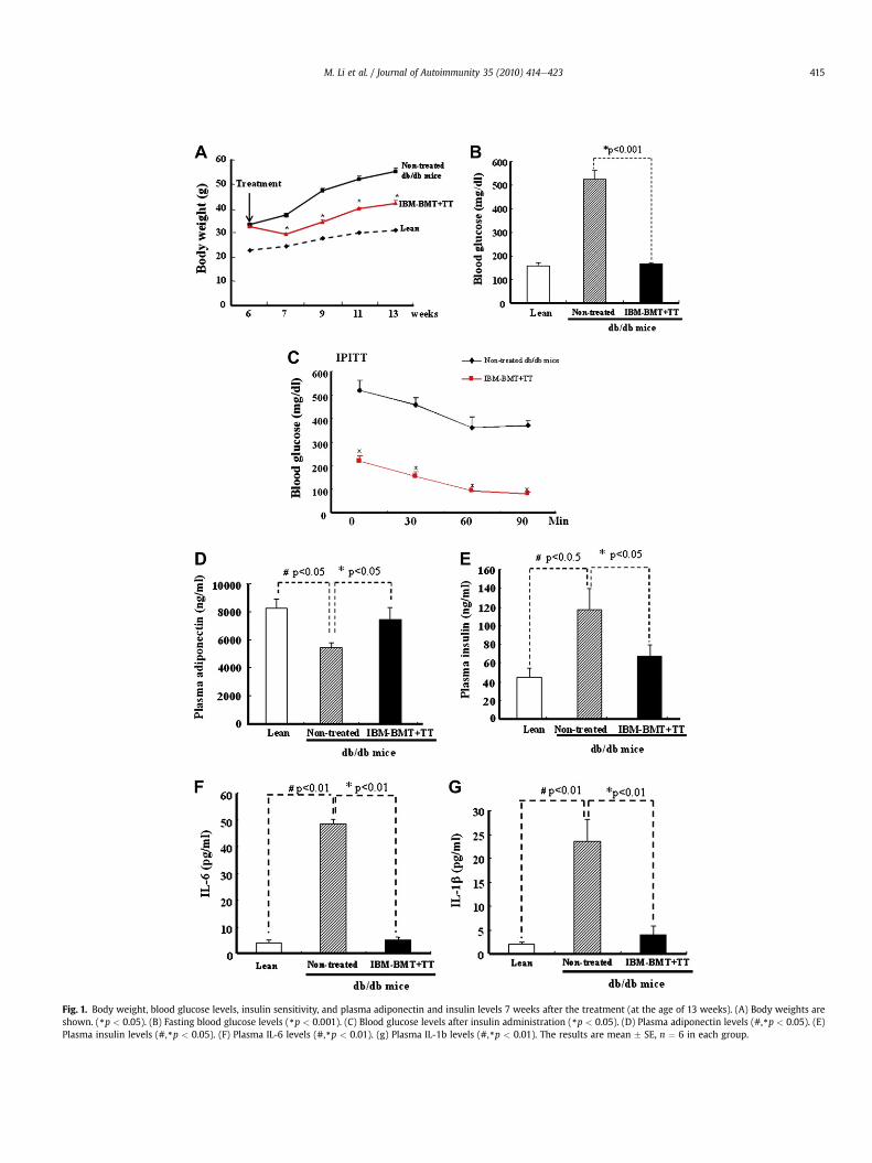
Fig. 1. Body weight, blood glucose levels, insulin sensitivity, and plasma adiponectin and insulin levels 7 weeks after the treatment (at the age of 13 weeks). (A) Body weights areshown. (*p < 0.05). (B) Fasting blood glucose levels (*p < 0.001). (C) Blood glucose levels after insulin administration (*p < 0.05). (D) Plasma adiponectin levels (#,*p < 0.05). (E)Plasma insulin levels (#,*p < 0.05). (F) Plasma IL-6 levels (#,*p < 0.01). (g) Plasma IL-1b levels (#,*p < 0.01). The results are mean � SE, n ¼ 6 in each group.
M. Li et al. / Journal of Autoimmunity 35 (2010) 414e423 415
M. Li et al. / Journal of Autoimmunity 35 (2010) 414e423416
mice, insulin sensitivity increases and blood glucose levelsdecrease, resulting from the normalization of balance of lympho-cyte subsets and cytokines, followed by enhanced expression ofpAKT, pLKB1, pAMPK, insulin receptor phosphorylation and HO-1.This suggests that the maintenance of the balance of lymphocytesubsets and cytokine production by IBMeBMT þ TT treatment isessential for the amelioration of T2 DM in db/db mice.
2. Materials and methods
2.1. Animals
Five-week-old BKS.Cg-mþLeprdb/þLeprdb/J (H-2kd) (db/db)mice, BKS. Cg-mþ/þLeprdb/J(H-2kd) (lean) mice and C57BL/6 (B6)(H-2kb) mice were purchased from Charles River Laboratories(Yokohama, Japan) and SLC (Shizuoka, Japan) and maintained inanimal facilities under specific pathogen-free conditions. Allprocedures were performed under protocols approved by theInstitutional Animal Care and Use Committee at Kansai MedicalUniversity. Body weight and blood glucose levels were measuredeach week. Six-week-old mice with blood glucose levels higherthan 250 mg/dl on two consecutive measurements were consid-ered to have the onset of diabetes, and these mice were separatedinto three groups (n ¼ 6 in each group): non-treated, treated withIBM alone, and treated with IBMeBMTþ TT. All data were collectedat 4 weeks and 7 weeks after treatment. The same experiment wasrepeated three times.
2.2. IBMeBMT þ TT
The mice received fractionated irradiation twice a day (5.0Gy � 2, 4-hour interval). One day after the irradiation, whole BMCsfrom B6mice were injected into the recipient mice (1�107/mouse)by IBMeBMT using our previously described method [23]. Simul-taneously, the newborn thymus from B6 mice was grafted underthe renal capsule of the left kidneys of the recipient mice.
2.3. Flow cytometric analyses
The peripheral blood mononuclear cells were obtained from thetail vein of the recipients 30 days after transplantation. These cellswere stained with antibodies against PE-H-2kb, FITC-CD4, FITC-CD8a, FITC-B220 and FITC-CD11b (BD Bioscience Pharmingen, SanDiego, CA) for 30 min on ice. After washing twice with 2% FCS/PBSand lysing red blood cells, the 10000 events acquiredwere analyzedby FACScan (BD Bioscience Pharmingen). Isotype-matched immu-noglobulins were used as controls.
2.4. Insulin tolerance test
Insulin tolerancewas tested at 7weeks after treatment. After a 6-h fast, mice were injected intraperitoneally with insulin (2.0 units/kg). Blood samples were taken at various time points (0e90 min)and blood glucose levels were measured.
2.5. Cytokine and insulin measurements
Adiponectin, IL-6, IL-1b and TNF-a were determined in mouseplasma using an ELISA assay (R&D Systems, Inc. MN and InvitrogenCorporation CA). Insulin was measured using an ELISA kit (Mor-inaga, Yokohama, Japan).
2.6. Immunochemistry
The pancreata, adipose tissue, and livers of the recipients, leanand db mice were removed 2 months after the transplantation.After the tissues were fixed in 10% formalin for 24 h at roomtemperature, they were embedded in paraffin. The sections (3 mmthickness) were stained with hematoxylin and eosin. To confirmthe presence of glycogen deposits, they were stained with PeriodicAcid Schiff (PAS) after diastase digestion. The pancreata werestained with polyclonal guinea pig anti-swine insulin antibody(N1542, Dako Cytomation, CA). The stained sections were examinedon a microscope. The size of adipocytes was randomly measuredusing DP2-BSW application software (Olympus, Japan).
2.7. Mitogen response
The spleen was removed from the db/db mice at 7 weeks aftertreatment. A total of 2 � 105 splenocytes collected from chimericmice, and untreated B6 and db/db mice as responders, were platedin 96-well plates (Corning Glass Works, Corning, NY) containing200 ml of RPMI 1640 medium (Nissui Seiyaku, Tokyo, Japan)including 2 ml glutamine and 10% FCS. Responder cells were incu-bated with 2.5 mg/ml of Con A (Calbiochem, San Diego, CA) or 25 mg/ml of lipopolysaccharide (Difco Laboratories, Franklin Lakes, NJ) for72 h. 20 ml of 0.5 mCi of 3[H]-TdR (New England Nuclear, Cambridge,MA) was introduced during the last 18 h. Incorporation of 3[H]-TdRwasmeasured usingMicrobeta TriLux (Perkinelmer,Wellesley,MA).
2.8. Western blot analysis of pancreata pLKB1, HO-1, AMPK,pAMPK, AKT, pAKT and insulin receptor phosphorylation
At sacrifice, pancreata were dissected, pooled for each mouseand used to measure signaling molecules. Specimens were storedat �140 �C until assayed. Frozen pancreatic tissues were pulverizedunder liquid nitrogen and placed in a homogenization buffer(mmol/l:10 phosphate buffer, 250 sucrose, 1 EDTA, 0.1 PMSF and0.1% v/v tergitol, pH 7.5). Homogenates were centrifuged at27,000�g for 10 min at 4 �C, supernatant was isolated and proteinlevels were visualized by immunoblotting with antibodies. Anti-bodies against pLKB1, AMPK, pAMPK, AKT, pAKT and HO-1 wereobtained from Cell Signaling Technology, Inc. (Beverly, MA). Anti-bodies were prepared by dilution of HO-1, pAMPK, pAKT andinsulin receptor as we described previously [24,25].
2.9. Statistical analysis
Statistical significance between experimental groups wasdetermined by the Fisher method of analysis of multiple compar-isons (p < 0.05 was regarded as significant). For comparisonbetween treatment groups, the null hypothesis was tested by eithera single-factor ANOVA for multiple groups or unpaired t test for twogroups. Differences between experimental groups were evaluatedwith ANOVA with Bonferroni corrections. Statistical significancewas regarded as significant at p < 0.05.
3. Results
3.1. Body weight, blood glucose levels, insulin sensitivity, andplasma adiponectin, insulin, IL-6 and IL-1b levels
In our preliminary experiments, we carried out IBMeBMT alone(without TT). The IBMeBMT-treated db/db mice showed decreasedblood glucose levels (<150 mg/ml) one week after the treatment butrapid increases in blood glucose levels 2 weeks after the treatment;the mice became susceptible to severe infection due to a rebound
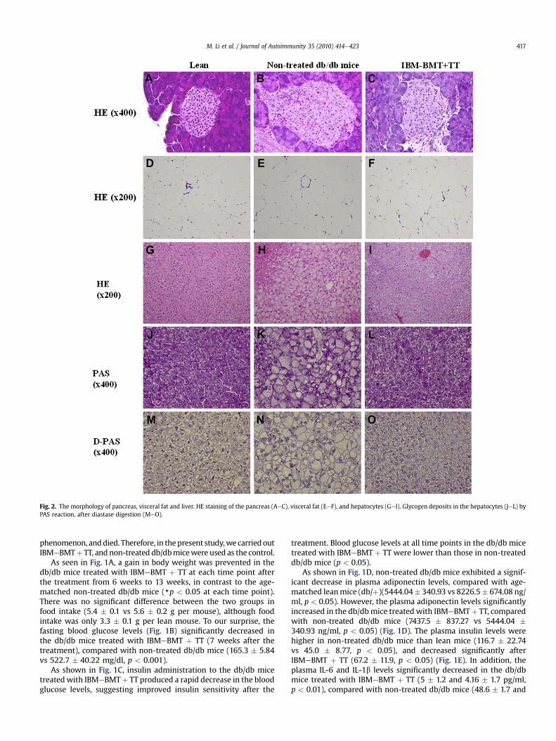
Fig. 2. The morphology of pancreas, visceral fat and liver. HE staining of the pancreas (AeC), visceral fat (EeF), and hepatocytes (GeI). Glycogen deposits in the hepatocytes (JeL) byPAS reaction, after diastase digestion (MeO).
M. Li et al. / Journal of Autoimmunity 35 (2010) 414e423 417
phenomenon, anddied. Therefore, in thepresent study,wecarriedoutIBMeBMTþ TT, andnon-treated db/dbmicewere used as the control.
As seen in Fig. 1A, a gain in body weight was prevented in thedb/db mice treated with IBMeBMT þ TT at each time point afterthe treatment from 6 weeks to 13 weeks, in contrast to the age-matched non-treated db/db mice (*p < 0.05 at each time point).There was no significant difference between the two groups infood intake (5.4 � 0.1 vs 5.6 � 0.2 g per mouse), although foodintake was only 3.3 � 0.1 g per lean mouse. To our surprise, thefasting blood glucose levels (Fig. 1B) significantly decreased inthe db/db mice treated with IBMeBMT þ TT (7 weeks after thetreatment), compared with non-treated db/db mice (165.3 � 5.84vs 522.7 � 40.22 mg/dl, p < 0.001).
As shown in Fig. 1C, insulin administration to the db/db micetreatedwith IBMeBMTþ TT produced a rapid decrease in the bloodglucose levels, suggesting improved insulin sensitivity after the
treatment. Blood glucose levels at all time points in the db/db micetreated with IBMeBMT þ TT were lower than those in non-treateddb/db mice (p < 0.05).
As shown in Fig. 1D, non-treated db/db mice exhibited a signif-icant decrease in plasma adiponectin levels, compared with age-matched leanmice (db/þ)(5444.04� 340.93 vs 8226.5� 674.08 ng/ml, p < 0.05). However, the plasma adiponectin levels significantlyincreased in the db/dbmice treatedwith IBMeBMTþ TT, comparedwith non-treated db/db mice (7437.5 � 837.27 vs 5444.04 �340.93 ng/ml, p < 0.05) (Fig. 1D). The plasma insulin levels werehigher in non-treated db/db mice than lean mice (116.7 � 22.74vs 45.0 � 8.77, p < 0.05), and decreased significantly afterIBMeBMT þ TT (67.2 � 11.9, p < 0.05) (Fig. 1E). In addition, theplasma IL-6 and IL-1b levels significantly decreased in the db/dbmice treated with IBMeBMT þ TT (5 � 1.2 and 4.16 � 1.7 pg/ml,p < 0.01), compared with non-treated db/db mice (48.6 � 1.7 and
M. Li et al. / Journal of Autoimmunity 35 (2010) 414e423418
23.5 � 4.7 pg/ml, p < 0.01) (Fig. 1F and G). However, there were nosignificant differences in the levels of TNFa between the treated db/db and non-treated db/db mice (data not shown).
3.2. Morphology of pancreas, visceral fat, and liver
In the HE staining, non-treated db/db mice (Fig. 2B and E)showed larger islets and larger adipocytes in the visceral adiposetissue than leanmice (Fig. 2A and D). In contrast, smaller islets werenoted in the db/db mice treated with IBMeBMTþ TT (Fig. 2C and F)than non-treated db/db mice: The adipocytes were significantlylarger in non-treated db/db mice than in lean mice (140.4 � 8.17 vs87.9� 7.52 mm in diameter, p< 0.01), but smaller in the db/db micetreated with IBMeBMT þ TT than in non-treated db/db mice(106.0 � 1.29 vs 140.4 � 8.17 mm in diameter, p < 0.01). In addition,enlarged hepatocytes were found in non-treated db/db mice(Fig. 2H), although the hepatocytes in the db/db mice treated withIBMeBMT þ TT (Fig. 2I) were similar in size to those in lean mice(Fig. 2G). Glycogen deposits were seen in the hepatocytes in allgroups (Fig. 2JeL) by PAS reaction, and disappeared after diastasedigestion (Fig. 2MeO). However, the density was lower in non-treated db/db mice than lean mice and the db/db mice treated withIBMeBMT þ TT (Fig. 2I and L), suggesting that the impairedglycogen synthesis resulted from impaired insulin sensitivity in thenon-treated db/db mice. Thus, the glycogen synthesis was alsoimproved after IBMeBMT þ TT.
3.3. Insulin content of pancreas islet
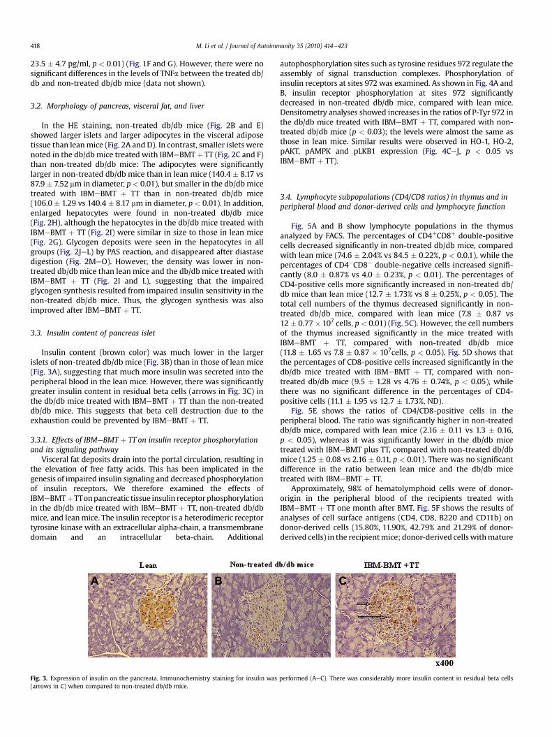
Insulin content (brown color) was much lower in the largerislets of non-treated db/db mice (Fig. 3B) than in those of lean mice(Fig. 3A), suggesting that much more insulin was secreted into theperipheral blood in the lean mice. However, there was significantlygreater insulin content in residual beta cells (arrows in Fig. 3C) inthe db/db mice treated with IBMeBMT þ TT than the non-treateddb/db mice. This suggests that beta cell destruction due to theexhaustion could be prevented by IBMeBMT þ TT.
3.3.1. Effects of IBMeBMT þ TT on insulin receptor phosphorylationand its signaling pathway
Visceral fat deposits drain into the portal circulation, resulting inthe elevation of free fatty acids. This has been implicated in thegenesis of impaired insulin signaling and decreased phosphorylationof insulin receptors. We therefore examined the effects ofIBMeBMTþ TTonpancreatic tissue insulin receptor phosphorylationin the db/db mice treated with IBMeBMT þ TT, non-treated db/dbmice, and leanmice. The insulin receptor is a heterodimeric receptortyrosine kinase with an extracellular alpha-chain, a transmembranedomain and an intracellular beta-chain. Additional
Fig. 3. Expression of insulin on the pancreata. Immunochemistry staining for insulin was(arrows in C) when compared to non-treated db/db mice.
autophosphorylation sites such as tyrosine residues 972 regulate theassembly of signal transduction complexes. Phosphorylation ofinsulin receptors at sites 972 was examined. As shown in Fig. 4A andB, insulin receptor phosphorylation at sites 972 significantlydecreased in non-treated db/db mice, compared with lean mice.Densitometry analyses showed increases in the ratios of P-Tyr 972 inthe db/db mice treated with IBMeBMT þ TT, compared with non-treated db/db mice (p < 0.03); the levels were almost the same asthose in lean mice. Similar results were observed in HO-1, HO-2,pAKT, pAMPK and pLKB1 expression (Fig. 4CeJ, p < 0.05 vsIBMeBMT þ TT).
3.4. Lymphocyte subpopulations (CD4/CD8 ratios) in thymus and inperipheral blood and donor-derived cells and lymphocyte function
Fig. 5A and B show lymphocyte populations in the thymusanalyzed by FACS. The percentages of CD4þCD8þ double-positivecells decreased significantly in non-treated db/db mice, comparedwith lean mice (74.6 � 2.04% vs 84.5 � 0.22%, p < 0.0.1), while thepercentages of CD4�CD8� double-negative cells increased signifi-cantly (8.0 � 0.87% vs 4.0 � 0.23%, p < 0.01). The percentages ofCD4-positive cells more significantly increased in non-treated db/db mice than lean mice (12.7 � 1.73% vs 8 � 0.25%, p < 0.05). Thetotal cell numbers of the thymus decreased significantly in non-treated db/db mice, compared with lean mice (7.8 � 0.87 vs12 � 0.77 � 107 cells, p < 0.01) (Fig. 5C). However, the cell numbersof the thymus increased significantly in the mice treated withIBMeBMT þ TT, compared with non-treated db/db mice(11.8 � 1.65 vs 7.8 � 0.87 � 107cells, p < 0.05). Fig. 5D shows thatthe percentages of CD8-positive cells increased significantly in thedb/db mice treated with IBMeBMT þ TT, compared with non-treated db/db mice (9.5 � 1.28 vs 4.76 � 0.74%, p < 0.05), whilethere was no significant difference in the percentages of CD4-positive cells (11.1 � 1.95 vs 12.7 � 1.73%, ND).
Fig. 5E shows the ratios of CD4/CD8-positive cells in theperipheral blood. The ratio was significantly higher in non-treateddb/db mice, compared with lean mice (2.16 � 0.11 vs 1.3 � 0.16,p < 0.05), whereas it was significantly lower in the db/db micetreated with IBMeBMT plus TT, compared with non-treated db/dbmice (1.25 � 0.08 vs 2.16 � 0.11, p < 0.01). There was no significantdifference in the ratio between lean mice and the db/db micetreated with IBMeBMT þ TT.
Approximately, 98% of hematolymphoid cells were of donor-origin in the peripheral blood of the recipients treated withIBMeBMT þ TT one month after BMT. Fig. 5F shows the results ofanalyses of cell surface antigens (CD4, CD8, B220 and CD11b) ondonor-derived cells (15.80%, 11.90%, 42.79% and 21.29% of donor-derived cells) in the recipientmice; donor-derived cellswithmature
performed (AeC). There was considerably more insulin content in residual beta cells
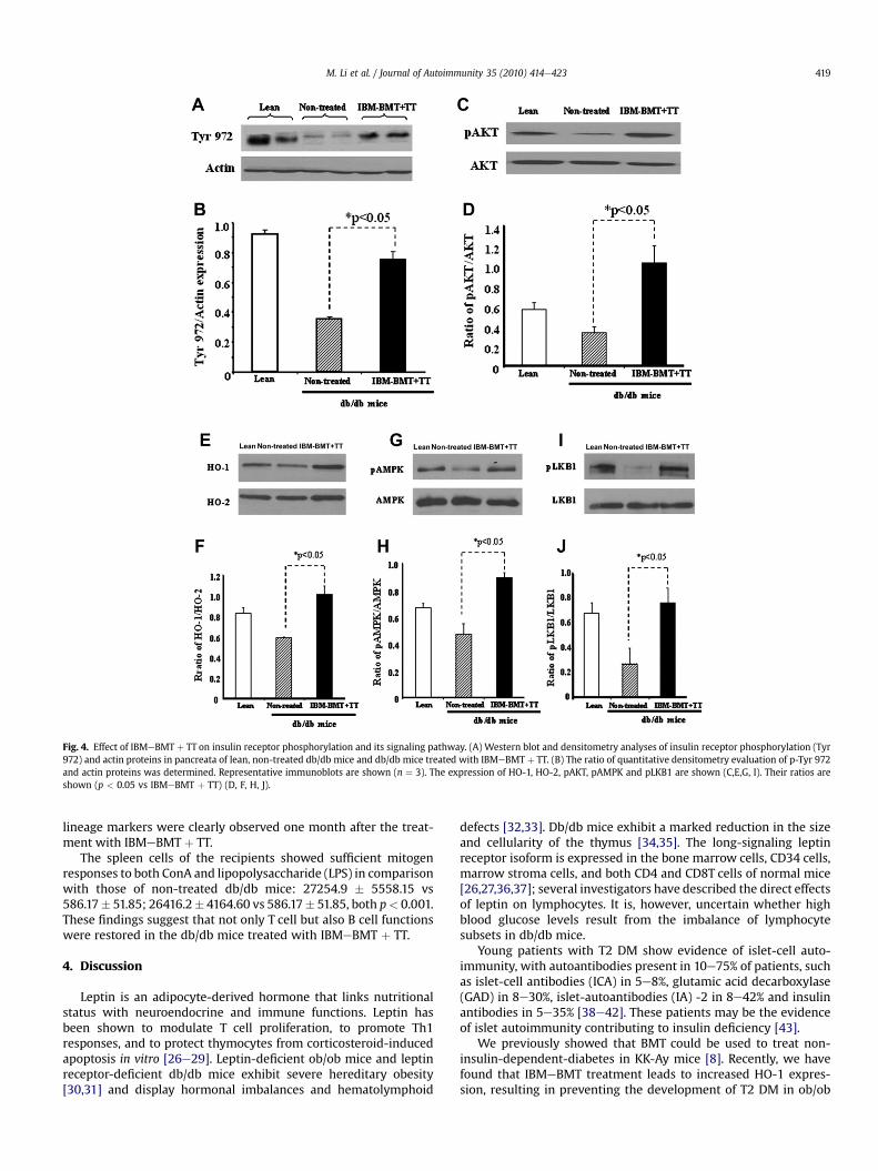
Fig. 4. Effect of IBMeBMT þ TT on insulin receptor phosphorylation and its signaling pathway. (A) Western blot and densitometry analyses of insulin receptor phosphorylation (Tyr972) and actin proteins in pancreata of lean, non-treated db/db mice and db/db mice treated with IBMeBMT þ TT. (B) The ratio of quantitative densitometry evaluation of p-Tyr 972and actin proteins was determined. Representative immunoblots are shown (n ¼ 3). The expression of HO-1, HO-2, pAKT, pAMPK and pLKB1 are shown (C,E,G, I). Their ratios areshown (p < 0.05 vs IBMeBMT þ TT) (D, F, H, J).
M. Li et al. / Journal of Autoimmunity 35 (2010) 414e423 419
lineage markers were clearly observed one month after the treat-ment with IBMeBMT þ TT.
The spleen cells of the recipients showed sufficient mitogenresponses to both ConA and lipopolysaccharide (LPS) in comparisonwith those of non-treated db/db mice: 27254.9 � 5558.15 vs586.17� 51.85; 26416.2� 4164.60 vs 586.17� 51.85, both p< 0.001.These findings suggest that not only T cell but also B cell functionswere restored in the db/db mice treated with IBMeBMT þ TT.
4. Discussion
Leptin is an adipocyte-derived hormone that links nutritionalstatus with neuroendocrine and immune functions. Leptin hasbeen shown to modulate T cell proliferation, to promote Th1responses, and to protect thymocytes from corticosteroid-inducedapoptosis in vitro [26e29]. Leptin-deficient ob/ob mice and leptinreceptor-deficient db/db mice exhibit severe hereditary obesity[30,31] and display hormonal imbalances and hematolymphoid
defects [32,33]. Db/db mice exhibit a marked reduction in the sizeand cellularity of the thymus [34,35]. The long-signaling leptinreceptor isoform is expressed in the bone marrow cells, CD34 cells,marrow stroma cells, and both CD4 and CD8T cells of normal mice[26,27,36,37]; several investigators have described the direct effectsof leptin on lymphocytes. It is, however, uncertain whether highblood glucose levels result from the imbalance of lymphocytesubsets in db/db mice.
Young patients with T2 DM show evidence of islet-cell auto-immunity, with autoantibodies present in 10e75% of patients, suchas islet-cell antibodies (ICA) in 5e8%, glutamic acid decarboxylase(GAD) in 8e30%, islet-autoantibodies (IA) -2 in 8e42% and insulinantibodies in 5e35% [38e42]. These patients may be the evidenceof islet autoimmunity contributing to insulin deficiency [43].
We previously showed that BMT could be used to treat non-insulin-dependent-diabetes in KK-Ay mice [8]. Recently, we havefound that IBMeBMT treatment leads to increased HO-1 expres-sion, resulting in preventing the development of T2 DM in ob/ob
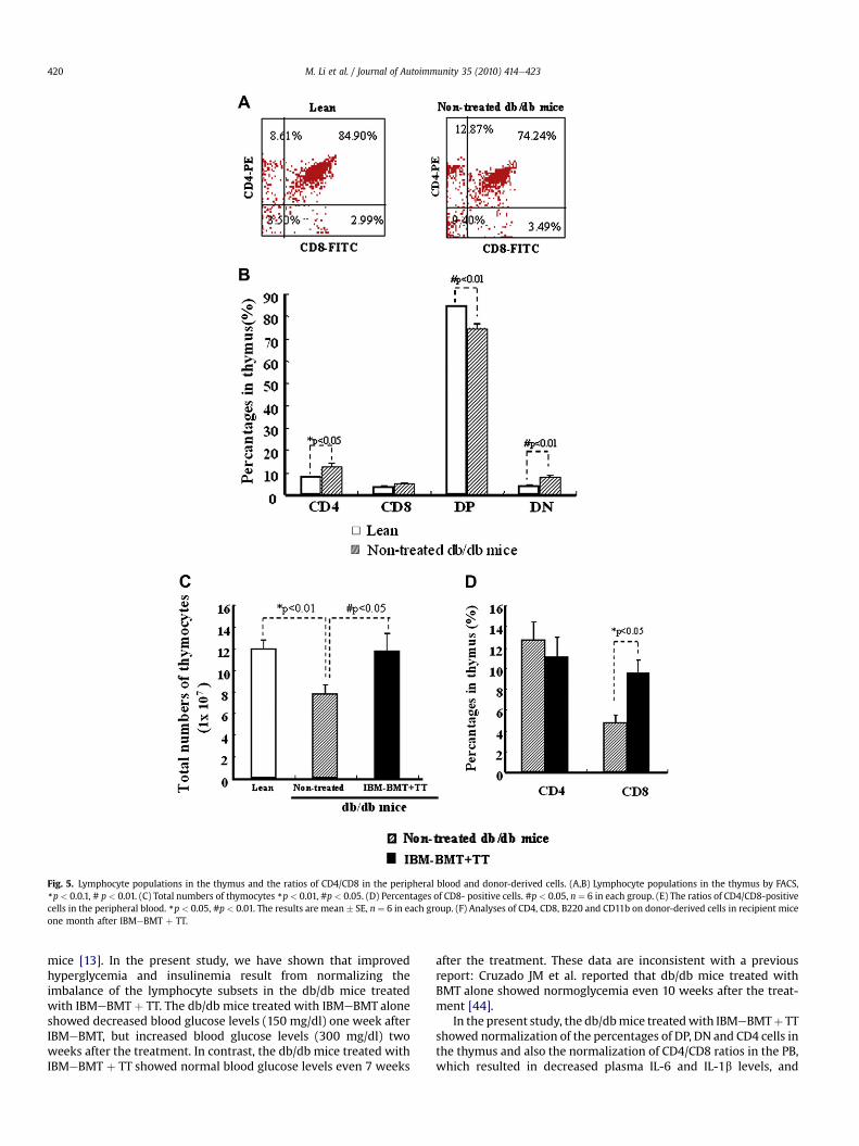
Fig. 5. Lymphocyte populations in the thymus and the ratios of CD4/CD8 in the peripheral blood and donor-derived cells. (A,B) Lymphocyte populations in the thymus by FACS,*p < 0.0.1, # p < 0.01. (C) Total numbers of thymocytes *p < 0.01, #p < 0.05. (D) Percentages of CD8- positive cells. #p < 0.05, n ¼ 6 in each group. (E) The ratios of CD4/CD8-positivecells in the peripheral blood. *p < 0.05, #p < 0.01. The results are mean � SE, n ¼ 6 in each group. (F) Analyses of CD4, CD8, B220 and CD11b on donor-derived cells in recipient miceone month after IBMeBMT þ TT.
M. Li et al. / Journal of Autoimmunity 35 (2010) 414e423420
mice [13]. In the present study, we have shown that improvedhyperglycemia and insulinemia result from normalizing theimbalance of the lymphocyte subsets in the db/db mice treatedwith IBMeBMT þ TT. The db/db mice treated with IBMeBMT aloneshowed decreased blood glucose levels (150 mg/dl) one week afterIBMeBMT, but increased blood glucose levels (300 mg/dl) twoweeks after the treatment. In contrast, the db/db mice treated withIBMeBMT þ TT showed normal blood glucose levels even 7 weeks
after the treatment. These data are inconsistent with a previousreport: Cruzado JM et al. reported that db/db mice treated withBMT alone showed normoglycemia even 10 weeks after the treat-ment [44].
In the present study, the db/dbmice treatedwith IBMeBMTþ TTshowed normalization of the percentages of DP, DN and CD4 cells inthe thymus and also the normalization of CD4/CD8 ratios in the PB,which resulted in decreased plasma IL-6 and IL-1b levels, and
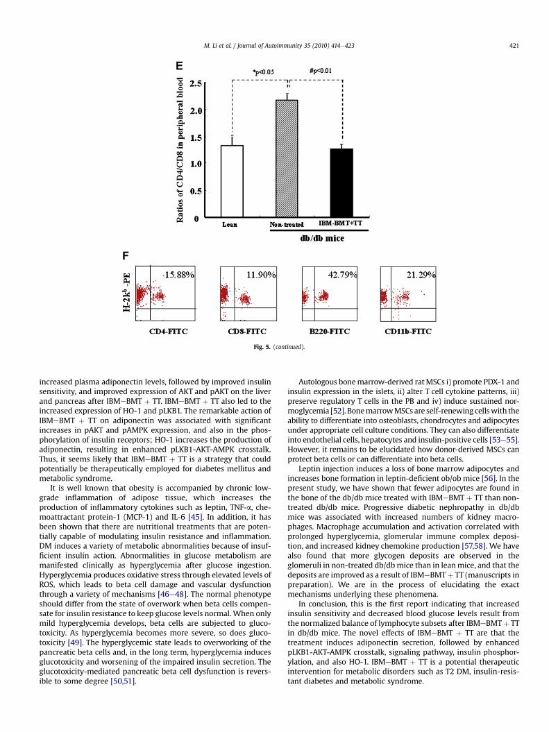
Fig. 5. (continued).
M. Li et al. / Journal of Autoimmunity 35 (2010) 414e423 421
increased plasma adiponectin levels, followed by improved insulinsensitivity, and improved expression of AKT and pAKT on the liverand pancreas after IBMeBMT þ TT. IBMeBMT þ TT also led to theincreased expression of HO-1 and pLKB1. The remarkable action ofIBMeBMT þ TT on adiponectin was associated with significantincreases in pAKT and pAMPK expression, and also in the phos-phorylation of insulin receptors; HO-1 increases the production ofadiponectin, resulting in enhanced pLKB1-AKT-AMPK crosstalk.Thus, it seems likely that IBMeBMT þ TT is a strategy that couldpotentially be therapeutically employed for diabetes mellitus andmetabolic syndrome.
It is well known that obesity is accompanied by chronic low-grade inflammation of adipose tissue, which increases theproduction of inflammatory cytokines such as leptin, TNF-a, che-moattractant protein-1 (MCP-1) and IL-6 [45]. In addition, it hasbeen shown that there are nutritional treatments that are poten-tially capable of modulating insulin resistance and inflammation.DM induces a variety of metabolic abnormalities because of insuf-ficient insulin action. Abnormalities in glucose metabolism aremanifested clinically as hyperglycemia after glucose ingestion.Hyperglycemia produces oxidative stress through elevated levels ofROS, which leads to beta cell damage and vascular dysfunctionthrough a variety of mechanisms [46e48]. The normal phenotypeshould differ from the state of overwork when beta cells compen-sate for insulin resistance to keep glucose levels normal. When onlymild hyperglycemia develops, beta cells are subjected to gluco-toxicity. As hyperglycemia becomes more severe, so does gluco-toxicity [49]. The hyperglycemic state leads to overworking of thepancreatic beta cells and, in the long term, hyperglycemia inducesglucotoxicity and worsening of the impaired insulin secretion. Theglucotoxicity-mediated pancreatic beta cell dysfunction is revers-ible to some degree [50,51].
Autologous bonemarrow-derived ratMSCs i) promote PDX-1 andinsulin expression in the islets, ii) alter T cell cytokine patterns, iii)preserve regulatory T cells in the PB and iv) induce sustained nor-moglycemia [52]. BonemarrowMSCs are self-renewing cellswith theability to differentiate into osteoblasts, chondrocytes and adipocytesunder appropriate cell culture conditions. They can also differentiateinto endothelial cells, hepatocytes and insulin-positive cells [53e55].However, it remains to be elucidated how donor-derived MSCs canprotect beta cells or can differentiate into beta cells.
Leptin injection induces a loss of bone marrow adipocytes andincreases bone formation in leptin-deficient ob/ob mice [56]. In thepresent study, we have shown that fewer adipocytes are found inthe bone of the db/db mice treated with IBMeBMT þ TT than non-treated db/db mice. Progressive diabetic nephropathy in db/dbmice was associated with increased numbers of kidney macro-phages. Macrophage accumulation and activation correlated withprolonged hyperglycemia, glomerular immune complex deposi-tion, and increased kidney chemokine production [57,58]. We havealso found that more glycogen deposits are observed in theglomeruli in non-treated db/dbmice than in leanmice, and that thedeposits are improved as a result of IBMeBMTþ TT (manuscripts inpreparation). We are in the process of elucidating the exactmechanisms underlying these phenomena.
In conclusion, this is the first report indicating that increasedinsulin sensitivity and decreased blood glucose levels result fromthe normalized balance of lymphocyte subsets after IBMeBMTþ TTin db/db mice. The novel effects of IBMeBMT þ TT are that thetreatment induces adiponectin secretion, followed by enhancedpLKB1-AKT-AMPK crosstalk, signaling pathway, insulin phosphor-ylation, and also HO-1. IBMeBMT þ TT is a potential therapeuticintervention for metabolic disorders such as T2 DM, insulin-resis-tant diabetes and metabolic syndrome.

M. Li et al. / Journal of Autoimmunity 35 (2010) 414e423422
Acknowledgments
We would like to thank Mr. Hilary Eastwick-Field and Ms. K.Ando for their help in the preparation of the manuscript. This studywas mainly supported by the 21st Century Center of Excellence(COE) program of theMinistry of Education, Culture, Sports, Scienceand Technology. This study was also supported by grants fromHaiteku Research Center of the Ministry of Education, Health andLabour Sciences Research Grants, the Science Frontier program ofthe Ministry of Education, Culture, Sports, Science and Technology,the Department of Transplantation for Regeneration Therapy(sponsored by Otsuka Pharmaceutical Company, Ltd.), MolecularMedical Science Institute, Otsuka Pharmaceutical Co., Ltd., JapanImmunoresearch Laboratories Co., Ltd. (JIMRO), NIH grantsDK068134, HL55601 and HL34300 (NGA).
References
[1] Despres JP, Lemieux I. Abdominal obesity and metabolic syndrome. Nature2006;444:881e7.
[2] Nishimura S, Manabe I, Nagasaki M, Eto K, Yamashita H, Ohsugi M, et al. CD8þeffector T cells contribute to macrophage recruitment and adipose tissueinflammation in obesity. Nat Med 2009;15:914e20.
[3] HotamisligilGS. Inflammationandmetabolic disorders. Nature2006;444:860e7.[4] Suganami T, Nishida J, Ogawa Y. A paracrine loop between adipocytes and
macrophages aggravates inflammatory changes: role of free fatty acids andtumor necrosis factor alpha. Arterioscler Thromb Vasc Biol 2005;25:2062e8.
[5] Clift RA, Thomas ED. Follow-up 26 years after treatment for acute myeloge-nous leukemia. N Engl J Med 2004;351:2456e7.
[6] Ikehara S, Ohtsuki H, Good RA, Asamoto H, Nakamura T, Sekita K, et al.Prevention of type I diabetes in nonobese diabetic mice by allogenic bonemarrow transplantation. Proc Natl Acad Sci U S A 1985;82:7743e7.
[7] Ikehara S, Good RA, Nakamura T, Sekita K, Inoue S, Oo MM, et al. Rationale forbone marrow transplantation in the treatment of autoimmune diseases. ProcNatl Acad Sci U S A 1985;82:2483e7.
[8] Than S, Ishida H, Inaba M, Fukuba Y, Seino Y, Adachi M, et al. Bone marrowtransplantation as a strategy for treatment of non-insulin-dependent diabetesmellitus in KK-Ay mice. J Exp Med 1992;176:1233e8.
[9] Gershwin ME. Bone marrow transplantation, refractory autoimmunity and thecontributions of Susumu Ikehara. J Autoimmun 2008;30:105e7.
[10] Ikehara S. A novel method of bone marrow transplantation (BMT) forintractable autoimmune diseases. J Autoimmun 2008;30:108e15.
[11] Burt RK, Testori A, Craig R, Cohen B, Suffit R, Barr W. Hematopoietic stem celltransplantation for autoimmune diseases: what have we learned? J Auto-immun 2008;30:116e20.
[12] Ozawa K, Sato K, Oh I, Ozaki K, Uchibori R, Obara Y, et al. Cell and gene therapyusing mesenchymal stem cells (MSCs). J Autoimmun 2008;30:121e7.
[13] Abraham NG, Li M, Vanella L, Peterson SJ, Ikehara S, Asprinio D. Bone marrowstem cell transplant into intra-bone cavity prevents type 2 diabetes: role ofheme oxygenase-adiponectin. J Autoimmun 2008;30:128e35.
[14] Sonoda Y. Immunophenotype and functional characteristics of human prim-itive CD34-negative hematopoietic stem cells: the significance of the intra-bone marrow injection. J Autoimmun 2008;30:136e44.
[15] Marmont AM. Will hematopoietic stem cell transplantation cure humanautoimmune diseases? J Autoimmun 2008;30:145e50.
[16] Ratajczak MZ, Zuba-Surma EK, Wysoczynski M, Wan W, Ratajczak J,Wojakowski W, et al. Hunt for pluripotent stem cell e regenerative medicinesearch for almighty cell. J Autoimmun 2008;30:151e62.
[17] Hara M, Murakami T, Kobayashi E. In vivo bioimaging using photogenic rats:fate of injected bone marrow-derived mesenchymal stromal cells. J Auto-immun 2008;30:163e71.
[18] Rezvani AR, Storb RF. Separation of graft-vs.-tumor effects from graft-vs.-hostdisease in allogeneic hematopoietic cell transplantation. J Autoimmun2008;30:172e9.
[19] Deane S, Meyers FJ, Gershwin ME. On reversing the persistence of memory:hematopoietic stem cell transplant for autoimmune disease in the first tenyears. J Autoimmun 2008;30:180e96.
[20] Hosaka N, Nose M, Kyogoku M, Nagata N, Miyashima S, Good RA, et al.Thymus transplantation, a critical factor for correction of autoimmune diseasein aging MRL/þmice. Proc Natl Acad Sci U S A 1996;93:8558e62.
[21] Ryu T, Hosaka N, Miyake T, Cui W, Nishida T, Takaki T, et al. Transplantation ofnewborn thymus plus hematopoietic stem cells can rescue supralethallyirradiated mice. Bone Marrow Transplant 2008;41:659e66.
[22] Hosaka N, Ryu T, Miyake T, Cui W, Nishida T, Takaki T, et al. Treatment ofautoimmune diseases in MRL/lpr mice by allogenic bone marrow trans-plantation plus adult thymus transplantation. Clin Exp Immunol2007;147:555e63.
[23] Kushida T, Inaba M, Hisha H, Ichioka N, Esumi T, Ogawa R, et al. Intra-bonemarrow injection of allogeneic bone marrow cells: a powerful new strategy
for treatment of intractable autoimmune diseases in MRL/lpr mice. Blood2001;97:3292e9.
[24] Peterson SJ, Kim DH, Li M, Positano V, Vanella L, Rodella LF, et al. The L-4Fmimetic peptide prevents insulin resistance through increased levels of HO-1,pAMPK, and pAKT in obese mice. J Lipid Res 2009;50:1293e304.
[25] Li M, Kim DH, Tsenovoy PL, Peterson SJ, Rezzani R, Rodella LF, et al. Treatmentof obese diabetic mice with a heme oxygenase inducer reduces visceral andsubcutaneous adiposity, increases adiponectin levels, and improves insulinsensitivity and glucose tolerance. Diabetes 2008;57:1526e35.
[26] Lord GM, Matarese G, Howard JK, Baker RJ, Bloom SR, Lechler RI. Leptinmodulates the T-cell immune response and reverses starvation-inducedimmunosuppression. Nature 1998;394:897e901.
[27] Martin-Romero C, Santos-Alvarez J, Goberna R, Sanchez-Margalet V. Humanleptin enhances activation and proliferation of human circulating T lympho-cytes. Cell Immunol 2000;199:15e24.
[28] Lord GM, Matarese G, Howard JK, Bloom SR, Lechler RI. Leptin inhibits theanti-CD3-driven proliferation of peripheral blood T cells but enhances theproduction of proinflammatory cytokines. J Leukoc Biol 2002;72:330e8.
[29] Fujita Y, Murakami M, Ogawa Y, Masuzaki H, Tanaka M, Ozaki S, et al. Leptininhibits stress-induced apoptosis of T lymphocytes. Clin Exp Immunol2002;128:21e6.
[30] Zhang Y, Proenca R, Maffei M, Barone M, Leopold L, Friedman JM. Positionalcloning of the mouse obese gene and its human homologue. Nature1994;372:425e32.
[31] Chua Jr SC, Chung WK, Wu-Peng XS, Zhang Y, Liu SM, Tartaglia L, et al.Phenotypes of mouse diabetes and rat fatty due to mutations in the OB(leptin) receptor. Science 1996;271:994e6.
[32] Fantuzzi G, Faggioni R. Leptin in the regulation of immunity, inflammation,and hematopoiesis. J Leukoc Biol 2000;68:437e46.
[33] Matarese G, Moschos S, Mantzoros CS. Leptin in immunology. J Immunol2005;174:3137e42.
[34] Fernandes G, Handwerger BS, Yunis EJ, Brown DM. Immune response in themutant diabetic C57BL/Ks-dtþ mouse. Discrepancies between in vitro and invivo immunological assays. J Clin Invest 1978;61:243e50.
[35] Kimura M, Tanaka S, Isoda F, Sekigawa K, Yamakawa T, Sekihara H. T lym-phopenia in obese diabetic (db/db) mice is non-selective and thymus inde-pendent. Life Sci 1998;62:1243e50.
[36] Gainsford T, Willson TA, Metcalf D, Handman E, McFarlane C, Ng A, et al.Leptin can induce proliferation, differentiation, and functional activation ofhemopoietic cells. Proc Natl Acad Sci U S A 1996;93:14564e8.
[37] Thomas T, Gori F, Khosla S, Jensen MD, Burguera B, Riggs BL. Leptin acts onhuman marrow stromal cells to enhance differentiation to osteoblasts and toinhibit differentiation to adipocytes. Endocrinology 1999;140:1630e8.
[38] Hathout EH, Thomas W, El-Shahawy M, Nahab F, Mace JW. Diabetic autoim-mune markers in children and adolescents with type 2 diabetes. Pediatrics2001;107:E102.
[39] Umpaichitra V, Banerji MA, Castells S. Autoantibodies in children with type 2diabetes mellitus. J Pediatr Endocrinol Metab 2002;15(Suppl. 1):525e30.
[40] Gilliam LK, Brooks-Worrell BM, Palmer JP, Greenbaum CJ, Pihoker C. Auto-immunity and clinical course in children with type 1, type 2, and type 1.5diabetes. J Autoimmun 2005;25:244e50.
[41] Songer T, Glazner J, Coombs LP, Cuttler L, Daniel M, Estrada S, et al. Examiningthe economic costs related to lifestyle and pharmacological interventions inyouth with type 2 diabetes. Expert Rev Pharmacoecon Outcomes Res2006;6:315e24.
[42] Reinehr T, Schober E, Wiegand S, Thon A, Holl R. Beta-cell autoantibodies inchildren with type 2 diabetes mellitus: subgroup or misclassification? ArchDis Child 2006;91:473e7.
[43] Klingensmith GJ, Pyle L, Arslanian S, Copeland KC, Cuttler L, Kaufman F, et al.The presence of GAD and IA-2 antibodies in youth with a type 2 diabetesphenotype: results from the TODAY study. Diabetes Care; 2010.
[44] Flaquer M, Franquesa M, Barquinero J, Lloberas N, Gutierrez C, Torras J, et al.Bone marrow transplantation induces normoglycemia in a type 2 diabetesmellitus murine model. Transplant Proc 2009;41:2282e5.
[45] Fantuzzi G. Adipose tissue, adipokines, and inflammation. J Allergy ClinImmunol 2005;115:911e9. quiz 20.
[46] Baynes JW. Role of oxidative stress in development of complications in dia-betes. Diabetes 1991;40:405e12.
[47] Li M, Peterson S, Husney D, Inaba M, Guo K, Terada E, et al. Interdiction of thediabetic state in NOD mice by sustained induction of heme oxygenase:possible role of carbon monoxide and bilirubin. Antioxid Redox Signal2007;9:855e63.
[48] Li M, Peterson S, Husney D, Inaba M, Guo K, Kappas A, et al. Long-lastingexpression of HO-1 delays progression of type I diabetes in NOD mice. CellCycle 2007;6:567e71.
[49] Weir GC, Marselli L, Marchetti P, Katsuta H, Jung MH, Bonner-Weir S. Towardsbetter understanding of the contributions of overwork and glucotoxicity tothe beta-cell inadequacy of type 2 diabetes. Diabetes Obes Metab 2009;11(Suppl. 4):82e90.
[50] Kosaka K, Kuzuya T, Akanuma Y, Hagura R. Increase in insulin response aftertreatment of overt maturity-onset diabetes is independent of the mode oftreatment. Diabetologia 1980;18:23e8.
[51] MacDonald MJ, Efendic S, Ostenson CG. Normalization by insulin treatment oflow mitochondrial glycerol phosphate dehydrogenase and pyruvate carbox-ylase in pancreatic islets of the GK rat. Diabetes 1996;45:886e90.

M. Li et al. / Journal of Autoimmunity 35 (2010) 414e423 423
[52] Boumaza I, Srinivasan S, Witt WT, Feghali-Bostwick C, Dai Y, Garcia-Ocana A,et al. Autologous bone marrow-derived rat mesenchymal stem cells promotePDX-1 and insulin expression in the islets, alter T cell cytokine pattern andpreserve regulatory T cells in the periphery and induce sustained normogly-cemia. J Autoimmun 2009;32:33e42.
[53] Theise ND, Nimmakayalu M, Gardner R, Illei PB, Morgan G, Teperman L, et al.Liver from bone marrow in humans. Hepatology 2000;32:11e6.
[54] Oswald J, Boxberger S, Jorgensen B, Feldmann S, Ehninger G, Bornhauser M,et al. Mesenchymal stem cells can be differentiated into endothelial cells invitro. Stem Cells 2004;22:377e84.
[55] Li M, Inaba M, Guo KQ, Hisha H, Abraham NG, Ikehara S. Treatment ofstreptozotocin-induced diabetes mellitus in mice by intra-bone marrow bone
marrow transplantation plus portal vein injection of beta cells induced frombone marrow cells. Int J Hematol 2007;86:438e45.
[56] Hamrick MW, Della-Fera MA, Choi YH, Pennington C, Hartzell D, Baile CA.Leptin treatment induces loss of bone marrow adipocytes and increases boneformation in leptin-deficient ob/ob mice. J Bone Miner Res2005;20:994e1001.
[57] Chow FY, Nikolic-Paterson DJ, Atkins RC, Tesch GH. Macrophages in strepto-zotocin-induced diabetic nephropathy: potential role in renal fibrosis.Nephrol Dial Transplant 2004;19:2987e96.
[58] Chow F, Ozols E, Nikolic-Paterson DJ, Atkins RC, Tesch GH. Macrophages inmouse type 2 diabetic nephropathy: correlation with diabetic state andprogressive renal injury. Kidney Int 2004;65:116e28.