Autologous transplantation of CD34(+) bone marrow derived mononuclear cells in management of...
13
1 23 Cytotechnology Incorporating Methods in Cell Science International Journal of Cell Culture and Biotechnology ISSN 0920-9069 Cytotechnology DOI 10.1007/s10616-014-9828-7 Autologous transplantation of CD34 + bone marrow derived mononuclear cells in management of non-reconstructable critical lower limb ischemia Ahmed M. Ismail, Said M. Abdou, Hassan Abdel Aty, Adel H. Kamhawy, Mohammed Elhinedy, Mohammed Elwageh, Atef Taha, Amal Ezzat, et al.
-
Upload
independent -
Category
Documents
-
view
3 -
download
0
Transcript of Autologous transplantation of CD34(+) bone marrow derived mononuclear cells in management of...

1 23
CytotechnologyIncorporating Methods in Cell ScienceInternational Journal of Cell Culture andBiotechnology ISSN 0920-9069 CytotechnologyDOI 10.1007/s10616-014-9828-7
Autologous transplantation of CD34+
bone marrow derived mononuclear cells inmanagement of non-reconstructable criticallower limb ischemia
Ahmed M. Ismail, Said M. Abdou,Hassan Abdel Aty, Adel H. Kamhawy,Mohammed Elhinedy, MohammedElwageh, Atef Taha, Amal Ezzat, et al.

1 23
Your article is protected by copyright and all
rights are held exclusively by Springer Science
+Business Media Dordrecht. This e-offprint
is for personal use only and shall not be self-
archived in electronic repositories. If you wish
to self-archive your article, please use the
accepted manuscript version for posting on
your own website. You may further deposit
the accepted manuscript version in any
repository, provided it is only made publicly
available 12 months after official publication
or later and provided acknowledgement is
given to the original source of publication
and a link is inserted to the published article
on Springer's website. The link must be
accompanied by the following text: "The final
publication is available at link.springer.com”.

ORIGINAL RESEARCH
Autologous transplantation of CD34+ bone marrow derivedmononuclear cells in management of non-reconstructablecritical lower limb ischemia
Ahmed M. Ismail • Said M. Abdou • Hassan Abdel Aty • Adel H. Kamhawy •
Mohammed Elhinedy • Mohammed Elwageh • Atef Taha • Amal Ezzat •
Hoda A. Salem • Said Youssif • Mohamed L. Salem
Received: 29 December 2013 / Accepted: 19 November 2014
� Springer Science+Business Media Dordrecht 2014
Abstract Patients with a decrease in limb perfusion
with a potential threat to limb viability manifested by
ischemic rest pain, ischemic ulcers, and/or gangrene
are considered to have critical limb ischemia (CLI).
Because of this generally poor outcome, there is a
strong need for attempting any procedure to save the
affected limb. The aim of this work is to evaluate the
possibility to use stem cell therapy as a treatment
option for patients with chronic critical lower limb
ischemia with no distal run off. This study includes 20
patients with chronic critical lower limb ischemia with
no distal run off who are unsuitable for vascular or
endovascular option. These patients underwent stem
cell therapy (SCT) by autologous transplantation of
bone marrow derived mononuclear cells. 55 % of
patients treated with SCT showed improvement of the
rest pain after the first month, 60 % continued
improvement of the rest pain after 6 months, 75 %
after 1 year and 80 % after 2 years and continued
without any deterioration till the third year. Limb
salvage rate after STC was 80 % after the first year till
the end of the second and third years. SCT can result in
angiogenesis in patients with no-option CLI, provid-
ing a foundation for the application of this therapy to
leg ischemia.
Keywords Bone marrow � G-CSF � Leg ischemia �Mobilization � CD34? cells
Introduction
Prevalence of peripheral arterial disease (PAD) is in
the range of 3–10 %, increasing to 15–20 % inAhmed M. Ismail and Said M. Abdou have equally contributed
to this manuscript.
A. M. Ismail � H. A. Aty � A. H. Kamhawy �M. Elhinedy � M. Elwageh
Vascular Surgery Unit, Tanta University, Tanta, Egypt
S. M. Abdou � A. Ezzat
Clinical Pathology Department, Tanta University, Tanta,
Egypt
A. Taha
Internal Medicine Department, Faculty of Medicine,
Tanta University, Tanta, Egypt
H. A. Salem
Faculty of Pharmacy, Al-Azhar University, Cairo, Egypt
S. Youssif
Faculty of Medicine, Ain Shams University, Cairo, Egypt
M. L. Salem (&)
Immunology and Biotechnology Unit, Zoology
Department, Faculty of Science, Center of Excellence in
Cancer Research, Tanta University, Tanta, Egypt
e-mail: [email protected];
123
Cytotechnology
DOI 10.1007/s10616-014-9828-7
Author's personal copy

individuals over the age of 70 years. Patients with a
decrease in limb perfusion with a potential threat to
limb viability manifested by ischemic rest pain,
ischemic ulcers, and/or gangrene are considered to
have critical limb ischemia (CLI) (Hirsch et al. 2006).
Major amputation is linked with substantial mor-
bidity and mortality, with a particularly high preva-
lence of co-morbid diseases (Dormandy et al. 1999).
About half of patients who undergo above-knee
amputation will die within 1 year after the procedure,
and high percentage of patients are considered unfit for
prosthetic rehabilitation. After 2 years, only 40 % of
those fitted with prosthesis can ambulate, and even
fewer are independent outside the home. Despite
advances in vascular and endovascular techniques,
14–20 % of patients with chronic lower limb ischemia
will not be eligible for distal arterial reconstruction
due to occlusion of crural and pedal vessels (Attanasio
and Snell 2009). Because of this generally poor
outcome, there is a strong need for attempting any
procedure to save the affected limb (Casamassimi
et al. 2012; Inderbitzi et al. 1992).
Therapeutic angiogenesis by autologous bone
marrow cell transplantation can improve blood supply
in patients with critical limb ischemia (Mizuno et al.
2010). Bone marrow derived stem and progenitor cells
have been identified as a potential new therapeutic
option to induce therapeutic angiogenesis. Encourag-
ing results of preclinical studies have rapidly led to
several small clinical trials, in which bone marrow-
derived mononuclear cells were administered to
patients with limb ischemia. Clinical benefits were
reported from these trials including improvement of
ankle-brachial pressure index (ABPI), transcutaneous
partial pressure of oxygen (TcPO2), reduction of pain,
and decreased need for amputation (Lawall et al.
2010). The main goal of this study was to assess the
influence of autologous mononuclear cell transplan-
tation in the treatment of non reconstructable critical
lower limb ischemia.
Patients and methods
This study included 20 patients with reconstructable
chronic critical lower limb ischemia with no distal run
off. All patients were admitted to the vascular surgery
Unit, Tanta University Hospital, Tanta, Egypt in the
period from January 2009 till January 2012. All
recruited patients were exposed to complete history
and physical examination, including measurement of
ankle brachial pressure index (ABPI). Duplex study
and CT angiography were also done for all patients.
The age ranged from 42 to 83 years with mean age
62 years. Fourteen patients were males and 6 patients
were females. The study was approved by the ethical
committee of the Faculty of Medicine in Tanta
University. A written informed consent was taken
from all patients. The procedure was explained in
details and in clear simple language to all recruited
patients. All possible complications of therapy were
explained to all patients with emphasis that the patient
can withdraw from the study at any stage if he wishes.
Inclusion and exclusion criteria
Patients with chronic critical lower limb ischemia stage
III or IV and with ABPI below 0.5 were included in
which Duplex examination showing no flow in tibial
and pedal arteries. Patients with angiography revealing
no distal run off were included. Patients with acute
ischemia and who are suitable for balloon angioplasty
or vascular reconstruction were excluded. Other exclu-
sion criteria included hematological abnormalities as
anemia (Hb \ 10 g/dl), leukopenia (WBCs \ 4,000/
cc), thrombocytopenia (platelets\ 100,000/cc), malig-
nancies as leukemia and lymphoma, organ failure as
liver cell failure and renal failure, and viral infections,
such as HIV and hepatitis B.
Preparation of patients before bone marrow
aspiration
Complete blood count (CBC), abdominal ultrasound,
and examination of viral markers including hepatitis B
and C and HIV viruses were performed in all patients
before aspiration. The patients have received recombi-
nant human Granulocyte Colony Stimulating Factor
(rhGCSF) (GeneLeukim Injection from Shandong
Geneleuk Biopharmaceutical Co., Ltd., Jinan, Shan-
dong, China). Each 1 ml vial contained 600 lg Filgra-
stim given by subcutaneous injection in a dose of 5 lg/
kg per day for 3–5 days to mobilize stem/progenitor
cells. Meanwhile, a perfusion of 10,000 units/day
heparin for 5 days by intravenous drop was used to
avoid the possible risks of embolism because G-CSF
induces the increase of circulating blood cells. CBC was
done just before harvesting bone marrow by aspiration
Cytotechnology
123
Author's personal copy

and repeated daily to check the effect of G-CSF and final
CBC just before harvesting bone marrow by aspiration.
Bone marrow aspiration
Under complete antiseptic precautions, a prophylactic
dose of antibiotic (Cefotaxim (1 g via I.V. injection;
purchased from Sanofi Company, Cairo, Egypt)) was
given to all patients prior to bone marrow (BM)
harvest. Two approaches were used to obtain a BM
aspirate: the first one was from the anterior superior
iliac spine; and the second one from the posterior
superior iliac spine (which gives a higher amount of
bone marrow aspirate). A volume of 100–150 cc of
BM was aspirated from the iliac crest through anterior
superior or posterior iliac spine of the patient then sent
to the laboratory to separate the mononuclear cell
fraction. All steps were performed under sterile
conditions in a laminar flow hood (in the Clinical
Pathology Department, Tanta University Hospital).
Preparation of human bone marrow mononuclear
cells (BM MNCs)
The BM aspirate prepared above was diluted at a ratio of
4:1 with clinical buffer (Clini MACS PBS/EDTA buffer
1,000 ml, CE approved for clinical use catalogue number
#700–25, from Miltenyi Biotec Company, Bergisch
Gladbach, Germany). The diluted cell suspension was
then carefully layered over 15 ml of Ficoll-Paque (GE
Electric, Pharmacia, Piscataway, NJ, USA) in a 50 ml
conical tube, and then centrifuged at 2,000 rpm for
20 min at 20 �C in a swinging out bucket rotor without
brake. The upper layer was aspirated leaving the mono-
nuclear cell layer undisturbed at the interphase containing
lymphocytes, monocytes, and thrombocytes. The middle
layer was carefully transferred to a new 50 ml conical
tube. The cells were then washed twice with clinical
buffer, mixed gently and centrifuged at 1,200 rpm for
15 min at 20 �C. Then the supernatant was carefully and
completely removed. The cell pellet was resuspended in
the appropriate amount of clinical buffer with the final
volume of 300 ll of clinical buffer for up to 108 total cells.
Purification of stem cells by magnetic labeling
This purification was performed according to the previ-
ously described protocol (Lawall et al. 2010). The cells
were kept cold and all the solutions used were at room
temperature. The cells were passed through 30 lm nylon
mesh (pre-separation filter) to remove cell clumps which
might clog the column and the cell number was
determined by using hemocytometer. The cell suspension
was centrifuged at 1,200 rpm for 10 min and the
supernatant was aspirated completely and the cell pellet
was resuspended in 2 ml clinical buffer. CD34 Micro-
beads (150 lm; (Clini MACS CD34 microbeads, from
Miltenyi Biotec Company, catalogue number #171-01)
were added to the cell suspension, mixed well and
refrigerated for 30 min. The cells were washed with the
clinical buffer and centrifuged at 1,800 rpm for 20 min,
then the supernatant was aspirated completely and the
cells were suspended in 500 ll buffer. For magnetic
separation, the column (MS column) was placed in the
magnetic field of the Mini MACS separator. Cell
suspension was applied to the column. Unlabelled cells
that passed through were collected and the column was
washed with Clinical buffer. Washing steps were per-
formed by adding clinical buffer three times (3 9 500 ll
clinical buffer), new clinical buffer was only added when
the column reservoir was empty. The total effluent was
collected and this was the unlabelled cell fraction. The
column was removed from the Mini MACS separator
(Miltenyi Biotec) and placed on a suitable collection tube.
The clinical buffer was pipetted onto the column and the
magnetically labelled cells were immediately flushed out
by firmly pushing the plunger into the column.
Flow cytometry analysis
The purity of the CD34? cells harvested by magnetic
labeling after the application of Mini MACS separator
as described above was determined by flow cytometry.
Aliquots of the fresh samples from the collected cells
were stained with anti-human mAbs (BD Biosciences,
Franklin Lakes, NJ, USA). The cells were incubated
for 20–30 min at 4 �C in the dark with the anti-human
CD34 (PE) and anti-human CD45 (Percep) mAbs
using the concentrations recommended by the manu-
facturer. The cells were then washed twice using
HBSS. Acquisitions were performed with a FACS
Calibur (BD Biosciences) and data analysis was done
by FlowJo software (BD Biosciences).
Treatment with cells
The patients were taken to the operating room and placed
under general anaesthesia. The ulcers were surgically
Cytotechnology
123
Author's personal copy

debrided under sterile conditions to ensure a clean base
with no scars, fibrotic or necrotic tissues. This allowed
direct contact of bone marrow cells to a viable wound
tissue bed. The cells were injected into the ulcer edge and
the ulcer bed by using a 3 ml syringes with 19 gauge 1.5
needle. Thereafter, the wound surface was protected with
ointment gauze (Biotulle (Betadine gauze (Povidone-
Iodine) was purchased from Minapharm Company,
Cairo Egypt)) and sterile dry gauze dressings (obtained
from the local Pharmacy, Tanta Hospital University,
Tanta, Egypt). This dressing was left on the wound for
24 h then removed, the wound was washed with saline
0.9 % only and a new dressing was used (Fig. 1).
Follow up
All patients were followed up for signs of improve-
ment of circulation and tissue perfusion, including
limb salvage/amputation, ulcer healing, disappearance
of rest pain, increased pain-free walking distance,
improvement of the ABPI. Angiography was also
followed after 6 weeks, 3 and 6 months intervals after
stem cell transplantation to detect the formation of
new collateral vessels.
Statistical analysis
All data of the patients were entered into a database
and analyzed using statistical software (SPSS Version
15.0 for Windows, SPSS, Chicago, IL, USA). Paired-
samples t test was used to prove the differences
between before and after intervention. Chi square test
was performed for determination of the P value to
compare the rates, and the probability ratio among the
studied groups. Statistical significance was assumed at
a value of P \ 0.05. So P value above 0.05 is
considered statistically non-significant.
Results
Level of arterial occlusion in patients
The main clinical presentation was rest pain in all
patients. Gangrene was found in 5 patients, 3 of them
in the toes and 2 in the forefoot. Ischemic ulcer was
found in 4 patients. The level of occlusion was in the
popliteal artery in 8 patients, femoral artery in 5
patients and external iliac artery in 2 patients.
Clinical responses to G-CSF
After G-CSF administration, the WBCs count was
increased in the peripheral blood indicating the
increase in mononuclear cell production and mobili-
zation. The maximum WBCs count was 45,300/cc and
the minimum was 19,000/cc (with mean value of
34,700).
AB C
Ischemic ulcer Healed ulcer after 2 m.
Big toe gangrene Big toe amputation stump
Healed big toe
after 4 months
Fig. 1 a Injection of CD34? cells around ischemic ulcer in the left leg. b Healed ulcer after 2 months. c Healed big toe amputation
stump after 4 months
Cytotechnology
123
Author's personal copy

Effect of SCT on rest pain
By the use of Visual Analogue Scale (VAS), the severity
of pain was assessed before and after SCT (Table 1).
There was an improvement in the rest pain after SCT in
11 patients (55 %) after the first month. The VAS
decreased from 7.3 ± 1.1 points to 3.7 ± 1.6 points
(P value = 0.047). At 6 months duration after stem cell
therapy, a number of 12 patients (60 %) showed
continuous improvement of the rest pain with VAS
being 3.3 ± 1.7 points (P value = 0.038). At the end of
the first year, 15 patients (75 %) continued improve-
ment of the rest pain with no need for analgesics after
stem cell therapy with VAS being 3.2 ± 1.4 points
(P value = 0.044). At the end of the second year 16
patients (80 %) continued improvement of the rest pain
after 2 years and continued without any deterioration till
the third year with VAS being 3.4 ? 1.7 points
(P value = 0.058). All patients with limb salvage
showed improvement of the rest pain.
Effect of SCT on physical activity and pain-free
walking distance
As shown in Table 2, the pain free walking distance
increased after 6 months following stem cell therapy in
12 patients (60 %), it increased from 85.3 ± 81.7 m to
163.5 ± 127.6 m (P value = 0.028). At the end of the
first year, 15 patients (75 %) showed slight improve-
ment and the pain free walking distance was
169.6 ± 131.8 m (P value = 0.031). This improve-
ment remained stable at the end of the second year 16
patients (80 %) showed stable improvement and the
pain free walking distance was 156.6 ± 127.3 m
(P value = 0.046). Finally after 3 years, the number
of patients was the same as in the second year with
improvement and the pain free walking distance being
149.8 ± 132.3 m (P value = 0.052). Ten out of the 16
patients (80 %) who had limb salvage showed improve-
ment and the pain free walking distance.
ABPI after SCT
As shown in Table 3, eleven patients (55 %) showed
improvement in the ABPI after SCT in the first
6 months. ABPI increased from 0.27 ± 0.18 to
0.71 ± 0.19 (P value = 0.026). At the end of the first
year, ABPI continued improving in 12 patients
(60 %). ABPI was 0.72 ± 0.18 (P value = 0.031).
At the end of the second year ABPI continued
improving in 14 patients (70 %). As compared to the
values before treatment, ABPI follow up was
0.74 ± 0.12 (P value = 0.038). After the third year
ABPI continued improving in 16 patients (80 %).
ABPI follow up was 0.69 ± 0.22 (P value = 0.048).
Limb salvage and major amputation
The total number of saved limbs without major
amputation after SCT after the first, second, and third
year was 16 patients (80 %). Major amputation was
done for 4 patients (20 %) during the 6 months due to
Table 1 Rest pain improvement after SCT
Duration Rest pain
improvement
Visual analogue
scale (VAS)
P value
No. % Before
CLS
After
CLS
1 month 11 55 7.3 ± 1.1 3.7 ± 1.6 0.047
6 months 12 60 7.3 ± 1.1 3.3 ± 1.7 0.038
First year 15 75 7.3 ± 1.1 3.2 ± 1.4 0.044
Second year 16 80 7.3 ± 1.1 3.4 ± 1.7 0.058
Third year 16 80 7.3 ± 1.1 3.4 ± 1.7 0.058
Table 2 Pain-free walking distance after SCT
Duration Patients Pain-free walking distance
(in meters)
P value
n % Before SCT After SCT
6 months 12 60 85.3 ± 81.7 163.5 ± 127.6 0.028
First year 15 75 85.3 ± 81.7 169.6 ± 131.8 0.031
Second
year
16 80 85.3 ± 81.7 156.6 ± 127.3 0.046
Third year 16 80 85.3 ± 81.7 149.8 ± 132.3 0.052
Table 3 Ankle brachial pressure index after SCT
Duration Patients ABPI P value
n % Before SCT After SCT
6 months 11 55 0.27 ± 0.18 0.71 ± 0.19 0.026
First year 12 60 0.27 ± 0.18 0.72 ± 0.18 0.031
Second year 14 70 0.27 ± 0.18 0.74 ± 0.12 0.038
Third year 16 80 0.27 ± 0.18 0.69 ± 0.22 0.048
Cytotechnology
123
Author's personal copy

extensive necrosis, resistant infection and persistent
rest pain.
Effect of SCT on healing of ischemic ulcer
As presented in Fig. 2, four patients (out of 20) were
presented with toe gangrene in the SCT group. Two
patients (50 % of the 4 patients) had gangrene in one
toe and showed good healing after toe amputation.
Two patients (out of 20) with forefoot gangrene did
not show any improvement after SCT and ended with
major amputation. Three patients (75 %), out of the
four who presented with ischemic ulcer, showed
healing of the ulcer in the first 6 months after SCT
and continued till the end of the third year. The fourth
patient ended with major amputation.
Effect of SCT on angiography
As shown in Fig. 3, angiography was done after SCT
in 12 patients (60 %). No angiography was performed
for the remaining 8 patients (40 %) including the 4
patients having undergone major amputations and 4
patients having refused to do angiography because all
symptoms improved. Among the 12 patients who had
follow up angiography, 9 patients (75 %) showed
angiogenesis and 3 patients (25 %) did not show
angiogenesis despite clinical improvement.
Adverse effects of SCT
The complications of SCT included injection site pain
having occurred in 5 patients (25 %), it was mild pain
and was treated with non-steroidal anti-inflammatory
drugs. Small intramuscular hematoma occurred in one
patient (5 %) and resolved spontaneously after
2 weeks. Three patients (15 %) developed mild edema
in the injected leg which improved after 3 weeks. Low
grade fever appeared in 2 patients (10 %) despite
routine antibiotic use in all patients after injection.
This fever resolved spontaneously within 4 days after
injection. Extensive necrosis and gangrene with no
response to stem cell therapy were encountered in 4
patients (20 %) and major amputation was performed.
No procedure-related mortality or thrombo-embolic
complications were observed after stem cell therapy
(Fig. 3).
Discussion
The main challenge in the therapeutic approach to
critical limb ischemia is to establish an effective
reperfusion of the ischemic limb. Conventional arte-
rial reconstruction by a bypass surgery can solve the
problem of a long segment occlusion while percuta-
neous trans-luminal angioplasty can be done in short
segment lesions. Peripheral vascular disease with
arterial occlusion with poor distal run off is considered
unsuitable for both bypass and angioplasty. Indeed,
several variables still remain to be elucidated for stem
cell therapy, including the type of cells to be used, the
infusion route, and more importantly, the stage of
patients to be treated (Casamassimi et al. 2012).
Growth of new vessels develops by proliferation of
endothelial cells in vascular extremities as well as by
BM-mobilized HSCs, which are transformed into
endothelial progenitor cells. These cells contribute to
generation of new endothelial cells and vasculariza-
tion. Therefore, some studies used mononuclear BM
cells and others used differentiated mesenchymal stem
cells. In our study presented here, patients with
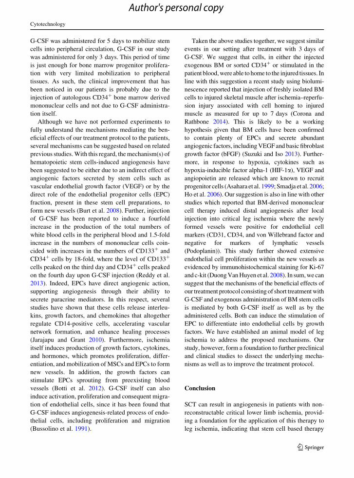
Fig. 2 a Before injection of CD34? cells. b One month after injection. c 3 months after injection with complete healing
Cytotechnology
123
Author's personal copy

chronic critical lower limb ischemia with no distal run
off who were unsuitable for vascular or endovascular
option underwent SCT by autologous transplantation
of BM-derived mononuclear cells after mobilization
of stem cells with G-CSF. Our results showed that
55 % of patients treated with SCT showed improve-
ment of the rest pain after the first month, 60 %
continued improvement of the rest pain after
6 months, 75 % after 1 year and 80 % after 2 years
and continued without any deterioration till the third
year. Limb salvage rate after STC was 80 % after the
first year till the end of the second and third years,
indicating that stem cell based therapy can result in
angiogenesis in patients with no-option CLI.
Similar to our results, intravascular injection of
mononuclear BM cells laterally through a 4 Fr sheet
in 24 patients with CLI, 11 of 14 defects were healed
(78 %) and Fontaine grade of ischemia changed from
median grade 3.5 to median grade 2 associated with
improvement in the collateral vessel development
(Chochola et al. 2008). The utility of BM derived
mononuclear cells to produce angiogenesis in 33
Indian patients with Buerger’s disease was evaluated,
where major amputation was done for 3 patients
(12 %). The mean ABI improvement after 6 months
in the salvaged limbs was 0.14 (Motukuru et al.
2008). A similar study investigated BM-mononuclear
cell transplantation in 7 patients with critical lower
limb ischemia (3 with Buerger’s disease, 4 with
arteriosclerosis obliterans undergoing chronic he-
modialysis). Three out of 7 patients responded to the
therapy. The objective criteria of improvement were
ABI, laser thermography, transcutaneous oxygen
tension and angiography. Interestingly, the numbers
of circulating CD34? and CD133? cells persistently
increased for 1 month after the treatment only in the
responders (Kajiguchi et al. 2007). Similarly, neo-
vasculogenesis in CLI diabetic patients was evalu-
ated after infusion of unfractionated autologous BM-
derived mononuclear cell generating a significant
increase in the vascular network in ischemic areas
and promoting remarkable clinical improvement
(Ruiz-Salmeron et al. 2011).
Besides BM-derived mononuclear cells, MSCs are a
source of pericyte progenitors and angiogenic regulah-
tors and thus represent preferential stimuli for the
development of blood vessels (Rastegar et al. 2010).
Therefore, several studies have utilized these cells in
treatment of leg ischemia. For instance, treatment with
intravenous infusions (3 pulses) of expanded autolo-
gous MSCs in 1 patient with critical limb ischemia due
to systemic sclerosis who developed acute gangrene of
the upper and lower limbs reduced the area of necrotic
skin and associated with revascularization of the
patient’s extremities, indicating that this approach
may foster the recovery of the vascular network, restore
blood flow, and reduce skin necrosis in this patient
population (Guiducci et al. 2010).
A B
angiogenesis
Fig. 3 a Arteriogram of a
patient with critical Left
lower limb ischemia with
popliteal artery occlusion
with no distal run-off before
CD34? cell transplantation.
b Good collaterals and
angiogenesis after 6 weeks
following CD34?
transplantation
Cytotechnology
123
Author's personal copy

It was found in recent studies that the efficacy of
combinatorial cell therapy based on infusion of
autologous BM-derived mononuclear cells (as a
source of EPCs progenitors) and MSCs is higher than
that of the single therapy. For instance, in phase I and
II clinical trials on patients with CLI, the use of a
combination cell product (mesenchymal stem cells in
conjunction with a source of endothelial progenitor
cells) was found to be safe and efficient and optimized
the clinical results obtained with the use of endothelial
progenitor cells alone in term of improvement in
walking time and ankle-brachial index with a signif-
icant increase in blood flow in the ischemic legs, and
quality of life (Lasala et al. 2010).
One mechanism that might mediate the induced
improvement in the clinical responses to our G-CSF
mobilizes SCT is the presence of progenitors of
WBCs, in particular it has been found that the flow rate
of fractionated granulocytes and mononuclear cells, as
well as unfractionated mixed WBC from leg ischemia
patients was impaired. However, amputation of the
ischemic leg or pharmacological intervention (with
Pentoxyphylline infusion) improved the filterability of
granulocytes from severe ischemic patients (Nash
et al. 1991). This could explain the improved clinical
response in our patients with leg ischemia after
injection of the unfractionated bone marrow cells
which might release WBC progenitors with normal
flow rate due to G-CSF mobilization of BM.
The route of administration of stem cells has been
found to be critical for its efficacy. Intravenous stem
cell delivery for regenerative tissue therapy has been
increasingly used in both experimental and clinical
trials. However, recent data suggest that the majority
of administered stem cells are initially trapped in the
lungs (Zonta et al. 2010). After intra-artery and
intravenous infusion, MSCs were detected primarily
in the lungs and then secondarily in the liver and other
organs. When sodium nitroprusside was used, more
labeled MSCs cleared the lungs resulting in a larger
proportion detected in the liver. Most importantly, the
homing of labeled MSCs to the marrow of long bones
was significantly increased by the pretreatment with
vasodilator. These results indicate multiple homing
sites for injected MSCs and that the distribution of
MSCs can be influenced by administration of vasodi-
lator (Fischer et al. 2009; Gao et al. 2001; Omlor et al.
2010). Therefore, there are numerous doubts about the
best route of stem cell administration to achieve
implantation into the injured site. With this regard, the
comparison of various administration routes of MSCs
in a porcine model of myocardial infarction showed
that the mean number of engrafted cells within the
infarct zone was significantly greater after intracoro-
nary infusion than either intramyocardial or endocar-
dial injection. Fluorescent cells were not observed in
healthy zones of the myocardium or in healthy animals
(Moscoso et al. 2009). Another study found that intra-
arterial (in the renal graft) administration route of
MSCs achieved higher immunomodulating effects
than intravenous route in experimental rat kidney
transplantation after bilateral nephrectomy (Zonta
et al. 2010). The effects of CD34? stem cells delivered
by different routes on cardiac function were compared
in rats with ischemic cardiomyopathy reproduced by
ligation of left anterior descending coronary artery. It
was found that intravenous and trans-epicardial
delivery of hematopoietic stem cells (HSC) can
significantly improve cardiac function, and both
methods may be safe and effective for the treatment
of AMI (Zhang et al. 2008). Therefore, one explana-
tion for the efficacy of our treatment protocol for leg
ischemia might be the route of administration, where
we injected BM cells into and around the diseased area
rather than a systemic administration. Under this
setting, the injected cells are localized in the injured
site which might contain some growth factors that
mediate the differentiation of the injected cells into
endothelial cells and as a consequence enhance
angiogenesis.
Onodera et al. (2011), compared the use of BM
mononuclear cells and G-CSF-mobilized peripheral
blood mononuclear cells in treatment of no-option
critical limb ischemia. Their results suggest that there
was no significant difference in long-term prognosis
between patients treated with BMMNC and those
treated with M-PBMNC (Zhang et al. 2008). Lara-
Hernandez et al. (2011) studied the safety and efficacy
of therapeutic angiogenesis in critical limb ischemia
patients. They reported no adverse effects and limb
salvage rate of 74.4 % after 1 year (Zhang et al. 2008).
In our study, however, we have measured several
parameters before and after therapeutic angiogenesis,
including rest pain by visual analogue scale, walking
distance before and after therapy in meters, ankle
brachial pressure index, limb salvage over all versus
amputation and degree of ischemic ulcer healing.
Moreover, differently from these studies in which
Cytotechnology
123
Author's personal copy
G-CSF was administered for 5 days to mobilize stem
cells into peripheral circulation, G-CSF in our study
was administered for only 3 days. This period of time
is just enough for bone marrow progenitor prolifera-
tion with very limited mobilization to peripheral
tissues. As such, the clinical improvement that has
been noticed in our patients is probably due to the
injection of autologous CD34? bone marrow derived
mononuclear cells and not due to G-CSF administra-
tion itself.
Although we have not performed experiments to
fully understand the mechanisms mediating the ben-
eficial effects of our treatment protocol to the patients,
several mechanisms can be suggested based on related
previous studies. With this regard, the mechanism(s) of
hematopoietic stem cells-induced angiogenesis have
been suggested to be either due to an indirect effect of
angiogenic factors secreted by stem cells such as
vascular endothelial growth factor (VEGF) or by the
direct role of the endothelial progenitor cells (EPC)
fraction, present in these stem cell preparations, to
form new vessels (Burt et al. 2008). Further, injection
of G-CSF has been reported to induce a fourfold
increase in the production of the total numbers of
white blood cells in the peripheral blood and 1.5-fold
increase in the numbers of mononuclear cells coin-
cided with increases in the numbers of CD133? and
CD34? cells by 18-fold, where the level of CD133?
cells peaked on the third day and CD34? cells peaked
on the fourth day upon G-CSF injection (Reddy et al.
2013). Indeed, EPCs have direct angiogenic action,
supporting angiogenesis through their ability to
secrete paracrine mediators. In this respect, several
studies have shown that these cells release interleu-
kins, growth factors, and chemokines that altogether
regulate CD14-positive cells, accelerating vascular
network formation, and enhance healing processes
(Jarajapu and Grant 2010). Furthermore, ischemia
itself induces production of growth factors, cytokines,
and hormones, which promotes proliferation, differ-
entiation, and mobilization of MSCs and EPCs to form
new vessels. In addition, the growth factors can
stimulate EPCs sprouting from preexisting blood
vessels (Botti et al. 2012). G-CSF itself can also
induce activation, proliferation and consequent migra-
tion of endothelial cells, since it has been found that
G-CSF induces angiogenesis-related process of endo-
thelial cells, including proliferation and migration
(Bussolino et al. 1991).
Taken the above studies together, we suggest similar
events in our setting after treatment with 3 days of
G-CSF. We suggest that cells, in either the injected
exogenous BM or sorted CD34? or stimulated in the
patient blood, were able to home to the injured tissues. In
line with this suggestion a recent study using biolumi-
nescence reported that injection of freshly isolated BM
cells to injured skeletal muscle after ischemia–reperfu-
sion injury associated with cell homing to injured
muscle as measured for up to 7 days (Corona and
Rathbone 2014). This is likely to be a working
hypothesis given that BM cells have been confirmed
to contain plenty of EPCs and secrete abundant
angiogenic factors, including VEGF and basic fibroblast
growth factor (bFGF) (Suzuki and Iso 2013). Further-
more, in response to hypoxia, cytokines such as
hypoxia-inducible factor alpha-1 (HIF-1a), VEGF and
angiopoietin are released which are known to recruit
progenitor cells (Asahara et al. 1999; Smadja et al. 2006;
Ho et al. 2006). Our suggestion is also in line with other
studies which reported that BM-derived mononuclear
cell therapy induced distal angiogenesis after local
injection into critical leg ischemia where the newly
formed vessels were positive for endothelial cell
markers (CD31, CD34, and von Willebrand factor and
negative for markers of lymphatic vessels
(Podoplanin)). This study further showed extensive
endothelial cell proliferation within the new vessels as
evidenced by immunohistochemical staining for Ki-67
and c-kit (Duong Van Huyen et al. 2008). In sum, we can
suggest that the mechanisms of the beneficial effects of
our treatment protocol consisting of short treatment with
G-CSF and exogenous administration of BM stem cells
is mediated by both G-CSF itself as well as by the
administered cells. Both can induce the stimulation of
EPC to differentiate into endothelial cells by growth
factors. We have established an animal model of leg
ischemia to address the proposed mechanisms. Our
study, however, form a foundation to further preclinical
and clinical studies to dissect the underlying mecha-
nisms as well as to improve the treatment protocol.
Conclusion
SCT can result in angiogenesis in patients with non-
reconstructable critical lower limb ischemia, provid-
ing a foundation for the application of this therapy to
leg ischemia, indicating that stem cell based therapy
Cytotechnology
123
Author's personal copy

can result in angiogenesis in patients with no-option
CLI. Transplantation of autologous bone marrow
derived mononuclear cells is safe and feasible. Long
term follow up is required for standardization of
therapy with the possibility of repetition for recurrence
of ischemic manifestations. Based on our results these
studies merit validation by randomized controlled
studies in patients with less critical limb ischemia.
Acknowledgment This work was funded by a grant from the
Research Development Fund, Tanta University, Egypt.
Conflict of interest The authors indicate no potential conflicts
of interest.
References
Asahara T, Takahashi T, Masuda H, Kalka C, Chen D, Iwaguro
H, Isner M (1999) VEGF contributes to postnatal neovas-
cularization by mobilizing bone marrow-derived endothe-
lial progenitor cells. EMBO J 18:3964–3972
Attanasio S, Snell J (2009) Therapeutic angiogenesis in the
management of critical limb ischemia: current concepts
and review. Cardiol Rev 17:115–120
Botti C, Maione C, Coppola A, Sica V, Cobellis G (2012)
Autologous bone marrow cell therapy for peripheral arte-
rial disease. Stem Cells Cloning 5:5–14
Burt K, Loh Y, Pearce W, Beohar N, Barr G, Craig R, Kessler J
(2008) Clinical applications of blood-derived and marrow-
derived stem cells for nonmalignant diseases. JAMA
299:925–936
Bussolino F, Ziche M, Wang M, Alessi D, Morbidelli L, Cre-
mona O, Mantovani A (1991) In vitro and in vivo activa-
tion of endothelial cells by colony-stimulating factors.
J Clin Invest 87:986–995
Casamassimi A, Grimaldi V, Infante T, Al-Omran M, Crudele
V, Napoli C (2012) Adult stem cells and the clinical arena:
are we able to widely use this therapy in patients with
chronic limbs arteriopathy and ischemic ulcers without
possibility of revascularization? Cardiovasc Hematol
Agents Med Chem 10:99–108
Chochola M, Pytlik R, Kobylka P, Skalicka L, Kideryova L,
Beran S, Linhart A (2008) Autologous intra-arterial infu-
sion of bone marrow mononuclear cells in patients with
critical leg ischemia. Int Angiol 27:281–290
Corona T, Rathbone R (2014) Accelerated functional recovery
after skeletal muscle ischemia-reperfusion injury using
freshly isolated bone marrow cells. J Surg Res 188:100–109
Dormandy J, Heeck L, Vig S (1999) Major amputations: clinical
patterns and predictors. [Review]. Semin Vasc Surg
12:154–161
Duong Van Huyen P, Smadja M, Bruneval P, Gaussem P, Dal-
Cortivo L, Julia P, Emmerich J (2008) Bone marrow-
derived mononuclear cell therapy induces distal angio-
genesis after local injection in critical leg ischemia. Mod
Pathol 21:837–846
Fischer M, Harting T, Jimenez F, Monzon-Posadas O, Xue H,
Savitz I, Cox S (2009) Pulmonary passage is a major
obstacle for intravenous stem cell delivery: the pulmonary
first-pass effect. Stem Cells Dev 18:683–692
Gao J, Dennis E, Muzic F, Lundberg M, Caplan I (2001) The
dynamic in vivo distribution of bone marrow-derived
mesenchymal stem cells after infusion. Cells Tissues
Organs 169:12–20
Guiducci S, Porta F, Saccardi R, Guidi S, Ibba-Manneschi L,
Manetti M, Matucci-Cerinic M (2010) Autologous mes-
enchymal stem cells foster revascularization of ischemic
limbs in systemic sclerosis: a case report. Ann Intern Med
153:650–654
Hirsch T, Haskal J, Hertzer R, Bakal W, Creager A, Halperin L,
Vascular Disease F (2006) ACC/AHA 2005 Practice
Guidelines for the management of patients with peripheral
arterial disease (lower extremity, renal, mesenteric, and
abdominal aortic): a collaborative report from the Ameri-
can Association for Vascular Surgery/Society for Vascular
Surgery, Society for Cardiovascular Angiography and
Interventions, Society for Vascular Medicine and Biology,
Society of Interventional Radiology, and the ACC/AHA
Task Force on Practice Guidelines (Writing Committee to
Develop Guidelines for the Management of Patients With
Peripheral Arterial Disease): endorsed by the American
Association of Cardiovascular and Pulmonary Rehabilita-
tion; National Heart, Lung, and Blood Institute; Society for
Vascular Nursing; TransAtlantic Inter-Society Consensus;
and Vascular Disease Foundation. [Practice Guideline]
Ho L, Phyliky L, Li Y (2006) B-cell chronic lymphocytic leu-
kemia: correlation of clinical stages with angiogenic
cytokine expression. Appl Immunohistochem Mol Mor-
phol 14:154–160
Inderbitzi R, Buttiker M, Pfluger D, Nachbur B (1992) The fate
of bilateral lower limb amputees in end-stage vascular
disease. Eur J Vasc Surg 6:321–326
Jarajapu P, Grant B (2010) The promise of cell-based therapies
for diabetic complications: challenges and solutions. Circ
Res 106:854–869
Kajiguchi M, Kondo T, Izawa H, Kobayashi M, Yamamoto K,
Shintani S, Murohara T (2007) Safety and efficacy of
autologous progenitor cell transplantation for therapeutic
angiogenesis in patients with critical limb ischemia. Circ J
71:196–201
Lasala P, Silva A, Gardner A, Minguell J (2010) Combination
stem cell therapy for the treatment of severe limb ischemia:
safety and efficacy analysis. Angiology 61:551–556
Lawall H, Bramlage P, Amann B (2010) Stem cell and pro-
genitor cell therapy in peripheral artery disease. A critical
appraisal. [Review]. Thromb Haemost 103:696–709
Mizuno H, Miyamoto M, Shimamoto M, Koike S, Hyakusoku
H, Kuroyanagi Y (2010) Therapeutic angiogenesis by
autologous bone marrow cell implantation together with
allogeneic cultured dermal substitute for intractable ulcers
in critical limb ischaemia. J Plast Reconstr Aesthet Surg
63:1875–1882
Moscoso I, Barallobre J, de Ilarduya M, Anon P, Fraga M,
Calvino R, Domenech N (2009) Analysis of different
routes of administration of heterologous 5-azacytidine-
treated mesenchymal stem cells in a porcine model of
myocardial infarction. Transplant Proc 41:2273–2275
Cytotechnology
123
Author's personal copy

Motukuru V, Suresh R, Vivekanand V, Raj S, Girija R (2008)
Therapeutic angiogenesis in Buerger’s disease (thrombo-
angiitis obliterans) patients with critical limb ischemia by
autologous transplantation of bone marrow mononuclear
cells. J Vasc Surg 48:53S–60S; discussion 60S
Nash B, Thomas R, Dormandy A (1991) Therapeutic aspects of
white blood cell rheology in severe ischaemia of the leg.
J Mal Vasc 16:32–34
Omlor W, Bertram H, Kleinschmidt K, Fischer J, Brohm K,
Guehring T, Richter W (2010) Methods to monitor distri-
bution and metabolic activity of mesenchymal stem cells
following in vivo injection into nucleotomized porcine
intervertebral discs. Eur Spine J 19:601–612
Rastegar F, Shenaq D, Huang J, Zhang W, Zhang Q, He C, He C
(2010) Mesenchymal stem cells: molecular characteristics
and clinical applications. World J Stem Cells 2:67–80
Reddy M, Kwak K, Shim J, Jang C, Park J, Park E, Ahn C (2013)
A long-term outcome of therapeutic angiogenesis by
transplantation of peripheral blood stem cells in critical
limb ischemia after interventional revascularization. Diagn
Interv Radiol 19:76–80
Ruiz-Salmeron R, de la Cuesta-Diaz A, Constantino-Bermejo
M, Perez-Camacho I, Marcos-Sanchez F, Hmadcha A,
Soria B (2011) Angiographic demonstration of neoangio-
genesis after intra-arterial infusion of autologous bone
marrow mononuclear cells in diabetic patients with critical
limb ischemia. Cell Transplant 20:1629–1639
Smadja M, Laurendeau I, Avignon C, Vidaud M, Aiach M,
Gaussem P (2006) The angiopoietin pathway is modulated
by PAR-1 activation on human endothelial progenitor
cells. J Thromb Haemost 4:2051–2058
Suzuki H, Iso Y (2013) Clinical application of vascular regen-
erative therapy for peripheral artery disease. Biomed Res
Int 2013:179730
Zhang H, Li M, Li Y, Zhao P, Jing L (2008) Effects of different
delivery routes of CD34? stem cells on cardiac function in
the ischemic cardiomyopathy of rats. Zhongguo Wei
Zhong Bing Ji Jiu Yi Xue 20:214–217
Zonta S, De Martino M, Bedino G, Piotti G, Rampino T,
Gregorini M, Alessiani M (2010) Which is the most suit-
able and effective route of administration for mesenchymal
stem cell-based immunomodulation therapy in experi-
mental kidney transplantation: endovenous or arterial?
Transplant Proc 42:1336–1340
Cytotechnology
123
Author's personal copy