VITRICATIONTION OF MONONUCLEAR CELLS ISOLATED ...
33
1 VITRICATIONTION OF MONONUCLEAR CELLS ISOLATED FROM HUMAN UMBILICAL CORD BLOOD By Mark Lalduhsaka 108BM020 SUPERVISOR Prof.(Ms.) Krishna Parmanik Department of Biotechnology and Medical Engineering National Institute of Technology, Rourkela
-
Upload
khangminh22 -
Category
Documents
-
view
6 -
download
0
Transcript of VITRICATIONTION OF MONONUCLEAR CELLS ISOLATED ...
1
VITRICATIONTION OF MONONUCLEAR CELLS
ISOLATED FROM HUMAN UMBILICAL CORD
BLOOD
By
Mark Lalduhsaka
108BM020
SUPERVISOR
Prof.(Ms.) Krishna Parmanik
Department of Biotechnology and Medical Engineering
National Institute of Technology, Rourkela
2
National Institute of Technology
Rourkela
CERTIFICATE
This is to certify that the project report entitle “Vitrification of mononuclear cells isolated
from umbilical cord blood” submitted by MARK LALDUHSAKA (108BM020) in the
partial fulfilment of the requirement for the degree of the B.Tech in Biomedical Engineering,
National Institute of Technology, Rourkela is an authentic work carried out by him under my
supervision. To the best of my knowledge the matter embodied in the report has not been
submitted to any other Institute/University for any degree.
Date: 9th
May 2012
Prof. Krishna Parmanik (Supervisor)
Department of Biotechnology and Medical Engineering
National Institute of Technology,
Rourkela-769008
3
ACKNOWLEDGEMENTS
I would like to take this opportunity to extent my hearty gratitude to my guide and advisor
Prof. Krishna Parmanik, Department of Biotechnology and Medical Engineering, National
Institute of Technology, Rourkela, whose constant guidance and encouragement made the
completion of my B.Tech thesis possible.
I also thank National Institute of Technology Rourkela, for permitting me to utilise the
facilities in its laboratories to carry out my experiment.
I would also like to thank Akalabya Bissoyi, PhD scholar, for giving me constant support
with all the necessary things involve in this whole project.
Submitted by:
Mark Lalduhsaka
Roll No: 108BM020
Department of Biotechnology and Medical Engineering
National Institute of Technology,
Rourkela-769008
4
Table of Contents CERTIFICATE ................................................................................................................................... 2
ACKNOWLEDGEMENTS ................................................................................................................ 3
List of Figures ..................................................................................................................................... 5
List of table ......................................................................................................................................... 6
ABSTRACT ........................................................................................................................................ 7
1. INTRODUCTION ..................................................................................................................... 8
1.1. Different techniques used for cryopreservation: ...................................................................... 8
1.2. Umbilical cord blood: .............................................................................................................. 9
1.3. Objective .................................................................................................................................. 9
2. LITERATURE REVIEW ........................................................................................................ 10
3. MATERIALS AND METHODS .......................................................................................... 11
3.1. Isolation of mononuclear cells from cord blood .................................................................... 11
3.2. Experiment No.1 .................................................................................................................... 12
3.3. Experiment No.2 .................................................................................................................... 13
3.4. Thawing and cell recovery ..................................................................................................... 15
3.5. Cell Viability .......................................................................................................................... 16
3.6. Peristaltic pump ..................................................................................................................... 18
3.7. Experimental design ............................................................................................................... 20
4. RESULTS AND DISCUSSION ........................................................................................... 23
4.1. Vitrifiction using plunging method ........................................................................................ 23
4.2. Vitrification using N2 vapour gas ........................................................................................... 23
4.3. Comparison ............................................................................................................................ 29
5. CONCLUSION ..................................................................................................................... 30
6. REFERENCES ..................................................................................................................... 31
5
List of Figures
Figure1: Density gradient centrifugation……………………………………..…………….12
Figure2: Time vs rpm graph for peristaltic pump……………………………………...……19
Figure3: Setup design for the new vitrification technique…………………………….…….20
Figure4: N2 vapour outlet (cone-like plastic) for dispersion of droplets……………….……21
Figure5: N2 gas passage from Liquid Nitrogen tank…………………………………..…….22
Figure6: Flow rate of 5ml/min…………………………………………………………..…..24
Figure7: Flow rate of 2.5ml/min……………………………………………………….……25
Figure8: Flow rate of 0.5ml/min and 0.8bar pressure for N2 gas………………………...….26
Figure9: 3x zoom of the significant result using Sony Cyber-shot …………………….….27
Figure10: Trypan blue assay using haemocytometer ……………………………………….28
Figure11: MVE 120L vapour phase LN2 vessel (to maintain -180 c temp)………………28

6
List of table
Table1: Change of time with rpm……………………………………………………………18
Table2: flow rate into rpm………………………………………………………………...…23
Table3: comparison between the two vitrification methods………………………………....29

7
ABSTRACT
Mononuclear cells present in the blood are responsible for producing the different
components of the blood as they contain hematopoietic stem cells. They are also found to
contain mesenchymal stem cells which can be differentiated into cartilage, connective tissue
and bone tissue. For these reasons, steps had been taken to preserve them for use in tissue
engineering. Three different techniques of cryopreservation had been carried out for MNCs
isolated from umbilical cord blood and the viability of each was compared. Here, we
introduced a new set up for vitrifying cells which proved to be a scalable cryopreserving tool.
In this set up N2 vapour was used for dispersing the droplets of MNC solution supplied by a
pump into the liquid nitrogen. N2 not only dispersed the droplets but also pre cooled the
dispersed droplets before it reached liquid nitrogen. The MNC droplets were collected by cell
strainers kept in liquid nitrogen it was stored in vapour phase liquid nitrogen (-150oC) after
vitrification, which was later thawed and tested for viability.
8
1. INTRODUCTION
Cryogenics is that branch in physics that deals with the production of very low temperatures
and the study of its effect on matter. The temperatures involved with cryogenics should be
lower than -150°C (-238°F or 120°K) [1]. Cryobiology is the study of different effects of low
temperature on biological system [1]. Cryopreservation is an aspect of cryobiology that deals
with different process of storing and preserving biological material by cooling it down to
cryogenic temperatures [1]. It had been successfully cryopreserved different biological
material such as Red blood cells, embryo, stem cells, etc. [2,3,4]. In this way,
cryopreservation will provide continuous source of genetically stable living cells and tissues
for a variety of purposes, including research and biomedical processes in the near future.
According to cryobiology, at these low temperatures, all biological activities are stopped.
This includes the biochemical reaction that may lead to cell death [1]. However, most of the
cell is made up of water so cells may undergo damage due to freezing i.e., damage due to ice
formation in the extracellular and intracellular level or damage due to the change in phase
when approaching room temperature [5,6,7]. In order to prevent this damage, an anti-freeze
compounds known as cryoprotectant agents (CPA) are used to protect the cells or tissues by
avoiding or decreasing ice crystal formation [8]. Some of the conventional CPAs are
glycerol, ethylene glycol (EG) and dimethyl sulfoxide (DMSO). However, cryoprotectants
can be toxic in different ways, and different studies showed the different toxicity of CPAs for
different types of cells which will be discuss later.
1.1. Different techniques used for cryopreservation: There are different techniques used in
cryopreservation. Slow freezing and rapid cooling are the main classification. In slow
freezing the cooling rate are controlled, usually below 10oC/min [9] or kept at constant rate of
1oC with the help of some freezing container like “Mr. Frosty” [10]. Different experiment had
been performed for cells with the slow freezing process [11]. Although this method was
considered effective, cryoinjury still occurs as a result of cell shrinkage during cooling and
warming [12,13], toxicity due to increasing solutes concentration [14,15,16] and intracellular
ice formation (IIF) [17]. In contrast, vitrification as a method of cryopreservation has
9
provided a means to reduce these cryoinjuries significantly [18,19]. The use of N2 gas to for
developing micro-droplets was also introduced [2]. This shows significant result over classic
straw plunging technique [20] of vitrification.
1.2. Umbilical cord blood: Human umbilical cord blood proved to be a rich source of
multipotent stem cells for clinical application in neurodegenerative diseases [23,24].
Umbilical cord blood is obtained from the umbilical cord at the time of child birth [21]. The
plancenta is one of the best sources of stem cells, and as the cord blood is coming from the
placenta, the cord blood itself is a good source of stem cells [22]. Mononuclear cells (MNCs)
derived from cord blood mainly comprises of a heterogeneous population of hematopoietic
stem cells and mesenchymal stem cells [25,26]. This is why MNCs are used in therapy for
degenerative conditions [25].
1.3. Objective
To create a set up for scalable vitrification method for cells suspension and to
compare its viability with the normal straw plunging method.
10
2. LITERATURE REVIEW
The main objective of cryopreservation is to increase the viability of cells for each technique
and each CPA used. Viability rate simply defines the quality of preservation. But viability of
cells greatly affected by the rate of cooling a well as the toxicity of CPA. Conventional CPAs
such as glycerol, EG and DMSO may cause different effects on the physiology of human
being [27]. But these are the long term problems if we relate this to cryoprotectant toxicity in
cryobiology. A study of mouse blastocysts using six CPAs in 30% (v/v) shows EG to be least
toxic than DMSO and glycerol [28]. However, for oyster embryos, DMSO is less toxic than
EG [29]. In different studies, appropriate mixture of these CPA shows significant result. The
most toxic compound, formamide, when combined with DMSO shows the least toxicity as
CPA for kidney slices [30]. DMSO considerably reduces formamide toxicity (and apparently
vice-versa). So, the main aim of cryobiologists is to create a good mixture of CPA with low
toxicity, low viscosity and good vitrifying capability.
It is found that unlike polyols (glycerol, EG etc), DMSO toxicity can be reduced by mixing it
with other CPAs [31].
Toxicity decrease as:
Formamide > Ethylene glycol > Propylene glycol > N-methyl formamide
In the year 1993, mixture of EG and DMSO is used for the vitrification of bovine embryos
[32]. The mixture show significant result. The similar mixture of CPA was again used for the
vitrification of blastocyst in the year 2000 [33]. Here the same mixture with a bit of change in
concentration is used.
11
3. MATERIALS AND METHODS
Different protocols and methods followed for all the steps involving in the cryopreservation
on mononuclear cells are:
3.1. Isolation of mononuclear cells from cord blood
Collection of blood sample
Cord blood is collected from blood bag under sterile conditions.
Isolation of mononuclear cells
1. The bio-safety cabinet is swap with 70% ethanol.
2. The unprocessed cord blood is diluted in the ratio 1:1 with phosphate buffered saline
(PBS).
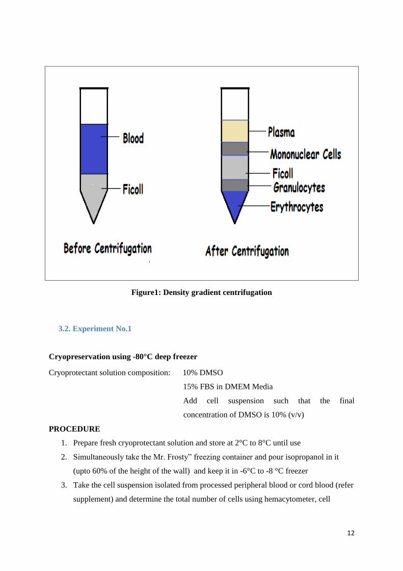
3. Carefully, 30 ml of diluted cells are layered on 15 ml of Ficoll-Hypaque (1.077 g/ml
density) in each 50-ml conical tube without disturbing the Ficoll-Hypaque interface.
4. It was centrifuged for 20-30 min at 450g at room temperature with the brake set to the
“off” position.
5. After centrifugation, a layer of mononuclear cells should be visible on top of the Ficoll-
Hypaque phase, as they have lower density than Ficoll-Hypaque solution as shown in Fig.
1.
6. Carefully the mononuclear cellswas harvested from the buffy layer located at the interface
between the medium and Ficoll using 3-ml plastic Pasteur pipette.
7. The cells were washed with PBS and recover the cells by centrifugation for 10-15 min at
300g and room temperature.
8. The supernatant was discarded, the cell pellet was re-suspended and the cells were counted
using a haemocytometer.
12
Figure1: Density gradient centrifugation
3.2. Experiment No.1
Cryopreservation using -80°C deep freezer
Cryoprotectant solution composition: 10% DMSO
15% FBS in DMEM Media
Add cell suspension such that the final
concentration of DMSO is 10% (v/v)
PROCEDURE
1. Prepare fresh cryoprotectant solution and store at 2°C to 8°C until use
2. Simultaneously take the Mr. Frosty” freezing container and pour isopropanol in it
(upto 60% of the height of the wall) and keep it in -6°C to -8 °C freezer
3. Take the cell suspension isolated from processed peripheral blood or cord blood (refer
supplement) and determine the total number of cells using hemacytometer, cell
13
counter and Trypan Blue exclusion. Cell count should not be less than 1x 10⁷
viable
cells /ml
4. Centrifuge the cell suspension at approximately 100–200 g for 5 - 10 minutes
5. After centrifugation decant the supernatant without disturbing the cell pellet
6. To the pellet add pre-cooled cryoprotectant solution, FBS and basal media as per
requirement
7. Place 1.0 ml of cell mixture in each cryovial
8. Lable the cryovials
9. Rapidly transfer the vials to a precooled (4°C) Nalgene freezing container (containing
isopropanol), and place immediately in a freezer at –80°C
10. Freezing is generally carried out overnight
11. After freezing for appropriate time, remove “ Mr. Frosty” freezing from the -80 °C
deep freezer and take out the vials containing the frozen cells
12. Now thaw the cells as per protocol mentioned in 3.4.
13. Perform cell viability analysis according to protocol in 3.5.
3.3. Experiment No.2
Vitrification by plunging straw containing the solution into Liquid Nitrogen
Cryoprotectant solution composition: 10% DMSO
10% FBS in DMEM Media
20% EG
1M sacrose
Add cell suspension such that the final
concentration of DMSO is 10% (v/v)
PROCEDURE
Cell Preparation for vitrification
1. Prepare fresh vitrification solution, filter it using Syringe filter and store at 2°C to 8°C
until use.
14
2. Take the cell suspension isolated from processed peripheral blood or cord blood(refer
supplement) and determine the total number of cells using haemocytometer, cell
counter and Trypan Blue exclusion. Cell count should not be less than 1x 10⁷ viable
cells /ml.
3. Centrifuge the cell suspension at approximately 100–200 g for 5 - 10 minutes.
4. Decant the supernatant without disturbing the cell pellet.
5. To the cell pellet add vitrification solution.
6. Allow MNCs to sit in freezing medium (hypertonic vitrification solution) until they
reach osmotic equilibrium.
Loading MNCs into Plastic Straw
1. Using 1ml syringe load the vitrification solution containing MNC’s into the plastic
straw.
2. Label the straw using printed labels attached to either a straw sealing plug or to
another straw connected with a straw adapter.
3. Remove the straw from the vitrification solution containing MNC’s, and aspirate
approximately 0.15 cm of air to create a tiny air bubble within the straw.
4. Aspirate a single MNC in approximately 0.8 cm of hypertonic vitrification solution
into the straw.
5. Remove the straw from the vitrification solution and aspirate approximately 0.15cm
of air to create a second tiny air bubble within the straw (air bubbles serve to
physically isolate the MNC within the straw).
6. Aspirate approximately 9.1 cm of vitrification solution to completely fill the straw,
being certain to draw in a sufficient volume of vitrification solution to moisten the
polyvinylchloride (PVC) powder that exists within the cotton plug end of the straw.
7. The vitrification solution causes the PVC powder to gel and seal that end of the straw.
8. Seal the other end of the straw with PVC powder, plastic straw sealing plugs, or a heat
sealer.
9. Place the straws loaded with vitrification solution and MNC’s into the control rate
freezer whose temperature has been cooled from ambient temperature to -6°C
10. Allow the sample to sit at this temperature for at least 2 minutes before proceeding
15
Seeding of the sample
1. Once the MNC’s have cooled to -6°C, use a pair of tongs supercooled in liquid
nitrogen (or a cotton-tipped stick immersed in liquid nitrogen) to induce ice crystal
formation in the vitrification solution inside the straw by touching the tongs (or cotton
tipped stick) to the column of solution.
2. The water in the vitrification solution will crystallize in the region exposed to liquid
nitrogen, and ice crystals will spread to the column of vitrification solution
immediately surrounding the MNC’s.
3. Hold MNC’s at seeding temperature for 10 minutes before further cooling.
4. Cool the MNC’s at a rate of 0.5°C/min down to a temperature of -34°C. This cooling
rate is important to ensure continued dehydration of the MNC’s
5. Hold the MNC’s at -34°C for 10 minutes before plunging MNC’s into liquid nitrogen
(-196°C).
6. Place cryopreserved MNC’s into an appropriately labelled goblet (filled with liquid
nitrogen) attached to an appropriately labelled cane, and place the cane into a canister
of a liquid nitrogen dewar for short- or long-term storage
After completing the above procedure, thawing is done using the procedure in 3.4 and cells
are counted to check the viability.
3.4. Thawing and cell recovery
After the cells are stored in liquid nitrogen for a night, the cells are thawed and recovered by
the following procedure:
1. Carefully remove the MNCs (or mononuclear cells) vial from Liquid Nitrogen Storage
Tank.
2. Rapidly thaw frozen vial of cells in a 37°C water bath, until just a small frozen chunk
remains in the vial.
3. Spray vial with 70% ethanol to decontaminate it and transfer it to the sterile tissue culture
hood.
4. Aseptically transfer the entire contents of the vial into a 15 ml conical tube using a 5 ml
pipette.
5. Rinse the vial with 1 ml of thawing media and transfer it to 15 ml centrifuge tube.
16
6. Slowly, add 4 ml of thawing media drop-wise to cells in the 15ml conical tube. While
adding the medium, gently move the tube back and forth to mix the cells. (This reduces
osmotic shock to the cells).
7. Centrifuge the 15 ml tube with the cell suspension at 200g for 2 minutes at room
temperature.
8. Carefully aspirate and discard the supernatant without disturbing the cell pellet.
9. Re-suspend the cell pellet in pre-warmed thawing media (e.g. 4ml/60 mm dish) using a 5
ml pipette and gently pipette the cells up and down until cell pellet is fully dispersed.
10. Now check the cell viability using trypan blue and haemocytometer following the protocol
mentioned in page no. 15
11. A viability of greater than 70% is expected, however if the viability is less than 70%, it
may take longer for the cells to reach confluence.
12. Now take sufficient amount of pre warmed DMEM and gently pipette the cells up and
down in it.
13. Now transfer the above into a culture dish.
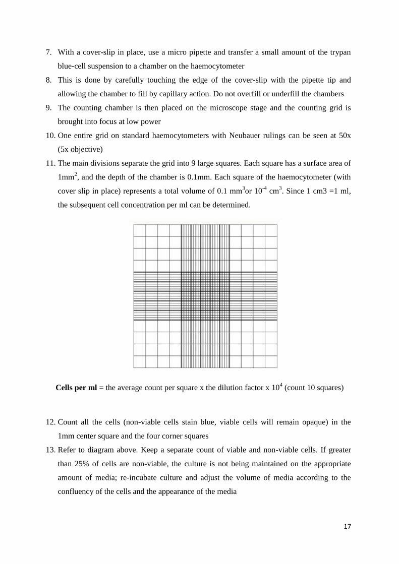
3.5. Cell Viability
To test the viability, cells are counted twice, before cooling and after thawed, and are
compared to see the percentage of cells survived. Following is the procedure for counting the
number of cells.
1. To prepare the haemocytometer, the mirror-like polished surface is carefully cleaned with
lens paper and ethanol
2. The coverslips (which is thicker than those for conventional microscopy) should also be
cleaned
3. Take 200 μl of the cell suspension into a 1.5 ml in the centrifuge tube
4. Add 300 μl of PBS and 500 μl of 0.4% trypan blue solution to the cell suspension
(creating a dilution factor of 5)
5. Mix thoroughly and allow to stand for 5 to 15 minutes
6. Note: Trypan blue dye is taken up by the cell membrane of dead cells only, however if
cells are exposed to trypan blue for extended periods of time, viable cells may begin to
take up dye as well as non-viable cells, thus, try to do cell counts within half an hour after
dye solution is added
17
7. With a cover-slip in place, use a micro pipette and transfer a small amount of the trypan
blue-cell suspension to a chamber on the haemocytometer
8. This is done by carefully touching the edge of the cover-slip with the pipette tip and
allowing the chamber to fill by capillary action. Do not overfill or underfill the chambers
9. The counting chamber is then placed on the microscope stage and the counting grid is
brought into focus at low power
10. One entire grid on standard haemocytometers with Neubauer rulings can be seen at 50x
(5x objective)
11. The main divisions separate the grid into 9 large squares. Each square has a surface area of
1mm2, and the depth of the chamber is 0.1mm. Each square of the haemocytometer (with
cover slip in place) represents a total volume of 0.1 mm3or 10
-4 cm
3. Since 1 cm3 =1 ml,
the subsequent cell concentration per ml can be determined.
Cells per ml = the average count per square x the dilution factor x 104 (count 10 squares)
12. Count all the cells (non-viable cells stain blue, viable cells will remain opaque) in the
1mm center square and the four corner squares
13. Refer to diagram above. Keep a separate count of viable and non-viable cells. If greater
than 25% of cells are non-viable, the culture is not being maintained on the appropriate
amount of media; re-incubate culture and adjust the volume of media according to the
confluency of the cells and the appearance of the media
18
14. If there are less than 50 or more than 200 cells per large square, repeat the procedure
adjusting to an appropriate dilution factor
15. Repeat the count using the other chamber of the haemocytometer
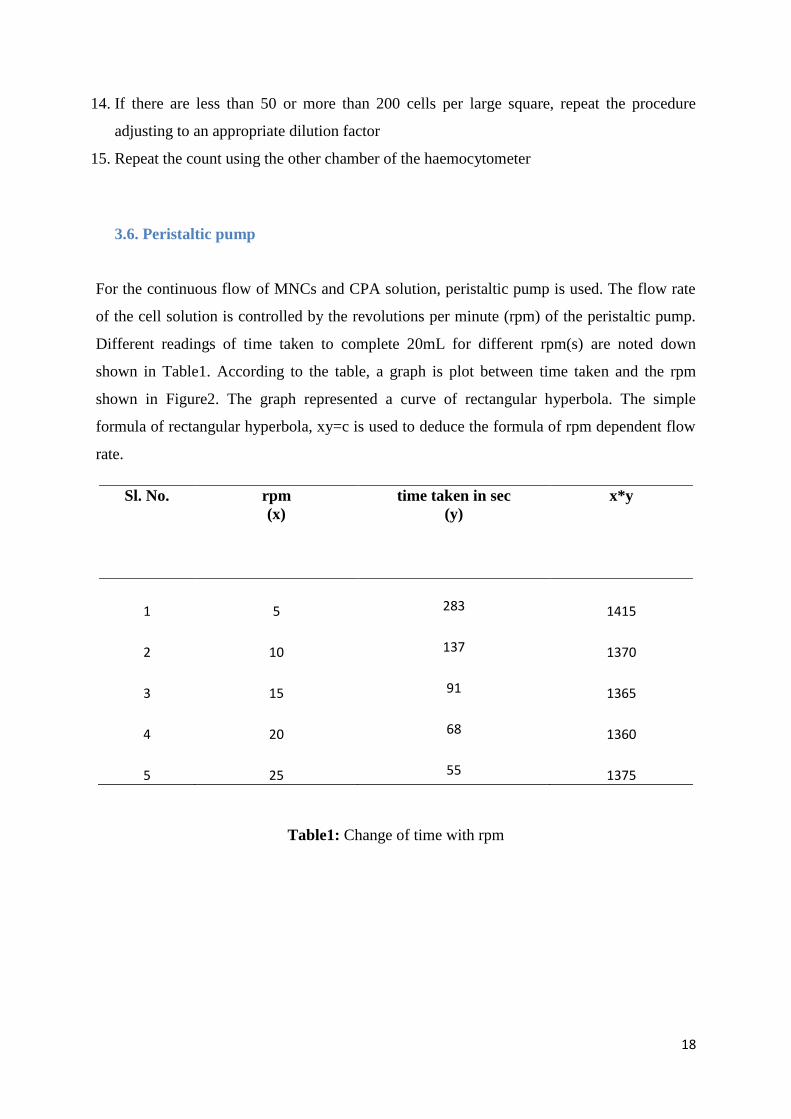
3.6. Peristaltic pump
For the continuous flow of MNCs and CPA solution, peristaltic pump is used. The flow rate
of the cell solution is controlled by the revolutions per minute (rpm) of the peristaltic pump.
Different readings of time taken to complete 20mL for different rpm(s) are noted down
shown in Table1. According to the table, a graph is plot between time taken and the rpm
shown in Figure2. The graph represented a curve of rectangular hyperbola. The simple
formula of rectangular hyperbola, xy=c is used to deduce the formula of rpm dependent flow
rate.
Sl. No. rpm
(x)
time taken in sec
(y)
x*y
1 5
283 1415
2 10
137 1370
3 15
91 1365
4 20
68 1360
5 25
55 1375
Table1: Change of time with rpm
19
Figure2: Time vs rpm graph for peristaltic pump
From the table we can approximate the value of ‘c’ which is always constant for any values
of ‘x’ and ‘y’. By taking the mean of each values in column number 4 (x*y column) we get
the approximate value of ‘c’.
c = (1415+1370+1365+1360+1375)/5 = 1377 ~1370
Each of the noted time(s) are for 20 mL, so,
Flow rate = 20/y in mL/sec
But we need mL/min, so we multiply the whole term by 60, therefore,
Flow rate (say z) = (20*60)/y in mL/min
Hence,
x = c/y = 1370/(1200/z) = (1370*z)/1200
Putting the value of z (flow rate required), we can find out the rpm required ‘x’ for the
required flow rate.
20
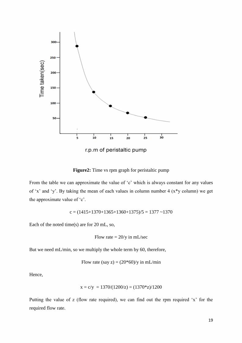
3.7. Experimental design
Figure3: Setup design for the new vitrification technique.
21
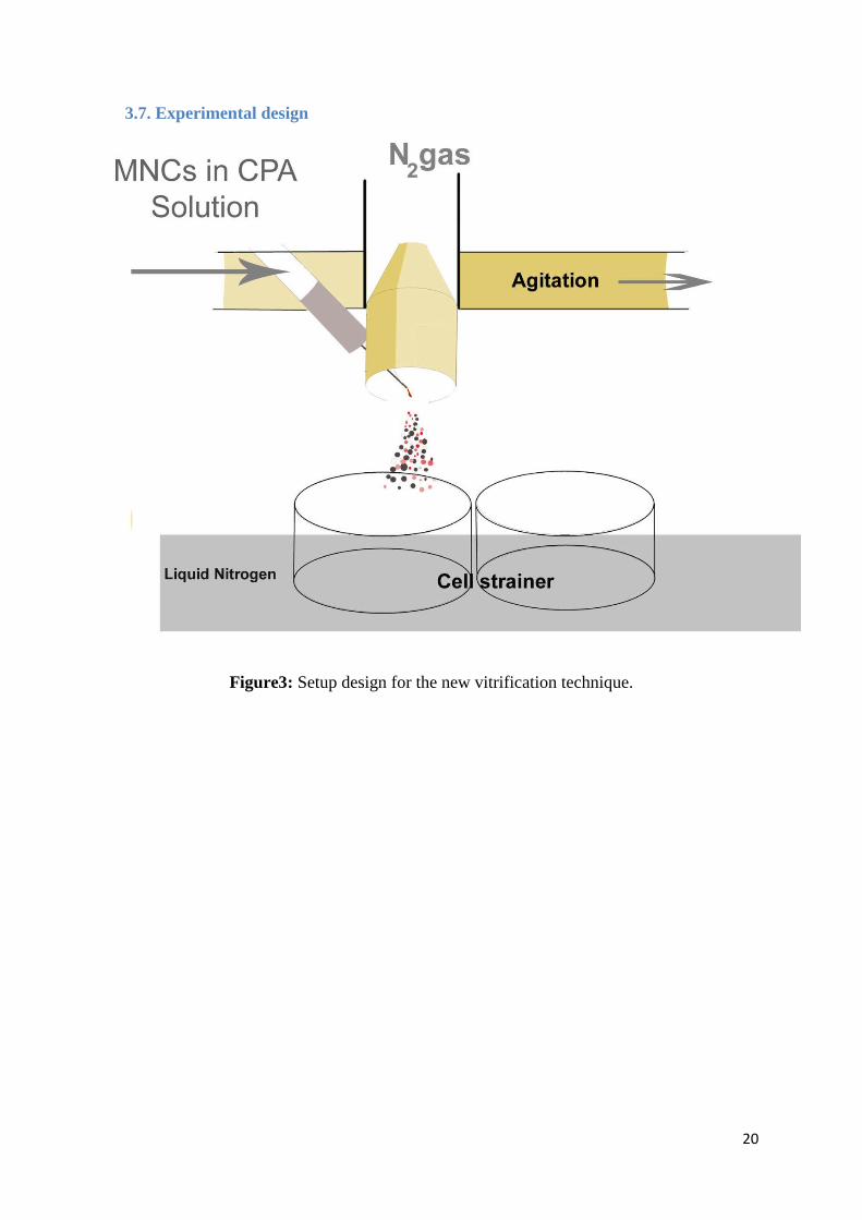
A silicon rubber pipe 4.8mm diameter is connected to the peristaltic pump. One end of the
pipe is kept inside a beaker containing a sample mixture of CPA and MNCs and the other end
is connected with a hypodermic needle, 22G × 1¼" (0.7 × 30mm), where the ejection of
sample takes place (Figure4). This needle is inserted through the N2 vapour outlet, which is a
cone shape plastic opening at the base and the top (0.8mm) as shown in Figure2. N2 vapour
flows through a plastic pipe connected to a Liquid Nitrogen tank.
Figure4: N2 vapour outlet (cone-like plastic) for dispersion of droplets coming from
hypodermic needle.
22
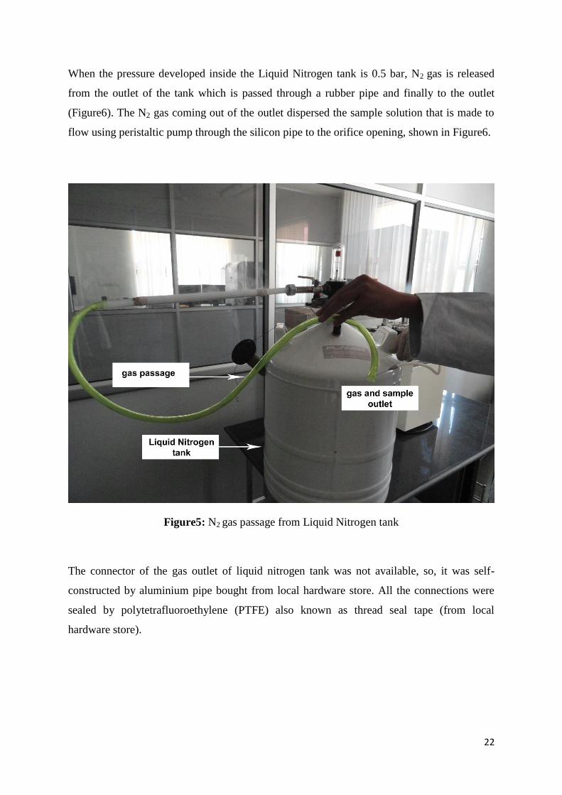
When the pressure developed inside the Liquid Nitrogen tank is 0.5 bar, N2 gas is released
from the outlet of the tank which is passed through a rubber pipe and finally to the outlet
(Figure6). The N2 gas coming out of the outlet dispersed the sample solution that is made to
flow using peristaltic pump through the silicon pipe to the orifice opening, shown in Figure6.
Figure5: N2 gas passage from Liquid Nitrogen tank
The connector of the gas outlet of liquid nitrogen tank was not available, so, it was self-
constructed by aluminium pipe bought from local hardware store. All the connections were
sealed by polytetrafluoroethylene (PTFE) also known as thread seal tape (from local
hardware store).
23
4. RESULTS AND DISCUSSION
4.1. Vitrifiction using plunging method
Before vetrifying the sample prepared in 3.3, cells were counted using trypan blue and
haemocytometer. The cell count was 1.2x10⁷cells/ml. After vetrification was done following
section 3.3 in Experiment no 2, the cell were thawed using the procedure in section 3.4, and
the cells were counted as in section 3.5. Viable cells were counted to be around
0.8x10⁷calls/ml. This is approximately around 67% viability.
4.2. Vitrification using N2 vapour gas
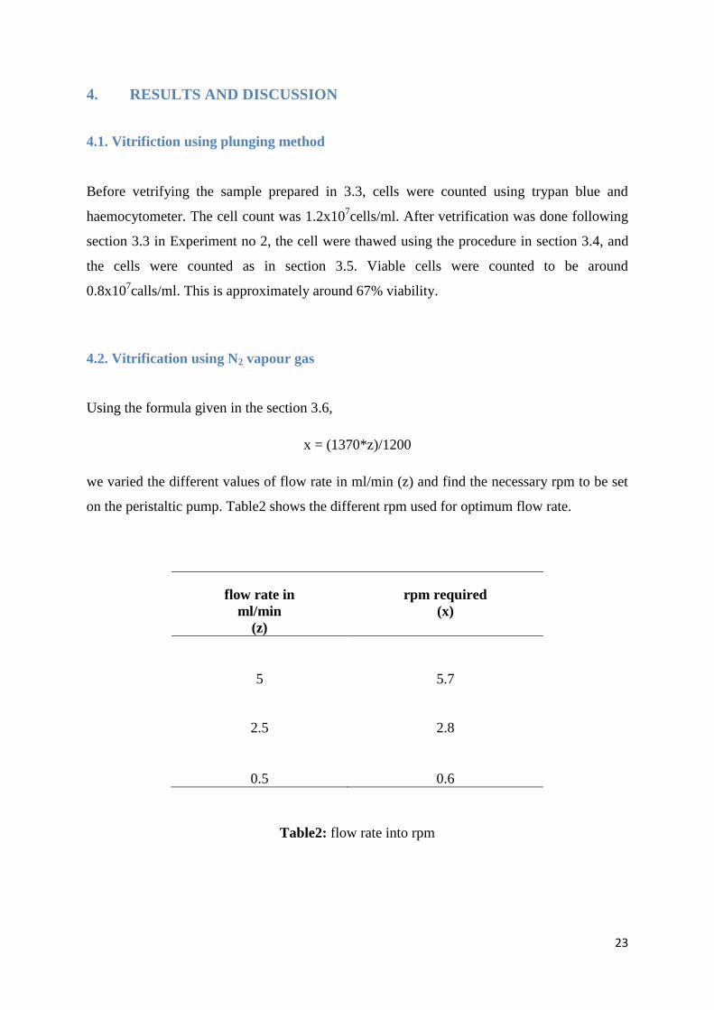
Using the formula given in the section 3.6,
x = (1370*z)/1200
we varied the different values of flow rate in ml/min (z) and find the necessary rpm to be set
on the peristaltic pump. Table2 shows the different rpm used for optimum flow rate.
flow rate in
ml/min
(z)
rpm required
(x)
5 5.7
2.5 2.8
0.5 0.6
Table2: flow rate into rpm

24
This different flow rates were tested. A solution was prepared with 10% DMSO (v/v), 3%
trypan blue (v/v) and water that made up rest of the volume. This solution had almost similar
density with cells and CPAs solution. The solution prepared was made to disperse over
Fabriano handmade-paper (9.5” X 12.5”, 300g/m2), using N2 vapour maintained at 0.5 bar
pressure.
Figure6: Flow rate of 5ml/min.
In Figure6, we can see that droplets forme over the paper are big, and dispersion effect of N2
gas is not that effective.
25
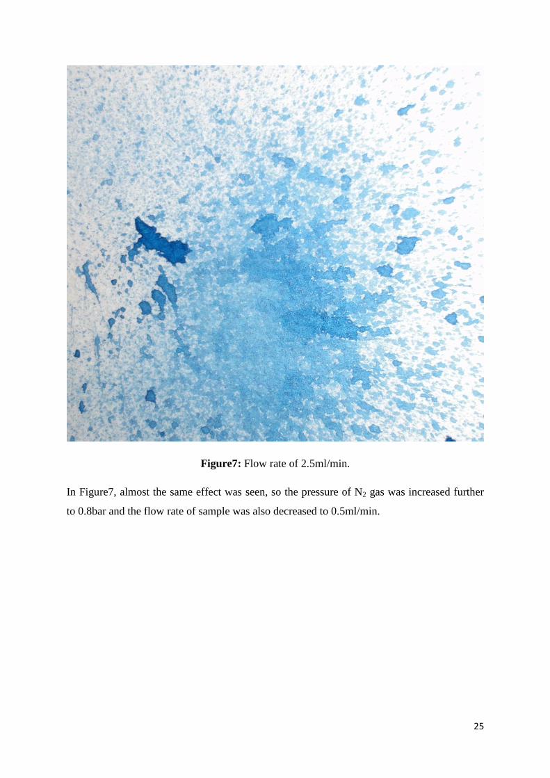
Figure7: Flow rate of 2.5ml/min.
In Figure7, almost the same effect was seen, so the pressure of N2 gas was increased further
to 0.8bar and the flow rate of sample was also decreased to 0.5ml/min.
26
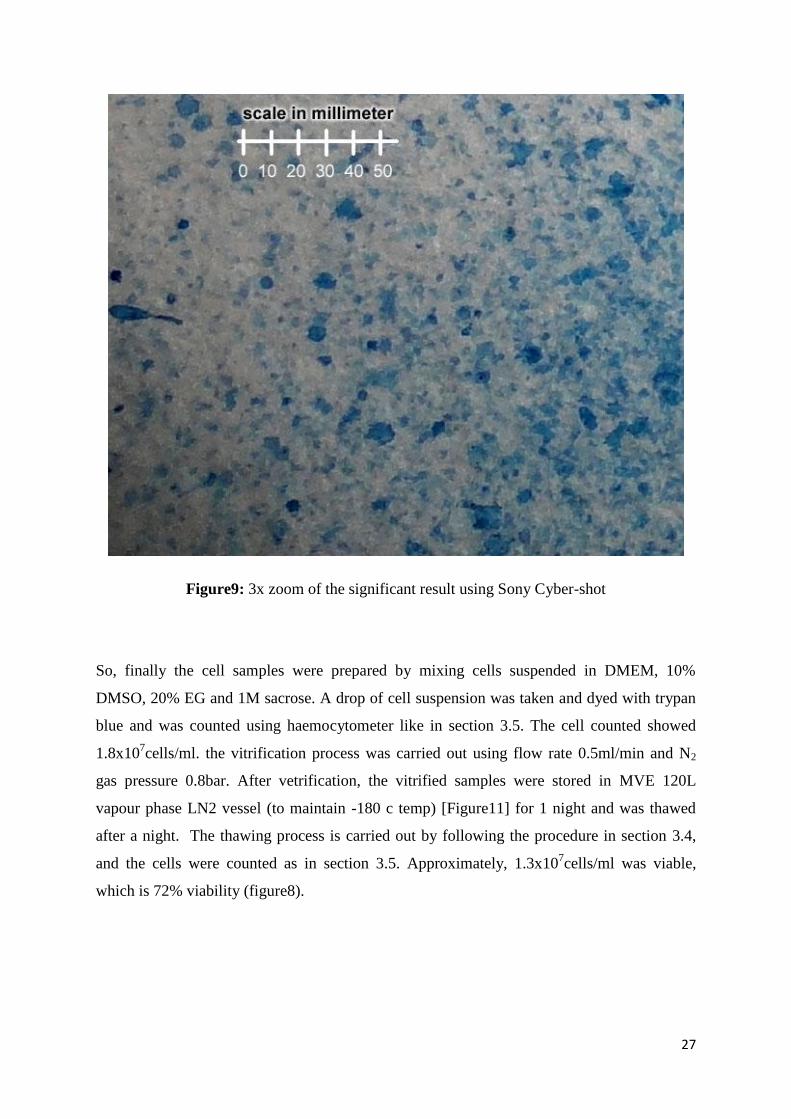
Figure8: Flow rate of 0.5ml/min and 0.8bar pressure for N2 gas.
After these changes, from Figure8, we can see the effective disperse effect of N2 vapour gas
over the paper. As we cn see, the droplets were evn more dispersed, which results in smaller
size of droplets, till 1mm-3mm in diameter (shown in Figure10).
27
Figure9: 3x zoom of the significant result using Sony Cyber-shot
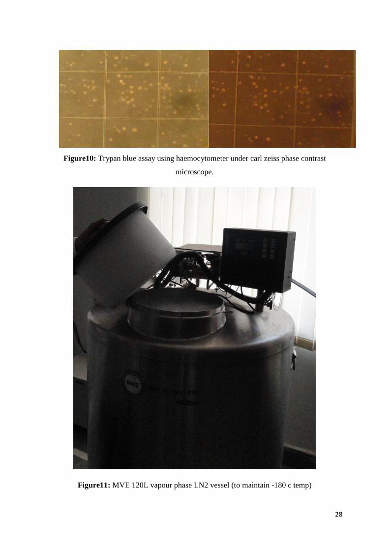
So, finally the cell samples were prepared by mixing cells suspended in DMEM, 10%
DMSO, 20% EG and 1M sacrose. A drop of cell suspension was taken and dyed with trypan
blue and was counted using haemocytometer like in section 3.5. The cell counted showed
1.8x10⁷cells/ml. the vitrification process was carried out using flow rate 0.5ml/min and N2
gas pressure 0.8bar. After vetrification, the vitrified samples were stored in MVE 120L
vapour phase LN2 vessel (to maintain -180 c temp) [Figure11] for 1 night and was thawed
after a night. The thawing process is carried out by following the procedure in section 3.4,
and the cells were counted as in section 3.5. Approximately, 1.3x10⁷cells/ml was viable,
which is 72% viability (figure8).
28
Figure10: Trypan blue assay using haemocytometer under carl zeiss phase contrast
microscope.
Figure11: MVE 120L vapour phase LN2 vessel (to maintain -180 c temp)
29
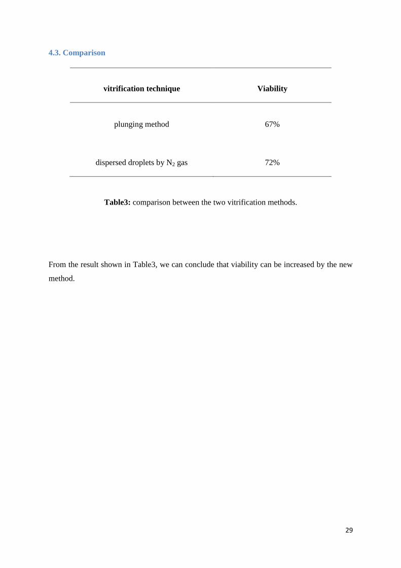
4.3. Comparison
vitrification technique
Viability
plunging method
67%
dispersed droplets by N2 gas
72%
Table3: comparison between the two vitrification methods.
From the result shown in Table3, we can conclude that viability can be increased by the new
method.

30
5. CONCLUSION
In summary, an effective way of vetrification for the cryopreservation of MNCs using
Nitrogen gas vapour to create small droplets is introduced. The N2 gas also helped in
precooling of the cells sample. And the most challenging part is that, it can be carried out in
large scale in compare to the use of straw.
31
6. REFERENCES
1. New American Desk Reference Encyclopedia. February (1989) Concord Reference
Books, Inc.
2. Samot J, Moon S, Shao L, Zhang X, Xu F, et al. (2011) Blood Banking in Living
Droplets. PLoS ONE 6(3): e17530. doi:10.1371/journal.pone.0017530]
3. Trounson A, Mohr L. Human pregnancy Following cryopreservation, Thawing and
transfer of an eight-cell embryo. Nature 1983; 305 (5936): 707-9.
4. Yokota, Y., Sato, S., Yokota, M. et al. (2000) Successful pregnancy following blastocyst
vitrification: case report. Hum. Reprod., 15, 1802–1803.
5. Wolstenholme GEW, O’Connor M, eds. (1970) The Frozen Cell London Churchill.
6. Krijnen HW, Wit JJFMD, Kuivenhoven ACJ, Loos JA, Prins HK (1964) Glycerol treated
human red cells frozen with liquid nitrogen. Vox Sang 9: 13
7. J Card Surg. (1987) Basic principles of cryobiology. J Card Surg. 1987 Mar;
2(1Supple):137-43.
8. Zdenek Hub, alek (2003) Protectants used in the cryopreservation of microorganisms,
Cryobiology 46 (2003) 205_229.
9. Thompson M, Nemits M, Ehrhardt R (May 2011). "Rate-controlled Cryopreservation
and Thawing of Mammalian Cells". Nat. Prot. Exch.. doi:10.1038/protex.2011.224.
10. 5100 Cryo 1°C Freezing Container, "Mr. Frosty", Nalgene labware.
11. Pegg DE, Diaper MP (1988) On the mechanism of injury to slowly frozen erythrocytes.
Biophysical Journal 54: 471–488.
12. Wolstenholme GEW, O’Connor M, eds. (1970) The Frozen Cell London Churchill.
13. Zadeoppe AMM (1968) Posthypertonic hemolysis in sodium chlorid systems.Acta
Physiologica Scandinavica 73: 341–&.
14. Pegg DE, Diaper MP (1991) The effect of initial toxicity on freeze/thaw injury to human
red cells suspended in solutions of sodium chloride. Cryobiology 28: 18–35.
15. Pegg DE, Diaper MP (1988) On the mechanism of injury to slowly frozen erythrocytes.
Biophysical Journal 54: 471–488.
16. Lovelock JE (1957) The denaturation of lipid-protein complexes as a cause of damage by
freezing. Proceedings of the Royal Society of London Series BBiological Sciences 147:
427–433.
32
17. Mazur P, Leibo SP, Chu EH (1972) A two-factor hypothesis of freezing injury. Evidence
from Chinese hamster tissue-culture cells. Exp Cell Res 71: 345–355.
18. Fahy GM, MacFarlane DR, Angell CA, Meryman HT (1984) Vitrification as an
approach to cryopreservation. Cryobiology 21: 407–426.
19. Luyet BJ, Gehenio PM (1940) The mechanism of injury and death by low temperature.
Biodynamica 3: 67
20. Vajta, G., Lewis, I.M., Kuwayama, M. et al. (1998b) Sterile application of the open
pulled straw (OPS) vitrification method. Cryo-Letters, 19, 389–392.
21. Cord Blood Banking: Donating Umbilical Cord Blood, Buzzle.com.
22. Cairo MS, Wagner JE (1997). "Placental and/or umbilical cord blood: an alternative
source of hematopoietic stem cells for transplantation.". Blood 90 (12): 4665–
4678. PMID 9389681.
23. Garbuzova-Davis S, Sanberg CD, Kuzmin-Nichols N, Willing AE, Gemma C, Bickford
PC, Miller C, Rossi R, Sanberg PR: Human umbilical cord blood treatment in a mouse
model of ALS: optimization of cell dose. PLoS One 2008, 3(6):e2494.
24. Park DH, Borlongan CV, Willing AE, Eve DJ, Cruz LE, Sanberg CD, Chung YG,
Sanberg PR:Human umbilical cord blood cell grafts for brain ischemia. Cell
Transplant 2009, 18(9):985-998.
25. Yang WZ, Zhang Y, Wu F, Min WP, Minev B, Zhang M, Luo XL, Ramos F, Ichim TE,
Riordan NH, Hu X: Safety evaluation of allogeneic umbilical cord blood mononuclear
cell therapy for degenerative conditions. Journal of Translational Medicine 2010, 8:75-
80.
26. Javed MJ, Mead LE, Prater D, Bessler WK, Foster D, Case J, Goebel WS, Yoder MC,
Haneline LS, Ingram DA: Endothelial Colony Forming Cells and Mesenchymal Stem
Cells are Enriched at Different Gestational Ages in Human Umbilical Cord Blood.
Pediatr Res 2008, 64:68-73.
27. AMERICAN JOURNAL OF PHYSIOLOGY; Guidet,B; 257(3 Pt 2):F440-F445 (1989).
28. JOURNAL OF REPRODUCTION AND FERTILITY; Valdez,CA; 96(2):793-802
(1992).
29. Aquatic living resources; Chao,N; 7(2):99-104 (1994).
30. Cryobiology; Baudot,A; 40(2):151-158 (2000).
31. Cryobiology; Fahy,GM; 24(3):196-213 (1987).
32. Ishimori, H., Saeki, K., Inai, M. et al. (1993) Vitrification of bovine embryos in a
mixture of ethylene glycol and dimethyl sulfoxide. Theriogenology, 40, 427–433.

33
33. Yokota, Y., Sato, S., Yokota, M. et al. (2000) Successful pregnancy following blastocyst
vitrification: case report. Hum. Reprod., 15, 1802–1803.