Deviations in populations of peripheral blood mononuclear cells and endometrial macrophages in the...
39
1 Deviations in populations of peripheral blood mononuclear cells and endometrial macrophages in the cow during pregnancy Lilian Oliveira and P.J. Hansen* Dept. of Animal Sciences, University of Florida, Gainesville, FL 32611-0910 USA Running Head: Leukocyte changes during pregnancy in cows * Correspondence: PO Box 110910, Gainesville FL 32611-0910 USA. Ph: (352)392- 5590. Email: [email protected] Page 1 of 39 Reproduction Advance Publication first posted on 17 July 2008 as Manuscript REP-08-0218
-
Upload
independent -
Category
Documents
-
view
0 -
download
0
Transcript of Deviations in populations of peripheral blood mononuclear cells and endometrial macrophages in the...

1
Deviations in populations of peripheral blood mononuclear cells and endometrial
macrophages in the cow during pregnancy
Lilian Oliveira and P.J. Hansen*
Dept. of Animal Sciences, University of Florida, Gainesville, FL 32611-0910 USA
Running Head: Leukocyte changes during pregnancy in cows
* Correspondence: PO Box 110910, Gainesville FL 32611-0910 USA. Ph: (352)392-
5590. Email: [email protected]
Page 1 of 39 Reproduction Advance Publication first posted on 17 July 2008 as Manuscript REP-08-0218

2
Abstract
The presence of conceptus alloantigens necessitates changes in maternal immune
function. Here we used the bovine to evaluate whether species with epitheliochorial
placentation have changes in specific leukocyte populations during pregnancy similar to
those reported in species with hemotropic placentae. At Day 33-34 of pregnancy, there
was no effect of pregnancy status on number of cells positive for CD8, CD4, γδT cell
receptor, or the monocyte marker CD68 in the peripheral blood mononuclear cell
(PBMC) population. There was, however, an increase in the proportion of CD4+ cells
that were positive for CD25. There was no effect of status on the proportion of PBMC
that were CD8+ when comparing preparturient cows to nonpregnant cows. However,
preparturient cows had an increased percent of PBMC that were γδ T-cells and that
were CD4+CD25+ and a tendency for a lower percent that were CD68+ cells. The
increase in CD4+CD25+ cells reflects an increase in the proportion of cells that were
CD4+ rather than in the proportion of CD4+ cells that were CD25+. Using
immunolocalization with anti-CD68, it was found that pregnant cows had increased
numbers of CD68+ cells in endometrial stroma as early as Day 54-100 of gestation; this
increase persisted through the last time examined (Day 240 of gestation). Cells positive
for CD68 were also positive for another macrophage/monocyte marker CD14. In
conclusion, pregnancy in the cow is associated with changes in peripheral and
endometrial leukocyte numbers that is similar to patterns observed in other species.
Page 2 of 39

3
Introduction
Pregnancy is an immunologically-distinct period for the eutherian female because
the presence of conceptus alloantigens necessitates changes in maternal immune
function to prevent immunological destruction of the conceptus. These changes can be
observed systemically and at the fetal-maternal interface. Maternal immune adjustments
to pregnancy have been most clearly defined in the human and mouse. Both of these
species have an invasive, hemotropic placenta in which trophoblast invades the
endometrium. Among the changes in immune function during pregnancy are an
increase in circulating regulatory T cells (Treg), defined as CD4+CD25+FoxP3+ cells, that
function to down-regulate T lymphocyte function (Aluvihare et al. 2004; Somerset et al.
2004; Yang et al. 2007), a temporary anergy of maternal lymphocytes to conceptus
MHC class I antigens (Tafuri et al. 1995), and synthesis of immunosuppressive proteins
at the maternal-fetal interface including interleukin (IL)-10 (Hanna et al. 2000; Murphy
et al. 2005) and transforming growth factor-β (TGF-β) (Suzuki et al. 1995; Gorivodsky et
al. 1999; Simpson et al. 2002). In addition, pregnancy is characterized by an increase
in numbers of specific leukocyte populations within the uterus including macrophages
(Hunt et al. 1985; Heikkinin et al. 2003 Cupurdija et al. 2004; Kim et al. 2007), γδT cells
(Heyborne et al. 1992; Mincheva-Nillson et al. 1992, 1997) and NK cells (Bilinski et al.
2008 Shigeru et al. 2008). These cells have been implicated in vascular remodeling (NK
cells – Bilinski et al. 2008); immunosuppression through secretion of IL-10 and TGF-β
(γδ T cells - Suzuki et al., 1995; Nagaeva et al., 2002; NK cells - Murphy et al. 2005) and
parturition (macrophages – Thomson et al. 1999; Mackler et al. 2000).
Page 3 of 39
4
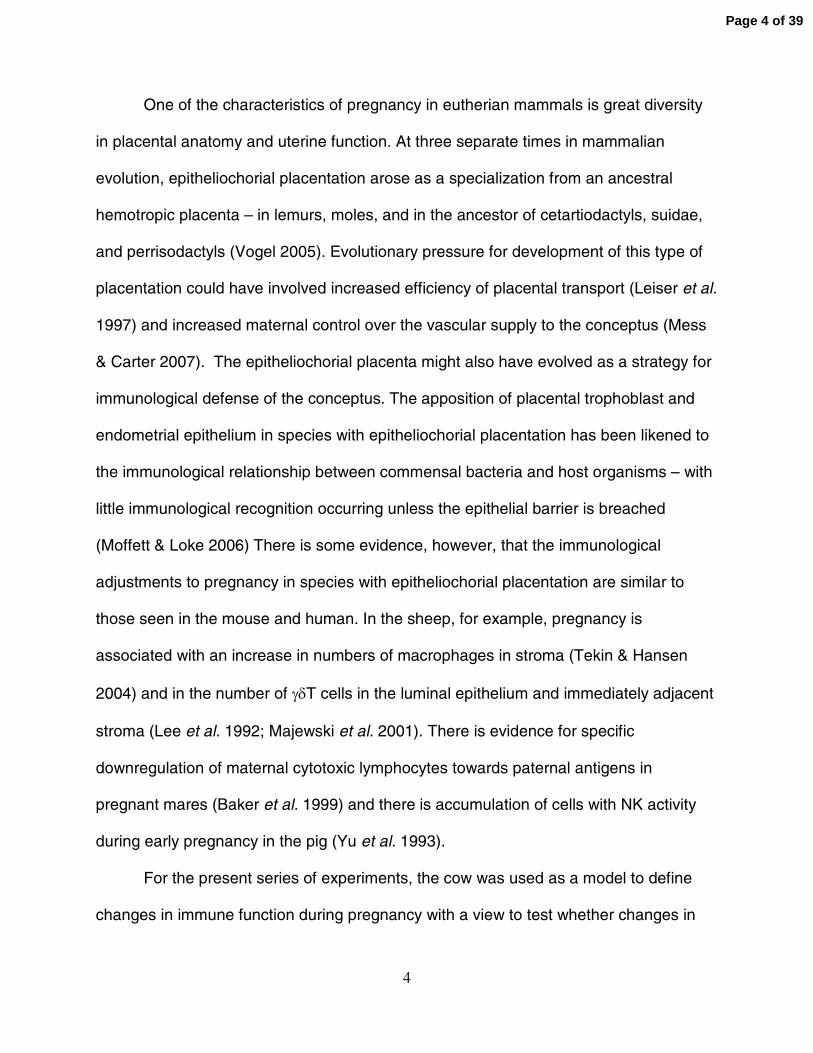
One of the characteristics of pregnancy in eutherian mammals is great diversity
in placental anatomy and uterine function. At three separate times in mammalian
evolution, epitheliochorial placentation arose as a specialization from an ancestral
hemotropic placenta – in lemurs, moles, and in the ancestor of cetartiodactyls, suidae,
and perrisodactyls (Vogel 2005). Evolutionary pressure for development of this type of
placentation could have involved increased efficiency of placental transport (Leiser et al.
1997) and increased maternal control over the vascular supply to the conceptus (Mess
& Carter 2007). The epitheliochorial placenta might also have evolved as a strategy for
immunological defense of the conceptus. The apposition of placental trophoblast and
endometrial epithelium in species with epitheliochorial placentation has been likened to
the immunological relationship between commensal bacteria and host organisms – with
little immunological recognition occurring unless the epithelial barrier is breached
(Moffett & Loke 2006) There is some evidence, however, that the immunological
adjustments to pregnancy in species with epitheliochorial placentation are similar to
those seen in the mouse and human. In the sheep, for example, pregnancy is
associated with an increase in numbers of macrophages in stroma (Tekin & Hansen
2004) and in the number of γδT cells in the luminal epithelium and immediately adjacent
stroma (Lee et al. 1992; Majewski et al. 2001). There is evidence for specific
downregulation of maternal cytotoxic lymphocytes towards paternal antigens in
pregnant mares (Baker et al. 1999) and there is accumulation of cells with NK activity
during early pregnancy in the pig (Yu et al. 1993).
For the present series of experiments, the cow was used as a model to define
changes in immune function during pregnancy with a view to test whether changes in
Page 4 of 39

5
immune function seen in human and mice are also seen in a species with
epitheliochorial placentation. The specific hypotheses tested were that CD4+CD25+ cells
increase in circulation during pregnancy while circulating concentrations of γδT cells and
macrophages decrease. Moreover, it was hypothesized that the reduction in numbers of
the latter two cell types reflects recruitment to the uterus. Identification of these or
similar changes in immune function during pregnancy would strengthen the notion that
existence of an epitheliochorial placenta does not minimize the need for immunological
adjustments in pregnancy. Moreover, such changes could result in altered immune
function in the periparturient period when the female is susceptible to uterine infection
and other immune challenges.
Materials and Methods
Materials
Tissue Culture Medium-199 (TCM-199), normal goat serum, bovine serum
albumin (BSA) Fraction-V, Dulbecco’s phosphate buffered saline (DPBS) and Hoescht
33342 were purchased from Sigma-Aldrich (St. Louis, MO, USA). The Fico-Lite 1077
was from Atlanta Biologicals (Norcross, GA, USA). The Zenon Alexa Fluor 488 mouse
IgG1 labeling kit, Zenon Alexa Fluor R-phycoerythrin mouse IgG1 labeling kit, Zenon
Alexa Fluor 647mouse IgG2a labeling kit and the mounting medium (Prolong® Antifade
Kit) was obtained from Invitrogen Molecular Probes (Eugene, OR, USA).
Paraformaldehyde (8%, w/v) was purchased from Electron Microscopy Sciences (Fort
Washington, PA, USA).
Page 5 of 39

6
Hybridoma cells producing monoclonal antibodies against bovine CD8 (clone
7C2) and ovine γδT (clone 86D) were purchased from European Type Cell Culture
Collection (Salisbury, UK). These monoclonal antibodies were obtained as culture
supernatants of hybridoma cell cultures prepared by the Hybridoma Core Laboratory of
the University of Florida Interdisciplinary Center for Biotechnology Research.
Mouse anti-human CD68 (clone EBM11; clarified ascites fluid, 2.3 µg /ml) was
obtained from Biomeda (Foster City, CA, USA), mouse anti-bovine CD14 (clone
MM61A, clarified ascites fluid, 10 µg /ml), mouse anti-bovine CD4 (clone CATC 138A;
clarified ascites fluid, 10 µg /ml) and mouse anti-bovine bovine CD25 (clone CATC
108A; clarified ascites fluid, 10 µg /ml) were from VMRD (Pullman, WA, USA). Control
mouse ascites fluid (clarified, clone NS1) was from Sigma-Aldrich (St Louis, MO, USA).
Normal goat serum was purchased from Pel-Freez Biologicals (Rogers, AR, USA). The
Histoscan Monoclonal Detector kit and mounting medium were obtained from Biomeda.
Tissue freezing medium was obtained from Biotech Medical Corporation (Kuala
Lampur, Malaysia). Lab-Tek® Glass Chamber Slides™ were obtained from Electron
Microscopy Sciences (Hatfield, PA, USA).
Flow cytometric analysis of peripheral blood mononuclear cells
Animals
Cows were maintained at the University of Florida Dairy Research Unit at Hague,
Florida. The first experiment was designed to determine differences in peripheral blood
lymphocyte populations between nonpregnant and pregnant cows at Day 33-34 of
gestation. A total of 33 lactating Holstein cows were subjected to timed artificial
insemination using a modified Presynch-OvSynch procedure (Brusveen et al. 2008) for
Page 6 of 39

7
insemination at Days 233 + 21 days after calving (range = 127-389). Cows were
diagnosed for pregnancy using transrectal ultrasound examination at Day 33-34 after
insemination and a blood sample was collected by coccygeal venipuncture into
heparinized tubes and used for flow cytometry. Blood samples were collected from a
total of 18 non-pregnant and 15 pregnant cows.
The second experiment was designed to determine differences between
nonpregnant and preparturient cows. A coccygeal blood sample was collected from a
total of 8 nonpregnant cows that were non-lactating and at random stages of the estrous
cycle and from 8 pregnant cows that were nonlactating and were at an average of 281.3
+ 2.9 days of gestation (range = 273-289). The preparturient cows were at an average
of 4.9 + 1.7 days before parturition (range 1-14 days).
Isolation of peripheral blood mononuclear cells (PBMC)
Blood (10 ml) was centrifuged at 600g for 30 min to obtain the buffy coat. This layer was
mixed with 2 ml TCM-199 and the cell suspension transferred to the top of 2 ml Fico/Lite
LymphoH placed in a 15 ml conical tube. Cells were centrifuged at 600g for 30 min.
Mononuclear cells were collected at the top of the Fico/Lite, centrifuged at 600g for 10
min, resuspended in DPBS, and used to determine cell concentration and viability by
trypan blue exclusion using a hemotocytometer. Cells were resuspended to a final
concentration of 5 x 10 7/ml.
Flow cytometry
5 x 10 6 cells were placed into 13 x 100 mm polyethylene tubes in staining buffer [DPBS
supplemented with 0.1% (w/v) BSA and 0.1% (w/v) sodium azide], washed twice with 2
ml staining buffer and resuspended in the smallest volume possible with staining buffer.
Page 7 of 39

8
Cells were stained for single color analysis using anti-γδ and anti-CD68 antibodies and
for dual-color analysis with anti-CD4 and anti-CD8 and anti-CD4 and anti-CD25. A
mouse IgG was used in same dilution of the primary antibody as a control to nonspecific
antibody staining. The antibodies, including the IgG control, were tagged with Fab
fragments against mouse IgG conjugated to Alexa Flour 488, Alexa R-phycoerythrin
and Alexa 647 using the Zenon® Mouse Labeling IgG kits as per manufacturer’s
instructions. The labeled antibody complex was then diluted in antibody staining buffer
to a final concentration of 10 µg/ml at room temperature for 30 min. After incubation,
samples were washed with 2 ml staining buffer, and resuspended with DPBS containing
4% (w/v) paraformaldehyde for fixation. Before analysis, cells were washed once with 1
ml of staining buffer and resuspended in 300 µL staining buffer. The flow cytometry
profiles were obtained on Fluorescent Analysis Cell Sorter “FACSCalibur” using
CELLQuest flow cytometry software (Becton-Dickinson, Franklin Lakes, NJ USA). The
cell populations analyzed were gated on the basis of forward and side scatter at the
lymphocyte and monocyte regions.
Immunohistochemistry for CD68 and CD14
Uteri were obtained from pregnant and nonpregnant cows of various breeds at a
local abattoir. Fetal crown-rump length was measured to estimate fetal age (Noden &
Lahunta, 1984). Reproductive tracts from nonpregnant cows were used only if a corpus
luteum was present. Tissues from a total of 20 pregnant cows (estimated fetal ages
ranging from 54-240 days of pregnancy) and 7 nonpregnant cows were collected.
Samples of intercotyledonary uterine endometrium ipsilateral to the corpus luteum were
snap-frozen in Tissue-Tek OCT embedding compound. Tissues from pregnant and
Page 8 of 39

9
nonpregnant cows were processed in parallel to avoid confounding physiological stage
with procedural replicate.
For immunohistochemistry, 5 µm tissue sections were prepared with a cryostat
microtome. Sections were placed onto precleaned glass slides, fixed in ice-cold acetone
for 10 min and air dried. The sections were rehydrated in histochemistry buffer at 4oC
[10 mM NaPO4, pH 7.4 containing 0.9% (w/v) NaCl supplemented with 1% (v/v) normal
goat serum] for 30 min.
Procedures for immunohistochemistry were carried out according to
manufacturer’s instructions. Briefly, sections were sequentially incubated with
peroxidase blocking buffer [histochemistry buffer with 0.3% (v/v) H2O2] for 5 min and
washed three times in histochemistry buffer. The tissue conditioner supplied in the kit
was applied to the samples for 4 min before incubation with primary antibody overnight
at 4oC; primary antibodies used were anti-CD68 (2.3 µg/ml) and anti-CD14 (10 µg/ml) in
histochemistry buffer. As controls, other sections were incubated with IgG (10 µg/ml) in
histochemistry buffer. Slides were then sequentially incubated with secondary antibody
(biotinylated anti-mouse immunoglobulin as supplied in the kit) for 30 min, streptavidin-
alkaline phosphatase reagent (from kit) for 30 min, and hematoxylin (as supplied in the
kit) for 2 min. Slides were washed under tap water, cover slips were mounted, and
slides were examined for staining using light microscopy.
The concentration of CD68+ cells in the endometrial stroma was estimated
subjectively according to a scale ranging from 0 (no positive cells) to 5 (very intense
accumulation of positive cells). Evaluation was performed blindly with respect to
treatment.
Page 9 of 39

10
Two-color immunofluorescence for CD68 and CD14
Uterine sections were prepared as described above. Sections were incubated
with blocking buffer [DPBS supplemented with 20% (v/v) goat serum] for 20 min
followed by incubation overnight at 4oC with anti-CD68 (2.3 µg/ml); anti-CD14 (10
µg/ml) and isotype controls (10 µg/ml) labeled using the Zenon labeling system as
described before. Sections were then washed three times for 5 min using PBS and then
labeled with Hoescht 33342 reagent (2.3 µg/ml) for 15 min. Sections were washed an
additional three times for 5 min each, coverslips mounted using Prolong® Antifade
reagent and slides examined using a Zeiss Axioplan 2 epifluorescence microscope
(Zeiss, Gottingen, Germany) with a 40x objective and using Zeiss filter set 02 (DAPI
filter), Zeiss filter set 03 (FITC filter) and Zeiss filter set 15 (rhodamine filter). Digital
images were acquired using AxioVision software (Zeiss) and a high-resolution black and
white Zeiss AxioCam MRm digital camera.
Two-color immunofluorescence for CD68 and CD14 in endometrial adherent cells
Two-color immunofluorescence was performed using preparations of dispersed
adherent cells from endometrium of a pregnant cow at Day 166 of pregnancy. The
uterus was obtained at a local abattoir and transported to the lab on ice (~2 h). A
sample of the intercaruncular region was taken for isolation of stromal endometrial cells.
Endometrial cells were removed from intercaruncular areas of the endometrium by
mechanically scraping the inner surface of the endometrium with a sterile surgical
blade. Cell scrapings were collected into a 50 ml sterile culture tube containing 50 ml
TCM-199 supplemented with type I collagenase at 150 U/ml. Cells were incubated at
37°C for 1 h under gentle rotation. Cells in suspension were then filtered through a
Page 10 of 39

11
sterile 100-µm cell strainer into 50 ml sterile culture tubes and centrifuged at 110×g for
5 min. The cell pellet was resuspended with 5 ml TCM-199 supplemented with 10%
(v/v) fetal bovine serum and cell number was determined using a hemacytometer. The
cell suspension was placed into 8-well sterile chamber slides (Lab-Tek® Glass
Chamber Slides™; Electron Microscopy Sciences, Hatfield, PA, USA) with a cover and
incubated at 37°C for 2 h to allow cells to adhere. The wells were washed three times
with DPBS to eliminate non-adherent cells. The remaining adherent cells were fixed in
DPBS containing 4% (w/v) paraformaldehyde for 15 min. Wells were washed three
times in DPBS and two-color immunoflourescence staining was performed as described
above.
Statistical analysis
Data were analyzed by least square analysis of variance using the General
Linear Models procedure of SAS (SAS for Windows, version 9.3, SAS Institute Inc.,
Cary, NC, USA). For the flow cytometry studies, the model included effect of
physiological status (pregnant vs nonpregnant), date samples were collected (i.e,
replicate) and the interaction. For intensity of CD68 staining in endometrium, cows
were grouped into 4 groups based on stage of pregnancy (nonpregnant, n=7; Day 54-
100 of pregnancy, n=7; Day 101-200 of pregnancy, n=8 and Days 201-240 of
pregnancy, n=5). The mathematical model included the effect of stage of pregnancy.
Results
Representative flow cytometry patterns
Page 11 of 39

12
Typical dot plots are shown in Figure 1. Analysis of PBMC by side scatter and
forward scatter resolved cells into two populations (Figure 1A). The first, more abundant
population was of small size and little granularity, and represents lymphocytes primarily.
The second population, which represents monocytes primarily, was of large size and
granularity. Expression of markers for T cells (CD4, CD8 and CD25) was based on
cells in the lymphocyte gate. Expression of γδT and CD68 were analyzed in both
lymphocyte and monocyte regions. Cells positive for antibody could be readily
distinguished from negative cells based on increased fluorescence as compared to cells
stained with IgG (Fig 1B-G). Cells positive for CD25 were resolved into two populations
– a CD25bright and a CD25dim population (Figure 1C). There were, however, no treatment
differences in the percent of CD25+ cells that were bright and dim. Therefore, data were
analyzed after pooling both populations.
Differences between pregnant and nonpregnant cows at Day 33-34 of gestation in
subpopulations of PBMC
There were no differences between pregnant and nonpregnant cows in the
proportions of PBMC that were positive for CD4 (Fig 2A) and CD8 (Fig 2A). Pregnant
cows had, however, a higher (P<0.06) percent of PBMC that were CD25+ (Fig 2A;
P<0.06). Moreover, the percent of CD4+ cells that were also positive for CD25 was
higher (P<0.05) for pregnant cows (Fig 2C).
There was no effect of pregnancy status on the proportion of PBMC in the
lymphocyte gate and monocyte gate that were γδT+ and CD68+ cells (Fig 2D and 2E).
Differences between preparturient and nonpregnant cows in subpopulations of
PBMC
Page 12 of 39

13
There was no effect of pregnancy status on the percent of cells in the lymphocyte gate
that were positive for CD8 (Fig 3A). However, the percent of cells in the lymphocyte
gate that were CD4+, CD25+ and CD4+CD25+ were higher (P<0.05) for preparturient
cows than for nonpregnant cows (Fig 3A and 3B). The increase in number of
CD4+CD25+ cells represents an increase in CD4+ cells rather than an increase in the
proportion of CD4+ cells that were CD25+ because the number of CD4+ that were
CD25+ cells did not change with pregnancy status (Fig 3C). Preparturient cows also had
a greater proportion of cells in the lymphocyte gate that were γδT+ (P<0.01) and a
tendency for a greater proportion of cells in the monocyte gate that were γδT cells
(P<0.07) (Fig 3D). There was also a tendency (P<0.10) for the percentage of cells that
were CD68+ to be lower for preparturient cows for the monocyte gate (Fig 3E).
Immunolocalization of CD68+ and CD14+ cells in endometrium as affected by
pregnancy status
Cells positive for CD68 were very abundant in the lamina propria of the
endometrial stroma in pregnant cows (Fig 4B and 4C). A fewer number of positive cells
were present in the submucosa. There were no CD68+ cells in luminal or glandular
epithelia. In contrast to the pattern in pregnancy, there were very few CD68+ cells in
endometrium from nonpregnant cows (Fig 4A). The pattern of CD14+ cells was very
similar to that for CD68 (figure 4D).
The high degree of staining for CD68 in pregnant endometrium made counting of
individual cells impossible. Instead, a subjective score for staining intensity was used to
determine pregnancy status effects on numbers of CD68+ cells. Staining intensity was
Page 13 of 39

14
greater for pregnant cows than for nonpregnant cows at all stages of pregnancy
examined (P<0.05) for both the lamina propria (Fig 5A) and submucosa (Fig 5B).
The co-localization of CD68 and CD14 expression was analyzed by dual-color
immunofluorescence. In one experiment, two color immunofluorescent labeling of
endometrial sections was performed using antibodies to CD68 and CD14. The majority
of CD14+ cells were positive for CD68 (Fig 6). In the second experiment, two color
immunofluorescent labeling of single cell preparations of adherent endometrial cells
revealed that cells that labeled with CD14 were also labeled with CD68 (Fig 7).
Discussion
Results presented here indicate that pregnancy in the cow, as in other species, is
characterized by changes in immune cell populations in the periphery and uterus.
During early pregnancy, at Day 33-34 of gestation, there was an increase in the
proportion of peripheral blood lymphocytes that were CD4+CD25+ cells. These cells,
which could be analogous to the Treg cells described as increasing during pregnancy in
mice and humans (Somerset et al. 2004; Aluvihare et al. 2004), were also present in
higher amounts in preparturient cows as compared to nonpregnant cows. Late
pregnancy was also associated with an increase in the proportion of γδT-cells in
peripheral blood and a tendency for decrease in the proportion of cells (presumably
monocytes) positive for CD68+. The most striking change associated with pregnancy
was large-scale recruitment of macrophages positive for CD68 and CD14 to the
endometrial stroma.
Page 14 of 39

15
Pregnancy-associated changes in immune cell populations are likely to be
important for protection of the conceptus from maternal immune attack or for removal of
cellular debris and microorganisms from the uterus following parturition. The
observation that pregnancy causes changes in immune function in the cow that parallel
what is seen in other species suggests that the trophoblast-maternal immunological
relationship in species with epitheliochorial placentae is not an inert one, as has been
implied (Moffatt and Loke 2006), but one characterized by immunological adaptation.
At both Day 33-34 and late pregnancy, there were changes in the population of
CD4+CD25+ cells in periheral blood. At Day 33-34, this change reflected an increased
proportion of CD4+ cells that expressed CD25+. In preparturient cows, there was an
increase in CD4+CD25+ cells that resulted from an increase in relative numbers of CD4+
in the periphery rather than in an increase in the proportion of CD4+ cells that were
positive for CD25. In both women and mice, a subpopulation of CD4+CD25+ that
expresses the transcription factor FoxP3 is increased in peripheral blood during
pregnancy (Somerset et al. 2004; Aluvihare et al. 2004). These cells have been
identified as Treg cells that can secrete cytokines such as IL-4 that inhibit activation of
cytotoxic T cells against alloantigens (Mjösberg et al. 2007). The importance of Treg cells
for pregnancy success is indicated by the observation that the percent of CD4+CD25+
cells in PBMC is reduced in women with unexplained recurrent spontaneous abortion
(Yang et al. 2007). In mice, depletion of CD25+ cells decreased the ability the female to
sustain pregnancy (Aluvihare et al. 2004). It is not clear from the present results
whether some or all of the CD4+CD25+ seen in peripheral blood of cows are Treg cells.
Antibodies to bovine FoxP3 are not available and several unsuccessful attempts were
Page 15 of 39

16
made to identify antibodies raised against FoxP3 in other species that crossreact with
bovine.
The immune changes coincident with pregnancy seen here do not represent the
earliest such changes in pregnancy. Ruminant species are unique among mammals in
that they have evolved to use a type I interferon (interferon-τ) as the trophoblast signal
that inhibits endometrial prostaglandin F-2α synthesis and allows prolonged lifespan of
the corpus luteum (Roberts et al. 2003). Interferon-τ has retained its
immunomodulatory properties and changes in expression of interferon-stimulated genes
have been described in PBMC in cattle as early as Day 16 of gestation (Han et al. 2006,
Gifford et al. 2007). By Day 32 of pregnancy, expression of interferon stimulated gene
15 in blood cells was similar to that for non-pregnant cows (Han et al. 2006) but it
cannot be ruled out that changes in PBMC seen at Day 33-34 in the present study were
caused by interferon-τ secretion by the conceptus earlier in pregnancy.
It was hypothesized that the relative numbers of γδT cells in peripheral blood
would decline during pregnancy, especially in the preparturient period, because of
recruitment to the uterus. Indeed, accumulation of γδT cells to the endometrium is a
characteristic of pregnancy in humans (Mincheva-Nilsson et al. 1992, 1997), mice
(Heyborne et al. 1992) and sheep (Lee et al. 1992; Meussen et al. 1993; Majewski et al.
2001). In contrast to our hypothesis, the relative numbers of γδT cells in peripheral blood
was not affected by pregnancy status at Day 33-34 after insemination and was higher
for preparturient cows than for non-pregnant cows. An increase in relative numbers of
γδT cells in peripheral blood 3 weeks before parturition followed by decrease to initial
values at the time of parturition has been seen by others (Van Kampen & Mallard 1997).
Page 16 of 39

17
The recruitment of macrophages to the pregnant uterus has been described in
many species such in humans (Heikkinen et al. 2003; Cupurdija et al. 2004; Kim et al.
2007), mice (Hunt et al. 1985) and sheep (Tekin & Hansen 2004). Present results
indicate that this process occurs in the cow, also, with numbers increasing by Day 54-
100 of pregnancy. For preparturient cows, there was a decline in the relative number of
CD68+ cells in peripheral blood and this change could reflect increased recruitment to
the uterus. That the CD68+ cells in endometrial stroma are macrophages is indicated by
the co-expression of CD14, which are also expressed in tissue macrophages such as in
pulmonary alveoli (Yang et al. 1995) The signals for movement of monocytes from the
blood to the uterus and their differentiation once resident in the endometrium are not
known. Mouse trophoblast can cause migration of blood monocytes in vitro (Fest et al.
2007) and change monocyte cytokine prolife and activation in response to
lipopolysaccharide.
The accumulation of large numbers of macrophages in the endometrium during
pregnancy strongly suggests an important role for these cells in the uterus. One
possibility is that uterine macrophages participate in parturition by promoting placental
detachment. Evidence for immunological participation in the parturition process is
indicated by the increased incidence of retained placenta in cows which share major
histocompatability class I antigens with their conceptus (Joosten et al. 1981). Retained
placenta in cattle has also been related to decreased activity of macrophages in the
placentomal area (Miyoshi et al. 2002). The postpartum uterus is characterized by
abundant lochia and microorganisms (Lewis 1997, Thatcher et al. 2006) and
macrophages may participate in the involution process whereby these materials are
Page 17 of 39

18
removed in the postpartum period. Finally, it may be that macrophages function during
pregnancy to promote survival of the allogeneic conceptus. In human, placental
macrophages express markers of alternative activation (Cupurdija et al. 2004) and
markers such as stabilin-1 that have been suggested to be involved in an anti-
inflammatory function (Politz et al. 2002). It has been proposed that clearance of
apoptotic trophoblast cells by uterine macrophages alters macrophage cytokine
secretion to reduce inflammation and promote conceptus survival (Mor et al. 2006).
Also, macrophage-associated stabilin-1 can bind to and process placental lactogen in
vitro, suggesting regulation of extracellular levels of placental lactogen by alternatively
activated macrophages (Kzhyshkowska et al. 2008).
There is evidence that the preparturient dairy cow is immunosuppressed with a
decline in numbers of CD4+, CD8+ and γδ cells in peripheral blood (Van Kampen and
Mallard 1997, Kimura et al. 1999, 2002) and reduced proliferation and interferon-γ
secretion by mitogen-stimulated lymphocytes (Detilleux et al. 1995; Nonnecke et al.
2003). There was little evidence of immunosuppression in the present study.
Preparturient cows did not have reduced proportions of CD8+ cells in peripheral blood
as compared to nonpregnant cows and numbers of CD4+ and γδT cells were increased.
Karcher et al. (2008) also did not observe declines in CD8+ cells as parturition
approached and there was a tendency for an increase in the proportion of γδT cells as
parturition approached. In the present study, the only direct evidence for reduced
immune competency in preparturient cows was the tendency for a reduction in numbers
of CD68+ cells. It is possible, however, that the increase in numbers of CD4+CD25+ and
Page 18 of 39

19
γδT cells seen in preparturient cows is an indication for immunosuppression in the
preparturient period if some of these cells are Treg cells.
Moffett & Loke (2006) have likened the maternal-fetal immunological relationship
in species with epitheliochorial placenta to the immunological relationship between
commensal bacteria and host organisms. In this view, little immunological recognition
of the conceptus occurs unless the epithelial barrier is breached. The current results
indicate that development of the epitheliochorial placenta during evolution has not
changed the occurrence of several immunological adjustments to pregnancy. Like the
human and mouse, there is an increase in CD4+CD25+ lymphocytes in peripheral blood
and accumulation of macrophages in the endometrium. These pregnancy-associated
changes in immune function indicate that, rather then being an immunologically-inert
tissue, the bovine conceptus is a tissue whose presence requires maternal
immunological adjustments. These adjustments can be detected as early as Day 16
(Han et al. 2006, Gifford et al. 2007), are present at Day 33-34 of pregnancy and are
still occurring close to parturition. Thus, the maternal immune system in the cow is
constantly adjusting to the conceptus. Given this pattern of immunological change,
which is also seen in other species with epitheliochorial placenta (Lee et al. 1992 Yu et
al. 1993 Baker et al. 1999 Majewski et al. 2001 Tekin et al. 2004), it seems more likely
that evolution of a epitheliochorial placenta occurred because of increases in efficiency
of placental transport (Leiser et al. 1997) or increased maternal control over the
vascular supply to the conceptus (Mess & Carter 2007) rather than to change the
fundamental characteristics of the maternal-fetal immunological relationship.
Page 19 of 39

20
Acknowledgements
Research was supported by the Florida Agricultural Experiment Station and by a grant
to LO from CAPES/Fulbright (Grant 2062046). There is no conflict of interest that would
prejudice its impartiality. The authors thank the following for their assistance with the
project: Dr. Sally Johnson, William Rembert, Luciano Bonilla, Dr. Robson Giglio, Dr
William W. Thatcher, Flavio Slivestre, Dr. Carlos Risco, the Flow Cytometry Facility and
Hybridoma Core Laboratory of the University of Florida Interdisciplinary Center for
Biotechnology Research, and Central Packing, Center Hill, Florida.
Page 20 of 39

21
References
Aluvihare VR, Kallikourdis M & Betz AG 2004 Regulatory T cells mediate maternal
tolerance to the fetus. Nature Immunology 5 266-271.
Baker JM, Bamford AI & Antczak DF 1999 Modulation of allospecific CTL responses
during pregnancy in equids: an immunological barrier to interspecies matings?
Journal of Immunology 162 4496-4501.
Bilinski MJ, Thorne JG, Oh MJ, Leonard S, Murrant C, Tayade C & Croy BA. 2008
Uterine NK cells in murine pregnancy. Reproductive Biomedicine Online 16 218-
226.
Brusveen DJ, Cunha AP, Silva CD, Cunha PM, Sterry RA, Silva EP, Guenther JN &
Wiltbank MC 2008 Altering the time of the second gonadotropin-releasing
hormone injection and artificial insemination (AI) during Ovsynch affects
pregnancies per AI in lactating dairy cows. Journal of Dairy Science 91 1044-
1052.
Cupurdija K, Azzola D, Hainz U, Gratchev A, Heitger A, Takikawa O, Goerdt S,
Wintersteiger R, Dohr G & Sedlmayr P 2004 Macrophages of human first
trimester decidua express markers associated to alternative activation. American
Journal of Reproductive Immunology 51 117-122.
Detilleux JC, Kehrli ME Jr, Stabel JR, Freeman AE & Kelley DH 1995 Study of
immunological dysfunction in periparturient Holstein cattle selected for high and
average milk production. Veterinary Immunology and Immunopathology 44 251-
267.
Page 21 of 39

22
Fest S, Aldo PB, Abrahams VM, Visintin I, Alvero A, Chen R, Chavez SL, Romero
R & Mor G 2007 Trophoblast-macrophage interactions: a regulatory network for
the protection of pregnancy. American Journal of Reproductive Immunology 57
55-66.
Gifford CA, Racicot K, Clark DS, Austin KJ, Hansen TR, Lucy MC, Davies CJ & Ott
TL 2007 Regulation of interferon-stimulated genes in peripheral blood
leukocytes in pregnant and bred, nonpregnant dairy cows. Journal of Dairy
Science 90 274-80.
Gorivodsky M, Torchinsky A, Zemliak I, Savion S, Fein A & Toder V 1999 TGF β2
mRNA expression and pregnancy failure in mice. American Journal of
Reproductive Immunology 42 124-133.
Han H, Austin KJ, Rempel LA & Hansen TR 2006 Low blood ISG15 mRNA and
progesterone levels are predictive of non-pregnant dairy cows. Journal of
Endocrinology 191 505-512
Hanna N, Hanna I, Hleb M, Wagner E, Dougherty J, Balkundi D, Padbury J &
Sharma S 2000 Gestational age-dependent expression of IL-10 and its receptor
in human placental tissues and isolated cytotrophoblasts. Journal of Immunology
164 5721-5728.
Heikkinen J, Mottonen M, Komi J, Alanen A & Lassila O 2003 Phenotypic
characterization of human decidual macrophages. Clinical & Experimental
Immunology 131 498-505.
Page 22 of 39

23
Heyborne KD, Cranfill RL, Carding SR, Born WK & O'Brien RL 1992
Characterization of γδ T lymphocytes at the maternal-fetal interface. Journal of
Immunology 149 2872-2878.
Hunt JS, Manning LS, Mitchell D, Selanders JR & Wood GW 1985 Localization and
characterization of macrophages in murine uterus. Journal of Leukocyte Biology
38 255-265.
Joosten I, Sanders MF & Hensen EJ 1991 Involvement of major histocompatibility
complex class I compatibility between dam and calf in the aetiology of bovine
retained placenta. Animal Genetics 22 455-463.
Karcher EL, Beitz DC & Stabel JR 2008 Parturition invokes changes in peripheral
blood mononuclear cell populations in Holstein dairy cows naturally infected with
Mycobacterium avium subsp. paratuberculosis. Veterinary Immunology and
Immunopathology 124 50-62.
Kim JS, Romero R, Cushenberry E, Kim YM, Erez O, Nien JK, Yoon BH, Espinoza
J & Kim CJ 2007 Distribution of CD14+ and CD68+ macrophages in the placental
bed and basal plate of women with preeclampsia and preterm labor. Placenta 28
571-576.
Kimura K, Goff JP, Kehrli ME Jr & Harp JA 1999 Phenotype analysis of peripheral
blood mononuclear cells in periparturient dairy cows. Journal of Dairy Science 82
315-319.
Kimura K, Goff JP, Kehrli ME Jr, Harp JA & Nonnecke BJ 2002 Effects of
mastectomy on composition of peripheral blood mononuclear cell populations in
periparturient dairy cows. Journal of Dairy Science 85 1437-1444.
Page 23 of 39
24
Kzhyshkowska J, Gratchev A, Schmuttermaier C, Brundiers H, Krusell L, Mamidi
S, Zhang J, Workman G, Sage EH, Anderle C, Sedlmayr P & Goerdt S 2008
Alternatively activated macrophages regulate extracellular levels of the hormone
placental lactogen via receptor-mediated uptake and transcytosis. Journal of
Immunology 180 3028-3037.
Lee CS, Meeusen E, Gogolin-Ewens K & Brandon MR 1992 Quantitative and
qualitative changes in the intraepithelial lymphocyte population in the uterus of
nonpregnant and pregnant sheep. American Journal of Reproductive
Immunology 28 90-96.
Leiser R, Krebs C, Ebert B & Dantzer V 1997 Placental vascular corrosion cast
studies: a comparison between ruminants and humans. Microscopy Research
and Technique 38 76-87.
Lewis GS 1997 Uterine health and disorders. Journal of Dairy Science 80 984-994.
Mackler AM, Green LM, McMillan PJ & Yellon SM 2000 Distribution and activation of
uterine mononuclear phagocytes in peripartum endometrium and myometrium of
the mouse. Biology of Reproduction 62 1193-1200.
Majewski AC, Tekin S & Hansen PJ 2001 Local versus systemic control of numbers of
endometrial T cells during pregnancy in sheep. Immunology 102 317-322.
Meeusen E, Fox A, Brandon M & Lee CS 1993 Activation of uterine intraepithelial γδ T
cell receptor-positive lymphocytes during pregnancy. European Journal of
Immunology 23 1112-1117.
Page 24 of 39

25
Mess A & Carter AM 2007 Evolution of the placenta during the early radiation of
placental mammals. Comparative Biochemistry and Physiology. Part A,
Molecular & Integrative Physiology 148 769-779.
Mincheva-Nilsson L, Hammarström S & Hammarström ML 1992 Human decidual
leukocytes from early pregnancy contain high numbers of γδ+ cells and show
selective down-regulation of alloreactivity. Journal of Immunology 149 2203-
2211.
Mincheva-Nilsson L, Kling M, Hammarström S, Nagaeva O, Sundqvist KG,
Hammarström ML & Baranov V 1997 γδ T cells of human early pregnancy
decidua. Evidence for local proliferation, phenotypic heterogeneity, and
extrathymic differentiation. Journal of Immunology 159 3266-3277.
Miyoshi M, Sawamukai Y & Iwanaga T 2002 Reduced phagocytotic activity of
macrophages in the bovine retained placenta. Reproduction in Domestic Animals
37 53-56.
Moffett A & Loke C 2006 Immunology of placentation in eutherian mammals.Nature
Reviews Immunology 6 584-594.
Mor G, Straszewski-Chavez SL & Abrahams VM 2006 Macrophage-trophoblast
interactions. Methods in Molecular Medicine 122 149-163.
Murphy SP, Fast LD, Hanna NN & Sharma S 2005 Uterine NK cells mediate
inflammation-induced fetal demise in IL-10-null mice. Journal of Immunology 175
4084-4090.
Nagaeva O, Jonsson L & Mincheva-Nilsson L 2002 Dominant IL-10 and TGF-β
mRNA expression in γδT cells of human early pregnancy decidua suggests
Page 25 of 39

26
immunoregulatory potential. American Journal of Reproductive Immunology 48 9-
17.
Noden DM & de Lahunta A. Extraembryonic membranes and placentation. In: Noden
DM, de Lahunta A, The Embryology of Domestic Animals. Baltimore: Williams
and Wilkins; 1990: 47–69
Nonnecke BJ, Kimura K, Goff JP & Kehrli ME, Jr. 2003 Effects of the mammary
gland on functional capacities of blood mononuclear leukocyte populations from
periparturient cows. Journal of Dairy Science 86 2359-2368.
Politz O, Gratchev A, McCourt PA, Schledzewski K, Guillot P, Johansson S,
Svineng G, Franke P, Kannicht C, Kzhyshkowska J, Longati P, Velten FW,
Johansson S & Goerdt S 2002 Stabilin-1 and -2 constitute a novel family of
fasciclin-like hyaluronan receptor homologues.The Biochemical Journal 362 155-
164.
Roberts RM, Ezashi T, Rosenfeld CS, Ealy AD & Kubisch HM 2003 Evolution of the
interferon tau genes and their promoters, and maternal-trophoblast interactions in
control of their expression. Reproduction Supplement 61 239-251
Shigeru S, Akitoshi N, Subaru MH & Shiozaki A. 2008 The balance between
cytotoxic NK cells and regulatory NK cells in human pregnancy. Journal of
Reproductive Immunology 77 14-22.
Simpson H, Robson SC, Bulmer JN, Barber A & Lyall F 2002 Transforming growth
factor β expression in human placenta and placental bed during early pregnancy.
Placenta 23 44-58.
Page 26 of 39

27
Somerset DA, Zheng Y, Kilby MD, Sansom DM & Drayson MT 2004 Normal human
pregnancy is associated with an elevation in the immune suppressive CD25+
CD4+ regulatory T-cell subset. Immunology 112 38-43.
Suzuki T, Hiromatsu K, Ando Y, Okamoto T, Tomoda Y & Yoshikai Y 1995
Regulatory role of γδ T cells in uterine intraepithelial lymphocytes in maternal
antifetal immune response. Journal of Immunology 154 4476-4484.
Tafuri A, Alferink J, Möller P, Hämmerling GJ & Arnold B 1995 T cell awareness of
paternal alloantigens during pregnancy. Science 270 630-633.
Tekin S & Hansen PJ 2004 Regulation of numbers of macrophages in the
endometrium of the sheep by systemic effects of pregnancy, local presence of
the conceptus, and progesterone. American Journal of Reproductive Immunology
51 56-62.
Thatcher WW, Bilby TR, Bartolome JA, Silvestre F, Staples CR & Santos JE 2006
Strategies for improving fertility in the modern dairy cow. Theriogenology 65 30-
44.
Thomson AJ, Telfer JF, Young A, Campbell S, Stewart CJ, Cameron IT, Greer IA &
Norman JE 1999 Leukocytes infiltrate the myometrium during human parturition:
further evidence that labour is an inflammatory process. Human Reproduction 14
229-236.
Van Kampen C & Mallard BA 1997 Effects of peripartum stress and health on
circulating bovine lymphocyte subsets. Veterinary Immunology and
Immunopathology 59 79-91.
Page 27 of 39

28
Vogel P 2005 The current molecular phylogeny of Eutherian mammals challenges
previous interpretations of placental evolution. Placenta 26 591-596.
Yang H, Qiu L, Chen G, Ye Z, Lu C & Lin Q 2008 Proportional change of CD4+CD25+
regulatory T cells in decidua and peripheral blood in unexplained recurrent
spontaneous abortion patients. Fertility and Sterility 89 656-661.
Yang Z, Carter CD, Miller MS, & Bochsler PN. 1995. CD14 and tissue factor
expression by bacterial lipopolysaccharide-stimulated bovine alveolar
macrophages in vitro. Infection and Immunity 63 51-56.
Yu Z, Croy BA, Chapeau C & King GJ 1993 Elevated endometrial natural killer cell
activity during early porcine pregnancy is conceptus-mediated. Journal of
Reproductive Immunology 24 153-164.
Page 28 of 39

29
Figure 1 Representative acquisition dot-plots to analyze subpopulations of bovine
peripheral blood mononuclear cells. (A) Forward (x axis) and side scatter (y axis) plots
to analyze cells on the basis of size (x axis) and granularity (y axis). The lymphocyte
(Gate 1) and monocyte (Gate 2) gates are circled. (B) Dual labeling for CD4 (x axis) and
CD8 (y axis) among cells in the lymphocyte gate. (C) Dual labeling of CD4 (x axis) and
CD25 (y axis) for cells in the lymphocyte gate. Note that CD25 labeling could be
categorized as dim or bright. (D-F) Labeling of γδT cells (DE) and CD68 (FG) in the
lymphocyte gate (DF) and monocyte gate (EG). For D-G, the x axis represents
fluorescence and the y axis represents forward scatter (FSC; y-axis).
Figure 2 Effect of pregnancy status at Day 33-34 after insemination on populations of
peripheral blood mononuclear cells as determined by flow cytometry. Data are least-
squares means + SEM for results for nonpregnant (NP) and pregnant cows (P). (A)
Percent of cells in the lymphocyte gate positive for CD4, CD8, and CD25. (B) Percent of
cells in the lymphocyte gate that are positive for both CD4 and CD25+. (C) Percent of
CD4+ cells that are also CD25+ (D) Percent of cells in the lymphocyte gate (G1) and
monocyte gate (G2) that are positive for γδT. (E) Percent of cells in the lymphocyte gate
(G1) and monocyte gate (G2) that are positive for CD68.
Page 29 of 39

30
Figure 3 Differences in peripheral blood mononuclear cell populations between
nonpregnant (NP) and preparturient cows (P) as determined by flow cytometry. Data
are least-squares means + SEM. (A) Percent of cells in the lymphocyte gate positive for
CD4, CD8, and CD25. (B) Percent of cells in the lymphocyte gate that are positive for
both CD4 and CD25. (C) Percent of CD4+ cells that are also CD25+ (D) Percent of cells
in the lymphocyte gate (G1) and monocyte gate (G2) that are positive for γδT. (E)
Percent of cells in the lymphocyte gate (G1) and monocyte gate (G2) that are positive
for CD68.
Figure 4 Immunolocalization of cells positive for CD68 (A-C) and CD14 (D) in bovine
endometrium. Represented are sections of endometrium from a nonpregnant cow
(Panel A) and pregnant cows at Day 75 (Panel C) and 209 of pregnancy (Panel B and
D). Note that fetal chorion has been retained for Panel B only. Immune reaction product
is brown and hemotoxylin counterstain is blue. LE= luminal epithelium, LP = lamina
propria, Myo=myometrium, UG=uterine gland, SM=submucosa and CE=chorionic
epithelium.
Figure 5 Effect of pregnancy status on intensity of staining for CD68 in endometrium.
Shown are data for nonpregnant cows (n=7) and pregnant cows at Day 54-100
(n=7); 101-200 (n=8) and 201-240 of pregnancy (n=5). Intensity was estimated
Page 30 of 39

31
subjectively using a scoring system from 0 to 5. Data are least squares mean ± SEM.
Bars with different letters are different from each other (P<0.05).
Figure 6 Co-localization of CD68 and CD14 in endometrium at Day 209 of pregnancy.
The top panels represent sections that were dual-labeled with anti-CD14 (red) and anti-
CD68 (green). The blue is nuclear stain. The bottom panels represent sections
incubated with red and green labeled isotype controls.
Page 31 of 39

32
Figure 7 Co-localization of CD68 and CD14 in adherent cells isolated from dispersed
endometrial cells from a pregnant cow at Day 166 of pregnancy. Cells were labeled with
anti-CD14 (red), anti-CD68 (green) and Hoescht 33342 for nuclei (blue).
Photomicrographs represent images obtained using differential interference contrast as
well as fluorescence with the green and blue filters (CD68), red and blue filters (CD14)
or with all three colors merged (Merge).
Page 32 of 39

A
B C
E
GF
D
Page 33 of 39

0
2
4
6
8
10
0
5
10
15
20
0
2
4
6
8
10
0
10
20
30
40
0
1
2
3
4
5CD4 CD8 CD25
Perc
ent
of g
ated
cells
NP P NP P
P<0.06P<0.06
NP P
CD4+CD25+
NP P
CD4+CD25+
NP PPerc
ent o
f CD
4+ce
lls th
at
are
also
CD
25 +
cells
Perc
ent
of g
ated
cel
ls th
at
are
dual
labe
led
B C
G1
NP P
G2
CD68+γδT
Perc
ent
of g
ated
cel
ls
Perc
ent
of g
ated
cel
ls
D E
NP P
G1
NP P
G2
NP P
A
P<0.05P<0.05
Page 34 of 39

0
1
2
3
4
5
0
5
10
15
20
0
5
10
15
20
0
2
4
6
8
10
0
5
10
15
20
25
30
CD4
NP P
CD8 CD25
Perc
ent
of g
ated
cells
NP P NP P
CD4+CD25+
NP P
CD4+CD25+
NP PPerc
ent o
f CD
4+ce
lls th
at
are
also
CD
25 +
cells
Perc
ent
of g
ated
cel
ls th
at
are
dual
labe
led
B C
G1
NP P
G2
CD68+γδT
Perc
ent
of g
ated
cel
ls
Perc
ent
of g
ated
cel
lsD E
NP P
G1
NP P
G2
NP P
P<0.05
P<0.05
P<0.05P<0.05
P<0.01P<0.07 P<0.10
APage 35 of 39

Page 36 of 39

b
b
b
a
0.0
0.5
1.0
1.5
2.0
2.5
3.0
0.0
0.5
1.0
1.5
2.0
2.5
3.0
Stage of Pregnancy
Nonpregnant
Day 54-100
Day 101-200
Day 201-240
CD
68 in
tens
ity (s
core
)b b
b
a
Page 37 of 39

Page 38 of 39

Page 39 of 39