Increased HMGB1 expression and release by mononuclear cells following surgical/anesthesia trauma
10
RESEARCH Open Access Increased HMGB1 expression and release by mononuclear cells following surgical/anesthesia trauma Valeria Manganelli 1 , Michele Signore 2 , Ilaria Pacini 3 , Roberta Misasi 1 , Guglielmo Tellan 3 , Tina Garofalo 1,4 , Emanuela Lococo 1 , Piero Chirletti 5 , Maurizio Sorice 1,4*† , Giovanna Delogu 3† Abstract Introduction: High mobility group box 1 (HMGB1) is a key mediator of inflammation that is actively secreted by macrophages and/or passively released from damaged cells. The proinflammatory role of HMGB1 has been demonstrated in both animal models and humans, since the severity of inflammatory response is strictly related to serum HMGB1 levels in patients suffering from traumatic insult, including operative trauma. This study was undertaken to investigate HMGB1 production kinetics in patients undergoing major elective surgery and to address how circulating mononuclear cells are implicated in this setting. Moreover, we explored the possible relationship between HMGB1 and the proinflammatory cytokine interleukin-6 (IL-6). Methods: Forty-seven subjects, American Society of Anesthesiologists physical status I and II, scheduled for major abdominal procedures, were enrolled. After intravenous medication with midazolam (0.025 mg/Kg), all patients received a standard general anesthesia protocol, by thiopentone sodium (5 mg/Kg) and fentanyl (1.4 μg/Kg), plus injected Vecuronium (0.08 mg/Kg). Venous peripheral blood was drawn from patients at three different times, t 0 : before surgery, t 1 : immediately after surgical procedure; t 2 : at 24 hours following intervention. Monocytes were purified by incubation with anti-CD14-coated microbeads, followed by sorting with a magnetic device. Cellular localization of HMGB1 was investigated by flow cytometry assay; HMGB1 release in the serum by Western blot. Serum samples were tested for IL-6 levels by ELISA. A one-way repeated-measures analysis ANOVA was performed to assess differences in HMGB1 concentration over time, in monocytes and serum. Results: We show that: a) cellular expression of HMGB1 in monocytes at t 1 was significantly higher as compared to t 0 ; b) at t 2 , a significant increase of HMGB1 levels was found in the sera of patients. Such an increase was concomitant to a significant down-regulation of cellular HMGB1, suggesting that the release of HMGB1 might partially derive from mononuclear cells; c) treatment of monocytes with HMGB1 induced in vitro the release of IL-6; d) at t 2 , high amounts of circulating IL-6 were detected as compared to t 0 . Conclusions: This study demonstrates for the first time that surgical/anesthesia trauma is able to induce an early intracellular upregulation of HMGB1 in monocytes of surgical patients, suggesting that HMGB1 derives, at least partially, from monocytes. * Correspondence: [email protected] † Contributed equally 1 Department of Experimental Medicine, “Sapienza” University of Rome, Viale Regina Elena 324, Rome 00161, Italy Full list of author information is available at the end of the article Manganelli et al. Critical Care 2010, 14:R197 http://ccforum.com/content/14/6/R197 © 2010 Manganelli et al.; licensee BioMed Central Ltd. This is an open access article distributed under the terms of the Creative Commons Attribution License (http://creativecommons.org/licenses/by/2.0), which permits unrestricted use, distribution, and reproduction in any medium, provided the original work is properly cited
-
Upload
independent -
Category
Documents
-
view
0 -
download
0
Transcript of Increased HMGB1 expression and release by mononuclear cells following surgical/anesthesia trauma

RESEARCH Open Access
Increased HMGB1 expression and release bymononuclear cells following surgical/anesthesiatraumaValeria Manganelli1, Michele Signore2, Ilaria Pacini3, Roberta Misasi1, Guglielmo Tellan3, Tina Garofalo1,4,Emanuela Lococo1, Piero Chirletti5, Maurizio Sorice1,4*†, Giovanna Delogu3†
Abstract
Introduction: High mobility group box 1 (HMGB1) is a key mediator of inflammation that is actively secreted bymacrophages and/or passively released from damaged cells. The proinflammatory role of HMGB1 has beendemonstrated in both animal models and humans, since the severity of inflammatory response is strictly related toserum HMGB1 levels in patients suffering from traumatic insult, including operative trauma. This study wasundertaken to investigate HMGB1 production kinetics in patients undergoing major elective surgery and to addresshow circulating mononuclear cells are implicated in this setting. Moreover, we explored the possible relationshipbetween HMGB1 and the proinflammatory cytokine interleukin-6 (IL-6).
Methods: Forty-seven subjects, American Society of Anesthesiologists physical status I and II, scheduled for majorabdominal procedures, were enrolled. After intravenous medication with midazolam (0.025 mg/Kg), all patientsreceived a standard general anesthesia protocol, by thiopentone sodium (5 mg/Kg) and fentanyl (1.4 μg/Kg), plusinjected Vecuronium (0.08 mg/Kg). Venous peripheral blood was drawn from patients at three different times, t0:before surgery, t1: immediately after surgical procedure; t2: at 24 hours following intervention. Monocytes werepurified by incubation with anti-CD14-coated microbeads, followed by sorting with a magnetic device. Cellularlocalization of HMGB1 was investigated by flow cytometry assay; HMGB1 release in the serum by Western blot.Serum samples were tested for IL-6 levels by ELISA. A one-way repeated-measures analysis ANOVA was performedto assess differences in HMGB1 concentration over time, in monocytes and serum.
Results: We show that: a) cellular expression of HMGB1 in monocytes at t1 was significantly higher as compared tot0; b) at t2, a significant increase of HMGB1 levels was found in the sera of patients. Such an increase wasconcomitant to a significant down-regulation of cellular HMGB1, suggesting that the release of HMGB1 mightpartially derive from mononuclear cells; c) treatment of monocytes with HMGB1 induced in vitro the release of IL-6;d) at t2, high amounts of circulating IL-6 were detected as compared to t0.
Conclusions: This study demonstrates for the first time that surgical/anesthesia trauma is able to induce an earlyintracellular upregulation of HMGB1 in monocytes of surgical patients, suggesting that HMGB1 derives, at leastpartially, from monocytes.
* Correspondence: [email protected]† Contributed equally1Department of Experimental Medicine, “Sapienza” University of Rome, VialeRegina Elena 324, Rome 00161, ItalyFull list of author information is available at the end of the article
Manganelli et al. Critical Care 2010, 14:R197http://ccforum.com/content/14/6/R197
© 2010 Manganelli et al.; licensee BioMed Central Ltd. This is an open access article distributed under the terms of the CreativeCommons Attribution License (http://creativecommons.org/licenses/by/2.0), which permits unrestricted use, distribution, andreproduction in any medium, provided the original work is properly cited

IntroductionUp to the present the stress response to an injury such assurgical/anesthesia trauma has represented a complex,poorly understood phenomenon. Nevertheless, there is agrowing body of research on this important aspect of thefield. Surgical/anesthesia trauma-induced stress responseis mediated by a massive neuro-endocrine-hormonal flux,resulting in activation of intracellular signaling pathwaysand production of several molecules among which cyto-kines play a crucial role in regulating the function of acti-vated cells and in preserving body homeostasis [1,2]. Theintensity of such an inflammatory response is dependenton many factors, including the magnitude of tissuedamage, the patient’s pre-existing diseases, the type ofsurgery and surgeon’s experience, as well as the anesthe-sia regimen [3,4].In particular, anesthetic agents are suspected of
impairing the perioperative inflammatory process byaffecting the host cell-mediated immune balance bothdirectly and indirectly [5]. For example, several in vitroand in vivo investigations demonstrated the directimmunosuppressive effect of volatile and non-volatileanesthetics on various lymphocyte cell lines. Moreover,drugs employed for inducing and maintaining generalanesthesia, such as opioids and muscle relaxants, as wellas sevoflurane, exhibited a pro-apoptotic effect on lym-phocyte cells by decreasing mitochondrial transmem-brane potential or activating extrinsic cell deathpathways [5,6].Recently, an endocrine family of biomolecules, termed
“alarmins” by J. Oppenhaim and co-workers, is receivinggrowing attention as innate danger signals. High Mobi-lity Group Box 1 (HMGB1) is a 30 KDa protein thatshows all the typical features of alarmins. HMGB1 playsa pivotal role in inducing and enhancing immune cellfunctions as well as in tissue injury and repair [7,8].In particular, HMGB1 was first described as a DNA-
binding non-histone chromosomal protein that has beenimplicated in diverse cellular functions, such as stabiliza-tion of nucleosomal structure and regulation of tran-scription factors [9,10].Later, several research groups showed that HMGB1
exhibits an extracellular role as a cytokine, being activelysecreted by peripheral blood mononuclear cells (PBMCs).In particular, recent studies have shown a delayed releaseof HMGB1 by activated monocytes via a non-classicalvesicle-mediated secretory pathway [11]. Functionally,HMGB1 is involved in various inflammatory processesthat culminate in the release of cytokines and otherinflammatory mediators [12-15]. Perhaps most of theseeffects are initiated by the binding of HMGB1 to thereceptor for advanced glycation end products (RAGE), amulti-ligand receptor of the immunoglobulin superfamily.In addition to RAGE, members of the Toll-like receptor
(TLR) family, such as Toll-like receptor 2 and 4 have beendemonstrated to participate in the HMGB1 signaling path-way [16-18].It has also been demonstrated that HMGB1 is released
in the serum of subjects undergoing traumatic/surgicalinjury [19,20]. However, neither the kinetics of thisevent nor how the cellular compartment is involved inthis process is actually known.Therefore, the aim of this study was to measure
HMGB1 levels in circulating monocytes as well as in theserum of patients undergoing elective surgical trauma.In addition, we evaluated a possible relationship betweenHMGB1 and Interleukin-6 (IL-6) production, since IL-6is a key cytokine involved in surgical stress response.
Materials and methodsPatientsFollowing approval by the Human Subjects ReviewCommittee and the Research Ethics Board, 47 adult sub-jects, American Society of Anesthesiologists (ASA) phy-sical status I and II, scheduled for major abdominalprocedures, were included in a prospective study.Patients with diabetes, cardiac, pulmonary, renal, vascu-lar, immunologic, neurodegenerative, infectious or hepa-tic diseases were excluded from the study.Subjects who were taking medication known to inter-
fere with hormonal, metabolic or immunological func-tion as well as pregnant or breast feeding women werealso excluded.Written informed consent was obtained from eligible
patients during the screening period, at which time phy-sical examination and medical history were evaluated.Postoperative complications were recorded throughoutseven post-surgery days. Fifteen control subjectsmatched for sex, age and weight were also enrolled.Informed consent was obtained from the control sub-jects as well as the patients.
Anesthesia techniqueAfter intravenous medication with midazolam (0.025mg/Kg), all patients received a standard general anesthe-sia protocol. Anesthesia induction was performed bythiopentone sodium (5 mg/Kg) and fentanyl (1.4 μg/Kg).Vecuronium (0.08 mg/Kg) was injected to facilitate oro-tracheal intubation during direct laryngoscopy.Anesthesia was maintained with 60% air in oxygen
supplemented with 1 to 2.5% inspired concentration ofsevoflurane, fentanyl and vecuronium administeredaccording to clinical need. In all patients a radial arterycatheter was inserted for continuous monitoring ofarterial blood pressure.In addition, standard parameters such as electrocar-
diogram (ECG), oxygen saturation (SaO2), End-Tidalcarbon dioxide (ETCO2) and hemoglobin (HB) were
Manganelli et al. Critical Care 2010, 14:R197http://ccforum.com/content/14/6/R197
Page 2 of 10

measured during surgery. All patients’ lungs weremechanically ventilated by means of S/5 AVANCEdevice (Datex-Ohmeda, Helsinki, Finland) with the goalof achieving an ETCO2 level of 38 to 40 mmHg. Normalsaline and Ringer Lactate solutions were administeredwith the infusion rate being adjusted from 6 to 10 ml/Kg/h according to blood loss. Rectal temperature wasmaintained at 37°C by warming fluids before administra-tion and using an upper body Bair Hugger (ArizantHealthcare Inc., Eden Prairie, MN, USA). Duration ofboth surgery and anesthesia was recorded. The samesurgical team performed all operative procedures.After surgery neuromuscular blockade was antago-
nized with 0.5 to 1.5 mg atropine and 1 to 2.5 mgintrastigmine. Post-operative pain relief was provided byintravenous morphine bolus administered (0.20 mg/Kg)30 minutes before the anticipated end of surgery andcontinued by means of elastomeric pump containingmorphine 0.3 mg/Kg throughout 24 postoperative hours.
SamplesVenous peripheral blood was drawn from patients atthree different times, that is, t0: before anesthesia andsurgery, t1: immediately after surgical procedure; and t2:at 24 hours following intervention. After allowing theblood to coagulate, the serum was isolated by low-speedcentrifugation at 4°C, frozen and stored at -80°C untilused. In parallel, human peripheral blood mononuclearcells were isolated from fresh heparinized blood byLymphoprep (Nycomed AS Pharma Diagnostic Division,Oslo, Norway) density-gradient centrifugation andwashed three times in phosphate buffered saline (PBS),pH 7.4.
Isolation of monocytesHuman peripheral blood mononuclear cells werewashed three times in PBS, pH 7.4. CD14+ monocyteswere purified by incubation with anti-CD14-coatedmicrobeads (Miltenyi Biotec, Bergisch Gladbach, Ger-many), followed by sorting with a magnetic device(MiniMacs Separation Unit; Miltenyi Biotec), accordingto the manufacturer’s instructions [21].
Flow cytometric analysis of HMGB1 expressionCellular localization of HMGB1 was investigated by indir-ect immunofluorescence assay. Monocytes cells were col-lected, washed in PBS and then fixed with 2%paraformaldehyde (PFA) for 20 minutes at room tempera-ture. The cellular suspension was then washed with coldPBS and permeabilized with 60 μM digitonin (Calbiochem,San Diego, CA, USA) in the presence of polyclonal rabbitanti-human HMGB1 (1 μg/ml, Abcam) for one hour atroom temperature. After washing with cold PBS, cellswere incubated with Fluorescein isothiocyanate (FITC)-
conjugated goat anti-rabbit IgG (Sigma Chem Co, StLouis, MO, USA) in the presence of 60 μM digitonin for30 minutes at room temperature.The unbound Ab was removed by the addition of PBS
containing 0.1% bovine serum albumin (BSA) and centri-fugation at 5,000 g for three minutes (twice). Nonspecificbinding was determined by an unlabeled isotypic controlantibody (Coulter-Immunotech, Hamburg, Germany).Cells were analyzed by flow cytometry by an Epics
XL-MCL Cytometer (Coulter Electronics, Hialeah, FL,USA) equipped with a 488 nm argon-ion laser. For eachhistogram, 10,000 cells were counted. Antibody reactiv-ity was reported as mean fluorescence intensity. Thepurity of the monocyte population was checked bystaining with FITC-conjugated monoclonal antibody(MoAb) anti-CD14 (Sigma Chem Co).Blood samples collected from 15 healthy volunteers
were analysed as controls.
Preparation of cytosolic and nuclear extractsMonocyte cells were resuspended in buffer A (20 mMHEPES, pH 7.9, 20 mM KCl, 3.0 mM MgCl2, 0.3 mMNa3VO4, and freshly added 200 μM leupeptin, 10 mME64, 300 μM PMSF, 0.5 μg/ml pepstatin, 5 mM DTT,0.1% Nonidet P-40) and vortexed. After 30 minutes onice, cells were centrifuged for 30 minutes at 10.000 × gat 4°C. The pellet was resuspended in buffer A + 0.1%Nonidet P-40 and vortexed. After centrifugation at10,000 g for five minutes at 4°C, supernatants weretaken as cytosolic extracts and frozen.Pellets were resuspended in buffer B (40 mM HEPES,
pH 7.9, 0.84 M NaCl, 0.4 mM EDTA, 50% glycerol, 0.3mM Na3VO4, and freshly added 200 μM leupeptin, 10μM E64, 300 μM PMSF, 0.5 μg/ml pepstatin, 5 mMDTT), and vortexed. After one hour on ice, nuclearextracts were cleared at 10,000 × g for one hour at 4°Cand supernatants were transferred to new vials. Proteincontent was determined by Bradford assay using BSA asa standard (Bio-Rad Lab., Richmond, CA, USA) andsamples were frozen at -80°C.Equal amounts of nuclear or cytosolic extracts were
separated in 15% SDS-PAGE under unreducing condi-tions. The proteins were electrophoretically transferredonto nitrocellulose membrane (Bio-Rad Lab.) and then,after blocking with PBS, containing 1% albumin, probedwith monoclonal anti-HMGB1. Bound antibody visua-lized with HRP-conjugated anti-mouse IgG (SigmaChem Co) and immunoreactivity was assessed by thechemiluminescence reaction, using the ECL Westernblotting system (Amersham Pharmacia Biotech, Buckin-ghamshire, UK). As a control for purity mouse anti-a-tubulin monoclonal antibodies (Sigma Chem Co) andgoat anti-laminin B polyclonal antibodies (Santa CruzBiotechnology, Santa Cruz, CA, USA) were used.
Manganelli et al. Critical Care 2010, 14:R197http://ccforum.com/content/14/6/R197
Page 3 of 10
Immunoblotting analysisTotal protein concentration of serum and plasma sam-ples was evaluated using the Bradford assay. Equalamounts of diluted serum samples were then subjectedto sodium-dodecyl sulphate polyacrilamide gel electro-phoresis (SDS-PAGE). The proteins were electrophoreti-cally transferred onto polyvinilidene difluoride (PVDF)membranes (Bio-Rad, Hercules, CA, USA). Membraneswere subsequently blocked with 5% defatted dried milkin Tris buffered saline (TBS) containing 0.05% Tween-20 and probed with anti-HMGB1 monoclonal antibody(Abcam, Cambridge, MA, USA). Bound antibodies werevisualized with HRP-conjugated anti-mouse IgG (SigmaChem Co) and immunoreactivity assessed by chemilu-minescence reaction using the ECL Western blockingdetection system (Amersham). Densitometric scanninganalysis was performed on Mac OS 9.0 version, usingNIH Image 1.62 software, developed at the U.S. NationalInstitutes of Health [22].We measured HMGB1 in both serum and plasma and
the results were virtually the same in all the samplesunder test (data not shown).
IL-6 assaySerum samples were tested for IL-6 levels by enzyme-linked immunosorbent assay (ELISA), using a commer-cially available ELISA kit (R&D Systems, Inc., Minneapolis,MN, USA), according to the manufacturer’s instruction.Preliminary experiments were designed to determine thedetection limits as well as the linearity and range of theELISAs, essentially in accordance with the InternationalConference on Harmonisation Q2A and Q2B guidelines(Committee for Proprietary Medicinal Products, EuropeanMedicines Evaluation Agency). The intra-assay variationranged from 3% to 6% and the inter-assay variation from4% to 9%. The limits of detection were 0.7 pg/ml. In paral-lel experiments, monocytes, isolated as above from thepatients under test, were incubated in the presence or inthe absence of 100 ng/ml recombinant histidine-taggedHMGB1 (Sigma Chem Co), 100 ng/ml lipopolysaccharide(LPS) (Sigma Chem Co) or 100 ng/ml LPS plus 100 ng/mlHMGB1 for 24 h at 37°C. IL-6 levels in the supernatantwere detected by ELISA as reported above.
Statistical analysisSummary statistics are presented as mean and Standarddeviation (SD). A one-way repeated-measures analysisANOVA was performed to assess differences inHMGB1 concentration over time, in both monocytesand serum.Bonferroni post tests were used to determine the sig-
nificant differences between group means in anANOVA setting. Differences were considered statisti-cally significant when p was less than 0.05.
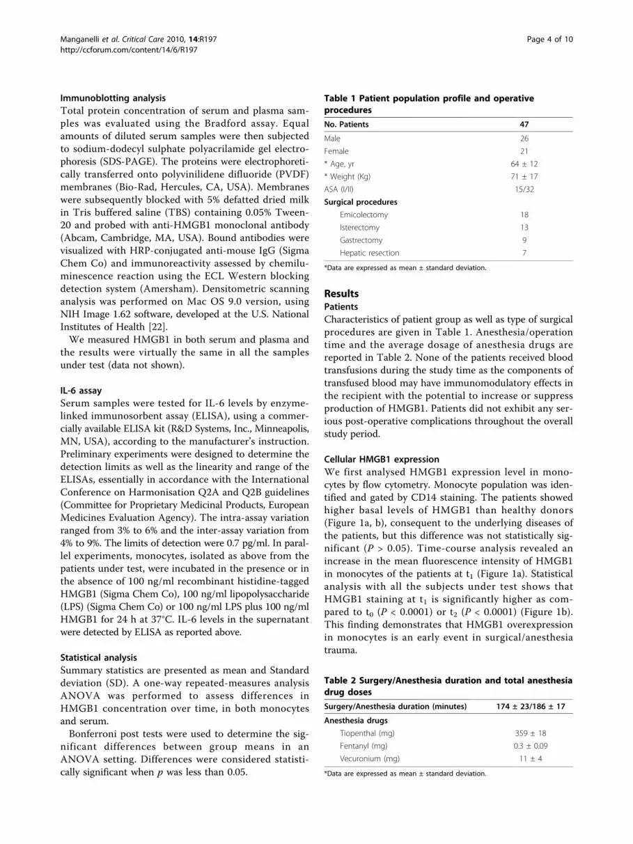
ResultsPatientsCharacteristics of patient group as well as type of surgicalprocedures are given in Table 1. Anesthesia/operationtime and the average dosage of anesthesia drugs arereported in Table 2. None of the patients received bloodtransfusions during the study time as the components oftransfused blood may have immunomodulatory effects inthe recipient with the potential to increase or suppressproduction of HMGB1. Patients did not exhibit any ser-ious post-operative complications throughout the overallstudy period.
Cellular HMGB1 expressionWe first analysed HMGB1 expression level in mono-cytes by flow cytometry. Monocyte population was iden-tified and gated by CD14 staining. The patients showedhigher basal levels of HMGB1 than healthy donors(Figure 1a, b), consequent to the underlying diseases ofthe patients, but this difference was not statistically sig-nificant (P > 0.05). Time-course analysis revealed anincrease in the mean fluorescence intensity of HMGB1in monocytes of the patients at t1 (Figure 1a). Statisticalanalysis with all the subjects under test shows thatHMGB1 staining at t1 is significantly higher as com-pared to t0 (P < 0.0001) or t2 (P < 0.0001) (Figure 1b).This finding demonstrates that HMGB1 overexpressionin monocytes is an early event in surgical/anesthesiatrauma.
Table 1 Patient population profile and operativeprocedures
No. Patients 47
Male 26
Female 21
* Age, yr 64 ± 12
* Weight (Kg) 71 ± 17
ASA (I/II) 15/32
Surgical procedures
Emicolectomy 18
Isterectomy 13
Gastrectomy 9
Hepatic resection 7
*Data are expressed as mean ± standard deviation.
Table 2 Surgery/Anesthesia duration and total anesthesiadrug doses
Surgery/Anesthesia duration (minutes) 174 ± 23/186 ± 17
Anesthesia drugs
Tiopenthal (mg) 359 ± 18
Fentanyl (mg) 0.3 ± 0.09
Vecuronium (mg) 11 ± 4
*Data are expressed as mean ± standard deviation.
Manganelli et al. Critical Care 2010, 14:R197http://ccforum.com/content/14/6/R197
Page 4 of 10

Figure 1 Analysis of HMGB1 cellular expression. (a) Flow cytometric analysis of HMGB1 expression in monocytes from one patient and onecontrol subject (healthy donor). Mononuclear cells were drawn from the patients at three different times, that is, t0: before surgery, t1:immediately after surgical procedure; t2: at 24 hours following intervention. Cells were stained with polyclonal anti-human HMGB1 1 μg/ml(Abcam) for one hour at room temperature. Nonspecific binding was determined by an unlabeled isotypic control antibody (Coulter-Immunotech, Hamburg, Germany). After washing with cold PBS, cells were incubated with FITC-conjugated anti-rabbit IgG and then analyzed byflow cytometry. Antibody reactivity was reported as mean fluorescence intensity. Histograms show the log fluorescence versus the cell number.(b) Results of flow cytometric analysis of HMGB1 expression in monocytes from controls (healthy donors) and from the patients under test atthree different times: t0 = before surgery, t1 = immediately after surgical procedure; t2 = at 24 hours following intervention. Mean fluorescenceintensities were measured and plotted values represent mean ± SD. ***t1 vs t0, t1 vs t2: P < 0.0001. (c) Monocytes cells were sampled at theindicated time points and subjected to nuclear (N) and cytoplasmic (C) fractionation. The levels of endogenous HMGB1 in the nuclear andcytoplasmic fractions were determined by immunoblotting with anti-HMGB1 antibodies. Laminin B served as nuclear contamination marker anda-tubulin as cytoplasmic contamination marker. Protein loading within each compartment was also normalized with Laminin B and a-tubulin,respectively.
Manganelli et al. Critical Care 2010, 14:R197http://ccforum.com/content/14/6/R197
Page 5 of 10
To verify whether the enhanced expression of HMGB1observed in monocytes may be derived by the nucleus,both cytosolic and nuclear extracts from monocytes ofall the patients were probed with anti-HMGB1 Ab byWestern blot. The results showed that an increasedexpression of HMGB1 in the cytoplasm was observed atT1 (Figure 1c). Since it was accompanied by a corre-sponding decrease of HMGB1 expression in the nucleus,our results suggest that neo-expression of HMGB1 inthe cytoplasm may result from a translocation from thenucleus.
Serum HMGB1 concentrationIn parallel analyses, we detected HMGB1 levels in seraof patients at the same time points, using Western Blot(Figure 2a). Densitometric analysis (Figure 2b) revealedthat HMGB1 concentration significantly increases at 24hours (t2) (P < 0.001) (Figure 2c). On the other hand,levels of HMGB1 in serum were not significantlyaffected immediately after surgical procedure (t1) (P >0.5), as compared with samples collected before surgery(t0). These findings provide direct evidence that overex-pression of HMGB1 by monocytes precedes the increaseof serum HMGB1 concentration in patients.
Serum IL-6 concentrationIL-6 is commonly produced at local tissue sites and thenreleased into circulation. Perturbation of tissue homeos-tasis causes IL-6 release in almost all situations andsuch a key cytokine is involved in surgical stressresponse as well. We, therefore, preliminary analyzedwhether treatment of monocytes with HMGB1 inducedin vitro release of IL-6. Monocytes from the patientsunder test were incubated in the presence or in theabsence of HMGB1, LPS or LPS plus HMGB1. The ana-lysis revealed that all the treatments induced a signifi-cant increase of IL-6 (P < 0.001) (Figure 3a),demonstrating that HMGB1 is able to trigger in vitrorelease of IL-6 by monocytes. As expected, the levels ofIL-6 following LPS treatment were lower as comparedto those following LPS plus HMGB1 treatment, support-ing the view of a synergic action between LPS andHMGB1 [23].Then, we tested IL-6 levels in serum samples by
ELISA. The results show that this proinflammatory cyto-kine markedly increases at t2 if compared to t0 and t1time points (t2 vs t0, P = 0.006; t2 vs t1, P = 0.003) (Fig-ure 3b), indicating that IL-6 release is temporally relatedwith the observed increase in HMGB1 concentration inthe sera of patients.
DiscussionThis study was undertaken to investigate HMGB1 pro-duction kinetics in patients undergoing major elective
surgery and to address how circulating mononuclearcells are implicated in this setting. Measurement ofserum level of IL-6 allowed us to study the eventualrelationship between HMGB1 and IL-6, a widely knownmarker of surgical stress being directly correlated withthe severity of surgery and the extent of traumatic injury[20,24]. The results obtained in this work showed that:a) cellular expression of HMGB1 in monocytes immedi-ately after the end of surgical procedure was signifi-cantly higher as compared to preoperative values; b) at24 hours following surgery, a significant increase ofHMGB1 levels was found in the sera of patients, (inter-estingly, such an increase was concomitant to a signifi-cant down-regulation of cellular HMGB1, suggestingthat the release of HMGB1 might, at least partially,derive from mononuclear blood cells); and c) at thesame time, high amounts of the circulating proinflam-matory cytokine IL-6 were detected as compared tobaseline preoperative levels.These current data are consistent with previous obser-
vations demonstrating that HMGB1 is secreted by acti-vated monocytes and is passively released by damagedcells following different types of injury, including surgi-cal/anesthesia stress [19,20,25,26]. It is conceivable thatan increase of HMGB1 in patient sera may also dependon passive protein release from damaged cells by surgi-cal procedures as well as from intestinal manipulationleading to endotoxin translocation which in turn couldinduce HMGB1 release [27].Furthermore, our findings support the view that
increased levels of HMGB1 constitute an early phenom-enon in traumatic insult, in contrast to the evidencereported for human sepsis as well as for experimentalmodels of endotoxemia, in which HMGB1 is considereda late mediator [28-30]. In particular, the present studyshows for the first time the intracellular overexpressionof HMGB1 in monocytes of patients immediately aftersurgery. This finding suggests that surgical stimuli mayrapidly activate intracellular pathways leading to secre-tion of HMGB1, which is subsequently spilled out intothe circulatory stream. In fact, at 24 hours followingsurgery, we observed a down-modulation of cellularHMGB1in mononuclear blood cells and a significantincrease of HMGB1 levels in serum. It is conceivablethat an increase of HMGB1 in patient sera may alsodepend on a passive release of such a protein fromdamaged cells following surgical procedures [8]. Never-theless, following surgical injury, monocytes display anabnormal intracellular expression of HMGB1 and thiscould represent an early event in surgical injury-inducedstress response. The ultimate mechanism underlyingregulation of this active HMGB1 release by surgical sti-muli as well as the position that surgery per se or gen-eral anesthesia occupies in the phenomenon, still
Manganelli et al. Critical Care 2010, 14:R197http://ccforum.com/content/14/6/R197
Page 6 of 10

remains elusive. In this respect, it was found that Reac-tive Oxygen Species (ROS) were able to induce activeHMGB1 secretion from monocytes in culture andhypoxic conditions or oxidative stress also trigger hepa-tocytes to produce HMGB1 through a calcium mediatedcell signaling [31,32].
It is noteworthy that in previous works we demon-strated both overproduction of ROS by PBMCs inpatients undergoing surgery and general anesthesia andthe capacity of some anesthetic compounds to induceoxidative stress by altering the mitochondrial redoxstate [33,34]. Based on these findings, we hypothesize
Figure 2 HMGB1 serum concentration. (a) Western blot analysis of serum HMGB1 concentration. Serum samples, obtained from the patientsat three different times: t0 = before surgery, t1 = immediately after surgical procedure; t2 = at 24 hours following intervention, were analyzed byWestern blot for reactivity with anti-human HMGB1 MoAb (1 μg/ml). A representative patient is shown together with a control serum from ahealthy donor. (b) Densitometric analysis of serum HMGB1 concentration was revealed by Western blot at three different times: t0 = beforesurgery, t1 = immediately after surgical procedure; t2 = at 24 hours following intervention (arbitrary units). (c) Values of densitometric analyses ofall the patients under test are shown as mean ± SD (arbitrary units). ***t2 vs t0, t2 vs t1: P < 0.001. t1 vs t0: NSS.
Manganelli et al. Critical Care 2010, 14:R197http://ccforum.com/content/14/6/R197
Page 7 of 10

that the postoperative upregulation of HMGB1 isrelated to the impact of surgery and anesthesia onredox metabolism and subsequent increased ROSproduction.Moreover, although it is known that apoptotic cells
are not capable of HMGB1 release, since they retainsuch a molecule within their nuclear compartment itwas recently demonstrated that macrophages engulfingapoptotic cells are induced to secrete HMGB1 [12].Indeed, there is evidence that an accelerated rate ofapoptosis in circulating lymphocytes occurred in theearly postoperative period [35-37]. Thus, we can furtherhypothesize that the accelerated rate of apoptosis
following surgery/anesthesia trauma, could be implicatedin the massive HMGB1 release found in patients within24 hours after a surgical procedure.Together with an increase of circulating HMGB1, an
additional finding of our study was the demonstrationthat: a) treatment of monocytes with HMGB1 inducedin vitro release of IL-6; b) at t2, high amounts of circu-lating IL-6 were detected as compared to t0. Thisstrongly suggests that HMGB1 postoperative increasemight be able to induce IL-6 secretion. It has providedevidence that HMGB1 binds Toll-like receptor 4 (TLR-4) on monocytes surface, thus triggering a signal trans-duction cascade. TLR pathway activation involves the
Figure 3 Analysis of IL-6 levels. (a) Analysis of IL-6 levels in the supernatants of monocytes from the patients under test. Monocytes wereincubated in the presence or in the absence of 100 ng/ml HMGB1 or 100 ng/ml LPS plus 100 ng/ml HMGB1 for 24 h at 37°C. The samples werecollected and analyzed using a commercially available enzyme-linked immunosorbent assay kit. Values are plotted as mean ± SD. ***HMGB1 vscontrol: P < 0.001; LPS vs control: P < 0.001; LPS plus HMGB1 vs control: P < 0.001. (b) Analysis of IL-6 levels in serum samples from the patientsat three different times: t0 = before surgery, t1 = immediately after surgical procedure; t2 = at 24 hours following intervention. Sera from healthysubjects served as controls. The samples were collected and analyzed using a commercially available enzyme-linked immunosorbent assay kit.Values are plotted as mean ± SD. **t2 vs t0 : P = 0.006, t2 vs t1: P = 0.003. t1 vs t0: NSS.
Manganelli et al. Critical Care 2010, 14:R197http://ccforum.com/content/14/6/R197
Page 8 of 10

phosphorylation of myeloid differentiation factor 88(MyD-88) and interleukin-1 receptor-associated kinase(IRAK), which in turn promotes activation and nucleartranslocation of nuclear factor kB (NF-kB) ultimatelyleading to the release of cytokines, including IL-6 [17].In line with our results, M.J. Cohen et al. found a
positive correlation between IL-6 and HMGB1 levels inseverely injured patients [25].Further evidence of the potential induction of IL-6
secretion by HMGB1 comes from the studies demon-strating that HMGB1 significantly correlates with IL-6in cerebrospinal fluid of humans. Moreover, it has beenshown that intracerebroventricular administration ofHMGB1 enhances brain IL-6 production in animalmodels [29,38].
ConclusionsIn conclusion, this study demonstrates for the first timethat surgical/anesthesia trauma is able to induce anearly intracellular upregulation of HMGB1 in monocytesof surgical patients. A statistically relevant increase inboth IL-6 and HMGB1 serum levels at 24 h after sur-gery fosters the hypothesis that serum post-operativeHMGB1 derives, at least partially, from monocytes andexhibits the potential to trigger IL-6 secretion. The clini-cal impact of these findings as well as the ultimatemechanism by which surgical/anesthesia stimuli modu-late HMGB1 production, opens an interesting debatedeserving of further studies.
Key messages• Surgical/anesthesia trauma can induce an earlyintracellular upregulation of HMGB1 in monocytesof surgical patients.• HMGB1 is released in the serum of subjectsundergoing traumatic/surgical injury 24 hours later.• A role is suggested for released HMGB1 as a trig-ger for IL-6 secretion.
AbbreviationsASA: American Society of Anesthesiologists; BSA: bovine serum albumin;ECG: electrocardiogram; ELISA: enzyme-linked immunosorbent assay; ETCO2:End-Tidal carbon dioxide; FITC: fluorescein isothiocyanate; HB: haemoglobin;HMGB1: high mobility group box 1; IL-6: interleukin-6; IRAK: interleukin-1receptor-associated kinase; LPS: lipopolysaccharide; MoAb: monoclonalantibody; MyD88: myeloid differentiation factor 88; NF-kB: nuclear factor kB;PBMCs: peripheral blood mononuclear cells; PBS: phosphate buffered saline;PFA: paraformaldehyde; PVDF: polyvinilidene difluoride; RAGE: receptor foradvanced glycation end products; ROS: reactive oxygen species; SaO2:oxygen saturation; SD: standard deviation; SDS-PAGE: sodium-dodecylsulphate polyacrilamide gel electrophoresis; TBS: Tris buffered saline; TLR:toll-like receptor.
AcknowledgementsThis work was supported by grants from “Sapienza” University Rome, Italy toMaurizio Sorice.
Author details1Department of Experimental Medicine, “Sapienza” University of Rome, VialeRegina Elena 324, Rome 00161, Italy. 2Department of Hematology, Oncologyand Molecular Medicine, Istituto Superiore di Sanità, Viale Regina Elena 299,Rome 00161, Italy. 3Department of Anesthesia and Intensive Care, “Sapienza”University of Rome, Viale del Policlinico 155, Rome 00161, Italy. 4Laboratoryof Experimental Medicine and Environmental Pathology, “Sapienza”University, Viale dell’Elettronica, Rieti 02100, Italy. 5Department of GeneralSurgery, “Sapienza” University of Rome, Viale del Policlinico 155, Rome 00161,Italy.
Authors’ contributionsVM, M Signore, IP, RM, TG and EL performed research and analysed data. GTand PC selected the patients and performed clinical and laboratory analyses.M Sorice and GD designed the research and wrote the paper.
Competing interestsThe authors declare that they have no competing interests.
Received: 30 March 2010 Revised: 8 June 2010Accepted: 2 November 2010 Published: 2 November 2010
References1. Koch A, Zacharowski P, Boehm O, Zacharowski K: Innate immunity,
coagulation and surgery. Front Biosci 2009, 14:2970-2982.2. Lin E, Calvano SE, Lowry SF: Inflammatory cytokines and cell response in
surgery. Surgery 2000, 127:117-126.3. Ni Choileain N, Redmond HP: Cell response to surgery. Arch Surg 2006,
141:1132-1140.4. Kurosawa S, Kato M: Anesthetics, immune cells, and immune responses. J
Anesth 2008, 22:263-277.5. von Dossow V, Sander M, MacGill M, Spies C: Perioperative cell-mediated
immune response. Front Biosci 2008, 13:3676-3684.6. Delogu G, Moretti S, Antonucci A, Marandola M, Tellan G, Sale P,
Carnevali R, Famularo G: Apoptogenic effect of fentanyl on freshlyisolated peripheral blood lymphocytes. J Trauma 2004, 57:75-81.
7. Oppenheim JJ, Yang D: Alarmins: chemotactic activators of immuneresponses. Curr Opin Immunol 2005, 17:359-365.
8. Klune JR, Dhupar R, Cardinal J, Billiar TR, Tsung A: HMGB1: endogenousdanger signaling. Mol Med 2008, 14:476-484.
9. Harris HE, Raucci A: Alarmin(g) news about danger: workshop on innatedanger signals and HMGB1. EMBO Rep 2006, 7:774-778.
10. Yang H, Tracey KJ: High mobility group box 1 (HMGB1). Crit Care Med2005, 33:S472-474.
11. Gardella S, Andrei C, Ferrera D, Lotti LV, Torrisi MR, Bianchi ME, Rubartelli A:The nuclear protein HMGB1 is secreted by monocytes via a non-classical, vesicle-mediated secretory pathway. EMBO Rep 2002, 3:995-1001.
12. Bianchi ME: DAMPs, PAMPs and alarmins: all we need to know aboutdanger. J Leukoc Biol 2007, 81:1-5.
13. Erlandsson Harris H, Andersson U: Mini-review: The nuclear proteinHMGB1 as a proinflammatory mediator. Eur J Immunol 2004,34:1503-1512.
14. Hreggvidsdottir HS, Ostberg T, Wähämaa H, Schierbeck H, Aveberger AC,Klevenvall L, Palmblad K, Ottosson L, Andersson U, Harris HE: The alarminHMGB1 acts in synergy with endogenous and exogenous danger signalsto promote inflammation. J Leukoc Biol 2009, 86:655-662.
15. Yang H, Tracey KJ: Targeting HMGB1 in inflammation. Biochim BiophysActa 2010, 1799:149-156.
16. Pullerits R, Brisslert M, Jonsson IM, Tarkowski A: Soluble receptor foradvanced glycation end products triggers a proinflammatory cytokinecascade via beta2 integrin Mac-1. Arthritis Rheum 2006, 54:3898-3907.
17. van Zoelen MA, Yang H, Florquin S, Meijers JC, Akira S, Arnold B,Nawroth PP, Bierhaus A, Tracey KJ, van der Poll T: Role of toll-likereceptors 2 and 4, and the receptor for advanced glycation endproducts in high-mobility group box 1-induced inflammation in vivo.Shock 2009, 31:280-284.
18. Rauvala H, Rouhiainen A: Physiological and Pathophysiological Outcomesof the Interactions of HMGB1 with Cell Surface Receptors. BiochimBiophys Acta 2010, 1799:164-170.
Manganelli et al. Critical Care 2010, 14:R197http://ccforum.com/content/14/6/R197
Page 9 of 10

19. Suda K, Kitagawa Y, Ozawa S, Saikawa Y, Ueda M, Abraham E, Kitajima M,Ishizaka A: Serum concentrations of high-mobility group boxchromosomal protein 1 before and after exposure to the surgical stressof thoracic esophagectomy: a predictor of clinical course after surgery?Dis Esophagus 2006, 19:5-9.
20. Peltz ED, Moore EE, Eckels PC, Damle SS, Tsuruta Y, Johnson JL, Sauaia A,Silliman CC, Banerjee A, Abraham E: HMGB1 is markedly elevated within 6hours of mechanical trauma in humans. Shock 2009, 32:17-22.
21. Pickl WF, Majdic O, Kohl P, Stöckl J, Riedl E, Scheinecker C, Bello-Fernandez C, Knapp W: Molecular and functional characteristics ofdendritic cells generated from highly purified CD14+ peripheral bloodmonocytes. J Immunol 1996, 157:3850-3859.
22. NIH Image. [http://rsb.info.nih.gov/nih-image].23. Youn JH, Oh YJ, Kim ES, Choi JE, Shin JS: High Mobility Group Box 1
Protein binding to lipopolysaccharide facilitates transfer oflipopolysaccharide to CD14 and enhances lipopolysaccharide-mediatedTNF-αlpha production in human monocytes. J Immunol 2008,180:5067-5074.
24. Servis D, Busic Z, Stipancic I, Patrlj L, Gagro A: Serum cytokine changesafter gastric resection or gastrectomy for gastric cancer.Hepatogastroenterology 2008, 55:1868-1872.
25. Cohen MJ, Brohi K, Calfee CS, Rahn P, Chesebro BB, Christiaans SC, Carles M,Howard M, Pittet JF: Early release of high mobility group box nuclearprotein 1 after severe trauma in humans: role of injury severity andtissue hypoperfusion. Crit Care 2009, 13:R174.
26. Scaffidi P, Misteli T, Bianchi ME: Release of chromatin protein HMGB1 bynecrotic cells triggers inflammation. Nature 2002, 418:191-195.
27. Kim JH, Kim SJ, Lee IS, Lee MS, Uematsu S, Akira S, Oh KI: Bacterialendotoxin induces the release of High Mobility Group Box 1 via the IFN-beta signaling pathway. J Immunol 2009, 182:2458-2466.
28. Gibot S, Massin F, Cravoisy A, Barraud D, Nace L, Levy B, Bollaert PE: High-mobility group box 1 protein plasma concentrations during septicshock. Intensive Care Med 2007, 33:1347-1353.
29. Wang H, Yang H, Tracey KJ: Extracellular role of HMGB1 in inflammationand sepsis. J Intern Med 2004, 255:320-331.
30. Hou LC, Qin MZ, Zheng LN, Lu Y, Wang Q, Peng DR, Yu XP, Xin YC, Ji GL,Xiong LZ: Severity of sepsis is correlated with the elevation of serumhigh-mobility group box 1 in rats. Chin Med J (Engl) 2009, 122:449-454.
31. Tang D, Shi Y, Kang R, Li T, Xiao W, Wang H, Xiao X: Hydrogen peroxidestimulates macrophages and monocytes to actively release HMGB1. JLeukoc Biol 2007, 81:741-747.
32. Tsung A, Klune JR, Zhang X, Jeyabalan G, Cao Z, Peng X, Stolz DB,Geller DA, Rosengart MR, Billiar TR: HMGB1 release induced by liverischemia involves Toll-like receptor 4 dependent reactive oxygenspecies production and calcium-mediated signaling. J Exp Med 2007,204:2913-2923.
33. Delogu G, Moretti S, Famularo G, Marcellini S, Santini G, Antonucci A,Marandola M, Signore L: Mitochondrial perturbations and oxidant stressin lymphocytes from patients undergoing surgery and generalanesthesia. Arch Surg 2001, 136:1190-1196.
34. Delogu G, Antonucci A, Moretti S, Marandola M, Tellan G, Signore M,Famularo G: Oxidative stress and mitochondrial glutathione in humanlymphocytes exposed to clinically relevant anesthetic drugconcentrations. J Clin Anesth 2004, 16:189-194.
35. Delogu G, Famularo G, Moretti S, De Luca A, Tellan G, Antonucci A,Marandola M, Signore L: Interleukin-10 and apoptotic death of circulatinglymphocytes in surgical/anesthesia trauma. J Trauma 2001, 51:92-97.
36. Koerner P, Westerholt A, Kessler W, Traeger T, Maier S, Heidecke CD:Surgical trauma and postoperative immunosuppression. Chirurg 2008,79:290-294.
37. Yamada R, Tsuchida S, Hara Y, Tagawa M, Ogawa R: Apoptoticlymphocytes induced by surgical trauma in dogs. J Anesth 2002,16:131-137.
38. Nakahara T, Tsuruta R, Kaneko T, Yamashita S, Fujita M, Kasaoka S,Hashiguchi T, Suzuki M, Maruyama I, Maekawa T: High-Mobility Group Box1 Protein in CSF of patients with subarachnoid hemorrhage. NeurocritCare 2009, 11:362-368.
doi:10.1186/cc9316Cite this article as: Manganelli et al.: Increased HMGB1 expression andrelease by mononuclear cells following surgical/anesthesia trauma.Critical Care 2010 14:R197.
Submit your next manuscript to BioMed Centraland take full advantage of:
• Convenient online submission
• Thorough peer review
• No space constraints or color figure charges
• Immediate publication on acceptance
• Inclusion in PubMed, CAS, Scopus and Google Scholar
• Research which is freely available for redistribution
Submit your manuscript at www.biomedcentral.com/submit
Manganelli et al. Critical Care 2010, 14:R197http://ccforum.com/content/14/6/R197
Page 10 of 10