Local and regional anesthesia
702
-
Upload
khangminh22 -
Category
Documents
-
view
0 -
download
0
Transcript of Local and regional anesthesia

THE LIBRARY
THE UNIVERSITYOF CALIFORNIALOS ANGELES
GIFT OF
SAN FRANCISCOCOUNTY MEDICAL SOCIETY


LOCAL AND REGIONAL- ANESTHESIA
With Chapters on Spinal, Epidural, Paravertebral, and Para-
sacral Analgesia, and on other Applications of Local and
Regional Anesthesia to the Surgery of the Eye, Ear, Nose and
Throat, and to Dental Practice
BY.
CARROLL W. ALLEN, M. D.
ASSISTANT PROFESSOR OF CLINICAt SURGERY AT THE TULANE UNIVERSITY OF LOUISIANA,NEW ORLEANS; LECTURER AND INSTRUCTOR IN GENITO-URINARY AND RECTAL DISEASES
AT THE NEW ORLEANS POLYCLINIC; VISITING SURGEON TO THE CHARITY HOSPITAL
WITH AN INTRODUCTION BY
RUDOLPH MATAS, M. D.
PROFESSOR Or GENERAL AND CLINICAL SURGERY AT THE TULANE UNIVERSITY OF
LOUISIANA, NEW ORLEANS, ETC.
SECOND EDITION, RESET
PHILADELPHIA AND LONDON
W. B. SAUNDERS COMPANY1918

Copyright, igi4, by W. B. Saunders Company. Reprinted April, 1915, and November, 1916.
Revised, entirely reset, reprinted, and recopyrighted March, 1918
Copyright, 1918, by W. B. Saunders Company
PRINTED IN AMERICA
PRESS OF
W. B. SAUNDERS COMPANVPHILADELPHIA

Libra."
TO
RUDOLPH MATASSURGEON, SCHOLAR, TEACHER, FRIEND
ONE OF THE PIONEERS IN THE FIELD OF LOCAL AND REGIONAL
ANESTHESIA, UNDER WHOSE GUIDANCE THE AUTHOR WAS IN-
ITIATED INTO SURGERY, WHOSE EXAMPLE AND FRIEND-
SHIP PROMPTED THE CONCEPTION OF THIS WORK,AND WHOSE TEACHINGS AND WRITINGS HAVE CON-
TRIBUTED MANY PAGES OF THE TEXT, THIS
VOLUME IS GRATEFULLY DEDICATED.
53S2G5


INTRODUCTION
FOR nearly twenty years the control of pain in surgical opera-
tions by local and regional methods has been the subject of our earnest
study. As director of the surgical clinics of the College of Medicine
of the Tulane University since 1895, we began to utilize the large
clinical material at our command in the effort to diminish the indica-
tions for general narcosis, and to substitute for the immediate dangers
of chloroform, which was then the routine anesthetic in almost all
Southern clinics, the more laborious but far safer methods of periph-
eral analgesia. Beginning with a purely local and peripheral technic,
in which intradermal infiltration and massive edematization with
dilute isotonic cocain solutions were chiefly utilized, in accordance
with the principles laid down by Corning, Halsted, Reclus, and
Schleich, we soon advanced from the minor work of the dispensary
to the more ambitious fields of major surgery.
In 1897 we discarded cocain and adopted beta-eucain and soon
became engrossed in the neuroregional methods1 alone or combined
with massive infiltration, which rapidly expanded in every direction,
yielding the most gratifying and, at that time, almost incredible
results. The pursuit of this regional method was carried out with
so much vigor and enthusiasm that in I9OO2 we were able to publish
two extensive reports which reviewed the progress of our work and
1 In referring to regional methods we exclude the spinal or subarachnoid method
(L. Corning, 1886-1894); Bier (1899) as a central method.
After an experience with over 300 applications of this method with cocain and its
various substitutes, we experienced a transition from a state of great enthusiasm to one
of decided depression, having learned by hard experience and careful study of our results
that the benefits of this procedure were more apparent than real. Since then we have
restricted the application of spinal analgesia to a very circumscribed and steadily smaller
group of indications. ,
2 "The Growing Importance and Value of Local and Regional Anesthesia in Minor
and Major Surgery," Transaction of Louisiana State Med. Assoc., April, 1900, pp. 1-78;
"Local and Regional Anesthesia with Cocain and Other Analgesic Drugs," Philadelphia
Med. Jour., November 3, 1900. pp. 1-72.
1

2 INTRODUCTION
that of others and gave an account of the considerable success that
we had obtained in the invasion of new territories.
By blocking the nerve-trunks at their exit from the cranial foramina,
jaws were resected, the tongue and floor of the mouth excised, and, bya similar process, craniotomy, thyroid and laryngeal resections, ampu-tations of the extremities, resection of joints, thoracotomies, hernias,
and the entire domain of genito-urinary, rectal, and a considerable
share of pelvic and abdominal surgery became subservient to the
new methods. In this way we were able to show in 1900 that fully
50 to 60 per cent, of the operations, which six years before would
have required a general narcosis, had become amenable to local and
regional anesthesia.
Fourteen years have elapsed since that time. Great transforma-
tions have taken place in our methods of general narcosis. Chloro-
form, which for half a century had reigned supreme as the autocrat
of the operating theater, has been practically banished from the
clinics of the South its last stronghold. Ether by the open mask
and drop method has entirely supplanted it; and now nitrous-oxid
gas in combination with ether, alone or with oxygen, is gaining favor
steadily in our main operative and especially private clinics. The
effect of this revolution in reducing the immediate mortality of
general narcosis, and, to some extent, in diminishing the postanes-
thetic risks, is universally recognized. However, the problem of
shock, the secondary nausea and vomiting, the pulmonary com-
plications, embolism and thrombosis, and, above all, the degen-
erative auto-intoxications following the action of these somatic
poisons on the eliminating and other organs still remain to be
reckoned with.
On the other hand, the synthetic chemist and pharmacologist
have not been idle, and their untiring and brilliant efforts to find sub-
stitutes for the dangerous and costly cocain have given us a succes-
sion of remarkable synthetic products, such as beta-eucain, nirvanin,
alypin, stovain, anesthesin, etc., which have been successively dis-
placed by what now appears to be the nearest approach to the ideal
local analgesic novocain.
In like manner, the genius of synthetic chemistry and the bio-
logic laboratory have found in suprarenin a less perishable substi-
tute for adrenalin, the active product directly obtained from the
gland. The advent of adrenalin and its synthetic substitutes has
marked a new era in the history of local anesthesia. By its powerful
and lasting vasoconstrictor and ischemic action it gives the operator
INTRODUCTION 3
a bloodless field, which has deserved for it the name of the "chemical
tourniquet" (Braun). Combinations of novocain and adrenalin in
various isotonic dilutions by practically eliminating the toxicity of
the analgesic, increasing its stability, durability, and intensity
have so expanded the technic that in the hand of an expert peripheral
analgesia may be made to encompass in its grasp almost the entire
domain of operative surgery.* * *
But with all its great achievements the art of local and regional
anesthesia is still young. Barely three decades have elapsed since
Karl Koller made his epochal demonstration of the anesthetic prop-
erties of cocain at Heidelberg in 1884, and yet, in spite of the stupen-
dous distance that we have traveled since then, the horizon of peripheral
anesthesia is ever widening and offering new opportunities for profitable
exploitation. It is still in process of development; it still offers manydifficult problems that await solution.
In dealing with major operations, its successful application de-
mands patience, time, and skill a skill that can only be acquired
and exercised on the human cadaver by those who, being anatomists,
can alone survey the field of operation with fluoroscopic eyes. For
this reason the practice of peripheral anesthesia, especially in its
neuroregional aspects, appeals most pointedly to the young, ambi-
tious, and well-trained surgeon, who, fresh from the anatomic labora-
tory, finds here, as nowhere else, an immediate and practical applica-
tion for a knowledge that he has acquired at the cost of long nights
of vigil, labor, and thought.
In these days when exact topographic and applied anatomy is
rated somewhat at a discount, it is a source of no small gratification
for the young but well-trained man to discover that his anatomic
knowledge is a living, palpable, and productive asset. Not a thing to
be learned solely as a matter of academic culture and soon to be for-
fotten, but a practical tool to be used in unlocking his most immediate
technical problems. It is only through the aid of applied anatomythat regional anesthesia is what it is to-day. It is for this reason that
all, or nearly all, the notable advances that have been made in its
technic have been due to the enterprise and the activities of young
surgeons. Leonard Corning, Halsted, Reclus, Schleich, Crile, Gush-
ing, Bier, Oberst, Braun, and a host of others who have laid the funda-
mentals of this work did so in their earlier professional years. It is
this same potential spirit in the young man fresh from the anatomic
and physiologic laboratories that animates their followers the

4 INTRODUCTION
builders of the present day. Such men as Offerhaus, Hartel, Peuckert,
Hirschel, Kulenkampf, Danis, Finsterer, Lawen, and others in
Europe, not to mention a group of young surgeons in this country
and in our own immediate surroundings who are enriching the
foundation laid down by the masters by their contributions, based
chiefly upon anatomic and physiologic researches.
Whatever may be the limitations of regional anesthesia and the
objections that have been argued against it, no one can deny that it
has given a new impetus to anatomic teaching; that it has placed a
high valuation upon an exact anatomic training, and that in this wayit is making it less possible for the mere cutter the "cut and tie"
type of practitioner to be confused with the real surgeon. For this
reason alone it deserves the encouragement and fostering care of
every surgeon and every teacher who has at heart the higher welfare
of his science and his art.* * *
To review and summarize the evidences of progress in local and
regional analgesia; to study and analyze the copious and constantly
growing literature which is rapidly piling up to pyramidal and almost
inaccessible heights; to scrutinize the various analgesics that are born
yearly in the laboratory of the chemist, and try the methods by which
they may be utilized with special advantage in the different regions
of the body and in connection with the surgical specialties; to gauge
the value of the various technics proposed by the criterion of clinical
observation and personal experience, and, in a like manner, to judge of
their advantages and limitations in their relation to the general
narcosis, was a task which I had set to myself, and which, after an
experience of over two decades in this mode of practice, I felt might
prove profitable to the profession, if only in the interests of a useful
propaganda.
But, unfortunately, many circumstances and more urgent in-
terests directed my attention into other channels, and the time has
never come when I could sit down peacefully and calmly to the
realization of my project. Fortunately for my purpose, the seed
sown in earlier years appears to have yielded good and seasonable
fruit. Associated with me as pupils and assistants were a group of
young men who entered into the spirit of the work with zeal and
enthusiasm. The results obtained in our clinics and exhibited in
our reports of 1900, and subsequently, have been made possible
largely through their faithful collaboration. Several of these have
already attained enviable reputations in our community and else-

INTRODUCTION 5
where, as teachers and surgeons especially skilled in the methods of
local and regional anesthesia, and to all these I owe a debt of grati-
tude. Conspicuous among these is Dr. Carroll W. Allen, whose
steadfast loyalty to these methods for many years has been rewarded
by a reputation for special skill and judgment in their application
which is eminently deserved. He has assiduously cultivated the
technic in all its variations, many of which are his own, and in our
joint services at the Charity Hospital the results obtained have
proved so satisfactory that fully 55 or 60 per cent, of the major opera-
tions in the division under his charge are performed solely by periph-
eral anesthetic procedures, exclusive of the spinal or subarachnoid
analgesias which are not included in this category. One of the best
proofs of the success of any method of practice is the confidence it
inspires among the men of the profession and in their willingness to
have it applied to themselves. Schleich, in his "Schmerzlose Opera-
tionen," tells us how his clinic was besieged by doctors who, needing
surgical relief for various ailments, were anxious to be operated on byhim painlessly, but without the unconsciousness of general narcosis.
This is the experience of every operator whose reputation for skill in
local and regional methods is confirmed by his results. Dr. Allen is
no exception to this rule.
Now, returning to the book. I had almost abandoned all expecta-
tion of accomplishing this self-appointed task when Dr. Allen gener-
ously offered his collaboration. I had hoped that this valued offer
would have made the task lighter. Dr. Allen set himself seriously
and earnestly to work and gathered a large mass of material which I
found it impossible to edit with him without the sacrifice of other and
more pressing obligations, or subjecting the publishers to unwar-
ranted delays. All that I could do was to give him the full and free
use of my previous writings and original observations on this subject
and such general counsel as my experience dictated. This volume
as it stands is, therefore, the result of Dr. Allen's sole industry, thought,
and labor. My regret is that I have not been able to join forces with
him in accomplishing a task which it was my privilege to initiate even
though indirectly, and in which I have always had a deep and abiding
interest. Without having had an opportunity to revise the text or
to read it thoroughly through no fault of Dr. Allen or lack of willing-
ness on my part I am satisfied, by many years of professional and
friendly association with the author, that the methods and teachings
expounded for the last twenty years in the surgical clinics of the
Tulane University will not only be well represented, but will be

6 INTRODUCTION
strengthened, and thereby diffused over a greater and growingarea.
If Dr. Allen's book will only encourage others to follow his ex-
ample, and stimulate his contemporaries, and especially the young
surgeons of the rising generation, to cultivate the "qualities of head,
heart, and hand" that are necessary for the successful practice of the
art of peripheral anesthesia, it will have served a useful purpose and
discharged a worthy mission. In this hope I wish it God-speed.
RUDOLPH MATAS.

PREFACE TO THE SECOND EDITION
DURING the three years that have elapsed since the appearanceof the first edition of this book some improvement and progress has
been made in the field of local anesthesia.
With a general broadening of this field many additional opera-tions have been added to the already long list of those commonlyperformed by surgeons skilled in the use of these methods. Notable
among these additions is the removal of the prostate, a subject
upon which I have worked for some years and which was but briefly
touched upon in the first edition as there were many details not yet
perfected at that time.
I regard the removal of the prostate as my best accomplishmentin local anesthesia; this is largely on account of the great reduction
of mortality and the ease with which it is done by these methods.
It is particularly in these subjects that general anesthesia is so much
to be dreaded.
The addition of sacral anesthesia and some practical additions
to abdominal surgery are among the recent advances.
Many valuable and useful additions will be found in the chapter
on the head; the progress here has been very marked. Much of the
text has been rearranged, and the chapter on spinal anesthesia has
been largely rewritten.
Every change is not always a reform and apparent triumph is not
always progress. There is often work that must be done without
enthusiasm, work that brings only unrelieved weariness and a plod-
ding gait, but the direct value of what we do is that we are workingin the direction of progress. This has been my inspiration and with
these thoughts in mind I send this edition to press.
CARROLL W. ALLEN.NEW ORLEANS, LA.,
Marck, 1918. . 7


PREFACE
IN presenting this volume to the profession I have hoped to fill
what I have learned by my experience as a teacher is a real want in
the surgical literature of the English language.
Many small monographs have been available for the general
surgeon, and some excellent books dealing exclusively with the spe-
cialties have been published, but no book in our language has at-
tempted to survey the entire field, giving the essential elements in
the successful application of local anesthesia to major surgery, as
well as a systematic and detailed description of the methods of anes-
thesia suitable to operations in the different regions of the body.The excellent work of Professor Heinrich Braun is a masterpiece
and a model of German thoroughness and comprehensiveness, and
I have availed myself of this fountain source of information in both
text and illustrations through the courtesy of Professor Braun him-
self and of his obliging publisher, Herr J. A. Barth.
When this volume was first undertaken it was intended that it
should be a joint contribution from Professor Rudolph Matas and
myself, an accomplishment of which I would truly have been proud;
however, lack of time and the urgent press of other duties forced
Doctor Matas to withdraw his direct collaboration, leaving to me the
responsibility of this publication.
I feel it is fitting that a pupil and close associate of his should
assume this task. It was at his side that I received my first lesson
in local anesthesia, and derived from him that enthusiasm and zeal
for the work that has made this book possible. It was his hand
that opened the door to my surgical career, and from that hand I
have received a generous bounty since. His name will always be
numbered among the pioneers of local and regional anesthesia with
Corning, Halsted, Crile, and Gushing in this country; Schleich, Braun,
Reclus, and Barker abroad.
While deprived of his collaboration in the authorship of this
work, I have quoted liberally from his writings and drawn still more
liberally from his ideas and spoken teachings on this subject. To9

10 PREFACE
him is due the credit of working out successfully the first route to
the second division of the trigeminus and blocking it with Meckel's
ganglion and its branches, through the sphenomaxillary fissure and,in this way, painlessly resecting the upper maxilla, a method which
by German authors is still erroneously credited to Payr. The Germans
(Braun, Hartel, et al.), however, credit Matas with the inframalar
route for reaching the inferior maxillary division at the foramen ovale
to which they have attached his name. With the aid of this proced-
ure he had resected the lower jaw many times, long before Schlosser
had popularized this route for the alcoholization of this nerve in
trifacial neuralgia. Matas first worked out a satisfactory method of
regional anesthesia of the forearm by blocking the nerves at the
elbow, and, independently of Crile's earlier work, he had amputatedthe leg and thigh several times by blocking the sciatic, anterior crural,
obturator, and saphenous nerves. He performed the first operation
under spinal analgesia in America, and devised several types of ap-
paratus for massive infiltration anesthesia. Such terms as "intra-
neural," "perineural," and "paraneural," as applied to regional
neural methods, were first introduced by him, as acknowledged by
Braun, at a time when such niceties of classification were unknownin the literature.
The earlier accomplishments of Matas in this field have been over-
shadowed by his later and far-reaching contributions to other depart-
ments of surgery, more particularly the various operations for the
radical cure of aneurysm which are permanently linked with his name.
In this way, his work in anesthesia has been overlooked or forgotten
by many, who aware only of the marvelous efficiency of this branch
of surgery at the present time, are oblivious of the laborious steps
that have led to its present evolution. I feel it a fitting task, there-
fore, that the recital of Professor Matas' early achievements as they
appear in the following pages should devolve upon me.
The fundamental work on "nerve-blocking," which has so intimately
and inseparably associated the name of Crile with the early history
of regional anesthesia, is now supplemented by his epoch-makingstudies in anoci-association and in their practical application. The
growing appreciation of these principles has made a thorough knowl-
edge of local, and especially regional, analgesia more than ever neces-
sary to the progressive surgeon who would follow the teachings of
this eminent leader.
A very extensive bibliography had been prepared upon which the
author had expended much time and laborious research; it was in-

PREFACE I I
tended as an appendix to the volume, which would have been of service
to the student of the history and literature of the subject. It em-
braced a list of over six thousand references, covering several hundred
pages. Unfortunately, as the text grew in size, it was found that
even an abridged bibliography would have so far exceeded the pro-
posed dimensions of the volume that it would have been too ponder-
ous for the purpose for which it was originally intended. At the sugges-
tion of the publishers it was deemed best to abandon this publication, a
determination which has been a sore disappointment to the author,
who in this way had expected to make a full acknowledgment of
every publication referred to in the text; as it is, many importantreferences have been regretfully omitted.
The author now desires to express his special and grateful obliga-
tion to the many authors and investigators quoted, whose writings
have so largely and generously contributed to the making of this
book.
In the preparation of this volume I am under particular obliga-
tion to my friend, Professor M. Feingold, for valuable assistance
and advice in the chapter on the Eye as well as in the general text;
to Professor C. J. Lanfried for assistance in the chapter on the
Ear, Nose, and Throat; to Drs. E. C. Samuel and R. M. Blakely,
of Touro Infirmary, for their kind assistance in the illustrations; and
to Miss L. Ambrose for her assistance in the translations. I am also
much indebted to Professors Arthur E. Barker, of London; Fritz
Hartel, of Berlin; and Guido Fischer, of Marburg, for the privilege of
making many quotations and the use of valuable illustrations.
CARROLL W. ALLEN.NEW ORLEANS, LA.


CONTENTS
CHAPTER I
PAGE
HISTORY 17
CHAPTER II
NERVES AND THEIR SENSATIONS ESPECIALLY PAIN 26
Distribution of Sensation 37
Philosophy of Pain 44
CHAPTER III
OSMOSIS AND DIFFUSION 46
CHAPTER IV
THE ANESTHETIC EFFECTS OF PRESSURE-ANEMIA COLD AND WATER ANESTHESIA 58Pressure 58Cold 60
Water Anesthesia 63
CHAPTER V
LOCAL ANESTHETICS 67
tCocain 72
Eucain 77
Akoin 81
Holocain 81
Tropacocain 82
Stovain 83
Alypin 83
Novocain Hydrochloric! 85
Chloretone 89
Orthoform 92Nirvanin 93Anesthesin 93
Subcutin 95
Propasin 96
Apothesine 96
Comparative Action of Anesthetic Agents 99
Toxicity 104
Anesthetic Properties of Quinin Salts 109Anesthetic Properties of Magnesium Salts 123
13

14 CONTENTS
CHAPTER VIPAGE
TOXICOLOGY 125
CHAPTER VII
ADRENALIN 137
Surgical Uses 148
CHAPTER VIII
PRINCIPLES OF TECHNIC 155General Considerations 155Solutions and Their Methods of Use 158Classification of Methods of Local and Regional Anesthesia in which Cocain
and Other Allied Analgesic Drugs are Utilized as the Active Agents 161
The Armamentarium 1 73
Clinical Application 1 76
Cold 181
Regional Anesthesia 182
The Constrictor 186
Technic of Handling Wounds in General 187
Hemostasis and Closure of Wounds 190The History of the Hypodermic Syringe i go
CHAPTER IX
THE USE OF MORPHIN AND SCOPOLAMIN AND .COMBINED METHODS OF AN-
ESTHESIA 192
Morphin and Scopolamin 192
Combined Methods of Anesthesia 198
CHAPTER X
INDICATIONS, CONTRA-INDICATIONS, AND SHOCK 201
Indications and Centra-indications 201
Shock 202
CHAPTER XI
ANOCI-ASSOCIATION 204
CHAPTER XII
INTRA-ARTERIAL ANESTHESIA 211
Intravenous Anesthesia 217
CHAPTER XIII
GENERAL ANESTHESIA THROUGH THE INTRAVENOUS INJECTION or LOCAL ANES-
THETICS. . 221

CONTENTS 15
CHAPTER XIVPAGE
THE UPPER AND LOWER EXTREMITIES 224Bones and Joints 225The Brachial Plexus 227Nerves of the Upper Extremity 238The Fingers and Hand 245The Lower Extremity 257The Hip and Thigh 267The Knee-joint 270The Leg 272
CHAPTER XV.
THE NECK 279
Operations on the Neck 283The Larynx and Trachea 287Goiter 291
CHAPTER XVI
THE THORAX AND BACK 302The Sternum 312The Back 312
CHAPTER XVH
THE ABDOMEN 317Possible Scope of Operations within the Abdomen 349
CHAPTER XVin
HERNIA 359
Inguinal Hernia 360Femoral Hernia 372Umbilical Hernia 374
Postoperative Hernia 376
CHAPTER XIX
GENITO-URINARY, ANORECTAL, AND GYNECOLOGIC OPERATIONS 379
Genito-urinary Organs 379Penis , 383
Scrotum 389
Chancroids 393
Bladder 394
Prostatectomy 395
The Kidney and Ureter 47Anorectal Region 4 11
Gynecologic Operations 4 20

I 6 CONTENTS
CHAPTER XXPAGE
SPINAL ANALGESIA 432
Anatomy 435Anesthetic Agents 436Isotonic Qualities and Specific Gravity of Anesthetic Solutions and Their
Movements within the Canal 439Indications and Centra-indications 450Technic 453Failures ". 463In Obstetrics and Gynecolog> 464
Military Surgery 465
Physiological Action 466Vascular System 467
Respiration 468Abdomen 469After-effects 470
Experimental Work 472
Urinary Changes 476Effects on the Nervous System 477Ocular Palsies 481The Method of Jonnesco , 484Treatment of After-effects 485
Epidural, Caudal or Sacral Anesthesia 486
CHAPTER XXI
PARAVERTEBRAL AND PARASACRAL ANESTHESIA 494Paravertebral Anesthesia 494Parasacral Anesthesia 5 2
CHAPTER XXII
THE HEAD, SCALP, CRANIUM, BRAIN, AND FACE 507
The Face 528
Internal Maxillary Artery 60 1
CHAPTER XXIII
THE ORGANS or SPECIAL SENSE, WITH DENTAL ANESTHESIA 625
The Eye 625
The Ear ....... 630
Nose and Throat 634
Dental Anesthesia 644
INDEX . . 663

LOCAL ANESTHESIA
CHAPTER I
HISTORY
Divinum est opus sedare dolorem (divine is the work to relieve
pain). Thus spoke Hippocrates.The history of the efforts of the human race to find a means to
control pain during operative procedure forms one of the most inter-
esting chapters in medicine. The writings of authors, from earliest
antiquity down through the long centuries, deal with efforts in be-
half of human suffering. Sometimes surrounded by superstitions at
times the most ridiculous; later, as knowledge increased, based uponmore or less reason, but all futile and attaining the desired end onlyto a limited degree.
Among the earlier references to the use of narcotics is to be found
the following from Homer's "Odyssey," when Helen gave to Ulyssesand his comrades the "sorrow easing drug," which probably con-
sisted of the juice of the poppy and Indian hemp:
"Presently she cast a drug into the wine, whereof they drank
a drug to lull all pain and anger and bring forgetfulness of everysorrow. Whoso should drink a draught thereof, when it is mingledin the bowl, on that day he would let no tear fall down his cheek, not
though his mother and father died, not though men slew his brother
or dear son with the sword before his face and his own eyes beheld it."
During the siege of Troy the Greek surgeons used anodyne and
astringent applications to ease the pain of their wounded, which
probably had some antiseptic effect of which they were not aware.
The following is found in the "Iliad," when Patroclus, in admin-
istering to the sufferings of Euryphylus, removed a dagger from his
thigh:
"Cut out the biting shaft; and from the wound
With tepid water cleansed the clotted blood; r
Then, pounded in his hands, the root applied
Astringent, anodyne, which all his pain
Allay'd; the wound was dried, and stanched the blood."
2 17

18 LOCAL ANESTHESIA
It is probable that primitive men used pressure and cold to be-
numb the parts and thus lessen pain. In time they no doubt learned
that pressure over the region of the nerves and arteries had a more
pronounced effect, though they probably did not know why. Theancient Assyrians employed pressure over the carotids and produceda certain degree of anesthesia by cutting off the blood-supply to the
brain, and performed their operation of circumcision in this way.The aboriginal natives of some countries practice this method to-day.
That this practice must have been widespread is borne out by the
fact that the literal translation of the Greek and Russian names of
the carotid artery is "the artery of sleep."
The ancient Egyptians used the juice of the poppy and Indian
hemp before surgical operations. They also used a certain kind of
"Stone of Memphis," which was supposed to have special virtues,
and was probably a carbonated rock. This they wet with sour wine
and applied it to the wound or the region to be operated upon, thus,
no doubt, generating carbonic acid gas. Accounts do not say whether
they were aware of this chemical reaction or knew the action of
carbonic acid gas. The Egyptians also used the fat of the "holyanimal of the land," the crocodile, or its dried and powdered skin,
to produce local anesthesia. What results were obtained by these
methods is not known, but they were nearly always combined with
the internal administration of alcoholics and narcotics in use at that
time.
Gold and silver instruments were supposed to cause less pain than
others; also warmed and greased instruments. This practice was
made use of in later times. It is stated that Lord Nelson was so
painfully affected by the chill of the surgeon's knife when his right
arm was amputated at Teneriffe, that at the Battle of the Nile he
ordered his surgeons to keep hot water ready to warm their knives
before using them.
The ancient Greeks alsoknew of the sedative and anodyne proper-
ties of many plants, from which they made ointments and lotions.
Aphrodite is said to have thrown herself on a bed of lettuce and
mandragora to lessen her feelings of grief over the death of Adonis.
There is probably no medicinal plant with which was associated
more ridiculous and absurd superstition than Mandragora atropa.
Much of this superstition no doubt grew out of its fancied resem-
blance to parts of the human body, and the more accurate this re-
semblance, the more highly was it valued. The growth of this plant
must have been widely distributed throughout Europe, Asia, and

HISTORY 19
Africa, for it was used by all of the ancient races. The Babyloniansused it two thousand years before Christ. The ancient Egyptians,
Hebrews, Hindus, and Chinese all used it.
The Chinese early recognized the local anesthetic action of manydrugs. Certain subjective tribes were made to pay their tribute in
such plants. In the middle of the twelfth century a pupil of the
Salernitana School wrote a treatise on the local sedative action of
opium, mandragora, and hyoscyamus. Even up to comparativelyrecent times many native Chinese surgeons, who knew of the dis-
covery of chloroform, continued to practice anesthesia by the older
methods. Fat, marrow, and lizard oil were also used by the Chinese,
who attributed to them certain sedative action.
Freezing by the use of ice or snow was sometimes resorted to to
produce local anesthesia, thus foreshadowing the use of ether and
ethyl chlorid for this purpose. Thomas Bartholinus, a pupil of the
Neapolitan anatomist, Marcus Aurelio, first introduced it in the
middle of the sixteenth century.
At times many methods were forgotten and again revived. In
the middle ages pressure, which seemed to have been forgotten, was
again brought into use. Constrictors were then first used to deaden
the sensibility of the parts by cutting off the circulation and to pre-
vent hemorrhage after amputation. Velpeau later recommended it.
In 1784 J. Moore, of England, devised a constricting apparatus
which, when left in place one and a half hours, combined with the use
of large doses of morphin, permitted painless peripheral operations.
Moore's apparatus produced a high grade of venous stasis, and,
through many failures, fell into disuse and was forgotten.
In the middle of the last century Esmarch introduced his con-
strictor and bloodless method of operating, which was soon adoptedin all countries, and is the same as is in use to-day.
Cold, like other anesthetic methods, was forgotten, but revived
again by J. Hunter, who carried out painless experiments on animals.
Larrey, Napoleon's chief surgeon, reported that at the battle of
Eylau in 1807, with a temperature of i9F., amputations were
almost painless. Later, through the observations of Arnott in 1848,
Guerard and Richet, 1854, but especially through Richardson, 1866,
was the refrigerating of the tissues for surgical purposes put upon a
firm foundation by the use of ether sprays.
Percival in 1772 discovered the anesthetic properties of carbonic
acid gas when sprayed on a raw or denuded surface, but it was found
to have little or no action on the intact epidermis.

20 LOCAL ANESTHESIA
The electric current was first used in the middle of the last centuryto produce local anesthesia through cataphoresis with various drugs.
The discovery of the hypodermic syringe by F. Rynd, of Edin-
burgh in 1845, though erroneously attributed to Wood, marked the
beginning of a new era. Morphin solutions and tincture of opiumwere injected into the tissues and around nerve-trunks with the idea
of deadening them, but, while these agents possess some slight local
anesthetic action, any decided effect which was obtained was due to
their general action; however, many operations were performed under
their use, administered in this way, and are reported as having been
comparatively painless.
Other substances, such as chloroform, which also has slight local
anesthetic action, were similarly used, but the irritating results of
their injection soon caused them to be abandoned.
The introduction of general anesthesia about this time, instead
of lessening the interest in local anesthesia, seemed only to intensify
the efforts and increase the zeal of those engaged in the search for
a safe and efficient local anesthetic; these labors were soon to be
rewarded.
The first cocain was obtained from the coca leaves, but later was
prepared synthetically. The first report of the anesthetic properties
of cocain was when Scherzir reported anesthesia of the tongue after
chewing the leaves.
Godeke, as early as .1855, had isolated a principle from the leaves
of the plant, which he called erythroxylin. A few years later Nie-
mann, in a further investigation of its action, noticed that it producednumbness of the tongue, both when the leaves were chewed and whenthe alkaloid was placed on the tongue. He first gave the name cocain
to the active principal.
In 1874 Bennet demonstrated that cocain possessed anesthetic
properties.
Von Anrep in 1879 made a thorough investigation of the drug,
and used it hypodermically upon himself, injecting a weak solution
under the skin of his arm, and found that it first produced a sense
of warmness, followed by anesthesia. The stick of the needle at this
point no longer gave pain. The anesthesia lasted about thirty-five
minutes. In his discussion he suggests the possibility of its beingused as a local anesthetic for surgical purposes.
Cocain had already been known as a mydriatic, but Coupard and
Borderon in 1880 discovered its local anesthetic action when droppedinto the eye.

HISTORY 21
Karl Roller undertook a series of experiments on animals in Prof.
Sticker's laboratory, and demonstrated the complete anesthesia of
the eye by the use of a 2 per cent, solution. The anesthesia lasted,
on an average, ten minutes. This was followed in 1884 by his
announcement at the Ophthalmological Congress at Heidelberg.The tremendous value of this discovery soon led to the universal
use of the drug in ophthalmic operations all over the civilized world.
Its use soon spread to other fields, and was applied to the mucous
membrane of the nose, throat, and larynx, with gratifying success as
an anesthetic." Within the short period of twelve months the newly discovered
properties of the drug had been tested in every important clinic of
the world, and the utility of cocain as a surface anesthetic had been
put to trial in every form of intervention in which the insensibility
of exposed or accessible mucous or cutaneous surfaces, could serve the
purpose of the surgical specialist or therapeutist. Thus it happened
that, within an incredibly short space of time, a new literature spranginto existence, in which was reflected the experiences of ophthalmolo-
gists, otologists, stomatologists, dermatologists, genito-urinary sur-
geons, gynecologists, and obstetricians" (Matas).
Untaught by experience, and too early yet for experimentation to
have shown the toxicity of the potent yet dangerous drug, manycases of poisoning and death naturally followed its use in concen-
trated solutions and in large quantities.
Owing to the importance of this drug, the first and representative,
as well as the standard to which similar agents are compared, it
seems that a few remarks regarding its early history may provedesirable.
The plant formerly played a large part in the religious rites of the
natives of Peru. It was considered as a heavenly gift, which "satis-
fied the hungry, gave life to the tired and exhausted, and made the
unfortunates forget their troubles" (Novinny). Those forced to
heavy labor or long, fatiguing journeys found exhilaration and stimu-
lation by chewing the leaves. During the time of the Incas its
cultivation was controlled by the royal family, who levied a tax on
its production. When Pizarro invaded the country in 1532 he found
its use widely distributed and much abused by excessive use. After
conquering the country the Spaniards first forbade its culture, but
later monopolized it and levied a heavy tax upon its cultivation.
The leaves in use by the natives are obtained from cultivated
plants, the wild leaves are unfit for use; its cultivation is generally

22 LOCAL ANESTHESIA
like that of coffee and tea shrubs. It is now more particularly culti-
vated in Bolivia, and large quantities are exported to Peru. Other
varieties of the plant grow in most South American countries
Mexico, India, and Java.
The coca bush grows from 5 to 8 feet in height and is widely
branched, its flower is white or cream colored, and grows in little
fascicles, close against the bark on the older and leafless part of the
twigs. There is no particular season for gathering the leaves, which
are picked when they reach a certain degree of maturity. The first
crop can be gathered after about two and a half years from plants
grown from the seed, and continue to bear for about twenty to thirty
years. The leaves are picked by hand and dried in the sun, and must
be kept absolutely free from wetting by rain or other moisture. Con-
siderable care is necessary for their proper curing, as much deteriora-
tion may result when improperly done, resulting in change of taste,
due probably to the formation of other products in the leaf.
In the countries in which the plant is indigenous the lower classes
still chew the leaves, but the better classes drink it when prepared as
a kind of cordial, liquor, or pousse cafe.
There are several varieties of the plant the Huanuco or Bolivian
leaf, the Peruvian and Truxillo varieties all varying slightly in some
particulars, as regards to size and shape of leaf, as well as to their
value therapeutically. In a general way the leaf is about i to 3
inches in length, and from % to i^ inches in breadth, and of oval
shape. There is not much doubt that the species originated uponthe eastern slope of the Andes, probably in Peru, where it grows wild
and has lost some of its cultivated characteristics.
The following history of the plant is quoted from Rusby's article
in the Reference Handbook Medical Sciences, 1901 :
"The coca plant was under cultivation at the time of the discov-
ery, and no clew to its introduction to cultivation could then be, or
has since been, obtained. It occupied an important place in the
religious and mythologic history of the people. This is of interest
here only because of the unquestionable fact that such esteem was
the result of an appreciation of its useful properties rather than, uponthe contrary, and as for centuries believed, the superstitious reason
for its being used.
"We may, therefore, dismiss its mythical history (see 'Coca at
Home and Abroad,' Ther. Gaz., March and May, 1888; also p. 14,
1886) as being here unimportant, and consider its physiologic and
therapeutic history. Its expectorant, sialogogue, stomachic, carmina-

HISTORY 23
live, emmenagogue, and aphrodisiac properties are among the
minor ones for which it was and is used by natives. As a stomachic
it is recognized that its use before meals detracts from the appetite,
but its use thereafter relieves any discomfort resulting from excess,
while not appreciably inhibiting digestion. In fact, its general re-
pute is that of aiding digestion. The more important objects of its
use is as a limited cerebral stimulant, an anesthetic, a very peculiar
muscular stimulant, and an ordinary masticatory. As a cerebral
stimulant it filled the place of coffee. It was used before the latter
was introduced, and after that event it continued to be used by the
natives, while the much more expensive coffee was used by the for-
eign element In this direction its characteristics were to promotecheerful and hopeful views and sentiments, without excitability, but
rather with increased calm. As an anesthetic its use was a general
more than a local one, though it was locally applied to ease pain, and
its carminative and stomachic uses were clearly of this nature." The object of overcoming the pains of hunger and fatigue was
preeminent. Securing relief from pain by a mild anesthetic was in
general use even though the result was increased wakefulness.
"The term 'muscular stimulant' is not accurate, but is used for
want of a better. The plant was used to enable man to perform
more labor with less fatigue and with less nutrition. Without regard
to the facts of the case, this was the belief of its users. In conse-
quence of these effects, bodily or mental, those using the plant per-
formed almost incredible physical tasks, long-continued, upon a food
supply the scantiness of which is astonishing, and with results not
injurious beyond causing temporary inconvenience.
"The special adverse conditions to be met with in these efforts
were the continued scaling of steep and high acclivities, with little
food and with a very scanty supply of oxygen, and under the neces-
sity of either attaining a high speed or transporting heavy loads.
"The above statements, in substance, were among the earliest
historic records concerning its use by the people of the countries
concerned, and they have been repeated, with assurance, by all sub-
sequent investigating travelers.
"Many of these travelers went to extraordinary lengths to test
their accuracy, and always with affirmative results.
"Travelers and foreign residents verified them by personal ex-
perience and very frequently relied upon them for personal help.
These assertions were met abroad by religious opposition because of
the heathen relations of the coca customs; by great professional

24 LOCAL ANESTHESIA
conservatism; and, by discredit, because the leaves exported for use
largely failed, in the condition in which they were received, to verify
these assertions. All the present importance of the drug in its own
form, or that of cocain, cannot be said to cover the same groundinvolved by the native uses of coca leaves.
"There appears to be but one rational explanation of this broad
discrepancy, namely, change in properties which the leaves undergoafter being dried. This view has been verified by the writer by num-
erous assays of the leaves soon after collection compared with others
made later.
"Preparations made upon the spot have also been found, by ex-
tended trial, to act more like the leaves as chewed by the natives
than like preparations made from the exported leaves.
"The details of the methods of use have been so often publishedthat any account of them appears scarcely necessary in this article.
"The use of Llipta, or ashes with the bolus, is to be regarded
partly like that of condiments. Holmes makes the suggestion that
the effect of this alkali is to decompose the alkaloid, cocain, develop-
ing new constituents which exert the desired physiologic action.
This gives us food for experiment."
The earliest record I can find of the use of any coca preparations
for their anesthetic effects is a letter published in the New York
Med. Jour., October 24, 1885, by Dr. W. O. Moore, of New York,
who states that for the past ten years Dr. Fauvel (address not given)
had been using the fluidextract of coca applied to the pharynx and
larynx by a brush or a spray as a local anesthetic of these parts.
Few agents have sprung so rapidly into such general use, and in
so short a time after their introduction been so universally tried
in all departments of medicine. Being a practically new departurein therapeutics, medical and surgical, it was taken up by specialists
in all lines, and was the first step in the introduction of agents which
were to fill a long-felt want. The literature of the first year or two
following its introduction is teeming with articles on its use, covering
a wide range of subjects.
As early as the last half of 1885 the New York Medical Journalcontained twenty-eight separate articles and several editorials on its
uses;articles in other journals were equally as numerous. It was, as
would be expected, already claiming its mortality from injudicious
use and the cocain habit was even then reported.
Some of the interesting papers, even at this early time, taken
from the above-mentioned list, are "Cocain Anesthesia in Supra-

HISTORY 25
condyloid Osteoma and Excision of the Hip-joint" (by Roberts);
"Cocain as A Remedy in Seasickness; As an Anesthetic in Fractures
and Dislocations; In Hay Fever, Opium Addiction, Sore Nipples,
Vaginismus, Whooping-cough; As A Means of Isolation of the Tem-
perature Sense in the Oropharyngeal and Nasal Cavity." In the
treatment of facial neuralgia, gynecology, labor, nervous affections,
and in the eye and ears, as well as numerous cases of minor surgery,
it would be difficult to-day to conceive of a more extended use of the
drug; we- have improved the technic and manner of its use, but
certainly have not extended the field.
While the history of the use of local means of analgesia precedes
that of the use of general anesthesia, yet the practical use of general
anesthesia preceded by many years that of local (chloroform, 1831;
ether, 1842; cocain 1884), and its administration had reached a high
degree of development before local anesthesia was discovered. Hadthis not been the case but the position reversed and local anesthesia
discovered first, general anesthesia might now be struggling to dis-
place it from its coveted pedestal, and it is not to be doubted but
that local anesthesia would have reached a much higher plane of
development for in all operations suited to its use general anesthesia
cannot compare with it in safety and comfort. The survival or
failure of any method advocated for practical daily use must rest
entirely upon the clinical results obtained. The prime object of all
surgery, as well as all medicine, is the relief of suffering and the pro-
longation of life; those measures which attain these ends with the
least disturbance to the patient and the least suffering must ulti-
mately prevail to the exclusion of all other harsher and less agreeable
methods.

CHAPTER II
NERVES AND THEIR SENSATIONS ESPECIALLY PAIN
IN the practical part of this discussion we are interested only in
the afferent nerves, particularly those that transmit painful impres-sions the sensory nerves. The subject of pain and nerve sensations
is of tremendous interest to the physician as well as to the surgeon,
as it is this one subjective symptom which brings us most of our
patients, and which in its protean and manifold manifestations weare daily striving to relieve.
No other phenomena connected with the life-history of the humanrace has been so great a factor in the historic development of medi-
cine as pain. It can readily be conceived that the first medical
thought and first effort on the part of primitive man was directed to
the relief of pain. And yet, though it is the most universal symptomof disease, it is the least understood, as there has been no adequateor entirely satisfactory explanation of its nature and mode of action.
It would, therefore, not seem out of place, particularly in a discussion
of this kind, to deal more liberally with the subject and attempt to
advance some theory as to what is pain. We must admit that weknow less about the nervous system than about any of the other great
systems of the human body, and the function of many parts of the
brain is as great a mystery to-day as it was to our medical forefathers.
We know absolutely nothing about the metabolism of the nervous
system, but certain anatomic and functional facts have been es-
tablished upon which various theories have been built, and it is from
this information that we will draw in the present discussion, consider-
ing first such anatomic and physiologic points as should be borne in
mind.
To many, most of these facts are an old familiar story, and their
repetition would scarcely be excusable, and may be regarded as a
superfluous waste of time, were it not necessary to consider them
for a proper conception of the theories to be later advanced.
The sensory nerves have their sensory organs at their peripheral
termination. These are of several kinds touch corpuscles, end
bulbs, touch cells, and free nerve-endings most of which are dis-
tributed to the peripheral tissues, cutaneous, mucous, etc. In addi-
26

NERVES AND THEIR SENSATIONS ESPECIALLY PAIN 27
tion to the above, there are the Pacinian corpuscles, distributed in the
subcutaneous parts, usually lying in cellular tissue, at times deeplysituated between muscle bundles; their function is not clearly under-
stood, but they seem to be connected with the sensory apparatus,
probably with the pressure sense.
In addition to these, we have the nerves of special sense, which
are sensory nerves, only highly specialized in their function. Aside
from nerves of special sense, the various qualities ascribed to these
nerves are: (i) pain; (2) tactility, or common sensation; (3) locality;
(4) pressure sense, and (5) temperature sense. While in all opera-tions under local anesthesia we are concerned more especially at the
time with the pain-conducting function of the nerve, we must not
lose sight of the fact that most cutaneous nerves are trophic as well,
and the deeper nerves contain, in addition, motor fibers. The opera-
tor, under local anesthesia, becomes especially a nerve anatomist,
learning to search out, inject, and protect each individual nerve, and
does not needlessly divide them, thus saving its sensory as well as
its motor and trophic function.
We have said that sensory nerves have their sensory organs at
their peripheral terminations, and we say that it is the brain that feels,
but the brain is absolutely devoid of painful sensations; the exposedbrain of a thoroughly conscious patient can be operated upon with-
out any sensations whatever of pain; stimulation of various parts of
the brain may give rise to other sensations, but never pain.
The nerves themselves have very little sensation, but refer
any stimulation or irritation applied to them to their periphreral
distribution.
What is pain? Is it a special sense of these afferent nerves, or is
it an exaggeration of common sensation, a quantitative increase of
sensibility? If pain were a special sense and traveled along definite
nerve paths there ought, logically, to exist a pain center;for all special
senses possess a special center, and the same may be said of the other
cutaneous senses. All of our numerous experiments and many clin-
ical observations have failed to locate such centers.
The destruction in animals of the gyrus fornicatus, or the hippo-
campal region, is said to be followed by more or less loss of common
or tactile sensation, and the entire destruction of these regions on
one side of the brain is followed by protracted hemianesthesia.
There is, however, no pathologic evidence to make the conclusions
drawn from these experiments applicable to man, and the anatomic
distribution of the sensory fibers, as their path turns outward from
28 LOCAL ANESTHESIA
the internal capsule, seems to prove that there is no such center. It
is, indeed, a wonderful thing that the most highly organized and com-
plex structure within the human body should be entirely devoid of
painful impressions.
Although we are most familiar with the sensibility of the skin,
and believe that we perfectly understand the nature of the impres-
sions upon it, and the mode of conveyance to the sensorium, yet
there is a difficulty in comprehending the operation of all the organs
of the senses a difficulty not removed by the apparent simplicity
of the sense of touch.
But, although the impression be thus traced to the extremity of
the nerve, still we comprehend nothing of the nature of that impres-
sion or of the manner in which it is transmitted to the sensorium.
To the most minute examination the nerves in all their course, and
when they are expanded into the external organs of sense, seem the
same in substance and in structure. The disturbance of the ex-
tremity of the nerve, the vibrations upon it, or the images painted
upon its surface, cannot be transmitted to the brain according to any
physical laws with which we are acquainted. Experiments provewhat is suggested by anatomy, that not only the organs are appro-
priated to particular classes of sensation, but that the nerves inter-
mediate between the-brain and the outward organs are respectively
capable of receiving no other sensations but such as are adapted to
their particular organ. Any impression on the nerve of the eye, the
ear, or on the nerve of smell or of taste, excite only ideas of vision,
sound, or smell, etc. No education or amount of exercise will enable
one nerve to replace the other. We cannot comprehend anything
of the manner in which nerves are affected; certainly we know noth-
ing of the manner in which sensation is propagated or the mind ulti-
mately influenced.
The manner of determining the relative sensibility of different
nerves by comparison or a study of the many different causes affect-
ing sensibility is, at times, made extremely difficult. The observer
must depend entirely upon the statements of the individual experi-
mented upon for his information; and in animals, as can be well
understood, the difficulties and possibilities of error are greater.
The senses are not equally developed in all individuals, and are
differently developed in man and animals, according to their differ-
ent needs. We find every organ of sense, with the exception of that
of touch, more highly developed in the brute than in man. In the
eagle and the hawk, in the gazelle and the feline tribe, the perfection

NERVES AND THEIR SENSATIONS ESPECIALLY PAIN 2Q
of the sense of sight is admirable; in the dog, wolf, hyena, andmost animals and birds of prey the sense of smell is uncommonlyacute.
The term "anesthesia" denotes the loss of tactility and in its
broad acceptation of all other sensations as well; "analgesia" meansthe loss of the sense of pain alone; "thermo-anesthesia," the loss of
temperature sense.
Some individuals are affected peculiarly by what should be pain-ful stimuli, and do not complain of pain as the most trying symptom;thus, it is related that in the pre-anesthetic days a French surgeonwas amputating a limb, and, noticing an expression of great distress
upon the patient's face, said, "I fear that I am causing you great
pain." The reply was, "No; the pain is nothing, but the noise of
the saw sets my teeth on edge."
We find it equally difficult to give a satisfactory definition for
pain. It may, however, be regarded as a peculiar discomfort or
suffering caused by disturbances of the sensory nerves or nerve-cells,
which induce a condition of overstimulation; thus, any of our sensa-
tions may become painful if the stimulus is sufficiently strong or
prolonged. This will be illustrated later.
From a restricted philosophic point of view pain may be con-
sidered as a reaction of the organism, in part or in whole, to harmful
influences. This latter is more in accord with the views of the biolo-
gists who see in the contractions and expansions occurring in minute
protoplasmic life an expression, in a primordial way, of the senses of
pleasure and pain, expanding in response to pleasurable, healthful
influences, and contracting in reaction to painful or harmful stimuli.
These reactions are considered the germ of the idea which, by manymultiplications, complications, and added phenomena, have come to
make the many-sided, complex figure of the human pleasure-pain
sense.
There may be many kinds of pain, and no less real than those
pains due to the injury of a sensory nerve. We may have pain in
consciousness connected with the more complex processes, such as
fear, anxiety, anger, or the pain of sorrow or a "broken heart," and
other conditions.
If pain is to be regarded as a reaction, there must be at least two
factors involved in its production: first, the susceptibility of the in-
dividual; and, second, the character or intensity of the stimuli or
inducing agency.Pain may be to many but an incident of little concern, they are
30 LOCAL ANESTHESIA
either anesthetic or stoical, feeling very little or able to control their
expressions of pain; others are hyperesthetic or exaggerational, either
being extremely susceptible or they possess little or no control over
their feelings. These differences are largely individual, althoughthere exists certain factors in the race, age, social, and educational
status of the individual which influence this susceptibility; thus, it is
stated that the dark skinned races, and Slavs and Teutons, are less
susceptible to pain than other races, while the Latin and Semitic
stock are most susceptible. Old age generally is less susceptible than
youth or adolescence, due to the more sluggish condition of the
nervous system, while infancy, due to the absence of the psychic in-
fluence and poor sense of locality, may bear certain pain well, but
is easily shocked by severe trauma.
The social condition, refinement, and educational status and oc-
cupation have much to do with the susceptibility to painful impres-
sions, as we would naturally suppose; thus, a highly refined individual,
following an intellectual pursuit, would be expected, from his modeof life, breeding, and occupation, to have a more highly developedand sensitive nervous system than the laborer or farm hand, accus-
tomed to exposure with the knocks and buffets of a hard life. Sensi-
tiveness to pain varies with individuals. A person with a strong will
may suffer great pain without flinching, while a mere trifle may cause
great complaint from another. Those of thin build and neurotic
temperament suffer more than the hardy and stout. Carlyle said
"with stupidity and a sound digestion, man may confront much."
Carlyle himself was a neurotic. Individuals with heightened reflexes
as lively knee-jerks, the very ticklish and those who are easily startled
and highly nervous bear pain badly, while those not so responsive
make less complaint; this establishes an association between reflex
activity and sensitiveness to pain. To prove the existence of pain,
besides the facial expression, complaints and bodily movements, the
circulation should be observed. Great pain almost always causes
a decided rise in blood-pressure and when this does not occur, doubt
may be felt regarding the existence, at least of severe pain. Cush-
man and Cabot corroborate this statement. Cushman found that
in 90 per cent, of those examined with normal sensibility when stimu-
lated with a strong faradic current on the thigh, the blood-pressure
rose 10 mm. of mercury, the remaining 15 per cent, showed a rise
of 1 5. During severe attacks of pain as in gall-stone and renal colic,
crises of tabes, lead colic and labor, the blood-pressure may rise 60
to 70 and 80. The inability to bear pain on the part of certain

NERVES AND THEIR SENSATIONS ESPECIALLY PAIN 31
high-strung individuals of nervous temperament must not be as-
cribed always to cowardice, for such persons often bear themselves
with great fortitude and heroism when exposed to grave danger; this
has often been noticed in military officers who have always shown
great bravery on the battlefield, but who would complain bitterly
when pain was inflicted during some minor attention.
In this last class of cases the psychic state of the individual playsa large part. Of this factor we shall have more to say later.
Any of our sensations may become painful if the stimulus is suffi-
ciently strong or prolonged; the skin touched lightly affords normal
tactile sensations, but if the pressure is severe, a general impression
approaching that of pain is produced.The same may be said of thermic sensations; while the power of
the skin to recognize differences in temperature is very acute, the
ability to judge the absolute degree of temperature is very slight.
When the degree of temperature is raised or lowered beyond a certain
point the thermic sense is no longer excited, but sensations of painare produced. If we put our hand inot freezing or very hot water, it
is difficult to say at once whether it is hot or cold, in either case pain
being the only sensation produced. The time for the arrival of tem-
perature impressions at the brain is remarkably long when comparedwith the rate at which tactile impressions travel. That there must
be special nerve-endings for the reception of thermic impressions
would seem proved by the following facts : When heat or cold is ap-
plied to a nerve-trunk it does not give rise to these sensations; if a hot
or cold object is moved slowly over the surface of the skin some parts
feel no temperature change, some feel increased heat, and others only
cold. These "hot" and "cold" perception areas are said to possess
different kinds of nerve terminals. It would seem that these nerve-
endings are different from those which receive tactile and pressure
impressions, because the appreciation of differences of temperature
is very delicately developed in certain areas where tactile sensation
is not acute. Thus, the cheeks and the eyelids are very sensitive to
heat, while sensation is not acute here; the middle of the chest is
also very sensitive to heat, but very dull to tactile impressions.
That all the different sensations of the skin possess different
nerve-endings or paths for their transmissions is again argued in the
difference between the senses of locality and pressure, as the pressure
sense is found to be not so keenly developed in parts where the sense
of locality is most acute. This sense of pressure may be more ac-
curately determined by the skin of the forearm than by that of the

32 LOCAL ANESTHESIA
finger-tip, although the latter is nine times more sensitive to ordinarytactile impressions.
Any of these sensations, with the exception of that of locality,
may become painful if increased beyond a certain point. The same
may be said, in a modified way, of the exercise of the functions of
special sense. Moderate light does not prove of discomfort to the
normal eye, but if intense the pain may be severe. It, however, has
been observed that in cases of total blindness due to atrophy of the
optic nerve very intense light may produce pain. It is probable
then, not the optic nerve, or it alone, which feels the pain of over-
stimulation, but the trigeminus. Sounds, such as music, cause pleas-
ure when conveyed to the brain over the auditory nerve, but if it
were possible that these pleasurable sounds could be magnified to a
high degree they would undoubtedly become painful, but here, as in
the case of the other noises which set up violent sound-waves, it is
probable not the auditory nerve, or it alone, as in the case of the eye,
which feels the pain, as it is most likely due to mechanical injury
to the tympanum and ossicles supplied by the fifth nerve. Certain
tastes or odors, when of moderate intensity, are pleasant, but maybecome decidedly disagreeable, or provoke other unpleasant sensa-
tions when markedly increased. But here these special end-organs
seem to have a chemical function, while the excitation of nerves
generally is rather of a mechanical nature.
It will now not be out of place to consider certain other facts in
connection with pain and sensations generally. Pain may be caused
by mechanical, thermal, chemical, electric, or other means.
The duration and extent of a stimulation may determine in great
measure the sensations produced, as illustrated by the contact of a
hot surface for a short or long time, or by picking the skin lightly
with one pin or with a number at the same time.
. There are some facts which seem to point to the conclusion that
pain has a functional independence, whatever may be said regarding
its anatomic independence. Whether there are or are not special
nerve fibers which conduct pain is a subject on which laboratory ex-
periments are in doubt. As an illustration, pain may be abolished
without destroying or impairing any of the other sensibilities as is
seen in analgesia, brought on by the administration of a general anes-
thetic. Observations prove the fact that pain disappears first, then
memory.On the other hand, other sensations may be destroyed while
pain remains. When a part of the body (an extremity) is rendered

NERVES AND THEIR SENSATIONS ESPECIALLY PAIN 33
anemic, tactility disappears first, followed by pain, then the thermic
sense.
Pain rarely ever remains constant in the same degree, but inter-
mits, while the stimulus may remain constant. This intermittance
may take the nature of a throb as in headache, jumps as in tooth-
ache, or as in bone- felons, in which the paroxysms become overpower-
ing. These intermissions in some cases are no doubt synchronouswith the pulse, or due to other reactions in the vascular system,
bringing about distention or vascular contractions. Other influences
also determine the onset of the paroxysms or increases of intensity
as seen in neuralgias.
Certain other phenomena are the delays noticed in recording a
painful impressions following a blow. The shock from the blow is
often felt an appreciable interval of time before the pain is felt; this
may or may not be due to the shock having paralyzed, for a moment,the sensory nerve-endings or their power of transmission. But this
would hardly seem the case in injuries of moderate severity which yet
cause pain.
While we know that tactile impressions travel at the rate of 42
meters per second, and painful impressions only at the rate of 10
meters per second, still the delay is much greater than would be
accounted for by this difference.
Again, the lasting quality of a painful impression is sometimes
remarkable. Pains do not always pass away when the stimulation
ceases, but may remain for some time as an after-image. This is
probably due to the fact that the intense stimulation necessary for
the production of pain produce a more decided and lasting character
in the nervous changes than other sensations do. The demonstrated
fact that there exists definite pain-points, cold-points, heat-points,
and pressure-points in the skin would argue for the distinction and
independence of each of these sensations.
The sensory apparatus, once excited, does not immediately sub-
side into a non-active state, but the pulse or wave of molecular change
which has been set up in the nerve centers remains for a longer or
shorter time. To better understand this phenomenon, we can take
for an illustration the optical delusion produced by a very rapidly
revolving torch which appears as a circle of fire, because the impres-
sion created by the torch at any one point of the circle does not dis-
appear before it has again reached the same point; or the same maybe illustrated in the revolving spokes of a wheel.
A contrast noticed in the apparent absence of pain when the in-

34 LOCAL ANESTHESIA
tensity of a painful stimulus is suddenly lessened, even though the
lessened intensity would be painful under other conditions, is ex-
plained in the above way.
Practically, all physiologists agree that we cannot feel two en-
tirely different sensations at the same time. One must be paramountand the other subordinate, or each impression will be diminished, so
that their united influence would only equal what either would be
alone. And the same is true of painful sensations: a man with both
legs broken feels pain in but one at a time. The same thing takes
place continually with reference to all of our sensations, whether of
pleasure or pain; we are only conscious of what may be the paramountinfluence. This fact explains in a great measure the psychic control
over pain. With the mind and attention occupied by some all-ab-
sorbing and engrossing subject, great enough to hold the attention,
pain is not felt, as illustrated elsewhere in this discussion.
Another important consideration in the exercise of our sensations
is the necessity for a change of stimuli! Any sensation, whether
pleasurable or otherwise, if too long continued becomes weakened or
exhausted It is only by constant change, contrast, and comparisonthat we continue to exercise our many senses, but no two of them at
the same time. We can illustrate this by pleasurable sensations, wewill say at the theater, where the senses of sight and hearing are both
exercised, but alternately, the change enhancing and increasing the
pleasure derived from the exercise of the other. Music to the blind
is not so pleasing as to the more fortunate who can see, and the deaf
derive less pleasure from the sense of sight alone, although in either
case it may be the only amusement or distraction which they have.
Cold and heat are distinct sensations, and this is so far importantthat without such contrast we should not continue to enjoy the sense,
for the variety of contrast is absolutely necessary to sensation. Thehand placed in moderately hot water soon becomes accustomed to it,
and we no longer feel the sensation, or less so, and the same with cold.
The first shock is the greatest, and the hand alternately plunged from
moderately hot into cold water feels the contrast more keenly as the
sense is excited by the change. It is by a comparison of cold and
heat that we enjoy either sensation. All senses are exhausted by ex-
ercise without change, but some are more lasting than others. Wenote the relish with which one enjoys cool air after a long and ex-
hausting high temperature, or the comfort experienced by a warmfire during the midst of a cold winter.
If we take, for example, vision, and gaze fixedly at a single
NERVES AND THEIR SENSATIONS ESPECIALLY PAIN 35
color or a single object, the sense is soon exhausted until we see
nothing.
The psychic control over pain is very great indeed, probablymuch greater than even the medical mind fully appreciates on casual
thought. This psychic control over pain, as well as over the other
senses, is thoroughly in accord with the recognized physiologic law
that we cannot be conscious of two sensations at the same time.
With the mind intently fixed on the idea that pain is to be inflicted
the suffering is always more acute, and vice versa, with the mind
intently fixed and absorbed by some object or aim in view, the great-
est mutilations are possible without complaint. This is seen in the
case of religious devotees and fanatics, who often inflict the severest
personal chastisement without apparent pain.
With the attention fixed on the idea that pain is to be inflicted,
and all the senses keenly alive and active, awaiting the impression,the least touch or manipulation may excite the idea of pain and cause
the patient to cry out. One feels the stick of a pin much more keenlywhen watching and waiting for it to pierce the flesh. On the other
hand, the most severe injuries may often be inflicted when the atten-
tion is diverted or the mind intensely fixed upon other things, as can
be illustrated by frequent incidents upon the battlefield, where arms
have been shot away or other severe injuries inflicted without the
individual being conscious of it until his attention is drawn to it.
For instance, we are unconscious of noises when our mind and atten-
tion is firmly fixed upon other things, and with our mind so occupied
we may even look at things without seeing them.
Numerous illustrations could be given of the psychic control over
pain or its influence in producing shock. It is related that a French
criminal was experimented upon, being led to believe that he was to
be bled to death. He was accordingly blindfolded and prepared.
His arm was severely pinched, when he was told that a vein had been
opened. The surgeons who were making the experiment allowed a
small stream of warm water to trickle over the arm, pretending that
it was the escaping blood. One observer then took charge of the
pulse, and, pretending to count it, reported from time to time that
it was gradually growing weaker and the patient's strength failing.
The psychic impression was too much for the man to resist. He
accordingly grew weaker and weaker, being influenced by the sug-
gestions of those about him, who very seriously announced every few
minutes that he was gradually sinking. This was carried to the point
of producing psychic inhibition of the heart, resulting in arrest of its

36 LOCAL ANESTHESIA
action and death. Numerous other instances could be related, but
one more will suffice to illustrate this extreme psychic influence some-
times exercised. A French soldier (Boutibonne) was in the thick of
the fight at Wagram. Men were falling all around him, when he
felt both his legs carried away by a cannon-ball. He sank downabout 18 inches, and fell back benumbed by the shock. He was told
by those around him that if he remained perfectly quiet it would
lessen the hemorrhage; he accordingly lay absolutely quiet until the
next morning, when the surgeons reached him and found that the
cannon-ball had passed through the ground beneath his feet, which
sank into the furrow, but that he had been entirely unhurt. (Related
in "Sensation and Pain," Taylor, p. 55.)
The state of the mind has much to do with the activity of all
our senses. By our own mental operations we can deceive ourselves
by delusions of vivid reality, which at times can be controlled only
by our reason. By a mental state of dread, fear, or hope continu-
ously exercised we can excite in our senses sounds, visions, and other
sensations. Shipwrecked sailors anxiously waiting and hoping for
rescue, with their eyes strained across a waste of water, eagerly seek-
ing a sail, often in their imagination see ships approaching, and these
delusions occur long before the bodily forces are exhausted by hungerand thirst. Numerous similar accounts have been published byhunters and travelers lost upon the prairies or desert, knowing that
searching parties would be sent out, have heard and seen in their
anxiety the approach of galloping horsemen in vivid reality, only to
have the sight and sounds fade away like a mirage on the exercise of
reason. A similar experience is related by Taylor in "Sensation and
Pain." In the early days of Illinois he was lost on a dark night uponthe prairie. There was no danger, only the discomfort of remaining
out all night. He wandered for several hours trying to find his way,but to no avail. He realized that his absence from home would makehis friends anxious and he would be searched for, he accordingly was
on the alert for the sound of horses' feet and a voice calling. Helistened intently, and felt sure of the approach of a galloping horse.
The sound gradually approached and grew more and more distinct,
but finally faded away, only to be repeated time and again. In
reasoning over the matter he concluded that his senses were deluding
him; he then turned in the opposite direction, and, after listening
intently, he heard the same sounds from that direction and from anydirection from which he listened; he concluded that he was deceived
by his own senses. He then laid down to sleep and next morning

NERVES AND THEIR SENSATIONS ESPECIALLY PAIN 37
found his way home, and learned that no one had been searchingfor him. A scared child or nervous woman will hear and see a thief
in the room at night when none is there.
Under similar conditions the senses of touch and pressure are
equally and vividly deceptive, and the same may be said of all our
senses. Hypnotism is simply an extreme concentration of the
attention.
Very practical use can be made of the fact that sensations of
whatever kind are not only mental, but depend for force and quality
on the actual present state of the mind. Conscious sensation,
whether objective or subjective, is a mental act. A sensory impulsebecomes a conscious sensation only by producing a display of energyin the cerebral nerve centers or brain of a certain or cognizable degreeof force, and then only when the attention is not engaged with other
relatively paramount sensations. "Attention, occupied with one
sensation, excludes other sensations while thus occupied" (Taylor).
Having recognized this psychic influence over our sensations, wecan readily understand why children and nervous individuals whoare unable to exercise any self-control suffer such mental torture
when about to undergo some trivial attention, and why such sub-
jects, when taken into an operating-room, with its strange surround-
ings, white-capped and masked operators, to undergo some operation
under local anesthesia, with all their senses keenly alert in dreadful
anticipation of the impending procedure, magnify so greatly in their
own minds their sensations that tactility is often interpreted as pain,
the least touch causing them to jump and start with fright.
"Cowards die many times before their deaths;
The valiant never taste of death but once."
It is for this reason that the preliminary hypodermic of a small
dose of morphin, alone or combined with scopolamin, by dulling
their sensibilities and mental activities, producing a somnolent, tran-
quil, or inactive state of mind, thus protecting the patient against
himself, has proved so useful a preliminary or adjunct in all local
anesthetic procedures upon nervous or highly apprehensive indi-
viduals, thus rendering valuable aid in the anoci-association of fear.
DISTRIBUTION OF SENSATION
The skin is the great sensory organ of the body, and to it are dis-
tributed most of the sensory nerves, but the distribution of these
nerves vary within certain limits. It is provided that the more a

38 LOCAL ANESTHESIA
part is exposed, and in proportion to its delicacy of organization, the
more exquisitely contrived and highly developed is the apparatus for
its protection, and the more peremptory is the demand for the ac-
tivity of that mechanism, as in the case of the eye protected by its
lids, which acts involuntarily for protection and before the will
could set them in motion; and the same with the hand, which is
involuntarily withdrawn from the first touch of danger before the
will can act. The more exposed a part, the more highly developedis its sensibility. The sensibility of the back and buttocks is dull
when compared to that of the face or hands. Tickling the lip with
a straw or feather becomes extremely unpleasant, while on the back
it may not be felt.
Certain senses are limited almost exclusively to the skin, as tac-
tility, locality, and thermic sense; although with the latter certain
mucous surfaces feel both heat and cold, as experienced in the case of
hot or cold drinks too rapidly swallowed, when the stomach dis-
tinctly feels the sensation, or in the case of ice-water enemas, given
in cases of fever, the bowel feels the sense of cold.
Subcutaneous cellular tissue and fat have very little sensation.
In the subcutaneous fat-tissue and other parts further removed
from the surface are encountered the pacinian corpuscles, which
are visible to the naked eye as little globular-like masses. Theyare connected with sensory nerves and transmit painful impres-
sions; what other function they possess, if any, is not known.
Between the muscle bundles are numerous small nerves which
are quite sensitive to pain, otherwise muscle-fiber is almost devoid
of sensation.
The periosteum is quite sensitive, acutely so in the inflamed state.
Bones receive nerve-fibers from the overlying periosteum, but when
the periosteum has been anesthetized or has been denuded, the bone
is then quite insensitive. Marrow is sensitive, but varies greatly
in different individuals. It receives nerve-fibers from the same
source as the bone, and when these have been anesthetized or de-
stroyed the marrow is then insensitive. The same can be said of
perichondrium and cartilage the perichondrium is sensitive, but
cartilage not so.
Tendon-sheaths are sensitive, but tendons and aponeurosis pos-
sess very little, if any, sensation. Synovial membranes are quite
sensitive. The mucous membrane of all the passages communicat-
ing with the surface are quite sensitive, that covering the gums and
hard palate much less so than that of the surrounding parts.

NERVES AND THEIR SENSATIONS ESPECIALLY PAIN 39
Some distance from the external openings these parts lose their
sensation. The mucous membrane of the esophagus and trachea
are insensitive; the esophagus, however, has a limited sensibility
for heat.
Vessels (arteries and veins), except of the smallest size, are sensi-
tive to pain, and this even in parts ordinarily devoid of sensation.
Fat has no sensations, but the vessels which course through fatty
tissue are quite sensitive.
In the omentum, which has no painful sensations, the large ves-
sels are quite sensitive and should not be clamped, ligated, or cut
without first blocking them. The vessels of the mesentery are also
quite sensitive.
These latter facts, and the sensitiveness of the various cavities
and their contents cranial, thoracic, and abdominal will be dealt
with in dealing with these parts.
All organs have certain sensations and respond to certain im-
pulses, nervous and otherwise, although normally we are not con-
scious of their actions. Thus, the heart, while insensible to touch,
is yet alive to every variation in the circulation, subject to changefrom every alteration of posture or exertion, and is in sympathyof the strictest kind with the constitutional processes.
One of the most interesting theories of pain, and to us the most
plausible, at least in the present state of knowledge, is the theoryof quantitative increase of normal sensation. This beautiful theory
was admirably presented by that great philosopher of medicine,
Prof. C. Schleich, in his own inimitable, yet simple and effective
style, in an address on anesthetics at the von Bergmann Memorial.
The following quotations are extracts from this address:
"Is pain a sensation of physical discomfort conducted over nerv-
ous paths designed for this specific impression, or is this general
sensation of a threatening character only an increase or abnormal
excitation of the tactile sensation?
"Are these special nerves of pain implanted in the living organ-
isms to receive disturbing impressions, or do all sensory nerves,
that is, all ramifications of the cerebrospinal plexus, if abnormally
stimulated, become conductors of exceptionally perceptible cerebral
impressions? (2) Is pain only a quantitative increase of sensibility
or is it a psychonervous function of a special kind?
"If we accept Darwin's theory of evolution, all living tissue
must have been evolved by adaptation to the conditions of organic
life. Thus certain nervous paths, originally only serving the simplest

40 LOCAL ANESTHESIA
tactile and reflex functions, might have evolved themselves byadaptation and heredity into carriers of impressions of discomfort.
"This theory seems to me to be amply borne out by the observa-
tions, first reported by me and afterward confirmed by Lennander,
Block, and Braun, that all nervous paths appertaining to the vis-
ceral system, including the sympathetic system, that intermediary
brain, as it has been called, are primarily non-susceptible to pain-
ful impressions, only after the surgeon has worked for some time
on the intestines, the walls of the stomach, or the uterus; the aston-
ished ganglia and nerve branches, never before bothered by external
interference, so to speak, recovering from their perplexity, become
sensible of the abnormal lesion and conduct and thus produce the
sensation of pain. Does not the accumulation of visceral painsafter some laparotomies, with their sudden attacks of postoperative
colic, speak plainly of the possibility of nervous pain, which, in the
economy of nature, originally were designed for entirely different
functions? Thus we see in operations, for instance, on the visceral
peritoneum, the evolution of nerves in a primarily insensible region
into conductors of pain, and the same process of evolution has taken
place on the external surface of the body. The tactile nerves have,
in the course of many thousands of years, learned to send, at the
irruption of external forces, a quick, incisive warning to the soul,
saying, 'there is something threatening and destructive.' Hence
pain is a warner, an exhorter, calling for defense, for fight, for the
employment of all measures of resistance and self-preservation. But
how is it that in these central messages a contact, which is usually
transmitted as tactile, heat or muscular sensation assume at once
the character of a fiery streak, arousing the brain? How is it that
such a peculiarly eccentric stormy wave rushes over the special
paths usually transmitting only local impressions? This can only
be explained by the assumption that the impression of pain necessi-
tates a defect in the transmission, a disturbance in the current, and
the isolation. Here comes my theory of the inhibitory and isolating
function of the neurilemma and the neurolgia, which may be con-
densed in the sentence that pain is the effect of an electric short
circuit of the sensory nerve paths. All nerves are embedded in an
isolating sheath of connective tissue. The neurilemma plays the
same role as the green silk thread covering the copper wire of our
electric batteries. If the neurilemma is forcibly broken from the
outside, or pathologically loosened or softened from the inside, there
is a lateral short circuit comparable to a fiery spark into which all

NERVES AND THEIR SENSATIONS ESPECIALLY PAIN 41
the radiating nerve currents are discharged, and this short circuit
causes a general collective message of alarm to be registered in the
brain, notifying it of a defect at the periphery, differing greatly from
the usual impressions received over the same paths. This producesa general impression of discomfort at being unable to quickly localize
the unusual general message, a sensation of confusion, with threats
of destruction, which chaotically rushes through the different centers
of perception, and it is this sensation which we conventionally call
pain.
"Its cause is an organic or dynamic lesion of the lateral inhibition
or isolation of the nerve branches. We must assume that the normal
tissue fluids have an inhibitory isolating influence, favorable to the
nerve currents, and that pathologic or artificial changes in the fluids
surrounding and permeating the neurilemma may as readily cause
lateral short circuits, as foreign bodies, crystals, or micro-organismsdo which directly injure the isolations of the nerve branches. At this
point my deductive views had reached a promising stage. If this
theory of the function of connective tissue for the mutual isolation
was true, then there must exist ways and means to increase or de-
crease this isolation at will by the infiltration of fluids. That was
simpler than to investigate why, in some cases of edema of the skin,
the pain on introducing a needle is less than usual and in others
stronger. What was most obviously indicated was to determine
the saline contents of such edematous effusions, which proved that
the anesthesia of the swollen skin depended on an abnormally low
amount of salt present, while the hyperesthesia was caused by un-
usually high percentages of sodium chlorid, and this observation
was immediately confirmed by personal experiments. Welts in the
writer's skin, produced with a 0.2 per cent, saline solution, were anes-
thetic, others from a i to 2 per cent, solution were painful, while
physiologic solution produced no disturbances of sensation."
Equally interesting is the vibrating theory of nerve function,
which presumes for all nerve tissue a certain degree of rapidity of
vibration for functional activity, and is thoroughly compatible
with the theory of a quantitative increase of stimuli necessary for
the production of pain. This vibratory theory deals more with
the transmission of pain than with its cause. There is much to provethis theory, both anatomically and physiologically. Many points in
the structure of nerve-cells is decidedly suggestive that these cells,
or their numerous processes, are in a state of active vibration at
least during functional activity.
42 LOCAL ANESTHESIA
We know that all matter in the universe is in constant motion;
nothing is ever at rest, organic or inorganic. Even the densest rocks
are constantly undergoing a molecular readjustment. This rule
applies also to all cells which go to make up animal life. Motion
never ceases in any kind of matter; in animals after death the kind of
motion may change, but no kind of matter is ever at rest. It is this
unceasing motion which contributes to bring about the constant
changes which are occurring in the world about us through the prog-ress of time.
Nerve function or nerve force is very closely allied to electricity,
with which all animal bodies are charged. Galvani first demon-
strated the electric current in the sciatic nerve of the frog. Since
then it has become an accepted fact that all animal tissue was cap-
able of producing electric currents, and that electric and nerve cur-
rents obey the same general laws (Helmholtz, Humbolt, DuBois-
Reymond).
Electricity is capable of exciting the function of nerves. Appliedto a motor nerve, muscular contraction takes place; applied to the
cheek, taste is excited; over the forehead, light is produced; and when
applied to the ears, sound is heard.
Humphries, in quoting from Abrams, states: "Artificial electric
stimulation of nerve-fibers corresponds most nearly to their natural
excitation, and we, therefore, assume in our present state of knowl-
edge that nerve force and electricity are identical."
If electricity is a form of motion, and moves along wires, and
if nerve currents, obey the same general laws which govern electric
currents, we are probably not far wrong in presuming that all nerve
function is a special kind of motion which takes place in nerve tissue.
We do not mean the constant molecular changes which are con-
stantly taking place in all tissue and have to do with repair and
growth, but a special vibratory motion, which takes place duringfunctional activity and is stilled or lessened during rest.
If this be accepted and nerves (their atoms or ions) be in a con-
stant state of vibration, an alteration or change in this vibration
affecting the conductivity or resistance may make itself known
to our consciousness by various sensations. We know that manyof these sensations, which are known to our senses as sound, heat, or
light, are various degrees of motion. Sound means a vibration of
36,000 per second; heat, 18,000,000 per second; while 462,000,000,000
vibrations per second produce light. Different colors are due to

NERVES AND THEIR SENSATIONS ESPECIALLY PAIN 43
different rates of vibration. Violet is the highest degree of vibra-
tion which we can appreciate, 733,000,000,000 per second.
Any disturbance which may bring about a readjustment of the
nerve elements, causing an altered conductivity or resistance, mayproduce abnormal sensations; any stimuli, able to increase these vi-
brations beyond the normal limit, producing pain, and when able to
lessen or alter them other sensations occur, a diminution or com-
plete stilling of the vibrations producing anesthesia; thus heat,
which is motion when increased beyond a certain point, causes pain ;
and cold, which is the absence of motion, when lowered to a certain
degree by diminishing or stilling motion, produces anesthesia. This
vibratory theory explains why nervous or neurotic individuals, with
highly active and impressionable nervous systems, stand pain so
poorly, and why the phlegmatic, with sluggish and inactive nervous
systems, stand it comparatively well.
Some observers, accepting this vibrating theory, have claimed
that pressure, by bringing about an altered conductivity or resist-
ance, producing an alteration in the nerve-cells or in the nerve cur-
rents, produced sensations of pain, and have claimed that all pain is
pressure; thus, headache, toothache, burns, inflammations, malaria,
etc., by irritating the cells, causes them to swell, and this increased
pressure causes pain. Stasis is a form of pressure; this, however, is
not always felt at the point of pressure, but may be referred.
This theory, as pointed out by Humphries, is thoroughly com-
patible with the action of many agents used to control pain or produce
anesthesia; thus, general anesthetics paralyze the higher centers,
narcotics numb them or lessen their activity, and local anesthetics
paralyze the nerve-fibers or end-organs with which they come in
contact. Many agents act in a mechanical way; thus, external heat
or cold, a mustard poultice, massage, electricity, etc. these may act
by drawing the blood to the surface or stimulating the circulation,
thus relieving the stasis or pressure at the effected point. This
theory has many advocates, and is one of the most rational advanced.
The difference noted in the rapidity with which painful and tactile
impressions travel, and spoken of elsewhere, is not al all incompat-ible with the theory, for pain being an abnormal sensation greater
resistance may be offered to the transmission of the more violent and
abnormal vibrations.
In connection with the theory that motion of nerve tissue is
necessary for function, may it not be that in producing anesthesia
by infiltration, particularly when using sterile water, that the swell-

44 LOCAL ANESTHESIA
ing of the cells induced by their taking up water (and in this case
giving off salts) may so interfere with their vibration as to prevent
the transmission of painful impressions.
This analgesic effect of the absorption of hypotonic solutions does
not necessarily contradict the above-mentioned views of some authors,
for, as shown elsewhere, as originally proved by Schleich, it is only
hypotonic solutions which possess this power; isotonic solutions when
injected have no effect upon sensation and hypertonic actually cause
pain.
PHILOSOPHY OF PAIN
Numerous writers and thinkers have devoted much time to the
philosophy of pain, and much that is worthy of the time and attention
of physicians has been written on this subject.
Plato and Aristotle have well said that neither pure pleasure nor
unqualified displeasure exist in man. Both feelings are mixed in
unequal proportions by the subtile art of Nature, and the definite im-
pression on our consciousness is a resultant in which one or the other
dominate. Pain is due to exhaustion, destruction, or rupture of sen-
sitive tissue; an increase of expenditure, with insufficient reparation,
produces fatigue and positive pain. All suffering is partial death
which comes upon some organ or function.
"Pain is not to be reckoned as abnormal, but as Nature's protest
against the abnormal. It is her finger sternly pointing the other
way that she means us to go" (Crutcher).
The more consideration which we give to the subject, the more
convincing becomes the proofs that the painful sensibility of the
skin is a benevolent provision, making us conscious of those injuries
which, but for this quality of the nervous system, would bruise and
destroy the internal and vital parts which have little sensation. In
the first place, we must consider that if a sensibility similar to that
of the skin had been given to these internal parts, it would either
have remained unexercised or have made us painfully conscious of
our normal organic functions. Had they been made sensible to
pricking, burning, etc., they would have possessed a quality which
would never have been useful, since no such injuries could reach them,
or only after ample warning had been given through the sensitive
skin, and it would further inflict needless and unnecessary pain.
The deeper parts have different kinds of sensations, but a limited
degree of sensibility, for they may be injured without injury to the
skin, as in fractures, etc.

NERVES AND THEIR SENSATIONS ESPECIALLY PAIN 45
"If we could imagine beings to have ever been created, by any
sport of nature, whose pleasure was connected with injurious
actions and their pains with useful ones, they must have died out
speedily by virtue of the vice in their constitutions."
''All. suffering is a partial death which comes upon some organor function" (Fouillee).
To suppose that we could be moved by solicitations of pleasure,
and have no experience of pain, would be to place us where injuries
would meet us at every step and in every motion, and, whether felt
or not, would be destructive to life. To suppose that we were to
move and act without experiences of resistance and of pain is to sup-
pose not only that man's nature be changed, but the whole of the
exterior nature also. There must be nothing to bruise the body or
hurt the eye, nothing noxious to be drawn in with the breath. In
short, it is to imagine altogether another state of existence. Pain
is the necessary contrast to pleasure; it ushers us into existence or
consciousness; it alone is capable of exciting the organs into activity.
It is the companion and guardian of human life.
In a broader conception of the statement we know of no instance
of pain being bestowed as a source of suffering or punishment, purely,
without rinding it overbalanced by great and essential advantages,
and without being forced to admit that no happier contrivance could
be found for the protection of the body.

CHAPTER III
OSMOSIS AND DIFFUSION
THE consideration of the subject of osmosis brings up many diffi-
cult problems, and it is necessary that the reader reach a f?ir under-
standing of the action of fluids of different osmotic pressure, when
injected into the body tissues. At one time, following the discoveryof osmosis by De Vries and his co-workers, the problem was thought
solved, and was supposed to be limited to crystalloids or substances
capable of solution, while colloids either did not diffuse at all or onlywith great difficulty; since then, as the result of the labors of manyable investigators, the subject has been found to be not so simple.
The perfection of delicate instruments and improved methods of
observation have shown that the process, when applied to the move-
ments of fluids within the human body, may at times be extremely
complicated and influenced by many factors which escaped the
observation of the earlier investigators, and is to-day crowded with
problems difficult of solution, the discussion of which would take
large volumes. It would probably suffice, for all practical purposesin a work of this kind, to make a few general statements which could
be applied for all clinical purposes, but, for a more thorough under-
standing of the subject, we are compelled to go further and sum upa certain amount of experimental and clinical evidence which bears
more or less directly upon the subject, which if it serves no other
purpose, will at least show some of the complicated problems which
surround this process. In discussing this subject, if we advance
from the simple to the complex, and consider the process as it takes
place outside of the body, we will ultimately arrive at a clearer
understanding of some ot the complicated processes taking place
within the body.If two solutions are brought together, containing different -per-
centages of salts in solution, the process by which they mix is called
diffusion.
If they are put in different containers, separated by a permeableanimal membrane, they will also mix until the percentage of salts
in both containers is equal; this process, discovered by De Vries, is
called osmosis, and the force which brings it about osmotic pressure.
46

OSMOSIS AND DIFFUSION 47
The rapidity of this movement depends upon the permeability of the
membrane and the difference in the concentration of the two solu-
tions. During this process of interchange a continuous current
takes place in both directions, drawing salt from the stronger to the
weaker solution and water from the weaker to the stronger solution;
this process continues until the percentage of salt is equal in both
solutions, osmotic equilibrium is then established, and the resultant
solutions are isotonic with each other.
If the contents of one is increased over the other it is hypertonicor hyperosmotic, and the one containing the lesser percentage of salt
is hypotonic or hyposmotic.The above is the process in its simplest form outside the body, but
this process at once becomes more complicated when the solutions
contain different salts, where the molecular weight and diffusibility
vary, and is further influenced by the presence in one or the other
solution of a colloid to which the membrane is impermeable, but
which exercises its influence upon the interchange and the resulting
tonicity of the two fluids.
Osmotic processes taking place in organic life become extremely
complicated, and play an important part in regulating the tissue
fluids of both animal and vegetable life. The life of the cell depends
upon a continuous flow of the fluids which furnish the nutrient
materials, consisting, for the most part, of water, salts, and albumen,which are present in certain proportions.
In plant life we do not have a complicated vascular system to
deal with, such as exists in animals, which adds further problems to
complicate the process; it was accordingly in plant life, with its
simpler physiology, that the problem was first understood and is
still being studied by those interested in this branch of investigation.
The human body is made up largely of proteins, fats, and carbo-
hydrates, all of complex molecular composition; the laws of osmosis,
when applied to such organisms, are highly complicated, and are not
yet thoroughly worked out. The colloidal proteins undoubtedly ap-
propriate the major part of this phenomenon, but the colloidal fats,
or lipoids and the carbohydrates, play their part in so far as theyhave an affinity for water.
We can best obtain a conception of some of these processes by
considering the action of certain well-known colloids toward water
outside of the body. Fisher, in his book on edema, cites the action
of the two well-known animal colloids, gelatin and fibrin, toward
water; in the presence of water both swell to enormous proportions,

48 LOCAL ANESTHESIA
absorbing large quantities of water; we may add to these the action
of the vegetable colloid starch, which acts in a similar way. The
behavior of gelatin and fibrin is influenced largely by the reaction of
the solution in which they are placed, taking up water more rapidly
and in larger quantities when of slightly alkalin or acid reaction,
however, the rate of increase does not always correspond to the in-
crease of alkalinity or acidity.
Many colloids may at times exist in crystalline condition, such as
egg-albumen and hemoglobin; there also exists many grades between
these two states when a substance may have a tendency in one or
the other direction. Fibrin, a typical colloid, is readily exuded into
the tissue spaces and as readily absorbed, apparently regardless of
the laws of osmosis. These colloids do not form true solutions, but
heterogeneous solutions, and show little or no tendency to osmosis,
yet many of them readily pass in and out of the tissues; such of the
colloids as gelatin and fibrin, which absorb large quantities of water,
are said to be hydrophilic.
These, and other facts to be mentioned, show that osmosis
is not the only factor at play in the movement of the body-fluids.
It may play a large part, but still leaves many phenomena which
can now only be explained as the vital functions of cell life, excre-
tion or absorption as their function may be. Cells are not simplyinert bodies which absorb or give off water to a surrounding medium
regardless of other conditions, certain chemical affinities may exist
which exercise a strong influence in one or the other direction. Thus
Fischer, in writing on this subject in regard to the role played byacids and alkalis, states the following:
"Two groups of substances have always stood out prominently as
exceptions to the laws of osmotic pressure, as considered active in
protoplasm, acids, and alkalis. The various tissue elements which
have been examined in dilute solutions of these substances red and
white blood-corpuscles, muscle, kidney, and liver-cells all show an
absorption of water which is vastly greater than can be accounted
for on the basis of any idea of osmotic pressure. In fact, the amount
that muscle can swell in dilute acids has been employed by Overton
as a powerful argument against the ordinary osmotic conception of
absorption in general. He has shown very clearly that were all the
proteins, carbohydrates, and fats contained in muscle split up into
their simplest products, a sufficient yield of molecules and ions would
not be obtained to furnish an osmotic pressure adequate to account
for the water absorbed. We have no trouble in accounting for this

OSMOSIS .AND DIFFUSION 49
behavior of the acids and alkalis on the basis of our colloidal con-
ception. The acids and alkalis are the substances most capable of
altering the affinity of the hydrophilic colloids for water.
"The observations of Hamburger, that the volume of red blood-
corpuscles and the diameter of white blood-corpuscles increase pro-
gressively with every increase in the concentration of the acid or the
alkali in the solutions surrounding them, finds a ready explanation
in the facts outlined regarding the swelling of fibrin and gelatin."
The action of animal tissue, particularly muscle, has been studied
in a similar way.Loeb showed that muscle tissue does not change in weight if sus-
pended in watery solutions having the same osmotic pressure as the
blood, but that it gains or loses weight if placed in solutions of higher
or lower osmotic pressure.
This varies with the kind of salts forming the solutions, being
greater with potassium chlorid than it is with NaCl and least of all
with calcium chlorid.
"A muscle swells more in the solution of any acid than it does in
pure water, but the amount of the swelling is greater in some acids
than in others."
"An important relationship exists between the concentration of
the acid employed and the amount that the muscle will swell."
"After a time a point is reached beyond which a further increase
is followed by a diminished absorption of water."
"We have also no difficulty in accounting for the unequal swelling
of cells in ostomotically equivalent solutions. We have found the
same to be true of the swelling of fibrin and gelatin. We have been
able to go even farther: we have found that the same group of sub-
stances which have proved exceptions in the osmotic studies on cells
also show a like exceptional behavior when we deal with fibrin."
"To find an analogue for the failure of muscle, red blood-corpus-
cles, and cells in general to shrink the calculated amount with everyunit increase in the concentration of the added salt is also simple.
We need only refer once more to the experiments on the swelling of
fibrin and of gelatin, in which we found that here, too, doubling the
concentration does not halve the volume;the amount of decrease is
always less than anticipated.""It is somewhat difficult to say what is the effect of alkalis on the
absorption of water by muscle. The statement is unquestionablytrue that muscles swell more in the solution of any alkali than water."
This depended upon the condition of muscle (the acid content).

50 LOCAL ANESTHESIA
"When such muscles are placed in alkaline solutions the alkali com-
bine with the acid and the salt formed by the union inhibits the
swelling."
Many conditions may influence the process. "It may at first
show a decided decrease, and later on equally decided increase, or the
reverse may be the case."
"The addition of any salt to the solution of an acid decreases the
amount that a muscle will swell in that solution, and the higher the
concentration of the salt the greater is the amount of this inhibition,"
but different salts act unequally in this respect. Cases illustrating
the spread of anesthetic solutions by diffusion through the tissues,
where osmosis must play a small part, is seen in the wide distribution
of anesthesia following massive infiltrations, when the anesthesia
spreads quite a distance beyond the site of injection; also in hypo-
dermoclysis the fluid is seen to diffuse over a wide area; similarly in
Bier's vein-anesthesia, the solution filters through the vein wall under
pressure and diffuses through the entire thickness of the limb; such
extensive permeation would hardly be expected from osmosis alone.
We must also not lose sight of the fact that in the human body we are
dealing with a circulatory apparatus, and that the pressure in the
vessels is always greater than in the cellular interspaces, and that the
bulk of the absorption is done by the capillaries (lymphatics playingbut a small part). While it is not impossible for salts in a solution
under less pressure to find their way by osmosis into a solution under
greater pressure, it is nevertheless likely that in such a vital process
as absorption that osmosis must play a minor role, the same as seen
in the absorption of fluids from the intestinal tract.
Theoretically, according to the laws of osmosis, a hypertonic
solution should be absorbed more slowly than a hypotonic one, as the
fluid must first be rendered isomotic before absorption can take place.
The hypertonic solution must first abstract enough water from the
surrounding tissues, while largely retaining its salts, until it becomes
isotonic, then absorption can take place. A hypotonic solution
begins at once to give up its water. According to the above, on
theoretic grounds at least, the salts contained in a hypertonic solution
should be retained in situ longer, and in the case of anesthetic solu-
tions produce a longer anesthesia. In considering this question,
however, we must not lose sight of the fact that osmosis is a purely
chemicophysical process which takes place through a membrane, and
cannot be unqualifiedly applied to living tissue, which exercises
certain physiologic functions and may absorb, regardless of the laws

OSMOSIS AND DIFFUSION 51
of osmosis, in much the same way as fluids are absorbed from the
alimentary canal. That osmosis does play a certain part we must
concede, and that we can favor this process by bringing about con-
ditions which will act favorably. Thus, it has been shown that
dilute acids and alkalis favor this process, more particularly acids;
alkalis, the weaker of the two in this respect, will not be considered,
and should be carefully avoided in all anesthetic solutions, as their
presence produces a decomposition of nearly all the anesthetic salts
used.
On the other hand, acids are favorable to the stability of the
anesthetic salts, which are acid derivations; the presence of small
quantities of NaCl and adrenalin solution used in most anesthetic
solutions, both of which have slight acid reactions, are sufficient for
our purpose, and enhance decidedly the absorption of the injected
solution by the tissues. The presence of more than a minute trace
of acid is hemolytic in its action upon the blood-corpuscles and so
further additions are prevented.
Considering osmotic processes governing fluids injected into the
tissues, we must also bear in mind that these laws govern fluid under
equal tension, and when the tension of one or the other is increased
the fluid under greater tension is forced into the other by filtration.
Consequently, when we infiltrate the tissues with anesthetic solutions
under pressure regardless of its being hypotonic, isotonic, or hyper-
tonic, the injected solution, in consequence of this pressure, diffuses
in all directions, regardless of the laws of osmosis, and it is only when
lightly injected, in small quantities or in loose tissues, that osmosis
plays any decided part in the diffusion and absorption which takes
place. Hypertonic solutions may be absorbed with less rapidity
than isotonic or hypotonic ones. We are not concerned so much with
the value of the solutions from this standpoint as we are in selecting
solutions which have no injurious action upon the tissues, for, since
the introduction of adrenalin, we are able to control to a great extent
the rapidity of absorption from the site of injection into the general
circulation by the use of this agent.
The effect of adrenalin in influencing the osmosis and absorptionof fluids injected into the tissues must also be considered; where
the circulation is decidedly lessened or almost entirely arrested
(except in very vascular parts) by the use of this agent, osmosis has
a better opportunity to exercise its influence than in the presenceof an active circulation. Nose and throat specialists, who use con-
centrated solutions with a large content of adrenalin, are able to

52 LOCAL ANESTHESIA
closely observe its effect. Many such operators claim less constitu-
tional effect from the same amount of the anesthetic agent, in concen-
trated form, that would be the case in using a larger quantity of a
weaker solution; most nose and throat specialists still prefer to use
cocain, which also exerts a vasoconstrictor effect. Solutions of high
density are not readily absorbed by the blood-vessels, and by the
time they are sufficiently diluted to be absorbed the circulation has
been largely arrested by the adrenalin plus the vasoconstriction effect
of cocain, if that agent is used. When injected into practically ische-
mic tissues, or tissues quickly rendered ischemic, the concentrated
anesthetic osmoses into the surrounding cells and is largely fixed bythem, and is only washed out by the returning circulation, which is
delayed or held in check by small quantities of the drug as it is being
absorbed. In this way many of them explain the facility with which
they use such strong solutions.
The following well-recognized laws of physiology explain the
local retention in the parts of strong cocain-adrenalin solutions and
the prolonged anesthesia and ischemia following its use:
1. A fluid passes through a membrane with a rapidity inversely
proportional to the destiny of the fluid.
2. The rate of absorption varies directly with the fulness and
tension of the blood-vessels and lymphatics.
3. The slower the movement of the blood and lymph-streams the
slower will be the rate of the absorption of the fluids.
In tissues rendered ischemic from the use of an Esmarch bandage,or by gravity with a constrictor, injected solutions have a better
opportunity to enter the tissue-cells and exert the maximum effect,
though a limited amount of circulation may be favorable to a distribu-
tion of the injected solution, and the same effect can be obtained by
massage.As the object of this discussion is to develop a thorough under-
standing of the best way and means to produce an anesthesia of
surgical intensity and of sufficient duration and extent to serve
every purpose, we have seen that osmosis can play but a limited
part; the body-fluids are in motion within the vessels, and the anes-
thetic is constantly being carried away. As will be shown the ac-
tion of any anesthetic is increased by a sojourn in the tissues;
arrest or absolute stilling of the circulation, as obtained by the con-
striction of adrenalin, favors this effect. Our object, then, is to
develop an anesthetic solution which will possess as many of the
desirable qualities as possible, and at the same time prove non-

OSMOSIS AND DIFFUSION 53
injurious to the tissues. A NaCl solution, containing 0.97 per cent.,
is isosmotic for human blood-serum, a physiologic salt solution, and
has a freezing-point at .56C., although red and white corpuscles
are affected different by changes in the strength of solution as well
as by the kind of salts used, yet these changes are, for the most part,
slight, and for all practical purposes where NaCl is the salt used,
as in the accepted anesthetic solutions of to-day, the above should
serve as a basis for calculation.
The simplest method of determining the osmotic tonicity of a
solution as compared to another is to determine their freezing-points ;
this, however, does not always decide whether or not a solution is
best for our purposes. Many solutions may, as far as their os-
motic tension is concerned, be perfectly isotonic with blood-serum,
yet their contained salts exert a hemolytic influence upon the blood-
corpuscles, as will be shown in discussing the different agents used,
and as pointed out by Barker in the case of stovain, which is dis-
cussed in greater length in the chapter on Spinal Analgesia.
If a physiologic salt solution, 0.97 per cent. NaCl, is slowly in-
jected at body temperature into the loose connective tissue of the
body in moderate quantity, neither swelling or shrinkage of the
cells is produced, and no after-irritation results, consequently no
pain is felt. If instead simple distilled water be injected, pain is
produced, due to an abstraction by the water of the salts contained
within the surrounding cells, while the cells absorb water causingthem to swell, thus macerating their contents, which may result in
the death of the cell; after the initial pain of the injection has sub-
sided a certain degree of anesthesia is obtained, the "anesthesia
dolorosa" of Liebreich. If concentrations of NaCl greater than
0.97 per cent, are used, the solution abstracts water from the cells
and causes them to shrink, giving rise to more or less pronounced
pain. These manifestations are proportionally the more intense
the greater the concentration of the solution, until tissue disturb-
ances may result which may terminate in gangrene.
The relative freezing-points of a large number of solutions com-
pared with blood-serum has been worked out by Prof. Braun. Wequote the following from his recent work, as well as copy the table
(Fig. i) which he has prepared:
"On the horizontal line we find a list of the saline solutions from
o per cent, (plain water) to 10 per cent.; of some of these solutions
the freezing-points are given. The black curve represents sensory
stimulation, which is felt as pain when the solutions are injected into

54 LOCAL ANESTHESIA
the cuticle;the dotted curve shows the sensory paralysis (anesthesia)
which follows this stimulus; the distance between the two curves
at any one point corresponds with the relative intensity of the stimu-
lus and the paralysis. The central point is occupied by the saline
solution of 0.9 per cent, with a freezing-point of 55C. This solu-
tion has, therefore, about the same osmotic tension as human blood;
all solutions on the left causing a swelling of the tissues, while those
on the right extract water from them."
If the 0.9 per cent, saline solution is injected into the cuticle at
body temperature no pain is felt, no stimulation is induced, nor can
any change of sensibility be observed in the region of the welt, espe-
cially no sensory diminution. The welt thus produced disappears verysoon without leaving any trace. If we now gradually decrease the
Irritation
Anesthesia.
freeziny /""VT^?
Na.cl. sol. <
Coeain sot. _____
Eucatfi
....7* _.
t\ *t
........ -*P-----4s-...........-
tScfileich SoLl_____ .....-....... I freeziitf point-o./ss'
Human Blood.
Fig. i. Diagrammatic representation illustrating the irritating and anesthetic
action of various hypo- and hypertonic solutions on the tissues compared with humanblood (after Braun).
strength of the solution, pain appears on injection, usually around
0.55 per cent. On further dilution this pain quickly increases in
intensity and reaches its maximum when pure water is used.
"This latter injection is extremely painful; this pain which wecall welt pain is only of short duration, and is followed by a diminu-
tion and then cessation of pain in the injected area. The inten-
sity and duration of this phenomenon gradually increases and lasts
longest (about fifteen minutes) with pure water. We call it 'welt
anesthesia.' Very dilute solutions cause an injury to the tissues,
that shows itself in a painful infiltration at the site of injection. Pure
water causes, in a number of cases, superficial necrosis of the tissues,
"infiltration necrosis.'
"With solutions containing more than 0.9 per cent, sodium chlorid

OSMOSIS AND DIFFUSION 55
the symptoms caused by their affinity for water can be observed.
These symptoms also consist in stimulation, paralysis, and tissue
injury.
"But the stimulation manifests itself in a different way than in
the 'welt pain'
;it follows the injection, which in itself is painless and
lasts for several minutes, the site of the injection being strongly
hyperesthetic ;this is followed by anesthesia. The welt at the same
time shows very peculiar and typical changes of form; when the
burning pain commences to diminish and the anesthesia begins the
center of the welt rapidly sinks down and forms a depression, while
the margin forms a circular raised wall. The anemic margin and
the anemic center are generally separated by a narrow red ring;
after about fifteen minutes the welt again grows uniformly flat and
its periphery becomes larger; finally, it disappears and sensation
returns. These concentrated saline solutions damage the tissues.
All these symptoms grow more intense with the increasing strength
of the solution. With 2.5 per cent, they are already noticeable,
and injection of saline solutions of more than 10 per cent, can scarcely
be tolerated. A small degree of infiltration and taking up of water
cannot be observe'd; hence, there exists around the 0.9 per cent, saline
solution an indifferent zone, comprising a number of solutions (from
0.55 to 2.5 per cent.), which do not cause the above-mentioned
symptoms."But it must be remembered that the curves showing the pain
and sensory reduction do not represent absolute values. They were
the result of experiments on the skin of our own forearms. If saline
solutions are injected in hyperesthetic tissues, or in very sensitive
persons, solutions of less strength will cause the above-mentioned
symptoms. The curves will here reach the horizontal line nearer its
center than in our experiments, and the apparently indifferent zone
will be narrower" (Braun).
But little work has been done on the osmotic pressure exercised
by the various local anesthetic agents and their hemolytic effect
upon the blood. As the hemolytic action of any agent is of prime im-
portance, the following conclusions, drawn by Biinte and Moral, which
deals particularly with novocain, the anesthetic salt now attracting
the most favorable attention, are deserving of consideration:
"i. The enveloping membrane of blood-corpuscles is perfectly
permeable for novocain, tropococain, etc."
2. Novocain solutions, etc., should never exercise an osmotic in-
fluence on blood-corpuscles.

56 LOCAL ANESTHESIA
"3. Novocain solutions, to be isotonic for blood-corpuscles,
should be dissolved in a solution containing 0.29 per cent. NaCl.
"4. Novocain exercises a slight hemolytic action, which, in the
presence of a 0.29 per cent. NaCl solution and when the novocain is
not too concentrated, disappears completely.
"5. Solutions of novocain in 0.625 Per cent. NaCl solution do not
cause hemolysis, but produce a swelling of the blood-corpuscles; after
prolonged standing the corpuscles show distinct signs of beginning
hemolysis.
"6. If the content of a novocain-saline solution is 0.6 per cent.,
NaCl or less hemolysis does occur.
"7. The values of lowering the freezing-point found in the
Beckmann apparatus on a novocain-saline solution cannot without
any further work be applied to the calculations of the osmotic pres-
sure of blood-corpuscles.
"8. No alkali, or even salts with alkaline reaction, can be added
to the novocain-saline solution, since a precipitation of the novocain
would occur.
"9. The 2 per cent, novocain solution can, without diminishing
its action, be reduced to 1.5 per cent, with children and feeble indi-
viduals, even so far as 0.5 per cent."10. Solutions containing non-indifferent substances are unfit
for injection; as such are to be considered alcohol-ether injections
(after Eckstein), since they coagulate the blood-corpuscles.
"n. The 1.5 and 0.5 per cent, novocain-saline-thymol solution,
the end-result of our examinations, have come up to all expected
requirements, theoretic as well as practical.
"Its osmotic pressure is equal to that of the tissues, they have no
hemolytic action on the blood-corpuscles and produce no injury to
the tissues."
After a consideration of the foregoing, the selection of a men-
struum for anesthetic agents becomes a matter of considerable im-
portance. The use of distilled water, as practised by some, while
under some conditions producing fair surgical analgesia, is hardly to
be recommended for any extensive operative undertaking, as, on
physiologic grounds, it may be followed by sufficient injury to the
tissues to lead to necrosis. For similar reasons the 0.2 per cent, solu-
tions of NaCl, as recommended by Schleich, has not been followed
by us, although producing good anesthesia, and, we must admit,
yielding good results clinically in the healing of wounds. In the
light of physiologic investigations the osmotic tension is too low;

OSMOSIS AND DIFFUSION 57
it is probable that less injury would result and less after-pain occur
from the use of a solution containing slightly more NaCl. It is for
that reason that we have adopted solutions containing 0.4 per cent.
NaCl, which, however, according to physiologic observations, is
still low enough to produce decided hemolysis, yet we have never
observed any unpleasant action from its use in several hundred cases,
but believe that there is a slight advantage in its favor, in that there
was less after-pain and soreness complained of following its use, par-
ticularly in large operations, than when using solutions of lower
concentration.
Schleich claims for his solutions that the anesthesia is largely due
to their hypotonicity, upon which they largely depend for their
action, the content of anesthetic salts serving principally to lessen the
pain of infiltration. While their anesthetic influence is unquestion-
ably enhanced by their hypotonicity, their anesthetic content, though
weak, is still sufficient, when the tissues are thoroughly saturated,
to exert a decided anesthetic influence, as can be proved by using the
same strength of cocain and other salts dissolved in normal salt solu-
tion. While clinically we have no fault to find with Schleich's solu-
tions after an extended use covering many years, yet, in the light of
our present knowledge, on physiologic grounds we feel that a nearer
approach to an isotonic solution with human blood would have its
advantage and throw less traumatic burden upon the tissues in the
operative field; we have accordingly, for this reason, preferred to use
0.4 per cent, salt solutions.
Braun, in his solutions, uses 0.8 per cent, sodium chlorid for
ordinary purposes of infiltration, but when stronger solutions are
used for special purposes he reduces the content of the NaCl
proportionately.

CHAPTER IV
THE ANESTHETIC EFFECTS OF PRESSURE-ANEMIACOLD AND WATER ANESTHESIA
PRESSURE
PROLONGED pressure upon a nerve paralyzes its function, either
motor, sensory, or both. This is seen in many illustrations in daily
life, such as when the leg "goes to sleep" after crossing it, becomingnumb and difficult of motion for a few minutes. In sleeping with the
arms- above the head, by pressure of the clavicle upon the brachial
plexus, one may awaken with a feeling as if the arms were dead; it
may require some effort to lower them, when the feeling soon passes
off.
This pressure, if persisted in for a sufficient length of time, and
particularly if combined with anemia of the part, may produce such
a degree of paralysis that the parts are practically analgesic, whenit is possible to perform peripheral operations with little or no pain.
However, pressure, persisted in to this extreme degree, becomes
highly dangerous, and may be followed by serious consequences as a
result of traumatic neuritis of the nerve-trunks, leading in extreme
cases to possible atrophic changes; this is seen usually in mild form in
crutch paralysis, or in paralysis of the upper extremity following anes-
thesia, when, during complete muscular relaxation, the arms are
held above the head, causing the clavicle to compress the brachial
plexus.
These consequences sometimes follow the injudicious use of the
Esmarch constrictor, but here it is more especially the circulation
which is interrupted, though occasionally damage may result from
pressure upon the nerves.
These procedures, resorted to in earlier days, were the best that
the surgeons then had at their command, as they knew nothingabout exsanguination; the constrictor was placed upon the limb
with its full content of blood within the parts, and was used more to
prevent hemorrhage than to obtund nerve sensibility, though some
made use of it for this purpose. In either case the patient was
generally narcotized with alcoholics and drugs in use at the time.
It is highly probable that prehistoric man made use of these
58

THE ANESTHETIC EFFECTS OF PEESSUEE-ANEMIA 59
physical means, as well as the application of cold (ice or snow) to
lessen sensibility, as these practices are in use to-day among un-
civilized races, and are handed down by tradition from one genera-
tion to another. The carotid arteries were called by some of the
ancients the arteries of sleep, as prolonged pressure upon them pro-
duced sleep, and this practice has been reported to have been in use
in fairly recent times.
It must, however, be borne in mind that, while the resulting
peripheral paralysis (motor and sensory) becomes more pronouncedthe longer the pressure is continued, the discomfort at the point of
constriction is also progressively increasing the longer it is main-
tained, until it becomes decidedly painful, and may become un-
bearably so before any very decided impression is made upon the
peripheral sensibility.
The ability of the patient to stand an effective amount of pressurewill of course be determined to a large extent upon the care with
which the constrictor is applied; the same amount of pressure, if ap-
plied to a narrow area by applying the successive rolls of the con-
strictor one on top the other, becomes much more painful than whenthe successive rolls, are applied progressively to a higher or lower
level of the limb, thus embracing a wider area. This care in havingthe parts well padded before applying the constrictor has much to do
with the ability of the patient to comfortably stand the needed pres-
sure. While we now never use constriction or pressure for the pur-
poses discussed here, these facts must be borne in mind in applyinga constrictor to a limb for the purposes of ischemia when operatingunder local anesthesia, when it is very unpleasant to stop during the
progress of an operation to loosen an uncomfortably tight constric-
tor. The stoutness of the patient is also a factor which must be con-
sidered in applying a constrictor; the required amount of pressure
must necessarily be much greater over a stout limb than over a thin
or emaciated one, and should be graduated accordingly.
The use of constriction for holding local anesthetic solutions
in situ will be spoken of elsewhere.
The benumbing effect of long-continued pressure upon any
part of the body is well known; although some pain may be pro-
duced in the surrounding parts, it is possible to carry it to a pointof depressing both tactile and painful impressions to a consider-
able degree; this is brought about in two ways, first the compres-sion directly paralyzes the nerve-endings of the part, and, secondly,
the anemia intensifies this effect.

60 LOCAL ANESTHESIA
COLD
It is highly probable that the first use of cold for its sedative
effect must have occurred in the remote past. Primitive man, with
his meager supply of aids and necessities, most likely made use of all
physical means at his command.
Military history contains many references to the sedative and
analgesic effect of cold. Larrey, Napoleon's chief surgeon, reports
that at the battle of Eylau, with a temperature of i9F., that
peripheral wounds caused very little suffering, and that amputationswere practically painless when the limbs were first freely exposed to
the air.
The first record we have of the use of cold in modern surgery was
by Arnott in 1848, who employed bags or bladders filled with ice and
salt for their depressing or sedative effect upon the sensibility of the
part.
The usual means by which cold is employed for its local anesthetic
effect in modern surgery is by the use of various gases or liquids of low
boiling-point, which are usually projected in the form of a spray uponthe skin, their rapid evaporation producing an intense cold which
freezes the parts.
Sulphuric ether was the first agent used in this way. The first
atomizer or spraying apparatus was devised by Richardson in 1866,
and furnished the idea for all such instruments in use to-day. It
was found that a perfectly pure, water-free ether was necessary,
such as that used for anesthesia; it should have a specific gravity of
0.720, and boil at 34-5C.It was later found that many substances other than sulphuric
ether could be used for the same purpose, and the lower the boiling-
point the more intense was the cold generated by their evaporation.
These agents belong principally to the ethyl or methyl groups.
Ethyl chlorid (C2H 5C1), known under various trade names as kelene
or antidolorin, and also called hydrochloric ether, is used for both
general and local anesthesia; it is a colorless gas, liquified in tubes,
and has a boiling-point at i2.5C.; was first introduced by Rotten-
stein, and has proved the most satisfactory and useful of all these
agents.
In the methyl group there are several local anesthetics; methyliodid (CH 3I) is a colorless or brownish liquid, which exerts decided
local anesthetic powers, but is rarely used on account of its irritant
action.
Methyl oxid is a gaseous or liquid substance which is strongly

THE ANESTHETIC EFFECTS OF PRESSURE-ANEMIA 6 1
refrigerant. Methyl chlorid (CH 3C1), the most useful of this groupis a powerful agent; under high pressure it is a colorless fluid, with a
boiling-point of 23C., and has to be kept in strong metal con-
tainers. The rapid evaporation of this liquid is said to produce a
temperature of 55C., while ethyl chlorid produced 35C.Such powerful agents as methyl chlorid have to be used with great
caution, as the intense cold generated may injure the tissues and
cause necrosis; to avoid this danger it has been recommended to
saturate tampons with the solution and place them upon the part to
be frozen; this lessens the intense cold produced by retarding evap-
oration; in this way freezing is said to occur in a few minutes, but
even with this precaution damage may result to the tissues.
To moderate the powerful effect of methyl chlorid various com-
binations with ethyl chlorid and other substances have been sug-
gested; thus methylil, which is a proprietary mixture, is a combina-
tion of methyl and ethyl chlorid with chloroform.
The rapidity and intensity of the local freezing action of anyof these agents, aside from their power to abstract heat, depends
upon the vascularity of the part, and the duration of its action is
influenced by the same factors; in highly vascular tissues this action
is less marked and of shorter duration than when the opposite condi-
tions exist. In parts where the circulation can be controlled a
much more intense action is obtained, which is also of much longer
duration, and this action is further increased if the part is first ren-
dered ischemic. When used under these conditions great care
is necessary to avoid permanent injury to the parts, resulting from
coagulation of the blood in the superficial vessels terminating in
localized gangrene.
The local freezing action of all of these sprays first causes an ex-
treme degree of vasoconstriction, which is followed by a vasodilation
more or less marked, depending upon the intensity and duration
of the freezing process; this may persist as a red hyperemic spot at
the site of application for some time. According to Boeri and Sil-
vestro, the sense of pain is affected first and most intensely, tactility
next, while the pressure sense is affected least. The reaction of the
tissues from this freezing process is not always without pain, which
at times may be considerable and is of an aching, burning character.
When it is desired to obtain the most intense action of the cold,
in addition to the exsanguination of the part above suggested, it
is well to remove all fat from the skin by either ether or benzine.
On the other hand, it may be desirable to protect the parts, par-

62 LOCAL ANESTHESIA
ticularly such tender tissues as the face, scrotum, etc., from a too
violent action of the agent, by first smearing them lightly with
vaselin, or, as Prosoroff has suggested, by the interposition of thin
metal plates; something of this kind should be used particularly
about the eyelids. These agents are not suited for use about the
anus, and when used about the anal region these parts should be
first protected by tampons before applying the spray to the sur-
rounding parts.
In using an ether spray care is necessary about an open fire or
near a cautery, but the same danger does not exist with ethyl chlorid,
which is not inflammable. However, none of these agents are quite
satisfactory when it is intended to use a cautery upon the parts.
If they are deeply frozen, a superficial cauterization may be ac-
complished; with such evanescent anesthesia as is obtained with these
agents the after-burning will be considerable; it is, therefore, better
when cauterization is intended to use other means of anesthesia.
These freezing sprays are best suited to superficial minor opera-
tions, which at most do not involve more than an incision, and mayhave particular indications where it is not advisable to infiltrate an
inflamed or infected area with local anesthesia. Their especial claim
for usefulness is in the time that is saved. They will accordingly
be found most useful in opening boils or superficially situated ab-
scesses, and the removal of foreign bodies, such as splinters, etc.,
from beneath the skin. When skillfully used as a continuous spray
they are also efficient in removing ingrowing toe-nails.
Certain venturesome operators have even attempted major
operations by their use alone; thus, Dolbeau has resected a scapula
practically without pain by a continuous spray, freezing as he ad-
vanced, but it is highly probable that such prolonged freezing would
be followed 'by serious after-consequences, and the same could prob-
ably have been done by simpler and easier measures.
Spencer Wells attempted an ovariotomy and succeeded in getting
through the abdominal walls without pain, but had to abandon the
attempt and resort to general anesthesia to complete the operation.
Richardson and Greenhalgh were more successful with a Cesarean
section, which they completed with this means alone and almost with-
out pain. Such procedures as these are not of practical clinical value,
and are not to be recommended; they are principally of value in
showing what can be done under extreme conditions by skillful opera-
tors with these agents.
To obtain the maximum effect from any of these sprays it is neces-

THE ANESTHETIC EFFECTS OF PRESSURE-ANEMIA 63
sary to hold the tube just far enough from the skin so that evapora-tion is at its height by the time the spray strikes the skin; if held
too far away much effect is lost, and if too close the liquid will run
down on other parts or evaporation be delayed.
Regional anesthesia, by freezing the tissues over superficial situ-
ated nerves, such as the ulna at the condyle of the humerus, has been
tried, but there are hardly any indications or emergencies, which
would arise to make this method preferred over the safer, surer, and
more surgical use of other measures.
Ethyl chlorid and other sprays are particularly useful in den-
tistry; for this purpose certain mechanical arrangements have been
devised with a two-pronged spray, so shaped as to spray both sides
of the gum at the same time. Kiihnen was the first to invent and
use such an apparatus. Their particular claim for merit in den-
tistry is that in inflamed conditions of the gums less injury is done
by a freezing process than would be the case with infiltration, which,
under such conditions, may be followed by suppuration.
Many other means and agents have been used to obtain a local
anesthetic effect through the agency of cold. Dr. Mellish has called
attention to the fact that alcohol at ioF. produces complete
analgesia, but does not abolish tactility.
Carbon dioxid snow may also be used for its freezing effect, but
is not so easily handled, as it has to be placed in lumps or balls uponthe parts to be acted upon; it is principally used in this way for other
therapeutic purposes; when so used, its action can be greatly inten-
sified by dropping ether upon it. The injection of cold solutions into
the tissues to obtain a direct action of the cold in this way has been
tried, but such practices, to say the least, are unsurgical, highly
painful, and produce but a very ephemeral effect, and may be
followed by serious consequences.
Cold intensifies the action of all local anesthetics, and is often used
as an adjunct to local methods of anesthesia and will be spoken of in
this connection elsewhere.
WATER ANESTHESIA
Pure water was first used for its analgesic action by Potain, who
introduced it in 1869. The term aquapuncture was applied to these
injections, and they were used extensively in the treatment of
neuralgia. Mathieu (1869) and Siredey (1872) also describe this
procedure.

64 LOCAL ANESTHESIA
This injection was used only for its therapeutic effect, and it has
not been recorded whether any analgesia of the overlying skin had
been observed to follow its use or not; its action was the same as
that made use of to-day for surgical purposes. The first use of
the hypodermic infiltration of the tissues for surgical analgesia has
been credited to foreign surgeons, particularly Germans; this, how-
ever, as far as the literature can be depended upon, is probably an
error.
W. S. Halsted, in a letter published in the New York Medical
Journal, September 19, 1885, makes the following statement regard-
ing water anesthesia:
"i. The skin can be completely anesthetized to any extent bycutaneous injections of water.
"2. I have at times of late used water instead of cocain in minor
operations requiring skin incisions.
"3. The anesthesia seldom oversteps the boundary of the original
bloodless wheal, but does not always vanish just as soon as hyperemia
supervenes."This use of it is again referred to by Prof. Halsted in a personal
communication to Dr. Dawbarn in 1885 ("Water as a Local Anes-
thetic, Its Discovery American and not German," Med. Rec., 1891,
Dawbarn). It was not, however, until some years later that this
method was brought forward by our German confreres, and it is to
them, notably Liebreich, in his "Anesthesia Dolorosa," followed
shortly by Schleich that credit is due for the thorough application
and study of the method.
Prof. Bartholow, in his "Materia Medica," as early as 1885, p.
690, states the following;
It is a remarkable circumstance that aquapuncture has the
power to relieve pain in a superficial nerve. So decided is this effect
that there are physicians who hold that the curative effect of the
hypodermic injection of morphin is due not to morphin, but to the
water."
Since its introduction it has been tried and accepted by nearly
all surgeons using local anesthesia to any extent that a satisfactory
operative analgesia can be secured by the injection of sterile water
alone into the tissues. Just how this analgesia is produced is not
clearly understood, but it is probably due to the imbibition of the
water by the cells of the tissues causing them to swell and thus inter-
fere or prevent the transmission of painful impressions. (See
chapters on Osmosis and Diffusion and Infiltration.)

THE ANESTHETIC EFFECTS OF PRESSURE-ANEMIA 65
This method can be most effectively demonstrated in loose and
relaxed tissues where infiltration can be readily carried out, and is
less satisfactory when the tissues are compact or dense. The term
"anesthesia dolorosa" is most appropriate to this method, as anes-
thesia is only secured at the expense of a certain amount of pain or
discomfort of a burning character;this however, is influenced largely
by certain conditions; it is more marked in dense tissues, when the
injection is rapidly made and when the temperature of the injected
water varies from that of the body; the pain, is not severe and
is of short duration, and is followed by an analgesia of about ten to
fifteen minutes' duration. In the hands of skillful operators, when
the injected water is about body temperature and slowly and gently
infiltrated with a sharp needle, little or no discomfort is complainedof. To obtain the full analgesic effect, it is necessary to infiltrate
the tissues to the point of producing a glassy edema, the skin or
mucous membrane must be infiltrated intradermally and the infiltra-
tion carried down the full depth of the proposed incision; when this
is done analgesia is usually as profound as after infiltration with
the weaker anesthetic solution, but tactility is little or not at all
affected;the after-pain or discomfort is about the same as that follow-
ing the use of other anesthetic solutions; although Gant claims that
the after-pain is less severe and less prolonged than that following
other local methods. The injurious action upon the tissues should,
by reasoning along purely physical grounds, be greater than when
using other anesthetic solutions, which are more nearly isotonic.
The injection of sterile water causes the surrounding cells to take up
large quantities as well as giving up a large part of their salts, which
certainly should produce profound physical changes in the cells, and
it would be expected that certain reactions or even inflammation
would follow the physical readjustment of the tissues. Gant, who is
a great advocate of water anesthesia in this country, and employsthe method extensively for operations about the anorectal region,
denies any after-inflammatory reaction.
In the experience of the writer, the experimental use of plain
water upon himself, as well as a limited operative experience with the
method, has failed to notice any after-pain or other unpleasant reac-
tion following its use in a very limited way. In opening a furuncle
upon my own face water anesthesia was used for the purpose of study-
ing its action; the water was injected at about body temperature,
and, when slowly injected, caused only a slight burning sensation
but when the injection was made too rapidly, this burning sensation

66 LOCAL ANESTHESIA
was increased, and might, if used on a sensitive person, cause some
complaint. There was absolute analgesia during the incision and no
after-inflammatory action was observed.
Notwithstanding its demonstrated utility as a practical means of
obtaining a surgical analgesia its use for any but minor surgical pro-
cedures is hardly to be recommended on physiologic grounds; as it is
known that the use of such hypotonic solutions as distilled water
causes the cells to absorb large quantities macerating their proto-
plasm and may be followed by necrosis.
Experimentally, it has been found that the injection of distilled
water into dogs, at the ratio of 10 drams to the pound of body
weight, is followed in a short time by the death of the animals.
Dr. Dawbarn, of New York, after witnessing Prof. Halsted, in
1885, perform minor operations with pure water infiltration, con-
ceived the idea that the analgesia was due to a purely mechanical
separation of the tissue-cells, and that any agent which could ac-
complish this purpose would yield like results. He accordingly
undertook a series of experiments upon himself, injecting sterile
air into the tissues instead of water; although he persisted to the
point of producing a very decided degree of emphysema, there was
no diminution in the sensibility of the part.

CHAPTER V
LOCAL ANESTHETICS
THE history of the use of local means of analgesia precedes that
of the use of general analgesics or narcotics. Many of the older
agents or methods have long since been forgotten; some few, such
as cold, in its more improved use, fill an important place in our
therapeutics of to-day.
Most agents used for this local anesthetic action, except purely
physical means, such as pressure and cold, exert this influence throughtheir toxic or paralyzing effects upon the tissues and their nerve-
endings. All such agents when absorbed in sufficient quantities
produce the same constitutional effects, though often associated with
other symptoms which may predominate.So great was the effort to find safe and practicable means of
producing local anesthesia that any agent reported to possess these
properties was at once put to clinical tests, and many found a field
of more or less usefulness.
The accepted local anesthetics of to-day, of which cocain is the
type, exert this influence through their paralyzing action upon all
protoplasm, and this action is central as well as local. The con-
stant effort to improve our methods and produce an agent havingless central toxic action while retaining its local effect has lead to vast
improvements, novocain representing the highest attainment in
this direction at the present time, having largely displaced many of
the older agents. How long novocain will hold this place remains
to be answered by future discoveries.
The following is a brief review of some of the many agents used
before and since the discovery of cocain, for these agents a more or
less degree of local analgesia or anesthesia had been claimed.
Carbolic Acid. The anesthetic action of topical applications of
pure carbolic acid has been long known, but the escharotic action
following its use has limited its employment to the most superficial
of applications on external and exposed parts; its solutions when
injected into the tissues are painful, and, while producing anes-
thesia, are likely to be followed by tissue necrosis. It has been vari-
ously employed in combination with other agents, as with cocain in
"cocaine phenate," at one time put upon the market by Merck.
67

68 LOCAL ANESTHESIA
This was supposed to be a distinct chemical combination, but it was
later determined to be only a mixture, and to consist of 3 parts of
cocain to i part of carbolic acid. It is now but rarely used.
Various combinations of carbolic acid and oil were also sug-
gested with or without the addition of cocain. While the anesthesia
produced by these combinations was at times intense, the objec-
tionable feature was the tendency of carbolic acid to cause tissue
necrosis, which brought these mixtures into disfavor. They are,
consequently, at present rarely used except by a certain class of
practitioners, who make use of the anesthetic and escharotic proper-ties of the combination for the injection of hemorrhoids and such
accessible growths, as they wish to destroy by these measures; a
rather unsurgical and often dangerous procedure.
Chloroform has been credited with a certain degree of local seda-
tive action following its injection within the tissues. Eulenburg,in 1867, recommended it for this purpose. Its anesthetic action
is very slight and is preceded by considerable burning pain.
Methoxycaffein is a white amorphous or crystalline powder, used
as a local anesthetic and antineuralgic. It has been recommended
for hypodermic use in doses of about 4 gr., given in the neighborhoodof the nerve in neuralgic conditions.
Alcohol. More recently (1903) alcohol had been brought for-
ward by Schlosser as a highly valuable agent for destroying the
sensibility of purely sensory nerves when used as an intraneural
or paraneural injection. Its action is due to the resulting inflamma-
tion and fibrous changes which it induces in the tissues, blocking or
destroying the nerve at this point. It was intended originally for use
in neuralgias, especially in the trigeminus, and more recently for use
as a paraneural injection to the superior laryngeal as a means of
relieving pain in tuberculosis and cancer of the larynx. Particularly
in the trigeminus is its action of considerable duration, often pre-
venting the return of pain for periods of six to eighteen months,
when, as regeneration slowly recurs, the pain usually returns, pro-
vided the original causative conditions still persist.
Reclus at one time, following the suggestion of Billon, used 20
parts of 90 per cent, alcohol in his local anesthetic mixtures (stovain
was the agent used) ;this was done with the view of prolonging and
intensifying the action of the stovain. (See latter part of this
chapter.)
Dr. Hellish recorded the observation that a finger immersed in
alcohol at ioF. produced analgesia, but did not destroy tactility;

LOCAL ANESTHETICS 69
here we are probably dealing entirely with the effects of cold. This
use of alcohol, particularly by Reclus in solutions intended for infil-
tration, prompted the author to undertake some experiments uponhimself, with a view of determining its action upon some superficial
part where observations could be constantly made. It was intended,
should the knowledge gained be likely to prove useful, to make a more
extensive series of tests, but in view of the results only 10, 2or and 50
per cent, strengths were used in various ways.Ten per cent, solutions in distilled water, when injected intra-
dermally, caused a sharp burning pain for about half a minute, skin
turning slightly pale, later becoming hyperemic. Sensibility slightly
dulled over area, but would not permit a painless incision normal
sensibility returned in about two hours, but area remained slightly
hyperemic for several days.
Twenty per cent, solutions in distilled water gave nearly similar,
though slightly more pronounced, results. As this was the strengthused in his anesthetic solutions by Reclus, it was accordingly com-
bined with local anesthetics; infiltration of this mixture caused,
when injected, a burning pain, nearly as intense as when used in dis-
tilled water alone, but of shorter duration (only a few seconds) ;this
gave place to anesthesia; the anesthesia, however, seemed no more
intense or prolonged than when the same solution was used without
alcohol, and was followed by hyperemia of about two days' duration.
Normal salt solution was then used as a diluting agent, but did
not seem to influence the reaction to any noticeable degree; both
were painful and produced hyperemia.
Alcohol, 50 per cent.; distilled water, 50 per cent. (20 minims),
10.50 P. M. Injection intradermal caused a very short burning pain,
lasting about one minute, produced a wheal about the size of a 5-cent
piece, surrounded by an injected area about as large as a dollar;
center of wheal at point of needle stick is perfectly white and slightly
depressed, and surrounded by a bright red circle. If wheal is not
disturbed by manipulation no sensation is experienced, but when
manipulated with the fingers caused a slight return of burning sensa-
tion of short duration. It continues to react in this manner for
eighteen minutes.
The depressed white area became immediately anesthetic, but
remainder of the wheal only slightly less sensitive than the sur-
rounding healthy tissue.
In thirty minutes the entire wheal, except the white center, has
regained normal sensibility; the center looks as if the skin has been

70 LOCAL ANESTHESIA
completely destroyed. After eight hours wheal has entirely disap-
peared, though tissues at that point are slightly thickened. Thecentral white spot is still absolutely dead to feeling, but is no longer
depressed below surrounding skin and the red circle has disappeared.
After twelve hours slight return of red zone around central white area.
Repeated observations during next ten days showed a gradual
enlargement of red zone, which became quite inflamed and about 3^
inch in diameter. Central white area shows signs of gradually
sloughing out. Observations discontinued after ten days, with the
conclusion that its anesthetic action depends entirely upon its
destructive influence upon the tissues, and may in the use of strong
solutions be followed by necrosis.
That this does not occur in deep facial injections is probably due
to the high vascularity and nutrition of the parts.
J. L. Corning has used alcohol and chloroform subcutaneouslyin studying their anesthetic effect, and found that they producedconsiderable pain, but no anesthesia.
"These observations tend to dissipate the expectations of
Nunnely, of Leeds, who declared that by exposure to the vapor of
chloroform he had been able to cause sufficient insensibility in a finger
to render the performance of a surgical operation painless."
Morphin. A solution of 4 per cent., which has the same freezing-
point as the blood-serum when injected into the tissues, producessevere burning, then hyperesthesia, followed by analgesia. As the
solution is diluted it rapidly loses its analgesic effect. A solution of
o.i per cent, produces a well-marked wheal, which itches and burns
like the bite of an insect, but not analgesia. This local irritating
influence is felt with solution as weak as i : 100,000 parts of water.
Bromids of sodium and potassium, when in solutions injected
within the tissues, are said to produce a certain degree of analgesia,
but are preceded by an intense degree of irritation.
Chloral has also been credited with similar action.
Brucin in 5 per cent, solution produces a limited degree of local
anesthesia.
Antipyrin is an agent which possesses mild but sufficiently well-
marked anesthetic, antiseptic, and hemostatic properties to have
claimed for it a decided field of usefulness in the past before the
introduction of better and more active agents, and occasionally is still
employed.
Cycloform is isobutyl paramidobenzoic acid and possesses some
local anesthetic properties.

LOCAL ANESTHETICS 71
Many agents like thymol, menthol, guaiacol, ichthyol, monotal,
spirosal, etc., exhibit local sedative or analgesic action when topically
applied, and are useful over inflamed and painful parts, but are not of
especial interest to us here.
Similar sedative or varying degrees of anesthetic action have been
claimed for many other drugs, such as the digitalis group digitalin,
strophanthin, convallarin, helleborin, adonidin, and others too
numerous to mention and of no practical value.
There are many combinations of anesthetic agents which have
been upon the market under various trade names; thus, andolin con-
tains beta-eucain, stovain, and adrenalin; eusemin, a mixture of
cocain and adrenalin; and codrenin, a mixture of cocain, chloretonej
and adrenalin; and many others.
Electric anesthesia is also a medical possibility, both for general
anesthesia as well as for local use, where the general consciousness is
not disturbed; it may promise much for the future.
Since the advent of cocain many local anesthetics have been in-
troduced for which various claims have been made; some have ful-
filled these claims, others have not; some, by virtue of their merits in
general surgery or some special field, will probably always be re-
tained in our armamentarium and find a more or less limited use
according to their special indications, while many, after having been
put through the test of practical clinical application and found want-
ing, will be dropped and likely soon forgotten.
In spite of the many new agents introduced, and the many ad-
vances made in synthetic chemistry, cocain still remains the standard
and most universally employed anesthetic, although it should now be
entirely superseded by novocain for all general surgical purposes.
In judging the comparative merit of any new claimant for surgical
favor in the field of local or regional anesthesia, we must ask our-
selves at least three questions:
1. What are the requirements that we must demand of the
ideal local analgesic or anesthetic, utilizing these requirements as the
basis or standard of comparison?2. What are the claims made for the new anesthetic?
3. To what extent does experience confirm these claims?
To answer these questions we would require: that the anesthetic
should be efficient in producing a durable, diffusible, and maximum
analgesic effect; that it be non-toxic to the organism and producethe minimum of local tissue disturbance. It should be absolutely

72 LOCAL ANESTHESIA
sterilizable by heat; soluble; compatible with adrenalin; com-
mercially accessible at reasonable cost.
After considering the above, and reading the description of the
following anesthetics and the critical and comparative review at the
end of this chapter, it is found that novocain is the only agent which
comes nearest filling all these requirements ;and anesthesin, if put to
equally rigorous tests for purely topical applications, will be found
equally satisfactory in this particular field of usefulness.
COCAIN
Cocain (methyl benzoylecgonin) ,C 5Hi7(CH 3)NCH(OCOCH5)-
(CH2COOCH 3).
The alkaloid and hydrochlorate are the only two preparations
official in the United States and British Pharmacopeias; the oleate
and ointment are mixtures.
Many salts of cocain have at times been put upon the market
by the manufacturers in the hope of producing a better preparation
than the hydrochlorate, but none have so far fulfilled these claims
except for special purposes. Most of these preparations are true
salts, some are double salts, and a few are found to be only mixtures.
Cocain aluminum citrate and cocain aluminum sulphate are as-
tringent preparations, and are intended for topical applications; the
borate has antiseptic properties, and has been particularly advocated
as an eye-wash; its hypodermic use is at times irritating.
Cocain cantharidate has been recommended for hypodermic use
in certain forms of nasal catarrh and for tubercular conditions of
the upper air-passages and larynx; its use causes a mild inflammation
which, through the increased blood-supply to the parts, had been
hoped to favor curative processes in these lesions; it is not often
used; the carbolate, as mentioned elsewhere, is a mixture; the lac-
tate has been recommended as a sedative application and injection
in cystitis; the nitrate has a particular field of usefulness in gonor-
rheal inflammations and for combination with some of the manysilver salts used in this infection; the phosphate is a useful preparation,
but is not very soluble and has little to recommend it; cocain saccha-
rate has been suggested for topical applications and for use in throat
and mouth operations; the salicylate was at one time advocated for
use in asthma; it is now rarely used, its continued employment maylead to a habit; the stearate is a mixture used for topical applications,
for suppositories and ointments, but is now rarely used.

LOCAL ANESTHETICS 73
The alkaloid was first isolated by Gardeka in 1855, who named it
erythroxylin, but renamed slightly later by Niemann, who made a
much fuller investigation concerning its action. It began to be used
in medicine as early as 1880 in a very limited way, although its dilat-
ing effect upon the pupil had long been known. It was not, how-
ever, until 1884, when brought forward by Roller, that its true value
began to be known, and it came very shortly into general use. This
immediate increased demand for the drug far exceeded the limited
facilities for its manufacture, and had the effect of markedly increas-
ing the price, which is reported to have risen as high as $3 per grain;
as the manufacturing facilities developed this price rapidly fell in a
few years to one cent per grain, and has since been further decreased
by improvements in the method of manufacture as well as a steadi-
ness of the supply. During the early days of its use the methods of
manufacture and purification were very imperfect, and led to its
admixture with many impurities, giving rise often to serious acci-
dents when used about the eye and to a misinterpretation of its nor-
mal action. The synthetic preparation of the drug has been another
means of reducing the cost of manufacture, as well as having led to
the discovery of many valuable synergistic drugs which have proved
highly valuable.
Tests. The following tests have been offered as a means of de-
termining its purity:
Maclagan 's: Dissolve i grain of cocain hydrochlorate in 2 ounces
of distilled water, to which add i or 2 drops of ammonia solution;
after stirring for a few minutes, if free from amorphous cocain, cocain
hydrate will separate from the solution in crystalline form and settle
at the bottom, leaving the supernatant solution clear and free from
opalescence, any cloudiness indicating the presence of amorphouscocain.
Gartier 's: Mix i drop of a 2 per cent, solution of permanganate of
potassium with a solution of cocain hydrochlorate (2 cgm. to 0.5 gm.).
The resulting fluid must assume a red color and remain transparent.
To this solution add drop by drop more of the permanganate solu-
tion, when there should appear a red precipitate of permanganate of
cocain; this should become brown only after heating and without
giving off an odor of bitter almonds. If the addition of i drop of the
permanganate solution produces a brown color or brown precipitate,
or when on heating the mixture there is produced an odor of bitter
almonds, the preparation is impure and unfit to use.
In discussing the physiologic action of the local anesthetics

74 LOCAL ANESTHESIA
cocain and its congeners we will describe in full only cocain, for
what applies to cocain is equally applicable to almost the entire
group; any differences that exist are slight and vary in degree not in
kind.
Where these differences are of consequence attention will be
called to them.
Physiologic Action. Cocain exercises a universal action on all
living protoplasm, first stimulating and then paralyzing it; this action
applies to plant as well as animal protoplasm.
Locally applied cocain acts as a very decided anesthetic, as first
brought out by Moreno. Y. Maiz, in 1862, and later by Von Anrep,in 1880.
The soluble salts of cocain are absorbed with great rapidity;
they pass with the greatest facility through nearly all mucous mem-
branes, and are taken up with an almost equal rapidity from de-
nuded surfaces, but are not absorbed from the intact skin. Theabove fact explains the large number of cases of serious poisoningwhich have resulted from its use on mucous surfaces.
Extensive researches, undertaken to study the action of cocain
upon the vital manifestations of various kinds of cells of animal
life, was made by P. Albertoni. According to the concentration of
the solution and the duration of its action, it either stimulates or
paralyzes all cells in their functional activities. A 0.25 per cent, solu-
tion applied to the palate of a frog stimulates markedly the activity
of the ciliated epithelium, so that particles of colored dust are moved
along at four times their normal rate, while 2 per cent, solutions so
paralyze this action that it amounts to one-fourth or one-sixth the
normal rate of movements. Similar experiments were conducted
upon other kind of cells or low animal life, such as grubs, spermato-
zoids, and the large blood-cells of some animals, as well as the white
blood-cells of man, all showed a similar action, being stimulated byweak solutions when acting for a short time, later being paralyzed.
Muscle-fiber, when similarly treated, fails to respond either to nerve
or electric stimulation. A peculiar action of cocain upon the livers
of mice was brought out by the studies of Ehrlich who fed mice food
containing a small quantity of cocain, which killed them after a few
days ;examination showed their livers increased in volume and look-
ing much like stuffed goose livers. Microscopic examination showed
a vacuolar degeneration of the cells with small fragments of proto-
plasm about the nucleus; the blood-vessels showed fatty degeneration;
the connective tissue was undergoing fatty degeneration with points

LOCAL ANESTHETICS 75
of coagulation necrosis. No glycogen was found in the entire livers.
Its action upon the nerve-tissues cannot, therefore, be strictly re-
garded as of a specific kind, as it exercises this same action upon
protoplasm generally, although its action upon nerve-tissue may be
of a more marked degree, as manifested upon the end-organs of sen-
sory nerves or upon the conductivity of their trunks when injected
intraneurally. This constitutes one of the most marked and striking
properties of cocain, and enables it to claim the high position which
it holds as a therapeutic agent.
The well-known vasoconstrictor action of cocain, when broughtin contact with the vessel walls by direct application or injection into
the tissues, can only be expained by a direct action upon the smooth
muscle-fibers within the vessel walls. This action is also seen, but to
a less degree, when the drug is given constitutionally, when it is
seen to raise the blood-pressure, but in large doses this influence
upon the vessels is overshadowed by the greater powers of the drug.In the early days of its use the anesthetic action of the drug was
erroneously attributed to the ischemia that its injection caused, as it
was known then that ischemic tissues were less sensitive than whenin a normal state. It may be possible that the anesthetic influence
is slightly affected by this ischemia, but this action must be very
slight, and it is also further disproved by the fact that since the
introduction of cocain other anesthetic agents have been discov-
ered which exert little or no influence upon the vascularity of the
part, and some even producing vasodilation, yet with marked anes-
thetic action.
If the sequence of the phenomena following the local application
of cocain (say to the cornea) are closely observed it will be found
that the anesthesia precedes the anemia by a short interval of time.
As demonstrated by Mosso and amply confirmed by others, as
well as by daily clinical observation, cocain locally applied suspendsthe activity of motor nerves, although sensory nerves are first and
more decidedly influenced. Applied to the nerves of special sense,
where these nerves are accessible for experimentation, it has caused
them to lose their particular function, sight, smell, or taste beinglost during the action of this agent. Tumass has been able to demon-
strate that it exercises this same influence when applied to the cere-
bral cortex. The motor areas of dogs were exposed by trephining,
then cocainized, using solutions up to 4 per cent, strength; after
these applications it was found that the stimulation of these areas
barely produced any response; the full effect of this action lasted

76 LOCAL ANESTHESIA
for fifteen minutes, and required forty-five minutes to entirely
disappear.
Alms also experimented by injecting it into the iliac artery of a
frog, in this way carrying it to the entire distribution of this vessel
in the lower limb, bringing about complete paralysis of the entire
limb. This was the first attempt at arterial anesthesia. (See chap-ter on this subject.)
It is generally stated by all observers that after large doses injected
into the general circulation the sensory nerves are finally paralyzed,but that doses not dangerous to life have very little effect upon the
general sensibility. This statement may, however, prove only par-
tially correct, as in this connection we would like to call attention
to the experiments by Kast and Meltzer, discussed in the opening
part of the chapter on Abdominal Surgery, and to its general anes-
thetic action, as demonstrated by Ritter and Harrison; also to the
observation by Ott, that in a certain stage of cocain-poisoning irrita-
tion of the central end of the cut sciatic causes no response, while
irritation of the distal end excites muscular action.
Central Nervous System. The higher centers are first stimu-
lated, making ideas flow freer; laughing, singing, or loquacity are also
usually produced, associated with a feeling of joy, happiness, or buoy-
ancy, which are the usual causes which encourage addiction to this
drug; these sensations are followed by mental hebetude, dullness, or a
sense of fatigue. Respiration is always stimulated, large doses pro-
ducing dyspneic breathing, increasing to tetanic convulsions of the
respiratory muscles followed by paralysis in fatal doses.
According to Dodd, very distinct histologic changes can be dem-
onstrated in the nerve-centers after poisoning and that these lesions
are most marked in the cerebral cortex. While Verebily and Hor-
vaith have been able to demonstrate nearly similar changes in nerve-
tissue following its local action. As ordinarily used its action must
be of temporary effect. Of the many thousand cases in which cocain
has been used there are comparatively very few, and most of these
spinal puncture cases, in which there has been any serious results or
permanent changes following its use. It is most likely that in manyof these cases impure or non-sterile solutions were used or the technic
faulty.
The action of cocain upon the heart and vascular system is rather
complicated, and many points regarding its action here are far from
settled. However, it may be fairly safely stated that small or me-
dium doses stimulate the force and frequency of the heart action and

LOCAL ANESTHETICS 77
raise the arterial tension by contracting the peripheral arterioles.
When a solution of cocain is applied to the eye there occurs first a
contraction of the pupil followed by a dilatation in a few minutes.
This initial contraction is no doubt reflex, the result of mechanical
irritation, or due to the acid reaction of the solution. With medium
strength solution (4 per cent.) the maximum dilatation is reached in
an hour, and begins to decline by the end of the second hour, requir-
ing from twelve to twenty-fours to return to normal. The dilated
pupil is slightly responsive to light and to accommodation; the dilata-
tion can be increased by atropin and very rapidly overcome by eserin.
This mydriasis is due to a peripheral influence, and is the result of
stimulation of the sympathetic nerve-endings, for when this nerve
is first divided in animals and the injection then made these symptomsdo not appear. Cocain is capable of producing a very decided rise
in temperature, sometimes to as much as 8F. in cases of poisoning;
as reaction takes place this is followed by a fall before death. Reich-
ert, in a series of experiments, has been able to determine that this
rise was due to a stimulation of the thermogenic centers in the cau-
date nucleus, as well as to motor excitement the result of stimulation
of the motor centers. Considerable variation has been encountered
by different investigators regarding the renal secretory function;
however, it is generally conceded that cocain markedly lessens the
elimination of urea, and single large doses have produced an anuria
sufficiently prolonged to bring on uremic symptoms.The secretion of the saliva and perspiration is lessened by its
local or constitutional action, due to its influence in constricting the
peripheral circulation.
Intestinal peristalsis is increased by moderate doses, large does
paralyzing the intestines and rendering them hyperemic. The ulti-
mate fate of cocain within the body is at present uncertain; when
large doses have been used a small quantity, about 5 per cent., has
been recovered from the urine, but it is no doubt very largely de-
stroyed in the body and broken up into its molecular constituents.
EUCAIN
The discovery of eucain was the first decided advance in the field
of synthetic chemistry to crown the efforts of the many investiga-
tors laboring to discover a less toxic agent than cocain.
This, like other anesthetics to be discovered later, is a benzoyl
derivative. Alpha-eucain or eucain a was the first discovered. This
however, was found to be too irritant and did not meet with much

78 LOCAL ANESTHESIA
favor. Efforts on the part of synthetic chemists (Vinci, 1897) soon
led to the discovery of eucain b, or beta-eucain, which eliminated the
irritant qualities and reduced the toxicity. Eucain a was soon en-
tirely displaced and is now no longer manufactured. All the eucain
to be obtained is now of the beta kind; this is chemically benzoyl-
transvinyldiaceton-alkamin and is closely related to tropacocain.
This free base of beta-eucain is almost insoluble in water, but
its acid salts (hydrochlorate) are fairly soluble 3.5 per cent. This
limited solubility is a decided disadvantage and led to the intro-
duction of eucain lactate, which is soluble to 22 per cent, and slightly
less toxic, due to its containing a slightly lesser quantity of eucain,
100 parts, compared to hydrochlorate, 119. The lactate is a white
hygroscopic powder of decidedly bitter taste.
The various degrees of solubility of the two salts is given as fol-
lows at ordinary temperature:
Hydrochlorate Lactate
In water 3.5 per cent 22 per cent.
In alcohol 3.5 per cent. 1 1 per cent.
In chloroform 15.0 per cent. 20 per cent.
In glycerin 2.0 per cent. 5 per cent.
The solubility of the hydrochlorate is slightly increased by warm-
ing, and, as it does not precipitate immediately, increased strengths
can be used in warm solutions. Extensive chemical experience with
this drug proves that when injected hypodermically for surgical pur-
poses it is practically non-irritant, but produces a slight vasodilata-
tion. Compared with cocain it is slightly weaker in action, 1.5 per
cent, solutions equaling in intensity and duration the action of a i
per cent, solution of cocain.
Regarding its toxicity, a point on which its claims for preference
largely depend, investigators are not all of one opinion. Vinci and
many others claim it to be three to five times less toxic than cocain,
and this would seem to be borne out by the extensive clinical tests
to which the drug has been submitted, in which few if any cases of
poisoning have been reported. However, the careful investigations
of Piquand and Dreyfus (see latter part of this chapter on the com-
parative study of the different anesthetic agents) give beta-eucain a
toxicity very slightly less than cocain. In this the author does not
agree; for after an extended use of this agent in many extensive
major operations in which 6, 8 and 10 gr. of this agent has been used
in weak solution no unpleasant effects have been observed. Further
points in the action of eucain are that it is slightly slower in action
LOCAL ANESTHETICS 79
and slightly less diffusible than cocain. Investigations seem to
clearly prove that it possesses well-marked but slight antiseptic
action, and to possess the particularly desirable quality of being
capable of being boiled without effecting its efficiency, and its solu-
tions may be kept for considerable time without suffering deteriora-
tion. This agent, while possessing many advantages over cocain, was
yet far from proving thoroughly satisfactory to the earlier operators,
largely due to the resulting hyperemia, which frequently gave rise to
troublesome after-hemorrhage; this was particularly the case in
dental surgery. After the introduction of adrenalin in 1900 this
disadvantage was practically entirely overcome, and the agent came
into more extended and general use.
Following the advent of adrenalin, the advantage of a combina-
tion with eucain was quickly recognized by the pioneer workers in
the field of local anesthesia, notably Braun, Matas, and Barker, whoutilized solutions of eucain and adrenalin for the performance of an
extensive range of major surgical procedures, Matas devising an
ingenious infiltrating apparatus for edematization of the operative
field.
The following solution, recommended by Braun, became very
popular:
Beta-eucain 0.2
NaCl 0.8
Aqua 100. o
This solution, capable of boiling, could always be rendered thor-
oughly sterile. Another advantage being that the solution could be
kept considerable time without change. The adrenalin was alwaysadded to the solution just before use, estimating the total quantity
likely to be used, and adding to this the necessary amount of
adrenalin.
Barker's solution was very similar to that of Braun's but both
have the disadvantage of being too weak for satisfactory use for
anesthetizing the skin, unless it is thoroughly edematized also for
blocking large nerve-trunks; for these last-mentioned purposes it is
better to employ solutions slightly stronger (about 0.4 per cent.) ,
but for infiltration of all subcutaneous tissues the Braun solution is
found thoroughly satisfactory. It is necessary, for a delay of ten or
fifteen minutes after the infiltration before beginning the operation,
this allows the solution ample time to thoroughly saturate the tissues
and exert its maximum anesthetic effect.
In special fields of work eucain has proved highly satisfactory,

80 LOCAL ANESTHESIA
more particularly in the nose, although here it has not been univer-
sally adopted, and it has never seriously threatened cocain in ophthal-
mology, though possessing some few advantages here.
In the nose the ischemia produced by cocain is at times a dis-
advantage, whereas, under the use of eucain, this disadvantage does
not occur. Instilled into the eye its solution causes mild hyperemia,but does not produce dilatation of the pupil or loss of accommodation,and its use is not followed by the changes in the corneal epithelium
sometimes produced by cocain; notwithstanding these advantages,
the anesthesia which it produces has not been thoroughly satisfactory,
and it has never become very popular with ophthalmologists. This
agent, in its time the most satisfactory substitute for cocain for use
in general surgery, and marking a decided advance in the progress of
local anesthetics, has now been largely superseded by novocain,
which possesses all the advantages claimed for eucain with others in
addition.
During the European War when novocain was no longer obtain-
able, eucain became the agent of choice and has been in daily use in a
large number of cases in our clinic with the most satisfactory results.
For very sensitive regions and those well supplied with nerves as in
herniotomy, hemorrhoidectomy and prostatectomy I prefer a 0.4 per
cent, solution with 0.4 per cent, sodium chloride but for ordinary
infiltration the Braun solution 0.2 percent, will prove amply sufficient.
I have much to say in recommendation of this agent and place it Ion
a parity very little below that of novocain. The slight delay neces-
sary (ten to fifteen minutes) to obtain its full anesthetic action and
that of the adrenalin is its only disadvantage, but if as much of the
injecting of the field be performed as possible before any incisions are
made, and as successive planes of the tissues are divided and the
infiltration kept well in advance of the operation this loss of time is
avoided and the objection to its use is not apparent. From my ownclose observation of its action I have the following to say : When first
injected into the tissues it produces a decided hyperemia and is less
readily affected by adrenalin. These qualities may prompt the
operator to form an unsatisfactory opinion of this agent on his first
experience with it, this opinion will however change on a more thor-
ough acquaintance with its action. The principal points to be ob-
served are the following:
i. It is slower in action than cocain or novocain, but apparently
fully equal to either in equal strength after a short interval for sat-
uration, usually ten to fifteen minutes.

LOCAL ANESTHETICS 8 1
2. It is far less toxic than cocain and very closely approaches in
this respect the weak toxicity of novocain.
3. With adrenalin: when first injected no effect from the adrenalin
is observed but a decided vaso-dilatation from the eucain, this
gradually disappears and is replaced by a decided ischemia, fully as
marked as that resulting from the use of cocain or novocain with
adrenalin in equal strength.
4. The duration of anesthesia compares very favorably with the
above agents and usually exceeds one hour.
5. No irritant action has been observed following its use and
wounds heal as kindly as with any other method.
AKOIN
Closely related to holocain is a white crystalline powder of bitter
taste. The hydrochlorate is soluble in water to 6 per cent., and freely
so in alcohol. It possesses decided antiseptic qualities. It is decom-
posed by alkalis. Compared with cocain, it is slightly slower and
weaker in action and slightly more toxic, and its poisonous action is
of longer duration. It, however, possesses the particular quality of
producing prolonged anesthesia, sometimes lasting several hours. Its
injection is quite irritating, strong solutions decidedly so; 5 per cent,
solutions are said to have caused necrosis. As ordinarily recom-
mended, in 0.20 to 0.5 per cent, solutions, its injurious action is not
so manifest, but even these weak solutions frequently leave behind a
slight painful induration. Possessing as it does the power of produc-
ing a prolonged anesthesia, it at one time enjoyed considerable popu-
larity, but since the introduction of adrenalin this advantage is not so
apparent. It was particularly combined in weak solutions with
other anesthetics, thus utilizing some of its desirable qualities while
preventing its primary irritation, and greatly prolonging the post-
operative anesthesia. These qualities are particularly desirable in
all operations about the anus, hemorrhoids, etc.
HOLOCAIN
Holocain hydrochlorid, a synthetic preparation introduced into
medicine in 1897, is derived from the same source as phenacetin,with which it is often adulterated. It is decomposed by alkalis, but
stands a moderate degree of boiling. It is moderately soluble in
water, and more toxic than cocain. Its action is quite irritant, fol-
lowed later by anesthesia. Owing to its irritant action it is rarely

82 LOCAL ANESTHESIA
used for infiltration, but finds its principal field of usefulness in ocular
surgery. When instilled into the eye in i per cent, solution, the
strength usually advised, it produces a moderate degree of burning,
followed in about fifteen seconds to one minute by anesthesia, which
lasts about ten to twenty minutes. Its particular claims for useful-
ness here are its decided antiseptic qualities; it does not affect the
circulation or produce corneal drying as does cocain, does not produce
mydriasis, affect accommodation or intra-ocular pressure; these
qualities make it valuable in treating corneal ulcerations and in re-
moving foreign bodies from the eye ;it does not control hemorrhage, as
does cocain and this is considered in its favor, as the escaping blood
often washes away bacteria which might otherwise gain an entrance
into the tissues. Notwithstanding its irritant qualities, its other
desirable features, when used in the eye in combination with its dual
properties, of anesthesia and antisepsis, demand for it a certain place
in ophthalmologic surgery.
An investigation undertaken at the Johns Hopkins Hospital to
determine its value as an antiseptic arrived at the following conclu-
sions: It exerts a distinct antiseptic influence upon ordinary pus-
organisms and the Micrococcus epidermidis albus. No attempt was
made to determine the exact point of time in which these organismslose their vitality when exposed to i per cent, solutions, but it is
somewhere around twenty-four hours, but these organisms were
found to grow on agar containing 0.5 per cent, holocain.
TROPACOCAIN
This agent, benzoyl-tropein, was first isolated from the coca plantof Java by Giesel, and studied physiologically by Chadbourne, whoascribes to it an action identical with cocain, except that it is of much
quicker action and of shorter duration than cocain and is about one-
half as toxic; it produces no change in the vascularity of the tissues
with which it is brought in contact. Toxic symptoms arising from
its use are usually of much shorter duration than those produced bycocain. When instilled into the eye in watery solution it producesanesthesia of the cornea in about
'
one-half the time necessary for
similar solutions of cocain, but producing no ischemia or paralysis of
accommodation and but slight mydriasis; the anesthesia is slightly
less intense than that produced by similar strength solutions of
cocain.
It has little to commend it for general use, and has consequentlynot become very popular, but seems to be more suited to spinal

LOCAL ANESTHETICS 83
analgesia than any other agent so far introduced, as fewer fatalities
have followed its use in this field.
STOVAIN
A derivative of the benzoyl group, first produced by Tourneau
after whom it was named. Tourneau in French means stove, hence,
the Anglocised, S tovain. Introduced as a substitute for cocain, is a
white powder easily soluble in water; its solutions stand a limited
amount of boiling, but are decomposed when heated to i2oC.; it is
said to be unsuited to combination with adrenalin.
The French school by which this drug was introduced have been
particularly active in pushing it forward, notably Tuffier and Reclus.
It was especially recommended for spinal puncture, in which it was
at one time extensively used. After a more extended use its irritating
qualities, especially to nerve-tissues, becoming more apparent, it has
been less used than formerly. For especial consideration of the
changes induced in nerve-tissue, consult chapter on Spinal Analgesia.
A claim advanced for it in spinal analgesia is that it induces a
greater relaxation of all the sphincteric outlets than is accomplished
by the use of any other agent. It is generally conceded as being
slightly less toxic and less powerful than cocain and its anesthesia of
slightly less duration; its toxic symptoms when manifest are verysimilar to those induced by cocain. Its dilute solution, injected into
the tissues, causes a slight burning pain, which is soon followed by
anesthesia, and frequently leaves a postanesthetic inflammatory
reaction; strong solutions up to 10 per cent, are very likely to be
followed by tissue necrosis. Sinclair reports this occurrence follow-
ing the use of a 2 per cent, solution. It has been tried in the eye, but
has not met with much favor here owing to its irritating qualities.
This agent will be discussed more at length in the latter part of this
chapter, in the comparative study of the action of the different
anesthetics.
ALYPIN
Introduced in 1905 by Impens, is of rather complex chemical
formula, a derivative of the benzoyl group, and closely related to
stovain; is a white crystalline powder of neutral reaction, easily solu-
ble in water and alcohol, sparingly so in ether, not decomposed bymoderate boiling nor precipitated by moderate quantities of sodium
bicarbonate. This agent was introduced as a substitute for cocain to
overcome some of the unpleasant effects of the latter; in this it has

84 LOCAL ANESTHESIA
been only partially successful. The results obtained by the investi-
gations of the German school of investigators on the one hand, and
the French and English on the other, do not entirely agree in all
particulars regarding the toxicity and action of this agent. TheGermans claim it to be non-irritant and less toxic than cocain, while
the French and English claim it to be irritant and slightly more toxic
than cocain. Our personal observations in the use of this agentrather incline us to a position intermediate between to the two. For
the result of the comparative study of this and other agents, consult
the latter part of this chapter. Regarding its anesthetic power it is
about equal to cocain, and is especially recommended for ophthalmic,
nose, and throat surgery, although it finds certain indications for use
in general surgery. Schleich combines it with cocain in his three
anesthetic solutions, reducing the cocain in each one-half and addingan equal quantity of alypin; by this combination lessening the tox-
icity of each as well as enhancing the total anesthetic effect, according
to Burgi's contention, explained elsewhere in this volume. While
the combination of different anesthetic salts in solution is thoroughlyrational and has certain advantages, if such combinations were to be
made we would prefer the use of novocain to alypin. The injection
of solutions of alypin as ordinarily used at times causes a slight burn-
ing, and is followed by some hyperemia, and in some cases slight
inflammation has followed its use; this, however, is less marked than
with stovain. Compared with cocain it exercises about an equalanesthetic power, but of slightly less duration; this is probably due to
the ischemia induced by cocain retarding absorption, while the hypere-mia induced by alypin favors it; this can, however, be overcome
by the addition of adrenalin, with which alypin is thoroughly
compatible.
Instilled into the eye, its 4 per cent, solution causes a slight burn-
ing followed by anesthesia in about twenty-five seconds, and in one
minute this anesthesia is sufficiently profound to permit the curetting
of corneal ulcers, their cauterization, the removal of foreign bodies,
and other superficial operations. Its advantages within the eye are
that it does not cause drying of the corneal epithelium, dilatation of
the pupil, or changes in accommodation or tension.
In the nose and throat, but more particularly in the nose, are
its advantages sometimes apparent, particularly in the removal of
posterior hypertrophies of the turbinates, where the shrinkage in-
duced by cocain is sometimes a decided disadvantage; the same ad-
vantages are noticed with polypi; further advantages claimed for it are

LOCAL ANESTHETICS 85
that its taste is not so bitter as cocain, and it does not cause the same
sensation of choking or lump in the throat, which is sometimes annoy-
ing to nervous patients. Some operators claim an advantage for it
when used without adrenalin for the removal of tonsils, as it does not
cause a vasoconstriction; any hemorrhage that will occur takes place
at the time of operation and not postoperative.
Prof. Bransford Lewis has especially recommended it for use in
the posterior urethra and bladder as a means of anesthesia prepara-
tory to cystoscopy, using for this purpose specially prepared tablets
containing i^ gr. of the drug; these are deposited in the posterior
urethra with a special depositor, and, after allowing time for the
tablet to be softened by the mucus, the mass is then smeared over the
adjacent membrane by a to-and-fro movement of the depositor. Heclaims that this agent gives thorough satisfaction when used in this
way, requiring about five to ten minutes to produce sufficient anes-
thesia for the introduction of the cystoscope.
Dr. Willy Meyer also recommends it in the genito-urinary tract,
but uses instead of the tablet instillation of a 2 per cent, solution.
If the claims of a lesser toxicity of this agent as compared with cocain
had been thoroughly established, it might find a more extended use,
but from our experience we must regard it as fully as toxic; it, never-
theless, finds a certain field of usefulness for special work, particularly
in the eye, nose, and throat. Where silver nitrate is to be applied to
mucous surfaces and external parts, alypin nitrate has been intro-
duced by the manufacturers as a substitute for the plain salt, which
to some extent neutralizes the effect of silver nitrate through chemical
decomposition. This is not the case with alypin nitrate, and its
anesthetic effects are not destroyed by the application of silver
nitrate; its chemical characteristics, solubility, and strength of solu-
tions essentially correspond to those of alypin.
NOVOCAIN HYDROCHLORID
With the introduction of this agent the ceaseless efforts and
zealous endeavors of the numerous workers in the field of synthetic
anesthetics has at last been crowned with a very decided degree of
success in obtaining an effective agent absolutely non-irritant and of
low toxicity, which qualities are of vital consideration for the general
use of a loeal anesthetic. Some of the many substitutes for cocain
already introduced have exhibited advantages in one or the other
direction, but none of them have fulfilled all of the prime considera-
tions, particularly those of lack of toxicity and irritating qualities,

86 LOCAL ANESTHESIA
which in some of these agents has been so marked that their strong
solutions produce almost a corrosive action. The absence of irritat-
ing qualities in novocain is most marked, even when applied in
powder form or concentrated solution to sensitive wounds in the most
delicate tissue and on such surfaces as the cornea. Novocain, which
is the hydrochlorid of para-aminobenzoyldiethylaminolthanol, is a
white crystalline powder of neutral reaction and possesses the
formula :
NH2
HC CH
HC CH
5) 2 ; HC1, was introduced by Einhorn in 1905.
It is soluble in water i to i and in alcohol in i to 30; it melts at i56C.and can be heated in i2oC. without decomposition. Its solutions
possess slight antiseptic properties, and are capable of repeated boil-
ings without apparently affecting their strength, and may be kept for
long periods of time (several months) ,a quality not possessed by any
of the other anesthetic agents.
Like other agents of this group its solutions are precipitated byalkalis and alkaline carbonates, with the exception in favor of novo-
cain that it is not precipitated or its solutions rendered turbid bysodium bicarbonate.
The physiologic investigation of novocain shows that it producesno mydriasis, no disturbances of accommodations, and no increase hi
intra-ocular pressure. The effect of moderately large doses upon the
general system, when absorbed by either the intravenous or subcuta-
neous route, show almost no perceptible change either upon the
circulation or respiration, and practically no changes were observed
in the blood-pressure or respiration when studied by the kymograph.Numerous investigations by competent observers regarding its
relative toxicity (see latter part of this chapter) all agree in giving it
a toxicity from one-fifth to one-seventh of that of-cocain, while
studies made to determine its relative activity seem to show 1.25 per
cent, solutions equal in anesthetic activity to i per cent, solutions of
cocain, though possessing a slightly shorter duration of action. Onthe other hand, this agent when combined with adrenalin solutions

LOCAL ANESTHETICS 87
possesses the highly desirable quality of having its action greatly
intensified, more so than that of any other similarly used agent.
Solutions of equal strength equal in activity those of cocain, though
slightly slower in action, but often yielding an anesthesia of longer
duration than equal strengths of cocain similarly used.
Injected within the tissues, even in strong solution, novocain
exerts but little or no influence upon the vasomotors of the part; its
injections are without pain and seem to be absolutely free from all
irritation; no after-pain, inflammation, hyperemia, or induration has
been observed to follow its action. It is further claimed for novocain
that where it is necessary to make repeated use of this agent that no
danger of the formation of a habit need be anticipated.
The remarkably favorable action obtained by the combination of
adrenalin preparations with novocain, as well as its total absence of
all irritation, is well shown in the following experiments of Prof.
Braun, published in the "Deutsch. Med. Wochenschrift," 1905, No.
42:
"i. Isotonic solution of novocain (o.i per cent.). Formation of a cutaneous wheal
on the forearm. Injection painless. The anesthetic action, like that of tropacocain,was of very short duration, and in from three to five minutes cutaneous sensibility
returned. No hyperemia. The wheal vanished without leaving a trace.
"2. Solutions of novocain (0.5 and i per cent.). Formation of cutaneous wheals.
Injection painless. Duration of wheal anesthesia fifteen minutes. Wheals vanished
leaving no trace. No hyperemia.
"3. Solutions of novocain (5 and 10 per cent.). Formation of wheals. Injection of
5 per cent, solution painless; 10 per cent, solution produced very slight irritation. Dura-
tion of anesthesia seventeen and twenty-seven minutes, respectively. Very slight
hyperemia at site of injection. Wheals vanished. No infiltration or tenderness
remained.
"4. Novocain solution (i per cent.), i c.c. injected subcutaneously into forearm
in region of superficial radial nerve. Soon after the skin over the injected area showed
diminished sensibility. No distinct evidence that the peripheral nerve-twigs were
anesthetized.
"5. Novocain solution (0.5 per cent.). Constriction of little finger with rubber
tube. Injection of i c.c. of solution circularly into the subcutaneous tissue of the first
phalanx. After eleven minutes entire finger completely insensible. Rubber tubingremoved. In five minutes sensibility had returned. No swelling or sensitiveness
remained in finger." We have to do, therefore, with a local anesthetic with a strong, yet in comparison
with some others, a transitory action, like that of tropacocain. In order to obtain results
comparable with those from cocain, it would be necessary to use concentrated solutions
and large doses in proportion to the slight toxicity of novocain. However, this neces-
sity is readily and successfully overcome by the addition of suprarenin to the novocain
solutions.
"6. Isotonic novocain solution (o.i per cent.). To 100 c.c. add 5 drops 1:1000
suprarenin solution. Formation of cutaneous wheals on the forearm. Injection pain-less. Very marked anemia. Duration of anesthesia more than an hour. No reaction
of any kind.

88 LOCAL ANESTHESIA
"7. Novocain solution (i per cent.), each cubic centimeter of which contained 2
drops of suprarenin solution i : 1000. Formation of wheals on forearm. Anesthesia
extended far beyond limits of wheals. Duration nearly four hours. Marked suprarenin
anemia, upon subsidence of which some after-pain. No other reaction.
"8. i c.c. of the same solution injected beneath skin of forearm. The skin over the
site of injection, as well as in the course of the sensitive nerves, was anesthetic for two
hours. Marked suprarenin action. No reaction.
"9. Novocain solution (0.5 per cent.) with addition of i drop of suprarenin solution
(i : 1000) to each cubic centimeter; i c.c. injected beneath the skin of the first phalanx of
the fourth finger. In ten minutes finger anesthetic and anemic. Sensibility began to
return in sixty-five minutes. Another hour elapsed before complete return of sensibil-
ity. No after-pain."
The conclusion of Braun's observations are that novocain actually
increases the action of adrenalin, while Biberfeld, after studying the
same subject, states that novocain is the only local anesthetic which
does not arrest or weaken the action of adrenalin. Our own obser-
vations on this subject, drawn from a large number of clinical cases,
is not thoroughly in accord with those of the above investigations on
some few points- viz., we have never observed the same degree of
ischemia of the tissues when working with our solution No. i (0.25
per cent, novocain) plus 15 to 20 drops of adrenalin (i : 1000) to each
3 or 4 ounces of solution, as when using equal quantities of adrenalin
with similar solutions of cocain, although the intensity of anesthesia
was fully equal to that produced by the cocain solutions and the
duration of its anesthesia often longer.
Novocain has not become universally popular for purely topical
applications in the nose and throat, although it always succeeds
satisfactorily when used for infitration, especially in combination with
adrenalin. It is probable that the failure of some operators to secure
satisfactory results from its local application in the concentrated
solutions (10 per cent.) usually used for this work is due to the fact
that insufficient time has been allowed for its absorption, as it is
somewhat slower in action than cocain; but, in view of its much re-
duced toxicity, its action here should be encouraged, as it is in this
particular field that so many toxic cases occur.
Its action in the genito-urinary tract, urethra and bladder, has
proved fully as satisfactory as that of any other similarly used agentwhen a slightly longer time has been allowed for its action.
Novocain nitrate has been introduced for especial use within the
urethral tract and elsewhere when silver nitrate and other silver salts
are to be used, as it is compatible with combinations of silver. It is
particularly recommended for employment with the various silver
salts for urethral injections, using the novocain nitrate in i to 3 per

LOCAL ANESTHETICS 89
cent, solutions in combination with such agents as albargin, protar-
gol, etc.
For reference to its other methods of use here, see chapter on
General Technic, as well as the chapters on the special subjects.
Thus far the observations made with novocain in ophthalmologic
practice rather point to the conclusion that cocain will still remain the
anesthetic of choice in this particular field, due largely to the slowness
of action of novocain and its inability to penetrate and anesthetize
the tissues deeply following topical applications. However, certain
advantages possessed by it over cocain are the absence of drying and
injury to the superficial corneal epithelium so often noted following
the use of cocain. This was studied by Gebb on the cornea of rabbits
holding the eye open by self-retaining speculi and treating the eyewith 10 per cent, solutions of novocain; after twenty minutes abso-
lutely no change could be noted in the epithelia, which was in marked
contrast to the effects noticed following the similar use of cocain.
When dropped into the eye in powder form slight transitory changes
were noted which had entirely disappeared after two hours, while
cocain similarly employed may be followed by more lasting or serious
changes, sometimes terminating in leukoma.
Notwithstanding the advantages possessed by it, due to its com-
parative lack of irritation, cocain when cautiously and carefully used
still remains the agent of choice in this field.
After a rather extended experience, including a large number of
cases embracing the entire field of surgery in which this agent has been
almost exclusively used, we have failed to note a single case in which
there has been any unpleasant local or constitutional action. We,
therefore, feel thoroughly justified in unqualifiedly recommending it
as the safest, most reliable, and satisfactory of any local anesthetic
agent yet introduced.
Novocain base (soluble in oils) has also been put upon the market,
and is intended for special uses where oily preparations are to be
employed.
CHLORETONE
Chloretone was discovered in 1881 by Willgerodt, and suggested
as a substitute by him for chloral in 1884. It was, however, not
until 1897 that its active manufacture was undertaken by Hoffman,
La Roche & Co.
It is formed by the action of potassium hydroxid upon acetone
and chloroform; the result of this action is a white camphoraceous
90 LOCAL ANESTHESIA
powder, first called aneson or anesin and later renamed chloretone.
It is soluble in warm water to i per cent., 0.8 per cent, in cold water,
quite soluble in oils and glycerin, and very soluble in alcohol, ether,
benzin, glacial acetic acid, chloroform, and acetone. It is a verystable chemical compound and is unaffected by heat or light. It is
quite compatible in mixtures of bichlorid of mercury, carbolic acid,
thymol, etc.
It is particularly an antiseptic, local anesthetic, and hypnotic.
It was expected that a drug possessing such valuable chemical and
therapeutic properties would prove highly useful, but in this respect
it has not fulfilled the expectations of the profession.
Internally administered, it readily passes into the circulation and
is decomposed within the body, as none of it can be recovered from
the urine or expired air. In large doses chloretone causes in lower
animals a profound sleep, associated with complete and prolonged
anesthesia; this occurs without marked effect on respiration heart,
action, or blood-pressure. This sleep may sometimes last, several
days and the animal awake unharmed, but if too large a dose is ad-
ministered death will occur from asphyxia after two or three days'
sleep.
One inconvenience regarding its administration is its insolubility
in ordinary menstruums; it is, however, fairly safe and 20 to 40 gr. can
be administered to an adult at one time.
When locally applied to denuded areas it first exerts an irritant
action, followed in a short time by a very decided degree of anesthesia
injected hypodermically into the tissues it is quite irritating, but is
followed by marked anesthesia, the site of the injection remaining for
some time as a painful induration. According to Kossa and Vam-
ossy, it possesses greater anesthetic powers than cocain, but is slower
in action and less penetrating. They state that a i per cent, solu-
tion equals in activity a 2.8 per cent, solution of cocain. This remark-
able statement has, however, not been confirmed by others. How-
ever, its undesirable irritating action condemn it as a useful local
anesthetic; combining marked anesthesia with antisepsis and hypno-
sis it will always enjoy a fair range of usefulness.
Its anesthetic action is manifested along the gastro-intestinal
tract, where it proves a valuable gastric sedative. Possessing as it
does decided antiseptic properties, it is quite useful in such conditions
as gastric ulcer and gastric irritation, with emesis from other causes,
such as seasickness. It is useful as a preventive to nausea incident
to general anesthesia;when used for this purpose it should be given in

LOCAL ANESTHETICS 9 1
io- to i5-gr. doses about one hour before anesthesia. It has been
stated by Hirschman that in cases so treated very few are nauseated
after the anesthetic, and that few, if any, vomit during the anesthesia;
its further advantages during this state are due to its hypnotic quali-
ties, which lessens the quantity of the anesthetic necessary and pro-
longs the anesthetic sleep. Locally applied, chloretone would
suggest itself as applicable to a multitude of surgical conditions, such
as ulcers, burns, wounds, hemorrhoids, rectal fissures, insect bites,
etc., particularly so owing to the facility with which it lends itself to
combination in solutions with other antiseptics as well as in powders;the disappointing feature, however, is its irritating properties, which
at times are quite marked, while in other cases this is not so apparent,
its anesthetic action quickly setting in. We have used it repeatedly
for burns, and in nearly all cases with very happy results; as it is very
slightly soluble, it remains in action for some time. It is best used in
solution, poured over the dressings as often as the occasion demands,its antiseptic action greatly lessening the surface infection; if the
dressings are kept constantly wet the irritant action is rarely com-
plained of, as the anesthetic action is maintained. Its absolute in-
nocuousness, even in large doses, renders it a safe application even
for large surfaces; any effect exercised from its absorption will be
hypnosis, rather a desirable action, in many cases lessening or entirely
removing the need for narcotics. Irritable ulcers and chancroids
similarly treated often prove very satisfactory. As a postoperative
sedative it may prove useful in circumcision, hemorrhoids following
the use of the cautery, and many other conditions, either in solution,
ointment, or dry upon the wound; incorporated in gauze, it proves
useful in packing irritable wounds; it may also occasionally prove
useful as an application to painful cancerous ulcerations.
In nose and throat surgery it finds a field of usefulness in sprays
for ulcerated and inflamed conditions or upon packs following
operation.
It also serves a use in dentistry, exercising its antiseptic and
anesthetic qualities in excavations, alveolar abscess, etc.
It was tried in combination with other anesthetics in spinal anal-
gesia and reported on by Stone, who cites 200 favorable cases, but
it is hardly to be recommended here owing to its irritant action.
Owing to this unfortunate quality it has largely been super-
seded by other agents, notably anesthesin, but its greater solubility
and antiseptic properties will always claim for it a certain range of
usefulness.

92 LOCAL ANESTHESIA
ORTHOFORM
Orthoform, nirvanin, anesthesia, and subcutin were produced
largely as the results of the efforts of the synthetic chemists to deter-
mine if the complete cocain molecule was necessary to produce
anesthesia, as well as to see if by certain changes in this molecule the
toxicity could be reduced. Orthoform (old) is a white powder, al-
most insoluble, and possessing decided anesthetic properties when
brought into contact with exposed nerve-endings, such as are found
on raw and denuded surfaces, as in wounds, ulcers, gastric vesical,
and rectal lesions, etc. When in contact with such surfaces its in-
solubility renders it active for a long time unless washed away. This
agent possesses decided antiseptic properties, which, combined with
its anesthetic power, claimed for it an extensive range of usefulness
in ointments and powders to burns, ulcerated surfaces, chancroids,
etc., for this reason it came into rather extensive use until its toxic
action began to be reported, or in certain cases apparently possessing
idiosyncrasies.
This led to the introduction by Einhorn, in 1897, of orthoform
(new), which is meta-amido-para-oxybenzoic-acid-methylester. In
this preparation an attempt was made to eliminate the objectionable
toxic qualities of orthoform (old), these efforts were, however, only
partially successful, as irritant and toxic symptoms, though, as a
rule, less severe and less frequent, began to be reported from the
new preparation. One advantage which this agent possesses is the
ready facility with which it lends itself to combinations with manyother drugs, being thoroughly compatible with, bichlorid of mer-
cury, carbolic acid, iodin, salicylic acid, calomel, and many other
preparations.
It also found a rather extended field of usefulness for many in-
ternal as well as external conditions, being used for lesions about the
nose, throat, and larynx with the same facility as in those of the ex-
posed parts.
Toxic symptoms would occasionally develop and brought this
agent into disfavor.
The disturbances likely to arise from the use of orthoform mani-
fest themselves as a dermatitis with more or less severe constitu-
tutional reaction, and occasionally loss of tissue at the point of ap-
plication. These symptoms may arise from a few days to several
weeks after the powder has been in use, and frequently come on
abruptly in cases where the powder had previously given perfect

LOCAL ANESTHETICS 93
satisfaction; its action in this respect and symptoms are very sim-
ilar to those occasionally encountered with iodoform.
The symptoms generally begin by more or less burning, smart-
ing, or pain in the wound or at the site of application, a pustulardermatitis develops about the wound and elsewhere over the bodyassociated with itching, elevation of temperature; rapid pulse and
prostration may be noted in the severe cases; there is often a sticky
discharge af a peculiar branny or doughy odor which takes place
from the wound, and this is occasionally accompanied by loss of
tissue in the severe cases.
Since the advent of anesthesin, which possesses none of the ob-
jectionable features of orthoform, this agent has now been almost
entirely supplanted.
NIRVANIN
Nirvanin, of rather complex chemical formula, is a soluble form
of orthoform, introduced by Einhorn and Heinze in 1898, is a white
powder, of neutral reaction, easily soluble in water, and possessing
antiseptic as well as anesthetic qualities, and is not decomposed byheat. Luxenburger, who investigated this substance, found it to be
much less poisonous than cocain and fixed the maximum dose at 8
gr. Its injection causes some burning pain, while no injurious action
on the tissues has been reported. It leaves behind a slightly tender
hyperemic area. It is about one-tenth as powerful as cocain, and
the duration of its action is much shorter. Its feeble action preventsits being used effectively as a topical application to mucous mem-branes
;its irritant action makes it objectionable in the eye. At first
it was thought that it would become very popular and largely super-
sede cocain, but it is now rarely used.
ANESTHESIN
Two notable advances were recorded in the pharmacology of local
anesthetics in the introduction of anesthesin for purely topical appli-
cation and novocain for infiltration. These agents possess so manyvaluable qualities that they threaten to largely supersede the use of
all other agents, particularly in general surgery. Anesthesin is ethyl-
para-amido-benzoate, and was introduced in 1890 by Ritsert, to
whom we already owed much in the synthesis of local anesthetics.
The need for a new substance was felt in the disappointing qualities
sometimes exercised by orthoform, which anesthesin was intended
to replace. That this want has been well filled is evidenced by the
94 LOCAL ANESTHESIA
tremendous satisfaction expressed on all sides wherever anesthesin
has been used. This agent is a fine, white crystalline powder, melting
at 9OC., almost insoluble in cold water, but slightly so in hot water,
easily soluble in alcohol, ether, benzin, and fatty oils (in the latter
from 2 to 3 per cent.). It is not decomposed by a moderate amount
of heat, but is by prolonged boiling, as well as by heating it with
alkalis. The particular qualities of this drug are that it is absolutely
non-irritating, almost non-poisonous, and possesses decided anes-
thetic qualities. In animal experimentation by Binz, in which large
doses were given to rabbits, it was found to exert no injurious action,
very large doses producing a transient methemoglobinemia, but no
renal irritation or methemoglobinuria. These facts, with numerous
clinical data, in which large doses (30 to 40 gr. daily) have been given
internally without noticeably bad effect, would tend to prove that
it possesses very mild toxic properties.
Regarding its physiologic activity anesthesin very closely parallels
orthoform, but is superior to it in some ways, as it exerts a decided
influence on intact mucous surfaces; its insolubility requires a few
minutes for it to exert its full influence.
Experiments and numerous clinical observations have provedthat anesthesin is tolerated by even the most delicate tissues without
the slightest irritation; it can, therefore, be applied quite freely to all
kinds of fresh operative wounds, burns, ulcers, chancroids, etc., with-
out producing the least irritation or other unpleasant after-effects.
Internally it is highly useful in all forms of gastralgia, ulcer of
the stomach or hyperesthesia, vomiting of pregnancy, etc. In the
nose, throat, and larynx it finds a very decided field of usefulness, as
insufflations, inhalations, painting as well as in the form of pastils, in
tuberculous, syphilitic, and cancerous ulcerations, also in many acute
inflammatory conditions. In a series of experiments, on patients
suffering from tubercular laryngitis, conducted by Prof, von Noorden,in which the drug was used in 10 per cent, emulsions, 3 per cent, solu-
tions (water with 45 per cent, alcohol) and by insufflations, all three
methods gave relief, but the insufflations proved most satisfactory.
In the auditory canal, for the many inflammatory conditions of
these parts, after a preliminary cleansing, the insufflation of the pow-der or its use in strong oily emulsion often affords very gratifying
relief.
The full effect of anesthesin is noted in about ten minutes, and on
external surfaces, where it remains undisturbed, this action persists
from several hours to a day.

LOCAL ANESTHETICS 95
In genito-urinary surgery it may also prove quite useful when
used in emulsion, or as soluble pencils in vesical irritation, due to
hyperesthesia, ulcer, tuberculoses, or malignancy or as a palliation
in stone, and in similar form within the urethra in combination with
other remedies.
In all operations about the rectum it is highly useful, as well as
in the palliative treatment of such conditions as ulcers, fistula, painful
hemorrhoids, or anal pruritus. Following operations in this region
the free use of the preparation as a powder, or 20 per cent, ointment,
will relieve almost entirely all postoperative pain. Following the use
of the cautery on chancroids or phagadenic ulcers it will allay any
after-burning. For irritable and painful chancroids we have found
nothing better. A marked illustration of the benefits of this agent
were seen in a case of chancroids which, during self-treatment, was
severely burned with pure carbolic acid as well as the entire head of
the penis; the patient was in great distress, nearly frantic from the
pain; all measures which had been tried had failed to give relief.
Strong solutions of cocain afforded some benefit, but were too tran-
sient and seemed to increase the inflammation, besides producing
symptoms of absorption; at this juncture the case was seen by one
of us, and pure anesthesin powdered over the parts; relief was com-
plete in about five minutes and lasted for about six hours, when the
application was repeated. Under this treatment no further pain was
complained of and the wound healed in about the usual time.
Applied in 10 per cent, ointment form to the skin it has proved
highly useful in allaying the pain of erysipelas and pruritus from toxic,
diabetic, nephritic, and other causes, also in the intense irritations
sometimes seen in cases of urticaria.
Solutions of the acid salts of anesthesin had been used for
hypodermic use for infiltration with but little success, as during the
transformation into acid salts some irritating qualities seem to
be developed.In conclusion, we may say that, after an extended use of this agent
in a great variety of conditions, we have never yet been disappointed
where sedative topical applications would be expected to give relief.
SUBCUTIN
Subcutin is a soluble anesthesin introduced by Ritsert, and formed
by the action of paraphenolsulphuric acid upon anesthesin; it is said
to be germicidal and non-toxic. It is obtained as a white crystalline
powder of acid reaction, soluble in water up to i per cent., and not

g6 LOCAL ANESTHESIA
decomposed by boiling. Its injection is painless, anesthesia taking
place at once, but it is stated by some observers to be followed byconsiderable inflammatory after-effect. It is a much less powerful
anesthetic than cocain and of shorter duration. Its irritant action
(like anesthesin) when injected makes it little used for infiltration.
PROPASIN
A white crystalline powder of neutral reaction, almost tasteless
and odorless, slightly soluble in water, easily so in alcohol and ether,
melting at 74C. It forms salts with mineral acids and is decom-
posed after prolonged boiling with alkalis.
It is recommended for use in dermatology and in the gastroin-
testinal tract. Used as an ointment, 10 to 15 per cent., upon the
skin, and after being rubbed in it produces at first a feeling of prick-
ing, followed shortly by anesthesia of prolonged duration; it is recom-
mended as a dressing for ulcers, pruritus, etc.
In the form of pastils it has been suggested for sore throat and
internally for gastric ulcers. It is said that after about ten minutes
the pain leaves and a numbness sets in which lasts about two hours.
The internal dose is 2 or 3 gr. which can be repeated several times
daily without any apparent harmful effect.
APOTHESINE
It is with much pleasure that we note an effort on the part of our
chemical manufacturers to provide us with drugs, the original prod-
ucts of their own laboratories and make us independent of foreign
sources which have so long been our only source of supply. Apo-
thesine, recently presented in experimental quantities by an
American firm, is a synthetic product of their laboratory. Chemic-
ally, it is the cinnamic ester of gamma-diethylamino propyl alcohol
hydrochloride. Apothesine occurs in the form of small snow-white
crystals, having a melting point of approximately i37C. It is
easily soluble in alcohol; very soluble in water; and slightly soluble in
acetone and ether.
During a clinical experience of several months, I have been very
favorably impressed with the anesthetic properties of this productand have performed hernia, hemorrhoid, fistula, varicocele, circum-
cision and plastic operations. I have uniformlyused a ^-per cent, solu-
tion in 0.4 per cent, sodium chloride with 5 drops of adrenalin chloride
solution, i : 1000 to each ounce. The anesthesia has been complete

LOCAL ANESTHETICS 97
in all cases and has invariably lasted in excess of an hour. Noimmediate or late irritating effect was noted and the wounds healed as
well as after the use of any other anesthetic solution. No toxic or
other unpleasant immediate or after-effects were noted, although,
large quantities were purposely used to determine this point; as muchas 4 ounces in one case, which represented nearly 10 gr. of the drug.
This limited experience, with the absence of other data, is not suffi-
cient to give it its proper place among local anesthetic agents. Data
regarding its toxicity is not obtainable, but from my experience it
must be very low. In this connection, the manufacturers state that
its toxicity is not greater than that of novocain and considerably less
than that of other anesthetics commonly employed.I have not been able to obtain any more exact information than
the above and the time at my disposal does not permit of further
observation.
The following are the results of injections made upon myself in
studying its action. The preparation experimented with was in
liquid form put up with a saturated solution of chloretone for pur-
poses of preservation, which I believe is a mistake as noted later.
The solution was first boiled for ten minutes. Intradermal wheals
were made in my skin at various points, using for each injection i
c.c. of solution.
1. One per cent, of apothesine, injection 9. 55 P.M., slight negligible
burning if injected too rapidly, which immediately subsided; no sen-
sation if slowly injected. Immediate anesthesia. Within about ten
minutes slightly pale area had developed around wheal and center
became slightly pink. After fifteen to twenty minutes slight itch-
ing sensation. 10.30 P.M., pink center has become more marked.
1 1. 10, anesthesia still complete. 11.25, returning sensation. 11.55,
nearly normal sensation. 12.30 not yet quite normal. Completeanesthesia for one hour fifteen minutes. Pink center to wheal still
persists with a very slight suggestion of infiltration in surrounding
tissues.
2. One per cent, apothesine, 5 drops of adrenalin, i : 1000 to the
ounce, injection 10.02, results similar to last injection, except that
pale area surrounding wheal was slightly more marked, with pinkcenter more pronounced.
12.15, anesthesia still complete. 12.30, returning sensation.
Complete anesthesia one hour fifteen minutes. Subsequent appear-
ance of area same as with i per cent, without the adrenalin.
3. One-half per cent, apothesine, sodium chloride, 0.4 per

98 LOCAL ANESTHESIA
cent.; injection 10.07, no sensation during injection. Immediate
anesthesia. Pink center and pale surrounding area as with other
injections. 10.35, complete anesthesia. 10.40, returning sensation.
10.45, nearly normal, n o'clock, normal. Thirty minutes, com-
pletes anesthesia.
4. One-half per cent, of apothesine, adrenalin solution i : 1000, 5
drops to the ounce. Injection, u o'clock. Immediate anesthesia.
Appearance same as above. Forty-five minutes, complete anesthesia.
5. One-half per cent, apothesine, sodium chloride, 0.4 per cent.,
adrenalin solution, i :iooo, 5 drops to the ounce. Injection, 10.13.
11.30, complete anesthesia. 11.37, returning sensation. 11.50,
nearly normal. Complete anesthesia one hour fifteen minutes.
Appearance of injected area same as above.
6. One-fourth per cent, apothesine, injection, 10.33. No sensa-
tion. Immediate anesthesia. 10.55, complete anesthesia. 11.05,
returning sensation. 11.15, nearly normal. 11.28, normal. Pink
center and pale surrounding area less marked than with the stronger
solutions.
7. One-fourth per cent, apothesine, 0.4 per cent, sodium chloride,
injection, 10.38. No sensation. 11.15, complete anesthesia. II - I 9>
returning sensation. 11.35, nearly normal. 11.50, normal. Thirty-
seven minutes complete anesthesia. After-appearance same as
above.
8. One-fourth per cent, apothesine, adrenalin solution i : 1000,
5 drops to ounce. Injection, n o'clock. 11.40, complete anesthesia.
11.45, returning sensation. Forty minutes complete anesthesia.
After-appearance same as above.
9. One-fourth per cent, apothesine, 0.4 per cent, sodium chloride,
adrenalin solution i : 1000, 5 drops to ounce. Injection, 1 1.13. 1 1 .50
complete anesthesia. 12 o'clock, returning sensation. 12.35, nearly
normal. Thirty-seven minutes complete anesthesia.
The next day, a slightly reddish punctate spot marks the site of
the wheals. This is more marked in those in which the stronger
solutions have been used and slightly more so where adrenalin had
been added. Slight soreness and itching present in all spots when
manipulated, but not noticeable without manipulation. In the two
i per cent, injections a very slight infiltration of the tissues notice-
able. The frequent examination of the areas in which they were
stuck by a needle every few minutes during the period of the examina-
tion, should be considered in drawing conclusions from their appear-
ance. The anesthesia in all cases was immediate and complete and

LOCAL ANESTHETICS 99
disappeared very slowly. It was often longer than one-half hour
from the time returning sensation was first noted until it appeared
normal. The pink appearance in the center of the wheal in the case
of the stronger solutions, persisted for forty-eight hours, but no simi-
lar reaction had been noticed in the clinical use of the drug, although
the wound had been closely watched. To determine the cause of
this reaction, two injections were made into my skin with solutions,
prepared by tablets of the same drug which did not contain chloretone.
In each case there was no sensation during the injection and the cen-
tral pink area and surrounding pale areola were less marked and dis-
appeared after a few hours. Otherwise, the resulting anesthesia and
its duration were the same. The site of injection, if disturbed by
manipulation, gave a slight itching sensation, otherwise, it was not
noticeable.
To test the effect of chloretone when injected alone, I injected myforearm with 10 minims of a saturated solution of chloretone, 0.8
per cent., the same strength with which the apothesine is put up;
slight burning occurred if injected too rapidly. When tested with a
point of a needle for anesthesia, it was found to be completely anes-
thetic only in the center of the wheal and wherever the needle was
entered, a red punctate spot appeared immediately and remained
permanently. Otherwise, the wheal presented no change in appear-
ance from the surrounding tissue. Anesthesia lasted about forty
minutes only in the center; the periphery quickly returning to normal.
I am consequently forced to the conclusion that much of the after-
appearance of the injected areas in these experiments must be due
to the presence of chloretone in the solution and accordingly suggest
that this method of preparing the solution be changed, as it probably
will, and the drug marketed in powder form, as is the case with other
local anesthetics.
In conclusion, I wish to state that I feel this preparation has a
decidedly useful future and should be given a fair and impartial trial.
In my own observation it compares, at least in its anesthetic pro-
ducing properties and its low degree of toxicity, very favorably with
novocain.
COMPARATIVE ACTION OF ANESTHETIC AGENTS
The most thorough and careful investigation of the comparativeaction of the different anesthetic agents and their relative toxicity
has been undertaken by Piquand and Dreyfus. Their first investiga-

100 LOCAL ANESTHESIA
tion was on the toxicity of different mixtures of cocain and stovain,
and was undertaken in 1907 and reported to the Society de Biologic.
In this investigation rabbits and guinea-pigs were used, and the in-
jections slowly and uniformly made in all cases.
The following are the results of these investigations:
INTRAVENOUS INJECTIONS IN RABBITS
Stovain:
Rabbit, 3 kg. (0.150), death with 9 eg. (0.5), or 0.0301 per kilo of animal.
Rabbit, 2 kg. (0.870), death with 8 eg. (0.5), or 0.0299 per kilo of animal.
Cocain :
Rabbit, 2 kg. (0.975), death with 4 eg., or 0.0168 per kilo of animal.
Rabbit, 2 kg. (0.850), death with 5 eg. (0.5), or 0.0192 per kilo of animal.
Rabbit, 3 kg. (o. 150), death with 6 eg., or 0.019 per kilo of animal.
Stovain (%), cocain, i part to 200 dilution:
Rabbit, 3 kg. (o. 150), death with 8 eg., or 0.0266 per kilo of animal.
Rabbit, 3 kg. (o. 150), death with Q eg., or 0.0285 Per kilo of animal.
Rabbit, 2 kg. (0.500), death with 7 eg. (0.5), or 0.03 per kilo of animal.
Stovocain and cocain, each i to 200:
Rabbit, i kg. (0.900), death with 4 eg. (o. 25), or 0.025 per kilo of animal.
Rabbit, i kg. (0.930), death with 6 eg., or 0.031 per kilo of animal.
Rabbit, i kg. (0.870), death with 5 eg., or 0.026 per kilo of animal.
INTRAPERITONEAIJ, INJECTIONS IN THE GUINEA-PIG
Cocain :
Guinea-pig, 620 grams, death with 5 eg. (0.2), or 8 eg. (0.3) per kilo of animal.
Guinea-pig, 600 grams, survived with 4 eg. (0.5), or 7 eg. (0.5) per kilo of animal.
Guinea-pig, 730 grams, survived with 6 eg., or 8 eg. per kilo of animal.
Stovain:
Guinea-pig, 420 grams, survived with 8 eg., or 19 eg. per kilo of animal.
Guinea-pig, 520 grams, survived with 9 eg., or 17 eg. per kilo of animal.
Guinea-pig, 430 grams, death with 8 eg. (0.5), or 19 eg. per kilo of animal.
Stovain (%), cocain, i part to 200 dilution:
Guinea-pig, 400 grams, death with 9 eg., or 22 eg. (0.5) per kilo of animal.
Guinea-pig, 700 grams, survived with 9 eg., or 12 eg. (0.5) per kilo of animal.
Guinea-pig, 540 grams, death with 9 eg. (0.2), or 17 eg. per kilo of animal.
Guinea-pig, 510 grams, survived with 7 eg. (0.75), or 15 eg. per kilo of animal.
Guinea-pig, 480 grams, survived with 7 eg. (0.75), or 16 eg. per kilo of animal.
Stovain and cocain, each i part to 200:
Guinea-pig, 930 grams, survived with 12 eg. (0.5), or 13 eg. per kilo of animal.
Guinea-pig, 700 grams, death with 10 eg. (0.5), or 15 eg. per kilo of animal.
It will be seen from a careful perusal of the foregoing that stovain
is about three-fourths as toxic as cocain, at least for the smaller ani-
mals, and that by using mixtures of the stovain and cocain that much

LOCAL ANESTHETICS IOI
larger total quantities could be used than would have been the case
with either agent alone. This last point is of practical interest in
bearing out Burgi's contention spoken of elsewhere in this book, and
made use of by Schleich, who now combines alypin with cocain in
equal quantities in all his local anesthetic solutions.
The following is drawn from investigations by the same authors
(Piquand and Dreyfus), and appeared in the "Jour. Phys. et Path.
Gen.," for January, 1910:
"Comparing cocain with stovain on peripheral nerves and on the
cornea of animals they found that in the same strength and quanti-
ties stovain was slower in action and of shorter duration than cocain
twenty minutes as compared to twenty-five.
"Comparing tropococain with alypin in the eye of animals, it was
found that with equal quantities of the same strength solution alypin
was slightly slower in action and of somewhat longer duration than
tropococain- twenty minutes as compared to fifteen. The eyetreated with alypin was slightly inflamed for forty-eight hours
afterward.
"Comparing cocain with stovain injected intradermally gaveabout the same comparative results as when used on peripheral
nerves or in the cornea. Comparing tropococain with alypin intra-
dermally, tropococain anesthesia (i per cent.) takes place immedi-
ately and persists nineteen to twenty minutes.
"Alypin (i per cent.) anesthesia at the end of two to three
minutes, and lasted twenty to twenty-two minutes, the skin re-
maining red and painful for several hours afterward.
"Novocain (i per cent.) gave immediate complete anesthesia for
twenty minutes. Novocain (5 per cent.), with i drop of adrenalin
solution (i per cent, per cubic centimeter), gave an immediate anes-
thesia with anemia that persisted a very long time. At the end of
one hour the skin was insensitive to pricking and pinching.
"In clinical use cocain (i : 200) in Reclus' solution produced anes-
thesia after two to three minutes, lasting fifty to sixty minutes; i
per cent, solution gave an anesthesia lasting eighty to ninety minutes.
"In less concentrated solutions (i 1400) anesthesia was obtained
in six to seven minutes, but often incomplete, its effect passing off
after twenty to thirty minutes; i : 1000 solution, as recommended bySchleich, gave unsatisfactory results.
"Solution i : 200 when kept in prolonged contact with mucous
membranes produced anesthesia. Stovain in clinical use in equal
strength as cocain was found to be less effective and of shorter dura-

102 LOCAL ANESTHESIA
tion. M. Billon found 0.75 per cent, solution of stovain equal to
0.5 per cent, solution of cocain.
"Stovain in pure watery solutions was often found to be painful
and the development of anesthesia somewhat delayed; in physiologic
salt solution its action was more prompt."To augment the degree and duration of the action of the stovain
solution Billon recommends either the combination with cocain or
alcohol:
Alcohol (90 per cent.) 20 c.c.
Aq. dest 75 c.c. (0.05)
Stovain (o . 50)
Anesthesia produced by this solution is superior to that of stovain
in pure watery solutions, but clearly inferior to cocain in 0.5 per cent,
solutions."Clinical experiments with beta-eucain showed it to be less active
and of shorter duration than that produced by cocain.
"In clinical experiments tropococain was found to produce a
slightly longer anesthesia than stovain. Novocain was found to pos-
sess anesthetic properties superior to stovain and tropococain and
almost equal to cocain. In pure watery solution of 5 per cent, it was
very slightly painful, but not at all so in normal salt solution its
anesthesia was of short duration, about twenty-five minutes.
"In two patients operated upon in which one-half the field was
anesthetized with cocain and the other half with novocain, each i : 200
solution, the following observations were made: The injection of each
was painless; in the fields infiltrated with cocain the anesthesia ap-
peared slightly quicker than the half infiltrated with novocain; in
one there was no difference in the intensity of the anesthesia; in the
other there was a slight difference in favor of cocain; in each the
anesthesia was slightly longer in the cocain half of the fields.
"In several patients anesthetized partly with stovain and partly
with novocain, each i : 200 solution, the injections of stovain were
always slightly painful, while the novocain injections were always
painless, more rapid and more complete in action, but the stovain
anesthesia was maintained slightly longer.
"Two patients anesthetized partly with novocain and partly with
cocain-stovain (in equal parts) each presented a perfect, complete
anesthesia, but more durable in the cocain-stovain field.
"A patient, anesthetized one-half the field with novocain and the
other half with tropococain i : 200, presented an anesthesia more
complete with novocain, but more durable with tropococain."

LOCAL ANESTHETICS 103
To augment the durability of novocain anesthesia, Reclus has
suggested the following:
" Normal salt solution 100 gramsNovocain 50 centigramsAdrenalin (i : 1000 solution) 25 drops
With this mixture, in an experience of over 300 cases, anesthesia was
immediate, complete, and lasted in general over an hour."
The comparative value of the agents experimented with is givenas follows:
1. Cocain, the most efficient.
2. Novocain-adrenalin, nearly equal in power to cocain, but more
durable.
3. Novocain, alypin, and coca-stovain in equal parts; these three
have an anesthetic power nearly equal but less durable for novocain.
4. S tovain, tropococain, beta-eucain; these three about equal.
They hold the same general opinion as all experienced observers,
namely, that the toxicity of cocain and its substitute depends uponthe concentration of the solution and the rapidity with which it is
injected and taken into the general circulation. The same dose that
will kill an animal when injected intravenously in concentrated solu-
tion can be given subcutaneously in dilute solution without noticeable
ill effects; or, if injected in concentrated solution and its absorption
delayed through constriction, its toxic action is weakened propor-tionate to the delay.
"i. By injecting into the vein of a rabbit weighing 2 kg. (0.330)
a solution of cocain i : 200 in such a way as to control the flow to
5 c.c. per minute, the animal died when it had received 15 eg. of the
alkaloid or 6 eg. (0.4) per kilogram of weight.
"2. By increasing the flow to 10 c.c. per minute in a rabbit
weighing 9 kg. (0.130) death occurred when it had received 9 eg. of
the alkaloid or 4 eg. (0.2) per kilogram of weight.
"3. By diminishing the flow to 5 c.c. per two minutes in a rabbit
weighing 3 kg. (0.200) death occurred when the animal had received
20 eg. of the alkaloid or 9 eg. per kilogram."Similar experiences were had with cocain, stovain, and tropo-
cocain, and demonstrates the law given by Reclus that the toxicity
of the drug depends upon the quantity introduced into the circula-
tion and reaching the central nervous system at the same time.
"By administering the drug in interrupted doses or by delayingits absorption it is possible to administer three or four times the toxic
dose without injury.

IO4 LOCAL ANESTHESIA
TOXICITY
"All injections were made in the vein on the ear of the rabbit,
using solutions of i : 200 strength, with the Roger apparatus, which
regulated the flow to 5 c.c. per minute.
1. Cocain:
Rabbit, 2 kg. (0.975), death with 4 eg., or 0.0168 per kilo of animal.
Rabbit, 2 kg. (0.850), death with 5 g. (0.5), or 0.0192 per kilo of animal.
Rabbit, 3 kg. (o. 150), death with 6 eg., or 0.019 Per kilo of animal.
2. Stovain:
Rabbit, 3 kg. (o. 150), death with 9 eg. (0.5), or 0.0301 per kilo of animal.
Rabbit, 2 kg. (0.870), death with 8 eg. (0.5), or 0.0299 Per kilo of animal.
Rabbit, 2 kg. (0.300), death with 8 eg. (0.7), or 0.030 per kilo of animal.
3. Stovain-cocain (%), i to 200.
Rabbit, 3 kg., death with 8 eg., or o. 266 per kilo of animal.
Rabbit, 3 kg. (o. 150), death with 9 eg., or 0.0285 per kilo of animal.
Rabbit, 2 kg. (0.500), death with 7 eg. (0.5), or 0.03 per kilo of animal.
4. Stovain-cocain, each i to 200:
Rabbit, i kg. (o. 700), death with 4 eg. (o. 25), or 0.025 Per kilo of animal.
Rabbit, i kg. (0.930), death with 6 eg., or 0.031 per kilo of animal.
Rabbit, i kg. (0.870), death with 5 eg., or 0.026 per kilo of animal.
5. Beta-eucain:
Rabbit, 2 kg. (0.900), death with 5 eg. (0.5), or 0.0187 per kilo of animal.
Rabbit, 2 kg. (0.800), death with 5 eg. (0.5), or 0.0196 per kilo of animal.
6. Tropococain:
Rabbit, 2 kg. (0.900), death with 5 eg. (0.8), or 0.02 per kilo of animal.
Rabbit, 3 kg. (o. 100), death with 6 eg., or 0.019 Per kilo of animal.
Rabbit, 2 kg. (0.800), death with 6 eg. (o. i), or 0.022 per kilo of animal.
7. Alypin:
Rabbit, 2 kg. (0.600), death with 4 eg. (0.5), or 0.0155 per kilo of animal.
Rabbit, 2 kg. (0.850), death with 5 eg., or 0.0178 per kilo of animal.
Rabbit, 3 kg. (0.050), death with 5 eg. (0.5), or 0.0182 per kilo of animal.
8. Novocain:
Rabbit, 2 kg. (0.330), death with 15 eg., or 0.064 per kilo of animal.
Rabbit, 2 kg. (0.328), death with 15 eg. ( 0.5), or 0.066 per kilo of animal.
Rabbit, 2 kg. (0.380), death with 14 eg. (0.5), or 0.06 per kilo of animal.
9. Novocain (i to 200) and i drop of adrenalin solution (1:1000) per 2 c.c.:
Rabbit, 2 kg. (.150), death with 20 c.c. of the sol., or 4cg. (.6) of novocain
per kilo of animal.
Rabbit, i kg. (.950), death with 17 c.c. of the sol., or 4cg. (.6) of novocain
per kilo of animal.
Rabbit, 2 kg. (.250), death with 25 c.c .of the sol., or scg. (.1) of novocain
per kilo of animal.
Rabbit, 2 kg. (.100), death with 20 c.c. of the sol., or 4 eg. (.7) of novocain
per kilo of animal.

LOCAL ANESTHETICS 105
10. From the above the following relative averages of toxicity were obtained:
i eg. (0.7) per kilo of animal for alypin.
i eg. (o . 83) per kilo of animal for cocain.
1 eg. (o . 9) per kilo of animal for beta-eucain.
2 eg. per kilo of animal for tropococain.
2 eg. (0.7) per kilo of animal for stovian and cocain equal parts.'
2 eg. (0.83) per kilo of animal for stovain and cocain (% parts).
3 eg. per kilo of animal for stovain.
4 eg. (0.6) per kilo of animal for novocain-adrenalin.
6 eg. (0.3) per kilo of animal for novocain.
INTRAPERITONEAL TOXICITY IN THE GUINEA-PIG
1 . Cocain, i per cent. :
Guinea-pig, 620 grams, survived with 5 eg. (0.2), or 8 eg. (0.3) per kilo of animal.
Guinea-pig, 600 grams, survived with 4 eg. (0.5), or 7 eg. (0.5) per kilo of animal.
Guinea-pig, 730 grams, survived with 6 eg., or 8 eg. per kilo of animal.
2. Stovain, i per cent.
Guinea-pig, 420 grams, survived with 8 eg., or 19 eg. per kilo of animal.
Guinea-pig, 520 grams, survived with 8 eg., or 17 eg. per kilo of animal.
Guinea-pig, 430 grams, death with 8 eg., or 19 eg. per kilo of animal.
3. Stovain and cocairl (%), i per cent. :
Guinea-pig, 700 grams, survived with 9 eg., or 12 eg. (0.5) per kilo of animal.
Guinea-pig, 540 grams, death with 9 eg. (0.2), or 17 eg. per kilo of animal.
Guinea-pig, 510 grams, survived with 7 eg. (0.75), or 15 eg. per kilo of animal'
Guinea-pig, 480 grams, survived with 7 eg. (0.75), or 16 eg. per kilo of animal.
4. Stovain and cocain, each i per cent. :
Guinea-pig, 930 grams, survived with 12 eg. (0.5), or 13 eg. per kilo of animal.
Guinea-pig, 700 grams, death with 10 eg. (0.5), or 15 eg. per kilo of animal.
Guinea-pig, 700 grams, death with 10 eg. (0.5), or 15 eg. per kilo of animal.
Guinea-pig, 570 grams, survived with 7 eg. (0.5), or 13 eg. per kilo of animal.
5. Novocain, i per cent.:
Guinea-pig, 595 grams, with 19 eg. per kilo showed no trouble.
Guinea-pig, 357 grams, with 20 eg. per kilo showed no trouble.
Guinea-pig, 362 grams, with 30 eg. per kilo showed no trouble.
Guinea-pig, 372 grams, with 40 eg. per kilo showed no trouble.
Guinea-pig, 550 grams, with 50 eg. per kilo became very ill.
Guinea-pig, 350 grams, with 60 eg. per kilo, death.
Guinea-pig, 510 grams, with 50 eg. per kilo, death.
6. Novocain-adrenalin (i per cent.) with i drop of adrenalin (1:1000) per 2 c.c.:
Guinea-pig, 360 grams, with 30 eg. per kilo showed no trouble.
Guinea-pig, 375 grams, with 40 eg. per kilo showed no trouble.
Guinea-pig, 420 grams, with 45 eg. per kilo became ill.
Guinea-pig, 500 grams, with 50 eg. per kilo, death.
"From the above experiments the following comparisons were
drawn for the relative toxicity of intraperitoneal injections:

106 LOCAL ANESTHESIA
8 eg. (0.15) per kilo of animal for cocain.
14 eg. per kilo of animal for cocain-stovain
16 eg. (0.5) per kilo of animal for cocain-stovain (%).
19 eg. per kilo of animal for stovain.
50 eg. per kilo of animal for novocain.
SP eg. per kilo of animal for novocain-adrenalin, 25 drops.
" Novocain was almost three times less toxic than stovain and
six times less toxic than cocain. What was particularly importantwas that novocain-adrenalin was notably more toxic than novocain
alone only in intravenous injection, but was not more toxic in intra-
peritoneal injection.
"Subcutaneous injections on various animals show that novocain-
adrenalin is not appreciably more toxic than novocain alone.
"Action on the Tissues. Cocain when injected into the tissues
causes no pain or inflammation and is absorbed without leaving be-
hind any trace of its action. When instilled into the eye it causes
no pain or alteration in the cornea. It has a marked vasoconstrictor
action.
"Concentrated solutions when instilled into the eye cause a
momentary burning pain. This is more marked with some prepa-
rations than with others, and is probably due to slight differences in
the method of manufacture.
"Stovain when instilled into the eye causes a marked sensation,
as of a foreign body, lacrimation, and photophobia, the vessels be-
coming injected with moderate contraction of the pupil. Injected
intradermally and subcutaneously it provokes pain lasting two to
three minutes.
"The lacrimation, photophobia, and congestion produced bythe instillation of stovain in the eye, and when injected into the tis-
sues the pain and vasodilatation indicated an irritation due to its
acid reaction."
German authors, particularly Braun, insisted upon this irritant
action of stovain and cite 4 cases, observed by Sinclair, in which gan-
grene of the tissues followed the use of a 2 per cent, solution. -But
Reclus, in an experience of over 3000 cases with stovain in 1.5 per
cent, solution, did not see a single such accident, and when used on
the dog and rabbit in 10 and 15 per cent, solutions did not see a trace
of gangrene. Clinically, it produced a slight irritation of moderate
duration.
"3. Beta-eucain, injected subcutaneously, causes a sharp pain
which lasts several minutes. Instilled into the eye it causes lacrima-

LOCAL ANESTHETICS IO*J
tion, photophobia, and a persistent redness. These phenomena of
irritation are notably more marked and more durable than with
stovain.
"4. Tropococain, when injected into the tissues, is not irritating,
and has no effect upon the vessels. Instilled into the eye of the
rabbit it causes a slight lacrimation and redness of the conjunctiva.
"5. Alypin is extremely irritant; intradermal injection of i per
cent, solution are painful, and accompanied by marked redness and
vasodilatation. Following the injections the tissues remain painful
and infiltrated for a long time. Five per cent, solutions are ex-
tremely painful, and may be followed by gangrene. Instilled into
the eye in 5 per cent, solutions causes pain, lacrimation, photophobia,redness of the conjunctiva, and transient paralysis of accommodation.
"6. Novocain does not appear at all irritant; with injections of
0.5 or i per cent, there is no vasoconstriction or vasodilatation and
leaves no after-effect upon the tissues. Injections of 10 per cent, are
slightly irritant and produce slight congestion of the tissues; this
rapidly disappears and does not leave behind any appreciable lesion.
Applied to the mucous membranes on tampons it produces a rapid
anesthesia without any disturbing effects upon the tissues. Wheninstilled into the eye it causes no disturbance; if a little of the pure
drug is dropped on the cornea it causes a slight irritation of short
duration; if pure cocain is dropped on the cornea it produces pro-
nounced disturbances.
"7. Novocain and adrenalin (1:200 with i drop of adrenalin
solution i : 1000 to each 2 c.c.) do not appear to be more irritating
than novocain alone, and cause no disturbance either at the time of
the injection or afterward. With the adrenalin it produces prolongedanesthesia and a pronounced vasoconstriction, lasting for several
hours. These conclusions were drawn from an experience of over
300 cases. In this series there was, however, 3 cases of gangrene,
which occurred during a change of staff, and it is presumed was due to
some error in the technic of sterilization or preparation of the fluid, as
no similar cases had been reported except 2 cases by Strohe ("Deut-
sche Zeit. f. Chir.," T. x, C. T. x, p. 264), but in these 2 cases the
quantity of adrenalin was very large.
"From the above observations the following conclusions were
drawn. Cocain is the most powerful of all local anesthetics, but its
high toxicity renders it dangerous; a safe dose should not exceed 14 to
15 eg. in i : 200 solution, care being taken to maintain the recumbent
position during and after its use.

108 LOCAL ANESTHESIA
"Six cases of death occurred from the use of cocain, in one the
dose was 28 eg. in 2 per cent, solution.
"Beta-eucain appears to present no advantage over cocain; it is
equally as toxic, much less anesthetic, and more irritant."Alypin should be proscribed in view of its toxicity and irritating
qualities.
"Stovain presents considerable advantage over cocain; it is two
times less toxic, and a safe dose is placed at 30 eg. of a i : 200 solution,
but this dose was exceeded several times, reaching as high as 37 eg.
without observing any trouble. Precautions are notably less im-
portant than with cocain.
"The irritant action following its use and its weaker anesthetic
power can be largely overcome by using it in normal salt solution and
in slightly greater strength."Tropococain. Little clinical experience was had with this alka-
loid, but it appears to be a good anesthetic. Judging from the experi-
mental results, its toxicity and anesthetic value are very close to
that obtained with stovain-cocain.
"Novocain. This appears at the present time the most com-
mendable of local anesthetics; its feeble toxicity permits large doses
to be used without inconvenience; it has considerable anesthetic
power, is non-irritant, and not a vasodilator. The only inconven-
ience is that its action is comparatively a little shorter than cocain,
but this can be overcome by the addition of adrenalin, which pro-
duces a prolonged anesthesia of slightly more marked degree without
increasing its toxicity. The solution that has given the best results
is the following, recommended by Reclus:
Normal salt solution 100 c.c.
Novocain So eg.
Adrenalin (i : 1000 solution) 25 drops."
The above most interesting report, which covers almost the entire
range of local anesthetics as employed to-day, will bear the careful
study of those interested in working out any problems in connection
with the action of local anesthetics.
Some interesting points worth noting are the increased toxicity
shown when novocain was administered intravenously with adrena-
lin. The value of this observation is lessened, as other agents were
not similarly used for a comparative study, and leaves us to draw the
only likely conclusion that the increased toxicity was due to the
action of the adrenalin per se, and not to the fact that it was in com-
bination with novocain.

LOCAL ANESTHETICS IOQ
Another interesting point is that beta-eucain is given a toxicity
equal to cocain;that is, however, entirely against all clinical experi-
ence, which has seemed to show that it possessed a much lower
toxicity.
Their observations regarding the irritant action and toxicity of
alypin are decidedly at variance with the German school, but in
this respect our own clinical observations are more nearly in line
with the above.
After a consideration of the foregoing table it is seen that novocain
possesses advantages unequaled by any other local anesthetic, being
absolutely non-irritant and six to seven times less toxic than cocain,
and, when in combination with adrenalin, producing an anesthesia
that for intensity and duration equals that obtained by any other
agent, claims sufficient to give it first place in all surgical considera-
tions, which, combined with the fact that it forms stable solutions
capable of repeated boilings without deterioration, make it, at least
for the present time, the ideal local anesthetic.
The above subject was similarly, though less thoroughly, studied
by the Therapeutic Committee of the British Medical Association,
who arrived at nearly similar conclusions, and as a result of their
studies, have fixed the following scale of toxicity, taking cocain as
the standard of comparison and having it represent I :
Alypin i . 25
Cocain i . oo
Nirvanin o. 714
Stovain.__
0.625
Tropococain o. 500
Novocain 0.490Beta-eucain lactate 0.414
ANESTHETIC PROPERTIES OF QUININ SALTS
Dr. Griswold of Fredonia, N. Y., first reported the local anes-
thetic action of quinin, but this announcement seemed to have been
forgotten until independently discovered by Dr. Henry Thibault
of Scott, Arkansas, in 1907. Prior to this the only record of the seda-
tive action of quinin is a report by Dr. Fulton, who used it as a
local application to the nose in hay fever ("Jour. Amer. Med.
Assoc.," July 20, 1904).
The hydrochlorid of quinin and urea, which was first discovered
by Driguine in 1881, and extensively used all over the world as the
most soluble salt of quinin and the best adapted to hypodermic use
in the malarial infections, was not recognized as a local anesthetic

110 LOCAL ANESTHESIA
until many years later when attentionwas first called to this property,
which it possesses in common with other salts of quinin, notably the
bisulphate. In this the analogy of historic experience is not unlike
that of cocain.
Dr. Thibault informs me that he discovered the anesthetic prop-
erties of the agent while administering it hypodermically to himself
for malaria in June, 1905, by taking a second injection six hours after
the first in the same place. His experiments and surgical use of it
quickly followed, which he reported in the "Journal of the Arkansas
Medical Society,"September 15, 1907.
We know that quinin is antiseptic, antiperiodic, antiphlogistic,
antimiasmatic, a diminisher of reflex action, a protoplasmic poison,
emmenagogue, and oxytoxic ;we have now to add its anesthetic prop-
erties and swell the list of its already many uses.
I used it in an experimental study several years ago in 33 cases,
which included inguinal hernia, varicocele, circumcision, hemor-
rhoids, anal fissure, fistula in ano, superficial abscesses, ulcers of leg,
epithelioma of face, galactocele of breast, and removal of sebaceous
cysts a fair range of cases and sufficient to arrive at some conclusion
regarding its merits as a local anesthetic. I will give the report of
my observations made at that time.
The first case of any consequence was a large perirectal abscess
and fistula in ano, with multiple perirectal sinuses. It was a trying
case for any form of local anesthesia, and was intended to put the
method to a decided test and develop certain technical details which
I had found necessary from my experience in the office.
The patient, Burnell, aged sixty-seven, was operated from Ward 69, Delgado Memo-rial, December 2, 1909. No preliminary morphin or sedatives were used. A i per cent,
aqueous solution of quinin and urea hydrochlorid was selected. The injection was com-
menced in healthy tissue and advanced toward the inflamed area. The initial
injection caused some burning and pain, which lasted about five minutes. By advanc-
ing slowly into the surrounding parts practically no discomfort was caused, no morethan was to be expected from the manipulation of the yet unanesthetized parts. If the
infiltration was advanced too rapidly it produced a return of the burning pain in the
freshly invaded area, but when slowly done this did not occur. Also, if the needle was
entered too near the margin of the infiltrated area, without waiting for anesthesia to be
established, it caused pain, but if a long needle was used and entered some distance back
in the anesthetized area and advanced gradually by distending the tissues, no pain was
produced. The infiltration process lasted fifteen minutes, and by the time it was com-
plete the area first injected showed profound anesthesia; those last injected showed
sensation to both touch and pain.
But by operating in the order of the infiltration, by the time the area last infiltrated
was reached, anesthesia was well established. The infiltration was about as thorough as
would have been obtained with any other local anesthetic. Several injections were made
deep into the tissues behind and to the side of the anus to meet the branches of the pudic

LOCAL ANESTHETICS III
nerve as they came down from the spine of the ischium. In all, 4 ounces of a i per cent,
solution was used, 19.2 gr. of quinin salt. The anesthesia produced was everywhere pro-
found. The anus was dilated, the sinuses slit up freely and curetted, and pieces of tissue
removed.
There was no apparent effect on the circulation in situ; there was considerable bleed-
ing and hemostats and ligatures were used. The wound was finally well cleaned and
packed freely.
At no time did the patient suffer any discomfort beyond the burning pain following
the first injection.
At the completion of the operation no infiltration of the tissues was apparent; they
presented the same appearance as would have been expected after a general anesthetic.
No peripheral zone of hyperesthesia could be detected.
After his return to the ward observations were made at intervals during the afternoon
for return of sensibility, and he was instructed to note the time at which he noticed any
painful sensations in the wound. He reported next day that there had been no painful
Sensations, but a feeling of deadness about the operated parts. The only after-effect,
either local or general, was a slight ringing in the ears for several hours.
The wound was then examined and the pack removed. Infiltration of the tissues
was now very apparent. They looked and felt much thickened and presented a pale,
edemic, grayish appearance. Two striking points were noticed. The removal of the
pack caused no pain and was followed by very little oozing of blood. This was in
marked contrast to what would have been expected in removing a pack within twenty-four hours from a wound of this kind. The wound was dressed daily for the purpose of
making observations, and very little change noticed from day to day. About the third
day the tissues in the wound became sensitive to the prick of an instrument or the grasp
of a dressing-forceps, but up to the sixth day the removal of the pack caused no pain.
The tissues were slow in losing the pale, edemic appearance, and some infiltration was
still noticed eight days afterward. The progress of healing seemed much retarded.
Three weeks later, when the patient left the ward, there was still quite a wound,which he was instructed to care for. He returned at intervals for observation and it was
about six weeks before healing was complete.
Through the kindness of Dr. Matas, I was permitted to operate,
December 7, 1909, on a galactocele of the breast of sixteen years'
duration in Mrs. M., aged thirty-eight, Ward 70, Delgado Memorial.
The operation was performed in the general amphitheater, and as the patient was
quite nervous, a preliminary injection of morphin, % gr., and scopolamin, J^50 gr., was
given a short time before; i per cent, quinin and urea in sterile water was used.
The first injection caused some little burning pain. The tissues around the cyst and
at the base of the gland were well infiltrated. A large incision at the base of the gland,
under its dependent portion, was then made and the breast turned up; bleeding was very
free; the cyst was dissected out and its ramifications entirely removed. Aside from a
little nervousness on the part of the patinet, she made no complaint and the procedurewas satisfactory.
Six ounces of the solution was used, 28.8 gr. of the quinin salt. The wound healed
by first intention without much apparent infiltration and in about the usual time.
On January 3 I attempted to operate on an old ulcer of the leg, the result of a com-
pound fracture. It was my intention to curet the base of the ulcer, liberate its edges,
and draw them together.
The patient, Mr. B., aged fifty-three, a railroad conductor. A i per cent, quinin and
urea solution was used. Infiltration was very difficult, as the tissues everywhere were
much thickened and bound down to the underlying bones. After much effort at infiltra-

112 LOCAL ANESTHESIA
tion, in which about 3 ounces of the solution was used and a delay of twenty minutes for
anesthesia to become established, it was finally abandoned, as the tissues seemed as sen-
sitive as at first. Cocain was then used, anesthesia secured, and the operation performed.
Inguinal hernia: Davis, aged thirty-three, Ward 69. Left oblique inguinal hernia,
duration, four years; operation, January 14, 1910, i per cent, quinin and urea.
No complaint was made at any time by the patient and anesthesia was very satis-
factory; 7 ounces of solution were used, as the hernia was very large. By the time the
superficial injection of the skin was completed anesthesia was established. The field was
very vascular. No hyperesthesia was noted and no induration was seen at the comple-tion of the operation. The dressings were not disturbed for one week, as the wound had
remained perfectly comfortable and the ptaient had no temperature.When the dressings were changed the wound presented a brawny induration, ex-
tending over the entire area of infiltration; the tissues were much thickened and felt
leathery. A few superficial stitches were loosened without any pain and fresh dressings
applied. These were changed in two days. Some serous exudate was found. Thewound remained much the same in appearance. About the tenth day suppurationbecame more apparent, and finally extended down to the aponeurosis of the external
oblique, a portion of which sloughed away. Healing was very slow. The patient
remained in the ward over one month and became restless and left before healing was
complete. I do not know what effect the infection will have upon the final result. I
asked the patient to return for later observation, but he did not do so.
Epithelioma of right cheek: Mr. C., aged fifty-one, carpenter". Growth was as large
as a quarter and had existed for three years. Operation in office, January 5, 1910;
Y of i per cent, quinin and urea was attemped, but proved insufficient after fifteen
minutes' delay. The strength was gradually increased until i per cent, was used, which
produced profound anesthesia. The growth was removed by a wide incision and good
approximation of the wound secured with silk sutures. A suitable dressing was applied
and changed in two days, whe much induration of the wound was noticed. Infection
became apparent by the fifth day. The wound was three weeks in healing and left
quite a scar.
Many other minor operations were performed before and since
these detailed cases, including circumcisions, hemorrhoids, rectal
fissures, fistulas, varicocele, buboes, etc., most of which were done in
the office. From 0.25 to i per cent, in sterile water or salt solution
was used. The weaker solutions proved effective in loose cellular
tissues, like the scrotum or skin of the penis, and their use was alwaysfollowed by less induration and less danger of slough than the stronger
solutions. I did not find that the addition of normal salt solution
influenced the results to any marked extent. About the rectum the
i per cent, solution was always found necessary, and succeeded well
in all but one case, when it was abandoned and cocain used.
The after-effects, when used about the rectum, are in marked
contrast to that following the use of cocain solution. When the
quinin solution was used in the removal of hemorrhoids, practically
no after-discomfort was complained of, the anesthesia lasting until
healing was well under way, while similar operations performed with
a cocain solution are always followed by much burning and pain after

LOCAL ANESTHETICS 113
the anesthesia dies out. Particularly about this region is a prelim-
inary injection of a syringe full of Schleich solution, to prevent the
burning sensation following the first injection of the quinin solution,
advisable. As some infection always follows operations on these
parts, I have not found that the quinin solution added to the sup-
puration sufficiently to be objectionable, but judgment must be used
in selecting the operation for its use or embarrassing results mayfollow.
I would not care to undertake a resection of the bowel or extensive
Whitehead operation with quinin as the anesthetic, but if a local
anesthetic was to be used would much prefer a novocain solution.
In circumcisions the 0.25 per cent, solution has some points to
commend it. The pain and discomfort associated with the trying
erections which follow this operation are absent when quinin is used
and no discomfort is experienced when changing the dressings.
In seven circumcisions, performed in this way, I have had fairly
good results, and much time and annoyance was saved myself and
the patient, and I have not found that healing was interfered with
to any great extent. The wound was generally well in about ten
days.
However, I noticed in several of the cases that the infiltrated skin
often had a dark ecchymotic appearance afterward, which, at times,
took on a threatening aspect. I have accordingly discontinued its
Use in these parts for fear of possible serious consequences.I have used it in the bladder in 15 and 20 per cent, solutions, but
did not obtain very satisfactory results. Used topically in the rec-
tum in the above strengths it has given fair results. A very thorough
study of its action and surgical uses was undertaken by Drs. Hertzler,
Brewster, and Rogers, and reported in the "Journal of the American
Medical Association," October 23, 1909. I quote the following from
their report:
"Hertzler undertook to determine experimentally the cause of
the induration. Experiments performed on rabbits showed that the
thickening was not due to cellular infiltration at all, as was supposedon clinical grounds, but was due to a pure fibrinous exudate free from
cells. This exudate was proved to be fibrin by Mallory's and Weig-ert's stain. The reaction appears, therefore, to be purely chemical
in nature. The exudation of the fibrin begins to appear within a few
minutes. In a general way it determined the amount of exudate de-
pending on the strength of the solution used; the attempt was made,
therefore, to determine a strength of solution which would not cause

114 LOCAL ANESTHESIA
V
this exudation of fibrin. In 0.5 per cent, solutions the exudate is
less than with i per cent., and with only 0.25 per cent, solutions only
traces can be discovered. To what extent this fibrinous exudate is
subsequently converted into fibrous tissue has not yet been definitely
determined, but apparently nearly all is absorbed.
"In order to determine the subjective sensations of the injection,
and to determine the question of a possible zone of hyperesthesia
about the anesthetized zone, one of us (Hertzler) studied the effect
by the injections in the skin of his leg. Injections of i, 0.5, 0.25,
and 0.167 Per cent, solutions and an injection of plain water as con-
trols were used in each series. The i and 0.5 per cent, solutions gaveimmediate and complete anesthesia without a particle of pain duringits introduction. Within a few minutes there was a distinct indura-
tion. With the 0.25 per cent, solution, anesthesia was not completefor a few minutes, but was then as complete as after the use of the
stronger solutions. The 0.167 per cent, solution gave delayed anes-
thesia, but after a few minutes was complete. In neither of these
weaker solutions was induration noted on palpation. The water-
control caused intense pain on injection, and the anesthesia, at no
time perfect, lasted but a few minutes. There was a zone of hyper-
esthesia, i or 2 inches in width, about the area injected. Curiously
enough the hyperesthesia seemed to be for touch and not for pain.
"The duration of the anesthesia in the i and 0.5 per cent, solu-
tions was perfect for four or five days, and sensation in the 0.5 per
cent, strength was not restored to any great extent for ten days, and
in the i per cent, solution sensation was not completely restored
after two weeks. At no time was there the least pain, though the
induration about the i and 0.5 per cent, solutions was yet marked
at one and two weeks, respectively.
"The above observations were made with the solution of the
quinin in water. When physiologic salt solution was used as the
solvent, the induration was little or not at all marked, but the dura-
tion of the anesthesia was much lessened. Hypotonic and hyper-tonic solutions also were used without notable variation.
"The result of this experimentation indicated that the delayedskin union above noted was due to fibrinous exudate. This was pres-
ent in the i and 0.5 per cent, solutions, but not in the 0.25 percent,solution to any notable degree. The 0.25 per cent, solution seemed,
then, on laboratory grounds, to be the strength most desirable for
anesthesia in the class of work where speedy primary union of the
skin is desirable, and where duration of anesthesia beyond several

LOCAL ANESTHETICS 1 15
hours is not required, and clinical experience seems to bear out the
laboratory determinations.
"Any operations ordinarily done with cocain can be done with
quinin. The technic of its use is the same. As in the use of cocain,
only those tissues known to be sensitive should be injected. In clean
tissue the 0.25 per cent, solution seems to be strong enough to pro-
duce anesthesia, lasting several hours. -In regions where primaryunion is not necessary, particularly in tissue, the seat of inflammatory
reaction, the stronger solutions are more satisfactory. In the open-
ing of the abscesses, for instance, and operations for anal fistuals,
hemorrhoids, etc., the stronger solutions are the ones of choice. In
regions where the operation is attended by hemorrhage, too, notably
tonsillectomy, tubinectomy, etc., the i per cent, solution or stronger
(3 per cent., Brown) is the solution of choice. The strong solution
is desired here because of the hemostatic effect exercised by the
fibrinous exudate. The exudate being fibrin in the strict chemical
sense, the usual natural processes of hemostasis are anticipated. The
coagulum occurs, it is true, about and not in the vessels, and their
occlusion, therefore, results from the pressure from without. The
important point, however, is that the effect lasts from seven to four-
teen days, a time abundantly sufficient to allow healing by granula-
tion to become well advanced. This is in marked contrast to the
ephemeral influence of cocain and adrenalin, which act only by
causing a contraction of the muscular walls of the blood-vessels.
"We have done the following operations, among others, under
quinin anesthesia : Drainage of the gall-bladder, drainage of appendi-ceal abscesses, exploratory laparotomies, hernias, castrations, varico-
cele and hydrocele operations, etc., and the removal of all sorts of
tumors ordinarily undertaken under cocain.
"We desire particularly to emphasize the value of this anesthetic
in two operations. In operations about the anus it is for us the
anesthetic of choice. In both fistulas and hemorrhoids any of the
radical operations can be performed with the same thoroughness as
under a general anesthetic. The advantage consists in that the
duration of the anesthetic is from seven to ten days, which does
away entirely with the after-pain ordinarily attending these opera-
tions. In tonsillectomy the results have been equally satisfactory.
For this operation a large amount of the solution is injected about
the tonsil, between it and the faucial pillars.' This forms an artificial
edema about the tonsil which much facilitates its removal. An un-
limited amount of solution may be used with impunity, so that a

Il6 LOCAL ANESTHESIA
satisfactory anesthesia can be easily secured. Because of its safety,
both tonsils may be operated on at one sitting. The absence of after-
pain is as desirable here as following an operation about the anus.
"As a local application about the eye we have no experience, but
turbinectomies and septal spur operations have been done with a
fair degree of satisfaction when the drug was used as a topical appli-
cation. For local application the strength must be from 10 to 20
per cent., as correctly stated by Thibault. When the solution is in-
jected beneath the mucosa, however, anesthesia is perfect and hem-
orrhage slight.
"In the bladder, as a preliminary to cystoscopy, the result has
been very satisfactory. A solution of from 10 to 20 per cent, is used
and allowed to remain from twenty to thirty minutes. The only
objection to this solution is the difficulty of removing the precipitated
flocculi from the bladder after the anesthesia is complete. These
flocculi work no further mischief than to obscure the vision."
Quinin and urea hydrochlorid has been recommended in the
treatment of neuralgia. We have had but a limited experience with
it in this field, and that has not been satisfactory. The following is
from a recent article by Dr. Matas:
"I have had occasion to try both the bichlorid of urea and quinin
and the bisulphat in the treatment of trigeminal neuralgia, and as a
preliminary to the extirpation of the second and third divisions of
the trigeminus, associated with the alcohol injections into these
nerves at their exit from the base of the skull (Schlosser's method).
My experience has brought out most forcibly the objections above
stated:
"In the case of an aged gentleman, Judge H., aged seventy-three years, who con-
sulted me two months ago for a most violent tic douloureux of the infra-orbital and
inferior maxillary divisions of the trigeminus, I felt especially anxious to avoid anyextensive operation which might require a general anesthetic, because he was a corpu-lent man with a dilated heart, chronic asthma, and emphysematous lung. I decided in
this case to try, as on many previous occasions, the effect of a deep, massive infiltration
of the nerve-trunks at the base of the skull, and thus obtain a regional anesthesia, as a
preliminary to the excision of the nerves after injection of the nerve-trunks with alcohol.
I used a solution of quinin bisulphate (i per cent.) with adrenalin solution (i : 1000),
20 minims of the adrenalin to 5 ounces of quinin solution. With my special infiltration
apparatus I edematized the sphenomaxillary and zygomatic fossae by introducing the
needle of the pump into these regions through the sigmoid notch of the lower jaw.The anesthetic effect on the peripheral distribution of the nerves was pronounced in half
an hour, but in a few hours I was much worried by the persistence of the edematous swell-
ing of the entire cheek and face on the corresponding side and extreme induration of the
infiltrated parts. The paroxysms of pain, which subsided for a day, gradually returned
to their original violence, the hard swelling of the cheek persisting for nearly two weeks.

LOCAL ANESTHETICS 1 17
I then decided to reinject the nerves with my regular beta-eucain (0.2 per cent.) and
adrenalin solution. With this infiltration the anesthesia was so complete that I was
able to resect both nerves painlessly. The inferior maxillary was exposed above the
origin of the inferior dental by deepening the sigmoid notch and following the nerves
toward the foramen ovale (Victor Horsley's method). The infra-orbital nerve was
exposed and followed through the orbit to the sphenopalatine fossa by a simplified Car-
nochan method. Both nerve-trunks were injected interstitially with alcohol as near the
point of exit as possible from the skull, and then torn away by twisting with forceps, the
peripheral distribution being extracted by Thiersch's method. The relief obtained bythis procedure was complete and satisfactory.
"In this case I learned, first, that the anesthetic effect of the
quinin solution was not as pronounced as when beta-eucain was used;
and, second, that the long-lasting hard swelling after the quinin, even
when used in combination with adrenalin, was not a negligible after-
effect."
The intra-abdominal use of the drug was tried by Dr. Thibault,
and reported in the "Journal of the American Medical Association"
in the article which I quote below. In view of the non-toxicity of the
drug, and the consequent freedom with which it can be used, its
action here should be borne in mind, as it may prove of advantageunder certain conditions.
The following report of an experience with the bimuriate of quinin
and urea hydrochlorid may prove of some value to surgeons doingabdominal work, especially in cases in which general anesthesia is
undesirable:
"History. Strangulation of an old inguinal hernia occurred March 10, in a negress
aged sixty-four, who had, in addition, inoperable cancer of the uterus and rectum. Thecirculation was poor. There were arrythmia, edema, considerable arterial sclerosis,
beginning dilatation of the heart; slight cough, some preliminary secretion, and a par-
enchymatous nephritis.
"Operation. Immediate operation was necessary, and both physicians called in con-
sultation thought that general anesthesia would almost certainly prove fatal. The
operation was done under local anesthesia, induced by injecting 0.25 per cent, solution of
quinin and urea hydrochlorid. The tissues above the canal were moderately infiltrated
with the solution and there was no pain until after the canal was laid open, when the
peritoneum was found to be quite sensitive. About 2 drams of the warmed solution was
poured into the canal and in a few minutes there was perfect anesthesia of the parietal
peritoneum and the operation was finished without the patient at any subsequent time
feeling any pain, although considerable adhesions were broken up. There was no local
reaction in the peritoneum, union was primary, and there was no shock. The fluid
poured into the canal gradually escaped into the abdomen as the adhesions were broken
up. There was no pain after the operation and nothing to indicate that any peritoneal
irritation had taken place.
"While it is dangerous to draw conclusions from a single case,
this report is at least worth attention, and suggests that the solution
might be poured into the abdomen and more extensive operations

Il8 LOCAL ANESTHESIA
done without pain or injury to the patient, as the presence of the
solution seems to render the handling of the abdominal viscera
painless."
Judging quinin by the standard set for any new local anesthetic,
and comparing the many claims made for it with our clinical expe-
rience, we find that (i) compared with cocain, novocain, and beta-
eucain, its local anesthetic effect is not as rapidly obtained. This
is especially true of its topical application to mucous membranes.
On the other hand, when the anesthesia is obtained, it is of verymuch longer duration, the after-pain in some operations being thus
avoided, a great advantage in nasal and rectal work when painful
dressings must be removed shortly after the operation. (2) Thelocal anesthetic effect is not only slower in its appearance, five to
fifteen minutes, but is less diffused. It spreads over a more restricted
area than with cocain and other local anesthetics. (3) Quinin acts
as a vasodilator and favors capillary oozing. (4) It produces a sec-
ondary indurative reaction in the tissues, due to a fibrinous exudate,
which appears a few minutes after injection- and in a general way is
dependent upon the concentration of the solution.
While from the point of view of repair this excess of fibrinous reac-
tion is a disadvantage, since it tends to interfere with the healing of
wounds, it is also an advantage in producing a secondary and per-
manent hemostatic effect by producing a perivascular compression,
which may be utilised profitably in some operations associated with
much secondary oozing. This may be the case in rhinologic work and
in hemorrhoidal operations, where, in addition to long anesthesia,
a permanent hemostasis is desirable.
The primary vasodilator effect and interference with healing,
with long persistence of hard swelling when the more effective quinin
solutions are used, is a serious drawback in aseptic operations where
quick primary healing is desirable, and will militate against the gen-
eral acceptance of quinin as a routine anesthetic, its non-toxicity
notwithstanding. It is possible that by combining the quinin and
urea hydrochlorid with adrenalin solution the objectionable oozing
due to the primary vasodilator effect may be overcome; but it would
appear that by this combination the vasoconstrictor effect of the
adrenalin is diminished and the ischemia is not obtained, as is the
case when some of the cocain substitutes are combined with adrenalin
(Gaudier). Neither does this combination appear to have a verymarked influence in diminishing the objectionable fibrinous exuda-
tion of quinin in my experience.

LOCAL ANESTHETICS
The practice of combining two or more drugs in solutions weaker
than any one could have been effectively used alone has lately met
with much favor, thereby often retaining the good points of each
while being able to eliminate objectionable effects. Schleich, in this
way, has combined alypin with cocain in the formulae for his solutions
which he recommends at present. It may be possible to combine
quinin in this way, thereby retaining some of its desirable qualities.
In conclusion, it is only fair to state that whatever may be the
objections to the routine use of the quinin as a local anesthetic in
surgical practice, we must admit that there is always place for as
reliable an anesthetic as quinin has proved to be. (Brewer has in-
jected 100 gr. of the bichlorid of urea and quinin intravenously in
the course of six hours in a case of pernicious malarial infection with-
out ill effects.)
This non-toxicity, coupled with the extraordinary duration of the
anesthesia (one to six days), will always keep this remarkable drugin the mind of every surgeon who is constantly facing the problemof -local anesthesia in its multitudinous phases in the daily routine of
surgical practice. Furthermore, in view of the remarkable proper-
ties which quinin possesses, as above stated, it is to be hoped that
every effort will be made to overcome the objections which we have
previously noticed by combining its salts with other agents that will
modify or neutralize its undesirable reaction in the tissues.
In discovering this unknown and most valuable property in a
long-familiar drug, Dr. Thibault has contributed a valuable addition
to the surgeon's resources in annulling pain and has proved himself
an unusually keen and perspicuous observer.
While the study of the local action ofthis drug is highly interesting,
we do not feel, in the present stage of its development, that we can
recommend its use for any but a limited number of rectal operations;
possibly the surgeon specialist may select it for certain nose and
throat work.
An interesting point upon which we have been unable to secure
any information is its surgical use as an anesthetic on those said to
possess an idiosyncrasy to the drug. In the large number of cases
already reported no such observations have been made.
Investigations conducted by the author with other salts of quinin
to determine if some salt could not be found retaining the desirable
qualities of quinin analgesia while lacking its irritant or necrotic
action has been disappointing, these properties are essentially the
action of the quinin regardless of its form. Some salts are more

120 LOCAL ANESTHESIA
irritant than others but all possess this quality; quinin and urea
apparently are the least objectionable. Several local anesthetics
were tried in various combinations but particularly nocovain. This
drug when combined with quinin and urea hydrochlorid makes
an unstable mixture if kept for any length of time and if used this
way should be mixed just before using. Quinin and urea hydro-chlorid like most other xanthin salts is unstable in any mixture.
From a chemical standpoint such salts as the bimuriate are quite
stable, but not so soluble; this salt and the chlorid combine well
with novocain, but are more irritant than the urea combination.
Limited experiments conducted by myself have not proved satis-
factory but the subject is deserving of further investigation.
While this book is going through the press there appears in the
literature an article by Dr. F. W. Parham, on Quinin and Tetanus
("New Orleans Med. and Surg. Jour.," October, 1913), in which this
valuable drug is incriminated as an exciting cause of tetanus. This
arraignment is so convincing that I record it here as a caution against
its unguarded use, particularly as there appears the tendency to ex-
tend the field of usefulness of quinin as an anesthetic. At least one
fatal case of this dread disease has occurred recently in New Orleans
in which quinin seemed to have been the exciting cause.
In all cases so far in which tetanus has followed the quinin was
in concentrated solution, and usually administered for malaria. The
determining factor seems to have been the area of necrosis which the
injection produced, this would seem possible in the solutions ordi-
narily used for purposes of anesthesia, 0.25 to i per cent., which at
times has been found to produce necrosis. As this mere statement
of facts may fail of its purpose without the production of further
proof or argument, I copy the following taken from Dr. Parham's
article, which is a quotation from Major S. P. James, January, 1911,
number of Paludism, in which he summarizes the work of Sir David
Semple on this subject:"Cases of tetanus sometimes occur after the hypodermic or intra-
muscular administration of quinin, and it may now be regarded as
proved that such cases are not always due to a contaminated needle
or solution, but sometimes occur in circumstances in which the ster-
ility of the apparatus used, of the fluid injected, and of the patient's
skin at the site of injection is assured. The results of the present
investigation indicate the probable cause of such cases, the danger
attending the hypodermic or intramuscular administration of quinin,
and the procedure by which that danger may be avoided.

LOCAL ANESTHETICS 121
"The author's explanation of the occurrence of tetanus when no
tetanus spores have been injected with the quinin solution rests uponthe following findings: (i) Many people in good health harbor
tetanus spores in their bodies, either in healed wounds or in the in-
testinal canal. Hidden away in the tissues the spores remain alive
and retain their virulence, but, for one reason or another, they do not
grow into toxin-producing bacilli. It appears that such tetanus-
spore carriers may be quite common, for, as regards the intestinal
canal carrier, Colonel Semple found the spores in the feces of four
out of every ten persons examined. The frequency of' healed-wound
carriers'
is not known, but probably is considerable, for it is reason-
able to suppose that the majority of people have suffered slight in-
juries accompanied by the introduction of tetanus spores, but not
followed by tetanus, and that at least some of these people harbor
in the healed tissues a few spores which have not been destroyed bythe phagocytes, and which, from the absence of anerobic conditions,
or from some other cause, do not grow into toxin-producing bacilli.
In the thirteenth series of experiments described by Colonel Semple,
eight guinea-pigs were inoculated in the hind leg with spores entirely
free from toxin ('washed tetanus spores'). The animals remained
healthy. At periods varying from five weeks to seven months after
inoculation the guinea-pigs were killed, and small pieces of the sub-
cutaneous tissue at the site of inoculation were removed aseptically,
placed in tubes of broth, and incubated. In all the eight experi-
ments true tetanus bacilli, which were found to be virulent, were
recovered. These results prove that living tetanus spores can remain
in the tissues for at least seven months without being destroyed bythe phagocytes and without causing tetanus; and it is reasonable to
suppose that a similar condition obtains in persons who have suffered
an injury accompanied by the introduction of tetanus spores, but
hot followed by tetanus; most of the spores are followed by phagocy-
tosis, but some of them escape and become hidden away in the tissues,
where they remain for months or years after the wound has healed.
(2) The second finding is that these tetanus-spore carriers are in
danger of suffering from tetanus: (a) on the occurrence of circum-
stances (such as great fatigue or exposure to extremes of heat and
cold) which lower their normal power of keeping at bay the germswhich they harbor; (b) when the site where the spores are lodged
becomes converted into a medium which, from being anerobic and
from a failure of phagocytosis, is favorable for the growth of the spores
into toxin-producing bacilli; (c) when a focus of dead tissues forms in

122 LOCAL ANESTHESIA
a part of the body at a distance from the site where the spores are
lodged.
"For our present purposes the third of these conditions is the
most important, and in regard to it Colonel Semple has proved, espe-
cially by his series of experiments numbered III, VII, XVI, and
XVII, that the 'latent' or 'dormant' tetanus spores are sometimes
conveyed from the site where they were harmless to a site (such as
that of a quinin injection) where they can develop abundantly and
produce sufficient toxin to cause tetanus. (3) The third finding is
that the results of injecting quinin hypodermically or intramuscu-
larly are, (a) local destruction of tissue, and in most cases the forma-
tion of a slough which includes the true skin, the subcutaneous tissue,
and the deep fasciae; this means the formation of a subcutaneous
necrotic area which is an anerobic medium very favorable to the
growth of tetanus spores; (&) the paralysis of the leukocytes so that
their phagocytic action is hindered.
"If we have interpreted Colonel Semple's paper rightly, the ex-
planation of the occurrence of tetanus after an uncontaminated and
aseptic hypodermic or intramuscular injection of quinin, is, on the
basis of the above findings, not difficult. Suppose the malaria pa-tient to be a tetanus-spore carrier, the spores being situated in the
intestinal canal, and suppose we inject the quinin solution into the
patient's buttock, and by so doing produce there a local subcutaneous
patch of dead tissue, leukocytes from all parts will crowd to the in-
jected area, and it may happen that some of them contain tetanus
spores gathered from the alimentary canal as a result of an abrasion
of the mucous membrane. The spores that have been conveyed to
the necrotic patch will find the conditions there very suitable for
development into toxin-producing bacilli, and tetanus will ensue.
Similar events might happen if the tetanus-spore carrier was a personin whom the 'latent' or 'dormant' spores were situated in the site
of an old wound on any part of the body."If we accept this explanation, it is easy to understand why, even
in tetanus-spore carriers, injections of non-irritating drugs, such as
morphin, cocain or digitalin, are not followed by tetanus. These
solutions are quickly absorbed and no local destruction of tissue re-
sults, so that the person remains free from a focus suitable for the
germination and growth of the spores; and as regards those drugs,
even if tetanus spores were injected along with the solution, it is
probable that, the activity of the leukocytes being unimpaired, all
the spores would be destroyed at the site of the injection.
LOCAL ANESTHETICS 123
"From this brief sketch it will be clear that there is considerable
danger in administering quinin hypodermically or intramuscularly,
even with the strictest aseptic care. For this reason it is fortunate
that Colonel Semple has been able to prove, by his nineteenth series
of experiments, that tetanus antitoxin is a trustworthy prophylactic
against tetanus when it is necessary to administer quinin by those
methods. When the drug has to be administered hypodermically or
intramuscularly, an injection of antitetanic serum should be given
immediately before, or immediately after, the quinin injection.
Colonel Semple recommends an injection of 10 to 15 c.c. of the serum
into the loose subcutaneous tissues of the side of the abdomen, and
states that this amount would confer upon the patient a passive im-
munity to tetanus for two or three weeks. If this procedure is
adopted, the hypodermic and intramuscular administration of quinin
can, so far as the danger of tetanus is concerned, be carried out with
safety."
ANESTHETIC PROPERTIES OF MAGNESIUM SALTS
The anesthetic properties of magnesium salts were discovered
through the experiments of Dr. S. J. Meltzer, of New York. He had
reasoned that the phenomena of life results from the interaction of
excitation and inhibition. There are four principal inorganic con-
stituents of the body sodium, potassium, calcium, and magnesium.Of these, the first three have been shown to possess a stimulatory
effect on muscle and nerve. It, therefore, remained for magnesiumto exert an antagonistic or inhibitory effect. The theory was ac-
cordingly put to test. The application of magnesium sulphate to
nerve-trunks was found to block conductivity and abolish excita-
bility. The intracerebral injection of magnesium sulphate was next
tried, and found to induce a state of general inhibition; subcutane-
ously, it produced deep narcosis and complete muscular relaxation;
intravenously, it produced the same effect, also arresting intestinal
peristalsis. Both the subcutaneous and intravenous injections pro-
duced complete muscular relaxation in tetanus, lasting often as long
as twenty-four hours. These experiments were tested by many and
found to be correct, but when locally applied to an open wound it
did not seem to exert any sedative action.
Guthrie and Ryan, in testing the action of magnesium salts, came
to the conclusion that they produce a general muscular paralysis,
and in this state the animals were unable to respond to sensory stimu-
lation, and when general anesthesia was produced it was due to the
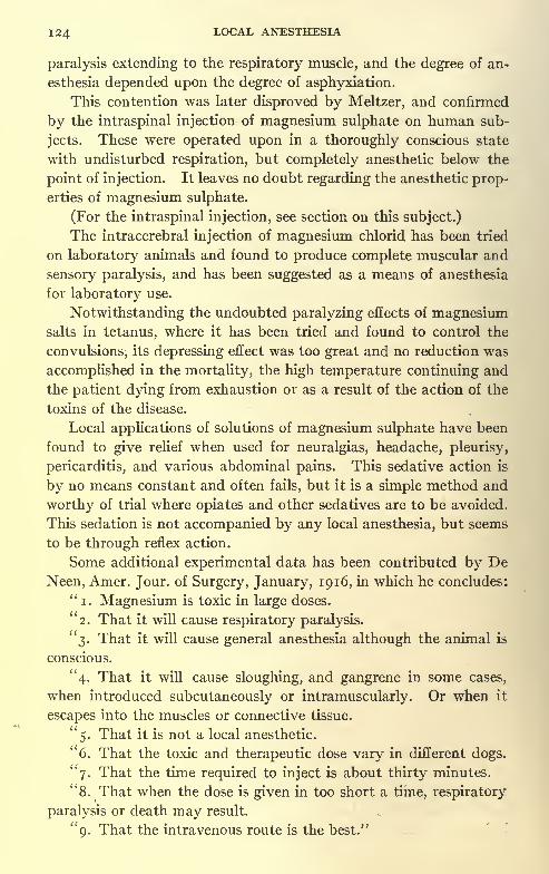
124 LOCAL ANESTHESIA
paralysis extending to the respiratory muscle, and the degree of an-
esthesia depended upon the degree of asphyxiation.
This contention was later disproved by Meltzer, and confirmed
by the intraspinal injection of magnesium sulphate on human sub-
jects. These were operated upon in a thoroughly conscious state
with undisturbed respiration, but completely anesthetic below the
point of injection. It leaves no doubt regarding the anesthetic prop-erties of magnesium sulphate.
(For the intraspinal injection, see section on this subject.)
The intracerebral injection of magnesium chlorid has been tried
on laboratory animals and found to produce complete muscular and
sensory paralysis, and has been suggested as a means of anesthesia
for laboratory use.
Notwithstanding the undoubted paralyzing effects of magnesiumsalts in tetanus, where it has been tried and found to control the
convulsions, its depressing effect was too great and no reduction was
accomplished in the mortality, the high temperature continuing and
the patient dying from exhaustion or as a result of the action of the
toxins of the disease.
Local applications of solutions of magnesium sulphate have been
found to give relief when used for neuralgias, headache, pleurisy,
pericarditis, and various abdominal pains. This sedative action is
by no means constant and often fails, but it is a simple method and
worthy of trial where opiates and other sedatives are to be avoided.
This sedation is not accompanied by any local anesthesia, but seems
to be through reflex action.
Some additional experimental data has been contributed by De
Neen, Amer. Jour, of Surgery, January, 1916, in which he concludes:"
i. Magnesium is toxic in large doses.
"2. That it will cause respiratory paralysis.
"3. That it will cause general anesthesia although the animal is
conscious.
"4. That it will cause sloughing, and gangrene in some cases,
when introduced subcutaneously or intramuscularly. Or when it
escapes into the muscles or connective tissue.
"5. That it is not a local anesthetic.
"6. That the toxic and therapeutic dose vary in different dogs.
"7. That the time required to inject is about thirty minutes.
"8. That when the dose is given in too short a time, respiratory
paralysis or death may result.
"9. That the intravenous route is the best."
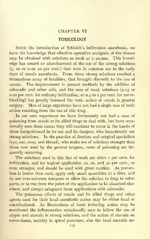
CHAPTER VI
TOXICOLOGY
SINCE the introduction of Schleich's infiltration anesthesia, wehave the knowledge that effective operative analgesia of the tissues
may be obtained with solutions as weak as i : 20,000. This knowl-
edge has caused an abandonment of the use of the strong solutions
(5, 10 or even 20 per cent.) that were in common use in the early
days of cocain anesthesia. From these strong solutions resulted a
tremendous array of fatalities, that brought discredit to the use of
cocain. The improvement in present methods by the addition of
adrenalin and other aids, and the uses of weak solutions (0.25 or
0.20 per cent, for ordinary infiltration; or 0.5 to i per cent, for nerve-
blocking) has greatly lessened the toxic action of cocain in general
surgery. Men of large experience have not had a single case of toxic
action resulting from the use of this drug.
In our own experience we have fortunately not had a case of
poisoning from cocain or its allied drugs to deal with, but have occa-
sionally seen these cases; they will continue to occur in the hands of
those inexperienced in its use and its dangers, who incautiously use
strong solutions. In the practice of dentists and surgical specialists
(eye, ear, nose, and throat), who make use of solutions stronger than
those now used by the general surgeon, cases of poisoning are fre-
quently occurring.
The solutions used in this line of work are often i per cent, for
infiltration, and for topical application 10, 20, and 30 per cent., or
even stronger, and should be used with great caution. As preven-
tion is better than cure, apply only small quantities at a time, and
do not over-saturate tampons or allow the solution to drop to other
parts, or to run from the point of the application to be absorbed else-
where, and always safeguard these applications with adrenalin.
The poisonous effects of cocain and its allied drugs and other
agents used for their local anesthetic action may be either local or
constitutional. As illustrations of local irritating action may be
mentioned the inflammation occasionally seen to follow the use of
alypin and stovain in strong solutions, and the action of stovain on
nerve-tissue, notably in spinal puncture; also the local necrotic ac-
125

126 LOCAL ANESTHESIA
tion of quinin and urea, or the local destructive and inflammatoryaction of carbolic acid. The prolonged freezing by ethyl chlorid will
produce coagulation with destruction of tissue. All these local ef-
fects are discussed in the chapters with the action of these different
agents; here we propose to discuss the general or constitutional action.
In speaking of this toxic action cocain will be taken as the type,
for what applies to cocain is equally applicable to all of its congeners,
with perhaps very slight or inconsequential differences in some few
cases.
Cocain is recognized as a universal protoplasmic poison effecting
all protoplasm, animal and vegetable alike. When gradually ab-
sorbed in toxic doses, acting first as an excitant, paralysis follows after
a more or less brief period of excitement. When injected into the
circulation in toxic doses the stage of excitement is so short as to
escape observation, paralysis taking place almost immediately.It must be remembered that the local anesthetic action of cocain
is the result of a local paralysis of the parts affected; all tissues are
similarly effected by its use, nerves of special sense, motor nerves,
muscle-fiber as well as sensory nerves, and white blood-corpuscles
loose their ameboid movements when in contact with its solutions;
its constitutional or central action is the result of this paralysis uponthe higher nerve-centers.
This paralysis is the result of a definite chemical combination,
and the longer the solution remains in contact with the tissues at
the point of injection the more pronounced becomes this chemical
combination, and consequently the more pronounced the anesthesia 1
(paralysis).
These facts should guide us in its use: first, to keep within the
limits of safety; secondly, to use means to retain it at the point of
injection or application; and, finally, should toxic symptoms arise,
to apply at once such measures (constriction) if possible as will check
or delay further absorption, as well as such other measures as have
been suggested elsewhere to combat this toxic action.
The soluble salts of cocain are absorbed with great rapidity.
They have the power of passing with great facility through nearly
all mucous membranes, so that their absorption is almost immediate
when topically employed on such surfaces as the nose, throat, mouth,
urethra, eye, and rectum, consequently the greater number of cases
of poisoning have resulted from their use in this way.The ultimate fate of cocain after absorption into the body is
somewhat in doubt; it is believed, when slowly absorbed, to be en-

TOXICOLOGY 127
tirely broken up by the body-cells (Moreno y Maiz); when more
rapidly absorbed very small quantities (5 per cent.) have been re-
covered from the urine.
It is believed that cocain once fixed by the body-cells (in combina-
tion with them) is not liberated from these combinations as cocain,
but as constituent products, ecgonin, etc.; Glasenap believes he has
isolated ecgonin from the urine. These derivatives of cocain are
slightly anesthetic and slightly toxic, but much less so than cocain.
As a result of the preceding statements it can be said that cocain
exerts its full anesthetic or toxic action but once, and if exhausted
locally there will be no constitutional reaction.
It can be further stated that that portion of the cocain which is
absorbed and acts upon the general system producing toxic symp-toms, is the excess over that fixed by the tissues locally, consequentlythat much in excess of the amount needed to thoroughly saturate
and combine with the tissue-cells producing in them complete paraly-
sis. In considering this statement it must be borne in mind that on
very actively absorbing srufaces and in very vascular tissues, where
no aids are used to retard absorption, such as constriction or adrena-
lin, that much of the drug is rapidly taken up and transported bythe veins and lymphatics to the central nervous system before a verylimited quantity of it has had time to be fixed by the tissues and act
locally. This and other statements are borne out by clinical experi-
ences in the cases of poisoning by small doses in comparison to the
amount used under other conditions without ill effects. Mattison
reports a case by Knabe where 12 drops of a 4 per cent, solution
given hypodermically to a young girl of eleven years caused death in
less than one minute. Garland reports a death following the appli-
cation of 20 drops of a 5 per cent, solution to the gums. Hundreds
of such cases have been reported, and many cases of idiosyncrasy
showing poisonous symptoms from remarkably small doses; these,
however, have all been in strong solutions, i per cent, and over, and
only serve to emphasize the caution given that when using such
agents, keep well within the safe limits and use only the weakest
dilutions compatible with efficiency; the toxic action will vary in
direct ratio to the strength of the solution and the rapidity of
absorption.
Three-quarters of a grain of cocain is given as a safe average dose,
but this varies within wide limits; less than this amount may produce
poisonous symptoms if too rapidly thrown into the circulation in
susceptible individuals, while many times this amount can be given
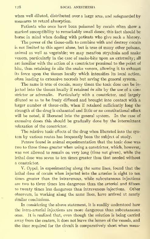
128 LOCAL ANESTHESIA
when well diluted, distributed over a large area, and safeguarded bymeasures to retard absorption.
Patients who once have been poisoned by cocain often show a
marked susceptibility to remarkably small doses; this fact should be
borne in mind when dealing with patients who give such a history.
The power of the tissue-cells to combine with and destroy cocain
is not limited to this agent alone, but is true of many other poisons,
animal as well as vegetable; we may mention strychnin and snake
venom, particularly in the case of snake-bite upon an extremity; all
are familiar with the action of a constrictor proximal to the point of
bite, thus retaining in situ the snake venom, allowing it to exhaust
its force upon the tissues locally which intensifies its local action,
often leading to extensive necrosis but saving the general system.
The same is true of cocain, many times the toxic dose can be in-
jected into the tissues locally if retained in situ by the use of a con-
strictor or adrenalin. Particularly with a constrictor, and largely
diluted so as to be freely diffused and brought into contact with a
larger number of tissue-cells, when if retained sufficiently long the
strength of the drug is exhausted and little or no constitutional effect
will be noted, if liberated into the general system. In the case of
excessive doses this should be gradually done by the intermittent
relaxation of the constrictor.
The relative toxic effects of the drug when liberated into the sys-
tem by various routes has frequently been the subject of study.
Petrow found in animal experimentation that the toxic dose was
two to three times greater when using a constrictor, which, however,
was not allowed to remain on very long (time not given), while the
lethal dose was seven to ten times greater than that needed without
a constrictor.
V. Oppel, in experimenting along the same lines, found that the
lethal dose of cocain when injected into the arteries is eight to ten
times greater than the intravenous, while subcutaneous injections
are two to three times less dangerous than the arterial and fifteen
to twenty times less dangerous than intravenous injections. Other
observers, in working along the same lines, have arrived at nearly
similar conclusions.
In considering the above statement, it is readily understood howthe intra-arterial injections are more dangerous than subcutaneous
ones. It is realized that, even though the solution is being carried
away from the centers, it does not leave the lumen of the vessels, and
the time required for the circuit is comparatively short when meas-

TOXICOLOGY I2Q
ured by the time necessary for cocain solutions to combine with the
tissues, besides the entire volume is delivered at once into the gen-
eral circulation with the returning blood. The subcutaneous injec-
tions are weakened by the action of the tissue-cells outside of the
vessels, and are gradually taken up to be slowly liberated into the
general circulation.
The age and condition of the patient is also an active factor in
considering the toxicology of these drugs; childhood and early youth,due to the highly sensitive and impressionable nervous system, as
well as the influence which psychic impressions may play, are rela-
tively much more susceptible to the toxic influence of these drugsthan are adults; on the other hand, old age, where so often generalanesthetics may be contra-indicated, is particularly favorable to all
local anesthetic procedures. This question of age is considered more
thoroughly under Indications and Centra-indications, as well as
other conditions of the patient which may operate for or against the
toxic action of these drugs. While % gr. of cocain is given as the
maximum safe dose that can be absorbed into the circulation at anyone time, still this dose may be many times exceeded with perfect
safety when largely diluted, diffused over a large area, and slowly
absorbed; the danger of toxicity depends entirely upon the strengthof the solution and rapidity of absorption; we have repeatedly used,
8, 10, or 12 ounces of Schleich's solution No. i, containing 0.96 gr.
of cocain to the ounce, or our solution No. i, containing 1.2 gr. of
novocain to the ounce, when performing very large and extensive
operations under infiltration anesthesia, without ever having seen
any toxic effects; 32 ounces of solution No. i, containing 38.4 gr. of
novocain, was used in doing an extensive lipectomy without any dis-
turbance. Of course, in these procedures some of the solution es-
capes from the tissues through the incisions, and the total quantityabsorbed is in this way somewhat reduced. The precaution men-
tioned elsewhere, of keeping the patient recumbent with head low for
several hours after operation where large doses of the drug have been
used, may again be emphasized here; also when operating upon an
extremity under similar conditions to intermittently relax the con-
strictor rather than removing it entirely upon the completion of the
operation.
Cocain produces a veritable general analgesia as a final stage in
all severe intoxications, but only when the life of the animal is seri-
ously threatened; this is not only of interest to the physiologist,
but to the surgeon as well. (See General Anesthesia with Cocain.)

130 LOCAL ANESTHESIA
The local action of cocain, aside from its anesthetic action, is that
of a vasoconstrictor, producing a decided degree of anemia; it is be-
lieved that its central toxic action is ushered in by similar phenom-ena anemia of the cerebrum and vital nerve-centers, producing at
first a brief period of excitement or irritation, followed by paralysis;
these symptoms may be of mild degree and slow to develop, passing
off without serious results, or appear suddenly and end in a fatal
termination within a few moments, depending upon the size of the .
dose and the rapidity of absorption.
Many operators have tried to prevent or lessen this central toxic
action by adding to the cocain solution various drugs to combat this
central vasoconstriction, or by using a combination of anesthetic
drugs ;have hoped to be able to reduce the quantity of cocain needed
to a point well below the toxic dose, even when large quantities of the
solution was necessary. Thus, Stuver advised a mixture consisting of
one part of cocain to two parts of antipyrin; Gluck advised carbolic
acid and Parker resorcin. To combat the vasoconstriction, Thomasand Guitton have recommended the addition of nitroglycerin. All
these combinations are objectionable from many points, some of
them being irritant and others fully as toxic or depressing. Whatis wanted is to simplify rather than complicate the mixtures. Theuse of vasodilators having a local action is especially to be avoided,
for many reasons the local anemia is desired and we try to intensify
it by the use of such aids as adrenalin or by the use of cold; this
local anemia, besides increasing its local action, lessens or preventsits central or constitutional action by prolonging the sojourn of the
drug in the tissues^ where its action is weakened or entirely exhausted.
It is also quite doubtful that such drugs are of any value in com-
bating the toxic effects; what is better is to keep well within the limits
of safety, and, should toxic symptoms occur, to meet them by other
more effective means.
The symptoms of mild intoxication may be evident in loquacity,
laughing, or singing, later slight nausea, vertigo, faintness, thoracic
oppression; as the severity of the symptoms increase, the pulse which
at first is stimulated becomes rapid and weak, respiration may be
oppressed or quite rapid, great mental excitement and anxiety mayoccur, the patient becoming very restless with twitching or trembling
of the muscles, these symptoms indicating the threatening onset of
convulsions; at times the stage of excitement may manifest itself bymaniacal delirium, the patient becoming violent and uncontrollable;
convulsions with unconsciousness may now supervene and be fol-

TOXICOLOGY 131
lowed by death. During the onset of symptoms the pupils are usu-
ally dilated, but may at times be contracted. The order and char-
acter of symptoms may vary greatly in different individuals, the
stage of excitement may be absent, unconsciousness coming on at
once, followed by convulsions. In some cases where the toxic dose
is very large, or the patient is particularly susceptible, death mayoccur almost immediately from cardiac inhibition.
The onset of mild symptoms, such as loquacity or faintness, are
usually controlled by having the patient maintain the horizontal
position or by lowering the head of the table; this position should be
continued for half an hour or longer following the disappearance of
all symptoms. The use of drugs to combat poisonous symptomsmust be largely symptomatic. If syncopy occurs, or the heart be-
comes weakened, strychnin and digitalis should be used, preferably
given by hypodermic, while ammonia or amyl nitrite are given byinhalation. For nervous excitement or convulsions H. C. Woodrecommends chloroform by inhalations, but it would appear that
ether should be better, particularly if it be proved that the central
action is associated with the same vasoconstriction and anemia that
takes place in its local field of action;inhalations of ether, due to the
tremendous congestion which it produces in these parts, should proveof great value, besides stimulating vasomotors, heart and respiration
and controlling the convulsions; nitrous oxid would be equally as
valuable; amyl nitrite, while producing the same congesting effect,
would not exert the same controlling influence upon the convulsions.
Regarding the use of ether a very interesting report has lately
been published in the "Journal American Medical Association" byDr. J. E. Engstadt, which we quote in part as follows:
"In the first few cases I was called on to treat, strychnin and
morphin in combination were used with a marked benefit. But, as
cases kept multiplying, I found the action of these drugs too slow, and
I decided that there must be something to counteract the poison
more rapidly when life was in extreme danger. It was necessary to
find a remedy that could be administered at any time and be in-
stantaneous in its action. I soon found ether to be the required drug.
This was administered as ordinarily given to produce surgical narco-
sis. Ether stimulates the vasomotor system, is a tonic to the heart
muscles, stimulates the action of the respiratory centers of the brain
and of the pneumogastric nerve, and increases the pulmonary circu-
lation in the first stages. While cocain inhibits the action of the
heart, especially on the right side, it has also a marked inhibitory

132 LOCAL ANESTHESIA
action on the respiratory centers of the brain. Death may occur
from feeble respiratory movements of the so-called Cheyne-Stokes
type or asphyxia.
"To me ether has proved extremely valuable. It has saved what
seemed hopeless cases. It stimulates the heart and the respiratory
system almost instantly. The pulse becomes fuller at once and of
normal tension. The marked mental excitement is allayed as the
patient goes under the influence of the ether and the effect of the
poison rapidly disappears. The individual regains consciousness as
soon as the effect of the small amount of ether has disappeared."
To get the best results, the anesthetic is administered only to the
degree of mild surgical narcosis, or, at times, even less than this.
A mask should be employed and the ether given by the drop method.
This is all-important. Given by the old method, the ether would
only add to the danger of asphyxia by excluding air from the venous-
blood engorged lungs.
It is quite interesting to compare the sedative and controlling
influence of ether upon the symptoms of cocain intoxication, as re-
ported by Engstadt, with the sedative effects of a hypodermic of
cocain upon animals coming out of ether narcosis (although not
operated upon), as reported by Kast and Meltzer in the chapter on
Abdominal Operations.
It was thought for some time that adrenalin lessens the toxic
action of cocain upon the central nervous system, but upon later
investigation this has been found to be in error, and that after the
cocain once enters the general circulation the use of adrenalin mayincrease its toxic action
;this may be understood when it is considered
that both produce vasoconstriction. Adrenalin greatly lessens the
likelihood of development of toxic symptoms by retaining the cocain
in the field of injection, and greatly intensifying and prolonging its
action there, where it is largely exhausted by action upon the tissues,
but after it has once entered the general circulation the adrenalin
may prove a distinct disadvantage.
The observations of J. M. Berry on this subject are particularly
interesting. He concludes his remarks as follows: "In the use of
adrenalin-cocain care should be exercised not to inject a toxic dose
of the latter, for not only does the adrenalin fail to protect the body
against the toxic doses of cocain, but it seems to enhance the toxic
action."
Thriss found, by experiments on cats, that cocain and adrenalin,
when injected into the lumbar sac, had the same toxic effect as when

TOXICOLOGY 133
cocain was used alone. Miles and Muhlberg, in a series of experi-
ments upon animals for the study of the comparative value of adrena-
lin and other substances upon vasomotor depression artificially
produced, conclude that "adrenalin subcutaneously is indicated on
theoretic ground for the vasomotor collapse following cocain or
chloroform poisoning, etc." Here, however, it is to be used to com-
bat a symptom, and not in any sense as an antidote.
Braun, in his book on local anesthesia, and elsewhere in the
"Archiv. f. klin. Chir.," vol. Ixix, does not concur in these views, but
believes that adrenalin lessens the central toxic action; on the other
hand, Petrow found that adrenalin did not seem to exert any great
influence upon the toxic action.
In considering the toxic action of local anesthetics it should be
borne in mind that the too free use of adrenalin may excite symptomsin susceptible individuals notably in nervous conditions and high
blood-pressure, these symptoms are usually a feeling of oppression
in the chest, a fulness and throbbing in the head with headache, the
face is usually flushed and the pulse tense; quite a different picture
from cocain poisoning. (See chapter on Adrenalin.)
The toxic symptoms of cocain poisoning are due to its paralyzing,
action on the respiratory, cardiac and vasomotor centers as well as
some influence being exerted by its direct action upon the cardiac and
vasomotor muscles due to its universal action on all protoplasm.
Opium, its alkaloids and all narcotic drugs antagonize this action by
dulling the sensibility and response of these centers and are therefore
true antagonists. This action is, however, best exerted as a prophy-
lactic, or preventative by their preliminary use before the injection
of the anesthetic when it does certainly permit the use of much larger
doses of the anesthetic and in this way acts as a direct antagonist.
Mild toxic symptoms are favorably influenced by morphin or panto-
pon administered hypodermically after their onset but with severe
symptoms, except for a slight lessening of the convulsions its bene-
ficial influence is not so marked.
Strychnin by its stimulating effect upon all nerve-tissue should
certainly antagonize the paralytic influence of cocain and by its in-
fluence help to keep the flagging respiratory centers at work and sup-
port the blood-pressure until the crisis has been passed, while these
and other means may be of benefit, ether by light inhalation unques-
tionably meets all the indications and except in absolutely lethal
doses is usually able to control the symptoms.The use of intravenous salt injections, as recommended for all

134 LOCAL ANESTHESIA
poisons, may be tried for cocain when time permits. The diluting
effects of a pint or quart of normal salt solution should have a favor-
able influence in weakening the toxic strength of the drug, as well as
stimulating the heart and favoring more rapid elimination.
Carlo Bozzo found that the minimum fatal dose of cocain for dogs,
injected hypodermically, was 0.025 gram per kilo, without infusion,,
but when infusion was resorted to the minimum fatal dose rose to
0.03 per kilo; he concludes that besides favoring rapid elimination
it retards the absorption of further quantities of the drug, owing to
the fulness of the blood-vessels.
The development of toxic symptoms from the use of cocain maybe considerably delayed, or, after apparent recovery, the patient mayagain sink into a state of syncopy or collapse. As an illustration
of this condition, the author was called to see a case of convulsions in
a young man twenty-four years old, and obtained the following his-
tory: Two hours previously he had two molar teeth extracted by the
use of local injections of cocain; it was necessary for the dentist to
make repeated injections before securing the necessary anesthesia;
the extractions were finally painless; following the procedure there
was slight nausea and some vertigo, for. which he was given a drink of
whisky, when he appeared to recover, and was able to go home in the
street cars a distance of about twenty city blocks; after arriving at
home he felt uneasy and restless and sat in a chair in his gallery; he
was found sometime later by his family, still in the chair, but in a
state of convulsion; when the writer arrived he had had several such
seizures, at intervals of about fifteen or twenty minutes, becoming
quite uncontrollable and violent just before their onset; as I had
learned the nature of the case I went prepared. The patient was
frantic upon my arrival, requiring the combined efforts of several of
the family to hold him;his pupils were widely dilated, face very pale,
studded with large drops of perspiration, and his expression one of
terror; the respirations were rapid and shallow, the pulse small, feeble,
and rapid. We at once threw the patient across the bed, where he was
held while I administered ether by the drop method to the point of
superficial anesthesia;with the beginning of the ether administration
there was an immediate change, the respirations deepened, the pulse
slowed and became fuller, the color returning to the face, the muscles
which had been tense soon relaxed;the entire picture was changed, the
patient presenting the usual appearance of one under light ether
narcosis; this was kept up for about fifteen or twenty minutes and
gradually suspended. The feet and hips were then elevated upon

TOXICOLOGY 135
pillows until the head was quite dependent; this position was main-
tained while the patient came from under the influence of the ether
and for sometime afterward. I remained with him for about one-
half hour after he appeared normal to make sure that there would be
no return of the symptoms, but beyond a feeling of exhaustion there
was no further disturbance. Next day he appeared quite normal;later examination of his kidneys and other organs failed to show any-
thing abnormal.
Parsons reports that following collapse in spinal anesthesia light
ether inhalations have proved of immediate and marked benefit.
J. K. Pedley reports a case related by Dr. B. Christensen, in which a young womanaged twenty-eight had the root of a tooth extracted under novocain anesthesia and died
several hours later: "At 1.45 P. M. I injected about i% c.c. of a little less than a 2 percent, solution of novocain-suprarenin (Tab. B. Containing novocain, o.i gram, supra-
renin, 0.00045 gram), but as the anesthesia was insufficient ten minutes later I injected
some more, in all about 3 c.c. The patient felt rather unwell afterward and was advised
to remain in the office and lie down." From 4 to 4.30 she was sitting up and chatting; at 5.30 she had so much improved
the doctor left her; at 6.30 his wife noticed her and she seemed to be in a natural sleep;
shortly later noticed her breathing rather deeply and on examination found her almost
pulseless; gave camphor by needle.
"At 7 P. M. doctor returned and performed artificial respiration, at 8 p. M. was
removed to the hospital, and died one hour later, without regaining consciousness, with
symptoms of edema of lungs."
Cases of this kind would be reported in great number, but the
above will suffice for an illustration.
To recapitulate: With the onset of the first symptoms immedi-
ately place the patient in a recumbent position and lower the head; if
the operation has been upon an extremity apply a constrictor proxi-
mal to the field; give ammonia or amyl nitrite by inhalation; if the
case seems severe, lightly narcotize with ether by the drop method;use digitalis or oil of camphor by needle if the heart is weak; in severe
cases use infusions of normal salt solutions if convenient; should the
respiration cease artificial respiration should be resorted to and per-
sisted in as long as the pulse or heart is perceptible or even longer, as
there may be a chance of resuscitation. Legrand reports a case where
it was necessary to continue artificial respiration for five hours before
the function became normal. In such cases, where the facilities are
at hand, use the Meltzer-Auer intratracheal intubation with forced
respiration.
Some of the earlier cases of poisoning may have resulted from the
presence of certain impurities in the preparation used, and this mayeven occur now, though not so likely, as the methods of manufacture

136 LOCAL ANESTHESIA
and safeguards placed around it have so far improved as to reduce
this likelihood to a minimum.
Some of these impurities have never been determined, but a few
have been isolated and positively identified;two of these, which in the
past have been most likely to occur, are isatropylcocain and cinnamyl-
cocain, both highly toxic; as these impurities act in different ways,some of the peculiar toxic symptoms reported may be accounted for
in this way.

CHAPTER VII
ADRENALIN
ADRENALIN, the therapeutic constrictor, is known under several
names as suprarenin, paranephrin, epirenin, eudrenal, and epinephrin,
but probably is better known in this country as adrenalin, the name
proposed for it by Takamine. Adrenalin and the synthetic prepara-
tions of arterenol, homorenon, and suprarenin synthetic are powerful
local and constitutional vasoconstrictors, and, except the latter, are
obtained as an extract from the suprarenal glands of animals.
The introduction within the last ten years of this highly valuable
and wonderful agent has proved a great boon to many departmentsof surgery, and has given a decided impetus to all local anesthetic
procedures. Next to the possession of safe local anesthetics and the
Schleich infiltration method (with dilute solutions), there is no single
agent or factor which has so fostered and encouraged the developmentof local anesthesia, and enabled surgeons to enlarge the field and
broaden the scope of all purely local procedures.
This has been made possible by the unique power this agentexercises of producing vasoconstriction, and retaining within the tis-
sues the dilute anesthetics which both intensifies and prolongs their
action for a period of time usually well beyond that required for the
performance of any ordinary operation; and this without injury to
the tissues.
To Prof. Heinrich Braun, of Zwickau, Germany, from whom we
have quoted quite liberally in this volume, is largely due the credit of
first introducing, developing, and perfecting the use of this agent in
local anesthesia. Already a staunch advocate of local anesthesia, in
which field he has been a constant worker, he was quick to see the
advantages of a combination with adrenalin and early advocated its
use, and it is largely due to his efforts in this direction that adrenalin
soon became so popular an adjunct in all local anesthetic solutions.
Adrenalin has become almost indispensable to the surgical special-
ists, particularly in the nose and throat, where it is in constant daily
use, both for purposes of examination as well as for operations; in
examinations it greatly facilitates the procedure when swabbed or
sprayed on turgid and congested mucous passages, causing them
to shrink and permit free access and inspection of the deeper parts.
Nearly all operations upon these parts are greatly facilitated and
13?

138 LOCAL ANESTHESIA
simplified by its use, which renders the field almost entirely bloodless,
greatly expediting the work; and enabling the operator to undertake
many operations in the office without assistance under local anesthe-
sia and without the loss of blood. These operations formerly were
done only in institutions under general anesthesia, with considerable
loss of blood and a much more complicated and tedious technic.
The history of the early work which led to the discovery of adren-
alin is not without interest, but only brief mention will be made of it
here in expressing our gradtitude to those who have given us this
valuable agent. Early anatomists observed that the juice of the
medullary susbstance of the suprarenals darkened upon exposure to
light and air; they called this substance atra bibis. It was not, how-
ever, until the nineteenth century that the color change was under-
stood. Vulpian in 1856 noticed that this juice, when brought into
contact with ferric chlorid and iodin, turned emerald green and then
rose carmin. These reactions were characteristic of this organ, and
led to the opinion that the gland contained a physiologic substance.
Pellacani, as early as 1879, performed a very interesting series of
experiments in Foa's laboratory by injecting an extract of the fresh
glands into various animals. Mattel later repeated Pellacani's
experiments, and came to the conclusion that his results were those of
septicemia rather than from any special active principle of the gland.
In 1883 Foa and Pellacani again took up the study and published
some interesting results, and it would seem, after reading the original
papers of these early writers, that more credit should be given them,
for they certainly describe symptoms of poisoning which are now
recognized as characteristic of adrenalin.
Other investigators followed up this line of work: Krukenberg
(1885), Marino-Zuco (1888), Guarnieri and Marino-Zuco (1888).
The synthetic preparation of this agent began to be fore-
shadowed as early as 1893 by the work of Dr. Zierzgowski, whoshould be given credit for pioneer work in this line.
Gluzinski (1895), Moore (1895), Dubois (1896), and Vincent
(1897) were other workers with these glands. In 1896 Frankel sug-
gested sphygmogenin as a name for the active principle of the gland;
this was, however, later shown to be a mixture.
The work that had been done by the earlier investigators preparedthe way for that which was to follow; the subsequent investigations
began to yield better results in isolating the active principle of these
glands. Dreyer had also been able to isolate the active principle of
the suprarenals in the veins coming from these glands.

ADRENALIN 139
Batelle (1902) claimed to have obtained a purer adrenalin than
that produced by Takamine's method.
In the meantime enough data had accumulated to attempt the
synthetic preparation, although, as has been mentioned, Dzierzgow-ski in 1893 had done creditable work in this line. Stolz succeeded in
producing the so-called dl-product, which was found to be somewhat
different from the natural base. This was found by Flacher to be due
to dextro and levo components which he was able to separate, and
found that the levo-adrenalin was identical with that obtained from
the glands. The action of this principle began now to be extensively
studied, and Cybulskilgo pointed out that concentrated doses were
much more poisonous than when diluted; and Gluzinski determined
that intravenous doses were more toxic than subcutaneous. The
physiologic investigators began now to become more numerous, and
the literature of this subject is filled with the names of prominent
investigators.
In 1895 Oliver and Schafer, and Szymonowicz and Cybulski
working independently, discovered the action of certain suprarenal
bodies upon the circulation.
Abel in 1897, working along the same lines, was able to isolate a
body which he called epinephrin.
In 1900 Von Furth described suprarenin, and in 1901 Takamine
and Aldrich isolated a principle which Takamine called adrenalin.
It is probable that these two last substances are identical, while
epinephrin seems to be different both chemically and physiologically.
Abel regards adrenalin as an epinephrin hydrate.
Investigations regarding the relative merits of the synthetic
preparations (dl-adrenalin, known commercially as arterenol, and the
ethyl-amino-aceto-catechol, known as homorenon) conclude that the
former is about two-thirds as active as the natural product and the
latter about one-eightieth as strong.
The structural formula, which for a time was somewhat in dispute,
now seems definitely settled by both analysis and synthesis and is a
most precise definition of a pure chemical compound and is defined as
the levo-rotatory isomer of the formula
HO -C
HO - C CH
HC C - CH(OH) - CH2- NH(CH3)vC
H

140 LOCAL ANESTHESIA
The dextro-rotatory isomer of this formula which is only found in
the synthetic product, is practically inert. From a careful analysis
of this formula, it is seen that the molecule contains groups that
characterize it as an amine base, an alcohol and a phenol, observations
which explain its chemical behavior.
Adrenalin is found naturally in the medulla of the suprarenal
glands of all vertebrates. The adrenalin of commerce is obtained
from beef cattle, but there is no reason to doubt that the active sub-
stance is the same from whatever animal obtained. The adrenalin of
commerce is substantially pure containing but a small portion of
foreign matter and is a nearly white microcrystalline powder.When stored under proper precautions, it should keep for many
years perhaps permanently. Air, heat, light, moisture, ammoniaand certain other gases, affect it injuriously. Under the combined
influence of air, heat and moisture, it decomposes rapidly. It
should, consequently, be preserved in absolutely dry colored bottles;
sealed air tight, and if to be kept for an indefinite time, the air should
be thoroughly displaced by some inert gas.
The pure adrenalin base is slightly soluble in cold water, slightly
more so in warm water. Certain salts increase this solubility: thus
solutions of borates render soluble a large quantity of the adrenalin
and prevent the precipitation of solutions of its salts by alkalies. Astrong aqueous solution of chemically neutral adrenalin chloride will
dissolve an appreciable quantity of the adrenalin base.
Due to its phenol characteristics, it forms water soluble compoundswith fixed caustic alkalies, but not with their carbonates or with
ammonia. From strong solutions of most of its salts, the adrenalin
base is partly precipitated by hydroxides or carbonates of strong
alkalies, including ammonia and redissolved by an excess of the fixed
caustic alkali only.
Due to its amine function, adrenalin forms definite salts with
acids, usually very hydroscopic and difficult to preserve in dry form.
Most of these salts are quite soluble in water and alcohol and can be
made quite stable for ordinary uses; but are not very soluble in sol-
vents other than water and alcohol. For most purposes we are,
therefore, limited to the use of its salts in aqueous or alcoholic
solution.
In the preparation of ointments, it is best to first prepare a con-
centrated aqueous solution in dilute hydrochloric acid: this is incor-
porated with sufficient lanolin which takes up large quantities of
water, after which any other ointment base may be added. In case

ADRENALIN 141
the fatty base is soluble in alcohol, it may at times be desirable to first
dissolve the adrenalin in sufficient alcohol, when the ointment maythen be added.
Commercially obtained, adrenalin chlorid contains i part per1000 of adrenalin chlorid in physiologic salt solution, containing
0.5 per cent, of chloretone to preserve it and is saturated with carbon
dioxid used in its manufacture to expel the air. It has a faintly acid
reaction, smells and tastes of chloretone and sodium chlorid. Whenfresh, it should be colorless.
When properly prepared and bottled, it should remain colorless
and retain its activity for a long period. When exposed to the air
and light, color changes and deterioration begins. It is therefore
best kept in small colored bottles. When opened for use, it should be
immediately stoppered and put back in a dark place : only the quan-
tity to be immediately used should be removed.
The color change is a fair index of its deterioration. It first be-
comes pink, then red, finally brown and a brown precipitate settles
out. Experiments have been undertaken to determine the degree of
deterioration based on these color changes, which might be used as a
guide in its use.
The following is approximately reliable: When pink, practically
no change is noticeable. When red, it is appreciably weakened,which may amount to 10 to 20 per cent, of its strength. When brown
with a precipitate, it should not be used, although may still retain
some activity.
When used for surgical purposes during these color changes and
injected within the tissue, it may be used as long as the color change is
no greater than a light red; but when it becomes brown, it should
never be used, as it is practically inert as a vaso-constrictor and mayproduce decided irritation.
The following chemical test given by Beckwith may at times be
useful :
"A rough qualitative test, to show the presence of active adrenalin
in the commercial solution of adrenalin chlorid of the composition
already stated, is based on its conduct with ferric chlorid. As youare aware, ferric chlorid gives striking and more or less characteristic
color reactions with many of the phenols. Catechol, the parent
phenol of adrenalin, gives in dilute aqueous solution with a very little
dilute ferric chlorid solution, a brilliant green color, which upon care-
ful addition of very dilute alkali, passes through a series of color
changes from bluish-green to purple-red. Under like conditions,

142 LOCAL ANESTHESIA
adrenalin acts similarly. The catechol nucleus is responsible for this
reaction, so that it is not peculiar to adrenalin. If, however, dilute
solutions of catechol and adrenalin chlorid be treated with a little
very dilute ferric chloride solution without subsequent addition of
alkali, and if the two solutions be allowed to stand in the air for some
minutes, a difference in their behavior will manifest itself. In the
case of catechol, the green color persists; in that of adrenalin, it
changes slowly to pink or red. While the test is not absolutely final,
it is fair to conclude that a commercial solution of adrenalin chlorid
retains some activity when a sample, highly diluted, gives with a dropof very dilute ferric chlorid solution a green color changing soon to
pink or red. Many foreign substances interfere with the test, so that
it may not be applicable to adrenalin in mixtures."
Solutions of adrenalin chlorid are injuriously affected by manysubstances. Besides air and light, alkalies oxidizing agents and iron
are particularly to be avoided, while nearly all acids are harmless.
The alkali contained in glass bottles may at times be a source of de-
terioration and should be guarded against by obtaining as nearly as
possible an alkali-free glass and by keeping the solution acid which
should always contain ^{QQ of i per cent. C. P hydrochloric acid.
Stoppers for bottles should preferably be of glass and when cork
is used, it should be protected by some impervious material.
Physiologic Action. The part of the physiologic action of adren-
alin which concerns us here is limited to its action upon the circulation
in any given operative area when locally used. For a consideration
of its highly interesting action in other spheres of its influence, particu-
larly its constitutional effect in its many clinical uses, the reader is
referred to the various articles which deal with this subject. (See'
especially Crile, Boston Med. and Surg. Jour., March 5, 1903, and
Amer. Jour. Med. Sciences, April, 1909; Miles and Muhlberg, Cleve-
land Med. Jour., Dec., 1902; also Winters, Lancet, June, 1905, and
others.)
Only such clinical illustrations will be mentioned here as will serve
to emphasize its great power as a vasoconstrictor, and, under certain
conditions, a stimulant to all smooth muscle-fibers.
The action of adrenalin in constricting the blood-vessels is both
local and constitutional; the latter effect is quite general throughoutthe body; its local effect is best studied when injected locally in dilute
solutions, when it produces a high degree of anemia of marked dura-
tion; that portion of the drug absorbed exercises a constitutional effect.
Lehman found that by injecting solutions of adrenalin into the liver

ADRENALIN 143
of experimental animals he was enabled to excise large sections of this
organ without loss of blood;this marked anemia lasted thirty to forty
minutes and was not followed by- secondary hemorrhage. This
vasoconstrictor action does not seem to depend upon any influence
upon the vasomotor nerves, but is no doubt due to direct action uponthe smooth muscle-fibers in the vessel walls, as shown by the follow-
ing experiments. Crile was able to keep the heart of a decapitated
dog acting for over ten hours by the action of adrenalin and saline
solution upon the heart and blood-vessels, and in sufficient dosageto be able to produce a marked rise in blood-pressure even when
the vasomotor center was proved to have been exhausted (complete
shock) ;the same result was produced when the center was cocainized
or had previously been destroyed; it also occurred after the division
of both vagi and both accelerantes when the animal was under the in-
fluence of curare. It was also noted during these experiments that
adrenalin was capable of constricting the blood-vessels after the circu-
lation had ceased.
Animals killed by asphyxia, and apparently dead for periods upto fifteen minutes, were restored to conscious life again by artificial
respiration and the simultaneous injection into the jugular vein of
adrenalin and salt solution.
The circulation and respiration of dogs electrocuted by a shock
of 230x3 volts of an alternating current were again re-established by
injecting adrenalin solution into the circulation.
During these experiments it was determined that adrenalin was
rapidly oxidized by the solid tissues of the body as well as by the blood.
Animal experimentation conducted by writers under slightly dif-
ferent conditions showed similar results.
From these and similar experiments it is concluded that the action
is a direct one exercised upon the smooth muscle-fibers in the vessel
walls; all smooth muscle-fibers seem influenced in a similar way,
though not always to the same degree (Jacoby and Schafer). It is
found to exert a marked influence upon the uterine muscles, as illus-
trated in a case of Cesarean section operated on by Bogdanovics, in
which, after the delivery of the child, the uterus was found flabby and
inert. The uterine wound was first closed and i c.c. of a i : 10,000
solution of adrenalin injected into the uterine walls at four different
points ;this at once excited muscular action and the contracted uterus
became as hard as stone.
The blood-vessels of the different organs are not all influenced to
the same degree ;the action on the vessels of the skin is most marked,

144 LOCAL ANESTHESIA
less so in the gastro-intestinal tract and bladder, and hardly at all in
the vessels of the lungs (Langley). The urine of animals injected
with large doses of adrenalin is capable when injected into other
animals of raising the blood-pressure; but it would seem that this
agent is very largely destroyed in the body, very little of it being ex-
creted. Ott and Harris, Meltzer and Auer found that by mixing
strychnin with adrenalin before injection into frogs the toxic action
is both delayed and diminished; this observation is of great practical
value in local anesthesia, as will be presently pointed out.
A similar favorable action of adrenalin has been noted in the treat-
ment of snake-bite in 3 cases recently reported by Drs. Hooker, Men-
ger, and Ferguson, all occurring in the state of Texas. In 2 cases the
snake was a rattler, in the other case a moccasin. All bites were uponthe extremities, and occurred from one to two hours before treatment.
Various procedures were resorted to scarification or incisions into
the wound, injections of permanganate, with the use of a constrictor
reported in i case but all were treated with adrenalin in addition,
injected into the tissues near the site of the bite. All cases recovered
in a short time without any notable local or constitutional after-
effect; the adrenalin was largely given the credit for their favorable
termination. It is probable that in these cases the adrenalin,
through its vasoconstriction, retains the snake venom in the parts
locally until it is largely oxidized or destroyed by action upon it of the
tissues.
Dr. K. C. Bose, of Calcutta, gives his experiences in the treatment
of enlarged spleens, where he claims invariably satisfactory results.
Tremendous enlargements have yielded to 5-drop doses three times a
day, continued for a period of several weeks. Interesting and some-
what similar experience was reported by Dr. Tarry, of Long Beach,
Miss. Another practical clinical application of adrenalin, which
illustrates in a striking way its local action upon the vascularity or
congestion of a part, is readily seen in urethral stricture, particularly
of the deep urethra; tight strictures of these parts, when it seems al-
most impossible to pass even a filiform, if first treated with adrenalin
(about 5 to 10 drops of a 1:5000 solution), deposited in the urethra
with an urethral instillator just in front of the structure and allowed to
remain about ten minutes, will often so relieve the congestion of the
parts, as to easily permit the passage of a moderate-sized instrument.
Further clinical applications could be enumerated in great numbers,
but the above will suffice to illustrate its highly valuable local action
as a vasoconstrictor.

ADRENALIN 145
Before, however, concluding these illustrations a few remarks may"be made regarding its highly beneficial and useful action in certain
rectal conditions, notably hemorrhoids. In this condition, when the
parts are badly swollen and congested, which increases their irritabil-
ity, marked relief may be obtained subjectively and objectively bythe use of adrenalin, either alone or combined with other indicated
agents in ointment form, this relives the congestion, causing the parts
to shrink and assume a more normal aspect.
In all operations upon these parts when they are badly congestedand bleed freely, either under general or local anesthesia, the injection
of adrenalin solution is always of decided benefit; under general an-
esthesia the injection of a few syringefuls of a solution of 10 dropsto the ounce distributed in the field will prove of decided aid.
Hurter and Richards, as well as others, have demonstrated that
this agent in large doses or when repeatedly used will produce a
glycosuria.
Vincent claims that this agent is a muscle poison, death resulting
from the use of large doses by paralysis of respiration with small
weak pulse, which follows a short period of high blood-pressure.
The dose of adrenalin should be carefully considered, as large
doses are very dangerous; the greater the concentration the greater
the toxic action. In very dilute solutions a much larger quantity can
be safely given; the toxic dose is naturally much smaller when given
intravenously than when subcutaneously administered.
Batelle, Bouchard, and Claude give the toxic intravenous dose
for rabbits as o.oooi to 0.0002 gram per kilo, while the subcutaneous
toxic dose is 0.002 to 0.02 per kilo. Batelle states that the toxic dose
subcutaneously is forty times greater than the intravenous.
The degree of action of this drug depends entirely upon the
strength of the solution. While a decided influence is obtained by
remarkably weak dilutions, the use of stronger solutions will abso-
lutely obliterate the lumen of vessels the size of the palmar digital
arteries and even larger, so that not a drop of blood will escape from
their cut ends while the adrenalin remains in action.
It is, however, never necessary for our purposes to use strong
solutions, as a very decided influence is produced by very dilute solu-
tions. Moore and Purinton found that the blood-pressure of dogswas noticeably increased by doses as small as 0.000,000,245 to
0.000,024 gram of the extract per kilo of dog weight.In animals experimented upon with toxic doses, after a short
initial rise of blood-pressure, there is a collapse of the vasomotor

146 LOCAL ANESTHESIA
system with small rapid and feeble pulse, paralysis of the extremities,
tonic and clonk spasms, opisthotonus, mydriasis, rapid respiration,
edema of the lungs, anemia of the abdominal organs, and death
usually from paralysis of respiration.
Its continuous injection in small doses produces in animals ex-
perimented upon calcareous degeneration of the heart, aorta, and
great vessels with glycosuria.
The experiments carried out by Braun with adrenalin upon him-
self during 1902 are given in -his "Die Lokal Anesthesie," and are as
follows: He injected under the skin of his forearm adrenalin solu-
tion i :iooo in increasing doses; with a little over % c -c. constitutional
symptoms were noticeable. He states: "Five minutes after in-
jection I had a feeling of oppression in the breast, cardiac palpitation
with quickened and deepened respirations; the number of heart-beats
rose from 64 to 94 per minute. I was compelled to lie down, al-
though after one and a half minutes the symptoms disappeared ;there
was no glycosuria. When I diluted the adrenalin solution with ten
times the quantity of normal salt solution, I was able to increase the
quantity injected to i c.c. before any effect was observed."
The pronounced effect exercised by adrenalin in delaying the
absorption, and consequently the constitutional action and excretion
of any substances or drugs injected into the tissues in combination
with it, is best studied when the adrenal in is used with some agentlittle or not at all affected by its passage through the system. This
action is strikingly illustrated by the following experiment of Klappwith the use of milk-sugar, first carried out as a control experimentwithout adrenalin, and later repeated under identical conditions with
the addition of adrenalin.
A dog was injected subcutaneously in the region of the back with
10 c.c. of a 6^ per cent, milk-sugar solution, and the excretion of the
sugar through the urine studied from hour to hour. Three dayslater the same quantity of the same solution, with the addition of 2
drops of adrenalin i :iooo, was similarly injected and the excretion bythe kidneys studied in the same way.
A study of Fig. 2 will give in a schematic way the results of this
experiment. It will be seen at a glance that the maximum excretion
of sugar without adrenalin, as indicated by the heavy line, begins
immediately after the injection and reaches its maximum intensity
within the first hour, when it rapidly falls off and entirely ceases
after the sixth hour, by which time a total of 0.569 gram had been
recovered from the urine.

ADRENALIN 147
The dotted line indicates the absorption and excretion of the
sugar under the local influence of adrenalin, and presents a striking
and interesting contrast to the former in the following points : Duringthe first hour, in which the maximum is excreted without adrenalin,
absolutely none was recovered when the . adrenalin was used, the
excretion not beginning until the commencement of the second hour;
from this time it slowly increases, reaching its maximum at the end of
the fifth hour, and that at this maximum point of excretion the
amount was one-half that recovered during the interval of maximumexcretion without the use of adrenalin; further, that the excretion is
prolonged over a very much longer period, as traces were still found
fframm.0,20
0,15
0,10
0,05
0,00

148 LOCAL ANESTHESIA
occurring in the course of an operation when the adrenalin is the
disturbing agent.
SURGICAL USES
In the strengths ordinarily employed its use is not followed by
any after-reaction; there is not hyperemia, but the tissues gradually
resume their normal vascularity, and there seems to be no retarding
or injurious action upon the healing of wounds; w;hen slightly more
than the dose to be recommended has been used, it has seemed to the
writer that there was more after-pain in the wound than would have
been the case under other conditions associated with a reactionary
hyperemia. If an excessive dose is used there follows after the
primary anemia, which is marked and prolonged, a vaso-motor
paralysis during which time the vessels remain dilated with openmouths and secondary hemorrhages may occur. Following the use
of excessive doses the resulting anemia may be so profound and pro-
longed as to lead to death of the tissue with sloughing.
As ordinarily used by the author for infiltration, the dose
should never exceed 5 drops of a i riooo solution to the ounce of the
anesthetic fluid, and less than this, 2 or 3 drops, will be found amplysufficient.
It is well in all large operations to estimate about the total quan-
tity of the anesthetic solution likely to be needed say, for a rather
large hernia, 4 to 6 ounces may be required ;this is measured off and
put in a convenient receptacle, and to this total quantity about 15
or 20 drops of the adrenalin solution (i : 1000) is added; in this waywe know exactly how much is being used and the safe dose need not
be exceeded; we obtain by these weak dilutions all that can be ac-
complished by stronger doses, and without noticeable constitutional
action or other ill effects. As the action of adrenalin is immediate it
is not necessary to wait to obtain this influence, but a delay of five
to ten minutes is usually necessary after the infiltration to obtain the
full anesthetic effect of the drug used. When the incision is made the
first effect noticed will be the anemia of the parts, practically no blood
being lost except from the mouths of divided vessels. When adren-
alin is used in the above-mentioned strength this anemia lasts approxi-
mately one hour; this is influenced to a considerable extent by the
normal vascularity of the part; it is consequently of shorter duration
about the face and in abnormally vascular parts; in these parts the
maximum dose, 5 drops to the ounce of anesthetic solution, may be
found necessary.

ADRENALIN 149
The advantages of this anemia are the greater facility and free-
dom with which delicate dissections may be performed in a compara-
tively bloodless field. Aside from the hemostasis, the most notable
gain derived from the use of adrenalin is its power to retain the anes-
thetic agent within the tissues for a considerable length of time, from
three-quarters of an hour to an hour and a half in the strength above
mentioned (not over 5 drops to the ounce of solution) ;when stronger
solutions are used this anemia and prolongation of the anesthetic
action of the agent used may be considerably extended as long as
three or four hours in some cases.
This retention within the tissues intensifies the anesthetic action
and lessens the likelihood of repeated injections being necessary in a
prolonged operation. When adrenalin is used the trauma of repeatedinfiltrations upon the tissues is avoided and the possible toxic action
of such constant infiltrations is eliminated. The likelihood of anytoxic action developing, is lessened, even if the solution injected be
retained in the tissues a long period of time. When absorption does
occur it is so gradual that no constitutional effects are to be noted.
This permits the use of much more extensive infiltrations than would
otherwise be safe without the addition of adrenalin; in this way wehave repeatedly made use of 8, 10, 12 or more ounces of Schleich
solution No. i, or our solution No. i, in extensive operations without
once having seen any toxic effects arising from the large quantityused. Of course, in all operations under infiltration a certain vari-
able quantity of the solution infiltrated escapes through the incisions,
which to some extent lessens the total amount of the drug finally
absorbed.
Ordinarily, in operations upon the peripheral parts of the ex-
tremities under local anesthesia, with the addition of adrenalin, the
constrictor may often be dispensed with, except for hemostatic con-
trol of the larger vessels, as in amputations, when it should always be
used. When used in these operations it should always be applied
after the infiltration or nerve-blocking has been completed; in the
case of the latter procedure it may be necessary to apply it below the
point of the nerve injection, but in case of infiltrations it should al-
ways be applied proximal to the field of infiltration. When used in
this way with adrenalin it intensifies both the action of the adrenalin
as well as that of the local anesthetic used (the anesthesia is probablyalso contributed to by the fact that anemic tissues are always less
sensitive than vascular), and prolongs indefinitely the local anesthetic
action of the agent used as well as the adrenalin. This prolongation

LOCAL ANESTHESIA
of the action of these agents in this way is only limited by the time
that the constrictor may safely be allowed to remain in position (from
one to two hours, depending upon the age of the patient and condition
of the parts locally). A very important point in the technic in
operating in any field with the use of adrenalin is to make ample
provision for hemostasis after the effects of the adrenalin have sub-
sided : bleeding points which barely permit a capillary ooze at the time
of operation may, after the vasoconstriction subsides, give rise to a
rather free hemorrhage; for this reason, it is absolutely necessary to
100
1L
'to
'JO 30 60 W 60 90 JOO //ff J20 /JO
Fig. 3. Illustrating diagrammatically the action of adrenalin in combination with
different anesthetics (after Lawen) : A, Adrenalin; B, adrenalin with cocain; C, adrenalin
with eucain; D, adrenalin with tropococain (Braun).
secure and ligate all visible bleeding points, and leave no dead spacesfor the accumulation of hematomas; this may be accomplished byapproximating the different planes of tissues, as in a herniotomy,
by anchoring the overlying plane to the one beneath by occasionally
passing the suture down and catching a bite in the plane beneath,thus uniting the various layers at the suture line and preventing a
possible space for the accumulation of ooze. In operating upon veryloose tissues, as the scrotum, it is very essential to finish the operation
by a firm supporting dressing, held snugly in place by a well-fitting

ADRENALIN 151
suspensory. In operations within the nasal cavity, and in such
wounds as are left open, postoperative bleeding should be guarded
against by the proper use of packs.
All the agents in use as local anesthetics are not equally affected
by adrenalin;this degree of influence varies with the different agents.
Beta-eucain is affected to a less extent than cocain and the same
with stovain, while with tropococain it has little or no effect. Novo-
cain is decidedly affected, its action being greatly intensified and
prolonged by the addition of adrenalin.
Other effects noticed by these combinations are that when used
with cocain, which has a slight vasoconstrictor effect, the resulting
anemia is slightly more marked than with such agents as novocain
(Fig. 3).
Large operative fields can be rendered completely bloodless byits use; here it is not necessary to thoroughly saturate the entire field
with the solution, but to make the injections in a peripheral or cir-
cumferential manner around the field, as with the Hackenbruch planfor local anesthesia, or only in the directions from which the circula-
tion enters, thus constricting all vessels which enter the operative
area;this use of the agent may often be of value when operating upon
very vascular areas even under general anesthesia.
To obtain this perfect anemia in operating upon the extremities,
unless at the extreme distal parts, it will be necessary to make the
injections on the distal as well as the proximal side of the field, to
influence, on the one hand, the veins; on the other, the arteries as
they enter the field.
The synthetic preparations, such as arterenin, homorenon, and
suprarenin synthetic, are found to be identically the same in formula
and action as adrenalin. Braun found that homorenon was about
fifty times less toxic and fifty times less active than adrenalin, and
does not cause any injury or irritation to the tissues.
The synthetic preparations of adrenalin have until recently been
little used in this country, surgeons preferring the natural product,
which had so far proved best and has answered all requirements ;all
that the synthetic chemists have attempted is to imitate adrenalin,
none have surpassed it. The comparison of these synthetic prepara-tions with adrenalin has been undertaken by some observers, thus,
Biberfeld states that synthetic suprarenin is identical both by quali-
tative and quantitative test with the natural preparation, while
arterenin seemed slightly weaker, at least in rabbits, the toxic dose
being two or three times greater. Braun, who has experimented with

152 LOCAL ANESTHESIA
both solutions, finds them in 1:1000 dilutions about equal to the
natural preparations; both preparations possess certain qualities in
common with the natural preparation; they must be kept in slightly
acid solutions (HC1) ; they have but a limited stability, and must not
be used when cloudy or discolored. Regarding the synthetic prepara-tions of homorenon Braun has much to say in favor of it; it is quite
soluble and stable and presents identical pharmacologic qualities with
adrenalin, but about fifty times weaker in action and fifty times less
toxic. A 5 per cent, homorenon solution is, therefore, equal to a
i : 1000 solution of adrenalin, and produces a vasoconstriction equalto that of corresponding strength. When used intracutaneously and
subcutaneously it produces no irritation or after-reaction.
That ideal synthetic preparations of the adrenal glands will
eventually be obtained which will entirely replace the animal ex-
tracts is to be expected, as has been the case with local anesthetics,
novocain representing the greatest and latest achievement in this line.
The animal extracts have many disadvantages, principally their
lack of stability, inconstancy of action, the inability to properlysterilize them by heat, and their cost, which, when considered with
their poor-keeping qualities, makes this cost relatively greater.
The latest achievements in synthetic adrenalin is suprarenin
synthetic; this agent has now been very thoroughly tested, and seems
to meet all requirements and demands made upon it. In i : 1000
solution, as ordinarily used, its rapidity, intensity, and duration of
action compare very favorably with the natural product, some ob-
servers claiming for the synthetic product slight advantages.It is further capable of a fair degree of sterilization by heat (boiling
from three to five minutes) ;it is more stable in keeping qualities and
is cheaper.
While the writer has had but a limited experience with this prepa-
ration the reports from others are rather encouraging, and it gives
promise of fulfilling all requirements exacted of it, and will, if further
experience justifies these claims, largely, if not entirely, replace the
organic preparations.
The following experiments, reported by Braun in his book on local
anesthesia, should be compared with similar experiments made by the
same author with similar solutions, but without the addition of
adrenalin, and quoted on page 87. (See chapter on Local Anes-
thetics.)"
i . To 100 c.c. of a i per cent, alypin solution 5 drops of adrenalin
(1:1000) were added. With this solution an intradermal wheal is

ADRENALIN 153
formed; the injection is painful. No hyperemia occurs, but the
adrenalin anemia develops to full extent. The white wheal lies with-
in an area covered with large white blotches. The anesthesia lasts
for about two hours, when the sensibility gradually returns. An
hyperemic infiltrate remains at the point of injection until next day.
"2. 0.5 per cent, solution, with the addition of 0.8 per cent,
sodium chlorid; i c.c. of this solution, to which has been added i dropof i : 1000 adrenalin, is injected in a circular manner into the subcu-
taneous tissue at the base of the fourth finger.
''The injection is painful; after ten minutes the entire finger as
far as the tip is entirely anesthetic. After two hours the sensibility
begins to gradually return, and after three hours is completely
normal.
"The base of the finger remains red, infiltrated, and painful for
several days; 0.5 per cent, cocain or eucain solution with the same
addition does not produce this final phenomena.
"3. i per cent, isotonic novocain solution and 5 drops of adrenalin
solution (i : 1000) to each 100 c.c. The formation of cutaneous
wheals on the forearm by intradermal injections were painless and
produced a very pronounced anemia. The duration of the anesthesia
lasted longer than an hour and left no reaction.
"4. i per cent, novocain solution with 2 drops of i : 1000 adrenalin
to each cubic centimeter. Formation of wheals on the forearm;
injection painless. The anesthesia, which extended considerably
beyond the limits of the wheal, lasted about four hours. The action
of the adrenalin is very marked.
"After subsidence of the adrenalin anemia some after-pain results
at the site of injection. No other reaction."
5- M c -c - of the same novocain-adrenalin solution was injected
subcutaneously in the forearm. The skin over the point of injection,
as well as the distribution of the sensory nerves which passed throughthe injected area, was insensitive to pain for from two and a half to
three hours. Pronounced adrenalin influence. No reaction.
"6. 0.5 per cent, novocain solution with the addition of i drop of
adrenalin (i : 1000) to each cubic centimeter; i c.c. of this mixture was
injected subcutaneously in a circular manner around the base of the
fourth finger. After ten minutes the entire finger is anemic and
insensitive.
"After sixty-five minutes sensation begins to return in the finger-
tip, requiring a full hour for the complete return of sensation. There
was no after-pain or swelling."

154 LOCAL ANESTHESIA
In conclusion, a few words regarding the dose of adrenalin: 10
minims is about the safe maximum dose which should be thrown into
the circulation of a normal healthy adult at any one time, but as the
dose varies with the concentration this amount may be exceeded
when largely diluted and distributed over a large area, as in infiltra-
tion, from which it will be slowly taken up. The dose should vary
according to the age and condition of the patient; childhood, old age,
arteriosclerotics, and those suffering from lesions of the vascular
system, high blood-pressure, Graves' disease, diabetes, etc., are more
susceptible to its influence. The dose in these cases should be less-
ened accordingly.

CHAPTER VIII
PRINCIPLES OF TECHNIC
GENERAL CONSIDERATIONS
IN considering in its broadest sense the advisability or utility of
performing major operations, under purely local methods of anes-
thesia, as opposed to the use of cerebral anesthetics, one must con-
sider primarily the risk to the life of the patient.
Notwithstanding the many advances that have been made in the
administration of general anesthetics, particularly in the adminis-
tration of ether by the open method, the risk from general anesthetics
remains relatively high. Many well-appointed institutions, where
the anesthetics are given by professional anesthetists, are able to pre-
sent large series of cases, 10,000 to 15,000, without a death. These
are exceptional illustrations, and cannot be accepted as representing
the average conditions which prevail in the great majority of insti-
tutions, or which occur during the administration of anesthetics out-
side of institutions.
We had been led to believe that due to our improved methods and
the diffusion of knowledge regarding the administration of anes-
thetics, the mortality had been very materially lessened of late.
This has been largely dissipated by the report of Neuber to the Surg-
ical Congress of 1909, in which he shows that the mortality remains
about what it was many years ago. Many of the more recent statis-
tics published represent results in large surgical centers, where the
administration of anesthetics is largely in the hands of experts and
cannot be accepted as representing the general results. Neuber col-
lected many thousands of cases (previously published and unpub-
lished) ,and was able to show that the deaths from chloroform average
i to 2060 and those from ether i to 5930, thus raising the mortality
to about where it stood a decade ago.
"In view of the preceding fact, is it not proper that while we are
seeking by every means suggested by reason, ingenuity, and experi-
ence to minimize the dangers of these necessary evils general an-
esthetics that we also continue to develop and perfect the various
methods of local and regional anesthesia, which permit us to accom-
plish the same results without peril to the organism or injury to the
part involved? If this great desideratum can be realized in a con-

156 LOCAL ANESTHESIA
stantly increasing number of surgical conditions by a skilful and
judicious application of cocain and its succedanea, why not resort to
these methods oftener whenever they can be advantageously applied
and thus help to eliminate, or at least diminish, one of the greatest
sources of anxiety in surgical practice?" (Matas).
Admitting that local methods of anesthesia possess certain disad-
vantages to the operator, in view of the increased time required and
the greater attention often necessary to bestow upon the patient,
such methods of operating will always be unpopular in very large
clinics, where a large number of cases are operated daily; still the ad-
vantages to the patient are often so great as to make local methods of
operating the method of choice in many cases.
To the great majority of operators, whose patients are broughtinto the operating-room before being anesthetized while the opera-
tor waits the completion of anesthesia, this loss of time is unnecessarywith local anesthesia, as the operator may begin the anesthetizing
process at once, and with many of the commoner performed opera-
tions the actual time spent in the operating-room is no greater than
under general anesthesia.
We must also face the broad proposition of whether or not it is
desirable that the patient retain consciousness during the perform-ance of the operation. Obviously, if we took the view that such con-
sciousness was wholly undesirable, except in minor operations, it
would be a serious objection to local anesthesia, but such is not the
case, particularly where small doses of morphin and scopolamin are
used beforehand, as we advocate in all major procedures, which allays
anxiety and uneasiness. There are many patients who dread more
than anything else the loss of consciousness, and many who havingonce had a general anesthetic do not care to repeat the experience.
The great majority of our cases operated under local anesthesia
come to us especially for this purpose, and many operators are able
to attract a large clientele by the skilful development of purely local
methods and prefer local anesthesia for all suitable cases. Kohler
almost invariably uses it for the removal of the thyroid gland. Cer-
tain other desiderata are, however, essential, besides a knowledge of
the purely technical procedures; these are, first of all, a thorough
knowledge of the nerve-supply of the part; local anesthesia makes of
the surgeon especially a nerve anatomist; other essentials are gentle-
ness and patience on the part of the operator; rough handling and
gross dissections, often indulged in under general anesthesia, should
be avoided here.

PRINCIPLES OF TECHNIC 157
By employing a methodical plan of procedure, identical in all
cases, and developing and studying this method with a view of short-
ening the time consumed while maintaining or increasing the effi-
ciency should be the object of all who employ local and regional
methods as a routine procedure. By employing this plan the time
consumed is little in excess of that needed for the performance of the
same operation under general anesthesia and may often compare very
favorably with it. Aside from selecting a quick and effective method
of anesthesia and operation much time can be saved by makingas many injections and infiltrating as much of the field as possible
before beginning the operation; occasionally the entire field can be
blocked, but where this cannot be done an effort should be made to
carry this to some definite stage and when the operative procedurehas advanced as far as the anesthesia, additional planes injected or
other nerve-trunks blocked, and while waiting for these last injec-
tions to become effective the time is utilized by ligating all necessary
points thus saving any actual loss of time. This plan of procedureis well illustrated in the operations for hernia and varicose veins.
"We are also convinced that an unfounded and unjustifiable
skepticism still prevails among many excellent, skilful, and other-
wise progressive surgeons, who, having neither the inclination nor the
patience needed to acquire the latest, most advanced, and efficient
methods of local anesthesia, or still confusing the imperfect and
dangerous methods of the past with the safe and efficient methods
of the present, still doubt and cling to general narcosis as the only
means of abolishing pain in their operations. Others, again, whohave not familiarized themselves with the more recent applications
of regional anesthesia in major surgery will occasionally performminor or superficial operations, such as the removal of a wart or the
opening of an abscess, but will smile with incredulity if in a case re-
quiring the amputation of a limb some one suggests the propriety of
using cocain as the anesthetic. There is still a lingering tendency on
the part of many surgeons, and especially the more conservatively
inclined of the past generation, to regard those who practice local
anesthesia in major surgery in the light of experimentalists or enthu-
siasts, and to class them, as a whole, among the impractical class of
surgeons. To dispel this illusion, and to demonstrate that the value
and efficiency of cocain and its allies are not restricted to the purelyminor or superficial cases that occur in surgical practice, but that
they are still of greater service in dealing with many of the gravest
and most critical emergencies of surgery, in which the role of the

158 LOCAL ANESTHESIA
anesthetic is of paramount importance, will be the object of our en-
deavor in this volume" (Matas).
We must also consider that had the discovery of local anesthesia
preceded instead of followed that of general anesthesia, it would cer-
tainly have now been established on a firmer foundation and its
principles more generally understood, instead of having to contest
with general anesthesia and to displace it in certain cases from its
firmly entrenched position.
SOLUTIONS AND THEIR METHODS OF USE
The following quotations are from the report of Prof. Matas on
"Local and Regional Anesthesia," Louisiana State Medical Society,
April, 1900:
"To the student of American surgical history it will be a source
of pleasure to recall the fact that probably the first clinical demon-
stration of the value of cocain, when used by the subcutaneous
method for purposes of surgical anesthesia, was made by American
investigators. Beginning with the earlier experiences of Hepburn(November 15, 1884), of Hall and Halsted (December 6, 1884), and
of J. Leonard Corning (1885-86), it is gratifying to note that the
essential and fundamental principles upon which rests the most ef-
fective technic in cocain anesthesia had been foreshadowed, and in
some particulars completely elaborated, by these early pioneer efforts
of American surgeons."
Before attempting a further discussion of the subject, it will be
well to present a brief statement of the discoveries which have exer-
cised the most potent influence in widening and perfecting the
methods of local and regional anesthesia.
1. "The discovery that anesthesia of the skin or derm proper byintradermal infiltration with cocain or similar analgesic agents, as
distinguished from the hypodermal method, is the key to success in
local anesthesia, i.e., the anesthesia of the field of operation. This
fundamental fact seems to have suggested itself at the same time
to several observers, but the names of W. S. Halsted (1884), J. L.
Corning (1885), Reclus and Ich Wall (1886), and Schleich (1890) are
the first and most prominent that occur in the literature on the sub-
ject, though Halsted was the first to insist upon the importance of
the intradermal method and to demonstrate by a large clinical expe-
rience its great practical importance."2. "The discovery that the tissues are sensitive to the anesthetic
action of extremely dilute solutions of cocain and other analgesics

PRINCIPLES OF TECHNIC 159
(i: 20,000 parts, Schleich, Heinze), and that these can be used effect-
ively in exceedingly weak and positively non-toxic doses.
"Corning showed the effectiveness of solutions (warm) of % of i
per cent, cocain in 1885. Reclus rendered great service by his for-
cible and constant pleading in favor of solutions not stronger than
i per cent., which he used as early as 1885; but to Schleich belongsthe great credit of reducing the strength of the surgical solutions to
MJ Ho 5and /l oo f I Per cent. His experiments began in 1888, but
their value was not fully recognized until the publication of his great
work, 'Schmerzlose Operationen/ in 1896."
3. ''The discovery by Schleich (1888) that the thorough edemati-
zation of the tissues with standard isotonic solutions of sodium chlorid
(0.2 per cent., Schleich; 0.8 per cent., Heinze) at a low temperatureis in itself, as a process, an anesthetic agent. The experimental evi-
dence on this point began with the observations of Potain (1869),
Dieulafoy (1870), Lebroue (1870), and with Liebreich's researches
on the anesthetic properties of pure water. Halsted independentlycalled attention to the same property of distilled water when infil-
trated into the derm, and also called attention to the efficacy of verydilute solutions of cocain as early as 1884. While saline infiltration
is not to be ranked as a surgically practical anesthetic, it is a most
powerful adjuvant to local anesthesia by increasing the effectiveness
of extremely dilute solutions of cocain in many ways that will be
referred to later.
4. "The very important discovery made by Dr. J. Leonard Corn-
ing, of New York (1885), that the action of cocain can be indefinitely
prolonged as long as the circulation of the anesthetized area is ar-
rested by elastic constriction or other mechanical devices. This is
Coming's great discovery, undoubtedly the most important of his
many original suggestions, unless it be his discovery of the spinal sub-
arachnoid method of cocainization, in which his name will always be
coupled with that of Bier. The value of circulation stasis in pro-
longing and intensifying the effect of cocainization occurred sepa-
rately to Mayo Robson, of Leeds, in 1886, Chandelux, of Lyons, 1885,
and to Kummer, of Geneva, 1889, but it is Corning who first sug-
gested and popularized it by his numerous practical demonstrations
and contributions on the subject."
5. "The discovery that the infiltration of the sectional area of a
nerve-trunk in any part of its course with cocain or similar analgesics
is followed by a sensory paralysis of its entire peripheral distribution,
thus causing a complete anesthesia of all the parts that it supplies.

160 LOCAL ANESTHESIA
The infiltration of the nerves in this manner immediately 'blocks'
the way to all afferent or sensorial impressions up to the point where
the injection or 'blockade' exists. This procedure is equivalent to
a complete section of all the centripetal fibers of the nerve, only that
the effects are transitory as long as the circulation is not controlled.
This discovery, which is now recognized as a law in cocain technic,
is the foundation of the regional, as distinguished from the purely
local, methods of anesthesia. The first demonstration of its surgical
value we owe to Hall and Halsted's clinical experiments in 1884,
undertaken almost immediately after Koller's announcement. It
was also demonstrated by Barrenechea, of Santiago, Chili, 1885; and
to some extent recognized, but not utilized, as we now understand it,
by J. Leonard Corning in 1885. It was more fully established as a
physiologic fact by U. Mosso (1886) and by Francois Franck (1892).
"In German clinics it was probably first practised by Kochs in
1886, who was inspired by the researches of Mosso (1886) and Fein-
berg (1885), but it was popularized by Oberst, of Halle (1886), and
his pupils, who still refer to it as 'Oberst's' method, and by Braun, of
Leipsic, a little later. Up to 1897 the principle was utilized only in
an indirect manner, i.e., by paraneural subcutaneous injections and
in small operations. The application of this principle by direct in-
jection into the nerves exposed by dissection was first made system-
atically by Dr. George W. Crile, of Cleveland, Ohio, who amputateda leg painlessly after injecting the sciatic and anterior crural nerves
on May 18, 1897, and by myself (Matas), independently (January,
1898), in amputating the hand, after a preliminary cocainization of
the ulnar, median, and musculospiral nerves at the bend of the elbow.
The same principle was most admirably utilized by Dr. Gushing and
others in Dr. Halsted's clinic (Johns Hopkins Hospital) about the
same time (1907) for the radical cure of inguinal hernia. Dr. Young,of the same institution, had also previously utilized this method in
securing anesthesia of the thigh for Thiersch grafting, which he did
by injecting the external cutaneous nerve under Poupart's ligament."
6. "The greater appreciation in recent years of the physiologic
fact that all the tissues and organs of the body, with the notable
exception of the papillary layer of the skin and the nerves, are, in
normal conditions, practically devoid of sensibility, and that if the
sensation of the derm and of the nerves that supply a given region
is subdued by an artificial anesthetic, the sensibility of the tissues can
be practically disregarded from the operative point of view. Onthe other hand, the importance of psychic pain in the course of op-

PRINCIPLES OF TECHNIC l6l
erations is not to be underestimated; and, as this cannot be con-
trolled by purely local anesthetic agents, it remains a serious obstacle,
which in major surgery frequently compels a recourse to general or
cerebral anesthetics in spite of the total abolition of sensation in the
field of operation. The evidence on this point can be traced to
numerous and even ancient sources, but its great significance in the
practice of local anesthesia has been most forcibly presented by Dr.
O. Bloch, of Copenhagen. (See 'Bibliotek forLaeger,' Copenhagen,
1898; 'Revue de Chir.,' Paris, January 10, 1900; also H. Lilienthal,
'Anp. of Surg./ 1898, vol. xxvii.)
"As a result of the practical applications of the principles em-
bodied in these discoveries or generalizations, the technic of local and
regional anesthesia has been gradually evolved into a method, or a
variety of methods, which for efficiency and safety far outstrip the
most sanguine expectations of the early advocates of local anesthesia.
Not only all the exigencies of minor surgery are met with success bythe new methods, but they are applicable with still greater force in
a constantly increasing number of grave, critical, and major condi-
tions which, a few years ago, would have been regarded as absolutely
impracticable without the aid of general anesthetics.
"Before proceeding to consider the field of application of the new
technic, and the advantages that can be gained by its more frequent
and systematic application in general surgical practice, let us first
define and classify these methods in order that their indications and
limitations may be the better understood."
CLASSIFICATION OF THE METHODS OF LOCAL AND REGIONALANESTHESIA IN WHICH COCAIN AND THE OTHER ALLIEDANALGESIC DRUGS ARE UTILIZED AS THE ACTIVE AGENTS
"It should be first clearly understood that the artificial anesthesia
of any given tissue or organ of the body is entirely dependent for its
production upon the suppression of all sensorial (irritant) impressionsmade upon that region through the agency of the nervous system.This suppression can be effected by: (i) Paralyzing the peripheral
nerve-endings or terminal organs of sensation, as in the papillary
layer of the skin; or (2) by 'blocking' or obstructing the path of all
sensorial impressions in the nerve-trunks, including the sensory roots
in the spinal cord that connect the field of operation with the sen-
sorium."
Before considering the different methods of local anesthesia, wemust bear in mind that it is an operative analgesia that is aimed at,

1 62 LOCAL ANESTHESIA
and not an anesthesia in the true meaning of this term;it is a paraly-
sis of the pain-conducting fibers, and not those which conduct purelytactile sensations, consequently the patient is always able to feel the
contact of instruments, fingers, etc., in the operative area, but painis absent. True anesthesia can be secured, but it is necessary to use
much stronger solutions, as the tactile conducting nerve-fibers are
much more resistant to the influence of the weaker solutions; for this
purpose it is accordingly often necessary to use solutions of from i to
2 per cent, strength, which are clearly unnecessary for surgical pur-
poses where a perfect analgesia can be secured by solutions of from
0.25 per cent, and often weaker.
"Schleich, who is the father of the infiltration method, was first
to call attention to the value of salt in preventing the pain produced
by plain water infiltration, and, while many of his conclusions have
been more or less contradicted by the experimental studies of
Custer, Heinze, and Braun, the fact remains that his first appreciation
of the remarkable sensitiveness of the tissues to such weak dilution of
cocain as i : 20,000 revolutionized the technic of local anesthesia and
gave new impetus to this mode of practice. According to Schleich,
the edematization of the tissues with a salt solution (0.2 per cent.) at
a lower temperature than the body heat is the essential condition
required for the production of local anesthesia. The small quantityof the analgesic drug that he adds to his solutions (%, ^{Q, J^oo f I
per cent, cocain) is simply intended, he claims, to suppress the ab-
normal hyperesthesia of pathologic tissues. When dealing with nor-
mal tissues he believes that a plain 0.2 per cent, salt solution is
sufficient to anesthetize, provided the tissues are thoroughly edema-
tized. The modus operandi of the simple infiltration method, as he
admits, does not depend solely upon the injection of a hypotonic salt
solution; there are other factors which enter more powerfully into the
causation of the anesthesia. These are: (i) The ischemia of the
tissues and partial stasis caused by the great pressure exercised bythe injected fluid on the capillaries and blood-vessels; (2) the com-
pression of the terminal nerve elements themselves from the same
cause; (3) the lower temperature of the infiltrated area caused by
using cold solutions, or by cooling these after their injection into the
parts. These purely physical conditions are undoubtedly of great
importance in favoring and intensifying the action of the analgesic
drug, and upon the thoroughness with which they are brought to
play largely depends the success of the infiltration method as it is
practised by Schleich. That Schleich has underestimated the im-

PRINCIPLES OF TECHNIC 163
portance of the paralyzing effects of the cocain which enters into the
composition of his solution cannot be doubted. Heinze and Braun
contend, as a result of numerous experiments, that Schleich's solu-
tions owe their entire analgesic effect to the cocain they contain, and
my personal experience has convinced me that if the cocain were
excluded from them they would cease to be of value as practical
surgical anesthetics. On the other hand, we must recognize that
without the process of edematization the weak solutions of cocain
which Schleich has taught us to use so effectively would become
practically worthless.
"From the preceding discussion, it is evident that there are two
efficient factors concerned in the production of infiltration anesthesia
which must be clearly differentiated from one another. One is the
physical effect of the infiltration from pressure, differences of tem-
perature, etc. (Schleich) ;the other is the chemical action of the drug
employed (cocain, etc.) to paralyze the sensitive structures. Ac-
cording to the preponderance of the physical or the chemical factors
we may classify the practice of local anesthesia by infiltration into
two distinct methods: (i) Schleich's method, with a very weak cocain
solution, which depends upon the infiltration itself as the effective
agent and lays the greatest possible stress upon its physical action;
and (2) the method of Corning, Reclus, and the earlier Germananesthetizers (Wolfler, Landerer, etc.), in which the tissues are in-
jected, layer by layer, with stronger solutions (i to 4 per cent, cocain),
and which depends for its efficiency almost exclusively upon the
diffusion of the chemical analgesics dissolved in the solutions.
"The preference given in the selection of these methods will be
determined by certain conditions, which will be referred to in dealing
with the topographic application of the technic in the various regions
of the body. In a general way, Schleich's method of infiltration is
indicated in all operations in which the circulation cannot be con-
trolled and in which the major part of the infiltrating solution must
be allowed to remain in the tissues. In this, as in all other methods
of local anesthesia, it is most important to remember that the derm
proper, and especially its papillary layer, must be first edematized
by intracuticular infiltration before beginning the infiltration of the
deeper planes; the same rule applies to the mucous surfaces. This
is a sine qua non in local anesthesia which cannot be repeated too
often."
The original solutions, as advocated by Schleich, are the following:

164 LOCAL ANESTHESIA
No. i No. 2 No. 3
Cocain mur 0.2 Cocain mur o . i Cocain mur o . 01
NaCl 0.2 Nad 0.2 Nad 0.2
Morphin sulph. . . . 0.02 Morphin sulph . . . . 0.02 Morphin sulph . . 0.005
Aquae destil 100.0 Aquae destil 100.0 Aquas destil 100. o
Solution No. i was intended for the skin, sensitive and inflamed
tissues.
Solution No. 2, for less sensitive tissues, such as the subcutaneous
planes.
Solution No. 3, for massive infiltration of the deeper tissues, which
possess very little sensibility.
The idea in the addition of morphin was that it exercised some
slight local anesthetic influence and exerted its constitutional effect
by the time the anesthesia was passing off, thus relieving the after-
pain in the wound. The idea of depending upon the anesthetic in-
fluence of such weak solutions of NaCl is objectionable, as mentioned
elsewhere in this book, and the addition of morphin to the anesthetic
solutions for its constitutional effect is inadvisable; our aim should
be rather to simplify the solution, and when morphin is to be used to
give a definite dose some time before operation, as we advocate in the
combination of morphin and scopolamin. .
Recently, Schleich has modified the formula of his solutions to
the extent of reducing the quantity of cocain in each one-half and
adding an equal quantity of alypin, otherwise the solutions are the
same as originally advocated.
The admixture of different anesthetic salts in solution should,
according to Burgi's views (discussed in the chapter on Scopolamin-
morphin Injections), exert a more decided influence than when a
total equivalent quantity of any one agent is used;thus retaining the
good points of each, while being sufficiently weak in each constituent
to prevent any unpleasant results that may arise from the use in too
large quantities of that particular constituent. This fact has re-
cently been made use of by Schleich in combining alypin with cocain
in his solutions. The advantages in the use of novocain are so great
and its toxicity so low, permitting so much more of it to be used, and
the clinical results so entirely satisfactory that we have not found it
necessary to resort to any combinations, but, if such were done,
novocain would be used as one of the constituents.
Schleich is opposed to the use of adrenalin for general use, but
approved of it for the extraction of teeth and on mucous surfaces.
The views of Braun, Heinze, and other prominent advocates of

PRINCIPLES OF TECHNIC 165
local anesthesia are opposed to those of Schleich in the use of solutions
of such low freezing-point, heterotonic solutions, claiming that solu-
tions of such low specific gravity injure the tissues; preferring to
depend exclusively upon the chemical influence of the solution rather
than upon any physical influence for their anesthesia, and conse-
quently utilize only normal salt solutions as their solvent medium.
On theoretic and physiologic grounds this would seem to be correct
(see chapter on Osmosis), but from a very extensive personal clinical
experience, extending over many years, and the accumulation of
thousands of cases throughout the surgical world, it would appearthat these fears have not been borne out on clinical grounds. Ourown reasons, for discarding the use of the Schleich solutions, have
been owing to the many advantages presented by the use of some of
the newer anesthetics, notably novocain, which we will discuss later.
Following the introduction of eucain, Braun advocated the follow-
ing solution :
Eucain B 0.2
Nad 0.8
Aquas destil 100.0
to which adrenalin was added. This was a very serviceable solution,
and is discussed under Eucain.
Later Braun suggested the following solutions, which are those
recommended in his book on "Local Anesthesia":
Solution No. i
Cocain hydrochlorate o. i
or Novocain 0.25Normal salt solution 100 . o
Adrenalin solution (i : 1000)
or Homorenon solution (4 per cent.) 5 drops
Solution No. 2
Cocain hydrochlorate o . i
or Novocain 0.25Normal salt solution 50 . o
Adrenalin solution (i : 1000)
or Homorenon solution (4 per cent.) -. 5 drops
Solution No. 2 diluted one-half with normal salt solution gives
solution No. i.
Solution No. 3
Cocain hydrochlorate o . 05
or Novocain o . i
Normal salt solution 10 . o
Adrenalin solution (i: 1000)
or Homorenon solution (4 per cent.) 5 drops

1 66 LOCAL ANESTHESIA
Solution No. 4
Cocain hydrochlorate o . 05or Novocain o . i
Normal salt solution 5.0Adrenalin solution (i : 1000)
or Homorenon solution (4 per cent.) 5 drops
Solution No. 4 diluted one-half with normal salt solution gives
solution No. 3.
Solution No. i is the one recommended for general use, while in
more sensitive or inflamed tissues solution No. 2 may be used. Solu-
tions No. 3 and No. 4 are intended for such purposes as nerve-block-
ing, or for use in highly inflamed or sensitive tissues, and for use in
special regions (nose, throat, teeth, etc.).
The above solutions serve an extensive range of usefulness, and
are found equal to the demands of any condition except the purely
topical applications, as used in the eye, nose, and throat, and for such
special work.
In selecting solutions for practical clinical purposes we have tried
to simplify to the minimum the number of solutions used, and have
found it advisable to reduce the content of sodium chlorid as advo-
cated in the Braun solutions. In this respect we have adopted a
medium between that recommended by Schleich and the Braun for-
mula; securing a certain degree of purely physical action from the
infiltration, at the same time having the content of sodium chlorid
sufficiently high to prevent any possible objection being found to it
on purely physiologic grounds. At the same time we have tried to
forestall the possibility, however remote, of any injury resulting in
highly sensitive tissues as the result of too pronounced imbibition bythe tissue-cells. Consequently, after an extensive trial in several
hundred major operations we suggest the following as used by us:
Solution No. i
Novocain o. 25 (^ per cent.)
Normal salt solution (one-half) 100.0 (0.45 per cent. NaCl)
The above solution is the one recommended for general use, and
in the great majority of cases will be found amply sufficient for all
purposes. It has been utilized by us for the performance of major
operations about the body generally, as well as in such highly sensi-
tive regions as the face and anus;it is amply sufficient for the skin,
and, owing to the mild toxicity of the novocain, can be used for
massive infiltration of the deeper parts as well; it is found equally
effective for the blocking of medium-sized nerves, even as large as

PRINCIPLES OF TECHNIC 167
those of the brachial plexus, and can be used on the sciatic, but for
the latter, as well as occasionally for the former, Solution No. 2 maybe found more desirable.
Solution No. 2
This solution is intended for use in more sensitive parts, such as
the nose, throat, mouth (teeth), for intraneural injections (brachial
plexus, sciatic), and for paraneural injections, about the branches of
the trigeminus, pudic, etc., when reaching these nerves in their deep
positions with long needles. The solution can be made in 0.5 to 2
per cent, strength, according to the apparent needs of the particular
case, and is as follows:
Novocain 0.5, i or 2 (% to 2 per cent.)
Normal salt solution (one-half) 100.0 (0.45 per cent. Nad)
This solution will, however, be found rarely needed, except in
special fields of work, as above mentioned.
The advantages of novocain, and the reasons for discarding the
Schleich solutions which we had so long used, is the lessened toxicity
of novocain (one-fifth to one- seventh that of cocain), its perfect
toleration by the tissues, and its ability to stand thorough steriliza-
tion by heating, as it can be repeatedly boiled without suffering de-
terioration;these and other advantages mentioned in the discussion of
novocain place it, for the present at least, at the highest pinnacle of
success of the synthetic chemist's art.
The probability of the discovery of an anesthetic agent absolutely
devoid of toxicity or irritating qualities seems very unlikely; how-
ever, later advances may be able to still further reduce the toxicity.
In the preparation of solutions for purely topical applications,
5, 10, 20 per cent, and stronger, it is inadvisable to add sodium chlorid;
the concentration of these solutions places their freezing-point con-
siderably above that of blood-serum (they are hypertonic).
Regarding the addition of adrenalin considerable care should be
exercised, as this is an agent not free from danger itself, and manyunpleasant symptoms arising during the course of an operation at-
tributed to the anesthetic agent are in reality due to the adrenalin.
It is well to estimate the total quantity of solution likely to be needed
for an operation, allowing slightly an excess, and to this total quan-
tity, which has been previously sterilized, add the adrenalin from a
sterile bottle and with a sterile dropper, using not over 10 drops to
a 3-ounce mixture, or 20 drops to a 4- or 6-ounce mixture, which will

1 68 LOCAL ANESTHESIA
be found amply sufficient for all ordinary uses; by confining one's
self within these limits of safety no unpleasant symptoms will arise.
Additional precautions may be necessary in using adrenalin uponthose with very high blood-pressure and in patients suffering from
Graves' disease, where the vascular system is very easily excited.
Aside from the unpleasant constitutional effects which adrenalin mayexercise at the time of its use, when used too strong, it is likely to be
followed by pain in the wound, and its injudicious use in strong solu-
tion has been followed by gangrene.
It will often be found convenient for office use, and for those doinga limited amount of surgery to procure the novocain in tablet form
of definite strength, with or without sodium chlorid and alwayswithout adrenalin, these tablets are then added to the necessary
amount of water and the whole sterilized, when the adrenalin is then
added.
The disadvantage in the tablets already containing adrenalin is
that its keeping quality in this condition is very questionable unless
quite fresh; and more particularly as in the sterilizing process the
adrenalin is largely destroyed.
The idea of adding other agents, antiseptics, etc., to the solutions
is to be avoided, as these substances often exert a hemolytic influence
or otherwise prove irritant to the tissues; the possible contamination
of the solutions by alkalis (so often used in the sterilization of in-
struments) is" particularly to be avoided, being both destructive to
the anesthetic agent and when sufficiently strong exerting decided
hemolytic influence. Notwithstanding this knowledge, Bignon at
one time claimed that cocain in alkaline solution was more effective
than in other media, alkalinizing the solution with sodium carbonate,
making a milky-like mixture. Braun tested the efficiency of such
solutions, and found them inferior in duration, intensity, and diffu-
sion power to the ordinary method of preparation, which gives a
solution nearly neutral in reaction.
More recently 3 per cent, solutions of sodium phosphate have been
recommended for use with novocain as a substitute for the sodium
chlorid usually employed; it was claimed for this combination that it
produced a more profound and prolonged anesthesia. After a rather
extended trial in our clinics we failed to note any advantages, and
have accordingly returned to NaCl. It may be said, however, that
the combination is well tolerated by the tissues as no unfavorable
reaction was noted, and the anesthesia, while good, had nothing to
commend it over the sodium chlorid solution.

PRINCIPLES OF TECHNIC 169
The addition of other agents to the anesthetic solution has more
recently been advocated, potassium sulphate 0.25 to i per cent, or
calcium chlorid 0.25 to 0.5 per cent., the claim has been made that
both of these agents intensify and prolong the anesthetic effect.
After a rather limited trial I failed to note any advantageand accordingly discontinued them, but it is possible that from a
more extended use the benefits claimed may have become more
apparent.
The use of highly concentrated solution of cocain is so generalwith surgeon specialists, particularly in the nose and throat, that a
few remarks regarding the action of such solution may not prove out
of place here, and should be considered in connection with informa-
tion given in the chapter on Osmosis.
The employment of such strong solutions as are sometimes used
is only possible in such highly vascular (high nutrition) tissues as in
the nose and throat and in superficial wounds, which heal largely bygranulation and can be kept freely irrigated; if used elsewhere, the
hydroscopic action of these solutions would so desiccate the tissue-
cells as to be likely to produce serious consequences (the injection
into the skin of a 10 per cent, solution of cocain is painful and leaves
behind an inflamed indurated area).
The views of the specialist on this point are so ably put forth byDr. John Leshure ("New York Med. Jour." of February 6, 1909)
that I quote his arguments at length:
"The marked absorptive power possessed by mucous membranes
renders them peculiarly susceptible to the action of drugs applied
directly to their surface." In the case of cocain used for the purpose of inducing local anes-
thesia a certain amount of absorption is desirable, that is, it is neces-
sary that the drug should reach the level of the nerve-endings, but
it is undesirable that it should enter the large venous and lymphatic
radicles, which are placed at a deeper level, since, by way of these
vessels, general absorption takes place, and toxic symptoms of greater
or lesser degree are likely to occur.
"Both cocain and adrenalin have the power of contracting super-
ficial and deep vessels, but the degree and rapidity of this contraction
appears to be proportionate to the strength of drug solution used.
"This is particularly true of the deep vessels, and it is necessary
to apply strong solution of cocain and adrenalin to contract these
deeper structures promptly, for the solutions are rapidly diluted bythe copious mucous secretions and osmosis through the vessel walls

1 70 LOCAL ANESTHESIA
can then take place readily. We wish to bring the drug to the vessel
wall, but not through it, and to influence the vasomotor fibers which
surround the vessel.
"Fluids of high density, such as the cocain solution to be men-
tioned, are not readily taken up by the blood-vessels, and by the time
they are sufficiently diluted to be so taken up the local circulation has
been blocked off by the drug.
"By a strong cocain-adrenalin solution is meant one made by dis-
solving i gram of cocain hydrochlorid (flaky crystals) in i c.c. of a
i : 1000 solution of adrenalin chlorid. This solution contains about
55 per cent, of cocain by volume, and has a specific gravity of i.no.
"The following table gives the specific gravity of some commonlyused solutions of cocain:
2 per cent =sp. gr. i . 004
4 per cent =sp. gr. i . 008
10 per cent =sp. gr. i .020
20 per cent =sp.gr. i . 040
25 per cent =sp. gr. i .050
55 per cent =sp. gr. i.no
"Certain laws governing the absorption of aqueous drug solutions
are: (i) A fluid passes through a membrane with a rapidity inversely
proportional to the density of the fluid. (2) The rate of absorptionvaries directly with the fulness and density of the blood-vessels and
lymphatics. (3) The slower the movement of the blood and lymph-streams the slower will be the rate of absorption of the fluid.
"These well-recognized laws of physiology explain the local re-
tention in the tissues of the strong cocain-adrenalin solution and the
lasting anesthesia and ischemia following its use.
"As the specific gravity of blood-serum is from 1.025 to 1.032,
reference to law (i) shows that other things being equal the strong
cocain solution, having a specific gravity of i.no, will pass throughthe mucous membrane of the nose slowly as compared with the
weaker solutions (4 to 20 per cent.).
"The sequence of events resulting from the application of the
strong cocain-adrenalin solution to the mucous membrane of the
nose seems to be as follows:
"A prompt, powerful stimulus is transmitted to the vasoconstric-
tor fibers surrounding the more deeply placed arterioles. The latter
then strongly contract, slowing the local blood-stream. At the same
time the caliber of the venous and lymphatic radicles is narrowed, and
the proximal pressure having been reduced venous stasis occurs, as is
evidenced by the deep redness of the membrane.

PRINCIPLES OF TECHNIC 171
"General absorption is thus blocked off, and, the membrane con-
tracting, the nerve-endings and nerve-trunks are brought nearer to
the periphery, and consequently more directly under the influence of
the local anesthetic.
"All this time the cocain solution is becoming less dense, beingdiluted by the mucous membrane secretion, and a certain amount of
absorption is taking place into the nerve-trunks through the axis
cylinder, since this latter structure is non-medullated near its distal
end.
"Areas quite remote from the point of application often are com-
plained of by the patient as being anesthetic, e.g., the teeth. The
passage of the drug up the axis cylinder to a ganglion, distributing
fibers to neighboring regions, may explain this phenomenon."It has been recently demonstrated that toxic substances may
reach the central nervous system by way of the axis cylinder, also
that absorption may take place at the nodes of Ranvier, there being a
defective insulation of the axis cylinder at these nodes.
"In operating nerve-trunks as well as nerve-endings are sure to
be wounded, and the former must be rendered absolutely anesthetic
to insure the patient immunity from pain.
"The physiologic action of the strong cocain-adrenalin solution
can be practically demonstrated, so far as its effect upon the blood-
vessels is concerned, using tadpoles as the subjects of investigation.
"When from 30 to 35 mm. in length these animals have a thin,
membranous, lateral outgrowth from the caudal appendage. This
is highly vascular, and each half is supplied by branches from the
aorta and central vein of the corresponding side, which pass downthe thick central stem. The point of practical importance is that
there is no direct communication between the blood-vessels of the
two sides.
"It is possible, therefore, to compare the results obtained by
simultaneously applying drug solutions of different strength to
corresponding portions of the structure referred to, which resembles
in many respects a mucous membrane. The animal is first curarized
by placing it in a small dish, containing about 15 ounces of water, in
which ^5 gr. of curarin sulphate has been dissolved. In from fifteen
to twenty minutes the muscular system is paralyzed, and the tadpolewill l^e quietly upon the microscopic stage. The small vessels can be
satisfactorily studied with a two-thirds objective and a i-inch eye-
piece. A mechanical stage contributes greatly to the ease of exami-
nation. A small drop of the strong cocain-adrenalin solution (55

172 LOCAL ANESTHESIA
per cent, strength) is placed upon the membranous structure near the
tail of the tadpole, and a drop of the same size of a 4 per cent, solution
of cocain in i : 1000 adrenalin is placed at a corresponding point on
the opposite side of the caudal appendage. Slowing of the blood-
stream and venous stasis occurs at a much earlier period on the side
with the first-named solution than on that treated with the weaker
solution. In about twenty seconds the circulation in the smaller
vessels has practically ceased. The tadpole, being a gill breather at
this stage of its existence, cannot be kept alive more than five or six
minutes out of water, but control tests made with uncocainized
animals showed that death occurred as early in these individuals as
when cocain was used.
"The fact would seem to prove that general absorption could
hardly have taken place, since cocain is a powerful cardiac paraly-
zant, and would have caused death promptly had it entered the gen-
eral circulation."
The above from Leshure deserves careful consideration, and ex-
plains admirably the action of highly concentrated solution when
brought in contact with the tissues; such solutions are swabbed on
mucous surfaces, as the nose and throat, and are not intended to be
injected into the tissues. Strong solutions should never be used whenit is possible to accomplish the purpose with the weaker dilutions.
If they must be used, as seems necessary in nose and throat work,then we must have a rational explanation for the action of such solu-
tions, founded on sound physiologic grounds and amply borne out byclinical experience. Such an explanation, I believe, is given above.
The safe use of such strong solutions requires great skill, and is
acquired only after long practice and experience and is not to be
lightly undertaken by the novice.
The idea of using other than watery solutions of the anesthetics
(as in oils) , might seem to have some claims but has been found upon
practical tests to be unsatisfactory and possessing many disadvan-
tages. Water solutions, which are taken up by both veins and
lymphatics, are absorbed comparatively rapidly, while oily solutions
are absorbed exclusively by the lymphatics, which act much more
slowly, the oil globules choking the lymphatics and further delaying
the process. This prolonged retention in the tissues should intensify
the local effect of the anesthetic, as well as permitting it to be almost
entirely exhausted locally, thus diminishing the likelihood of con-
stitutional effects.
These solutions have been tested by Braun and found impractical;

PRINCIPLES OF TECHNIC 173
the oily solutions are unsatisfactory to use, diffuse very poorly, exert
a weaker anesthetic influence, and frequently prove irritating to the
tissue.
For the sterilization of cocain solutions, where it is desirable to
use this salt, the solution will stand heating almost to the boiling-
point, and will not suffer any appreciable loss of strength, but re-
peated heatings render the solution inert.
Mikulicz has suggested the following method: He dissolves a
definite quantity of cocain in alcohol, allows the alcohol to evaporate,
and dissolves the precipitate in sterile water or salt solution.
A solution of cocain should not be kept for more than a few daysas it very rapidly deteriorates, and should frequently be made fresh.
THE ARMAMENTARIUM
It is not at all necessary to have a complicated outfit for the
application of the various methods of local anesthesia; all that is
necessary is to have a supply of suitable syringes, preferably two or
more of each, so should one become defective or broken the work is
not interrupted. The syringe should preferably be all glass, with
glass plungers, and have no washers; the needles should slip on the
ground ends; needles which screw on and require washers are objec-
tionable; the screwing on process takes time, the washers frequently
give trouble, leak, and are otherwise undesirable. The simplest
outfit compatible with efficiency is the best.
In selecting such syringes the best makes will be found the cheap-
est in the end;care should be exercised in selecting them to be sure
that the plungers work easily and do not jam; the points which fit
the needles should be tapering and not pointed too acutely, other-
wise the needle will not fit securely and may fly off under pressure
from the syringe; the needles should be as fine as compatible with
efficiency. The idea of needles which slip on and off readily without
having to unscrew them is of decided practical value as well as
facilitating the refinements of technic. It is well in selecting syringes
to have the different sizes fit the same needles, as this interchange-
ability will often be found of great practical advantage (Figs. 4
and 5).
1. The syringe is more readily and quickly filled when the needle
is off. When this process has to be frequently repeated much time
is saved.
2. Where several syringefuls are to be deposited in the same
position the needle is allowed to remain in situ in the tissues; simply

174 LOCAL ANESTHESIA
slip off the syringe, which is refilled and again attached, thus avoid-
ing the necessity of making repeated skin punctures, which is an un-
necessary trauma. This method is particularly of advantage in
infiltrating the subcutaneous tissues, we will say, over a hernia; here
the long needle, after one puncture in the skin, is advanced in the
Fig. 4. This illustration is reduced to about one-third size: the large syringe is the
plain ground glass of the Luer or phylocogen (P. D. & Co.) type, 10 c.c. size; the small
syringe is of the same type and is the ordinary hypodermic of 25 to 30 m. capacity
(P. D. & Co. Glaseptic). The illustrations are intended to show the absence of all
washers or threads upon syringe and needle; they both have the same size beveled glass
tip to fit the needles and each fits the large or small needle as occasion requires.
subcutaneous tissues, and several syringefuls deposited at different
points, or diffused generally as may seem advisable.
Two sizes of syringes are recommended, the small ordinary hypo-dermic size and a fairly large syringe, which will hold at least 10 c.c.
with long needles (about 3 inches) ;such syringes stand boiling well
and are otherwise surgically satisfactory. When through using a

PRINCIPLES OF TECHNIC
syringe the plunger should always be removed, wiped, and kept out
of the barrel; if allowed to remain in the barrel, it may become
jammed and only be removed after much difficulty.
DiameterLength 25
Fig. 5. Assortment of needles (after Braun).
For massive infiltration, and where the use of large quantities of
solution are necessary, the Matas infiltration apparatus will be found
highly serviceable, as it permits the easy infiltration of large areas
within a few minutes.

176 LOCAL ANESTHESIA
Most of the more commonly used formulas (Schleich, Braun, etc.)
of the various local anesthetics can be obtained on the market in
convenient tablet form, which when dissolved in a stated quantityof water will give the desired solution. These tablets are usually
sterilized and some contain adrenalin;this last ingredient is inadvis-
able in tablet form, as its keeping qualities are very poor.
More recently manufacturers have put upon the market sterile
tablets in sterile containers consisting of novocain, Nad, and the
synthetic adrenalins (notably suprarenin synthetic). These are the
best and are in graded strength of novocain and suprarenin, but with
a uniform strength of NaCl, so that their solution in sterile water
yields standard solutions.
The tablets are highly useful for office use and for the extempo-raneous preparations of small quantities of solution. The objection
to tablets containing the animal extract adrenalin does not hold good
here, as the synthetic preparations have been proved to possess
greater keeping qualities, and, especially with suprarenin synthetic,
capable of a moderate amount of sterilization (boiling for from three
to five minutes).
The sterilization of these tablets can be depended upon when
obtained from reliable manufacturers, but it is impossible to keepthem sterile when the container is constantly being opened for the
removal of tablets. We frequently prefer for institution or hospital
work to prepare our own solutions freshly sterilized, to which weadd just before use the desired quantity of adrenalin or suprarenin
synthetic as preferred. This method has been found more satis-
factory in major operations, where it is imperative to have an
absolutely sterile and dependable solution.
CLINICAL APPLICATION
In starting to anesthetize any area the first step should be the
production of intradermal anesthesia, and should be done with a
small syringe and fine needle. In highly sensitive individuals the
point of entrance of the needle may first be anesthetized with ethyl
chlorid, but this is ordinarily unnecessary; if the skin at the selected
point is first pinched up between the thumb and finger and held
firmly it lessens its sensibility; with a quick but light thrust the
needle is advanced beneath the epidermis. While making this ini-
tial stick the thumb should be on the plunger, so that at the momentthat the needle enters .the skin the solution can be injected; in this
way this initial stick is often made without the patient's knowledge.

PRINCIPLES OF TECHNIC 177
This injection must be intradermal and not subcutaneous; it
should develop a distinct wheal, which stands up from the surround-
ing surface like an urticarial wheal (Fig. 6). This anesthetic point
should be regarded as a "station" from which the anesthesia is dis-
tributed in the desired direction, either continuously in an intra-
Fig. 6. Formation of an intradermal wheal (Braun).
dermal line (Figs. 7, 8, and 9), or the long needle on the large syringe
can be advanced through the "station" to subcutaneous or deeper
parts and paraneural or other injections made as indicated (Figs. 10,
n, 12, 13). The proper method of anesthetizing the skin by intra-
dermal injections was first taught us by Schleich and Reclus; for
Fig. 7. Illustrating technic of cutaneous infiltration (Schleich method) (Braun).
this purpose the needle should be advanced in the deeper planes of
the skin (the papillary layer contains the nerve-end organs); the
needle is inserted within the margins of the anesthetic wheal first
made and progressively advanced, injecting the solutions as the
needle is being pushed forward, developing a ridge of infiltration

i 78 LOCAL ANESTHESIA
Fig. 8
Fig. 9
Figs. 8, 9. Intradermal infiltration (after Reclus) (Braun).
Fig. 10. Subcutaneous infiltration from opposite points of entrance (Braun).

PRINCIPLES OF TECHNIC 179
edema along the line of injection. When the needle is reintroduced,
this should always be done just within the margins of the last in-
jection, otherwise each additional needle stick will be felt.
Fig. ii. Methods of making subcutaneous injections (Braun).
Fig. 12. Schematic representation of cross-section through forearm and method of
infiltration from four points (Braun).
Fig. 13. Method of producing a plane of anesthesia in subcutaneous or other tissues,
when injecting beneath a tumor, etc. (Braun).
The same plan is followed in anesthetizing a tract for aspiration
or exploratory puncture we will say, for illustration of the pleural
cavity. This method of procedure is clearly shown in Fig. 14.

i8o LOCAL ANESTHESIA
In making an injection over a wide area subcutaneously, or in
the deeper planes of tissues, one, two, or more points are first anes-
thetized on the overlying skin, and the needle advanced in various
Skin
Muscle 'V, ;'.
Pleura
Fig. 14. Method of infiltrating successive planes of tissue for exploratory puncture or
aspiration (Braun).
directions, continuously injecting as the needle is pushed to deeper
depths, and withdrawing the needle only sufficiently to direct its
point in another direction, thus avoiding repeated unnecessary punc-
V ~^, ^" ,' /

PRINCIPLES OF TECHNIC 181
While it is generally advisable to precede any incision by an in-
tradermal infiltration along the proposed line, this is not invariably
necessary. In cases where extensive dissections are to be under-
taken, the massive infiltration of the subcutaneous tissues reaches
and anesthetizes the nerves in their course to the skin; an indirect
method of anesthesia.
Fig. 16. Method of anesthetizing area of bone from two puncture points in surrounding
soft parts (Braun).
COLD
The sedative influence of cold when used alone has already been
mentioned. Here a brief reference will be made to its intensifying
effects upon the anesthetic solutions.
Experimentation led to the information that cold solutions ex-
erted a more pronounced effect than those used at body tempera-
ture, but when injected cold they excited pain in proportion to the
lowness of their temperature; it was accordingly recommended that
they be injected at ordinary temperature and the area then cooled;
this was done by packing it in ice or by the use of sterile bags filled
with ice; this refrigeration of the injected area was practised some
years ago, but is now rarely ever employed. Ethyl chlorid spray was
also used upon the surface to produce this refrigeration. To favor

182 LOCAL ANESTHESIA
the diffusion of the anesthetic solutions they were often injected warmand the cold later added to intensify the effect.
The injection of solutions at temperature noticeably above or
below that of the body always excites pain, while the injection of such
solutions as are advocated in this volume, at body or room tempera-
ture, at which point they should always be used, is absolutely devoid
of any appreciable sensation.
REGIONAL ANESTHESIA
Regional methods of anesthesia include all those methods which
control sensation of a peripheral part or area of distribution, of anynerve or plexus of nerves, or of any artery, by proximal injections
into the trunk of the nerve or lumen of the vessel some distance from
the peripheral distributions. The same results are obtained byBier's intravenous anesthesia, and, in a broader sense, by spinal
analgesia.
The following is a classification of regional methods:
Paraneural, injections made in contact with a nerve.
Intraneural, injections made within a nerve.
Spinal analgesia (including epidural injections of Cathelin) .
Intravenous anesthesia (Bier).
Intra-arterial anesthesia.
Hackenbruch regional anesthesia, by circumferential injections.
By these methods the operator is often able to demonstrate the
high state of perfection to which purely local methods of anes-
thesia have been developed. These procedures may be divided into
the paraneural (indirect) and intraneural (direct) methods. Spinal
analgesia is also a regional method, which is discussed under a
separate heading.
A paraneural injection is made by inserting a needle into the tis-
sues to the known position of a nerve-trunk and there making the
injection; the solution surrounding the nerve-trunk envelops it in
an anesthetic atmosphere, which gradually diffuses itself into the
nerve-tissue. Obviously, an injection thus made should be of larger
quantity and greater strength than when made directly into the sub-
stance of the nerve, as in the intraneural method; when such an in-
jection is accurately made, and the solution deposited in close con-
tact with a nerve-trunk, time being allowed for thorough diffusion,
anesthesia of the entire nerve distribution will result. This method
is clearly open to objections, as many errors are likely to result; in
cases where the injection has not been accurately placed, no anes-

PRINCIPLES OF TECHNIC 183
thesia will result; also it is possible to injure other structures or to
make the injection into a vessel. This method may often be re-
garded as unsurgical, and is hardly to be recommended where more
exact methods can be employed; however, it may be necessary under
certain anatomic conditions, as when blocking the branches of the
trigeminus at their exit from the skull or the branches of the pudicnerve near the base of the tuberosity ischium. In making the in-
jections in the above cases, and elsewhere in positions where large
veins may be encountered, it is advisable never to make the injec-
tion when the point of the needle is stationary, but always when it
is being advanced or withdrawn; or after the exact position has been
reached by the point of the needle, slight aspiration on the syringe
can be made to determine if a vein has been entered, before makingthe injection.
In such cases the injection of the solution into a vein is more to
be avoided than its introduction into an artery; the puncture of
either vessel by a fine needle is not in itself of any consequence, as
no hemorrhage is likely to occur from such a small puncture. Wepurposely make such punctures at times in intra-arterial anesthesia,
where we wish to anesthetize the area of distribution of a particular
artery and use the arterial blood as a means of distributing the
solution to the tissues; but in making the injection into a vein the
concentrated solution is carried at once into the general circulation
and may reach the higher nerve-centers in such quantity as to pro-
duce serious toxic results. We must remember that the intravenous
administration of cocain is the most toxic; thetoxicity
of any injec-
tion of cocain depends upon the concentration of the solution and
the amount reaching the circulation at any one time, and here wewould have the maximum action.
While the intra-arterial injection is to be avoided as an accidental
occurrence, it is never as toxic as the intravenous administration.
The solution has first to travel through the ultimate distribution of
the artery and the capillaries, and if adrenalin is used these are com-
pletely occluded together with the arterioles leading to them by the
first contact of the adrenalin. This response to adrenalin is imme-
diate, the solution is thus retained for some time in contact with the
tissues and its action largely reduced before it is finally carried bythe return circulation to the heart. In the case of a vein, if of any
size, this action of adrenalin is insufficient to occlude it.
The intraneural (direct method) is more accurate, and decidedly
to be preferred whenever possible. It is applicable to any large

184 LOCAL ANESTHESIA
nerve-trunks, brachial (above the clavicle), ulna, median, and
musculo-spiral at the bend of the elbow, or at any other accessible
points along their course, also the sciatic and its divisions in the thigh
and leg. This method was first perfected by Gushing, Crile, and
Fig. 17. Apparatus for rapid massive infiltration anesthesia. Charging the cylinder
with air-pump (Matas).
Matas; as it is discussed in detail in the surgery of the extremities
it will not be repeated here. This method is also utilized in the course
of any operation whenever nerves are encountered, as in herniotomies,
thoracotomies, etc.
Fig. 18. Cylinder charged and inverted. The pumping outfit is detached when the
apparatus is in operation (Matas).
The method of making the intraneural injection is of importance;
the nerve should not be pinched up by forceps or other instruments,
as any such manipulations cause pain referred to its peripheral dis-
tribution, and may be sufficiently severe to make the patient cry out
or lose confidence in the promise of a painless operation; the injec-

PRINCIPLES OF TECHNIC 185
tion should be made with the nerve lying in its bed, by inserting a
fine needle in the long axis of the nerve, first within its sheath, which
is edematized; the needle is then gently advanced between the dif-
ferent nerve-bundles and the infiltration continued until the nerve
presents a fusiform swelling at this point, this may require from
5 to 15 minims of solution.
Complete anesthesia of its entire distribution usually results in
from five to ten minutes, but may exceptionally be delayed to twentyminutes or longer. After making the injection the wound made to
expose the nerve should not be immediately closed, but loosely ap-
proximated by stitches and protected by dressings, as it may occa-
sionally be necessary to make additional injections, particularly if
the operation is at all protracted.
Regional anesthesia may also be employed by the Schleich infil-
tration method by creating a circular ring of infiltration edema
around a peripheral part, such as a finger, and might, in exceptional
cases, be utilized higher up on the extremities when quite thin, and
in parts where the nerves which are encountered are not of such
size as cannot readily be penetrated in effective quantities by the
weak infiltrating fluid. The above method while simple, effective,
and often quickly executed, with suitable instruments, such as the
Matas infiltrator (Figs. 17 and 18), is not to be recommended when
regional methods or vein anesthesia can be applied.
Conditions may, however, arise in which, through lack of facili-
ties or lack of technic, vein anesthesia cannot be carried out, and
amputation or other extensive operation on the peripheral part is
necessary. Sometimes, owing to unhealthy conditions of the tis-
sues, nephritis, diabetes, etc., particularly when complicated bycardiac or pulmonary disease, it becomes desirable to reduce the
number of incisions to a minimum; in such conditions, when the
field of operation is in the region of large nerves, a combined method
of procedure may be followed. First thoroughly edematize the
entire thickness of the limb; the large nerve-trunks can then be
sought for as the operation progresses and blocked by an intraneural
injection, slightly proximal to the field by slightly stronger solutions
than that used for the infiltration, when they can be then safely
divided. This method, while open to objections, may still be the
method of choice under certain extreme conditions; true, the edema-
tization of the field may favor suppuration in badly diseased or
devitalized tissues, yet in cases of amputation it is at a favorable
site for drainage should suppuration occur, and may offer the best

i86 LOCAL ANESTHESIA
and safest means of getting rid of an offending member when
gangrenous or otherwise diseased.
Hackenbruch recommended a method of regional anesthesia,
which he called circular anesthesia, by creating a wall of infiltration
edema around the region to be operated upon, and in this way inter-
rupts the conductivity of all nerves entering the area (Fig. 19). This
is a highly useful method, but applicable only to limited areas, for,
if too extensive, nerves may enter the area from below at points
which cannot readily be reached by the infiltrating solution. When
operating by this method, the infiltration of the entire area should
be completed before beginning to operate. This plan is particularly
applicable to cysts, carbuncles, boils, infected and inflamed areas,
of cutouitous Jlnestheaia.
Surrounding field of operation
Vertical, section through tissues taking in
margin of infiltrated area, showing method
of undermining field of operation with a
wall of anesthetic fluid.
Fig. 19. Shows method of using Hackenbruch anesthesia around a tumor, carbuncle,
or other superficially situated lesion.
where direct infiltration, of the inflamed tissues, is to be avoided; it
is also useful in the removal of epitheliomata and other malignant
disease, when superficially situated and of limited extent; this method
of operating, and that by the other regional methods, are the onlylocal anesthetic procedures which should be considered when deal-
ing with malignancy, as no injections should be made which approachthe limits of the growths, as their infiltration may produce a dis-
semination of the cancer cells into the surrounding tissues or general
circulation.
THE CONSTRICTOR
The important discovery made by Corning in 1885, that the ac-
tion of cocain can be indefinitely prolonged when the circulation of the

PRINCIPLES OF TECHNIC 187
part is arrested by the use of constrictors or other mechanical devices,
proved a decided advantage in all operations upon the peripheral
parts ;since the advent of adrenalin, the therapeutic constrictor, this
advantage has been less apparent, but nevertheless of decided benefit
in many cases. Briefly, the advantages of constricting and arresting
the circulation of the part permit an indefinite prolongation of the
anesthesia. In addition, by prolonging the retention of the anes-
thetic agent in contact with the tissue-cells, with which it becomes
largely fixed, and their physiologic activity so reduced that doses
which may have been regarded as dangerous or toxic, used by other
methods, can often be safely used; or, by the intermittent relaxa-
tion of the constrictor, permitted to enter the system gradually,
so that no untoward symptoms are produced. Obviously, too, the
immediate constriction of a peripheral part will arrest further absorp-
tion in cases of poisoning, and permit the system to recover before
more is allowed to enter the general circulation.
The intensifying effect of constriction upon the anesthetic influ-
ence of any agent used is further emphasized if the part is first
rendered ischemic; if blood is absent from the part with its diluting
and neutralizing influence removed, the drug can act exclusively uponthe tissue-cells and their nerve-endings.
The method of applying the constrictor, when used above the
anesthetized area on normally sensitive parts, is a point which should
receive careful attention; pressure from a constrictor carelessly
applied may be borne without complaint for a short time, but the
continuous pressure soon becomes intolerable, and the operation is
often interrupted by having to stop and readjust the constrictor.
This is best avoided by applying it only over well-padded parts,
distributing rolls over an area of 6 or 8 inches in width, and when
possible within the margin of the anesthetized area.
TECHNIC OF HANDLING WOUNDS IN GENERAL
In the surgical treatment of wounds, such as contused and lacer-
ated, incised, punctured, gunshot- or stab-wounds, of the scalp, face,
and other parts of the body, requiring suture, incisions for freer
drainage, or painful manipulations necessary for cleansing, local or
regional methods of anesthesia may often be used to great advantagein permitting the painless handling of the tissues and freedom of
work necessary to thoroughly cleanse or trim up ragged or crushed
edges or insert sutures. When these wounds are of small extent and
superficially situated, purely local methods of anesthesia will suffice;

188 LOCAL ANESTHESIA
where they are very extensive or involve the deeper parts or im-
portant structures, such as penetration or opening of a joint, division
of tendons, nerves, or other important structures, and even under
some conditions in compound fractures or crushing or mangling of
the limbs, regional anesthesia may often be used to great advan-
tage, by blocking the nerve-paths, thus preventing shock or per-
mitting painless manipulations necessary for the repair of the
damage.Where purely local methods of anesthesia are used, as, for instance,
in a contused and lacerated wound of the scalp, the hair should
first be shaved from around the wound, protecting the wound mean-
while with a compress; this surrounding area then lightly cleansed,
and, if preferred, painted with 5 per cent, tincture of iodin. Theanesthesia is then carried out as indicated in Figs. 20 and 21, the
small circles indicating the points in the skin at which stations of
anesthesia are established by intradermal infiltration; the dotted
arrows indicating the course and direction of a long needle, which is
to be inserted subcutaneously, making a rather free injection of solu-
tion as the needle is advanced, so as to create a wall of anesthesia
which will entirely embrace the wound; the depths of the subcu-
taneous injections will, of course, depend upon the depth of the wound;when situated upon the scalp the injection should be carried downto the pericranium; when situated in other parts of the body, the
injection should be made according to the Hackenbruch method,
by carrying the long needle down into the tissues to below the depthof the wound, but always keeping outside the wound in the surround-
ing uninjured tissues; the injections should be made more liberally
in the subcutaneous tissues, as it is here that the sensory nerves are
more freely distributed in their course to the overlying skin; the
deeper parts, being more sparsely supplied with sensory nerves, will
not require such free injections unless along the course of recognized
nerve-paths.
After the anesthetizing procedure has been thoroughly carried
out, the compress may be removed from over the wound and the
entire area freely cleansed by further shaving if necessary, and the
wound washed out or irrigated and otherwise treated, as the indica-
tions require, with a freedom of manipulation so necessary for
thorough work that is rarely possible except in anesthetized
tissues.
Fig. 2 1 shows a contused and lacerated wound with undermined
edges; it is to be treated the same as Fig. 20; the radiating lines from

PRINCIPLES OF TECHNIC 189
the area of laceration will require slitting up to permit access to and
drainage from the deeper parts.
Poisoned wounds from snake-bites, rabid animals, or other
dangerous sources will require handling as expeditiously as possible;
this may not permit of the use of local anesthesia, although in the
hands of those skilled in its use but a very few minutes are required
for the infiltration, which of course should always be carried out bythe above-mentioned Hackenbruch plan, keeping well away from
the possible area of infection, and never by making the injections
directly into the wounds. Many such wounds occur in surround-
ings where the necessary facilities and instruments are not at hand
Figs. 20 and 21. Method of surrounding scalp or other cutaneous wounds with zone
of anesthesia (Braun).
for the practice of local anesthesia, or any other form, and the indi-
cations may be sufficiently urgent to demand a heroic procedure to
remove or lessen the influence of the poison without any anesthetic.
However, many cases will present themselves where the indications
are not so urgent; here the application of a constrictor will prevent
any further absorption, and the few minutes delay necessary for the
anesthesia will be more than repaid by the greater facility and thor-
oughness with which incisions or cauterizations can be carried out,
and the great satisfaction on the part of the medical attendant that
he is not inflicting pain on a screaming and writhing but otherwise
willing patient.

IQO LOCAL ANESTHESIA
Wounds of the palm of the hand or sole of the foot, but especially
the latter in hard-working people, where the tissues are dense and
leathery, are often very unsatisfactory for treatment by any method
of infiltration; the infiltration of such dense tissues is often verydifficult and frequently accompanied by a great deal of pain to the
patient, even though quite strong solutions are used. 1 have often
seen the barrel of the ordinary hypodermic syringe break under the
pressure necessary for infiltration in such cases. It is far simpler
and more satisfactory, both to physician and patient, to practice an
intra- or paraneural injection, as described in the chapter on Surgeryof the Extremities.
HEMOSTASIS AND CLOSURE OF WOUNDS
The closure of any operative wound made under local anes-
thesia where adrenalin is a constituent of the solutions used calls
for increased care and thoroughness in securing all bleeding-points,
even the smallest ooze, for what appears at the time of no conse-
quence may, as the effects of the adrenalin subsides, increase and
give rise to hematoma, which may jeopardize the results of an other-
wise satisfactory operation. To prevent such consequences, hemo-
stasis should be perfect, and it is also advisable to anchor the over-
lying planes of tissue to the ones beneath during closure to obliterate
the possibility of any dead space. The skin sutures should not
be drawn too tightly, but should allow of the escape of serum from
the wound should any collect. A firm, snug, bandage, exerting a
moderate amount of pressure, is also a valuable adjunct as a final
step in the case.
THE HISTORY OF THE HYPODERMIC SYRINGE
The invention of the hypodermic syringe, that wonderfully useful
instrument, is generally credited by most writers to Wood in 1855.
However, the idea seems to have originated with Monteggia (1813),
who suggested the use of a cannula for this purpose. But the real
credit for the invention of the modern hypodermic, as we know it
to-day, according to the investigations of Pfender ("WashingtonMed. Ann.," vol. x, No. 6), who reviewed the literature thoroughly,
seems undoubtedly to belong to F. Rynd, an Irish surgeon, who
introduced the instrument in 1845. Pravez in 1851 introduced a
cannula of capillary size, following out the idea of Monteggia, which
was used for injections. In 1885 Wood first wrote on the subject of

PRINCIPLES OF TECHNIC IQI
the syringe, and popularized it through his numerous writings and
brilliant demonstrations, but the real discoverer seems undoubtedly
to have been Rynd.The French manufacturers in 1862 introduced an excellent in-
strument which would compare favorably with those of today, but,
even with this hypodermic, it was recommended that its use be
preceded by the Richarson ether douche, as the ether spray was
then called. Rapid improvements in manufacture followed, until
we have the perfected instrument of to-day.

CHAPTER IX
THE USE OF MORPHIN AND SCOPOLAMIN AND COMBINEDMETHODS OF ANESTHESIA
MORPHIN AND SCOPOLAMIN
IT is hard to determine exactly when morphin and other
synergistic drugs were first used as a preliminary or preparatorytreatment to operations under local anesthetics. The discovery of
morphin considerably antedates that of cocain, and its hypodermicuse began with the introduction of the hypodermic syringe by Ryndin 1845, when it was used extensively as an injection into the site
of pain for such affections as neuralgia, pleurodynia, arthritis, etc.,
with the idea then prevailing that it exercised a considerable local
as well as constitutional action. The prevalence of these ideas no
doubt largely influenced medical thought later when morphin was
combined with cocain in local anesthetic mixtures, such as in the
Schleich solutions. These views were found to be largely in error.
Morphin does exert a certain limited local action, but it is necessaryto use 4 per cent, solutions to produce any decided local analgesic
effect, a concentration clearly beyond any possible consideration; it
is, therefore, more rational to give the morphin separately and pre-
ceding the operations, rather than to include it in the anesthetic solu-
tion, for, if used in an effective strength in these solutions, in manyoperations where an undetermined amount of solution will be used,
a toxic amount of the drug may be given. What is decidedly better
is to administer a definite dose of morphin separately and before the
operation besides, the aim should be to simplify rather than com-
plicate the anesthetic solution; it should accordingly contain no agentwhich is not of decided value locally, either for its anesthetic action
or vasoconstriction, as adrenalin, or NaCl, used to make the solution
more nearly isotonic with the blood.
However, the discovery that the central analgesic effect of mor-
phin on the cortex and psychic centers greatly assists in preparingthe mental attitude of the patient for the action of local anesthet-
ics has materially contributed to the success of local and regional
anesthesia.
This suggestion independently occurred to many operators simul-
192

THE USE OF MORPHIN AND SCOPOLAMIN 193
taneously, but Ceci, of Genoa, has, since 1897, insisted upon the
systematic use of morphin as a preliminary to local anesthesia, and
the value of the suggestion has been recognized in almost all clinics
where local anesthetics are most frequently followed. The ad-
vantages in the use of morphin, in doses of Y to 34 gr -> given hypo-
dermically from one-half to one hour before any major operation
under local anesthetics as a preparatory injection, are many and are
quite apparent to those who resort often to these measures. Here
it may be well to refer to the chapter on Pain, particularly the psychiccontrol over pain, to better understand the advantages of this pro-cedure. The mental state of attention and anticipation influences
the acuteness of painful impressions; it must, indeed, be a trying
ordeal on nervous patients to undergo an operation of any magnitude
by purely local means of anesthesia. When all the senses are
thoroughly active, anticipating the first touch of the knife, when
tactility is often interpreted as pain, when the strange surroundingsof the operating-room add to the nerve tension, the natural -feeling
of dread and anxiety on the part of the patient may be allayed by the
use of morphin or other opiates given hypodermically.
Some individuals of placid or phlegmatic temperament, whohave confidence in the operator, are quite satisfied with his promisethat there will be no pain, but many others, nervous or high-strung
individuals, are not so fortunate temperamentally, and become rest-
less and uneasy, and will find one-half hour or an hour spent on the
operating table in itself quite a severe trial, although they actually
experience no pain, but are conscious of the operation being
performed.
With such patients it is highly desirable to substitute a more
placid, tranquil mental stage for that of anxiety and uneasiness; this
is best insured by the administration hypodermically of % to }/ gr.
of morphin, either alone or in combination with scopolamin, ^{50
gr., about one hour before operation; this induces a drowsy, pleasant
state of mind, and the patient approaches the operating-room in
a cheerful attitude, in marked contrast to many not so treated, whoare fearful and trembling, and declare at the last minute that theydo not feel equal to the ordeal. (See chapter on Anoci-association
for effects of fear upon the central nervous system.)
There are other advantages in this preliminary hypodermic,as both morphin and scopolamin congest the cerebrum. Cocain, in
exerting a toxic influence, is supposed to produce an anemia or vaso-
constriction centrally, the same as it does locally, as an initial phenom-13

194 LOCAL ANESTHESIA
enon in its toxic action; while morphin is not the ideal antagonist,
as discussed in the chapter on Toxicology, it nevertheless does seem
to exert a prophylactic influence in preventing the development of
toxic symptoms; this is particularly so in nervous excitable indi-
viduals, who are undoubtedly more likely to develop unpleasant
symptoms, even if nothing more than a slight palor, nausea, or un-
easiness. These manifestations, as well as other unpleasant dis-
turbances and reflexes, are all less likely to occur following the
preliminary hypodermic.As spoken of elsewhere, when toxic symptoms arise it is due to
the use of an excess of the drug beyond that needed to produce com-
plete local anesthesia, the result of too strong solutions, poor technic,
or its injudicious use. Personally, I have never had any toxic symp-toms to combat, but in highly susceptible individuals, with marked
idiosyncrasy, such disturbances may occur, and it is well to forestall
their development if possible. The effect of such a hypodermic is
quite lasting, six to eight hours, and often eliminates the necessity of
a postoperative injection being needed for after-pains.
Two such agents as morphin and scopolamin in combination,
while not syngergistic in the entire range of their action, are in so far
as they dull the mentality and produce a tranquil somnolent state;
morphin, acting more especially in its influence over pain, while
scopolamin is used entirely for its somnolent effect. This idea of
the combination of such narcotics as morphin and scopolamin, which
are extensively used together, bear out Burgi's contention that the
sum of the combined action of two or more narcotics administered
simultaneously, or shortly after each other, produce a much more
powerful effect than when a total equivalent quantity of either one
narcotic had been administered alone. This increased action is
particularly marked when the two narcotics have different cell
receptors, or belong to different chemical series, in which case the
two drugs seem to potentiate each other, and that a dose of any one
drug acts much more markedly when given in frequent small doses
than when administered at once in a single dose; this last part of his
contention is, however, not of value to us here.
Other advantages of the preliminary hypodermic are that it
seems to intensify and prolong the action of the local anesthetic
used, either by removing the psychic state favorable to the develop-ment of pain or by dulling the pain perception centers, enabling the
operator to succeed with minimum amount of the anesthetic solu-
tion. This last view is entirely in accord with Burgi's contention;

THE USE OF MORPHIN AND SCOPOLAMIN 195
the morphin centrally is synergistic to the action of the cocain
locally.
The central action of cocain and other local anesthetics following
the usual surgical doses is one of stimulation and after large doses
a subsequent depression particularly of the respiratory centers.
The physiological action of scopolamin, morphin, pantopon and
other opiates, by dulling the higher nerve centers entirely preventsthe stimulating effect of cocain, and this same action on all the nerve
centers lessens their suceptibility to other influences. Clinical ex-
perience has well demonstrated that when the nervous system is
under the influence of such drugs, very much larger doses of local
anesthetics can be safely used for their peripheral action.
Thus the use of opiates serve a double purpose in diminishingor entirely preventing the psychic strain, as well as permitting the
use of larger doses of the anesthetic. It has further been amply
proven that it prevents to some extent the damage to the organs of
the kinetic system resulting from the trauma of the operation, as it
has been shown both in the laboratory as well as in the clinic that
deep morphinization can completely prevent the development of
shock. In preparing special risks for operation, and those whoclaim to possess idiosyncrasies, it is well to give a tentative hypo-dermic during the period of preparation to observe its effects.
While advocating the single preliminary injection of the two agents
in medium-sized doses as desirable in nervous and excitable indi-
viduals, as a means of allaying this excitement and thus protecting
the patient against himself, we do not invariably make use of the
procedure except in operations of considerable magnitude, and never
use it as a means of anesthesia alone, and wish to very positively
condemn such a practice as highly dangerous and unsurgical, to say
the least.
The idea of using these two drugs for anesthesia alone or in com-
bination with cactin or other agents is fraught with the greatest risk
possible, and had its origin in the suggestion in 1900 by Schneiderlin
that they be used in large doses as a means of producing surgical anes-
thesia. This idea was founded on an erroneous conception that the
two drugs exerted a certain cardiac and respiratory antagonism while
being synergistic in their analgesic and hypnotic qualities; this,
however, was soon shown to be an error, and Wood in 1905 was able to
collect 2000 cases with 9 deaths, or i to 221 a frightful mortality
and in 69 per cent, of the cases a general anesthetic was necessary
to complete the operation.

196 LOCAL ANESTHESIA
This method when condemned in general surgical practice was
later taken up by obstetricians of the Friburg school and exploited
as "Twilight Sleep." Here as in general surgery its use is irrational
and unsafe and it will undoubtedly be abandoned. The substitu-
tion of pantopon, or narcophen for the morphin does not in any wayalter the objection.
A few words regarding the action of scopolamin may not prove
uninteresting. Scopolamin hydrobromid is claimed by some to be an
impure hyoscin hydrobromid; however, its action seems identical
with the latter drug. Its principal action is upon the cerebrum,
inducing sleep; it is also feebly depressant to the spinal cord, but it
exerts no influence as an analgesic.
The pulse-rate, while usually slightly lessened, is not markedlyaffected. The respiration is depressed by large doses, but seems little
or not at all affected by medium doses, Y\w gr.; when death does
occur, which, however, is said to be extremely rare, with even very
large doses, it occurs as the result of asphyxia; 7}-^ gr. have been in-
jected intravenously into a dog without destroying life.
The skin is usually quite moist following its action, the nose,
throat, and mouth dry, and the pupils, as a rule, dilated.
According to Metzenbaum, scopolamin acts as though it had
two radicals, one acting as a cerebral sedative and soporific, allay-
ing fear and excitement and usually producing sleep; the other
radical acts much like atropin, drying the secretions of the mouth,
pharynx and bronchi and dilating the pupils.
In my own experience scopolamin is a dangerous drug if used
injudiciously. The dose should rarely exceed ^50 gr. and never
more than J-foo and it is preferable to use the smaller dose.
Its usual action in moderate doses is that of a sedative and hyp-
notic, and when combined with morphin but especially pantoponthis action is intensified. Its over action resembles that of atropin
in producing excitement and delirium.
It seems more than a coincidence that in several cases in
which the above dose was increased by accident the patients became
flighty at intervals for several days following operation, removing
dressings and attempting to get out of bed. In one case these spells
recurred for as long as a week afterward. When spoken to, the
patients would correct their behavior and seem in all respects per-
fectly normal, but would revert to the disturbed mental state when
left alone. This action has seemed to be more pronounced in
neurotics and old alcoholics.

THE USE OF MORPHIN AND SCOPOLAMIN
Pantopon (pant opium) introduced by Sahli, of Berne, in 1909, is
now occupying much attention, owing to the therapeutic advantagesclaimed for it. It consists essentially of a mixture of the combined
alkaloids of crude opium, said to exist in a definite stable solution
in the form of chlorids in a fairly constant proportion viz., morphin,
narcotin, codein, papaverin, narcein, thebein, hydrocotarnin, coda-
min, laudanin, laudanidin, laudanoein, miconidin, papaveramin, pro-
topin, lanthopin, cryptopin, gascopin, oxynarcodin, xanthalin, and
tritopin. It is obtained as a yellowish-brown amorphous powder
resembling powdered opium, easily soluble in water, less so in alcohol.
Pantopon is particularly recommended for administration before
general anesthetics, but it may be, if it fulfill the claims made for it,
that it may largely supersede morphin in a more general use; further,
the use of such multiple combinations of alkaloids as exist in pantoponbear out Burgi's contention.
The dose of pantopon is given as slightly greater than that of
morphin, 0.3 gr. of pantopon equalling 0.25 gr. of morphin. The
anodyne effect is very marked, the pulse is slow and regular, the
respirations are quiet, regular, and deeper than after morphin, and
their frequency but slightly less than normal; in other respects its
action is very similar to that of morphin. Due to its very slight
action upon the respiratory frequency and depth, it has been par-
ticularly recommended for administration before general anesthetics,
but these advantages are not of much value to us here. Other ad-
vantages claimed for this drug are that the after-nausea and other
unpleasant disturbances are much less than after morphin.
Representing as it does all the alkaloids in the same proportion as
they exist in the gum, its action is the same as that of opium. It is
more narcotic, soporific and sedative than morphin but in propor-
tionate doses exerts less control over pain. In many nervous or
neurotic subjects, when morphin excites and produces insomnia
without relieving pain pantopon will be found to exert an oppositeeffect. As a preliminary to major operations under local anesthesia,
I much prefer it to morphin, usually using it alone in from Y- to
3^-gr. doses, depending upon the type of individual and the nature
of the operation. It combines well with scopolamin, and whenused in this way the dose of the latter need not exceed 3-fso gr.
The combination tends to eventually increase the sedative and sopo-rific action of each. In the last few years I have used it to the
practical exclusion of morphin.

1 98 LOCAL ANESTHESIA
COMBINED METHODS OF ANESTHESIA
While in the preceding remarks the use of morphin or pantoponand scopolamin are not used for their anesthetic effect, they un-
doubtedly exert some influence in that direction by acting as cere-
bral anodynes, and may often contribute, when in combination with
local anesthetics and the light superficial use of general anesthetics,
to accomplish safely a delicate surgical procedure.
"By utilizing the anesthetic properties of cocain and other local
anesthetics (including ethyl chlorid, Bloch) with morphin, a pre-
liminary stage of diminished sensibility is produced, which is also
most favorable to the action of general anesthesia. An important
group of major operations which cannot be undertaken with local
anesthesia alone, and in which the condition of the patient centra-
indicates chloroform or ether, can be painlessly performed with the
aid of a very superficial, intermittent, and purely cortical anesthesia
(Morphin-cocain -ether Anesthesia) ."
In this method the essential point is also to subdue the sensi-
bility of the skin as a preliminary; after this is accomplished verylittle general anesthetic will be required to complete the operative
work in the deeper tissues. No saturation with ether, as a rule, will
be needed, and in this way the dangerous effects of the drug will be
avoided or will be reduced to a remarkably safe minimum. (See O.
Bloch, loc. cit.; Schleich, loc. cit., and H. Gushing, "Annals of Surg.,"
January, 1900). (Matas.) This idea put into actual practice as a
routine is the essential feature in the Anoci method of Crile.
Such conditions may arise in the badly septic or marasmic patient
nephritic, diabetic, endocarditic, and other constitutional states
where surgical relief seems imperative yet inadvisable by any single
means alone, as when such patients are suffering from appendicitis
with an adherent, embedded, or retrocecal appendix, or from a chole-
cystitis with a difficultly accessible gall-bladder, ectopic gestation,
etc.
In such cases the combination of all methods may prove ad-
visable, making use of a morphin-scopolamin-cocain-ether, chloro-
form or nitrous oxide anesthesia. If the field of operation is so
situated that spinal anesthesia will prove effective this should, of
course, be given consideration, but often this will not be advisable or
suited to the case. Under such conditions, with the patient quieted
with a preliminary hypodermic, the peripheral or easily accessible
parts are anesthetized by local or regional measures, advancing as

THE USE OF MORPHIN AND SCOPOLAMIN 199
far as possible by these means to the seat of trouble;then a few whiffs
of a general anesthetic, sufficient only for a purely cortical anesthesia,
inducing at most a subconscious state, in which pain alone is arrested
but memory and the other senses are often retained, and thus enable
the operator to execute the deeper parts of the work without pain.
The patient is allowed to recover as soon as this is accomplished,when the closing steps of the operation are completed by local meas-
ures. While conditions justifying combinations of this kind do not
often occur, they are occasionally met with, and can often be more
safely handled by combined measures than by any single method
used alone.
On the other hand, one may often undertake intra-abdominal
operations by local anesthesia, and, once within the abdomen, en-
counter unexpected difficulties by meeting more extensive pathologyor complicated conditions not anticipated, and be forced to resort
to a general anesthetic to perform the more difficult parts of the
operation, withdrawing the general anesthetic when this is completed.In the above connection we may call attention to the intra-
abdominal action of urea and quinin hydrochlorid, as spoken of byDr. Thibault in the chapter on Quinin, but which we have not so far
had occasion to use in this way.Another class of patients where combined methods of anesthesia
may prove highly useful is in operation upon the neurotic, emo-
tional, or highly sensitive individual, where, owing to centra-indica-
tions, it is inadvisable to operate by general anesthesia. Resort can
often be had to purely cortical anesthesia while performing the opera-
tion painlessly under local methods, thus preventing both the psychic
influences in their production of shock as well as all reflexes from
the field of operation.
Psychic impressions bear no small part in the production of shock,
and reflexes thus excited from the brain may affect the vital centers
just as seriously as the effects of trauma at the periphery.
Crile has always insisted on this element in the production of
shock, and the published results of his experiments show clearly that
definite demonstrable changes occur in the cells of the brain as the
result of fear. He has amply shown that trauma, anemia, infection,
and fear produce not only very definite symptoms, but that singly or
in combination may damage the brain-cells, and so influence the
immediate results of surgical operations. (See chapter on Anoci-
association.)
The idea and advantages of combining spinal analgesia with a

200 LOCAL ANESTHESIA
light superficial ether narcosis, sufficient to prevent the shock from
psychic impressions, has been advocated by some writers, but the
advantages of such combinations do not appear very apparent ex-
cept that the stimulation of the ether may help to support the blood-
pressure.
Combined methods of anesthesia may often be advantageouslyutilized by the surgeon specialist, as in operations upon children or
highly nervous patients for affections about the nose and throat;
here all that is necessary is to keep the patient asleep, using a very
superficial anesthesia, which can often be administered in a semi-
recumbent position, ether being preferred; the anesthesia of the part
is secured in the usual way by the local use of cocain; as the field of
operation is blocked by the use of the local anesthetic, shock is often
less than when operating by general anesthesia alone.
The rationality for the use of combinations such as morphin-
scopolamin or cocain-ether is amply explained by Burgi's contention,
and if this contention is correct, and it seems to have been amply
demonstrated, we can readily understand how the skilful use of small
quantities of each agent, each too small to exert any possible in-
jurious action alone, often enables one to accomplish absolutely
painlessly and safely operations of considerable magnitude.As just stated, it will probably be seen, on further thought,
that all four agents are actually more synergistic than might ap-
pear. It will be readily conceded that opiates, scopolamin, and
ether (or chloroform) enhance the central action of each other and
prepare these centers for the maximum benefit to be derived from
the use of local anesthetics. While not of practical value it has been
amply demonstrated that cocain exerts a well-marked central action
as well as local. For a confirmation of this statement we refer to
the abdominal experiments of Kast and Meltzer, spoken of in the
opening part of the chapter on Abdominal Surgery; also to general
anesthesia by cocain, as proved by Harrison's and Hitter's experi-
ments, and cited under General Anesthesia with Cocain.

CHAPTER X
INDICATIONS, CONTRA-INDICATIONS, AND SHOCKINDICATIONS AND CONTRA-INDICATIONS
IT has been said that the advantages of operation under local
anesthesia are entirely with the patient; this is so in so far as life is
concerned, but the surgeon too often shares in the benefits that arise
from this method of operation, his aim being always to relieve suffer-
ing and save the life of the patient; the reduction of his mortality
by safely tiding an operative case through the many dangers which
threaten must, indeed, be a source of great satisfaction to the con-
scientious operator. The special advantages offered to the comfort
of the patient are the absence of the disturbances incident to general
anesthesia. The fear of general anesthesia entertained by manypeople, especially if they have had one experience and suffered muchfrom postoperative nausea, will often deter them from subsequent
operative treatment unless imperative.
The distinct advantages in addition to those of relief from fear
are:
It is unnecessary to starve the patient beforehand. The alimen-
tary canal should, however, be well emptied by a suitable cathartic,
and a light nutritious meal given at the regular .meal time precedingthe operation. All patients stand local anesthetics better when fed
beforehand, and it is a distinct advantage in preventing weakness or
shock in debilitated subjects.
There is no postanesthetic disturbance to the alimentary canal,
which is often so trying to both patient and physician, such as the
vomiting and straining accompanying the act, causing both pain and
frequently, when severe or prolonged, jeopardizing the results of the
work when this has been about the face, mouth, or abdominal walls.
The possibility of dilatation of the stomach and intestinal paresis
or tympanites is eliminated.
The regular postoperative nourishment is not interfered with,
this is of great importance in weakened individuals, and permits a
more rapid recovery and convalescence from the operative proce-
dure. Many weakened subjects may survive the operation, but die
from exhaustion due to interruption of nutrition as the result of a
disturbed alimentary canal.
201

202 LOCAL ANESTHESIA
The pain in the back so many suffer from after prolonged general
anesthesia, due to complete relaxation of all ligamentous supports
to the vertebral column, permitting sagging of the lumbar curve
with necessary strain, is avoided.
General anesthesia is particularly dangerous to the cachectic,
the feeble, aged, arteriosclerotic, those suffering from advanced
cardiac, pulmonary, renal and hepatic disease, in alcoholics, as well
as in many other conditions, such as shock, to be mentioned later.
Chloroform is particularly dangerous in all cases of septic infec-
tion. It may be argued that in some of the above-mentioned condi-
tions local anesthesia may present certain dangers; this may be true
in some few cases, but the danger is always less than that of general
anesthesia.
Local anesthesia seems actually contra-indicated in very few
conditions, among which may be mentioned children, epileptics,
highly nervous or neurotic subjects; there are actually very few
pathologic conditions in which local anesthesia is not safer than
general.
The additional trauma suffered by the tissues, due to the infiltra-
tions, might in some few cases of low vitality be regarded as a disad-
vantage, but it is in just such cases of low vitality that general anes-
thesia is more dangerous.
SHOCK
Shock when severe is a condition in which general anesthesia is
contra-indicated; the administration of a general anesthetic does not
necessarily relieve the reflexes to the higher centers, and when this
condition is marked may prove highly dangerous, and should not be
used when it is possible to employ local, regional, or even spinal anes-
thesia, which block the afferent nerve-paths and prevent further im-
pressions being recorded at the higher centers.
Dr. Crile's studies, demonstrations, and brilliant presentation of
this subject has elucidated many points previously but imperfectly
understood. In the Brit. Med. Jour, of September 17, 1910, on the
"Prevention and Treatment of Shock," he writes as follows:
"It is well also to bear in mind that in inhalation anesthesia
only a part of the brain is asleep. Complete anesthesia of the brain
produces suspended animation or death. The medulla at least is but
little affected, and the response of the unanesthetized portion of the
brain is constantly observed in the course of operations; for example,
the altered rate and rhythm of the pulse and respiration, the change in

INDICATIONS, CONTRA-INDICATIONS, AND SHOCK 203
the vasomotor tone, as indicated by the fluctuation in the blood-
pressure, the contraction of muscles, and, under light anesthesia,
purposeless movements of the body, all show that a large portion of
the brain is either partially or not at all anesthetized. These sub-
conscious phenomena represent the discharge of nervous energy in
response to mechanical stimulation of the nociceptors, and are vain,
subconscious efforts of defense or escape. The greater such sub-
conscious action, the greater the shock. In bad risks the subcon-
scious response should, if possible, be wholly excluded by the com-
bination of local with general anesthesia, the local anesthesia phys-
ically blocking the afferent impulses, thus sequestering the brain from
harmful impulses."
In operations upon the larynx the reflex inhibitory impulses
should be prevented by the local application of cocain.
In all operations where the impressions from the field of operation
may favor the development of shock, local anesthesia should be com-
bined with general; this also lessens the necessity of profound
general anesthesia. Also spoken of under the heading of Combined
Methods of Anesthesia.
Whenever in the course of an operation large nerve-trunks are
to be divided, and the possible effects of shock are to be avoided, as in
the division of the sciatic branches of .the brachial plexus, etc., the
nerves should first be injected with an anesthetic solution before
division.
In the presence of marked shock, spinal anesthesia should never
be used as it further lowers the blood-pressure, but in all injuries to
the extremities where the nerve-trunks are readily accessible, opera-
tion may be indicated, even in profound shock, unless the patient
is moribund; as reaching and blocking the nerve-trunks arrests fur-
ther impressions from the periphery and may permit the more ready
recovery of the patient, providing same can be done without too
much manipulation. While shock under strictly local methods of
procedure is often negligible or reduced to a minimum, it does never-
theless occasionally occur in depressed or weakened individuals,
particularly in extensive operations involving the visceral cavities,
and this may occur without the least pain having been inflicted;but
while it is always markedly less than under any method of general
anesthesia, it must occasionally be reckoned with as a possibility.

CHAPTER XI
ANOCI-ASSOCIATION
A NEW interest and importance has been given to local anesthesia
by the recent work of Crile on "Anoci-association." This principle,
founded on sound physiological grounds and well tested clinically, is
the gradual evolution and outgrowth of the Morphin-cocain-etheranesthesia advocated in the earlier days of cocain by Block, Schleich,
Matas, Gushing and Crile. Crile's early work, as a pioneer in local
anesthesia, had done much to advance the art and help place it upona firm and scientific foundation, and now by reviving, amplifyingand perfecting this method he has shown the general surgeon its
many hitherto unrecognized advantages.
Trauma, hemorrhage, and psychic influences are the three great
shock producers. The effects of trauma may be either conscious or
unconscious (as when under an anesthetic) .
Crile compares the nerves, whether of special sense or common
sensation, to fuses, which when stimulated cause a release of energyin the magazine (the brain-cells).
In inhalation anesthesia only a part of the brain is asleep; if the
entire brain were anesthetised it would produce death. The purpose-
less movements of a patient under an anesthetic are efforts for defense
or escape, and represent so much discharge of energy. Muscular con-
tractions under operation, the quickened heart-beat, or disturbed
rhythm of respiration, all represent reflexes arising from the field
of operation and producing their impression on the brain-cells.
During fear or other psychic strain tremendous energy is given
off; if continued, leading to exhaustion and shock. We do not pos-
sess a single anesthetic capable of protecting the brain during opera-tion from harmful stimuli or reflexes. General anesthetics preventthe psychic stimuli; local anesthetics block the nerve-paths and pre-
vent traumatic reflexes; the combination furnishes the ideal.
In children and nervous individuals who enter the operating room
in fear and trembling, the above combination is undoubtedly the best.
Too little attention has been paid in the past to these psychic influ-
ences and the effect of trauma under anesthetics, and it is in prevent-
ing these nocuous influences that much improvement can be attained
204

ANOCI-ASSOCIATION 205
in the future. In the stout and robust, who have energy to spare,
this may not be so apparent, but few of our patients are from this
class; many are already reduced by the condition which brings themto surgeons. We too often see patients requiring weeks or months to
regain their health following an ordinary operation, their depression
being out of all apparent proportion to the operation and coincident
confinement in bed, or we see the neurotic or neurasthenic made worse
by the procedure which was intended to improve.In these cases too little attention has been paid to, and no provi-
sions made for, the psychic strain and trauma under anesthetics,
which persists from the incised and traumatised parts until healing
has taken place.
It is along these lines that improvement must come in our opera-
tive surgery of the immediate future. Improvements will come in
our operative technic, but the fundamental principles underlyingthe commonly performed operations of to-day are not likely to suffer
any radical change, at least with the present conception of surgical
principles. But improvements can come, and are within reach of us
all, for the operative handling of our cases.
Children and highly nervous individuals should not be oper-
ated on by purely local methods alone, but by combinations of
local with opiates or general, and if a general anesthetic is needed it
need not be pushed to the point of profound narcosis, but only suffi-
ciently deep to prevent psychic and cortical reflexes. By this plan
we will have accomplished the ideal for these patients by removingthe dangers of profound narcosis, and favor a pleasanter and more
rapid and complete recovery by removing all elements of shock.
In the average individuals who make up the great majority of our
patients, when suffering with conditions favorable for operation
under purely local or regional methods of anesthesia, the general
anesthetic can be entirely dispensed with, and the psychic influences
controlled by a preliminary hypodermic of morphin, % gr., or pan-
topon Y gr., with scopolamin ^f50 gr.; this produces a somnolent,
indifferent frame of mind quite favorable for any operative under-
taking; when, if our technic is perfect and no pain inflicted, we have
accomplished the ideal for this class of patients which make up our
great majority, and no major operation should be performed under
local anesthesia without this preliminary hypodermic.
See Chapter IX for a discussion of the benefits of opiates.
Dr. Crile, in employing his anoci principle, begins the injection
of the local anesthetic after the patient is unconscious from the gen-

206 LOCAL ANESTHESIA
eral anesthetic; he prefers to use 0.5 per cent, quinin and urea for
blocking purposes, and the technic is the same as if the operation
was to be performed under purely local methods alone, infiltrating
or blocking all regions; consequently the methods described in this
book can be followed. We, however, feel some hesitation in using
quinin and urea too extensively and in all tissues for this purpose;while it does produce a lasting analgesia for several days to a week, its
objectionable quality of often producing massi fibrinous exudates
in the infiltrated area, with an occasional tendency to suppuration,
Fig. 22. Anoci-association diagram: i, Auditory, visual, olfactory, and traumatic
noci impulses reaching the brain; 2, auditory, visual, olfactory associations excluded;
3, nerve blocked by cocain; patient in anoci-association. (After Crile.)
must not be lost sight of, as pointed out in the chapter dealing with
this subject. We consequently prefer to use throughout either 0.25
or 0.5 per cent, novocain with adrenalin, even though its effect maynot last much beyond the time consumed in the operation. As the re-
sults obtained by Dr. Crile with quinine and urea cannot differ essen-
tially from the results obtained following our own use of this drug, I
look forward to the time when Crile also will abandon this agent.
The employment of the anoci principle is graphically illustrated in
Fig. 22, taken from Crile. Figures 23 and 24, taken from the same
author, show in a striking way the effects of shock and fear upon the

ANOCI-ASSOCIATION 207
Normal dog. Anoci shocked dog. Cerv. cord severed.
Shocked dog. Ether anesthesia.
Normal rabbit.
Shocked dog. NaO anesthesia.
Fig. 23. (From Crile.)
Rabbit. Fright.
)&m r*
*#
*.
Rabbit two hours after fright. Characteristic changes Characteristic changesin brain cells in fright, in brain cells in shock.
Hyperchromatic during Note swelling and rup-
fright. Exhausted after ture of nucleus and nist-
fright. bodies.
Fig. 24. (From Crile.)

208 LOCAL ANESTHESIA
brain-cells of animals. Figures 25 and 26 show the comparative
results obtained by different methods of anesthesia, and illustrate
very forcibly the advantages of the anoci principle. This latter
tDe?TCs

ANOCI-ASSOCIATION 2OQ
prime object of all surgery, as well as all medicine, is the relief of suffer-
ing and the prolongation of life; those measures which attain these
ends with the least disturbance to the patient and the least suffering
must ultimately prevail to the exclusion of all other harsher and less
agreeable methods.
Here local anesthesia in its anoci features, as applied to major
operations, has a decided claim in offering to the patient a pleasanter
convalescence.
How often one hears the complaint from a patient disturbed bya persistent postanesthetic nausea, or racked by gas pains following
a laparotomy :
"Oh, doctor, if I had known what I had to go through
I would never have consented." Compare this picture with a similar
case operated by local anesthesia and note the contrast; the great
nightmare of ether, with its resulting nausea is removed, and the
entire absence or reduction to a minimum of postoperative gas pains
in abdominal operations, or lessened painful reaction in other wounds
produces a pleasanter, easier, less dreaded convalescence, with prac-
tically no disturbance of the nervous equilibrium. There are no ex-
hausting demands for unnecessary and wasted energy upon the cen-
tral nervous system, less constant and exacting attention on the
part of nurses and doctors afterward, less use of the stomach-tube,
and less of hot stupes and high rectal flushes. These are the ad-
vantages offered by local anesthesia. Crile states that 90 per cent,
of his cases operated by the anoci principle have no unpleasant rec-
ollection of the day of their operation. How can we explain the
differences?
First, The absence of a general anesthetic, or its superficial use
for purely cortical anesthesia, removes the postanesthetic nausea.
This is in itself a decidedly disturbing factor in its constant retching
and efforts at vomiting in disturbing the field of operation, particu-
larly so if it is abdominal, lighting up with each effort new pains bythe tug and pull on the incised and sutured parts, requiring for relief
repeated hypodermics of morphin, which often aggravate the nausea.
Second, The blocking process by the local anesthesia in the field
of operation absolutely prevents all reflexes from reaching the sur-
rounding parts as well as centrally, consequently there is less dis-
turbance of normal equilibrium and less after-reaction, and if the
wound has been handled without undue trauma, by the time the
operation is completed and the anesthesia subsides a slight soreness
is all that is complained of.
The final proof must then come in the clinical use of these meas-14

2IO LOCAL ANESTHESIA
ures, and this proof I feel has been already amply furnished by the"
pioneer workers in this field and to be now past the experimental
stage and beyond all controversy. I have often seen cases operated
for serious abdominal conditions practically free from postoperative
disturbance and hard to convince that they had gone through a severe
major operation.
In this, the physiological era of surgery we are striving for the
ideal shockless surgery. We may never attain it but we hope to
approach it. All surgery of the future must embody the anoci prin-
ciples in their broadest sense. The patient must be protected from
all physical and psychical disturbances which it is possible to remove,and only then have we done our best. The method as employed byCrile to-day must undergo a change, as it has its defects, but the
principle is eminently correct; but no man can apply those principles
who cannot first operate under local alone without the aid of general
anesthesia. A few purposeless injections into an anesthetized patient
who cannot remonstrate is not anoci. Local anesthesia is the foun-
dation of anoci and these principles must come first with those who
practice this method as taught to-day.

CHAPTER XII
INTRA-ARTERIAL ANESTHESIA
THE first records we have of the injection of cocain into arteries
was with a view of determining its toxicity when administered in
this manner.
Alms in 1886 was the first to report its anesthetizing effect in the
field supplied by the artery; he experimented by injecting cocain
into the iliac artery of the frog, in this way carrying it to the entire
distribution of this vessel in the lower limb, bringing about completemotor and sensory paralysis.
Since the introduction of Bier's intravenous anesthesia, investiga-
tors began to consider the arterial route as a means of diffusing anes-
thetic solutions for surgical purposes. The method was first used
by Oppel and Goyanes, who injected weak solutions of cocain into
arteries between two constrictors; their results were quite interesting,
but not of much value clinically.
The method has, however, recently been brought forward byseveral operators working along slightly different lines.
Terminal arterial anesthesia has been introduced by Ransohoff,
of Cincinnati, who reported his results in the "Lancet-Clinic," 1909,
and later, in a more thorough article, in the "Annals of Surgery,"
1910. The following is from Prof. Ransohoff's report:
" CASE I. Male, aged seventy-two, in the service of Dr. Robert Carothers, throughwhose courtesy I am enabled to report this case. The patient had been suffering for
three years from a chronic osteomyelitis of the hand, which became so painful as to
necessitate an amputation. His age and condition contra-indicated general anesthesia.
Operation at Good Samaritan Hospital, July 12, 1909. An Esmarch bandage was
applied about the arm 2 inches below the insertion of the deltoid. Under infiltration
anesthesia the brachial artery was exposed and the needle of a hypodermic syringe
inserted into its lumen, and i c.c. of a 2 per cent, cocain solution injected into the artery
in the direction of the blood-current. In two minutes anesthesia was absolute and
antibrachial amputation done without the patient's knowledge."There are two features of special interest in this case the rapidity of anesthesia
and the fact that the operation was performed without the patient's knowledge. After
the operation had been completed the patient asked when we would begin. This
absolute anesthesia is a salient feature of this method, as well as one of its greatest
advantages.
"CASE II. Female, aged fifty, service of Dr. Robert Carothers, Cincinnati Hospital.
Diagnosis : Osteoma of scaphoid bone. Operation: Esmarch strap applied tightly above
knee. Under infiltration anesthesia the anterior tibial artery was exposed just above

212 LOCAL ANESTHESIA
the ankle and i c.c. of i per cent, cocain solution injected into the artery. This
was immediately followed by complete anesthesia of the entire foot, during which
the osteoma was removed without the patient suffering the slightest pain. Thefurther history was uneventful.
"A series of animal experiments was now done to determine the
certainty of anesthesia, its safety, and its applicability in operations
other than amputations. In all, ten experiments were done the
first series in rabbits, the second in dogs. It will be seen that in
operations other than amputations a 2 per cent, cocain solution is
too strong to be consistent with safety, because of the danger of
absorption into the general circulation. A 0.5 per cent, cocain solu-
tion was used and found in every way adequate."In the experiments on rabbits the femoral artery was selected
as the site of injection. The artery was exposed in the upper part
of Scarpa's triangle; i c.c. of 0.5 per cent, cocain solution was in-
jected into the artery in the course of the blood-stream, and tests
for anesthesia were immediately made. The experiment was in each
case controlled by testing the sensibility of the other leg and distant
parts of the body. The following uniform results were obtained:
Irritation of the anesthetized leg caused no response; that is, the ani-
mal gave no evidence of pain, as, for instance, by drawing away the
leg. Irritation of the opposite leg was invariably followed by all the
evidences of pain.
"Experiment i. The bone was exposed as roughly as possible,
the knife rubbed up and down on the bone, stripping the periosteum.
No pain.
"Experiment 2. The femur was broken by manual force and the
two ends of the bone rubbed roughly together.
"Experiment 3. The foot was charred with a Bunsen flame. Noevidence of pain.
"Experiment 4. The femoral artery was torn, causing great
hemorrhage, and necessitating the abandonment of the experiment.
This accident, very likely to occur in the thin-walled artery of a
rabbit, is impossible, as will be shown, in the thicker walled artery
of a dog or man.
"Experiments 5 and 6 were in all respects similar to the preceding
experiments, and need not be detailed.
"The disadvantage of working on rabbits is manifest. The punc-ture of the thin-walled artery was invariably followed by hemor-
rhage, necessitating the killing of the animal after the experiment.The perfection of the anesthesia was determined, it is true, by the

INTRA-ARTERIAL ANESTHESIA 213
rabbit experiments, but not its freedom from danger. Therefore
another series of experiments was done on dogs and the animals
allowed to live.
"Experiment 7. Large black-and-tan dog. Under ether anes-
thesia the femoral artery was exposed and 2 c.c. of 0.5 per cent, cocain
solution injected into the artery. The animal was then lifted from
the dog board and allowed to recover from the anesthesia. After
fifteen minutes the dog seemed perfectly normal, running about the
room in the usual way. It was particularly noticed that there was
an absence of any muscular paralysis. The animal was now tested
for anesthesia. The anesthetized leg was pinched, scratched, and
slightly burned. No symptoms of pain were elicited. Irritation of
the other leg and other parts of the body gave immediate response.
After testing the anesthesia for half an hour the wound was united
with a continuous suture. During the maneuver the most perfect
demonstration of the anesthesia was obtained. The point of injec-
tion into the artery lay about the middle of the wound. The lower
half of the wound was sutured without any evidence of pain, the
animal lying perfectly quiet and seemingly unconcerned. As soon
as the needle entered the skin above the point of injection the animal
gave all evidences of severe pain squealing and struggling. This
demonstrated that the anesthesia extends to the point of injection.
The dog was watched for a week, during which no untoward symp-toms were evidenced. The animal then escaped, none the worse for
his experience.
"Experiment 8 was in every particular similar to the above ex-
periment. The subject was a smaller animal, and only i c.c. of 0.5
per cent, cocain solution was used.
"Experiment 9 is, according to present indications, more of scien-
tific interest than of practical value. The dog was large.. Under
ether anesthesia the common carotid artery was exposed and 2 c.c.
of 0.5 per cent, cocain solution injected into the artery. The wound
was closed with a continuous suture and the animal allowed to re-
cover from the anesthesia. After about fifteen minutes recovery
was complete and the animal was apparently normal. What was
most interesting was the complete absence of any deviation from
normal intelligence. The animal ate and drank from a bowl, also
gave evidence of knowing what was going on about him. The ani-
mal was now tested for anesthesia. The results were most gratifying.
There was a complete anesthesia of the entire head, face, and upper
part of the neck. The skull was exposed and a piece of bone chipped

214 LOCAL ANESTHESIA
out. Deep incisions were made into the skin of the face, ears, and
neck. Even the very sensitive nose and lips were scarified without
causing pain. Irritation of other parts of the body elicited symptomsof pain. The bilateral anesthesia of the face and head may be ex-
plained by the very free anastomosis between the two carotid sys-
tems. A very interesting feature of this experiment is that sight
was not interfered with, as shown by persistence of lid reflexes.
"Experiment 10. Medium-sized dog. Under ether anesthesia
the femoral artery was exposed and i c.c. of 0.5 per cent, novocain
solution was injected. The experiment was a failure, the dog show-
ing no diminution of sensation.
"The nature of the anesthesia is terminal that is, the cocain is
carried by the capillaries to the individual nerve-endings. The solu-
tion is diffused through the capillary walls into the surrounding tis-
sues, and very little, if any, is returned through the veins to the
general circulation. This is shown by the purely local character of
the anesthesia.
"The following technic is to be used in man: The main artery
supplying the part to be anesthetized is exposed under infiltration
anesthesia. An Esmarch strap is now bound around the limb some
distance above the point of proposed injection into the artery. The
Esmarch should be used as in the Bier hyperemic treatment; that is,
snug enough to constrict the veins, but not so tight as to interfere
with the arterial circulation. From 4 to 8 c.c. of 0.5 per cent, cocain
in normal salt solution should be injected into the artery in the direc-
tion of the blood-stream. The needle used should be as fine as pos-
sible. After anesthesia is complete the Esmarch may be tightenedif perfect hemostasis is desired. At the end of the operation the
Esmarch is removed and the wound closed. The maximum dose
suggested, that is, 8 c.c. of 0.5 per cent, cocain solution, contains only
0.6 cocain, a safe dose. This method of anesthesia is an ideal one
for certain areas of the body when general anesthesia is contra-
indicated. It is particularly applicable to the upper extremity,
where the brachial, radial, or ulnar artery may be exposed with little
difficulty."
In commenting upon the procedure, particularly Experiment 9,
in which 2 c.c. of 0.5.per cent, cocain was injected into the commoncarotid artery (he does not say which side) and produced a "com-
plete anesthesia of the entire head, face, and upper part of the neck,"
while not questioning the correctness of the observations, it is inter-
esting to know just how the cocain acted. Was it distributed to the

INTRA-ARTERIAL ANESTHESIA 21$
parts by the external carotid on the side injected? If so, it is diffi-
cult to understand how it reached the other side in sufficient quan-tities to produce anesthesia, unless the external carotid of that side
had been previously ligated; in this case the blood could easily cross
over through the numerous anastomoses. Or, did it reach the cen-
ters of the fifth nerve (the nerve principally concerned in the sensa-
tion of these parts) in the floor of the fourth ventricle through the
distribution of the internal carotid artery? If this were the case, it
is more readily understood how both sides were equally affected; but,
on the other hand, the rest of the brain was bathed in a solution of
the drug equally as strong, and should have shown more disturbance
of sensation of the entire body as well as paralyzing other centers.
Just where the general centers for the pain are we do not know;but
we know that it is the brain that feels, and that it is capable of gen-eral anesthesia by cocain injected into the blood-current, as shown
by Dr. Harrison's experiments upon himself, mentioned elsewhere in
this volume, and Ritter's experiments upon dogs. (See General
Anesthesia with Cocain.)
The fatal dose of cocain injected into the arteries is eight to ten
times greater than the intravenous dose (Oppel), but here he was
speaking of arteries in the extremities, where the effect of the cocain
was largely reduced and weakened by making the circuit of the
blood-stream, and much of it being fixed by contact with the com-
paratively large capillary area. As its toxic action is due entirely to
the amount of the drug reaching the central nervous system throughthe circulation, when delivered into such arteries as the carotid, the
ratio between the intravenous and intra-arterial toxicity must here
be reversed and the intra-arterial dose here be many times smaller.
Besides, when delivered intravenously most of that reaching the
heart is distributed to the trunk and peripheral parts with the bulk
of the circulation, and only a small part of it, certainly not over one-
fourth, reaching the brain by the vessels going in that direction.
With these facts it is hard to reconcile the two observations, that byDr. Harrison, who injected 5 gr. intravenously in himself in thirty
minutes and obtained general anesthesia of the entire body, and that
of Dr. Ransohoff, who injected into the carotid 2 c.c. of a 0.5 per cent,
solution, 0.15 gr. ; although the dog was large, it must have been a
relatively good-sized dose for that method of administration, and pro-
duced only an anesthesia of the head, face, and upper part of the
neck. Letting alone the action of the drug in this particular case of
carotid injection, the method of arterial anesthesia is certainly in-

2l6 LOCAL ANESTHESIA
genious and of scientific interest. It is too early yet to state of what
practical value it may become, as the writer has had but a limited
experience with the method, and it has had but a limited trial in the
hands of others who have introduced it. The fact that the main
artery of the part must first be exposed is not in itself an objection,
for the same dissections frequently are made to expose nerves for
regional anesthesia. Of course the method should be tried only in
those having healthy arteries; in such vessels the puncture of a fine
hypodermic needle is not likely to be followed by any after-result.
It would, however, seem preferable, in operating upon the extremi-
ties, where infiltration and nerve-blocking cannot be used, to use
venous anesthesia, which is a simpler method, and when properly
carried out produces very satisfactory results and is free from any
possible after-effects.
The following case, operated upon by the author, presents some
features of interest:
M., an aged man, presented an advanced carcinoma of the parotid gland, with his
general condition contra-indicating the use of general anesthesia. It was decided to
ligate the external- carotid at its origin, as a preliminary step, after first utilizing it for
arterial anesthesia. The problem then presented itself as to how soon after the injection
should the ligature be applied; if done too soon no good would be accomplished, as the
injected solution, not having reached the capillaries, could not readily diffuse into the
tissues, and if delayed too long it would be swept into the return circulation.
Adrenalin was accordingly utilized for determining the proper time for the applica-
tion of the ligature. After free exposure of the carotid, with the ligature in place but not
tied, 5 cc. of a i per cent, novocain solution, containing 15 drops of adrenalin (i: 1000),
was now slowly injected with a fine needle; in ten seconds, and before the entire quantityhad been injected, the effect came like a flash in the peripheral parts; the face, cheek, and
parotid region, previously florid with dilated capillaries, was suddenly blanched almost
a perfect white; the effect was so sudden and complete that it was startling and extended
over the entire side of the face and head; the remaining solution was now quickly injected
and the ligature tied.
Tests for sensibility showed a decided diminution over the entire blanched area, but
not sufficient to permit operation. This was probably due to the small amount of novo-
cain used, and its too slow injection to permit of the maximum amount reaching the
peripheral parts at one time in such a vascular region. We now injected the trigeminus
at the base of the skull and obtained perfect anesthesia, proceeding with the operation in
a completely ischemic field.
This procedure of the utilization of adrenalin suggests itself as a
simple and reliable means of determining when the injected anesthetic
has reached the capillaries and may prove a useful adjunct. It also
occurred to the author that it might find an occasional field of useful-
ness in ligating a large vessel in the presence of anomalous arterial
formation when other tests, as peripheral pulse, etc., give uncertain
results.

INTRA-ARTERIAL ANESTHESIA
INTRAVENOUS ANESTHESIA
This unique and simple method of anesthesia was introduced byProf. August Bier, of Berlin, to whom medical science already owes
much. In addition to this, his latest dis-
covery in this field, he has done much to-
ward spinal analgesia in placing it upon the
plane which it now occupies. This method
was first presented by Bier before the Thirty-
seventh Congress of German Surgeons, April,
1908. It is applicable only to the extremi-
ties. The limb to be operated upon is first
rendered completely ischemic by an Esmarch
or soft-rubber bandage applied from the distal
end to a point above the proposed site of in-
jection. This must be done thoroughly, the
presence of blood in the veins interfering with
the production of a perfect anesthesia. Asoft-rubber bandage (the kind used for stasis
hyperemia) is now applied at the upper partof the ischemic area
;it must be tight enough
to prevent the circulation entering the part,
and should be applied over a broad area, so
that the pressure does not become painful.
A second similar bandage is placed below the
proposed site of injection, from 4 to 6 inches
below the first. Under infiltration anesthesia
the vein is now exposed. The principal vein
of the part should be selected and not one of
its radicles. In the case of the leg, the saphe-
nous; in the case of the arm, the median
cephalic or median basilic, or one of their
large trunks, in case the injection is madebelow the elbow. The vein should be ex- .
,
lg '
j7 '
",.'. ,
e
imal; c-d, the distal or per-
posed as near the upper bandage as possi- ipheral bandage; vb, vena
ble, ligatures passed around it, and the upper basilica; vc, vena cephalka;
end tied. An infusion cannula is now passedvm
'vena media; +> ?e
. place where the injectioninto its lumen, either through an opening may be made in the ceph.
Or by having the vein sectioned across, alic and at a corresponding
This is firmly secured in the lumen of Point.
in the basilic veins -
, , . , , , . , . . , . Shading shows area of sen-the vein and the anesthetic solution in-
sibility below the proximal
jected through the cannula. If the operation bandage (Bier).

2l8 LOCAL ANESTHESIA
is upon the upper extremity, 50 c.c. of 0.5 percent, novocainin normal
salt solution is used; if upon the lower extremity, 80 c.c. of the same
solution is used. Any large syringe can be used for making the in-
jection, although to facilitate the work a special syringe has been
devised (Fig. 28). A stout piece of tubing can be used for making the
connection between the nozzle of the syringe and the cannula within
the vein;it must be firmly attached to both and the syringe be in
good working order, as it requires some little pressure to drive the
solution into the veins. The Matas infiltration apparatus is admirablysuited for this injection. The injection should not be made too rapidly,
but done slowly, allowing time for the solution to flow into the veins,
which are seen slowly distending as they are filled with the solution.
The solution diffuses through the vein walls into the surrounding
tissues, the distended veins becoming less and less distinct until they
Fig. 28. Large syringe for Bier intravenous anesthesia. (From Braun.)
are no longer discernible. The valves in the veins offer no obstruc-
tion to the injection, as they are forced by the fluid and the distention
of the veins.
Anesthesia is said to be produced almost immediately in the area
between the bandages (direct anesthesia) . In the parts distal to the
lower bandage anesthesia is complete in from fifteen to twenty min-
utes (indirect anesthesia) . Anesthesia does not reach quite to the
upper bandage, and frequently leaves a strip on the side opposite to
the vein which reaches down to the second bandage only partially, or
not at all, anesthetic.
The circumferential spread of the anesthetic solution can be
favored and increased by massage and kneading of the parts after the
injection has been made and while waiting for the anesthesia to
develop.

INTRA-ARTERIAL ANESTHESIA 219
The muscular relaxation is said to be more prompt and pro-
nounced than under ether anesthesia.
The duration of the anesthesia is absolutely under control and per-
sists as long as the upper bandage remains in place, and rapidly
disappears after the removal of the latter, sensation returning in a
very few minutes. For this reason it is necessary that the entire
operation be completed and, if possible, the dressings applied before
the upper bandage is removed. The duration of the operation is said
to bear no relation to the rapidity of the return of sensation.
Care should be exercised in securing all bleeding-points, which are
easily overlooked when operating by this method. Salt solution in-
jected through the cannula, when operating in the area of direct
anesthesia, may be used to show the points of venous hemorrhage.After the completion of the operation the veins may be washed out
with salt solution to remove any excess of the anesthetic solution
which may still remain in them. This process does not lessen the
anesthesia or seem to hasten the return of sensation, which confirms
the observation that the anesthetic agents have formed compoundswith the tissue-cells which is only broken up by nutrition with arte-
rial blood.
In the numerous cases reported operated on by this method no
cases of intoxication from the anesthetic used have been recorded.
In the case of amputation, or extensive operation in the area of
direct anesthesia, the washing-out process of the veins may be omit-
ted, as the incisions furnish ample opportunities for the escape of anyexcess of solution.
In preparing the limb for injection it is said to cause less discom-
fort to sensitive patients if the main artery of the part is first com-
pressed and the limb elevated and rendered ischemic in this position.
Should the pressure from the upper bandage become troublesome to
the patient during operation, an additional bandage can then be
applied within the anesthetic area and the upper one removed.
Precautions suggested to guard against toxemia in addition to the
washing-out process above mentioned are to release the bandages
gradually after the completion of the operation or to tighten the
upper one after it has been released for a few moments.
Here, as in all major operations under any method of local or
regional anesthesia, it is desirable to give a preliminary hypodermicof morphin, y gr., with scopolamin, ^50 gr., about one hour before
the operation. It greatly lessens the fears and anxiety of the patient
and overcomes any undue sensitiveness.
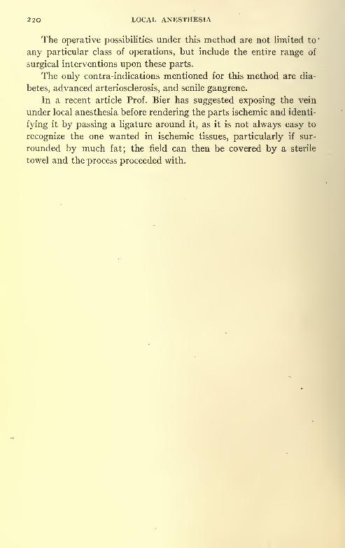
22O LOCAL ANESTHESIA
The operative possibilities under this method are not limited to'
any particular class of operations, but include the entire range of
surgical interventions upon these parts.
The only centra-indications mentioned for this method are dia-
betes, advanced arteriosclerosis, and senile gangrene.
In a recent article Prof. Bier has suggested exposing the vein
under local anesthesia before rendering the parts ischemic and identi-
fying it by passing a ligature around it, as it is not always easy to
recognize the one wanted in ischemic tissues, particularly if sur-
rounded by much fat; the field can then be covered by a sterile
towel and the process proceeded with.

CHAPTER XIII
GENERAL ANESTHESIA THROUGH THE INTRAVENOUSINJECTION OF LOCAL ANESTHETICS
RITTER, experimenting in Payr's clinic, produced complete general
analgesia by injecting into superficial veins of dogs 10 c.c. of a i percent, solution of cocain, or 5 c.c. of a 3 or 5 per cent, solution in a o.i
per cent, salt solution. The animals lay perfectly quiet, but alert.
Respiration and circulation were not disturbed, but they were com-
pletely insensible to every kind of irritation that could be used, even
to the actual cautery applied to the penis, vagina, anus, tail, face, ear,
and lining of the mouth. There was no sign of pain, and the dogs
wagged their tails during these performances. Only when forceps
were applied to the tongue did they seem to object, but here not
apparently from pain.
The duration of the anesthesia was from fifteen to twenty min-
utes or longer, and was not followed by serious after-disturbances.
Only a few showed any unpleasant by-effects, and these were small
animals upon which the larger doses had been used. One dog, how-
ever, always reacted in the same way, even to small doses. The
disturbances were always of the same kind, the animal becoming very
restless, tossing his head about, and, if placed on the floor, ran around
in circles. This would continue for about fifteen minutes, after
which time the animal quieted down and remained apparently nor-
mal. Actual convulsions were never observed with any dosage.
None of the larger dogs showed any by-effects.
Still more interesting are the observations made by Dr. B. W.Harrison upon himself, and reported in the Boston Med. and Surg.
Jour., February 2, 1911. The doctor showed great courage in using
upon himself, by the method he employed, what would have been
considered a thoroughly toxic dose of the drug; but he observed the
precaution of proceeding very slowly and stopping with the first
unpleasant symptoms. He states that, except for minor operations,
there had been no other use of cocain or allied drugs upon himself.
The experiment was performed as follows:
Into one of the superficial veins on the back of the hand there
was slowly injected 5 gr. of cocain in a 2 per cent, solution. The

222 LOCAL ANESTHESIA
injection was made very slowly, and was completed in thirty minutes.
It was deemed advisable to stop here, as dizziness and palpitation
occurred. Tests of the patient's condition as to general anesthesia
were now made, and were found to conform in an incomplete waywith those observed in animals similarly experimented with. There
was fairly marked analgesia everywhere. An incision % inch long,
and carried well down into the fat, was made on the anterior surface
of the leg. The incision could be felt, but caused a mere trifle of pain.
When several small nerves in the fat were cut each caused a small
twinge of pain, but, apparently, operative procedure might readily
have been undertaken with only moderate discomfort. Two hours
later a similar incision was made on the opposite leg; by now the
sensation of pain had nearly recovered its normal intensity. Duringthe experiment cerebration was normal, except for a restless inability
to keep the mind long on one subject. Motor power was unimpaired.This experiment and those on dogs by Ritter are highly interesting,
and, at least, of scientific value. The enormous dose necessary uponDr. Harrison, and then producing only an imperfect analgesia, makes
it an impossibility in human surgery. The dose used by Dr. Harrison
was several times that necessary to show toxic symptoms, and mayhave proved fatal had he injected it all at once, but he made the in-
jection slowly over a period of thirty minutes.
It is interesting to compare the results obtained by these injec-
tions with that by Prof. Ransohoff, when he injected 0.15 gr. of cocain
in the common carotid of a dog, and obtained only analgesia of the
head, face, and upper part of' the neck. (See Arterial Anesthesia,
Experiment 9, and the discussion which follows.)
A comparison of the toxicity of cocain,when injected in the various
ways, is given by Oppel as follows:
"Subcutaneous injections are two to three times less dangerousthan the arterial and fifteen to twenty times less dangerous than the
intravenous injection, and arterial injections are eight to ten times
less dangerous than the intravenous." (Here he was no doubt ex-
perimenting with arteries of the extremities, but he does not state.)
Meyer in experimenting with intravenous injections of cocain for
the production of general anesthesia corroborated the findings of
previous investigators but discovered that the large nerve-trunks
remained as sensitive to pain as before, even with very large doses of
cocain, and that the anesthesia seemed limited to the peripheral nerve-
terminals to which it is carried by the blood. To confirm this as-
sumption one leg was ligated before the injection was given, so that

GENERAL ANESTHESIA 223
the circulation was cut off; following the injection the entire bodybecame anesthetic except this leg.
These methods at present are of scientific rather than practical
interest but may become of some importance, like the intravenous
use of ether, with the discovery of safer agents than those now in use.
It is also known that magnesium sulphate when injected intraven-
ously produces a general anesthesia, but this agent is too toxic and
depressing for clinical use. (See this subject.)

CHAPTER XIV
THE UPPER AND LOWER EXTREMITIES
" As a general proposition, it may be safely asserted that all opera-
tions can be made painless by local or regional anesthesia in all parts
of the body in which the circulation can be absolutely controlled bycircular constriction. Hence, the entire surgery of the upper and
lower limbs (with exceptions to be considered later) can be made
tributary to these methods. It is in the surgery of the extremities
that the combined local (infiltrations) and regional (neural) anesthe-
sia has attained its degree of efficiency and accomplished its most
convincing, if not most brilliant, results. On the other hand, the
efficiency and applicability of these methods is decidedly restricted,
impaired, and at times wholly inefficient at the root of the limbs.
This is more especially the case in the hip and gluteal regions, where,
on account of the overlapping of the cutaneous nerve distribution and
because of the great depth of the most important nerves as, e.g.,
the sciatic, where it issues from the pelvis it is impossible to exposethe great nerve-trunks without inflicting an additional traumatism,
which is scarcely compatible with the conservative aims of the local
anesthetic methods.
"It is only fair to state that these objections do not apply to all
cases, and that even in these most difficult regions many perfect
successes can be obtained by purely local and intraneural methods
when they are applied to suitable subjects. This is particularly true
of emaciated, wasted patients, in whom disarticulation at the hip
can be performed by simple edematization and intraneural infiltration
as effectively as in the minor amputations and disarticulations of the
fingers and toes" (Matas).In the upper extremities the difficulties to be encountered are
much more successfully met by the comparatively easy access to the
brachial plexus, which can be exposed and infiltrated above the
clavicle.
But whatever doubts may exist as to the invariable success of
cocain and its allies in controlling the sensibility of the root of the
limbs, there can be no doubt in asserting that all operations, in-
cluding amputations, disarticulations, and excisions below the in-
sertion of the deltoid in the arm, and below the middle third of the
thigh in the lower limbs, can be made painless by purely local or
224

THE UPPER AND LOWER EXTREMITIES 225
neural (peripheral) methods of anesthesia. In anesthetizing the
extremities, the methods will vary with the individual regions, and
the technic will demand more skill and anatomic knowledge as the
surgeon proceeds from periphery to center. In all major procedures,
in which a large part of the thickness and circumference of the limb
is to be exposed to the knife and to painful manipulations, the neuro-
regional method is to be preferred, as in excision of bones and joints
and in amputations. In more superficial or well-circumscribed
lesions the simple infiltration method of Schleich will be most appli-
cable. In thin and marasmic subjects this method will also find fre-
quent and ready application because of its greater simplicity, even
when amputations are required, provided they are strictly typical
and do not involve extensive excursions away from the infiltrated
area. But whether the method adopted be the edematization of
Schleich or the neuroregional method, the circular elastic constrictor
applied on the Corning principle should be applied after the analgesic
drug has been injected and the exsanguination of the limb by eleva-
tion and gravity has been obtained.
The introduction of vein anesthesia by Bier has greatly simplified
all procedures upon the extremities where this method can be em-
ployed; however, there still remain many conditions in which it
cannot be successfully used the necessary appliances may not be at
hand, or the operator may prefer to use other methods.
The ability to control the blood-supply in the extremities greatly
facilitates all surgical procedures in these parts, and in the use of
local anesthetics intensifies and prolongs their action.
The course of the long cutaneous trunks is fairly constant in both
upper and lower extremities, and should be carefully studied, as well
as their points of emergence through the deep fascia;the smaller cuta-
neous branches are, however, subject to variations within certain
limits, and cannot always be definitely located. But the main
trunks of these parts are quite constant throughout their entire course
and can be easily reached, either through deep paraneural injections
along their course or by free exposure and direct (intraneural) in-
jections.
BONES AND JOINTS
In all operations upon the bones it must be remembered that bone
and cartilage have no sensation, but that the periosteum, perichon-
drium, and synovial membranes are nearly as sensitive as the skin;
bone-marrow is also slightly sensitive.
15

226 LOCAL ANESTHESIA
In operating here by infiltration the periosteum should be in-
cluded; after this has been infiltrated or denuded from the bone no
further sensation is felt.
In operating upon bones for inflammatory conditions, such as
periostitis or osteomyelitis, it is preferable always to use the regional
methods of anesthesia. In cases where this is not feasible, or in the
absence of inflammations, where it is preferred to operate by infiltra-
tion, as in the removal of an osteophyte or for a simple osteotomy, it
is desirable, where the bone is superficial and easily accessible, to
infiltrate the periosteum before making the incision. The infiltration
is done with a long needle passed down from the skin from two or more
points and directed in different directions, so as to embrace the en-
tire operative field on the bone (see Fig. 15). In the ordinary case it
is only necessary that the injections be made proximately and on
each side, in a somewhat crescentic manner, the two horns embracingthe growth. In this way all nerve-fibers leaving the field are effect-
ively blocked. When an operative field is sequestered in this way bycircumferential or crescentic injections it is n6cessary to wait about
ten or fifteen minutes for full anesthesia to become established. In
case the bone is deeply situated or overlaid by heavy muscles, as in
the case of the femur, it would be preferable to anesthetize the perios-
teum later, after the bone has been exposed by the division of the over-
lying soft parts. Where the periosteum has been well anesthetized
the use of chisels or other bone-cutting instruments is unaccompanied
by any pain. Such instruments should always be sharp and the bone
cut obliquely rather than at right angles, to avoid the shock of the
blow, which is always trying on the patient even if it causes no pain.
In operating by regional methods, the injections should be made at
sufficiently high levels to include the nerve supply to the periosteumwhen possible, otherwise the periosteum will have to be infiltrated.
The humerus receives its nerve-supply from the musculopsiral and
musculocutaneous nerves; the radius and ulna, from the median
nerve; the elbow-joint and wrist receive nerves from the three large
trunks in the arm; the femur, from the sciatic and obturator nerves;
the tibia, from the anterior and posterior tibial nerves; the fibula,
from the peroneal; the knee-joint, from the internal and external
popliteal, obturator, and crural nerves.
The contents of the joints contained within the synovial sacs can
be anesthetized by passing a needle into the sac and filling it to a
point of moderate distention with solution No. 2 (0.5 per cent, novo-
cain) ,or a slightly lesser quantity of a stronger solution, and allowing

THE UPPER AND LOWER EXTREMITIES 227
it to remain for about five minutes, when it can be withdrawn or will
escape from the incision. This anesthesia is only superficial and does
not extend deeper than the synovial sac, and is suitable only for
drainage or simple incision. If more is to be done, and in cases where
much inflammation exists, it is better to use some regional method by
blocking the curves higher up.
Fractures. Local anesthesia has been used occasionally in reduc-
ing fractures of the long bones. While in certain cases it may be
useful where general anesthesia is contra-indicated, it is certainly not
the method of choice, for with it we do not get the complete muscular
relaxation so necessary for the perfect reduction of the fractured
fragments. Lerda, in the"Centralblatt fur Chirurgie," 1907, states
that during the last two years extensive use has been made of local
anesthesia for fractures in Isnardi's service at Turin, where he is
assistant; he applied this technic in 30 cases before reduction of the
fracture and has never observed the slightest inconvenience. Heuses a long, strong needle, and injects the anesthetic mixture at vari-
ous points between the fractured ends and tangential to them, so that
the entire focus of the fracture, the bone-marrow, periosteum and
surrounding tissue become impregnated with the anesthetic. Headds a drop of i to 1000 solution of adrenalin to each cubic centimeter
of a 0.5 per cent, solution of cocain. Sometimes as much as 0.08
gram of cocain (about i gr.) was injected without appreciable by-effects. The contraction of the vessels aids in preventing hematoma
at the point. The anesthesia is generally complete in about eight
minutes. Not only is the pain abolished, but the fracture can be
reduced much more perfectly, attaining results otherwise impossible
without general anesthesia.
While the method recommended by Lerda has the advantage of
simplicity, it may be preferred in many cases to use regional anesthe-
sia, which produces a certain amount of muscular relaxation, though
always less than that obtained from general anesthesia; for this
reason general anesthesia is always to be preferred except in those
cases which positively centra-indicate its use; where some form of
anesthesia is needed in complicated cases, the local or regional
methods may prove of valuable assistance.
THE BRACHIAL PLEXUS
The nerves of the upper extremity are all derived from the brach-
ial plexus except the intercostohumeral;the lateral cutaneous branch

228 LOCAL ANESTHESIA
of the second intercostal, which crosses the axilla, pierces the deepfascia at the inner side of the arm, and is distributed to the skin of the
upper parts of the arm on its inner and posterior surface; sometimes
the third intercostal gives off a similar branch. The brachial plexusis quite easily exposed and blocked; for this purpose it should be
exposed above the clavicle by an incision running downward and out-
ward from the outer border of the sternomastoid over the course of
the plexus (see Fig. 67), which is easily recognized lying on the surface
of the scalenus medius, where each of its branches may be separately
injected, each with a few drops of 0.5 per cent, novocain solution with
a few drops of adrenalin to the ounce.
This is best done in the following manner: with a flat sand-bagunder the base of the neck and the head extended and rotated to
the opposite side, a line is drawn from the middle of the sterno-
mastoid down to the middle of the clavicle. A wheal is made about
the center of this line; with a long fine needle and large syringe the
skin and subcutaneous tissue along this line are anesthetized by pass-
ing the needle through the wheal, directing it up and then down
just under the skin along the line, injecting as it is advanced. Bychanging the angle it is directed down beneath the deep fascia
toward the plexus and about one-half ounce of solution distributed
here at two or more points.
In making these deep injections care should be taken to use only
a fine needle, preferably one with a short beveled point such as is
commonly used for spinal puncture, also, always, to inject the solution
while the needle is being advanced. At any point should there be
any question of having entered a vessel, slight aspiration upon the
syringe will show blood should a vessel be entered, when the needle
can be slightly withdrawn and redirected. Beyond a little staining
of the tissues no harm will result from a fine needle stick, but injec-
tions within vessels, particularly veins, are to be avoided. There is
practically no danger of injuring the subclavian vessels, which are
some little distance away, but there are many other small vessels in
the neighborhood, and the transversalis coli with its vein, both vessels
of some size, cross the field over the plexus. The subclavian artery
lies beneath the plexus, the vein is in front and below, not rising as
high as the artery and separated from it by the scalenus anticus.
Having finished the injections, and having in mind the above
anatomy, an incision is made down through the deep fascia; it maybe necessary to double ligate and divide the transversalis coli vessels
to obtain a free field. With a little blunt dissection and palpation

THE UPPER AND LOWER EXTREMITIES 229
the plexus is located lying just above the subclavian artery, by lo-
cating the pulse in this vessel with the tip of the finger the exact
position of the plexus is more readily located. By keeping the shoul-
der well depressed the plexus is easily brought into view.
The results of an intraneural injection of the brachial plexus are
shown in from five to ten minutes in a complete analgesia of the
shoulder and entire arm, and can be made use of in extensive opera-
tions upon these parts, and is particularly suited to high amputationsand disarticulations at the shoulder. Where the operative field
enters the region of distribution of the intercostohumeral nerve, this
may be blocked by a few drams of solution injected subcutaneously
along the floor of the axilla on its outer and posterior border. All
operations above the elbow, when too extensive to be readily per-
formed by infiltration, should be done by blocking the brachial
plexus, thus controlling all nerves, superficial and deep, of this part as
well as the forearm and hand. The localization and injection of the
cutaneous trunks of this region is unsatisfactory, as they are derived
from a variety of sources and overlap each other; superficial or minor
operations should, therefore, be done through infiltration, reserving
blocking of the brachial plexus for the more extensive or major pro-
cedures. Operations at or near the elbow, involving extensive dis-
sections, resections, or amputations, had best be performed by the
above method, which, of course, may also be used for any operation
on the distal parts of the forearm or hand, but here it would be pre-
ferable to block the nerves at the elbow.
"We believed in January, 1898, that in cocainizing the three great
nerves of the arm at the elbow by direct intraneural infiltration a
considerable territory had been conquered from the domain of general
anesthesia. We were not then aware that a few months before our
first operation at the Charity Hospital the same principle had been
successfully applied to the lower extremity by the direct infiltration
of the sciatic and anterior crural nerves in the performance of an
amputation of the leg. The credit of applying this direct intraneu-
ral method in major amputations is due, I am pleased to say, to Dr.
Geo. W. Crile, of Cleveland, Ohio, whose remarkable and most ex-
haustive experimental study of shock has made his name familiar to
all readers of surgical literature. It was precisely with the view of
diminishing shock that Dr. Crile was led to apply this method, which
he very appropriately designates the 'blocking method/ because the
infiltration of a nerve-trunk with cocain 'blocks' or completely inter-
rupts the conduction of all afferent, irritant impressions made upon

230 LOCAL ANESTHESIA
the nerve below the blockage. Crile's first operation was performed
May 1 8, 1897, and was suggested by the well-known experiments of
the physiologists, U. Mosso (1886) and Francois Franck (1894)"
(Matas).
In a personal communication addressed to Dr. Matas, August 24,
1899, Dr. Crile stated that he had operated by the"blocking"
method, up to that time, on 7 patients, i of these being a case of am-
putation at the shoulder-joint anesthetized by "blocking" the brach-
ial plexus above the clavicle. Dr. Crile's first case of amputation of
the leg was reported to the Ohio State Medical Society in 1897, and
excited the attention of that body. This performance is one of the
most brilliant and useful contributions to the technic of regional anes-
thesia that have emanated in recent years from an American surgeon.
Since the first successful demonstration of these methods byMatas and Crile they have been used repeatedly by other operators
and are now no longer novelties.
"The effect of intraneural injections is usually and promptly felt,
and the effect is almost identical with that following the completesection of a mixed nerve. The only difference between this and com-
plete anatomic section lies in the remarkable fact that the voluntarycontrol of the parts below the 'blocked' nerves is largely retained, so
that the patient can materially assist the surgeon in his manipula-tions. All pain conduction, all thermal sense is entirely lost; the
muscular sense is impaired, but the deep reflexes are not lost; common
sensation, tactile sense, is profoundly obtunded, but is not altogether
abolished. This affinity of cocain for the pain-conducting and
thermal fibers is one of the many remarkable features of its marvelous
anesthetic action" (Matas).
In some unusual cases the anesthesia following intraneural in-
jection has been retarded; this retardation has been sometimes so
prolonged that on more than one occasion I have felt worried at the
prospect of total failure. After waiting patiently for fifteen and in
one case twenty minutes, the characteristic subjective paresthesia be-
gan, and in a few seconds thereafter the anesthesia was complete.Once established, the anesthesia will remain as long as the circulation
is arrested by the constrictor, but in places where constriction is not
satisfactory, as at the groin (anesthesia of the anterior crural) and
at the brachial plexus, the nerves will remain anesthetized for forty-
five minutes to an hour, if a i per cent, novocain or a 4 per cent,
beta-eucain solution is used with adrenalin.
The anesthesia is more transitory if the weaker solutions are in-

THE UPPER AND LOWER EXTREMITIES 23 1
jected, and for this reason a i per cent, novocain or 0.4 per cent,
beta-eucain should be preferred. Should the anesthesia begin to dis-
appear before the completion of the operation, the nerves can be re-
injected. In injecting the nerves great care should be observed to
use the finest needle, and to employ only fresh and perfectly sterile
solutions. The fluids must be introduced into the center of the nerve,
with the needle directed parallel to the nerve-fibers. Very few dropswill usually suffice to give the nerve a slight fusiform swelling at the
point of injection, which is characteristic of a thorough infiltration.
Injections made in this manner into a healthy nerve are never painful,
provided that the nerve is held slack, so that the injection can be
made without the least traction upon its fibers. Should traction
be made upon the nerve-trunk or tension made upon its fibers, painwill be produced. In case a nerve is inflamed from the extension of a
surrounding inflammation, intraneural injections will always cause
pain. For this reason, when practising regional anesthesia, the nerve-
trunks should be injected sufficiently high above the area of inflam-
mation to be well beyond a possible lymphangitis of the nerve-sheath.
For instance if it be necessary to amputate a finger, and the inflamma-
tion extend well up toward its base, it would be preferable to inject
the nerves above the wrist; or should it be necessary to open a deep
palmar infection or resect a metacarpal bone, and the inflammation
extend to or above the wrist, it would be better to inject the nerves at
the elbow.
The paraneural injection of the brachial plexus both above the
clavicle and below it in the axilla, as has been recommended and prac-
tised by some operators, but particularly in the axilla, is a far too
dangerous procedure to find a place in the operative methods of con-
servative operators.
It is far better, safer, and surer, as well as quite simple, to resort
to the free exposure of the plexus above the clavicle, and inject each
individual nerve by the intraneural method, as first advocated byCrile. These nerves are too large to be readily penetrated in effective
quantities by the anesthetic fluid with any degree of certainty, and
the use of strong solutions at these points, highly vascular and close to
the trunk, in any effective quantity is likely to prove dangerous, as
absorption is active, and there is no practicable means of retaining it
in situ by constriction.
Aside from the danger in the solution injected, if made in effective
quantity and strength, the anatomy of this region should be sufficient
to deter any but the most venturesome from this practice.
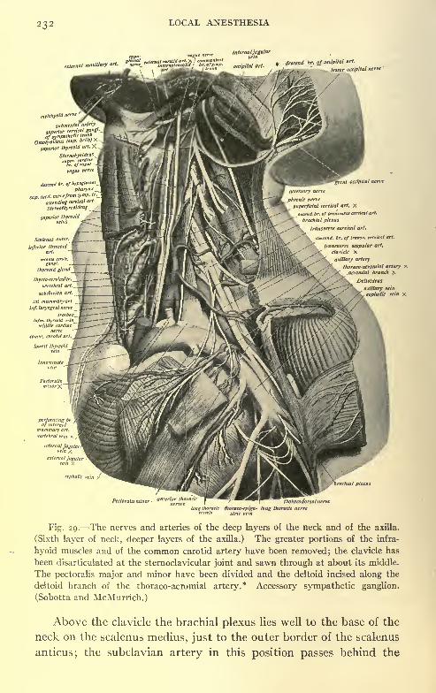
232 LOCAL ANESTHESIA
external maxillary art.
superficial cervical art. X
-,ctnd.br. of transverse cervical art.
brachial plexus
transverse cervical art.
'. cerrtcat arl.
,''^ transverse scapular art.^ clavicle X
^^^ Mary artery
stibmfftfal
superior ""*of sympathetic
trunk
Omolirolaeus IW- *'W
superior thyreoidarl. X
Sternohyoidcu
*"&. cf '"I"
vasus nerv
axillary vein
phalic vein X
Fig. 29. The nerves and arteries of the deep layers of the neck and of the axilla.
(Sixth layer of neck, deeper layers of the axilla.) The greater portions of the infra-
hyoid muscles and of the common carotid artery have been removed; the clavicle has
been disarticulated at the sternoclavicular joint and sawn through at about its middle.
The pectoralis major and minor have been divided and the deltoid incised along the
deltoid branch of the thoraco-acromial artery.* Accessory sympathetic ganglion.
(Sobotta and McMurrich.)
Above the clavicle the brachial plexus lies well to the base of the
neck on the scalenus medius, just to the outer border of the scalenus
anticus; the subclavian artery in this position passes behind the

THE UPPER AND LOWER EXTREMITIES 233
scalenus with the plexus above, giving off in this neighborhoodbranches of large size (Fig. 29). Many veins are encountered in all
directions, and, while their puncture with a fine needle would not be of
much consequence, an intravenous injection may prove a more serious
matter.
Below the clavicle the plexus lies to the outer side of the first por-
tion of the artery, embraces the second portion, and lies somewhatmore widely distributed around the third. The vein in both cases is
fairly out of the way.
Notwithstanding these anatomic arrangements, paraneural injec-
tions have been made in both positions. The Kulenkampff method,
Fig. 30. Thorax from above, after Kulenkampff. On one side is shown the posi-
tion of the brachial plexus and subclavian artery to the clavicle, on the other the direc-
tion the needle should take in making the injection: a, Subclavian vein; b, point of
attachment of anterior scalenus muscle; c, subclavian artery; d, brachial plexus; e,
point of attachment of scalenus medius muscle. (From Braun.)
above the clavicle, has been favorably spoken of by Braun, and
practiced by himself and many others, and is done in the following
manner: That portion of the plexus is selected for injection at the
point where it passes over the first rib; in this position the artery
lies below and on the inner side, the clavicle above and in front, the
pleura and lung beneath (Fig. 30) .
The direction of the brachial plexus, as it passes under the clavicle,
is at about right angles to the lorig axis of this bone, and passes under
at about its midpoint in the erect position of the body; for this reason,
as well as the fact that in this position the clavicle descends slightly

234 LOCAL ANESTHESIA
downward and forward, thus affording a better exposure of the field,
it is advised that the injection be made in the sitting position (Fig.
31). The method of procedure is as follows:
With the patient sitting erect, the finger is passed over the mid-
point of the clavicle and accurately locates the artery by its pulsations,
the skin and subcutaneous tissue is now lightly infiltrated, and a long
fine needle, unattached to the syringe, are passed in a direction down-
ward, inward, and backward from the midpoint of the clavicle in such
a direction that it aims at the spinous process of the second or third
dorsal vertebra; the distance to be penetrated and the amount of fat
vary, but it is usually from 2 to 4 cm., the plexus lying just under
the deep fascia. When the plexus is reached a slight radiating pain
Fig. 31. Position of patient for Kulenkampff brachial plexus injection. (From
Braun.)
or paresthesia is felt down the branches of the radial or median nerve
in the hand or fingers; at this point the needle is held stationary, the
syringe attached, and the injection made. The object in not attach-
ing the syringe earlier is that should the artery be entered blood will
flow;should this occur with a fine needle it is not likely to be of much
consequence, the needle being withdrawn slightly, and the point di-
rected a little more laterally. About 10 c.c. of a 2 per cent, novocain-
suprarenin solution is injected; the needle is now slightly withdrawn,and an additional 10 c.c. injected around in the neighborhood, to be
sure to reach any cord of the plexus that the first injection may have
missed.
It is said that when paresthesia occurs, which indicates that the

THE UPPER AND LOWER EXTREMITIES 235
plexus has been reached, anesthesia is certain, and usually is estab-
lished in from one to three minutes; occasionally, however, it may
17
Fig. 32. Areas of distribution of cutaneous nerves (after Toldt), showing effect of
Kulenkampff plexus anesthesia: I, Anesthesia; ++ , hyperesthesia; D, normal
sensation; i, supraclavicular nerves; 2, circumflex; 3, external cutaneous; 4, musculo-
spiral; 5, radial; 6, musculocutaneous; 7, median; 8, radial (terminal branches); 9,
lateral cutaneous from second intercostal; 10, musculospiral; n, ulnar; 12, internal
cutaneous; 13, plamar branch of ulnar; 14, dorsal branch of ulnar; 15, palmar branch
of ulnar; 16, digital branches of ulnar; 17, digital branches of median. (From
Braun.)
require a longer delay, from ten to fifteen minutes;failure to obtain
anesthesia by this time usually indicates the need of another puncture,
when 5 to 10 c.c. of a 4 per cent, solution is used in the same manner,

236 LOCAL ANESTHESIA
except that the paresthesia in the extremity does not occur unless the
first injection has gone wide of the mark; should the first injection
fail, the second is not near so likely to succeed (Fig. 32).
In the hands of Kulenkampff and his associates, who injected a
large number of cases in this way, very few failures were recorded.
The duration of the anesthesia is from one-half hour to three hours,
and is associated with complete muscular relaxation of the part.
This is certainly a very ingenious and simple procedure and when
properly carried out would ordinarily be supposed to be free from
danger. Clinical reports, however, describe a great many by- and
after- effects;the most common is a transient paralysis of the phrenic
nerve on the injected side. This has been demonstrated by x-ray ex-
aminations of the diaphragm and is always associated with some dis-
turbance of respiration. As a rule this phrenic paralysis clears up in a
few hours but in some few cases it has persisted for several days and
longer.
Transient paresis of the sympathetic of the neck, with contrac-
tion of the pupil and the orbicularis oris, occurs quite frequently, occa-
sionally associated with a sinking back of the eye. These symptomsare quite transient.
Less frequently paralytic disturbances have been noted in the
arm and in some few cases these injuries have been more or less
permanent.
Reports of these after-effects must make the conservative oper-
ator hesitate to employ the method. It is readily understood howthe solution can diffuse along the plexus mesially and reach the
phrenic and sympathetic nerves, producing a temporary effect, but in
many cases where the effects have been quite lasting it has no doubt
resulted from injury by the needle, as no after-effects result from the
many intraneural injections of nerve-trunks where the nerves are
exposed and carefully injected without trauma. The possibility of
a small percentage of failures, as with all paraneural injections of
large nerve-trunks, is another objection to its use. In view of the
possible paralysis of the phrenic nerve it would seem unnecessary to
caution against its simultaneous use on both sides.
Paraneural Injection Within the Axilla. The arm is abducted to
a right angle, the index-finger of one hand is passed up on the outer
side of the fossa, and the brachial artery located and slightly displaced
downward and inward;a long fine needle is now passed over the tip
of the finger and directed up in the long axis of the limb until well
within the axilla, when the injection is made; the precaution should be

THE UPPER AND LOWER EXTREMITIES 237
followed here, as elsewhere, when making an injection in the neighbor-hood of vessels to continuously inject the solution as the needle is
being advanced; in this way the vessels may be
pushed aside and their puncture avoided.
The needle is now passed behind the
artery near the insertion of the tendon
the latissimus dorsi to reach the
posterior cord of the plexus, and
additional injection madeFig. 33. Cuta^^ ^^ at f-his point; 30 to 40
neous nerve areas of^^^^ B^k. r
the upper extremity (an^^ ^C.C.of a 2 per cent,
teriorview) : A, Circumflex^^B ::\>~^i ^^^ nOVOCain-adren-
C, lesser internal cutaneous; #^^lhkN ^^. alin Solutioninternal cutaneous; E, musculospiral;
F, musculocutaneous; G ulnar; H,median. (Campbell.)
Fig. 34- Cuta-
neous nerve areas of
upper extremity (poste-
rior view) : A, Supraclavicu-
lar; B, circumflex; C, musculo-
spiral; D, lesser internal cutane-
ous; E, internal cutaneous; F, ulnar;
G, musculospiral. (Campbell.)
is recommended as the necessary amount
(containing in this concentration a quantityof the drug clearly beyond the safe limits) . While
the supraclavicular injection will find a place in our
recognized methods of procedure, this last injection within
the axilla is, to say the least, unsurgical, and is not to be recommended.
The location and distribution of the nerve-supply from the elbow
down should be carefully studied for the application of regional
methods of anesthesia to these parts, particularly the cutaneous dis-
tribution, which is fairly constant (Figs. 33 and 34).

238 LOCAL ANESTHESIA
NERVES OF THE UPPER EXTREMITY
The Musculocutaneous (External Cutaneous Nerve). The cutane-
ous portion winds around the outer border of the tendon of the biceps,
and, piercing the deep fascia, becomes superficial, passing behind the
median cephalic vein; it divides opposite the elbow-joint into anterior
and posterior branches.
The anterior branch descends along the radial side of the fore-
arm as far as the ball of the thumb, giving off cutaneous branches
to this region back of the wrist and carpus; at the wrist-joint it is
placed in front of the radial artery.
The posterior branch passes along the back part of the radial
side of the forearm, supplying this region as far as the wrist.
The internal cutaneous nerve pierces the deep fascia with the basilic
vein about the middle of the arm, and, becoming cutaneous, divides
into anterior and posterior branches. The anterior branch descends
usually in front of, but occasionally behind, the median basilic vein,
and is distributed to the skin on the anterior part of the ulnar side of
the forearm as far as the wrist.
The posterior branch passes obliquely downward and backward
over or in front of the internal condyle to the posterior surface of
the inner side of the arm, and is distributed to the skin as far as the
wrist.
The lesser internal cutaneous nerve pierces the deep fascia on the
inner side of the brachial artery at the middle of the arm, and is
distributed to the skin on the inner and posterior surface as far as
the elbow.
The median nerve, at the bend of the elbow, lies beneath the bi-
cipital fascia to the inner side of the tendon of the biceps, separatedfrom it by the brachial artery. It passes between the two heads of
the pronator radii teres, and it is deeply situated until about 2 inches
above the wrist, when it becomes superficial, lying between the ten-
don of the flexor sublimis and flexor carpi radialis beneath or slightly
to the ulnar side of the tendon of the palmaris longus, and follows
this course into the hand.
This nerve can be easily reached by an open dissection at the bend
of the elbow for an intraneural injection, or by passing a needle be-
neath the tendon of the palmaris longus above the wrist for a para-
neural injection. In the hand this nerve supplies the superficial mus-
cles of the thumb, two outer lumbricales, both sides of the thumb,
index, and middle fingers, and radial side of the ring finger on their
palmar aspect; each digital nerve opposite the base of the first pha-

THE UPPER AND LOWER EXTREMITIES 239
lanx gives off a dorsal branch which joins the dorsal branch from the
radial, and runs along the side of the dorsum of the finger to end in
the skin over the last phalanx.The ulnar nerve, at the bend of the elbow, lies against the bone
between the internal condyle and olecranon, and is easily reached in
this position for a paraneural injection by passing the needle downto it through the skin. In the forearm the nerve is deeply situated,
but becomes more superficial near the wrist, lying to the radial side
of the tendon of the flexor carpi ulnaris covered by the skin and fas-
cia, where it can be fairly easily reached by a needle for paraneural
injections.
In this position the ulnar artery lies to the radial side and slightly
more superficial than the nerve, and is to be carefully avoided by
keeping the needle nearer the tendon of the flexor carpi ulnaris; the
injection should be made sufficiently free to permit some of the solu-
tion reaching the artery upon which the palmar cutaneous branch,
given off higher up in the arm, descends to the skin of the palm.The injection should be made about 2 inches above the wrist, to
reach also the dorsal cutaneous branch which is given off in this
position, and curves around the wrist beneath the tendon of the
flexor carpi ulnaris, to divide into branches to be distributed to the
inner side of the little finger and adjoining sides of the little and ring
fingers; if the injection is made too low these branches will escape.
The nerve continues down on the outer side of the tendon of the
flexor carpi ulnaris to its attachment to the pisiform bone, and im-
mediately beyond divides into superficial and deep palmar branches.
The superficial branch supplies the palmaris brevis and skin on the
inner side of the palm, sending digital branches to the inner side of
the little and adjoining sides of the little and ring fingers. The deep
palmar branches supply the deep muscles of the palm.The musculospiral nerve appears at the bend of the elbow after
piercing the external intermuscular septum, and descending between
the brachialis anticus and supinator longus to the anterior surface of
the external condyle, where it divides into the radial and posterior
interosseous nerves. In addition to these terminal branches there
are three cutaneous branches: an internal cutaneous, which is dis-
tributed to the arm above the elbow, and is not of much concern to
us here, as all of these nerves should be reached by blocking the brach-
ial plexus above the clavicle.
The upper and smaller branch passes to the front of the elbow,
lying close to the cephalic vein, and is distributed to the skin on the

240 LOCAL ANESTHESIA
anterior surface of the arm;some fibers from this nerve may descend
below the elbow.
The lower branch is the more important to us here, and its posi-
tion should be borne in mind in all nerve-blocking operations at the
elbow. It pierces the deep fascia below the insertion of the deltoid,
running down along the outer side of the arm and elbow, then along
the posterior surface of the radial side of the forearm as far as the
wrist.
The radial nerve passes down the arm from the bend of the elbow,
lying beneath the supinator longus to the outer side of the radial
artery ;about 3 inches above the wrist it turns outward, passing be-
neath the tendon of the supinator longus, pierces the deep fascia on
the outer border of the forearm, and becomes superficial. In this
position it gives off its digital branches, an external branch which
descends to the radial side of the thumb, and an internal branch
which divides into three digital branches to supply the adjoining
sides of the thumb and index-finger, index- and middle fingers, and
the adjacent sides of the middle and ring fingers.
This nerve can best be reached for a paraneural injection about
2 inches above the wrist to the" outer side of the tendon of the supina-
tor longus, making the injection into the deep fascia and carrying it
across the outer border of the forearm for about an inch, to insure
reaching all branches of the nerve.
The above is a brief review of the nerve-supply of the forearm
and hand which concerns us in the regional anesthesia of these parts.
It can be seen from a study of the points at which these nerves are
accessible, the opportunities offered for blocking them either by direct
exposure and intraneural injection or through paraneural injection
by directing the needle down to the positions in which the nerves
will be found. But the possibilities here of an occasional anomalous
distribution must also be remembered. For all operations below
the elbow, including forearm, wrist, and hand, the intraneural methodof blocking at the elbow the three principal nerve-trunks in this
region radial, ulnar, and median after free exposure by open dis-
section, as first practised by Dr. Matas, will produce a perfect anes-
thesia of all distal parts where the technic has been properly carried
out. It must, however, be remembered that, in addition to the
three above-mentioned nerves, the skin of the forearm is supplied
by the internal and external cutaneous nerves, and if account is not
taken of these in making the injection the resulting anesthesia will
be imperfect or unsatisfactory.

THE UPPER AND LOWER EXTREMITIES 241
The musculocutaneous nerve at the bend of the elbow passes
behind the median cephalic vein, then divides into anterior and poste-
rior branches. The anterior branch of the internal cutaneous passes
in front of or behind the median basilic vein, at the bend of the elbow,
its posterior branch passing over the inner condyle.
On account of the existence and position of the above cutaneous
nerves the infiltration done at the bend of the elbow to expose the
three principal nerve-trunks radial, ulnar, and median should be
made in an oblique or transverse course, and not vertical over the
course of the large trunks, unless it should be preferred to inject the
lateral subcutaneous tissue by a separate injection. In exposing the
radial in the groove between the brachialis anticus and supinator
longus, if the infiltration is carried in toward the middle line in the
neighborhood of the median cephalic vein, it will include in the anes-
thetic atmosphere the external cutaneous nerve.
It must also be remembered in injecting the radial not to omit
the posterior interosseous nerve, which is found in the substance of
the supinator brevis. These two nerves are best exposed by dividing
the intermuscular septum between the supinator longus and brachialis
anticus and retracting the supinator longus when the nerves are
seen in the depths of the wound or lying in the substance of the
muscles. The infiltration to expose the median nerve will probablyreach and anesthetize the anterior branch of the internal cutaneous
which lies under the median basilic vein, but, to be sure that this
nerve as well as the posterior branch have been reached by the solu-
tion, it is advisable to inject a few small syringes of solution sub-
cutaneously between the median nerve and the internal condyle.
The ulnar nerve in thin and emaciated subjects, where it can be
readily felt, need not be directly exposed, but can be injected para-
neurally, but where overlaid by much tissue it is safer and more
surgical to directly expose it. In discussing this particular procedureProf. Matas, in his report on "Local and Regional Anesthesia"
before the Louisiana State Med. Soc., 1900, states the following:
"Personally, I regard the open intraneural method of cocainiza-
tion of the three nerves musculospiral, median, and ulnar at the
bend of the elbow as the most effective, certain, and simple means of
securing total anesthesia of the hand, wrist, and forearm.
"It is a strictly anatomic procedure which admits of no guess-
work, and for this reason is not likely to be popularized except in the
clinics of surgical specialists.
"The practicability of this method suggested itself to me in 1897,i 6

242 LOCAL ANESTHESIA
but no opportunity presented itself for its application until January,
1898, when an old man, aged seventy-six, applied to my clinic for
the relief of an extensive and deep epitheliomatous ulcer, which in-
volved a large part of the dorsal and hypothenar regions of the right
hand. The patient was profoundly arteriosclerotic, his radials were
hard and rigid as a pipe-stem, and his heart was the seat of loud aortic
and mitral murmurs, which indicated advanced valvular lesions.
He was a decidedly unfavorable subject for general anesthesia, and
I decided to anesthetize the hand by the direct neuroregional
method.
"The musculospiral, the median, and the ulnar were readily and
painlessly exposed (under infiltration anesthesia, Schleich No. i)
by separate incisions, made over the region of the individual nerve-
tracts, where they are most superficial at the bend of the elbow;the nerves were then exposed, and each injected with 5 to 8 minims of
a i per cent, solution of cocain. This caused a slight fusiform swell-
ing at the point of injection.
"The wounds were sutured, but the threads were not tied, to
provide for further injection, and the entire region was protected bya carefully applied aseptic dressing. The arm was then exsanguinated
by elevation, and the elastic constrictor was applied over the middle
of the arm. The anesthesia of the extremity was now complete from
the finger-nails up to the elbow. We were then able to extirpate the
growth very freely, including the fourth and fifth fingers with their
metacarpals and the corresponding palmar and dorsal aspects of
the hand, proceeding throughout with all the freedom that is per-
mitted by general anesthesia. After completing the work in the hand
the incision at the elbow was closed by tying the knots of the loose
catgut sutures, which had been purposely left untied before the
constrictor was removed. The operation was in this way not only
painless, but bloodless. Before the operation the patient was givena hypodermic, consisting of Y gr. morphin, 34o gr. strychnin, and
Koo gr- digitalin.
"Since the first operation (January, 1898) was performed the
procedure has been repeated by myself several times and once by
my assistant, Dr. Larue. In all these cases the intervention was
necessitated by bone lesions of either the hand, wrist, or forearm.
"In all of these cases the patients were.able to walk to their beds
after leaving the operating-table. None suffered from the least shock
or constitutional disturbance, and in none were the postoperative
sequelae such as to suggest that any injury had been done by cocainiza-

THE UPPER AND LOWER EXTREMITIES 243
tion of the nerves. All the small wounds made to expose the nerves
healed kindly under the usual aseptic dressing. In all of these cases
the anesthesia of the regions tributary to the nerves injected con-
tinued for a variable period, extending from ten to fifteen minutes
after the removal of the constrictor.
"In view of the practical success of this method of obtaining com-
plete insensibility of all the parts below the elbow, it is superfluous to
enumerate or discuss all the operations that can be performed in this
region without the help of general anesthesia. It is evident that in
absolutely anesthetic fields all operations are possible.
"I would again lay stress upon the fact that the method here
described is a regional method, in which the anesthesia is obtained
by the direct infiltration of the nerves at a distance from the field
of operation, and differs from all other methods suggested to accom-
plish the same regional object except that of Dr. Crile, of Cleveland,
Ohio, which is identical in principle and technic, except that it is
applied at a higher level by injecting the brachial plexus in the supra-
clavicular space.
"At the time that my first operation was performed I was not
aware that very nearly the same results had been obtained by Reclus
some time before the publication of his remarkable book, 'La Cocaine
en Chirurgie/ in 1896. Reclus' operation differs, however, from
the one here described in the essential fact that he attacked the three
nerves at the elbow by subcutaneous paraneural injections. He
erroneously attributes the suggestion to Krogius, of Helsingfors, Fin-
land, and his results, though apparently satisfactory, were not
sufficiently encouraging to decide him to continue its further applica-
tion. In addition to his doubts as to the general reliability of this
method, he fears that traumatic neuritis may result from the direct
injection of the nerves, and also believes, very justly, that the intro-
duction of the needle in search for the nerves in the vascular sheaths
at the root of the limb is fraught with too much risk to justify the
general adoption of this practice. We concur in these criticisms, as
they apply to the subcutaneous paraneural method, which is largely
a matter of approximation and guessing. These objections do not
hold, however, with the open intraneural method, in which the nerve
to be injected is directly exposed to view.
"As to the possibility of traumatic neuritis, which Reclus fears, I
have never noticed the least evidence or trace of it in the many cases
in which I have had an opportunity to practice this method in various
regions of the body. Le Fort ('Soc. Centrale de Med. du Nord.,'

244 LOCAL ANESTHESIA
October 27, 1899, and 'Gaz. des Hopitaux/ November 25, 1899) has
also more recently directed attention to the paraneural regional
method and applies it at the elbow and wrist, just as Reclus, Manz,and Holscher have done. He refers to Desoutte's experiments with
neural anesthesia in horses and expresses confidence in its value. Healso expresses a theoretic fear of neuritis from trauma of the nerves
by direct injection, but this fear, as I have stated, is unfounded. I
do not doubt that the subcutaneous paraneural method is a feasible
procedure, and will yield satisfactory results in emaciated, fleshless
patients, in whom the larger nerve-trunks are almost visible under
the skin; in such patients there should be no difficulty in reaching
the immediate vicinity of the nerves, or the nerves themselves for
that matter, since they are practically exposed to view. It is also
in just such patients that the Schleich's general infiltration anesthesia
will find a successful application. Not long since Dr. Gessner re-
ported a case of amputation of the arm above the elbow for tuber-
cular arthritis of the elbow-joint, in which the anesthesia was ob-
tained with perfect success by the Schleich infiltration method.
Schleich, Reclus, and their numerous followers have reported manycases of the same kind (vide among other recent contributions).
"'La Nacose et 1'anesthesie locale par J. Richbon Kjamerund,Bull, gen'l de Therap.,' January 15 and 30, 1899; and T. Wieker-
hauser, 'Operationen mit Schleichscher Analgesic, Centralbl. fur
Chir.,' October 21, 1899.
"In the earlier years of my experience with cocain I also per-
formed an amputation of the arm by Coming's infiltration for ad-
vanced tubercular arthritis with excellent results;but these successes
do not mean that Schleich's infiltration method is applicable to all
cases;it only illustrates the advantage to be derived from the adop-
tion of the various methods of anesthesia to different conditions."
Dr. Matas, in his previously mentioned report, in writing of the
hand and wrist, states the following:
"The anesthesia of these regions is obtained by any one of three
methods: (i) Direct infiltration (Schleich); (2) paraneural infiltra-
tion at the wrist (Reclus, Braun, Manz, Lefort) ; (3) regional direct
(open) intraneural infiltration at the elbow (Matas)." To these wecan now add the intravenous method of Bier and intra-arterial
anesthesia.
"The utility of the infiltration method is practically limited to
fractional areas of these parts, and can be applied successfully in the
evacuation of purulent collections, palmar abscesses, the removal of

THE UPPER AND LOWER EXTREMITIES 245
well-defined tumors, warts, epitheliomata, etc., foreign bodies, the
extirpation of ganglions, etc. It has also proved sufficient in mypractice, as in that of others, for the amputation of one or two fingers
with their metacarpals, but in all these the effectiveness of the in-
filtration will be very materially increased by a knowledge of the dis-
tribution of the cutaneous and deeper nerves supplying the area of
operation. Thus in resecting or disarticulating the metacarpals, the
infiltration is not only carried into the entire periphery of the bone,
including the periosteum, but the deep, adjoining interosseous nerves
must likewise be enveloped in a cocain atmosphere. When the
injuries or lesions are such that the operation is likely to be extensive,
irregular, or ill-defined, as, for instance, when several digits with their
metacarpals are to be amputated with a part of the palmar tissues,
a recourse to the neuroregional method at a higher level is preferable,
if not absolutely necessary, from the point of view of effectiveness
and simplicity. In anesthetizing the hand and wrist in its totality,
the radial, ulnar, and median nerves can be anesthetized by injecting
the anesthetizing fluid deeply into the perineural tissues along the
well-known anatomic paths of these nerves in the lower forearm, just
above the wrist, where they are known to be most superficial. This
procedure was first described and practiced by O. Manz (Kraske's
clinic; 'Centralbl. f. Chirurg./ 1898, No. 7), by Holscher ('Muench.Med. Wochenschr.,' February 21, 1899), and by F. Berndt ('Muench.Med. Wochenschr.,' 1899, No. 27), and in their hands has yielded some
fairly good results. Holcher and Berndt, following Oberst and Braun,
apply an elastic constrictor i or 2 inches above the wrist, and inject
20 c.c. of a 2 per cent, cocain solution, distributed in the region of the
three nerve-trunks, and wait fifteen minutes, when the insensibility
of the entire hand will follow. But the paraneural method applied
in this blind subcutaneous fashion is, as Manz himself admits, an un-
certain and unsatisfactory procedure at best, and it is not likely to find
many adherents."
THE FINGERS AND HAND
In the practice of regional anesthesia of the hand and fingers we
have many opportunities for blocking the nerve-trunks just above
the wrist by taking advantage of their superficial position and ex-
posing them by dissection for intraneural injection, or through para-
neural injections by passing the needle through the skin down to the
points where these nerves are to be found. The following experi-
ments, made by Prof. Heinrich Braun, of Zwichau, and given in his

246 LOCAL ANESTHESIA
book on "Die Lokal Anasthesie," illustrates the possibilities of its
use here:
"Experiment i (June 18, 1898, Dr. B.). Firm constriction of the arm. Injection of
i c.c. of i per cent, tropococain solution 3 cm. above the wrist under the tendon of the
palmaris longus (Fig. 35). The constricting rubber band was now sufficiently loosened
to permit a marked stasis hyperemia of the arm. Fifteen minutes after the injection
complete anesthesia of the distribution of the median nerve, as well as paralysis of the
short muscles of the thumb, was produced. The anesthesia remained fifteen minutes
after the removal of the constrictor.t
,
"Experiment 2 (May 14, 1902, Dr. B.). One-half cubic centimeter of 2 per cent,
cocain solution was injected at 10.45, 4 cm - above the wrist under the tendon of the
palmaris longus; the arm was not constricted; at 10.47 a feeling of tickling and warmth
in the first, second, third, and fourth fingers and palm of the hand; 10.55, complete anes-
thesia on the flexor surface of the thumb, second and third fingers, and the radial side
of the fourth finger, in the palm of the hand a very marked depression of the sensibility
in the entire nerve territory, with a paresis of the thumb muscles; 11.25, sensation
returned.
"Experiment 3 (Oct. 10, 1902, medical student, B.). 11.55, injection of i c.c. of 0.5
per cent, cocain solution with 3 drops adrenalin around the median nerve above the
wrist. The arm was not constricted. After fifteen minutes the sensibility, as indicated
in Fig. 36, Nos. I and II, almost completely disappeared; on the ball of the thumb, in the
palm of the hand, and on the flexor surfaces of the thumb and index-finger completely
disappeared. There was very pronounced paralysis of the short muscles of the thumb,and the skin over the distribution of the median nerve was hyperemic, red, and showed
increased temperature, while the skin over the neighboring ulna distribution remained
normal. The sensibility returned about 2 o'clock, two hours after the injection."
By taking advantage of the superficial position of the radial
nerve just above the wrist, where it passes beneath the tendon of the
supinator longus on the outer border of the arm, and making the in-
jection in a transverse manner at this point beneath the skin and
superficial veins, an anesthesia of its peripheral branches is obtained,
as illustrated by the following experiments:
"Experiment 4. After constriction of the arm i% c.c. of 0.5 per cent, cocain solu-
tion was injected in the above manner. After five minutes anesthesia appeared, as
indicated in Fig. 36, Nos. Ill and IV.
"Experiment 5 (May 13, 1899, Dr. B.). The same experiment with 2 c.c. of 0.5 per
cent, tropocain solution with constriction of the arm resulted in an anesthesia of about
similar extent.
"Experiment 6. Forms the continuations of experiment No. 3 on the same hand,where previously the median nerve had been blocked, i c.c. of 0.5 per cent, cocain solu-
tion, with the addition of 3 drops adrenalin solution (1:1000) was now injected on the
radial nerve above the wrist. Fifteen minutes later the hand, as indicated in Fig. 36,
Nos. V and VI, was completely insensible and remained so for about four hours."
As mentioned by Braun in commenting upon this experiment, it is
not likely to be of much value alone except for very limited superficial
operations, but when combined with a simultaneous injection of the
median nerve it is a simple and effective means of anesthetizing the
*

THE UPPER AND LOWER EXTREMITIES 247
entire radial side of the hand. Higher up in the forearm, at the junc-tion of the middle and lower third on the outer border, where the
intermuscular septum divides the flexor from extensor muscles, the
radial nerve is also fairly accessible, and may be successfully blockedat this point by passing the needle vertically inward beneath the
supinator longus. The following experiment by Dr. Braun illustrates
the results obtained:
"Experiment 7 (May 2, 1902, Dr. D.). Twelve o'clock, an injection of i c.c. of 2 per
cent, cocain solution in the above-described way, the needle had exactly met the nerve-
trunk, as indicated by the radiating paresthesia. No constriction. Immediately after
the injection occurred a marked radiating paresthesia and sense of warmth in the thumb.
12.10, complete regional anesthesia of the nerve; anesthesia of the skin is indicated, as
in Fig. 37, No. II. Motor paralysis of the radial. After forty minutes sensibility and
motility returned.
Minterosseus dors
Af.alnaris
i/i a j - i <sf N.medianusMflexor ulnsns # A, . ,M.palm. long.
Fig- 35. Cross-section through forearm three fingers-breadth above pisiform bone
(From Braun.)
The ulnar nerve is accessible, either for exposure by dissection and
intraneural injection or for paraneural injection, above the wrist-
joint, preferably three or four fingers' breadth above to insure reach-
ing the posterior branch, which may be given off this high up. In
this position the nerve lies between the tendon of the flexor carpi
ulnaris and the ulnar, as shown in Fig. 35, and is best reached for
paraneural injections by introducing the needle from the ulnar side
between the tendon and the bone in the direction indicated by the
arrow. It is rather unsafe and inadvisable to attempt to reach it
from in front (except by dissection) on account of the proximity of
the ulnar vessels, which here lie slightlymore superficial than the nerve
and slightly to the radial side. Figure 36, VII and VIII, indicate
the extent of the resulting anesthesia after an injection of i c.c. of
a 0.5 per cent, cocain solution with 3 drops of adrenalin (i : 1000) as
practiced by Braun in the above-mentioned way. It may, however,

248 LOCAL ANESTHESIA
be easier and preferable, instead of injecting the nerve at this point
to reach it back of the internal condyle. In thin subjects, where
the nerve can be readily felt, a paraneural injection may be under-
taken by first locating the nerve between the thumb and finger of
one hand while making the injection with the other; the inferior
profunda artery, which lies in this position, is more deeply situated
in the muscle just over the bone. The following experiments by
Fig. 36. Resulting areas of anesthesia of hand and fingers from subcutaneous and
paraneural injections. (From Braun.)
Braun illustrates the result obtained, while Experiment 9 is a para-
neural injection of the ulnar and median nerves above the wrist:
"Experiment 8 (May 13, 1902, Dr. L.). 12.50 o'clock, injection of i c.c. of 2 percent, cocain solution in the previously mentioned way. No constriction. Immediately
paresthesia and sense of warmth as far as the ends of the fourth and fifth fingers. After
six minutes complete regional anesthesia of the skin occurred, as indicated in Fig.
37, I. Sensibility returned fifty minutes after the injection."Experiment 9 (Dec. 9, 1902, medical student). One cubic centimeter of i per cent,
cocain solution with 3 drops of adrenalin solution was injected three fingers' breadth
above the wrist on .the ulnar and median nerves. After twenty minutes anesthesia
appeared in the territory, as indicated in Fig. 36, IX and X. The sensibility returned
after four hours in the ulnar territory and after five hours in the median."

THE UPPER AND LOWER EXTREMITIES 249
Experiment 10 represents the results of a linear injection made
subcutaneously from the region of the radial artery across the back of
the wrist to the pisiform bone.
"Experiment 10 (Feb. 10, 1899, Dr. B.). Three cubic centimeters of i per cent,
cocain solution was injected in the previously mentioned way in the arm after constric-
tion. After five minutes anesthesia appeared in the territory, as indicated in Fig. 36, XIand XII. Twenty minutes after removal of the constriction sensibility returned.
"Experiment n. Five cubic centimeters of 2 per cent, cocain solution was injected
in a line across the extensor surface of the forearm, 6 cm. above the head of the ulna.
Fig. 37. Resulting areas of anesthesia of arm, hand, and fingers from subcutaneous and
paraneural injections. (From Braun.)
The arm was not constricted. After fifteen minutes anesthesia appeared, as indicated in
Fig. 37, III. A transverse subcutaneous injection was made on the flexor side of the
wrist, which resulted in an anesthetic field being produced, as shown in Fig. 37, IV. It is
clear from a study of the picture that only the more superficial cutaneous branches in
the immediate neighborhood were effected, and none of the deeper branches."
The result of a circular subcutaneous injection above the middle
of the forearm is shown by Braun in Fig. 37, VI; 8 c.c. of a 0.5 per
cent, tropacocain solution was used, the forearm being constricted.
The anesthesia, as indicated in the shaded area, was complete in
ten minutes. The following experiments are also of interest. In

250 LOCAL ANESTHESIA
commenting upon experiment No. 15, Braun states that he has often
employed this method for opening and excising an inflamed bursa
over the olecranon:
"Experiment 13 (Dr. B.). Four cubic centimeters of 0.5 per cent, cocain solution
with 8 drops of adrenalin solution, i: 1000, was injected in a continuous subcutaneous
line, which began posteriorly over the olecranon and extended laterally over the external
condyle to the middle of the biceps tendon in front. It required twenty-five minutes for
anesthesia to be produced, as indicated in Fig. 38, III. The anesthesia then remained
several hours.
"Experiment 14 (Dr. P.). Four cubic centimeters of 0.5 per cent, cocain solution
with 8 drops of adrenalin, i : 1000, was injected in a subcutaneous line, which began over
the olecranon posteriorly and extended over the internal condyle to the middle of the
biceps tendon. After thirty minutes the anesthetic area, as indicated in Fig. 38, IV,
Fig. 38. Resulting areas of anesthesia from subcutaneous infiltration of forearm.
(From Braun.)
appeared. In the lower half of the forearm, as in the preceding experiment, the anes-
thesia was not complete.
"Experiment 15 (Dec. 12, 1902, Dr. L.). Four cubic centimeters of 0.5 per cent,
cocain solution with 8 drops of adrenalin solution, i : 1000, was injected in a subcutaneous
line beginning over the internal condyle and extending in a curve over the posteriorsurface of the arm and ending over the belly of the supinator longus at the external
condyle. After fifteen minutes the anesthetic area,as indicated in Fig. 38, II, was com-
plete. Deep needle sticks over the olecranon and over the posterior surface of the ulna
showed that the periosteum was also insensible.
"Experiment 16 (Nov. i, 1902, Dr. B.). Four cubic centimeters of 0.5 per cent.
cocain solution with 8 drops of adrenalin, i : 1000, was injected subcutaneously in a line
which began over the internal condyle and extended deeply across the bend of the elbow
to terminate over the belly of the supinator longus. The veins were avoided without
difficulty, passing the needle under them. After thirty minutes the area, as indicated

THE UPPER AND LOWER EXTREMITIES 251
in Fig. 38, 1, also almost the entire flexor surface of the forearm and a part of the extensorsurface had become anesthetic. The injection was made at 12 o'clock, and about 4o'clock in the afternoon sensation returned."
Anesthesia of a part of a finger can be obtained by direct local
infiltration;more often the anesthesia of an entire finger is necessary,
especially in inflammatory affections (bone felons, panaritium, teno-
synovitis, traumatism, foreign bodies, etc.). In all such cases the
paraneural infiltration method applied at the root of the fingers will
yield perfect results. This is the method which we have continu-,
ously followed in our practice. If, for example, it is a bone felon
that we wish to open, the skin of the root of the finger, a little abovethe level of the palmar web, is infiltrated on the dorsal side (Fig. 39);a fine hypodermic needle is used for infiltration, and a wheal of intra-
cuticular edema serves as the starting-point from which a circle of
Fig. 39. Cross-section of finger: a, Flexor tendon; b, bone; c, extensor tendon; i, and
2, points of entrance of needle to reach dorsal and palmar nerves. (From Braun.)
anesthesia is carried around the base of the digit. After this has
been done, the needle is driven in painlessly through the infiltrated
skin into the lateral aspect of the finger in search of the digital nerves,
which lie on each side of the phalanx in close proximity to the blood-
vessels but superficial to them. The palmar nerves are the larger
and more important, and for operations on the palmar aspect of the
last phalanx their anesthesia will prove sufficient, but elsewhere it
is better to anesthetise all four nerves as they overlap each other in
their distribution.
The palmar nerves are found just beneath the lateral slope of the
palmar surface about % inch beneath the skin, and the dorsal nerves
in a similar position dorsally. A few drops (5 to 10) of strong solu-
tion of novocain (i per cent.) are injected into the paraneural regions
so as to create an anesthetic atmosphere around the nerves.
The arm is now raised and the finger is exsanguinated by gravity,

252 LOCAL ANESTHESIA
after which the circulation is arrested by applying a narrow elastic
band around the finger, just above (centrad of) the ring of infiltra-
tion. In a few minutes the finger will be "numb," and will bear
any operation that may be required in any part of the digit.
"In inflammatory affections the action of the anesthetic will be
intensified by injecting the solution warm in order to favor its diffu-
sion (Corning, Tito-Costa, Hackenbuch). After the constrictor had
been applied and the limb has assumed a cadaveric appearance, the
application of ice-cold water, or ethyl chlorid spray, for a few minutes
to the finger will hasten and greatly intensify the anesthetic effect.
This is particularly true of acute inflammatory conditions, which are
the most rebellious to local anesthetic influences. If the anesthesia
is retarded we should be in no hurry to add more anesthetic. The
best plan is to relax the constrictor, allow the circulation to return,
and diffuse the anesthetic for half a minute, and again exsanguinate
and constrict the digit. The elastic constrictor combined with ex-
sanguination is not only valuable in prolonging the anesthesia in-
definitely, but it helps to intensify it as well. In fact, it is possible
by simple exsanguination and prolonged elastic compression at the
root of the finger and limbs to produce a degree of anesthesia which
is itself compatible with the painless performance of small and super-
ficial operations. (This fact, long ago utilized by James Moore,
1784, and by Hunter, has been especially insisted upon in recent
times by Corning, Kauffman, Kummer, and every surgeon who has
had experience with it.) The paraneural method which we have
described is simply a regional application of Coming's principles
(1885)" (Matas).
"In Germany it is known as Oberst's method, the only difference
between his method and Oberst's consisting in the fact that Oberst
applies the constrictor first; it is also referred to by some writers as
Kummer's (1886) and Kroguis' (1896) method, but the principles
of the method are really of American origin, and began with the ex-
periments of Hall and Halsted (1884) and Corning (1885).
"An effort has been made in some quarters to establish an an-
tagonism between Schleich's method and the paraneural regional
method, as here described, but this, as Briegleb and others have
shown, is not really true. Schleich's infiltration method, as appliedto the anesthesia of a finger or toe, is a regional method, since he
completely edematizes the circumference of the finger at its base and
thus controls the entire nerve-supply of the digit. The regional
method simply accomplishes the same results in a more economic
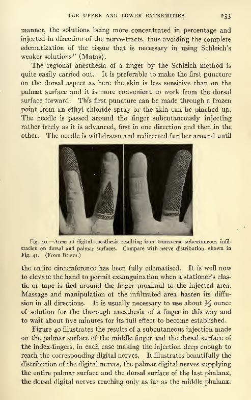
THE UPPER AND LOWER EXTREMITIES 253
manner, the solutions being more concentrated in percentage and
injected in direction of the nerve-tracts, thus avoiding the completeedematization of the tissue that is necessary in using Schleich's
weaker solutions" (Matas).
The regional anesthesia of a finger by the Schleich method is
quite easily carried out. It is preferable to make the first punctureon the dorsal aspect as here the skin is less sensitive than on the
palmar surface and it is more convenient to work from the dorsal
surface forward. This first puncture can be made through a frozen
point from an ethyl chloride spray or the skin can be pinched up.
The needle is passed around the finger subcutaneously injecting
rather freely as it is advanced, first in one direction and then in the
other. The needle is withdrawn and redirected further around until
Fig. 40. Areas of digital anesthesia resulting from transverse subcutaneous infil-
tration on dorsal and palmar surfaces. Compare with nerve distribution, shown in
Fig. 41. (From Braun.)
the entire circumference has been fully edematised. It is well nowto elevate the hand to permit exsanguination when a stationer's elas-
tic or tape is tied around the finger proximal to the injected area.
Massage and manipulation of the infiltrated area hasten its diffu-
sion in all directions. It is usually necessary to use about % ounce
of solution for the thorough anesthesia of a finger in this way and
to wait about five minutes for its full effect to become established.
Figure 40 illustrates the results of a subcutaneous injection madeon the palmar surface of the middle finger and the dorsal surface of
the index-fingers, in each case making the injection deep enough to
reach the corresponding digital nerves. It illustrates beautifully the
distribution of the digital nerves, the palmar digital nerves supplyingthe entire palmar surface and the dorsal surface of the last phalanx,
the dorsal digital nerves reaching only as far as the middle phalanx.

254 LOCAL ANESTHESIA
nervus digitalis dorsalis
arteria digitalisdorsalis
arttrio digitalis/'"
roininiinis
Fig. 41. A lateral view of the nerves and vessels of the index-finger. (Sobotta and
McMurrich.)
Fig. 42. Points of injection and lines of infiltration in operating upon fingers and
hand. (From Braun.)
Fig. 43. Points of injections and lines of infiltration for anesthetizing two or more
fingers. (From Braun.)

THE UPPER AND LOWER EXTREMITIES 255
The disparity in the two shaded areas represents the overlapping of
the fields of the two nerves (Fig. 41).
A study of Figs. 42-46 will suggest the further application of
regional methods to the base of one or more fingers and parts of the
Fig. 44. Points of injections and lines of infiltration for resecting part of hand.
(From Braun.)
hand, the large dots indicating the points at which the nerves are to
be reached by subcutaneous injection for paraneural infiltration, the
dotted lines marking the course for intradermal infiltration. As the
Fig. 45. Points of injections and lines of infiltration for anesthetizing abscess at base
of fingers. (From Braun.)
nerves in the hand are all small it is practicable to use Solution No. i
throughout, but, if preferred, the paraneural or deep injections can
be made with 0.5 per cent, novocain solution, using 10 to 20 minims
about each nerve. It is evident that in extensive resections of the

256 LOCAL ANESTHESIA
hand it is preferable to resort to regional anesthesia at the elbow or
above the wrist, rather than resort to too extensive infiltrations in
this region.
It may, however, be desirable to use methods of infiltration for
securing regional anesthesia, as these methods are always simple and
quickly carried out and when properly done, thoroughly effective.
For illustration take the dorsum of the hand as shown in Fig. 44,
in which we will say there has been an injury which will require the
amputation of all four fingers at the metacarpo-phalangeal joint. Apoint is selected about the middle of the dorsum and an intradermal
wheal established with a fine needle. The long fine needle and large
Fig. 46. Points of injections and lines of infiltration for resecting digits or part of hand.
(From Braun.)
syringe is now used throughout; the needle entered through this
wheal is directed subcutaneously, first towards one side of the hand
and then the other, injecting freely as it is advanced. The needle
is now directed down through each interosseous space injecting as it
is advanced, all the way to the skin of the palm. The needle is nowentered in either one or the other end of the subcutaneous infiltration
made along the dorsum which is continued subcutaneously across
the palmar surface, thus encircling the hand. This establishes a wall
of anesthesia through the hand from skin to skin and all nerve-fibers
passing through it are effectively blocked. It is usually necessaryto wait about five minutes for full anesthesia. As a rule no constric-
tor is needed as the adrenalin in the solution acts as an effective

THE UPPER AND LOWER EXTREMITIES 257
hemostatic, and the pressure from the infiltration is also a material
hemostatic aid. About 2^2 or 3 ounces of solution are usually needed
for this operation. ->
By following the above method a smaller section of the hand can
be similarly blocked off as suggested by Figs. 42-44.
While this plan is always quick, simple and effective, it has its
objections and should not be used in diseased conditions where the
vitality of the tissues is already compromised, as it may be followed
by sloughing, but will find its most useful field in injuries to healthy
young adults. In diseased conditions it is preferable to block the
nerves higher up.
THE LOWER EXTREMITY
"What has been said of the upper extremity may be, in a great
measure, repeated in regard to the lower limbs. The general prin-
ciples and methods are the same, except that they vary in their topo-
graphic application. The infiltration, the paraneural infiltration,
and the regional (open) intraneural methods can all be utilized with
advantage according to the regions involved and the local and con-
stitutional indications furnished by the patients themselves.
"The infiltration method with weak solutions, according to
Schleich, with or without constriction, and the mixed infiltration-
neural methods are alone able to meet a vast number of indications.
The surgery of the toes and of their metatarsals and limited areas
of the soft parts, including the ligations of all vessels of the lower
extremity from the external iliac (R. N. Hartley, Leeds, 1895) to the
dorsalis pedis, can be made subservient to the infiltration method.
In our own practice we have notes of cases of ligation of the super-
ficial femoral at Scarpa's triangle, of the anterior and posterior tibial
(in one case a traumatic aneurysm of the middle third), and of the
anterior tibial, in which these operations were performed with infil-
tration on the Corning plan. We have repeatedly extirpated the
varicose saphenous vein from the groin to the knee, and performed
Sonnenburg's operation for varicose veins by ligation and partial
excision of the internal and external saphenous with simple infiltra-
tion anesthesia. Infiltration is also sufficient for opening abscesses,
including large, diffuse, purulent collections; in draining joints; in
the extraction of foreign bodies; in the removal of tumors, and in the
excision of ulcers of moderate size. It is particularly valuable in
making all variety of exploratory incisions to clear up doubtful diag-
nosis, etc. As early as 1888 I was able to extirpate a subperiosteal

258 LOCAL ANESTHESIA
sarcoma of the femur in a very thin subject. Josiah Roberts, of NewYork was able to perform a femoral supracondyloid osteotomy (Mac-ewen's operation for genu valgum) in a boy four years old, and an
excision of the hip (the head of the femur being removed below the
great trochanter) in a child six years of age by the same procedure.
These operations were performed by Coming's method as early as
1885, a 5 per cent, cocain solution being used ('New York Med.
Jour.,' October 24, 1885). Varick, by utilizing the same technic,
successfully amputated the thigh in 1886 ('New York Med. Jour.,'
vol. x, No. in).
"Trapani was also one of the first to report an amputation of the
thigh by Schleich's method (vide Alessandri's reports on anesthesia
to the Italian Surgical Society, Transactions for 1897). Schleich
and his followers, Rhodes (of California), Cowan, and others, were
among the earliest to report instances of amputation of the leg by
simple infiltration, and Wilkerhauser (' Operationen mit Schleichscher
Analgesie,' abstract, 'Centralbl. f. Chir./ October 21, 1899, No. 42)
reports 18 extensive bone operations out of a list of 113 major opera-
tions done by this method, in which the sections of the thigh and leg
bones were required. All of these earlier reports, which can now be
multiplied many hundred times, simply confirm the statement pre-
viously made that it is necessary to exercise judgment in the adap-tation of the various methods of local and regional anesthesia to
special conditions. The operator should not be wedded to any single
method, but knowing the capabilities of each can select his technic
and, at times, obtain surprising results with a method that would
appear to the inexperienced as theoretically inadequate to meet the
demands of the case" (Matas).
"But, in spite of the numerous interventions on the lower limbs
which have been obtained by simple infiltrations with the Corningor Schleich methods, it must be recognized that these successes have
been (with the notable exception of the toes) more conspicuous bytheir rarity than by their frequency. They simply illustrate what
can be done with the method in exceptionally favorable conditions,
both as regard to the morale of the patient and the favorable ana-
tomic condition of the parts. This is particularly true of all extensive
operations involving the skeleton of the foot, leg, and thigh in robust,
fleshy subjects. In this class of patients local anesthesia is, as a rule,
inadequate, and when an excision of a large joint (the ankle or knee),
or when a large sequestrotomy, osteotomy, or an amputation is con-
templated, a method more positive and reliable is required to accom-

THE UPPER AND LOWER EXTREMITIES 259
plish the intervention with that freedom of action that can only come
from absolute analgesia. It is precisely under such circumstances,
and when the contra-indications to general anesthesia are positive,
that the regional intraneural method can be confidently appealedto" (Matas).
The Nerve-supply of the Lower Extremity. The external cut-
aneous nerve emerges from the pelvis, close to the anterior superior
spine of the ilium, beneath Pouparts' ligament. In thin subjects it
is easily reached in this position or just below it by a paraneural
injection. The anterior branch of this nerve emerges from beneath
the fascia lata, about 4 inches below Poupart's ligament, and be-
comes subcutaneous, supplying the skin on the anterior and outer
side of the leg as far as the knee.
The posterior branch curves backward and supplies the skin on
the outer and posterior aspects of the thigh as far as the middle of
the limb.
Dr. Hugh Young was one of the first to utilize paraneural injec-
tions of this nerve to obtain skin-grafts from the outer side of the
thigh.
The obturator nerve enters the thigh through the upper part of
the obturator foramen and divides into anterior and posterior
branches, which are separated from each other by fibers of the ob-
turator externus and adductor brevis muscles. The anterior branch
passes down behind the pectineus and adductor longus and communi-
cates with the internal cutaneous and internal saphenous nerves,
forming a plexus around the femoral artery, which descends on this
vessel to near the knee-joint; occasionally this communication fur-
nishes a cutaneous branch to the thigh and leg; when this occurs this
nerve passes beneath the adductor longus and sartorius muscles and
becomes superficial at the inner side of the knee, communicating with
the long saphenous nerve, and is distributed to the inner side of the
leg as low as its middle. When this branch is absent its place is
supplied by the internal cutaneous. The posterior branch supplies
the adductor muscles, and sends a branch to the knee-joint which
descends upon the popliteal artery to the back of the joint.
The deep position of the obturator nerve where it enters the thigh,
and its occasional contribution to the cutaneous nerve-supply of the
leg, makes this nerve often a troublesome factor in the regional anes-
thesia of the lower extremity.
The anterior crural nerve emerges from beneath Poupart's liga-
ment, lying on the outer side of the femoral artery; it immediately

260 LOCAL ANESTHESIA
divides into an anterior and posterior set of branches, which are sep-
arated by the external circumflex vessels.
The middle cutaneous, from the anterior division of the crural,
becomes superficial about 3 inches below Poupart's ligament by pier-
cing the fascia lata; it divides into two branches, which descend on the
front of the thigh supplying the skin as far down as the knee.
The internal cutaneous nerve, from the anterior division of the
crural, passes obliquely across the upper part of the sheath of the
femoral artery, and divides in front or at the inner side of that vessel
into anterior and posterior branches. The anterior branch passes
down on the sartorius muscle and perforates the deep fascia at the
lower third of the thigh, and is distributed to the skin of this region
and the inner, anterior, and outer surfaces of the knee. The pos-
terior or internal branch pierces the fascia lata on the inner side of
the knee in front of the sartorius tendon, and is distributed to the
skin on the inner side of the leg.
The internal or long saphenous nerve, from the posterior division
of the anterior crural, approaches the femoral artery beneath the
sartorius muscle, lying first in front and then on the inner side of
this vessel; continuing down with it in Hunter's canal, it becomes
superficial by piercing the deep fascia on the. inner side of the knee
between the tendons of the sartorius and gracilis muscles; it then
descends along the inner side of the leg in company with the internal
saphenous vein, lying just behind the inner border of the tibia, dis-
tributing branches to the inner and anterior aspects of the leg, and
terminates by passing in front of the internal malleolus, to be dis-
tributed to the skin on the inner side of the foot as far forward as the
great toe.
The small sciatic nerve descends beneath the pyriformis muscles
with the great sciatic, lying slightly to the inner side of the latter; the
perineal and pudendal branches curve upward to these regions, while
the femoral cutaneous branches pass down the back of the thigh to
supply the skin as far down as the back of the leg.
The great sciatic nerve descends into the thigh beneath the pyri
formis muscle, lying midway between the tuberosity of the ischium
and the great trochanter, and passes down to about its lower third,
where it divides into internal and external popliteal nerves; this
division may, however, take place at any part of its course from the
pelvis down, and should be borne in mind in injecting this nerve high
up to insure getting both trunks.
The internal popliteal, the larger of the two branches, descends

THE UPPER AND LOWER EXTREMITIES 261
along the back part of the thigh and middle of the popliteal space,
lying first on the outer side of the artery, then crossing behind it to its
inner side and passing with it beneath the arch of the soleus, whenit becomes the posterior tibial.
The posterior tibial nerve, deeply situated above, becomes more
superficial lower down, where it is covered by the skin and fascia; in
the lower third of the leg it lies just internal to the margin of the
tendo achillis; in the interval, between the malleolus and the heel, it
lies external to the artery against the posterior surface of the tibia
and about i cm. internal to the tendo achillis; in this position it is
readily accessible for paraneural injections. Lower down in this
space the nerve divides into internal and external plantar branches.
The internal branch supplies the sole and inner side of the foot and
gives off digital branches to the inner side of the big toe, adjoining
sides of the big and second toe, second and third and fourth toes. It
will be observed that the distribution of this nerve is almost identical
to the distribution of the median in the hand, the digital branches
giving off dorsal cutaneous branches at the base of the toes in the
same manner as occurs in the hand.
The external plantar nerve supplies the outer side of the foot,
little toe, and adjoining side of the fourth toe, together with deepmuscles of the foot, closely corresponding to the distribution of the
ulnar in the hand.
The external popliteal or peroneal nerve descends close along the
inner margin of the biceps tendon on the outer side of the popliteal
space; it then passes between the tendon of the biceps and outer
head of the gastrocnemius muscle and curves around the head of the
fibula, where it can be readily felt, and is again accessible for ex-
posure or for paraneural injection; it then descends into the substance
of the peroneus muscles. The cutaneous branches from this nerve
supply the skin of the back part and outer side of the leg as far down
as the heel. In the substance of the peroneal muscles this nerve
divides into anterior tibial and musculocutaneous branches.
The anterior tibial nerae is deeply situated in the upper part of its
course, but becomes more superficial near the ankle, lying to the
outer side of the dorsalis pedis artery and between the extensor pro-
prius hallucis and the extensor longus digitorum; at this point, just
above the annular ligaments, it is fairly accessible for exposure and
direct injection, or for paraneural injection after locating the dorsalis
pedis pulse by passing the needle down to the deep fascia just external
to the artery; however, it would be preferable in making a para-

262 LOCAL ANESTHESIA
Fig. 47. Cutaneous nerve-supply of
the lower extremity (anterior view): A,Genitocrural; B, ilio-inguinal; C, external
cutaneous; D, middle cutaneous; E, inter-
nal cutaneous; F, lateral cutaneous of
peroneal; G, internal saphenous; H, ex-
ternal saphenous; 7, musculocutaneous;
J, external plantar; K, internal plantar;
L, anterior tibial. (Campbell.)
Fig. 48. Cutaneous nerve-supply of
the lower extremity (posterior view): A,Small sciatic; B, internal cutaneous; C,
external cutaneous; D, lateral cutaneous;
E, internal saphenous ; F, external saph-
enous; G, musculocutaneous; H, internal
calcaneus. (Campbell.)

THE UPPER AND LOWER EXTREMITIES 263
neural injection to do so higher up, where the peroneal nerve winds
around the head of the fibula, thus reaching the anterior tibial and
muscular cutaneous distribution. At the ankle-joint the nerve
divides into an internal and an external or tarsal branch. The in-
ternal branches supply the adjoining sides of the great and second
toes. The external branch supplies the adjoining sides of the second,
third, and fourth toes and extensor brevis muscles.
The musculocutaneous nerve becomes superficial at the lower
third of the leg by passing forward between the peronei muscles
and extensor longus digitorum; in this position it can be reached bya subcutaneous injection, made across the lower portion of the leg
at this point, over the tendons of the above muscles. As the nerve
descends it divides into two branches, an internal, which passes
over the front of the ankle-joint and supplies the inner side and dor-
sum of the foot, inner side of the great toe, and adjoining sides of
the second and third toes. The external branch supplies the skin
on the outer side of the foot and ankle, and the adjoining sides of
the third, fourth, and fifth toes (Figs. 47, 48).
A study of the above nerve-supply, with an observance of the
points at which the nerve-trunks and their principal branches are
accessible, will suggest many opportunities for the practice of regional
anesthesia. The following operative procedures are a fairly thoroughreview of the surgical possibilities.
SCARPA'S TRIANGLE and the inguinal region receive their
nerve-supply from a variety of sources which are not capable of being
dealt with collectively by regional methods, consequently, all opera-
tive procedures here should be done under infiltration, from the
simple incising of a suppurative bubo to the removal of the entire
superficial group of glands. Where the deep group is involved,
requiring dissections beneath the fascia lata, a general anesthetic
should if possible be used.
In operating here it is preferable to complete the entire infiltra-
tion before making the incision^ as it is difficult to anesthetize the dif-
ferent planes of tissue after they have been divided, as the solution
runs out as fast as injected. The infiltration had best be done on the
Hackenbruch plan, by first encircling the mass by a line of intrader-
mal anesthesia; the needle is then passed from this line into the deeper
tissues, all around and under the mass of glands, thus completely
enclosing them within a wall of anesthesia (Figs. 19-49). Solution
No. i and a few drops of adrenalin to the ounce is used; after a few
minutes' delay allowed for thorough saturation of the tissues the

264 LOCAL ANESTHESIA
operation may be begun, and should be entirely painless. Where the
deep glands are involved, and it is necessary to go below the fascia
lata, the different tissues should be infiltrated as the operation ad-
vances, but this procedure here may sometimes prove difficult. Care
should be observed to bear in mind the position of the vessels, and
when infiltrating in a doubtful region to make the injection only
when advancing or withdrawing the needle.
Other tumors of this region may be removed in the same way.
Fig. 49. Shows method of infiltrating base of bubo area by passing needle obliquelydownward and inward after embracing area within circle of cutaneous anesthesia.
The removal of skin-grafts is quite easily performed from the an-
tero-external aspect of the thigh, the usually selected site, either bydirect intradermal infiltration of the entire area, from which the
grafts are to be removed (this intradermal edematization of the skin
greatly facilitates their removal without apparently affecting the
vitality of the grafts), or by a pareneural injection of the external
cutaneous nerve, where it emerges from beneath Poupart's ligamentclose to the anterior superior spine, as first practised by Dr. Young of
Johns Hopkins. For this injection the needle is best entered from the
outer side and penetrated to a sufficient depth to reach beneath the
fascia lata, under which it is advanced, depositing 2 drams of about a

THE UPPER AND LOWER EXTREMITIES 265
0.5 per cent, solution of novocain in the recognized position of the
nerve. Anesthesia should set in after a few minutes, and be suffi-
ciently extensive to allow of a fairly liberal removal of tissue.
Varicose Veins of Leg. In the removal of varicose veins of the
leg, the operation which the author prefers is the removal of the en-
tire vein with such of its tributaries as seem necessary, from the
saphenous opening above to the ankle below.
After trying many methods for producing anesthesia, the follow-
ing plan has been found the most satisfactory.
Usually four points are selected for making the injections, which
are the areas through which the incisions will be made; one just below
the saphenous opening in the fascia lata, one slightly above the
knee, one just below the knee, and one just above the ankle (see
Fig. 50). At each of these points a transverse area of anesthesia is
'/)'surface
in wheat'andarea ofinfiltration
Vein
FIG. 50. Removal of various veins of leg. The smaller illustration shows the detail.
established running across the leg at right angles. This infiltration
is done in the same methodical manner at each point. Very weak
solutions are sufficient for this work, either 0.25 per cent, novocain or
0.20 per cent, eucain; usually 8 ounces are required, to which is added
20 drops of adrenalin solution i : 1000. All four areas are infiltrated
before the operation is begun; this is quickly done by pursuing the fol-
lowing plan. With the small syringe and fine needle, an intradermal
wheal is first produced in the center of each of the four areas to be
infiltrated; this point should be about over the saphenous vein. The
small syringe is now discarded and the large syringe and long needle
used throughout. Starting above at the wheal near the saphenous
opening, the needle is directed subcutaneously close under the skin
at right angles to the axis of the leg, injecting as the needle is ad-
vanced for a distance of about 2 inches. The needle is partly with-

266 LOCAL ANESTHESIA
drawn and redirected in the opposite direction, which is similarly
injected.
Partially withdrawing the needle again, it is directed at a slight
angle to the first injection and passed down nearer to the deep fascia
for a distance of about 2 inches; always injecting steadily when the
needle is being advanced. The opposite side is treated similarly.
Two deep injections are now made down to or slightly below the deep
fascia, one on each side of the position of the vein. During these
several injections, the needle is not withdrawn from the skin, the
angle being changed by partially withdrawing it, and the syringe
detached from time to time for refilling. Each of the four areas for
infiltration is treated similarly, using about 2 ounces of solution at
each point; about 8 ounces in all, slightly more for very stout cases
and slightly less for thin ones.
By completing the infiltration of all four areas before the opera-
tion is begun, the solution is allowed ample time to thoroughly satu-
rate the tissues, producing the maximum degree of anesthesia and
diffusing outward toward the skin, rendering it unnecessary for direct
infiltration here. This methodical method of procedure is very
quickly done and saves much unnecessary loss of time.
By beginning the anesthesia above and progressively workingdown the leg, many of the cutaneous nerves which reach the lower
parts are effectively blocked by the upper injections and brings each
succeeding area of injection somewhat under the influence of the
injection above. Whereas, if the opposite order of procedure were
followed, we would constantly be encountering fibers from the same
nerve in a sensitive state.
My method of operating has some advantages and is particularly
suited to local anesthesia. It consists essentially in enucleating the
vein from the surrounding tissue by the use of the finger, which is
much more certain of removing the entire vein than an enucleator
which often breaks it off, particularly if diseased and friable. Anincision is made through the upper anesthetized area just below the
saphenous opening and the vein secured, ligated proximally and
divided, a stout forcep securing the distal end. By traction on this
fcreep, the vein is held taut and its course well outlined; the finger is
now worked down circumferentially around it, separating it from the
surrounding tissue, any branches that are encountered are either
broken off if small, or by locating them with the finger, a bistoury is
passed down alongside the finger and they are divided. After reach-
ing as far down as can be readily done from the upper opening, the

THE UPPER AND LOWER EXTREMITIES 267
area next below is incised, by traction on the vein above its exact
position is readily made out, but if not quickly located, a long probeis passed down through the lumen and can be readily palpated
through the tissues and unnecessary dissection avoided. The proc-ess of enucleation is similarly continued from this point down to the
ankle. The entire procedure is quickly carried out with little trauma
to the tissues.
THE HIP AND THIGH
"Regional anesthesia, in amputation of the middle third of the
thigh, was first accomplished by Crile in 1899 ('Cleveland Medical
Gazette,' July i, 1899, vol. xiv), and by Berndt (Gritti's osteoplastic
amputation) ('Muench. Med. Wochenschr.,' 189*9, No. 27), the former
by the intraneural and the latter by the paraneural methods. I
know of no case in which the disarticulation of the hip has been done
by 'blocking' the nerves, though I believe that this is feasible whenit is possible to cut the soft parts at a lower level, as in the Furneaux-
Jordan amputation. In such a case the preliminary anesthesia of the
anterior crural and external cutaneous at the groin, and the sciatic,
just below the gluteus maximus muscle, will suffice, if care is taken
not to cut the obturator, when this is reached in making the deeperinner section of the thigh, until this nerve has been recognized and
infiltrated. In amputation at a higher level (Wyeth's operation)
the anesthesia could only be accomplished by a preliminary circular
infiltration, including the individual nerve-trunks, which would have
to be anesthetized as they were met. Such a procedure would tax the
self-control of the patient to the utmost, and would be so tedious that
it could scarcely be recommended except in very thin and wasted
subjects" (Matas).In amputations at the lower third of the thigh, as well as at the
middle, disarticulations at the knee or amputations at the knee, as
in the Gritti-Stokes operation, the difficulties presented by the ob-
turator nerve are more easily met. The anterior crural, external
cutaneous, great sciatic, and lesser sciatic should all be injected intra-
neurally at the root of the limb with 0.5 of i per cent, novocain solu-
tion, with a few drops of adrenalin solution (1:1000) to the ounce.
It is very necessary not to overlook the lesser sciatic, which lies just
to the inner side of the great sciatic, where this nerve enters the limb,
as its branches are distributed to the skin as low down as the popliteal
space and back part of the leg; it is probable that some failures re-
ported by this method have been due to this neglect as well as to
other details. It is more convenient, when operating by the neuro-

268 LOCAL ANESTHESIA
regional method, to use a posterior rachet incision. After the poste-
rior incision has been made, and the deep muscles slightly separated
with the finger to expose the vessels, a long needle is used to infiltrate
the region around the vessels (the path of the obturator nerve) with
solution No. i or less freely with 0.5 per cent, novocain. A few min-
utes following this last injection all parts involved in the field of
operation should be as anesthetic as under general narcosis and the
steps of the operation proceeded with in the usual way. It would
seem unnecessary to state that all these operations should be per-
formed with the use of a constrictor applied to the upper part of the
thigh after the injection of the crural, external cutaneous, and sciatic
nerves.
To expose the sciatic nerve for intraneural injection the following
method will be found to be quick, simple and accurate. In the upper
part of the thigh just below the gluteal fold the nerve lies on an
antero-posterior plane that passes midway between the great trochan-
ter and tuberosity of the ischium, laterally it lies on a plane that
passes through the posterior surface of the femur lying i inch internal
to that bone. To expose it proceed as follows:
Make an intradermal wheal on the above midline just below the
gluteal fold; take a long fine needle and large syringe and enter
through this wheal directing the needle subcutaneously down the leg
for about 4 inches injecting as it is advanced, partly withdraw the
needle and direct it on a slightly deeper plane for the same distance.
Now direct it straight down through the wheal for a distance of about
3 inches injecting freely as it is advanced; repeat this deep injection
in a similar manner at one or two more points along the infiltrated
line.
A few minutes delay will establish complete anesthesia, when the
incision can be made and should be carried down through the deep
fascia, no further incisions are needed, as the hamstring muscles are
easily separated by the finger which feels its way down to the level
of the femur where the nerve, a trunk as large as the little finger, will
be found lying i inch on its inner side.
The small sciatic nerve also lies on the antero-posterior line just
superficial to the deep fascia and is consequently blocked by the in-
filtration.
The incisions made to expose these nerves should not be perma-
nently closed until the operation is completed, but only loosely ap-
proximated with superficial stitches, for if anesthesia is not completeit may be necessary to reopen the wounds for further infiltration of

THE UPPER AND LOWER EXTREMITIES 269
the nerves; this, however, will not be at all likely if the nerve has been
properly infiltrated, producing a fusiform enlargement at the pointof injection, in the case of a very large nerve like the great sciatic
entering the needle at two or more points in the nerve. As men-tioned elsewhere, this should be done with a very fine needle enteredin the long axis of the nerve-fibers; care should also be observed notto make traction on the nerve, which will cause pain, but to makethe injection when the nerve is slack. Some operators in discussingthese operations have preferred, after injecting the anterior crural
and external cutaneous nerves at Poupart's ligament, to infiltrate
Fig. 51. Method of securing anesthesia of femur for supracondyloid osteotomy. (Braun.)
the superficial tissues on the back of the thigh, making the handle of
the racket incision first, and exposing the sciatic nerve or its branches
at the upper part of this incision and injecting them high up here,
then infiltrating the recognized course of the obturator nerve. The
objection we have to offer to this procedure is that the small sciatic is
not injected, and its territory, together with that of any of the bran-
ches of the great sciatic given off above the point of its injection, will
have to be infiltrated. In the operations about the knee-joint
disarticulations and Gritti-Stokes amputation these objections are
not of as much consequence, as the area here to be infiltrated is nec-
essarily much smaller.

270 LOCAL ANESTHESIA
For operations upon the femur, as in the removal of osteomas,
sequestrotomy, etc., the method of injecting around the bone is shown
graphically in Fig. 51 . If the operation is to be performed exclusively
by infiltration, the soft parts must be infiltrated from the skin to the
periosteum along the proposed line of incision; or superficial regional
methods may be employed, in addition to the periosteal injections,
by blocking the external cutaneous or anterior crural nerves just be-
low Poupart's ligament, and this would seem the preferable plan
except in emaciated subjects.
THE KNEE-JOINT
As this joint receives, either in its cutaneous covering or deeper
parts, branches from practically all the nerves in the lower part of
the thigh, what has been said regarding the neuroregional methods of
that part are equally applicable here.
It will be seen that the deeply situated and difficultly accessible
obturator nerve may offer serious obstacles to a thorough and satis-
factory anesthesia of this part; it is here, then, that Bier's venous
anesthesia or spinal puncture may be advantageously used; however,in the hands of the skilful operator, this disadvantage may be over-
come, and almost any operation on the joint performed by purely
regional methods.
First block the anterior crural, external cutaneous, and sciatic
nerves at the root of the limb;this will leave only the territory sup-
plied by the obturator unanesthetized, which is represented by a
small area on the inner side of the knee and a part of the joint. Withall the other parts anesthetized, the operative incision could be madein such a way as to expose or easily approach the path of the obtura-
tor nerve on the inner side and just above the knee, and sufficiently
deep to feel freely the femoral vessels; rather free infiltration between
and around the vessels and on their inner side should reach all
branches of the nerve and leave the parts below completely anes-
thetic. Such a thorough procedure as the above will, however,
only be necessary in extensive resections of the joint, many lesser
procedures involving only the anterior parts of the joint (the partsmost frequently the site of surgical intervention) can be easily
performed through infiltration, or by blocking the anterior crural
and external cutaneous nerves at Poupart's ligament. The latter
procedure will suffice for the operative treatment of fracture of the
patella, drainage in infected arthritis, the removal of foreign bodies,
lipomatosis, and other similar conditions.

THE UPPER AND LOWER EXTREMITIES 271
In operating upon fracture of the patella by infiltration, the joint
cavity should be filled with i or 2 ounces of solution No. 2, with 5
drops of adrenalin (i : 1000), and allowed to remain for from five to ten
minutes before the joint is opened; this will anesthetize the synovialsurfaces and permit the painless removal of clots or a thorough wash-
ing out of the joint if infected. Even strong solutions, up to 2 percent, novocain, could be used if necessary, as most of it escapes after
the joint is opened. It is, of course, advisable to use a constrictor
above the knee in extensive operations here under infiltration andafter making a strong intra-articular injection. The method of
infiltrating around the patella region is seen in Fig. 52, and is best
done as follows: Make an intradermal wheal about 2 inches abovethe patella, enter a long fine needle here and advance it subcuta-
Fig. 52. Peri-articular infiltration for operation in patella region. (Braun.)
neously along the line as shown in Fig. 52, injecting freely as it is
advanced, repeat the same in the opposite direction, then again on each
side on a deeper plane close down around the capsule of the joint. Theneedle is now entered laterally where the last injections stopped and
subcutaneous and deep injections made as above on the lateral as-
pects of the joint curving forward so as to partially surround the
joint. This embraces the patella in a horseshoe- like area of infiltra-
tion reaching from the skin to the capsule of the joint. After a delayof a few minutes it should sufficiently diffuse to block all nerves enter-
ing the field from above and on the sides. As no nerves enter from
below it is unnecessary to complete the circle by the injection over
the ligamentum patella as shown" in the figure.
Intra-articular injections may be made use of in breaking up

272 LOCAL ANESTHESIA
adhesions within the joint when not too firm; it should be withdrawn
after five or ten minutes and the necessary manipulations resorted
to; or, after withdrawing the anesthetic solution, it can be replaced
with the 2 per cent, formalin-glycerin solution of Murphy, which, byits hydroscopic action, moderately distends the joint cavity and thus
prevents further immediate contact of the joint surfaces, particularly
when combined with extension.
THE LEG
All operations below the knee, involving the leg, ankle, and foot,
no matter what their extent, can be painlessly performed by a single
method. When near the knee, by injecting the external cutaneous,
anterior crural, and sciatic nerves as in the higher operations; if some
distance below the knee (middle third of the leg and below), it will be
sufficient to inject the sciatics at the root of the thigh and the long
saphenous by a pareneural injection, made transversely over the
inner surface of the knee between the tendons of the sartorius and
gracilis muscles, where this nerve becomes superficial.
It would seem superfluous to detail or describe the many opera-
tions possible, for where a part is thoroughly anesthetic all operations
are possible.
The following is taken from Prof. Matas' report on "Local and
Regional Anesthesia," etc., 1900, and cites one of the many clinical
cases which might be mentioned to illustrate these procedures:"Without any previous knowledge of Crile's work, and encour-
aged by previous successes with the same methods, as applied to the
upper extremity, I performed a Pirogoff operation for frost-bite bythis method in March, 1899. From March, 1899, to present date I
have availed myself of this mode of anesthesia many times, mycolleagues operating on other cases in their practice at the Charity
Hospital and elsewhere. In my cases there were reasons which madethe administration of a general anesthetic undesirable.
"In one of these, operated on before the medical class of Tulane
University, the inestimable advantage of possessing a reliable safe
method of analgesia as an alternative to general narcosis was made
particularly apparent. This case not only illustrates the circum-
stances in which this method is especially applicable, but it will
serve to describe the technic of the method as well.
"F. S. W., aged thirty-two, was admitted to the hospital December 18, 1899, for the
treatment of a diffuse tubercular arthritis of the right tarsus. The patient was suffering
with advanced pulmonary tuberculosis (cavity in lung), but his sufferings were so great

THE UPPER AND LOWER EXTREMITIES 273
that an operation was decided upon. In view of his weakened condition, special precau-tions were taken to guard against the accidents of general anesthesia. In addition to
the preparatory administration of strychnin, digitalis, and nitroglycerin by hypodermic,the nares were sprayed with a 2 per cent, cocain solution to diminish the nasolaryngealreflexes (Franck-Rosenberg). Chloroform was then administered over an Esmarchmask by the 'guttatim' method. Notwithstanding all the care taken, the patient
rapidly entered into a most violent stage of excitement and became rigid and cyanosed;
respiration was arrested, the pulse became irregular and imperceptible, and when the
tetanic rigidity ceased the patient sank as if completely collapsed, and it was only by the
immediate application of artificial respiration and other measures that he finally cameback to consciousness again.
" As the operation was imperative and all general anesthetics were not to be thoughtof (ether being contra-indicated by the phthisis), I decided to try the intraneural
method of regional anesthesia, which should have been the method of election at the
start. Accordingly, after careful preparation of the parts, the skin and underlyingtissues of the upper popliteal space were infiltrated with a Schleich No. i cocain solution,
and an incision 4 inches long was made so as to bring the sciatic nerve into view. This
done, an injection of 25 minims of the same No. i (J^ of i per cent.) solution was injected
into the trunk of the nerve. A constrictor was applied after exsanguinating the limb
by gravity care being taken to pad the limb well so as to minimize the discomfort it
might produce. Eighteen minutes after the injection of the cocain some sensibility
still existed in the foot; fearing that the solution would be insufficient, 20 minims of a i
per cent, cocain were then injected into the exposed nerve. In three minutes the anes-
thesia of the entire region below the sciatic infiltration was complete and the operationwas begun twenty-five minutes after the first injection into the nerve had been given.
The tarsus was then explored by making a free externolateral incision, and all the bones,
including the tarsometatarsal articulation, were found to be involved in a diffuse
tuberculosis. The astragalus alone was saved. The chisel, gouge, and bone curet were
used freely with the hope that a simple excision might suffice, but the lesions of the skele-
ton and soft parts, including the tendon-sheaths, were so extensive that an atypical
subastragaloid amputation on the Roux-Lignerolles plan was decided upon. The
patient, who had been perfectly quiet and passive, was now asked his consent to the
amputation, which at first he refused, but, after showing him the extent of the lesions
and explaining to him the advantages of a radical extirpation in a man in his condition,
he consented, and the amputation was performed."The patient gave us very material assistance in this operation, not only by holding
his foot and leg in the most favorable attitudes for our work, but by turning his bodyaround without assistance when, at the termination of the operation on the foot, weclosed permanently the sciatic incision. The contrast between the alarming condition
induced by the general anesthetic (chloroform) and the passive and calm attitude of the
patient throughout the operation was most impressive. In this case it should be men-
tioned the saphenous nerve was not injected, as in the other similar cases; but, instead
of this, the short incision through the skin connecting the inner border of the foot with
its outer edge, which is supplied by this nerve, was bridged over by a line of infiltration
edema. The operations successfully performed by this method in my practice have
been (i) Pirogoff's amputation for frost-bite; (2) Syrne's operation; (3) two atypical
resections of the tibio-astragaloid joint, in which the astragalus and calcaneum were
excised together with the tibiofibular surfaces and their malleoli, for tuberculosis; (4)
Guyon's supramalleolar amputation of the leg for trauma; and (5) an extensive search in
the thigh for a lost bullet embedded in the neighborhood of Hunter's canal. In the last
case the anterior crural nerve and external cutaneous were cocainized under Poupart's
ligament. In this case we were misled in the situation of the bullet as indicated by
radiograph, and failed to find the bullet even after a most extensive dissection in the
18

274 LOCAL ANESTHESIA
lateral and posterior femoral aspects of the thigh had been made. The anesthesia in
this case was complete from the middle of the thigh to the toes, but there was marked
sensation in the upper femoral regions in consequence of the preservation of the lesser
sciatic filaments which overlapped beyond the points of the greater sciatic infiltration
which had been effected just below the crossing of the lower gluteal fibers. The small
area of persistent dermal sensibility could have been easily controlled by a short trans-
verse line of purely dermal infiltration, the deeper parts being completely insensitive."
The Toes, Metatarsals, and Sole of the Foot. What has been
said regarding the fingers and metacarpals may be largely repeated
for the foot. With certain modifications, any of the smaller toes maybe easily anesthetized by edemetization carried around its base.
The big toe is, however, more effectually treated by paraneural in-
jections made around the base, or, as in Fig. 53 , where, it is necessary
to remove the entire toe..
Fig. S3- Points of injections and lines of infiltration for bunion operations or resection
of great toe. (From Braun.)
Bunions. This common annoyance and source of discomfort is
effectively operated in the following manner. Make an intradermal
wheal just back of the head of the metatarsal bone on the inner side
of the foot, enter the long fine needle here and direct it subcutaneouslyover the dorsum of the foot obliquely toward the interval between
the first and second toes injecting freely as the needle is beingadvanced. Partially withdraw the needle and direct it subcuta-
neously in a similar direction on the plantor surface, again partially
withdraw it to near the starting point and again direct it toward the
interval between the first and second toes this time passing it close in
contact with the bone, between the bone and the muscles, on the under
surface always injecting freely while it is being advanced. After a
few minutes delay anesthesia will be complete for both bone and soft

THE UPPER AND LOWER EXTREMITIES 275
parts; the bone can be resected and the operation performed by anytechnic preferred. I have used this method a large number of times,
it is quick, simple and effective and has always been followed byexcellent results. In operating for ingrowing toe-nails, Dr. Braun
speaks highly of the use of ethylchlorid spray, used about the base
of the toe, claiming it is to be quite sufficient to remove the matrix as
well as the nail. We have never used this method, always preferring
to use infiltration or paraneural injections at the base with solution
No. i, to which is added 5 drops of 1:1000 adrenalin to the ounce.
This method has the advantage of producing quite a lasting analgesia,
and by the time sensation does return very little pain is experienced ;
should the operation be performed in the office and the patient
allowed to go home afterward, he is likely to reach his destination
before any discomfort is felt, although few rarely complain of anybut slight soreness following. Figure 56 shows method of anesthetiz-
ing the lesser toes, and should be done in the same manner described
for similar operations on the hand.
In operations upon the sole of the feet for removal of splinters and
other foreign bodies it is often quite a difficult matter to satisfactorily
infiltrate the pulp of the foot; this tissue is so dense and unyielding
that even with solutions of considerable strength much difficulty is
experienced. Rather than continue at efforts of infiltration after
this has been found difficult, it would be simpler and preferable to at
once make a paraneural injection around the posterior tibial nerve, as
described in the following experiments, thus securing at once anesthe-
sia of the entire foot; additional injections being made over the
inner or outer ankles to reach the branches of the long saphenous or
peroneal nerves should this be necessary. A review of the follow-
ing quotations from Braun ("De Lokal Anesthesie"), will suggest
many useful applications in practical surgery when limited in extent
or confined to the superficial parts, but any extensive operations
involving resections of the foot had better be performed by the
intraneural methods of blocking the nerve higher up, as already
described.
Figure 54, from Braun ("Die Lokal Anesthesie"), shows a cross-
section through the ankle-joint, at the level of the most prominent
portion of the internal malleolus; the posterior tibial nerve is best
reached as indicated by the arrow, the artery lying internal to the
nerve. Braun gives the following directions for reaching it at this
point: "The needle is inserted about i cm. from the inner side of
tendo achilles, and directed from behind forward until the posterior

276 LOCAL ANESTHESIA
Extensor hall.
Superf. peroneal nerve "/*
Deep peroneal nerve
Extensor dig.'
..'Tibialis ant.
,. Tibialis post.
.^Flexor dig.
Peroneal mus
Flexor hall. *'
Ext. saphenous nerve''
- Method of reaching posterior tibial nerve at ankle-joint for paraneural
injection. (From Braun.)
i
L
Fig. 55. Lines of subcutaneous infiltration and resulting areas of anesthesia in foot.
(From Braun.)

THE UPPER AND LOWER EXTREMITIES 277
surface of the tibia is reached; the needle is then slightly withdrawn
and the solution injected. He states that the injury of the vessel
which lies on the inner side of the nerve is hardly to be feared, but it
should first be made sure that the point of the needle is not in the
vessel, by resorting to a little aspiration before the injection is made
by slightly withdrawing the plunger. Should it be found that the
vessel has been punctured, no unpleasant consequences are likely
to result if the needle has been fine."
The following experiment indicates the results of an injection
made in this manner:
Fig. 56. Disarticulation of third toe. (From Braun.)
"Experiment i (Dr. B.). One-half cubic centimeter of a i percent, solution with i
drop of adrenalin (i: 1000) was injected in the above-described manner, and almost im-
mediately anesthesia appeared as indicated in Fig. 55, No. I, and lasted for three hours.
The affected area on the extensor surface of the foot and toe is indicated by the shaded
surface."
This procedure was once employed for opening an abscess and
removing a foreign body from the sole of the foot. The anesthesia
extends to the metatarsals and tarsus:
"Experiment 2. Two cubic centimeters of o . 5 per cent, cocain solution with 4 drops
adrenalin solution, injected subcutaneously over the inner ankle, beginning behind the
tendo Achillis and extending around to the middle line of the joint in front. This will
meet the terminal branches of the internal saphenous nerve; the extent of the resulting
anesthesia is shown in Fig. 55, No. II."

278 LOCAL ANESTHESIA
The injection in Experiment 2 reaches the terminal branches
of the internal saphenous nerve, and may often be combined with the
injection of the posterior tibial, as in Experiment i, for anesthesia of
the inner side and sole of the foot.
In commenting upon these experiments, Braun states that a
subcutaneous injection, made across the extensor surface of the
ankle-joint, reaches only a few of the fibers of the internal saphenous
and produces only a limited area of anesthesia on the back of the
foot, the same as in a corresponding injection made on the back of the
hand, while a much more extensive area is affected if the injection is
made slightly higher, as in Experiment 3, where the superficial
branches of the peroneal nerves are reached.
"Experiment 3. Three cubic centimeters of 0.5 per cent, cocain solution with the
addition of 10 drops adrenalin solution (1:1000) was injected a hand's breadth above
Fig. 57. Area of anesthesia for tenotomy of tendo Achillis. (Braun.)
the outer ankle, across the axis of the limb, from the tendo Achillis behind to the edge of
the tibia in front. After six minutes the skin, as indicated in Fig. 55, No. Ill, had be-
come anesthetic and remained so for three to four hours."
In discussing this experiment, Braun states that this injection
reaches all of the superficial fibers of the peroneal nerve, the anes-
thetic field extending from the territory of distribution of the internal
saphenous at the inner ankle and inner side of the foot across the
dorsum to the outer side of the foot. The results obtained in Ex-
periment 4 may also at times be applied practically.
"Experiment 4. Three fmgers'-breadth above the internal ankle the needle was en-
tered laterally, between the tendons of the tibialis anticus and extensor hallucis longus,
vertical to the cutaneous surface till the bone was reached; the needle was now turned
laterally under the tendon of the extensor hallucis and an injection of i c.c. of 0.5 percent, cocain with 3 drops adrenalin solution (i : 1000) was injected; ten minutes later
anesthesia was established in the terminal branches of the peroneus profundus, as indi-
cated in Fig. 55, No. IV."
The method of infiltration for tenotomy of the tendo Achillis is
shown in Fig. 57, the infiltration carried well down around the tendon.
CHAPTER XV
NECK
"!N the surgery of this region local anesthesia has made large
and permanent conquests. The neck is most favorable for the dis-
play of the infiltration method, the paraneural and intraneural meth-
ods of regional anesthesia having found comparatively few typical
applications. In the neck the lesion of the skin and its appendagesand those of the supra-aponeurotic planes are everywhere submissive
to cocain or its allies. Local infiltration is most valuable in dealingwith inflammatory lesions abscesses, boils, inflamed sebaceous cysts,
and carbuncles of moderate size. In opening deep cervical abscesses
connected with submaxillary and pharyngotonsillar infections, in
which the suppurative focus must be reached by careful dissection
(the Hilton-Rose method), it is invaluable.
"In the major surgery of the neck, local infiltration finds its most
brilliant applications in the anterior cervical and subclavian regions,
and in the operations on the vessels in the carotid triangles.
"Apart from the avoidance of postoperative constitutional dis-
turbances, the immediate advantages local of anesthesia are that it
permits the dissection of the parts with the precision, neatness, and
deliberation that are required in all the deep vascular regions; that
the great turgidity of the veins and general increase in vascularity
incident to the use of inhalation anesthetics is avoided; and that the
surgeon is materially assisted in his work by the different attitudes
that the patient can assume to favor the better exposure of the parts.
"In the postcervical triangle the conditions for local anesthesia
are less favorable, except in the supraclavicular space, in which the
subclavian artery and brachial plexus are readily exposed for opera-
tive purposes" (Matas).
Nerves of the Neck. In the neck the only opportunity for the
application of regional methods of anesthesia, aside from paraverte-
bral methods, is the superficial branches of the cervical plexus as they
emerge around the posterior border of the sternomastoid about the
middle of the neck (Fig. 58). Here the occipitalisminor, auricularis
magnus, superficialis colli, and the descending or supraclavicular
branches are all fairly accessible, and in their emergence from the
279

280 LOCAL ANESTHESIA
deeper parts are all met within a comparatively limited area. To
reach and block these nerves in this position it is, however, not;
\
posterior facial vein.
Ptatysma x
cervical branch o,
facial nerve
posterior/auricular vein
Sternocleido-
/' mastnidcus
cervical nerve
external jugular vein^^
cutaneous eervical nerve JPv
-great occipital nmroccipital vein
occipital artery
sailor brs. of cervical plexus
Omohyoideus (inferior telly)
^ posterior supraclavicularnerves
anterior supraclavicular nervesmiddle supra-
clavicular nerves
Fig. 58. The superficial nerves and veins of the left side of the neck (second layer
of neck). The platysma has been divided, the upper portion reflected toward the jaw,
and the lower portion removed. The fascia has been divided along the facial veins.
X = Anastomosis of accessory nerve with cervical plexus, -f-= Communication of ex-
ternal jugular vein with deep veins. The upper perforating branches of the internal
mammary vessels (not represented in the illustration) make their appearance between
the origins of the sternocleidomastoideus. (Sobotta and McMurrich.)
necessary to make an open dissection, though this can be done;
applying intraneural or perineural injections to each individual nerve,

NECK 28l
it will, however, be found equally satisfactory and much simpler to
pass a long needle down to the posterior region of the sternomastoid
at the midpart of its course to the point of emergence of these nerves,
and here making a fairly liberal infiltration of from 3 to 4 drams of
i per cent, novocain, containing a few drops of adrenalin, distributing
the solution up and down this area for about 2 % inches, thus effectu-
ally reaching all these nerves. The result of such an injection is
seen in the anesthetic area, as indicated in Fig. 59 (ten or fifteen
minutes' delay is necessary for the full effect to be shown). Theanesthesia of the superficial parts is complete almost to the mid-
line of the neck; here the nerves from the opposite side lap over;
it will consequently be necessary to make the injection on both
sides if the operation is to be near the midline.
Fig. 59. Line of deep subcutaneous infiltration over sternomastoid and resulting area
of anesthesia. (From Braun.)
The depth of the anesthesia will depend upon the depth of the
injection; however, in making the injection into the deep parts,
care should be taken not to pass the needle too far forward under
the sternomastoid, for fear of injuring the deep vessels in this posi-
tion.
Some of the deep branches will be found, upon deep dissections,
to have escaped the effect of the injection; these deep branches are
for the anterior parts, communicating branches to the pneumogastric,
hypoglossal, and sympathetic nerves, communicans hypoglossi and
muscular branches; posteriorly, these are communicating branches
to the spinal accessory and a deep muscular set.
However, for extensive dissections of this region, the above-
mentioned method will be found extremely helpful, greatly lessening

282 LOCAL ANESTHESIA
the amount of infiltration which will be needed, and this only in the
deeper parts.
Innervation of the Larynx. This is through the superior and
inferior laryngeal nerves. The superior laryngeal divides, by the
side of the pharynx, into internal and external branches; the internal,
the principal nerve of sesation, passes through the thyrohyoid mem-
brane, about ^ inch below the posterior extremity of the hyoid bone,
and, after coursing a short distance forward between the membranes,enters the larynx; this nerve supplies sensation to all parts above the
vocal cords as far as the base of the tongue.
Fig. 60. Method of making paraneural injection for superior laryngeal nerve.
To reach this nerve for regional infiltration have the patient lie
on his back, with a small pillow under the back of his neck; have an
assistant, by pressure on the opposite side, displace the hyoid bone
from one side, rendering it more prominent; with an index-fingeron the great cornu, the needle is passed down in this direction and 2
or 3 c.c. of 0.5 per cent, novocain with i drop of adrenalin (1:1000)is injected into the thyrohyoid membrane, a little below and in front
(about % inch) of the great cornu (Fig. 60); the opposite side is
treated the same way; the anesthesia appears in from five to ten
minutes, and frequently lasts one hour or longer, and is sufficient for
all operations above the vocal cords. The external laryngeal passes

NECK 283
down the side of the larynx beneath the sternothyroid and is distrib-
uted to the cricothyroid muscle; it also contains sensory filaments.
The inferior or recurrent iaiyngeal passes up in the groove betweenthe trachea and esophagus, and passes under the lower border of the
inferior constrictor; entering the larynx behind the articulation of
tributed to all muscles except the cricothyroid, and supplies sensation
to all parts below the vocal cords.
Before the introduction of cocain and its successful application
to this region investigators attempted to produce anesthesia of the
larynx in many ways. Eulenburg injected solutions of morphin into
the thyrohyoid membrane at the point of entrance of the internal
laryngeal nerve, and obtained in this way a certain amount of anes-
thesia of the larynx. Later other substances, such as saponin, were
used in a similar way by Pelikan, Kohler, and others. Rossbach has
produced an anesthesia of the larynx by freezing the tissues over the
point of entrance of the above nerve for two or three minutes by the
use of ether spray. Since the advent of cocain and its congeners,
these earlier efforts, while in the right direction, are only of interest
historically.
OPERATIONS ON THE NECK
Ligation of the Common Carotid Artery. This is easily access-
ible in any part of its course. Infiltration with a few drams of solu-
tion No. i, used first in the skin and subcutaneous tissue and success-
ively in the deeper layers as they are approached (Fig. 61), renders
this procedure extremely easy; with very few exceptions this vessel
should never be ligated except under local anesthesia, and, instead of
the ordinary ligature, it is far safer to use aluminum bands, as recom-
mended by Matas and Allen. It has been recognized that it is never
safe, even in young subjects (here we may have deficiencies in the
circle of Willis) ,to cut off the blood-supply from such vessels as the
common or internal carotid without first being sure of the com-
petency of the collateral circulation; for this reason ligation or oc-
clusion should, if possible, always be done under local anesthesia, so
that the sensations of the patient may at once be determined, which
would not be the case with a general anesthetic, and, instead of usinga ligature which produces permanent damage to the artery, the
aluminum bands, as recommended by the author, should be substi-
tuted, which are capable of removal without damage to the vessel,
if necessary, as long after as seventy-two hours.

284 LOCAL ANESTHESIA
It is accordingly always our practice to occlude these vessels in
the above manner and always under local anesthesia. 1
The internal and external carotids, except in very stout subjects
and in case of abnormally high division, are easily exposed at their
origins through infiltration with local anesthesia and need no special
description (Fig. 61).
The internal jugular vein, except at the base of the skull, is easily
accessible throughout its entire course, and may require ligation or
excision, as in the case of septic thrombosis from middle-ear disease.
In operating under conditions of this kind, where it is often uncertain
Fig. 61. i, Area of anesthesia for exposing external carotid artery; 2, common carotid.
to what extent the vessel will have to be exposed, it is preferable to
block the cervical plexus as already described, thus securing anes-
thesia of the superficial parts throughout the entire extent of the vein;it would then be necessary to use only light infiltration anesthesia
as the deeper parts are approached.The subclavian artery is easily exposed by infiltration in its
second and third portions, but has been ligated without much diffi-
culty, and painlessly by Matas in the first portion.1 Occlusion of large surgical arteries with removable metallic bands to test the effi-
ciency of the collateral circulation (Matas and Allen, "Jour. Amer. Med. Assoc.," Jan-uary 28, 1911).

NECK 285
Lymphatic Glands. Isolated groups of diseased glands, when
well denned and circumscribed, are best removed by the Hacken-
bruch method which is illustrated in Figs. 19-49 and described in
connection with Scarpa's triangle and the inguinal region on page 263.
In these injections care should be exercised not to make the injections
within the substance of a diseased gland which might cause its
rupture with the liberation of the infection.
When necessary to resort to a complete extirpation of all the
glands of the neck, as may occasionally be necessary in tubercular
adenitis, this can be quite satisfactorily done with local methods,
where there is not much infiltration and matting of the tissues and
the glands have not broken down too extensively. For these exten-
sive operations we should avail ourselves of regional methods as
much as possible, as described on pages 281 and 289. In this case
after making the paraneural injections with stronger solutions the
infiltrations elsewhere can be made with quite weak solutions, and
as in all similar extensive dissections it is preferable to completethe entire infiltration of the field as much as possible before beginningthe operation. The location and extent of the glands will govern the
areas to be infiltrated: in addition to the cervical plexus already
mentioned two sources of nerve-supply should be blocked; first in
the upper part of the neck in the upper portion of the carotid triangle
an extensive nerve plexus is formed largely from fibers of the pneumo-
gastric, hypoglossal and cervical nerves which descends around the
great vessels. On the outer side of this space is found the spinal
accessory. These deep nerves are best reached by directing a long
fine needle down through the tissues just below the parotid gland
injecting as the needle is advanced to just beneath the deep fascia
where from % to i ounce of solution is injected at two or more
points. In advancing a needle down into such regions where the
vessels are large and numerous the solution should be continuously
injected as the needle is being advanced, and if any doubt is enter-
tained regarding the needle having entered a vessel slight aspiration
on the piston will clear any doubt. If properly made and in suffi-
cient quantity these injections will reach all necessary nerves in
this space and occasionally may reach the pneumogastric and sym-
pathetic cord in effective quantities-. For this reason these injections,
as well as deep ones low down in the neck where they may reach the
phrenic nerve, should not be done on both sides of the neck at the
same time. Deep injections similar to the above may be necessary
in the lower part of the neck to reach fibers from the brachial plexus

286 LOCAL ANESTHESIA
and other cervical nerves which are distributed to these deeper parts.
This last injection is best made by passing the needle down just above
the clavicle on the outer edge of the sternomastoid muscle to just
beneath the deep fascia and again slightly higher and distributing
about ^ ounce of solution at these points, observing the same pre-
cautions for making these injections as given above. With these
several injections properly made it is possible to control practically
the entire innervation down to the deep muscles. Occasionally
small sections of tissue will be found to have escaped the anesthesia
and will have to be separately injected.
Malignant disease unless superficial or well defined, had better
be operated by general anesthesia unless contra-indicated; when
operating by local methods, as already advised, care must be taken
not to infiltrate diseased tissues, but to create a zone of anesthesia
around the area to be extirpated by the Hackenbuch plan; non-
malignant growths when well defined are easily removed (Fig. 19).
Malignant disease of the submental and submaxillary glands associated
with lesions of the lip are described in the chapter on the head and
face.
The following case, reported by Dr. Matas in 1900, illustrates the
possibilities here:
"One of the most extensive operations performed with cocain anesthesia was the ex-
tirpation of a large retropharyngeal fibroma of more than thirty years' duration in a very
aged negro, who sought relief in our hospital service four years ago. In this case the
removal of the growth became imperative, on account of progressive inanition and mar-
asmus induced by the outward displacement of the pharynx and esophagus. The larynxand trachea were also so displaced by the neoplasm that breathing was seriously ob-
structed. The huge mass occupied the right half of the neck and bulged under and to
the outer side of the sternomastoid, which was spread like a thin sheet in front and to
the inner side of it. The right carotid was displaced to the left of the median line and
could be felt pulsating under the skin. The skin was cocainized in a line extendingfrom the mastoid to the sternoclavicular joint in the long axis of the tumor. The tumor
immediately bulged out the moment the tension of the overlying aponeurosis was
relieved. The division of the sternomastoid materially aided in prolapsing the tumor,which was easily enucleated by peeling it away from the surrounding tissues. It was
then lifted out of the wound, and a broad pedicle attached to the posterior pharyngeal
wall, tonsil, and basilar process of the occipital was divided, after securing a number of
nutrient vessels, while an assistant controlled the exposed carotid, at a lower point,
by digital pressure.
"In removing the growth from its tonsillar and pharyngeal attachments the pharynxwas opened and part of its lateral wall was excised. The fauces, root of the tongue, and
glottis were exposed. The opening was closed with silk, and after the extirpation of the
parts the dislocated larynx and other organs were replaced in their natural position. Themass was a fibroma and weighed 4^ pounds. The manner in which the old man with-
stood this huge traumatism was remarkable. He never moved or uttered a word of
complaint, and his slow pulse never wavered until traction was made upon the pharyn-

NECK 287
geal pedicle. In this case it must be recognized that we were dealing with a stoic of
Spartan type, and that as much credit is due to his heroism as to the cocain, which was
only used to anesthetize the skin and pharyngeal attachment of the tumor.
"It is a source of genuine sorrow and regret that so brave a man should not have been
awarded by a better result than that which followed this extraordinary exhibition of
psychic fortitude. After lightly packing the vast cavity left in the neck with a weakiodiform gauze and reducing the length of the cutaneous incision by a few stitches, the
patient was sent to bed and thoroughly stimulated. His pulse was slow and full, and he
expressed himself as being very comfortable. He was well until about seven hours after
the operation, when suddenly and without any warning he sank into a syncopal spell
and died in a few minutes, before any assistance could be rendered. The exact cause of
death was never ascertained, but it is presumed that death was caused by thrombus or
embolus."
THE LARYNX AND TRACHEA
The great advantage of operating without inhalation narcosis in
the asphyxiating diseases of the larynx and trachea, requiring laryngo-
tomy and tracheotomy, led to the early trial of cocain in these
operations.
In small children, suffering from diphtheria, the restlessness
and psychic disturbance of'
the patient contra-indicates its use;
here, however, incubation has practically supplanted tracheotomy
altogether.
But in operations on the laryngotracheal passages in adults local
anesthesia has become the routine procedure, and its success in these
cases is as fully and indisputably established as it is in the removal
of an ingrowing toe-nail.
Dr. Matas first performed tracheotomy under cocain in 1889 in
relieving a laryngeal stenosis from abductor paralysis, and since
that time it has become the routine anesthetic in our practice. Wehave had occasion to test its value in such delicate intralaryngeal
operations as the extirpation of the vocal bands for paralytic steno-
sis, using a Trendelenburg tampon-cannula to prevent the entrance
of blood into the lower trachea and in the removal of foreign bodies.
In these operations the reflex irritability of the mucosa must also be
subdued by spraying or swabbing the larynx directly with cocain
solution.
Tracheotomy, high or low, is easily performed by infiltration
anesthesia in the midline of the neck, and needs no special descrip-
tion. It is unnecessary to infiltrate the trachea before opening it,
as the mucous membrane is insensitive to pain, but will excite cough-
ing as soon as instruments or the tracheal tube is placed within it; if
for any reason this is to be avoided at the time, the opening may be
retracted and a spray of cocain or novocain solution gently applied
to the interior before the tube is inserted.

288 LOCAL ANESTHESIA
Alcohol Injections of the Internal Laryngeal Nerve in Tubercular
Laryngitis. By this method, first recommended by Hoffman, Roth
has treated 33 cases and Levy 3 others. In Roth's series all had se-
vere pain on swallowing, which had resisted all other methods of
treatment. In these cases a painful pathognomonic spot is found be-
tween the hyoid bone.and the thyroid cartilage corresponding to the
point of entrance of the nerve.
After disinfection of the skin an assistant makes pressure uponthe opposite side. The painful point is then located with the finger,
and with a somewhat blunt needle, though an ordinary hypodermic
Fig. 62. Area of anesthesia for laryngectomy. Over double-lined area on sides
infiltration is more liberal and is carried well down to sternomastoid muscle.
will do, the tissues are penetrated to a depth of about i ^ cm.;the needle is then directed upward and outward (Fig. 60) ;
when the
nerve is reached the patient complains of pain radiating toward the
ear. For the accurate location of this nerve see innervation of the
larynx, page 282. Then i to 2 c.c. of 85 per cent., alcohol, warmed
slightly above body heat (45C.) is slowly injected ;some immediate
discomfort is produced which soon subsides, and the resulting
analgesia lasts from one to twenty-one days, one week being the
average.
There is no loss of cough reflex or aspiration of food following.
There seems no objection in repeating the injection as often as seems

NECK 289
necessary, the patients often requesting that this be done;there seems
no diminution of the effect in the repeated injections. Both sides
may be injected as well as one, but when one injection is made it is
always over the nerve which seems the tenderest on pressure.
In all operations in which any part of the larynx, trachea or
esophagus is to be opened or excised, these operations should be
invariably done in at least two stages in all cases that will possibly
permit of this delay. The chief source of danger in all these opera-tions is that the inevitable infection which always occurs around the
site of the opening into the trachea or esophagus dissects its waydown
along the cellular planes into the mediastinum resulting in either a
mediastinitis or pneumonia which is almost invariably fatal. In the
case of a resection of the larynx without the necessary preliminary
steps the sudden and continuous entrance of cold air, and often dust-
ladened, into the lower air passages may result in pneumonia. Toavoid this last danger these operations in addition to the preliminary
exposure of the parts should have a small tracheotomy opening madewith the use of a tracheotomy tube for a few days before the resection
to gradually accustom the parts to do without the moderating influ-
ence exerted by the upper respiratory tract upon the inspired air.
By performing all operations upon these parts in this manner the
special danger which has heretofore attended these procedures is
entirely eliminated.
Laryngectomy. This operation should be invariably done
in three stages. First, the thorough exposure of the larynx and
upper part of the trachea with packing off of the surrounding tissues
to permit an impervious wall of granulation tissue to become estab-
lished which will prevent any extension of infection into the sur-
rounding tissues. This usually requires about one week. Duringthis time the wound is kept packed to keep the larynx and trachea
isolated in the center. Second stage. The trachea is opened below
the proposed field of operation by a small opening and a tracheotomytube used for a few days or until the patient becomes accustomed to
the condition. Third stage. Removal of the larynx in whole or partas the condition requires. When done in this way the operation
presents no special difficulties and is quite easily done and should
have no greater mortality than any other ordinary operation.
The area of anesthesia is shown in Fig. 62. At each of the heavydots an intradermal wheal is established. The long needle is entered
through these points and the intervening subcutaneous tissue freely
infiltrated. On the sides over the sternomastoid muscles this infil-
19

290 LOCAL ANESTHESIA
tration is somewhat more liberal and carried down into the substance
of the muscle to block the superficial branches of the cervical plexus
as they curve forward over the surface of this muscle. When
properly done this procedure produces an anesthesia of the skin and
superficial parts within the injected area. The incision is made in
the midline from the hyoid bone down almost to the sternal notch
and carried down to the sterno-hyoid muscles, the superficial tissues
are retracted to either side;with a little blunt dissection the interval
between the larynx and trachea in the midline and the muscles on
the side is identified the finger gradually working its way down until
the position of the great vessels is clearly outlined by palpation. The
needle is then slipped down alongside the finger and this interval
infiltrated at the upper and lower extremities of the field and in the
middle, this procedure is repeated on the opposite side. The tissues
just over the trachea immediately, around the isthmus of the thyroid
gland are also infiltrated. This completes the anesthesia. The
sterno-hyoid and sterno-thyroid muscles are now divided or removed
as preferred but if the contemplated resection is not to be extensive
an attempt should be made to preserve some part of these muscles
for their subsequent aid in deglutition this can be done particularly
with the sterno-hyoid by allowing its lateral attachments to the
hyoid bone to remain and retracting the muscle quite freely and
fixing it in the lateral tissues with a stitch. The isthmus of the
thyroid gland is then divided and the two lobes of the gland pushedoff into the wound on either side. All other tissues overlying the
proposed field of resection are similarly divided and pushed well to
one side leaving the larynx and trachea freely exposed back to the
vertebral column. The wound is well packed to maintain the parts
in this position and dressed as occasion requires. When granulations
are well established a tracheotomy opening is made and a tube in-
serted, this usually requires no anesthesia but can if preferred be
lightly infiltrated before incision. The violent coughing which
usually accompanies the first insertion of the tube can be greatly
lessened by using a 10 per cent, cocain spray down the throat havingthe patient inspire deeply during the procedure and waiting a few
minutes following this before opening the trachea. After a few
days the resection is undertaken and requires very little anesthesia.
The thyro-hyoid membrane is infiltrated on each side just below the
cornu of the hyoid bone where the superior laryngeal nerve enters;
similar injections are made at the lower extremity of the field between
the trachea and esophagus where the recurrent laryngeal nerve

NECK 291
passes upward. This completes the anesthesia and the resection
can be proceeded with. The granulation tissue on the lateral walls
of the wound is not usually sensitive unless roughly handled and
can be anesthetised if necessary by passing a fine needle beneath
it at several points and infiltrating the underlying tissues. If the
larynx is to be completely removed it is usually best to first divide
the trachea below the field and draw its lower end slightly forward
and secure it to prevent aspiration of blood and mucous.
With slight modifications in the technic such as suggest them-
selves this same general plan of procedure can be applied to openingor resecting the esophagus.
In any of the above procedures where it is not possible to adoptthe two- or three-stage operation the subsequent dangers are greatlylessened by keeping the patient's head and shoulders lowered byelevating the foot of the bed which favors drainage away from the
mediastinum.
GOITER
"One of the most convincing proofs of the great extension of local
anesthesia in the surgery of the neck has been given by Kocher and
his followers in their numerous operations for the cure of goiter.
When we consider that the statistics of operations for goiter, as fur-
nished by the clinics of Kocher, Roux, the Reverdins, Socin, Bruns,
Mikulicz, Burkhardt, and other surgeons who practice in the great
zone of goiter infection in Europe, amount to thousands of cases, and
that since the value of cocain as an anesthetic was first established
by Kocher (who alone claims a large majority of many thousand
goiter cases as cocain operations) local anesthesia has become a
routine practice in such cases, we will realize what a large slice of
surgical territory has been wrested from the domain of general anes-
thesia in this region alone" (Matas).
When we realize that those who have had the greatest experience
with these operations unconditionally oppose general anesthesia we
are impressed with the importance of considering the advantages of
local anesthesia in these cases. Aside from the general objections
to general anesthesia the special objections are: Postoperative com-
plications of a toxic character are more likely to occur due to degenera-
tive changes in the gland. The heart is often badly involved in
these cases, particularly in the exophthalmic type and is subjected
to considerable additional strain from the anesthetic. The blood-
pressure is often considerably reduced in bad cases and may collapse
during the administration of the anesthetic or after its withdrawal.

2Q2 LOCAL ANESTHESIA
The trachea is often softened, compressed or distorted and maycollapse under the general anesthetic. The increased risk of injury
to the recurrent laryngeal nerve from inclusion within the grasp of
forceps or ligatures or even being divided. These accidents are less
likely to occur in the conscious patient and should they occur the
immediate disturbance in phonation will attract attention and the
damage may be corrected.
Hemorrhage is markedly reduced under local anesthesia, permit-
ting a more careful anatomical dissection which lessens the danger
of possible accidents. The postoperative vomiting following the
general anesthetic aside from the danger of soiling the dressings
may be decidedly painful and the effort may cause secondary
hemorrhage.The strain on the kidneys is decidedly less under local anesthesia
and this is particularly an advantage in exophthalmic cases where
there is usually an increase in toxic symptoms immediately following
operation, here the system can be flushed by large quantities of
water given by mouth when the need is greatest.
There is less operative and psychic shock as the patient is likely
to regard an operation under local as less serious than one under
general anesthesia.
Simple colloid goiter, unless excessively large, is comparatively
easily removed by local anesthesia. Here we do not have the great
nervous tension, with the psychic effect of fear, to contend with as
seen in the exophthalmic type. These goiters may involve one or
both lobes of the gland. The principal nerve-supply is from the
cervical plexus, the nerves running forward from the posterior edge of
the sternomastoid. Consequently, most of our anesthetic solution is
distributed at this point, also creating a light zone of anesthesia
around the gland (Figs. 63 and 64).
First produce a "station" in the skin on the outer side of the
gland, then the long needle can be passed down to the posterior
border of the sternomastoid and an area of infiltration created at this
point, care being taken not to penetrate too deeply for fear of injury
to the carotid and jugular, which He just in front; from this point the
needle should be directed around the gland, subcutaneously injecting
as it is advanced, first above and then below, the margin of the
tumor, injecting as you go. In case only one lobe is involved
a station is now produced in the skin of the midline over the tra-
chea, and a free injection made in this position from the skin downto the trachea as the nerves of the opposite side lap over the midline.

NECK 293
Fig. 63. Points of injection and area of anesthesia for thyroidectomy. (From Braun.)
-
Fig. 64. Points of injection and area of anesthesia for thyroidectomy. (From Braun.)

2Q4 LOCAL ANESTHESIA
In the event of both lobes being involved this last injection can
be very light and made principally in the deeper structures. An
injection is then made on the outer side of the opposite lobe over the
posterior border of the sternomastoid similar to the first side.
If the tumor is very large, stations may have to be established
above and below to properly reach the entire circumference of the
growth.
The skin incision is usually made across the most prominent partof the gland, either transverse or curved, according to the shape of the
growth, and the muscles in the midline retracted. When workingwith local anesthesia it is better to attack the isthmus of the gland
first; the capsule is peeled back and the isthmus divided, preferably,
between clamps; a syringeful of solution may have to be injected
thoroughly under the isthmus over the trachea before this can be
done. After the isthmus is divided the gland is rolled out; this early
division of the isthmus and its separation from the trachea relieve
the traction upon this structure in manipulation elsewhere, which
might otherwise cause the patient to complain. As the gland is
rolled out the posterior capsule and underlying tissues will need
infiltration. This is best done by making gentle traction upon the
gland when a finger can be directed under it from the tracheal side
and the various parts, particularly the large vessels, identified. Along needle can now be directed up alongside of the finger and the
tissues behind the gland injected especially toward the upper and
lower poles, about ^ ounce of solution being distributed at each of
these points. This usually completes any injections necessary and
ordinarily makes the operation quite easy. In case the isthmus is
not divided first as described above these deep injection can be madefrom the side after freely retracting the muscles and slightly elevating
the gland.
The capsule is pushed back from the gland as it is delivered; the
superior pole is caught between clamps and divided and later ligated,
thus leaving a portion of the gland at this point with the capsule,
which is left behind, containing the parathyroid bodies. The rest of
the operation is simple. The opposite side, if involved, is removed
the same way; if not, the isthmus is ligated.
In closing the wound the muscles in the midline should be replaced
in nearly their same position and the platysma sutured separately,
being careful not to include the platysma or any of the deeper muscles
in the skin suture, as they will be bound together this way in the

NECK 295
resulting cicatrix, which will later pull the skin up and down in an
unpleasant way whenever these muscles act.
In making the first incision the skin and platysma can be divided
at different levels, which further obviates the above result.
A small rubber drain is left in the wound.
Exophthalmic Goiter. In this type of goiter we are concerned
more especially with the condition, the result of the hyperthyroi-dism than with the local condition itself.
The extremely nervous and psychic state of the patient often
associated with grave cardiac changes makes the condition one of
extreme danger when operating by any method of anesthesia. Webelieve, however, that many of these cases are better operated bylocal methods of anesthesia than by general inhalation narcosis,
using in these cases a slightly larger preliminary dose of morphinand scopolamin, giving % gr. of the former and ){QO gr - of the latter
one hour beforehand, or ^ gr. of pantopon with the scopolamin.This very effectually relieves the fear and dread so terrifying in
these patients, and produces a state of apathy in which, if the opera-tion is carefully and gently performed with a thorough observance
of the anesthetic technic and no pain inflicted, the patient is enabled
to leave the table in much better condition than after a general
anesthetic.
The best method of dealing with these patients has been the sub-
ject of much thought and investigation on the part of many operators,
and has resulted in a voluminous literature. Dr. Crile at one time
advocated local methods of anesthesia exclusively, but now uses a
combined method, not letting the patient know when operation is
to be performed. Various aromatic substances are administered
daily on an inhalation cone as a presumable part of the treatment,
which is done in the patient's room. On the day of the operation
anesthetics are gradually substituted without the patient's knowledgeuntil narcosis is produced; the patient is then removed to the operat-
ing room. It is not necessary that this anesthesia be very profound,as the field is injected freely with local anesthetic solutions which
prevent the transmission of painful impressions. The general anes-
thesia is used only to control the psychic state of the patient; it is,
consequently, only necessary to produce a subconscious state. (See
chapter on Combined Methods of Anesthesia for a further considera-
tion of this method.)
The technic of the operation is the same as that given for colloid
goiter.

296 LOCAL ANESTHESIA
We have not had occasion to resort to the combined method of
anesthesia in these cases very often, as we have found local anes-
thesia alone was usually very satisfactory, but there is, of course, no
objection to allowing a few whiffs of ether or chloroform or even alco-
hol on a cone should it appear advisable.
We do not mean to convey the impression that all these cases
are operated on by us under local anesthesia, but the majority of
them are. We could relate numerous clinical illustrations, but the
following brief review of a rather severe case, operated on by the
author, will suffice.
Exophthalmic goiter. Trahan, aged twenty-six. Entered Ward 9 July 26, 1008.
Trouble of two years' duration. Markedly emaciated and weak; prominent, staring
eyes, with a pulsating tumor as large as the fist in the thyroid region. Heart enormously
dilated, weak, rapid, and irregular, with murmurs over the entire chest. Respiration
rapid and irregular, pulse very irregular and weak, varied from 98 to 140. Temperaturefrom 99 to ioiF. The right lobe and isthmus were removed; the left lobe, being but
slightly affected, was not disturbed. Anesthesia was perfect (technic same as for colloid
goiter). The patient conversed with us during the procedure, and rendered assistance
by turning his head in different positions. He winced once or twice when we pulled on
the trachea in lifting the gland from its bed. He left the table in good condition,
apparently not affected by the operation. His progress for a few days was much dis-
turbed by a weak and rapid heart, but he finally made a good recovery and left the hos-
pital August 2oth. He wrote me a letter later that he was entirely well.
These cases are particularly suited to local anesthesia, and I doubt
if this particular one could have stood the operation with a general
anesthetic.
It is hardly the purpose of a book of this kind to enter into a dis-
cussion of the many conditions arising in those patients which have
to be considered in selecting the time for operation, also the pre-
paratory and postoperative treatment, all of which are found in the
general surgeries, and do not, as a rule, differ in any respect, whether
the operation is done under general or local anesthesia.
There is, however, a procedure which I would like to speak of here,
either as a palliative operation or as one preparatory to a radical pro-
cedure; namely, ligation of the thyroid vessels. This operation,
while done before, owes much of its popularity to the Mayos. Fol-
lowing the ligation of the vessels a Colloid degeneration takes placein the gland with subsidence of the symptoms of hyperthyroidism.
After two or three months' delay the radical removal of the partsof the glands affected can be undertaken with no greater diffi-
culties than those attending ordinary goiter. As a preliminary step
in the handling of all severe cases this should be borne in mind, as the
procedure is very easily carried out and involves practically no risk

NECK 2Q7
under local anesthesia, offers quite a boon to these patients, and
should prove a decided factor in reducing the mortality rate in these
cases.
The following is from an article by Dr. Chas. H. Mayo, which
appeared in the "Annals of Surgery," December, 1909:
"The earliest ligation of vessels as an operation for the relief
of goiter is credited to Wolfler. Our experience with this procedurecovers over 200 operations, and, with the results obtained by this
method, we consider that the ligation of certain thyroid arteries and
veins, and at times a portion of the gland, seems indicated in some
cases of hyperthyroidism."First. In those suffering from mild symptoms of hyperthyroid-
ism, and those in whom the diagnosis is made early, possibly before
the less important eye symptoms or even goiter is present. In cases
which are hardly severe enough to warrant a thyroidectomy the
ligation of the vessels will often produce a cure in a few- weeks with
but little risk and without the necessity of special medication.
"Second. Ligation is indicated in that larger group of acute,
severe exophthalmic goiters, and very sick patients, who, havingexhausted all forms of treatment, are now suffering with various sec-
ondary symptoms dilatation and degeneration of the heart, fatty
liver, soft spleen, diseased kidneys, which have resulted from the
chronic toxins, as seen in the later stages of Graves' disease changes
which, after all, are the final cause of death. This operation is of par-
ticular value in those cases with a marked pulsation and peculiar
thrill of the superior thyroid arteries.
"All severe cases of hyperthyroidism when suffering from edema,
ascites, dilatation of the heart, diarrhea, or gastric crisis of vomitingshould be under observation for a short time at least, and some of
them for a considerable period of time, to improve their condition,
if possible, before even a ligation be attempted. There is a time
in the progress of these cases when terminal degeneration of essential
organs has advanced so far that they are no longer curable. When
surgery is applied as a last resort it may be possible, by using some
special great dexterity and care, to remove part of the gland without
an immediate fatal result. While the disease may be checked, these
patients are seldom sufficiently benefited to warrant the immoderate
risk of an extirpation. On the other hand, at such times many cases,
which have at first appeared to be unfavorable subjects, will so
for improve under symptomatic treatment, aided by rest, hygiene,
#-rays, etc., as to become suitable operative subjects at a later period.

2Q8 LOCAL ANESTHESIA
It is in this class of cases that ligation as a preliminary procedure is of
great value. The relative safety of ligation, as compared with that
of thyroidectomy, may lead the operator to accept as surgical risks
patients so far advanced in the disease as to have but little prospect
FIG. 65. Ligation of the superior thyroid vessels. (C. H. Mayo.)
of cure. In operating upon these cases the surgeon should use his
judgment as to the time and method of operation and the anesthesia
to be used from observations, according to the improvement manifest
under preparatory treatment.

NECK 2Q9
"Operation. A transverse incision gives the best working space
as well as the least disfiguring scar. It is made 2% inches in length,
crossing the central part of the thyroid cartilage. The incision
should be made in a natural skin crease if possible, and should in-
clude the platysma myoides, this one incision being better than two
lateral. The inner border of the sternomastoid is tracted laterally.
This exposes the omohyoid muscle, which is tracted up and in
toward the midline. Beneath this muscle is the upper pole of the
gland with the superior thyroid artery and vein (Fig. 65).
"The ligating material is linen, passed by an aneurysm needle.
Should a vein be pierced and a hemorrhage follow the placing of the
ligature, it is tracted upon, and a second loop is passed around includ-
ing more tissue. In most cases this is preferable to a more generous
incision with freer dissection. The veins are purposely included to
secure venous obstruction, the free anastomosis within the gland
capsule making this of advantage. One need not fear the ligation of
a nerve in this location, as the inferior or recurrent laryngeal is below.
The wound is closed by a subcuticular suture without drainage.
"The location of the ligation at the pole of the gland is important,
as in one of our cases in which the superior thyroid arteries had been
previously ligated at a point where they were given off from their
origin at the external carotid there was but partial and temporary re-
lief. At the second operation we found a reversal of the circulation
in the large inner branch anastomosing with the inferior thyroid,
and in the upper part of the gland the circulation was but little
reduced.
"In the large hard glands of hyperthyroidism, where some rever-
sion has occurred with colloid deposit, ligation is not indicated.
The changes in the gland after ligation are most interesting. There
is a change from the great increase in cell development back to the
condition of simple goiter. This is produced by a simple exfoliation
of cells, and does not resemble the degenerative changes which are
found in the glands removed in the late stages of Basedow's disease
or those in which serum treatment has been used. In both of these
there is a true cytolysis or chemical destruction of the cell.
"While many patients reported indefinite gain in weight, there
were 68 cases in which an accurate report was given, showing that
62 patients gained an average of 20 ^ pounds from three to five
months after operation. If cases were excluded that were about
normal weight at the time of operation the average gain would exceed

300 LOCAL ANESTHESIA
this. Six patients lost an average of 6 pounds. Most of these were
but little reduced at the time of the operation.
"In the majority of cases the ligation is made as a definite step
in a graduated operation to reduce excessive secretion of the gland,
and some of the reported cases are yet to be operated upon for the
removal of part of the gland as a secondary procedure. Some of the
patients in this series consider themselves too well at present to
undergo another operation, and will probably do so only under the
stress of a relapse of their symptoms, when it may be advisable to
ligate the right inferior thyroid artery as a second step toward thy-
FIG. 66. Line of infiltration anesthesia for double ligation of superior laryngeal artery.
On each side the injection is made freely into the deep tissues.
roidectomy. We found this procedure of value in 9 cases. Onseveral occasions, because of the various seemingly urgent reasons in-
volving the safety of the patient, we deemed it advisable to convert
a thyroidectomy into a ligation of vessels."
For the ligation of the superior -thyroid arteries the area of the
superficial anesthesia is shown in Fig. 66. After the skin has been
passed, deeper injections are made into the parts before their dissec-
tion, making these injections rather liberally around the superior
poles, which must be well blocked before being ligated.
In the preceding pages I have endeavored to point out some of
the uses of local anesthesia in the major surgery of the neck. It is

NECK 301
only fair now to state that there still remain conditions in which it
is an impracticable and unsatisfactory mode of anesthesia. This is
particularly true of all atypical operations in which the lesions and
the limits of the field of operation are ill-defined, as in multiple
lymphatic tuberculosis, where the chains of infected glands are held
fast to the periglandular tissues by dense adhesions.
In the removal of chains of malignant lymph-nodes the same
objections hold with still greater force, and a general anesthetic
becomes necessary. Malignant tumors, unless well defined, should
rarely if ever be operated upon by infiltration in any region, and when
done the Hackenbuch plan should be followed. These remarks do
not refer to the application of regional methods for this purpose,
which can always be employed.

CHAPTER XVI
THE THORAX AND BACK
IN the major surgery of this region the local infiltration and
neuroregional methods have yielded excellent results. Except in
children and very nervous patients, many of the commonly per-
formed operations can be as easily and often more safely performed
by these methods than with general anesthesia. This is especially
the case with empyema and hepatic abscess, where the patient is
often exhausted by profound sepsis as well as the antecedent dis-
eases (pneumonia, tuberculosis, or dysentery), and from dyspneadue to the encroachment upon the pulmonary area by these accu-
mulations. In bad cases of this kind the administration of an anes-
thetic may be extremely hazardous or even absolutely contra-indi-
cated. Here local anesthesia has proved of great value and should
always be given the preference. The infiltration and operation must,
however, be gently and delicately executed. Any undue traction or
pressure on the surrounding sensitive parts will give pain and cause
the patient to complain and lose confidence in the promise of a pain-less procedure. Serious operations in the depths of the thoracic
cavity requiring extensive manipulation of the circulatory and respira-
tory organs difficult at best under general anesthesia are not likely
to ever prove attractive under local. The psychic effect of the
disturbance of these parts must make the procedure inadvisable
upon conscious patients.
The nerves of the thorax are principally the intercostals (anterior
divisions of the dorsal nerves). The upper six, with the exception of
the first and intercostohumeral branch of the second, supply the
chest wall alone; the lower six, after being distributed to the parietes
of the chest for the anterior half of their course, are distributed to
the abdominal wall, the last one (twelfth dorsal) sending a filament
as low down as the hip. In the intercostal spaces these nerves lie
just below the arteries, near the lower border of the ribs, about the
middle of their course, near the anterior axillary line;each gives off a
lateral cutaneous branch, which pierces the muscles to the subcuta-
neous tissues and divides into anterior and posterior branches, the
anterior branch running forward as far as the sternum, the posterior
302

THE THORAX AND BACK 303
coursing backward in the skin of this region (Fig. 67). In the inter-
costal spaces the nerves lie first between the pleura and internal
lesser splanchn
t nerve'aeus nerve
<hrenic nerve Xleft subclavian artery
ternal mammary artery Xbronchial arteries
current nerve
'ulmonary branches
of left vagus nervt
left bronchus X
oesophageal art
igeal ch
vagus i
iphagus
vesophageal chords
of left vagus nerve X
coeliac artery X
tinalsuperior mesenteric art.X
Fig. 67. The large vascular and nervous trunks of the posterior thoracic wall asviewed from in front and somewhat from the right.
* = Location of twelfth rib.**= Communication between azygos and hemi- azygos veins. (Sobotta and McMurrich.)
intercostal muscle, then between the two intercostals to near themiddle of the ribs, when they continue their course between the

LOCAL ANESTHESIA
fibers of the internal intercostal muscle. In the anterior half of
this course the nerves lie well under the overlapping lip of the rib
and often have to be hooked out to be brought into view, in the
posterior half of this course they are not so deeply situated and are
readily seen upon opening the intercostal spore.
The twelfth intercostal unlike the other intercostals gradually
draws away from the rib so that at about the tip of the twelfth rib
Fig. 68. Areas of distribution of supraclavicular nerves overlapping field of anterior
thoracic nerves. Area of postthoracic seen laterally: i, Line of anesthesia for exposingbrachial plexus; 2, line of anesthesia over clavicle for blocking supraclavicular nerves;
long needle is entered over middle of the clavicle and directed subcutaneously toward
each end of the bone; 3, deep infiltration of pectoral muscles from point near middle of
clavicle to axillary margin.
it lies about i inch below and gives off its lateral cutaneous branch
much further back than the other intercostals. In the midline of
the chest the nerves of each side overlap for some little distance.
In the upper and lateroposterior regions of the thorax the branches
of the brachial plexus and supraclavicular nerves are distributed to
these parts.
In front the supraclavicular nerves send branches to the skin of
the thorax nearly as far as the nipple; the external or supra-acromialbranches supply the skin on the upper and back part of the shoulder;

THE THORAX AND BACK 305
these branches pass obliquely across the outer surface of the trapezius
and the acromion.
In addition to the above, branches of the anterior thoracic nerves
supply the entire area covered by the pectoral muscles, though on a
deeper plane ; they send branches through to the surface.
On the side of the chest the posterior or long thoracic extends
downward to the lowest digitations of the serratus magnus.The typical course of an intercostal nerve is seen in Fig. 8 1
,and
in Fig. 68 is seen the area in which the supraclavicular anterior and
posterior thoracic nerves intermingle in their distribution with
branches from the intercostal.
Fig. 69. Shows line of anesthesia and points for entering long needle for blocking inter-
costal nerves at angle of ribs.
In the scapula region behind, the thoracic wall is overhung bythis bone and its attached muscles, which will have to be dealt with
in any procedure which involves the chest wall at this point; however,
this is not often the site of surgical intervention.
It will be seen from the above and a study of Fig. 68, which repre-
sents diagrammatically the intermingling of the areas of distribu-
tion of these nerves, that any methods of regional anesthesia, when
applied to the anterior chest wall above or the lateral chest wall be-
hind, must deal with nerves which enter the field from a variety of
directions.
To block the intercostal nerves over a wide area of distribution is
best done behind near the angle of the ribs, where they approach

306 LOCAL ANESTHESIA
close to the posterior wall and before the lateral branches are given
off, though this can be done at any point of their course.
A vertical line of cutaneous anesthesia is carried down the back
over the angle of the ribs, as seen in Fig. 69; the scapula is carried
well forward and the finger locates the rib; a long fine needle is now
passed down to the interval between the ribs; this is best done ob-
liquely from below; with a finger pressed firmly on the rib, the needle
is made to pass upward and inward, injecting as it is advanced until
it strikes the bone; it is then pushed upward and inward for about i
cm. further into the intercostal space above, and this freely infil-
trated. This procedure is similarly carried out for as many spaces
as indicated, taking in two or three spaces above and below the
proposed field, as the lateral branches of these nerves freely overlap.
It must be remembered that the intercostal nerves lie deep down
near the pleura, and the injections must be made well down in the
intercostal spaces; this is best shown by a reference to Fig. 67 ; punc-
turing the pleura should be avoided, but if done no damage will result,
only the solution is wasted.
To anesthetize the anterior chest watt in front an injection is made
subcutaneously over the clavicle to block the branches of the supra-
clavicular nerves as they descend over this bone. This is best done
by making an intradermal station over the middle of this bone, and
passing the long needle subcutaneously in both directions, injecting
as the needle is advanced until the ^entire area has been infiltrated.
(See Fig. 68.)
A wall of anesthesia must then be established from the middle of
this bone outward to block the branches of the anterior thoracic
nerves as they descend beneath the pectoral muscles; this must
extend from the skin to the chest wall through the substance of the
pectoral muscles through which the nerves run, and outward as far
as the axilla. (See Fig. 68.)
These two last injections, when combined with anesthesia of the
upper intercostals, produce an area of anesthesia of the pectoral
region and underlying chest wall including the pleura.
The removal of a rib along its entire course for tuberculosis, os-
teomyelitis, etc., is fairly satisfactorily done under local methods,
except for the upper ribs, which underlie the shoulder girdle and are
difficult of access, but any of the lower ribs can be easily removed
from their angle forward by blocking the intercostal nerves for about
two spaces above and below; this, combined with a fairly free subcu-
taneous infiltration along the course of the posterior thoracic, where

THE THORAX AND BACK- 307
this nerve crosses the rib, will be found to be sufficient. Where several
ribs are involved this procedure will be found very satisfactory, but
where only one is involved through but a part of its course it will be
found that simple infiltration is to be preferred. Starting at the
proximal end of the field an intradermal station is made over the
rib, and the long needle entered at this point and advanced in the
deep tissues close to the rib, injecting as the needle is advanced, re-
entering it further on if necessary, until the entire subcutaneous field
has been injected; we then return to the skin and finish the injection
Fig. 70. i, Method of injecting field for thoracotomy: i and 2, Direction of longneedle to intercostal spaces above and below rib to be resected; 3, crescentic wall of
anesthesia made subcutaneously embracing field; 4, line of incision. 2, Method of
anesthetizing sternal region.
intradermally along the proposed line of incision over the rib. The
object in making the deeper injections first is to allow ample time
for the solution to diffuse while making the skin injection and thus
save the necessity of having to wait later.
Less is known about the sensibility of the parts within the thorax
than about the contents of any other cavity of the body, but it is
believed that the same general rules governing the sensibility of the
abdominal contents hold good here, that is, that the parietal pleura
is sensitive and the visceral insensitive; the same with the pericar-

308 LOCAL ANESTHESIA
dium. The lung is said to have no sensation. After the chest walls
and parietal pleura have been anesthetized, an exploring needle can
be passed freely within its substance without complaint, and it can
also be sutured to the chest wall without pain. The diaphragm is
usually not sensitive.
The operation of thoracotomy is quite, easily performed on almost
any part of the thorax wall. After a consideration of the course of
the nerves, it is seen that an injection proximal to the field of opera-
tion will block all nerves entering the area. Suppose we were to do a
thoracotomy for empyema in the axillary line, with resection of the
seventh rib, a point over this rib and just behind the field is selected
and an intradermal injection made with fine needle. The large
syringe and long needle is now taken, and the needle entered at this
anesthetized point and passed down to the interval between the sixth
and seventh rib, injecting lightly as it is advanced, until the plane be-
tween the intercostal muscles is reached. This can be fairly accu-
rately determined by placing a finger firmly between the ribs and over
the point of the advancing needle. It should be remembered that
the nerve lies near the lower border of the rib. When the desired
point is reached, about i or 2 drams of solution is injected. The
needle is then slightly withdrawn and passed in the opposite direction
in the space between the seventh and eighth ribs, and a similar injec-
tion made. While we are waiting for the injection to act here, the
infiltration of the skin is finished. This is done rather freely, in a
crescentic-like course, over the sixth, seventh, and eighth ribs, the
horns of the crescent turned toward the operative area (Fig. 70).
The anesthesia resulting from the above injection in the area just
in front should be perfect, including the bone and pleura, and the
operation can be commenced by the time the skin infiltration is
finished.
In the event that the operative field is slightly in front of the
anterior axillary line a rather free subcutaneous injection is made,in addition to the above, to meet the anterior divisions of the lateral
cutaneous branches of the intercostals given off at this point. If
preferred, a rib may be resected in any part of its course by thoroughinfiltration from the skin to the rib along the proposed line of incision
and after the rib has been exposed blocking the intercostal spaces
above and below at the proximal end of the field. (Fig. 71).
Transthoracic hepatotomy for abscess is quite satisfactory under
local anesthesia; the larger and more superficial the abscess the
easier is the procedure. It should not, however, be done without

THE THORAX AND BACK 309
first positively locating the abscess with an exploring needle, andthe needle left in position while the infiltration is being carried out.
This is done the same as for thoracotomy, either by blocking the
nerves or by massive infiltration. The rib next below the exploringneedle is exposed and resected for about 2 or 3 inches. If it is found
now that the diaphragmatic pleura is adherent to the parietal, the
incision can be made at once down to the abscess, along the course of
the exploring needle which has been left in position. The diaphragmis not usually sensitive, but, if it is found so, a few small syringes of
solution, distributed along the course of the proposed incision, will
suffice to control it. The liver itself is never sensitive. If it is
found that the pleural space is still open at this point, the diaphragmmust be sutured to the chest wall before the abscess is incised. In
doing this, if pain is occasioned, the diaphragm is easily reached
with a long needle and infiltrated. To illustrate the extensive pro-
Fig. 71. Schematic representation of method of anesthetizing rib for resection in thor-
actomy. (From Braun.)
cedures, which are possible under local anesthesia in this region, we
quote the following from a paper by the author, which appeared in
the "Transactions of the Orleans Parish Medical Society for 1909:""The next case is rather unusual, and one of the most interesting
upon which I have ever operated, and, owing to the rare combination
of conditions found, I would like to put it upon record at this time:
"H., admitted to Ward 9, had been suffering from dysentery for several weeks,
having frequent bloody stools, in which amebae had been found. Medical treatment
checked, but did not stop, the bloody evacuations. He shortly developed pain and
swelling over the region of the liver; aspiration showed pus, which again showed the
amebae. He was prepared for operation and the liver again aspirated. No pus was
located, but instead a large quantity of clear fluid was withdrawn from the pleural cav-
ity; as the patient was very weak the chest was not opened, as I did not think thoraco-
tomy justified for a serous accumulation. He continued to do badly, so on August 9,
1009, under local anesthesia, the eighth rib was resected and a large pleural effusion
evacuated;the fluid was now of a sanguinous character. I felt this was not sufficient to
account for the marked sepsis and effusion, as well as the physical signs we had obtained

3 io LOCAL ANESTHESIA
on examination, so explored further, enlarging the opening in the chest wall for inspec-
tion. The lung was seen bound down to the diaphragm in the middle line, some distance
from the chest wall, and looked and felt boggy. An aspirating needle was passed into it
a short distance and withdrew a thick, white creamy pus. A free incision was then
made, opening an enormous pus cavity, which must have contained several pints, and
extended in toward the median line about 8 inches. The incision in the lung caused no
pain. Through this opening in the lung I explored the region of the diaphragm. At one
point it felt distinctly fluctuating, and with my finger I broke through the diaphragm,
opening a small pocket of pus of a chocolate color. It was of small size and at rather an
inaccessible point, which explained our missing it the second time with the aspirator.
We thus had in this case three distinct cavities, each yielding a different kind and color
of pus. The openings into these several cavities were all enlarged and made to drain
through the common opening in the chest wall by large-sized drainage-tubes. The oper-
ation was entirely without pain. The different specimens of pus were differently col-
lected and examined and found to contain an organism resembling the Shiga bacillus,
but no amebae. It was possible he was suffering from a double infection. The only
way I can account for this peculiar abscess combination is that the liver abscess rupturedinto the lung, and when relieved of its tension the opening closed. The pleural effusion
was a secondary phenomena.
Operations upon the breasts for galactocele, fibromas, or other
benign growths, as well as mammary abscesses, when not too diffuse,
can be quite satisfactorily operated by local anesthesia, but for the
radical operation in malignant disease of this gland a general anes-
thetic should be given. Though when necessary this can be done bylocal means by first blocking the brachial plexus above the clavicle,
the supraclavicular nerves as they pass over the clavicle and the
upper intercostals behind, which procedures are amply discussed.
(See Index.)
For removal of benign tumors of the breast, when well defined,
the skin infiltration can be begun on the outer side at the base, and
carried around the base of the gland at its attachment to the chest
wall on its outer and under surfaces (Fig. 72). Before commencingthe skin injection it is well to infiltrate the tissues at the base of the
gland, when the skin infiltration can be returned to and finished,
allowing the other opportunity to act, thus saving time. This is done
in the following manner: With a large syringe and long needle, or
Matas infiltrator, the needle is advanced through the anesthetized
skin and directed into the cellular tissue, well under the base of the
gland and mass to be removed; while these are held up with one hand
to better define this space, about i ounce of the solution is usually
distributed here, but more may be necessary if the gland and mass
are large. After the skin infiltration has been finished, an incision
is made at the base of the gland, on its undersurface, at its attach-
ment to the chest wall. The gland is then turned up, exposing its
base, when the removal of the mass is accomplished from the under-

THE THORAX AND BACK 311
Fig. 72. For operations upon the base of the female breast, as in the removal of
cysts and benign growths, a crescentic line of intradermal infiltration is producedaround the base and outer side in the sulcus formed by the attachment of the breast
with the chest wall. The breast is then raised, and with the long needle and large
syringe the cellular space beneath the breast is infiltrated in all directions with the
anesthetic solution. The incision is made in the sulcus and the breast turned up and
operated upon from beneath; it is then dropped back in place and sutured, leaving
very little scar visible. If the operative field involve the upper part of the breast
near its cutaneous covering, this upper part should be surrounded by subcutaneous
infiltration to block the supraclavicular nerves. The nerve-supply of the breast is
from the intercostals, which approach it from the outer side and beneath, the anterior
thoracic nerves from above and externally, and the supraclavicular nerves from above,
these latter supplying only the skin and subcutaneous tissue as far down as the nipple.
Fig. 73. Method of creating a zone of anesthesia around a benign mammary tumor.
(From Braun.)

312 LOCAL ANESTHESIA
surface and the gland dropped back in place and sutured, the resulting
scar not being visible.
Where a simple growth or other lesion is superficially situated
on the surface of the gland, a wall of infiltration anesthesia is created
around and beneath it in all directions after the Hackenbuch plan
(Fig- 73)-
The method of dealing with mammary abscesses will dependsomewhat upon their location; but, as these usually point superfi-
cially, they are best opened by direct infiltration over their most
prominent point.
THE STERNUM
This is blocked by making two vertical intradermal lines of anes-
thesia just to the outer side of the costochondral junction, awayfrom the line of the internal mammary; these lines should meet above
and be made subcutaneous here to block the suprasternal branches of
the supraclavicular (see Fig. 70), the long fine needle is then used,
and intercostal injections then made on each side in the same manner
as already spoken of for blocking these nerves behind. This plan
gives complete anesthesia of this region and the sternum can be re-
sected if necessary.
Where the field of operation is limited to a small area, either of
the overlying soft parts or of the chest wall, an area of anesthesia
surrounding these parts is ample, as already described.
Operations on the thorax with the above technic have proved very
satisfactory, and it is the procedure usually adopted by us, but where
preferred simple massive infiltration can be employed, either directly
in the line of the proposed incision or in a crescentic or circular man-
ner, embracing the field and carried from the skin to the ribs, without
regard to the course of the nerves. This procedure, while satis-
factory, requires much more of the solution, which may be objec-
tionable if the operative field is very large.
THE BACK
The surgical affections of this region are rather limited, and con-
sist chiefly of carbuncles, furuncles, superficially situated growths,such as epitheliomas, moles, and the removal of an occasional bullet
from beneath the skin; the back is also a favorite site for lipomasand fibrolipomas, the latter often attaining a large size and usually
pedunculated.
Carbuncles, unless they penetrate too deeply, are quite satisfac-

THE THORAX AND BACK 313
torily operated upon by local anesthesia; the superficial extent of
the lesion, unless enormous, is not usually a centra-indication, but
the depth to which it extends, should it burrow down into the deepmuscles and be situated over the midline of the back, may preventour reaching the nerves at their exit from the spinal canal, except bygoing through diseased tissue, which is objectionable; however, the
depth to which lesions penetrate is of no consequence if sufficiently
removed from the midline to permit a long needle to be passed down
through healthy tissue to reach the interval between the ribs.
In patients suffering from carbuncles we often have complicatingconstitutional conditions, such as diabetes, nephritis, or profound
sepsis, which contra-indicate the safe employment of a general anes-
thesia. In these conditions local methods should be given the prefer-
ence if it is possible to employ them.
The nerves of the back are divided into two sets of branches
anterior and posterior. The anterior branches (intercostals) run
downward and forward, and have been described. The posterior
branches run backward and are distributed to the soft parts lying
on either side of the midline.
To block the nerves of the back, to the side of the midline, the
procedure is the same as for operations upon the thorax by block-
ing the intercostals. If the field is high up in the dorsal region, an
additional wall of anesthesia will be necessary above the field, and
should be made well down to the deep muscles to reach any nerves
descending from above.
If over the midline, the procedure, as indicated in Fig. 75, may be
carried out on the Hackenbuch plan (see Fig. 19), which may also be
used to advantage in any region of the back.
In the lumbar region, except close to the spine, the exact point
of the nerve cannot be determined with any degree of accuracy ;it is
then necessary to create a wall of anesthesia in the deep muscles to
meet the nerves as they come through, having the injections surround
the field on its inner and upper parts to meet the nerves as they run
downward and forward (Fig. 75), in the thorax between the ribs
(see Nerve-supply of Thorax) ,in the lumbar region between the deep
lumbar muscles (Fig. 74). We must remember that the area of
distribution of any one nerve is overlapped by the nerves lying above
and below it, and in some occasions, such as the upper part of the
thorax, is crossed by nerves running in a different direction.
By operating in this way, procedures of considerable magnitudecan be satisfactorily and painlessly performed, and often with com-
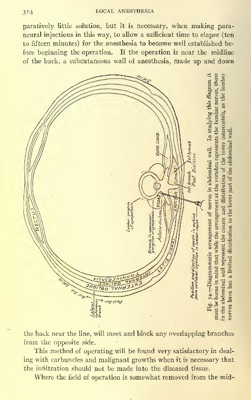
LOCAL ANESTHESIA
paratively little solution, but it is necessary, when making para-
neural injections in this way, to allow a sufficient time to elapse (ten
to fifteen minutes) for the anesthesia to become well established be-
fore beginning the operation. If the operation is near the midline
of the back, a subcutaneous wall of anesthesia, made up and down
the back near the line, will meet and block any overlapping branches
from the opposite side.
This method of operating will be found very satisfactory in deal-
ing with carbuncles and malignant growths when it is necessary that
the infiltration should not be made into the diseased tissue.
Where the field of operation is somewhat removed from the mid-

THE THORAX AND BACK 315
line, the depth of the tissues is much lessened and the procedure
simplified. It is necessary here to create a somewhat crescent-
shaped wall of anesthesia on the proximal side from the surface to
the deep parts, injecting between the ribs, and having the horns of
the crescent to slightly embrace the field of operation, the same as
has been suggested in Fig. 75.
Fig. 75. i, Hackenbuch plan of anesthesia embracing operative area: Circle of
cutaneous anesthesia first surrounds field; long needle enters at several points on this
circle and is directed obliquely downward and inward infiltrating deep planes. (See Fig.
19.) 2, Method of anesthetizing operative field in lumbar region: Dotted line extent
and direction of cutaneous anesthesia; heavy dots points for inserting needle for deep
injections into muscle walls; oblique lines area of resulting anesthesia; cross-lines
operative field.
By the above procedures, we have often operated large carbuncles
or removed growths and have frequently resected ribs, and on one
occasion removed an entire rib for tuberculous disease.
Large pedunculated fibrolipomas, common to the back, can be
very easily removed by local anesthesia by injecting a collar of anes-
thesia in the skin around the base of the growth. After the skin
injection has been started, a long needle is passed deep into the tissues
at the base of the pedicle and from J^ to i ounce or more of Solution

316 LOCAL ANESTHESIA
No. i (plus adrenalin), according to the size of the growth, is dis-
tributed in the tissues at the base. While this is being allowed time
to diffuse we can return to the skin injection and complete this, whenthe removal of the mass can be easily accomplished. In this waythe author has removed a fibrolipoma about half the size of a water-
bucket from between the shoulders of an elderly gentleman. Of
latter years the size and unsightly appearance of the mass had kepthim indoors, and had almost anchored him to bed.

CHAPTER XVII
THE ABDOMEN
THE question of the sensibility of the contents of this Pandora's
box of the human body, in spite of the many experimental observa-
tions on animals and man and the many daily operations performedwithout general anesthesia, still remains largely in doubt. At least
it is a much disputed question between the followers of Lennander on
the one side, and those who have arrived at their conclusions as the
result of purely animal experimentation on the other.
Of one thing we are sure, there is nothing more certain or real
than the existence of intra-abdominal pain peritonitis, appendicitis,
enteric, biliary and renal colic, etc. Such are daily observations in
the routine of the physician's work; still, we are confronted with the
statement, as the result of accumulated surgical evidence from re-
liable sources, that the intra-abdominal organs feel no pain, whether
normal or inflamed.
While this subject has been investigated by earlier writers, both
physiologists and surgeons, it had not been given the attention and
study it merited until undertaken by the late K. G. Lennander, whomade a most careful and thorough investigation, publishing his
results in 1901, and since that date up to the time of his death. His
conclusions were briefly that the parietal peritoneum is intensely
sensitive to pain, but not to pressure, heat, or cold; this sensibility is
increased by inflammation; the visceral peritoneum and abdominal
organs are entirely devoid of any sense of pain, even when inflamed,
and may be cut, crushed, torn, or burned without exciting pain; in
other words, that all pain arising from disturbances within the ab-
domen is caused by irritation through spread of inflammation to the
abdominal walls or pressure exerted upon these parts innervated bythe cerebrospinal system, and that those parts supplied by the vagusand sympathetic system of nerves have no afferent fibers for the
transmission of painful impressions. While Lennander's findings
for the mesentery were not positive, he considered it also insensitive
to any irritations or manipulations, except pulling upon it, which
naturally exerted tension on the posterior abdominal wall and ex-
cited pain. Other investigators have stated that the occasional
317
318 LOCAL ANESTHESIA
production of pain in the mesentery, as observed during operations
upon man, may be due to the presence of an occasional cerebrospinal
nerve, which may have found its way into these parts through an
anomalous distribution. This view, however, is not satisfactory, for
if nerves are found in any tissue it is intended that they be there and
they should be present in all cases. Nothnagel has attributed the
pain of intestinal colic to an anemia of the intestinal walls and pres-
sure exerted upon the nerves of those parts through the violent
muscular contractions; Wilms, on the other hand, accounts for the
same pain by the violent peristalsis producing a stretching of the
mesenteric attachments. Lennander, in investigating the same sub-
ject, found that he could stimulate a loop of the human intestine to
such a degree that it became hard and anemic, without the produc-tion of pain until the mesentery was pulled upon. The last paperfrom Lennander's pen, which gives his views on this subject, was read
at the meeting of the Amer. Med. Assoc., 1907, and is as follows:
"From my published works it may be gathered that I have not
been able to find any abdominal organ, innervated only by the vagusor the sympathetic nerves, which is provided with the sense of pain.
Sensations of pain within the abdominal cavity are, according to myexperience, transmitted only by the phrenic nerve, the lower six
intercostal, the lumbar, and the sacral nerves.
"My former pupil, M. Ramstrom, professor of anatomy at Upsala,has given us the first exact description of the course of these nerves
within the diaphragm and the peritoneal lining of the anterior ab-
dominal wall. He has shown that some of the older descriptions of
the distribution of these nerves are incorrect.
"For instance, he has not seen any branches of the phrenic nerve
running down from the diaphragm to the anterior abdominal wall,
nor has he been able to trace a single branch of the phrenic nerve
through the suspensory ligament to the capsule of the liver. Simi-
larly, he has been unable to find any twigs from the intercostal nerves
of the diaphragm which extend to the capsule of the liver." These anatomic observations of Ramstrom agree with my own
experience in regard to the sensitiveness of the liver. Even a strongfaradic or galvanic current, applied to the surface of the liver above
the gall-bladder, does not excite pain. In some cases, in which the
position of the liver was low, I have separated the attached surface
of the gall-bladder as far as the cystic duct without causing any pain,
whereas the patient complained as soon as I tilted the liver or draggedon the common bile-duct, thus putting the cerebrospinal nerves of the

THE ABDOMEN 3ig
abdominal wall on the stretch. Not only the sense of pain, but also
the other modalities of sensibility pressure, cold, and heat are
absent from the liver and gall-bladder as well as from the stomach
and intestines.
"We have often been able to ascertain that viscera involved in
disease are quite as insensitive to operative measures and to electric
stimuli as are sound ones. Thus, the old theory defended by Flour-
ens has been destroyed.
"After many investigations some of which have been attended
by the well-known physiologist, Hj. Ohrvall, and several by Professor
Ramstrom as recorder we had come to the conclusion that the
parietal peritoneum of the anterior abdominal wall possesses only the
sense of pain, not the senses of pressure, cold and heat. (In most
cases it was the peritoneum behind the recti muscles, from a point
4 to 5 cm. above the umbilicus to a point midway between the umbili-
cus and the symphysis pubis, which had been examined.) Should
this view of OUTS prove correct, it speaks decidedly in favor of the
specific character of the nerves of pain. In other words, it goes to
prove that the entire parietal peritoneum is provided only with
nerves of pain a condition previously known to exist in the cornea
alone.
"It is my opinion that all painful sensations within the abdominal
cavity are transmitted only by means of the parietal peritoneumand its subserous layer, both of which are richly supplied with cerebro-
spinal nerves around the whole of the abdominal cavity, possibly with
the exception of a small area in front of the verebral column lying
below the crura of the diaphragm and between the two chains of
sympathetic nerves. Here, as far as I am aware, no cerebrospinal
nerves have as yet been demonstrated, and on a few occasions I have
observed that within this area the patient does not respond to hard
pressure with a finger or with an instrument; nor, furthermore, does
he experience any sensation when a small portion of the mesenteric
attachment at this point is put on the stretch.
"The opportunity is given during operations of observing that
the manipulations which cause pain are those which occasion stretch-
ing of the parietal peritoneum as well as of the parietal attachments
of the mesenteries. For example, pain is occasioned by the placing
or removal of gauze compresses between the viscera and the parietal
peritoneum, by the dragging forward of the cecum, of the vermiform
appendix, or of any other organ whose normal attachment to the
abdominal wall is put on the stretch; and the same principle applies

320 LOCAL ANESTHESIA
to the stretching of any abdominal adhesions which may connect the
viscera with the abdominal wall. On the other hand, should a com-
press lie between the viscera without coming in contact with the
abdominal wall, the patient experiences no sensation when it is
removed. Similarly, no sensation attends the stretching or breaking
up of adhesions which have no connection with the abdominal
parietes. As far as I can judge from my observations, the parietal
peritoneum along the thoracic aperture and around the foramen of
Winslow is especially sensitive to stretching, displacement, etc.
"A slow and gradual stretching of all the layers of the abdominal
wall by ascites or meteorism occasions distress rather than pain,
although a high degree of meteorism may be attended by great dis-
comfort. If in a severe case of paresis of the bowel one succeeds in
emptying the intestine by means of a typhilitic, a jejunal, or gastric
fistula, the procedure 'is followed by such evident relief that the dis-
tress of the previous condition is emphasized. That a maximum
degree of rapidly forming meteorism is an extremely painful condi-
tion, and one which may rapidly endanger life, I have witnesses in the
case of a young student who had a coincident volvulus of the ileum
and acute dilatation of the stomach. Four hours and a quarter after
the appearance of the first symptoms he was pulseless from intense
pain attended with a sensation of bursting."Infectious processes involving the abdominal viscera (ulcera-
tions, acute inflammations, etc.) are attended by lymphangitis and
lymphadenitis of the mesenteries. The infection spreads along the
lymph-vessels to the subserous tissue of the abdominal wall, and,
inasmuch as the lymph-vessels follow the course of the arteries, the
lymphangitis very soon reaches the sides of the aorta, along which it
then may continue up to the thoracic cavity. A lymphangitis of the
parietal subserous connective tissue greatly increases the sensitive-
ness (excitability) of the cerebrospinal nerves to any manipulationwhich occasions pain even under normal circumstances.
"All that we know of lymphangitis and lymphadenitis attendingaffections of the mouth, of the pharynx, of the extremities, etc., ap-
plies equally well to corresponding processes within the abdominal
cavity. We know, for example, that the severity of pain attendinga lymphangitis and lymphadentis of the above-named regions varies
according to different infections, and the same thing is true for the
abdominal cavity.
"The irritability of the nerves of pain of the parietal peritoneumis much increased even by the slight peritoneal inflammation. In

THE ABDOMEN 321
the case of a serous peritonitis (peritonale, Reizung) the boundaries
of the hyperemic zone of the parietal serosa can be mapped out almost
to the centimeter by gentle palpation of the abdominal wall. With
further increase of the hyperemia and of the inflammation the sensi-
tiveness is at first almost proportionally increased. The fact that so
many infectious processes within the abdominal cavity begin with
diffuse abdominal pain may be explained by (i) an increased sensi-
tiveness of a large portion of the parietal peritoneum, owing to the
lymphangitis or peritonitis; (2) a considerably increased irregularity
of peristaltic action which, in addition to pain, often produces a feel-
ing of sickness, vomiting, and one or more actions of the bowels at the
commencement of these illnesses. On account of the increased sensi-
tiveness, the movements of the stomach and intestines against the
parietal peritoneum and the stretching of their respective mesenteries
are felt as severe pains. In most cases, however, the general perito-
neal irritation soon passes off. Only the part more especially infected
remains in a condition of inflammation, and the abdominal pain will
become localized at this spot. In those cases where the infection
spreads over a large portion of the peritoneal cavity, thus givingrise to a more or less general peritonitis, the pain will diminish as
soon as the bowel becomes paretic and the nerve-endings of the
parietal peritoneum have been more or less destroyed by the severe
inflammation."
"Pain in Connection with Perforation. In case of visceral per-
foration or of an abscess which ruptures into the free abdominal
cavity, the primary pain is caused by the contents of the organ, or of
the abscess, coming into contact with the parietal peritoneum. The
severity and character of the pain depends on the nature and quantityof the escaping fluid, the extent to which it immediately comes into
contact with the parietal peritoneum, and, lastly, on the intensity of
the contractions of the stomach and bowels brought on by the irrita-
tion of the peritoneum.
"Many clinical observations are explicable if one bears in mindthe fact that only the parietal peritoneum can transmit painful
sensations. For example, the primary pain occasioned by a duodenal
perforation may be referred to the iliac fossa. Again, the paroxysmal
pains in connection with a gastric ulcer are elicited by the movements
of the stomach; that is to say, by its dragging on a parietal serous
membrane which is hyperesthetic on account of a lymphangitisfrom an infected ulcer. If the stomach is put at rest by the aid of a
jejunostomy the pains cease. Further, in the case of a patient with

322 LOCAL ANESTHESIA
an inflammatory focus, surrounded by small intestine and covered bya thick omentum, pressure on the abdomen will disclose no tenderness,
whereas palpation per rectum may cause pain."
"The Hypotheses of Nothnagel as Regards Colic.- In human
beings suffering from intestinal fistulae, and to whom no anodynehas been given, sensation of pain cannot be evoked by means of
chemical, thermic, mechanical, or electric stimuli applied to a portion
of the gut lying outside the abdominal cavity as long as the stimulus
or contractions which it causes only affect the bowel. When, on the
other hand, the contracting bowel drags on adhesions connecting it
with the abdominal wall it at once produces pain.
"Both theories of Nothnagel are hereby disproved, for the colicky
pain cannot be due to pressure on the nerves of the bowel wall in
consequence of a tonic spasm of its muscular coat, since the intestinal
wall can be crushed with a strong pair of forceps without eliciting
any sensation whatsoever. Further, the pain cannot be due to
anemia of the intestinal wall due to a spasm of its muscular coat,
since it is possible by means of electric stimulus to produce so power-ful a contraction of the bowel that it becomes of tumor hardness and
assumes a yellowish-white color from anemia, without the patient
experiencing any sensation, even of being touched."Wilms, in an article and later in his splendid work on ileus (1906),
tried to show that all those pains which we are accustomed to term"intestinal colic" are entirely due to stretching of the mesenteric
attachments. In my first publication (1901) I made the statement
that every distention or contraction of a gut which is attended by a
pull on its attachments to the abdominal wall is necessarily painful,
as it involves a stretching of the cerebrospinal nerves of the parietal
serous and subserous layers. With regard to the duodenum, the
duodenojejunal flexure, the three flexures of the large intestine, and
the most distal part of the ileum, it goes without saying that these
portions of the bowel cannot contract on their contents in front of an
obstruction without giving rise to a powerful dragging of their mesen-
teries, and at the same time a painful stretching of the nerves of sensi-
bility of the parietal serosa. In similar fashion, powerful intestinal
contractions are bound to involve a painful stretching of the parietal
serosa in case the bowel has become fixed to the abdominal wall by
adhesions, and there exists at the same time some, obstruction at this
point.
"The pains in connection with ileus, due to kinking, volvulus, etc.,
were thus easy to understand. On the other hand, it was my opinion

THE ABDOMEN 323
that the stretching of a high and free mesentery of the small intes-
tines or of the transverse colon could not account for the pain attend-
ing a stricture of these parts of the bowel when the stricture has not
become adherent to the abdominal wall or in some way fixed, because
I could not think that a contraction of the bowel around its contents
in front of the stricture could drag on the parietal serosa and sub-
serosa by that high and free mesenterium. Wilms also accounts for
the pain in these cases as being only the result of the stretching of the
mesentery proximal to the stricture. He considers that a loop of
bowel, contracting on its contents at the proximal side of a constric-
tion, endeavors to assume a straight form in the same way as does the
gut in sausage-making. The mesentery, however, prevents the
bowel from assuming the shape of a straight cylinder. The result
is a stretching of the mesentery, and this is what causes the pain. In
this connection, an observation was made a year ago in a case in
which I had to resect more than i meter of the ileum, together with
the cecum and a large portion of the colon, this portion of the intes-
tine having been excluded some months previously by means of an
ileocolostomy, on account of multiple fistulas and adhesions which
could not be loosened.
"Under local and a short ether anesthesia the excluded portion of
the bowel was completely freed from all adhesions, only the normal
mesenteric attachment being left; the patient being awake, a piece
of the ileum, about 40 cm. long, was clamped and inflated with air.
The bowel straightened, the mesentery got stiff and assumed a fan-
shaped form. While in this position, standing out from the vertebral
column, the patient complained of pain, but this passed off as soon
as the bowel was emptied of air and the mesentery was allowed to
resume its normal position. The experiment was repeated with a
considerably shorter piece of bowel. The result was the same, for the
mesentery again became tight and stood out from the vertebral
column, occasioning pain. Lastly, a piece of the intestine, only 5 or
6 cm. long, was shut off. Although it was inflated ad maximum,
causing the serous membrane to burst and the bowel to sink between
the two layers of the mesentery for fully i cm., the patient felt noth-
ing. As soon, however, as the mesentery was stretched for a few
moments with the fingers pain was felt. These observations are in
full accordance with the above-mentioned view of Wilms as regards
the stretching of the mesentery, a view which I believe to be
correct.
"The pain in connection with a volvulus of the intestine or of an

324 LOCAL ANESTHESIA
ovarian cyst naturally increases in proportion as the twisting comes
on quickly and more parietal peritoneum is drawn into the pedicle."
"Pain Caused by Displacement of the Serous Membrane of the
Anterior Wall. On many occasions, when performing a laparotomy,I have passed into the abdominal cavity a finger, covered with a thin,
smooth Indian rubber glove, dipped into saline solution, and exerted
a slight pressure on the anterior abdominal wall. Of this the patient
has no perception, but as soon as the serous membrane is displaced
against the muscles or aponeuroses of the abdominal wall the patient
has a feeling sometimes of touch, more often of pain, according to
lesser or greater sensitiveness of the individual, and according to the
degree of pressure and displacement employed. When asking the
patient, 'Have you ever felt anything like this?' I have usually re-
ceived such answers, 'It feels like colic;' 'It feels as if the bowel is
being expanded by wind;' 'It's like bad griping pains;' 'It's worse
than gripes.'
"Such sensations are occasioned by displacing a small, limited
area of the serous membrane at nearly any point of the anterior ab-
dominal wall. On account of these observations, I believe that a
displacement of the serous lining takes place and gives rise to pain
as soon as a loop of intestine contracts on its contents, hardens,
rises, and presses against the parietal peritoneum. According to myopinion, these pains occur not only in connection with ileus, but also in
connection with temporary irregularities of the peristaltic movement
of the bowels in people not suffering from abdominal diseases.
I have myself felt these griping pains, and I have been strongly in-
clined to localize the same at the anterior abdominal wall, most often
to the left lower quadrant. As a rule, I have attributed them to the
contractions of the sigmoid flexure.
"Here I may mention a case illustrating the displacement of the
serous membrane of the anterior abdominal wall at the thoracic aper-
ture. It was brought about by a subserous myoma of the uteri of
about the size of a mandarin in a woman eight or nine months preg-
nant. She had suddenly been taken ill with severe pains, vomiting,
and inability to pass flatus. A diagnosis was made by her medical
attendant of a twisted ovarian cyst. As soon as the myoma had been
removed under the local anesthesia all symptoms disappeared. Theovaries were normal. The pregnancy went on to its normal termina-
tion. I have record of another patient, who had a small subserous
myoma of the uterus, and in whom, after the seventh month of preg-
nancy, abdominal pains were produced by any movement. They

THE ABDOMEN 325
were felt as a very painful friction, and disappeared as soon as the
tumor was removed.
"If the bowel wall on the proximal side of an obstruction is con-
siderably thickened and the serous coat rough, the'
Darmsteifung'is naturally attended by a much more extensive displacement of the
parietal peritoneum than in the case of a normal intestinal wall.
Contrary to Wihns, I consider this factor also to be the cause of the
colicky pain which attends intestinal obstruction. I have, conse-
quently, come to the conclusion that 'gripes' are due partly to a
stretching of the parietal attachments of the bowel and partly to a
displacement of the serous lining of the abdominal wall."
"Reflex Rigidity of the Abdominal Muscles. The normal re-
sponse of the abdominal muscles to an acute sensation of pain which
originates in the parietal peritoneum or subserous tissue lies in a
reflex contraction (defense musculaire). In case of violent and veryextensive irritation (hyperemia, slight edema) the abdomen assumes
a board-like rigidity and the respiration becomes costal in type, for
the abdominal muscles and the diaphragm are in a state of tonic
contraction. In this way the range of movement -of the abdominal
organs is greatly diminished, and consequently the abdominal painis greatly lessened. Compare with this the endeavor of patients suf-
fering from severe abdominal pains to get relief by lying on the
'belly/ or by fixing something tightly round the abdomen. The ex-
tension of the reflex muscular rigidity corresponds closely to the area
of peritoneal irritation of the parietal serosa. We know little as yet
about defense musculaire in connection with a mechanical ileus be-
fore the onset of peritonitis. It is necessary to observe with care
every case of severe colicky pain attending an intestinal obstruction,
in order to see whether a tonic contraction of the abdominal muscles
and diaphragm takes place, with the object of diminishing the move-
ments of the bowels and indirectly the severity of the pains.
"In acute inflammatory diseases of the abdomen I have not ob-
served the presence of cutaneous hyperalgesia so often as one might
expect, especially in consideration of the 'triade douloureuse' of
Dieulafoy (i8gg), which he regarded as necessary for the diagnosis
of an acute appendicitis cutaneous hyperalgesia, reflex muscular
rigidity, tenderness on pressure. Before applying Head's theory of
cutaneous hyperalgesia to a given case, one ought to consider the
question whether in that special case an infectious lymphangitis,
along the posterior abdominal wall and around the vertebral column,

326 LOCAL ANESTHESIA
might not cause a hyperesthesia of the sensitive nerve-trunks and
spinal ganglia of that region.
"It will, of course, be clear to every one that this is only a working
hypothesis. In those few cases of acute appendicitis with cutaneous
hyperalgesia, which have come under my care since I began to paymore attention to this matter, I have observed co-existing tender-
ness on deep pressure in the angle between the twelfth rib and the
erector spinea or somewhat lower down at the border of this muscle.
"When considering the pains connected with infectious diseases
of the liver and gall-bladder, one has to remember that well-known
embryologic facts, as well as my own researches and the investiga-
tions of Ramstrom, all lead to the assumption that the liver, the gall-
bladder, and the extrahepatic bile-passage do not possess nerves of
pain. One has further to consider the distribution of the lymph-vessels from these organs to the posterior abdominal wall and dia-
phragm, as well as their anastomoses with the lymphatics of the
duodenum and pancreas. One can then easily understand that in-
fectious diseases of the liver and gall-bladder are apt to be followed
by spasms of the diaphragm, and that the movements of the common
bile-duct, the duodenum, and stomach may be attended by pain.
When the gall-bladder contracts spasmodically in order to expel its
contents there results a stretching of the cystic and common bile-
ducts, and consequently a displacement of the parietal peritoneumand the extremely sensitive retroperitoneal connective tissue around
the common bile-duct. If a tube has been fixed (water-tight) into
the gall-bladder in a case of cystotomy for cholecystitis, 100 c.c. or
more of saline solution may be made to pass from the gall-bladder
into the duodenum (the biliary passage being free) without the
patient feeling anything so long as the solution is being slowly in-
jected into the gall-bladder. If the injection is made with a little
greater force, the patient almost immediately complains of colicky
pain in the back.
"With a shrunken gall-bladder and very wide common bile-duct,
biliary colic, due to the stretching or distension of the common bile-
duct, is inconceivable.
"It is quite necessary to consider carefully the account which the
patient gives of his pain. Lately a patient of mine, suffering from
an acute hemorrhagic pancreatitis, stated that the attack of pain
began with a sensation as if the large blood-vessel at the back had
burst near the pit of the stomach. The autopsy showed, in addition
to the pancreatic hemorrhages, a large retroperitoneal extravasation

THE ABDOMEN 327
around the celiac artery and the aorta. One must never forget how
difficult it is to localize pain, and how great an extent a correct inter-
pretation of the site of painful sensation is a matter of practice."
"Summary. In estimating abdominal pain, and especially in
connection with illnesses giving the symptoms of 'ileus,' we must
bear in mind, briefly, that:
"i. Pains do not originate within the abdominal organs, which
are supplied only by sympathetic fibers and the vagus nerves.
"2. All pains originate in the abdominal wall, more especially
in the parietal serous membrane and subserous connective-tissue
structures which are innervated by the cerebrospinal nerves.
"3. Stretching of the parietal (mesenteric) attachments of the
stomach and intestines, as well as of string- or band-like adhesions
to the abdominal parietes, invariably elicits pain.
"4. The same thing holds true for the displacement of the parietal
serosa from its normal relation to the muscles or aponeuroses of the
abdominal wall.
"5. Most of the diseases connected with ileus are, at their com-
mencement, attended by increased and, as a rule, irregular peristalsis.
"6. Chemically different substances, such as the contents of the
stomach, gall-bladder, intestine, or abscesses, give rise to severe pains
when they come into contact with a healthy or hyperemic parietal
peritoneum (pain due to perforation).
"7. Even that form of acute peritonitis which goes under the
name of peritoneal irritation (peritoneale, Reizung) greatly increases
the sensitiveness of the parietal serous membrane.
"8. The sensitiveness of the parietal peritoneum at first increases
part passu with the inflammation, but later decreases again whenthe inflammation has reached a certain high degree, and in manycases may ultimately cease altogether. (Compare herewith erysipe-
las of the skin, more especially the gangrenous kind.)
"I believe, finally, that we are on the way to completely under-
stand the pains of ileus, though a great amount of work still remains
to be done. I consider it to be a very happy thought to bring to-
gether anatomists, physiologists, physicians, and surgeons for the
discussion of this question, and I feel not only greatly honored, but
also deeply grateful, for the invitation to contribute this introductory
paper."
Lennander's views have been largely confirmed and accepted by
many surgeons the world over, although there still remain some
observations which cannot be satisfactorily explained from his stand-

328 LOCAL ANESTHESIA
point. On the other hand, the character and standing of those op-
posing these views, and the highly scientific nature of their experi-
ments, eliminating every possible avenue of chance, accident, or
doubt in their experiments, almost forces conviction.
The latest investigations in this direction have been by Kast and
Meltzer, at the Rockefeller Institute, New York. In a large number
of experiments, mostly upon dogs (but also cats and rabbits), theyhave proved in a most convincing way that Lennander's views are
in error on almost every point, at least in so far as they concern these
animals. They further made the astounding revelation that the
amount of cocain used in the infiltrating solution for operations uponanimals (most previous observations, both on animals and men, had
been made after exposing the abdominal contents under local anes-
thesia) was sufficient to control through its central action all sensa-
tion of intra-abdominal pain. This observation becomes the more
important and interesting when it is remembered that the general
sensibility over the entire body has been so inhibited, through the
central action of cocain, as to permit the painless or almost painless
performance of operations. (See chapter on General Anesthesia with
Cocain.) Kast and Meltzer also found that through the injection
of a moderate dose of cocain given in any convenient part of the bodythe whinnying, restlessness, and excitement of the animal following
the operations under general anesthesia were at once stopped, the
animal becoming quiet, and to all appearances remaining in a normal
condition.
A thorough appreciation of this subject by the reader, as well as
the difficulties in drawing conclusions from the conflicting evidence,
can best be obtained after a review of the facts presented by each
side. The following, "On the Sensibility of the Abdominal Organs,"
by Kast and Meltzer, appeared in the "Medical Record," December
29, 1906.
The animals were well narcotized by ether, the abdomen opened,and immediately closed by temporary ligatures held together by
clamps, and when the animal was partly out of the anesthesia one
or more of the clamps were taken off, thus permitting one or more of
the intestinal coils to come out. These, as a rule, were kept moist
and covered with towels saturated in warm saline solution. In other
cases the entire animal was kept in a saline bath at 39C., the viscera
being well covered with a warm saline solution. In still other ani-
mals the abdomen was opened under local (cocain) anesthesia.
The presence of sensation of pain was tested by pressing the or-

THE ABDOMEN 329
gans with the fingers or with thumb forceps, by touching them with
heated test-tubes, and by stimulating with the faradic current, and
watching the reaction of the animal to these irritations.
In all stimulations great care was taken to avoid pulling the
mesentery or touching the parietal peritoneum. They write:
"We have tested the various parts of the gastro-intestinal canal
the spleen, the kidneys, uterus, bladder, etc. but our present state-
ment refers essentially to the gastro-intestinal canal, which we have
studied mostly.
"All experiments lead up to one unmistakable result, which can
be stated in a few words the normal gastro-intestinal canal possesses
the sensation of pain. But, besides the difference in the subject of
observations, there was a difference in the condition under which
both observations were made. Lennander operated essentially under
Schleich's infiltration anesthesia. Schleich's mixture, as Lennander
employed it, consisted of 5 eg. of cocain, i eg. of morphin, and 200
c.c. of a normal salt solution.
"It seems advisable to us as a further step in our investigation
to study the possible effects of these ingredients upon the sensation
of pain in the abdominal organs.
"We began with cocain. The hitherto known effect of this drug
was its local anesthetic effect.
"Lennander and other surgeons employed it for this very quality
to deaden the pain during the incision, apparently without the remot-
est idea that the drug could also effect the sensibility of the distant
isolated gut.
"We nevertheless decided to test it. After establishing the un-
doubted sensitiveness of the intestines, etc., 2 eg. of cocain were in-
jected into the tissues of the abdominal walls near the incision. Wewere then surprised, indeed, when we discovered that a short time
after the injection all sensation disappeared from the intestines.
Even a very strong electric stimulus no longer produced any reaction
or effect. After thirty or forty minutes the sensation returned.
Such observations were then repeatedly made, and invariably with
the same results.
"Now, we could hardly think that the cocain crept over by capil-
larity or by some other manner to the intestines, and the observed
anesthetic effect was a local one. Neither did it seem probable that
the cocain crept along the spinal nerves to the spinal cord, and then
came in contact with the pain-carrying nerve-fibers from the intes-
tines. The most reasonable explanation was that the anesthetic

330 LOCAL ANESTHESIA
effect was produced through the circulation. That would mean that
cocain had not only a local but also a general anesthetic effect. This
assumption was easily tested.
"The cocain was now injected in parts distant from the abdomi-
nal cavity, in the thigh, arms, pectoral muscles, etc. The anesthetic
effect upon the intestines was prompt and complete just the same.
"In further experiments, we have established that i eg. was suffi-
cient to bring about the desired effect, and this even in large dogs
weighing 14 kilos.
"We have, then, thus far established two facts: that the gastro-
intestinal canal possesses the sensation of pain, and that the subcuta-
neous or intramuscular .injection of a comparatively small dose of
cocain is capable of abolishing this sensation for some time.
"We believe that we are now justified in offering the following
interpretation of the surgical observations: While we have not the
slightest doubt of the correctness of the facts, namely, that when
operating under Schleich's infiltration anesthesia the abdominal or-
gans are completely anesthetized, we suggest that this anesthesia is
due essentially to the general effect of the cocain employed, and not
to a normal absence of sensation in these organs.
"In the course of the investigations we exposed some intestinal
coils to the drying effect of the air in order to bring on some inflam-
mation, and we then found that inflamed organs are distinctly more
sensitive than normal ones. In fact, the sensitiveness is often greater
than that of the skin.
"Now, Lennander and other surgical observers stated that in
their experiences also inflamed organs are completely anesthetic.
We have, therefore, tested the effect of cocain upon the exaggeratedsensitiveness of inflamed intestines, and found that a somewhat larger
dose of cocain, say 3 eg., will completely abolish all sensations also
from inflamed organs.
"Another interesting point is the observation that the parietal
peritoneum also loses its sensation by a hypodermic injection in any
part of the body, but the anesthesia sets in here later and disappearsearlier than in the internal organs.
"It is possible also that the degree of the anesthesia is less, but
we are not yet ready to make any positive assertion on that point.
"An interesting and new fact is the observation which we madeon the effect which the injection of a small dose of cocain exerts uponthe psychic condition of the animal it promptly quiets excitement.
The animals, which were very restless, howling and crying, became

THE ABDOMEN 331
perfectly quiet one or two minutes after an intramuscular injection
of cocain. It may be claimed that the quietness was due to the
abolition of the pain.
"We have tested it on etherized, but not operated, dogs. On
awakening from ether they howl just as much as operated animals;
the howling is not due to pain, but to the ether intoxication. An
injection of cocain quiets them promptly."The psychic effect seems to last longer than the anesthesia of
internal organs. The injection has no narcotic effect; the animal is
apparently wide awake and follows one with his eyes. The lid reflex
is not abolished, but the cornea is anesthetic and the pupil is widelydilated.
"Whether the general sensibility is also reduced, that question
we are not yet ready to answer."
Still more disquieting than the foregoing to the earlier acceptedviews of Lennander appears a later paper by the same careful, thor-
ough, and painstaking investigators on the sensibility of the abdom-
inal organs, which appeared in the"Mitteilungen aus den Grenzge-
bieten der Medizin und Chirurgie" for 1909. As this most interest-
ing and instructive paper is a highly valuable contribution to this,
as well as collateral subjects, we hope the reader will pardon our
quoting from it at length in the author's own words:
"The surgery of our day denies the sensibility of all the abdominal
organs, notwithstanding numerous daily experiences to the contrary;
this question of normal sensibility belongs to the domain of physi-
ology. But we find here that for a decade nobody has paid any at-
tention to the subject, and most text-books do not mention a single
word on the subject. But it was not always thus in the first half
of the last century many prominent physiologists had contributed
to the solution of this question. The sympathetic ganglions and the
nerves distributed from them were studied in the range of the exam-
inations. The results were far from satisfactory and full of contra-
diction. Many observers asserted in a positive manner that no kind
of irritation of the sympathetic ganglions, their nerve-fibers, or the
intestines was capable of producing pain. Other authors, on the
contrary, equally well known in the history of physiology, asserted
that strong irritation of these parts was followed by severe pain.
"Megendie stated that cutting, tearing, etc., of the ganglions
made no impression upon the animal.
"Bichat has also reported that a dog had eaten its own exposed
intestines, and many other well-known authors have reported similar

332 LOCAL ANESTHESIA
observations. Bichat further reported irritating the celiac plexusand intestines of a dog by cutting or with acid without producing
pain. Johannes Miiller, on the other hand, states that mechanical
and chemical irritation of the celiac plexus or the connective tissue
of the renal vessels in the guinea-pig undoubtedly caused pain. Simi-
lar observations were made by Budge, Gianuzzi, and others. Again,other investigators, among them such brilliant names as Flourens,
Longet, Brachet, and Valentine, take a middle ground. Some stated
that only very strong or long-continued irritation caused pain, and
then of a mild degree. Others had pointed out that immediatelyafter the exposure of the ganglion, by no kinds of stimuli could painbe produced, but that, on the contrary, after longer exposure, when it
had become red or 'otherwise irritated,' the same stimuli were fol-
lowed by manifestations of pain.
"Since the middle of the last century, since which time practical
surgery as well as physiologic experiments have made use of general
anesthesia, we scarcely meet a report on investigation which deals
with our subject. They deal principally in consideration of other
problems in relation to the sympathetic nervous system of the ab-
domen, in which occasionally observations are mentioned which
show that even in narcosis the cutting, crushing, or tearing out of
the splanchnic nerves or the celiac ganglion always cause pain
(Haffter, Nasse, Braam-Heuckgeest, etc.).
"In numerous examinations, undertaken without regard to sen-
sibility, afferent nerve-fibers of the sympathetic were demonstrated
which serve many reflex purposes, as vasomotor, cardiac inhibition,
respiratory, etc. In relation to the sensibility to pain of the abdomi-
nal organs we find the physiologic literature of the last decades, as
far as we are acquainted with it, to contain not even occasional ob-
servations. Some authors, as Buch, Richet, etc., who have con-
cerned themselves with our problems, admit that these organs nor-
mally possess no sensibility, but that these organs can become the
seat of intense pain when inflamed or otherwise in a condition of ab-
normal irritability.
"In recent times the study of the sensation of the abdominal
organs has been undertaken by surgeons. An occasional clinical
statement regarding the lack of sensibility of the abdominal contents
is met in the older literature here and there.
"Such observations were: the apparent lack of sensibility of the
contents of herniae to mechanical or electric irritation; or of loops of

THE ABDOMEN 333
the bowel fixed outside the abdominal wall for the purpose of creating
an artificial anus; also of prolapsed portions of intestines."Since the introduction of local anesthesia and Schleich's infiltra-
tion method numerous extensive abdominal operations have been
undertaken without general anesthesia; these offered apparently a
very favorable opportunity for the study of our problems. In fact,
many surgeons have made the occasional observations that the ab-
dominal contents appear devoid of any sensation.
"Bier has stated that the intestines can be cut, squeezed, burned,
etc., without producing pain, but in a later contribution he adds
that tearing the intestines or the connective tissue of the mesenteryor separating adhesions will occasion pain.
"The question was gone into systematically and studied with
greater care by K. G. Lennander. The results of his investigations
are that the intestine, stomach, omentum, mesentery, spleen, liver,
gall-bladder and bile-passages, etc., in short, all organs which re-
ceive their nerve-supply exclusively from the sympathetic, possess
neither pain nor tactile or thermic sensations; and this applies not
only to the normal condition, but to the inflamed state as well.
"These organs, asserts Lennander, possess simply no fibers for
the transmission of touch, temperature, or pain. Only the parietal
peritoneum feels pain, and this because it is supplied with spinal
nerves. Inflammation increases the irritability of the nerves in gen-
eral, and increases, therefore, the sensibility of those in the parietal
peritoneum. Lennander refers the origin of the different kinds of
pain which occur in the abdomen to the parietal peritoneum and to
the spinal nerves in general.
"Therefore, would a distention of the intestines and intensive
peristalsis produce pressure and rubbing upon the parietal perito-
neum, or by producing a stretching of the mesentery which is felt
on the spinal nerves or the root of the mesentery?"In consequence of the inflammation of the abdominal organs
there occurs, as Lennander also assumes, not only an increased irri-
tability of the spinal nerves, but also the production of lymphangitis,
which later extends to the tissues which are richly provided with
spinal nerves.
"Infectious, toxic, or chemically irritating materials, therefore,
occasion pain; they are absorbed through the lymph-vessels, and are
immediately transported to parts supplied by the afferent spinal
nerves."Lennander's views are at present shared by many prominent

334 LOCAL ANESTHESIA
surgeons, among others von Wilms, who, on the contrary, sees in
the stretching of the mesentery the essential factor in the productionof abdominal pains.
"The most essential and most striking feature in Lennander's
views are that the abdominal organs, which receive their nerve-supply
exclusively from the sympathetic, are entirely incapable of feeling
the sensations of pain, pressure, heat, or cold.
"This view has recently obtained physiologic sanction, and has
been completely accepted by Thunberg in the chapter on Tac-
tile Temperature and Painful Sensation in Nagel's 'Handbook of
Physiology.'
"Some time ago we began a series of experiments on animals to
determine the sensation of pain in the abdominal organs. Our ob-
servations led us to the following conclusion : The abdominal organsare capable of painful sensation in the normal condition as well as
in the presence of inflammation. At the same time we made the ob-
servation that this pain disappears after an injection of cocain givenin any convenient part of the body.
"Conclusions. The abdominal organs of the dog, examined
through a small opening in the otherwise closed abdominal cavity,
are undoubtedly sensitive to pain, and are also sensitive outside of
the cavity, if only a small loop is brought out and tested immediatelyafter its exposure. Inflammation undoubtedly increases the sensi-
tiveness of the abdominal organs of the dog. If all the intestines or
a greater part are eventrated or otherwise freely exposed, there ap-
pears a more or less marked depression of the sensibility, which is
more complete the weaker the animal is; at the same time the animal
becomes more or less markedly apathetic with lessening of the cu-
taneous sensibility. Laparotomy also depresses the motor activity
of the gastro-intestinal canal. This motor and sensory depressionis a reflex inhibition of central nature, and can also extend to other
centers.
"In weak animals and in prolonged procedures this inhibition
may extend to the vital centers in the medulla oblongata, and mayoften lead to fatal shock.
"It also appears that the peripheral mechanism in the intestinal
canal may also be inhibited to a certain degree. It is evident, also,
that the intestines of cats and rabbits possess sensory nerves, but
they are easily exhausted and are very early and strongly influenced
by laparotomy; the intestines are affected much quicker and more
profoundly than the mesentery. The surgical experience on the

THE ABDOMEN 335
human subject does not at all prove that the intestines normally in
the normal closed abdomen possess no pain-conducting fibers. Until
exact proofs are brought forward that the sensory innervation of the
human abdominal organs differs radically from that of other animals
it will have to be assumed that as with animals, so also with man,the abdominal organs are provided with special nerve-fibers, and
that the sensation of these organs can be increased by inflammation
as we see it in animals.
"This theory explains in a simple way the well-known occurrence
of all kinds of violent pain in the human abdomen."
After reading the preceding, one is almost forced to conviction
of these views were it not for the daily repeated observations at the
operating-table upon the human subject, when it becomes self-evi-
dent that they cannot be unreservedly accepted for the human bodywithout further observations, and we realize that on this perplexing
subject the last word has not yet been spoken.It is very probable that in the highly organized human body con-
ditions of sensibility differ from those found to exist in the animal in
accordance with the well-known law that the higher we ascend in
the animal scale the more highly organized, complex, and sensitive
becomes the nervous system. The fact that moderate doses of co-
cain, i to 3 eg., is sufficient to abolish all intra-abdominal sensation
in dogs, and that large doses are capable of producing general anes-
thesia in man, must be taken into consideration in arriving at anyconclusions regarding intra-abdominal sensations during operations
upon man under local anesthesia, where it is also possible that the
acuteness of the sensibility of these parts may be somewhat lessened;
also the fact, demonstrated by Kast and Meltzer, that free exposure
of the abdominal contents inhibits or completely abolishes all local
as well as general sensibility; the fact that such exposure if prolonged
leads to shock has been recognized in man, but observations on the
sensibility, either local or general, long before shock appeared had
not been reported; it is, however, well known that during shock all
painful sensations are either greatly lessened or entirely abolished.
The "apathetic state" reported by Kast and Meltzer, even without
any fall of blood-pressure, must be recognized as a condition which
immediately precedes shock, as indicated by the blood-pressure.
The question of the lessened intra-abdominal sensibility throughthe use of cocain or its substitutes, and the depression of sensibility
through exposure of the abdominal organs in man, must now remain

336 LOCAL ANESTHESIA
an open question until proved by further observation on the human
subject made with this end in view.
In considering some of the above questions in the light of infor-
mation already obtained from operations performed on man, it has
been proved that the existence of adhesions between movable intes-
tinal coils does not excite apin as a symptom; other disturbances
may arise, but when adhesions have existed between the intestine
and the abdominal wall, pain has always been experienced. In op-
erating upon such cases under local anesthesia, the separation of the
adhesions between the several loops of intestines, no noteworthy
complaint is made by the patient provided the mesentery is not
pulled upon, but in separating adhesion between the intestines and
the abdominal wall pain is always felt.
If a finger is introduced into the abdominal cavity and firm pres-
sure made against the parietal peritoneum no pain is produced, as
the parietal peritoneum is insensitive to pressure, but by sliding the
finger about over the surface, traction is made on the delicate and
sensitive subperitoneal tissue and pain produced. In this way Len-
nander believes that the pain of some forms of colic not explained by
pulling on the mesentery may be accounted for. The gradual and
general distension occurring in ascites, large tumors, pregnancy, etc.,
may not cause pain, but the unequal distention and violent peristalsis
of a small loop may, by a sliding motion on the parietal peritoneum,excite acute pain.
The withdrawal of packs and drainage-tubes from between coils
of intestines excites very little pain, provided the mesentery is not
pulled on, in comparison to the pain produced by removing them
when in contact with the abdominal wall. In operations under local
anesthesia the careful, gentle application of packs around the field
and in contact with the parietal peritoneum does not excite any com-
plaint, but when being removed if they are roughly dragged out the
patient will always give unmistakable evidence of decided pain.
Investigations under similar conditions to those employed byKast and Meltzer have been undertaken by Miiller, but he did not
obtain the same results;also by Hotz, but here the observations were
made under morphin narcosis, and are, consequently, not of the same
value; however, he states that irritations of violent kinds, even in in-
flamed conditions, do not excite pain unless the mesentery is pulled
upon.
Ritter, on the other hand, tried similar experiments, and was able
to completely confirm Kast's and Meltzer's findings and oppose those

THE ABDOMEN 337
of Lennander. In 1909 he brought forth an entirely new theory.
He believes that the sympathetic nerves are capable of the transmis-
sion of painful impressions, but associates such sensation directly
with the blood-supply, and found that the more vascular parts were
the more sensitive, the non-vascular, less so; the vessels themselves
are most sensitive, and in every instance were painful when ligated.
He thinks that this means of testing the sensibility of these parts is
the only one that eliminates all possibility of error. Pulling upon the
mesentery was eliminated by a series of double ligatures; in placing
these, if the proximal was tied first it alone caused pain, the applica-
tion of the distal one being painless; but if the distal one was tied
first, then both caused pain. He found that cocain injected around
the blood-vessels renders anesthetic the viscera supplied by them,and thinks that injury to the vessels is important in the productionof shock, and advises that when working under local anesthesia all
vessels of any size should be cocainized before ligation and division.
He does not think that the cocain used in performing laparot-
omy has had so much to do with the negative findings of many sur-
geons as the inhibition of sensation brought about by the exposureof the delicate nerve-fibers in the abdominal cavity.
These later findings by Ritter have attracted the favorable at-
tention of some observers; further investigations may prove their
value, but we must bear in mind two points: first, that cutting off
the blood-supply always lessens the sensibility of the parts; second,
that the sympathetic nerves (if they have been proved to contain
pain-conducting fibers) are largely distributed upon the blood-vessels,
and the ligation of these vessels may completely block their power of
conduction.
In another publication Ritter has stated that the free exposureof the intestines in non-anesthetized patients, as well as pinchingthem with forceps, causes pain. Mitchell reports 2 cases and Haimi case operated without narcosis and without cocain, and they state
that the lack of sensibility of the intestines was similar to that ob-
served in similar operations under cocain. Two of these cases had
carcinoma of the stomach and the third an irreducible hernia. While
these examinations were more to the point, the cases must have been
very ill to have been operated without any form of anesthesia, and
this condition must have had some effect in shocking or inhibiting the
sensibility; however, the number is too small from which to draw anydefinite conclusions. Nystroem, a former assistant of Lennander, in
a recent paper champions the theories of his former chief, and ques-

338 LOCAL ANESTHESIA
tions the value of experimentation upon animals in settling these
points, and calls attention to the widely different results obtained bydifferent investigators in the same experiment. He tried the same
experiments carried on by Kast and Meltzer, and obtained exactly
opposite results, and could excite no pain unless the mesentery was
pulled upon or the parietal peritoneum irritated. He then experi-
mented upon a case of hernia in a man : the abdomen was first opened
by a small incision under ether narcosis, the peritoneum was then
temporarily closed, and the patient allowed to recover. The parietal
peritoneum was then tested and found very sensitive, but the irrita-
tion of a loop of intestine which was found present at the opening
gave no evidence of sensation until the mesentery was pulled upon.From these and other observations Nystroem concludes that the con-
tradiction of Lennander's work is not to be unhesitatingly accepted.
While he admits the existence of many points which cannot be satis-
factorily explained by these views at present, it is wise to withhold
judgment until further observations can be made.
Kast and Meltzer, in discussing the opposing views given by some
of the above-mentioned investigators, state the following:
"Now we will try to solve the question on the ground of our ex-
perimental experiences. In dogs we have almost, without excep-
tion, confirmed the sensibility of the abdominal organs; we have
found that by the free exposure of the intestines their sensibility is
reduced and that this reduction is more pronounced the weaker the
operated animal. We have further found that in cats and rabbits
the abdominal contents are sensitive, but that in these animals the
opening of the abdomen exercises a much stronger influence upon the
sensibility than with dogs, and that often a single exposure and irrita-
tion suffice to suspend reaction. The sensibility of the intestines
is yet more fleeting than that of the mesentery, and with rabbits more
fleeting than with cats. How does it stand now with the sensibility
of the abdominal organs of man? Here we have essentially the prin-
cipal question in view : Do the abdominal organs of man possess pain-
conducting fibers?
"This has been positively denied by competent surgeons. If one,
however, considers how such a denial involves theoretically and
practically very important assumptions, and if one considers still
further how such an assumption must now appear even more import-
ant, since it establishes a radical difference in the innervation between
man and other mammals, it. is, therefore, clear that such a state-
ment can only be accepted when based on exact proofs. But are

THE ABDOMEN 339
there such exact proofs? We have first the large number of observa-
tions which have been made under cocain anesthesia, but we have
proved that a cocain injection, even without touching the intestine,
is able to temporarily abolish this sensibility. The surgeons whohave not sufficiently investigated it doubt this statement. But it is
a certain fact, and it has also, as above mentioned, been recently
confirmed by Ritter. Here one must consider that to reply to our
question in the affirmative it is not at all necessary to assume that
the sensibility must be intense.
"It may appear in man after opening the abdomen as weak as we
have found it in cats and rabbits, and the small doses of cocain which
are ordinarily used may, therefore, completely suffice to suspend this
slight sensibility. Also the surgical observations which have been
made with the use of cocain are absolutely not such convincing proofs.
May the few recent observations which have been made without the
use of cocain in these cases be accepted as such positive proof?
Certainly not. Again, have we seen that other observations speakfor the contrary that the human intestines have painful sensations.
We must further mention that the above-cited three negative obser-
vations were made upon very sick patients, and here we must remem-
ber our experimental experiences that the weaker the animal the more
profoundly was the sensibility reduced by laparotomy ;and yet, again,
may we add that as regards the sensibility of the abdominal organs
of man after laparotomy they may behave as in cats and rabbits.
"To recapitulate, the observations which were made under the
use of cocain are on account of the cocain not of proportionate value.
The observations which were made without cocain are quite small in
number, are not without contradiction, and were made on very weak
patients. Moreover, laparotomy depresses the sensibility consider-
ably, and in quite weakened animals, also in several kinds of animals
in rather normal condition, was the sensibility completely suspended.
The surgical observations contain, therefore, not only no kind of posi-
tive proof, but contain in general no proof at all that the abdominal
organs of the normal man in the normal closed abdomen are unable
to feel painful sensations.
"As we have no proof to offer, we are justified in accepting that
the visceral innervation of normal man in the normal closed abdo-
men does not differ essentially from that of other mammals, and that
the abdominal organs are more or less richly provided with pain-
conducting nerve-fibers. We are further justified in accepting that

340 LOCAL ANESTHESIA
as with animals, so also with man, a marked inflammation strongly
increases the sensibility.
"Based on the above assumptions, the most widely different in-
tense pain that the human in his normal closed abdomen often has to
bear find their simple explanation, and do not need any interpretation
by forced hypothesis."
Much more might be said pro and con by each side in the contro-
versy, but, as our patients are human and not dumb animals, clinical
experience is the most valuable and the patient must be the judgein all cases and interpret his own sensation. While animal experi-
mentation is undoubtedly of tremendous value in furnishing infor-
mation and in settling mooted and doubtful points on many subjects,
it must necessarily always furnish very questionable data on such
subjects as pain, for the dumb animals may either suffer patiently
or wiggle, strain, whine or cry out as an evidence of their displeasure
at their restraint in much the same way they would be expected to do
when in pain and these actions are more likely to occur when we are
working over them. The rise of blood-pressure which is alwaysassociated with pain cannot always be interpreted this way in animals,
as fear will produce the same rise, and similarly the reaction which
invariably takes place with a fall of blood-pressure when either fear
or pain are prolonged is again a doubtful guide, as a very decided fall
of blood-pressure occurs in shock which we know from clinical ex-
perience may be accompanied by little or no pain. In my own ex-
perience with animals, which has been considerable, I am forced to
the conclusion that it is a very difficult matter to correctly interpret
their sensations.
Reports of animals under experimentation eating their own ex-
posed intestines cannot be accepted as an argument by either side,
as it is highly probable that the animal's normal sensibilities had
been perverted, the result of suffering, infection or temperature; as
we frequently see delirious patients mutilating themselves or tearing
open wounds without apparently any evidence of pain.
Some observations which I have made in resecting eviscerated
loops of the intestine in two-stage operations may be of interest.
During the last few years I have had repeated occasion to resect the
large intestine, usually the transverse colon, for obstruction usually
the result of tuberculous or other tumors. The condition of the pa-
tient has required that this be done in two stages by the Bodine or
Mikulicz plan, by first liberating and fixing the mass outside the
abdomen, approximating the two ends of the bowel within the cavity

THE ABDOMEN 341
and closing the abdominal wound around them; the resection of the
mass being done at a subsequent sitting. At the time of the re-
section careful observations were always made regarding the sensa-
tions experienced. In some of these cases considerable portion of
the mesentery was included in the mass to be resected. The re-
sections were usually done with the cautery under a low degree of
heat to control the hemorrhage but occasionally cutting instruments
were used with ligation for the bleeding points. These resections
were invariably done without general anesthesia and the results of
the observations in several such cases were quite uniform. Theentire circumference of the intestine was insensitive to cutting, burn-
ing or crushing but where the mesentery was included pain of an
aching or cramping character was always felt in the epigastrium, the
pain was the same whether the mesentery was cut, burned or crushed
and this pain was always stopped by proximal injections of the
mesentery with some local anesthetic. Symptoms of shock often
followed this procedure if the mass was large and more so following
the use of the cautery. The sensations following the application of
the clamp were also of interest. In case the approximated surfaces
of the bowel to which the clamp was applied had been so arrangedthat they were well away from the mesentery very little sensation
was complained of as a rule, but here the result of the observations
was not uniform, but when we felt certain that some mesentery was
included within the grasp of the clamp from previous notes as to its
location, more pain was always caused. This pain was always of a
cramping character and always in the epigastrium no matter where
the site of the resection. The pain did not always come on immedi-
ately following the application of the clamp. In one case it was
twenty minutes before any complaint was made when the pain was
then considerable and associated with some shock.
Many other clinical observations could be recorded from which
similar conclusions have been drawn, namely that the abdominal
viscera are insensitive to pain except at the hilum or mesenteric
attachments. The bladder is an exception to this rule as all parts are
sensitive, the fundus least so, and any trauma to its walls is felt in an
urgent desire to urinate.
In carefully considering the above observations in connection with
the findings by Kast and Meltzer we find a considerable difference in
the results obtained. As the extrusion of the mesentery with the
tumor mass did not cause it to lose or change its sensations from those
it is known to show within the cavity it is probable that the intestines

342 LOCAL ANESTHESIA
were likewise unaffected and that they are insensitive both within
and without the cavity.
Investigations have been undertaken with a view of determiningthe sensibility of the mucous membrane at various points along the
alimentary canal, and, while the results of these investigations agree
on nearly all points, there are still some dissenting opinions. These
tests were made through gastric and intestinal fistula or artificial ani,
or by passing instruments into the stomach through a stomach-tube,
or into the rectum through a speculum; the results of these examina-
tions have been that the mucous membrane of these parts has no
sense of touch, pain, heat, or cold. In 1909 Zimmerman publishedthe results of an extensive series of experiments upon himself and on
patients. These experiments were principally upon the stomach and
rectum, and were performed without any anesthesia; the mucous
membrane of these parts was irritated in a variety of ways by pinch-
ing with forceps, by electrodes, and by the cautery. In the stom-
ach there was no response to any form of irritation, but decided pres-
sure gave rise to a sense of fulness. In the rectum, 6 cm. above the
external sphincter, there was no sense of any kind except for pressure
and differences in pressure could be noted.
The esophagus was sensitive to both heat, cold, and pressure.
Regarding these experiments upon the rectum, the author has tested
the sensibility of the rectal mucosa and found that above the anal
canal there is practically no sense of pain to superficial irritation, and
the mucous membrane can be cut and cauterized without any com-
plaint. On one occasion a polypus was removed by cutting, with
cauterization of its attachments, with an electrode without any com-
plaint from the patient, although this region felt sore for several daysafterward. Schwenkenbecker in 1908 described his sensations after
taking large doses of menthol, which produced on the sensitive mu-
cous membrane of the mouth an intense feeling somewhat between
burning and cold. After the drug had passed the level of the larynxthere was no sensation until the anal canal was reached, when the
feeling of cold was again produced. He concludes from this observa-
tion that the mucous membrane of the alimentary canal is insenitive
except at its upper and lower ends.
We know that certain affections of the stomach, notably ulcer,
give rise to pain, although they may exist for long periods of time
without the patient's knowledge, and intestinal ulcers may go on to
perforation without the patient having been aware of their existence;
similarly, the ulcers of typhoid fever seem to excite no pain. Len-

THE ABDOMEN 343
nander maintains that a gastric ulcer excites no pain unless accom-
panied by a lymphangitis, and that hyperacidity excites pain whenthe irritating or chemical substances are carried by the lymphatics to
the sensitive abdominal wall (posteriorly). Mueller, in discussing
this point, admits that the stomach shows no reaction to touch, cold,
or heat, as far as external stimulation is concerned, but asserts that
it does react to certain internal irritations. He believes that the
abdominal organs do possess certain sensations necessary for protec-
tion from toxic or chemically irritating substances, and are capableof mechanical irritation by overdistension. The sensations thus
produced he attributes to the sympathetic nerves, which, under or-
dinary normal conditions, do not transmit painful impressions, but
become capable of feeling pain under the irritation of abnormal or
diseased conditions. When all the evidence, pro and con, regarding
the sensibility of the abdominal organs has been gathered and care-
fully sifted down, we have to admit that the crucial test must be the
application of these findings by the practical surgeon at the operating-
table upon the human subject, and here, so far, Lennander's views
have largely been substantiated. In the experience of the writer the
intestines are devoid of sensation unless the mesentery is pulled upon;after extensive'manipulations the patient may complain of a peculiar
visceral sense, hardly a pain, but at times sufficient to excite some
complaint. We have frequently explored limited parts of the ab-
dominal cavity by introduction of the fingers or hand, and when care-
fully done, avoiding friction on the parietal peritoneum, it caused no
complaint, except in the region of the foramen of Winslow over the
celiac plexus; here the parts seem particularly sensitive. The parie-
tal peritoneum and mesentery have always been found sensitive,
except when controlled by the injections of the anesthetic solutions;
it is certain that only those parts controlled by the injections would
stand operation; that the solution used may have had some control-
ling or lessening effect upon the sensibility of other parts may be
possible, but we have never observed the general analgesia, such as
that reported by Kast and Meltzer upon dogs, when using cocain or
any other local anesthetic agent, and any extension of the incisions
to the recognized sensitive parts, if unanesthetized, has always ex-
cited pain and required additional infiltration. In some few veryextensive intra-abdominal operations undertaken with local anesthe-
sia, where large quantities of the anesthetic solution were used and
large parts of the intestines and other organs exposed, the prolonga-

344 LOCAL ANESTHESIA
tion of the operation, instead of lessening the sensibility, seemed
rather to increase it.
In numerous operations for artificial anus where a loop of bowel,
usually the descending colon, has been fixed outside the abdominal
wall, either under local or general anesthesia, and opened several
days later without any form of anesthesia, I can remember no case
where any complaint was made. Here my observations have been
quite similar to those cases in which larger sections were removed as
noted above. This opening was made either with knife, scissors, or
cautery, and later, after retraction had taken place, the excess of
tissue was trimmed down level with the abdominal wall. Whether
the long exposure and changes occuring on the surface of the bowel
was sufficient to destroy the sensibility of the part or not is possible,
but it is certain that these cases have not, as a rule, required any form
of anesthesia for the opening of the bowel. The same may be said
about the stomach in operations for gastrostomy, as in the Ssabana-
jew-Frank operation, where the stomach is opened a few days later,
after adhesions have taken place.
A careful study of the preceding pages should prove particularly
interesting to the practical surgeon who attempts to deal with intra-
abdominal conditions under local anesthesia. Nowhere better than
in the abdominal cavity is the fact demonstrated that the skilful use
of local anesthesia is in itself an art. What one operator does with
ease seems to another impossible, and he may even discredit the
statements of the other. Here, in addition to a thorough knowledgeof technic, it is absolutely essential to possess an accurate knowledgeof the manipulations which cause pain, the parts most susceptible of
painful impressions, and the conditions which intensify these im-
pressions, such as inflammation.
We all agree that certain of the intra-abdominal contents possess
painful sensations; these are especially all blood-vessels except the
smallest divisions (and this rule holds good elsewhere in the body),and the mesenteries and attachments of the viscera to the abdominal
wall. As the blood-vessels almost invariably lie within the folds
of the mesentery, this limits the areas of sensibility under ordinaryconditions (the absence of inflammation) to the mesentery and the
parietal peritoneum. 1 have never found that incision, clamping, or
suture of the stomach, large or small intestines, gall-bladder, or uterus
upon which I have operated ever gave pain, providing the surround-
ing parts were not disturbed by rough manipulations that would
make traction upon the mesentery, and when it was necessary to in-

THE ABDOMEN 345
elude the mesentery in the field of operation, as in resections, a
moderate injection of anesthetic solution between its folds and at
some distance proximal to the field always sufficed to control these
sensations, providing there was no traction.
It is essential for these reasons that rather free incisions be madeto permit the ready manipulation of the parts as much within the
cavity as possible and render unnecessary undue traction and
displacement.
Abdominal Nerve-supply. We should now, for a thorough
understanding of our subject, be able to account for the pain-
conducting nerve-fibers within the cavity.
We know that all the abdominal organs are innervated almost
exclusively by the sympathetic system, and that all the sympathetic
ganglia as they lie against the vertebral column, both within the abdo-
men and above, receive fibers from the spinal nerves just after emerg-
ing from the vertebral foramina (Figs. 74-76). It has been impos-sible to trace these nerve-fibers to their ultimate distribution most
are soon lost in the intermingling of nerve-fibers of that region, some
few have been traced to the mesentery, but could not be followed
further, as it is impossible by any known methods to distinguish
between sensory and other nerve-fibers. As all painful impressionsmust come through the cerebrospinal nerves, it is in fibers supplied bythese communicating branches and other small branches given off
at the back of the abdominal cavity by the lumbar nerves that wemust look for the paths of these sensations.
An effort has more recently been made to reach these rami com-
municantes, and at the same time block the parent trunk by reaching
these nerves just as they emerge from the vertebral foramina; thus, bya paravertebral injection (by one injection) securing both visceral as
well as parietal anesthesia of the entire distribution of the nerve.
This method is spoken of more in detail under this heading.
The nerve-supply of the abdominal walls is from the six lower
intercostals and the upper branches of the lumbar plexus. (See
Figs. 74-76). In their course through the abdominal walls these
nerves all lie near the inner wall, the intercostals running between
the transversalis and internal oblique to the outer edge of the
rectus. The lumbar nerves emerge just beneath or through the
fibers of the psoas muscle, they then lie on the surface of the quadra-tus lumbarum until they reach the transversalis muscles beneath
which they pass, after which they pursue the same general course
as the intercostals. At the outer edge of the rectus muscle (linea

346 LOCAL ANESTHESIA
semilunares) branches are given off which pass forward through
the anterior rectal sheath to the subcutaneous tissues, these branches
are always encountered at or near the linea transversae, thoughgreater splanchnic ,//,,, x Cnriliax
' 'intercostal nerve XI 'us communicant
Coccyget
:raI^M-T-,
( (,
return x\ \pudendal- \
toccygeal ganglion coecygeai nerve sacral nerve V plexus xieral plexus
Fig. 76. The abdominal and pelvic portions of the sympathetic trunk. The
anterior abdominal and pelvic walls have been removed, the lumbar plexus exposed by
removal of the psoas major, and the aorta left in situ up to its bifurcation. * = Visceral
branches of the pudendal plexus. (Sobotta and McMurrich.)
others are met with between these points. The main trunks then
continue their course downward and inward lying on the posterior
rectal sheath just beneath the muscle giving off its terminal branches

THE ABDOMEN 347
in this position, which run forward through the muscle toward the
skin.
As a general proposition, it may be said that in operations upon
any intra-abdominal part a thorough infiltration of the abdominal
wall in the line of incision is the first essential feature. Should the
operation be upon the fundus of a part or at some distance from its
mesenteric attachment, no further anesthesia may be required, but
should the field of operation involve the cervix, hilum, or mesenteric
attachment of the part, these must be thoroughly blocked. These
principles, when properly applied to the various parts, are the essen-
tials of the anesthetizing process.
Anesthetizing the AbdominalWall. Braun, in infiltrating the ab-
dominal wall preparatory to its incision, makes the injection around
the field in more or less rhombus shape. This is best illustrated bymaking an injection around the middle line we will say, for an opera-
tion upon the stomach. A line of cutaneous anesthesia is established
on each side two or three fingers-breadth from the middle line;
starting on this line at two or more points, depending upon the extent
of the proposed incision, the long needle is entered and directed
obliquely outward, injecting as it is advanced, piercing the rectus
sheath, which is recognized by its slight resistance to the needle, and
advancing some little distance within this muscle until it is quite
freely injected; the needle is then partially withdrawn, when it is ad-
vanced again in two or more directions above and below, slightly
increasing the angle each time, thus making the injection in somethingof a fan-like shape ;
this is done along the line of cutaneous anesthesia
at two or more points, having the fan-like areas of infiltration come
in contact with the one above or below. The same procedure is re-
peated on the opposite side. These two lateral lines of infiltration
are joined above and below by subcutaneous infiltration, as shown
in Fig. 77.
While the above method of Braun is certainly found useful in
thin subjects, in thick abdominal walls heavily overlaid with fat
it is unsatisfactory, takes longer to carry out, and requires some little
delay before the solution has thoroughly diffused in all directions
and anesthesia established. I usually prefer to establish a wall of
anesthesia along the proposed line of incision from the skin to the
peritoneum, and have not found that muscular contractions interfere
if anesthesia has been perfect, and no pain excited either in the in-
cision or operation on the deeper parts, the muscles usually being
quite relaxed. The method of procedure is usually as follows: An

348 LOCAL ANESTHESIA
intradermalwheal is established midway along the line of the proposed
incision; the long needle with 10 c.c. syringe is entered at this point
and directed up and then down, without withdrawing the needle,
along the proposed line of incision in the subcutaneous tissues, in-
jecting freely as the needle is advanced, detaching and refilling the
syringe as occasion requires; without withdrawing the needle from
the skin its direction is now changed, and it is passed inward toward
the rectal sheath; this is the first plane of decided resistance which the
needle encounters; this is gently pierced, injecting as the needle is
Fig. 77. i. Braun's method of anesthetizing abdominal wall around area of incision;
2, author's method: infiltration of line of incision from one midpoint.
advanced to about i cm. within the sheath; the needle is then par-
tially withdrawn and redirected within the sheath at several pointsabove and below in a similar manner (Fig. 77). Returning now to
the skin, the intradermal infiltration is completed along the proposedline of incision. The deeper injections thus made first have ampletime to diffuse. Having completed the skin infiltration, the incision
is made down to the rectus sheath without need of further delay; with
the rectal sheath now within plain view, further injections can be
made within it if necessary. At this point the superficial vessels are

THE ABDOMEN 349
ligated, getting rid of the forceps and allowing the deeper injections
more time to diffuse during this interval. The rectal sheath is nowslit up and the muscle-fibers separated, exposing the posterior sheath;
this is now penetrated at several points with the needle and freely in-
filtrated posteriorly in the retroperitoneal tissue, this last injection
freely diffusing to the peritoneum; the posterior sheath and perito-
neum are now opened.This method I have found to be highly satisfactory and quickly
executed, and employ it almost invariably for incisions through any
part of the abdominal walls.
The solution usually preferred for these incisions, as well as for all
work within the cavity, is No. 2 (0.50 per cent, novocain in 0.04 per
cent. NaCl), adding about 10 drops of adrenalin to every 3 or 4 ounces
of the solution used.
POSSIBLE SCOPE OF OPERATIONS WITHIN THE ABDOMEN
After a careful consideration of the preceding pages it is seen that
many difficulties may attend the performance of any complicated or
extensive operation within the abdominal cavity and any operation
undertaken with any certainty of success by purely local means must
be limited to those parts which are in contact with the abdominal
walls or are readily accessible and can be operated on without undue
manipulation or traction. In the presence of an acute inflammation,
extensive adhesions or when the diagnosis is at all in doubt local
anesthesia is not indicated.
All simple operations, such as gastrostomy, gastrotomy, colos-
tomy, appendectomy, and gall-bladder drainage, are quite satisfac-
torily performed on suitable subjects (when not too nervous or ap-
prehensive) when the parts are fairly easily accessible, and not
matted down by inflammation or adhesions to the parietal periton-
neum or surrounding organs; consequently, only such operationsshould be undertaken when it is known beforehand that favorable
conditions exist. A fairly satisfactory exploration can be done
under local anesthesia by making a free incision to permit the easyintroduction of the hand, when, if it is gently insinuated without
pressure or traction, a rather thorough examination of the entire
cavity can be made. The sensations experienced by a patient
during a carefully made examination of this kind is that of a vagueintra-abdominal sensation, variously described as a weight or fulness,
becoming cramp-like if traction or pressure is exerted. This painis always referred to the epigastrium and is the same no matter what

350 LOCAL ANESTHESIA
the trauma inflicted and is best described as the abdominal pain.
Its seat seems to be about the position of the solar plexus.
It may often be found satisfactory to make such an exploration
under local means, then resorting to a light general anesthesia if
conditions are met with which cannot be easily undertaken by local
means alone.
Local anesthesia is not the method for routine work within the
abdomen, although in the interest of the patient many of the more
serious operations may be undertaken by these methods alone; here
it should always be, borne in mind that no traction upon a viscus is
ever tolerated.
If, on exploring through a midline incision, an appendix or dis-
eased gall-bladder is encountered, separate incisions should be madeover these parts rather than attempt to displace them toward the
midline.
As a general rule all very stout individuals and those with tense
rigid abdominal walls are difficult to handle in any serious intra-
abdominal operation, for as soon as the abdomen is opened the con-
tents bulge out and as any extensive packing back or the use of deepretractors excite pain the difficulties are at once increased by the
straining which these procedures bring on. Thin subjects with re-
laxed abdominal walls make very favorable subjects and opera-
tions of considerable magnitude may at times be performed. It is
only in these subjects with relaxed walls that the viscera can be
easily displaced permitting access to the posterior abdominal wall
where further infiltration can be safely done to block the visceral
innervation.
By consulting the pages on the abdominal nerve supply it will be
seen that these deep injections to be most effective must be along
the line of the sympathetic chain where the rami communicantes
join the sympathetic nerves; these injections must consequently be
made close to the vertebral column. In thin relaxed subjects when
much gentleness is used it is usually possible to slip in two fingers or
a long narrow retractor opening the way for the needle and the re-
troperitoneal tissue of these parts infiltrated, care being exercised in
the neighborhood of the large vessels. In the lower part of the ab-
domen this infiltration is done on the inner and outer sides of the
psoas muscle; in the upper part of the cavity close around the great
vessels on the vertebrae. It is not necessary always to get into
direct contact with these parts as the retroperitoneal tissue here is
quite loose and a fairly liberal injection of weak solution will diffuse

THE ABDOMEN 351
in all directions and reach the desired parts. When possible I have
always found the high injections in the neighborhood of the solar
plexus to be most effective. As can be readily understood these
procedures are always difficult even in favorable subjects and are
then only possible in the hands of those who have had much experi-
ence with local methods within the cavity.
A centra-indication to satisfactory intra-abdominal work is met
with in ticklish individuals. I have frequently noticed that the
intra-abdominal sensibility was much greater in these patients, whoare as a rule neurotic, and frequently prevented the successful
approach to the deeper parts where the visceral sensibility could be
controlled.
Many conditions of the patient may confra-indicate general anes-
thesia; it may then be advisable to attempt the more serious and
complicated procedures when the indications are urgent. Very ill
patients, and those suffering from the toxic effect of disease, often
have their general sensibility so reduced that they make favorable
subjects, provided they are not dangerously weakened or the field
of operation is not actively involved in inflammation; even in some
greatly weakened and reduced patients the danger of a general anes-
thesia may be greater than the difficulties likely to be encountered
with local anesthesia; it may, therefore, be advisable to proceed bythese methods. In some cases the combined method of operating is
advisable, using infiltration with light or superficial narcosis (see
chapter on this subject). In all intra-abdominal operations of any
severity the preliminary hypodermic of morphin, % to Y gr. or
pantopon % gr. with scopolamin 3^50 to Koo gr -> should always be
given one hour before operation.
The Stomach.- Simple operations upon the anterior wall of the
stomach when uncomplicated are quite easily performed under local
anesthesia by infiltration of the entire abdominal wall and subperi-
toneal tissue in the line of the proposed incision, as already explained,
making the incisions slightly longer than under a general anesthetic;
the cavity is opened, the wound lightly retracted, and the stomach
operated upon in position, or the viscus caught with a sponge-holder
or the fingers and gently drawn out; if care is exercised not to pull uponits attachments, no pain is produced. In this way we have operatedin many cases, and always with perfect satisfaction. It is quite easy
to perform gastrotomy for the removal of foreign bodies or the ex-
amination of the interior of the stomach; also gastrostomy by the
Ssabanajew-Frank or other methods, or the Heinecke-Mikulicz

352 LOCAL ANESTHESIA
operation for hour-glass contraction, when unaccompanied by sur-
rounding complicating conditions.
Gastro-enterostomy and gastric resections are now no longer
novelties under local anesthesia. The early and extensive work
done by Mikulicz, and later by Braun, Lawen, Bakes, and a host of
others, especially Finsterer with his classic contributions upon this
subject, have placed this method of operating upon a firmly estab-
lished foundation, and securing for it a recognition amoung other
accepted procedures.
Fig. 78. i, Line of anesthesia for exposing stomach. Upper oblique line is for addi-
tional incision for Ssabanajew-Frank gastrostomy; 2, for exposure of gall-bladder;
3, appendectomy through straight rectus incision.
In posterior gastro-enterostomy by the "no-loop method," the
operation now generally performed, the posterior stomach wall is as
tolerant of operative intervention without discomfort as in the an-
terior; to secure access to it the mesocolon, in the usually selected
non-vascular area, is first freely infiltrated between its layers before
it is divided and the stomach seized. In drawing the omentum and
transverse colon out of the field and displacing it above, as is usually
done, they should be carefully covered with wet towels (saline solu-
tion) ,as their prolonged contract with the air may excite some com-

THE ABDOMEN 353
plaint. After infiltration of the mesocolon no further infiltration is
necessary, and the various steps of the operation are carried out the
same as under a general anesthetic.
For gastric resections the gastrocolic and lesser omentums must
be freely infiltrated between their folds for an area some little dis-
tance beyond the proposed field of resection; this should not be under-
taken by local anesthesia alone, except under conditions of free mo-
bility of the stomach.
Finsterer, in his latest contribution to this subject in the"Beitragezur Klin. Chir./' 1912, cites in detail a large number of resections
and gastro-enterostomies performed by purely local means. Thenotable differences observed between such operations and similar
ones performed under general anesthesia was the marked absence
from shock, lung, and renal complications, and almost a total absence
of postoperative vomiting and gastric distention, viscious circle, etc.,
as there is not that paralysis of the stomach walls which follow such
operations under general anesthesia.
In colostomy for the establishment of an artificial anus the same
may be said here as for the stomach the operation is quite easy and
satisfactory where the mesentery is not pulled upon. The later
opening of the bowel after a day or two, when adhesions have taken
place, we have never found accompanied by any pain; the excess of
tissue can be trimmed down level with the abdominal wall by either
knife, scissors, or cautery.
The Appendix. The opening of a simple appendicular abscess
when in contact with the abdominal wall involves no greater difficul-
ties than an abscess situated elsewhere, and is always suitable for
operation under local anesthesia. The removal of the appendix is
a different matter; in the presence of inflammation or extensive ad-
hesions the operation is never suitable for local measures alone;
again, we never know where the appendix will be found, whether
lying loose on the intestines in its usual position, bound down to the
surrounding structures, attached in the pelvis, embedded in the
posterior abdominal wall or retrocecal. The ease with which the
appendix can be removed in non-inflammatory cases depends entirely
upon its position; when lying loose and easily accessible we have quite
frequently removed it without any difficulties or pain to the patient,
but always infiltrate lightly the meso-appendix, as its ligation will
cause pain, taking care not to enter a vein in making the injection;
when lying in other less accessible positions, by sufficiently enlarging
the abdominal incision to permit free access, it can be fairly satis-

354 LOCAL ANESTHESIA
factorily separated from other attachments, when not too extensive,
by lightly infiltrating these lines of attachment with the anesthetic
solution. On one occasion the author removed, with but verymoderate discomfort to the patient, a retrocolic appendix by infiltra-
tion of the attachments of the cecum and colon to the abdominal
wall as well as the retrocolic space, then divided these attachments
and rolled the colon inward. Similar methods of procedure may be
resorted to elsewhere.
Gall-bladder. Operations upon the fundus of the gall-bladder
for purposes of drainage in cholecystitis, or for the removal of stones,
are quite easily and painlessly performed when the bladder is fairly
accessible and not contracted or bound down by adhesions.
As traction will cause pain this should be carefully avoided;
scooping out stones or passing in forceps to extract them, if cautiously
done, will excite no complaint. Where stones are found in the cystic
or common duct, or the bladder small and contracted or badly ad-
herent, the case is hardly suitable for local anesthesia alone.
The Liver. Operations upon the right lobe for abscess are best
operated by the transthoracic route, which has been described in
operations upon the thorax. Abscesses of the left lobe are quite
easily operated over their most prominent points; in these cases the
liver is usually already adherent to the abdominal wall, and a simple
incision under infiltration is all that is necessary; when it is not ad-
herent, it is first secured by sutures through its capsule before open-
ing; this is not, as a rule, painful, but should complaint be made light
infiltration of the capsule will be sufficient to control it.
Intestines. Resection of the bowel has been done satisfactorily
under local anesthesia. In typhoid perforation Dr. Harvey Gushingreviews 5 cases, and notes their decidedly favorable postoperativecondition when compared with such cases (perforation or suspected
perforation) operated on with a general anesthesia, and comes to the
following conclusions :
"In consideration of the inevitable fatality of intestinal perfora-
tion in typhoid fever, and in the face of extreme difficulties of diagno-sis which often attend this complication in its early, and from the
surgical standpoint its elective stage, it can be understood that a
prompt exploration, could it be unattended by risk, would be most
desirable. From the discussion and reports of these cases it would
appear that in certain surroundings such an exploration under local
anesthesia can be satisfactorily accomplished painlessly and without
exposing the patient to danger."

THE ABDOMEN 355
Dr. Mitchell, in discussing the same condition, has the following
to say: "The danger is practically eliminated by the use of local anes-
thesia, and at the same time the necessity for a hurried operation is
practically done away with. The knowledge that a cocain explora-
tion is without danger must lead one to explore, without hesitation,
many cases where a positive diagnosis would be demanded before sub-
jecting these patients to a general narcosis. Typhoid patients, as a
rule, are ideal subjects for local anesthesia."
It will be seen after a study of the above that the clinical test on
the human subject agrees rather with the findings of Lennander than
with those of the animal experimenters, but without discrediting the
value of the latter. What effect in man the anesthetic solution, in-
filtrated in the abdominal wall, has upon the sensibility of the intra-
abdominal organs through its central action must indeed be very
slight, if any, and, at least for the present, has not been demonstrated
in man. It will also be seen from the range of operations mentioned
that the abdominal cavity is in many ways a free field for exploration
under local anesthesia, which is often limited as much by skill, dex-
terity, and gentleness of the operator as well as by the fundamental
principles and limitations already laid down.
Lipectomy. This operation is quite easily, quickly, and satis-
factorily performed under local anesthesia. The procedure, however,is more often done as a final step following laparotomy, hernia, or
other abdominal operations performed under general anesthesia.
Many stout people seek relief for excessive abdominal fat who are in
apparently good health, but to whom one may hesitate to administer
a general anesthetic owing to their excessive obesity; in these the
operation is particularly inviting under local anesthesia, and becomes
more -so in the presence of any organic lesion.
The length of the incision and mass of tissue to be removed may,in the minds of the uninitiated, preclude the possibility of its being
satisfactorily done under purely local methods; this, however, is not
the case.
The procedure should be undertaken as follows : Select any point
along the proposed line of incision, and with the small hypodermic
syringe produce an intradermal wheal, using solution No. i (0.25 percent, novocain with 2 or 3 drops of adrenalin to the ounce) then with
the large 10 c.c. syringe and long fine needle enter at this point, di-
recting it subcutaneously along the line of the proposed incision, in-
jecting the solution as the needle is advanced; another introdermal
wheal is now made, just over the point where the long needle stopped;

356 LOCAL ANESTHESIA
the long needle is inserted at this point and continued as before (Fig.
79). The above is repeated until the entire circumference of the
mass to be removed has been infiltrated along the proposed line of
incision. (See illustrations in chapter on Principles of Technic; also
Hackenbuch, Plan of Anesthesia, same chapter.) Having completedthe above the long needle is now directed down into the depths of the
mass, at almost right angles to the surface, through the line of infil-
tration, and the depths of the mass freely infiltrated, inserting the
needle at intervals of every few inches. Fat itself has no sensation,
Fig. 79. Line of cutaneous anesthesia and points for making deep injections down to
abdominal muscles for lipectomy.
but many nerves come through the mass on their way to the skin,
and these should be blocked by this deep infiltration. As the fatty
layers are divided these nerves can often be seen in the glistening
mass before they are cut, and should receive additional injections if
any question exists regarding the thoroughness of the infiltration.
These nerves often accompany blood-vessels, which aids in their
ready recognition. Blood-vessels, except the smallest, when un-
accompanied by demonstrable nerves should also be blocked, as
nerve-fibers exist in their sheaths and walls.
Large amounts of solution are often needed for the removal of the

THE ABDOMEN 357
fatty masses, and for this reason the content of adrenalin should be
somewhat reduced from that usually employed for smaller operations,
2 or 3 drops to the ounce being sufficient.
The possibility of such low-grade tissue as fat suppurating follow-
ing its infiltration, as in the above operation, has been advanced bysome as an objection to its use; this, however, I have not found to be
the case in my hands.
The following case illustrates the possibilities in this direction:
Mrs. L., rather short, middle-aged woman, weighing 285 pounds, with cardiac andrenal lesions. For many years the abdominal fat had been accumulating, until for sometime prior to operation a large fatty fold hung from the abdomen over the pubes, pro-
ducing an unsightly appearance, seriously interfering with her movements and comfort.
She had long sought relief, but being an unfavorable surgical subject had been refused
operation.
Fig. 80. Fatty mass : weight, 13^ pounds, 29 inches long, 14 inches wide,
inches thick, removed under local anesthesia.
She was operated on June 16, 1913, by the method outlined above and a mass of fat,
weighing 13% pounds, 29 inches long and 14 inches wide by 4)^ inches thick, was
removed. One quart of 0.25 per cent, novocain solution being required for the pro-
cedure no pain or shock was experienced by the patient. Recovery, except for some
gastro-intestinal disturbance, was free from incident, the wound healing very satis-
factorily. Specimen shown in Fig. 80.
As a final proposition, it may be stated that where the intra-ab-
dominal condition presents inflammation with adhesions that a resort
may be had to the paravertebral method of anesthesia discussed
under this heading; or, where it is desirable to lessen or reduce to a
minimum the general anesthetic, the abdomen can first be opened bylocal anesthesia, determining just what is to be done, resort may then

358 LOCAL ANESTHESIA
be had to light general anesthesia, combining infiltration of the region
operated upon to block all afferent nerves preventing shock or other
reflexes, as discussed in the chapter on Anoci-association.
Many procedures not discussed in these pages may be carried out
by an application of the principles laid down in the general remarks
on this subject.

CHAPTER XVIII
HERNIA
"ONE of the most notable benefits that surgery has derived from
the introduction of cocain has been the successful local anesthesia
of the hernial regions, notably the inguinal region.
"One of the earliest applications of local anesthesia by the use of
cocain for the relief of strangulated hernia was made by an American
surgeon (Hewlett, 1887). Since that time the reports from German,
French, Italian, and American clinics have so steadily increased that
it would be difficult to even mention the names of the operators with-
out the risk of serious omission.
"It would be difficult to trace the history of cocain to its first ap-
plication in the radical cure of hernia, but it is evident that manyoperators in this country and Europe began to resort to this mode of
practice even in the early days of cocain technic. Reclus, in his
book on 'Cocain in Surgery' (1895), describes his method of infiltra-
tion (with i per cent, solution) for the cure of hernia, which he has
performed as a typical procedure many times. Ceci, of Pisa, in a
contribution l/Semaine Medicale,' Paris, as early as 1899, vol. xix, p.
41), states that by combining the statistics of his clinics in Genoa and
Pisa (1885-1899) he had collected 543 radical operations for hernia,
of which 363 were anesthetized with cocain alone. Ceci made use of
a 5 per cent, cocain prepared with 3 per cent, boric acid solution.
He believed in deep infiltrations, including the hypoderm and the
subaponeurotic layers in his primary injections, without reference to
a separate analgesia of individual nerves of the region. The large
number of personal observations reported by Ceci alone indicate that,
up to 1899, great success had already been attained in. the radical cure
of hernia by the earlier methods of direct local infiltration" (Matas).
Since these early days these contributions have been too numerous
to mention, and the performance of this operation under local anes-
thesia is now no longer a novelty.
The value of the neuroregional method had not been tested in
this operation until 1897, and it remained for Dr. Harvey Gushing,of Johns Hopkins University (Prof. Halsted's Clinic), to do so.
And again, in 1900, in the "Annals of Surgery," he thoroughly359

360 LOCAL ANESTHESIA
discusses this method, which has been tried and accepted the world
over by all who resort to local anesthesia for this operation.
INGUINAL HERNIA
With the general improvement in local anesthesia and the logical
advancement in the technic as applied to any one operation, the re-
sult of time and experience, this operation under local anesthesia has
become quite commonplace and is often the first major procedure
attempted by the beginner in this field of work.
There is probably no commonly performed major operation that
is more inviting to local or regional methods of anesthesia than in-
guinal herniotomy. This is so on account of the superficial position
of the parts, the anatomic arrangement, and the course and distribu-
tion of the nerves involved.
Such operations under local methods require a thorough knowl-
edge of anatomy; often a more accurate knowledge than is required
for the same operations under general anesthesia. It, above all,
makes of us nerve anatomists, and forces us to respect and preservefrom injury all nerves encountered. While during the operation weare principally concerned with the sensory functions of the nerves,
we must not lose sight of the fact that most nerves are motor and
trophic, as well as sensory.
Division of an important nerve may be followed by muscular
atony and relaxation of the parts, and, in the case of herniotomy, be
followed by a recurrence of the trouble or an unpleasant sagging of
the scrotum in case the cremaster muscle is paralyzed by division of
the genital branch of the genitocrural, or a possible atrophy of the
testicle. Of course, such injuries should not occur in the hands of
careful operators even under general anesthesia, but under local anes-
thesia there are greater precautions taken, as we are forced to recog-
nize and respect each individual nerve.
One of the many advantages of local over general anesthesia, as
mentioned elsewhere, is particularly emphasized here in the absence
of vomiting; these efforts, if prolonged or severe, may compromisethe results of the operation by loosening sutures and favor a recur-
rence of the trouble. This is particularly likely to be the case in large
or complicated herniae, where often extensive plastic resections are
necessary to secure a satisfactory closure. For this, if for no other
reason, should the local method be preferred, and I believe that a
comparison of statistics will show a lesser percentage of recurrences
following closure in this way.

HERNIA 361
The size of the hernia is no centra-indication for this method, nor
is the age of the patient, providing he is enjoying fairly good health;
in fact, old age is particularly favorable to all local anesthetic pro-
cedures. Many of these old subjects may be refused operation by
general anesthesia, when they can be safely and easily operated upon
by this method. It is advisable that these old patients should be
put to bed for a day or two before operation to see how they stand
confinement, and to enable them to learn to empty their bladder and
bowels in the recumbent position.
Another important consideration which applies to all cases, but
more particularly to the aged, is that nutrition is not interfered with,
as there is no disturbance of the gastro-intestinal tract. A light meal
is always preferred just before operation, but nourishment should be
restricted to liquids after operation, excluding milk for the first day or
two. If the subject is very feeble, stimulating drinks, such as coffee,
toddy, or hot tea, may be administered during the progress of the
operation. By handling feeble and aged subjects in this way bylocal anesthesia- many can be safely carried through an operation
for hernia without any operative or postoperative disturbance what-
ever, who would most probably succumb, if not to the operation, at
least to the necessary postoperative disturbances following general
anesthesia.
Nerves. There are three nerves with which we are principally
concerned in inguinal hernia the iliohypogastric, ilio-inguinal, and
genitocrural.
The skin over this region receives branches from several of the
surrounding nerves, especially the last dorsal, but, as it is infiltrated
directly, these do not especially interest us (Figs. 81, 82).
The iliohypogastric nerve perforates the transversalis muscle at
its posterior part, near the crest of the ilium, and gives off its iliac
branch, which descends; the hypogastric branch continues forward
between the transversalis and internal oblique, perforating the in-
ternal oblique just above and a little to the outer side of the internal
ring. It then runs transversely inward toward the middle line on the
surface of the internal oblique, and just above and a little to the outer
side of the external ring pierces the aponeurosis of the external ob-
lique, and is distributed to the skin of the hypogastric region.
The ilio-inguinal nerve appears in the field after perforating the
internal oblique at or near the internal ring and descends along the
lower part of the inguinal canal; some of its fibers escape through
the external ring at its outer edge and curve upward and outward
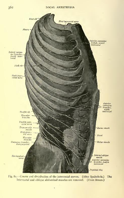
362 LOCAL ANESTHESIA
First rib
Pleura
Anterior cutaneousbranch, secondintercostal
Lateral cutane-ous branches,,fourth inter-
costal
Muscularbranches
Cutaneous branches,-
iliohypogastric
L Internal obliquemuscle
Anterior cutaneous
branches, twelfthintercostal
Inguinal ring
Fig. 81. Course and distribution of the intercostal nerves. (After Spalteholz.) Theintercostal and oblique abdominal muscles are removed. (From Braun.)

HERNIA 363
to the subcutaneous tissue in this region. Other fibers, which follow
the cord through the ring, are supplied to the upper portion and side
of the scrotum and thigh. This nerve is not constant, and occa-
sionally is found joined to the genital branch of the genitocrural to
form the external spermatic nerve, or it may form a common trunk
with the iliohypogastric.
anteriorcutarfs^tt.da
anterior cutarfsof L
(ilio-hypoof.)
Lateral cut.o/ 12. <L
Ant. sup. spine
tat.cut.o/11
Intern, ring"""
Exf. ring'
Fig. 82. Sites for local anesthesia in the inguinal region. (After Gushing.)
The genitocrural nerve, its genital branch, appears at the internal
ring and passes down the back part of the spermatic cord into the
scrotum, where it supplies the cremaster muscle, testicle, and other
contents of the scrotum. The skin of the scrotum receives fibers
from the inferior pudendal branch of the small sciatic and from the

364 LOCAL ANESTHESIA
superficial perineal branch of the pudic, in addition to the ilio-in-
guinal nerve already mentioned.
It will be seen from a study of the above that after the skin is
passed all nerves entering the field emerge at or near the internal ring,
and it is consequently here that we inject most of our solutions.
Preparation for the Operation. Preliminary hypodermic pan-
topon or morphin, % gr., with scopolamin, ^50 gr., one hour be-
forehand.
Four ounces of solution No. i (novocain, 0.25 per cent.; sodium
chlorid, 0.4 per cent.), to which add 15 drops of adrenalin solution
(i : 1000). If the hernia is very large, it is well to have on hand more
Fig. 83. Long needle passed through intradermal station to reach position of iliohypo-
gastric nerve beneath tendon of external oblique.
than the 4 ounces. Small hernias may not require this much, but
it is well to have an ample supply.Two small hypodermic syringes and one large 10 c.c. syringe,
with long fine needles, or a Matas' infiltration apparatus all well
tested beforehand to be sure they are in good working order.
Some operators prefer to inject the cases about fifteen minutes
to one-half hour beforehand, and allow them to wait for the solution
to diffuse and become fixed in the tissues. This practice, while ad-
visable elsewhere, we do not find necessary here, and proceed at once
with the operation. Also some prefer to use a i per cent, novocain
solution to infiltrate the nerves as they are encountered, but, as all

HERNIA 365
the nerves concerned are very small, it is unnecessary to use any but
the ordinary infiltration solution (No. i).
Begin the injection with the small hypodermic syringe at the
highest point of the proposed incision, at the upper and outer part of
the field, about i^ inches internal and slightly below the anterior
spine of ilium. Make the injection intradermally. With the large
syringe and long needle enter through this wheal directing the needle
downward, injecting as it is advanced and distribute about 2 drams
of solution in the subcutaneous tissues at this point (Fig. 83). By
advancing the needle slightly further through the subcutaneous tis-
Fig. 84. Needle is partially withdrawn from position shown in Fig. 83 and directed
subcutaneously toward pubes.
sues the aponeurosis of the external oblique is reached; this is recog-
nized as the first plane of resistance which the needle encounters
after the skin is passed and varies in depth according to the stoutness
of the individual. The aponeurosis is gently pierced at this pointand about 2 drams of solution deposited beneath it. This is the first
point at which the iliohypogastric nerve is encountered and in its
normal position will not be found to be y inch from the needle point.
The needle is now slightly withdrawn into the subcutaneous tissues
and its point directed toward the spine of the pubis injecting as the
needle is advanced the entire distance (Fig. 84), distributing in all
about 3^2 ounce of solution.

366 LOCAL ANESTHESIA
The long needle is now withdrawn and beginning at the primarywheal in the skin this tissue is infiltrated intradermally as far down
as the spine of the pubis along the proposed line of incision, which
may now be made. The idea in making some of the deeper injections
first is to allow them ample time to diffuse and thoroughly saturate
all nerve-fibers in their neighborhood. The first incision is made
through the skin the entire length of the field but carried down
through the subcutaneous tissue only in the upper part of the field
where the aponeurosis of the external oblique is exposed. The long
needle is now used and entered at this point, near where it first
Fig. 85. i, Iliohypogastric nerve; 2, ilio-inguinal nerve; 3, spermatic cord; 4, hernial
sac dissected free. The genitocrural nerve is not seen, as it lies within the cord on
its posterior aspect,
pierced the aponeurosis to reach the iliohypogastric nerve, and
passed downward beneath the aponeurosis to Poupart's ligament,
reaching this point about midway between the anterior-superior
spine of the ilium and the pubis; here the ilio-inguinal nerve will be
found in the inguinal canal below the cord. A finger held over the
skin can guide the progress of the needle; about 2 drams of solution
are deposited along the course of the needle and at this point.
The course of the needle is now directed slightly inward but still
close under the aponeurosis and a little additional solution deposited
on each side of the external ring which can now be readily identified

HERNIA. 367
by palpation through the subcutaneous tissues. We can now freely
expose the aponeurosis throughout its entire extent by dividing the
remaining subcutaneous tissue. I usually stop here to ligate the
superficial vessels, clearing the field of forceps, thus allowing the last
made injections a little time to work. The aponeurosis of the ex-
ternal oblique is now divided and its edges retracted; this exposesthe cord and hernia sac lying on top, the whole surrounded by the
cremasteric fascia through which run bundles of the cremasteric mus-
cle. This is opened above or on its outer side and the two edges re-
tracted, freely exposing the cord and sac; by rolling the sac and over-
lying tissues inward the cord is more plainly brought into view and
its component parts recognized. With a small syringe and fine
needle the cellular tissue of the cord is injected at several points
gently feeling the way high up toward the internal ring. The genito-
crural nerve which lies at the back of the sac may be identified, but
this is unnecessary as it is amply reached by the surrounding injec-
tions. The cord is now left and the inner edge of the cremasteric
fascia retracted inward exposing the neck of the sac where it comes
from under the edge of the conjoined tendon. The sac at this pointis surrounded by the transversalis or retro-peritoneal fascia which
varies greatly in amount in different individuals, if thin the perito-
neum of the sac which underlies it can be easily reached by the solu-
tion injected here; this is done circumferentially at the proximal
point where the sac comes off from the parietal peritoneum raising
the sac to get to the under surface. In the ordinary case, which is
handled this way, this completes the anesthesia and the operation
can proceed absolutely without any pain and by any method pre-
ferred by the operator.
An existing varicocele or any other complication should be dealt
with now and requires no further infiltration. The testicle may also
be exposed and handled if necessary. It should be borne in mind,
however, that any undue traction upon the cord by pulling upon the
parts within the cavity will cause pain, but none is otherwise ex-
perienced. The neck of the sac can now be closed by crushing,
if large, and ligated; or sutured, if preferred.
If the above technic is followed absolutely no pain should be felt
by the patient; where pain is inflicted the technic is at fault. In the
hands of a skilful operator an ordinary hernia can be closed by using
not over 3 ounces of solution (we often use much less), and the time
consumed is not over five or ten minutes longer than would have
been required with general anesthesia.

368 LOCAL ANESTHESIA
Fig. 86. Method of making injections around neck of sac, with finger within sac.
Fig. 87. Semi-diagramatic drawing from actual operation illustrating inguinal
canal opened, sac dissected from cord and opened, and separate fatty mass extending
down into internal ring. (Modified from Speed.)

HERNIA 369
In case the hernial ring is very large, it may be necessary to loosen
the internal oblique and transversalis from the edge of the rectus so as
to enable the conjoined tendon to be brought down; in case this is
necessary, and the dissection be carried very high, some additional
infiltration at this point may be needed, and should be made directly
into the tissues to be dissected.
In case the sac is large and thick, or there is much retroperitoneal
tissue around it, it is preferable to open it some distance from the
ring after first infiltrating the point of incision, a finger can then be
passed up its lumen toward the ring and the neck of the sac injected
circumferentially with the finger held in this position by passing the
needle through the walls of the sac down toward the finger (Fig. 86) .
Or, if preferred, the sac can be more freely opened and injected from
within as shown in Fig. 87.
In case the sac contain intestines or omentum, these should be
replaced if possible by manipulation before opening the sac. If this
cannot be done, the neck of the sac should be blocked as thoroughlyas possible before opening. It should always be borne in mind that
the sac is formed by the parietal peritoneum which is verysensitive.
After the neck of the sac has been well blocked and the sac openedthe pushing of the contents through the ring will cause no pain but
should be gently done. In case the omentum or intestines are ad-
herent to the sac wall their separation causes no pain after the neck
of the sac has been blocked, but if much mesentery accompaniesthe intestine, this is always sensitive and should be well injected
proximally where it comes through the internal ring before extensive
manipulations are resorted to. In making these injections muchcare is sometimes necessary not to injure the many vessels which are
often quite congested as a result of the constriction. If the omentumis found hard and fibrous from its long sojourn in the sac it will re-
quire resection, but, unless badly damaged, it should not be sacri-
ficed, as it is an organ of many valuable functions.
The exposure of the omentum never gives rise to any discomfort,
and its resection causes no complaint, as it has no sensation; but
large vessels within it are sensitive, and should first be blocked before
ligation or division.
In the case of very large hernias, where it is necessary to carry
the skin incision well down over the scrotum, the skin must be infil-
trated all the way.The following histories may prove interesting:
24

370 LOCAL ANESTHESIA
Floyd, aged seventy-five, Ward 9, an old and feeble man. Entered the service for
the removal of epithelioma of right temporal region, and while convalescing from the
operation, which was done with local anesthesia, a large inguinal hernia of the left side,
of fifteen years' duration, became incarcerated and irreducible. He was operated on
June 26, 1908, by the method described above, and was discharged cured in two weeks,
with no recurrence since. This was a particularly satisfactory case, in view of the ex-
treme feebleness of the patient.
Pear, aged sixty-four (Fig. 88). Left inguinal hernia of thirty-four years' duration
with enormous sac, extending to near the knee. The contents of the sac had not been
reduced for years; the photograph gives a good idea of the condition. The inguinal
ring was large enough to admit three fingers. In view of marked arteriosclerosis, with
renal and cardiac lesions, the operation was performed by the above-mentioned method
Fig. 88. Large irreducible hernia operated on by author under local anesthesia. Sac
contained omentum and intestines; large part of omentum was resected.
on August 27, 1908. A large part of the omentum was adherent to the sac and muchthickened and had to be resected. A portion of the rectus muscle was transplanted to
close the inguinal opening. Some of the redundant skin of the parts was resected. The
operation was entirely painless, with union by first intention, and the patient was up in
two weeks. This case was a bad hernia and a severe test of the method, and clearly de-
monstrated its usefulness and feasibility in these cases. I have recently seen the case,
and he had remained well, with no sign of recurrence.
In strangulated hernia, general anesthesia is contra-indicated, and
should rarely, if ever, be used. In very severe and prostrated cases
the general anesthetic may add sufficient additional depression to
cause a fatal issue. The sac should here be exposed and openedunder infiltration. If the patient is very weak a radical operation

HERNIA 371
should not be attempted at the time, but the bowel, if gangrenous,
opened and drainage permitted. It will here frequently be found
that the bowel is adherent around the ring and the general cavity
walled off. If this is not the case, a few sutures and packing may be
resorted to to close off the cavity and the bowel at once opened.The improvement permitted by this procedure revives the patient
and, after all gangrenous material has come away, the ends of the
bowel can be approximated by the Murphy button or suture.
We then wait for good union to take place and the wound to be-
come clean and covered with healthy granulation, when it may be
closed with a small drain. The following case illustrates this type:
Mr. S., aged seventy-two. Had had a large inguinal hernia since he was seventeen.
It had frequently become incarcerated, but he was always able to reduce it until a few
days prior to my seeing him, when it again became incarcerated and all efforts at reduc-
tion failed, strangulation following. Patient was very feeble, pulse almost impercepti-
ble, temperature subnormal with cold extremities, and almost constant stercoraceous
vomiting. Under local anesthesia the sac was opened. The bowel and fat attached to
it were found gangrenous, with a large quantity of foul fluid in the sac. The neck of the
sac was securely protected with sutures, packs and rubber tissue, the gangrenous fat
cut away, and the loop of bowel opened at the distal end. The patient revived at once,
all vomiting ceasing, next day was in a rolling chair on the gallery, and in a few dayswas sitting up.
Nourishment had been at once resumed, and his improvement was noticeable daily.
In ten days the bowel was closed by suture, and two weeks later the wound, now being
quite healthy, closure under local anesthesia was performed. The bowel, freed from
the old sac, dropped back into the cavity, the wound enlarged until the anatomy became
apparent, and the parts then closed with two rubber-tissue drains, one within the cavity
and one subcutaneously. The Fowler position was used for twenty-four hours, then dis-
continued, and the drain removed in three or four days. Recovery was without inci-
dent, the patient being discharged about three weeks later. This illustrates a type of
case which, I believe, if handled any other way would have resulted fatally.
Reclus very justly refers to operations for strangulated hernia as
"the triumph of cocain." In strangulation it is the anesthetic of elec-
tion, and it is only in special conditions, such as in herniae of unu-
sually large size, with eventration of the abdominal contents and
when extensive adhesions exist, that he would prefer a general anes-
thetic. In advanced strangulations, with vomiting of intestinal con-
tents, the dangers of septic pneumonia and secondary renal compli-
cations from chloroform and ether are especially to be feared, and
more particularly in the aged. Then, again, colostomy for strangu-
lation is an urgent operation which, in country practice, frequently
compels the surgeon to depend upon unskilled assistants. Under
these circumstances the value of a local anesthetic entirely under
the control of the operator becomes especially apparent. It is not

372 LOCAL ANESTHESIA
surprising, therefore, that Mehler enthusiastically asserts that he
who has tried local anesthesia in these conditions will never feel in-
clined to return to general anesthesia, unless compelled to by pres-
sure of unusual circumstances.
FEMORAL HERNIA
This hernia, except in very fat people, is easily operated uponwith local anesthesia, and is best suited to the infiltration method.
Regional anesthesia cannot be employed, as the nerve-supply is from
many sources and reaches the field from several directions, all being
small branches. The method of procedure will likely vary, accord-
Fig. 89. Line of cutaneous anesthesia for femoral hernia.
ing as to whether or not the patient is stout or thin, and whether the
hernial sac can be readily defined from the surrounding tissues.
In stout persons or in poorly defined sacs an incision had best be
made in the long axis of the tumor, infiltrating layer after layer, and
cutting as we go (Fig. 89). After the sac has been reached and
defined the tissues at its sides and neck can be infiltrated, care being
taken to locate and avoid the femoral vein which lies just to its outer
side, and, on account of the sac rising forward, often a little behind.
In case the patient is thin and the sac well defined a more satisfactory
plan may be followed by making a circumferential injection around
the sac subcutaneously; or, as recommended in umbilical hernia, the
sac can be opened early in the operation and a finger passed down
within, this is used as a guide to the needle which is passed down
along the outer wall infiltrating the tissues as far as the neck.

HERNIA 373
First anesthetize a point in the skin near the edge of the sac with
a small hypodermic syringe. If the Matas infiltration apparatuswith a curved needle is convenient, it will be found very useful here
;
if not, the large syringe with a long needle will answer. The advan-
tage here of the Matas apparatus with the curved needle is that the
entire circumference of the sac, unless very large, can be reached and
infiltrated from a single point of puncture of the skin; if a straight
needle is used several punctures will have to be made.
First advance the needle closely under the skin, then through the
deeper subcutaneous tissues in all directions around the sac on the
outer side, taking care not to penetrate beneath the fascia lata for
fear of injuring the femoral vein, but on the inner side a much greater
depth can be penetrated without fear of injury.
It may be necessary to avoid the saphenous vein at the lower por-tion. The precaution mentioned elsewhere, when injecting in the
neighborhood of large veins, should here be observed, of injecting
only when the needle is being advanced or withdrawn, and never
when the point is stationary, as a vein may be entered and a large
quantity of the solution thrown directly into the circulation. If a
vein should be pierced with a fine needle no serious consequences will
result.
The injection by the above method is made very quickly, and
all nerves entering the field from any direction are bathed in the
solution and anesthetized. It is unnecessary to inject the skin alongthe proposed line of incision if a few minutes' delay is permitted for
the solution to diffuse. This plan of Lennander, of waiting fifteen
to twenty minutes after the injection is of advantage here. But in
case it is desirable to proceed at once, and the skin in the middle
line is not anesthetic, it can be infiltrated intradermally and the
incision made at once, as the deeper parts will be found well anes-
thetized.
Before opening the hernial sac it should be observed whether or
not it is the bladder, which is very common in these herniae, and
may be opened in looking for the peritoneal investment.
The neck of the sac is freed and Gimbernat's ligament divided,
when reduction is usually easy. Closure can then be accomplished
by any recognized method, after first anesthetizing the neck of
the sac.
In case it is preferred the inguinal canal can be opened and the
sac pulled up from above. If this method is adopted, the skin and
subcutaneous tissues along the course of the inguinal canal should be

374 LOCAL ANESTHESIA
infiltrated and the needle passed down through the aponeurosis of
the external oblique into the canal and about % ounce of solution
deposited at several points here. After the incision has been madeand the canal opened, the parts are freely retracted exposing the
neck of the sac which is injected quite liberally circumferentially.
This should complete the necessary anesthesia for small hernias,
but in case the sac is very large and adherent to the tissues of the
thigh some peripheral injections may have to be made around it
at its peripheral parts before the sac can be freely separated; when
necessary these can be made by passing the needle through the woundover Poupart's ligament into the tissues around the sac. The sac
can be dealt with and the wound closed by any method preferred
by the operator.
The solution used here is the same as for inguinal hernia (No. i,
novocain, 0.25 per cent.; sodium chlorid, 0.4 per cent.; and adrenalin,
10 to 15 drops).
In very fat individuals with large herniae and many adhesions and
tense abdominal walls this operation may be difficult under any
method, but the difficulties are much less under local anesthesia and
the certainty of a cure greatly increased.
In all these operations the mechanical difficulties are very muchlessened by putting the patient to bed for a few days, on restricted
diet, with daily laxatives. This relaxes the abdominal walls and
relieves the intra-abdominal tension, and approximation of the gapcan be much more easily obtained.
With Solution No. i several stations in the skin are anesthetized:
if the hernia is small, one on each side; if large, one above and one
below, in the median line, in addition (Figs. 90, 91). By entering at
these points (Matas' infiltration apparatus or large syringe with long
needle) and passing the needle in all directions, a circumferential in-
jection is made into all the subcutaneous tissues, thus creating a zone
of anesthesia. If the underlying muscles are clearly outlined, and
there is no danger of going through them, these may be infiltrated
at the same time;otherwise this had better be delayed until they are
exposed. After a delay of a few minutes, to allow the solution
time to diffuse, the incision can be made. First, expose the muscles
and thoroughly infiltrate them down to the peritoneum, if this has
not already been done. While we are waiting for the last injection
to diffuse, bleeding points can be ligated and the sac opened and its

HERNIA 375
contents dealt with. By now the parietal peritoneum will probablyhave become anesthetized, and the sac can be cut away around the
margin of the gap; if the peritoneum is still sensitive, a long needle
is passed through the opening into the subperitoneal tissue and a
Fig. 90. Method of injection around umbilical hernia. (From Braun.)
moderate injection made in all directions. There will then be no
further sensation.
In dealing with the contents of the sac omental adhesions cause
no trouble, and can be separated or divided without sensation. If
the intestines are adherent any very extensive manipulation may
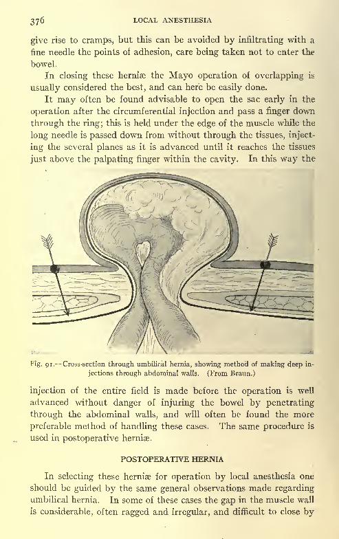
LOCAL ANESTHESIA
give rise to cramps, but this can be avoided by infiltrating with a
fine needle the points of adhesion, care being taken not to enter the
bowel.
In closing these herniae the Mayo operation of overlapping is
usually considered the best, and can here be easily done.
It may often be found advisable to open the sac early in the
operation after the circumferential injection and pass a finger down
through the ring; this is held under the edge of the muscle while the
long needle is passed down from without through the tissues, inject-
ing the several planes as it is advanced until it reaches the tissues
just above the palpating finger within the cavity. In this way the
Fig. 91. Cross-section through umbilical hernia, showing method of making deep in-
jections through abdominal walls. (From Braun.)
injection of the entire field is made before the operation is well
advanced without danger of injuring the bowel by penetrating
through the abdominal walls, and will often be found the more
preferable method of handling these cases. The same procedure is
used in postoperative herniae.
POSTOPERATIVE HERNIA
In selecting these herniae for operation by local anesthesia one
should be guided by the same general observations made regarding
umbilical hernia. In some of these cases the gap in the muscle wall
is considerable, often ragged and irregular, and difficult to close by

HERNIA 377
general anesthesia. Under local anesthesia these difficulties are not
increased, but often lessened, as you do not have the straining so
often encountered under general anesthesia. The patient can also
better take a position favorable to relaxation of the muscles, but
particularly valuable afterward is the absence of vomiting, which
if severe or protracted may often jeopardize the results of the work.
It is here often highly valuable to have the patient remain perfectly
quiet after operation, as he is able to do following the use of local
anesthesia. These remarks apply equally to all herniae.
Fig. 92. Method of making crescentic line of anesthesia around postappendicularhernia. Deep injections made through heavy dots.
One difficulty encountered in postoperative herniae is the large
amount of fibrous tissue encountered, which mats all the structures
together, but if a zone of anesthesia is created just outside of the
area no special difficulties are encountered.
Herniae in the midline should be dealt with the same as umbilical
herniae, by a complete circumferential injection around the gap,
thus sequestering all nerve-endings within the area.
Herniae just to either side of the midline will also require a circum-

378 LOCAL ANESTHESIA
ferential injection, as the nerve-fibers from the other side lap over the
midline some little distance; but where the hernia is some distance
removed, as is the case of those resulting from appendicular opera-
tions, a circumferential injection is not necessary. As all the nerves
in the anterior abdominal wall proceed downward and forward be-
tween the muscle planes, it is only necessary to make the injection
in such a way as to block these. Consequently, a crescentic-like
area of anesthesia on the outer side of the hernia will prove sufficient,
having the horns of the crescent to embrace the upper and lower
extremities of the gap and carried as a wall of anesthesia from the
skin to the peritoneum. (See Figs. 81 and 92). The anesthetising
of the tissues is done in the same manner as advised for umbilical
hernia. I have often closed large postappendicular herniae in this
way, and have found it very satisfactory.

CHAPTER XIX
GENITO-URINARY, ANORECTAL, AND GYNECOLOGICOPERATIONS
GENITO-URINARY ORGANS
WHILE the pudic nerve is the principal source of innervation of
the deeper parts of these organs (Fig. 93) and is capable of a fairly
accurate blocking for regional anesthesia, the skin of these parts re-
ceives its nerve-supply from a variety of sources and cannot be dealt
with collectively except by such more or less central' methods as
parasacral or epidural injections, thus blocking at one time the entire
innervation of the pelvis and a large part of the lower extremity.
The pudic nerve leaves the pelvis.through the great sacrosciatic
foramen, crosses the spine of the ischium with the pudic artery, and
re-enters the pelvis through the lesser sacrosciatic foramen. Accom-
panying the pudic vessels, it runs downward, forward, and inward
along the outer wall of the ischiorectal fossa. In this position it is
about i inch internal to the tuberosity of the ischium. Here it gives
off its perineal and inferior hemorrhoidal nerves, then continues as the
dorsal nerve of the penis.
The inferior hemorrhoidal nerves, several in number, pass down-
ward, inward, and slightly forward from the above position, and are
distributed to the sphincters of the rectum and anal canal.
The perineal branches pass downward and forward to the
perineum, giving off branches to the muscles of these parts, and
are distributed to the skin of this region, branches passing for-
ward to the scrotum in the male and labia majora in the female.
It also sends a branch to the bulb of the penis.
The dorsal nerve of the penis pierces the posterior layer of the
deep perineal fascia, and runs forward along the inner margin of
the ramus of the os pubis between the two layers of deep fascia.
Further forward it pierces the anterior layer of the fascia and passes
through the suspensory ligament to the dorsum of the penis. In
this position the nerves on each side lie to the outer side of the
artery (Fig. 94) . It gives off a large branch to the corpora caverno-
sum, and along the side of the penis branches to this organ; its
terminal filaments are distributed to the glans and prepuce.
379

38o LOCAL ANESTHESIA
Levalor
medial infer, cluneal
inferior haemorrhoidalartt
int. pudicvessels
pudic
Miiocoecygeal nerves
anococcygeal lig.
Glutaeus maximus'uberous lig.
internal pudic art.
uidic nerve
ospinous lig.
BuWoau'trnosus
\ dorsal nerre of penis
\ ^arttiy ojf, urethra! bulb
art. of penis
posfenor scroial arteries"'
corpus cavernosnm of'urethraposterior scrotal nerves
Fig. 93. The nerves and vessels of the male perineum upon the left side, the
superficial perineal musculature has been exposed and the ischiorectal fat removed;
upon the right the transversus perinei superficialis has been divided, the urogenital
diaphragm incised, and the ischiocavernosus drawn slightly to one side.* = Bifurca-
tion of internal pudic artery into the perineal and penile arteries. (Sobotta and
McMurrich.)

GENITOURINARY, ANORECTAL, AND GYNECOLOGIC OPERATIONS 381
Perineal branches of the small sciatic are distributed to the skin
of the posterior and lateral parts of this region, one branch larger than
the others; the inferior pudendal curves downward and inward
around the tuberosity of the ischium, and passes forward and inward
dorsal artery of penis fundiform ligament
dorsal nerve of penis ,<tf penis
spermatic ford^
\ \
\
dorsal vein of penis
external spermaticvessels
fascia ofpei
dorsal ve,
of penis
anteriorscrotal vessels
subcutaneousvein of penis
Fig. 94. The vessels and nerves of the penis spermatic cord and scrotum as seen
from in front. The skin and the greater portion of the fascia have been removed from
the penis; the vessels of the right spermatic cord have been exposed by dividing its
coverings. (Sobotta and McMurrich.)
beneath the superficial fascia and is distributed to the skin of the
perineum scrotum in the male and the labia majorum in the female.
The nerve in its passage around the tuberosity lies about % inch to
the outside of the bone, between it and the great sciatic nerve.
The scrotum, in addition to its branches from the pudic and in-
ferior pudendal, receives cutaneous branches from other sources,

382 LOCAL ANESTHESIA
principally the ilio-inguinal, and probably, on its anterior part, a few
branches from the iliohypogastric. Its nerve-supply is such that all
operations done upon it must be through infiltration. The testicle,
spermatic cord, and cremaster muscle are innervated by the genital
branch of the genitocrural, but this nerve does not give any branches
to the skin overlying these parts.
The bladder receives two nerves on each side from the third and
fourth sacral, which enter the organ near its base.
Method of Blocking Pudic Nerve. The tuberosity of the ischium
is located as a landmark; the skin over a point about i inch internal
and in front of the tuberosity is now anesthetized; a large syringe,
containing a few drams of 0.50 per cent, novocain solution, with 2 or
3 drops of adrenalin, and fitted to a long needle, is now used.- Theneedle is passed downward and outward toward the base of the tuber-
osity, varying in depth according to the stoutness of the individual,
but usually about i^ to 2 inches; at a point about % inch from the
base of the tuberosity 2 or 3 drams of the solution are injected. The
same procedure is repeated on the opposite side; or, if preferred, the
long needle may be entered at a point i^ or 2 inches back of the
anus in the midline, after previously anesthetizing this point in the
skin, and directing the needle obliquely outward and upward toward
the base of the tuberosity of the ischium, guided by the finger within
the rectum, when 2 or 3 drams of the solution are injected about ^inch from this bone. The method of making this injection is shown
in Fig. 1 06. The needle is then partially withdrawn and turned in
the opposite direction, where the injection is repeated. These
methods, however, are not often used, but when resorted to for
operations on the rectum the perianal infiltration, as described later,
should be made somewhat more liberal posteriorly between the anus
and the coccyx and well into the subcutaneous tissue to reach the
nerves that come into the field from this direction. The uncertainty
of reaching this nerve at the point where it enters the pelvis with any
degree of accuracy for a.paraneural injection has led to efforts to
reach it from without by an injection made through the gluteal
region. This procedure has been recommended by Franke, and is
done as follows: The skin of the gluteal region, at a point about over
the spine of the ischium, is located by a finger passed within the rec-
tum, and the long needle passed down from without through the
anesthetized point and an injection made in contact with the spine.
Neither of these methods have become very popular with others,
and are rarely if ever used by the author. The methods preferred

GENITOURINARY, ANORECTAL, AND GYNECOLOGIC OPERATIONS 383
are those described in dealing with the different regions, as de-
scribed later.
To anesthetize the inferior pudendal nerve an injection is made on
the outer side of the tuberosity of the ischium, where this nerve
passes close to the base of the process and between it and the great
sciatic.
The needle is entered in the perineum to the inner side of the
tuberosity and directed outward and upward, injecting as the needle
is advanced to a point over the base of the bone; the injection is
made after the needle is felt to pass the bone about % inch, usually
using about 2 drams of 0.50 per cent, novocain adrenalin solution.
This, however, like the injection of the pudic, is uncertain.
THE PENIS
To anesthetize the entire organ a circumferential line of intra-
dermal anesthesia is carried around the organ at its root, as seen in
Fig. 95. Method of procedure for anesthesia of entire penis. (From Braun.)
Fig. 95. From this line two deep injections are made about Y inch
on either side of the midline and carrTe4 down to the corpora caver-
nosa (Fig. 94), showing the nerve-supply and (Fig. 96) the point of
injection, using here about i dram of 0.50 novocain adrenalin solution
or a somewhat more liberal injection of solution No. i.
If the contemplated procedure involves the urethra, a smaller
quantity of the solution should be injected deep on either side of

LOCAL ANESTHESIA
this structure in the sulcus, between it and the corpora cavernosa.
Should a fine needle pierce the urethra in this injection no damage will
result. A small stationer's elastic band, used as a constrictor, should
now be placed around the base of the organ proximal to the injections,
but not too tightly, for fear of injury. After a few minutes' delay
anesthesia is produced. Gentle massage helps to diffuse the solution,
when any operation involving these parts may be undertaken, from
circumcision to amputation. Urethrotomy, internal or external, as
well as plastic work, involving the urethra or the rest of the organ,
Fig. 96 i, Shows line of anesthesia for suprapubic cystotomy; 2, points on each
side of midline for paraneural injection of dorsal nerves of penis; 3, area of anesthesia for
varicocele, hydrocele, or orchidectomy.
distal to the point of injection, can now be painlessly done. The above
is an excellent method for the cauterization of extensive or phago-denic chancroids of this region or for operations for paraphimosis.
The Oberst Method. This is really a form of arterial anesthesia,
and, while ingenious and effective, may at times be followed by hema-
toma, and for that reason is not very popular with the writer.
A constrictor is placed around the root of the organ. A syringeand fine needle filled with i per cent, cocain solution (which is the solu-
tion recommended by Oberst, though novocain could also be used)is now injected in the following manner:

GENITO-URINARY, ANORECTAL, AND GYNECOLOGIC OPERATIONS 385
The needle is thrust well into the corpora cavernosa, and from 5
to 7 minims of the solution is injected (Fig. 97). This is repeatedon the opposite side. About 5 minims of the solution is injected
into the subcutaneous tissues on each side of the organ, and about
the same quantity on the undersurface around the urethra.
Anesthesia takes place in about fifteen minutes, and is usually
very satisfactory and sufficient for any operation upon the organ. It
has been especially recommended for circumcision as a substitute
for the direct method of infiltration, on the ground that the edematiza-
Fig. 97. The Oberst method of cocain infiltration. A constrictor is first placedaround the root of the organ and the injection made with a very fine needle, as described
below. Oberst recommends a i per cent, solution of cocain, but the same strength of
novocain could also be used. (Miller.)
tion of the tissues resulting from infiltration was an objection in this
operation. The writer, however, has not found this to be the case.
Circumcision. Solution No. i, with an ordinary hypodermicwith fine needle, is sufficient. The skin is pulled well over the glans
and the point of incision determined. The injection is begun just
proximal to this point and a circumferential injection is made into
the subcutaneous tissues around the organ. If the prepuce is well
relaxed and can be freely retracted, a finger is passed up on its inner
surface between it and the glans. The needle is now directed down
through the already anesthetized parts on the surface toward25

386 LOCAL ANESTHESIA
this point, injecting as it is advanced, until it reaches a point on the
inner surface of the prepuce just back of the cervix (neck of the glans).
A station is produced here, the prepuce now retracted, and, begin-
ning at this anesthetized point, a collar of anesthesia is created around
the glans, carrying it well into the frenum; or, instead of the above,
the prepuce can be reflected and the anesthesia started from an
injection made from the inner surface. Anesthesia will now be com-
plete and the operation can be performed. A small stationer's elastic
should first be applied as a constrictor around the organ near its
base. In the event that the prepuce is very tight and cannot be re-
tracted, it may be filled with a i per cent, solution and held for a few
minutes, or the injection can be carried forward toward the con-
Fig. 98. Anesthetizing dorsal surface of foreskin from periphery toward base. (From
Braun.)
stricttd opening and this anesthetized and divided sufficiently to
permit retraction, when its inner surface can be anesthetized; or the
procedure as illustrated in Fig. 98 may be carried out.
The Urethra. Two or 3 drams of i or 2 per cent, novocain solu-
tion and i or 2 drops of adrenalin held in the urethra for five or ten
minutes will anesthetize the mucosa sufficiently to permit a painless
internal urethrotomy or the gradual dilatation of a stricture, but
will not permit divulsion (which, however, is now rarely practised),
as it does not anesthetize the submucous and periurethral tissues.
Stronger solutions than i or 2 per cent, are never necessary here.
The same effects can be obtained with the weaker solutions if re-
tained slightly longer. The urethra absorbs very actively, and
many cases of poisoning, at times fatal, have been reported from the

GENITO-URINARY, ANORECTAL, AND GYNECOLOGIC OPERATIONS 387
injudicious use of strong solutions; consequently, one should never
be tempted by haste to exceed the safe limits.
In irrigating a sensitive urethra by the Valentine method, or one
in which the cut-off muscle will not readily relax, this muscular
contraction can be easily overcome by first filling the urethra with a
weak anesthetic solution for a few minutes as above suggested.
The use of adrenalin solution when retained a few minutes in the
urethra is often of great advantage, particularly in inflamed or con-
gested conditions; the relaxation following the congestion will often
permit the easy passage of an instrument which would seem impos-sible or very difficult without its use. Strictures which have been
congested following alcoholic indulgence, causing acute retention of
urine, can often be sufficiently relieved in this way to permit the
passage of a small catheter when an external urethrotomy would
otherwise have seemed indicated.
Where the stricture or field of operation is in the penile portion of
the urethra a very satisfactory and exact method of anesthesia is to
inject the tissues circumferentially around the urethra by passing a
fine needle down to this structure from the under surface of the
penis. This is done just proximal to the field and after a few
minutes delay will anesthetize the uretha for a considerable distance
in front of the injections. If the entire penile urethra is to be
anesthetized, two points may have to be injected. This method
anesthetizes the entire urethra and is devoid of any danger which
may at times occur from absorption when strong solutions are injected
within the urethra and which at most anesthetize only the mucous
membrane.
The Meatus. To anesthetize the meatus a very satisfactory
method is to dip the end of a moistened sterile probe into a bottle
of cocain or novocain crystals, when a few of the crystals will adhere
to the end and can be conveniently deposited in the meatus. In a
few minutes this will have produced sufficient anesthesia to permit
fairly considerable dilatation for the passage of a sound through a
tight meatus, and will also permit a limited meatotomy. But it is
better for the latter to infiltrate the meatus in the line of the pro-
posed incision with a few drops of solution No. i.
The female urethra is easily anesthetized by a few drops of 5 or
10 per cent, novocain on a film of cotton wrapped around an appli-
cator, and passed into the urethra for a few minutes.
External urethrotomy in the ordinary case is quite easily per-
formed under local anesthesia. In the presence of extensive urinary

388 LOCAL ANESTHESIA
infiltration it may prove difficult and test the ability of any but an
experienced operator .under local measures, as the fibrous tissue en-
countered under these conditions is difficult of thorough infiltration.
If the strictured point is deeply situated the pudic and pudendalnerves had best be blocked, though it is possible to proceed entirely
with infiltration, which is the method preferred.
The urethra is first anesthetized, a sound passed, and the stric-
tured point located or a guide or filiform passed through it. Infil-
tration is commenced in the middle line (Fig. 99), just proximal to
the stricture, and carried well down into the subcutaneous tissues.
Fig. 99. Line of infiltration for external urethrotomy.
These are now divided, infiltrating further as we advance, until the
urethra is reached, when a little solution injected periurethrally
around the strictured point will permit its painless division or resec-
tion, if not too extensive, with subsequent approximation and suture
of the divided ends of the urethra.
In cases in which the stricture is long and tortuous or in which
there are several, the urethra is incised at the distal end of the stric-
ture and its lumen opened by retraction, exposing the strictured
orifice, when under direct guidance of the eye a much larger instru-
ment can be passed backward toward the bladder, and the exact course

GENITOURINARY, ANORECTAL, AND GYNECOLOGIC OPERATIONS 389
of the urethra recognized through the tissues, when a long needle can
be passed down in close contact with it and the tissues injected.
This is best done on both sides and will permit the free incision
of these parts or their dilatation as preferred. In cases in which
the stricture is very dense and its lumen hard to follow with anyinstrument, the urethra can be opened at the distal end of the stric-
ture after freely infiltrating the perineum, the strictured urethra is
then progressively followed backward infiltrating as you advance
until a patulous portion of the urethra is encountered.
The following history of a patient operated on by the author
illustrates a procedure which may sometimes prove useful:
Mr. A., aged forty-three, alcoholic, with chronic endocarditis and history of renal
disease, presented himself, with a large infiltrated scrotum with urinary fistulae on inner
side through which urine and pus dribbled, only a few drops appearing at the meatus
during efforts at urination. It was found impossible to pass a sound or filiform throughthe tortuous fibrous urethra. Through infiltration of the tissues at the base of the scro-
tum an incision was made down to about the position of the urethra, but it was found im-
possible to readily identify this structure. The wound was packed and suprapubic cys-
totomy done, liberating a large amount of foul urine. In the Trendelenburg position
(to dilate the bladder with air) the internal urethral orifice was readily seen, and a small
Poges silk-woven catheter passed forward along the urethra. This caused no discom-
fort. At a distance of about 3 inches the point of the catheter was arrested. While it
was held in this position by an assistant we returned to the perineal wound. Someadditional infiltration was found necessary here, as the parts had become sensitive.
The urethra was now readily recognized in a mass of fibrous tissue by manipulation of
the catheter and opened. The anterior portion of the urethra was easily followed and
several strictures divided under infiltration.
The case made an uneventful recovery. The urinary fistulae on
the side of the scrotum closed without any special treatment. Sub-
sequent examination of the urine showed albumin and granular casts.
This case would certainly have been a dangerous risk with general
anesthesia. Spinal analgesia could, however, have been employed,but we resort to the latter only when local and regional methods are
impracticable. These cases with extensive infiltration and multiple
fistulae are particularly well suited to epidural injections, which see.
Epispadias and hypospadias, or other plastic operations, are usu-
ally quite easily performed under local anesthesia, but should not be
done under infiltration. Instead, the regional method of blocking the
nerves of the root of the organ as already mentioned should be em-
ployed; when extensive, always combining the operation with an ex-
ternal urethrotomy.SCROTUM
All operations upon the scrotum can be performed under infiltra-
tion. Where resections are to be done, as in the case of superficial

390 LOCAL ANESTHESIA
Fig. 100. Method of surrounding penis and scrotum with zone of anesthesia for opera-tions upon scrotum. (From Braun.)
Fig. 101. Method of injecting posterior surface of scrotum. (From Braun.)

GENITOURINARY, ANORECTAL, AND GYNECOLOGIC OPERATIONS 391
growths or for elephantiasis, a zone of anesthesia should be created
around the part to be excised. If it involves the entire organ, infil-
tration should be done around the base (Figs. 100 and 101). If the
operation involves the contents of the scrotum, the cord should be
exposed just below the spine of the pubis on one or both sides, as the
case may require, and blocked there. The genitocrural nerve lies at
the back of the cord, near the vas (Figs. 94 and 102). By infiltrating
the cord freely in this position the nerve is reached;it is not necessary
to directly expose it. In making an injection here care should be
taken not to enter any of the large veins which may be found in this
region. Make the injection only when advancing or withdrawingthe needle, not when the needle is stationary, unless the parts are
plainly in view.
The following case, operated on by Prof. Matas, illustrates the
possibilities here:
"An adult negro laborer was admitted in my service in the Charity Hospital two
years ago for the removal of an immense scrotal tumor, which extended from the pubesto the knee. After making a linear infiltration, 13 inches in length, in the vertical axis
of the tumor, we reached a hernial sac which contained the cecum and a long appendixvermiformis. The hernial region was anesthetized. The appendix was removed, the
sac excised, and the hernial canal closed by a Bassini operation. The dissection was
then continued, and two enormous polycystic masses (originally hydroceles of the cord
and tunica vaginalis), containing over 3 pints of fluid, were tapped and excised with the
testis, which was incorporated in their walls. The patient never complained during this
long and tedious procedure, and enjoyed a hearty meal shortly after returning to the
ward. He made a perfectly uneventful recovery."
Operations Upon the Scrotal Contents.- Varicocele, hydrocele,
castration, etc., can all be done following a uniform method of anes-
thesia. For typical operations this is as follows: The skin on the
anterior surface is infiltrated longitudinally for about 3 or 4 inches
(Fig. 96). If the operation is for varicocele or for an orchidectomy,
the upper end of this line should begin at the spine of the pubis; if
for hydrocele, it may be lower, and the lower end reach well over the
surface of the tumor, but the upper end must reach sufficiently high
to permit ready access to the cord above. The cord is now picked up
through the scrotal wall with the finger, and the small needle passed
through the anesthetized area at its upper end and an injection made
on each side of the cord (Fig. 102) in close contact with it; some opera-
tors inject within the cord in this way, but I find it better not to do so,
as a vein might be injured, and, while of no consequence, it might
produce a hematoma or discolor the field with blood, and as the
cord is to be later exposed nothing is gained. The incision is then

392 LOCAL ANESTHESIA
made through the anesthetized skin and fascia down to the cremas-
teric fascia >this is divided and the cord freely exposed and drawn out
of the wound; the deep injections previously made around the cord
permit this manipulation without discomfort. With the cord now
freely exposed out of the wound it can be thoroughly injected with
the small syringe; the genitocrural nerve lies at the back of the cord
near the vas, but all veins should also have a wall of anesthesia around
them, as they are sensitive. These injections are all made high upat the proximal end of the field. Having completed this procedure,
the entire scrotal contents are anesthetic, and any contemplated
Fig. 102. Method of infiltrating around spermatic cord.
operation can be performed. The testicle can be drawn out of the
scrotum and freely exposed to view, but traction should not be made
upon the upper end of the cord, as this pulls upon the unanesthetized
parts above and will produce pain.
If the condition is one of varicocele, the veins of the entire cord
and about the epididymis can be resected.
If for hydrocele, the inversion operation or the removal of the
parietal portion of the tunic, as in the Volkmann operation, can be
performed with equal satisfaction.
In the event of inflammation creating adhesions within the scro-
GENITOURINARY, ANORECTAL, AND GYNECOLOGIC OPERATIONS 393
turn, these may have to be infiltrated before the sac can be dissected
away freely, particularly about the septum, for here nerve-filaments
cross over from the other side.
In operations for hydrocele it has been suggested that the tunica
vaginalis be filled for a few minutes with a i per cent, solution of novo-
cain, after first drawing off its contents. This, however, is not neces-
sary if the cord has been properly blocked, as the entire parts are
anesthetized. This procedure may, however, be done through a
cannula for the injection of irritating substances like iodin, but is
hardly necessary for carbolic acid if the sac is first thoroughly
emptied.
CHANCROIDS
The topical application of cocain or other agents except carbolic
acid, even in very strong solutions, for the purpose of producing anes-
thesia to permit the painless cauterization of these lesions, is quite
unsatisfactory, even for the use of nitric acid, the anesthetic effect
of the agent not penetrating deep enough. A satisfactory method of
treating these lesions is to first dry them thoroughly and then to
apply pure carbolic acid. This rarely causes any complaint or, if so,
too trifling to be of consequence. If a more thorough cauterization
than that produced by carbolic acid is desired, nitric acid can now be
added, and if not too liberally done, its action will be found painless.
In using either of these agents this way care should be taken not to
permit them to run over the skin of the surrounding parts. If the
lesion is very superficial, this can be prevented by surrounding it
with a smear of vaselin.
Cataphoresis can be made use of for carrying anesthetic drugs
into the tissues in such lesions as chancroids. The objection to the
method is the time necessary for the agent to penetrate to suffi-
cient depth to produce satisfactory operative anesthesia. It is,
nevertheless, possible.
A pledget of cotton saturated with a 10 per cent, solution of the
agent to be employed is placed over the lesion. The positive elec-
trode is placed over this and the negative at some nearby part of the
body. A mild galvanic current is used, when after about fifteen to
twenty minutes, sometimes longer, it will be found that the anesthetic
has penetrated to a sufficient depth to permit the painless use of the
galvano- or thermocautery. The time consumed in this process
renders it impracticable for the busy practitioner. A more satisfac-
tory method is the following: When the lesion is situated upon any

394 LOCAL ANESTHESIA
part of the foreskin the surrounding parts are well cleansed; a fine
needle fitted to a syringeful of solution No. i is entered some little
distance from the lesion, after first touching the skin at this point
with tincture of iodin. As the needle is advanced the solution is in-
jected until the surface beneath the lesion is reached, when the
entire underlying subcutaneous tissue is infiltrated. This can be
facilitated by sliding the skin toward the needle, rather than make
several punctures with the needle, which may carry infection down
with it. To replenish the syringe the needle is left in the tissues
and the syringe unscrewed, refilled, and fitted on again. After
thorough infiltration the actual cautery can be used. Where the
lesion is too large, where there are several in different parts, or where
they are situated upon the glans, the method of nerve-blocking, as
previously described, had better be employed.
THE BLADDER
The fundus of the normal bladder is almost insensitive to pain.
The base and neck are quite sensitive, but when inflamed even the
fundus may become sensitive. According to Lennander, the pain
caused by the overdistention of the normal bladder is due to the
stretching of its peritoneal covering. Traction upon the bladder wall
will cause pain. Any pain induced when operating upon the blad-
der, even at the fundus, is always referred to its neck, the urethra,
and head of the penis. Certain manipulations may sometimes
produce an urgent desire to urinate, although the bladder may be
open and empty. In the uninflamed bladder such operations as
lithotrity or suprapubic cystotomy for the removal of stones or
pedunculated growths is quite easily carried out under local anesthe-
sia. But in the acutely inflamed or old chronically inflamed and
contracted bladder such operations may give some difficulty and
will have to be handled gently under such methods as infiltration
and topical applications, and had best be operated by regional
methods. (See Parasacral, Epidural, and Spinal Anesthesia.)
Antipyrin was formerly much used as a vesical anesthetic, due to
its possessing styptic and mild antiseptic properties, but since the
advent of adrenalin it is now rarely used.
In the normal bladder the power of absorption is very limited, but
much more active in the urethra. In the acutely inflamed bladder
the power is much increased, and is always more active at the base.
To anesthetize the neck of the bladder or posterior urethra, for

GENITO-URINARY, ANORECTAL, AND GYNECOLOGIC OPERATIONS 395
the practice of cystoscopy, etc., it is more convenient to use small
tablets of alypin or novocain containing from ^ to i gr., which are
deposited at the desired point by specially constructed depositorsmade for this purpose (Fig. 103). After using any anesthetic, either
solution or tablet, it is necessary to wait from ten to fifteen minutes
for the full effect to be felt. Alypin has become quite a favorite
with urologists where it is desirable that the anesthetic used should
not produce any changes in the appearance of the tissues by vascular
constriction. This power alypin possesses probably better than anyother commonly used anesthetic, besides being decidedly active as
Fig. 103. Bransford Lewis depositor (lightly reduced in size) for depositing anes-
thetic tablets at neck of bladder and in posterior urethra. The curved figure with the
round end is the obturator; the other has a flat end and is intended to push the tablet
home after obturator has been withdrawn and tablet dropped into lumen of cannula.
an anesthetic, penetrating the tissues readily, and much less toxic
than cocain.
PROSTATECTOMY
In presenting a subject as important as prostatectomy, there
are many facts which at times may be of vital importance which do
not deal with the technical performance of the operation, or with the
details of anesthesia, and yet which are little understood and often
omitted in the practice of many fairly competent men. A thorough
knowledge of these essentials with the judgment necessary for their
application often distinguishes the surgeon from the operator.

396 LOCAL ANESTHESIA
General Considerations. My discussion of these details is due
to the appreciation of their importance, as their omission may lead
to error and bring to discredit a method which I am convinced has
much to commend it.
One factor of great importance is the age of these patients, as
most of the cases requiring surgical relief for this condition have
reached or passed middle age, and many of them are infirm or weak-
ened by suffering and infection. In the old and feeble, prostatectomyis a formidable operation, though not attended by a greater mortality
than that following any other major operation in the same class of
patients. However, it may even show a more favorable comparison
by observing certain methods in the handling of these cases.
The method which I wish to present is the result of a process of
gradual evolution and improvement in handling these patients.
Beginning with the two-stage operation and the adoption of the
anoci-association principles to control shock, and the logical addition
of adrenalin for the control of hemorrhage, it has gradually progressed
to the point of complete elimination of all general anesthetics,
which are now never necessary, but which, however, should be
preferred in undoubted malignancy of the prostate in which methods
of infiltration should be avoided.
The Control of Shock. The particular advantage claimed for
local anesthesia in this field is the avoidance of all shock. The two
great factors in the production of shock are trauma and hemorrhage,
and to these, in the great majority of surgical procedures, is added the
toxemia of the general anesthetic. In the recent and more improvedmethods of general anesthesia toxemia may be practically eliminated
as a shock-producing factor, yet general anesthesia, nevertheless, has
its dangers in the deranged stomach, possible pulmonary, and par-
ticularly in prostatectomy, renal, complications.
Dangers Peculiar to Prostatics and Prostatectomy.- These cases
present another danger fully as great as any of the above, which I
believe is responsible for a large proportion of the mortality in these
patients, a danger peculiar to these cases.
Few persons requiring prostatectomy present themselves for
operation before they have seriously felt the inconvenience of this
condition; many have probably already been initiated into catheter
life; some have had one or more attacks of acute retention of urine
from prostatic congestion, and practically all will show considerable
residual urine and possibly some renal complications; nearly all are
disturbed frequently at night by having to arise to urinate. The

GENITOURINARY, ANORECTAL, AND GYNECOLOGIC OPERATIONS 397
kidneys have gradually accustomed themselves to. this condition
and are working against considerable back pressure, and the sudden
relief of this pressure at operation completely upsets the renal equilib-
rium leading to congestion with diminished excretion or probablyanuria. Here lies the particular danger in these cases, and to avoid it
we must first relieve the bladder and permit the kidneys to recover by
performing these operations in at least two stages in all cases that show
much residual urine or are siiffering from retention at the time of
operation. The danger, too, of suddenly relieving a distended bladder
in these cases cannot be overestimated; vesical hemorrhage may occur,
associated with renal suppression. In my observation, this procedurealone has caused as great a mortality as prostatectomy.
In extreme cases such bladders should never be opened at once,
unless badly -infected and the danger of general infection too great
for delay. They should be gradually evacuated by catheter, remov-
ing but a portion of the urine at a time, at two or three-hour intervals,
or if almost completely emptied one-fourth to one-third as muchboric acid solution reinjected as there was urine removed. This
gradual emptying process should consume from twenty-four to
forty-eight hours before the bladder is opened.
Preliminary Cystotomy.' Rarely a case is met with in which there
is considerable distention and the passage of a catheter too painful,
difficult, or even impossible of accomplishment. In such cases, if
the suprapubic incision is carried down to the bladder, the bladder
can then be emptied by a gradual process of aspiration at intervals
of several hours, gradually withdrawing more and more at each
successive aspiration, thus overcoming the difficulty. During these
intervals the suprapubic wound is kept packed. After twenty-fouror forty-eight hours the bladder, which is now fairly collapsed, may be
opened with safety.
The method of performing the cystotomy and of dealing with the
bladder afterward is of some consequence. It may be opened with
a free incision with the introduction of a tube or catheter to its base
and the attachment of some syphoning apparatus, or the escape of
urine may be effectively controlled by making a small buttonhole
opening into which is passed a Pesser catheter. The incision is
then infolded and held by two stitches, one placed on either side of
the catheter. Such a valve-like closure will leak very little, if at
all.
The advantage of this last method is quite apparent, as it permitsthe collection of all urine and in this way the functional activity of

398 LOCAL ANESTHESIA
the kidneys can be accurately gauged. It will usually be found that
the urinary excretion for the first two days diminishes considerably
following the cystotomy, gradually increasing from the third to the
fifth day, and is about normal by the end of the first week. By this
time, if the patient's general condition is good as shown by normal
appetite with good digestion, free bowel movements and after a few
nights' normal restful sleep, free from the annoyance of frequent
urinations, the removal of the prostate can be undertaken. It is
usually noticed that the prostate diminishes decidedly in size follow-
ing the cystotomy due to the relief of the congestion and this diminu-
tion in size facilitates its later removal.
Functional Kidney Tests. If any question exists regarding the
condition of the kidneys a further delay is necessary or their capacity
may be tested by phenolsulphonephthalein, and under no conditions
should the prostatectomy be attempted until they have reached a
fairly normal condition of elimination. By handling patients in
this way many bad risks and feeble individuals may be safely carried
through the surgical ordeal.
During the interval between the suprapubic cystotomy and the
prostatectomy the bladder should be washed once or more daily
with warm boracic solution and the suprapubic wound kept lightly
packed, and any infection in the cellular planes which may have
occurred, which, however, is rare, should be well under control
before the final operation is attempted.
Improving Poor Surgical Risks. Occasionally a case is met
with in which the kidneys seem to have recovered their normal
equilibrium as shown by the output of urine, and the patient relieved
of all vesical symptoms yet seems much depressed with loss of
weight and poor appetite. Such cases should not have the final
stage of the operation completed until they have fully regained
their normal and show improvement in weight and appetite.
Nothing improves these cases more than frequent tub baths.
There is no danger of harming the bladder or infecting it by complete
immersion, but it is usually best to give the bladder irrigation follow-
ing the bath. If the patient is too feeble to walk, he is gotten out in
a rolling chair and kept in the fresh air and sunshine as much as pos-
sible; when he is able to be on his feet some form of urinal is used to
keep him dry and he is encouraged to get about as much as possible.
Unless the kidneys have been badly damaged by ascending infection
or other complications exist, such cases will soon show sufficient
improvement to safely permit the final step in the operation.

GENITOURINARY, ANORECTAL, AND GYNECOLOGIC OPERATIONS 3QQ
Technic for Suprapubic Cystotomy. For the suprapubic cys-
totomy the bladder is first irrigated freely through a catheter with
boracic acid solution and left moderately distended.
It is better to complete the infiltration of the entire field of
operation before making the incision; it is quicker, takes less solu-
tion and produces a more profound anesthesia. To do this quicklyand accurately requires some little skill and delicacy of technic,
which, however, can be readily acquired with a little practice and
careful attention to detail.
Infiltration. This is done in the following manner: An intrader-
mal wheal is produced in the skin about the middle of the proposed
Fig. 104. A, Introdermal wheal through which all injections are made; B, in-
jecting rectal sheath; C-D, dilatation of bladder; E, reflection of peritoneum during
dilatation; F, prostate and sheath; G, suppository of anesthesin.
incision, this is used as a station, a long fine needle is entered at this
point and directed upward under the skin in the subcutaneous tissues
injecting as the needle is advanced the full length of the proposed
incision, the needle is partly withdrawn and directed downward in
the opposite direction toward the pubes and the tissues here simi-
larly infiltrated. The needle having once entered the skin is not
withdrawn completely until the entire field is injected, by partly
withdrawing it its point can be directed in different directions, as
all parts of the field can be easily reached from a common point of
injection; in this way the unnecessary trauma from repeated punc-
4OO LOCAL ANESTHESIA
tures of the skin is avoided. During the passage of the needle
through the tissues the precaution is observed of continuously
injecting the solution when the needle is being advanced. This has
the advantage of insuring its more uniform distribution as well as
avoiding the puncture of any small vessel which may be encountered.
It is preferable to use a 5 or 10 c.c. syringe with slip-joint connec-
tion with the needle so that the syringe can be readily detached
for refilling, the needle remaining in situ.
Having infiltrated the subcutaneous tissues the needle is partly
withdrawn and its point directed downward in the midline toward
the rectal sheath, which is recognized as the first plane of resistance
which the needle encounters beneath the subcutaneous tissues; this
is gently penetrated at two or three points and the interval between
the recti infiltrated. In making these last injections care should be
taken to insure their being made in the midline; if made decidedlyto one side in the case of a well-developed rectal sheath it may be
largely retained within the sheath and not diffuse sufficiently to the
opposite side, resulting in an unsatisfactory anesthesia.
Having infiltrated the interval between the recti, the needle is
advanced slightly further and the posterior sheath gently penetrated;this offers slightly less resistance to the needle than the anterior
sheath. With a knowledge of the anatomy of the part and some ex-
perience in injecting the different planes of tissue one acquires a
certain proficiency and knows with certainty the position of the
needle point at all times. For all deep injections it is preferable to
use a fine needle with a sharply beveled point, such as is used for
spinal puncture, rather than the usual long tapering point which
has many disadvantages for this. work.
The degree of distention of the bladder and general adiposity
of the individual influence the depth to which the needle should
penetrate the posterior sheath at the different levels; should a needle
such as is described above enter the peritoneal cavity no harm will
result, particularly, if the added precaution is taken of always inject-
ing the solution when the needle is being advanced.
In the ordinary case with the bladder moderately distended I
usually make three subrectal injections; one 3 inches above the
pubes which just penetrates the posterior rectal sheath, at 2 inches
above the needle penetrates about ^ inch beyond the sheath, a last
injection made just above the pubes penetrates about i inch beyondthe posterior rectal sheath; about 2 drams of solution is injected
at each of the above points; this completes the deep injections. If

GENITOURINARY, ANORECTAL, AND GYNECOLOGIC OPERATIONS 40!
any uncertainty is felt regarding these last deep injections they can
be omitted until after the recti are separated and the parts broughtinto plainer view. Ordinarily, unless the subcutaneous injection has
been very free and sufficient time allowed for its diffusion outward
toward the skin, it will be necessary to inject the skin which has been
purposely left for the last; this is done intradermally along the
proposed line of incision starting at the wheal first produced. Theidea in making the deep injection first and the skin last is to allow
the deeper injections slightly longer time to diffuse and thoroughlysaturate the surrounding tissues.
Operation. After the incision the parts are gently retracted
progressively advancing until the bladder is reached; the cellular
tissue over it is divided and pushed up with the peritoneum out of
the danger zone. In making this suprapubic incision, it is advisable
not to approach too closely to the pubis, but to keep i or 2 inches
away from this point the distance depending upon the size of the
bladder and yet the incision must not be too close to the peritoneum.This method has the advantage of avoiding the possible danger of
suppuration in this space and facilitates the more rapid closure later
of the fistulous opening, for the nearer these openings are to the
peritoneal reflection, the quicker seems to be their closure. Those
suprapubic fistulae which have been difficult to close have always been
close to the pubes.
The superior bladder wall is not, as a rule, very sensitive but
it should be lightly infiltrated before being incised one stitch on
each side fixes the upper part of the bladder to the posterior rectal
sheath. The bladder is then freely irrigated and its cavity explored
determining the size and shape of the intravesical projection of the
prostate, removing calculi should they exist, and obtaining any other
information which may be necessary. If a direct visual inspection is
desired in cases in which complications are suspected, this can be
easily accomplished by evacuating the contents of the bladder and by
placing the patient in the Trendelenburg position, air will enter and
dilate the bladder and its interior can thus be freely inspected by
gently retracting the incision. Or, if preferred, a short proctoscopewith light attached may be passed within the cavity.
Intravesical Anesthesia. If much intravesical examination is
necessary, or it is desirable to examine the vesical cavity digitally,
some form of intravesical anesthesia then becomes necessary. For
this purpose I have found it both inadvisable and unnecessary, as
well as ineffective, to attempt to anesthetize the interior of the blad-26

402 LOCAL ANESTHESIA
der for cystotomy and suprapubic manipulations within it by filling
it a short time before with anesthetic solutions. This procedure is
now resorted to only for cystoscopy. For all manipulations and
operations within it, direct injections are made into, or around, the
field to be operated.
The particular sensation with which the bladder is endowed and
which is felt upon any abnormal contact with its walls, either inter-
nally or externally, is that feeling which we term the desire to urinate.
This feeling is more easily excited by manipulation from within and
always more acutely toward the vesical neck and prostate region.
Pain is complained of only when these manipulations have been
rough or when actual trauma has been inflicted. The introduction
of a finger within the bladder for purposes of exploration excites a
desire to urinate and this desire may become particularly urgent and
always becomes so when the parts near the vesical neck are touched.
It is not a pain but still may be quite unbearable and demands some
effective method to control it. This is accomplished in but a few
moments of time. The bladder is first well irrigated and then
emptied; with the patient in the Trendelenburg position to dilate
the cavity and bring its base into plain view, the anesthetic solution
is injected with a long, fine needle at four or five points around the
vesical neck, injecting about % dram at each point. The needle
is advanced just through the mucous membrane with a quick thrust,
injecting the solution as the needle is advanced. Unlike the skin
and most other tissues the bladder, unless inflamed, is tardy in
recording its sensations and anesthesia results before any sensation is
felt from the punctures. Ordinarily these injections around the
vesical neck are sufficient for all intravesical manipulations, which
can now be undertaken with the greatest freedom. However, in
complicating conditions where the lateral walls are to be operated
upon, further infiltration around the field becomes necessary. But
as most nerves reach the bladder near its base and around the vesical
neck, the injections made here are most effective in controlling its
sensation.
If the case is one that does not come within the class requiring
a two-stage operation, but the patient is in fairly good physical con-
dition with good kidneys and with but little residual urine and no
bladder infection, the prostate may be anesthetized and removed at
once.
Preparatory Medication.- Whether this be done in a one- or
two-stage operation, certain preparatory measures are advisable.

GENITOURINARY, ANORECTAL, AND GYNECOLOGIC OPERATIONS 403
One hour before operation a suppository containing 10 gr. of anes-
thesin is placed in the rectum to anesthetize this region and prevent
any discomfort when the finger is introduced here in elevating the
prostate; at the same time, one hour before operation, a hypodermicof morphin % gr. and scopolamin ^150 gr - is administered to lessen
psychical disturbances.
Technic of the Second Stage Operation. Infiltration.- If the
case is one in which a cystotomy has previously been done, the Pesser
catheter or tube is removed from the suprapubic opening. Thewound is found presenting a granular surface sloping down toward
the vesical opening. This is most effectively and quickly anesthe-
tized by passing a fine needle through this granular surface and
injecting just beyond. By beginning these injections above, under
the skin margin, the needle can be advanced obliquely in several
directions, creating a zone of anesthesia just external to this wall of
granulation tissue, which will diffuse in all directions, blocking nerve
fibers which come into the field. This is done on both sides and
carried down to the vesical opening. Injections are similarly madeabove and below the limits of the wound in the subcutaneous tissues
in the median line, as the wound has probably contracted and will
have to be enlarged. The passage of a fine needle through this
granular tissue causes no pain and for that reason is preferred to
passing the needle from the skin down. A finger is passed into the
bladder to outline its upper limits and determine the proximity of
the peritoneal cavity above. Additional injections are now madeinto the upper wall of the bladder with the finger within guiding the
point of the needle.
The bladder opening is enlarged and the patient placed in a
moderate Trendelenburg position. After the bladder is well irri-
gated and emptied, either with a large syringe or sponges, its walls
are then retracted by long, deep narrow retractors, bringing into
view the field of the prostate. Depending upon the size and shapeof the prostate, several points are selected for injection on the vesical
surface, usually one below the opening of the urethra, near the base
of the gland, and one on either side. The needle is passed throughthe mucosa, with the idea of making the injection between the true
and false sheath of the prostate, as it is in this plane that the solu-
tion must diffuse around the gland, and it is in this plane that its
enucleation is effected. It is here where the large venous plexuses
are situated and where the nerve-filaments are more easily reached
as they pass through to the prostate.

404 LOCAL ANESTHESIA
Two or 3 drams of a 0.5 per cent, novocain solution, containing
10 minims of adrenalin to the ounce, are injected at each of the
above points. The needle is then passed into the urethral openingand the lateral wall pierced first on one side and then on the other,
similar injections are made at these points. During these injections
the finger is kept within the rectum to better guide the passage of the
needle around the prostate where its point can be felt passing between
the gland and its false capsule; it also facilitates these injections byelevating or manipulating the gland and guards against the penetra-
tion of the false capsule by the needle.
If the gland is very large, or there is much of a projection above
the urethral opening, an additional injection can be made here.
Otherwise, the above will prove sufficient. It is well now to wait two
or three minutes for the solution to diffuse and thorough anesthesia
to be established before beginning the enucleation. While waiting
for the solution to diffuse, the action of the adrenalin is observed in
the prostate, which becomes quite pale and bloodless.
Anatomical Difficulties. Occasionally a case is met with in which
anatomical difficulties, such as are encountered in a deep pelvis with
sagging and overhanging bladder-walls, make the exposure of the
prostate region difficult and requiring much manipulation or deepretraction. Such cases are best handled in the following manner
without the loss of time in useless tentative methods;with moderate
retraction of the upper portion of the bladder walls, the long fine
needle is entered at the upper edge of the bladder mucosa just back
of the pubis and progressively passing downward toward the prostate
an anesthetic tract is produced in much the same manner as a tract
on the skin is anesthetized, this tract can be made to run over the
surface of the prostate or pass just to one side toward the base as
preferred.
The same thing can be accomplished by passing the needle down
submucously injecting as it advanced until the prostate region is
reached. In this way we approach the prostate by an anesthetic
pathway and easily reach the sensitive region at the neck of the
bladder and the points from which the sensibility of practically the
entire bladder can be controlled. The anesthetic area around the
vesical neck can be enlarged as required and the deep injections
around the prostate made through this anesthetized area. The adop-tion of the above method will often be found to solve the difficulties
in reaching an inaccessible prostate.
In making the deep injections, should they be made into the

GENITOURINARY, ANORECTAL, AND GYNECOLOGIC OPERATIONS 405
substance of the gland itself no harm will be done, only they are not
quite as effective as when injected peripherally between the true
and false sheath; any excess of the solution thrown into the gland in
this way is removed during its enucleation and not absorbed. Fol-
lowing these injections a catheter is passed into the bladder. Theenucleation of the gland can now be undertaken by any method pre-
ferred by the operator and will be absolutely free from all pain or
other discomfort. If the intraurethral method is chosen the passageof the catheter can be omitted until later, but I have always found
its presence a convenient guide to the location of the urethra during
the different stages of the operation.
Fig. 105. A, Gauze pack in position in prostatic wound; B, heavy silk doubled
attached to pack; C, end of pack projecting from suprapubic wound.
Control of Hemorrhage. A most striking feature is the absence
of all bleeding, only a few sponges being slightly soiled, the loss of
blood amounting to not more than i or 2 drams at most. There is
no blood to swab out of the bladder afterward.
The cavity left by the prostate is now packed with a Mikulicz
pack. This is done in the following manner:
The catheter which has been left in the urethra is now utilized
to draw through the urethra, from the bladder outward, a stout piece
of silk which has been doubled and passed through a plug or pad of
iodoform gauze arranged somewhat cone-shape and about the size
of the cavity left by the removed gland. The silk thread is long
406 LOCAL ANESTHESIA
enough to reach beyond the glans penis and when pulled upon draws
this plug effectively into the cavity, thus insuring against any
possible secondary hemorrhage. The plug in passing into the cavity
also has the effect of turning in any free edge or shreds of mucous
membrane against the raw surface of the capsule. One end of the
pack is left long enough to protrude through the suprapubic opening
to facilitate its removal later. The removal can be simplified bythe method of arranging the pack ;
a piece of gauze is first folded into
a strip from i to 2 inches wide, one end is spread open to make a
covering for the remainder which is packed within this outer por-
tion in successive folds, one above the other, the end from the last
fold is left long to project from the suprapubic opening. Held in
this position each fold is transfixed by the needle and thread. The
pack when in position and arranged in this manner is gradually
unfolded when the suprapubic end is pulled upon and comes away as
a long strip rather than en masse, thus making its removal easier and
safer.
This is a most effective and simple method of providing against
possible secondary hemorrhage, which is impossible when the packhas been properly placed. As the pack is entirely under your con-
trol, it can be forced in tighter by drawing upon the urethral string,
or loosened by manipulating the suprapubic end. For this valuable
procedure I am indebted to my friend and teacher, Prof. .Matas,
who taught me its use and advantages.A drainage tube placed in the suprapubic opening and a few
approximating sutures complete the operation.
The pack is removed in twenty-four or forty-eight hours when
danger of hemorrhage is past and the case is handled by the usual
methods following these operations.
Advantages. A notable feature is the absence of all shock or de-
pression, the pulse showing very little change after operation. Often
there is not enough pain to justify a hypodermic. These cases are
usually up in a chair in a few days and on their feet by the end of a
week. The nourishment is usually restricted to liquids for the first
day, after which they are permitted to eat what their appetite calls
for. Many cases operated by this method show absolutely no after-
disturbance of any kind and feel as if they had not been operated at
all. The solution used for this work should preferably be novocain
0.5 per cent, in 0.4 per cent, sodium chlorid solution. Ten or 15
drops of adrenalin solution i : 1000 are added to 3 or 4 ounces of the
solution. For the injections around the prostate slightly more

GENITOURINARY, ANORECTAL, AND GYNECOLOGIC OPERATIONS 407
adrenalin may be desirable. I usually use about 10 drops to each
ounce of the anesthetic solution.
If novocain is difficult to obtain, as is the case at present, the
operation can be as satisfactorily performed with eucain or Schleich's
No. i solution of cocain.
Prostatic abscesses, when pointing toward the perineum, can be
opened under infiltration. With the finger in the rectum as a guide,
False sheathoffro-s.
Fig. 106. Author's method for injecting prostate: Lines 1-3 indicate points for
injection above and on side of prostate; 4, beneath prostate, this may at times be more
conveniently made by a curved needle; 5, enters urethral opening, penetrates urethra,
and is made between lobes of gland. While the lines show the axis of the injections
with the prostate lying normally in its bed when the injections are made, the prostate
is lifted up by a finger in the rectum, so that the needle can be more readily entered in
the proper position through the suprapubic opening.
the infiltration and dissection is advanced until the abscess is reached.
It is hardly necessary to block the pudic nerves, unless the abscess is
very deeply situated.
A particularly favorable method of operating upon these parts is
by parasacral anesthesia, as described under that heading.
THE KIDNEY AND URETER
The free exposure and anesthesia of the kidney, and ureter as
far down as the brim of the true pelvis, can be quite satisfactorily

408 LOCAL ANESTHESIA
carried out. While several methods have been suggested, including
paravertebral anesthesia, the author prefers the following plan as
easier, quicker and always certain of satisfactory anesthesia. The
only condition under which it cannot be satisfactorily undertaken
is in the presence of extensive perirenal adhesions, or complicating
surrounding pathology. Under such condition local methods should
not be attempted alone.
The use of the Lillianthai bridge, or sand -bags, to elevate the
kidney region should not be made use of until just at the time the
kidney is ready to be delivered and then used to a less degree than
under general anesthesia, as they may become very uncomfortable
or even painful and cause the patient to complain or squirm about
in a trying way. After the kidney is delivered, these aids may be
partially or entirely removed and it is often found possible to per-
form the operation without them, or a satisfactory substitute may be
had in a soft pillow doubled up under the lumbar region. With
the patient in a comfortable position, a point midway between the
last rib and crest of the ilium along the proposed line of incision is
selected and an intradermal wheal created. A large syringe and
long needle are now used; 0.25 per cent, novocain or 0.20 per cent,
eucain solution will be found amply sufficient. The long needle is
entered through the wheal and directed up subcutaneously over the
last rib, injecting as the syringe is advanced as far as the eleventh
rib. The point is now slightly withdrawn and directed close under
the lip of the rib and an injection made here to reach the eleventh
intercostal nerve. The needle is now withdrawn almost the entire
length and redirected on a slightly deeper plane always injecting
the solution as the needle is being advanced, removing the syringe
from time to time for refilling. The needle is now advanced in
contact with the twelfth rib. The twelfth intercostal nerve, unlike
the other intercostals, leaves the rib early in its course running ob-
liquely away from it downward and forward, and in the position
encountered here about 3 inches from the spine, it lies from ^ to i
inch away from the rib. As the needle passes this point, a slightly
larger quantity of solution is injected. The needle is now almost
withdrawn and redirected down close in contact with the sheath of
the quadratus lumborum and advanced up along it for several
inches, distributing the solution. By changing the direction of the
needle, the aponeurosis of the abdominal muscles at the point where
they fuse with the sheath of the quadratus lumborum, is sought for:
this is about 2 to 2^ inches from the lumbar spinous processes."

GENITOURINARY, ANORECTAL, AND GYNECOLOGIC OPERATIONS 409
This is recognized as the first plane of resistance which the needle
encounters. It is penetrated at several points and a few drams of
solution deposited beneath it. The needle is now directed slightly
deeper into the perirenal fat and about ^ ounce of solution depositedhere extending as high as a point well up under the last rib. If
any uncertainty is felt in making these last deep injections, they can
be omitted until the aponeurosis of the muscles is exposed and the
deeper parts of the field brought closer within reach.
Having completed the above, the lower half of the field is now
injected in a similar manner by directing the needle downward from
the skin wheal toward the crest of the ilium, but on a slightly anterior
plane approaching the anterior portion of the iliac crest to conform
to the curve of the proposed incision.
The tissues here are injected in several planes as above the
first injection being made subcutaneously. When proceeding by this
method, it is unnecessary to inject the skin independently, as the
subcutaneous injection has ample time to diffuse outward while
making the deeper ones. The operation can now be proceeded with
and as it will usually be found advisable to resect the twelfth rib,
this is done at once and furnishes ample room for access to the parts
beneath without undue traction.
A limited amount of injecting in the deeper parts will now be
all that is needed. The advantage of practically completing the
injection before making the incision is obvious. With the weak
solution employed ample time is allowed for thorough saturation of
the tissues and only the excess escapes when the incision is made.
It is also much quicker and by following a methodical plan of this
kind, no portion of the field is left uninjected.
After dividing the aponeurosis of the abdominal muscles the
twelfth dorsal nerve, a large branch, is encountered between their
planes, giving off in this position its lateral cutaneous branch.
By gently following these two trunks proximately they are seen
to approach each other and join on the anterior surface of the
quadratus lumborum in the depth of the wound, they can now be
injected intraneurally which should be done as deep down as they
can be conveniently reached. They are the principal sensory nerves
of the superficial parts and their thorough anesthesia insures a
painless wound.
Having freely divided the aponeurosis of the abdominal muscles,
the finger is gently passed inward through the cellular tissue along the
anterior surface of the quadratus lumborum muscle; this tissue is

410 LOCAL ANESTHESIA
loose and easily separated, searching the way for the needle which is
now advanced in this direction toward the vertebral column and a
few drams of solution deposited here in the tissues underlying the
hilum of the kidney. If the kidney is adherent, this injection
should be more liberal than in a simple case and should be carried
well in toward the psoas muscle which is easily felt and can be seen
with retraction of the tissues. This deep injection is very easily
made and is of much importance should the kidney be adherent or
the abdominal cavity likely to be opened, as it controls the entire
nerve-supply of this portion of the cavity by blocking the sensory
fibers of the rami communicantes which run in the sympatheticchain on each side of the vertebral column.
This completes the anesthesia and no further steps of the opera-
tion need be explained here, as it can now be performed by any tech-
nic preferred, but where the kidney is to be removed, I usually makea slight additional injection along the vessels at its hilum after
the organ is exposed and before separating them from the ureter,
but this injection is unnecessary if the injections close to vertebral
column have been ample.
By following this method of procedure, which is the gradual
evolution of much clinical experience, 1 have frequently removed
kidneys or freely opened them along their entire length down to the
pelvis in removing calculi. In one case of polycystic kidney, which
was considerably larger than a man's head, filling the entire right
side of the abdominal cavity and badly adherent, the organ was
separated from its adhesions and the cysts ruptured, reducing the kid-
ney to about normal size, without any discomfort on the part of
the patient.
The thorough anesthesia of the field which permits such muscular
relaxation often makes the use of retractors superfluous and the use
of kidney pads of the Lillianthal bridge unnecessary.
Operations upon the ureter are similarly performed, omitting
the deep anesthesia .toward the vertebral column and anesthetizing
only the cellular tissue along the recognized course of this organ.
By carrying this anesthesia to a lower level and dividing the abdom-
inal muscles at their attachment to the iliac crest, the ureter can be
easily followed as far down as the brim of the true pelvis. Beyondthis point local methods are not feasible. As the ureter descends,
it approaches the peritoneum and is adherent to it throughout the
lower part of its course and is easily found in this position as these
tissues are separated from the posterior abominal wall. High up in

GENITOURINARY, ANORECTAL, AND GYNECOLOGIC OPERATIONS 411
its course, the ureter is best located by following it down from the
kidney.
Perinephritis abscess requires no special technic, but can be
readily opened by infiltrating the several planes of tissue which
overlie it. As any pressure excites pain, the infiltrating should be
gently and slowly proceeded with, using slightly stronger solutions
than advocated above, at least 0.50 per cent, novocain.
ANORECTAL REGION
Any of the many affections involving the easily accessible partsof this region may be quite satisfactorily operated upon by local
anesthesia, provided the procedure is not too complicated; where this
is the case, as in extirpation of the rectum, parasacral or epiduralanesthesia should be resorted to.
The region of distribution of the pubic nerve may, in many re-
spects, be compared to that of the fifth nerve, the two most sensitive
areas in the body. The disturbances arising from disease of these
parts are often considerable and out of all proportion to the size of the
lesion if situated elsewhere; their reflexes are numerous and varied,
and often involve remote parts of the body.In the rectum the sensitive area is practically limited to the
terminal 2 inches of the bowel or anal canal. Above this pointthere is very little sensation. It is in this terminal 2 inches that
disease is most frequently encountered in fact, more often than in
all the rest of the alimentary canal. When we consider the nature
of these affections, we are forced to the conclusion that the great
majority of them may be claimed by the domain of local anesthesia,
reserving a few of the most serious operations, such as extensive resec-
tions, for general narcosis; these, however, are a small percentage of
the operations performed in this region. Persons affected with
anorectal disease are, as a rule, more nervous and apprehensive,
and for this reason the preliminary hypodermic of morphin, ^ gr.,
scopolamin, 3^50 gr->recommended elsewhere for all major opera-
tions, should not be omitted here.
All operations under local anesthesia in this highly sensitive
region have to be performed with great care, and the technic of anymethod of anesthesia employed carried out with exactness and thor-
oughness to insure success; the solutions need not be of any greater
strength than those used elsewhere (0.25 per cent, novocain for infil-
tration and 0.50 per cent, for nerve blocking, with the addition of the
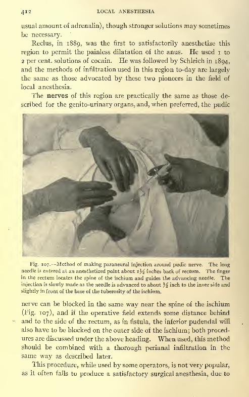
412 LOCAL ANESTHESIA
usual amount of adrenalin), though stronger solutions may sometimes
be necessary.
Reclus, in 1889, was the first to satisfactorily anesthetize this
region to permit the painless dilatation of the anus. He used i to
2 per cent, solutions of cocain. He was followed by Schleich in 1894,
and the methods of infiltration used in this region to-day are largely
the same as those advocated by these two pioneers in the field of
local anesthesia.
The nerves of this region are practically the same as those de-
scribed for the genito-urinary organs, and, when preferred, the pudic
Fig. 107. Method of making paraneural injection around pudic nerve. The longneedle is entered at an anesthetized point about i)^ inches back of rectum. The finger
in the rectum locates the spine of the ischium and guides the advancing needle. The
injection is slowly made as the needle is advanced to about J inch to the inner side and
slightly in front of the base of the tuberosity of the ischium.
nerve can be blocked in the same way near the spine of the ischium
(Fig. 107), and if the operative field extends some distance behind
and to the side of the rectum, as in fistula, the inferior pudendal will
also have to be blocked on the outer side of the ischium; both proced-ures are discussed under the above heading. When used, this methodshould be combined with a thorough perianal infiltration in the
same way as described later.
This procedure, while used by some operators, is not very popular,as it often fails to produce a satisfactory surgical anesthesia, due to

GENITOURINARY, ANORECTAL, AND GYNECOLOGIC OPERATIONS 413
the uncertainty of accurately reaching the nerve at the point of
injection.
The following method is much to be preferred, being simpler,
quickly executed, and absolutely reliable in producing a perfect sur-
gical anesthesia.
This technic is so simple and quickly executed that the writer
almost invariably uses it for all operations in this region (hemor-
rhoids, fissure, prolapse, etc.) in preference to a general anesthesia.
Fig. 108. Points of injection for surrounding anal canal with zone of anesthesia.
(From Braun.)
The tissues are first infiltrated subcutaneously around the anus
at the mucocutancous junction, as seen in Fig. 108. It is better to
start the injection an inch or more away in the less sensitive skin,
and advance toward this region, when the injection is then carried
out circumferentially, rather than to make the first puncture in this
area, which is highly sensitive, and will always excite some 'complaint,
and in nervous patients cause them to become uneasy and lose con-
fidence in the promise of a painless operation. The author alwaysuses an ethyl chlorid spray on the skin at the point of puncture, first

414 LOCAL ANESTHESIA
Fig. 109. Method of making subcutaneous injection around anus. Patient in Sims'
position.
Fig. no. Method of making deep perirectal injections.

GENITOURINARY, ANORECTAL, AND GYNECOLOGIC OPERATIONS 415
thoroughly protecting the anus against any contact with the spray
by holding a gauze sponge well against it.
-^Sphincter muscle
Fig. in. Schematic representation of method of producing anesthesia of anal canal.
(Taken from Braun, slightly modified.)
The circumferential injection is made subcutaneously, as the
skin and mucous membrane at their point of junction are very thin,
and an intradermal injection difficult and not at all essential.
'
\.
Fig. 112. Method of dilating rectum with hand in cone shape.
By drawing out the skin of this region with one hand the tissues
are put upon the stretch, and all folds and creases obliterated (Fig.
109), making it less likely to transfix a fold causing pain; the solution
is injected as the needle is advanced; for each re-insertion of the

416 LOCAL ANESTHESIA
needle starting just back of the point where the needle last stopped;
having completed the circumferential injection, a finger is now
passed within the rectum as a guide, and the large syringe and long
needle used; the needle is passed through the anesthetized area of
skin and directed up the bowel, just outside of the sphincters, in-
jecting, as the needle is advanced, to a depth of about 2% or 3
inches (Fig. no); four points are injected; one on each side, injecting
in each of these about 10 c.c., and one in front and behind the bowel,
injecting in each of these about 5 c.c.
Fig. 113. Method of anesthetizing fistulous tract. (From Braun.)
Anesthesia results almost immediately, at most after a delay of a
few minutes, when dilatation may be begun and can be as thoroughlycarried out as- under a general anesthetic.
A graphic illustration of the method of making these injections
is shown in Fig. in. The author always prefers to use the hand as
a dilating medium, which is less likely to tear or lacerate the parts,
using soap as the lubricating medium; first one finger is passed, then
two and three, and, finally, the whole hand in a cone-shape is rotated
around in a screw-like fashion (Fig. 112) until dilatation is complete.This is the method always used by Prof. Matas for dilatation and is
superior to any other.
Braun instead of making these deep injections just outside the

GENITO-URINARY, ANORECTAL, AND GYNECOLOGIC OPERATIONS 417
bowel wall makes them submucously between the mucous mem-brane and the sphincters, the amount of solution used and the depthof the injections is about the same. This method is simpler and
easier for the inexperienced. The subsequent manipulations for
dilatation serve to thoroughly diffuse the anesthetic.
In operations for fistula, in addition to the above method of
anesthesia, which anesthetizes the anal canal and permits dilatation,
the fistulous tract must also be anesthetized by injections made on
each side and beneath it, so as to thoroughly embrace it within a
wall of anesthesia. The method of making these injections is
illustrated in Fig. 113. The fistulous tract can then be slit up,
excised, or curetted as preferred.
For methods of infiltration it is well to precede the injections in
badly inflamed or sensitive cases by placing a pledget of cotton
saturated with 5 or 10 per cent, novocain solution within the anal
canal, and allow it to remain while injecting elsewhere; in this waythe canal will permit the painless introduction of the finger to guide
the needle in the deep infiltration.
In the original method, first advocated by Reclus and Schleich,
the infiltration was made directly into the substance of the sphincters,
but this is not necessary, and it would seem advisable to infiltrate the
loose cellular tissue surrounding the bowel rather than the muscle
itself. When the above technic has been well carried out it is not
necessary to infiltrate the mucosa;this is then, consequently, a para-
neural regional anesthesia. By the above technic any of the ordinary
operative procedures for hemorrhoids, fistula, polypi, ulcers, fissure,
or resection of the rectal mucosa for prolapse may be quite satis-
factorily and painlessly performed. In many operations upon this
region it is not necessary to dilate the sphincter. While this is
desirable in all operations of any magnitude to paralyze the muscles
and permit free inspection and access to the anal canal, there are
many cases of sentinel piles and other superficially situated lesions,
as fissures, where this practice may be dispensed with and the
lesions dealt with by simple infiltration. This is particularly suited
to office practice, where many of the minor affections of these parts
may be operated upon.
In operations for fissure, while it is always desirable to stretch
the sphincter, this procedure alone often sufficing for a cure in super-
ficial lesions, it is not absolutely necessary. With the proper care
and delicacy in manipulation these cases can often be operated in
the office or at the patient's home with satisfactory results. The

418 LOCAL ANESTHESIA
needle is entered in healthy tissue, just below the lesion, and infil-
tration gently carried out, advancing the needle under the fissure
in the substance of the sphincter, infiltrating gently as it is advanced,
until the entire underlying area is well infiltrated;the finger will then
be quite easily tolerated in the rectum, and the extent of the lesion
well explored.
A blunt-pointed bistoury is now advanced on the flat against the
finger until the upper part of the area is reached, then turned edge
down, and the fibers of the sphincter at the base of the fissure incised
to a depth of about y inch. This may be done at one point in the
middle or on each side, and effectually puts the muscle-fiber at rest
and permits the ulcer to heal; it is then dressed with ichthyol and
anesthesin ointment, 15 or 20 per cent, of each.
In performing the operation this way, care should be exercised
not to incise too far up the bowel or too deeply for fear of openingsome small artery, which may give rise to an unpleasant hemor-
rhage; it is only in that portion of the canal surrounded by the
external sphincter that the incision should be made; more extensive
ulcerations, extending up the bowel, should not be treated this
way. Malignant disease of these parts unless quite limited, super-
ficially situated, and of easy access should be reserved for para-
sacral, epidural, spinal, or general anesthesia. Perirectal and ischio-
rectal abscess if superficial may be easily opened by infiltration. In
the case of the former, where it is desirable to dilate or divide the
sphincter, one of the above-mentioned methods should be used to
secure anesthesia.
The use of sterile water as an anesthetic agent when injected
into the tissues has long been known, and its application to surgery
of these parts has frequently been tested; Dr. S. G. Gant, of NewYork, is particularly enthusiastic in its use, and has done much to
popularize it here. For hemorrhoids or fistula operations of limited
extent the anesthesia is quite satisfactory, but it is not suited for
extensive operations where the deep parts are involved. The objec-
tion to its use is the burning pain produced by the infiltration; this,
while greater in some cases than others, is often quite severe, and is
not a negligible factor in considering this form of anesthesia. The
pain in making these injections is much lessened if the injection is
very slowly made, so as not to distend the tissues too rapidly; by the
addition of a small quantity of cocain or novocain, o.i per cent., this
infiltration pain is entirely relieved; this, however, is no longer pure-

GENITO-URINARY, ANORECTAL, AND GYNECOLOGIC OPERATIONS 419
water anesthesia. For a further consideration of water anesthesia
(anesthesia dolorosa), see chapter on this subject.
The use of ethyl chlorid about the anus is rather unsatisfactory,
as it often produces considerable burning, but for superficial incisions
in areas removed from the anal margin, or where this can be protected,
it is often quite satisfactory.
Before dismissing this subject reference should be made to the
use of quinin and urea, which are applicable for the surgical treat-
ment of a limited number of rectal affections, and the reader is
referred to the chapter on this subject.
The topical application of the various analgesic and anesthetic
preparations is often of great value here for the palliative relief of the
inflammatory affections of these parts, such as hemorrhoids, fissures,
ulcers, etc. Anesthesin has largely replaced the use of antipyrin and
orthoform, as it is a more active agent, and, in view of its slow solu-
bility, maintains this action for a long time; it is also practically non-
toxic, even in concentrated solutions (10 to 15 gr. can be safely
administered internally at a time) . It is best used in ointment form
in 10 and 20 per cent, strengths. Combined with other astringent
and sedative drugs (adrenalin, hemamelis, belladonna, etc.), in this
form its application externally or to the anal canal with a pile-pipe
or by suppository often affords gratifying relief in many painful
affections. Cocain, novocain, alypin, etc., when used in a similar
way, have to be frequently repeated, and may prove dangerousfrom their rapid solubility if used in concentration or, if long con-
tinued, may encourage a habit, and for these reasons are rarely
employed in this way.
Operative procedure under the topical application of pure carbolic
acid, while practical for limited procedures involving nothing more
than a superficial incision, is rather unsurgical, and is not to be recom-
mended for more than an incision such as would be needed for turning
out the clot in a thrombotic hemorrhoid or opening a superficial ab-
scess; it can also be made use of for anesthetizing a small point to
permit the painless introduction of the hypodermic needle. Whenused for anesthesia the surface upon which it is applied should be
perfectly dry, and after allowing it to remain a few minutes the
excess is wiped off.
As a postoperative application the topical use of one or more of
the various sedative and analgesic preparations is often of great
value in allaying the after-pain and burning common to most opera-
tions upon these parts, particularly hemorrhoids, where the cautery

420 LOCAL ANESTHESIA
has been used. Dr. James P. Tuttle claims for sodium bicarbonate
a sedative action superior to anything else; he says for this purposeit is incomparable; it has no analgesic action under other conditions.
Following other operations the immediate free use as a primary dress-
ing of a 10 or 15 per cent, ointment of anesthesin or orthoform will
be found to greatly lessen the postoperative discomfort as the
anesthesia passes off.
For the non-operative or palliative treatment of chronic tubercu-
lous, syphilitic, or cancerous ulcers, anesthesin, orthoform, or car-
bolic acid in ointment form prove effective analgesic applications.
A quite satisfactory treatment for fissures, one of the most painful
of rectal conditions, is by tampons soaked in ichthyol and freely
sprinkled with anesthesin, such applications giving relief often for
many hours; the same may be said of quinin and urea used in
ointment form.
Much of the after-pain from rectal operations is due to spasm of
the sphincters, and when opiates are used for this purpose it may take
unsafe doses to control it; better agents are chloral and bromids
(per orem) ,which are often more effective and safer, used in conjunc-
tion with an anesthesin ointment. For the chronic aching or neu-
ralgic pain of this region a satisfactory combination is antipyrin,
acetanilid, and codein administered internally.
GYNECOLOGIC OPERATIONS
The surgery of the female generative organs forms a large partof the operative work of the present time. Much of this work on
the external and readily accessible parts may be quite easily and
satisfactorily performed under local methods of anesthesia, and
even some of the more complicated procedures on the deeper parts
may, with skill and gentleness, be painlessly, or almost painlessly,
accomplished in suitable subjects.
Women, as a rule, are more apprehensive and fearful than men,and often so extremely nervous, particularly when having suffered
long from their various affections, that they make poor subjects
for any form of local anesthesia. Many prefer to take a general
anesthetic, and be treated as if they were really not there at all.
It is, accordingly, advisable with the timorous and fearful not to
attempt any but the simpler operations on the exposed parts bylocal anesthesia, reserving all complicated procedures for parasacral,
epidural, spinal, or general narcosis. However, in the presence

GENITOURINARY, ANORECTAL, AND GYNECOLOGIC OPERATIONS 421
of centra-indications to general anesthesia, and with positive indi-
cations for operative interventions, many of the more complicated
procedures may be safely and satisfactorily performed by the skilful
use of local measures alone, or in combination with light superficial
anesthesia for the more painful and deeper parts. (See chapter on
Combined Methods of Anesthesia.)
Sphincter ani exlrrnus
medial inferior cluntnl nerve
inferior liaemorrhoidal a,
internal pudic ves
Olntneus maxim
anococcygeal nenes anommeeal tig.
Fig. 114. The nerves and vessels of the female perineum. Upon the right side the
bulbocavernosus has been partly removed and the vestibular bulb exposed, the trans-
versus perinei superficialis divided, and the urogenital diaphragm incised.|c * = The
origin of the internal pudic vein from the vestibular bulb (vena bulbi vestibuli). (So-
botta and McMurrich.)
In nervous and sensitive patients care should be taken to always
administer one hour before operation a preliminary or preparatory
hypodermic of morphin, % to % gr., with scopolamin, ^IQQ gr ->as
recommended in the preceding part of this volume. It is also well to
have a sympathetic nurse stand by the patient and hold her hand or
encourage her if she is uneasy. For a consideration of the nerve-sup-

422 LOCAL ANESTHESIA
ply of this region see Fig. 114, and for description and methods of
blocking same see section on Genito-urinary Organs.
In all the external parts and lower 2 or 3 inches of the vaginal
tract sensation is very acute, but the vault and upper parts of the
vagina have very little sensation. The cervix and uterus are
not very sensitive to incisions volsellum, forceps, or needle punc-
tures but are quite sensitive to stretching, as in dilatation of the
cervix. Also, the mucosa of the cervix and uterine cavity has very
little sensation, but will not stand a thorough curettage without
anesthesia. The same may be said of the peritoneal investment of
the uterus, which should not be operated upon without some
infiltration.
Solution No. 2, 0.50 per cent, novocain, is used in all operations
upon the external parts, which are highly sensitive. Solution No. i,
0.25 per cent., is ample for the deeper infiltrations, but if preferred
No. 2 can be used throughout.
To each solution add from 5 to 10 drops of adrenalin, i : 1000 to
3 or 4 ounces, if the field is extensive and much solution will likely be
injected, using the smaller quantity of adrenalin.
The Perineum and Postvaginal Wall. A point on the perineum
midway between the anus and vaginal outlet is anesthetised intra-
dermally; establishing here a station through which the long needle
is entered, in the event of an extensive laceration up to or including
the sphincter ani, this point can be made just within the vaginal
outlet.
The long needle is entered here and passed up in the middle line,
injecting as it is advanced as far as the contemplated field of opera-
tion, using often as much as 5 or 10 c.c.; a finger can be used either in
the vagina or rectum as a guide. If the plane of tissue is quite thick
it is best to pass the needle well below the vaginal mucosa in the
deeper planes, as the solution can better diffuse in these deeper layers,
but when dealing with an extensive laceration with rectocele, where
the rectum and vaginal mucosa are in close contact, the needle had
best be passed just beneath the vaginal mucosa, and here the finger
is kept in the bowel as a guide. By injecting the solution as the
needle is advanced, the solution separates the plane of tissues and
there is less danger of the needle puncturing the rectum.
Having made the midline injection, the needle is partially with-
drawn and directed slightly laterally and upward on first one side
and then the other, using in each an additional 5 or 10 c.c., depending
upon the extent of the field. Similarly, a third or fourth injection

GENITOURINARY, ANORECTAL, AND GYNECOLOGIC OPERATIONS 423
can be made just lateral to the preceding, until practically the entire
vaginal canal except the roof has been infiltrated. A crescentic-like
injection, made subcutaneously with the long needle around the
vaginal outlet and carried upon each side the full extent of the field,
completes the anesthetizing process.
If a perineorrhaphy is to be done, and the tear involves the sphinc-
ter, the anal canal must then be anesthetized, as described in that sec-
tion. The above method of injection gives a perfect anesthesia and
is very quickly done after a little practice, and the latter steps of the
Fig. 115. For anesthesia of vaginal outlet, including labia majora. (From Braun.)
operation, if for perineorrhaphy, much facilitated through the sepa-
ration of the different planes of tissues by the injected solution.
To Anesthetize the Entire Vaginal Outlet. This is done by a
circumferential injection, as illustrated in Fig. 115, the lower portionas described above. Another crescent-like injection is made from
above, which meets the lower field, starting preferably over the exter-
nal ring of the inguinal canal on each side, making the injection
fairly deep and liberal here to thoroughly block all fibers of the ilio-
inguinal and genitocrural nerves, as they emerge from this opening to
be distributed to the tissues of the labia majora.

424 LOCAL ANESTHESIA
Anterior colporrhaphy is done by pulling down the cervix with a
volsellum; at this point on the cervix a little infiltration can first be
done before applying the instrument. With the cervix well down
and on the stretch the submucous tissues between the cervix and
urethral opening are well infiltrated (Fig. 116), carrying the infiltra-
tion well out laterally to permit free exposure of the deep fascia in the
subsequent dissections.
Fig. 116. Area of infiltration for anterior colporrhaphy. (From Braun.)
The cervix is anesthetized by drawing it down with a volsellum
and making a free submucous infiltration around its neck, at its
junction with the vaginal vault. In making this injection in front
care should be exercised not to injure the bladder; the point of the
descent of this organ and its proximity to the vaginal vault had best
be located beforehand by a sound passed within the bladder. A longfine needle with large syringe is now used, the needle directed up in

GENITOURINARY, ANORECTAL, AND GYNECOLOGIC OPERATIONS 425
the long axis of the cervix on each side, just within the cervical tissues,
to a depth of from i to 2 inches, and about ^ ounce of 0.50 per cent,
novocain injected on each side, injecting as the needle is advanced.
After a few minutes this will permit a fair degree of dilatation,
when trachelorrhaphy can be done, combined with a curettage of the
cervical canal. Curettage of the body of the uterus is not often very
satisfactory under local anesthesia when thoroughly done, but a
limited amount is often well tolerated after the above injections, or
a few whiffs of ether can be given just at this point in the operation.
In cases requiring curettage following abortion, the procedure is
usually very much simplified as the cervical tissues are softened and
usually quite relaxed and the above method of anesthesia proves
quite sufficient. I have occasionally adopted the following plan in
cases where the cervix was quite patulous or could be first dilated bythe preliminary cervical injections sufficiently to permit the intro-
duction of a finger. With the cervix well pulled down the index-
finger of one hand is passed up in its canal to the uterine cavity.
The long needle is then entered on the vaginal surface of the cervix
close to the fornix and passed upward just within the cervical wall
or in close contact with it, under the guidance of the finger within the
canal which should at all times be able to feel the course and position
of the needle, the precaution is observed of freely injecting the solu-
tion while the needle is being advanced. Deep injections made
high up in the external walls of the uterus in this way on each side
will reach any nerves from the broad ligaments and completely anes-
thetize the uterus sufficiently to permit a very thorough curettage,
such as may be needed for the removal of a submucous fibroid. It
is, however, not always possible to follow this plan as in many of
these cases the cervix is dense and unyielding and I have alwaysfelt some hesitation in making these deep injections in this position
without some reliable guide.
Ruge describes the method of anesthesia for a vaginal hysterec-
tomy as follows:
"A long needle is introduced to one side of the cervix to a depthof 4 to 5 cm., being directed in a somewhat lateral direction, in order
to strike the nerve-trunks before they have undergone their ultimate
division. If the needle is introduced slowly, most vessels and anycoils of intestines with which it may come in contact will be pushedaside and not injured. When the needle has been satisfactorily
introduced, the 10 c.c. syringe is attached and the solution injected
as the needle is withdrawn.

426 LOCAL ANESTHESIA
"The process is repeated on the opposite side, then at two points
on the anterior and two on the posterior vaginal vault injections of
3 to 5 c.c. are made. In the anterior vaginal vault it is necessary to
introduce the needle 2 to 3 cm. deep, but in the posterior just throughthe mucosa.
"Vesical symptoms are controlled by using instillations to pre-
vent pulling on the viscus from being unpleasant."
This operation has never been performed under local anesthesia
by the author, and it would seem somewhat questionable to pass a
needle in any direction in which it might perforate the bowel. If
the needle is fine and the solution injected as it is advanced the dan-
ger of injuring the ureter or vessels at these points is very slight, and
if punctured with a fine needle no damage is likely to result, but wecannot feel the same about the intestines. It would seem safer to
the author in doing this operation to first free the bladder from the
uterus and open the peritoneum above, as was done in the case de-
scribed later, then with a finger in the cavity the broad ligaments or
their internal attachments can be infiltrated under the guidance of
the eye and finger.
Thaler, in describing the technic for the Diihrssen-Bumin opera-tion of anterior hysterotomy, as done in Schauta's clinic for cases
of placenta praevia, eclampsia, etc., where rapid delivery is indi-
cated, states that the injection is made high up on the anterior lip
of the cervix and to the right and left of the midline, well down into
submucous tissue, about i cm.; the cervix is slit up to this point,
when further injections are made into the anterior uterine wall as
the procedure progresses. The use of adrenalin in the solution
prevents hemorrhage.The female bladder can be quite easily opened through the vagina
by infiltrating above and in front of the cervix in the middle line,
carrying the infiltration well down to the submucous tissue of the
bladder. A sound .is passed into the bladder and turned point downto present the bladder at this point; unless the bladder is inflamed or
hypersensitive it is not necessary to anesthetize it; when necessary
it is done in the same way recommended for the male bladder. The
infiltration of the submucous tissues over the point of incision is
sufficient to anesthetize the mucosa here, and its incision causes no
discomfort.
Operations for vesicovaginal fistula, if easily accessible, can be
performed in this same way by infiltrating around the opening, the

GENITO-URINARY, ANORECTAL, AND GYNECOLOGIC OPERATIONS 427
infiltration facilitating the separation of the bladder from the vaginal
wall.
The female urethra is easily anesthetized by a film of cotton
placed around the end of a probe, and saturated with a 5 or 10 percent, solution of novocain passed into the urethra and allowed to
remain for a few minutes.
Caruncles are easily extirpated by infiltrating around and beneath
them; a swab with 10 per cent, solution can be used on the surface
for a few minutes at the point at which the needle is entered.
The removal of Bartholin's glands or benign growths is quite
easily performed by infiltration; also epitheliomata when superficial
and of limited extent; malignant diseases of the cervix, uterus, or
deeper parts should not be undertaken by these measures, but
reserved, for parasacral, epidural, spinal, or general anesthesia.
In operating upon the cervix and uterus, pulling down these parts
to the vaginal outlet is attended with some discomfort, and should
not be attempted where they are bound down by adhesions or fixed
in the abdominal cavity, but in cases where these parts are well
relaxed and freely movable operation can be quite satisfactorily
undertaken.
Polypi can also be removed in this way, or even without anes-
thesia, as they have no sensation and the division of their pedicle
is without pain. Where the parts can be well brought down, as in
prolapse, the peritoneal cavity can be easily opened in front of the
uterus, its fundus brought down, and any of the various fixation
operations performed.The following history illustrates an extensive operation upon
these parts on a favorable subject:
Mrs. H., aged sixty-three, a stout lady with flabby and relaxed tissues, had been suf-
fering with a complete prolapse of the uterus with marked rectocele and cystocele for the
past fifteen years, the result of extensive lacerations during the child-bearing period.
The bladder and rectum hung down from the vagina like two distended pouches, the
uterus protruding from between them, making it necessary for her to replace it before she
could sit down. She suffered the usual disturbances with the bladder and rectum as well
as the other symptoms common to this condition; her dread of an anesthetic had forced
her to tolerate these discomforts for many years. When I assured her, much to her
surprise, that she could be easily and painlessly operated on under local anesthesia she
embraced the opportunity readily.
The Operation. The anterior vaginal wall in the midline, between the cervix and
meatus, was first infiltrated and then incised down to the bladder wall; the infiltration
and dissection was carried well out to the sides to freely separate the bladder from the
surrounding parts; the same was done with its attachment to the uterus. The perito-
neal cavity between the bladder and uterus was then opened, and, with one finger holding
up the bladder, a long needle was used to inject a small area on the anterior surface of

428 LOCAL ANESTHESIA
the uterus to prevent any pain which might be caused by catching the organ at this point
with tenaculum forceps; the uterus was then secured and anteverted, so as to bring its
fundus forward into the wound in the vagina; it was held here while the bladder was
pushed up well into the cavity and behind it, and the fundus secured to the deep fascia
behind the pubis; thus firmly anchored in this position it prevented the descent of the
bladder and was itself prevented from retroverting, the first step necessary for its
descent; the superficial parts were then closed. The perineum was now dealt with by
commencing at the vaginal outlet and making a rather free submucous infiltration,
extending well back in the middle line and well out on each side. The mucous mem-brane was then incised from side to side at the vaginal outlet and dissected up freely,
this step being markedly facilitated by the infiltration which separated the rectum from
the vaginal mucosa and greatly lessened the danger of opening the rectum so likely to
happen in bad cases of this kind. The muscles in the vaginal walls were next sought for,
and their atropic remnants approximated in the middle line, restoring a fairly satisfac-
tory perineum and normal vaginal outlet. After trimming away the excess of vaginal
mucosa the wound was closed. This entire procedure was without pain and the con-
valescence without incident.
It is now six years since the operation was performed. Duringthis interval I have heard from the patient repeatedly, and she has
remained entirely well. The infiltration method was used for the
above case instead of the regional injection of the pudic nerves, as
the infiltration greatly facilitates the separation of the different
planes of tissues in cases of this kind, and the work is more quickly
and easily done that under a general anesthesia without the aid of
infiltration.
Operations upon the round ligaments in the inguinal canal for
purposes of shortening them by the Alexander method or any of its
modifications is quite easily done under infiltration, and should be
governed by the same indications as when, operating under general
anesthesia, that is, the free movability of the uterus and other internal
parts.
Infiltration is first done over the external ring and along the
course of the inguinal canal; the superficial parts are incised, and
the external ring and aponeurosis of the external oblique exposed;
an injection is then made through" the fibers of the external oblique
into the inguinal canal, the canal then opened, and round ligaments
located and freed. As it is being drawn through the internal ring,
the tissues around this point down to the peritoneum are infiltrated
with a fine needle, bearing in mind the position of the deep epigastric
artery. By proceeding in this way the drawing of the ligament
through the internal ring and stripping back of the peritoneum causes
no pain.
Suprapubic cystotomy is performed the same in the female as in
the male, but it is easier to perform cystotomy by the vaginal route,

GENITOURINARY, ANORECTAL, AND GYNECOLOGIC OPERATIONS 429
as has already been described, and if to be left open for drainage it
is more convenient, as the patient can wear a urinal and not be
continually soiled as with a suprapubic opening.In operating within the abdominal cavity only a limited number
of operations are feasible, and only then under favorable conditions,
with free movability of the parts. In the presence of adhesions or
inflammation about the tubes or ovaries the case should be operated
by other methods. The abdomen is opened in the middle line after
infiltration, first of the skin and subcutaneous tissues, then passingthe needle down to the interval between the recti muscles; after
these have been opened the subperitoneal tissue, which is quite
L ine of ane.sf/>e-si'a.
Wall C.uJ"
Fig. 117. Shows uterus, broad ligament, and attachments. Series of circles shows
line of infiltration beneath anterior peritoneal fold of broad ligament. Where this line
crosses uterus, in shaded portion, infiltration is more liberal. On left is seen small line
joining longer one at about right angles, and shows area infiltrated when limited to one
side as in the case of removal of tube and ovary only. Similar infiltrations are made
posteriorly, as described in text.
relaxed at this point, is now infiltrated, this infiltration anesthetizing
the peritoneum. (See chapter on Abdominal Operations.) After a
few minutes this is opened. Retractors should be gently used, as
any undue traction on the abdominal wall will cause pain. The
uterus should be raised into the wound with the hand, not with
volsellum or other toothed instruments, unless the point at which
they are applied has first been infiltrated. A variety of operations
are now possible salpingo-oophorectomy, when these parts are
free, by lightly infiltrating the broad ligaments, pelvic and uterine
attachments, of these parts along the proposed line of incision on their
anterior and posterior surfaces (Fig. 117); pedunculated ovarian
cysts, when not adherent, are quite easily removed in the same

430 LOCAL ANESTHESIA
way; subperitoneal fibroids removed, or resection of the body of the
uterus performed by first lightly infiltrating the proposed line of
incision; this can often be omitted, as the uterus has very little sensa-
tion; ventrosuspension, or fixation, is quite easily done, and usually
without any discomfort; if any is experienced, light infiltration can
be resorted to on the fundus, where the sutures are to be placed.
Intra-abdominal operations for purposes of shortening the round
ligaments may be done under local anesthesia, but it would be prefer-
able to do an external Alexander, unless the cavity has already been
opened. The ligaments may be doubled on themselves and sutured,
using light infiltration at these points if necessary or fixed behind the
uterus. The various operations of drawing the ligaments throughthe abdominal wall, at or near the internal ring, may also be done
by first raising the abdominal wall gently and infiltrating the perito-
neum and subperitoneal tissue around these points, as well as the
tract through which the forceps will be passed through the abdominal
wall in grasping the ligaments, remembering the location in this
neighborhood of the deep epigastric artery and vein and making the
injection only when advancing or withdrawing the needle.
Cesarean section can be quite easily performed under local
methods when other forms of anesthesia are contra-indicated.
In thes'e cases the abdominal wall is much thinned out and no
muscles in evidence in the midline. The proposed point of incision
is located and the skin and subcutaneous tissue infiltrated for a dis-
tance of 6 or 8 inches; these are incised down to the aponeurosis; the
needle now penetrates this at several points and freely infiltrates the
subperitoneal tissue beneath for the entire length of the field.
The aponeurosis and peritoneum are then incised bringing into
view the uterus. The wound is now retracted and subperitoneal
injections made in all directions to enlarge the field of anesthesia.
The uterus is ordinarily not sensitive but may be infiltrated lightly
along the proposed line of incision. Before opening the uterus it is
best to have it well secured at several points as well as the woundin the abdominal wall to follow down the uterus as it contracts and
prevent the possible escape of intestines which is unlikely but mayoccur if the patient bear down. The steps of the operation are other-
wise no different than when using a general anesthetic. The possi-
ble psychical effects upon the patient should not be overlooked and
she should receive an hour beforehand the preliminary hypodermicof pantopon or morphia and scopolamin.
The following is from a paper by Drs. R. K. Smith and Jacob

GENITO-URINARy, ANORECTAL, AND GYNECOLOGIC OPERATIONS 431
Schwarz, of San Francisco, read at the San Francisco Medical Society,
May, 1910. In both cases there was a contracted pelvis with contra-
indications to general anesthesia; in both cases mother and child
survived :
"The solution used was novocain (0.50 per cent.), to each 10 c.c.
of which was added i drop of adrenalin solution (1:1000). This
solution was freshly made and boiled for five minutes before using.
Two points, one 9 cm. above the umbilicus and the other a like dis-
tance below it in the median line, were infiltrated with a drop of the
solution, and from these, as points of departure, the solution was
injected about a diamond-shaped area subcutaneously and then sub-
fascially. The line of the incision was not infiltrated in either of these
cases. The amount of solution used was about 75 c.c. in Case i and
60 c.c. in Case 2, and it is my belief that a smaller amount might be
sufficient.
"The operation was carried out as follows: Incision through the
abdominal wall, 15 cm. long, with its center opposite the umbilicus,
peritoneal cavity packed off with gauze, uterus incised with knife
down to the placenta for about i inch, and the incision rapidly en-
larged with scissors to about 15 cm.; the placenta pushed aside, the
membranes ruptured, the child grasped by its feet and extracted, and
the placenta removed from the uterus while it was in situ; the uterus
lifted out of the abdominal cavity and surrounded by pads dippedin hot saline solution. In Case i the hand was introduced throughthe incision into the cavity of the uterus and one finger passed
through the cervix, this was followed by a Goodell dilator, which was
carried through the cervix and stretched open. This was not neces-
sary in Case 2, operated upon on April 4, 1910, as the cervix was com-
pletely dilated before beginning the operation. The uterine incision
was closed with deep and superficial interrupted sutures of chromic
catgut, and there buried with a continuous Lembert suture of the
peritoneum, the peritoneal cavity wiped out, and the wound closed."

CHAPTER XX
SPINAL ANALGESIA
SPINAL analgesia1 had its beginning in the experiments of Dr. J.
Leonard Corning, which were published in the "New York Medical
Journal," October 31, 1885.
Corning first experimented on a dog, injecting a 2 per cent, cocain
solution in the lower dorsal region, and obtained paralysis of motion
and sensation in about five minutes, followed by complete recovery,
without noticeable ill effects. He next injected a man suffering from
sexual disturbances, using 30 minims of a 3 per cent, cocain solution,
between the eleventh and twelfth dorsal vertebrae. There was no
result in eight minutes, and the injection was repeated, producinganesthesia and incoordination of the lower extremities. The anes-
thesia was complete, as proved by various tests; urethral sounds were
passed and other manipulations used about the genitalia. This was
done in the office. In an hour the patient was able to leave with
sensation still impaired, but otherwise no worse for his experience.
Corning, in concluding, states:
"Whether the method will ever find an application as a substitute
for etherization in genito-urinary or other branches of surgery further
experiences alone can show. Be the destiny of the observation what
it may, it has seemed to me, on the whole, worth recording."
Corning was not a surgeon, and did not have the opportunities
of applying this method further, and, as it did not attract favorable
1 To Prof. R. Matas is probably due the credit of having performed the first opera-
tion under spinal analgesia in America. An operation for hemorrhoids was performed
upon a young colored male in the Charity Hospital Clinic on December 18, 1899, Profs.
F. A. Larue and H. B. Gessner assisting, with the author, then an intern in his service.
The spinal canal was reached between the fourth and fifth lumbar vertebras, with escapeof spinal fluid; two injections were made five minutes apart, each about i c.c. of i percent, cocain in normal salt solution; anesthesia immediately followed, and was completefrom the waist-line down, with a gradually lessening degree of anesthesia reaching as far
up as the neck. Some reaction followed the operation (chill, nausea, vomiting, and
temperature), which shortly subsided, the patient making a good recovery.An unsuccessful attempt had previously been made on November loth with beta-
eucain, but the resulting anesthesia was unsatisfactory. (Jour. Amer. Med. Assoc.,
December 30, 1899, p. 1659.)
432

SPINAL ANALGESIA 433
attention at the time on the part of his American confreres, it was
accordingly dropped until revived some years later by Continental
surgeons.
In these experiments Corning had aimed to inject the fluid be-
tween the spinous processes, and permit it to be carried by the veins
to the cord. Corning deals with the subject again in 1888, and in
1894 appeared his book on "Pain in its Neuropathological, Diagnos-
tic, Medicolegal, and Neurotherapeutic Relations."
Coming's intention was to make the injection into the neighbor-hood of the cord; he did not aim to puncture the membranes;whether this occurred or not, he must at least have gotten within the
canal, else it is hard to understand how anesthesia resulted, as it
could not take place from diffusion, as the cord is well isolated from
its perivertebral surroundings, and it is not at all likely that it could
be carried to the cord by the surrounding circulation in any effective
quantity. This, then, was the first attempt at a paravertebral in-
jection, but was, no doubt, intraspinal if not intrameningeal.
Real interest in the method was aroused in 1891 by the lumbar
puncture of Quincke, which was developed largely by the activity of
Continental surgeons, notably by Bier and Tuffier.
Bier, with admirable courage, first tried the method upon himself,
to more accurately observe its effects. The anesthesia was satis-
factory. It was followed by a slight headache.
The method was soon in general use on the Continent and in
America, but did not so early gain followers in England, probably due
to conservatism, as well as to the fact that here general anesthesia
had reached a high plane of development, being regarded as a spe-
cialty and given largely by professional anesthetists.
The wave of enthusiasm which followed the general introduction
of this method like all other radical innovations was followed by a
reaction. As statistics accumulated it was found that the method
could not compare in safety with ether anesthesia. Following this
reaction the method was dropped as a routine procedure from most
large clinics but was persisted in by a few who assiduously sought to
improve the results obtained, by the general handling of the patient,
the preparation of the solution used and the technic of its adminis-
tration. While much has been accomplished in this direction and
the results somewhat improved, it is yet not a method for routine use,
but has, however, a decided field of usefulness in selected cases and
under certain conditions.
Within the last five years some improvement has occurred in the

434 LOCAL ANESTHESIA
results obtained from spinal anesthesia and a vast addition made to
the clincical experience on the subject, but remarkably little on
the experimental or physiological aspects of the subject. The im-
mediate danger following spinal analgesia may occur as a result of
paralysis of the respiratory, bulbar vaso-motor and other higher cen-
ters, but what is more common is a paralysis of the efferent nerve-
fibers which control the blood-pressure in the splanchnic area and
may lead to an immediate and profound fall in the blood-pressure in
these areas and the patient literally bled to death by dilatation of the
great venous trunks. Smith and Porter show that rabbits can be
bled to death within their portal system by a section of the splanchnic
nerves. A sudden and profound fall in the general blood-pressure
produced by this result may so exsanguinate the "master cells" in
the medulla and leave them insufficiently supplied with oxygen as
to produce the most serious consequence. The higher the punctureis made the greater the danger of these results. For this reason the
high dorsal and cervical punctures, which at one time were tried,
should now be permanently abandoned in the present stage of de-
velopment of this method and these injections should be confined to
the lumbar region.
In considering the limited laboratory findings available, it is
probable that many deaths reported as due to paralysis of the res-
piratory centers from the drug having reached this level, were in
reality due to splanchnic paralysis and vaso-motor collapse lowering
the blood-pressure within these points to a point incompatible with
life.
It would certainly be ideal if a drug were found capable of para-
lyzing alone the afferent sensory paths in the cord, but drugs cannot
differentiate between nerve-tissue, and the efferent vasomotor
fibers must inevitably feel this influence.
According to the results of Smith and Porter, the bulk of the
solution seemed to be of greater importance than its strength
diluted solutions as a rule spread further than concentrated ones.
The work of Smith and Porter has proved extremely valuable and
shows that in the safe use of spinal anesthesia, we must seek only to
paralyze the afferent sensory paths, without a too pronounced effect
upon the efferent vasomotors, and that any rational surgical prog-
ress with this method must be in this direction.
A thorough understanding of spinal anesthesia is not possible
without the consideration of certain anatomic, mechanical, and
physiologic facts.

SPINAL ANALGESIA
ANATOMY
435
The spinal cord ends opposite the Jower border of the first lumbarvertebra (in the child, and occasionally in women, opposite the third
lumbar), in the filum terminalis, which is given off from the conus
terminalis (Fig. 118).
coccygeat nerve
Fig. 118. An anterior view of the lower portion of the spinal cord. The dura mater has
been divided longitudinally. (Sobotta and McMurrich.)
The spinal cord and cauda equina are surrounded by the same
membranes as the brain viz., dura, arachnoid, and pia.
The dura, continuous with that which invests the brain, is a loose
sheath, not attached to the .bony framework of the spinal canal, but
separated from it by loose areolar tissue containing a plexus of veins
which are most numerous in front and on the sides, less so posteriorly.

436 LOCAL ANESTHESIA
The dural sac terminates at the third sacral segment. It is attached
by fibrous slips to the posterior common ligament, and is largest in
the cervical and lumbar regions. At the beginning of the cauda
equina the nerves lie in bundles on each side, with an appreciable
interval between, through which runs the filum terminale. Theyapproach each other lower in the lumbar region and surround the
filum, which continues to the termination of the dural sac and blends
with its attachment to the periosteum of the coccyx.
On the side of the cord and in the cauda equina the motor nerves
lie in front, the sensory behind; on the side of the cord, separated
by the ligamentum denticulatum; in the cauda equina, still separated
by an irregular cribriform membrane. This accounts for the motor
nerves not being more regularly reached and affected by the anes-
thetic fluid in spinal puncture.
The arachnoid is separated from the dura by a slight interval,
the subdural space.
Within the arachnoid membrane, the subarachnoid space, is the
cerebrospinal fluid. This space is of considerable size and is largest
at the lower part of the spinal canal. Within this space is the cauda
equina. This space communicates above, through the foramen of
Magendie, with the subarachnoid and general ventricular cavity of
the brain. The space is partially divided by a longitudinal cribri-
form membrane, connecting the dura with the pia membrane.
It will be seen from the above description, as well as by consulting
Fig. 1 1 8, that the most favorable site for the spinal puncture is the
midlumbar region, for here the cauda equina lies in two bundles on
each side of the middle line, and is less likely to be injured by a
needle introduced at this point, which has been termed the "cisterna
terminalis" by Donitz.
ANESTHETIC AGENTS
Nearly all agents that have been used for local anesthesia have
at some time or other been used for spinal analgesia. That none of
these have proved thoroughly satisfactory accounts for the changefrom one to the other.
The advent of spinal anesthesia was before the introduction of
some of the more recently discovered anesthetics.
Cocain was the first used, and was soon found to be too dangerousto justify its continuance by the majority of operators, but is still
used by some few; 6 to 10 minims of a 2 to 4 per cent, solution is the
strength usually employed.

SPINAL ANALGESIA 437
Beta-eucain was employed, but was found unsatisfactory.
Stovain next claimed attention. It was introduced by Fourneauin 1904, and was first used by Chaput and Turner, and for a time be-
came the agent most in use. It possessed some noteworthy proper-ties. Its solutions were able to stand boiling without decomposition,and it possessed mild antiseptic properties. It is freely soluble in
water and is of a feeble acid reaction. It has a more marked effect
upon motor nerves than has cocain, paralyzing all the sphincters-
anal, uterine and vesical as well as producing general muscular
relaxation when it comes in contact with the motor roots. This is of
decided advantage in operating upon the abdomen and perineum ;in
laparotomy it permits wide retraction of the abdominal muscles,
which greatly facilitates the work. This paralyzing action on the
motor nerves may reach high enough to effect the respiratory nerves
or even the centers in the medulla, and thus add a grave danger to its
action. It is somewhat irritating to the tissues as well as to the nerve-
fibers.
Alypin has been used, but has been discarded as being unsuited
for use in the spinal canal.
Novocain, the least toxic (one-sixth as toxic as cocain) and least
irritating of all the local anesthetics, has been applied to spinal anes-
thesia, but has not proved generally satisfactory, and is, accordingly,
less used at the present time. Its action on motor nerves is muchless marked than that of stovain, and there is, accordingly, less dan-
ger of respiratory paralysis. The usual dose is about i gr.
Tropococain is the agent most popular at the present time, and
is being generally adopted by most operators. Less unfavorable re-
sults have been reported from its use. From % to i gr. is the dose
usually employed. The smaller dose, for peripheral operations, the
larger dose for abdominal operations and those upon the trunk.
The method of preparing the various agents differs largely in the
hands of different operators.
Most operators prefer to use 5 or 10 per cent, strengths in sterile
solutions of the various agents, using a sufficient number of minims
to give the desired strength of the drug. The solution used maycontain a definite quantity of sodium chlorid to make it isotonic with
the cerebrospinal fluid. This may be injected directly into the
spinal cord, or, as practised by Bier, and Tuffier, at present, the re-
quired quantity of the solution is placed in the syringe and an equal
quantity of cerebrospinal fluid drawn into the syringe before in-
jecting into the canal. Similarly, the dry sterile salt may be placed

438 LOCAL ANESTHESIA
in the barrel of the syringe and dissolved in the aspirated fluid before
injection. This last method is becoming more popular.
Tablets of the various drugs used in the usual strength employed,with or without adrenalin, but usually containing a small amount of
sodium chlorid, are placed on the market by various manufacturers.
The tablets are sterilized and in sterile containers, and when
mixed with a definite quantity of sterile water produce a solution
isotonic with the cerebrospinal fluid.
The keeping qualities of the tablets for any length of time is some-
what in question, particularly if they contain adrenalin preparations;
also the power of rendering and keeping them sterile.
Sterile ampules, similar to those used for serums, each containing
the recognized dose of the agent in use, are put up by the various manu-
facturers in this country and abroad. It is a convenient and safe
method, provided the contents of the ampule can be thoroughly
depended upon. When about to be used they are first immersed in a
strong antiseptic solution bichlorid, carbolic acid, or alcohol
before being opened by the operator, who opens them just before he
is ready to withdraw their contents.
Adrenalin. Whether or not adrenalin should be used in the
spinal canal is a question much in doubt. At one time it was most
favorably thought of by most of the leading operators Bier, Tuffier,
Braun, and Donitz but later there has been a reaction against its
use. Braun has explained its favorable action by stating that it
contracts the vessels in and around the cord, thus creating a larger
space in the dural sac and producing a flow of cerebrospinal fluid in
this direction, thus lessening the tendency of the anesthetic solution
to ascend and produce disturbing symptoms. These views, how-
ever, have radically changed. . That adrenalin does prolong and in-
tensify the action of some anesthetics, but limits the extent of their
action, when injected with them into the spinal canal is confirmed
by some observers, but its use did not seem to prevent the drop in
blood-pressure. With some anesthetics, notably tropacocain, it is
contra-indicated, as the anesthetic agent opposes the action of adrena-
lin. We cannot draw a conclusion here by a comparison of the action
of adrenalin when used in the tissues with a local anesthetic, where it
is of decided value.
In the spinal canal we are dealing with an open lymph-sac. Theadrenalin here must expend its influence upon the vessels of the cauda,
and cannot aid directly in retaining the anesthetic in situ.
The congestion and ecchymosis sometimes seen to follow its ac-

SPINAL ANALGESIA 439
tion in the tissues may here, in the loosely supported vessels of the
cord and meninges, have a more pronounced effect, which may lead
to unpleasant sequelae.
ISOTONIC QUALITIES AND SPECIFIC GRAVITY OF ANESTHETICSOLUTIONS AND THEIR MOVEMENTS WITHIN THE CANAL
It is generally believed that the cerebrospinal fluid moves freely
in and out of the spinal canal with changes in the position of the body,that is, from the erect to the recumbent or inverted positions. Thetension within the membranes must certainly be influenced by such
changes, but I doubt that there is as free movement to and from the
cranial cavity as we have been led to believe, for the following reasons :
The spinal canal is surrounded by an unyielding bony framework,and is uninfluenced by pressure upon it from the outside. The spacewithin the canal must always be rilled, a vacuum cannot exist. If
the body is inverted and the fluid runs into the cranial cavity, what
is to take its place? The position is certainly not favorable for an
engorgement of the venous plexuses around the canal; besides, the
inverted position readily congests the large venous cavities within
the skull, and must increase the pressure here, making less room for
the entrance of the spinal fluid. That such change of position does
influence the pressure within the canal we must readily admit, but
that there is any extensive to-and-fro movement is no doubt an error,
but probably takes place to only a limited extent.
Investigations made upon the open canal of animals is of no value,
for here the atmospheric pressure which enters through the opening
permits the fluid to be displaced. The results of experiments uponanimals cannot be applied to man. The dog has only about 6 c.c. of
cerebrospinal fluid throughout the entire subarachnoid space, while
in the monkey it is still less, and the cord and meninges fill the canal
closely.
It is absolutely necessary that the injected fluid be as nearly iso-
tonic with the cerebrospinal fluid and as free from irritating qualities
as possible. This, as determined by the usual physical tests to
which the liquid is subjected, is not reliable, as proved by the investi-
gations of Dr. A. E. Barker. As the subject is so thoroughly handled
by him, I will give it in his own words:
"To secure isotonicity might appear an easy matter at first sight,
but from my observations is not so. A 5 per cent, solution of stovain
in distilled water freezes at about o.58C., almost the same point as
that of blood-serum. If this were the only test applied it ought to be

440 LOCAL ANESTHESIA
isotonic with the blood. But if a drop of blood be added to a little
4 or 5 per cent, solution of stovain under the microscope, in five
minutes the red corpuscles swell and become pale, in ten minutes are
almost invisible, and in twenty minutes are all gone. The same is
seen if a drop of blood is added to 5 c.c. of these solutions in a test-
tube; but here the changes are apparently slower, as at the end of an
hour a few swollen, pale cells can still be seen, but in an hour and a
half they are all invisible.
"In a really isotonic fluid, such as normal saline (0.91 per cent,
sodium chlorid) or normal glucose solution (5 per cent, of glucose),
the cells are seen in twenty-four or forty-eight hours unchanged."The hemolytic action of stovain, which I have tested in every
way I could think of, appears hitherto to have escaped notice. It has
been supposed too readily that if its 5 per cent, solution has the same
freezing-point as the blood it would be isotonic with it, but, as we have
seen, the blood-cells are destroyed by it. Even the solution preparedfor Bier, in the belief that it was isotonic (stovain, 4 per cent.
;sodium
chlorid, o.n per cent.; epirenin borate, o.oi per cent.), I find to be
markedly hemolytic, tested as above on the microscopic slide and
in a test-tube. But further than this I have found that if to an iso-
tonic solution of sodium chlorid or glucose, in which blood-cells are
seen to be unaltered at the end of twenty-four hours, 5 per cent, of
stovain be now added, the cells rapidly swell, grow pale, and disap-
pear, no trace of them being found in one and one-half hours. Nocombination of sodium chlorid or glucose with stovain which I have
made hitherto has prevented this hemolytic action of the drug.
"Furthermore, it may be added that I have added a 5 per cent,
solution of stovain with a freezing-point of o.58C. to an equal part
of cerebrospinal fluid, and found in this compound destruction of all
blood-cells in about one hour. Nothing is seen then under the
microscope but debris and oily globules. This is as much as to say
that a 5 per cent, solution of stovain injected into the spinal cord
would be hemolytic too.
"Leaving the point of osmotic tension for the present, and admit-
ting that we have no evidence to show that the small amount of the
drug injected has produced any injurious effect as the result of its
hemolytic action, there are other physical qualities which an injected
compound may possess which also appear to have attracted little or
no attention.
"There are three ways in which an analgesic fluid injected in the
second lumbar interspace can make its direct effects felt in the mid-

SPINAL ANALGESIA 441
dorsal region, or even higher, as is sometimes the case in this proced-ure. These are either:
"(i) By slow diffusion; (2) by shifting of the whole column of
cerebrospinal fluid, in which it is suspended upward; or (3) by gravita-
tion, if the injected compound be distinctly heavier than the liquor
spinalis."
i. Diffusion alone of one fluid in another is a slow process, and,as we shall see, is unlikely to be the mode of spread of the injected
fluid in this procedure.
"2. Bier and his followers have aimed at shifting the injected
compound upward or downward, with the whole mass of the cerebro-
spinal fluid, by raising or depressing the pelvis. That the cerebro-
spinal fluid does recede somewhat toward the head on elevation of
the pelvis is undoubted, but it is hard to imagine its doing so to such
an extent as to carry with it a cloud of fluid lighter than itself from
the second lumbar to the fifth dorsal vertebrae. I venture to think
that with such a fluid as he has used, whose specific gravity is 1.0058,
suspended in the liquor spinalis, whose specific gravity is 1.0079, that
what he has achieved by elevation of the pelvis has rather been a
more rapid diffusion of the injected drug, due to the consequent oscil-
lation of the spinal fluid, aided perhaps by vascular pulsation.
"3. There remains, then, the third possibility, namely, that an
injected compound heavier than the liquor spinalis may be affected
by gravity, and sink through the latter in a way quite different to the
behavior of a fluid of less specific gravity such as that just referred to.
It is easy to observe the behavior of one fluid injected slowly into
another through a needle if the fluid be colored with anilin blue.
Provided that each be of the same temperature and specific gravity,
the injected liquid forms at first a distinct blue cloud, which slowly
diffuses itself through the whole mass, into which it enters if the latter
be in a state of rest. On the other hand, if the injected fluid be of
the same temperature, but of much greater specific gravity, it sinks
rapidly from the point of the needle in a definite stream to the bot-
tom of the second fluid, and remains there as a distinct stratum,
without diffusion for a time, proportionate to its density and viscidity.
"The densities of the only three compounds used in our series
compared with that of the cerebrospinal fluid are as follows at 15
to i7C.:
"Liquor spinalis (from three patients, mixed fresh) = i . 0070
1. Chaput's Compound: ,
Stovain, 10 per cent.; NaCl, 10 per cent., distilled water, 80 per cent. = 1.0831
2. Writer's Compound:

442 LOCAL ANESTHESIA
Stovain, 10 per cent.; glucose, 5 per cent.; distilled water, 85 per cent. = 1.0300
3. Bier's Compound:Stovain, 4 per cent.; Nad, o.u per cent.; eperenin borate, o.oi .
= i .0058
"All these are, as we have seen, more or less hemolytic, tested byimmersion in them of blood-cells, the first much the most so (three
minutes) ,the two last about the same in this respect (one to one and
one-half hours) . The first, a very heavy fluid, in which the commonsalt is present at the point of saturation at ordinary temperature,has long been used by Chaput and Tuffier with good results, but ap-
parently for other reasons than its density. Their grounds for em-
ploying such a high percentage of common salt, as stated by the
former, were that M. Billon (Paris), who prepared the compound for
them, hoped thereby to prevent the splitting up of the stovain bythe alkalinity of the spinal fluid. That it does not do so is evident
to any one who adds some of their compound to the cerebrospinal
fluid drawn off. It will then be seen that the latter becomes almost
at once milky. If a little of the fluid in this state be examined under
the microscope it will be seen that this turbidity is due to the presenceof small globules of an oily nature, which, in the course of time, run
together into larger and larger globules.
"A solution of 5 per cent, pure glucose in distilled water freezes at
about o.56C., and is really isotonic, producing no effect on the blood
or tissue cells in twenty-four hours. Five per cent, stovain in normal
solution gives a specific gravity of 1.0126. The hemolytic action of
stovain cannot be avoided.
"Alypin,
5 per cent.; distilled water, 95 per cent.; sp. gr., 1.0036; freezing-point, 0.53
Novocain,
5 per cent.; distilled water, 95 per cent.; sp. gr., 1.0090; freezing-point, 0.555
Tropococain,
5 per cent.; distilled water, 95 per cent.; sp. gr., 1.0160; freezing-point, 0.545-
Stovain,
5 per cent.; distilled water, 95 per cent.; sp. gr., 1.0064; freezing-point, 0.585
Cerebrospinal fluid, sp. gr., 1.0070; freezing-point, 0.56
Blood-serum, freezing-point, 0.56
"Drawing conclusions from the above, these fluids should behave
differently."Tropacocain, with its high specific gravity, should sink. The
uniformly good results obtained by many with this agent may be
due partly to its physical properties, perhaps more so than to any
specific action on nervous structures. Allowance should be madefor the behavior of any fluid after injection and during operation on
the patient.

SPINAL ANALGESIA 443
"By glass tubes bent to conform to the spinal canal, filled with
salt solution (specific gravity 1.0070), and having an opening to con-
form with the position of the second lumbar space, the action of dif-
Fig. 119. A photograph of a tracing from Braune's well-known plate of a frozen
mesial section of the female cadaver lying level. Details omitted for the sake of clear-
ness. Over the spinal canal, and following its curves accurately, is a glass tube filled
with saline solution of the same specific gravity as that of the cerebrospinal fluid =
1.0070. Through the middle vertical arm over the second lumbar interspace the hollow
needle has been introduced into the curved tube, and i c.c. of Chaput's solution (specific
gravity 1.0831) colored with methyl-violet has been slowly injected. This has run
down rapidly into the dorsal curve, and at the end of two minutes is seen as a dark stra-
tum opposite the fifth and sixth dorsal vertebrae. The dark area in the cervical portion
of the tube is a shadow on the glass. (Barker, in"Brit. Med. Jour.")
Fig. 120. Same as Fig. 119, but with the pelvis raised 3 inches from the level. In
this case i c.c. of Bier's solution has been similarly injected colored. This, having a
specific gravity of only 1.0058, has not altered its position, except that, being lighter
than cerebrospinal fluid (1.0070), it has risen in the vertical arm. In this position it
remains for a long time undiffused. (Barker, in"Brit. Med. Jour.")
ferent solutions in vitro has been studied. Of course, it is conceded
that certain vital phenomena would modify the conditions some-
what, but, in the main, what occurs here furnishes us with fairly

444 LOCAL ANESTHESIA
correct evidence on most of the scientific physical points connected
with these injections in the living patient (Figs. 119-125).
"Each of the compounds to be colored with the same quantity
of methyl-violet, and used at the ordinary temperature of the air
and fluid filling the tube. It has been gently passed into the latter,
Fig. 121. Three tubes, as in Figs. 119 and 120, but without the tracing behind.
Into the top one Bier's light solution has been injected, into the middle tube glucose
stovain (author's solution, specific gravity 1.030x3), and into the lower tube Chaput's.The pelvis has been raised 3 inches as before. In the top tube Bier's compound, being
lighter than the fluid in the canal, has remained stationary; the glucose-stovain (1.0300)
is still running down. Chaput's, the heaviest compound, has already reached the dor-
sal curve and is at rest. Photographed two minutes after injection. (Barker, in
"Brit. Med. Jour.")
as on the living patient, with the usual needle through the vertical
arm over the second lumbar interspace. From frozen sections of
the cadaver, lying on its back, it may be seen that the highest pointof the canal from the level is in the cervical region. Next to this a
point between the third and fourth lumbar that is, the point at
which the puncture for spinal analgesia is made. From this last

SPINAL ANALGESIA 445
point the dural canal slopes downward in both directions. Thecaudal incline ends for the dura opposite the third sacral vertebra.
The cephalic incline slopes from the point of puncture downward as
far as the fifth or sixth dorsal vertebra, when it begins to run up againto reach its highest point at the third cervical vertebra. With the
head thrown downward on a pillow, as the writer believes it should
always be during intradural injections with heavy fluids, the foramen
Fig. 122. Sitting position for puncture when analgesia of the perineum is required.
The line from iliac crest to crest crosses the fourth lumbar spinous process, above which
the needle is entered. The patient is then gently laid on his back with the head and neck
well raised, all unnecessary movement being avoided. From a photograph by Dr. E.
Worrall. (Barker, in "Brit. Med. Jour.")
magnum would be the highest point in the spinal canal. These
curves vary considerably in individuals and at different ages, but the
above may be taken as generally correct. Now, if it were possible
to puncture the lumbar sac from behind, at the classical point, with
the patient lying on the back perfectly horizontal, a liquid heavier
than the spinal fluid would always flow for the greater part from the
injection needle toward the dorsal curve and settle in a layer about

446 LOCAL ANESTHESIA
the fifth dorsal spine, while some of it would gravitate toward the
caudal end. This is actually what happens when the experiment is
Fig. 123. Glass tubes accurately bent to curves of spine, and filled with solution
of sodium chlorid of the same specific gravity (1.0070) as that of the cerebrospinal
fluid. A, Sitting position. Tube curved to the line of the dural sac from occiput to
caudal termination (frozen section, Braun). This has been injected with i c.c. of our
heavy analgesic compound (specific gravity 1.0230) of the same temperature as the solu-
tion in the tube. The injection was previously colored with i per cent, methyl-violet.
In the photograph taken two to three minutes later it is seen to have run down to the
sacral sac and to remain unmixed there. In the living subject subsequently laid on the
back it would again flow a very little with the cerebrospinal fluid toward the head. B,
The same, quite horizontal. Shows the injection pooled in the dorsal curve two minutes
after introduction in the second lumbar space. A little has run down into the sacral sac.
Of course, the patient can never be injected actually on the back for practical reasons,
c, Tube bent to a tracing of the lateral curve with head and pelvis raised before injec-
tion, as in Fig. 1 24. The injection is seen collected in a pool about the sixth and seventh
dorsal vertebras two minutes after introduction through the second lumbar interspace.
This curve is greater than usual, but represents what increase may be produced in it by
inclining the back a little toward the operator when the patient lies on the side, as in
Fig. 124. From a photograph by Dr. E. Worrall. (Barker, in "Brit. Med. Jour.")
made with the glass tube bent accurately to the curves of the spinal
canal. But in the living body there are practical difficulties in pene-

SPINAL ANALGESIA 447
trating from behind with the patient supine. However, if the injec-
tion be made while the patient lies on the side (or face), as I have
occasionally done, and he then turns over on the back with the head
thrown forward, a heavy fluid should take the same course. That
it does so is almost proved by the regularity with which the analgesia
produced by the Chaput heavy saline stovain-sodium-chlorid com-
pound or the writer's stovain-glucose one, though not quite so heavy
(see above), rises to about the episternal notch or a little higherthat is, the region supplied by the sixth and seventh dorsal nerves.
This varies a little, according as the head and neck are raised, and so
Fig. 124. Photograph of patient in typical position on side, with head and neck
raised and a i-inch padded board under the trochanter and iliac crest. The line of the
iliac crests is given crossing the fourth lumbar spine. The level of the first cervical spineis seen to be well above that at which the injection compound "pools," and will be
relatively higher when the patient rolls on the back, as in Fig. 125. From a photographby Dr. E. Worrall. (Barker, in
"Brit. Med. Jour.")
the dorsal curve increases. This was so frequently observed in the
second 50 cases, where the glucose-stovain compound was alone used,
as to be remarkable. Of course this may be modified somewhat byraising the pelvis a little, as has usually been done, to hasten the flow
of the injected fluid toward the dorsal curve before it becomes diluted
by diffusion. It must not be forgotten, however, that in raising the
pelvis, while the head and neck are supported forward on a pillow,
the lumbar curve is diminished, while that of the dorsum is increased.
The pelvis would have to be raised very high indeed to bring the

448 LOCAL ANESTHESIA
level of the dorsal curve at the fifth or sixth spine above that of the
foramen magnum, with the head and neck bent forward, as described.
There is, therefore, but little likelihood of the heavy compound reach-
ing the medulla, or even into the cervical region at all. In some cases
in Germany, inversion has been carried to a very extreme degree, the
head being unsupported, with the idea of displacing the whole mass
of the cerebrospinal fluid toward the cranial cavity. But we must
remember that in these cases the compounds have usually been of
low specific gravity, and in the case of Bier's (see above) actually
lighter than the spinal fluid, so that it would not be likely to move as
far as the neck by any oscillation of the column of the spinal fluid.
There have been a few cases, however, both in France and Germany,
Fig. 125. Photograph of the same patient gently rolled over on the back with the
same relations of head, neck, and pelvis. The line across the fourth lumbar spine is seen,
and also that the dorsal curve is deeper than the previous lateral curve. From a photo-
graph by Dr. E. Worrall. (Barker, in "Brit. Med. Jour.")
in which the analgesia has extended over the whole head, as well as
the rest of the body, without injury, but the details as to injection
and pelvic elevation are not given. They were probably instances
of diffusion helped by oscillation. Personally, I have never aimed at
getting a higher analgesia than to the transverse nipple line. At the
caudal curve the effects of a heavy compound can be limited in the
same way by position. If the patient be seated on the edge of the
operating-table, with his feet on a low chair and his back rounded,the heavy fluid injected at the second lumbar interspace at once

SPINAL ANALGESIA 449
tends to run into the sacral dura, as we see also in our tube experi-
ments. Here it accumulates at the end of the dural sac, where it
quickly affects the roots of the nerves supplying the parts about the
anus and the perineum. This is seen so constantly, even where
analgesia is less satisfactory in other parts, that it suggests that whenthe injection is made in the sitting position most of it makes its waycaudally, and it requires much and immediate elevation of the pelvis
and oscillation of the cerebrospinal fluid to dislodge it from the sacral
sac. That is why I have thought it well in some cases to puncturewith the patient lying face downward, with a hard pillow crosswise
under the umbilicus, so as to decrease the lumbar curve. In this posi-
tion a heavy fluid runs toward the head, and if after half a minute
the patient roll over on the back and have a pillow placed under
the head and neck pretty high, the injected fluid collects in the lower
dorsal curve. This prone position, otherwise desirable, has the de-
fect that the flow of cerebrospinal fluid is not so good unless the
patient is told to raise his head and to bear down, but I think this
can be overcome. But so great is the tendency for the injection
fluid to be in part locked up in the sacral sac when the injection is
done in the sitting position, that if I want the analgesia to reach to
the border of the ribs for an abdominal operation, the patient is
placed on the side with the knees drawn up as high as possible for
puncture, and thus all the compound flows at once upward and all
of it collects in the dorsal curve as the patient rolls over on the back.
In such cases, where the patient remains for any time on the side
after the puncture, we have noticed that the analgesia reaches higher
up at the end of the operation on the side on which he has been lying.
This seems to indicate clearly that the bulk of the compound has
run along the roots on that side, but that it ultimately becomes seg-
mental, affecting both sides, though still unequally."
Dr. Babcock, of Philadelphia, who has had extensive experience
with spinal analgesia and has carried out some original investigations
in this field, reverses the procedure of Barker and uses solutions of
lighter specific gravity than the cerebrospinal fluid, using alcohol as
the means of accomplishing this purpose. Stovain is the agent usu-
ally employed, and is put up in sterile ampules containing 10 per
cent, alcohol.
The high lumbar puncture is usually employed, and the patient
immediately placed in the inverted or Trendelenburg position, with
the idea that the injected fluid being lighter will float upward in this
position toward the caudal end of the dural sac.

450 LOCAL ANESTHESIA
BABCOCK'S SOLUTIONS
Light Solutions.
A. Stovain o.oSgm.Lactic acid o . 04 c.c.
Absolute alcohol 0.2 c.c.
Distilled water... . 1.8 c.c.
B. Tropacocain o. i gm.Absolute alcohol 0.2 c.c.
Distilled water 1.8 c.c.
C. Novocain i . 16 gm.Absolute alcohol 0.2 c.c.
Distilled water 1.8 c.c.
Heavy Solution.
D. Stovain 0.08 gm.Lactic acid 0.4 c.c.
Milk sugar , o. 10 c.c.
Distilled water to make 2.0 c.c.
These solutions are kept in sealed ampules each containing 2 c.c.
They are prepared under rigid aseptic precautions and sterilized bythe intermittent method at a temperature not to exceed 65C. as
boiling caused deterioration of the anesthetic to which is attributed
many of the bad effects.
The dose for an adult is from i to 1.5 c.c. Babcock usually pre-
fers to use solution A, which is the most powerful anesthetic, the
most toxic and hemolytic, B and C, containing tropococain and novo-
cain, are less active and less toxic. Solution D, the heavy solution, is
used when it is intended to keep the patient's head elevated.
INDICATIONS AND CONTRA-INDICATIONS
In the last few years general interest in spinal anesthesia has con-
siderably abated and it is much less frequently resorted to than for-
merly. A few of its earlier advocates still adhere to it as a routine and
by persistent efforts have improved their technic and are able to give
a more favorable mortality report. Notable among these advocates
are Chassaignac, Delaup and Babcock who collectively have had
many thousand cases. Babcock's last report gave over 6000 cases.
The opinions of these men who have had extensive experience is of
much greater value in drawing conclusions than scattered reports of
a few cases. While undoubtedly much progress has been made in
the technic and in the results obtained, the mortality is still too high
to recommend it as a method of choice. Its selective field of influence
SPINAL ANALGESIA 451
may make it particularly well indicated in certain conditions, nota-
bly those associated with high blood-pressure as no other anesthetic
produces such complete vasomotor relaxation, and it is consequentlycontra-indicated in the opposite condition of hypotension.
It may prove of decided advantage in cases of aneurysm, threat-
ened decompensation in cardiac disease, in the high tension of eclamp-
sia, nephritis, labor, and advanced arteriosclerosis; the vasomotor
relaxation which it produces may prove decidedly protective. For
this same reason it lessens the tendency to hemorrhage in labor and
does not seem to favor secondary hemorrhage.Due to its stimulating action upon intestinal peristalsis it is
particularly well suited to certain abdominal conditions in which
there is intestinal distention as in appendicitis. In these cases its
power to increase intestinal peristalsis by paralyzing the inhibitory
fibers often permits the immediate emptying of the bowel while on
the table, producing a flaccid and relaxed abdomen which greatly
facilitates the operative procedure. In paralytic ileus this action is
so marked as to often afford immediate relief. Babcock reports
several cases of this condition in which operative intervention had
been planned and in which the relief was so prompt following the
spinal injection that operation was unnecessary.
In the above conditions associated with much shock it should be
cautiously used and may be inadvisable in extensive peritonitis as it
is always questionable in any general infection. Within the abdomen
its use should be confined to the lower portion of the cavity, as it is
impossible to obtain an effective anesthesia of the upper abdomen
without anesthetizing the lower intercostals, which materially em-
barrasses respiration and profoundly affects the blood-pressure. It
is indicated in pronounced alcoholics and may prove of decided
benefit in pelvic neuroses, vesical, uterine, prostatic and rectal in
much the same way as the epidural injections of Cathelin. While
frequently of benefit here it should be used cautiously as neurotics
are prone to develop an increase in their symptoms or the addition
of others from any disturbance of the nervous system and will most
probably blame the operator and the spinal anesthesia. It has been
tried empirically in herpes zoster and in sciatica often with pro-
nounced benefit.
Spinal puncture has been demonstrated to be irritating to the
kidneys, but less so than general anesthesia; it may consequently
find an indication here. Its use in diabetes has often been followed
by coma.

452 LOCAL ANESTHESIA
In shock the result of injury to the lower extremities, spinal
analgesia would theoretically seem to be indicated, but owing to its
marked tendency to seriously depress the blood-pressure it should be
employed with great caution in any condition of shock and then only
when the patient begins to react.
Fig. 126. Author's simple device for testing movements of solutions of different
specific gravity when injected one within the other, the injected solution being colored.
Consists of two sections of glass tubing connected by a short section of rubber tubing.
For the same reason it should never be employed after severe
hemorrhage.
J. Blumfeld, in writing on the subject, states the following: "Theeffects of spinal anesthesia in minimizing shock, by cutting off
peripheral impulses, will probably prove its great claim to utility in
the future. There is no reason why this valuable effect should not
be employed in conjunction with general anesthesia. The extremely
small amount of general anesthesia that need be administered to a

SPINAL ANALGESIA 453
patient who has been subjected to stovain reduces any risk of the
general anesthesia to a minimum; only enough need be given to in-
sure absence of all consciousness."
Jonathan M. Wainwright, of Scranton, Pa., in his Address on
Surgery before the Medical Society of the State of Pennsylvania
("Pennsylvania Med. Jour.," November, 1905), makes a careful
study of spinal analgesia and other anesthetics, especially in their
relation to shock. He concludes from experimental evidence, that in
conditions where (i) shock exists ether very markedly increases the
shock; (2) if the spinal canal be injected with cocain or stovain,
traumatism, amputations, etc., which would otherwise cause marked
shock, do not have any effect; (3) the amounts of cocain or stovain
needed for spinal analgesia do not have any systemic effect when
absorbed into the general circulation; (4) the fall of the temperature,
noted in some cases after spinal injection, is a mechanical effect, and
is not due to the drug. The following general conclusions are also
offered: Ether and chloroform are much more dangerous than has
formerly been supposed. In many cases of shock, ether or chloro-
form will cause death, even without an operation. They should not
be given where local or regional anesthetics are at all practicable.
In all conditions in which respiration is seriously embarrassed such as
empyema, hydrothorax and large intrathoracic growths it should not
be employed. It is contra-indicated in the very stout, plethoric and
dyspneic, nervous and hysterical individuals, the aged and debilitated
and in all conditions of physical and mental depression, in recent
syphilis, locomotor ataxia and other diseases of the spinal cord and
central nervous system in all extensive suppurating processes, high
temperature and infectious diseases. While as a rule contra-indi-
cated in children, in favorable subjects it may be used; the size of
the child and general robustness rather than the age should be taken
as a guide and the dose proportioned according to the weight.
As a rule, young or middle-aged adults of robust type make the
best subjects. Jf after puncture the spinal fluid is found to be turbid,
no injection should be made.
TECHNIC
Spinal analgesia demands a more highly developed technic and a
greater degree of watchful supervision than does the use of ether;here
the surgeon is both anesthetist and operator. For this reason its use
by those in situations where anesthetists are not obtainable may be
questionable as occasional operators may not possess efficient technic
or the judgment or experience necessary.

454 LOCAL ANESTHESIA
There are definite limitations placed upon our means of combating
dangerous symptoms. An excessive dose, whether absolute or rela-
tive, as in the case of idiosyncrasy, is more immediately and hope-
lessly fatal than in the case after ether or chloroform, because it
cannot be antagonized' by mechanical eliminative means. In the
treatment of emergencies one runs the danger of fatal syncope if wesit the patient up; if we invert him, we increase the toxic action on
the higher centers and he may succumb.
About one hour before the administration of spinal puncture it is
advisable to give a hypodermic of a small dose of morphin (% or ^
Hint, for'tnbar ftinctsrt.
Fig. 127. The point for lumbar puncture. ("Keen's Surgery.")
gr.). Many observers prefer the combination of morphin and scopo-
lamin, as recommended by the writer, before major operations under
local anesthesia. By this method the bad after-effects are muchlessened and the analgesic effect intensified and prolonged. The dose
should never be large enough to produce somnolence, but just suffi-
cient to allay the fears and anxiety of the patient by inducing drowsi-
ness and indifference. Morphin, gr. % or Y^ with scopolamin, gr.1(so, is the dose recommended by the writer. Under this influence
the fear and psychic influences which may contribute to shock are
greatly lessened or entirely eliminated. This is particularly useful in

SPINAL ANALGESIA 455
nervous patients and in all patients for amphitheater work. Comingbefore large crowds, and being operated in the conscious state, is
bound to have some disturbing effect even upon the most stoical.
Dr. Fowler has recommended }{ Q gr. of strychnin a quarter of an
hour before the puncture, stating that it lessens shock, respiratory,
and circulatory disturbances. While this method is theoretically
good, it does not seem to have found much favor, and it would seem
better to use the morphin and scopolamin as above suggested. Their
action in allaying nervousness and excitement in the patient, and
thus arresting psychic influences, operate more toward lessening
shock than stimulation of the centers with strychnin.
This technic should vary according to whether you are using an
anesthetic solution lighter or heavier than the cerebrospinal fluid.
The selection of the solution will depend upon the part of the bodyto be operated and consequently upon the position the patient is to
take. As the majority of operators rarely use the method for opera-tions higher than the lower abdomen it is likely that a moderate
Trendelenburg position will be used; consequently a light anesthetic
solution should be selected. This position has many advantages for
spinal anesthesia and it is probable that less disturbances occur with
it than when the head and shoulders are elevated due no doubt to
it favoring the maintenance of the blood-pressure in the higher nerve
centers and should any disturbance occur the position is more favor-
able for combating it. After the use of a heavy solution it may be
highly dangerous to lower the head and shoulders below the level of
the hips. If a light solution is to be used those recommended byBabcock may be selected and under conditions where a heavy solu-
tion would seem desirable those suggested by Barker have much to
recommend them. By using solutions either distinctly lighter or
heavier than the cerebrospinal fluid you can more easily regulate and
control its movements within the subarachnoid space.
As the spinal cord ends at the second lumbar vertebra, no injec-
tion should be made above this point. The method of Jonnesco,
which we regard as dangerous, will be discussed in a subsequent
section. Jonnesco's method is not necessary, however, to obtain high
analgesia. By using solutions of high specific gravity, and changing
the position of the patient, we can control the movements of the solu-
tion within the canal and secure high analgesia if desired.
There are three points for puncture, as ordinarily practised the
intervals between the second and third, third and fourth, and fourth
and fifth lumbar vertebrae (Fig. 127).

456 LOCAL ANESTHESIA
The interval between the third and fourth vertebrae is commonlyknown as Quincke's point; between the fourth and fifth, as Tuffier's.
Also to determine whether the injection is to be made with the
patient :
1. Lying on the side and remaining subsequently in the hori-
zontal position;
2. Sitting during puncture, with subsequent horizontal position;
or,
3. In either of the above, with subsequent inverted (Trendelen-
burg) position.
The larger the quantity of anesthetic solution used, the higher
the anesthesia.
The value of the use of solutions of high specific gravity, and
controlling their action within the canal, is amply stated by Dr.
Barker.
The positions in which the patient is placed after having received
the injection have the most important influence upon the extension
upward of the anesthesia. A comparison of these may be made, tak-
ing only three varieties for the sake of brevity:
1 . Injection, with the patient lying down on the side and remain-
ing horizontal;
2. Injection in the sitting posture, the patient subsequently lying
on the back;
3. Injection in the sitting posture, followed by elevation of the
pelvis.
With the first of these we find the lowest, with the second a higher,
and with the third the highest extension upward of anesthesia.
The cause of the difference is very simple.
If the horizontal position is changed into the sitting, the liquid
cerebralis runs out of the cranial cavity into the spinal cord. If the
patient again lies down, the fluid runs back once more into the skull.
When the Trendelenburg position is produced a still larger amountof the cerebrospinal fluid flows toward the head. Seeing that the
analgesic compound injected is carried with the spinal fluid, the ex-
tension of the analgesia upward or downward is determined by the
movement.
"This all means simply that the heavy fluid containing the drugflows from the highest point of the lumbar curve (point of puncture)to the lowest, in the lateral or dorsal depression, by virtue of the
specific gravity (1.0230), the liquor spinalis (specific gravity 1.0070)
being in a state of rest.

SPINAL ANALGESIA 457
"But if any other proof were needed of the behavior of our heavy
analgesic fluid in the canal, it is furnished by those cases in which
we have kept the patient on the side from before the injection to the
end of the operation, without any change of position at all. One or
two cases out of many will suffice: It was necessary to amputate a
young man's lett leg below the knee. He was laid on the left side,
with the head well raised on pillows, the left shoulder resting on the
table. In this position, which was not altered in the least until he
left the table, the injection was done in the second lumbar interspace.
In the course of five or six minutes paralysis of sensation was absolute
in the dependent left leg. In the right leg sensation was never lost at
all. The patient was quite comfortable throughout the amputation."In another case of operation for varicose veins of the left thigh
and leg the same was done with like results. Dr. Henry Head, whowas present and was kind enough to test the phenomena of sensation
and motion most minutely, stated that the right (upper) thigh and
leg remained entirely unaffected, both as to sensation and motion.
This can only be explained by the flow of our heavy analgesic com-
pound along the roots of the lumbar nerves of the left side without
any diffusion. It certainly was not augmented by diffusion, as the
functions of the other limb remained unaffected throughout. It
would be incorrect, then, to call this medullary anesthesia, as is some-
times done" (Barker).
In operations upon the petineum Barker uses the sitting position;
in all others, the injection is made in the recumbent position.
Only in high abdominal operations is it necessary to slightly ele-
vate the pelvis, so that the fluid will more readily gravitate toward
the dorsal curve.
Several operators, notably Bier, Braun, and Donitz, have spokenof the use of a band, which is placed around the neck sufficiently
tight to produce venous congestion of the head. This raises the in-
tracranial tension and forces the cerebrospinal fluid downward.
After its removal the fluid flows back again. They have at times
resorted to this procedure, but it would seem superfluous, and not at
all necessary, as an adjunct to our technic.
If the injection is to be made in the sitting position the patient
sits on the side of the table, a stool being provided for his feet. Theelbows are placed upon the knees, with the head and shoulders bent
far forward the scorcher position so as to arch backward the lum-
bar regions and increase the interval between the lumbar spines.
The kind of needle used in making the puncture is of much im-

458 LOCAL ANESTHESIA
portance. It should have a sharp but short point; if the point is
made too long only a part of it may enter the membranes, permitting
the escape of the cerebrospinal fluid, causing you to think you are
well within the sac, but may slip out, or, when the injection is made,
only a part of it may enter the subarachnoid space, the remainder
escaping extradurally and lead to a failure in anesthesia (Figs. 128
and 134).
A long sharp point may also produce damage to the cauda. It is
preferable, therefore, that the needle have a short, sharply beveled
point, and be from 3^ to 4 inches long, and of as small a caliber as
Fig. 128. Spinal puncture needle (i) compared with ordinary needle (2). Note
short, sharply beveled point on spinal needle; this is the same type of needle as is used
for reaching the branches of the fifth nerve at base of skull.
possible consistent with strength, and permitting a lumen of suffi-
cient size so as not to be readily choked.
Some use a cannula for making the injection, which is passeddown the lumen of the needle after the puncture is made, thus insur-
ing the entrance to the subarachnoid space; this, however, does not
seem necessary and increases the size of the needle (Figs. 129, 130).
With skill and care, in a normal subject, no great difficulty is ex-
perienced in entering the sac. Any good all-glass syringe will answer
for making the injection. An all-metal syringe should never be used,
as you should always be able to see the condition and watch the
movements of the fluid within the syringe.
A syringe of 2 c.c. capacity is ordinarily sufficient; but if youdecide to use the cerebrospinal fluid as the solvent medium for the
dry sterile powder previously deposited in the barrel of the syringe,
then a large one, up to 5 c.c., is to be preferred; or, if it is preferred
to mix the anesthetic fluid within the syringe with an equal quantity

SPINAL ANALGESIA 459
of cerebrospinal fluid before final injection, as practised by Bier and
Tuffier, the larger syringe should be selected.
The syringe, needles, etc., used for spinal puncture should never
be used for any other purpose, and should be sterilized by boiling in
plain water. No alkalis or antiseptics should be used. Alkalis de-
stroy the anesthetic agents, and a small dose of antiseptic may prove
irritating to the cord.
The site of puncture should be prepared by cleansing with soapand water only, or, if antiseptics are used, they should be carefully
washed away before making the puncture. Tincture of iodin maybe as satisfactorily used here as elsewhere for sterilizing purposes.
Fig. 129. Syringe and cannulas for
subarachnoid anesthesia. (According
to Barker.) ("Keen's Surgery.")
Fig. 130. Demonstrating the use of
the inner cannula for injection into the
subarachnoid space. (According to
Barker.) (" Keen's Surgery.")
Before beginning everything should be tested to make sure that
it is in perfect working order. Sterile water should be injected
through the needle to determine if the lumen is freely open, as well
as to clear out any possible small particles of metal loosened from
its lumen during the process of sterilization.
In making the puncture the needle may be used alone or fitted
to an extra syringe, which will serve as a handle. The objective site
for the injection is the midline of the subarachnoid space, between
the two divisions of the cauda equina. Tf the needle enters on either
side, its point may enter the bundle of nerves and the discharged
solution be more or less retained among them, leading to one-sided or
unsatisfactory anesthesia. Some operators make the puncture di-

460 LOCAL ANESTHESIA
rectly in the middle line, between the spines of the vertebra. Inthis position it is more difficult to avoid the bony prominences with
which the needle may come into contact.
An easier and equally reliable method is to enter slightly fromthe side (Figs. 131-133).
The point of puncture having been decided upon, we will say the
interval between the third and fourth lumbar vertebrae (the spineof the fourth vertebra lies on a level with a line drawn between the
Fig. 131. Side view of lumbar punc-ture between the third and fourth lum-
bar vertebrae. ("Keen's Surgery.")
Fig. 132. Showing flexed posture of
patient and point for making lumbar punc-
ture, i cm. to the side of the median line,
and between the third and fourth lumbar
spines. ("Keen's Surgery.")
highest points of the iliac crests), the finger of the left hand is placed
on the spine of the fourth vertebra, and the needle entered about
y inch to the right and just below the highest point of the spine,
directing the needle slightly upward and inward at such an angle
that after penetrating 2^ or 3 inches it will reach the dura in the
midline. The distance from the surface to the dura varies within
certain limits, according to the stoutness or size of the individual,
but it is usually about 2^2 or 3 inches. Before making the punc-
ture, it is, of course, desirable to render the skin anesthetic, either

SPINAL ANALGESIA 461
with ethyl chlorid or with a syringeful of weak novocain or Schleich
solution.
After the skin is passed very little sensation is felt by the patient.
Just before entering the canal the needle is felt to encounter the dense
fibrous ligaments of the spine. When this is pierced, no further re-
sistance is felt and we feel we are in the spinal canal. The only proofof entering the subarachnoid space is the escape of cerebrospinal fluid.
If this does not escape we cannot feel that we are properlywithin the
Fig. 133. Section through vertebral column. Needle in position between spines of
fourth and fifth lumbar vertebrae.
membranes. If this does not occur we may advance the needle a
little further, but we should be careful not to advance too far, or we
may completely pass through the subarachnoid space into the parts
on the anterior surface of the canal. The failure to secure a proper
entrance within the membranes may be due to their flaccid condition
and their being puslfed forward in front of the needle. If such is
the case, and the patient is asked to hold his breath and bear down,
the membranes become tense and the needle will enter more readily.
If now the fluid does not escape, and we feel sure we are within

462 LOCAL ANESTHESIA
the membranes, the failure of the flow may be due to the needle hav-
ing become plugged during its passage through the tissues. Gentle
aspiration can be made by fitting the empty syringe to the needle.
If nothing comes, the needle had better be withdrawn and re-inserted,
either in the same interspace or in another. Should only blood ap-
pear and no cerebrospinal fluid, the needle had better be withdrawn
and re-inserted, care being taken to first free its lumen of any clots.
Occasionally the flow of fluid is preceded by a drop of blood, which
is of no movement.
The plexus of veins surrounding the membranes are more numer-
ous in front and on the sides, less so behind;the escape of blood with
the anesthetic fluid into the sac is one of the causes of failure in anes-
thesia as well as a possible cause of after-trouble.
After the subarachnoid space has been reached, it is generally
advisable to allow a quantity of cerebrospinal fluid to escape equalto the volume of anesthetic fluid to be injected. Some allow the
escape of much more, as much as 5 to 10 c.c., or even 15 c.c., claim-
ing to have less unpleasant after-effects when this is resorted to, but
it is thought best not to allow too much to escape.
Dr. S. P. Delaup, of New Orleans, states, "It has been a commonobservation that patients with a high spinal pressure, as evidenced
by a strong, continuous flow of the cerebrospinal fluid, are more
powerfully influenced by the analgesic solution than those in whomthe spinal fluid escapes by drops. It is possible that the diffusion
occurred too rapidly in such cases."
The syringe containing the anesthetic solution is now fitted to
the needle and very slowly injected. The injection should never be
made rapidly, but always slowly, for we must remember that the
cerebrospinal fluid is really a water cushion on which rest the brain
and cord, and any shock transmitted to it will traverse throughoutits entire extent. The point of puncture is sealed with sterile ad-
hesive plaster or cotton and collodion.
After the puncture and injection have been successfully madeanesthesia sometimes fails to set in. In this event we may have
resort to one of two procedures either we may repeat the injection,
provided the two injections will not exceed the safe maximum dose
of the agent employed, or we may resort to general anesthesia if the
case is suitable.
It is usually advisable to allow the patient a light meal before
making the puncture, the same as before any other major procedurewith local anesthesia. They stand the puncture and subsequent op-

SPINAL ANALGESIA 463
eration better, and are less liable to be disturbed by nausea and faint-
ing while on the table. The objection to this is that, in the event of
failure to secure the needed anesthesia, it may prevent the adminis-
tration of a general anesthetic. During the progress of the operation,after successful puncture, it is a good practice to allow the patientsome stimulating drink toddy, coffee, or milk-punch.
FAILURES
They average about 9 per cent., but differ greatly with different
operators.
This includes cases of complete failure, partial, incomplete, or
unilateral anesthesia, and short or delayed anesthesias.
Fig. 134. Schematic representation of proper and improper kind of needle punctur-
ing membranes of cord, showing how use of improper needle may withdraw cerebro-
spinal fluid by point partially entering membranes, but permit escape of most of injected
fluid outside of membrane.
Failures may occur even when every detail of the technic is care-
fully carried out and the injection is apparently successful. Many of
these cases have been attributed to idiosyncrasy on the part of the
patient, but this is hardly likely to be the case, except in a very limited
number of cases, for if such frequent idiosyncrasies existed wewould have more failures from local anesthesia. It is more than
likely due to some technical error made possible by anatomic ab-
normalities, an imperfect puncture of the membranes, the lumen of
the needle only partially entering them, permitting an escape of
cerebrospinal fluid, but when the injection is made most of the solu-
tion escapes extradurally (Fig. 134), or may have become entangled
in the bundles of the cauda equina, producing only partial or unilat-
eral anesthesia, due to the puncture being made too laterally.
464 LOCAL ANESTHESIA
The agent used may have become inert through oversterilization
or age.
The delayed appearance of anesthesia cannot be satisfactorily
accounted for. In some few cases the delay has been as long as half
an hour. Hollander reports a case in which, after three-quarters of
an hour's delay, anesthesia set in.
Schleich and other observers have shown that the admixture of
the various anesthetic agents with blood renders them inert. This
has been attributed to the strong alkalinity of the blood, but, as the
cerebrospinal fluid is also alkaline, failure must be due to other factors.
It may be that the wounding of a vein, permitting an escape of blood
into the subarachnoid space, may account for some of the failures.
IN OBSTETRICS AND GYNECOLOGY
Various results have been recorded from the use of spinal analgesia
in obstetric work, some reporting fairly satisfactory results, while
others report indifferent results with numerous failures. It has been
stated by those whose experience qualifies them to speak, that when
once labor has well started the spinal puncture does not interfere with
the uterine contractions, but may, if it ascend high enough in the canal,
lessen the power of the abdominal muscles, and thus remove a val-
uable aid to the expulsive power of the uterus. When used it should
be the aim to limit its action to the pelvic canal and perineum, con-
sequently low puncture, between the fourth and fifth lumbar, should
be used, with elevated shoulders following. Its particular claim in
obstetrics aside from its facilitating operative delivery is that it
lessens hemorrhage and reduces cardiac and pulmonary strain; for
these reasons it finds an indication in cases of pulmonary tuberculo-
sis and in eclampsia by lowering the blood-pressure while favoring
delivery through relaxation of the perineal muscles. The great ob-
jection is that the analgesia is not of sufficient duration and often
passes off before the completion of labor, but when successful, aids
greatly in the relaxation of the pelvic outlet, permitting the painless
application of forceps and later repair of the perineum when neces-
sary. It is, however, not a method for routine use in obstetrics, but
may be advisable in exceptional cases. If used only for its action
upon the pelvic outlet, this can be obtained safer and better throughsacral anesthesia. Abdominal cesarean sections have been success-
fully performed under its action, but here it is simply abdominal
surgery, and meets with the same success and is governed by the
same conditions influencing other abdominal work with this method.

SPINAL ANALGESIA 465
The same may also be said of abdominal gynecologic operations.
Regarding vaginal operations, we have shown that the perineum and
external genitals are particularly favorable for spinal analgesia; their
nerves come from the lowermost portion of the dural sac. Operatingwith solutions of high specific gravity, with head and shoulders
elevated, there should be little danger from toxic effects upon the
higher centers. These regions are the first to feel the anesthetic
effect and the last to return to normal sensation.
If spinal analgesia were the method of choice this would be a
favorite field for work; but we must accept the weight of the evidence
of statistics, and admit that the mortality is greater even under the
most favorable conditions. If general anesthesia is positively contra-
indicated, a large number of vaginal operations can be safely and
easily performed under local or sacral injections, leaving a few opera-
tions, which, if necessary, may be performed under spinal analgesia.
MILITARY SURGERY
Here it may find a field of usefulness, but as yet no opportunities
have arisen where it could be put to practical tests. Military sur-
geons have taken different views on the subject. The great dangerwould be that the absolutely necessary details of asepsis may be
neglected. This is more likely to be the case on the field.
Dr. Thomson, in the"Journal of the Association of Military
Surgeons," writes as follows:
"Tropacocain spinal analgesia has its place in military surgery,
especially field work in time of war, because it offers the following ad-
vantages: (i) It obviates the necessity for the storage and transpor-
tation of the bulk of general anesthetics. (2) Is much more eco-
nomical than general anesthesia. (3) The immense saving of time
and attention in its administration. (4) The saving in operative
personnel, dispensing with the necessity of anesthetizers. (5) The
saving in the number of attendants for individual patients after
operation under spinal anesthesia the patient does not require such
attention as under general anesthesia. (6) The saving of a number
of bearers under spinal anesthesia, patients are much more able to
assist themselves. (7) Its employment on the field of battle, at
dressing stations, ambulance stations, etc., must be the means of
relieving much suffering, as well as the prevention of shock from pain,
and, at the same time, render the wounded man better able to assist
himself to reach the field hospital."
I cannot entirely agree with all of the above conclusions. The

466 LOCAL ANESTHESIA
preparation and sterilization of the solution and the technic of its
administration must be accurate and done only by those who have
knowledge and experience in this branch of anesthesia. The wounded
on the field of battle are often badly shocked or have suffered from
hemorrhage, two distinct contra-indications to its use. If improp-
erly used, the bad after-effects are likely to give the surgeons and
attendants much more work than its advantages can save. The
equipment and methods of all military organizations must be uniform
and it would seem impossible that spinal anesthesia could form any
part of them. We hear nothing of it in the great struggle now
taking place in Europe.
PHYSIOLOGICAL ACTION
When the anesthetic solution is injected into the dural sac it falls
or ascends according as its specific gravity is greater or less than that
of the cerebrospinal fluid. The retention of the anesthetic within
any given area of the cord is accomplished by the position of the
patient and the use of light or heavy solutions as explained above.
The results of the injection are of two kinds: the immediate due to the
toxic action of the drug on the nerves and their centers;and the late,
the irritating results of the injection, the sequelae. As a rule, the
bad effects increase in number and severity the higher the analgesia
and the larger the dose used. Our aim should be to find an anesthetic
agent, combination, or technic which will leave the heart's action,
vascular pressure and respiration uninfluenced. The blood-pressure
and respiration are the two most reliable guides to the condition of
the patient. As the injected solution rises or falls it comes into con-
tact with the nerve roots, sensory and motor, arresting their power of
conductivity, producing sensory, sympathetic and motor paralysis
in the segments affected. The centers and columns in the cord it-
self are only superficially influenced and may continue their function.
As the loss of pain sense is much greater than that of touch the patient
may continue to feel the knife although the incision is not painful.
The loss of sensation and motion indicates the more profound action
of the drug and is greater with such agents as stovain and less with
weak drugs like novocain.
In the great majority of cases the onset of analgesia is without anynoticeable disturbing effect upon the patient, and generally begins to
make itself felt in from three to five minutes, sometimes longer, being
ushered in by a sense of numbness or tingling in the lower extremities.
Analgesia appears first in the external genitals, perineum, and inner

SPINAL ANALGESIA 467
side of thighs, then progresses down the limbs and up toward
Poupart's ligament or higher, depending upon the point of puncture,
position of patient, volume and strength of the agent used. It ex-
tends always to a higher level posteriorly than anteriorly on the trunk,
owing to the general direction of the spinal nerves.
The return of sensation is in inverse order to its development,
disappearing first in the parts last affected and last in the perineumand external genitals.
The duration is from about three-quarters of an hour to an hour
and a half; tropacocainsJightly shorter than cocain or stovain.
As anesthesia develops the reflexes begin to disappear. Somemuscular incoordination is usually seen, and usually more or less
paresis of the lower extremities sometimes complete paralysis. Themotor disturbances are always more marked with stovain, hence its
danger in high analgesias, where it may paralyze respiration.
Tactility is usually not affected, except by large doses, which
paralyze all sensation.
The symptoms vary much with the size of the dose as well as in
different individuals. By using only the smallest efficient dose manyof the unpleasant symptoms will be avoided.
Occasionally analgesia is ushered in by muscular twitchings of the
lower extremities, more or less violent; slight weakness, nausea or
vomiting may occur, or sweating may be noticed. As a rule, there is
not much difference in the pulse in low anesthesia. In a few cases
the above symptoms are most markedly associated with symptoms of
collapse. The respiration may at first be rapid, labored, or sighing,
becoming more shallow later; the pulse becomes rapid and feeble.
The patient may be seized with a feeling of terror or be so collapsed
as to be indifferent. Respiration may cease entirely and death be
imminent.
The muscular relaxation of the anesthetized parts will depend
upon whether the anesthetic fluid has reached the anterior roots of
the spinal cord, and varies with the different solutions used alwaysmore markedly with stovain.
VASCULAR SYSTEM
Following the phenomena of anesthesia the most notable effects
are upon the circulation and are in proportion to the height and inten-
sity of the action. If the upper dorsal segments are reached the
phenomena are quite marked and the pulse rate may drop to 40 or
30 and the blood-pressure at the wrist almost to zero. This results

468 LOCAL ANESTHESIA
through the paralysis of the vaso-motors of the splanchnic area and
the paralysis of the intercostals limiting the thoracic movements;the aspirating influence upon the vena cavae is lessened and the heart
fills more slowly. The drop in blood-pressure lessening the intra-
cardiac pressure, the normal stimulus to vigorous action is lost. The
vagi which are unaffected exert their full inhibitory action. Bier
and Zur Verth have studied the blood-pressure on a large number of
cases and often noted a fall of 25 per cent. A few whiffs of ether was
found to augment the heart's action without affecting the blood-
pressure. Adrenalin used alone raised the blood-pressure if the
heart was acting normally but if weakened it reduced its energy, and
as the blood-vessels are contracted at the same time its action was
more that of a general collapse of the whole cardiovascular system.That this collapse does not regularly occur as a rule is due to some
special counteracting influence of some component of the suprarenal
preparations acting directly on the heart function. When symptomsof collapse are marked, the patient may be anxious and excited or so
apathetic as to be oblivious of the surroundings. The respirations
are coincidently embarrassed due to paralysis of the intercostals and
the anemia of the respiratory centers, and the skin is cold and clammywith a profuse perspiration.
The best procedure in collapse is undoubtedly saline infusion and
this should be slowly and cautiously given. Stimulants should be
administered by hypodermic needle at the same time, caffein, oil
of camphor, strychnine, digitalis preparations and adrenalin. If
preferred they can be injected into the infusion solution by inserting
the needle into the lumen of the tube close to the patient. If adrena-
lin is used it should not exceed 20 minims slowly injected as an
initial dose, more may be needed later, and when combined with
infusion we have the conditions present for its favorable action which
may not be apparent when used alone. Parsons reports that in
several cases of collapse following novocain and cocain used intra-
spinally that light ether inhalations proved of immediate and marked
benefit.
RESPIRATION
The direct effect upon the respiratory muscles depends upon the
assent of the anesthesia; with paralysis of the lower dorsal segments
respiration becomes embarrassed mechanically and the action is
largely diaphragmatic, but without the aid of the accessory abdomi-
nal muscles. Respiration is also affected in another way by the
tremendous fall in the blood-pressure resulting from the vaso-motor

SPINAL ANALGESIA 469
paralysis lessening the blood-supply to the respiratory centers which
may often be carried to a degree which arrests their function. As the
degree of vaso-motor involvement is always proportional to the as-
sent of anesthesia along the cord it is always profound when the
lower dorsal segments are involved.
The best method to combat respiratory failure or embarrassment
is undoubtedly artificial respiration which should be carried out bythe best means available, a pulmotor or the Sylvester method, or as
suggested by Babcock, a large rubber tube may be passed into the
trachea through which the surgeon blows his own breath withdraw-
ing his lips for expiration and continuing this process rhythmically.In the event of impending death, respiration ceases before circulation,
and artificial means should be persisted in until after the heart stops
beating and even then it may be possible to revive the patient bycontinuing the efforts. Coincident with the artificial respiration
infusion and stimulation should be instituted as recommended under
"Vascular System."ABDOMEN
The effect of a spinal injection upon the abdominal contents de-
pends entirely upon the degree of ascent of the anesthesia and is most
marked when the lower dorsal segments have been reached; to obtain
anesthesia of the entire abdominal wall it is necessary that the anes-
thesia reach the sixth dorsal segment. The most notable effect uponthe abdomen is the completely relaxed and flaccid condition of the
abdominal wall and the active peristalsis and vigorous contractions
noticed in the intestines, which often results in copious evacuations
upon the operating- table, which is facilitated by the relaxed sphincter.
Intestinal distention, if it exists, is usually immediately relieved. Thestomach participates to some extent in this increased peristalsis and
should nausea occur vomiting is difficult unless the head is lowered,
owing to the paralysis of the abdominal muscles. This effect uponthe intestines results from paralysis of the sympathetic nerves, the
source of inhibition, leaving the pneumogastric and nerve plexuses
within the intestinal walls unopposed in exercising full stimulation.
As a result of this double effect in relaxing the abdominal walls and
contracting the intestines all operative manipulations within the
cavity are greatly facilitated. This full effect is not obtained without
a marked fall of blood-pressure due to the paralysis of the vasomotors
with a dilatation of the large venous trunks, and if the anesthesia
has involved the lower dorsal segments decidedly embarrassed respi-
ration which may proceed to a dangerous extreme.

470 LOCAL ANESTHESIA
Uterus. Low lumbar injections with the solution held in the
caudal end of the dural sac do not seem to materially influence uterine
contractions when used during labor, but as the lower abdominal
muscles are more or less affected the loss of this normal voluntaryaid retards the progress and does not seem compensated for by the re-
laxation of the pelvic outlet, and due to the absence of pain there is
not the usual stimulus to induce the woman to bear down even if
she retains some control over her abdominal muscles. High injec-
tions arrest all uterine contractions. Due to the greatly lowered
blood-pressure uterine hemorrhage following labor and during curet-
tage for abortion is decidedly less than under general anesthesia, and
the after-contractions of the uterus are not apparently retarded.
Postpartum hemorrhage is not more frequent than that following
other methods.
McCardie gives the following statistics, gathered from the large
clinics :
"In 23,955 cases of spinal analgesia, collected from forty observers,
there were 29 deaths, or one in every 826. Strauss collected, 30,000
cases, with i death to 1800 cases. At another time he said that
tropacocain had a record of 7059 cases, with 5 deaths, or i in 1411.
Hochmeier and Konig, when speaking of the present position of
spinal anesthesia, collected from many hospitals and clinics 2400
cases, with 12 deaths, or i in 200. Hochmeier concludes that spinal
analgesia should only be used when ether-rausch and local anesthesia
will not suffice, and there is marked centra-indication to general
anesthesia."
AFTER-EFFECTS
About one-third of the cases have slight headache and nausea,
coming on within an hour or two after the injection and passing off
within a iew hours. Slight elevation of temperature (about iooF.)is usual, but subsides in a few hours.
In a small percentage of cases, 2 or 3 per cent., the headache is
quite severe, and in some may become quite unbearable and last for
five or six days or longer. Occasionally the temperature rises quite
high, sometimes reaching io4F., and may require a day or two to
subside. The pulse may become rapid and weak and profuse sweat-
ing occur. Collapse may come on immediately or is sometimes de-
layed for from a few hours to several days after the puncture. More
marked symptoms of meningeal irritation (meningismus) may appear,
with headache, stiffening of the muscles of the back and neck, which

SPINAL ANALGESIA 471
may persist for several days, associated often with disturbances of mo-tion and sensation in the lower extremities. These cases may clear
up in a few days or go on to the development of purulent meningitis.
Vertigo, more or less persistent, is occasionally observed. Thewriter had a case in which the vertigo lasted for six weeks. The
patient was almost unable to walk during this time.
The character of after-symptoms may vary greatly, and are by no
means regular as to kind or time of onset.
Guinard's observations upon patients who had been operated
upon under spinal puncture showed that in those suffering from bad
after-effects there was a marked rise in the pressure of the cerebro-
spinal fluid, as demonstrated by a second puncture, and the symp-toms were relieved by allowing the escape of 10 to 20 c.c. of fluid.
Patients who showed no after-symptoms were found to have no
change in the pressure of the cerebrospinal fluid.
Ravaut and Aubourg found in disturbed cases a great number of
leukocytes in the spinal fluid, but no bacteria. In cases that were
not disturbed the appearance of the fluid and its tension were not
changed.This aseptic puriform condition of the cerebrospinal fluid is met
with in other conditions. It has been reported occurring with otftis
media, syphilis, and many suppurative conditions. When encoun-
tered in the course of spinal puncture, it should be a contra-indication
to further procedure and the injection should not be made.
The possibility of hemorrhage within the dural sac, the result of
the puncture wounding some small vessel, must also be considered
as a cause for the after-effects in some cases. When it is remembered
how often the puncturing needle withdraws blood, it is not unlikely
that complications from this cause occur oftener than is suspected.
The simple tapping of the spinal canal for purposes of examina-
tion has at times been followed by after-effects similar in kind, though
usually less severe, than those we are accustomed to see following
spinal analgesia. The experiments of Guinard and Kozlowski, and
later confirmed by Stolz and Schwarz, show that the intraspinal in-
jection of sterile water, or even normal salt solution, is followed byafter-disturbances. Any change in the tonicity of the cerebrospinal
fluid will cause a change in the cerebrospinal pressure, lowering or
raising it accordingly as hypo- or hypertonic solutions are used.
Clinically, we are well familiar with the symptoms of increased
cranial pressure the result of other causes. While the immediate
after-effects are undoubtedly toxic, it is claimed by some observers
472 LOCAL ANESTHESIA
that many of the later after-effects, such as secondary headaches,
vertigo, stiffness of the muscles of the back and neck, abducens
palsy, etc., are the result of the use of contaminated or decomposedsolutions. These debatable points can only be settled by time.
Babcock reports a case who was operated eleven times by spinal
puncture without bad results.
EXPERIMENTAL WORK
The introduction of spinal analgesia has stimulated experimental
work within the spinal canal, and investigators have tested the effects
of various substances introduced into the canal. The introduction
of sterile water has been found to produce disturbances of motion,
but affecting sensation very little. Sicard has injected dogs weigh-
ing about 20 or 30 pounds with 200 c.c. of 5 per cent, salt solution
and produced marked disturbances in motility, but it disturbed sen-
sation only slightly.
Within the last five years some improvement has occurred in the
results obtained from spinal anesthesia and a vast addition madeto the clinical experience on the subject, but remarkably little on
the experimental or physiological aspects of the subject. The im-
mediate danger following spinal analgesia may occur as a result of
paralysis of the respiratory, bulbar vasomotor and other higher cen-
ters, but what is more common is a paralysis of the efferent nerve-
fibers which control the blood-pressure in the splanchnic area and
may lead to an immediate and profound fall in the blood-pressure in
these areas and the patient literally bled to death by dilatation of the
great venous trunks. Smith and Porter show that rabbits can be
bled to death within their portal system by a section of the splanchnicnerves. A sudden and profound fall in the general blood-pressure
produced by this result may so exsanguinate the "master cells" in
the medulla and leave them insufficiently supplied with oxygen as
to produce the most serious consequence. The higher the punctureis made the greater the danger of these results, for this reason the
high dorsal and cervical punctures, which at one time were tried,
should now be permanently abandoned in the present stage of devel-
opment of this method and these injections should be confined to the
lumbar region.
In considering the limited laboratory findings available, it is
probable that many deaths reported as due to paralysis of the respira-
tory centers from the drug having reached this level, were in reality

SPINAL ANALGESIA 473
due to splanchnic paralysis and vasomotor collapse lowering the
blood-pressure within these parts to a point incompatible with life.
It would certainly be ideal if a drug were found capable of paralyz-
ing alone the afferent sensory paths in the cord, but drugs cannot
differentiate between nerve-tissue, and the efferent vasomotor fibers
must inevitably feel this influence.
According to the results of Smith and Porter, the bulk of the solu-
tion seemed to be of greater importance than its strength diluted
solutions as a rule spread further than concentrated ones.
The work of Smith and Porter has proved extremely valuable and
shows that in the safe use of spinal anesthesia, we must seek only to
paralyze the afferent sensory paths, without a too pronounced effect
upon the efferent vasomotors, and that any rational surgical prog-
ress with this method must be in this direction.
Oelsner and Kroner report the experiences at Sonnenburg's clinic
and the results of considerable experimental research. They experi-
mented with injections of salt solution cooled to freezing-point, after
withdrawal of a corresponding amount of cerebrospinal fluid. Theice-cold fluid does not injure the tissues, while the anesthetic effect
justifies, they say, further trials of this method; especially, they add
in conclusion, as none of the methods of spinal anesthesia in vogueto date are entirely free from possible evil effects, and never can be
free from them, as they are based on the introduction of a foreign
chemical substance which must inevitably do more or less injury.
That the immediate effects are not due to the systemic action of
the drug, but to its extension upward and direct action on the higher
centers, is amply illustrated by the following experiments of Dr.
Ryall:
"Two possibilities at once strike us when we come to consider the
causation of respiratory paralysis : (i) Are they the results of reab-
sorption of the drug into the general circulation? or (2) are theycaused by the ascension of the analgesia solution in the dural sac and
the direct contact with the vital centers in the cerebral nervous sys-
tem? We know that the rapidity of the reabsorption of drugs dis-
solved in the cerebrospinal fluid of dogs, on account of the activity of
the reabsorption surfaces, is generally much more rapid than that
of the subcutaneous cellular tissues. The same amount of poison has
a much more toxic action in subdural than it has in subcutaneous
injections.
"That the extension of the drug in the dural sac is the essential
cause, and that the reabsorption into the circulation is quite, or for

474 LOCAL ANESTHESIA
the greater part, irrelevant, is proved by the following experi-
ments:
"i. When novocain, in the same dose (0.03 gm. per kilogram
body-weight) and same concentration is injected into rabbits, we find
(a) in intradural injections there is always at once intense and per-
sistent fall of the blood-pressure and frequently death within a few
minutes; (6) in intravenous injections there is an immediate fall of
the blood-pressure, but it is of very short duration, and death only
supervenes if the injection has been made with great rapidity; (c) in
intramuscular injections there is no action on the blood-pressure
which can be recognized. From this comparison one must draw the
conclusion that in subdural injections the reabsorption of the poisoncannot possibly be the only cause, and never the chief cause, of the
poisoning. For no matter how rapidly it may be sucked up out of
the dural sac and reabsorbed from the subcutaneous cellular tissue
and muscular system, the intensity of the action must still remain far
behind, as compared with the immediate flushing of the circulation
with the poison, such as takes place in intravenous injections, and
yet we see much more severe and prolonged poisoning which, more-
over, runs an entirely different course in intradural injections. This
fact can only be explained thus: that the course of the poisoning in
intradural injections is characteristically not caused through the
rapidity of the reabsorption, but through the direct action of the
poison on the substance of the central nervous system. It is only
after contact with the central organs that the course of the poisoning
becomes impressed with the characteristic stamp."2. In a second series of experiments the dural sac was closed
before the injection was given by means of a ligature encircling the
membranes and cord at the height of the upper thoracic portion of
the spine. When 0.03 gm. of novocain per kilogram body-weight was
injected subdurally below the ligature (which under normal circum-
stances would, without exception, cause a violent fall of the blood-
pressure, and which frequently resulted in the death of the animal)
the blood-pressure did not alter at all. Injections of the same dose
above the ligature generally killed the animal at once." Similar
experiments were undertaken and like conclusions drawn by Heneicke
and Laiven.
We know, through the experiments of Aducco and Mosso, that a
drop of concentrated solution of cocain deposited on the floor of the
fourth ventricle will cause the immediate death of the animal.
Klapp's experiments on dogs showed that the addition of oil to

SPINAL ANALGESIA 475
the solution of cocain entirely abolished all symptoms of intoxica-
tion. When the cocain was in an oily vehicle, total anesthesia could
be induced in the dog without the slightest symptoms of intoxication.
We know that in the use of oily solutions of anesthetics locally
the anesthetic effect is greatly prolonged, owing to the inability of
the lymphatics to take up the oil, and the danger of toxemia thus
greatly lessened, but the method has other disadvantages and has
never found favor.
When used in the spinal canal, if the solution was permitted to
come in contact with the higher centers, the danger would be just
as great as with watery solutions, or probably more so, as it would
take longer for the oily solution to be removed by absorption; and
oily solutions, being ordinarily lighter than the cerebrospinal fluid,
would be expected to float upward.The danger in spinal analgesia, as has been shown, is not that of
general toxemia through absorption, for the dose is always within
safe limits, but for its local action on the vital nerve-centers.
The addition of small quantities of gum arabic to the analgesic
solution has been experimented with by some, who claim for it that
it does not interfere with the anesthetic action, but minimizes the
dangers by preventing absorption by the higher centers; this, how-
ever, is unlikely. It no doubt owes any advantage it possesses to
its greater specific gravity, thus keeping the solution away from the
higher centers.
But if anything of its kind is to be used, it is far better to use
glucose, which is normally a constituent of certain parts of the body;this is advocated by Barker, and referred to at length elsewhere.
Of considerable interest was the discovery by Meltzer of the
anesthetic effects of magnesium salts when injected into the spinal
canal. Meltzer first experimented on monkeys, and found it to be
a motor and sensory paralysant. In one animal he injected what
would have been a lethal dose. In twenty-five minutes respiration
had ceased. Tracheotomy was done and artificial respiration was in-
stituted. The heart, which had nearly stopped through asphyxia,
now regained its force and rate. Artificial respiration was continued
for seven hours, but, as there was no effort on the part of the animal
to resume its own respiration, it was continued for seven hours
longer, the heart during this time acting perfectly. At the end of
this time spontaneous respiration was resumed. The animal re-
covered completely and was apparently in good condition.
This experiment seemed to prove conclusively that death is due

476 LOCAL ANESTHESIA
to paralysis of respiration alone, the heart apparently not being
affected.
Meltzer found that 0.06 gm. per kilogram was not dangerous in
monkeys. He suggested one-third this amount to be used on man,
giving i c.c. of a 25 per cent, solution of magnesium sulphate to
each 25 pounds body-weight. Following these suggestions, opera-
tions were performed under its use, and it was used for a time ex-
tensively in tetanus, both by spinal injection and by hypodermocly-
sis; but, while it controlled the convulsions often for twenty-four
hours at a time, the high temperature characteristic of this disease
continued and the patients succumbed from exhaustion, without
there being any gain in the reduction of the mortality. The method
was, accordingly, discontinued as offering no advantage.
Canestro experimented with it on dogs, using adrenalin in addi-
tion, and confirmed Meltzer's observations. He stated that it was
free from irritating effects on the tissues, and could find no histologic
changes in the nervous system or kidneys.
URINARY CHANGES
Numerous observations made on the urine show that spinal anal-
gesia is irritating to the kidneys. Albumin and casts have frequently
been noted, but of short duration. The changes may appear in a
few hours or be delayed several days, disappearing a few days later.
No permanent changes or no fatal results from renal effects have
been recorded.
All the agents used may show this effect; stovain slightly more so.
As compared with the renal changes following anesthesia, Tomas-
chewski gives 60 per cent, for spinal analgesia compared to 72 per
cent, for general anesthesia. He also states that 66 per cent, of
major operations under local anesthesia show slight traces of albumin.
Csermak, in a study of 60 cases with stovain, gives 39 in which
the urine remained normal. Albumin appeared in 12; albumin and
a few white blood-cells in 6;albumin and granular casts in 2
; albumin,
granular casts, and white blood-cells in i.
Hartleib, in making similar observations, found stovain to pro-duce albumin in 78 per cent, of cases; in 20 cases with tropacocain
only i showed albumin. These observations have been amply con-
firmed by others.
In a study of 35 cases Gellhorn reports acetonuria in 33, it ap-
peared within from eight to twenty-four hours and lasted usually
SPINAL ANALGESIA 477
five to six days, in i case as long as fourteen days. No bad effects
were attributed to this symptom.The urinary excretion immediately following the injection is
materially lessened, due to the general fall in blood-pressure, but this
effect is transient and is quickly recovered from with the return of
blood-pressure to normal.
While spinal anesthesia does excite certain functional disturbances
in the kidneys, in Babcock's experience it seems well suited to all
kidney and bladder surgery and he states that he has repeatedly
operated both kidneys at the same time without untoward results.
Few operators would care to assume this risk.
EFFECTS ON THE NERVOUS SYSTEM
Many observations and experiments have been made to show the
effect of spinal analgesia upon the spinal cord and nerves within the
canal. Nearly all of these investigations were with stovain. Weknow that stovain affects both motor and sensory nerves, also that
it is irritating. Necrosis has at times been noted following its use
locally. Prolonged paralysis, at times ending in death, has been
recorded following its use. It was consequently to be expected that
nerve changes should occur. They are, however, in the great ma-
jority of cases, transient. These findings emphasize the great dan-
ger of using spinal analgesia in patients suffering from diseases of
the nervous system.
Reynier reported a case of syncopal collapse in a patient the
evening after an operation under spinal anesthesia. She revived
under prompt artificial respiration. In another case unbearable
pains in the leg, commencing a week after the operation, persisted for
a week. In another case a fracture of the malleolus had been reduced
under spinal anesthesia without mishap. A month later the man was
affected with complete paralysis of the arms, legs, and back of the
neck. He could not hold his head erect or turn it; the head droppedback whenever it was passively lifted. He was like a jumping-jackwhose strings have all been cut. As there were no sensory disturb-
ances, hysteria was out of the question. The paralysis gradually sub-
sided, and he left the hospital in apparently normal condition at the
end of two weeks. Reynier has also heard complaints from patients
that they could not walk so well as before their operation under
spinal anesthesia. Guinard was one of the first and most enthusi-
astic adherents of spinal anesthesia, but he stated that he had com-
pletely abandoned it since his experience in 3 cases. In the first a

478 LOCAL ANESTHESIA
woman of fifty passed successfully through a vaginal hysterectomy.Three months later she developed paresis of the legs, with inconti-
nence of urine and feces, and died with symptoms of softening of the
brain within the year; this result also occurred in a second case. In a
third case the patient died suddenly three weeks after a simple suture
of a perineal laceration under spinal anesthesia.
Incontinence of urine and paralysis of the anal sphincter have
frequently been observed, more frequently the former. They usually
require no special treatment other than providing for the discomfort,
as they usually clear up in a few days.
That some of the later after-effects may be due to causes other
than the puncture and injection is, of course, possible, but the reports
of these cases are too numerous to leave any doubt that the great
majority are the direct results of the spinal analgesia.
Gangrene has frequently been reported as occurring in various
parts of the lower extremities and buttocks.
Spielmeyer has examined the central nervous system in 13 cases
dying after spinal analgesia. In i case death was the result of the
puncture, the patient dying forty hours after, the other 12 cases
dying of other causes following operation. In all cases stovain was
used.
The changes, for the most part, consisted in degeneration of the
motor ganglion cells of the anterior horn, and were seen low downand high up in the cord. In some the changes were so pronouncedthat they would seem to be irreparable, but that none of these changeswere discovered in cases where the dose did not exceed 0.9 gr. In
the case dying as a result of the puncture, 1.7 gr. had been used.
Here paralysis of respiration had been the first sign of trouble. His
experiments on dogs gave the same results; when small doses were
used, no change could be detected.
Klost and Vogt's experiments agree with Spielmeyer. Theyfound chromolysis in some of the anterior motor cells. Direct in-
jections of the anesthetics into the substance of the cord further con-
firmed their toxic action. When normal salt solution was used in-
stead of the anesthetic solution no changes were observed, showingthat they were not of mechanical origin.
Wossidlo and Lier's investigations were equally as interesting,
and agree in the main with the experiments of others. Wossidlo
concludes that tropacocain was less dangerous than stovain or novo-
cain, but that their effects were not serious enough to prevent the
use of the drugs in this method.

SPINAL ANALGESIA 479
Particularly interesting and thorough were the investigations of
Spiller and Leopold, which I quote as follows:
"The technic of our experiments consisted in performing lumbar
puncture on five dogs, with careful avoidance of infection. Thestovain solution used on dogs D and E was boiled. The dose of each
stovain injection varied from 0.05 to 0.2 gm., and, except in dogs Aand B, in each of which only one injection was given, the injections
were usually at intervals of two or three days.
"The symptoms may be divided into the temporary and the per-
manent. The temporary consisted of flaccid paralysis and completeor partial sensory loss. The hind limb showed flaccid paralysis, while
the entire body frequently showed the loss of sensation, implicatingeven the ear. Bladder and rectal control was lost, and the tendon
reflexes were either diminished or absent. These symptoms oc-
curred immediately or several minutes after the injection was given,
and persisted from one to several hours.
"The permanent symptoms consisted of ataxia, decreased sensa-
tion, and, in dog E, loss of patellar reflex. The symptoms became
permanent after the third injection, remaining until the end of the
experiment. The ataxia was recognized by the irregular gait, and
the tendency to stand with the hind legs well apart and the peripheral
part of the hind limbs well on the ground.
"Axis-cylinders in the periphery of the anterolateral and posterior
columns were found here and there considerably swollen; some were
of large size; swollen axis-cylinders were seen in the root entrance of
the posterior columns, but otherwise not in the posterior columns ex-
cept near the periphery. A moderate degree of cellular reaction to
the stovain was detected in the pia and roots of the cord in the form
of round-cell infiltration, but never as cells of polynuclear type.
This is not a surprising finding, and resembles what is seen in tabes.
One would expect some cellular reaction to a poison affecting the
nervous system as does stovain. A very slight perivascular round-
cell infiltration was seen here and there in the cord; it was so slight
as to be of no importance, and its existence was disputable. The
swelling of the axis-cylinders in the anterior and posterior roots was
very distinct, and the swelling affected most of these axis-cylinders.
One forms the impression that the axis-cylinders were more swollen
in the anterior than in the posterior roots.
"The posterior columns in the lumbar region were degenerated,
as shown by the Marchi method, throughout a transverse section,
but much less so in the ventral zones. The reflex collaterals were
480 LOCAL ANESTHESIA
also much degenerated, and the degenerated fibers could be traced
forward into the anterior horns. Small black dots were found along
some of the anterior roots within the spinal cord, and this finding
indicates a moderate amount of degeneration here. A slight degen-eration was found by the Marchi method along the periphery in the
anterolateral columns; it was far less intense than in the posterior
columns. In the thoracic region the degeneration of the posterior
columns was confined to the columns of Goll; the columns of Burdach
seemed to be intact. The degeneration of the anterolateral columns
in the thoracic region was insignificant. The lumbar sections were
taken fully i% to 2 inches above the point of injection.
"Anterior and posterior roots, taken between the dural cavity
and the posterior ganglia, were teased in the fresh state and stained
with a i per cent, aqueous solution of osmic acid. They presented
considerable degeneration, chiefly in the form of minute black gran-
ules within the neurilemma sheaths, and the degeneration was more
advanced in the anterior roots. One could not conclude from this
finding that stovain affects the anterior roots more than the posterior;
rather, the finding would seem to imply that the roots, having been
affected by the stovain within the dural sac, secondary degenerationwould be found in the portion of the anterior roots examined, and
retrograde degeneration in the portion of the posterior roots examined.
Nerve-roots taken from within the interior of the dural canal, unfor-
tunately, were not examined by this method.
"The nerve-fibers in one of the lower spinal ganglia, placed in the
tresh state in i per cent, osmic acid solution, showed intense degen-
eration of the fine granular variety. The cells of the ganglion
presented little degeneration.
"A nerve taken from the hind leg appeared intensely degeneratedwhen placed in the fresh state in osmic acid.
"It seems clearly demonstrated that stovain affects especially
the anterior and posterior roots; the degeneration of posterior root-
fibers in our sections was intense. What is worthy of note, the
degeneration of the intramedullary portion of the lumbar and sacral
posterior root-fibers in the thoracic region was still intense. The
posterior thoracic roots were unaffected. Stovain evidently also
causes slight degeneration in the periphery of the anterolateral col-
umns, but has less effect here than on the nerve-roots.
"These lesions obtained by us could not have been produced bythe trauma of the needle, as the sections of the lumbar region ex-
amined were i^ to 2 inches above the point of injection, and yet the
posterior and anterior roots were greatly degenerated.

SPINAL ANALGESIA 481
"It would be unwarranted to apply these findings too strictly to
man, as no grave changes have been found as yet in the human spinal
cord. At most, our findings would show that repeated injections of
stovain might be injurious, and would make one cautious in employ-
ing several injections within a short time in the same subject. Wedo not know whether stovain has more effect on the nervous systemof the dog than on that of man."
The investigation shows further that the paralysis produced bystovain is of the motor type, as the anterior roots were greatly de-
generated.OCULAR PALSIES
Associated with the nervous lesions following analgesia are those of
the ocular muscles . These lesions are usually transient, appearing five
to ten days after puncture and disappearing after four to six weeks.
Occasionally the lesions are more persistent and, in rare instances,
have been permanent. They occur much more frequently in high
analgesia. Their frequency has been stated to be i to 400 or 500
cases. Our colleague, Dr. Delaup, who employs almost exclusively
the low puncture, has had 1500 cases and his associates about 500
more. They have not met with a single case of ocular paralysis. In
the experience of the author and his associates no such cases have
been observed.
The pathogenesis of these palsies is not at all clear. They have
occurred most frequently following the use of stovain, but also happenwith the other agents. The irritating qualities of stovain and its
action on motor nerves is well recognized, but many of the other
agents are supposed to be free from such action. One theory is that
the irritation is due to changes in the pressure of the cerebrospinal
fluid, permitting pressure or traction on the nerves as they course
along the undersurface of the brain. This, however, is very unlikely,
as it would occur just as frequently with low puncture, and also with
operations upon the spinal cord, when frequently large quantities
of cerebrospinal fluid escape. The fact that occasionally palsies
have followed cerebral operations has no analogous bearing here, for
in such cases the disturbance was most likely due to edema or con-
gestion following the procedure.
The possibility of hemorrhage being the cause has been advanced,
but has not met with much support. It was suggested that the
change in the cerebrospinal pressure acting upon diseased vessels in-
duced minute ruptures, but, if such were the case, we would most
likely have associated disturbances elsewhere with greater frequencythan they occur.
31

482 LOCAL ANESTHESIA
I IIs*
.5 M KS3 the
time
he
lef
ted.
onl .
m
JO UOI1>-* A ABH;;;B MN ;
~u ...... n ~ H m
(s*Bp)
-dB'XSTPd WOOOO <*- O O * O MO Ov~- 00 fONfu^^/x-^A-oO - MOOtM
0.*''
*utf
6P "3 ~,_~,-,_,_~~- l-^^....^-..-^^! - n - ...-
. . 'o . . .cceo . a a a a .'o'o'o^*JJa l:1 ecc!c^'o .o .
_ ^j^j^h-lJ i-) tJJhJOOOOJOOOOJCQPQcq^ H OOOOWP5_)OhJOU -
l83 '3SOQ
^n "Lw ..:..:::: e \e :
"H
j 1 g.S.S.S.S.S.E'Sg (1 . .2.S.S.3 : ^.ggaciSaS c eeeSe = S^ 3 ?iliiilll 1 1 llll^lliiill i jn%n'$*>PL, ^ IIIIIIIIJ ,5 2 2S3S : JJMfl! 3 III^IIMI(^<gO
*o^)'O -^r;^^ .* -^ -o ^ ^0 i^t^riiC.; . . . .
&|I iflll il^lj & &&&& : : : : :
.- w . ^n MMM . !-,>->& & . M^ M<fi M MM MMUcSi 2 2 " S cJ3J3^ ^ &
"y"y"y 2~d a
o*~v ""y - "
'v'y'y'v'
""^titittiti
S S u^jj^j^j^j
".T3 c c_en3 c ess e .|i: . * . J-l-IJ S S I I $
::::::::: : ^jj
ao : :22
s's's I i i : : s :-S-u ^^ : : g.: g &'s :::::::::
^SsJ^M bc-a-f I js s? s 'g'g a s &"Ss- S ^ : rssssss

SPINAL ANALGESIA 483
The fact that these disturbances occur most often in high punc-tures would suggest the direct action of the agents or toxic propertiesinduced by these preparations acting directly upon the nerves, or their
origin in the floor of the fourth ventricle; to act on the nerve-trunks
themselves would necessitate a much higher ascent of the drug and
most probably produce other disturbances. A special affinity or sus-
ceptibility of these nerves or their centers must also be presumed.This last view is concurred in by Dr. Babock in a letter to Dr. Reber,
extracts from which I give below.
''There have now been given by Dr. Steele, Dr. Martin (and his
assistants), Dr. Applegate, and myself about 2000 injections for the
production of spinal analgesia. Personally, I have given about 1400
injections, having used stovain, tropacocain, eucain, cocain lactate,
novocain, and alypin. Most of the injections have been given with
stovain or tropacocain. These analgesics have been given dissolved
in water, with or without the addition of sodium chlorid, adrenalin,
10 per cent, alcohol, or strychnin. I have had great difficulty in se-
curing uniform solutions, although ampules of the solution have been
prepared for us by German, French, and several American chemists,
and we have also prepared the solutions extemporaneously. All
these local anesthetics seem to share with cocain, though perhapsto a lesser degree, instability in the presence of heat, so that boiling
may set free certain undesirable and even toxic substances. I have
noticed the clinical evidence of this with cocain in decreased anesthetic
action and severe pain after the injections of boiled solutions of
this alkaloid for purposes of ordinary local anesthesia. I have seen
local necrosis follow the injection of stovain in strong solution in the
prepuce. When used for spinal analgesia, boiled stovain solutions
give more frequent and more severe secondary headaches (and, at
times, even stiffness and rigidity of the muscles of the back of the
neck) than solutions which have not been exposed to high degrees
of heat. Moreover, the solutions which show the greatest untoward
after-effects seem to show a deficiency in analgesic power. Similar
observations have been made in reference to tropacocain.
"The interesting fact is that all of the 4 cases in which ocular
palsies have been noticed have occurred after injections for analgesia
of the lower abdominal segments.
"At the present time I would draw the following conclusions:"
i. We have no positive final proof that pure stovain or tropaco-
cain when used for spinal analgesia will be followed by paralysis of the
ocular muscles.

484 LOCAL ANESTHESIA
"2. The use of solutions of both stovain and tropacocain may be
followed by such palsies and by other symptoms suggesting the
presence of associated by-products.
"3. The palsy may occur irrespective of the use of adrenalin,
alcohol, glucose, or other admixture, although it is possible that some
of these substances may accentuate or favor the undesirable effect.
"4. The antiseptic properties of stovain and tropacocain, and the
fact that in quite a number of instances I have withdrawn cerebro-
spinal fluid from one to many days after the spinal analgesia, and
have never found the slightest turbidity or cellular exudate or other
indication of inflammatory action, inclines me to the belief that sepsis
or a bacterial irritation is not responsible for the ocular palsy.
"5. An incidence of ocular palsy in i to 400 or 500 spinal anal-
gesias and the occurrence of frequent headaches should make sur-
geons very careful to avoid heated or decomposed solutions for spinal
analgesias.
"6. Spinal analgesia should not be discredited by the untoward
effects resulting from decomposition or contaminating by-products.
Unfortunately, no Squibb has yet arisen to do for spinal analgesics
what has been done for ether and chloroform."
THE METHOD OF JONNESCO
Dr. Jonnesco first brought forward his method before the Inter-
national Society of Surgery in Brussels, Sept., 1908, when he reported
14 cases. Since then he has repeatedly been in print, either alone or
with Dr. A. Jiano, writing on the same subject. For the most part,
his later articles have been in defense of his method or in reply to
criticisms.
The essentials of his injection consist of high punctures over the
spinal cord proper, and in the addition of strychnin sulphate, which
he claims combats the bad effects. He writes as follows:
"There are two essential points of novelty in this method: (i)
The puncture is made at a line of the spinal column appropriate to
the region to be operated upon. (2) An anesthetic solution is used
which, thanks to the addition of strychnin, is tolerated by the high
nervous centers."
He at first advocated four points of puncture a mediocervical,
upper dorsal, mediodorsal, and dorsolumbar but later has dropped
two, using only the upper dorsal, between the first and second dorsal
vertebrae, and dorsolumbar, between the twelfth dorsal and first
lumbar.

SPINAL ANALGESIA 485
The drug used has been principally stovain. The strength of the
dose, as well as the dose of strychnin used, varies with the point of
puncture and the age of the patient, using less in high punctures and
in young subjects. The dose of strychnin varies between 0.5 to i.o
mg. If it were advisable to add strychnin it could be given before-
hand, as is scopolamin and morphin. Dr. Fowler has recommended
this, giving ^f gr - strychnin hypodermically a quarter of an hour be-
fore the puncture, but the procedure has not been generally adopted.
In administering such drugs as strychnin, in direct contact with
the nervous system, their action is much more active than when ad-
ministered subcutaneously ; but, as in the case of such drugs as strych-
nin, it would seem unnecessary to inject them into the canal. In
spinal puncture our aim should be to simplify, as much as possible,
the anesthetic solution, and to add nothing to it not absolutely neces-
sary. Reports from surgeons who have witnessed Dr. Jonnesco's
injections in this country and abroad are, on the whole, condemna-
tory of the method. In some the injections worked well, in others
they were complete or partial failures. Many were made quite ill,
and some barely escaped with their lives after heroic efforts at resus-
citation. The method, while possible, is fraught with too much
danger, and, from the humane standpoint, unjustifiable.
The necessity of practising artificial respiration on conscious but
terrified patients, with paralyzed respiration, must be an experience
they can never forget.
In surgery of the upper parts of the body, when general anesthesia
is inadvisable, local or regional anesthesia can be used in a large
number of cases, and this number is steadily increasing with our
improvement in technic. Where high spinal anesthesia seems ad-
visable the method, as suggested by Barker, would seem preferable,
but it seems doubtful, even with this method, that analgesia will be
safe higher than the clavicles.
TREATMENT OF AFTER-EFFECTS
Slight headache, nausea, or temperature usually require no treat-
ment, passing off in a few hours to a day or two at most. The head-
ache may be very severe, often unbearable, and may persist for a
week or longer. The usual headache remedies ice-bag, aspirin, anti-
pyrin, phenacetin, codein, etc.' may be used and sometimes do good.
Nitroglycerin and amyl nitrite have been recommended and claimed
to benefit some cases, though it would seem, on theoretic grounds,
that if the headache is due to reactionary increase in intracranial

486 LOCAL ANESTHESIA
tension or irritation they would be contra-indicated. Small doses of
atropin hypodermically have also been said to yield good results.
Several observers have reported benefit following tapping of the sub-
arachnoid space, allowing the escape of 5, 10, or 15 c.c. of cerebro-
spinal fluid, which in these cases is said to be under much greater
tension, as evidenced by the way the fluid will flow from the needle,
and is often turbid. When this practice has been followed, the head-
ache has been much benefited or entirely disappears; some cases,
where the fluid has been turbid, have required tapping several times.
The practice should be tried in severe cases that do not yield to other
means.
Temperature, if sufficiently high or continuous, should be com-
bated by the usual means sponging, wet back, or ice-water enemas.
Nausea or vomiting is not much benefited by remedies by the
mouth, as the trouble is central, but these may be tried, and some-
times seem to do good; washing the stomach may also be tried.
Keeping the patient perfectly quiet in bed, free from surrounding
disturbances and noises, is often of much benefit to the headache
and nausea; any movement on his part is often followed by an
increase in the headache or an attack of nausea.
The after-vertigo seems to yield to full doses of strychnin kept
up for some time, but it is often persistent and may last for several
weeks.
The numerous lesions and trophic disturbances should be treated
the same as those arising from other causes.
In the event of spinal meningitis developing as the result of a
lumbar puncture, it has been suggested to irrigate the subarachnoid
space by two punctures, one above the other below the area involved.
It is, however, not very likely that such a practice will do anymaterial good in a septic inflammation of the meninges. The irri-
gation would no doubt have to be of limited duration and practised
only at intervals. However, it is worth bearing the procedure in
mind, as it may prove of some -value in exceptional cases, although
the procedure itself is not free from danger.
EPIDURAL, CAUDAL OR SACRAL ANESTHESIA
In connection with spinal and paravertebral anesthesia, should
be considered the epidural injections of Cathelin. Cathelin first
conceived the idea of medicating the pelvic nerves in the sacral
canal for the relief of various pelvic neuroses, especially neuralgia
of the lower half of the trunk, incontinence of urine and sexual

SPINAL ANALGESIA 487
neuroses. He later tried the method for the purpose of inducing
anesthesia, but met with little success.
The technic of Cathelin's injections were practically the same
as those employed to-day for surgical purposes. The solutions which
he used were either plain water or salt solution alone, or containing
an appropriate quantity of cocain, novocain, codein or morphin,6 or 8 ounces or more were injected. The method of its action is
hard to explain, but seems to be due to the physical influence, as
well as such chemical changes as may be induced by bathing the
nerves in this space in the injected solution.
Cathelin and his associates reported a large number of cases
treated by this method, and in incontinence of urine reported 49
per cent, cured, 35 per cent, materially improved and 4 per cent.
2 Termination of~~<i 'Spinal Cora
1 {__ -Caudaetjuina.
T Dural sac
terminally
- . Hiatus sacralis
-Coccyx.
Fig. 135-
failures. It was occasionally necessary to repeat the injections
several times.
The method had been used in a desultory way from time to time
for purposes of anesthesia. Stoekel met with somewhat more suc-
cess in this direction, but it was not until Lawen's paper in 1910
that the method attracted any serious attention.
Anatomy. The dural sac ends opposite the lower border of the
second sacral segment, the terminal bundles of the cauda equina
pass through the dural sac (see Fig. 135) into the sacral canal on

488 LOCAL ANESTHESIA
their way to the sacral foramina through which they pass to form the
sacral plexus consisting principally of the sciatic, vesical and pudicnerves. The sacral canal also contains the sacral plexus of veins
and much loose cellular tissue through which passes the filum
terminalis of the cord.
The lower end of the sacrum, which most interests us, presents
a notch or triangle due to the lack of development of the spinous
processes, on the lower extremity of each side of the triangle is a
bony prominence the sacral cornu, which articulates with the coccyx.
At the apex of this triangle is found the sacral hiatus, a bony
canal, which opens into the sacral canal; the triangle is covered with
the sacro-coccygeal ligament which closes the sacral hiatus. Thesacral canal is flattened from before backward and progressively
diminishes in size from above downward, following the general curve
of the sacrum, which is always more marked in the male.
The hiatus varies much in size, but is nearly always large enoughto permit the ready passage of the needle.
The distance from the sacral hiatus to the dural sac is from 6 to
9 cm.
The nerves in the sacral canal are identical with peripheral nerves
and in reaching them with anesthetic solutions in this position theyare affected the same as perineural injections made around nerve
plexuses in other parts. The method is essentially different from
spinal anesthesia, in which the injection is made into the dural sac
and is confined within it, being distributed by the spinal fluid; in
sacral injections while the fluid injected may ascend the vertebral
canal between the dura and vertebrae for a considerable distance, it
is also escaping from the canal in all directions through the numerous
foramina.
(See chapter on Paravertebral Injections for the movements of
fluid in the vertebral canal.)
Another essential difference is that in spinal injections, but a
few minims of solution is used .containing usually less than i gr. of
the anesthetic agent, while in sacral injections 30, 40 or 60 c.c. of
solution may be used containing 4 or 5 gr. of anesthetic.
Solutions. Lawen's success was due to the use of large quanti-
ties of more concentrated solutions, 20 to 25 c.c. of 1% to 2 per cent,
solutions of novocain; he made the injection in the sitting position
and had the patient remain in that position for some minutes to
retain the solution at the lower end of the vertebral canal. He was
able to report fairly constant results. Gras recommended the addi-

SPINAL ANALGESIA 489
tion of the sodium bicarbonate, which he states permits the solu-
tion to more readily penetrate the nerve-sheaths. Two solutions
were used:
Sodium chloride o.io
Sodium bicarbonate o. 15
Xovocain o . 60
dissolved in 30 c.c. of water. Twenty to 30 c.c. of this solution, which
is 2 per cent, novocain, are injected; or a slightly larger quantityof the following, i^ per cent, novocain may be used:
Sodium chloride 0.20
Sodium bicarbonate o. 20
Xovocain 0.75
dissolved in 50 c.c. of water.
The solutions are first boiled for a few seconds just before use,
which converts a part of it into carbonate, increasing its efficiency
due to its increased hydrolytic action. After cooling 5 to 10 drops
of a i : 1000 adrenalin solution is added.
Strauss prepares a solution by the addition of sodium sulphate,
which he claims prevents the decomposition of adrenalin.
Harris who has had much experience with this method, claims
that the efficiency of the solution is increased by the addition of
0.25 to 0.50 per cent, calcium chlorid to a i p.er cent, novocain
solution, adding about 10 drops of adrenalin i : 1000 before injection;
30 to 40 c.c. are used.
While the author has used these various salts in the anesthetic
solution which he has employed, owing to the inevitable failure of
a certain number of all cases I have not felt certain that the effi-
ciency was increased, but am inclined to believe that the calcium
chlorid solution is the more effective.
If any additions are to be made to the anesthetic solution, it
would seem to the author that gelatin has some claims for recogni-
tion. Muroya has shown (see Paravertebral Anesthesia) in experi-
.ments on rabbits that 5 per cent, gelatin in the solution delays its
absorbtion from the point of injection and thus increases its effi-
ciency. I have recently been using this in my injections, but amnot yet prepared to make a positive statement as to its advantages.
Certainly the large amount of cellular tissue in the sacral canal would
seem favorable to its action. The free space in the sacral canal can
at most not exceed a few drams and the large amount of solution
injected 20, 30 and 40 c.c. must escape up the vertebral canal and
through the sacral foramina in all directions.

49 LOCAL ANESTHESIA
Hertzler recommends the use of quinin and urea using from 60
to 90 c.c. of a 0.6 per cent, solution. I have had no experience with
this agent here and would offer the same objections to its use as
enumerated elsewhere.
The cause of occasional failures (10 to 15 per cent.) is explained
by the difficulty of having anesthetic solutions when injected peri-
neurally, penetrate effectively all parts of the nerve- trunks. Sacral
anesthesia is essentially perineural anesthesia. About the same
percentage of failures is noted in all perineural injections where the
nerves are of any size as in KulenkampfFs injection of the brachial
plexus. The same may be more readily observed when injecting the
ulna nerve at the elbow, unless the needle be made to penetrate the
nerve the anesthesia is uncertain. The delay necessary to produceanesthesia (twenty to thirty minutes) is similarly explained.
Any method of anesthesia that shows 10 per cent, failures can-
not become universally popular, but if some means can be determined
to make it practically a certainty, the method will undoubtedly gain
great popularity.
It is possible in considering the above that the use of much smaller
but more concentrated quantities of solution containing 5 per cent,
gelatin, which the author is now using, may improve these results.
In clinical experience so far slightly better results have followed
the use of larger quantities of weaker solutions 80 to 90 c.c. of 0.5
per cent, solutions, the pressure effect exerted by the larger quantityof fluid as well as its greater diffusibility has favorably influenced the
results, although there still remains an excess of 10 per cent, failures.
Technic. The patient should lie on either side near the edge of
the table, with the limbs drawn up. I find lying on the right side
more convenient.
The last sacral spine is palpated with the left hand; immediatelybelow this is the sacral triangle with the hiatus at its apex above.
The skin and subcutaneous tissue over this region is anesthetized
with a small syringe and fine needle, using the same solution intended
for the injection. The large syringe and long needle are now used,
the needle entered about i inch below the last sacral spine and
advanced upward in the direction of the sacral canal injecting as the
needle is advanced searching for the sacral hiatus. This is usually
located without much difficulty in thin subjects by gently feeling the
way with the point of the needle. In very stout subjects more
difficulty may be encountered, and when difficult to locate, I find
a convenient aid in placing the left index-finger in the rectum out-

SPINAL ANALGESIA 4QI
lining the coccyx and lower end of the sacrum. With the finger in
this position, the needle can more easily be passed up over the base
of the coccyx and sacrum, seeking the opening. When found, the
needle is gently pushed up for a distance of 2 or 3 inches injecting as
it is advanced. When well within the canal, the needle point should
convey the sensation of being in a free space. Care should be taken
not to enter any veins or the termination of the dural sac. This
last is unlikely at this depth, but both should be insured against bygentle aspiration before making the injection, and if found to have
entered either a vein or the dural sac, the needle should be slightly
withdrawn and redirected at a different angle. When freely within
the sacral canal as the needle is being advanced, it is advisable to
depress the syringe slightly forward, thus directing the point of the
needle toward the back of the sacral canal to avoid wounding the
nerves which lie more in front.
Lawen prefers to make the injection with the patient in a sitting
position with the back near the side of the table, in much the same
position as is used for spinal puncture. The sacral triangle is out-
lined and an imaginary line drawn from its apex to base and the
needle entered at the middle of this line, advancing it upward in the
recognized direction. These landmarks are, however, not easily
located in stout individuals and the position is not a convenient one.
I much prefer to use the technic as outlined above. When well
within the canal, the solution should be slowly injected; detachingthe syringe from the needle for refilling as occasion requires. While
making the injections, the patient will occasionally complain of
pains in the legs, which are transient and should cause no concern.
If this or other complaints are made, the injection should be given
more slowly or stopped for a few minutes.
Toxic symptoms when they occur are no different than when
using the same agents elsewhere. Lawen has used as much as 5 gr.
of novocain here, but as these injections should be regarded as
slightly more toxic than subcutaneous infiltrations, these large
doses should be cautiously used, as here the entire quantity is thrown
into a free space at one time and it is well to reserve a margin of
safety, both to avoid toxic effects and in the event of failure in anes-
thesia which occurs in 10 to 15 per cent, of cases to permit the use
of direct injections into the field of operation should the case be
suited to it.
Having made the injection, the patient is turned on his back to
permit the uniform distribution of the anesthetic to both sides of
the sacral canal; otherwise a one-sided anesthesia may result.

492 LOCAL ANESTHESIA
The field can now be prepared while waiting for the anesthesia
to be well established, which takes from ten to twenty minutes,
though at times slightly longer.
Extent of Anesthesia. The full anesthetic influence is exerted
on the smaller nerves of the sacral plexus and the few terminal
filaments which make up the coccygeal plexus.
The great sciatic is only occasionally affected and rarely com-
pletely anesthetized. The principal nerves most constantly anes-
thetized are the pudic, small sciatic with its inferior pudendal branch,
the vesical nerve and other small branches. This group of nerves
gives us anesthesia of the lower part of the rectum, perineum, vaginal
floor, posterior half of the coverings of the external genitalia, the
bladder, prostate penis and clitoris; the anterior scrotal skin, scrotal
contents and anterior portion of the vulva in the female are not
anesthetized, as the nerve supply is from the ilio-inguinal and genito-
crural. The upper part of the vagina and uterus usually escape the
anesthesia, as their nerve-supply is partly from the lower lumbar
region. At times with a higher ascent of the anesthetic along the
vertebral canal, some of the parts usually exempt are brought -under
its influence. This ascent of anesthesia may be favorably influenced
by using the Trendelenburg position, in which the anesthesia mayascend as high as the umbilicus, but when this is done, the degree of
anesthesia is less certain.
The order in which the anesthesia usually manifests itself is,
first in the parts supplied by the coccygeal plexus, then the anus and
anal canal, perineum and lastly the penis or clitoris, and disappears
in the reverse order, the coccyx the last to feel pain.
The duration of anesthesia varies within wide limits. When a
good surgical anesthesia has been secured, the minimum time is
usually forty-five minutes, and it may last as long as two hours.
As is common with all local and regional methods, completemuscular relaxation is noted in the parts affected due to the absence
of all reflexes as well as to a certain amount of motor paralysis. This
is a decided advantage and permits the more ready handling and
retraction of the parts. The complete muscular relaxation in addi-
tion to the anesthesia, has prompted its use in labor, particularly
in old primipara, where it is often used to great advantage if properlytimed to meet the head at the perineum. For this purpose it is
usually given when the head is well engaged in the pelvis. It does
not seem to affect the progress or intensity of the pains.
Surgical Applications. In addition to the original therapeutic

SPINAL ANALGESIA 493
uses as suggested by Cathelin and its employment in labor, it finds
a ready field of usefulness in many surgical procedures. All opera-
tions in the ischio-rectal region and lower end of the rectum, includ-
ing its resection, perineorrhaphy, internal and external urethrotomyand all operations upon the penis, passage of sounds, cystoscopyand all intravesical operations, such as the destruction of growths
by electricity, perineal prostatectomy and suprapubic prostatectomywhen combined with infiltration for the suprapubic incision.
Complications and After-effects. These are few and as a rule
of no serious concern, aside from possible infection, which should be
regarded as due to improper technic. The only serious consequencescan result from the use of a toxic quantity of the anesthetic. Motor
paralysis which sometimes may occur, possesses no disadvantage,
except in such cases as cystoscopy, etc., where the patient may be
forced to remain recumbent for an hour or more.
While this method is still in its infancy, it has already gained a
wide range of popularity and as it possesses none of the disadvantagesor dangers of spinal anesthesia, it will probably be developed to a
high state of efficiency. The injection is, however, not made with the
same ease or certainty as spinal puncture, but requires some little
practice. The element of uncertainty, 10 to 15 per cent, failures,
and delay before anesthesia, ten to thirty minutes, operates con-
siderably against its universal adoption. Further experience will
no doubt largely eliminate these objections. As used at present, the
best results are obtained by the use of large quantities of weak
solution 40 to 50 c.c. of 0.5 per cent, novocain injected high in the
canal.

CHAPTER XXI
PARAVERTEBRAL AND PARASACRAL ANESTHESIA
PARAVERTEBRAL ANESTHESIA
THE introduction of paravertebral anesthesia is quite a new de-
parture among regional methods, and while even now beyond the
experimental stage, having been used successfully by a number of
operators, the method has not been sufficiently developed yet as a
routine procedure, but when perfected may promise much for the
future.
Undoubtedly the first attempt at a paravertebral injection was
by Corning in 1885, who attempted to inject an anesthetic solution
in close contact with the spinal cord by making a deep injection near
the vertebrae, these experiments were the beginning of spinal anes-
thesia. It is possible that he got within the membranes, though he
did not intend to.
The paravertebral methods of to-day were first conceived bySellheim, who in 1905 anesthetized the abdominal walls by an injec-
tion made around the roots of the eighth to twelfth dorsal, iliohypo-
gastric, and ilio-inguinal nerves.
The roots of the spinal nerves join within the intervertebral for-
amina, and immediately divide into anterior and posterior branches;
from the anterior branch a filament is given off, which runs forward to
communicate with the sympathetic system.In making these paravertebral injections the object is to reach the
nerves at their point of division, so as to anesthetize this communi-
cating filament (Fig. 137).
From a study of the vertebral column of the average adult, with
a view of obtaining information for the guidance of paravertebral
injections, we find on its posterior aspect that if a vertical line is
drawn down the tips of the spinous processes and lateral measure-
ments made from this line the free interval between the transverse
processes is about i inch on each side (Fig. 137). While the con-
formation of the vertebrae in the dorsal and lumbar regions is quite
different, this measurement holds good along the entire dorsal and
lumbar regions. As the intervertebral foramina are shielded pos-
494

D.
MOTORDISTRIBUTION.
8UPRASPINATU8.'NFRA8PINATU8TERES MINOR. I
BICEPS. BRACHIALIS.
DELTOID. BRACHIO-
RADIALI8. SUPINATOR
SU83CAP. PRONATOR8.
EXTENSORS OF WRIST.
FLEXORS OF THE WRI8
INTERCOSTALMUSCLES.
SARTORtUS, ADDUCTORS
FXTENSORS OF KNEE A
CALF MUSCLES.GLUTEPERONEI. ANTERIORGROUP OF LEO MUS-
[CLES. INTRINSIC MU8C-ES OF FOOT.
CCEL. URIN.iC.
LADDER AND RECTUM.
E. F.
SENSORY REFLEXES.AREAS.
^ NECK AND SCALP
NECK ANDSHOULDER.
JENSIFORM AREA.
ABDOMEN.
UMBILICUS (I
ANTERIOR ASPECT OFiH BELOW SECOND
LUMBAR ROOT.
NARROW STRIP ONBACK OF THIGH, LEG>*NO ANKLE; SOLE;PART OF OORSUM OFFOOT.
>REA OF BACK OF THIGH.
FOOT OLONU>.
Fig. 136. Topography and distribution of the spinal nerve-roots. (Gerrish, "AText-book of Anatomy.")
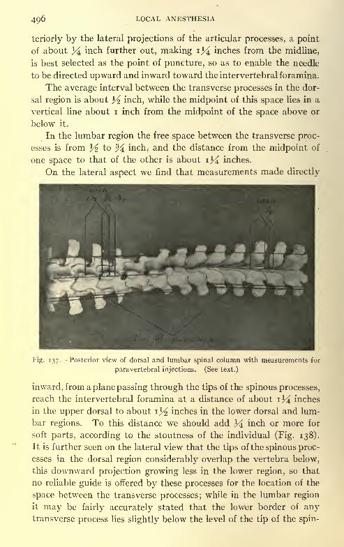
496 LOCAL ANESTHESIA
teriorly by the lateral projections of the articular processes, a point
of about Y inch further out, making i34 inches from the midline,
is best selected as the point of puncture, so as to enable the needle
to be directed upward and inward toward the intervertebral foramina.
The average interval between the transverse processes in the dor-
sal region is about ^ inch, while the midpoint of this space lies in a
vertical line about i inch from the midpoint of the space above or
below it.
In the lumbar region the free space between the transverse proc-
esses is from ^ to % inch, and the distance from the midpoint of
one space to that of the other is about \Y inches.
On the lateral aspect we find that measurements made directly
Fig. 137. Posterior view of dorsal and lumbar spinal column with measurements for
paravertebral injections. (See text.)
inward, from a plane passing through the tips of the spinous processes,
reach the intervertebral foramina at a distance of about 1% inches
in the upper dorsal to about i^ inches in the lower dorsal and lum-
bar regions. To this distance we should add Y inch or more for
soft parts, according to the stoutness of the individual (Fig. 138).
It is further seen on the lateral view that the tips of the spinous proc-
esses in the dorsal region considerably overlap the vertebra below,
this downward projection growing less in the lower region, so that
no reliable guide is offered by these processes for the location of the
space between the transverse processes; while in the lumbar region
it may be fairly accurately stated that the lower border of anytransverse process lies slightly below the level of the tip of the spin-

PARAVERTEBRAL AND PARASACRAL ANESTHESIA 497
ous process of the same vertebra, and that a needle directed inwardand upward at the proper lateral position (i} inches from the mid-
line) and about on a level with the middle of the spinous processshould pass freely between the transverse processes of that vertebra
and the one above.
In the dorsal region, as we have no such guide, it is best to feel
Fig. 138. Lateral view of dorsal and lumbar spinal column with measurements. (See
test.)
for the intertransverse space, with the point of the needle directed
inward from a line iY inches lateral from the tips of the spinous proc-
esses, and, having located this interval at any one point, the pointof puncture for the space above or below will be about i inch re-
moved on the lateral line.
These measurements were made on the vertebral columns of
several adult skeletons and utilized for verification on the cadaver,

4Q8 LOCAL ANESTHESIA
and, while subject to small variations, will, I believe, be found fairly
accurate for the adult of average size.
Lawen, in 1911, performed a nephrotomy by blocking the inter-
costal and three upper lumbar nerves.
The most noteworthy articles upon this subject to date have
been by Lawen, Kappis, Heile, Wilms, and Franke; but Finsterer's
work on this subject has been particularly thorough and
comprehensive.One and 1.5 per cent, solutions of novocain with adrenalin have
been the strength usually employed. The total quantity of novo-
cain used being 0.4 to 0.8 gm. Finsterer, in a case in which he used
0.4 gm., injecting the eleventh and twelfth dorsal and first and second
lumbar nerves, reports that the duration of the anesthesia was two
hours.
In extensive operations, where six or eight points are injected on
each side, using approximately 5 c.c. at each point, which, com-
bined with the skin injections at the puncture points, brings the
total quantity of solution used up to 75 to 100 c.c., which, if it be a
i per cent, solution, certainly would appear to exceed the safe limits
of the drug.
While no mishaps have occurred, some patients have shown
symptoms which have served as a warning note; this has stimulated
animal experimentation to determine the best method of procedure.As some of the injected solution must reach the epidural space
through the spinal foramina, rather free epidural sacral injections
were made upon animals to study the movements of a definite
quantity of solution injected within the epidural space.
Lawen and Gaza found with colored solutions plus adrenalin in
epidural sacral injections that the solution often ascended as far as
the lower portion of the thoracic cord, while Muroya, in a somewhat
similarly made injection, found that the ascent of the fluid was often
higher to the cervical region and frequently into the skull.
The difference in the results obtained by these investigations
may be accounted for, as Muroya states, by the quantity of the solu-
tion used and the pressure under which the injections are made.
He, accordingly, timed the rapidity of his injections and used 10 c.c.
per kilogram of body-weight of the rabbit, of a colored 5 per cent,
novocain adrenalin solution, injecting i c.c. per minute, and found,
as in his previous experiments, that the solution ascended often as
high as the skull, and but seldom was found to stop at the middorsal
region.

PARAVERTEBRAL AND PARASACRAL ANESTHESIA 499
Muroya found that in rabbits paravertebral injections are rela-
tively more toxic than subcutaneous; this may be explained in
several ways: (i) The solution may ascend to the higher centers
through the subdural space, or (2) the blood-vessels may more
rapidly absorb it in this position; (3) it may be rapidly taken up bythe large network of lymph-spaces which overlie the vertebral
column, particularly in the abdominal cavity, as the colored solution
has often been found in animals high up in the thoracic duct a few
minutes after injection. This absorption through the lymph-spaces
seems the most probable.
A comparison made by Muroya with subcutaneous and paraver-
tebral injections of novocain colored with methylene-blue showed
methylene-blue in the urine in ten to twenty minutes following sub-
cutaneous injections, and in five to ten minutes following para-
vertebral.
From the work of Muroya upon rabbits, he concluded that the
paravertebral injection is six times more toxic than the subcuta-
neous, but that this toxicity can be greatly reduced, making it about
equal to the subcutaneous injection, by combining 5 per cent, gelatin
with the adrenalin in normal salt solution, the adrenalin preventing
or delaying its absorption through the blood-vessels, while the
gelatin delays its diffusion into the cellular lymph-spaces.
By the use of this mixture its action at the point of injection is
intensified, as the solution is more or less retained at the point of in-
jection.
In drawing positive conclusions from the above it must be re-
membered that it is not always safe to apply the results of animal
experimentation to man. In the first place the formation of the
sacral canal may not exactly correspond to that of man, where the
numerous large foramina for the exit of nerve-trunks permit any
injected solution to readily escape in all directions, and besides the
difference in the distance between the sacrum and skull in the rabbit
and in man is considerable. These experiments are, however, more
valuable for determining the movements of the injected fluid in
epidural (sacral) injections.
Kappis has reported paravertebral injections of the cervical
region. A line on either side of the spinous processes was anes-
thetized, and a long needle advanced forward until the transverse
process was encountered. The interval between these processes
is sought for by the point of the needle, which is advanced from i
to i% cm. further and the injection made.

500 LOCAL ANESTHESIA
Kappis used 1.5 per cent, novocain-suprarenin solution. The
performance of paravertebral injections in such regions as the neck
must receive further experimental study before it can be popularized ;
the likelihood of the solution reaching the phrenic nerve in effective
quantity should not be lost sight of; its origin from the third, fourth,
and fifth cervical is practically the center of the field, and after forma-
tion its course is more superficial. If the solution is effectively used
this nerve should be paralyzed \ however, unless the procedure is car-
ried out on both sides, the temporary one-sided paralysis is not
likely to be of consequence.
Fig. 139. a and b, points of injection on line drawn over transverse processes of cervical
vertebrae. (From Braun.)
The paravertebral injection of the cervical region is carried out
by Braun in a somewhat different way, following the suggestion of
Heidenhain.
The injections are made from the side, between the third and
fifth vertebrae, rather freely with a 0.50 per cent, novocain-adrenalin
solution at the point where the nerves lie rather close together. Aline is drawn on the neck from the transverse process of the atlas
which is felt under the point of the mastoid process downward over
the transverse process of the sixth cervical vertebra (tuberculum
carotidum).
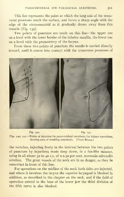
PAEAVERTEBRAL AND PARASACRAL ANESTHESIA 501
This line represents the point at which the long axis of the trans-
verse processes reach the surface, and forms a sharp angle with the
edge of the sternomastoid as it gradually draws away from this
muscle (Fig. 139).
Two points of puncture are made on this line the upper one
on a level with the lower border of the inferior maxilla, the lower one
on a level with the promontory of the larynx.
From these two points of puncture the needle is carried directly
inward, until it comes into contact with the transverse processes of
Fig. 140. Fig. 141.
Figs. 140, 141. Points of injection for paravertebral anesthesia for kidney operations,
showing area of resulting anesthesia. (From Braun.)
the vertebra, injecting freely in the interval between the two points
of puncture by injections made deep down, in a fan-like manner,
using in all about 30 to 40 c.c. of a 0.50 per cent, novocain-adrenalin
solution. The great vessels of the neck are in no danger, as they lie
somewhat in front of this line.
For operations on the midline of the neck both sides are injected,
and where it involves the larynx the superior laryngeal is blocked in
addition, as described in the chapter on the neck, and if the field of
operation extend to the base of the lower jaw the third division of
the fifth nerve is also blocked.

502 LOCAL ANESTHESIA
Braun states that this method produces an effective surgical
anesthesia for all major operations in this region.
Braun has performed three nephrotomies by paravertebral
methods, and made accurate observations of the extent of the field
of anesthesia.
The eighth to the twelfth dorsal nerves were injected each with
5 c.c. of a i per cent, solution; the line of puncture lay on a line which
corresponded to the upward extension of the outer margin of the
quadratus lumborum.
A point of anesthesia was now established over the crest of the
ilium at the outer border of the quadratus lumborum; between this
point and the point of puncture for the twelfth dorsal nerve was a
rather free and deep injection made down to and including the
fatty tissue around the kidney, using for this purpose 75 c.c. of a 0.50
per cent, novocain-suprarenin solution. The points of injection
and distribution of anesthesia are shown in Figs. 140 and 141.
Braun states that the entire procedure was painless, including
the luxation of the kidney, and while his patients were thin he be-
lieves that the anesthesia of this region by local methods has been
conquered.
Finsterer, who has employed this method for 6 laparotomies and
other major operations, gives his technic as follows:
The spine of the first lumbar vetebra is located, and a point on
the skin from 3 to 3^ cm. laterally is anesthetized; the needle
marked in centimeters is passed vertically inward through this anes-
thetized point to a depth of from 4 to 5 cm., according to the stout-
ness of the individual, until the transverse process is struck; the
upper border of the process is then felt for with the needle; when this
point is reached the syringe is carried outward and downward,
directing the needle-point upward and inward, when it is advanced
about 3^ to i cm. further, and 5 c.c. of a i per cent, solution of novo-
cain adrenalin is then injected in a fan-shaped area. Care should
be exercised not to push the needle too deeply, as the anesthetizing
fluid will pass beyond the ganglion and be useless.
The points for reaching the first, second, and third lumbar nerves
will be at intervals of 3^ to 4 cm. from each other, according to the
size of the individual.
PARASACRAL ANESTHESIA
The anterior surface of the sacrum shows that the anterior sacral
foramina lie almost always in a straight line, which from below uphas a slightly outward angle. The distance between the midpoints

PARAVERTEBRAL AND PARASACRAL ANESTHESIA 503
of the first sacral foramina is i% inches, and between the midpointsof the fourth is i% inches (the fifth sacral foramen is formed byarticulation with the coccyx, and does not appear as a separate
opening), so that if a line is drawn down the midline of the sacrum
a line running over the center of the foramina diverges but % inch
from the midline in passing upward from the fourth to the first
foramina.
The distance from the free margin of the sacrum at its lower free
i
Fig. 142. Anterior view of sacrum, slightly reduced, showing relative distances to
foramen.
border (sacrococcygeal junction) along the line passing over the
center of the foramina is % inch to the midpoint of the fourth
foramen, i^ inches to the midpoint of the third foramen, 2% inches
to the midpoint of the second foramen, and 3^ inches to the mid-
point of the first sacral foramen. This then makes the foramina
the following distances apart: approximately % inch from the fourth
to the third and from the third to the second, and the first is i inch
from the second (Fig. 142).
On the lateral view it is seen that if the needle is passed straight

54 LOCAL ANESTHESIA
in over the lower free margin of the sacrum % inch from the midline,
and directed upward almost in a straight line, with a very slight out-
Fig. 143. Distances and relative position of foramen on lateral view- of sacrum.
Fig. 144. Direction of long needle for parasacral injections. (From Braun.)
ward inclination, that it will pass directly over the fourth, third, and
second foramina, when, after meeting the bone above this opening,

PARAVERTEBRAL AND PARASACRAL ANESTHESIA 505
if it will be slightly withdrawn and redirected, with the point elevated
Y^ inch and advanced i inch further, that it will reach the first sacral
foramina (Figs. 143, 144).
It is seen, after a study of numerous sacrums, that the interme-laieral sacral artery
lumbosacral trunk
inf. mestnteric artay_
sacr. nerves IV and V
\\,;\'Cottntus\
/ iw-nw
pudcndal plexus
I pudic art. X
iddle haemorrhoidal art. X
Fig. 145. The blood-vessels and nerves on the right pelvic wall. The pelvis has
been halved by a sagittal section and the genitalia removed. X = Branches to the
coccygeus. X X = Branch to levator ani. + = Site of abdominal inguinal ring.
+ -f-= Branches to pyriformis. (Sobotta and McMurrich.)
diate foramina between the fourth and first in some few are placed
somewhat outside of the straight line, passing over the center of the
openings; this lateral variation is usually about % inch, and was
never seen to exceed Y inch.

506 LOCAL ANESTHESIA
As the nerves, as they emerge from the foramina run downward,
outward, and forward (Fig. 145), it would seem best to slightly
increase the lateral angle of the needle, so that at a depth of about
2 inches its point will be about Y inch lateral to the point of entrance;
in this way the needle cannot go astray, and may even transfix these
nerves though the foramina do not lie in a straight line.
The injections are made, according to Braun, as follows: With
the breech of the patient well presented, the long fine needle (for
this purpose it should be about 5 or 6 inches long) is entered about
1^2 or 2 cm - from the middle line, on a level with the sacrococcygeal
joint. As the inner surface of the sacrum up to about the second
foramina is but little curved, the needle is advanced straight up from
the point of puncture, in close contact with the bone, until it im-
pinges with it near the second sacral foramina; this point is about
6 to 7 cm. from the point of puncture at the sacrococcygeal joint in
the adult, not counting the thickness of the soft parts.
One proceeds in the following manner: The needle is entered on
the inner surface of the lower border of the sacrum and directed up-ward in a parallel direction seeking for the edge of the bone; the needle
is then pushed along the inner surface, parallel with the middle line,
until it reaches the bone at about the recognized depth; along the
entire way, from the second to the fifth sacral foramina, 20 c.c. of i
per cent, novocain-adrenalin solution is injected. The injections
should not be made except when the needle is in contact with the
bone. The needle is now drawn back to the edge of the bone and
redirected at a slightly increased angle, but still parallel to the middle
line toward the linea innominata; reaching the bone just above the
first sacral foramina, about 9 or 10 cm. from the point of entrance
(not including the soft parts), 20 c.c. of the i per cent, solution are
injected here.
Before finally withdrawing the needle 5 c.c. are injected between
the coccyx and rectum. The same procedure is repeated on the op-
posite side, using in all about 100 c.c. of solution. Braun states that
there is no danger of injuring the rectum if empty, as it is pushed out
of the way by the advancing needle, but a finger may be inserted
within it if preferred for guidance.
This method produces an effective means of anesthesia for such
operations as prostatectomy, prolapse of the uterus, and resections
of the rectum, as well as other minor operations upon these parts,
but it is not sufficient for a total hysterectomy, as here the upper
part of the field is not reached by the pelvic nerves.

CHAPTER XXII
THE HEAD, SCALP, CRANIUM, BRAIN, AND FACE
THE surgery of the head with regional methods of anesthesia is
one of the most attractive and fascinating in the entire body, and its
operative procedure among the most brilliant in the entire domain
of surgery. In itself a vast field for the application of intra- and
paraneural methods, presenting an intricate labyrinth of foramina,
canals, fissures, and tracts for the passage of the great nerve-trunks
and their branches, always appearing in a new and interesting light,
due to the many brilliant minds that have made this region a field
of study, and have evolved new ways and means of access and ap-
proach to the great nerve-trunks at their basal foramina and even
their injection within the skull.
Those who study the skull with the view of determining the most
accurate and reliable methods of reaching by deep injections, espe-
cially the branches of the fifth nerve at their points of emergence from
the various foramina or even within them, find in this study a
wealth of information. The deeper the study is pursued the more
absorbing and interesting does it become. The skull should be
constantly before us and the foramina viewed from every possible
angle of approach. The more versatile and proficient the student
becomes in this study the more successful will he be in making the
deeper and more difficult injections. The foramen ovale and fora-
men rotundum are the most difficult to reach and this difficulty is
only overcome by constant study of the skull and practice upon the
cadaver.
In this vast field the work is of interest alike to physician and
surgeon to one for the alcoholization of the great nerve-trunks for
the relief of neuralgia; to the other, for the purpose of regional anes-
thesia of the peripheral parts, as well as for the deep injections of
alcohol, which, in its clinical application, should always be regarded
as a surgical procedure. Whether devised originally for the thera-
peutic application of alcohol or the proximal cocainization of the
nerve-trunks, both alike serve the same end in offering an approachto the nerve-trunks and can alike be used for both purposes.
While my own personal experience in this field had been consider-
able up to the time of the first appearance of this book it has during
S7

508 LOCAL ANESTHESIA
recent years been very materially increased until, with few exceptions,
the entire range of operations upon the head and within the depthof the cranial cavity have been brought within the scope of regional
and local methods and often with surprising ea.se and facility when
once, the difficulties of anesthesia have been thoroughly mastered
and overcome, once this has been accomplished the advantages of
the local method of procedure becomes very apparent and the disad-
vantage of general anesthesia made more apparent. This is par-
ticularly the case in extensive resections of the maxillae where the ad-
ministration of the general anesthetic has many difficulties, the
patient may be constantly swallowing his tongue or aspirating blood
and mucus and the avoidance of these difficulties necessarily in-
creases the number of assistants in an already crowded field and often
causes the loss of much time. Within the cranial cavity the conges-
tion produced by the general anesthetic often causes much persistent
oozing which adds greatly to the difficulties and accounts for muchloss of time. Under local means these difficulties are entirely re-
moved as we have no congestive influences at work and the normal
vascularity is greatly lessened by the adrenalin in the anesthetic
solution.
From the earlier pioneer efforts in this direction to the present
time are to be found a galaxy of brillinat names Matas, Schlosser,
Ostwalt, Hecht, Lowenstein, Killiani, Patrick, Bodine, Keller, Wright,
Harris, Braun, Levy, Baudoin, Brissaud, Sicard, Taptas, and more
recently Offerhaus, and particularly Hartel, in his latest approach to
the gasserian ganglion and exhaustive presentation of this subject.
In originally undertaking this work I had hoped that at least this
chapter would be from the pen of my distinguished chief, Prof. Matas,
an original worker in this as well as other fields, and whose brilliant
achievements already fill many pages in the annals of surgery.
While deprived, at least for the present, of this benefit due to the
press of other matters, we may hope that should this chapter be re-
written it may come from his pen, and I hope that in this, as in
other chapters of this work in which I have so freely borrowed from
his knowledge, I may in some small measure reflect credit upon his
teachings.
A few of the original illustrations in this chapter are from the
private collection of Prof. Matas, kindly loaned for this purpose and
appear for the first time in these pages. These illustrations were
prepared by my colleague, Dr. Urban Maes, who, in association with
Prof. Matas, has done much work and developed great skill in this field.

THE HEAD, SCALP, CRANIUM, BRAIN, AND FACE 509
For a thorough understanding of the methods of local and
regional anesthesia applied to the head a thorough knowledge of
anatomy is essential, with a study of the subject from every viewpoint;
only then can we properly appreciate the difficulties and delicate
technic necessary for the clinical application of these methods.
Fig. 146 Diagram of the fifth cranial nerve. (From Flower.)
It is for this reason that the minutiae and details of anatomy have
been discussed so thoroughly in the following pages; for my own
part this study has been of immense value in developing any pro-
fiency I may have attained in this field and I cannot too strongly
urge the necessity of its careful study to all who seek success here.

LOCAL ANESTHESIA
Nowhere else in the entire range of local anesthesia is it more strongly
emphasized that there is no royal road to success in local anesthesia
and that the only education in this field is self-education.
long onauemosei between the branrhrs of theI Trigemfnus and ih,
cer\iml ncrvis
supcrfic. temporal
cervical >>r. of facial lerior facial /~
Fig. 147. Superficial nerves and arteries of the face (deeper layer). Most of the
parotid gland is removed. The facial muscles have been cut away, divided, or drawn
downward. (Sobotta and McMurrich.)
The Fifth Nerve and Its Branches (Figs. 146, 147). The ophthal-
mic or first division of the fifth (Fig. 148) is a sensory nerve, supply-
ing the eyeball, mucous lining of the eye, lacrimal gland, nasal fossa,
the skin of the nose, forehead, and front portion of the vertex. After
leaving the gasserian ganglion it passes forward along the outer wall
of the cavernous sinus, below the other nerves, which pass here (Fig.
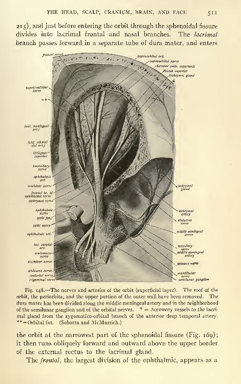
THE HEAD, SCALP, CRANIUM, BRAIN, AND FACE
215), and just before entering the orbit through the sphenoidal fissure
divides into lacrimal frontal and nasal branches. The lacrimal
branch passes forward in a separate tube of dura mater, and enters
frontal r,
supraorbital art.
SupratrocMcarnerve
(ant. meningealart.)
trochlear net
abiliicens nrrvi
tentorial nen
trigeminal net
andibularnerve
semilunar ganglion
Fig. 148. The nerves and arteries of the orbit (superficial layer). The roof of the
orbit, the periorbita, and the upper portion of the outer wall have been removed. The
dura mater has been divided along the middle meningeal artery and in the neighborhoodof the semilunar ganglion and of the orbital nerves.
* = Accessory vessels to the lacri-
mal gland from the zygomatico-orbital branch of the anterior deep temporal artery.** = Orbital fat. (Sobotta and McMurrich.)
the orbit at the narrowest part of the sphenoidal fissure (Fig. 169);
it then runs obliquely forward and outward above the upper border
of the external rectus to the lacrimal gland.
The frontal, the largest division of the ophthalmic, appears as a

512 LOCAL ANESTHESIA
direct continuation of this nerve. It enters the orbit above the
muscles through the highest and broadest part of the sphenoidal
fissure, and corftinues forward in the midline between the levator
palpebrae and the periosteum to about the middle of the orbit, where
it divides into supratrochlear and supraorbital branches.
The supratrochlear (Fig. 147) escapes from the orbit between the
pulley of the superior oblique and the supraorbital foramen; it curves
up on the forehead, close to the bone, beneath the occipitofrontalis
muscle, and is distributed to the skin of the forehead on either side
of the middle line.
The supraorbital nerve passes forward through the supraorbital
foramen, supplies the upper eyelid, and ascends beneath the occipito-
frontalis muscle, and is distributed to the scalp and pericranium as
far back as the parietal and occipital bones.
The nasal nerve enters the orbit between the two heads of the
external rectus, passing between the two divisions of the third nerve,
runs obliquely inward above the optic nerve, beneath the superior
oblique and superior rectus muscles to the inner wall of the orbit,
where it passes through the anterior ethmoidal foramen, giving off its
infratrochlear branch here, and enters the cavity of the cranium,
where it traverses a groove on the cribriform plate of the ethmoid
bone and passes down through the slit on the side of the crista galli
into the nose. Within the nasal cavity this nerve supplies the mucous
membrane in its upper and anterior parts. The nerve then descends
in a grove on the back part of the nasal bone, escaping between the
lower border of this bone and the upper lateral cartilage to supply
the end of the nose.
Its infratrochlear branch supplies the skin at the inner angle of
the lids and side of the nose.
The superior maxillary nerve (Fig. 149) passes forward through
the foramen rotundum into the sphenomaxillary fossa, passing
obliquely forward and outward. It enters the orbit through the
sphenomaxillary fissures, and passes into the infra-orbital canal and
appears upon the face at the infra-orbital foramen, where it divides
into three sets of branches palpebral, nasal, and labial which are
distributed to these respective parts (Fig. 147). In the sphenomaxil-
lary fossa this nerve gives off its temporomalar, sphenopalatine,
and posterior-superior dental branches.
The temporomalar branch enters the orbit, and divides at the
back part of this cavity into temporal and malar branches. The
temporal branch passes through a foramen in the malar bone and

THE HEAD, SCALP, CRANIUM, BRAIN, AND FACE 513
enters the anterior part of the temporal fossa; it ascends between the
bone and substance of the temporal muscle, piercing the muscle and
temporal fascia about i inch above the zygoma, to be distributed to
the skin of the temporal regions and side of forehead. The- malar
branch passes through a foramen in the malar bone, and appears
Fig. 149. Innervation of the teeth (modified from Spalteholz). The outer wall of
orbit and part of the outer wall of superior and inferior maxillae have been removed : i;
Infra-orbital nerve; 2, 3, and 4, posterior, middle, and anterior superior dental nerves,
5, sphenopalatine ganglion and nerves; 6, lateral mucous membrane, antrum of High-
more; 7, inferior dental nerve; 8, mental nerve. (From Braun.)
upon the cheek at the opening of this canal (Fig. 147), on the anterior
surface of the bone, about % inch below the rim of the orbital fossa,
where it is distributed to the skin of the cheek.
The sphenopalatine descends to this ganglion, which lies just
below the trunk of this nerve in the sphenomaxillary fossa.
33

514 LOCAL ANESTHESIA
The posterior superior dental branches are given off from the supe-
rior maxillary nerve in the sphenomaxillary fossa, just as it is about
to enter the orbit; these nerves pass down over the tuberosity of the
superior maxillary bone to enter the dental canals on the posterior
surface of this bone about its midolle; these nerves supply the three
molar teeth.
The middle and anterior-superior dental nerves are given off in the
infra-orbital canal, the middle supplying the two bicuspids, the
anterior branch the canine and incisor teeth.
The descending or palatine branches from the sphenopalatine
ganglion are three in number anterior, middle, and posterior. The
anterior descends through the posterior or palatine canal to appear
upon the hard palate at the posterior palatine foramen; it then
passes forward in a groove at the junction of the hard palate and
alveolar process, nearly as far as the incisor teeth, supplying the
parts in the vicinity of its distribution. In the palatine canal it
gives off branches to the nasal fossa. The middle and posterior
branches are distributed to the soft palate and tonsillar regions.
The nasopalatine nerve is the only other nerve from the spheno-
palatine ganglion which is of special interest. It enters the nasal
fossa through the sphenopalatine foramen, passes inward across the
roof of the nasal fossa to reach the septum, down which it runs to
the anterior palatine foramen, and appears at the opening of this
canal on the roof of the mouth to supply the surrounding soft parts.
The inferior maxillary nerve, largest division of the fifth, passes
through the foramen ovale and divides into two trunks anterior
and posterior. It is joined at this point by its motor root, most of
which passes into its anterior division, which divides into masseteric,
buccal, and pterygoid branches, to be distributed to these muscles,
the masseteric passing through the sigmoid notch to reach this
muscle; the buccal branch, in addition to supplying this muscle, is
distributed to the skin of the cheek as far forward as the angle of the
mouth.
The deep temporal branches, given off just after the nerve emergesfrom the skull and running outward and upward, are distributed to
the temporal muscle.
The auriculotemporal nerve curves upward with the temporal
artery, between the external ear and condyle of the jaw, beneath
the substance of the parotid gland, and, passing over the zygoma,divides into branches auricular, superior, and inferior, and a branch
to the meatus auditorius, which are distributed to these parts. The

TH* HEAD, SCALP, CRANIUM, BRAIN, AND FACE 515
temporal branches pass upward with the temporal artery and supplythe scalp as far as the vertex of the skull.
The lingual nerve reaches the inner side of the ramus of the jaw,
down which it descends on the inner side of the dental nerve to the
base of the tongue, crossing obliquely forward and inward to this
organ, running along its side as far as the tip. Where the nerve
passes from the ramus of the jaw to the base of the tongue it is quite
superficially situated beneath the mucous membrane, and is quite
easily reached in this position, particularly if the tongue is drawn for-
ward and to the opposite side.
The inferior dental neroe passes obliquely downward, forward, and
outward with the inferior dental artery to reach the dental foramen
on the inner side of the inferior maxilla. In the dental canal it gives
off branches which supply the teeth, and a mental branch, which
appears at the mental foramen and supplies the lower lip and the
soft parts of the chin.
In addition to the branches of the fifth the lower part of the face
receives branches from the cervical nerves. The side of the head
receives the auricular branch of the pneumogastric, which passes
upward between the mastoid process and external auditory meatus
to the back of the ear. The occipital region is supplied by the
occipitalis major and minor and the auricularis magnus. These
nerves will be spoken of in dealing with their areas of distribution.
Anesthesia. To anesthetize the supra-orbital and supratrochlear
nerves an injection of about 2 drams of solution No. 2, 0.50 per cent,
novocain, and a few drops of adrenalin to the ounce should be made
just over the supra-orbital notch, beneath the deep frontal fascia,
transversely for about i inch; or, where both nerves are to be in-
jected, this can be done by a subfascial injection, extending across
the base of the forehead, as indicated in Fig. 150, the shaded portion
indicating the anesthetic area; on the margins of this area for some
distance back there is lessened sensibility. While it is quite feasible
to practice strictly regional anesthesia for the entire scalp by blocking
the supra-orbital nerves in front, the occipital nerves behind, and the
temporal nerves on the side by a line of anesthesia just above the
zygoma, extending from the ear to the angle of the orbit and carried
down to beneath the temporal fascia, thus rendering the entire scalp
anesthetic. This procedure limitd to one side produces an anesthe-
sia reaching almost to the vertex, where the nerves of the oppositeside lap over. Such an extensive area of anesthesia is, not often
called for, while anesthesia of those parts supplied by the supra-

LOCAL ANESTHESIA
orbital and occipital nerves will more often be found quite useful,
easily and quickly carried out; but where the field of operation is
more or less central on the scalp, it will, usually be found simpler
and require less solution to surround the operative area by a wall of
LJ
Fig. 150. Resulting area of anesthesia after blocking supra-orbital and supratrochlearnerves along heavily shaded line. (From Braun.)
anesthesia carried well down to the pericranium, which will meet and
anesthetize all nerves entering the area and permit any contemplated
operation upon the soft parts, as well as resections of the bone and
operations upon the underlying dura and brain (Figs. 151, 152).
Fig. 151. Surrounding tumor of the scalp with zone of anesthesia. (From Braun.)
Here the operative field is surrounded by an area of anesthesia,
and at several points along this surrounding zone the long needle is
entered and passed down to the bone through the points indicated
by the heavy dots, injecting the solution as the needle is advanced.

THE HEAD, SCALP, CRANIUM, BRAIN, AND FACE 517
However, it is to be recommended, as a general rule, that all
extensive operations upon the soft parts or underlying bone in the
region supplied by the fifth nerve be operated by injections of gan-
glion gasseri, or its nerves at their foramina of exit; the injection of
the ganglion has the advantage that it anesthetizes the dura throughits meningeal branches as well as the overlying bone and soft parts.
As the occipital nerves are several in number, and reach the
scalp at different points, they may be dealt with collectively. If a
line of anesthesia (solution No. 2, with 5 drops adrenalin to the ounce)
is produced, extending from ear to ear across the base of the mastoid
Fig. 152. Surrounding a compound fracture of skull with zone of anesthesia. (From
Braun.)
processes, and carried well down to the deep tissues, it will block all
branches of the occipital nerves, as well as branches from the auricu-
laris magnus, and result in an anesthetic area, as indicated in Fig.
154 (the black line shows the line of infiltration), or the nerves maybe dealt with more or less individually in the following way:
The occipitalis major and minor and the auricularis magnus maybe blocked by making injections at their points of emergence at the
occiput (Fig. 154); the occipitalis minor and auricularis magnusbehind the posterior margin of the insertion of the sternocleido-
mastoid. The occipitalis major emerges upon the surface of the

LOCAL ANESTHESIA
occiput through a cleft in the trapezius muscle, along with the occipi-tal artery, the third occipital, sometimes an independent nerve, pass-
great occipital ner
occipital vessels. A
Occipitalis
'ccipital artery
real occipital nerve
occipital vein
'occipital nerve
descend.br. oftransv. cervical art. 'Ledescend, b,: oftrantv. cervical Vfia
Latissinuisdorsi
posterior cutaneous branches of intercostal nerves-
Fig. 153. The superficial and middle layer of the nerves and vessels of the nuchal
region. Upon the left side the trapezius, sternocleidomastoideus, splenius, and levator
scapulas have been divided. * = Occipital root of external jugular vein. (Sobotta and
McMurrich.)
ing slightly nearer the midline; to reach both of these nerves a deep
line of infiltration can be carried from the posterior occipital protu-
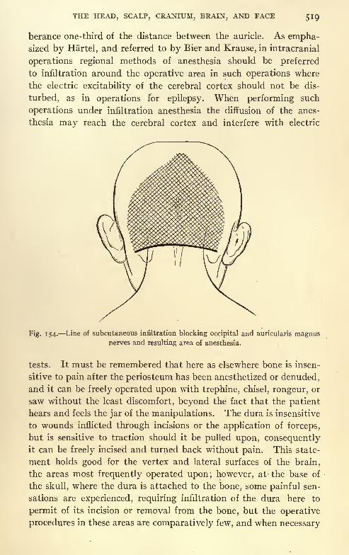
berance one-third of the distance between the auricle. As empha-sized by Hartel, and referred to by Bier and Krause, in intracranial
operations regional methods of anesthesia should be preferred
to infiltration around the operative area in such operations where
the electric excitability of the cerebral cortex should not be dis-
turbed, as in operations for epilepsy. When performing such
operations under infiltration anesthesia the diffusion of the anes-
thesia may reach the cerebral cortex and interfere with electric
Fig. 154. Line of subcutaneous infiltration blocking occipital and auricularis magnusnerves and resulting area of anesthesia.
tests. It must be remembered that here as elsewhere bone is insen-
sitive to pain after the periosteum has been anesthetized or denuded,and it can be freely operated upon with trephine, chisel, rongeur, or
saw without the least discomfort, beyond the fact that the patient
hears and feels the jar of the manipulations. The dura is insensitive
to wounds inflicted through incisions or the application of forceps,
but is sensitive to traction should it be pulled upon, consequentlyit can be freely incised and turned back without pain. This state-
ment holds good for the vertex and lateral surfaces of the brain,
the areas most frequently operated upon; however, at the base of
the skull, where the dura is attached to the bone, some painful sen-
sations are experienced, requiring infiltration of the dura here to
permit of its incision or removal from the bone, but the operative
procedures in these areas are comparatively few, and when necessary

520 LOCAL ANESTHESIA
it is quite easy to anesthetize the dura here by progressive injections
as you advance.
It is a remarkable fact that the brain, the great sensory organ of
the body, should itself be devoid of painful sensations. The entire
cerebral cortex of animals has been irritated and stimulated in manyways without ever exciting any response indicative of pain, and wehave often removed considerable areas from the brain of a thoroughlyconscious patient without ever exciting any pain.
The knowledge of these facts permits us to undertake in suitable
cases many operations under local anesthesia upon the skull and
brain which might otherwise seem impossible. In any extensive
operation upon the cranium it may prove desirable to use circular
constriction, with rubber tubing around the base of the cranium;
this, however, while often advisable with general anesthesia, is
superfluous with local methods, for here the use of adrenalin provesan effective hemostatic, besides the constriction is often in the way,and may prove annoying or painful to any but a patient under
general anesthesia unless the entire circumference of the cranium
were anesthetized. Usually in severe traumatisms of the skull, such
as extensive or simple depressed fractures, the patients are often
in a state of unconsciousness from shock or brain injuries, which
permit of painless operating with very little local anesthesia. Onthe other hand, many of these patients are in a condition of noisy
delirium or turbulent restlessness, which makes any operation under
local anesthesia, even if painless, inadvisable, and forces a resort to
general anesthesia.
Omitting the above class of patients, there are a large range of
operations which permit of the use of local anesthesia. Simpleabscesses will require nothing further than a line of infiltration over
the proposed incision.
Wounds. For the cleansing and suture of incised, contused, and
lacerated, stab or superficial gunshot wounds, the most satisfactory
plan is, after a preliminary cleansing of the surrounding parts, to
create a wall of anesthesia around the wound, cleansing it later with
a more liberal cleansing of the entire area after the anesthetizing
process has been completed, as described in the chapter on Principles
of Technic.
Sebaceous cysts may be removed the same way, by a surroundingwall of anesthesia and excising a triangular or oblong piece of the
skin with the tumor attached, approximating the gap with sutures;
or by infiltrating a line over the long axis of the cyst and splitting it

THE HEAD, SCALP, CRANIUM, BRAIN, AND FACE 521
open and pealing out the sac. While this is a simple and thoroughlyeffective method in non-infected sacs, in the event of infection it will
leave a suppurating wound.
Benign tumors may be dealt with by the same method, also malig-nant growths when superficial and limited, as in epitheliomas, but if
extensive or deeply infiltrating had best be left to regional methods or
general anesthesia. In those cases of malignancy suitable for local
methods care should be exercised in making the injections to keepwell away from the growth, and surround it by a wall of anesthesia,
extending down to the cranium, so that this may be resected if the
growth is found attached.
As this same technic is applicable to all operations upon the scalp
and underlying bone, it is sufficient to state that it is suitable in
depressed iractures and for the evacuation of epidural or subdural
hemorrhage, intracranial abscesses, and for the removal of necrotic
areas of the skull. Osteoplastic flaps of considerable extent are also
just as easily raised with local anesthesia and operations performed
upon the cortex of the brain. Here local anesthesia possesses de-
cided advantages over general anesthesia, particularly ether, which
greatly congests the entire cranial circulation, and is particularly
troublesome when operating within the skull on account of the per-
sistent oozing which occurs from even the smallest vessels, and greatly
adds to the danger of postoperative hematoma. This entire picture
is changed when operating with local anesthesia; instead of the tre-
mendous congestion encountered, with ever-ready tendency to
troublesome hemorrhage difficult to control, with possible later
oozing, the brain and its circulation is found normal, and hemorrhageeither does not occur, or is easily controlled, with little or no tend-
ency to postoperative oozing.
For this reason, if for no other, it is advised to consider local
anesthesia in all suitable intracranial operations, and when the opera-
tion is extensive to perform it by a two-stage operation, raising the
osteoplastic flap in the first stage under general anesthesia; this is
then returned to its place and lightly held with sutures, to be again
raised at a subsequent sitting, next day or later, the dura opened,and the intradural procedure executed.
The advantage of this method is illustrated by the following
reports :
Mr. S., very stout, short man, weighing over 200 pounds, gave symptoms of tumor
near left motor area. Under general anesthesia a large osteoplastic flap was raised, ex-
posing the entire left motor area. The patient was very full blooded, and the entire
circulation of the head, face, and cerebrum became greatly congested; the cerebral and

522 LOCAL ANESTHESIA
meningeal vessels were everywhere turgid with blood, and would certainly have given
trouble if the operation had been proceeded with; accordingly the wound was loosely
closed, after securing all bleeding-points in the external soft parts, and the patient
returned to bed. The next day, after lightly injecting a cocain solution into the incision,
the sutures were loosened and the flap raised; beyond this point no coeain was used.
The operation was everywhere painless, the patient conversing with us during the differ-
ent steps of the deeper procedure. A vast difference was now seen in the condition of
the meningeal and cerebral circulation, the vessels which previously had been turgid
with blood were now hardly to be seen, only a few small vessels of normal size encoun-
tered here and there. The brain, which had previously been tense and bulging from the
wound, now seemed shrunken by comparison and lay well within the skull.
Two tumors were located back of the motor area and removed with a surrounding
margin of brain, resulting in a paresis of the right side of the body and tongue. The
patient was able to indicate during the entire procedure that he felt no pain. The
wound was then closed with a small drain, the patient making a satisfactory recovery,
but died about six months later from a recurrence of the malignant process. This case
illustrates clearly the advantages of a two-stage operation, or the entire procedure could
have been performed at one sitting with local anesthesia. The subsequent stage could,
however, have been done without any anesthesia, a preliminary hypodermic of an
eighth or quarter of morphin would have sufficed to relieve the fears and uneasiness of
the patient; the raising of the flap by the second day would not have caused any amountof pain, and, as the deeper parts are without sensations, the second stage may possibly
have been performed this way.
This and many other experiences have strongly convinced meof the advantages of using local anesthesia in suitable cranial and in-
tracranial operations upon favorable patients, where it is known be-
forehand just what is going to be done and the parts are all fairly
accessible.
The following case illustrates a very satisfactory and fairly exten-
sive operation for the removal of necrotic bone and drainage of an
epidural abscess:
G. W. L. entered our service in the Delgado Memorial, March, 1911, with a history
of a luetic infection nine years before. There were multiple gummas about the head,
with a sinus over the right ear which ran down to the bone, also a fluctuating mass 3
inches above, which seemed to communicate with the sinus. Operation under local
anesthesia (the patient in poor physical condition), a wall of anesthesia was created with
solution No. i, which ran from the ear below to the midline of the vertex above and
measured about 3 inches across; this wall of anesthesia was carried well down to the peri-
cranium. In a few minutes anesthesia in the central area was complete. An incision
was made down to the bone connecting the swelling above with the sinus below; the
swelling was found to be broken-down gumma over a necrotic area in the skull which
communicated with the sinus below; the bone was removed between- these two points,
and revealed multiple areas of necrosis on the inner table of the skull with numerous
small sequestra; in all an irregular area of the skull was removed which measured about
2 by 3 inches. The outer surface of the dura, which was covered with foul granula-
tions, was then curetted and the wound packed and skin edges approximated. The
patient was later given 606, and discharged when the wound in the head had healed.
He returned again the following October with a similar condition near the site of the
first operation, and was operated on in a similar way. Both operations were entirely
painless, and were performed as thoroughly as would have been the case under a general
anesthetic.

THE HEAD, SCALP, CRANIUM, BRAIN, AND FACE 523
A very interesting case of a similar nature, illustrating the advan-
tages of a two-stage operation, or of performing the entire operation
under local anesthesia, is the following, reported by Drs. Thomas and
Gushing in the "Journal Amer. Med. Assoc.," March 14, 1908: The
case, a rather dangerous one, had been operated on four times for a
tumor of the upper posterior Rolandic area. Much difficulty was
experienced from hemorrhage, due to the fulness of the vessels pro-
duced by the anesthetic and forced an abandonment of the operation,
the patient also doing badly under the anesthetic. One operative
intervention was made necessary for the removal of clots which re-
sulted from the free oozing from the congested vessels.
The fifth and last operation, at which time the cyst was removed,was done under cocain anesthesia, a preliminary hypodermic of
}^ gr. of morphin and ^{QO gr. of atropin given a short time before.
The following are extracts from Dr. Cushing's notes:
"Although the undertaking, as Dr. Thomas has said, was pre-
meditated, etc., in consequence of our previous unfortunate expe-
rience in administering general narcosis to this patient we must con-
fess to surprise at its successful accomplishment. Contrary to all
expectations, the dura proved to be insensitive to such manipulationsas were necessary to freely open it. Only when it was put under
tension or displacement was any discomfort occasioned, otherwise
it seemed to be absolutely free from sensitivity. The conditions
were similar in many respects to those which are present in the vis-
ceral peritoneum, which, according to Lennander, as well as our own
observations, seem to possess no sensory nerves, pain being occa-
sioned only when the viscera are so dislocated as to put the pa-
rietal serosa under abnormal tension.
"The danger of doing all the operation at one stage, owing to
hemorrhage, made greater by the congestion brought about by the
anesthesia. If it should prove to be possible, however, to carry out
the second stage of an intracranial exploration without an anesthetic,
this would be a stronger argument for the two-stage procedure than
the mere avoidance of shock.
"It was truly remarkable in this patient's case to find that the
extensive manipulations which were essential to the removal of the
tumor could be carried out while the patient was perfectly conscious,
and was chatting and taking a lively interest in the progress of the
operation."
Numerous other cases could be mentioned in our own practice, as
well as in that of others, but the above will suffice to illustrate the

5 24 LOCAL ANESTHESIA
technic and possible extent of many cranial and intracranial opera-
tions.
Subtemporal Decompression. This procedure illustrates the
method of anesthesia in craniotomy which with little variations to
suit the case can be applied to many operations on the skull. The
field, as outlined in Fig. 155, shows the area for injection and the
most convenient points for entering the long needle. By following
a methodical plan as is advocated for all typical procedures the de-
tails of anesthesia are quickly executed with but little time and the
Fig. 155. Points of injection and line of infiltration for craniotomy in temporal region.
(From Braun.)
work greatly facilitated. The entire field is injected before the
operation is begun. Make an intradermal wheal at point i, Fig.
155, just above the zygomatic arch enter the long needle at this pointand direct it down to the bone through the substance of the temporalmuscle injecting as it is advanced, slightly withdraw the needle
and redirect it at a slight lateral angle again passing it down to the
bone. The same procedure is repeated in the opposite direction,
always injecting as the needle is advanced. The point of the needle
is now withdrawn until it is just beneath the skin, when it is directed

THE HEAD, SCALP, CRANIUM, BRAIN, AND FACE 525
laterally; injecting as it is advanced in the subcutaneous tissues above
the ear or as far back as the proposed field of operation. The same
procedure is repeated in the opposite direction toward the supra-or-
bital ridge. The needle is now entered along this horizontal line first
from one end and then the other and directed up over the head,
injecting first subcutaneously and then deeply beneath the tem-
poral muscle but above the temporal muscle in the upper parts of
the field the subcutaneous injections are sufficient. By entering the
Fig. 156. Points for deep injections and line of infiltration for carniotomy over cere-
bellum. (From Braun.)
needle at one or two additional points these subcutaneous lines of
anesthesia are made to meet over the upper part of the field. Nofurther anesthesia is needed and the operation can be proceededwith and done by any method preferred. Usually by turning downthe skin flap and splitting the muscle longitudinally when it can be
sufficiently retracted to permit of the use of bone instruments.
The bone will be found insensitive as well as the dura and brain.
All bone instruments for use with local anesthesia should be sharpto lessen the shock which is always unpleasant. In Fig. 156 is

526 LOCAL ANESTHESIA
figured the operative area and points of injection for operations uponthe occipital region,as well as for exposing the surface of the cerebellum.
To locate the supra-orbital, infra-orbital, and mental foramina,
Gray gives the following directions: "The supra-orbital foramen is
situated at the junction of the internal and middle third of the supra-
orbital arch, between the internal and external angular processes.
If a straight line is drawn from this point to the lower border of the
inferior maxillary bone, so that it passes between the two bicuspid
teeth in both jaws, it will pass over the infra-orbital and mental fora-
mina, the former being situated about i cm. (% inch) below the
margin of the orbit. The latter varying in position according to the
age of the individual. In the adult it is midway between the upperand lower borders of the inferior maxillary bone, in the child it is
nearer the lower border and in the edentulous jaw of old age it is
close to the upper margin" (Fig. 163).
The infra-orbital foramen is situated about i cm. below the or-
bital margin, about midway between the inner and outer angles, its
axis directed downward, forward, and inward, a continuation of
this line passing just over the middle incisor teeth.
The foramen is subjected to considerable variations, more par-
ticularly as to size, occasionally being so small as to be difficult of
entrance with a needle when exposed by open dissection, its position
and axis also vary slightly. However, the above will be found ap-
proximately correct in the great majority of skulls.
If a point of anesthesia be established on the cheek over the recog-
nized position of this foramen, and the needle advanced from the
middle line below, keeping the syringe in contact with the midline of
the lip, and injecting as the needle is advanced, when after coming in
contact with the bone the canal can be sought for and often entered
when sufficiently patulous; when this cannot be done the injection
should be made deep down in close contact with the bone and just
below the known position of the foramen; i to 2 c.c. of a 2 per cent,
novocain-adrenalin solution, when deposited at this point on both
sides, produces anesthesia after five to ten minutes. The anesthetic
area is shown in Fig. 164, as well as the underlying bone.
The infra-orbital and mental nerves can be reached at the fora-
mina through the mouth but when done careful asepsis should be
observed. It is impossible to get the needle within the canal from
the position but it can be deposited at the opening. It is, however,
much better to follow the first-mentioned method and make the
injection within the canal. The infra-orbital foramen is located by

THE HEAD, SCALP, CRANIUM, BRAIN, AND FACE 527
the tip of the finger, and is found just above the root of the first
bicuspid, about % to i cm. below the rim of the orbital fossa (Fig.
162). With the finger held at this point, the lip is raised and the
needle entered high up slightly posterior to the root of the canine
tooth, and some little distance from the bone nearer the labial than
the alveolar attachment of the mucous membrane, so that the
needle in being advanced can be directed slightly backward and
upward (Fig. 163). The solution is injected as the needle is ad-
vanced, until its point is felt just under the palpating finger which
locates the foramen; as the nerve hugs the bone in this position,
dividing into palpebral, nasal, and labial branches, the injection
must be made deep down in close contact with the bone; about
2 drams of a i per cent, novocain-adrenalin solution is required,
and in about ten minutes, if properly made on both sides, should
produce an area of anesthesia, as shown in Fig. 164. This injection
when well made diffuses back into the infra-orbital canal and reaches
the anterior superior dental nerves, whLch are given off but a short
distance back of the foramen and supply the canine and incisor teeth
(Figs. 149, 165).
Intraneural Injections at the Mental Foramen. The mental
foramen is situated at the base of the alveolar process, between
the first and second bicuspids. The lip is reflected, and the needle
advanced in this direction from the depth of the mucous fold close
down to the bone (Fig. 163), injecting as the needle is advanced,
depositing about > to i dram of i per cent, novocain-adrenalin
solution within the foramen.
As the terminal fibers of the inferior dental nerve, both within
the bone at the symphysis as well as after emerging from the mental
foramen, freely intermingle with the branches of the opposite side, it
is necessary in operating in this region to inject both sides.
The free intermingling of the nerve, which takes place at the
symphysis, limits the extent of anesthesia, which takes place follow-
ing the injection of the inferior dental nerve of one side to about
the position of the first bicuspid of that side, but can be madeto extend well beyond the middle line by a mental injection of the
opposite side.
Intraneural injections at the mental foramen made from without
are done in the following manner: after locating the approximate
position of the foramen, which is directed upward and backward and
lies between the two bicuspids at a variable distance from the infe-
rior margin of the maxilla, depending upon the age of the individual,

528 LOCAL ANESTHESIA
in infancy being near its lower border; in adult life, about i cm. or
slightly more above; in edentulous old age, lying near the alveolar
margin.
The needle is directed obliquely downward, forward, and inward
in the axis of the foramen, injecting as it is advanced;after reaching
the bone, the foramen is searched for, and, if possible, entered; if
not, the injection made over its orifice, but the foramen is nearly
always patulous and readily entered.
If the injection can be made within the canal or time allowed,
ten to fifteen minutes, if the injection is made over the foramen, the
solution diffuses into the canal, and the resulting anesthesia, when in-
jecting on both sides, involves the lower lip and tissues of the chin,
the gum on its labial side, the teeth between the two foramina, and
the involved bone;the mucous membrane and gum on the inner side
are not anesthetized, as their nerve supply is from the lingual.
THE FACE
In superficial and minor operations upon the soft parts of the
face infiltration anesthesia of the area involved is usually the method
Fig. 157. Method of procedure for anesthetizing area of tumor on external nose.
(Braun.)
employed ;and should be preferred for such operations as the removal
of moles, naevi, cysts, etc., and for the closure of superficial woundsor opening of abscesses.
For such lesions as epitheliomas, carbuncles, etc., some form of

THE HEAD, SCALP, CRANIUM, BRAIN, AND FACE 529
regional anesthesia should be employed; this will depend upon the
location of the lesion; if on the nose and superficially situated, the
field is surrounded by a zone of anesthesia made well outside the
limits of the growth, passing the needle down, and injecting into
the deep tissues at one or more points, as shown in Fig. 157.
In Fig. 158 the method of anesthetizing the upper lip and nose is
shown. This plan will be found simple and effective for the removal
of nsevi, cysts, and superficial operations, but it is often preferable to
inject the infra-orbital nerves in the canal when the field of operationis limited to this area (Fig. 164). For such operations on the lower
lip, a point of anesthesia is established over the midpoint of the chin
below, and the long needle with
large syringe entered at this point;
with the finger in the mouth as a
guide, it is advanced between the
Fig. 158. Outline of points of injec- Fig. 159. Method of procedure for anes-
tion and line of infiltration for anesthet- thetizing lower lip. (Braun.)
izing nose and upper lip. (Braun.)
skin and mucous membrane toward the angle of the mouth, infil-
trating as the needle is advanced with solution No. 2 (0.50 per cent,
novocain and adrenalin) ;the needle is now partially withdrawn and
redirected in the opposite direction and the procedure repeated (Fig.
159). This embraces the intervening area between two walls of
anesthesia, which in a few minutes diffuse to skin and mucous
membrane. Where it is desired to remove the submental or sub-
maxillary glands in addition the procedure is illustrated below.
Submaxilliary and Submental Glands. The removal of these
groups of glands are usually necessary in connection with malignant
disease of the lips and surrounding parts and is best done in the
following way which is quick, simple and effective. A single skin

530 LOCAL ANESTHESIA
puncture is made at the midpoint of the chin where an intradermal
wheal is established (see Fig. 160). A long fine needle and large
syringe is now used and entered through this wheal, the needle is
passed up through the tissues of the lip between the skin and mucous
membrane well to the side of the lesion to be removed, injecting as it
is advanced as far as the vermillion border of the lip. The needle is
now partially withdrawn and redirected on the opposite side of the
lesion in the same manner. These injections should be freely madeand should edematize the lip along their course. The needle is
now almost completely withdrawn and redirected backward under
Fig. 1 60.
the jaw about % inch internal to the edge of the mandible. Two
injections are made on each side along this line; in the first the needle
passes just beneath the skin injecting freely as it is advanced as far
as the angle of the jaw; the needle is now partially withdrawn and
redirected in the same line on a deeper plane under the deep fascia,
freely injecting as far as the angle. The opposite side of the jaw is
similarly injected.
About 2^2 ounces of solution is needed for the thorough injec-
tion of the lip and submaxillary region; after the long needle has
once entered the skin it is not withdrawn until the injections are
completed but the syringe is detached from time to time for refilling.
When properly done no additional injections are needed during
the progress of the operation. The lip is operated first and when

THE HEAD, SCALP, CRANIUM, BRAIN, AND FACE 531
this is completed the submaxillary region should be thoroughlyanesthetized in all directions by the diffusion of the solution. Theskin under the jaw is best incised in a horseshoe-like manner and
turned down from the chin.
The same general plan of procedure is followed for other parts
of the face but where the operation is very extensive the branches
of the fifth nerve should be injected where they leave the cranial
foramina as described in the chapter on the Head.
Superficial operations upon limited areas of the lower jaw may be
conveniently performed in a similar manner by surrounding the
area by a wall of anesthesia, carrying the infiltration well down to
the bone. Such an operative area is outlined in Fig. 161, which
Fig. 161. Points of injection and area of infiltration for minor operations on inferior
maxilla. (Braun.)
shows the points for entering the long needle and advancing it in the
deeper planes. In more extensive involvement of the bone this super-
ficial infiltration should be supplemented by blocking the inferior
dental nerve at the lingula, as referred to later. For operations
upon the soft parts of the face, advantage may be taken of the super-
ficial position of the infra-orbital and mental nerves as they emergefrom their foramina and block them at these points.
On the face all three branches of the fifth may enter the field,
(Fig. 146). At the inner angle of the lower lid we have the infra-
trochlear branch of the nasal and, approaching the ala of the nose,
branches from the nasal; over the malar region branches of the tem-
poromalar nerves, these from the first division of the fifth; in the
infra-orbital region, the second division, and over the side of the
cheek the masseteric and buccinator branches form the third
division, below the line of the mouth we have only the third division,
but here the field is also supplied by ascending branches from the
superficial cervical nerves. .
In operations in the infra-orbital region, extending down to and
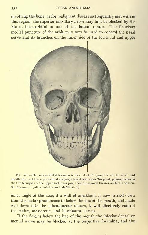
532 LOCAL ANESTHESIA
involving the bone, as for malignant disease so frequently met with in
this region, the superior maxillary nerve may first be blocked by the
Matas intra-orbital or one of the lateral routes. The Peuckart
medial puncture of the orbit may now be used to control the nasal
nerve and its branches on the inner side of the lower lid and upper
Fig. 162. The supra-orbital foramen is located at the junction of the inner and
middle thirds of the supra -orbital margin; a line drawn from this point, passing between
the two bicuspids of the upper and lower jaw, should pass over the infra-orbital and men-
tal foramina. (After Sobotta and McMurrich.)
inner angle of the face; if a wall of anesthesia is now carried downfrom the malar prominence to below the line of the mouth, and madewell down into the subcutaneous tissues, it will effectively control
the malar, masseteric, and buccinator nerves.
If the field is below the line of the mouth the inferior dental or
mental nerve may be blocked at the respective foramina, and the

THE HEAD, SCALP, CRANIUM, BRAIN, AND FACE 533
cervical nerves controlled by a subcutaneous line of infiltration over
the lower border of the inferior maxilla. Where two or more branches
Fig. 163. Regional anesthesia by way of infra-orbital foramen. (After Fischer.)
Fig. 1 64. Resulting area of anesthesia after blocking both infra-orbital nerves at infra-
foramen. (Braun.)
of the fifth nerve are involved in the field of operation it is always
preferable to block the gasserian ganglion when possible, but for

534 LOCAL ANESTHESIA
various reasons this may not be feasible we must then have alterna-
tive measures such as above.
In operations within the mouth which are not readily accessible,
as in lesions far back on the tongue, cheek wall, pillars of the pharynx,
etc., we often slit the cheek back to the ramus of the jaw, thus securing
Post. sup. dental nn.
Infra-orbital nn
Post. sup. dental tin.
Molars.
Incisors and Bicuspids Middle superior dental nn.
canines
Fig. 165. Areas of nerve supply of maxilla. Oblique shading: anterior superior
dental nerves (incisor and canine region). Horizontal shading: middle superior dental
nerve (bicuspid region); vertical shading: posterior superior dental nerves (molar
region). (After Fischer.)
greater room and freer access for work on these deeper parts. Such
incisions in the cheek, when properly closed, leave simply a linear
scar.
For the areas of distribution of the branches of the fifth nerve
to the maxillae, teeth, gums, and hard palate see Figs. 165-168, while
the points of emergence of the peripheral branches upon the face

THE HEAD, SCALP, CRANIUM, BRAIN, AND FACE 535
Nasopalatine notch
I
Anastomosis between anterior^
palatine and nasopalatinenn.
Anterior palatine n. 1 r**>J
Hard palate.
Fig. 1 66. Areas of nerve supply of palatine surface of maxilla, upper area, nasopalatinenerve. Lower area: anterior palatine nerve (molar region). (After Fischer.)
Mental foramen-
Area partly supplied by buccinator n.
Fig. 167. Area of nerve supply of anterior section of mandible. Dotted area : infe-
rior dental nerve. From the mental foramen emerges the mental nerve. The mucous
membrane in the molar region is partly supplied by sensory fibers of the buccinator
nerve. (After Fischer.)
and head is shown in Fig. 146. A study of this figure will prove
very useful for operations upon the peripheral soft parts. For the
innervation of the mucous passages and accessory sinuses a study of
Fig. 196 will be found very useful.

536 LOCAL ANESTHESIA
Operations upon the peripheral ends of the fifth nerve can be
easily done, as in resections for neuralgia. The foramina are first
exposed by an incision and an injection of solution No. 2 made into
the canal, advancing the needle very cautiously, injecting as it is
advanced so as to progressively anesthetize the deeper parts of the
nerve as they are reached by the needle; ^ dram of solution used in
this way will often flow back into the canal for some distance and
anesthetize the nerve far beyond the field of operation. After anes-
thetizing in this way, the bony opening to the canal can be enlarged
and the nerve reached further back and excised;divulsion may also
Lingual n.
Area supplied- -
by inferiordental andlingual nn.
Mandibular"canal
Mylohyoid n.
Area supplied by Lingual n.
inferior dental n.
Fig. 168. Area of nerve supply of lingual section of mandible. Dotted area: in-
ferior dental nerve. The mylohyoid nerve branches off at inferior dental foramen.
(Fischer.)
be practised in a limited way, but this method of anesthesia is not
suited to severe traction upon the nerve. For this practice the gan-
glion should be blocked.
Neuralgia in general, but especially facial neuralgia, was amongthe first conditions for which cocain was used, its employment in
this way being almost as old as the discovery of cocain, nevertheless
there have occurred apparent cures from the use of cocain alone;
it would, therefore, seem not inadvisable to bear it in mind, as the
single injection of medicinal doses cannot possibly prove harmful.
It must, however, further be remembered that its use for this pur-
pose has nearly always been in pure water, as well as in the presentcase reported. We know aquapuncture itself exerts this influence; it
may, therefore, in this and other cases not have been the cocain at

THE HEAD, SCALP, CRANIUM, BRAIN, AND FACE 537
all which accomplished the cure. The case in question is one byFitzmiller, which is said to have been very severe and in which all
other remedies had failed to afford relief (he does not state if aqua-
puncture had been used) : The patient a woman, aged thirty-two,
had suffered almost constantly with attacks often lasting a week in
length; a 0.17 per cent, watery solution, containing a few drops of
adrenalin, was used; of this solution a half Pravaz syringeful was
injected at the points of emergence of the supra-orbital, infra-orbital,
mental, and occipital nerves. It is stated that immediate relief
was afforded, the pain being as if "blown away," leaving only a
temporary feeling of numbness in the areas of distribution of the
nerves. During the next three weeks the return of the pain requirednine other injections, after which there was no further return whenlast seen six months later. This case is merely cited to call attention
to its possible use in this way.Since the introduction of novocain this agent has been similarly
used, either in plain water or in salt solution with adrenalin, and the
relief obtained from these injections has often been of prolongedduration. Its use for this purpose is not limited alone to facial
neuralgia, but seems to be equally beneficial when used elsewhere,
as for sciatica, when infiltrated around this nerve, and for neuralgia
of the spinal nerves as in intercostal neuralgia; here the solution had
best be used as a paraneural injection about the nerve-roots, thor-
oughly saturating the nerve-roots for one or two nerves above and
below the involved area;in the hands of the author this method has
often furnished prolonged relief.
Extirpation of the Tongue. Such mutilating operations are
rarely indicated under local anesthesia. While possible in the hands
of a skilful operator, the psychical effect is no doubt severe even uponthe most stoical. If the condition is one of malignancy, as is usually
the case, and well advanced, local methods of infiltration are contra-
indicated if they in any way encroach upon the diseased area.
Well-localized growths, when situated upon the anterior part of
the tongue, may be quite satisfactorily removed by creating a wall
of anesthesia across the tongue, proximal to the lesion, and involv-
ing its entire thickness; or, if the lesion is situated on the side near
the tip, a wall of infiltration anesthesia may be carried down the long
axis of the organ from its tip to beyond the lesion and joined at right
angles by a line of infiltration from the side; or, if limited to the
anterior two-thirds of the organ, the area of distribution of the
lingual, this nerve may be blocked on each side near the lingula.

538 LOCAL ANESTHESIA
To remove the entire organ the lingual and dental nerves should
be blocked on each side; this is preferable to blocking the inferior
maxillary higher up or to a ganglion injection, as the lingual receives
other nerve-fibers from its communications after it is given off from
its parent trunk.
As the base of the tongue receives fibers from the superior laryn-
geal and glossopharyngeal to its posterior one-third both of these
nerves must be dealt with. Block the superior laryngeal on both
sides by injections into the thyrohyoid membrane, as described in
the chapter on the Neck, and carry a line of subcutaneous infiltra-
tion through the soft parts above the hyoid bone from side to side,
to meet the ascending branches of the superficial cervical.
The glossopharyngeal is anesthetized by blocking the submucous
tissues below and in front of the tonsil toward the base of the tongueon each side; then, with a finger in the floor of the mouth, the long
needle is passed in beneath the maxilla, and, guided by the finger in
the mouth, the root of the tongue on each side is infiltrated.
After a few minutes' delay anesthesia should be complete and
the inferior maxilla divided at its symphysis, or an incision madeinto the mouth from beneath as preferred.
The author has never used local or regional methods for the
removal of tonsillar tumors, as, in
my opinion, this region when the
seat of malignant disease, as most
of these growths are, had best be
operated by general anesthesia;the
method as outlined below is
quoted from Hartel:
"Operations for Tonsillar Tu-
mors. In the sensory innervation
of the tonsils the following nerves
have a share the nervus maxil-
laris with the nervus palatinus
medius, the nervus lingualis with
Fig. 169. Schematic representationof the orbital planes according to Hartel.
(Hartel.)
the rami isthmi faucium, the nervus glossopharyngeus with the
ramus tonsillaris. While conduction anesthesia can easily be in-
duced in the nervus maxillaris and lingualis, this is impossible for
the trunk of the glossopharyngeus, because bone-points suitable for
its location are lacking. We must content ourselves with an infil-
tration of the peripharyngeal connective tissue situated laterally
to the tonsils, and reach this region from a puncture point which is

THE HEAD, SCALP, CRANIUM, BRAIN, AND FACE 539
situated in the most posterior point of the vestibulum oris laterally
from the ligamentum pterygomandibulare, which is there palpable;
from here the tissue situated laterally behind the tonsils is infiltrated
in a divergent direction. Also from the lateral region of the neck,
tonsillar tumors, which usually are connected with collections of
glands in this region, can easily be infiltrated accompanied bysimultaneous palpation from within."
The orbits are two quadrilateral pyramidal cavities at the upperand outer parts of the face, their bases directed forward and outward,
their apices backward and inward; their axes, passing through the
optic foramen, if continued backward, would meet over the bodyof the sphenoid bone. The base of this pyramid viewed from in
front presents somewhat of a rectangular appearance (Figs. 169
and 175), the plane of this rectangle sloping downward, backward,
and outward from the middle line of the face.
The roof of the orbit, triangular in shape, presents a perfectly
smooth concave surface, which slopes from the orbital margin first
upward, then downward, backward, and inward.
The superior margin presents at its inner extremity a depression
for the pulley of the superior oblique, sometimes marked by a spicule
of bone. External to this point on the margin, at the juncture of
the internal and middle thirds, is the supra-orbital notch or foramen.
On the lateral anterior surface, behind the orbital ridge, is the
depression for the lacrimal gland.
The inner wall, after the lacrimal groove is passed, presents a
smooth, irregular, slightly convex surface, directed almost directly
backward. This wall is extremely thin and paper-like in consistence,
forming the delicate bony external wall of the ethmoid and sphenoid
cells.
At the angle of the junction of the roof and inner wall, in the
suture between the frontal and the ethmoid bones, are seen the
anterior and posterior ethmoidal foramina; the anterior, situated
about the midpoint of the depth of the orbital wall, transmits the
anterior ethmoidal vessels and nasal nerve; the posterior foramen
is placed about midway between the anterior and the orbital foramen
and transmits the posterior ethmoidal vessels.
At the internal inferior angle is seen the lacrimal canal.
The floor presents an irregular smooth surface, sloping outward
and forward, slightly convex in its middle part, and concaved in
front as it approaches the orbital margin. It is crossed from before,
backward, and outward by the ethmoid maxillary suture. The bony

540 LOCAL ANESTHESIA
surface of the floor, like the inner wall, is extremely thin and forms
the roof of the antrum of Highmore.The outer wall, sometimes deficient at the sphenomalar articula-
tion, presents a fairly smooth, slightly concave surface, which slopes
sharply backward toward the foramen lacerum anterius; on it are
seen the orifices of the malar canals.
The sphenomaxillary fissure extends about two-thirds of the
distance along the angle of junction between the external wall and
floor of the orbit. It runs obliquely backward and inward to the
sphenomaxillary fossa. This fissure is widest in front, becomingnarrower and somewhat serpentine in direction behind.
It is formed above by the lower border of the orbital surface of
the great wing of the sphenoid, below and internally by the external
Lacrimal
Frontal
Fourth
Sup. division of third
Jfasal
Inf. division of third
Sixth
\c vein
Fig. 1 70. Relations of structures passing through the sphenoidal fissure. (After Gray.)
border of the orbital surface of the superior maxilla and a small
part of the palate bone.
At its internal extremity this fissure joins at right angles with
the pterygomaxillary fissure. This fissure forms a means of com-
munication between four fossae the orbital in front, sphenomaxil-
lary behind and internally, the temporal and zygomatic externally
and behind. Through this fissure pass the superior maxillary nerve
and its orbital branch, the infra-orbital vessels, and ascendingbranches from the sphenopalatine or Meckel's ganglion.
At the apex of the orbital fossa, below and external to the orbital
foramen, is seen the foramen lacerum anterius or sphenoidal fissure,
formed internally by the body of the sphenoid, above and internally
by the lesser wing of the sphenoid, below and externally by the greater
wing of the sphenoid.
Through this fissure pass the third, fourth, the three divisions of
the ophthalmic division of the fifth (lacrimal, frontal, and nasal),

THE HEAD, SCALP, CRANIUM, BRAIN, AND FACE 541
and sixth nerves, some filaments from the cavernous plexus of the
sympathetic, the orbital branch of the middle meningeal artery, a
Fig. 171. Ocular muscles viewed after removal of lateral wall of orbit: a, Eyeball;
b, optic nerve; c, c', eyelids; d, maxillary sinus; e, pterygoid plate;/, foramen rotundum;
g, roof of orbit; h, frontal sinus; i, supra-orbital nerve; k, septum orbitale; i,levator pal-
pebrae superioris; 2, 3, superior and inferior recti; 4, 4', portions of the cut external rec-
tus; 5, internal rectus; 6, inferior oblique; 7, insertion of superior oblique; 8, annular liga-
ment or tendon of Zinn. (Testut.)
Nasal nerve
I I
Rectus superior I
Annular tendon\ \
Lachrymal nerve
Ciliary ganglion and nerves
Fig. 172. Scheme of the ophthalmic nerve after Corning. (Braun.)
recurrent branch from the lacrimal artery to the dura, and the
ophthalmic vein.
The relative position of these structures is seen in Fig. 170.

542 LOCAL ANESTHESIA
In making deep orbital injections for the purpose of blocking those
branches of the trigeminus which pass through this fossa on their wayto other parts, we should try to select such routes of puncture as lie
along smooth and regular bony surfaces, using these surfaces as a
guide in approaching the deeper parts, and always keeping the needle-
point in close contact with the bone; in this way, by keeping well
toward the peripheral limits of the orbit, we are in the zone outside
Fig. 173. Base of skull with cranial nerves, from Arnold. Needles a and b same as Fig.
217, c, to nasal nerve, d to frontal and lacrimal nerves. (Hartel).
of the eye and its attached muscles. This idea of utilizing the orbit
as a means of approach to the intra- and retro-orbital nerve-trunks
may appear to the inexperienced as a hazardous procedure; this,
however, is a misconception, as the puncture under proper technic
should be a perfectly innocent "undertaking, except in the known
dangerous region of the orbit in its axis or at its apex. In extensive
operations upon the eye, as in enucleation, these regions are inten-
tionally invaded. (Figs. 171, 172 show the arrangement of the
nerves within the orbit.)

THE HEAD, SCALP, CRANIUM, BRAIN, AND FACE 543
The recognized routes of orbital puncture are:
1. Medial orbital route, first described by Peuckart for reachingthe nasal nerve (Figs. 173-181).
2. Lateral orbital route of Braun for reaching the frontal and
lacrimal branches of the ophthalmic (Figs. 173, 174).
3. Orbital route, through sphenomaxillary fissure to spheno-
maxillary fossa, to reach the second division of the fifth at the
foramen rotundum, the Matas route (Figs. 182-184).
Fig. 174. Horizontal section of skull in upper horizontal plane seen from above.
Needle c is at the ethmoidal foramina (nasal nerve); needle d at the superior orbita;
fissure (frontal and lacrimal nerves); i and 2, anterior and posterior ethmoidal foramina.
3, superior orbital fissure; 4, optic foramen. (Hartel.)
The retrobulbar methods of infiltration for bulbar operations
the methods of Seigrist, Lowenstein, and others.
5. In the method of Levy and Baudoin for reaching the oph-
thalmic division of the first through the orbit, the needle is entered
on the outer wall of the orbit at the level of the inferior extremity of
the external angular process of the frontal bone and advanced back-
ward and inward beneath the lacrimal gland, hugging the bone to a
depth of from 3^ to 4 cm.
Discussing the orbit and its various points of puncture, Hartel
has presented this subject with much thoroughness and detail, often
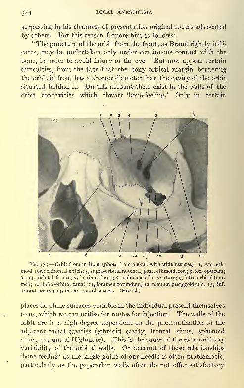
544 LOCAL ANESTHESIA
surpassing in his clearness of presentation original routes advocated
by others. For this reason I quote him as follows:
"The puncture of the orbit from the front, as Braun rightly indi-
cates, may be undertaken only under continuous contact with the
bone, in order to avoid injury of the eye. But now appear certain
difficulties, from the fact that the bony orbital margin bordering
the orbit in front has a shorter diameter than the cavity of the orbit
situated behind it. On this account there exist in the walls of the
orbit concavities which thwart 'bone-feeling.' Only in certain
Fig. 175. Orbit from in front (photo from a skull with wide fissures): i, Ant. eth-
moid, for.; 2, frontal notch; 3, supra-orbital notch; 4, post, ethmoid, for.; 5, for. opticum;
6, sup. orbital fissure; 7, lacrimal fossa; 8, malar-maxillaris suture; 9, infra-orbital fora-
men; 10, infra-orbital canal; n, foramen rotundum; 12, planum pterygoideum; 13, inf.
orbital fissure; 14, malar-frontal suture. (Hartel.)
places do plane surfaces variable in the individual present themselves
to us, which we can utilize for routes for injection. The walls of the
orbit are in a high degree dependent on the pneumatization of the
adjacent facial cavities (ethmoid cavity, frontal sinus, sphenoid
sinus, antrum of Highmore). This is the cause of the extraordinary
variability of the orbital walls. On account of these relationships'
bone-feeling' as the single guide of our needle is often problematic,
particularly as the paper-thin walls often do not offer satisfactory

THE HEAD, SCALP, CRANIUM, BRAIN, AND FACE 545
resistance. We need, therefore, here also the adherence to certain
instruction for direction as well as instruction for definite depth.
"The deepest concavities of the orbit lie above behind the margosupraorbitalis and outward under it, while the medial wall (lamina
papyracea), the lateral wall (orbital surface of the malar bone and
great wing of the sphenoid bone), and the medial part of the inferior
wall (orbital surface of the upper jaw) usually afford plane relation-
ships. We obtain, therefore, a medial, a lateral, and an inferior planesurface of the orbit. From the medial plane surface we reach the
foramina ethmoidalia, from the lateral plane surface the fissura
orbitalis superior with the entrance of the nervous .ophthalmicus,
from the inferior plane surface the nervus maxillaris and the foramen
rotundum.
"Concerning the quality of the plane surfaces, Table II, Nos. 15,
16, and 17, gives explanation. According to that, the medial planesurface offers the most favorable relationships (80 per cent, com-
pletely plane way; in the other 20 per cent, a very slight concavity or
convexity of the planum, not interfering with puncture). Less
favorable relations are found in the lateral and the inferior plane
surfaces.
"As the concavities of the orbit, as we have seen, lie in its anterior
part close behind the orbital margin, in every case information con-
cerning the quality of the plane surfaces is possible by palpation.
Therefore, before the puncture we can seek out the most favorable
place by examination, and, by shoving the bulbus to one side, we can
carry the needle into the depth, even if the relations are not entirely
plane, without injuring the bulb.
"If we now observe the orbital margin (Fig. 175) in the choice of
our puncture point, its configuration in the individual is so varied
that even to-day the anatomists are not united concerning the
designation of the margins and corners. For us it is important to
note definite points palpable through the skin. They are these : the
sutura malar-maxillaris on the inferior margin, the sutura malar
frontalis above laterally, the lacrimal fossa, as well as the usually
palpable incisurae supra-orbitalis and frontalis. We compare the
orbital margin to an obliquely placed rectangle (c, e, b,f, Fig. 169),
whose corners are formed laterally above (b), by the sutura malar
maxillaris, toward the median line; above (e) by the incisura frontalis,
toward the median line below (c) by the lacrimal fossa, laterally below
(/) by the rounded orbital margin. In most cases three of these
corners are palpable. Now this rectangle lies diagonally, so that the
35

546 LOCAL ANESTHESIA
horizontal line drawn through the superior outer angle (6) (sutura
malar maxillaris) meets the opposite short side in the middle (a),
and the horizontal line, drawn from the inner inferior angle (c)
(middle of the lacrimal fossa) ,the middle of the outer short side (d) .
We designate these two lines ab and cd as superior and inferior hori-
zontal lines of the orbit, and we shall find that the horizontal planes
passing through these lines offer the following important relations
to direction for the puncture of the orbit:
"If we observe the orbit exactly from the front, so that our direc-
tion of vision corresponds to the central axis of the orbit, then we
perceive as the middle point (Fig. 176) of the orbital infundibulum
Fig. 176. Orbit from in front, same specimen as Fig. 175 with the horizontal planes
outlined. (Hartel.)
the inferior wide part of the fissura superioris. It lies exactly be-
tween the two horizontals of the orbit. In the upper horizontal plane
lie, from the outside toward the median line, the upper part of the
fissura orbitalis superioris, the foramen opticum, the foramina
ethmoidalia, posterior and anterior. In the lower horizontal plane
lies the foramen rotundum. If we keep our needle in the horizontal
planes, then we assuredly avoid the puncture of the broad inferior
end of the fissura superioris, which contains the nerves of the muscles
of the eye and large veins. On puncture in the upper plane we
encounter laterally the place of entrance into the orbit of the nervi
frontalis and lacrimalis, medially the place of entrance of the nasal

THE HEAD, SCALP, CRANIUM, BRAIN, AND FACE 547
nerve. In this connection precaution is to be observed only in so
far as danger exists of injury of the nervus opticus by going too deep.In the lower horizontal plane we encounter the nervus maxillaris andits orbital branch, the temporomalar.
"Fig. 177 shows the orbit with bulb, conjunctival sac, palpebralfissure (after Merkel), as well as the horizontals ab and cd specified byus, and the puncture-points for medial (i) lateral (2) and inferior
(3) orbital puncture.
Fig. 177. Orbit and eye, showing outlines or orbital margin. Heavy circle shows
limits of the bulb; dotted circle, limits of conjunctival sac. a-b, Upper horizontal plane;
c-d, lower horizontal plane; i, median point of puncture for injecting nasal nerve; 2,
point of puncture for injecting frontal and lacrimal nerves; 3, point of puncture for
orbital injection of foramen rotundum. (After Merkel.)
"If we ask ourselves how the palpebral fissure is related to our
puncture points, then we must remember this, that only the inner
angle of the palpebral fissure is a fixed point, while the external angle,
when the eye is opened, moves upward several millimeters. The
inner angle of the palpebral fissure lies in the region of the lacrimal
fossa, and the palpebral fissure occurs varying in its height, in anycase always in the region situated between the two horizontal planes
of the orbit. With the eye moderately opened the superior and in-
ferior palpebral margins should correspond to the two horizontal
planes. Therefore, the puncture point for the medial puncture as
well as the puncture point for the lateral puncture lies above the
palpebral fissure, as is evident from Fig. 177.

548 LOCAL ANESTHESIA
"Further, we must bear in mind that the central axes of the two
orbital cavities converge posteriorly; consequently, the lateral orbital
wall runs diagonally from in front backward toward the median line
at an angle that deviates about 45 from the sagittal. The projec-
tions of the straight lines drawn along the outer orbital walls meet in
the region of the dorsum sellae at right angles. The medial orbital
walls, on the other hand, run approximately sagittally and divergebut little from behind forward.
"If we observe the rules here mentioned, then on making the
orbital punctures we can assuredly avoid an injury of the bulb and of
the nervus opticus. In practice we displace the bulb with the finger
from the point of entrance, and carry the needle into the depthbetween the orbital wall and the finger-tip that protects the bulb.
By this means we keep the needle in the region of the horizontal
planes mentioned, and guard against the point of the needle entering
the region bounded by both planes and from getting accidentally
into the point of the orbital infundibulum. On lateral orbital punc-ture the axis of the needle is at 45 from the sagittal direction, on
medial and inferior puncture approximately saggital.
"If the injury of the bulb and nervous opticus is thus technically
avoidable, this, however, is not true with equal certainty of the
vessels of the orbit. These are related in detail as follows : On medial
orbital puncture we come in contact with the terminal branches of
the ophthalmic artery, while the trunk of the artery itself lies within
the muscular infundibulum of the orbit and is avoided by keeping in
contact with the bone. On lateral puncture we may encounter the
lacrimal artery; on puncture of the foramen rotundum the anterior
infra-orbitalis and the communication of the ophthalmic vein with the
deep veins of the region of the cheek. One may say that, with cau-
tious work and by constant contact with the bone, the appearance of
hematoma during the orbital punctures is very rare. However, it is
not to be excluded, and while these puncture-hematoma are accom-
panied by no danger, yet they cause temporary exophthalmus and
leave behind suffusions of the lids and of the conjunctiva which are
visible for several days. For this reason we should use nerve punc-tures in the orbit only for the anesthesia of major procedures.
"i. Medial Orbital Puncture (Peuckart Route). Anesthesia of the
Nervi Ethmoidales (Figs. 173, 174^, 181). Where the upper horizon-
tal plane touches the wall of the orbital cavity, the foramina eth-
moidalia and the foramen opticum lie in one line. In front the same
plane meets the root of the nose. The puncture-point, therefore, lies

THE HEAD, SCALP, CRANIUM, BRAIN, AND FACE 549
on the inner orbital margin at the height of the root of the nose. Theneedle is entered in an exactly horizontal and approximately sagittal
direction in constant contact with the bone. The distance of the
foramen ethmoidale anterius from the inner margin of the orbit amounts,
according to Table II, No. 19, to from 15 to 22; on the average, 18.5mm.
; therefore, for the anesthesia of the nervus ethmoidalis anterius
(nasal nerve) we will carry the cannula in to the depth of about 2 cm.
The nervus ethmoidalis anterius (nasal nerve) is distributed to the
superior and anterior parts of the nasal mucous membrane (compareTable I) and to the tip of the nose. In order to strike the nervus
ethmoidalis posterior, which is distributed to the ethmoidal cells and
the sphenoidal cavity, we must carry the needle to greater depth.The foramen ethmoidale posterius lies at a distance from the inner
margin of the orbital cavity (see Table II, No. 20) of from 29 to 42;
on the average, 34 mm. This is not so typically placed and constant
as the foramen ethmoidale anterius; it is often met with double.
Concerning its relation to the foramen optieum which, as we saw, is
situated in the same plane, the following is true : In a series of cases
the medial orbital wall curves forward somewhat in consequence of
pneumatization of the small wing of the sphenoid bone, so that behind
the foramen ethmoidale posterius the cannula strikes against bonyresistance. But this is the case only in half of the skulls (Table II,
No. 1 8). The distance of the anterior margin of the foramen opti-
eum from the inner margin of the orbital cavity amounts to 37 to 47;
on the average, 40.8 mm.; in i case it amounted only to 33 mm.
(compare Table II, No. 21). If we compare with this the values
found for the depth of the foramen ethmoidale posterius, then we see
that on anesthesia of the nervus ethmoidalis posterius we come into
dangerous proximity to the optic nerve. Therefore, we do well to
carry the inner orbital puncture not deeper than 3 cm., and to forego
the deeper penetration to the diffusion of the injected solution.
"2. Lateral- Orbital Puncture (Braun Route). Anesthesia of the
Nervi Frontalis and Lacrimalis (Figs. 173, 174, 181). In the upperhorizontal plane of the orbit is the lateral end of the fissure orbitalis
superior with the passageway of the frontal and lacrimal nerves. Wereach this point by the lateral orbital puncture after Braun, and par-
ticularly by a puncture-point which is situated at the upper lateral
corner of the orbital margin (sutura malar-frontalis), or, with poor
development of the lateral plane surface, somewhat deeper on the
outer orbital margin. If we proceed from here with the needle in
horizontal position, and deviating from the sagittal direction about

55
45 toward the median line in constant contact with the bone into the
depth of the orbit, we strike the outer end of the superior fissure, and
on the further side of this in most cases encounter bone-resistance on
the superior roof of the orbit (small wing of the sphenoid bone).
Only with a wide superior fissure does the danger exist that the can-
nula without resistance may penetrate into the cranial cavity. Ac-
cording to Table II, No. 22, we find this relationship in 14 per cent, of
the skulls. The distance of the outer end of the superior fissure from
the lateral orbital margin is very variable; according to Table II, No.
Fig. 178. Left half of sphenoid bone seen from in front: i, Lesser wing; 2, superior
orbital fissure; 3, orbital surface of greater wing; 4, foramen rotundum; 5, groove for sec-
ond division of fifth nerve; 6, sharp bony edge; 7, anterior surface of pterygoid process;
8, sphenoid cells; 9, vidian canal. (Hartel.)
23, it amounts to from 27 to 40 mm. ;on the average, 33.5 mm. This
relationship makes the lateral puncture of the superior orbital fissure
somewhat uncertain, so that I do not believe that it will ever come
into consideration for the injection in cases of neuralgia. For local
anesthesia I advise penetration to a maximum depth of about 3 cm.
"3. Axial Puncture of the Foramen Rotundum. Orbital Way to
the Second Branch of the Trigeminus (the Matas Route, Author)
(Figs. 182, 183). If we observe the anterior surface of the sphenoid
bone, which is turned toward the orbit (Fig. 178), then we discover
the following details: the foramen opticum, the superior fissure, and

THE HEAD, SCALP, CRANIUM, BRAIN, AND FACE 551
underneath this a surface shaped like an irregular triangle, the ante-
rior wall of the process pterygoideus. This surface, which we wish to
name 'planum pterygoideum,' is limited above, opposite the orbital
surface of the large wing of the sphenoid bone, by a clearly percepti-
ble sulcus. This sulcus forms the path by which the nervus maxillaris,
leaving the foramen rotundum, reaches the sulcus infra-orbitalis.
Likewise, on the back of the upper jaw, a groove lies opposite this
sulcus, so that, by the closing together of these two half-grooves, a
kind of canal is formed. At the back end of this canal, in the bodyof the sphenoid bone, lies the foramen rotundum, at its anterior end,
in the upper jaw, the canal infra-orbitalis. The lower outer marginof the planum pterygoideum forms a sharp bone-corner, the limiting
ridge; beyond this lies the fossa infratemporalis.
Fig. 179. Fig. 180.
Figs. 179 and 180. Front and lateral views of needle in position in the orbital injec-
tion of foramen rotundum. Front view shows long axis of needle reaching upper inner
angle of the orbit; on lateral view it is seen to reach upper margin of ear. (From a
cadaveric specimen.) (Hartel.)
"If on the skull we carry a cannula from the lateral part of the
inferior orbital margin sagittally into the depth, then we arrive
through the fissura.inferior at the canal just mentioned, between the
sphenoid bone and the upper jaw, at the end of which canal lies the
foramen rotundum. Previously, however, the needle encounters
bone-resistance on the planum pterygoideum of the sphenoid bone.
If we now feel with the point of the needle along this resistance up-
ward and medially, then we must reach the foramen rotundum. The
supposition, by all means, is that the inferior fissure is wide enoughand is not too tortuous. On this account, the way described is,
according to our examinations (Table II, No. 24), accessible only in
89 per cent, of the skulls; in the rest of the cases it is obstructed by the
inferior fissure.
"The distance of the foramen rotundum from the inferior orbital

552 LOCAL ANESTHESIA
margin amounts (Table II, No. 25) to from 39 to 51 mm.; on the
average, 45.4 mm. For the direction of the cannula the following is
of value: the foramen rotundum never lies higher than the inferior
horizontal plane of the orbit. The cannula, when carried into the
foramen rotundum on lateral observation, points to the superior
margin of the auricle (Fig. 179); on observation from in front, it
points with much shorter axis to the inner superior angle of the orbit,
the incisura frontalis (Fig. 180).
"For the orbital puncture of the foramen rotundum the following
is important: The foramen is very narrow, and is completely rilled
by the nervus maxillaris, hence, on introducing the cannula we have
to reckon with the resistance of a tolerably firm mass of tissue, so
Anterior and posterior ethmoidal foramina
Fig. 181. Median and lateral orbital injections. (Braun.)
that the injection demands a certain pressure. If this resistance is
lacking and the needle glides easily into the depth, then we must
suppose that we have gone beyond the foramen rotundum into the
superior orbital fissure. In the living subject the most important
guide for our puncture is the subjective statement of the patient re-
garding radiating pain in the region of the second branch of the trige-
minus." (See Table I.)
The axial injection of the foramen rotundum, the Matas route
through the orbit and sphenomaxillary fissure, has been erroneously
credited by Braun, Hartel, and others to Payr; this, however, is an
error, as the conception of this method of approach and its first ap-
plication undoubtedly belong to Prof. Matas, who first used it in 1898,
and it was published by him in his report on "Local and Regional
Anesthesia," etc., to the Louisiana State Med. Soc., April, 1900.
Quotations from this report are given later on.

THE HEAD, SCALP, CRANIUM, BRAIN, AND FACE 553
This successful application stimulated other efforts in this direc-
tion, and much of the work which appeared in the few years follow-
ing along these lines undoubtedly received the idea and stimulus from
this procedure.
Fig. 182. Matas' intra-orbital route to foramen rotundum. (Braun.)
In studying the orbit, with a view of the application of the various
methods, we see that the foramen rotundum is concealed from view
Fig. 183. Needle in position in Matas' intra-orbital injection within foramen rotundum.
(Braun.)
just below the floor of the orbit, and that if this plane were used as a
means of reaching it, the needle, if directed toward the apex of the
orbit medially along the floor, would pass into the superior orbital

554 LOCAL ANESTHESIA
fissure, and meeting no bony resistance here may, if advanced too far,
pass backward into the cranial cavity. This route is discussed byHartel (Fig. 184).
This would seem a more dangerous route, and not likely to lead to
the foramen rotundum, but above it.
The original route, as advocated by Prof. Matas, traverses the or-
Fig. 184. Horizontal section of left half of skull in lower horizontal orbital plane,
seen from above, with needle in foramen rotundum: i, Infra-orbital sulcus; 2, zygomati-
co-maxillary suture; 2, infra-orbital fissure; 4, foramen rotundum; 5, foramen ovale.
(Hartel.)
bit for but a short distance, as the needle soon passes out of this cavity
into the sphenomaxillary fissure (Figs. 182, 183).
If the sphenomaxillary fissures are observed, they will be seen to
run at right angles to each other and about on a horizontal plane;
their axes if continued back would meet over the body of the sphenoid
bone, and if projected forward would emerge at the inferior external
angle of the orbit; also, that the axis of this fissure, if raised to a
slightly elevated plane, would pass through the orbital foramen or
superior orbital fissure at the apex of the orbit.
The axis of the foramen rotundum, if viewed from within the

THE HEAD, SCALP, CRANIUM, BRAIN, AND FACE 555
skull, passes downward, forward, and outward, and passes throughthe sphenomaxillary fossa, and for a short distance through the
sphenomaxillary fissure, and emerge upon the rim of the orbit just
internal to its inferior external angle, at a distance of from 4 to 5 cm.,
varying somewhat in different skulls.
It will be seen from the above that undoubtedly the safest method,as well as the surest of approach to the foramen rotundum is by the
Matas route, for if a plane above this route is taken the needle maypass on without resistance into the cranial cavity, but if the route is
followed by passing through the sphenomaxillary fissure, the needle-
point impinges upon the body of the sphenoid bone, on the posterior
surface of the sphenomaxillary fossa; and if unable to enter the fora-
men rotundum through insufficient play of the needle, due to the
narrowing of the sphenomaxillary fissure, it is at least in immediate
contact with the nerve; but if, after reaching the posterior wall of the
fossa, the foramen is felt for, by gentle manipulation immediatelyaround the axis of the needle, the foramen may often be entered, when
the needle may be advanced a few millimeters further and the in-
jection made.
The proof of contact of the needle with the nerve is recognized
by the radiating pains along the branches of this nerve, felt on the
cheek, in the upper teeth, and in the nose.
Having reached with certainty the position of the nerve, 2 c.c. of
a 2 per cent, novocain-adrenalin solution are injected, or if the point
of the needle be less accurately placed the injection of a slightly
larger quantity of a weaker solution (i to 2 drams of a i per cent.)
will flood the sphenomaxillary fossa and reach the nerve and all its
branches. According to the studies of Hartel the foramen rotun-
dum is accessible in about 89 per cent, of skulls.
The following case illustrates the early use of this method by Prof.
Matas, and is his first report of the use of this route ("Local and Re-
gional Anesthesia," etc., Proc. Louisiana State Med. Soc., 1900):
"A white man, laborer, aged forty-eight, addicted strongly to alcohol for years and
suffering with advanced arteriosclerosis, was admitted to Ward 7, Charity Hospital
(April 29, 1899), for treatment of a recurrent epithelioma of the palate, involving the
anterior alveolar arch and both upper maxillary processes. The neoplastic infiltration
extended to the right half of the palate and along the entire incisor region to the first
right bicuspid; the anterior half of the hard palate also presented a large ovoid swelling
caused by malignant periosteal invasion. I decided that I would try to move the entire
hard palate, including both palatine processes of the upper maxillae, the floor of the an-
trum, and the septal cartilage of the nose. A hypodermic of morphin, Y 8r-> was given
twenty minutes before the operation. In order to anesthetize the maxillaries and the
palate, the sphenopalatine fossae were filled with a No. i Schleich solution, introduced by

556 LOCAL ANESTHESIA
a long needle through the sphenomaxillary fissures. The needle was directed as closely
as possible through the fissure in the right orbit toward the infra-orbital nerve as it enters
the infra-orbital canal. In this way it was expected that not only the entire superior
maxillary division of the trigeminus could be anesthetized, but that Meckel's ganglion
with its palatine branches would be 'blocked' by the anesthetic. In a few minutes we
tested the sensibility of the cheeks, lips, and alse of nose, and were gratified to find the
entire cutaneous distribution of the infra-orbital had been completely anesthetized on
the corresponding (right) side. Encouraged by this result, the left sphenopalatine fossa
and infra-orbital nerve were treated in the same manner with identical results. Fully
50 minims of Schleich's No. i solution, reinforced by 25 minims of i per cent, cocain, were
injected into each sphenopalatine fossa. The nasal septum, which is supplied by the
nasopalatine and the nasal branch of the ophthalmic, was controlled by a separate infil-
tration, a long needle being introduced through the frenulum of the upper lip into the
root of the columna and septal cartilage of the nose. The anesthesia of the posterior pal-
atine nerves was also reinforced by direct infiltration in the palate. When the last
injection had been completed the patient said that his palate and face felt entirely
"numb," and gave the impression of a 'dead block of flesh' wedged in his head. Theanesthesia of the jaws was then tested by extracting a perfectly sound right canine which
was firmly implanted in its socket. The patient was surprised when he saw his tooth,
saying he had not felt the least pain in its extraction. The upper lip was then divided
in the median line and detached from the nose by two lateral incisions, which were car-
ried along the lower border of the columna nasi and to the nasolabial groove. The lips
were then dissected away from the gums and jaws as far back as the tuberosities of
the maxilla?. The two halves of the upper lip were then reflected outward, and then
held out of the way with loops of strong silk which acted as retractors. A very sharpMcEwen's chisel was then driven by hand into the body of the right maxilla on a
level with the floor of the nose. With a few sharp strokes of the mallet the palatine
process, including the tuberosity, was divided, and the antrum was exposed; the
same process was repeated on the other side, and the separation of the septum nasi
from the jaws completed the line of osseous section. In this manner the lower half oi
both upper jaws and the entire hard palate with the attached growth were mobilized
and displaced downward en bloc, the connections with the soft palate being severed
with a long pair of strong curved scissors. After the removal of the palate, both antral
cavities and nasal fossae were widely exposed. The bleeding was very profuse in the
last stage of the operation when the palate was being detached, the palatine arteries
spurting vigorously. A large tampon of iodoform gauze, impregnated with compoundtincture of benzoin, was immediately packed into the palatine region and promptlyarrested the hemorrhage. The lining mucosa of the right antrum was subsequentlyremoved in its entirety. During the intra-oral part of the operation the patient's head
was kept low, in Rose's position. Throughout the whole procedure, which lasted over
forty minutes, the patient gave us great assistance by spitting out clots and altering
the position of the head as we directed. He said that while the chisel was being used he
felt the jar of the instrument, the detachment of the vomer gave him some pain, but
what gave him more alarm than anything else was the sight of the blood that he spatout."
"In this case the preliminary injection of morphin was of decided
assistance in diminishing psychic anxiety. Fully 180 minims of
Schleich's No. i (% of i per cent.) solution and 60 minims of a i percent, solution were used in the operation. After the removal of the
palate the lip was still completely anesthetized, andthe facial (cuta-
neous) and intra-oral (mucous) sutures were introduced without pain.

THE HEAD, SCALP, CRANIUM, BRAIN, AND FACE 557
The patient was very much exhausted by the ordeal he had undergone,
but, after taking a good drink of whisky and a hypodermic of strych-
nin, he sat up, and said that, apart from the jarring of the chisel and
the excitement of the operation-, he had suffered comparatively little
pain. The pulse was no and there was little shock."
The use of any but straight needles in making these deep orbital
punctures is to be advised against, as cautioned by Braun; however,these are very useful for retrobulbar infiltration according to the
Lowenstein technic, but should not be used for other injections, and
in making any deep injections it is best to use specially constructed
needles and syringes. The needle should be of small caliber and
strongly made and with a short beveled point, combined with aids
for determining and controlling distance. Such an outfit, as designed
by Hartel, is seen in Fig. 238, and can now be obtained from American
manufacturers.
Such injections, made according to the proper technic, should not
effect the innervation of the bulb, optic nerve, ciliary nerve, or its
ganglion ;to reach these the injection should be made retrobulbar.
Injection of Inferior Dental Nerve. The following excellent de-
scription of the position of the inferior dental foramen and the method
of making a paraneural injection here has been taken from Fischer.
This, one of the most accessible of the cranial nerves, is not as
accurately reached as one might suppose, and in the hands of even
skilful surgeons, who resort to this procedure but occasionally, manyfailures may occur. It is far easier to make an accurate injection
in contact with the parent trunk (third division of the fifth) at the
base of the skull than to accurately inject this nerve here, both for
purposes of anesthetization as well as alcoholization in cases of
neuralgia.
In the one case the end may be obtained by using a large amount
of anesthetic fluid, but this as it diffuses in all directions producesa very unpleasant sense of paralysis of the throat, which is quite
terrifying to some patients, particularly if it occurs during the prog-
ress of an operation.
An alcohol injection, if too liberally made at this point, may result
in a more or less prolonged trismus through the action of the alco-
hol upon the masticatory muscles.
The inferior dental or oblique mandibular foramen in the internal
surface of the ascending ramus permits the passage of the inferior
dental nerve, which, with the inferior dental artery, passes forward
in the dental canal of the mandible as far as the mental foramen,

558 LOCAL ANESTHESIA
where it divides into two terminal branches, incisor and mental.
For the technic of injection in the oblique foramen the relationship
of the body of the jaw to the ascending ramus and that of the muscles
to the foramen is of vital importance.
In adults the ascending ramus begins a little behind the third
molar, sometimes in an abruptly ascending surface. At its basis,
which must be regarded as resting upon the alveolar process, the as-
cending ramus, in front view, shows an outer buccal anterior ridge,
representing the last ascending portion of the external oblique line.
(See Figs. 185-187). About 0.5 cm. inward and backward of this
Sigtnoid notch
\ Plerygoid depression
Condyloid process
Inferior dental foramen ~~^^Internal oblique line*.
'Angle
Mylohoidgroove
Internal genitaltubercles
Mylohyoidridge
Fig. 185. Side view of inner surface of right half of mandible. The long arrow indi-
cates the direction in which the needle should be pushed forward over the lingula. Thedotted circle indicates the area of injection. (Fischer.)
line runs a ridge bordering the lingual surface, the internal oblique
line, which gradually loses itself in the posterior section of the coro-
noid process. Between these two lines in the bony surface is situated
a more or less pronounced deep groove, which we might call the
retromolar fossa (Fig. 186). Above this fosa the mucosa is slightly
depressed, in what might be called the retromolar triangle.
About the middle of the internal surface portion of the ascendingramus the large inferior dental or mandibular foramen is situated,
extending downward and forward, at the same time marking the
termination of the mylohyoid groove which ascends from below ante-
riorly to above posteriorly. The orifice of the foramen itself is more

THE HEAD, SCALP, CRANIUM, BRAIN, AND FACE 559
or less protected anteriorly by a spicule of bone varying in size, the
mandibular lingula (Figs. 185-187).
This lingula may be developed as a pointed plate of bone, or as a
tongue-like cover, or only as a thickened process on the anterior
margin. Sometimes the lingula is connected with the lower free
margin of the orifice of the foramen by a small spicule or bridge.
Condyloid process
Inferior dental
foramen
Mylohyoid ridge
Coronoid process
Internal obliqueline
External obliqueline
Internal obliqueline
-Menial foramen
Fig. 186. Relationship of the ascending ramus to the body of the jaw. The arrow
indicates the direction in which the syringe should be advanced to the inferior dental
foramen. (After Fischer.)
The foramen itself, in adults, is always situated above the alveolar
ridge and in a horizontal plane, about 1.5 cm. from the anterior ridge
of the jaw (the external oblique line) (Figs. 187-189).
"The two halves of the mandible, when viewed from in front,
gradually diverge toward the angle, so that the inner surface of the
angle with the mandibular foramen is inclined posteriorly and
pharyngeally, and appears to be entirely covered by the internal
oblique line. (See Figs. 186, 187, 190, 101).

56 LOCAL ANESTHESIA
"Position of Syringe. The line of the body of the mandible is not
horizontally continuous in a straight line to the ascending ramus,
Coronoid
Pr,
Condyloid process
'Angle
Mylohyoid ridge Submaxillaryfossa
Fig. 187. Variations of the inferior dental foramen at different ages: A, Mandibleof a child, aged seven years (the needle should be inclined slightly downward); B, man-dible of a youth, aged eighteen years; C, mandible of a male adult, aged thirty years.The arrows indicate the direction of the needle. (After Fischer.)
but presents a lateral bulging at the angle, so that "the internal surface
of the ascending ramus is not parallel with the lingual surface of the
body of the jaw. (See Figs. 186, 190, 191.) The ramus opens pos-

THE HEAD, SCALP, CRANIUM, BRAIN, AND FACE 561
teriorly. (See Figs. 188, 189, 190, 191.) If, therefore, the oblique
foramen is to be reached, we must never advance posteriorly parallel
with the teeth (Figs. 192, 193), but with the internal surface of the
Fig. 188. Front view of position of syringe in mandibular anesthesia: i, Internal
oblique line; 2, external oblique line; 3, insertion of needle about i cm. above masticatingsurface of molars. (After Fischer.)
ramus, at an acute angle to the plane of the teeth (Figs. 188, 192,
193, 194). If the direction of the ascending ramus is projected ante-
Fig. 189. Position of needle in mandibular anesthesia: i, External oblique line; 2
internal oblique line; 3, position of needle at superior margin of lingula; 4, most suitable
length of needle behind lingula (a further advancement would result in failure); 6,
position of needle, i cm. above level of masticating surfaces of molars; 7, lingula; 8
inferior dental foramen. (After Fischer.)
riorly, the line will meet with the other side in the canine region, be-
tween the canine and the bicuspids (Figs. 188, 189, 192, 193, 194).
Thus, in order to reach the inferior dental foramen the syringe must36

562 LOCAL ANESTHESIA
be rested behind the canine on the opposite side. (See Figs. 188, 192,
193, 194.) The foramen in adults is situated at a higher level than
in children. The horizontal direction of the needle must, therefore,
be modified in children by slightly lowering it posteriorly and pharyn-
geally in order to reach the foramen directly. (See Fig. 187, A, B, C)."Character of the Tissues. The character of the tissues encoun-
tered is most favorable for injection in the oblique foramen.
Angle
Clique line
Line of reflection of
mucous membrane
Body of mandible
Menial foramen
Mental (incisor)
foramen
Canine fossa Mental protuberance
Fig. 190. Points of injection for mucous anesthesia in external surface of mandible.
The crosses indicate points of injection; small arrows, direction of needle; two large
arrows, direction of needle for injection in mental foramen and fossa. On the internal
surface of the ramus are marked the points for injection at mandibular foramen. (After
Fischer.)
"The temporal and external pterygoid muscles are inserted
above, the internal pterygoid below, the foramen, leaving the close
proximity of the foramen free from muscular fibers. (See Fig. 195.)
"Instead we find considerable accumulations of loose interstitial
connective and adipose tissue, which readily absorbs and retains the
injected solution. (See Fig. 196.)
"This cushion of tissue is situated about i or 2 cm. above the al-
veolar process.

THE HEAD, SCALP, CRANIUM, BRAIN, AND FACE
"Tecknic of Injection. With the left index-finger the anterior
portion of the base of the ascending ramus is palpated, the patient's
mouth being opened widely. Two very marked bony ridges are
felt here, one anterior external, the external oblique line, and one pos-terior internal, the internal oblique line (Figs. 188, 189, 192, 193).
Between these two lines at the root of the ascending ramus a shallow
bony grove is situated, which might be properly called the retro-
molar fossa, into which the palpating finger-tip sinks (Figs. 188, 192,
193, 194.) The mucous membrane is caved in over this fossa in some-
Fig. 191. Lingual points of injection for mucous anesthesia of mandible. Crosses
indicate points of injection; arrows, direction of needle; black line of dashes, the angle of
the ramus to the body of the jaw. (After Fischer.)
what triangular shape; Braun, therefore, calls it the retromolar
triangle.
"The internal oblique line is fixed with the finger-nail, and the
needle inserted close to the nail into the mucosa near to, yet not im-
mediately at, the edge of the bone (Figs. 188, 192, 193).
"The syringe is pushed forward horizontally and posteriorly from
the canine, on the opposite side along the internal surface of the man-
dibular half to be anesthetized (Figs. 188, 189, 192, 193, 194).
"The needle should be introduced to a depth of not more than
from 1.5 to 2 cm. under the mucosa, lest it advance too far beyond

564 LOCAL ANESTHESIA
the foramen and the correct point for the disposition of the solution
be missed.
"The injecting solution is then deposited, beginning to inject soon
after insertion of the needle, in order to anesthetize the lingual nerve
at the same time (if this be desirable). The bulk of the solution,
however, should be injected in the mandibular foramen.
"Insertion of the Needle. The point of the injection is selected so
that the needle is introduced in the molar triangle, about i cm.
Fig. 192. Position of syringe for injection at mandibular foramen: ix, External
oblique line; 2X, retromolar fossa; 3x, internal oblique line; 4, mandibular foramenbehind lingula; 5, incorrect position of syringe, parallel to teeth. (After Fischer.)
above the level of the masticating surfaces of the molars (Figs. 192,
193, 195, 197); in children and youthful persons, advancing a little
farther posteriorly while slightly lowering the needle;in old persons,
slightly raising the long needle (Fig. 187).
"Difficulties. The technic of this form of injection offers some
difficulties, which, after some practice are easily overcome; above
all, it must be observed that the insertion of the needle is made not
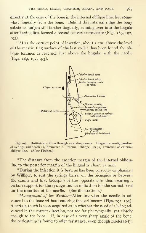
THE HEAD, SCALP, CRANIUM, BRAIN, AND FACE 565
directly at the edge of the bone in the internal oblique line, but some-
what lingually from the bone. Behind this internal ridge the bonysubstance bulges still farther lingually, running over into the lingula
after having first formed a second convex excrescence (Figs. 189, 192,
I93)-
"After the correct point of insertion, about i cm. above the level
of the masticating surface of the last molar, has been found the ob-
lique foramen is reached, just above the lingula, with the needle
(Figs. 189, 192, 193).
- -Inferior denial nerve
Lingual nene--
Inferior dental artery
Section through ascend-
ing ramus
-Retromolar triangle
^Mucous covering
Jnternal oblique line
Mylohyoid ridge JXA \"~External oblique line
^T^SvX Point of contact of syringewith third molar
Third molar
v. ^Correct direction
of needle
Direction of dental arch
Fig. 193. Horizontal section through ascending ramus. Diagram showing position
of syringe and needle: i, Eminence of internal oblique line; e, eminence of external
oblique line. (After Fischer.)
"The distance from the anterior margin of the internal oblique
line to the posterior margin of the lingual is about 15 mm.
"During the injection it is best, as has been correctly emphasized
by Williger, to rest the syringe barrel on the bicuspids or between
the canine and first bicuspids of the opposite side, thus securing a
certain support for the syringe and an indication for the correct level
for the insertion of the needle. (See illustrations.)
"Management of the Needle. After insertion the needle is ad-
vanced to the bone without entering the peritoneum (Figs. 192, 193).
A certain touch is soon acquired as to whether the needle is being ad-
vanced in the correct direction, not too far pharyngeally, yet closely
enough to the bone. If, in case of a very sharp angle of the bone,
the periosteum is found to offer resistance, even though moderately,

566 LOCAL ANESTHESIA
the needle should not be advanced any farther, and under no condi-
tion use force, else the needle bores into the periosteum of the bone
and is sure to break. It is best to carefully withdraw the needle for
Frtnum labii
superioris
ngiva
Anterior pillar
Posterior pillar
Tonsil
Point of insertion ofneedle in retromolar
fossa
Isthmus
Tongue
Cut portion of cheek
Lower dental arch
Cingiva
Frenum labii inferioris
Lower Up
Points for injection in mental fossa
Fig. 194. Oral cavity, widely opened. The solid black line indicates the correct
position of the syringe for mandibular anesthesia. The arrows at the anterior portion of
the mandible indicate the points of insertion of the needle in the reflection of mucous
membrane for injection in canine fossa. (After Spalteholz.)
a short distance, and, after slightly altering its direction pharyngeally,
to advance again posteriorly.
"The bone should not be reached before the needle has gone for
a certain distance from the point of introduction (Figs. 189, 192, 193),

yet not immediately at the internal oblique line, as has already been
demonstrated.
"Injection of Solution. The solution should be emptied slowly
and carefully, beginning immediately upon insertion of the needle in
order to anesthetize simultaneously the lingual nerve (should this be
desired Author), which descends in front of the inferior dental nerve
(Fig. 193). The bulk of the solution, however, is deposited at the
External pterygoid m.
Internal pterygoid m.
Genioglossus m.
Geniohyoid
Digastric m. Mylohyoid m.
Fig. 195. Origins and insertions of muscles upon inner surface of mandible. (Raubenand Kopsch.)
oblique foramen. Penetration of the muscles in this region is out
of the question, as has been shown above (Fig. 195).
"Neither is there any danger of puncturing the artery, which pos-
sesses thick walls, is protected by the lingula, and has enough space
to evade into the loose surrounding tissues or into the depth of the
inferior dental canal (Fig. 193). The corresponding vein is arrangedaround the artery in form of an intricate plexus and is equally well
protected. The injection in the left ramus offers somewhat greater
difficulties. While in the right oblique foramen the retromalar tri-
angle is palpated with the left hand and the injection is made with
the right, it is advisable to use the left hand for injection on the left
side, according to Peuckart's suggestion, palpating and fixing the re-
tromalar triangle with the right.
"Effect of Injection. About three minutes after the injection the
patient perceives a slight tingling in the lip and tongue on the in-
jected side. The tingling is the best indication as to the correct exe-
cution of the injection.
"The sensation gradually increases, and a certain numbness of
the entire half of the jaw ensues. The lip on the anesthetized side

568 LOCAL ANESTHESIA
depends slightly, exhibiting symptoms of partial paralysis, and the
patient usually feels as if it were greatly swollen. Difficult degluti-
tion is absent if the technic has been executed correctly. Its pres-
ence indicates that the injection has been too far pharyngeally and
posteriorly. The concomitant symptoms persist for about one hour,
Occipitofrontal m.
Aponeurosis
Temporal fascia
Temporal m.
Zygomatic arch
Parotid gland
Masseter m
External pterygoid m.
ateral plate of ptery-
goid process
Adipose tissue
Internal pterygoid m.
Mandible
Fig. 196. Frontal section of temporal region. The solid black line indicate" the
aponeurosis, the dotted line the periosteum and temporal fascia. At the mandibular
foramen a mass of adipose tissue is observed which offers no resistance to the advance
of the needle. (After Merkel.)
after which they gradually subside, the former normal condition
being reestablished after about three hours."
For anesthesia of the hard palate and roof of the mouth advan-
tage can be taken of the points of emergence of the palatine nerves
upon the hard palate. (See Fig. 166.) These can be easily reached
by injections over the opening of the anterior and posterior palatine
canals, the anterior just behind the incisor teeth in the middle line

THE HEAD, SCALP, CRANIUM, BRAIN, AND FACE 569
(nasopalatine nerve) and the posterior just to the inner side of the
last molar tooth (anterior palatine), where the hard palate joins the
alveolar process, in each case making the injections deep down in
close contact with the bone.
In using this method to secure perfect anesthesia it will be neces-
sary to block the anterior palatine nerves on both sides, as the
branches from each side cross over beyond the middle line.
The tongue can be anesthetized by blocking the lingual nerve
Glossopharyngeal n. Internal carotid arteryand pneumogastric n.
Cervical ganglion of supe-rior sympathetic trunk
Internal jugular vein
and glossopharyn-geal n.
Parotid fossa
Posterior focal vein
External carotid arter
Parotid gland
Facial nerve
Inferior dental arteryand nerve
Mandible
Masseter m
A tlas with prevertebral
fascia
Rectus capitis anlicus
major and longuscolli major
Eypoglossal n.
TonsilInternal pterygoid m.
Internal maxillary artery and vein
Fig. 197. Horizontal section through lower portion of oral cavity. Relationship of
lower teeth to ascending ramus and mandibular foramen. The large arrow indicates
the correct position of syringe and needle for mandibular injection. (Corning.)
with a paraneural injection. This nerve lies quite superficial under
the mucous membrane, where it crosses from the ramus of the jawto the base of the tongue ;
if the tongue is drawn forward and to the
opposite side this fold of mucous membrane is put upon the stretch.
A needle passed just under the surface of the anterior edge of this fold
to a depth not to exceed % inch, and ^ to i dram of solution No. 2
(0.50 per cent, novocain) injected in this position will block the nerve;
the opposite side can be similarly treated, thus anesthetizing the
anterior two-thirds of the tongue, the field of distribution of the lin-
gual, when resections or any operations needed can be performed.

LOCAL ANESTHESIA
The floor of the mouth receives nerves from other sources, and will
have to be separately anesthetized by infiltration beneath it or the
method advocated for anesthesia of the entire tongue, page 537, maybe utilized in whole or part, if the operative field encroaches uponthis region.
Alcohol Injection of Nerves. The alcoholization of nerves for
the treatment of neuralgia had its beginning in experiments under-
taken by Schlosser in 1900 and since elaborated and perfected by
Fig. 198. Sensory innervation of the mucous passage of the head and throat. (Hasse.)
Nerves shown in Roman characters, branches of the fifth in Arabic. (Hartel.)
Ostwald, Levy, Baudin, Taptas, Sicard, Harris, Patrick, Hartel,
Offerhaus and others, until now the method has become firmly es-
tablished and is universally accepted as the safest and best method
of treating that dreaded affliction trigeminal neuralgia. Schlosser
found that the injection of sensory nerves with i to 2 c.c. of 80 per
cent, alcohol produced a burning pain of momentary duration, fol-
lowed after a few minutes by numbness and anesthesia, which in the
course of a week disappeared and was followed by a return of tactile
sensation, but the pain sense remained absent. Following these ex-
periments Schlosser's first report on the treatment of neuralgia ap-

THE HEAD, SCALP, CRANIUM, BRAIN, AND FACE 571
peared in the" Transactions of the Heidelberg Ophthalmologische
Gesellschaft," 1903. Unfortunately he does not state very clearly
his technic, for the reason as stated, that this method of treatment
had not yet been given sufficient trial to determine its merits. Some-
what later he goes more into detail and describes a transverse punc-ture from beneath the zygoma; he also describes a method for reach-
ing the foramen ovale with a finger in the mouth locating the ptery-
goid process behind the tuber maxillare. The needle is entered
through the mouth and pushed through the mucous membrane under
the guidance of the palpating finger and advanced some distance
upward and backward beyond the external pterygoid plate, seekingout the foramen ovale.
This method originated by Schlosser has been elaborated and
perfected by others until now we have a variety of routes for reaching
the fifth nerve and its branches. The most approved of these routes
I have presented in this chapter, any one of which may be used alike
for alcohol injections or for purposes of anesthesia.
In the buccal method of Ostwalt for reaching the foramen ovale
he enters the needle through the mucous membrane at a point oppo-site the last molar tooth, and advances it along the external pterygoid
process toward the foramen ovale.
In the buccal route of Offerhaus he determines the position of
the foramen ovale from the last molar tooth as representing approxi-
mately an angle of 130, in which direction he enters the needle.
It is possible that by the intrabuccal routes of Schlosser, Ostwalt
and Offerhaus the ganglion was often reached, but never with the
same degree of certainty as by the Hartel route. Hartel uses prac-
tically the same route, except he adopts the more aseptic point of
puncture upon the cheek, and safeguards the passage of the needle in
the right direction by advancing it at determined angles and to a
definite depth, and from a position certainly less trying and unpleas-
ant to the patient.
Harris Method for Injecting the Gasserian Ganglion. Among the
first to report direct injections of the ganglion was Wilfred Harris of
London (Royal Soc. of Med. Neurolog., Feb. 25, 1909). He demon-
strated the successful injection of the ganglion with his technic by
using colored solutions upon the cadaver. A line is first drawn from
the lower border of the ala of the nose to the incisura notch above the
lobule of the ear. To follow this method accurately a dermographic
pencil can be used to line up the side of the head and definitely es-
tablish the landmarks. In the majority of skulls the above line

572 LOCAL ANESTHESIA
passes over the lower border of the sigmoid notch of the ramus of
the jaw.
The free space on the side of the head through which a needle
can be passed inward is limited below by this line, above by the
lower border of the zygoma and in front and behind by the caronoid
process and condyle respectively. These osseous limits are roughlyoutlined. The tubercle at the base of the zygoma is now located.
If this is not palpable, it is approximately 2.5 cm. in front of the
middle of the external auditory meatus. A vertical line at right
angles to the zygoma is drawn through the tubercle; this line repre-
sents a plane which if carried through the skull will pass through the
foramen ovale at a depth of 4.5 to 6.5 cm. from the surface, depend-
ing upon the size and shape of the skull. As the spine of the sphenoid
projects slightly downward and forward and may overhang the fora-
men ovale the opening of which is directed downward, forward and
outward, it is best to enter the needle about i cm. in front of the
vertical line and just about on a level with the ala nasi-incisura line
directing the needle slightly upward and backward at such an angle
that it will reach the foramen ovale at the recognized depth. This
method is a simple procedure and much more easily executed than
the Hartel injection and finds some advantage when considered in
connection with the Offerhaus routes with which it should be studied.
The objection the author finds to it is the greater danger of wound-
ing the internal maxillary artery which lies usually along the track
of the needle. But this is a negligible factor and rarely happenswith the proper needle and technic. The greater danger is within
the skull after the foramen ovale has been entered when, if the needle
is advanced too far, it may wound the cavernous sinus or internal
carotid which lie directly in the track of the needle; it should conse-
quently never be advanced further than i to 1.5 cm. within the skull.
The transverse methods of approach to the third division of the
fifth nerve by Alexander, Patrick, Kiliani, and others are practically
the same with minor modifications, and are well illustrated by the
procedure of Braun, which, to the author, seems established on some-
what better anatomic lines.
The area on the base of the skull within which lie the foramen
rotundum and foramen ovale is within a very limited space, yet their
approach is very often surrounded by many difficulties, which maynot be the same in any two skulls, due to the individual variations
in the osseous arrangement of the parts. For this reason no methodof approach that has yet been devised, or is likely to be, will guide

THE HEAD, SCALP, CRANIUM, BRAIN, AND FACE 573
us to the desired point with unerring accuracy, and no method of
computing these variations in the position of the basal foramina from
a study of the variations of the external configuration has been found
reliable. Much study has been spent upon this subject and manyskulls measured, with a view of obtaining some accurate information
to guide us in the proper direction. Many ingenious methods have
been devised, the more notable and recent of these are the Offerhaus
and Hartel routes, which, added to the information previously pos-
sessed on this subject, have placed our methods of approach on a
more accurate footing. We are now able in the great majority of
cases to make intraneural and even intraganglionic (gasserian) in-
jections, instead of contenting ourselves with paraneural injections,
which were often the best we were able to accomplish in the past in
reaching the third division of the trigeminus.
The problem of reaching the second division with accuracy was
solved by Matas, in 1898, by the intra-orbital puncture of the fora-
men rotundum, although this has by some writers been erroneously
attributed to Payr.
The foramen rotundum lies within the sphenomaxillary fossa, at
the point of junction of the sphenomaxillary and the pterygomaxil-
lary fissures. The foramen ovale, at the base of the skull, lies just
behind the zygomatic fossa at the base of the external pterygoid plate.
Either foramen may be approached from in front or laterally
in the case of the foramen rotundum, by the Matas route throughthe orbit, and for the foramen ovale by several routes through the
mouth, or the Hartel route through the cheek. The lateral or trans-
verse approach to either foramen is from above or below various
points on the zygoma through the temporal or zygomatic fossae.
Viewed from below (Fig. 201) we find the region of the foramen
ovale to have the following points worthy of note. Beginning for-
ward and laterally, on the great wing of the sphenoid at the pterygoid
ridge, and proceeding downward, backward, and inward toward the
foramen, we see a slightly concave surface at the base of the external
pterygoid plate, the area of attachment of the external pterygoid
muscle; this surface is perfectly smooth, and leads downward, back-
ward, and inward over a smooth convex rim of bone into the foramen
ovale.
Approaching the foramen from below and in front, along the ex-
ternal surface of the external pterygoid plate, we find this surface
likewise smooth and even and leading downward and backward into
the foramen ovale.

574 LOCAL ANESTHESIA
The external pterygoid plate, like the other parts concerned here,
is subject to a certain range of variations. Some of these, met with
by Hartel, are shown in Fig. 199.
This information is useful in the following way: If the needle is
advanced from in front through the cheek or mouth, and its point
carried rather low, meeting the external pterygoid plate, it must be
advanced upward and backward along the smooth surface, feeling
Fig. 199. External lamina of pterygoid process and foramen ovale on right side of
skull seen from the side: a, The external lamina and spina angularis are grown together
and form the foramen Civinini; b-f, broad form of external lamina; g-l, narrow form of
external lamina. Between the foramen ovale and external lamina can be1
seen more or
less of the deeper situated parts forming the base of the skull (pterygoid and scaphoid
fossae). (Hartel.)
its way to the foramen; if the point is directed high up, and first
meets the under surface of the sphenoid at the base of the pterygoid
process, it must be advanced backward and inward over the bone,
and, as this surface slopes downward, backward, and inward as the
needle successively feels its way backward, it must be gradually-
withdrawn in following the downward slope of the bone.
The posterior margin of the foramen is formed by a sharp ridge
of bone, which is directed downward, forward, and outward, running

THE HEAD, SCALP, CRANIUM, BRAIN, AND FACE 575
from behind, near the foramen spinosum, forward and inward, to be
lost in the pterygoid fossa. On a plane just back of the foramen
ovale we find the sharp irregular spinous process of the sphenoid,
which gives attachment to the internal lateral ligament of the jawand tensor palatine muscle. The foramen spinosum is seen running
through the spinous process for the passage of the middle meningeal
artery; it usually lies just behind and external to the foramen ovale.
At the apex of the petrous portion of the temporal bone, and inter-
nal and behind the foramen ovale, is the foramen lacerum medium.
Slightly internal to the foramen spinosum, and behind the fora-
men ovale, is seen the canal for the Eustachian tube and tensor' tym-
pani muscle;the anterior external boundary of this canal is formed by
the short ridge of bone which forms the posterior internal margin of
the foramen ovale, consequently -it is seen that the Eustachian tube
and middle meningeal artery lie on a plane just posterior to the fora-
men ovale. The posterior internal wall of the Eustachian canal is
formed by the hard, rough, and uneven convex surface of the petrous
portion of the temporal bone. The situation of the Eustachian tube
just back of the foramen ovale is of much importance. If the point
of the needle is advanced too far posteriorly, and comes in contact
with the rough and irregular surface bordering this tube, it should
be at once recognized and withdrawn and redirected more anteriorly.
A puncture of the Eustachian tube is recognized by its producinga sharp pain in the ear, and if the solution is injected it escapes down-
ward into the pharynx.
Just back of the Eustachian canal, in the petrous portion of the
temporal bone, is seen the carotid canal, usually lying in a line directly
back of the foramen ovale from ~y to 3^ inch, the Eustachian tube
lying between. Directly back of the carotid canal is the jugular
fossa. The internal jugular is formed within the jugular foramen bya juncture of the inferior petrosal and lateral sinuses. The three
nerves glossopharyngeal, pneumogastric, and spinal accessory
lie in the above order in front and to the innerside of the jugular
within the foramen.
The distance of the carotid foramen from the foramen ovale is,
according to the measurements of Hartel, minimum, 8 mm., maxi-
mum, 17 mm., with an average of 12.7 mm., and from the foramen
ovale to the jugular foramen 15 to 28 mm., with an average of 20 mm.
(See Table II, N. 40.)
It is seen from the above that the entire bony surface lying in
front and to the sides of the foramen ovale is smooth, and even while

576 LOCAL ANESTHESIA
the bony surface behind it is rough and irregular ; consequently, if the
needle as it is advanced feels the smooth undersurface of the sphenoid
it means we are still within safe territory, and that the needle must
be gradually insinuated backward, but if we are come into contact
with the rough and irregular surface lying posteriorly we are on
dangerous ground and the needle must be withdrawn and reinserted
further forward.
Lateral to the foramen ovale, on its outer side, is seen the eminentia
articularis, and on its outer extremity, on the lower margin of the
4-
5
6
o
,,
Fig. 200. Schematic representation of the differences in size and shape of the foramen
ovale obtained from 116 examinations, shown in natural size. (Hartel.)
zygoma at its root, is seen the articular tubercle, which is usually on
the same lateral plane as the foramen ovale.
For a further description of the anatomy., of this region 1 quote the
following from Hartel, who, in his classical presentation of this sub-
ject in the "Archiv. fur Klin, Chir.," vol. c, 1912, in a discussion of
the various routes, with a presentation of his own method, contributes
an exhaustive study of this region which must remain for all time as
a monumental contribution to the surgery of the head, and throughwhose kind permission I am permitted to quote him in this chapter.
In conjunction with the description of this region, see Fig. 201.
"The form and size of the foramen ovale vary extraordinarily.
Scarcely a skull is found whose foramina ovalia are equal to each

THE HEAD, SCALP, CRANIUM, BRAIN, AND FACE 577
other. The shape varies from the small longitudinal slit to the circu-
lar form; there occur also transverse, oval, as well as occasionally
roll and kidney forms. The length, on the average 6.9 mm., varies
Fig. 201. Base of the skull, external surface. (After Gray.)
between 5 and n mm. (Table II, No. i), the breadth between 2 and
7.5 mm., with an average of 3.7 mm. (Table II, No. 2).
"The accompanying Fig. 200 shows the size relationships of the
foramen ovale found by us in 116 examinations. According to this,
the way through the foramen ovale must always stand open for the
37

578 LOCAL ANESTHESIA
cannula (0.8 mm. thickness); still, according to my experience, a
breadth under 3 mm. means a difficulty in puncturing. We found
this unfavorable breadth in 8 per cent, of the skulls examined (Table
Fig. 202. The development of the foramen Civinini from the ossification of the Lig.
pterygospinosum. (Photo from a specimen in the Anatomical Collection) : i, Foramen
ovale; 2, foramen Civinini. (Hartel.)
II, No. 3). Occasionally the foramen ovale is not bony all the wayaround, and stands in open connection with the foramen spinosum
Fig. 203. Sagittal section through the foramen ovale. The section lies in a some-
what obliquely placed vertical plane corresponding to the direction of the needle to reach
the ganglion: i, Impressio trigemini; 2, petrous bone; 3, carotid canal; 4, occipital bone; 5,
great wing of sphenoid; 6, planum infratemporale; 7, needle in foramen ovale. (Hartel.)
or lacerum or both (Table II, No. 3). On the other hand, a multipleforamen ovale, which Offerhaus found unusually frequent (5 per
cent.), we could not observe in any case, nor are any similar cases
mentioned in the anatomic literature (Poirier, Testut). On the

THE HEAD, SCALP, CRANIUM, BRAIN, AND FACE 579
other hand, atypical venous emissaries (foramina innominata, venosa,
Vesalii) are frequent in the neighborhood of the foramen ovale.
"The entrance to the foramen ovale is overhung on the anterior
end by the lamina lateralis of the pterygoid process; behind, by the
spina angularis. In cases of strong development these ridges of bone
are united by a ligament which many times ossifies (ligamentumCivinini spina pterygospinosum) (Fig. 199). This ossification need
not present a hindrance to puncturing. If the foramen ovale lies
medially from the foramen Civinini, then the transverse way (from
beneath the zygomatic arch) must first go through the foramen Civ-
inini in order to reach the nervus mandibularis, which in practice
must involve difficulties. Likewise the way from in front throughthe cheek or mouth can be obstructed by an ossified ligamentum
pterygospinosum accompanied by narrowness of the foramen ovale.
However, we have found this relationship only once among 134 ex-
aminations (Table II, No. 3), while we observed the ossification it-
self nine times (7 per cent.). The distance of the posterior marginof the foramen ovale from the foramen spinosum is also subject to
great variation; it varies between o and 6 mm. (Table II, No. 4).
The shorter this distance, the greater, theoretically, is the danger of
injuring the arteria meningea media. We avoid this danger in punc-
turing as we seek the foramen from before, always feeling our wayvery gradually.
"The foramen ovale presents really not a hole, but a bone-canal
of about i cm. in length (Testut and Jacob), which penetrates the
wing of the sphenoid bone (at this place about 7 mm. thick) in diag-
onal direction in front from below laterally; behind, upward toward
the median line. If we observe the orifice of this canal from the
undersurface of the skull, then we find on the anterior outer side of
it and on its long side a smooth curvature gradually passing over into
the planum infratemporale, while the posterior inner circumference is
bounded by a sharp ridge which rises sharply posterior to the fissura
sphenopetrosa, the bed of the tuba Eustachii. Therefore, for a con-
venient introduction of the cannula the anterior outer long side offers
the best chances, for here the needle glides over a broadly curved
bone surface and catches the foramen from the broad side (Fig. 203,
diagonal vertical section through the left sphenoid bone and petrous
portion of the temporal bone). In addition I might remark that the
planum infratemporale in the cadaver skull, with soft parts in situ,
always offers to the puncture needle a completely smooth and hard
bone surface, while the vicinity of this planum posteriorly and in-

580 LOCAL" ANESTHESIA
wardly is covered unevenly, roughly, by cartilage and fibrous tissue,
and, therefore, gives to the needle the characteristic feel of a rough,
grating resistance. We must come into the foramen on a smooth, hard,
bony way; if we feel the grating unevenness we are wrong, and must re-
treat forward and outward.' ' This deviation of the needle on the inequalities of the pyramid
of the petrous portion of the temporal bone, of the foramen lacerum,
or of the fossa pterygoidea can easily occur if one confines himself
exclusively to the angle between the lamina externa of the wing of
Fig. 204. Hartel method for the injection of the gasserian ganglion. Base of skull
seen from above with the needle passing through foramen ovale to the impressio tri-
gemini. (Hartel.)
sphenoid and the planum infratemporale. One should consider
always that the foramen ovale is located outwardly from this angle,
and that in many cases the lamina externa is so small that between
its posterior margin and the foramen ovale a considerable portion of
way still remains open, which may be as large as 8 mm. Figure 199
shows some skull variations which illustrate this relation between the
foramen ovale and the pterygoid process. If one seeks to find a
measurable expression for these relations by measuring the breadth
of the outer wing of the sphenoid bone at the base, in other words,the distance of its anterior margin from the foramen ovale, then the
numerical relations described in Table II, No. 5, result.

THE HEAD, SCALP, CRANIUM, BRAIN, AND FACE 581
"The rule, therefore, which is to be observed for the sense of bonyresistance from the planum infratemporale to the foramen ovale is
as follows:
"One goes gradually from before backward, maintaining a posi-
tion laterally from the lamina externa, and never deviates from the
smooth, hard substratum. In so doing the point of the needle de-
scribes an outwardly convex curve.
"Let us now follow the way further into the skull. For the ac-
curate puncture of the ganglion Gasseri we have established the re-
Fig. 205. Same as Fig. 204, seen from the side, showing needle passing into foramen
ovale between ascending ramus of lower jaw and maxillary tubercle. (Hartel.)
quirement of adherence to the so-called axis of the trigeminus; that
is, a straight line going from the middle of the impressio trigemini
ot the petrous portion of the temporal bone through the middle of
the foramen ovale (Figs. 204, 205). Only a cannula introduced into
the skull in this direction avoids collateral injuries of the tissues adja-
cent to the cavum Meckeli (Figs. 212, 213), namely, of the sinus
cavernosus, of the carotis interna, of the sinus petrosus superior, and
of the brain. If, as we have said above, the foramen ovale is not a
simple hole, but forms a bone-canal about i cm. long, so we find nowthat the long axis of this canal corresponds to this axis of the trigemi-
nus; in other words, passes parallel to the anterior surface of the
pyramid of the petrous portion of the temporal bone (Fig. 206) ;if

582 LOCAL ANESTHESIA
it should not do this, and, for example, should pass more steeply
(Fig. 206, b), then the cannula would penetrate, not into the ganglion,
Fig. 206. Schematic representation of the trigeminal axis and the direction the
needle should take to the gasserian ganglion : a, Normal type. The long axis of the bonycanal of the foramen ovale and the inclination of the petrous bone lie in the direction A,
B; b, occasional variation. The long axis of the bony canal of the foramen ovale A, Bstands more steeply than the inclination of the petrosa C, D. (Hartel.)
but through the dura into the temporal lobe; if it passes more on a
level, then the danger exists that the cannula from above, through
r
Fig. 207. Projection upon the upper jaw of the different axes of entrance to the
foramen ovale, showing their variability: a, Medium steep; b, flat; c, steep; d, overflat;
e, oversleep. (Hartel.)
the foramen lacerum, may prick the carotis interna. The latter situa-
tion we never found; the former, less dangerous, situation, very

THE HEAD, SCALP, CRANIUM, BRAIN, AND FACE 583
seldom (three times in 114 examinations); and even in these cases of
incongruence between the inclination of the pyramid of the petrous
Fig. 208. Projection of an equally steep axis upon upper jaws of various height:
a, a', Short upper jaw, axis appears steep; b, b', medium high upper jaw, axis appears
medium steep: c, c', high upper jaw, axis appears flat. (Hartel).
portion of the temporal bone and the long axis of the canal of the
foramen ovale it suffices practically, if the needle, coming from below
Fig. 209. Position and size of area of skin anesthesia for injection of gasserian ganglion.
(Hartel.)
outward, then upward and inward, traverses the canal of the foramen
ovale in diagonal direction, and thereby arrives in the direction of the
inclination of the petrous portion of the temporal bone (Fig. 206,
b, c, d).

584 LOCAL ANESTHESIA
Fig. 210. Hartel route, showing axis of needle to pupil of eye when viewed from in
front and to articular tubercle at base of zygoma when viewed from the side (photo from
a cadaver.) (Hartel.)
Fig. 2 1 1. The carotid region and the chief structures. Note the relation of the
internal jugular vein, the common carotid artery, and the pneumogastric nerve.
(Campbell.)
"The question is of importance as to how deeply we may go into
the foramen ovale with the needle. We must, therefore, measure the

THE HEAD, SCALP, CRANIUM, BRAIN, AND FACE 585
distance between the superior margin of the pyramid of the petrous
portion of the temporal bone and the posterior inferior margin of the
foramen ovale, and we find (Table II, No. 6) a minimum of 14 mm.,a maximum of 23 mm., and an average of 19 mm. The minimum
(1.4 cm.) is the standard; if we go deeper, we run the danger of punc-
Gromt for Super. 1a*l<tuJ.&
i for Ant
T.
Vtpft
Fig. 212. Base of the skull, inner or cerebral surface. (After Gray.)
turing through the chief trunk of the trigeminus through the cisternae
of the posterior cranial fossa (cisterna pontis). This has actually
happened to us in the living subject, and emission of fluid resulted.
One secures, if one immediately draws the needle back somewhat and
then injects slowly, a very beautiful and certain conduction anesthesia

586 LOCAL ANESTHESIA
of the chief trunk of the trigeminus. This deep procedure is alwaysto be warned against, for thereby one runs the danger of prick-
ing the sinus petrosus superior, or of injecting the solution, instead of
superior sagittal sinus
V anttrior br of middle
^\ / meningeal art.
ophthalmic nerve
termediote nerve
il auditory art
jugular foramen
\ kieningeal or. of occipital art.
\root of abducent nerve
root \of accessory nerve
Fig. 213. The dura mater with its arteries and sinuses, the veins of the orbit, and the
course of the twelve pairs of cerebral nerves through the dura mater. The le.f t orbit has
been opened. Upon the right the tentorium cerebelli has been removed, the commence-
ment of the transverse sinus opened, and the dura mater excised along the emergingnerves and the middle meningeal artery. X Meningeal nerve and anastomosis with the
spinal nerve; *X = cut edge of tentorium. (Sobotta and McMurrich.)
into the ganglion, into the posterior cranial fossa, which has collateral
manifestations (vomiting) as a result.
"If we now follow the axis of the trigeminus already mentioned,

THE HEAD, SCALP, CRANIUM, BRAIN, AND FACE
Lining membrane of sinus
Dura mater liningpituitary fossa
Internal carotid,
Third nene
Fourth nerve
First division offifth nene
Sixth nerve
Fig. 214. Showing the relative position of the structures in the right cavernous sinus,
viewed from behind. (After Gray.)
Fig. 215. Nerves of the head (from Arnold) seen from the side: a, Needle directed
along orbital route (Matas) into foramen rotundum; b, Hartel route to gasserian gang-lion. (Hartel.)

588 LOCAL ANESTHESIA
then we find that it traverses the fossa infratemporalis, and passes
on exactly in the middle line between the ascending branch of the
lower jaw and the tuber maxillare. For choosing the puncture joint
it is important to know where the lateral projection of this axis on the
upper jaw cuts the alveolar margin. This point is dependent on two
different factors, namely: (i) On the more or less steep course of the
axis of the trigeminus; (2) on the situation of the upper jaw.
"According to Fig. 207, the steeper the axis is the farther behind
the upper jaw it strikes; on the other hand, according to Fig. 208, an
Fig. 216. Right pterygopalatine fossa, foramen rotundum and superior orbital
fissure seen from behind. Needle a is passed from the pterygopalatine fossa out of the
foramen rotundum. Needle b is pushed in a steeper direction through the inferior
orbital fissure and impinges within the superior fissure. (Hartel.)
axis with equally steep course will reach a more or less high built
upper jaw farther forward or behind, and so appear more level or
steeper. Whatever the real basis of this relation may be in the in-
dividual case, in practice both amount to the same thing; namely,that we may not seek the puncture point in an exactly designated
place, for example, at the height of a certain molar tooth, but that
the puncture point varies within certain limits. We may not expect
that we may penetrate forthwith into the skull by any one puncture
point selected and reach our mark, but we must frequently make
up our minds to repeated puncture. This changes the puncture

THE HEAD, SCALP, CRANIUM, BRAIN, AND FACE 589
point until it has reached the right axis, and now without resistance
attains the cranial cavity, a situation that we have indicated above bythe expression 'concentric puncture.'
attriculff'tefnpi
parietal br. of supti Orblntlartt otutt
Mipraorbttal nerve
'fgomanc bone X
'"X I'*'
menial l-
nerve "'J'"- X
Fig. 217. The nerves and vessels of the face (fourth layer, the deep facial veins).
The zygomatic arch has been removed, the temporalis with the mandibular coronoid
process reflected upward, the mandibular neck excised, the external ear cut off, and the
entire mandibular canal opened up.* = Anastomosis between supratrochlear and infra-
trochlear nerves. **= Branches of buccinator nerve passing to mucous membrane of
the cheek. + = Mylohyoid nerve. + on the vein = divided communication with ex-
ternal jugular vein. (Sobotta and McMurrich.)
"We have now examined the relation of the axis of the trigeminus
to the upper jaw in different skulls, and designate as the 'middle part'
an axis which strikes the upper alveolar margin at the height of the
middle molar tooth (Fig. 207, a). 'Steep' means (c) cutting point of
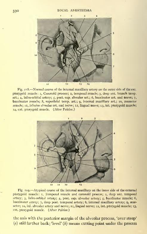
590 LOCAL ANESTHESIA
12 34
Fig. 218. Normal course of the internal maxillary artery on the outer side of the ext.
pterygoid muscle: i, Coronoid process; 2, temporal muscle; 3, deep ant. branch temp,
art.; 4, infra-orbital artery; 5, post. sup. alveolar art.; 6, buccinator art. and nerve; 7,
buccinator muscle; 8, superficial temp, art.; 9, internal maxillary art.; 10, masseter
muscle; n, inferior alveolar art. and nerve; 12, lingual nerve; 13, int. pterygoid muscle;
14, ext. pterygoid muscle. (After Poirier.)
Fig. 219. Atypical course of the internal maxillary on the inner side of the external
pterygoid muscle: i, Temporal muscle and coronoid process; 2, deep ant. temporal
artery; 3, infra-orbital artery; 4, post. sup. alveolar artery; 5, buccinator muscle; 6,
buccinator artery; 7, deep post, temporal artery; 8, internal maxillary artery; 9, mas-
seter; 10, inf. alveolar artery and nerve; n, lingual nerve; 12, int. pterygoid muscle; 13,
ext. pterygoid muscle. (After Poirier.)
the axis with the posterior margin of the alveolar process, 'over steep'
(e) still farther back; 'level' (6) means cutting point under the process

THE HEAD, SCALP, CRANIUM, BRAIN, AND FACE 591
of the malar bone at the height of the first molar tooth, 'over level'
(d) farther in front of it. The values found are entered in Table II,
No. 7. It follows from this that the 'middle part' axis is the most
frequent, and that in go per cent, of skulls the axis cuts the upper alveo-
lar margin in the region of the three upper molar teeth (Fig. 208, b, a, c}.
We will, consequently, accept as a standard puncture point that op-
posite the second upper molar tooth (of course, outside on the cheek),
and if we do not come to the mark here we will vary the point on a
Fig. 220. External lamina pterygoid process. Pterygopalatine fossa and max-
illary tubercle. Right side of skull seen from the side: a, Wide fossa with spinous tuber-
cle (#); b, medium wide; c-e, narrow fossa; d, anterior pterygoid spine (y); e, spinous
tubercle (x), and anterior pterygoid spine (y). (Hartel.)
line parallel to the alveolar margin, reaching back to the ascending
branch of the lower jaw and forward into the region of the upper pre-
molar teeth (Fig. 209).
"Now that we have established a bone-way for the foramen ovale,
we must consider the relations of the soft parts which our cannula
has to pass through from the cheek to the ganglion Gasseri.
"We had chosen our puncture point in the lateral region of the
cheek, opposite the alveolar margin of the second upper molar tooth.
The point of the cannula penetrates the skin and finds itself in Bi-
chat's fat of the cheek. The finger, placed in the mouth of the pa-
tient, feels the needle from the mucous membrane, and accompaniesthe advancing point of the same through the first strait between the
margin of the lower jaw and the tuber maxillare. The finger main-

592 LOCAL ANESTHESIA
tains the integrity of the mucous membrane of the vestibulum oris,
this being accomplished by a curved motion of the needle around the
buccinator muscle. The needle, therefore, goes between (medially)
the buccinator muscle on the one side, and the masseter muscle,
lower jaw, with processus coronoideus and temporal muscle (laterally)
on the other side, through into the fossa infratemporalis, and nowendeavors by perforation of the pterygoideus muscle externus, which
Fig. 221. Lateral route to foramen rotundum. (Braun.)
fills the entire fossa, to reach the planum infratemporale, in connec-
tion with which, as we have seen above, finger-feeling can be auxiliary
only in a portion of the cases. We need, therefore, other fixed
points. Such a point is the depth. Before we stick the needle in wemark with the sliding catch a distance of 5 to 6 cm.
;in case of forward
curving of the cheek by a tumor, still more. We are thereby alwaysinformed as to the depth reached, and can thus protect ourselves
from gross errors. In the second place we must now consider a
direction discernible on inspection of the whole skull", and we have

THE HEAD, SCALP, CRANIUM, BRAIN, AND FACE 593
been able by careful observation and many examinations to estab-
lish as essential for the puncture of the foramen ovale the following
fixed points :
"i. Viewed exactly from the front (for this determination of
direction one must, like the designer, see with one eye only, and pos-
sibly with the aid of a second cannula held freely before one), the
cannula introduced into the ganglion points to the pupil of the eye on
Fig. 222. Lateral injection of second division of fifth nerve in pterygopalatine fossa.
(Hartel.)
the same side (Fig. 210). If we observe this rule, then we avoid de-
viating outwardly into the fossa temporalis, inwardly into the tube
and pharynx region.
"2. On exact lateral inspection the cannula points to the tuber-
culum articulare of the zygomatic arch (Fig. 210). If we do not
observe this rule, then it may happen that we come too far forward
into the fossa pterygopalatina, or too far back into the region of the
foramen caroticum and of the foramen jugulare; the latter way, par-
ticularly namely, the introduction of the needle into the medial
part of the foramen jugulare instead of into the foramen ovale wehave several times taken wrongly on the cadaver, and the cannula
38

594 LOCAL ANESTHESIA
appeared at the base of the skull, at the place of entry of the nervus
vagus and glossopharyngeus into the dura.
"Viewed from below (with the skull inverted), the angle of the
needle is seen in Fig. 224. Figure 225 is a sagittal section of the
skull, and shows the axis of the needle seen from within. Figure 226
is a skiagraph of the needle transfixing the ganglion. In Fig. 227 is
Fig. 223. Pterygopalatine fossa and contents, showing the S form of the maxillary
nerve and the position of the terminal branches of the internal maxillary artery beneath
the nerve: i, Zygomatic nerve; 2, infra-orbital artery; 3, int. maxillary artery; 4, man-
dibular nerve; 5, maxillary nerve; 6, sphenopalatine ganglion; 7, ophthalmic nerve; 8,
int. carotid art.; 9, gasserian ganglion. (After Testut and Jacob.)
seen the angle and point of crossing within the skull of the axes of the
needle if continued backward in bilateral puncture."The observance of the rule given above for the direction, as well
as naturally the bone-feeling on the planum infratemporale, protectsus certainly from this error. The pterygoid muscle is perforated
near its origin on the pterygoid process and tuber maxillare; often
the cannula goes through between the two heads of origin.
"Before we conduct the point of the needle from the fossa infra-
temporalis into the foramen ovale, we take the precaution of slipping
back the sliding catch of the cannula 1.5 cm. from the skin-puncture
place, in order thus to be aware of the depth of the further advance."

THE HEAD, SCALP, CRANIUM, BRAIN, AND FACE 595
A summary of the essential points in making the Hartel punctureis given by Hartel as follows :
"(a) Puncture in the cheek at the height of the alveolar margin of
the second upper molar tooth, establishing first on the cheek a wide
Fig. 2 24. Shows direction of needle in transfixing gasserian ganglion by Hartel route,
viewed from base of skull. (Original illustration from collection of Prof. Matas.)
area of cutaneous anesthesia, which allows a variation of the punc-ture point toward the front or back according to the principle of the
concentric puncture.
Fig. 225 Shows axis of needle in transfixing gasserian ganglion by Hartel route,
seen from within on sagittal section of skull. (Original illustration from collection of
Prof. Matas.)
"(b} The cannula for puncture must be 0.8 mm. thin, 10 cm. long,
and be provided with a flatly ground point. Before the puncture
with the puncture-cannula, the anticipated puncture depth to the

596 LOCAL ANESTHESIA
planum infratemporale (5 to 6 cm.) is marked on the same with the
help of an aseptic ruler by the sliding catch used on the cannula."
(c) Introduction of the cannula, accompanied by finger-feeling
between the anterior margin of the ascending branch of the lower jawand the tuber maxillare around the buccinator muscle to the fossa
infratemporalis."(d) Determination of the direction seen from the front the can-
nula points exactly to the pupil of the eye on the same side; seen from
the side, to the tuberculum articulare of the zygomatic arch.
"(e) The puncture of the foramen ovale takes place under con-
tinuing feeling with the hard and smooth surface of the planum in-
fratemporale from the anterior exterior long side of the foramen."
(/) After the foramen ovale is reached (relaxing of resistance,
radiating pain in the area of distribution of the third branch) the
sliding catch is shoved back 1.5 cm. from the puncture point of the
skin, and the cannula is introduced into the foramen ovale until painis experienced also in the area of distribution of the second branch.
"(g) Attachment of the syringe containing 2 c.c. slow injection
of the solution, which must not exceed i c.c.
"(/?) Immediate testing of the anesthesia."
Within the middle fossa of the skull (Figs. 212, 213) the following
points are of interest: In front is seen the foramen lacerum anterius,
and immediately below this the foramen rotundum, with its axis di-
rected downward, forward, and outward into the sphenomaxillaryfossa. Posterior and external to the foramen rotundum is seen the
foramen ovale.
The foramen spinosum lies slightly external and behind the planeof the foramen ovale and its artery (middle meningeal) as it leaves
the foramen curves outward hugging the bone; this vessel also is out
of danger from the needle being advanced too far within the skull.
At the apex of the petrous portion of the temporal bone, where
this bone is received into the angular interval, between the basilar
process of the occipital and the posterior border of the great wing of
the sphenoid, is seen the opening of the internal carotid artery and
foramen lacerum medium.
The cavernous sinus courses along the inner margin of the middle
fossa, with the internal carotid artery lying along its inner wall, both,
internal and above the foramen ovale. The position of the vessel at
this point and its relation to the sinus and orbital nerves are shown in
Fig. 214. Above and behind the foramen ovale is seen the depressionfor the gasserian ganglion. Above and behind this depression on the

THE HEAD, SCALP, CRANIUM, BRAIN, AND FACE 597
superior margin of the petrous portion of the temporal bone is seen
the groove for the superior petrosal sinus.
The gasserian ganglion, slightly crescentic in shape, with its con-
vexity forward, lies within the above depression between the layers of
the dura which receive it in a slit- or pocket-like interval. It is inti-
mately attached to the layer above but loosely to the layer beneath.
Beneath it pass its motor root and large superficial petrossal nerve.
The large or sensory root runs forward toward the ganglion from its
origin in the pons through an oval opening in the dura mater, the
cavum Meckeli, which communicates posteriorly with the cisterna
pontis of the subarachnoid space, separated only by a reticulated
membrane the porus trigeminus. As the three branches of the gan-
glion blend intimately with the dura when leaving this space the onlyoutlet is through the porus trigeminus behind and any injection whentoo liberally made must seek this outlet and reach the cysterna pontisand posterior fossa of the skull. As this anatomical condition is of
considerable importance it will be dealt with more in detail later.
In injecting all branches of the third division of the nerve it is only
necessary to reach or enter the foramen ovale but if it is desired to
reach the ganglion or the first or second divisions of the nerve the
needle must be advanced considerably further or else much larger
quantities injected, but it is much preferable to get in direct contact
if not within the part to be injected as in this way the results are muchmore certain and accomplished with a smaller volume of solution and
this is more particularly important when using alcohol as its influ-
ence is destructive and we should limit it as much as possible to the
exact part we wish to affect. The lower edge of the ganglion lies
about Y inch above the inner surface of the skull and about % inch
above the opening of the foramen ovale on the undersurface of the
skull, the skull at this point being about % inch thick. The ganglionis about % inch in its long diameter and about^ inch thick. It lies
on the petrous portion of the temporal bone, its outer surface sloping
downward, outward and forward toward the foramen ovale. Thefirst or ophthalmic division, which is directed forward, is given off
from its upper edge and is consequently about % mcn above the
opening of the foramina on the undersurface of the skull. To effect-
ively reach this branch, it is only necessary to reach the ganglion or
get well within it, which effectively reaches all branches; consequently,
it is only necessary to advance the needle about % inch from the
undersurface of the skull or % inch within the skull. Once within
the foramen ovale an injection made in sufficient quantity will flow

598 LOCAL ANESTHESIA
upward and backward and if in sufficient volume will effectively
reach all parts. Once reached the ganglion is easily affected by anes-
thetic solutions, but a few minims of a 0.5 per cent, solution is suffi-
cient to anesthetize it and if made from the foramen ovale ^ dram of
0.5 per cent, solution will probably reach it in effective quantity. In
the case of an alcohol injection it is highly desirable to get in close
contact with the ganglion and accomplish the desired effect with the
minimum quantity rather than to make the injection too free and have
it diffuse in all directions and produce annoying temporary, if not seri-
ous, results. The various disturbances of vision, vomiting, vertigo,
etc., are accounted for in this way.The communication of the ganglion with the subarachnoid space
through the cavum Meckeli is of considerable consequence in the deep
injections of the ganglion, as spoken of later. The anatomy of these
parts, as discussed by Hartel, is as follows:
"Let us now consider the anatomy of the cavum Meckeli and of
the ganglion Gasseri (Figs. 173, 213, 215). The trunk of the nervus
trigeminus rises in the region of the posterior cranial fossa out of the
pons, next passes through the wide cavity of the cysterna pontis,
which is filled with cerebrospinal fluid, and then enters between the
sinus petrosus superior and the superior margin of the petrous portion
of the temporal bone through a wide, oval gate of the dura mater
(the porus trigemini) ,into the cavum Meckeli belonging to the middle
cranial fossa. It has less the form of a compact nerve-trunk than
that of a bundle of nerve-fibers, lying loosely together, which, as is
well known, are covered only with the pia mater. In the cavumMeckeli the nerve forms the area triangularis, and radiates into the
ganglion semilunare, which extends itself toward the front along the
root of the great wing of the sphenoid, and sends off the three trunks
of the trigeminus through the fissura orbitaKs superior, the foramen
rotundum, and ovale."The relation of the ganglion to the walls of the cavum Meckeli,
which is formed out of a fold of the dura mater, is as follows : With
the substratum, the dural membrane, that serves at the same time as
the cranial periosteum, the ganglion is but loosely connected bymeans of loose connective tissue; with the superior dural wall on the
contrary it is intimately united. The three trunks of the trigeminusleave the ganglion Gasseri as compact nerve-trunks closely adherent
to the dura. Of course, the motor portion of the trigeminus does not
participate in the formation of the ganglion. It takes its origin as
the portio minor before the sensory portio major; it passes then

THE HEAD, SCALP, CRANIUM, BRAIN, AND FACE 599
on the inferior side of the ganglion, and joins itself to the third
branch.
'From this situation of the nerves arises the fact that the resist-
ance to a liquid injected with a syringe under pressure is least on the
under side of the ganglion and at the place of the entrance of the
main trunk of the trigeminus into the cavum Meckeli. Hence, there
exists the possibility that the injected fluid may soak through the
porous trigemini into the cysterna pontis. On sudden injection of
staining solution in the cadaver this may be observed, and outside of
the ganglion may be produced a staining of all the arachnoid spaces
of the base of the brain. It appears to me that it may very well be
possible that the condition of sleep observed clinically by us and by
Heymann, in connection with an injection of a copious quantity of
solution into the ganglion Gasseri, is to be attributed to this arach-
noid infiltration.
"Of greatest importance for us, further, is the situation of the
medial wall of the cavum Meckeli, which forms the dividing wall of
the same from the sinus cavernosus. This medial wall is a thin,
translucent, dural membrane. The first branch of the trigeminus,
immediately after its emergence from the ganglion, turns into this
dural membrane with a geniculate bend, and fuses with it so intimately
that a macroscopic separation of the sinus wall from the nerve is
not possible. If in anatomic books (Fig. 214) the nervus trigeminus
I, oculomotorius, and abducens are represented as passing 'in the
lateral wall' of the sinus, then this statement must be supplementedthus: that the relation of the two nerves of the muscles of the eye to
the lateral wall is a much looser one than that of the first branch of
the trigeminus. If one injects with a syringe into the ganglion Gasseri
small quantities of solutions that are diffused with difficulty, such as
ink or tincture of iodin, then one obtains a beautiful infiltration of
the ganglion and of the chief trunk of the trigeminus, while the sinus
cavernosus and the cisternae of the arachnoid membrane remain free;
on the other hand, aqueous solution of methylene-blue is diffused
into the sinus as well as into the cisternae. On the relation of the
sinus cavernosus to the cavum Meckeli, just described, depends the
appearance observed by us at first on too sudden injection of quantities
of solution exceeding i c.c.; that is, an overlapping of the paralyzing
effect of the novocain on the nerves of the muscles of the eye, which
manifested itself either in transient dilation of the pupil concerned or
in a likewise transient paralysis of the abducens."
The distance between the various points of interest within the
skull, as measured by Hartel, are of interest.

600 LOCAL ANESTHESIA
From the superior margin of the pyramid of the petrous portion
of the temporal bone to the posterior-inferior margin of the foramen
ovale he gives a minimum of 14 mm., maximum of 23 mm., with an
average of 19 mm. (See Table II, No. 6.)
The minimum of 1.4 cm. is the safe maximum depth to penetrate
within the foramen; if a greater depth is reached, there is danger of
puncturing the membranes and entering the cisterna pontis. This
actually happened to Hartel, with the escape of cerebrospinal fluid;
besides, there is danger of wounding the superior petrosal sinus or of
injecting the solution beyond into the posterior fossa of the skull.
The shape and size of the foramen ovale vary considerably in
different skulls, and even on the two sides of the same skull, to such
an extent that a study of a large number of skulls is necessary to draw
any positive conclusion. A composite of the whole, giving the aver-
age condition and variations from this average, must necessarily be
less in a large number of cases than measurements or studies made
upon any single skull.
The same may be said about the relative position of the foramen
in relation to the surrounding parts; this variation of position, how-
ever, is less than that of the size and shape of the foramen.
The sphenomaxillary fossa is a small triangular space, situated be-
neath the apex of the orbit at the angle of junction of the spheno-
maxillary and pterygomaxillary fissures. Its posterior wall is formed
by the base of the pterygoid process and body of the sphenoid, which
slightly overhang it in this position; its inner wall is formed by the
vertical plate of the palate bone, which slightly overhangs it on the
inner side;in front is the middle portion of the tuber maxillare.
Externally, it opens into the temporal and zygomatic fossa throughthe pterygomaxillary fissure.
Above the roof is partially deficient, where it communicates with
the apex of the orbit beneath the sphenoid fissure.
Five foramina open within this fossa; on the posterior wall is the
foramen rotundum; above, below, and internal to this the vidian, and
still more inferiorly and internally, the pterygopalatine; on the inner
wall is the sphenopalatine foramen, by which the fossa communicates
with the nasal cavity. Below is the superior openings of the pos-
terior palatine canal. This fossa contains, besides the superior
maxillary nerve and its branches, Meckel's ganglion and the termina-
tion of the internal maxillary artery. It will be seen, from a studyof the above and the use of a needle on the skull, that the transverse
puncture made from below the zygomatic arch may, if the point of the

THE HEAD, SCALP, CRANIUM, BRAIN, AND FACE 6oi
needle is directed too high and advanced too far, enter the apex of the
orbit and transfix the structures passing through the sphenoid fissure
(Fig. 2 1 6) ,or may, if advanced sufficiently far, enter the orbital fora-
men; and, if directed more horizontally and advanced too far, mayenter the nasal fossa through the sphenopalatine canal.
This last fossa may even be entered from above the zygoma, but
from this angle it would be impossible to enter the orbit. For this
reason some operators prefer the transverse route above the zygomainstead of below it.
INTERNAL MAXILLARY ARTERY (Fig. 217.)
The larger of the two terminal branches of the external carotid
is given off at about the level of the lower extremity of the lobule of
the ear, at its origin embedded within the substance of the parotid
gland. It first runs inward, at right angles to its point of origin, to
the inner side of the neck of the condyle of the lower jaw, in this its
maxillary portion lying between the ramus of the jaw and the internal
lateral ligament. As it passes opposite the sigmoid notch it lies
usually a little above its lower border, but occasionally as high as i
cm. below the inferior border of the zygoma. In some few cases the
artery may lie below the tendon of the external pterygoid, and, in
crossing inward from this position, may be in danger of beingwounded
(Figs. 218, 219) when approaching the foramen ovale from below
and in front, as in the Hartel route, or slightly more below the level of
the lower border of the zygomatic process. Consequently, a needle
entered at the lower border of the zygoma should be well above it, but
if passed through the lower part of the sigmoid notch may come in
contact with it.
As the artery passes forward and inward it lies parallel to the
auriculotemporal nerve, above and in front of the inferior dental and
along the lower border of the. external pterygoid muscle. It then
runs obliquely forward and upward, over the surface of this muscle
(pterygoid portion). In its third or sphenomaxillary portion the
artery runs transversely in a tortuous manner, and, while somewhat
variable in its course, always lies below the superior maxillary nerve
in the sphenomaxillary fossa, where it lies in close relation to MeckePs
ganglion.
The only branch of this vessel of particular interest to us is the
middle meningeal, which is given off opposite the sigmoid notch, and
ascends almost vertically to the foramen spinosum in its course. It
usually lies behind the transverse tract of the needle in a lateral

6O2 LOCAL ANESTHESIA
puncture, but may occasionally lie more anteriorly. A small branch,
the small meningeal, passes up through the foramen ovale, but this,
with the internal maxillary vein, lies below and in front of the artery.
Other branches are either of small size or are situated outside of
the coarse of the needle, and are of no particular concern to us here.
The best protection against the injury of a vessel here, as well as
elsewhere, in making deep punctures is in the proper selection of the
needle or cannula, combined with its skilful and careful use.
By the use of a small-calibered needle with flat point, and not a
coarse needle with sharp, long point with cutting edges, there is little
likelihood of a serious injury to a vessel. In cases in which injury
does occur it amounts to a small hematoma or slight ecchymosis at
the point of puncture, which is probably from injury of a vein, the
arteries having tougher walls and being more easily displaced.
On the lateral aspect of the skull in the transverse methods of
approach to the foramen rotundum and sphenomaxillary fossa two
routes are available: one above, the other below, the zygoma. The
upper route rarely reaches the foramen rotundum, but enters the
sphenomaxillary fossa just below the foramen. The best method of
utilizing this route is to enter the needle high up on the cheek in the
notch formed by the union of the zygomatic and frontal processes of
the malar bone. From this point the needle is advanced transversely
inward, when it impinges against the great wing of the sphenoid just
above the pterygoid ridge. The point of the needle is now success-
ively lowered until it slips beneath this ridge and enters the fossa.
It is now advanced about i cm. further, and the injection made at a
depth of about 4.5 to 5 cm. from the surface.
In entering this fossa, if the point of the needle is advanced too far
forward it strikes upon the rough upper projection of the tuber maxil-
lare, along which it must feel its way backward; if advanced too far
backward it meets the external pterygoid plate near its base, and
must be successively advanced forward until it slips over the sharp
laterally projecting edge of this bone into the fossa beyond (Fig. 220).
If advanced too far within the fossa, it is possible to pass beyondand enter the nasal cavity through the sphenopalatine foramen, which
lies about on a level with this line of puncture.
In entering the fossa from below the zygoma (Fig. 221) there are
certain dangers to be avoided. The point of puncture lies below the
notch on the malar (formed by the zygomatic and frontal processes)
and about on a lateral plane with the posterior surface of the tuber
maxillare. From this point the needle is advanced inward with an

THE HEAD, SCALP, CRANIUM, BRAIN, AND FACE 603
upward inclination, passing between the tuber maxillare in front and
the pterygoid process behind, through the pterygomaxillary fissure
into the fossa just beyond, to a depth not to exceed 4.5 to 5 cm.; the
upward inclination of the needle is such that at this depth the pointshould be about 1.5 to 2 cm. above the point of puncture.
If the needle is advanced too far inward, and particularly if the
point of entrance be slightly below the edge of the zygoma and the
angle of the needle be too high, it is possible to pass beyond the
sphenomaxillary fossa and transfix the structures, passing through the
superior orbital fissure or even enter the orbital foramen.
As the axis of the foramen rotundum is at an angle with this and
other transverse methods of puncture, it is only possible in about
one-third of the cases to enter this opening; however, the fossa just
in front of the forearm is readily reached and the nerve often trans-
fixed at this point, or the solution deposited in direct contact with it.
A method erroneously attributed to Matas, while used at about
the same time by him, is probably to be credited to Schlosser. This
method has its puncture point slightly below and behind the malar
prominence. From this point the needle, directed backward, upward,and inward, feels its way along the posterior surface of the tuber max-
illare until it slips beyond its posterior projection through the ptery-
gomaxillary fissure into the sphenomaxillary fossa at a depth of about
4.5 to 5 cm. from the surface.
Harris Method for Injecting Superior Maxillary Nerve. This
method is quite similar to other lateral methods for reaching the
foramen rotundum. The needle is entered through the cheek in the
angle formed by the coronoid process and the undersurface of the
malar bone. The needle is advanced upward at an angle of 40 from
the horizontal and should be in line with the ascending orbital process
of the malar bone. The needle passes behind the back wall of the
antrum and in front of the anterior border of the external pterygoid
plate, passing through the pterygomaxillary fissure into the spheno-
maxillary fossa. The depth of the foramen rotundum is usually 5
to 6 cm. from the surface and if there is no evidence of the needle
having reached the nerve at this depth it is slightly withdrawn and
directed further backward in contact with the external pterygoid
plate and is then gradually advanced forward by repeated thrusts
until it slips over the anterior edge of this bone when it is advanced
i cm. within the sphenomaxillary fossa when if the proper angle of
40 upward has been maintained it should be in contact with the
nerve. This method should be studied in connection with the Offer-

604 LOCAL ANESTHESIA
haus method which gives the accurate depth from the surface at
which the foramen will be reached. This injection is difficult to
make and is one of the most dangerous of the deep injections as seri-
ous results may follow if carelessly or inaccurately done. If the
needle is directed too high and advanced too deeply it may wound the
third or sixth nerve at the sphenoidal fissure and slightly deeper
along this higher axis may reach the optic nerve, serious results mayfollow injury to these nerves particularly in making an alcohol in-
jection. If the track of the needle is too low or it is advanced too
deeply it may enter the posterior nares.
Occasionally the pterygomaxillary fissure is so closed as to makethe entrance of the needle here impossible; in this case resort maybe had to the Matas orbital route.
In discussing this fossa, the anatomic variations in its bony sur-
roundings, and the methods of puncture Hartel states the following:
(In this discussion of the various methods of approach the Matas
route is referred to as a transverse route beneath the zygoma. This
route, while used by Prof. Matas probably independently and about
the same time (1898) should, I believe, be credited to Schlosser. The
original Matas route is the orbital puncture through the sphenomaxil-
lary fissure.)
"Let us consider next the lateral entrance (Fig. 220). This has
a sickle shape, which in its superior end continues into the inferior
orbital fissure. The posterior margin of this sickle consists of a bone
corner which is formed by the anterior boundary of the lamina ex-
terna of the pterygoid process, and above tapers into a ridge which
separates the planum infratemporale from the planum orbitale of the
great wing of the sphenoid bone, and is furnished with a process called
tuberculum spinosum (Fig. 220, a, x). The anterior concave marginof the sickle is formed by the opposite surface of the tuber maxillare.
"According to the greater or less pneumatization of the antrum
of Highmore, the tuber maxillare juts out behind more or less, so
that the sickle form may vary from a small fissure (' type en cornue/
Fig. 220, c-e) to a half-circle ('typeovalaire,' Chipault, Fig. 220, a-b).
The transverse diameter is correspondingly variable. It amounts
(Table II, No. 9) in the minimum to 3 mm.; in the maximum, to nmm., and on the average, to 5.4 mm. A 'narrow fossa' with a width
under 5 mm. we find in about 40 per cent, of the cases.
"Besides, there are the varieties of the posterior margin, which
are dependent on the development of the masseters. The under partof the same, which belongs to the pterygoid process and almost

THE HEAD, SCALP, CRANIUM, BRAIN, AND FACE 605
always presents a very characteristic corner, which we wish to call
Grenzleiste (marginal ridge), projects in special. cases sharply and
like a knife opposite the entrance of the fossa, and may bear a prong,which is called spina pterygoidea (Fig. 220, d, e, y). Just so, the
superior part belonging to the great wing of the sphenoid bone maybe either smooth or form an elevation, soon becoming pyramidal in
shape or ridge-like, or running out into a point, the tuberculum spin-
osum already mentioned. Between these two spines a ligament maydevelop similar to the ligamentum pterygospinosum (Poirier) ,
which
has been described. The different types of the entrance of the fossa
pterygopalatina are placed together in Fig 220 and in Table II, No.
10. The relations of the entrance to the fossa are of importance for
the ways of access of Matas (i) and Offerhaus (3). As far as the
latter "way is concerned, it is, in general, not practicable if the point
of puncture above the zygomatic arch is chosen, for, according to
Table II, No n, the needle introduced in this way only in a small
portion of the cases (12 per cent.) reaches the superior part of the
fossa which receives the nervus maxillaris; but also for the Matas-
Braun way unfavorable varieties of the entrance present great
difficulties.
"If we now consider the interior of the fossa, this is also subject
to great changes. Of most importance to us is the posterior wall
with the surroundings of the foramen rotundum, for a puncture of
the nervus maxillaris can be successful in the whole region only if it
reaches it shortly after its exit from the foramen rotundum, before
it has given of! its branches. This posterior wall can be likewise
strongly changed only through the pneumatization of the bones form-
ing it. Sometimes one finds the entire fossa walled up transversely
or lengthwise with pneumatized walls, which belong to the sphenoid
bone or else to the palatine bone.
"If we now next test the bony skull as to the possibility of punc-
turing the foramen rotundum directly by the Matas way (should be
Schlosser Author), then it is shown that only in 33 per cent, of the
cases (Table II, No. 12) does the possibility exist of penetrating with
the point of the cannula more or less deeply into the foramen, a
possibility which by the orbital way (original Matas route Author)
incidentally noted is much greater (89 per cent.) . Consequently,with the lateral puncture we cannot practically reckon on a direct
injection of the foramen rotundum, but must content ourselves with
washing the nervus maxillaris in the fossa.
"If we now seek fixed points for a successful puncture of the fossa

6o6 LOCAL ANESTHESIA
sphenomaxillary, then here also the method of concentric puncture is
of importance : for, on the one hand, we must shift the puncture point
as much as possible to the front, in order to carry the needle to the
posterior wall of the fossa; on the other hand, it may happen that,
with a puncture point shifted too far to the front, the tuber maxillare
obstructs the approach. Most frequently the puncture point bywhich it is possible to reach the exit of the foramen rotundum lies
under the sutura zygomatico-malar, which is marked by a promi-
Fig. 226. Skiagraph of needle transfixing gasserian ganglion by Hartel route; shows
axis of needle in relation to teeth and bony parts of face and skull. Injected vessels are
also seen. (Original illustration from collection of Prof. Matas.)
nence, and in the living subject is usually palpable or somewhat be-
hind this same suture. (See Table II, No. 13.)
"Gliding backward on the tuber maxillare we come to the en*
trance of the fossa, and guide ourselves now to the opposite posterior
wall, while we seek to come into the turning-corner of ridges borderingthis wall, since with a higher puncture (Fig. 216) we are in danger of
coming into the orbit, with a deeper one into the nasal cavity.
Further, we must reflect that after passing the entrance we dare not
go too deep i cm. at the most, which we can control by our sliding
catch. The total depth amounts to 45 to 57 mm., on the average
50 mm. (See Table II, No. 14.)
"Favorable for the injection into the fossa is the circumstance

THE HEAD, SCALP, CRANIUM, BRAIN, AND FACE 607
that it is filled with loose masses of fat, which permit a good diffusion
of the injected solution into the vicinity. The nervus maxillaris
(Fig. 223) itself lies in the uppermost part of the fossa sphenomaxil-
lary, and is fixed to its roof by connective tissue. In its course it
takes an S-form, which in a sagittal direction comes out of the fora-
men rotundum; it bends laterally, in order to arrive at the sulcus
infra-orbitalis of the upper jaw, when it takes the sagittal direction
again. The orbital puncture follows the direction of this nerve-
trunk itself, which in its manner presents a similar axial puncture."Braun Method. This is an effective and simple means of reach-
Fig. 227. Angle and point of crossing within the skull of the axes of the needle in the
Hartel route, if continued backward in a bilateral puncture. (Original illustration from
collection of Prof. Matas.)
ing the trunk of the third division at its exit from the skull, and fairly
as accurate as the Offerhaus, Hartel, or any other route for reaching
the trunk of the nerve and much easier executed.
The needle is entered at about the midpoint of the zygoma on
its undersurface, and directed transversely inward until it strikes
upon the external plate of the pterygoid process near its base (Fig.
228). It will be seen, from a reference to the position of these parts,
that the foramen ovale lies directly back of the base of this plate and
on the same anteroposterior plane, consequently the depth to the
external plate at its base is the depth to the foramen ovale, but on a
slightly posterior plane, about i cm. Having now determined the

6o8 LOCAL ANESTHESIA
depth from the surface necessary to penetrate, this is marked on the
needle, which is partially withdrawn, and the point redirected slightly
backward, in which direction it is advanced to the determined depth.When the nerve is reached this is recognized by the usual paresthesia
along its branches.
The method of Ojferhaus is a decidedly ingenious and valuable ac-
quisition. To him is due the credit of attempting the first time to
i,. .- - Foramen
\_ -i ovale
Fig. 228. Lateral routes of injection for foramen ovale : i,Offerhaus; 2, Braun. (Braun.)
locate the relative positions of the foramina ovale and rotundum byanatomic measurements made on the base of the skull. This method
aims to make the injections immediately beneath the foramina,
reaching the nerves just as they leave the openings, at their approxi-
mately determined depth from the surface. This method, when ap-
plied with some judgment, making allowances for anatomic varia-
tions in individual cases by slightly manipulating the point of needle
until it comes in contact with the nerve, which is recognized by the

THE HEAD, SCALP, CRANIUM, BRAIN, AND FACE 609
characteristic paresthesia along its branches, will be found to be a
highly useful and valuable procedure.
In a study of 50 skulls Offerhaus found that the distance between
the foramen ovale is approximately the distance from the two outer
surfaces of the alveolar processes, opposite or just behind the last
molar tooth, at the point where the processus pyramidalis ossis pala-
Fig. 2 29. Offerhaus method for measuring base of skull to determine distance of fora-
men ovale from articular tubercle. (Braun.)
tini joins the maxillary bones; this, then, is the distantia interalveo-
laris externa (D. A. E.), and equals the distantia foramina ovale
(D. F. O.) (Fig. 229).
The distance between the foramina rotundum is the same as the
distance between the alveolar processes of the maxilla on the inner
side, behind or along the side of the last molar.
Distantia interalveolaris interna (D. A. I.).
39

6io LOCAL ANESTHESIA
The axis in which lie the foramina rotundum reaches the surface
at the upper border of the zygomas at the point where the temporal
portion merges with the malar; this is about the midpoint of the
zygomatic arch, the linea interzygomatica.
The foramina rotunda lie about 2 to 4 mm. just above and behind
this line. The axis in which lie the foramina ovale passes over the
eminentia articularis and through the articular tubercles; occasion-
ally the foramen lies 2 or 3 mm. back of this line, but as the axis of
the nerve is downward and forward it usually passes through this
axis. While the relative position and distances between these parts
Fig. 230. Method of using Offerhaus calipers. (Braun.)
may vary slightly in different skulls, as well as on the two sides of
the same skull, these measurements may be relied on as approxi-
mately correct, varying only within a few millimeters.
In the clinical application of this method the distantia interalveo-
laris externa is measured with an ordinary pair of calipers or a com-
pass; this is usually found to be about 5 cm., and equals (D. F. O.)
distantia foramina ovalis; the distantia intertubercularis is next de-
termined by specially constructed calipers (Fig. 230), though anyinstrument adapted to this purpose will do. On the Offerhaus cali-
pers there is a movable part, which is attached to the point and pro-
jects outward to indicate the direct angle of puncture. These are
usually placed on after the distance has been determined, which is

THE HEAD, SCALP, CRANIUM, BRAIN, AND FACE 6ll
shown by a scale to which the arms of the caliper are attached and
along which they move. As the articular tubercles are usually easily
felt, just in front of the temporomaxillary articulation at the root of
the zygoma, this measurement is very simply made. In fleshy in-
dividuals a small allowance, about i cm., may have to be allowed for
soft parts.
This distance is usually about 14 cm. We then subtract from
this the distance between the foramina ovale, 5 cm., previously de-
termined by measurement, between the alveolar processes, and divide
by two. - =4-5 cm., the distance along the linea intertuber-
cularis which the needle must travel to reach the nerve-trunk.
Occasionally, the ossification of the ligament pterygospinosum
presents a bony barrier to the passage of the needle along the linea
intertubercularis. When this exists there is usually an opening
through this plate near the base of the skull, just to .the side of the
foramen ovale;this is best found by feeling with the needle along the
smooth surface of the planum infratemporalis toward the foramen,
and the opening through the obstructing plate is usually entered
without much difficulty.
For injecting the second division the linea interzygomatic is de-
termined by measuring with the large calipers the distance between
the midpoints on the two zygomatic arches; from this is subtracted
the distance between the inner surfaces of the alveolar processes at
the last molar tooth, and this figure divided by two in the same wayas for the preceding method.
This then gives us the depth to which the needle must travel to
reach the second division of the fifth. Offerhaus advises that the
needle be entered above the zygomatic arch for reaching the trunk
of this nerve, but where it is desired to make the injection at the
foramen rotundum the needle is entered below the zygoma, directed
slightly upward. A very large coronoid process may check the pas-
sage of the needle here, in which case, if the mouth is opened wide,
this descends out of the way.The same difficulties may be encountered here in passing the
needle within the sphenomaxillary fossa, as already mentioned in
discussing those regions, and the same rule should be applied here to
overcome these difficulties. Occasionally, this method may have to
be abandoned in cases where the sphenomaxillary fissure is reduced
to a mere slip, too narrow for the passage of the needle by the too
close approach of the tuber maxillare to the pterygoid process. In
this case the Matas route should then be tried.

6l2 LOCAL ANESTHESIA
Offerhaus recommended that 2 c.c. of from 0.50 to 0.75 per cent,
cocain and adrenalin be used in making either injection. It would,
however, seem best, in the writer's opinion, to use stronger solutions
of novocain and adrenalin (2 c.c. of a 2 per cent, solution), as is sug-
gested by most operators making these injections.
Offerhaus states that complete analgesia usually occurs in about
fifteen minutes and lasts about one hour. This time could probablybe lengthened by the use of stronger solutions, as suggested.
Fig. 231. Novocain anesthesia of the
right gasserian ganglion, tested immedi-
ately after injection. (Hartel.)
Fig. 232. Anesthesia following alcohol
injection in the left ganglion, tested im-
mediately following injection. (Hartel.)
The utilization of these methods of measurement, as taught us
by Offerhaus, may be applied to any other route of puncture applied
to the same region, and should serve as a valuable guide in determin-
ing the depth of penetration.
Technic of Injection of the Gasserian Ganglion and Its Branches.
The technic of making all deep injections whether for purposes of
surgical anesthesia or for neuralgia should be the same. The neural-
gic patient should not have a general anesthetic unless it is impossible
to handle them otherwise but the nerve first sought for under local
anesthesia and blocked by these injections and the entire distribu-

THE HEAD, SCALP, CRANIUM, BRAIN, AND FACE 613
tion of the nerve anesthetized, as it is only then that we can feel abso-
lutely certain in the great majority of cases that the needle point has
reached or entered the nerve.
The point on the skin through which the needle is to enter is
first anesthetized. The needle entered at this point is slowly ad-
vanced injecting the solution lightly as it is pushed forward; 0.5 per
cent, novocain with about 5 drops of adrenalin to the ounce is usually
preferred. The injecting of the solution as the needle is advanced
makes it less likely to injure a vessel as they are more likely to slip
to one side and as the adrenalin immediately contracts them their
reduced size with contracted thickened walls makes them still less
likely to be injured.
The needle should be about 10 cm. long and 0.8 mm. thick and
have a short sharply beveled point. Such a needle should it enter
a vessel will at most produce a slight negligible hematoma which will
be absorbed and I have never seen any bad effects to result from such
an injury. No force should ever be used in advancing the needle,
but should an obstruction be encountered, which may be a bony wall,
should the direction of the needle be faulty, or an ossified ligament,
it should be slightly withdrawn and redirected slightly to one side
or the other so as to pass the obstruction. When the nerve is reached,
it is usually indicated by a slight pain like an electric shock or thrill
along its peripheral distribution;in the case of the superior maxillary
nerve along the side of the face, upper teeth or side of the nose, and
in the case of the inferior maxillary division the sensation is in the
lower jaw and tongue. If the gasserian ganglion is reached the
sensation may be in either of the above nerves, and in addition pain
in the eye or over the orbit.
When it is felt that the proper point has been reached, the needle
is not disturbed but an additional quantity of the local anesthetic
injected. If the needle is properly placed very little is needed to
anesthetize any nerve-trunk, ^ to i dram at most, and the ganglionand parts within the skull seem much more susceptible to its influence
than the external branches. After a delay of a few minutes the
peripheral distribution of the nerve is tested for anesthesia and if
not complete slightly more is injected but should there be no changein sensation it is quite certain that the needle has been inaccurately
placed when it can be either advanced farther or partially withdrawn
and redirected as the case may indicate. Occasionally the usual
peripheral sensations which occur when the needle reaches the nerve-
trunk may be absent; consequently they should not be invariably

6 14 LOCAL ANESTHESIA
depended upon, but if it is felt that the needle has been correctly
placed an injection of the local anesthetic will produce anesthesia in
the field of distribution of the nerve. In the event of the injection
being made for surgical anesthesia, as in an extensive resection of
the maxilla, the needle may be permitted to remain in position should
it not be in the way, as it may be necessary to repeat the injection
before the completion of the operation, should the anesthesia show
signs of failing. As a rule the thorough injection of the ganglion or
any of its branches should produce a surgical anesthesia of an hour
or longer but occasionally the anesthesia is much shorter.
In injections to reach the foramen ovale certain points are to be
observed. In passing the needle by the Hartel route through the cheek,
a finger of one hand is placed in the mouth to guide the needle be-
tween the ramus of the jaw and the buccal mucous membrane as
some manipulation of the needle may be necessary here to safely pass
this point without entering the mouth.
As the point of the needle searches out the foramen ovale beneath
the base of the skull, should it be directed too far backward, it maywound the Eustachian tube which is felt as a sharp pain runningback to the ear; in such case the needle should be slightly withdrawn
and redirected slightly forward of this position. Another accident
which may happen in searching for the foramen ovale, but more par-
ticularly by the lateral routes, should the needle be directed too low
is puncture of the pharyngeal wall, which is felt as a soreness or pain
in the throat.
Method of Injecting Alcohol. With the needle point accurately
placed and the nerve well injected with novocain as shown by com-
plete peripheral anesthesia, a delay of five minutes is permitted to allow
the thorough diffusion of the local anesthetic as well as to permit the
excess of fluid to escape into the surrounding parts to avoid its too
great diluting influence upon the alcohol. While ordinarily 80 per
cent, alcohol is sufficient to destroy nerve-tissue when properly in-
jected, it is preferable when injecting into a field already saturated
with fluid to use a smaller quantity of stronger alcohol. I almost
invariably use 95 per cent, when making the injection in this way.The alcohol should never be injected rapidly, but always very slowly,
a few drops at a time.
It is often possible if the nerve has previously been well anesthe-
tized that the alcohol will produce no disturbance but more often a
slight burning is felt which disappears after ten to twenty seconds
to reappear again as an additional 2 or 3 drops are injected. This

THE HEAD, SCALP, CRANIUM, BRAIN, AND FACE 615
recurs a few times when, if sufficient time has been allowed to elapse,
the last few injections may excite no sensation. If the nerve has been
insufficiently anesthetized before the alcohol is injected, this pain is
always more pronounced, and in such cases the alcohol should be
more slowly injected, allowing sufficient time for each 2 or 3 dropsto exert its full destructive influence and all pain to subside before
more is injected. When the alcohol is used without the preliminaryanesthetic injections this pain is of an intense burning character and
radiates along the distribution of the nerve, and in the case of the
ganglion it often feels like an electric shock in the side of the head,
subsiding in a few seconds, followed by a progressively increasing
anesthesia in the peripheral parts as more and more is injected.
The amount of alcohol necessary to produce a complete destruction
of the nerve with lasting anesthesia varies from % to 2 c.c., i c.c. being
sufficient for any nerve-trunk but in the case of the ganglion more
may be needed. It is only possible to determine the exact amount
necessary when injecting an unanesthetized nerve when the resulting
peripheral anesthesia serves as a guide and the injections continued
within the above limits until the peripheral anesthesia becomes per-
manent and complete. When injecting an anesthetized nerve it mayoccasionally be found necessary to repeat the injection, more often
with the ganglion; should the resulting anesthesia be incomplete,
and this plan is usually to be preferred rather than take the risk of
serious after-effects if the injection be too liberally made. Besides,
very few patients will permit an alcoholic injection without some
preliminary anesthetic injections.
Action of Alcohol Injections. The curative effects of alcohol in-
jections in neuralgia is due to its destructive action on the tissues.
All tissue suffers alike though not to the same degree, the more highly
organized and more delicate tissues are more markedly affected. In
deep injections of limited quantity the tissues undergo an aseptic
necrosis and are gradually replaced by fibrous tissue. If superficially
injected in contact with the skin sloughing may occur.
For these reasons the minimum quantity known to produce the
desired effect should be adhered to and the needle-point should
always be in contact, if not within, the nerve to be injected and the
alcohol always very slowly injected a few drops at a time. The re-
sulting destruction of tissue produces a break in the nerve-trunk
which is slowly replaced by fibrous tissue. Nerves with high trophic
supply such as branches of the fifth nerve gradually reform and often
with a return of the symptoms when they should be reinjected. This

6l6 LOCAL ANESTHESIA
process of regeneration takes from nine months to a year or longer.
In the case of the gasserian ganglion when once well injected the
effect lasts much longer and is usually permanent. Common sensa-
tion returns to the area of the injected nerve after several weeks but
the pain sense remains absent until after regeneration of the nerve,
although the area affected becomes gradually reduced in size. Asthe effect of an alcohol injection is equivalent to a section of the nerve
it should never be applied to a mixed or motor nerve unless it is in-
tended to paralyze it as in cases of muscle spasm. In a few cases of
sciatica in which the nerve has been successfully reached by the in-
jection, paralysis has resulted. The quantity usually injected is i
c.c. of 80 per cent, alcohol for a nerve-trunk, the ganglion often
requiring slightly more, up to 2 c.c. Where a nerve has previously
been injected by an anesthetic solution a smaller quantity, 10 or 12
minims of 95 per cent, alcohol, can be used to compensate for the
diluting effect of the fluid already in the tissues.
The minimum quantity necessary to produce the desired result
should always be adhered to, particularly in such dangerous regions
as the foramen rotundum or gasserian ganglion; otherwise the effect
upon the surrounding parts may produce the most serious conse-
quences, and if too liberally made at any point, it is possible for in-
fection to result with abscess formation. This, however, practically
never occurs with the proper technic and has never been observed
in the experience of the author and his associates;at most there re-
sults a slight soreness and induration at the point of injection.
Disturbances Following Alcohol Injection of Gasserian Ganglion.
As the motor nerves to the muscles of mastication, temporal, masse-
ter and pterygoid are distributed with the third division of the fifth
nerve more or less muscular paralysis of these parts is expected, fol-
lowing an injection at or within the foramen ovale. This results in
a falling of the jaw toward the paralyzed side with inability to mas-
ticate on that side, but this is often not noticed by the patient as he
has accustomed himself to masticate almost exclusively on the un-
affected side. Until the patient accustoms himself to the anesthesia
of the side of the tongue and cheek these parts may be bitten. Mo-
tion, like sensation, is restored much quicker than the pain sense
and the paralysis may not be noticeable after a few months, and is
rarely ever complete but the fact of its occurrence should make
double ganglion injections with alcohol a matter of some considera-
tion, and when done only the minimum quantity necessary to control
pain should be used and this accurately placed in the ganglion so

THE HEAD, SCALP, CRANIUM, BRAIN, AND FACE 617
that the motor root which lies behind and beneath may escape the
full effect.
Ocular Dangers. As the results of a successful alcohol injection
of the ganglion are equivalent to a gasserectomy we have all of the
resulting disturbances, anesthesia of the cornea with loss of trophic
control and the risk of keratitis. Consequently all patients should be
advised of this possibility, and it is desirable that an oculist take
charge of the eye. Should the anesthesia of the cornea persist for
an hour after the injection the upper lid should be fastened down byadhesive plaster which should be removed daily and the conjunc-
tival sac washed out with boracic acid at which time the eye may be
tested for returning sensation. If anesthesia persists for several daysit is better to permanently wear a collodion shield which can be re-
moved daily for attention to the eye. Closing the lid by means of
pads of gauze or cotton is dangerous as the lids may open beneath
the pad and the anesthetized eye, not feeling the contact, serious
damage may result.
Occasionally, dilatation of the pupil, paralysis of the abducens or
nystagmus may occur and are accounted for by the alcohol reaching
the nerves to the eye in the lateral wall of the cavernous sinus (see
Figs. 213, 214). As a rule these phenomena are transient and pass
off after a few hours. Other phenomena which may be observed
are dizziness, vomiting and severe pain in the base of the skull;
this is no doubt due to the injection having been too deeply made,the needle passing beyond the ganglion into the cavum Meckeli in
which case it flows back into the posterior fossa of the skull (see
Anatomy, page 598). Transient deafness and facial paresis may be
explained in the same way by the alcohol reaching the seventh and
eighth nerves as it flows backward along the dura mater over the
petrous bone. To guard against this danger when the needle is
deeply placed in the region of the ganglion, gentle aspiration should
be resorted to before injecting the alcohol. If clear fluid is with-
drawn, which is cerebrospinal fluid, the needle is too deeply placed
and should be slightly withdrawn until the fluid no longer appearsbefore making the injection. To minimize the possible unpleasantafter-effects the injections should always be made with the patient
in the recumbent position and this position should be maintained
for at least an hour.
Area Supplied by Fifth Nerve. Hartel has done considerable
work in clearing up certain inaccuracies and uncertainties which ex-
isted regarding the areas of distribution of the different branches of

6i8 LOCAL ANESTHESIA
the fifth nerve. He shows these areas as taught in most of our text-
books, and in Figs. 231-235 shows a number of anesthetized surfaces
outlined immediately after unilateral ganglion injections. The tests
were made with needles on patients sufficiently intelligent to make
comparatively accurate observations; certain inaccuracies are, how-
ever, bound to occur, as marginal areas show diminished sensibility
and adjacent surfaces are overlapped by the opposite nerve in a zig-
zag manner.
In the median line of the face the limits between the two sides
Fig. 233. Novocain anesthesia of
right gasserian ganglion, tested immedi-
ately after the injection. (Hartel.)
Fig. 234. Novocain anesthesia of
right gasserian ganglion, tested immedi-
ately after the injection. (Hartel.)
were rather sharply defined, as variations were not so numerous as
had previously been supposed; still this overlapping may take place
sufficiently in spots to make it always advisable to anesthetize both
sides in operations approaching the median line.
On the skull, in the midline, the area of anesthesia extended well
up toward the vertex capitis, but laterally in the region of the auricle
some variations were met with. He calls attention to this extended
area of anesthesia as offering favorable opportunities by this methodfor trephining, etc., upon the sinciput.
"In the face the area of distribution of the cervical nerves (nervus

THE HEAD, SCALP, CRANIUM, BRAIN, AND FACE 619
auricularis magnus, cutaneous colli) projects from below laterally
more or less extensively into the trigeminus region, so that we never
can reckon on pure trigeminus anesthesia in the region of the auricle,
lateral temples, cheeks on the sides, the parotid gland, at the angleof the jaw and chin, and hence must always prefer infiltration to
ganglion injection.
. "Relative to the innervation of the face, observations which wehave made after alcohol injection as to the capacity for regenerationof the sensibility are of interest. Figure 235 shows the areaofdif-
Fig. 235. Anesthesia twelve daysafter alcohol injection of right gasserian
ganglion. (Hartel.)
Fig. 236. Same as Fig. 235, twenty-five
days after injection. (Hartel.)
fusion of the analgesia twelve days after the alcohol injection into
the ganglion Gasseri; Fig. 236, the same twenty-five days after.
We see clearly how, especially in the frontal regions of the margins,
collateral tracts of sensibility are developed. In the same category
the observation belongs that after ganglion injection the anesthesia
died out earliest in those regions whose nerves were treated earlier
with peripheral alcohol injection. If we compare our areas of anes-
thesia with the anesthesias found by Krause, after the extirpation
of the ganglion Gasseri, then we find that ours are more extended
and approach more closely to the statements of the anatomists.

62O LOCAL ANESTHESIA
This is attributable to the fact that our tests were undertaken imme-
diately after the injection, while Krause, for independent reasons,
first undertood the tests of sensibility eighteen days after the
operation.
"2. Deep Sensibility. By ganglion injection the collective bones
and soft parts of the face become anesthetic, as far as they belong
to the area of distribution of the trigeminus. If the operation
approaches the median line,
bilateral anesthesia is to be
preferred. Resections of the
j upper jaw, operations on the
Fig. 237. Sensory innervation of the
thoroughly feasible under this
anesthesia.
"The mucous membranes of
the eye and nose are certainly
without feeling, as well as the
conjunctiva and cornea. Thecorneal reflex dies out, also the
sneezing reflexes of the nasal
mucous membranes, but on the
contrary the vomiting reflex of
the pharynx does not. The
accessory cavities of the nose
are likewise anesthetic. Rad-
tongue: i and 2, Vagus nerve (dotted); 3i al Operations of empyemas
and 5, glossopharyngeus (oblique lines) 54, of the antrum of Highmoreand 6, lingual nerve (horizontal lines). are feasible with unilateral(After Zander and Spalteholz.) .
ganglion anesthesia. For the
ethmoid cavity double anesthesia is always to be recommended
(Fig. 198). Observations concerning the sphenoid sinus and the
hypophysis are not available.
In the oral cavity complete anesthesia of the teeth, jaws and hard
palate can be depended upon. The soft palate usually receives
fibers from the glossopharyngeal plexus and consequently may retain
some sensation. The anterior two-thirds of the tongue normally
belong to the fifth nerve (Fig. 237) but this cannot be invariably
be depended upon as both sensory and taste fibers seem subject to
some variation. Hartel thinks that often sensory fibers are received
through the carda tympani, which communicates with the seventh,
thence to the fifth and through it to the glossopharyngeal. It is

THE HEAD, SCALP, CRANIUM, BRAIN, AND FACE 621
therefore safer in blocking the anterior part of the tongue to inject
the lingual nerve at the inner side of the ramus of the jaw, and for
more extensive resections the technic as described on page 537 can be
followed. Regarding the sense of taste, Harris has found it to be ab-
sent in 85 per cent, of cases of ganglion or third division injection and
that it comes on immediately and is coincident with anesthesia, ex-
tending as far back as the circumvallate papillae. He consequentlydraws the conclusion that in the large majority of cases taste fibers
from the tongue reach the pons through the third division of the fifth
nerve at the foramen ovale, and 1
probably reach the otic ganglion
through the small superficial petrosal from the geniculate ganglion on
a 1
csanafc Gr
Fig. 238. Instrumentarium for injection of gasserian ganglion (Windier, Berlin):
a, Needle, 10 cm. long, 0.8 mm. thick. Nickeled steel with short sharp point a 1 and
movable gauge a 2; b, fine needle for skin anesthesia; c, 2 c.c. record syringe; d, metal
centimeter measure (reduced). (Hartel.)
the facial, thus continuing the carda tympani fibers serving taste sen-
sation. In the minority of cases (15 per cent, in which taste is not
affected, there must be an alternative path in which cases it seems
probable that taste sensations are continued from the geniculate
ganglion to the gustatory nucleus in the medulla through the pars
intermedia of Wrisberg. While these conclusions are not accepted
by all observers, they at least appear reasonable and are worthy of
record here.
In 5 cases following novocain injections there was pain in the
head for several days following, which Hartel attributes to an aseptic
meningitis, and in i case reported by Hartel there was a septic
meningitis; the termination of this case is, however, not given. This
result, he believes, due to the use of a solution made from tablets,

622 LOCAL ANESTHESIA
and concludes that only those solutions prepared in ampules should
be used. This appears to me as hardly the explanation, as a tablet
solution can be rendered as absolutely sterile as when prepared in
any other way. This is of interest in connection with the fact that
recently, in English literature, appeared a report of extensive slough-
ing occurring, following the use of an old but re-sterilized solution of
novocain, which result was repeated when again tested on the arm
of the operator.
Herpes Facialis. An elderly gentleman was referred to me suf-
fering from an unusually aggravated case of herpes of the first division
.Fig. 239. Result of removal of one-half of inferior maxilla under regional
anesthesia for malignancy. (Case of Prof. Matas.)
of the fifth nerve; the eye was much inflamed and gave him con-
siderable and almost constant pain. The distribution of the various
branches of the first division were clearly outlined upon the side of
the nose, upper lid, forehead and scalp as far back as the occiput
by the shrunken condition of the skin which alternated in color
between a pale, bloodless white, and a livid red. This affected area
was the seat of an almost constant burning pain. This condition
had existed for eleven months and had practically invalided the
patient. Little if any relief had been obtained from the various
measures which had been applied. The case had come to me for
an alcohol injection but this I declined to do for obvious reasons

THE HEAD, SCALP, CRANIUM, BRAIN, AND FACE 623
but suggested that I inject the ganglion with cocain in salt solution,
hoping by this procedure to obtain some physiological readjustmentof the trophic cells of the ganglion in much the same way that the
Cathelin epidural injections accomplish benefit in pelvic neuroses.
The ganglion was injected by the Hartel route with ^ ounce of
normal salt solution containing ^ gr. of cocain. There was imme-
diate anesthesia of the entire anatomical distribution of the three
branches. With the subsidence of anesthesia which lasted about
one hour. There was some recurrence of the symptoms in the scalp
though much less marked than before. The eye began to improve
Fig. 240. Front and side view, showing result of removal of one-half of inferior
maxilla for malignancy. (Case of Prof. Matas.)
immediately and in a few days was apparently normal and remained
so. While it is hard to explain just what such an injection does
that accomplishes good, the fact remains that benefit follows. The
same principle applied elsewhere will naturally suggest itself and a
spinal puncture might be tried in herpes of the trunk.
Any peripheral branch of the fifth nerve may be injected with
impunity under the guidance of certain rules and technic. But
the injection of the gasserian ganglion is not always an easy matter
or free from danger of after-effects; it should consequently not
be lightly undertaken and should never be done when peripheral
injection will suffice and should not be undertaken by the inex-
perienced except after careful study and preparation.

624 LOCAL ANESTHESIA
As a final word of advice for making deep injections into the
trunks of the fifth nerve or at their foramina of exit, aside from the
information contained in the preceding pages, it will be found of
great help, as suggested by Braun, to have near-by a skull set in the
same position as the patient's head to further guide one in the
accurate passage of the needle,

CHAPTER XXIII
THE ORGANS OF SPECIAL SENSE WITH DENTALANESTHESIA
THE EYE
COCAIN was first used as an anesthetic in the eye; was broughtforward by Roller in his epoch-making announcement in 1884.
Although many other agents have since been introduced, each having
claims in one or the other direction, still cocain remains the anesthetic
of choice in this particular field, and will be the agent considered here
in discussing the various ophthalmologic operations.
For a consideration of the different drugs as substitutes for cocain
and their particular advantages, see chapter on Local Anesthetics.
A few brief remarks regarding the use of cocain in a general way will
first be made.
The prolonged or repeated use of cocain in the eye as a means of
controlling pain is objectionable on two grounds: First, the haziness
produced in the superficial cells of the cornea by its continued action,
made worse when combined with the use of coagulating antiseptics,
such as bichlorid of mercury (see chapter on Cocain) ; and, secondly,
in chronic conditions requiring its repeated use, in the hands of the
patient, may lead to the formation of a habit. The first objection
raised against it in the early history of its use has been largely over-
come by a better knowledge of its action gained by experience leading
to its more judicious and skilful use.
The slight cloudiness which is seen to follow the repeated applica-
tion of cocain to the cornea was first observed by Koller; this, how-
ever, clears up after a short time, but is most marked and persistent
when bichlorid of mercury is used as a cleansing and antiseptic wash
in the strengths ordinarily employed (1:4000-6000).
Koller undertook experiments upon rabbits to determine the
cause of this action ("Ref. Hand Book Med. Sci.," 1901, vol. iii, p.
156). Cocain was instilled into one eye and the lids closed and held
together with forceps ;the other eye, into which no cocain had been in-
stilled, was held open with an eye speculum. After some time it was
observed that the eye held open showed drying and loss of superficial
epithelium, while the cocainized eye showed very little change; from40 625

626 LOCAL ANESTHESIA
this it was concluded that the hazy changes seen to occur were not
due solely to the action of the anesthetic. His views on this point
were further substantiated by other observers.
It is highly useful in any examination of the eye, where pain, pho-
tophobia, and lacrimation would otherwise render an examination
extremely difficult, if not impossible, as in cases of conjunctival or
corneal troubles, superficially situated foreign bodies, injury from
chemical irritants, etc. When used for this and other purposes it is
better to make several applications of weak solutions, 0.50 to i per
cent., than to use stronger, 4 to 5 per cent, solutions, and, where
stronger solutions are necessary for operative purposes, it is better to
precede their use by the application of a few drops of a weak solution,
as the contact of strong solutions produce a burning, irritating pain of
some seconds' duration before anesthesia sets in. Its use for painwhich might possibly be of glaucomatous origin should be carefully
avoided, as it has repeatedly been proved to hasten the developmentof a threatened glaucomatous attack. An objection raised by some
operators against cocain in cataract operation is that it renders the
eye so hypertonic as to make the expression of the cataract more dif-
ficult; of recent years, however, this objection has been overcome bymore skilful methods, and its use has many advantages, the dilation
of the pupil making iridectomy unnecessary by avoiding prolapse of
the iris. The use of cocain and other local anesthetics in all ophthal-
mologic operations has gradually extended, until now the use of
general anesthesia has been reduced to a minimum and by some is al-
most entirely discarded.
In all operations upon the lids a triangular or crescentic line of
infiltration made subcutaneously with 0.25 to 0.50 per cent, solutions,
with or without adrenalin, as indicated, will block off the operative
area and secure a perfect anesthesia (Fig. 241).
In chalazion a light infiltration made deep under the subcutane-
ous tissues just under the growth, combined with one or two applica-
tions of a 2 to 5 per cent, solution over the region, will suffice to render
the field anesthetic.
While all operations upon the conjunctiva and cornea may be
made perfectly painless by instillations, in such operations in which
the iris is to be handled or cut, this is not always successful by instilla-
tions alone, and when used it is necessary to use strong solutions, 4 to
5 per cent., and begin about twenty to thirty minutes before the time
for operation, instilling a few drops every five minutes, allowing it
ample time to.be absorbed and affect the deeper parts. During this

ORGANS OF SPECIAL SENSE WITH DENTAL ANESTHESIA 627
time it is necessary to keep the eye closed to prevent evaporation and
drying of the cornea. The difficulty of rendering the iris absolutely
anesthetic by this method led surgeons in the earlier use of cocain to
inject some of the solutions into the anterior chamber after the cor-
neal section, a method which has now been almost entirely superseded
by the subconjunctival injection. This method was practised byRoller as far back as 1885, and is carried out as follows: After several
instillations into the conjunctival sac to render this and the cornea
anesthetic, a speculum is inserted, and the conjunctiva seized by
Fig. 241. Method of anesthesia of upper eyelid. (Braun.)
means of a mouse- tooth forceps; three points of injunction are usually
selected, one just below the cornea and one on each side just below
the middle line; it is necessary that these sites be so chosen that the
resulting edema will not interfere with the operation. At each point
2 drops of a 5 per cent, solution are injected, care being taken that
the needle does not penetrate into the subconjunctival layers, which
would result in too much edema. After these injections the eye is
closed for five minutes, by which time some of the edema subsides
and the solution has been given time to act and the iris is thoroughly
anesthetic, when the operation may be undertaken.
For the removal of cataract several instilla tions at intervals of a
few minutes of a 5 per cent, solution, keeping the eye closed during
the interval, will usually suffice;but when it is necessary to handle or
operate upon the iris, these instillations should be supplemented by
subconjunctival injections of 2 to 5 per cent, solutions.
For operations upon other parts, tendons or muscles, after the
preliminary superficial anesthesia', the conjunctiva is seized with for-

628 LOCAL ANESTHESIA
ceps just over the point of operation, and the point of the needle is
inserted as deeply as possible into Tenon's capsules and 2 or 3 drops
injected at the point of the intended operation.
The eye is now closed, and after a delay of five to ten minutes the
tendons can be painlessly divided; in cases where it is necessary to ad-
vance the tendons this is not entirely free from pain, as the necessary
pulling on the central and deeper parts of the muscle cause pain, as
these have not been reached by the anesthetic solution.
In operations upon glaucoma, acute and chronic, Roller recom-
mends 5 per cent, solutions of cocain containing 2 per cent, pilocarpin,
and states that after an experience with this method of over ten yearshe has not met with any bad effects which could be attributed to the
injection, and the results obtained compare favorably with those ob-
tained by any other method.
Prof. Koller, in speaking of the action of cocain as a mydriatic,
as well as its use in certain inflammatory conditions, states the
following :
" The pupil-dilating property of cocain is of great value in ophthal-
moscopy. While the dilatation is sufficient in the dark chamber to
allow a satisfactory examination it does not have the blinding effect
of belladonna, the pupil all the time responding to light.
"This is due to the fact that cocain dilates the pupil by constrict-
ing the blood-vessels of the iris, but leaves the sphincter intact; be-
sides, the accommodation is hardly interfered with.
"The pupil-dilating power of cocain, if combined with that of
atropin, is invaluable in cases of iritis.
"The mydriatic effect of this combination is stronger than that
of any other drug or any combination of drugs; it counteracts both
forces that contract the pupil, the sphincter and the blood-vessels.
(Hyperemia of the iris tends to contract the pupil by stretching the
tortuous course of the iris arteries, while the blood-vessels, when
empty, return to their tortuous course and so dilate the pupil.)
"The anemia of the blood-vessels is a strong check to the inflam-
mation, the pain ceasing mostly after a few installations and the dura-
tion of treatment being greatly shortened. The writer uses a mixture
of equal parts of a i per cent, solution of sulphate of atropin and a 5
per cent, solution of hypochlorate of cocain; at first he instils everyten minutes until the pupil is dilated (three to four instillations are
necessary), then only three times a day."The combination of the two drugs is also efficient in cases of
cyclitis."

ORGANS OF SPECIAL SENSE WITH DENTAL ANESTHESIA 629
For enucleation it is necessary to carry the injection deep downaround the origin of each recti muscle. The superficial parts are first
anesthetized in the usual manner by instillations and subconjuncti-
val injections, combining adrenalin with the latter as well as with the
deeper injections around the recti muscles, using not over i or 2 dropsof i : 1000 adrenalin at each point of injection, making use of a 2 to 5
per cent, solution of cocain. After the anesthesia of the superficial
parts the conjunctiva is divided and the orbit opened; the needle is
then passed deep down to the origin of each recti and 2 or 3 drops
deposited at each point; when this has been completed at all four
points, and a delay of a few minutes allowed for thorough saturation
of the tissues, the anesthesia should be complete and the operation
proceeded with.
The advantages of combining adrenalin with the cocain here is
decided in lessening the amount of hemorrhage which is otherwise
frequently profuse, as well as prolonging and lessening the possibilities
of toxic symptoms arising through absorption. Operators differ in
their views regarding the advisability of the use of adrenalin in other
operations, but most all agree that it is of advantage in such opera-
tions as enucleation, tenotomy, and advancement of the tendons,
where it both intensifies and prolongs the anesthesia. The injection
of the solution into the insertion of the tendons is not desirable as it
causes too much swelling, but during the operation pledgets of cotton
wet with cocain and adrenalin can be laid upon the field.
In operations for pterygium adrenalin appears to be contra-indi-
cated, as its blanching effect renders the outline of the growth less
distinct.
In the removal of foreign bodies adrenalin would seem contra-
indicated, except where the hemorrhage is severe, as slight hemor-
rhage may prove of benefit by washing infectious material out of the
wound, which might otherwise enter the deeper tissues; on the same
grounds cocain, on account of the ischemia it produces, might prove
objectionable and be replaced here by some other agent which does
not cause such vasoconstriction.
As a general thing, adrenalin should be very cautiously used about
the eye ;its too free use, or too strong solution, may give rise to an aching
pain or produce disturbances in the cornea. When used by instilla-
tion it should not exceed a few drops of a i : 10,000 to 15,000 solution.
In all operations upon the eye with cocain care should be exer-
cised, as idiosyncrasies are frequently encountered and may give rise
to unpleasant and often toxic symptoms.

630 LOCAL ANESTHESIA
For a diagram of the nerves of the eye, see Fig. 172, and for a
further description of the anatomy of these parts, see chapter on the
Head. In addition to the above, two special methods of anesthesia
are frequently employed for enucleation, the methods of Lowenstein
and Siegrist.
Lowenstein anesthetizes the ciliary ganglion and retrobulbar struc-
tures by a retrobulbar infiltration. After first anesthetizing the con-
junctiva by infiltration, a point on the outer orbital margin is selected
and a long, fine needle entered at this point and passed obliquely in-
ward and backward behind the bulb (see "Anatomy of the Orbit" in
chapter on the Head) , injecting the solution as the needle is advanced
until a depth of about 4^ cm. has been reached. Care is exercised
not to puncture the bulb by displacing it inward with the finger and
by a lever-like motion of the needle to determine that it is free in the
retrobulbar space; at this point from i to 2 c.c. of a i per cent, cocain
solution is injected.
In the method of Siegrist the same purpose is accomplished by
using curved needles, which are passed through the conjunctiva from
four puncture points around the margin of the orbit and passedaround the bulb into the retrobulbar tissues.
THE EAR
From a study of the nerve supply of the auricle it will be seen that
a horseshoe-shaped injection, embracing the ear from below, made
subcutaneously and carried down beneath the attachment of the deepfascia to the bone, will reach and block the entire nerve supply to
these external parts, or the procedure, as illustrated in Fig. 242, maybe adopted.
Where the operative field involves the external parts of the audi-
tory canal supplied by the auricular branch of the pneumogastric an
injection should be made deep at the root of the ear, on its posterior
aspect, where this branch of the pneumogastric passes upward and
forward through the auricular fissure between the mastoid process
and the auditory canal.
This simple procedure will permit of any operation on the exter-
nal parts. Solution No. i will be found sufficiently strong for this
purpose; the addition of adrenalin will render the field completelyischemic. In exceptional conditions of great vascularity, a proced-
ure, used by Prof. Matas and reported in the following case, will be
found of great value :
"The utility of Coming's principle of incarceration was most
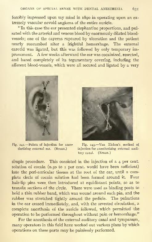
ORGANS OF SPECIAL SENSE WITH DENTAL ANESTHESIA 631
forcibly impressed upon my mind in 1890 in operating upon an ex-
tremely vascular nevoid angioma of the entire auricle.
"In this case the ear presented elephantine proportions, and pul-
sated with the arterial and venous blood by enormously dilated blood-
vessels; one of the cayerns ruptured by ulceration and the patient
nearly succumbed after a frightful hemorrhage. The external
carotid was ligated, but this was followed by only temporary im-
provement. A few weeks afterward the ear was cocainized, resected,
and bared completely of its tegumentary covering, including the
afferent blood-vessels, which were all secured and ligated by a very
Fig. 242. Points of injection for anes-
thetizing external ear. (Braun.)
Fig. 243. Van Eicken's method of
injection for anesthetizing external audi-
tory canal. (Braun.)
simple procedure. This consisted in the injection of a 4 per cent,
solution of cocain (0.50 to i per cent, would have been sufficient)
into the peri-auricular tissues at the root of the ear, until a com-
plete circle of cocain solution had been formed around it. Four
hair-lip pins were, then introduced at equidistant points, so as to
transfix sections of the circle. There were used as binding posts to
hold a thin rubber band, which was wound around each pin, and the
rubber was stretched tightly around the pedicle. The pulsations
in the ear ceased immediately, and, with the arrested circulation, a
complete anesthesia of the auricle followed, which permitted the
operation to be performed throughout without pain or hemorrhage."For the anesthesia of the external auditory canal and tympanum,
many operators in this field have worked out various plans by which
operations on these parts may be painlessly performed.

632 LOCAL ANESTHESIA
Von Eicken suggests the following:
For external parts he begins by spraying with ethyl chlorid, and
then follows with an injection of cocain and adrenalin solution in the
posterior fold of the pavilion, under the cartilage of the floor of the
canal. The needle is directed upward and backward to reach the
point of emergence of the auricular branch of the pneumogastric,which is the sensory nerve of the posterior part; without completely
withdrawing the needle, the point is then forced toward the anterior
and deeper part of the canal to attain the auriculotemporal filaments
(Fig. 243).
Complete anesthesia of the external canal is thus obtained in one
or two minutes. The tympanum is next anesthetized by injecting
the solution into the skin of the deeper part of the canal.
While the above may prove successful, it has not appeared to us
that the use of ethyl chlorid about the external auditory canal was
very satisfactory, as its application here has seemed unpleasant to
the patient.
The following plan by Tiefenthal appeals to us, and is equally
simple; a combination of the two, by injecting the auditory branch
of the pneumogastric as recommended above, followed by anesthesia
of the drum as practised by Tiefenthal, may prove more satisfactory.
Tiefenthal recommends that 4 drops of a 20 per cent, solution of
cocain with i drop of adrenalin (i : 1000) be placed in contact with
the drum membrane for fifteen minutes. This produces a slight
reduction of sensibility, but insufficient for paracentesis. Then,with a small syringe having a fine, angular needle, he injects throughthe lower part of the membrane 2 to 4 drops of a 5 or 10 per cent,
solution of cocain with adrenalin. After a few seconds the mem-brane appears whitish gray from the anemia of the tympanic cavity,
the anesthesia is complete, and paracentesis may be performedwithout pain or hemorrhage.
Professor Neumann, who has done much work with local anes-
thesia on the ear, secures anesthesia in much the same way. The
operative possibilities are not, however, limited to these simple
procedures, mastoid operations being performed with equal success.
For this purpose it is necessary to have a strong syringe. Neu-
mann used a metal syringe with specially modified needle, but any
strong syringe and needle will do. He recommends a i per cent,
eucain solution, with 5 drops of tonogen (an Austrian preparationof adrenalin) to each cubic centimeter for infiltration of the soft
parts. The deeper infiltration about the periosteum is done with a

ORGANS OF SPECIAL SENSE WITH DENTAL ANESTHESIA 633
i per cent, solution of cocain and 5 drops of tonogen to each cubic
centimeter. The solutions are warmed before injection to about
45C. to facilitate their diffusion.
The entire region over the mastoid is now thoroughly injected
subcutaneously with the eucain solution, from the base to the apexand forward to the ear (Fig. 244) ;
some of the solution is then injected
subperiosteally; the ear is now drawn forward, and the anterior wall
of the mastoid process injected subperiosteally down to the bonytermination of the auditory canal (Fig. 245).
Through a speculum, which is now inserted into the ear, i c.c. of
Fig. 244. Points of injection for sur-
rounding operative field with zone of anes-
thesia for mastoid operation. (Braun.)
Fig. 245. Point for deep injection
behind ear. (Braun.)
the cocain solution is injected into the superior wall of the auditory
canal, at the point of junction of the bony and cartilaginous portions,
another syringeful into the inferior wall, and slightly less into the
anterior and posterior walls. These injections must be made beneath
the periosteum, and in such a manner that they produce a distinct
bulging or protrusion, which disappears as absorption takes place.
A small pledget of cotton, saturated with 20 per cent, cocain solu-
tion, is now inserted into the tympanic cavity through the perfora-
tion in the tympanic membrane (which always exists in these cases) .
This is not removed until the antrum is opened during the progress
of the operation.
After a delay of about fifteen minutes the operation may be
begun. It is recommended that in cutting the bone only a very
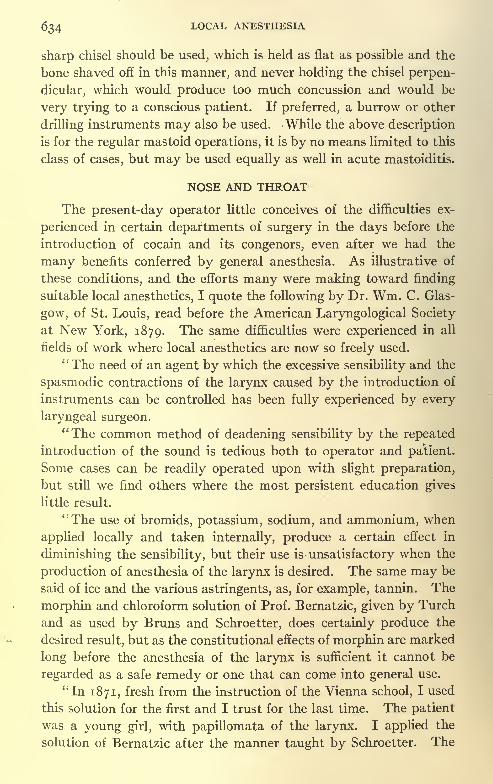
634 LOCAL ANESTHESIA
sharp chisel should be used, which is held as flat as possible and the
bone shaved off in this manner, and never holding the chisel perpen-
dicular, which would produce too much concussion and would be
very trying to a conscious patient. If preferred, a burrow or other
drilling instruments may also be used. While the above description
is for the regular mastoid operations, it is by no means limited to this
class of cases, but may be used equally as well in acute mastoiditis.
NOSE AND THROAT
The present-day operator little conceives of the difficulties ex-
perienced in certain departments of surgery in the days before the
introduction of cocain and its congenors, even after we had the
many benefits conferred by general anesthesia. As illustrative of
these conditions, and the efforts many were making toward finding
suitable local anesthetics, I quote the following by Dr. Wm. C. Glas-
gow, of St. Louis, read before the American Laryngological Society
at New York, 1879. The same difficulties were experienced in all
fields of work where local anesthetics are now so freely used.
"The need of an agent by which the excessive sensibility and the
spasmodic contractions of the larynx caused by the introduction of
instruments can be controlled has been fully experienced by every
laryngeal surgeon.
"The common method of deadening sensibility by the repeated
introduction of the sound is tedious both to operator and patient.
Some cases can be readily operated upon with slight preparation,
but still we find others where the most persistent education gives
little result.
"The use of bromids, potassium, sodium, and ammonium, when
applied locally and taken internally, produce a certain effect in
diminishing the sensibility, but their use is unsatisfactory when the
production of anesthesia of the larynx is desired. The same may be
said of ice and the various astringents, as, for example, tannin. The
morphin and chloroform solution of Prof. Bernatzic, given by Turch
and as used by Bruns and Schroetter, does certainly produce the
desired result, but as the constitutional effects of morphin are marked
long before the anesthesia of the larynx is sufficient it cannot be
regarded as a safe remedy or one that can come into general use.
"In 1871, fresh from the instruction of the Vienna school, I used
this solution for the first and I trust for the last time. The patient
was a young girl, with papillomata of the larynx. I applied the
solution of Bernatzic after the manner taught by Schroetter. The

ORGANS OF SPECIAL SENSE WITH DENTAL ANESTHESIA 635
constitutional symptoms preceded' the local anesthesia fully one
and one-half hours, and they became so grave during the operationthat it had to be suspended and the most energetic measures em-
ployed to combat the toxic efforts of the drug. The local anesthesia,
however, was complete."I have seen the morphin solution repeatedly used with great
success in the Vienna clinic, and it may be possible that my patientwas peculiarly susceptible to the drug; still, the method is subject to
too many risks ever to become popular.
"During the past winter I have been experimenting with two
remedies, both of which produce, in a measure, not only the desired
anesthesia, but also relief from pain. I refer to hydrate of chloral
and carbolic acid. Both remedies have been extensively used in
throat practice, but, as far as I am aware, they have never been
suggested or used for the purpose of producing anesthesia of the
larynx, etc."
The above gives a brief idea of earlier difficulties encountered
and the efforts made by the pioneer operators in their search for a
satisfactory local anesthetic; up to the very time of the discovery of
cocain this search had gone ceaselessly on; the literature of the
precocain period is full of similar reports, and many different measures
utilized to produce what was unsatisfactory local analgesia.
, In ophthalmology, nose and throat surgery, and to the surgeon
specialist local anesthesia has proved a great boon, reducing to simple
office procedures many operations which formerly required a sojourn
in an institution and a general anesthetic for their performance.The use of such instruments as sounds and dilators, is now done
away with, where the parts can be readily anesthetized. The ex-
amination of sensitive and inflamed parts can also be carried out
without the discomfort to the patient formerly necessary. Not-
withstanding the many advances in local anesthesia during the last
few years, and the newer and safer agents introduced, cocain still
remains the anesthetic of choice among the great majority of operators
in these special fields. This is no doubt due to the fact that cocain,
being the first agent introduced, the methods of application necessary
to its success have been studied and perfected, until now it is hard
to displace it from its firmly established position. However, we
are firmly convinced that the time and trouble required to under-
stand the slight differences necessary in technic of the use of some
of the safer agents, particularly novocain, to insure the same degree
of anesthesia will be more than amply repaid by the occurrence of

636 LOCAL ANESTHESIA
fewer sequelae and toxic symptoms. For this reason, we especially
urge the reader to consider carefully the description of these different
agents described in the chapter on Local Anesthesia, and that partof the chapter on Technic, dealing with the action of concentrated
and weak solutions as it is particularly applicable to nose and throat
surgery. In our discussions here we will follow the trend of the
present time and describe the operative procedures under cocain.
It is not alone in operative work that local anesthesia may proveof value in this field, as it may be used to advantage in a certain
limited number of cases in a diagnostic way; in reflex neurosis, start-
ing from the nose, the exact location of the trouble can often be
definitely determined, as an application of cocain to the starting-
point relieves the reflex (asthma, etc.).
There are three methods of applying cocain to these parts in
common use by sprays, swabs, and infiltration (cataphoresis rarely).
In using a spray it is advisable, as a rule, to use only weak solutions
(2 per cent.) in graduated bottles, so that the exact quantity used
may be definitely known, to avoid the possibility of poisoning.
Having the patient to expectorate any accumulations in the mouthor pharynx instead of swallowing them. Weak solutions, applied
repeatedly at intervals of a few minutes, will accomplish as muchas stronger ones and eliminate dangerous possibilities; the first appli-
cation, by constricting the blood-vessels, produces a certain degree
of ischemia when subsequent applications upon the ischemic area
act more profoundly and absorption is greatly lessened. For appli-
cation with a swab stronger solutions are advisable, 5, 10, or 15
per cent., although many operators use highly concentrated solutions
up to 50 per cent., as will be spoken of later.
For application to the larynx it is usually advisable to use solu-
tions of at least 20 per cent. For infiltration it is usually necessary
to use from 0.50 to 2 per cent, solutions.
Adrenalin plays an active part in nearly all these applications, but
it should be cautiously used, as it is an agent capable of producingconsiderable disturbance, and many symptoms erroneously attributed
to the anesthetic are in reality due to the adrenalin. In this respect,
it may be said that it often produces a peculiar tight feeling or painin the head when too freely or injudiciously used.
The advisability of administering to all patients about to undergoan operation of any severity upon these parts some preliminary
sedative a short while before, the same as is advocated for any
general surgical procedure under purely local means, has been dis-

ORGANS OF SPECIAL SENSE WITH DENTAL ANESTHESIA 637
cussed by rhinologists. The objections found with the usual morphinand scopolamin is that most of these patients are operated in the
sitting position, and while the medication accomplished the desired
end in relieving anxiety and uneasiness, it often makes the patientsso drowsy that they nod about and are unsteady in the chair.
Leshure makes use of the following, which is given by mouth about
one-half hour before operation morphin, 34 gr.; hyoscin hydro-
bromatic, >f o gr.; and strychnin sulphate, ^ gr., which is prac-
tically equivalent to our morphin and scopolamin, with the addition
of the strychnin.
Miller recommends the following sodium, potassium, and am-monium bromid, aa 10 gr.; spiritus ammoniae aromaticus, i dram;
aqua q. s.;this is given a short while before operation.
If nausea occurs during the progress of the operation it is usu-
ally relieved by the inhalation of ammonia, and with any evidence of
faintness the head should at once be lowered.
In operations upon the anterior end of the nose in such proceduresas the removal of dislocated septal cartilages, infiltration is usually
necessary, using 0.50 to i per cent, solutions of cocain with adrenalin,
injecting the solution beneath the skin and mucous membrane
surrounding the field sufficient to produce a moderate degree of
edema, and allowing a few minutes to elapse before beginning the
operation.
A somewhat similar technic can be followed in dissecting out
portions of the lateral cartilages, when these encroach upon the
breathing space; the injection is made beneath the skin and mucous
membrane, particular attention being paid to the region of the
nasopalatine nerves, for if these are not rendered anesthetic pain
will be complained of in the front teeth when those nerves are
reached.
In the anesthetization of those parts of the nasal tract, septum,and turbinates, which are usually accomplished by swabbing the
operative area, many operators of extensive experience and un-
doubted ability prefer to make use of very strong solutions, some-
times reaching 50 per cent, and stronger, ra.ther than follow the
example of the general surgeon, whose aim is constantly to reduce
the concentration of the solutions used to the minimum effective
strengths. It would seem to us advisable to use repeated applica-
tions of weaker solutions rather than such concentrated strengths,
but the use of the strengths in this especial field is not without a
rational basis founded upon physiologic laws, as is discussed in the
638 LOCAL ANESTHESIA
chapter on Principles of Technic. The merit of the procedure is
further borne out by the constantly accumulating clinical evidence
and the skill and ability of those making use of these practices.
Some operators, when operating upon these parts by means of the
swab, prepare their solution by placing a few grains of pure cocain
crystals in a dish or suitable receptacle and moistening them with
just enough adrenalin (i : 1000) to render them soluble, claiming
better results from this solution, which gives an anesthesia of from
three-quarters to one hour duration.
In operations upon the septum and turbinates two methods of
inducing anesthesia are in vogue: By packing the nasal cavity with
pledgets of cotton, wet in the anesthetic solution, and allowing them
to remain for about twenty minutes;this may be supplemented later,
if found necessary, by a light application with the swab. The objec-
tion of this method of packing is that large quantities of cocain are
likely to be absorbed, as the packs come in contact with the entire
nasal fossa and their presence stimulates the flow of mucus, which
washes the cocain down into other parts where absorption takes
place. A much safer and better method is to anesthetize the field
by the use of swabs, and here much skill can be shown in their use;
in beginning the application of a swab upon sensitive parts it can
always be preceded by the use of the spray; after the swab has been
applied, allow it to remain in position a few moments; as the anes-
thetic is taken up it diffuses in all directions, which can be recog-
nized by the blanching effect upon the tissues; the application is then
reapplied within the margin of this area in much the same way as a
skilful surgeon will infiltrate the skin; in this way the patient is not
conscious of pain or other discomfort during the anesthetizing
process.
Special attention should be paid to any irregularities, such as
spurs and deviations, to insure reaching all overhanging or posterior
surfaces, by bending the applicators in suitable directions. An
applicator once used should not again be placed in the solution, as
mucus and other secretions carried in with it dilute the solution.
The utilization of regional methods of anesthesia has a limited
application within the nose, by swabbing the solution in concen-
trated form over the trunks of the nerves at their points of emergence
upon the nasal septum. Some few operators recommend injecting
the solution at these points, and while this may in rare instances be
necessary, as in the case of extensive scar formation or other condi-
tions, the majority of operators find the swab sufficient, as the solu-

ORGANS OP SPECIAL SENSE WITH DENTAL ANESTHESIA 639
tion readily penetrates the overlying mucous membrane and reaches
in effective strength the underlying nerves. (See discussion of the
use of concentrated solutions upon mucous surfaces in chapter on
Principles of Technic.)
By a study of the accompanying illustrations, showing the course
apt. ethmoidal artery
spf,
cribriform plate post, ethmoidal i*^''post.art. ot nasal \ artery
nasopatatine nerve septum (sphe- N'icnoid bone > *\ \ "Paia '-J
\ \<
Qnterior ethmoidal nerve
anterior meningeal art. X
'dory nerves
(medial)anter. art. ofnasal septum
I/ br. ofat. tthm. nerve*
tandibula x
Mylohyoideus X7 / r , Aiyionyoiaeu
I; / / (jtniohyoirteus x'; / liypoglossnl nerve X/ deep lingual art.
Fig. 246. The nerves and arteries of the nasal septum and of the tongue.* = divided
posterior pharyngeal wall ** = sphenoidal sinus. (Sobotta and McMurrich.)
and distribution of the nerve supply, a knowledge of regional methods
here is readily obtained (Figs. 246-250).
The presence of scar tissue occasionally found in the septal
mucosa, the result of previous disease (small-pox, etc.) or injury,
may render the production of perfect anesthesia by means of appli-

640 LOCAL ANESTHESIA
cators impossible. In such cases resort must be had to infiltration;
this however, is rarely necessary.
When using infiltration, Killian recommends a regional anesthesia
blocking the septal nerves. He makes the injection at two points
(i) just anterior to the tuberculum septi in an upward direction,
lesser palatine art.
poster, lateral""sal nerves
posterior lateral nasal arteries
jf olfactory nerves (lateral)
/ 4anterior nasal br. oj(i/it. ethmoid, nerve
terior lateral nasal art.
asal concha
\opHaryngeul nerve \. .. .
,-
,.;
.
, \,ton- lingual vallate lingual ,
scendtng P at(
e art. . sil, brancll -, papinae follicles \tonsillur or.
"palatine tonsil
\ inaudible X'i dorsum of tonguefereat palatine art.
ant. palatine nerve
Fig. 247. The nerves and arteries of the outer nasal wall and of the palate. The
tongue has been drawn out, all of the nasal septum except its lower portion removed, and
the mucous membrane of the faucial isthmus divided along the glossopharyngeal nerve
and the ascending palatine artery.** = Sphenoidal sinus.
* = Divided branches to
nasal septum.*** = Anastomosis between nasopalatine and anterior palatine nerves.
* + = mucous membrane of hard palate. (Sobotta and McMurrich.)
and (2) at a point just below the middle of the lower border of the
middle turbinate (Fig. 250).
The Inferior Turbinate. This is anesthetized in much the same
way as the septum, except that greater difficulties are usually
encountered, particularly when it is large, overhanging, and en-

ORGANS OF SPECIAL SENSE WITH DENTAL ANESTHESIA 641
Fig. 248. Innervation of nasal septum. Fig. 249. Innervation of lateral nasal
(Braun.) wall: /, Olfactory nerve; II, nasal
nerve; ///, nasopalatine nerve. (Braun.)
opalat
Fig. 250. Points of anesthesia for Killian regional method. (Braun.)
41

642 LOCAL ANESTHESIA
croaching upon the surrounding parts; however, the application of
the cocain-adrenalin solution soon produces a certain amount of
shrinkage, making the concealed parts more accessible. At times
it is necessary to reach the posterior parts by using a curved ap-
plicator passed from behind through the nasopharynx. Rarelyis it found necessary to use infiltration, and when this is done great
care should be exercised as the absorptive power of this tissue is
tremendous.
The middle turbinate is usually much simpler, and is treated in
much the same manner.
The nasofrontal duct and antrum of Highmore are operated uponin the same manner by securing anesthesia by the use of swabs over
the surfaces to be operated upon.The uvula presents no difficulties, and is easily anesthetized by
a few applications with the swab or it can be infiltrated.
The lingual tonsil presents some difficulties, and should be anes-
thetized with great care, particularly if the cautery is to be used.
As cocain is destroyed by heat, it is necessary to have the anesthesia
penetrate well beyond the influence of the cautery, passing the
swabs well down to its base between the folds of the lymphoidtissue.
The nasopharynx is ordinarily rather difficult of operation under
local anesthesia, largely due t6 its being rather difficultly accessible,
and to its being covered in these cases with tenacious mucus which
protects the underlying membrane. However, certain operations
can be very satisfactorily performed. The anesthesia is accomplished
by swabs passed back through the nose on each side and up throughthe mouth; during this procedure care should be exercised not to
overlook the posterior border of the septum.The ordinary curet is unsatisfactory for operations under local
anesthesia, as the blade wounds the deeper structures, which have
been only imperfectly anesthetized; instead resort should be had to
such instruments as the Schultz adenotome, which are so constructed
as to protect the deeper parts.
The Faucial Tonsil. This structure, on account of its ready acces-
sibility, is ordinarily quite easily enucleated under local anesthesia,
different operators using different methods of anesthesia and various
anesthetic solutions; recently some have advocated quinin and
urea, on account of the absence of after-pain under its use and the
diminished tendency to postoperative hemorrhage.From our personal experience with this agent we would not care

ORGANS OF SPECIAL SENSE WITH DENTAL ANESTHESIA 643
to recommend it as it is at present used. (See chapter on Quinin and
Urea.) The anesthetizing process is begun by brushing the tonsil,
its anterior and posterior pillars, and supratonsillar fossa with a
swab wet in a strong 10 to 20 per cent, solution of cocain and
adrenalin. A i to 2 per cent, solution of cocain, containing i : 10,000
adrenalin, is then injected into the anterior and posterior pillars,
carrying some of the solution down under the base of the tonsil.
When carefully injected it is not necessary to use more than 40 to
60 minims, which should result in a perfect anesthesia in a few
Figs. 251, 252. Outline of points of injection for anesthesia of frontal sinus; i,
Peuchart point of injection for nasal nerve; 2, point of injection on side of cheek
for reaching sphenomaxillary fossa. (Braun.)
minutes. As the tonsil is being separated from the pillars and its
bed, if any pain is complained of, swabbing over the area will
control it.
Hemorrhage is usually very slight, owing to the use of adrenalin,
but if any occurs it must be perfectly controlled before leaving the
case, as it may increase as the effects of the adrenalin pass off.
Larynx and Trachea. Any applications to the larynx is best
preceded by a preliminary spraying with a 2 to 5 per cent, solution,
having the patient inhale at the time, preferably using graduatedbottles.
The superficial anesthesia secured in this way will permit of the
easy use of the swab later, which is used with a 20 per cent, solution.
To anesthetize the trachea a 5 to 10 per cent, solution is usually
necessary, which is sprayed in at the moment of inspiration, hav-

644 LOCAL ANESTHESIA
ing the patient expectorate any which may accumulate in the
pharynx.In all operations upon the above-mentioned parts, where the
procedure is at all protracted, the operator is warned of returning
sensibility by the return of vascularity and oozing of the parts, which
always precede the return of sensation and afford time for the
application of additional anesthetic.
In very extensive operations within the nasal cavities consider-
able advantages may often be offered by blocking the superior
maxillary nerve where it leaves the foramen rotundum, as described
in the chapter on the Head. This could be supplemented by the
use of adrenalin locally to control the hemorrhage, or, under extreme
conditions, ligating the external carotid artery.
Under certain conditions it may be advisable to use combined
methods of anesthesia, which are discussed under this heading.
For operations upon the frontal sinus the field is embraced in an
area of infiltration, as shown in Figs. 251, 252, carrying the infil-
tration deep down to the periosteum, at the orbital margin, to reach
the supra-orbital and supratrochlear nerves at this point; following
this very light infiltration is necessary laterally and above, but
may often be found unnecessary. Anesthesia of the mucous lining
of the sinus is controlled by a median orbital injection made at point
i, for the technic of which see chapter on the Head.
DENTAL ANESTHESIA
Here all methods of anesthesia find a field of application, from
ethyl chlorid and topical applications down to the more extensive
operations performed under regional anesthesia. Ethyl chlorid is
used in the opening of abscesses and other simple incisions, and is
sometimes employed for extractions. For this purpose the ordinary
container may be used, which sprays one side of the gum at a time,
or a specially devised instrument, with a fork-shaped or two-
pronged tip, which directs the spray to both sides at the same time.
For the purposes of infiltration about four agents are now em-
ployed namely, cocain, /3-eucain, alypin, and novocain together
with a large number of proprietary mixtures, which contain mixtures
of the above agents with other ingredients in different proportions,
most of them containing adrenalin.
Thymol is one of those agents, and seems preferred by the great
majority of dentists, as this agent combines antiseptic and anesthe-
tic qualities; in dilutions of 1:2000 it prevents the development of

ORGANS OF SPECIAL SENSE WITH DENTAL ANESTHESIA
bacteria, and in more concentrated solution, i :2oo it is destructive
to most organisms. Dentists frequently make use of this qualityas well as its anesthetic effect by applying solutions to sensitive
pulp cavities; this anesthetic property is quite decided. Experi-
ments upon animals show that 1:1000 solutions will paralyze the
cutaneous nerve-endings of frogs immersed in it for a short time.
Fig. 253. Injection syringe of glass and metal, designed by Dr. Guido Fischer. (See
Fig. 254 for explanation of lettering.)
As reccomended by Fischer, the proportion in anesthetic mixtures
should be about 1:5000. He prefers the following formula, which
he recommends for infiltration purposes:
Novocain 1.5
NaCl 0.92
Thymol 0.025
Distilled water.. 100.0

646 LOCAL ANESTHESIA
For all practical purposes our solution No. 2 (i and 2 per cent,
novocain), with 5 drops of adrenalin to the ounce, will be found
amply sufficient, much cheaper, and more satisfactory than the
many proprietaries now on the market, or the solution recom-
mended by Fischer may be used. For injection into the gumsspecially constructed syringes with short, stout needles, often directed
Fig. 254. Needles, awls, and wrench for injection syringe, designed by Dr. Guido
Fischer. At the left is a considerably enlarged reproduction of the new needle, showingthe details of construction as follows: i, The hollow needle, either of seamless steel, pure
nickel, gold, or iridio-platinum; 2, body of soft metal for firmly tightening the needle
upon the orifice of the syringe; 3, conical shell of hard metal, open below, from which the
soft metal cone protrudes. This arrangement remedies the deficiencies of the old styles
of needles in which the unprotected soft metal cone could not stand much use, became
flattened easily, and jammed in the hub so firmly that both hub and needle had to be
replaced, which was rather expensive if gold or iridio-platinum needles were used. Thenew needles are attached to the syringe absolutely tightly by inserting the needle in one
of the hubs (b or c) and screwing it firmly on the orifice of the syringe. In oder to enable
practitioners with sensitive fingers easily to manipulate the hubs, which heretofore were
milled, the hubs b and c, also the middle pieces d and e, are made with hexagonal con-
nections, so that they can be conveniently and firmly tightened by a slight turn of the
wrench. No force should be used, otherwise the soft metal cone of the needle becomes
unnecessarily worn. (Fischer.)
at an angle, are necessary, as the ordinary syringe and needle will
not stand the pressure needed to infiltrate such dense tissue. The
syringe and needles shown in Figs. 253 and 254 have, after a thorough
experience, been found to fill all requirements, and have been adopted
by Fischer.
The dental nerves and their areas of distribution are seen in

ORGANS OF SPECIAL SENSE WITH DENTAL ANESTHESIA 647
Figs. 148, 165, 166, 167, and 168, and a more thorough description is
given in the chapter on the Head.
It should be remembered that dental anesthesia, when applied
aFig. 255. Position of needle in mucous anesthesia, aperture of needle pointing
toward the bone: a, Correct position; b, incorrect position. The point of the needle is
forced into the periosteum and to the bone. (After Seidel.)
to the roots of the teeth, is practically always a paraneural injection,
and may require ten or fifteen minutes to become effective. These
injections should be made subperiosteally rather than under the
Fig. 256. Position of needle for horizontal injections in several upper teeth; o, Labial
injection; b, buccal injection. (After Fischer.)
mucous membrane, and under considerable pressure, as this solu-
tion must force its way through bony tissue to reach the nerve-fibers
at the root of the tooth.
Before making the injection the surface is cleansed and touched

648
b b
Fig. 257. Position of needle for in- Fig. 258. Position of needle for mu-
jection in upper canine: a, Labial injec- cous anesthesia in upper first bicuspid,
tion; b, palatal injection. (After Fischer.) Above is seen the infra-orbital foramen:
a, Buccal injection; b, palatal injection.
Fig. 259. Injection in palatal mucous membrane at lateral incisor region. Syringe is
held like penholder. (After Fischer.)

ORGANS OF SPECIAL SENSE WITH DENTAL ANESTHESIA 649
with iodin. The needle should be entered at a right angle to the
mucous surface, injecting as the needle is advanced, and slowly
pushed through to the periosteum, which is penetrated, and the
needle advanced a short distance along the bone and well up toward
the root of the tooth; the opening in the needle point should alwaysbe directed toward the bony surface (Figs. 255, 258), the remainder
of the solution now slowly injected, the needle withdrawn, and the
finger pressed upon the point of injection for a few seconds. In
Fig. 260. Position of needle for injection at maxillary tuberosity. (After Fischer.)
those parts of the mouth in which the needle cannot be advanced
at a right angle it must be done obliquely, but should be made as
nearly at right angles as possible.
Repeated punctures by the needle are to be avoided when pos-
sible, as two or more teeth can be injected by using a long needle
and advancing it in such a position that the area of injection can
be made to embrace several teeth (Fig. 256).
For injections upon the palatine surface the needle is made to
enter more nearly in the axis of the tooth (Figs. 257, 259), inserted
back from the gum margin, and advanced to a subperiosteal posi-
tion over the root apex. In dealing with the upper molars, instead
of making the injection as above, an injection can be made into the
posterior dental canals (regional anesthesia), this injection sufficing
for all three molars, as follows:
On the lateroposterior surface of the tuber maxillare of the

650 LOCAL ANESTHESIA
superior maxilla are seen a varying number of foramina, the open-
ings of the posterior-superior dental canals through which the
sensory nerve-filaments pass to the three upper molars; before enter-
ing these canals the nerves run downward and forward for a short
distance in the submucous tissue in close proximity to the parent
trunk. (See Figs. 148-165.)
To inject these nerves in this position the mouth is held half-
open, the cheek drawn outward and upward, and the zygomatic
process reached with the finger; the needle is entered high up in the
mucous membrane about over the second molar, with its point
directed upward, backward, and inward, the syringe being held
well away from the bone; the solution is injected as the needle is
advanced, with the point hugging as closely as possible the convex
surface of the tuberosity (Fig. 260) until its posterior surface is
reached. From ^ to i dram of a i to 2 per cent, solution of a
novocain-adrenalin solution is distributed along the track of the
needle. Ten or fifteen minutes may be required to attain the
maximum anesthetic effect.
The lingual portion of the inferior maxilla is injected in a similar
manner, but where several teeth are to be anesthetized it will be
found best to block the inferior dental nerve at its entrance into the
dental canal, as described in the chapter on the Head.
When this form of anesthesia is resorted to an additional injec-
tion will be necessary for the molars on their buccal surface, as this
tissue is supplied by the buccinator nerve. (See Fig. 167.)
A review of the appended table (pp. 652-654), taken from
Fischer, will be found useful, as indicating the points and methods
of injection in simple and complicated cases as well as the amount
of anesthetic solution to be used.
Some years ago a pressure method of anesthesia for the painless
extirpation of pulps was introduced. The exact origin of this
method seems hard to trace; it was, however, early reported on byDr. Kells, of New Orleans, and H. H. Hill, of St. Louis (1899). For
its success it is necessary that the pulp be exposed, the essential
feature being the driving of the anesthetic under pressure into the
root canal of the tooth. It is carried out as follows : A small piece
of cotton (or spunk, as originally recommended) is moistened with
alcohol, and then touched with the local anesthetic, preferred in
powdered form, so that a few small crystals adhere to the cotton; this
is placed in contact with the exposed pulp; the rest of the cavity is
then filled with ordinary red rubber (unvulcanized) ; light pressure

ORGANS OF SPECIAL SENSE WITH DENTAL ANESTHESIA 651
is then applied with a ball burnisher as large as can be fitted to the
cavity; as the pain ceases the pressure is increased until considerable
force is exerted, which is continued for a few minutes; this has the
effect of driving the anesthetic into the pulp canal. When the
exposure of the pulp is minute after the first application, the openingis enlarged and the process repeated. When this has been properlycarried out it should permit the painless use of the broach.
It is said that when arsenic has been previously used the resulting
anesthesia is not usually as pronounced.
Cataphoresis has been used in dental surgery, but is not very
popular and requires much time. Other electric devices have been
used, but do not properly come within the scope of this book.
For regional methods of anesthesia applicable to more extensive
dental operations reference should be had to the chapter on the
Head, which describes the technic necessary for regional anesthesia
of the upper and lower jaws, the illustrations being particularly
useful.
"Additional explanation of tables: The period of waiting in
Cases i to 10 is about ten minutes; after injection in the inferior
dental foramen as in Nos. n to 16, twenty minutes. In Nos. 13
to 1 6 no injection lingually is required. All combinations of anes-
thesia of several teeth on one side can easily be calculated by apply-
ing to the first and last tooth of the series to be anesthetized the
technic specially indicated for the same in the tables.
"If for example, the right half of the upper jaw, from the canine
to the third molar, is to be anesthetized, an injection is made in the
canine fossa at the root apex of the canine; the needle is then ad-
vanced along the periosteum to the root apex of the second bicuspid,
injecting altogether 2 c.c.; then i c.c. is injected at the maxillary
tuberosity, about 10 drops in the posterior palatine foramen, and
about 0.25 c.c. palatally, between the canine and first bicuspid."
The aim of this book has been to set forth methods of local anes-
thesia applicable to different parts of the body; hoping that as the
technic is perfected the field of use will broaden and humanity reap
the benefit.
"For I doubt not through the ages,
One increasing purpose runs,
And the thoughts of men are widened
With the process of the suns."

652 LOCAL ANESTHESIA

ORGANS OF SPECIAL SENSE WITH DENTAL ANESTHESIA 653
oM

654 LOCAL ANESTHESIA
>O IO<N C<
d d
o oO O
do
wo
, ,
3:3 3w j a
,Q H S313 T3 C H
T3 e H-O (36' aJ
ed tc.
pliuli
O *JT3 O
1111.5 Tg -tjcw"8
jso "2 ^ o ^T25y>i3*J--
tj _- _g go> c 13 3""
Jjeiia,qeJz; 3
.S 53 .S 53 .S
tn -rj -u *JO ^H C/3 O
d rt
^!g"
III!u 3
3 o 3 "
^^-K S-g 8^"2-KcS'-'cSoc'-''-'rt ra y3 d tn rt *"
'43o
re'lrf^'^g:
OJ rt-^ 4)
O U
1IS o|^ ^ -c3 o e8
fl S -
>-> P2<I-*
I
^'35d 8
1
C^ *^N C3 ^H
C !5C SC-feC8.2 8.2 8.2 53 8.2
i > tn "->" > C i/i-
C/3 -7-1
llrf*Nr!*Jlllllila g s<a a a a
.s a
II
pa <a_^'3S9*osjja S c o 6=3 o gs o g33
If 111 i| tilla>-H
gM_ Bi9M o-g a,
U U
53O -c j5
js ? Ts
^^ I -S s

ORGANS OF SPECIAL SENSE WITH DENTAL ANESTHESIA 655
3 a^ .9-3C/3 P C/3 H C/3 < S
o
NNERVA geminus
S M'-
CO
i N
1a
cMO
01 <c
I
IIJ"2
0*3 Se-S-Sao .ii *- sJ
lall^ rt o-*-S ~T* O
CQ
4 J"C *M *^ c4
r^ *f O< O.O <. 3 3^--Y 'C/2V3

656 LOCAL ANESTHESIA
K O.2
gtn tuO C
CO Sto Q^"^^_ u^ O

ORGANS OF SPECIAL SENSE WITH DENTAL ANESTHESIA 657
tfpu
ao
3
JS>" X
42

658 LOCAL ANESTHESIA

ORGANS OF SPECIAL SENSE WITH DENTAL ANESTHESIA 659

66o LOCAL ANESTHESIA
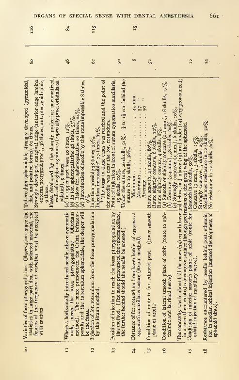
ORGANS OF SPECIAL SENSE WITH DENTAL ANESTHESIA 661
2
I -it 11j=2
" 213rt >O& -S 3 -R
voJ2sga^o iT-O cN M en
'383?!!141*113-3 4> 3 %
2*5 SSI-S 10-0 3"3 M S m
a .5"f s
2%.. mm.
,60%.
skulls,
der
(14)
enoid.
0%
%.%.ull
25 25sk 0%
.ull ull in
Jj m
Sl|
la al
2^-ur
00^^ M -3VP^ % v'3^^ S
ill 12 in-<5 S CO C ^-v-iJ2 .r^S -to^oo - --w
1 ^Ilf|t!!il*"t||1|||f||ifiHffc!Wlli2 .P.f5 g M"!^ s^ 8 w 5s. l-s-S 8 y g *- g >,s^<j<u<oSoi3J2o-Sg ToJH '<
^^ll^U^^ggog^alSla^|J|||2a sR R R
gl
rtT3 JO
"
I aM Q (1
^2-5"8 O-TJ
g S3- O O2B-
.1^3s*--^
'o-S-Ssio ^2
'^.2 4>
'5 -a0M
6d^ a
I4)
J2
a -s
s ^
<*H Oo'S
1 -a.
la 31 SIIf u 1QUO H U

662 LOCAL ANESTHESIA
ii
INDEXABDOMEN, 317
nerve supply, 345Abdominal cavity, operations within, 349
quinin anesthesia in, 117
muscles, reflex rigidity of, 325
operations, indications and contraindi-
cations for local anesthesia, 351, 353
organs, sensibility, 317, 328
packs, pain from, 336
pain, 325-327
wall, anesthesia of, 347Abscess of liver, operation for, 308, 354
Absorption, effects of acids on, 48of alkalies on, 48
laws governing, 170Acetanilid in rectal diseases, 420
Acids, effects of, on absorption, 48
Adhesions, intestinal, pain from, 336
Adrenalin, 137
action with different anesthetics, 151,
152
antagonism of, to strychnin, 144
calcareous degeneration of vessels from,
146
deterioration, 141
glycosuria from, 145
hemostasis after use, 150in arterial anesthesia, 216
in cocain poisoning, 131
in enlarged spleen, 144
in eye surgery, 629in rectal diseases, 145
in snake-bite, 144
in spinal analgesia, 438in testing collateral circulation, 216
maximum dose, 154
physiologic action, 142
surgical uses, 148
tests of, 141
toxic action, 145
Age, influence on toxicity, 129
Air, experiments with, as local anesthetic,
66
Akoin, 81
Alcohol, action on injected nerves, 615as local analgesic, 63-68
injections, disturbances following, 616
of internal laryngeal nerve, 288
of nerves, 570
technic, 614mixtures in spinal analgesia, 468observations following injection, 69sterilization of cocain with, 173
Alexander route to third division of fifth
nerve, 572
Alkalies, effects of, on absorption, 48Allen's method of abdominal anesthesia,
347solutions of novocain, 165
Alypin, 83in spinal analgesia, 437with adrenalin, 153
Analgesia, 29
Andolin, 71
Anemia, anesthetic effects of, 58
Anesthesia, 29
arterial, 76
combined methods, 198
dolorosa of Liebreich, 53, 65
of abdominal wall, 347
Anesthesin, 93
following cautery, 95
in hemorrhoids, 95, 420in rectal diseases, 419, 420in urticaria, 95
Anesthetic agents, comparative action, 99
effects of anemia, 58
Anesthetics, combinations of, 164
general, contraindications to, 202
local, antipyrin as, 70
contraindications to, 202
Ankle, 272
Anoci-association, 204
Anorectal region, 41 1
Antagonists to toxicity, 194
Anterior crural nerve, 259
palatine nerve, 568
tibial nerve, 261
663

664 INDEX
Antidolorin, 60
Antidotes to cocain poisoning, 131
Antipyrin, 394as local anesthetic, 70
in rectal disease, 420
Antrum of Highmore, 642
Aponeuroses, sensibility of, 38
Apothesine, 96
Appendectomy, 349-353
Appendix, operations on, 353
Aquapuncture, 63, 64
Arachnoid membrane of spinal cord, 436
Arm, anesthesia of, 229
Arterenol, 137
Arterial anesthesia, 76, 211
occlusion by bands, 283
Arteries, sensibilities of, 39, 337, 344
Artery, common carotid, ligation, 283
of sleep, 1 8
Artificial respiration in cocain poisoning,
135
in collapse from spinal analgesia, 468
Auditory canal, anesthesia of, 632
Auriculotemporal nerve, 514
Axilla, paraneural injections within, 236
BABCOCK'S spinal analgesia solutions, 450
Back, 302, 312
nerves of, 313Bands for arterial occlusion, 283
Bartholin's glands, removal of, 427
Beckwith adrenalin test, 141
Beta-eucain, 77
action of adrenalin with, 151
in spinal analgesia, 437
Bite, snake-, treatment, 189
Bladder, anesthesia of, 113, 116
female, 426
inspecting interior of, 401
nerve supply of, 382
sensibility of, 341, 394, 402
Blocking intercostal nerves, 305
nerve-, 159, 203
pudic nerve, 382
Blood-pressure, effect of pain on, 30,
340
Bones, 225
sensibility of, 38, 225, 379
Brachial plexus, 227
anatomy, 232
intraneural injection of, 236
Kulenkampff's injection of, 233
paraneural injection of, 231
Brain, operations on, 521
sensibility of, 27, 520
Braun's eucain solutions, 79, 165
injection of frontal and lacrimal nerve,
549of third division of fifth nerve, 607
method of abdominal anesthesia, 347
novocain solutions, 165
Breasts, operations on, 305Bromids as local anesthetics, 70
in rectal surgery, 420
preceding operations on nose and
throat, 637Brucin as local anesthetic, 70
Bubo, 263
Bunions, 274
Burgi's law, 194, 200
Burns, anesthesia in, 94chloretone in, 91
CALCAREOUS degenerations from adrena-
lin, 146
Calcium chlorid in sacral anesthesia, 489
in solutions, 169
Cancerous growths, 286
ulcers, 420Carbolic acid, 67, 419, 420Carbon dioxid snow as local anesthetic, 63
Carbonic acid gas, 19
first use, 18
Carbuncles, 312
Carotid artery, common, ligation, 283
internal, ligation of, 284
canal, 575
Cartilage, sensibility of, 38
Caruncles, 427
Castration, 391
Cataphoresis, first use of, 20
Cataract, operations on, 627
Cauda equina, 436Caudal anesthesia, 486
Cautery, anesthesin following, 95
use of sodium bicarbonate after, 420
Cavernous sinus, 596
Cavum Meckeli, 597, 598
Cellular tissue, sensibility, 38
Cerebrospinal fluid, movements of, 439
specific gravity of, 441
pressure, effects of spinal analgesia on,
471
Cervix uteri, operations on, 424Cesarean section, 430
Chalazion, 626

INDEX 665
Chancroids, anesthesia in, 94, 384, 393,
493
Chest wall, anesthesia of, 306Chloral as local anesthetic, 70
in rectal surgery, 420
Chloretone, 89in burns, 91
Chloroform, 393death from, 155first use, 25
in sepsis, 202
local use, 20, 68
Cinnamylcocain, 136Circular anesthesia, 186
Circumcision, 384, 385Cisterna pontis, 597
terminalis, 436Coca bush, leaves, use by natives, 22
plant, 22
Cocain, 72
aluminum citrate, 72
sulphate, 72
as affected by adrenalin, 151
borate, 72
cantharidate, 72
concentrated solutions, 169
death from, 127
disadvantages of continuous use in eye,
625
early history, 21
general anesthesia from, 221
hypodermic injection, for control of
psychic disturbances, 330, 331in spinal analgesia, 436
injections in neuralgia, 537
lactate, 72
local use, 20, 24
mydriatic action, 77, 628
nitrate, 72
origin, 20
phenate, 67, 72
in rectal surgery, 420
phosphate, 72
physiologic action, 74, 130
poisoning, 130adrenalin in, 132
antagonists to, 193
treatment, 131
saccharate, 72
salicylate, 72
solutions, sterilization of, 173
stearate, 72
synergistic agents, 194
Cocain, tests for, 73
toxic dose, 127
use in glaucoma, 626
Codamin, 197
Codein, 197
in rectal diseases, 420
Codrenin, 71
Cold, early use of, 19
effect on pain of operations, 18, 19, 60,
181
use of, as anesthetic, 60
Colic, intestinal, Nothnagel's hypothesis,
322
Collapse in spinal analgesia, 468Collateral circulation tested with adrena-
lin, 216
Colostomy, 349, 353
Colporrhaphy, 424Combined methods of anesthesia, 198,
205, 35i
Comparative action of anesthetics, 99
Conjunctiva, operations on, 626
Constriction, early use, 19
Constrictor, .Esmarch's, 19
in preventing absorption, 127, 128,
159
surgical use of, 149, 186
Contraindications to local anesthesia, 202,
350, 35i, 370Conus terminalis, 435
Cornea, operations on, 626
Cortical anesthesia, 199
Craniotomy, 521
Cremaster muscle, nerve supply, 382
Crocodile, powdered skin of, for local
anesthesia, 18
Crural nerve, anterior, 259
Cryptopin, 197
Curettage, 425
Cutaneous nerve, external, of lower ex-
tremity, 260
of upper extremity, 238
internal, lesser, 238
of upper extremity, 238
middle, 260
of lower extremity, 259
Cyclitis, 628
Cycloform as local anesthetic, 70
Cystoscopy, 493
Cystotomy, suprapubic, in female,
428in male, 397
vaginal, in female, 426
666 INDEX
DEATH from chloroform, 155
from cocain, 127
from ether, 155
Deep injections, technic, 183, 228
Dental anesthesia, 644
nerve, inferior, 515
injection of, 557
superior, injection of, at mental fora-
men, 527
Descending palatine nerves, 514
Diaphragm, sensibility of, 308
Diffusion, 46
Digital nerves, 251, 253
Dura mater of spinal cord, 435
sensibility of, 519
Dural sac, 436, 487
EAR, 630
Ecgonin, 127
Eclampsia, spinal analgesia in, 464
Elbow-joint, anesthesia of, 229
nerve-supply of, 226
Electric anesthesia, 71
current, first use, 20
stimulation of nerves, 42
Epidural anesthesia, 486
space, movements of fluid in, 498
Epileptics, dangers of local anesthesia in,
202
Epinephrin, 137
Epirenin, 137
Epispadias, 389
Erythroxylin, 20, 73
Esmarch constrictor, 19
Esophagus, operations on, 291
sensibility of, 39, 342
Ether as antidote in spinal anesthesia, 135
to cocain, 131
as vascular stimulant in spinal analge-
sia, 468death from, 155
first use, as general anesthetic, 25
as spray, 60
in combined anesthesia, 199
sprays, first use, 19
Ethmoidal foramina, 539
Ethyl chlorid, 60
in anal region, 419in dental surgery, 644
Eucain, 77
action of adrenalin with, 151
Braun's solutions of, 165
lactate, 78
Eudrenal, 137
Eusemin, 71
Eustachian canal, 575
Experiments in spinal analgesia, 472
Extremity, lower, 257
nerve supply, 259
Eye, 625anesthesia of, by Lowenstein method,
630
by Siegrist method, 630disturbances following alcohol injec-
tions, 617effects of spinal analgesia on, 481
enucleation of, 629removal of foreign bodies from, 629
Eyelids, operations on, 626
FACE, 528
Fat, sensibility of, 38Faucial tonsil, 642
Feet, soles, anesthesia of, 190Femoral hernia, 372
Femur, nerve supply, 226
operations on, 270
Fibrin, effects of, on osmosis, 47
Fibula, nerve supply of, 226
Fifth nerve, 510cutaneous distribution, 617
deep distribution, 620
resection of peripheral branches, 536Filum terminalis, 435, 487
Fingers, 245
anesthesia of, 251, 253, 255
Fischer dental anesthetic mixture, 645Fissure in ano, 413, 420Fistula in ano, 41 7
vesicovaginal, 426
Food, before operations, 201
Foot, 272
anesthesia of, 277
sole of, 274, 275
Foramen, Civinini, 579
civinum, 579lacerum anterius, 540
medium, 575
mental, injection of, 527
of Magendie, 436
ovale, 573, 576
methods of injecting, 571, 607, 608
Braun's, 607
Harris's, 571
Offerhaus', 571, 608
Ostwalt's, 571

INDEX 667
Foramen ovale, methods of injecting,
Schlosser's, 571
rotundum, injection of, 550, 602, 611
spinosum, 575
Forearm, anesthesia of, 229
Fossa, jugular, 575
middle, of skull, 596
Fractures, local anesthesia in, 227Frontal nerve, 511
sinus, 644
GALACTOCELE, 310Gall-bladder disease, pain in, 326
operations, 349, 354
sensibility of, 326, 344Gall ducts, sensibility of, 326
Ganglion, Gasserian, 597, 598
injection of, 571
technic, 612
Gascopin, 197
Gasserian ganglion, 597, 598
injection of, 371
technic, 612
Gastric mucosa, sensibility, 342
ulcer, pain in, 343
Gastro-enterostomy, 352
Gastro-intestinal tract, mucous membrane
of, sensibility, 342
Gastrostomy, 349-351
Gastrotomy, 349~35iGautier's test for cocain, 73
Gelatin, effects on osmosis, 47
in paravertebral anesthesia, 499in sacral anesthesia, 489
General anesthesia, contraindications,
202
through intravenous injection, of
local anesthetics, 221, 329Genitocrural nerve, 361
Genito-urinary organs, 379
Glands, lymphatic, removal, 285
of neck, removal, 285
Glaucoma, danger of cocajn in, 626
operations for, 628
Glucose solutions in spinal analgesia, 475
Glycosuria from adrenalin, 145
Goitre, 291
colloidal, 292
exophthalmic, 295
Great sciatic nerve, 260
intraneural injection, 268
Gum arabic solutions in spinal analgesia,
475
Gynecologic operations, external, 420
spinal analgesia in, 464within abdomen, 429
HACKENBRUCH'S method, 186, 188, 263
Hand, 245
anesthesia of, 241, 246, 255
palm of, anesthesia of, 190Hard palate, anesthesia of, 568Harris route to Gasserian ganglion, 571
to superior maxillary nerve, 603
Head, 507
Heart, action of cocain on, 76
sensibility, 307
Heat, vibrations causing, 42
Hemolytic action of solutions in spinal
analgesia, 440
Hemorrhage after use of adrenalin, 150control of, in prostatectomy, 405
Hemorrhoidal nerve, inferior, 379
Hemorrhoids, 413anesthesin in, 95
Hemostasis, 190
after use of adrenalin, 150
Hernia, 359
femoral, 372
inguinal, 360, 365
postoperative, 376
strangulated, 370
umbilical, 374
Herpes facialis, 622
zoster, spinal analgesia in, 451
Hip, 267
Holocain, 81
Homorenon, 137, 152
Humerus, nerve supply of, 226
Hydrocele, 391
Hydrocotarnin, 197
Hyperosmotic salt solution, 47
Hypertonic salt solution, 47
Hypodermic injection, preliminary, in
local anesthesia, 193.
syringe, discovery, 20
history, 190
Hyposmotic salt solution, 47
Hypospadias, 389
Hypotonic salt solution, 47
Hysterectomy, vaginal, 425
ICE, first use of, 19
Ichthyol, 420
Ileus, effects of spinal analgesia on, 469
pain in, 322

668 INDEX
Iliohypogastric nerve, 361
Ilioinguinal nerve, 361
Incontinence of urine, 487
Indications for local anesthesia, 210, 350
Infiltrations, deep, technic, 180
Infra-orbital canal, injection of, 526
foramina, 526
Infratrochlear nerve, 512
Inguinal adenectomy, 263
hernia, 360, 365
region, 263
Intercostal nerves, 302
blocking, 305
neuralgia, novocain injections in, 537
Intercostohumeral nerve, injection of, 229
Interosseous nerve, posterior, 240
Intestinal adhesions, pain from, 336
colic, pain in, 322
pain in, cause, 318
mucosa, sensibility of, 342
perforation, pain in, 321
Intestines, effect of cocain on, 77
of spinal analgesia on, 469
operations on, 354
sensibility of, 318, 329, 334, 341, 344
Intra-abdominal adhesions, sensibility of,
320Intra-arterial anesthesia, 211
injections, comparative toxicity of, 128,
183, 222
Intracranial operations, 521
Intraneural injections, 183
technic, 183
Intravenous anesthesia, 217
injections, comparative toxicity of, 1 28,
183, 222
salt solution as antidote in cocain poi-
soning, 133
Intravcsical anesthesia, 401
Iritis, 628
Isatropyl cocain, 136Ischiorectal abscess, 418Isosmotic salt solution, 47Isotonic salt solution, 47
JOINTS, 225, 226
Jonnesco's spiral analgesia, 484
Jugular fossa, 575
vein, internal, 283
KELENE, 60
Kidney, effect of cocain on, 77
of spinal analgesia on, 476
Kidney, operations on, 407, 502Kiliani route to third division of fifth
nerve, 572Killian regional anesthesia of nasal cav-
ity, 640
Knee, amputation at, 267
Knee-joint, nerve supply of, 226
operations on, 270, 271
Kulenkampff injection, complications fol-
lowing, 236of brachial plexus, 233
LABOR, sacral anesthesia in, 492
spinal anesthesia in, 464, 470Lacrimal nerve, 511
Lanthopin, 197
Laryngeal nerve, external, 283
inferior, 283
internal, alcohol injection of, 288
blocking, 282
Laryngectomy, 289
Larynx, 287, 643
innervation, 282
Laudanidin, 197
Laudanin, 197
Laudanocin, 197
Leg, 272
Levy and Baudoin, injection of, 543
Liebreich's anesthesia dolorosa, 53, 65
Ligamentum denticulatum, 436
Ligation of common carotid artery, 283
of internal carotid artery, 284
of subclavian artery, 284
of thyroid vessels, 296
Light vibrations, 42
Lingual nerve, 515, 569
tonsil, 642
Lip, lower, operations on, 529
Lipectomy, 355
Liver abscess, operation for, 308, 354
sensibility of, 318, 326
Llipta, 24
Local anesthesia, advantages, 360, 361, 374
contraindications, 202
general anesthesia from, 221, 329
in combination with quinin salts, 120
Lowenstein method in anesthesia of eye,
630Lower extremity, 257
nerve supply, 259
lip, operations on, 529
Lung, sensibility of, 307
Lymphatic glands, removal, 285

INDEX 669
MACLAGAN'S test for cocain, 73
Magendie, foramen of, 436
Magnesium salts, 123in spinal analgesia, 476in tetanus, 124
Malignant growths, 286
Mammary glands, operations on, 310
Mandragora atropa, 18
Marrow, sensibility of, 38Mastoid antrum, operations on, 632Matas infiltrator, 175, 184
route to superior maxillary nerve, 550Maxilla inferior, resection of, 531
Maxillary artery, internal, 601
nerve, inferior, 514
Meatus, urethral, anesthesia of, 387Median nerve, 238
injection of, 238, 241, 246, 248
Meningeal symptoms following spinal
analgesia, 471
Mental foramen, injection of, 527
Mesentery, sensibility of, 317, 322, 341,
344
Metatarsals, 274
Methoxycaffein, 68
Methyl chlorid, 61
iodid, 60
oxid, 60
Methylil, 61
Miconidin, 197Middle cutaneous nerve, 260
Mikulicz pack in prostatectomy, 405
Military surgery, spinal analgesia in, 465
Morphin and scopolamin in spinal anal-
gesia, 454
preliminary use, 192as antidote in cocain poisoning, 133as local anesthetic, 70in anesthetic solutions, 164local use, 20
Mucous membranes, sensibility of, 38, 342Muscle sensibility, 38Musculocutaneous nerve of lower extrem-
ity, 263
of upper extremity, 238
Musculospiral nerve, 239
NARCEIN, 197
Narcophen, 196
Narcotin, 197Nasal cavity, regional anesthesia of, 639Nasofrontal duct, 642
Nasopharynx, 642
Neck, 279
nerves of, 279
operations on, 283
Needles, 173
for making spinal puncture, 458
Nerves, occipital, blocking, 515alcohol injections of, 570anterior crural, 259
palatine, 568
blocking, 568
tibial, 261
auriculotemporal, 514
blocking, 159, 203, 269
descending palatine, 514
digital, 251
dorsal, of penis, 379external cutaneous, of lower extremity,
259of upper extremity, 238
laryngeal, 283
plantar, 261
popliteal or peroneal nerve, 261
fifth, 510
frontal, 511
blocking, 515, 549
functions, 27
electricity and, 42
vibratory theory, 41, 42
genitocrural, 363
great sciatic, 260
iliohypogastric, 361
ilioinguinal, 361inferior dental, 515
blocking, 557
hemorrhoidal, 379
maxillary 514or recurrent laryngeal, 283
pudendal, 381
blocking, 383
infraorbital, blocking, 526
infratrochlear, 512
intercostal, blocking, 305
internal cutaneous, of lower extremity,
260
of upper extremity, 238
laryngeal, 282
blocking, 282
or long saphenous, 260
plantar, 261
lacrimal, 511
lesser internal cutaneous, 238
lingual, 515, 569
blocking, 569

6yo INDEX
Nerves, median, 238
middle cutaneous, 260
musculocutaneous, of lower extremity,
263
of upper extremity, 238
musculospiral, 239
nasal, 512
blocking, 549
nasopalatine, 514, 568
blocking, 568
obturator, 259
occipitalis major, 517
minor, 517of ankle-joint, 226
of back, 313of elbow-joint, 226
of femur, 226
of fibula, 226
of humerus, 226
of knee-joint, 226
of lower extremity, 259of neck, 279, 281
blocking, 281, 285
of radius, 226
of thorax, 302
intercostal, 302
posterior or long thoracic, 305
supra-acromial, 304
supraclavicular, 304twelfth dorsal, 302, 304
of tibia, 226
of ulna, 226
of upper extremity, 226
of wrist-joint, 226
ophthalmic, 510
palmar, 251
perineal, 379, 381
posterior interosseous, 240
superior dental, blocking, 649
tibial, 261
injection, 275
pudic, 379
blocking, 382
radial, 240
result of injury to, 360sensations of, 26
small sciatic, 260
sphenopalatine, 513
superior dental, 514
laryngeal, 282
maxillary, 512
blocking, 550, 603
supraorbital, 512
Nerves, supraorbital, blocking, 515
supratrochlear, 512
temporomalar, 512
ulnar, 239Nervous system, effects of spinal anal-
gesia on, 477Neumann method for mastoid anesthesia,
632
Neuralgia, cocain injections in, 537
novocain injections in, 537
Nose, 634
external, operations on, 637
operations on, by combined methods,200
Nothnagel's hypothesis of intestinal colic,
322Nourishment after operation, 201
before operation, 201
Novocain, 85
action of adrenalin with, 151, 153
Allen's solutions of, 165
Braun solutions of, 165
in spinal analgesia, 437
injections in neuralgia, 537maximum dose used, 129
nitrate, 88
sloughing following use, 622
OBERST method of regional anesthesia,
252, 384
Obstetrics, sacral anesthesia in, 492
spinal analgesia in, 464, 470Obturator nerve, 259
Occipitalis major nerve, 517
minor nerve, 517
Occlusion of arteries, 283
Ocular disturbances following alcohol in-
jections of ganglion, 617
palsies following spinal analgesia,
481
Offerhaus route to foramen ovale, 571,
608
to foramen rotundum, 611
Oily solutions, 172
in spinal analgesia, 475
Omentum, sensibility of, 39
Ophthalmic nerve,. 510
Opiates, 192
Opium and alkaloids as antidotes, 133
local use of, 20
Orbits, 539
Orchidectomy, 391
Organs of special sense, 625

INDEX 671
Orthoform, 92, 420
Osmosis, 46
Osteomas, removal of, 270
Osteotomy, 270Ostwalt route to foramen ovale, 571
Ovarian cysts, 429
Oxynarcodin, 197
PACINIAN bodies, 27, 38
Packs, abdominal, pain from, 336
Pain, 26
abdominal, 325-327
definition, 29
effect on blood-pressure, 30, 340from abdominal packs, 336from intestinal adhesions, 336in ileus, 322in intestinal perforation, 321
in peritonitis, 321in volvulus, 322
philosophy, 44
pressure theory, 43
psychic control, 34, 35, 193
rate of transmission, 33
special nerves for, 33
theories, 39Painful impressions, lasting qualities, 33Palm of hand, anesthesia of, 190Palmar nerves, 251, 253
Pantopon, 196
as antidote in cocam poisoning, 133
Papaverin, 197
Paracentesis, 632Paraneural injections, 182
Paraphimosis, 384Parasacral anesthesia, 502Paravertebral anesthesia, 494
of cervical region, 499
Patella, fracture, operation on, 270, 271
Patrick route to third division of fifth
nerve, 572
Pelvic neuroses, 487Peuchart injection, 548
Penis, dorsal nerve, 379
operations on, 383, 493
Pericardium, sensibility, 307
Perichondrium, sensibility, 38Perineal nerves, 379, 381
Perineorrhaphy, 423, 493
Perinephritic abscess, 411
Perineum, operations on, 422
Periosteum, sensibility, 38Perirectal abscess, 418
Peritoneum, parietal, sensibility of, 317,
319, 324, 336
visceral, 317
Peritonitis, pain in, 321
Peroneal nerve, 261
Phenol, 67
Philosophy of pain, 44
Phimosis, 384Phrenic nerve, sensibility, 318Pia membrane of spinal cord, 436Plantar nerve, external, 261
internal, 261
Pleasure-pain sense, 29
Pleura, sensibility, 307
Pneumogastric nerve, sensibility, 317Poisoned wounds, 189
Poisoning, cocain, adrenalin in, 132
treatment, 131
Popliteal nerve, external, 261
Porus trigeminus, 597Posterior interosseous nerve, 240
or long thoracic nerve, 305
superior dental nerves, anesthesia of,
649tibial nerve, 261
injection, 275
Postoperative hernia, 376
nourishment, 201
Potassium sulphate in anesthetic solu-
tions, 169
Preparatory hypodermic injections, 193
treatment, 361, 364Pressure anesthesia in dental surgery, 650
anesthetic effects, 58first use of, 17, 19
theory of pain, 43
Principles of technic, 155
Proposin, 96
Prostatectomy, 395, 493, 506
control of hemorrhage in, 405
Prostatic abscess, 407
Protopin, 197
Psychic control of sensations, 36
by injection of cocain, 330, 331
Pterygium, 629Pudendal nerve, blocking, 382
inferior, 381
Pudic nerve, 379.
blocking, 382
QUINCKE'S point for spinal puncture, 456
Quinin and urea in sacral anesthesia,
490

672 INDEX
Quinin in intra-abdominal surgery, 117
in rectal surgery, 115, 420
salts, 109, 206
tetanus from, 120
RABID animals, bites of, 189
Radial nerve, 240
injection of, 240, 241, 246, 247
Radius, nerve supply of, 226
Rami communicantes, 345, 494Rectal diseases, adrenalin in, 145
mucosa, sensibility of, 342
neuralgia, 420
operations, anesthesia in, 95, 115, 413
prolapse, 413
surgery, carbolic acid in, 419sodium bicarbonate in, 420
ulcers, 417
Rectum, operations on, 413, 417, 493, 506
Recurrent laryngeal nerve, 283
Reflex rigidity of abdominal muscles, 325
Regional anesthesia, 159, 182
by Schleich infiltration, 185, 244
Respiration, artificial, in cocain poisoning,
135
in collapse from spinal analgesia, 469in poisoning, 135
effects of spinal analgesia on, 468
Ribs, resection of, 306Round ligaments, external operations on,
428internal operations on, 430
SACRAL anesthesia, 486
gelatin in, 489in labor, 492
quinin and urea in, 489sodium bicarbonate in, 489
sulphate in, 489hiatus and canal, 488
Saline infusion for collapse in spinal anal-
gesia, 468
Salpingo-oophorectomy, 429Salt solution, intravenous, in cocain poi-
soning, 133
normal, as antidote in cocain poison-
ing, 133
Saphenous nerve, long, 260
Scalp tumors, 520
wounds, 520
Scarpa's triangle, 263
Schleich's regional anesthesia, 185, 244,
253
Schleich's solutions, 164, 165
Schlosser alcohol injections, 570route to foramen ovale, 571
rotundum, 603
Schultz adenotome, 642
Sciatic nerve, great, 260
intraneural injection, 268
intraneural injection of, 273method of locating, 268
Sciatica, novocain injections in, 537
spinal analgesia in, 451
Scopolamin, action of, 196and morphin in spinal analgesia, 454
preliminary use of, 192
Scrotum, nerve supply of, 382
operations on, 389Sebaceous cysts, 520
Sensation, distribution of, 37
Sensibility of aponeurosis, 38of arteries, 39, 337, 344
Sensory nerve endings, 26
Sequestrotomy, 270
Shock, 195, 199, 202, 204
from local anesthesia, 203
spinal anesthesia in, 203
Siegrist method in eye anesthesia, 630Skin grafting, 259, 264
method of anesthetizing, 176
Skull, middle fossa of, 596
Sleep, artery of, 18
Small sciatic nerve, 260
Snake-bite, antagonism of adrenalin to,
144
treatment, 189
Snow, first use of, 19
Sodium bicarbonate, after cautery, 420in sacral anesthesia, 489
carbonate in solutions, 168
phosphate in solutions, 168
sulphate in sacral anesthesia, 489
Solutions, Allen's, 166
Braun's, 165
concentrated, action, 169
for spinal analgesia, 437
in oil, 172
uses, 158
Sound vibrations, 42
Spermatic cord, nerve supply of, 382
Sphenoidal fissure, 540
Sphenomaxillary fissure, 540
Sphenopalatine nerve, 513
Sphygmogenin, 138
Spinal analgesia, 432

INDEX 673
Spinal analgesia, adrenalin in, 438after effects, 470
treatment, 485
changes in spinal cord after, 478
collapse in, 468, 470, 472artificial respiration in, 469saline infusion in, 468
contraindications, 450, 471
effects of intraspinal pressure in,
462
on abdominal organs, 469on eyes, 481
on intestines, 469on nervous system, 477on pressure of cerebrospinal fluid,
47i
on respiration, 468on stomach, 469on sympathetic nerves, 469on temperature, 470on uterus, 470on vascular system, 467
experimental work in, 472, 479failures in, 462, 463for uterine hemorrhage, 464
gangrene from, 478
glucose solution in, 475
gum arabic in, 475
hemolytic action of solutions in, 440in eclampsia, 464in gynecology, 464in herpes zoster, 451in labor, 464in military surgery, 465in obstetrics, 464in sciatica, 451
incontinence of urine after, 478
indications, 450intradural hemorrhage in, 471
Jonnesco method, 484
magnesium salts in, 475salts in, in tetanus, 475
meningeal symptoms following, 471
needle for, 458
oily solutions in, 475
phenomena of, 466
physiologic action, 466
position of patient in, 456
preparation of patient, 462
puncture points in, 455solutions for, 437, 450
technic, 453-462
vertigo in, 471
43
Spinal anesthesia, ether in collapse from,
135in shock, 203
canal, curves of, 444movements of anesthetic solutions in,
445
cord, anatomy of, 435
changes in, after spinal analgesia, 478
Spleen, enlargement of, adrenalin in, 144
Sprays, use of, in nose and throat, 637Sterilization of cocain solutions, 173
Sternum, 312
Stomach, effects of spinal analgesia on,
469
operations on, 349, 351
sensibility of, 319, 344Stone of Memphis, 18
Stovain, 83
hemolytic action, in spinal analgesia, 440in spinal analgesia, 437
Strangulated hernia, 370
Strictures, urethral, anesthesia of, 387
Strychnin, antagonism of adrenalin to, 144
as antidote, in cocain poisoning, 133
in spinal analgesia, 455, 484Subarachnoid space, 436Subclavian artery, 284
Subcutaneous injections, comparative tox-
icity, 128, 222
Subcutin, 95Submaxillary glands, removal of, 529Submental glands, removal of, 529
Subtemporal decompression, 524
Superior dental nerve, 514
laryngeal nerve, 282
maxillary nerve, 512
blocking of, Harris method, 603
Supra-acromial nerves, 304
Supraclavicular nerves, 304
Supraorbital foramina, 526
nerve, 512
Suprarenin, 137, 152
Supratrochlear nerve, 512
Swabs, u'se of, in nose, 638
Sympathetic nerves, 345
effects of spinal analgesia on, 469
sensibility of, 317
Synergists to local anesthetics, 194, 200
Synovial membranes, sensibility of, 225
Synthetic adrenalin, 137, 151
Syphilitic ulcers, 420
Syringes, 173
for spinal analgesia, 458

674 INDEX
TABLETS, anesthetic, 176
Tactility, rate of transmission, 33
Taste, sense of, effects of ganglion injec-
tions on, 621
Technic, principles of, 155
Temporal fossa of skull, 596
Temporomalar nerve, 512
Tendo achillis, tenotomy of, 278
Tendon sheaths, sensibility of, 38
Tendons, sensibility, 38
Testicle, nerve supply, 382Tetanus from quinin injections, 1 20
magnesium salts in, 124, 475
Thebein, 197
Thermo-anesthesia, 29
Thigh, 267
amputation, 258, 267
Thoracic wall, anesthesia of, 306
Thoracotomy, 308
Thorax, 302
nerves of, 302
Throat, 634
operations on, by combined methods,200
Thymol in dental anesthetics, 644
Thyroid vessels, ligation of, 296
Tibia, nerve supply of, 226
Tibial nerve, anterior, 261
Tiefenthal method for paracentesis, 632
Toe nails, removal, 275
Toes, 274
Tongue, anesthesia of, 569removal of, 537
Tonogen, 632Tonsillar tumors, removal of, 538
Tonsils, 642
Toxic symptoms, anatagonists to, 194
Trachea, 287, 643
Trachelorrhaphy, 425
Tracheotomy, 287
Transthoracic hepatotomy, 308
Tritopin, 197
Tropacocain, 82
action of adrenalin with, 151
in spinal analgesia, 437, 450Tuberculous glands of neck, 285
ulcers, 420Turner's point for spinal analgesia, 456Turbinate bones, anesthesia of, 640
Twilight sleep, 195
Tympanum, anesthesia of, 632
ULNA, nerve supply of, 226
Ulnar nerve, 239
injection of, 239, 241, 247, 248Umbilical hernia, 374
Upper extremity, 224nerves of, 238
Urethra, male, anesthesia of, 386, 387, 493Urethral stricture, adrenalin in, 144
Urethrotomy, 384, 387
external, 384, 387
internal, 384
Urinary changes after spinal analgesia,
476
Urine, incontinence of, 486
Urticaria, anesthesia in, 95Uterine fibroids, 430
hemorrhage after labor, spinal analgesia
in, 464, 470
polypi, 427
suspension, 430
Uterus, action of adrenalin on, 143
effects of spinal analgesia on, 470
sensibility, 344
Uvula, 642
VAGINAL hysterectomy, 425
outlet, anesthesia of, 423
wall, operations on, 422
Varicocele, 391Varicose veins of leg, 265
Vascular system, effects of spinal anal-
gesia on, 467
Vein, internal jugular, 284
Veins, sensibility, 39, 337, 344
Vertigo in spinal analgesia, 471
Vesicovaginal fistula, 426
Vibratory theory of nerve function, 41
Visceral perforation, pain in, 321
Volvulus, pain in, 322
Von Eicken method for anesthetizing ex-
ternal auditory canal, 632
WATER anesthesia, 63, 159
in rectal surgery, 418Welt anesthesia, 54
Wheal, production of, 176
Wounds, closure of, 190
technic of handling, 187
Wrist, nerve supply of, 226
XANTHALIN, 197


SAUNDERS' BOOKSon
Nervous and Mental
Diseases, Children,
Hygiene, and Medical
Jurisprudence
W. B. SAUNDERS COMPANYWEST WASHINGTON SQUARE PHILADELPHIA
9, HENRIETTA STREET COVENT GARDEN, LONDON
Dercum's Mental DiseasesClinical Manual of Mental Diseases. By FRANCIS X. DERCUM,
PH. D., M. D., Professor of Nervous and Mental Diseases at JeffersonMedical College, Philadelphia. Octavo of 425 pages. Cloth, $3.00 net.
TWO PRINTINGS IN FIVE MONTHS
This is a book really useful to the family physician a book that tells youdefinitely how to diagnose, how to treat either at home or in an institution all
classes of mental diseases. First, Dr. Dercum takes up the various primaryforms of mental disease, giving emphasis to those' you meet in your daily practiceas general practitioner delirium, confusion, stupor. Then melancholia, mania,the insanities of early life, paranoia, the neurasthenic-neuropathic disorders, andthe dementias follow. The mental disturbances of the infections (syphilis, tubercu-
losis, malaria, pellagra, rheumatic fever, etc.), the various forms of intoxicational
insanities, those due to metabolic disorders, visceral disease, diseases of the
nervous system are all given you and from your viewpoint. An important sec-
tion is that devoted to the insanities of pregnancy. An entire part is devoted to
the psychologic interpretations of symptoms as evolved by Freud and his disciples.
You get a full discussion of the role of dreams. Published July, 1913
The Medical World" This book gives just the information necessary, and gives it in a style studiously adapted
to the needs of the general physician."

SAUNDERS' BOOKS ON
Church and Peterson's
Nervous and Mental Diseases
Nervous and Mental Diseases. By ARCHIBALD CHURCH, M. D.,
Professor of Nervous and Mental Diseases and Medical Jurisprudence,
Northwestern University Medical School, Chicago ;and FREDERICK
PETERSON, M. D., formerly Professor of Psychiatry at the College of
Physicians and Surgeons, N. Y. Handsome octavo, 944 pages; 350illustrations. Cloth, $5.00 net
;Half Morocco, $6.50 net. October, IQU
EIGHTH EDITION
For this new (8th) edition this standard work has undergone a thorough re-
vision; Vertigo and its labyrinthine relations, as developed by Barany, has
received careful consideration; much new matter has been added to the section
on Infantile Paralysis; syphilis of the nervous system has been brought into accord
with recent epoch-making discoveries. Throughout, references to the new inves-
tigations of the spinal fluid, and the relation of spinal fluid changes to the various
organic diseases of the brain and cord have been introduced. The bearing of
internal secretion upon nervous disorders has been brought right down to date.
Tetany has been given its place among nervous diseases associated with glandulardisorder. Altogether over 300 interpolations and alterations have been made.
It is more than ever the standard.
American Journal of the Medical Sciences" This edition has been revised, new illustrations added, and some new matter, and really
is two books. . . . The descriptions of disease are clear, directions as to treatment definite,
and disputed matters and theories are omitted. Altogether it is a most useful text-book."
Serology of Nervous and Mental Diseases. By D. M. KAPLAN,
M. D., Director of Clinical and Research Laboratories, Neurological
Institute, New York City. Octavo of 346 pages, illustrated. Cloth,
$3.50 net. Published June, 1914
This is an entirely new work, giving you the indications, centra-indications,
preparation of patients, technic, after-phenomena, after-care, and disposal of the
fluids obtained by lumbar puncture. You get a full discussion of the serology of
all nervous and mental diseases of non-luetic etiology (including disorders of
internal secretion), and of every type of luetic nervous and mental disease, giv-
ing the Wassermann reaction in detail, the use of salvarsan and neosal-varsan, etc.

MENTAL DISEASES AND HYGIENE
Herrick's NeurologyIntroduction to Neurology.
'
By C. JUDSON HERRICK, PH. D., Pro-
fessor of Neurology in the University of Chicago. 1 2mo of 360 pages,
illustrated. Cloth, $1.75 net. Published September, 1915
A KEY TO NEUROLOGY
Professor Herrick's new work will aid the student to organize his knowledgeand appreciate the significance of the nervous system as a mechanism right at
the beginning of his study. It is sufficiently elementary to be used by students
of elementary psychology in colleges and normal schools, by students of general
zoology and comparative anatomy in college classes, and by medical students as
a guide and key to the interpretation of the larger works on neurology.
Second Edition published May, 1914
Psychanalysis : Its Theories and Practical Application. By A. A.
BRILL, PH. B., M. D., Clinical Assistant in Neurology at Columbia
University Medical School. Octavo of 392 pages. Cloth, $3.25 net.
To the general practitioner, who first sees these "borderline" cases (theneuroses and the psychoses), as well as to those specially interested in neurologic
work, Dr. Brill's work will prove most valuable. Dr. Brill has had wide clinical
experience, both in America and in Europe. The results of this experience youget in this book. Here you get the practical application of all Freud's theories
and from the pen of a man thoroughly competent to write.
Hunt's Diagnostic Symptoms ofNervous DiseasesDiagnostic Symptoms of Nervous Diseases. By EDWARD L.
HUNT, M. D., formerly Instructor in Neurology and Assistant Chief of
Clinic, College of Physicians and Surgeons, New York. I2mo of 292
pages, illustrated. Cloth, $2.OO net. Second Edition published June, 1917
Stiles on the Nervous SystemThe Nervous System and its Conservation. By PERCY G. STILES,
Instructor in Physiology at Harvard University. I2mo of 230 pages,illustrated. Cloth, $1.25 net Published November, 1914
SAUNDERS' BOOKS OX
New t8th) Edition 1500 New Words
The American Illustrated Medical Dictionary. A new and com-
plete dictionary of the terms used in Medicine, Surgery. Dentistry,
Pharmacy, Chemistry, Veterinary Science, Nursing, and kindred
branches;with over 100 new and elaborate tables and many hand-
some illustrations. By W. A. NEWMAN BORLAND, M. D. Large
octavo, 1 1 37 pages, bound in full flexible leather, $4. 50 net;with thumb
index, $5.00 net. Published August, 191 s
The American Illustrated Medical Dictionary defines hundreds of terms not
denned in any other dictionary bar none. It gives the capitalization and pro-
nunciation of all words. It makes a feature of the derivation or etymology of the
words. Every word has a separate paragraph, thus making it easy to find a
word quickly. The tables of arteries, muscles, nerves, veins, etc., are of the
greatest help in assembling anatomic facts. Every word is given its definition a
definition that defines in the fewest possible words.
Howard A. Kelly, M. D., Johns Hopkins University, Baltimore.
" The American Illustrated Dictionary is admirable. It is so well gotten up and of such
convenient size. No errors have been found in my use of it."
Owen's Treatment of EmergenciesThe Treatment of Emergencies. By HUBLEY R. OWEN, M. D.,
Surgeon to the Philadelphia General Hospital. I2mo of 350 pages,
with 249 illustrations. Cloth, $2.oo net. Published June, 1917
Dr. Owen's book is a complete treatment of emergencies. It gives you not
only the actual technic of the procedures, but, what is equally important, the un-
derlying principles of the treatments, and the reason why a particular method is
advised. You get treatments of fractures, of contusions, of wounds. Particularly
strong is the chapter on gun-shot wounds, which gives the new treatments that the
great European War has developed. You get the principles of hemorrhage, to-
gether with its constitutional and local treatments. You get chapters on sprains,
strains, dislocations, burns, sunburn, chilblain, asphyxiation, convulsions, hysteria,
apoplexy, exhaustion, opium poisoning, uremia, electric shock, bandages, and
a complete discussion of artificial respiration, including mechanical devices.

DISEASES OF CHILDREX.
Kerley's Pediatrics
Practice of Pediatrics. By CHARLES GILMORE KERLEY, M. D.,
Professor of Diseases of Children, New York Polyclinic Medical School
and Hospital. Octavo of 878 pages, illustrated. Cloth, $6.00 net;Half MorOCCO, $7.5O net. Published March, 1914
FOURTH LARGE PRINTING
This work is not a cut-and-dried treatise but the practice of pediatrics, givingfullest attention to diagnosis and treatment. The chapters on the newborn and its
diseases, the feeding and growth of the baby, the care of the mother's breasts,
artificial feeding, milk modification and sterilization, diet for older children froma monograph of 125 pages. Then are discussed in detail every disease of child-
hood, telling just what measures should be instituted, what drugs given, 60 valu-
able prescriptions being included. The chapter on -vaccine, therapy is right down to
the minute, including every new method of proved value with the exact technic.
There is an excellent chapter on Gymnastic Therapeutics. Another feature con-
sists of the 165 illustrative cases case teaching of the most practical sort.
Dr. A. D. Blackader, Me Gill University, Montreal" Dr. Kerley is a pediatrician of large experience who thinks for himself and is never con-
tent to accept, without testing, the experiences or statements of previous writers. His book hasa verv definite value."
Handler's The Expectant MotherPublished August, 1916
This is decidedly a book for the woman preparing for childbirth. It has
chapters on menstruation, nourishment of mother during pregnancy, nausea,
care of breasts, examination of urine, preparations for labor, care of mother andchild after delivery, twilight sleep, and dozens of other matters of great interest
to the expectant mother.i2mo of 213 pases, illustrated. By S. WYLLIS HANDLER, M. D., Professor of Diseases of Women. New
York Post-Graduate Medical School and Hospital. Cloth, $1.25 net.
Winslow's Prevention of DiseasePublished November, 1916
This book is a practical guide for the layman, giving him briefly the meansto avoid the various diseases described. The chapters on diet, exercise, tea.
coffee, and alcohol are of special interest, as is that on the prevention of cancer.
There are chapters on the prevention of malaria, colds, constipation, obesity,nervous disorders, tuberculosis, etc. The work is a record of twenty-rive
years' active practice.i2mo of 348 pages, illustrated. By KENELM WINSLOW, M. D., formerly Assistant Professor of Com-
parative Therapeutics, Harvard University. Cloth. $1.75 net.
Kerr's Diagnostics of Children's DiseasesDr. Kerr's work is written absolutely for the general practitioner to aid him
in diagnosing disease in his child patients. He approaches his subject as 'the
child is approached in the sick-room. It is strictly a clinical work a first aidin the diagnosis of disease in children. Published February, 1907
Octavo of 542 pages, illustrated. By LE GRAND KERR. M. D., Professor of Diseases of Children in theBrooklyn Postgraduate Medical School. Brooklyn. Cloth. $5.00 net; Half Morocco, $6.50 net.

SAUNDERS' BOOKS ON
Hill and Gerstley's Infant Feeding' Ready soon
CLINICAL LECTURES IN INFANT FEEDING/ By LEWIS WEBB HILL,M. D., Alumni Assistant in Pediatrics, Harvard Medical School, and
JESSE R. GERSTLEY, M. D., Clinical Assistant in Pediatrics, North-
western University Medical School, ismo of 300 pages, illustrated.
In these clinics you are given the full details of the Boston method of infant
feeding as developed by Dr. Rotch, and of the Chicago method. You are given the
theory, use in both normal and abnormal cases, exact quantities and percentages,and concrete clinical examples. The book is equivalent to a postgraduate coursein infant feeding. It brings these two systems right to your door.
Abt's Preparation of Infants' FoodsTHE PREPARATION or INFANTS' FOODS. By ISAAC A. ABT, M.D.,
Professor of Diseases of Children, Northwestern University Medical
School. i2mo of 143 pages. Cloth, $1.25 net. Published juiy, 1917
This is a practical guide for infant feeding, giving to young mothers, nurses, andcaretakers minute directions on the preparation of food for infants and youngchildren. You get weights and measures; the mineral constituents and caloric
values of foods. 'You get such practical material as diet-lists for constipation in
older children, an outline of a plan for feeding babies, care of nipples and bottles,
etc., and a great host of recipes for beverages of all kinds, milk preparations, soupsand broths, puddings and cereal preparations, custards, eggs, vegetables, fruits,
meats, sea foods, and breads.
Aikens' Home Nurse's Hand-BookHOME NURSE'S HAND-BOOK. By CHARLOTTE A. AIKENS. i2mo of
303 pages, illustrated. Cloth, $1.50 net.
The point about this work is this: It tells you and shows you just how to do those
little but important things often omitted from other nursing books. "Home Treat-
ments" and "Points to be Remembered" terse, crisp reminders stand out as par-
ticularly practical. Just the book for those who have the home-care of the sick.
Galbraith's Personal Hygiene for WomenPERSONAL HYGIENE AND PHYSICAL TRAINING FOR WOMEN. By
ANNA M. GALBRAITH, M. D. i2mo of 393 pages, with original illus-
trations. Cloth, $2.25 net. Published January, 191 7
"It contains just the sort of information which is very greatly needed by the weaker
sex. Its illustrations are excellent." Dietetic and Hygienic Gazette.
Galbraith's Four Epochs of Woman's LifeNe
dit?odn
THE FOUR EPOCHS OF WOMAN'S LIFE. By ANNA M. GALBRAITH,
M. D. With an Introductory Note by JOHN H. MUSSER, M. D., Uni-
versity of Pennsylvania. 1 2mo of 296 pages. Cloth, $1.50 net.
" We do not as a rule care for medical books written for the instruction of the public ;
but we must admit that the advice in Dr. Galbraith's work h, in the main, wise and whole-
some." Birmingham Medical Review. Published March, 1917

CHILDREN AND HYGIENE
Griffith's Care of the BabyThe Care of the Baby. By J. P. CROZER GRIFFITH, M. D., Professor
of Pediatrics in the University of Pennsylvania. I2mo of 455 pages,
illustrated. Cloth, $1.50 net.
SIXTH EDITION published June, 1915
The author has endeavored to furnish a reliable guide for mothers. He has
made his statements plain and easily understood, so that the volume will be of
service to mothers and nurses.
New York Medical Journal"We are confident if this Itttle work could find its n&f into the hands of every trained
nurse and of every mother, infant mortality would be lessened by at least fifty per cent."
Infant Feeding. By CLIFFORD G. GRULEE, M. D., Assistant Pro-
fessor of Pediatrics at Rush Medical College. Octavo of 3 16 pages, illus-
trated, including 8 in colors. Cloth, $3.00 net.
SECOND EDITION published April, 1914
Dr. Grulee tells you how to feed the infant. He tells you and shows by clear
illustrations the technic of giving the child the breast. Then artificial feeding is
thoughtfully presented, including a number of simple formulas. The colored illus-
trations showing the actual shapes and appearances of stools are extremelyvaluable,
Ruhrah's Diseases of ChildrenA Manual of Diseases of Children. By JOHN RUHRAH, M. D.,
Professor of Diseases of Children, College of Physicians and Surgeons,
Baltimore. I2mo of 552 pages, fully illustrated. Flexible leather,
$2.75 net.
FOURTH EDITION published September, 1914
In revising this work for the fourth edition Dr. Ruhrah has carefully in-
corporated all the latest knowledge on the subject. All the important facts are
given concisely and explicitly, the therapeutics of infancy and childhood being
outlined very carefully and clearly. There are also directions for dosage and
prescribing, and many useful prescriptions are included.
American journal of the Medical Sciences
"Treatment has been satisfactorily covered, being quite in accord with the best teaching,
yet withal broadly general and free from stock prescriptions."
SAUNDERS\ BOOKS ON
Reefer's Military HygieneMilitary Hygiene and Sanitation. By LiEux.-CoL. FRANK R,
KEEPER, Professor of Military Hygiene, United States Military Academy,West Point. I2mo of 305 pages, illustrated. Cloth, $1.50 net.
Published July, 1914
This is a concise, though complete text-book on this subject, containing
chapters on the care of troops, recruits and recruiting, personal hygiene, physical
training, preventable diseases, clothing, equipment, water-supply, foods and their
preparation, hygiene and sanitation of posts and barracks, the troopship, hygieneand sanitation ofmarches, camps, and battlefields, disposal of wastes, tropical andarctic service, venereal diseases, alcohol and other narcotics, and a glossary.
Bergey's HygieneThe Principles of Hygiene : A Practical Manual for Students,
Physicians, and Health Officers. By D. H. BERGEY, A. M., M. D.,
Assistant Professor of Bacteriology in the University of Pennsylvania.
Octavo volume of 531 pages, illustrated. Cloth, $3.00 net.
FIFTH EDITION published September, 1914
This book is intended to meet the needs of students of medicine in the
acquirement of a knowledge of those principles upon which modern hygienic
practises are based, and to aid physicians and health officers in familiarizing
themselves with the advances made in hygiene and sanitation in recent years.
This fifth edition has been very carefully revised, and much new matter
added, so as to include the most recent advancements.
Buffalo Medical Journal"
It will be found of value to the practitioner of medicine and the practical sanitarian; and
students of architecture, who need to consider problems of heating, lighting, ventilation, water
supply, and sewage disposal, may consult it with profit."
Pyle's Personal Hygiene New (TM Edition-August, 1917
A MANUAL OF PERSONAL HYGIENE : Proper Living upon a PhysiologicBasis. By Eminent Specialists. Edited by WALTER L. PYLE, A. M.,M. D., Assistant Surgeon to Wills Eye Hospital, Philadelphia. Octavovolume of 555 pages, fully illustrated. Cloth, $1.50 net.
The book has been thoroughly revised for this new edition, and a new chapter onFood Adulteration by DR. HARVEY W. WILEY added. There are important chapterson Domestic Hygiene and Home Gymnastics, Hydrotherapy, Mechanotherapv andFirst Aid Measures.
" The work has been excellently done, there is no undue repetition, and the writershave succeeded unusually well in presenting facts of practical significance based on soundknowledge." Boston Medical and Surgical Journal.

LEGAL MEDICINE
Bohm and Painter's MassageMassage. By MAX BOHM, M. D., of Berlin, Germany. Edited, with an
Introduction, by CHARLES F. PAINTER, M, D., Professor of Orthopedic Sur-
gery at Tufts College Medical School, Boston. Octavo of 91 pages, with 97
practical illustrations. Published June, 191 3 Cloth, $1.75 net.
Golebiewski and Bailey's Accident DiseasesAtlas and Epitome of Diseases Caused by Accidents. By DR. ED.
GOLEBIEWSKI, of Berlin. Edited, with additions, by PEARCE BAILEY, M. D.,
Consulting Neurologist to St. Luke's Hospital, New York. With 71 colored
illustrations on 40 plates, 143 text illustrations, and 549 pages of text. Cloth,
$4.00 net. In Saunders Hand-Atlas Series. Published 1001
Hofmann and Peterson's Legal Medicine
Atlas of Legal Medicine. By DR. E. VON HOFMANX, of Vienna.
Edited by FREDERICK PETERSON, M. D., Professor of Psychiatry in the
College of Physicians and Surgeons, New York. With 120 colored figures
and 193 half-tone illustrations. Cloth, $3.50 net. Published April, 1898
Jakob and Fisher's Nervous System saunden*Atlases
Atlas and Epitome of the Nervous System and its Diseases. ByPROFESSOR DR. CHR. JAKOB, of Erlangen. Edited, with additions, by ED-WARD D. FISHER, M. D., University and Bellevue Hospital Medical College.
With 83 plates and copious text. Cloth, $3.50 net. Published 1001
Spear's Nervous Diseases Published November, ms
A Manual of Nervous Diseases. By IRVING J. SPEAR, M.D,. Professor
of Neurology at the University of Maryland, Baltimore. I2mo of 660 pages-
illustrated. Cloth, $2.75 net.
This is a comprehensive digest, supplying the means to a clear understanding of
neurology, and robbing that subject of much of its difficulty. You are given, first,
a brief description of the practical anatomy and physiology, with those facts and
theories that bear on the mechanism of organic nervous diseases. Then pathology-
is given, the simpler diseases being considered first, gradually preparing the reader
to grasp the more difficult ones. The descriptions are clear and -brief, differential
diagnoses and treatments being brought out very definitely. Only the most recent
accepted facts have been considered. For the treatments recommended, no special
apparatus is required beyond a galvanic and faradic battery; they demand no
special training, and they are easily remembered.

io SAUNDERS' BOOKS ON CHILDREN.
American Pocket Dictionary New (9th) Edition
AMERICAN POCKET MEDICAL DICTIONARY. Edited by W. A. NEW-MAN BORLAND, M. D., Editor "American Illustrated Medical Dic-
tionary." Containing the pronunciation and definition of the principalwords used in medicine and kindred sciences, with 75 extensive tables.
With 693 pages. Flexible leather, with gold edges, $1.25 net; with
patent thumb index, $1.50 net. Published April, 1915
"I can recommend it to our students without reserve." J. H. HOLLAND, M. D., Dear,
ofthe Jefferson Medical College, Philadelphia.
Morrow's Immediate Care of Injuredsecond Edition
IMMEDIATE CARE OF THE INJURED. By ALBERT S. MORROW, M. D.,
Adjunct Professor of Surgery at the New York Polyclinic. Octavo of 360pages, with 242 illustrations. Cloth, $2.50 net. Published March, 1912
Dr. Morrow's book on emergency procedures is written in a definite and decisive style,the reader being told just what to do in every emergency. It is a practical book for every
day use, and the large number of excellent illustrations can not but make the treatment to
be pursued in any case clear and intelligible. Physicians and nurses will find it indispensible.
Powell's Diseases of Children Third Edition, Revised
ESSENTIALS OF THE DISEASES OF CHILDREN. By WILLIAM M. POWELL,M. D. Revised by ALFRED HAND, JR., A. B., M. D., DispensaryPhysician and Pathologist to the Children's Hospital, Philadelphia.i2mo volume of 259 pages. Cloth, $1.25 net. In Saunders*
Question- Compend Series. Published March, 1001
Shaw on Nervous Diseases and Insanity Fifth Edition
ESSENTIALS OF NERVOUS DISEASES AND INSANITY : Their Symptomsand Treatment. A Manual for Students and Practitioners. By the late
JOHN C. SHAW, M. D., Clinical Professor of Diseases of the Mind andNervous System, Long Island College Hospital, New York. i2mo of
204 pages, illustrated. Cloth, $1.25 net. In Saunders' Question- Com-
pend Series. Published October, 1913
"Clearly and intelligently written ; we have noted few inaccuracies and several sug-
gestive points. Some affections unmentioned in many of the large text-books are noted.'
Boston Medical and Surgical Journal.
Brady's Personal Health published September, me
PERSONAL HEALTH: A Doctor Book for Discriminating People. ByWILLIAM BRADY, M.D., Elmira, N. Y. i2mo of406 pages. Cloth, $i.5onet
Hecker, Trumpp, and Abt on ChildrenATLAS AND EPITOME OF DISEASES OF CHILDREN. By DR. R. HECKER
and DR. J. TRUMPP, of Munich. Edited, with additions, by ISAAC A.
ABT, M.D., Assistant Professor of Diseases of Children, Rush Medical
College, Chicago. With 48 colored plates, 144 text-cuts, and 453 pagesOf text. Cloth, $5-00 net. Published April, J907
The many excellent lithographic plates represent cases seen in the authors' clinics, and
have been selected with great care, keeping constantly in mind the practical needs of the
general practitioner. These beautiful pictures are so true to nature that their study is
equivalent to actual clinical observation. The editor, Dr. Isaac A. Abt, has added all newmethods of treatment.


University of California
SOUTHERN REGIONAL LIBRARY FACILITY405 Hilgard Avenue, Los Angeles, CA 90024-1388
Return this material to the libraryfrom which it was borrowed.
JiI- QL,DUE
JAN 2 5 1999
Subject to Recill
N
eiom

3 1158 00346 9573