Lidocaine lozenges for pharyngeal anesthesia during upper ...
Upload
khangminh22Category
view
4download
0
IJAA / Volume 6 Number 2 / March - April 2019 (Part - II)
Indian Journal of Anesthesia and Analgesia
Editor-in-Chief
K.K. Mubarak
Professor & Head, Dept. of Anaesthesiology
Govt. Medical College, Kozhikode 673008, Kerala
Associate Editors
Lalit Gupta, SMH-Curie Cancer Center, Delhi
Mridu Paban Nath, Gauhati Medical College, Guwahati
Sandeep Sahu, Sanjay Gandhi Postgradaute Institute of Medical
Sciences, Lucknow
National Editorial Board
Gaurav S. TomarAll India Institute of Medical Sciences, New Delhi
Manjula Sudeep Sarkar Seth G S MC and K E M Hospital, Mumbai
Mukesh SomvanshiGovt. Medical College & AG Hospitals, Kota
Naresh Ganpatrao Tirpude Govt. Medical College, Nagpur
Pallavi Ahluwalia,Teerthanker Mahaveer Medical College &
Research Centre, Moradabad, Uttar Pradesh
Pramod KumarPDU Medical College, Rajkot
Saramma P AbrahamMOSC Medical College, Kolencherry
International Editorial Board
Amarjeet D. Patil, Manchester University Hospitals NHS Foundation Trust, Manchester, United Kingdom
Managing EditorA Lal
Publication EditorManoj Kumar Singh
Aims and Scope
The Indian Journal of Anesthesia and Analgesia (IJAA) is of cial peer-reviewed scienti c journal addresses all aspects of anesthesia practice, including anesthetic administration, pharmacokinetics, preoperative and postoperative considerations, coexisting disease and other complicating factors, cost issues, and similar concerns anesthesiologists contend with daily. The Journal seeks a balance between outstanding basic scienti c reports and de nitive clinical and management investigations. The Journal welcomes manuscripts re ecting rigorous analysis, even if unusual in style and focus.
Readership: Anesthesiologists, Critical Care Physicians and Surgeons.
Of ce of Publication: Red Flower Publication Pvt. Ltd., 48/41-42, DSIDC, Pocket-IIMayur Vihar Phase-I, Delhi – 110 091(India), Phone: 91-11-22754205, 45796900, Fax: 91-11-22754205
E-mail: [email protected], Website: www.rfppl.co.in
Satish G. Deshpande Government Medical College, Latur
Swarnalingam ThangaveluTagore Medical College & Hospital, Chennai
Swati BishtVydehi Institute of Medical Sciences, And
Research Centre, Bangalore
Uma HariharanDr Ram Manohar Lohia Hospital & PGIMER,
New Delhi
Vikas ChauhanAll India Institute of Medical Sciences, New Delhi

IJAA / Volume 6 Number 2 / March - April 2019 (Part - II)
526 Indian Journal of Anesthesia and Analgesia
Copyright Information Subscription Information
Advertisement
Disclaimer
For Authors
As soon as article is accepted for publication, authors will be requested to assign copyright of the article (or to grant exclusive publication and dissemination rights) to the publisher (respective the owner if other than Red Flower Publication Pvt. Ltd.). This will ensure the widest possible protection and dissemination of information under copyright laws.
More information about copyright regulations for this journal is available at www.rfppl.co.in
For Readers
All the advice and information in this journal are believed to be true and accurate at the date of its publication. Neither the editors, nor the publisher can accept any legal responsibility for any errors or omissions that may have been made. The author is totaly resposible for any error or omission in the article. The publisher makes no warranty, express or implied, with respect to the material contained herein.
All articles published in this journal are protected by copyright, which covers the exclusive rights to reproduced and distribute the article (e.g. as offprints), as well as all translation rights. No material published in this journal may be reproduced photographically or stored on micro lm, in electronic databases on video disks, etc, without obtaining written permission from the publisher (respective the copyright owner if other than Red Flower Publication Pvt. Ltd.). The use of general descriptive names, trade names, trademarks, etc., in this publication, even if not speci cally identi ed, does not imply that these names are not protected by the relevant laws and regulations.
© Red Flower Publication Pvt. Ltd. 2019
Journal Website
Red Flower Publication Pvt. Ltd. has partnered for permission to reuse our content please locate the material that you with to use on link rfppl.co.in and click on permission link and enter the title of the publication that you wish to use. For assistance in placing a permission request.
http://rfppl.co.in/about_journal.php?jid=24
The Indian Journal of Anesthesia and Analgesia is published four times a year.
Volume 6 (6 issues) will be published in 2019 pISSN: 2349-8471, eISSN: 2455-6238
For information on subscription rates please contact
Subscription and Marketing ManagerRed Flower Publication Pvt. Ltd.48/41-42, DSIDC, Pocket-IIMayur Vihar Phase-IDelhi - 110 091(India)Phone: 91-11-45796900, 22754205, [email protected]
The journal is distributed free of cost to members of editorial board. Institutional subscription rates (India) INR 7500 and (other countries) USD586.
All correspondence regarding individual and society subscriptions, subscription rates for 2019 and advertisements should be addressed to: [email protected].
E-mail: [email protected].
Red Flower Publication Pvt. Ltd. publishes advertisement in this journal reliance upon the responsibility of the advertiser to comply with all legal requirements relating to the marketing and sale of products or services advertised. Red Flower Publication Pvt. Ltd. and the editors are not responsible for claims make in the advertisement published in the journal. The appearance of advertisements in Red Flower Publication Pvt. Ltd. publications does not constitute endorsement, implied or intended, of the product advertised or the claims make for it by the advertiser.
The views expressed in this publication do not necessarily re ect those of Red Flower Publication Pvt. Ltd.
Red Flower Publication Pvt. Ltd. do not endorse the quality or the value of the advertised/sponsored products described therein.

IJAA / Volume 6 Number 2 / March - April 2019 (Part - II)
527
INDIAN JOURNAL OF ANESTHESIA AND ANALGESIA
March - April 2019 (Part - II)Volume 6 Number 2
Original Research Articles
Contents
Comparative Clinical Study of Clonidine and Fentanyl as Adjuvant to Intrathecal Ropivacaine for Lower Limb Orthopaedic Surgeries 529
Ajeet Jyotipurkar, Tripti Vatsalya
Comparison Intraarticular Ropivacaine and Ropivacaine Plus Dexmedetomidine for Post Operative Analgesia in Arthroscopic Knee Surgery 535
Chirag Patel, Anup Chandnani, Amit Sharma
To Compare the Efficacy of Midazolam and Triclofos as Oral Premedicant in Paediatric Patients 543
Anubhav Sardana, Pooja A Shah, Malini Mehta
Comparative Study of LMA Supreme versus I-gel in Patients Undergoing Laparoscopic Surgeries with Positive Pressure Ventilation 547
Arulmani A, Suresh Kumar K, Balasubramanian S, Suneeth P. Lazarus, Sanmugapiriya K, Rajprasath R
Intrathecal Hyperbaric Bupivacaine and Isobaric Levobupivacaine for Spinal Anaesthesia: Block Characteristics and Clinical Effects 555
Chandana M H, Gajendra Singh
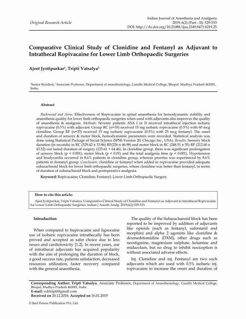
Evaluation of 25 Gauge Quincke and Whitacre Needles on Technical Problems and Post Dural Puncture Headache: A Prospective, Observational Study 563
K. Brinda, Aloka Samantaray, Mangu Hanumantha Rao
Comparison of Post Dural Puncture Headache with 23G, 25G and 27G quincke needle in Caesarean section 571
Kanvee M Vania, Dipti N Desai, Hetal Kanabar
Awake Fiberoptic Intubation with Two Different Techniques of Local Anaesthetic Administration (Transtracheal Injection Versus Ultrasonic Nebulization) in Patients Undergoing Maxillofacial Surgery 577
Karuna Sharma, Anil Kumar Bhiwal, Chintan Mukesh Kumar Patel
Identification of Epidural Space Using Modified Drip Method and Loss of Resistance Syringe Technique: A Comparative Study 585
Kinjal Sanghvi, Asmita Chaudhry, Sachin Patel, Devendra Makwana
Combined Psoas Compartment Block and Sciatic Nerve Block for Elective Lower Limb Surgeries 589
Lakshmisree M.S., Chitra Devi S.
Aero-Digestive Foreign Bodies in Tertiary Care Hospital of Southern Rajasthan: One Year Prospective Study 597
Lalit Kumar Raiger, Ravindra Gehlot, Anisha Banu

IJAA / Volume 6 Number 2 / March - April 2019 (Part - II)
528 Indian Journal of Anesthesia and Analgesia
Comparison of Analgesic Effect of Intrathecal Fentanyl & Clonidine with Hyperbaric Bupivacaine in Lower Limb Surgeries 605
Milan Vinaykant Mehta, Palak Anilkumar Chudasama
Comparative Evaluation of Butorphanol Versus Nalbuphine for Postoperative Epidural Analgesia in Lower Limb Orthopaedic Surgeries 611
Mukesh Kumar, Amit Kumar Lal, Haramritpal Kaur, Gurpreet Singh, Naresh Baghla, Harmanpreet Kaur Jhand
Effect of Dexmedetomidine in Attenuating Hemodynamic Responses During Extubation 619
Olvyna D'souza, Suman Sahu
Comparison of Tramadol & Dexamethasone as Adjuvants to Local Anaesthetic in Supraclavicular Brachial Plexus Block 627
Ruchik Solanki, Priya Kishnani, Jayshri Desai
Efficacy of Clonidine as an Adjuvant to Ropivacaine in Ultrasound guided Supraclavicular Brachial Plexus Block: A Prospective Study 632
Sarvesh. B, Prajwal Patel HS, Shivaramu BT
Comparison of the Ropivacaine and Ropivacaine with Fentanyl in Femoral Nerve Block Prior to Spinal Anaesthesia for Positioning in Orthopedic Lower Limb Surgeries 639
K. Senthil kumar, Rajasekar Janaki
Comparision of Fentanyl and Dexmedetomidine as Adjuvants to Ropivacaine for Potentiation of Post Operative Analgesia in Femoral Nerve Block for Knee Surgeries 646
Shagun Shory, Priya P. Kishnani, Chaitri Shah, Vibhav Gandhi
Comparative Study of Clinical Effects of Intrathecal Hyperbaric Bupivacaine with Fentanyl versus Hyperbaric Bupivacaine in Patients with Lower Limb Surgeries 651
Shilpa Bansal, Vaishali Vasantrao Deshpande, Sumit Bansal, Sujitkumar Dattatraya Khade
A Comparative Study Between Ultrasound and Peripheral Nerve Stimulator Guided Supraclavicular Brachial Plexus Block in Adult Patients for Elective Upper Limb Orthopaedic Surgeries 660
Srinivas HT, Girish BK, Vaibhav Nagar, Nivetha Babu
A Study to Compare the Effect of Intrathecal Midazolam and Nalbuphine as an Adjuvant to Bupivacaine for Infra-umbilical Surgeries 665
Sujay Thakkar, Tejash H Sharma, Dinesh Chauhan
Efficacy of Caudal Ropivacaine Vs Bupivacaine in Paediatric Population 670
Uma BR, Divija S
Comparison of Caudal Bupivacaine and Rectal Diclofenac for Postoperative Pain Relief in Pediatric Genitourinary and Lower Limb Surgery 678
Vishal Shrimali, Anup Chandnani, JC Vasava
Effect of Dexmedetomidine as an Adjuvant to Levobupivacaine in Spinal Anaesthesia for Infraumbilical Surgeries 684
Sofia Jaswal, Anil Ohri, Manoj Kumar Panwar, Ramesh Kumar, Vikas Jaswal
Guidelines for Authors 689
© Red Flower Publication Pvt. Ltd.
Original Research ArticleIndian Journal of Anesthesia and Analgesia
2019; 6(2) (Part - II): 529-533DOI: http://dx.doi.org/10.21088/ijaa.2349.8471.6219.25
Comparative Clinical Study of Clonidine and Fentanyl as Adjuvant to Intrathecal Ropivacaine for Lower Limb Orthopaedic Surgeries
Ajeet Jyotipurkar1, Tripti Vatsalya2
1Senior Resident, 2Associate Professor, Department of anaesthesiology, Gandhi Medical College, Bhopal, Madhya Pradesh 462001, India.
Corresponding Author: Tripti Vatsalya, Associate Professor, Department of Anaesthesiology, Gandhi Medical College, Bhopal, Madhya Pradesh 462001, India.
E-mail: [email protected] on 20.12.2018, Accepted on 16.01.2019
Abstract
Backround and Aims: Effectiveness of Ropivacaine in spinal anaesthesia for hemodyanamic stability and anaesthesia quality for lower limb orthopaedic surgeries when used with adjuvants also improves the quality of anaesthesia & analgesia. Methods: Seventy patients ASA I or II received intrathecal injection isobaric ropivacaine (0.5%) with adjuvant. Group RC (n=35) received 15 mg isobaric ropivacaine (0.5%) with 60 mcg clonidine. Group RF (n=35) received 15 mg isobaric ropivacaine (0.5%) with 25 mcg fentanyl. The onset and duration of sensory & motor block, hemodyanamic parameters were recorded. Statistical analysis was done using Statistical Package of Social Science (SPSS Version 20; Chicago Inc., USA). Results: Sensory block duration (in seconds) in RC (329.42 ± 33.86) RF(226 ± 46.98) and motor block in RC (248.51 ± 55) RF (212.60 ± 43.52) out lasted duration of surgery (125.61 + 64.46). In clonidine group, there was significant prolongation of sensory block (p < 0.001), motor block (p < 0.01) and the total analgesia time (p < 0.001). Hypotension and bradycardia occurred in 8.6% patients in clonidine group, whereas pruritus was experienced by 8.6% patients in fentanyl group. Conclusion: clonidine or fentanyl when added to ropivacaine provided adequate subarachnoid block for lower limb orthopaedic surgeries, where clonidine was better than fentanyl, in terms of duration of subarachnoid block and postoperative analgesia.
Keyword: Ropivacaine; Clonidine; Fentanyl; Lower Limb Orthopaedic Surgery.
How to cite this article:
Ajeet Jyotipurkar, Tripti Vatsalya. Comparative Clinical Study of Clonidine and Fentanyl as Adjuvant to Intrathecal Ropivacaine for Lower Limb Orthopaedic Surgeries. Indian J Anesth Analg. 2019;6(2):529-533.
Introduction
When compared to bupivacaine and lignocaine use of isobaric ropivacaine intrathecally has been proved and accepted as safer choice due to less neuro and cardiotoxicity [1,2]. In recent years, use of intrathecal adjuvants has acquired popularity with the aim of prolonging the duration of block, a good success rate, patients satisfaction, decreased resources utilization, faster recovery compared with the general anaesthesia.
The quality of the Subarachanoid block has been reported to be improved by addition of adjuvants like opioids (such as fentanyl, sufentanil and morphin) and alpha 2 agonists like clonidine & dexmedetomidine (DXM), other drugs such as neostigmine, magnesium sulphate, ketamine and midazolam, but no drug to inhibit nociception is without associated adverse effects.
Inj. Clonidine and inj. Fentanyl are two such adjuvants which are used with 0.5% isobaric inj. ropivacaine to increase the onset and duration of

IJAA / Volume 6 Number 2 / March - April 2019 (Part - II)
530 Indian Journal of Anesthesia and Analgesia
anaesthesia and analgesia. Clonidine is a partial agonist of alpha 2 adrenoceptors and acts as an analgesic and sedative. When administered intrathecally along with local anaesthetics, it improves the quality of the block and postoperative analgesia [3,4]. Fentanyl is an opioid that has shown to enhance the analgesic potency of Ropivacaine for spinal anaesthesia. Its addition to Ropivacaine for spinal anaesthesia has been shown to prolong the duration of analgesia in the early postoperative period, thus improving the quality of anaesthesia [3,4].
The aim of conducting this study in patients undergoing lower limb orthopaedic surgeries is to evaluate the ef cacity of adjuvants like fentanyl and clonidine to intrathecal ropivacaine.
Materials and Methods
The study was conducted after approval from Institutional Ethics Committee, written informed consent was obtained from all 70 patients of ASA grade I & II, 20-50 years in age scheduled for elective operations requiring subarachnoid block for lower limb orthopaedic surgery, after explaining nature of the clinical study and drugs to be used. Preoperatively all patients were explained regarding Visual analogue score (VAS) for pain.
Patient refusal, Local skin infection, Patient with allergy to study medication. Spinal deformity, bleeding diathesis, neurologic disease, Patients on antihypertensives, antipsychotics, anticoagulants, sedatives, beta blockers, MAO- inhibitors, were excluded from the study.
All eligible patients were assigned into two groups of 35 each. Group RC (n=35) - 15 mg of 0.5% isobaric Ropivacaine (3 ml) with 60 mcg Clonidine (0.4 ml + 0.1 normal saline). Group RF (n=35) - 15 mg of 0.5% isobaric Ropivacaine (3 ml) with 25 mcg Fentanyl (0.5 ml).
Detailed medical and surgical history and any previous anaesthetic exposure with its outcome were assessed. General examination including General condition, Built, Weight, Pulse rate, Blood pressure, Respiratory rate and presence of Cyanosis, Anaemia, clubbing, Jaundice or Edema were noted. Systemic examination to rule out any Cardiaovascular, Respiratory, Gastrointestinal and Neurological or any other systemic illness.
After con rming fasting patients were kept in supine positionin the operation theatre and received intravenous ringer lactate solution 10 ml/kg through large intravenous line before induction
of subarachnoid block and infusion continued during surgery. Baseline values of Heart rate (HR), blood pressure (BP), oxygen saturation (SpO
2)
,
Respiratory rate (RR) and electrocardiogram(ECG) were recorded. Subarachnoid block was performed after all aseptic precaution 25/23 gauge Quinke spinal needle was inserted in left lateral or sitting position between the L3-L4 or L4-L5 inter vertebral space. After con rmation of CSF ow study drugs were administered slowly. The spinal needle was removed and patient was immediately turned to supine position. And onset of sensory block, motor block and level of sensory block was checked with the pin prick test and the motor block level was determined according to the modi ed Bromage Scale:
1. Complete block (unable to move feet or knees)
2. Almost complete block (able to move feet only)
3. Partial block (just able to move knees)
4. Detectable weakness of hip exion while supine (full exion of Knees)
5. No detectable weakness of hip exion while supine
6. Able to perform partial knee bend.
PR, BP, RR, SpO2, pain score, discomfort and
occurrence of side effects were recorded in every 2 minutes interval for rst 10 minutes and then every 10 minutes for 120 minutes for rest of the operation. Any reduction in mean arterial pressure more than 20% from baseline or < 90 mmHg was recorded and treated with increasing dose of uids and 5-10 mg of intravenous (I.V) administration of bolus dose of inj. mephentermine, Bradycardia (Heart rate <60/min) with.inj. atropine (0.6 mg). Nausea and Vomiting with inj. Ranitidine 50 mg and inj. ondansetron 4 mg. Severe pruritus with inj.chlorpheniramine maleate, 10 mg as and when required.
Parameters noted were, Time of drug injection. Onset, Maximum height (level), Duration of sensory block. And onset and duration of motor block. Duration of analgesia ( rst rescue analgesia for pain postoperatively). Incidence of side effects.
Onset, height and duration of sensory block were assessed by pin prick method. The onset and duration of motor block was assessed by modi ed Bromage Scale. Pain intensity was evaluated using a 10 cm visual scale and patients were asked to grade the severity of their pain using this scale as mild (0-3), moderate (4-7) and severe (8-10).The level of sedation was assessed using Ramsay sedation score:

IJAA / Volume 6 Number 2 / March - April 2019 (Part - II)
531
Grade 0: Wide awake.
Grade 1: Calm and comfortable, responding to verbal commands.
Grade 2: Sleeping but arousable.
Grade 3: Deep sleep, not arousable.
Statistical analysis was done using Statistical Package of Social Science (SPSS Version 20; Chicago Inc., USA). Data comparison was done by applying speci c statistical tests to nd out the statistical signi cance of the comparisons. Quantitative variables were compared using mean values and qualitative variables using proportions.The difference in proportion was analyzed by using chi square test and the difference in means were analyzed by using student t test. Signi cance level for tests was determined as 95% (p < 0.05).
Results
The two groups were comparable with respect to demographic data and there were no signi cant differences in patient demographics and duration of surgery in both groups (Table 1).
Table 1: Patients characteristics
Demographic data (Mean ± SD )
Group RC Group RF
Patients 35 35
Age (years) 32.37 ± 9.397 35.66 ± 9.084
Sex (male/female) 26:9 26:9
Weight (kg) 57.77 ± 9.861 64.37 ± 9.991
Duration of surgery (min) 271.57 ±17.564 228.43 ± 10.345
p > 0.05 Not signi cant.
RC: Ropivacaine + clonidine group; RF: Ropivacaine + Fentanyl group.
The onset of sensory blockade was found early with fentanyl (RF4.80 ± 0.719) compared to clonidine group (RC5.46 ± 0.505) P<0.001. In our study the maximum sensory block achieved was T6 level in both the groups (p < 0.05).
The onset of Motor block was signi cantly more in RC (6.80 ± 0.797) as compared to RF (6.00 ± 0.767) p=0.001.
Mean duration of sensory block was signi cantly prolonged in Group RC (259.71 ± 16.085) as compared to Group RF (226.43 ± 10.402) p=0.001.
Mean duration of motor block was prolonged with clonidine (RC 225.71 ± 15.298) compared to fentanyl group (RF 210.29 ± 9.151) p=0.001.
Mean Duration of Analgesia also was
signi cantly more among Group RC (271.57 ± 17.564) as compared to Group RF (228.43 ± 10.345) p=0.001.
Table 2: Characteristics of Sabarachnoid blockade
Characteristics RC RF p value
Onset of Sensory Block (Min)
5.46 ± 0.505 4.80 ± 0.719 p<0.001
Onset of Motor Block (Min)
6.80 ± 0.797 6.00 ± 0.767 p<0.001
Level of Sensory Block
T6 T6 p=0.005
Duration of Sensory Block
(Min)
259.71 ± 16.085 226.43 ± 10.402 p<0.001
Duration of Motor Block
(Min)
225.71 ± 15.298 210.29 ± 9.151 p<0.001
Duration of Analgesia (Min)
271.57 ± 17.564 228.43 ± 10.345 p<0.001
Values in mean ± standard deviation. p > 0.05 Not signi cant, p < 0.05 signi cant, P <0.01 Highly signi cant, p < 0.001 Very highly signi cant. RC: Ropivacaine + clonidine group; RF: Ropivacaine + Fentanyl group.
Changes in hemodyanamic parameters were comparable in both groups. 3 patient (8.4%) in RC had hypotension (drop >25% SBP) compared to 2 patients (5.7%) in RF group and responded to inj. ephedrine, 6 mg alongwith IV uids. (5.7%) & 1 in RF group patient had bradycardia (hazard ratio <50/min) requiringinj. atropine 0.6 mg.
2 patients from both group experienced nausea/vomiting and responded to inj.ondensetron 4 mg, Pruritus was present in 3 patients (8.4%) in RF group only and inj. chlorpheniramine maleate 10 mg was given to control the same. There was no evidence of shivering and respiratory depression among both group patients. 9 patients in RC and 6 in RF required sedation and remaining patients in both the groups were calm and sleeping comfortably.
Table 3: Side effects
Side effectsGroup RC
N (%)Group RF
N (%)TotalN (%)
Hypotension 3 (8.4%) 2 (5.7%) 5 (14.1%)
Bradycardia 2 (5.7%) 1 (2.8%) 3 (8.5%)
Pruritis 0 3 (8.4%) 3 (8.4%)
Resp.depresion 0 0 0
Nausea/Vomiting 2 (5.7%) 2 (5.7%) 4 (5.72)
Shivering 0 0 0
Chi Square Value 5.081
Significance ‘p’ Value
0.024 (S)
Comparative Clinical Study of Clonidine and Fentanyl as Adjuvant to Intrathecal Ropivacaine for Lower Limb Orthopaedic Surgeries

IJAA / Volume 6 Number 2 / March - April 2019 (Part - II)
532 Indian Journal of Anesthesia and Analgesia
Discussion
The present study established that both RC and RF as an adjuvant provided satisfactory anaestheticrequisites for lower limb orthopaedic surgeries. Most features of subarachnoid block being comparable, there was signi cant early motor recovery with RF whereas RC provided prolonged postoperative analgesia.
Ropivacaine is a pure S enantiomer of bupivacaine with similarities in structural, pharmacological, physiochemical properties, and mechanism of action, with a shorter duration of motor blockade but with less cardiotoxic and neurotoxic effects than bupivacaine. Ropivacaine has been showed to be a well admissible anesthetic agent. Its ef cacy for spinal anesthesia, as compared with bupivacaine is in the ratio of 3:2, i.e.15 mg ropivacaine provided similar motor and hemodynamic effects but less potent anesthesia than 10 mg bupivacaine [5].
The main concern of our study was to evaluate the ef cacy of ropivacaine with adjuvant for major lower limb orthopedic surgeries. Yegin et al. [6] evaluated the effect of spinal fentanyl 25 mcg with 18 mg of ropivacaine for transurethral resection of prostrate and found signi cant in hancement in the duration and qualityof anesthesia without considerable increase infrequency of major side effects. This is comparable to ourstudy with the fentanyl group, where the subarachnoid features were satisfactorily met for the major lower limb surgeries. Our study showed the early onset of sensory blockade with fentanyl (RF) compared to clonidine (RC) when added to ropivacaine which is similar with the studies done by Anita R. Chhabra, Sheetal R. Jagtap, Sunny F. Dawoodi [7] And contrast to the same study who observed quick onset of motor blockade in clonidine group compared to fentanyl group and was 6.02±2 min in RC and 7.05 ± 3.2 min in RF (p > 0.05).
Clonidine, an alpha-2-agonist, when given intrathecally as an adjuant provides better quality of subarachnoid block and prolong the postoperative analgesia [8,9].
A study done by De Kock et al. [10] who used a small doseof intrathecal ropivacaine (8 mg) with different doses of clonidine (15, 45, 75 mcg) in four groups, for ambulatory surgery.
Two different doses of spinal ropivacaine 2.5 ml of 0.75% & 1% without adjuants by McNamee et al. [11] in total hip arthroplasy patients, Intraoperative hypotension found in 24% of patientsin both the groups. This could be because
of higher concentration of ropivacaine used. De Kock et al. [10], and Sagiroglu et al. [12], also noted a statistically signi cant decrease in mean BP with higher doses of clonidine when added to ropivacaine [8,16]. In our study,we used a lower dose of local anesthetic with adjuvants and the incidence of hypotension and bradycardia observed was lower in RF compared to RC group.
Thus present study showed adjuants offers prolong duration, excellent sensory & motor blockade with better sedation and comparitevely fewer side effects when used intrathecaly with isobaric ropivacaine, it could be concluded that the clonidine is a better alternative to fentanyl as an adjuant for spinal anaesthesia with isobaric ropivacaine for lower orthopaedic surgeries.
Whenever we use adjuants, it is needed to monitor hemodynamic parameters carefully.
Conclusion
Clonidine or fentanyl when added to ropivacaine provided adequate subarachnoid block for lower limb orthopaedic surgeries, where clonidine was better than fentanyl, in terms of duration of subarachnoid block and postoperative analgesia.
Acknowledgement
I convey my gratitude towards my guide Dr. Aditya Agarwal, Professor and HOD, Department of anaesthesiology, GMC Bhopal.
Con ict of Interest: Nil
References
1. Graf BM, Abraham I, Eberbach N, Kunst G, Stowe DF, Martin E. Differences in cardiotoxicity of bupivacaine and ropivacaineare the result of physicochemical and steroselective properties.Anesthesiology. 2002;96:1427-34.
2. Knudsen K, Beckman Suurkula M, Blomberg S, Sjovall J, Edvardsson N. Central nervous and cardiovascular effects of i.v. infusions of ropivacaine, bupivacaine and placebo in volunteers. Br J Anaesth 1997;78:507-14.
3. Mahendru V, Tewari A, Katyal S, Grewal A, Singh MR, Katyal R. A comparison of intrathecal dexmedetomidine, clonidine, and fentanyl as adjuvants to hyperbaric bupivacaine for lower limb surgery: A double blind controlled study. Journal of anaesthesiology, clinical pharmacology. 2013 Oct; 29(4):496.

IJAA / Volume 6 Number 2 / March - April 2019 (Part - II)
533
4. Sharan R, Verma R, Dhawan A, Kumar J. Comparison of clonidine and fentanyl as adjuvant to ropivacaine in spinal anesthesia in lower abdominal surgeries. Anesthesia, essays and researches. 2016 Sep;10(3):526.
5. Gautier PE, De Kock M, Van Steenberge A, Poth N, Lahaye-Goffart B, Fanard L, Hody JL. Intrathecal ropivacaine for ambulatory surgery a comparison between intrathecal bupivacaine and intrathecal ropivacaine for knee arthroscopy. Anesthesiology: The Journal of the American Society of Anesthesiologists. 1999 Nov 1;91(5):1239-.
6. Yegin A, Sanli S, Hadimioglu N, Akbas M, Karsli B. Intrathecal fentanyl added to hyperbaric ropivacaine for transurethral resection of prostate. Acta Anaesthesiol Scand. 2005;49:401-5.
7. Chhabra AR, Jagtap SR, Dawoodi SF. Comparison of clonidine versus fentanyl as an adjuvant to intrathecal ropivacaine for major lower limb surgeries: A randomized double-blind prospective study. Indian Journal of Pain. 2013 Sep 1;27(3):170.
8. Racle JP, Benkhadra A, Poy JY, Gleizal B. Prolongation of isobaric bupivacaine spinal anesthesia with epinephrine and clonidine for hip surgery in the elderly. Anaesth Analg. 1987;66:442-6.
9. Niemi L. Effects of intrathecal clonidine on duration of bupivacaine spinal anesthesia, hemodynamics, and post operative analgesia in patients undergoing knee arthroscopy. Acta Anaesthesiol Scand. 1994;38:724-8.
10. De Kock M, Gautier P, Fanard L, Hody JL, Lavand’ homme P. Intrathecal ropivacaine and clonidine for ambulatory knee arthroscopy: A dose-response study. Anesthesiology. 2001;94:574-8.
11. McNamee DA, Parks L, McClellands AM, Scott S, Milligan KR, Ahlén K, et al. Intrathecal ropivacaine for total hip arthoplasy: Double-blind comparative study with isobaric 7.5 mg/ml (-1) and 10 mg/ml (-1) solutions. Br J Anaesth. 2001;87:743-7.
12. Sagiroglu G, Sagiroglu T, Meydan B. The effects of adding various doses of clonidine to ropivacaine in spinal anesthesia. Eurasian J Med. 2009;41:149-53.
Comparative Clinical Study of Clonidine and Fentanyl as Adjuvant to Intrathecal Ropivacaine for Lower Limb Orthopaedic Surgeries

IJAA / Volume 6 Number 2 / March - April 2019 (Part - II)
534 Indian Journal of Anesthesia and Analgesia
Revised Rates for 2019 (Institutional)
Title of the Journal FrequencyIndia(INR)Print Only
India(INR)Online Only
OutsideIndia(USD)Print Only
OutsideIndia(USD)Online Only
Dermatology International Semiannual 5500 5000 430 391Gastroenterology International Semiannual 6000 5500 469 430Indian Journal of Anatomy Quarterly 8500 8000 664 625Indian Journal of Anesthesia and Analgesia Bi-monthly 7500 7000 586 547Indian Journal of Cancer Education and Research Semiannual 9000 8500 703 664Indian Journal of Communicable Diseases Semiannual 8500 8000 664 625Indian Journal of Dental Education Quarterly 5500 5000 430 391Indian Journal of Diabetes and Endocrinology Semiannual 8000 7500 597 560Indian Journal of Genetics and Molecular Research Semiannual 7000 6500 547 508Indian Journal of Hospital Administration Semiannual 7000 6500 547 508Indian Journal of Hospital Infection Semiannual 12500 12000 938 901Indian Journal of Medical & Health Sciences Semiannual 7000 6500 547 508Indian Journal of Pathology: Research and Practice Bi-monthly 12000 11500 938 898Indian Journal of Preventive Medicine Semiannual 7000 6500 547 508International Journal of Neurology and Neurosurgery Quarterly 10500 10000 820 781International Physiology Triannual 7500 7000 586 547Journal of Cardiovascular Medicine and Surgery Quarterly 10000 9500 781 742Journal of Global Medical Education and Research Semiannual 5900 5500 440 410Journal of Global Public Health Semiannual 12000 11500 896 858Journal of Microbiology and Related Research Semiannual 8500 8000 664 625Journal of Organ Transplantation Semiannual 26400 25900 2063 2023Journal of Orthopedic Education Triannual 5500 5000 430 391Journal of Pharmaceutical and Medicinal Chemistry Semiannual 16500 16000 1289 1250Journal of Practical Biochemistry and Biophysics Semiannual 7000 6500 547 508Journal of Radiology Semiannual 8000 7500 625 586New Indian Journal of Surgery Bi-monthly 8000 7500 625 586Ophthalmology and Allied Sciences Triannual 6000 5500 469 430Otolaryngology International Semiannual 5500 5000 430 391Pediatric Education and Research Quarterly 7500 7000 586 547Physiotherapy and Occupational Therapy Journal Quarterly 9000 8500 703 664Urology, Nephrology and Andrology International Semiannual 7500 7000 586 547Indian Journal of Maternal-Fetal & Neonatal Medicine Semiannual 9500 9000 742 703Indian Journal of Obstetrics and Gynecology Quarterly 9500 9000 742 703Indian Journal of Emergency Medicine Quarterly 12500 12000 977 938Indian Journal of Trauma and Emergency Pediatrics Quarterly 9500 9000 742 703Journal of Emergency and Trauma Nursing Semiannual 5500 5000 430 391Indian Journal of Forensic Medicine and Pathology Quarterly 16000 15500 1250 1211Indian Journal of Forensic Odontology Semiannual 5500 5000 430 391Indian Journal of Legal Medicine Semiannual 8500 8000 664 625International Journal of Forensic Sciences Semiannual 10000 9500 781 742Journal of Forensic Chemistry and Toxicology Semiannual 9500 9000 742 703Community and Public Health Nursing Triannual 5500 5000 430 391Indian Journal of Surgical Nursing Triannual 5500 5000 430 391International Journal of Pediatric Nursing Triannual 5500 5000 430 391International Journal of Practical Nursing Triannual 5500 5000 430 391Journal of Gerontology and Geriatric Nursing Semiannual 5500 5000 430 391Journal of Nurse Midwifery and Maternal Health Triannual 5500 5000 430 391Journal of Psychiatric Nursing Triannual 5500 5000 430 391Indian Journal of Ancient Medicine and Yoga Quarterly 8000 7500 625 586Indian Journal of Law and Human Behavior Semiannual 6000 5500 469 430Indian Journal of Medical Psychiatry Semiannual 8000 7500 625 586Indian Journal of Biology Semiannual 5500 5000 430 391Indian Journal of Library and Information Science Triannual 9500 9000 742 703Indian Journal of Research in Anthropology Semiannual 12500 12000 977 938Indian Journal of Waste Management Semiannual 9500 8500 742 664International Journal of Political Science Semiannual 6000 5500 450 413Journal of Social Welfare and Management Triannual 7500 7000 586 547International Journal of Food, Nutrition & Dietetics Triannual 5500 5000 430 391Journal of Animal Feed Science and Technology Semiannual 7800 7300 609 570Journal of Food Additives and Contaminants Semiannual 5000 4500 391 352Journal of Food Technology and Engineering Semiannual 5000 4500 391 352Indian Journal of Agriculture Business Semiannual 5500 5000 413 375Indian Journal of Plant and Soil Semiannual 6500 6000 508 469
Terms of Supply:1. Agency discount 12.5%. Issues will be sent directly to the end user, otherwise foreign rates will be charged.2. All back volumes of all journals are available at current rates.3. All Journals are available free online with print order within the subscription period.4. All legal disputes subject to Delhi jurisdiction.5. Cancellations are not accepted orders once processed.
6. Demand draft / cheque should be issued in favour of “Red Flower Publication Pvt. Ltd.” payable at Delhi
7. Full pre-payment is required. It can be done through online (http://rfppl.co.in/subscribe.php?mid=7).
8. No claims will be entertained if not reported within 6 months of the publishing date.9. Orders and payments are to be sent to our of ce address as given above.10. Postage & Handling is included in the subscription rates.11. Subscription period is accepted on calendar year basis (i.e. Jan to Dec). However orders may be placed any time throughout the year.
Order mRed Flower Publication Pvt. Ltd., 48/41-42, DSIDC, Pocket-II, Mayur Vihar Phase-I, Delhi - 110 091 (India),
Mobile: 8130750089, Phone: 91-11-45796900, 22754205, 22756995 E-mail: [email protected], Website: www.rfppl.co.in

© Red Flower Publication Pvt. Ltd.
Original Research ArticleIndian Journal of Anesthesia and Analgesia
2019; 6(2) (Part - II): 535-541DOI: http://dx.doi.org/10.21088/ijaa.2349.8471.6219.26
Comparison Intraarticular Ropivacaine and Ropivacaine Plus Dexmedetomidine for Post Operative Analgesia in Arthroscopic Knee Surgery
Chirag Patel1, Anup Chandnani2, Amit Sharma3
1Assistant Professor, 2Assistant Professor, 3Resident Doctor, Department of Anesthesiology, BJ Medical College, Ahmedabad, Gujarat 380016, India.
Corresponding Author: Anup Chandnani, Assistant Professor, Department of Anesthesiology, BJ Medical college, Ahmedabad, Gujarat 380016, India.
E-mail: [email protected]
Received on 06.02.2019, Accepted on 25.03.2019
Abstract
Arthroscopic knee surgery is routinely performed these days for various indications none the less with pain in post operative period requiring analgesic cover. Pain is mediated through Opiate receptors in intraarticular tissue. Dexmedetomidine, a alpha (α) 2 adrenergic agonist whose administration Intra articularly along with Inj. Ropivacaine (local anesthetic) does helps in providing good analgesia postoperatively. With this background this comparative we planned study between inj. ropivacaine and ropivacaine with dexmedetomidine deposited intra articularly for post operative analgesia in arthroscopic knee surgery. Duration of analgesia, efficacy of drugs and any side effects between both groups were compared. Fifty patients undergoing elective arthroscopy surgery of knee joint of ASA grade II & III; 18-65 years of age of either sex were included and after the surgery was over, they were randomly divided in two groups of 25; Group A (n=25) to receive Inj. Ropivacaine 0.25% and Group B (n=25) to receive Inj. Ropivacaine (0.25%) + Inj. Dexmedetomidine (1 µg/kg) making final volume 20 ml pushed intra-articularly under aseptic precautions by the operating surgeon. Patients were observed postoperatively for Pulse, RR, Temp, BP, Sedation using Modified Wilson Score and Pain assesment at rest using VAS every 2 hourly for first postoperative day. Rescue analgesic using Inj. Diclofenac sodium 75 mg IV was given at VAS score was ≥ 4. The duration (in hours) for first dose of analgesic was considerably more in group B [2.68 ± 0.48] than group A [1.88 ± 0.34]. There were no significant postoperative complications in both groups. So mixing of Intraarticular Dexmedetomidine with Ropivacaine provides longer duration of analgesia and reduced pain scores as compared to Intraarticular Ropivacaine alone safely.
Keywords: Ropivacaine; Dexmedetomidine; Post Operative Analgesia; Arthroscopy; VAS (Visual Analogue Scale).
How to cite this article:
Chirag Patel, Anup Chandnani, Amit Sharma. Comparison Intraarticular Ropivacaine and Ropivacaine Plus Dexmedetomidine for Post Operative Analgesia in Arthroscopic Knee Surgery. Indian J Anesth Analg. 2019;6(2):535-541.
Introduction
Effective postoperative pain control is an essential component of the care of the surgical patient. Inadequate pain control may result in increased morbidity or mortality. Postoperative pain de nitely indicates about some abnormal activity from recently damaged tissue being relayed
to Central nervous system, the intensity depending on type of tissue damage, the healing process and patient factors.
Arthroscopic procedures for knee is routinely performed surgery. Patients have moderate to severe post operative pain after knee arthroscopy. Pain affects patient’s activity level, discharge and has negative impact on psychology which

IJAA / Volume 6 Number 2 / March - April 2019 (Part - II)
536 Indian Journal of Anesthesia and Analgesia
causes discomfort and early ambulation is not achieved. These all factors advocate for use of an ideal analgesic with quicker onset, minimal side effects and help in early mobilization, recovery and discharge.
Various studies evaluated different factors and drugs which in uence post arthroscopy pain such as-Type of surgery, tourniquet used or not; its duration and timing with drug administration, Anaesthesia technique-GA or RA, Volume injected, adjuvant, Preoperative pain threshold, Gender of the patients, Residual effects of peri operative analgesia, amount of surgical trauma.
Pain is sensed by Opiate receptors and free nerve endings. To achieve postoperative pain relief many techniques have been tried e.g. nerve blocks, intra articular administration of various drugs [3,4,13,14] and systemic drugs to control post arthroscopic pain.
Various combinations of local anaesthetics (lidocaine, bupivacaine), opioids (morphine), α-2 adrenoceptor agonists (clonidine and magnesium sulphate) have been tried intra articularly for post operative analgesia. Dexmedetomidine is alpha (α) 2 adrenergic agonist. Its intravenous administration before regional anesthesia does provide postoperative analgesia but with some adverse hemodynamic effects and respiratory depression. Use of S (-) enantiomer, amide local anaesthetic 150 mg Ropivacaine 0.75% intraarticularly helps in providing good sensory and motor block with minimal side effects and cardiac stability than bupivacaine.
A study by DR. Anil. K. Paswan and DR. Shashi Prakash et al. [2] on Effect of intra-articular adjuvants like dexmedetomidine and opioids on postoperative analgesia for arthroscopic knee surgery concluded that post operative use of rescue analgesia was duly prolonged.
A similar study by S paul, D P Bhattacharjee, S Ghosh et al. (2010) [1] about ef cacy of intraarticular ropivacine with or without α-2 adrenergic agonist for postoperative analgesia in knee surgeries were of conclusion that Dexmedetomidine and as an addition to ropivacaine improves the quality and duration of postoperative analgesia without any adverse effects.
Henceforth this study comparing ropivacaine with and without dexmedetomidine for post-operative analgesia in arthroscopic knee surgery was done to:-
• Assess and compare post-operative pain free period.
• Demand of rescue analgesia.
• Ef cacy of drugs and any associated complications.
Methodology
After approval of ethics committee this prospective randomized double blind comparative study was done at civil hospital, BJ Medical College, Ahmedabad between intraarticular ropivacaine with and without dexmedetomidine for post-operative analgesia in knee surgery done arthroscopically involving 50 patients.
ASA II and III patients within age between 18-65 years of either sex were taken in this study. Patients with history of infection, cardiac disease, coagulopathy, hepatic or kidney disease were not a part of this study. After a detailed preoperative check up with all relevant investigations done, explanation of the procedure, type of anesthesia and participation in evaluation of post operative analgesia, patients were subjected to arthroscopy. A proper understanding of Visual Analogue Scale was made to the patients.
Tab diazepam 5 mg orally a night before operation was given to all patients. Preoperative monitoring of the basic vital parameters, preloading with Inj. Ringer Lactate and Inj. Ondansetron 75-100 mcg/kg IV was given prior to anesthesia. Spinal anaesthesia was given in sitting position using inj. bupivacaine 0.5% (heavy) 12-15 mg using a 23-gauge Quincke needle in L
2-L
3 or L
3-L
4 inter
vertebral space under strict aseptic and antiseptic precautions. Onset and level of sensory was to be achieved maximum between T8 to T10 segment and motor block was recorded after making the patient supine immediately after giving spinal anesthesia. A thigh tourniquet [14] was applied on the operative limb with a pressure 250-350 mm Hg continuously during surgery. Per operative monitoring of Pulse, BP, heart rate and SpO
2,
EtCO2, ECG, RR was done at every 15 minutes
throughout the surgery.
After the surgery was over, patients were randomly categorised in two groups to receive 20 ml of drug preparation; Group A (n=25): received Inj. Ropivacaine 0.25% and Group B (n=25): received Inj. Ropivacaine (0.25%) + Inj. Dexmedetomidine (1 µg/kg) which was given intra-articularly under aseptic care by the operating surgeon. Till then the torniquet was kept in ated and was de ated after 10 minutes of intraarticular injection of drug in both groups.

IJAA / Volume 6 Number 2 / March - April 2019 (Part - II)
537
Patients were monitored postoperatively for the vital parameters, Sedation (by Modi ed Wilson Score 1-5) (Table 1) and Pain assesment at rest using VAS (Table 2) (0= No Pain, 10 = Worst Possible Pain) upto 24 hr at interval of 2 hours. Rescue analgesia of inj. Diclofenac sodium 75 mg IV was given when VAS was ≥ 4. Total duration of analgesia was taken as the duration from intra-articular deposition of the drug to the requirement of rst analgesic. Improvement in VAS score, duration of analgesia and total number of rescue analgesics during 24 hrs. in post-operative period decided the ef cacy of the drug used. Time for regression of the spinal effect was also noted.
The patients were monitored for any undue complications in the post operative period. These observations were made by an observer who had no clue about the patient’s group.
Unpaired Student’s t-test was used to compare both groups statistically. ‘p’ value < 0.05 was considered signi cant. Mean and standard deviation were calculated and p value derived.
Results
Table 3 shows comparable date under both groups in terms of age and weight.
Table 4 shows the comparative duration of anesthesia and surgery in the both the groups, statistically comparable, group B (170 ± 29.64) and (152.6 ± 30.76) as compared to group A (156.48 ± 39.04) and (137.56 ± 38.06), (p>0.05) respectively.
Various types of arthroscopic assisted surgeries were included in the study as shown in (Table 5).
Table 1: Modified Wilson Sedation Scale:
Score Description
1Oriented, eyes may be closed but can respond to Questions ‘Can you tell me your name?’ ‘Can you tell me where you are right now?’
2Drowsy; eyes may be closed, arousable only to command:(Name), please open your eyes.
3 Arousable to mild physical stimulation (earlobe tug)
4 Unarousable to mild physical stimulation
Table 2: Visual Analogue Score (with or without Movement)
0 1 2 3 4 5 6 7 8 9 10
No pain Worse pain
Table 3: Demographic data (Mean ± SD)
Demographic data Group A Group B p value
Age (years) 29.92 ± 9.12 30.92 ± 10.64 0.72
Weight (kg) 65 ± 6.79 60.48 ± 7.64 0.32
Male: Female 20:5 20:5 -
(p value<0.05 is considered significant.)
Table 4: Duration of anaesthesia and surgery for two groups (minutes)
Group A Group B p Value
Duration of anaesthesia 156.48 ± 39.04 170 ± 29.64 0.16
Duration of surgery 137.56± 38.06 152.6 ± 30.76 0.13
(p value < 0.05 is considered significant.)
Table 5: Type of surgery in both groups
Type of Surgeries Group A Group B
Arthroscopic Acl Repair 15 15
Arthroscopic Pcl Repair 2 1
Arthroscopic Meniscal Repair 5 5
Arthroscopic Knee Release - 2
Arthroscopic Rim Reconstruction - 1
Arthroscopic Synovectomy 1 -
Arthroscopic Debridement - 1
Diagnostic Arthroscopy 2 -
Total 25 25
Comparison Intraarticular Ropivacaine and Ropivacaine Plus Dexmedetomidine for Post Operative Analgesia in Arthroscopic Knee Surgery

IJAA / Volume 6 Number 2 / March - April 2019 (Part - II)
538 Indian Journal of Anesthesia and Analgesia
In the post operative period the changes in the pulse rate was statistically insigni cant when compared between intra groups and inter group (Graph 1).
Graph 2 shows the comparative post-operative systolic blood pressure changes in both groups at different time intervals which were statistically comparable, except after 20 hours.
Table 6 and Graph 3 show none of the patient in both groups complained of pain initially for two hours. At 6 hrs patients in Group A had VAS score of 3.04 ± 0.98 whereas in group B it 1.76 ± 0.44, which is statistically signi cant (p value <0.05). Later at 2, 6 and 8-hour VAS score in Group A was 1.68 ± 0.8, 2.48 ± 0.77, 3.04 ± 0.98 respectively as compared to group B where it was 0.99 ± 0.42, 1.049 ± 0.2 and 1.76 ± 0.44 respectively. These changes were statistically signi cant (p=0.0001, p=0.0001, p=0.0013).
In Group A, up to 8 hr there was requirement of rescue analgesia in 24/25 whereas in group B
none of patients required rescue analgesics till 8 hours. Noting more speci cally, the requirement of rescue analgesics was in 13/25 patients at 6 hr, 9/25 patients at 14 hr in Group A and in Group B 14/25 patients at 12 hr, 10/25 patients at 20 and 22 hr. These have been shown in table 7.
Table 8 & Graph 4 show the duration of analgesia, which was signi cantly more in Group B (11.42 ± 1.25 hr) as compared to Group A (6.4 ± 1.29 hr). (p Value <0.0001). The number of doses of rescue analgesics was less in Group B (1.88 ± 0.34) as compared to Group A (2.68±0.48). (p Value < 0.0001).
The known and anticipated complications like nausea, vomiting, respiratory depression or convulsions post operatively were not seen in any patients of both the groups. Hypotension was seen in one patient from Group A and two patients from Group B which was treated with IV uids. Bradycardia was noted in two patients of group B.
Graph 1: Comparison of post operative heart rate between two groups
Graph 2: post operative systolic blood pressure changes

IJAA / Volume 6 Number 2 / March - April 2019 (Part - II)
539
Table 6: VAS score between two groups (Mean ± SD) (At rest)
Postoperative Time Group A Group B p Value(between the groups)
0 hr 0.16 ± 0.37 0 Not applicable
2 hr 1.68 ± 0.80 0.99 ± 0.42 0.0040
4 hr 2.48 ± 0.77 1.04 ± 0.20 0.0001
6 hr 3.04 ± 0.98 1.76 ± 0.44 0.0010
8 hr 2.92 ± 0.91 2.08 ± 0.28 0.0001
10 hr 2.20 ± 0.58 3.32 ± 0.99 0.0001
12 hr 2.64 ± 0.76 3.40 ± 0.87 0.0019
14 hr 3.16 ± 0.90 2.44 ± 0.65 0.0020
16 hr 2.88 ± 0.88 2.28 ± 0.46 0.0040
18 hr 2.92 ± 1.08 2.72 ± 0.54 0.1
20 hr 2.56 ± 0.77 3.32 ± 0.85 0.0018
22 hr 2.96 ± 0.79 3.00 ± 0.91 0.80
24 hr 2.68 ± 0.80 2.24 ± 0.52 0.02
(p value<0.05 is considered significant.)
Graph 3: VAS score between two groups (At rest)
Table 7: Number of patients required rescue analgesic.
2 Hours
4 Hours
6 Hours
8 Hours
10 Hours
12 Hours
14 Hours
16 Hours
18 Hours
20 Hours
22 Hours
24 Hours
Group A 0 3 13 8 1 4 9 7 5 4 6 6
Group B 0 0 0 0 9 14 2 0 1 10 10 1
Table 8: Duration of analgesia and no. of doses of rescue analgesics between two groups
Group A Group B p Value
Duration of Analgesia (hrs) 6.40 ± 1.29 11.42 ± 1.25 p<0.0001
No. of doses of Rescue analgesics 2.68 ± 0.48 1.88 ± 0.34 p<0.0001
(p value < 0.05 is considered significant.)
Table 9: Postoperative complications
Postoperative complications Group A Group B
Hypotension 1 2
Bradycardia - 2
Convulsions - -
Respiratory depression - -
Nausea - -
Vomiting - -
Dizziness - -
Comparison Intraarticular Ropivacaine and Ropivacaine Plus Dexmedetomidine for Post Operative Analgesia in Arthroscopic Knee Surgery

IJAA / Volume 6 Number 2 / March - April 2019 (Part - II)
540 Indian Journal of Anesthesia and Analgesia
Graph 4: Duration of analgesia and no. of doses of rescue analgesics between two groups
Discussion
Post operative pain contributes to patient dissatisfaction with their surgical experience. So a strategic programme during the recovery phase post operatively which covers the post operative pain management provides reduced sympathetic stress response, reduced postoperative pulmonary and cardiac complications becomes mandatory which also includes bene ts like early physiotherapy, early mobilization and early discharge.
Dexmedetomidine [5,6,7] is a centrally acting highly selective α-2 agonist with pharmacodynamics including anxiolysis, sedation, sympatholysis, analgesia and anesthesia without respiratory depression. It acts on the alpha 2 adrenergic presynaptic receptor agonist for providing analgesia; Dexmedetomidine is highly selective alpha-2 adrenoceptor agonist making it more potent as compared to clonidine [3] and also acts synergistically with the local anesthetic agent potentiating its action. This property of it de nitely helped in the post operative analgesia during its use intra articularly during knee surgery.
Ropivacaine [8,9,10] a local anesthetic blocks the action potential of the nerve conduction by its selective action on the sodium channels of the nerve. It was found to be more safe with no side effects when used intra articularly.
Demographically the patient data was comparable in relation to Age, Sex, Weight and ASA physical status.
50 patients posted for elective knee arthroscopy were anesthetized with spinal anesthesia with
bupivacaine heavy (15-20 mg). At the end of surgery they were divided into 2 groups; Group A (n=25) received Inj Ropivacaine (0.25%) (19 ml) and Inj.0.9% Normal saline (1 ml) while Group B (n=25) received Inj Ropivacaine (0.25%)(19 ml) and Inj. Dexmedetomidine (1 µg/kg)(1 ml), total volume 20 ml intraarticularly at the end of arthroscopy.
Tourniquet was kept de ated after 10 minutes of intraarticular drug administration to facilitate increase intraarticular pressure thereby enhanciing systemic absorption after the de ation.
R.R. Al Metwalli (2008) compared intraarticular and intravenous dexmedetomidine in 60 adult patients of ASA I-II, posted for arthroscopic partial meniscectomy under general anaesthesia for postoperative analgesia in a double blind randomized study. They concluded that the patients who had intra articular injection had quite low pain post operatively even upto 6 hours. Comparing the sedation after injection, it was found to be more in the intravenous group in their study. We used intraarticular dexmedtomidine in low dose (1 µg/kg) with LA to prevent systemic side effects like sedation in our study. The post operative analgesia time for intra articular injection in our study was comparable to theirs.
The results in our study were comparable to the study done by S paul, D P Bhattacharjee, S Ghosh et al. (2010) [1] who concluded that adding dexmedetomidine to ropivacaine does give a good post operative pain free period cutting short the use of rescue analgesics.
In our study, patient stayed stable hemodynamically derived statistically in both groups.

IJAA / Volume 6 Number 2 / March - April 2019 (Part - II)
541
Pain does stimulate sympathetic response but good analgesic cover helps to curtail this response which was seen in our study. The recovery time from spinal anesthesia was also similar in both groups with good analgesia cover in group B as compared to group A (where VAS was 2.48 ± 0.77 after four hours) which further increased to 3.04 ± 0.98 at end of 6 hours in post operative period.
It was observed that the mean duration of analgesia was longer in group B (11.42 ± 1.25 hr) as compared to group A (6.40 ± 1.29) (p<0.01) as derived from the VAS values at end of 4,6 and 8 hours (Graph 3) minimizing the use of rescue analgesics in group B. Using Dexmedetomidine in optimally low dose helped to keep the sedation score at bay with patients easily arousable. 1/25 patient (4%) in group A and 2/25 patients (8%), Group-B developed hypotension which were treated with IV uids. 2/25 patients of group B developed bradycardia which were treated by iv inj atropine. None patients developed convulsions or respiratory depression.
The only limitation to our study was to not able to get the plasma concentration of dexmedetomidine.
Concluding that dose of (1 µg/kg) dexmedetomidine along with Inj. Ropivacaine (0.25%) 20 ml used in intra articular space is suf cient enough for providing adequate pain relief in the post operative period (sparing few side effects).
Conclusion
Use of Ropivacaine local anesthetic with and without Dexmedetomidine for arthroscopic knee surgeries in our study revealed that post operative pain free period was longer and use of systemic analgesics to relieve pain was much less in the study group (Ropivacaine with Dexmedetomidine). These were all derived by use of VAS, Modi ed Wilsons sedation score done by double blind technique with bare minimum hemodynamic and respiratory side effects.
Henceforth, it can be concluded that addition of Intraarticular Dexmedetomidine to Ropivacaine produces signi cant longer duration of analgesia, advocating less use of systemic analgesics and better patient compliance and satisfaction in the post operative period as compared to Intraarticular Ropivacaine alone without any signi cant side effect.
References
1. S Paul, D P Bhattacharjee, S Ghosh, et al.: Use of intra-articular dexmedetomidine for postoperative analgesia in arthroscopic knee surgery; Ceylon Medical Journal. 2010;55:111-15.
2. Efthimios P Samoladas, Byron Chalidis, Hlias Fotiadis, et al. The intra-articular use of ropivacaine for the control of post knee arthroscopy pain; Journal of Orthopaedic Surgery and Research. 2006;1:17.
3. Gentili M, Juhel A and Bonnet F. Peripheral analgesic effect of intra-articular clonidine. Pain. 1996;64:593-6.
4. Anju Grewal. Dexmedetomidine: New Avenues; Journal of Anaesthesiology and Clinical Pharmacology. 2011 July-Sept;27(3):297-302.
5. Ankit Agarwal, R.K. Verma, et al. Ropivacaine – a new local Anaesthetic in Indian Market. J. Anaesth Clin Pharmacol. 2010;26(2):223-228.
6. Gaurav Kuthiala, Geeta Chaudhary Ropivacaine: A Review of its pharmacology and clinical use. Indian J Anaesth. 2011 Mar;55(2):104-10.
7. Joseph Baker; Use of Local Anaesthetic Agent in Arthroscopy. In book: Pain Management - Current Issues and Opinions.
8. Lawrence AJ, Joshi GP; et al. Evidence for analgesia transmitted by peripheral opioid receptors in inflamed synovial tissue. European J. Clin. Pharmacol. 1992;43:351-55.
9. Mukherji S, Rudra A. Postoperative pain relief for day care ambulatory surgery; Indian journal of anesthesia. 2006;50(5):355-362.
10. Whitford A, Healy M, Joshi GP, McCarroll SM and O’Brien TM. The effect of tourniquet release time on the analgesic efficacy of intraarticular morphine after arthroscopic knee surgery. Anesth. and Analg., 1997;84:791-793.
Comparison Intraarticular Ropivacaine and Ropivacaine Plus Dexmedetomidine for Post Operative Analgesia in Arthroscopic Knee Surgery

IJAA / Volume 6 Number 2 / March - April 2019 (Part - II)
542 Indian Journal of Anesthesia and Analgesia
Indian Journal of Anesthesia and Analgesia
Library Recommendation Form
If you would like to recommend this journal to your library, simply complete the form below and return it to us. Please type or print the information clearly. We will forward a sample copy to your library, along with this recommendation card.
Please send a sample copy to:
Name of Librarian
Name of Library
Address of Library
Recommended by:
Your Name/ Title
Department
Address
Dear Librarian,
I would like to recommend that your library subscribe to the Indian Journal of Anesthesia and Analgesia. I believe the major future uses of the journal for your library would provide:
1. useful information for members of my specialty.
2. an excellent research aid.
3. an invaluable student resource.
I have a personal subscription and understand and appreciate the value an institutional subscription would mean to our staff.
Should the journal you’re reading right now be a part of your University or institution’s library? To have a free sample sent to your librarian, simply fill out and mail this today!
Stock Manager Red Flower Publication Pvt. Ltd.48/41-42, DSIDC, Pocket-IIMayur Vihar Phase-IDelhi - 110 091(India)Phone: Phone: 91-11-45796900, 22754205, 22756995, Cell: +91-9821671871E-mail: [email protected]

© Red Flower Publication Pvt. Ltd.
Original Research ArticleIndian Journal of Anesthesia and Analgesia
2019; 6(2) (Part - II): 543-546DOI: http://dx.doi.org/10.21088/ijaa.2349.8471.6219.27
To Compare the Efficacy of Midazolam and Triclofos as Oral Premedicant in Paediatric Patients
Anubhav Sardana1, Pooja A Shah2, Malini Mehta3
13rd Year Resident, 2Assistant Professor, 3Professor, Dept. of Anaesthesiology, SBKS Medical Institute & Research Center, Sumandeep Vidyapeeth University, Piparia, Vadodara, Gujarat 391760, India.
Corresponding Author: Pooja A Shah, Assistant. Professor, Dept. of Anaesthesiology, SBKS Medical Institute & Research Center, Sumandeep Vidyapeeth University, Piparia, Vadodara, Gujarat 391760, India.
E-mail: [email protected]
Received on 19.12.2018, Accepted on 16.01.2019
Abstract
Context: The primary goals of premedication in children are to facilitate a smooth separation from the parents and to ease the induction of anesthesia. Aims: To compare the efficacy of midazolam and triclofos when given orally as premedicants in children. Material and methods: 50 patients aged 2-6 years of ASA I & II were divided randomly into two groups equally. Group M received syrup midazolam, 0.5 mg kg-1 and group T received syrup triclofos 75 mg kg-1, orally as premedication. Level of sedation and behavior [1,3] at the time of separation from parents and during mask acceptance [1,3]. Statistical analysis: Unpaired t-test was used for statistical analysis. p-value <0.05 is significant. Results: In group-M 56% and 40% patients while in group-T 24% and 8% patients achieved a sedation score of 4 and 5 respectively (p<0.05). In group-T 64% and 24% while in group-M 8% and 12% patients achieved a behavior score of 1 and 2 respectively at the time of separation from parents (p<0.05). In group-M 68% and in group-T 24% patients achieved a mask acceptance score of 4 (p<0.05). Conclusion: Oral midazolam 0.5 mg kg-1 was better in terms of level of sedation and behavior at the time of mask acceptance whereas, triclofos 75 mg kg-1 was better in terms of behavior at the time of separation from parents in paediatric age group 2 to 6 years. Hence, midazolam was found to be superior to triclofos.
Keywords: Paediatric; Midazolam; Triclofos; Sedation; Behaviour score
How to cite this article:
Anubhav Sardana, Pooja A Shah, Malini Mehta. To Compare the Efficacy of Midazolam and Triclofos as Oral Premedicant in Paediatric Patients. Indian J Anesth Analg. 2019;6(2):543-546.
Introduction
Paediatric patients are to be given special considerations not only with respect to anatomic, physiologic and pharmacologic differences but also behavioral aspect. Children admitted to hospitals are displaced from their comfort zone of home and family. The primary goals of premedication in children are to facilitate a smooth separation from the parents and to ease the induction of anesthesia. This study was conducted to compare the ef cacy
of midazolam and triclofos when given orally as premedicant in children.
Materials and Methods
This study was carried out in the department of Anaesthesiology after getting approval from the institutional ethical and research committee.Written and informed consent was taken from parents.

IJAA / Volume 6 Number 2 / March - April 2019 (Part - II)
544 Indian Journal of Anesthesia and Analgesia
Inclusion Criteria
• Children belonging to American Society of Anaesthesiologists (ASA) physical status I or II
• Age: 2-6 years.
• Either gender.
• Scheduled for elective surgery.
• Body weight up to 20 kg.
• No known history of drug allergy, sensitivity or other form of reaction.
• Patient whose parents were willing to sign informed consent.
Exclusion Criteria
• Patients with ASA III or IV or V.
• Children on anticonvulsant therapy and other sedative medications.
• Those likely to have anticipated dif cult airway.
• Known sensitivity to benzodiazepines.
• Scheduled for neurosurgical procedures.
• Children with mental retardation.
• Risk of pulmonary aspiration.
• Those patients whose parents were not willing to participate in the study.
• Patient allergic to any drug.
• Patient with renal, hepatic, cardio vascular and respiratory disease.
50 patients aged 2-6 years of ASA I & II were divided randomly into two groups equally.
Baseline heart rate (H.R.), systolic blood pressure (SBP), diastolic blood pressure (DBP), temperature and pulse oximeter (SpO
2) were monitored and
recorded. Intravenous (i.v.) line was secured and isolyte p was started. All patients received inj. glycopyrolate 0.004 mg kg-1 and inj. ondansetron
0.1 mg kg-1 i.v. Patients were allocated into either of the two groups. Group M received syrup midazolam, 0.5 mg kg-1 and group T received syrup triclofos 75 mg kg-1, administered orally as premedication 30 minutes and 60 minutes respectively before surgery. Level of sedation at the time of separation from parents [1,3]. Behaviour at the time of separation from parents and during mask acceptance were assessed [1,3].
Patients were pre-oxygenated with face mask with 100% oxygen for 3 minutes. Anesthesia was induced by a standard technique of intravenous induction with inj. sodium thiopentone 5 mg kg-1
i.v.. Endotracheal intubation was done after giving inj.succinyl choline 2 mg kg-1 i.v. Anaesthesia was maintained on O
2, N
2O, sevo urane and
inj. atracurium 0.5 mg kg-1 i.v. Intra-operatively children were monitored for HR, SBP, DBP, SpO
2 every 15 minutes till end of surgery. At the
end of surgery, neuromuscular blockade was reversed with inj. Neostigmine 0.05 mg kg-1 and inj.glycopyrolate 0.008 mg kg-1 i.v. Trachea was extubated after ful lling the recovery criteria and shifted to recovery room.
Postoperatively, HR, SBP, DBP, SpO2, any
adverse events such as nausea, vomiting, rigor, hypotension, bradycardia, and respiratory depression were observed every 15 minutes upto 2 hours.
Results
Total 50 patients were allocated for the study. Both groups were comparable in respect to age, sex, weight and duration of surgery (p > 0.05).
There were no any complications or side effects in any of the groups. There was no statistically signi cant difference in mean heart rate, systolic blood pressure, diastolic blood pressure and SpO
2
in both groups intraoperatively and postoperatively (p > 0.05).
Table 1: Level of sedation scores [1,3] between the groups
Level of sedation Group-M Group-T p-value
Score 1 = Child awake and oriented 0 0 -
Score 2 = Drowsy 0 2 (8%) <0.05
Score 3 = Eyes closed but arousable to command 1 (4%) 15 (60%) <0.05
Score 4 = Eyes closed, but arousable to mild physical stimulation
14 (56%) 6 (24%) <0.05
Score 5 = Eyes closed, but unarousable to mild physical stimulation
10 (40%) 2 (8%) <0.05

IJAA / Volume 6 Number 2 / March - April 2019 (Part - II)
545
Table 2: Behaviour at the time of separation from parents between the groups.
Behaviour at the time of separation from parents [1,3] Group-M Group-T p-value
Score 1 = excellent-happily separated 2 (8%) 16 (64%) <0.05
Score 2 = good-separated without crying, 3 (12%) 6 (24%) <0.05
Score 3 = fair-separated with crying, 12 (48%) 3 (12%) <0.05
Score 4 = poor need for restraint 8 (32%) 0 <0.05
Table 3: Behaviour during mask acceptance between the groups.
Behaviour during mask acceptance [1,3] Group- M Group-T p-value
Score 1 = Poor – afraid, combative, crying 0 0 -
Score 2 = Fair – moderate fear of mask, not easily calmed
0 13 (52%) <0.05
Score 3 = Good – slight fear of mask, easily calmed 8 (32%) 6 (24%) >0.05
Score 4 = Excellent – unafraid, cooperative, accepts mask easily
17 (68%) 6 (24%) <0.05
Table 4: Surgeries undergone by the patients between the groups.
Type of surgery Group-M Group-T p-value
Colostomy closure 5 (20%) 3 (12%) >0.05
Circumcision 2 (8%) 3 (12%) >0.05
Hernioplasty 13 (52%) 12 (48%) >0.05
Lords placation 4 (16%) 2 (8%) <0.05
Hypospadias repair 1 (4%) 5 (20%) <0.05
Discussion
Midazolam & triclofos were compared to identify effective and safe premedicant for pediatric patients aged between 2 and 6.
In the current study midazolam syrup was given in the dose of 0.5 mg kg-1. Studies by Kolathu PR et al. [1], Choudhary S et al. [2], Geetha L [3] showed that the dose of 0.5 mg kg-1 of midazolam orally has proven to be ef cacious in children with fewer side-effects. Hence a dose of 0.5 mg kg-1 was chosen for midazolam.
Triclofos syrup was used in the dose of 75 mg kg-1. Choudhary S et al. [2] and Parameshwari A et al. [4] used triclofos 75 mg kg-1 and concluded that this was the safe dose with fewer side effects, hence the dose of 75 mg kg-1 was chosen for triclofos.
Midazolam is rapidly absorbed in gastrointestinal tract and produces its peak effect in 30 mins [6].
Triclofos oral solution is well-absorbed and shows ef cacy within 30-40 minuites [7]. Choudhary S et al. [2] and Geetha L et al. [3], reported that maximum percentage of patients achieving excellent sedation scores at 60 min. Hence, the time for assessment of sedation for triclofos was selected as 60 minutes [2].
In our study differences in demographic data between the two groups were statistically not-signi cant (p>0.05), similar to the studies
conducted by Chaudhary S [2], Kolathu PR et al. [1] and Jose MR et al. [5].
On comparing level of sedation in both the groups, it was observed that in the group-M, 40% patients had achieved a score of 5 (eyes closed, but unarousable to mild physical stimulation) while only 8% patients in group-T achieved a score of 5 which is depicted in table 1. Our study was comparable to studies carried out by Kolathu PR et al. [1] and Geetha L et al. [3]. Jose MR et al. [5]
observed that in midazolam group 88% of patients had sedation score of 2 whereas in triclofos group 84% had sedation score 4, which was differed to our study.
In our study behavior score at the time of separation from parents was excellent (score 1 = happily separated) in 64% patients & good (score 2 = separated without crime) in 24% patients in group-T as compared to group-M wherein only 8% and 12% patients achieved score-1 and score-2 respectively. Thus triclofos resulted in excellent behavior at the time of separation from parents as compared to midazolam (Table 2). Kolathu PR et al. [1], Choudhary S et al. [2], Jose MR et al. [5], Geetha et al. [3] have observed no difference in separation score.
When behavior during mask acceptance was compared in both the groups, it was seen that a signi cantly higher proportion of patients in
To Compare the Efficacy of Midazolam and Triclofos as Oral Premedicant in Paediatric Patients

IJAA / Volume 6 Number 2 / March - April 2019 (Part - II)
546 Indian Journal of Anesthesia and Analgesia
group-M achieved a score 4 = Excellent – unafraid, cooperative, accepts mask easily (68%) as compared to group-T wherein only 24% patients achieved score-4 and this difference was clinically signi cant (p<0.05) (Table 3). Thus midazolam resulted in excellent behavior during mask acceptance as compared to triclofos which correlates with the studies carried out by Chaudhary S et al. [2], Parmeshwari A et al. [4].
Midazolam exerts it effects through reversible interactions with GABA receptor in the CNS which is an inhibitory neurotransmitter. It produces sedation, anxiolysis, amnesia and hypnosis.
Triclofos is converted to Trichloroethanol in the body which induces sedation by acting as melatonin agonist. It has rapid onset of action and produces drowsiness.
In our study, there was no statistically signi cant difference in mean heart rate, systolic blood pressure, diastolic blood pressure and SpO
2 in both
groups intraoperatively and postoperatively (p > 0.05).
Similar observations were seen in the study by Kolathu PR et al. [1], Chaudhary S [2], Jose MR et al. [5], Parmeshwari A et al. [4] and Geetha L et al. [3] wherein there was no signi cant difference seen in vitals or oxygen saturation in the intraoperative and post-operative periods.
None of the patients in any of the groups had side effects like nausea, vomiting, rigors, hypotension, bradycardia etc. which was similar to the studies carried out by Kolathu PR et al. [1], Chaudhary S [2], Jose MR et al. [5], Parmeshwari A et al. [4] and Geetha L [3].
Conclusion
We concluded that oral midazolam 0.5 mg kg-1 was better as compared to triclofos 75 mg kg-1 in terms of level of sedation and behaviour at the time of mask acceptance whereas, triclofos was better in
terms of behaviour at the time of separation from
parents in paediatric age group 2 to 6 years. Hence,
midazolam was found to be superior to triclofos.
Acknowledgement: None
Con ict of Interest: None
References
1. Kolathu PR, Melveetil SS, Konnanath TR. Efficacy
of midazolam as oral premedication in children in
comparison to triclofos sodium. Indian J Anaesth.
2016;60(6):415–19.
2. Chaudhary S, Jindal R, Girotra G, Salhotra R, Rautela
R, Sethi A. Is midazolam superior to triclofos and
hydroxyzine as premedicant in children? Journal
of Anaesthesiology Clinical Pharmacology. 2014;
30(1);53-8.
3. L. Geetha, K. S. Sunitha. Efficacy of oral Triclofos
compared with oral Midazolam as premedication
in paediatric age group. Pediatric Anesthesia and
Critical Care Journal. 2018;6(1):31-37.
4. Parameswari A, Maheedar G, Vakamudi M.
Sedative and anxiolytic effects of midazolam and
triclofos oral premedication in children undergoing
elective surgery: A comparison. J Anaesth Clin
Pharmacol. 2010;26(3):340-44.
5. Jose MR, Vinod S. Premedicant in children: efficacy
of oral Midazolam Vs. Triclofos. International
Journal of Recent Trends in Science and Technology.
2015;13(3):625-29.
6. Bhatnagar S, Das UM, Bhatnagar G. Comparison
of oral midazolam with oral tramadol, triclofos
and zolpidem in the sedation of pediatric dental
patients: An in vivo study. Journal of Indian Society
of Pedodontics and Preventive Dentistry. 2012;
30(2):109-14.
7. Jackson EA, Rabbette PS, Dezateux C, Hatch DJ,
Stocks J. The effect of triclofos sodium sedation on
respiratory rate, oxygen saturation, and heart rate
in infants and young children. Pediatr Pulmonol.
1991;10:40-5.

© Red Flower Publication Pvt. Ltd.
Original Research ArticleIndian Journal of Anesthesia and Analgesia
2019; 6(2) (Part - II): 547-554DOI: http://dx.doi.org/10.21088/ijaa.2349.8471.6219.28
Comparative Study of LMA Supreme versus I-gel in Patients Undergoing Laparoscopic Surgeries with Positive Pressure Ventilation
Arulmani A1, Suresh Kumar K2, Balasubramanian S3, Suneeth P. Lazarus4, Sanmugapiriya K5, Rajprasath R6
1,5Assistant Professor 2,3Associate Professor 4Professor and Head, Department of Anaesthesiology, 6Research Assistant, Central Research Lab, Sri Manakula Vinayagar Medical College and Hospital, Puducherry 605107, India.
Corresponding Author: Suresh Kumar K, Associate Professor, Department of Anaesthesiology, Sri Manakula Vinayagar Medical College and Hospital, Puducherry 605107, India.
E-mail: [email protected]
Received on 27.12.2018, Accepted on 02.02.2019
Abstract
Backround: The main objective of this study is to compare the efficacy of two newer supraglottic airway devices LMA Supreme and I GEL in patients undergoing laparoscopic surgeries with positive pressure ventilation. Materials and methods: A total of 50 patients with 25 in each group (‘LMA Supreme’ or ‘I GEL) undergoing laparoscopic surgery at SMVMCH from October 2014 to May 2016 were included in the study. Based on the score given by the inserting anaesthetist, parameters like ease of insertion, number of insertion attempts, ease of insertion of Ryles tube, airway seal by the device before and after creating pneumoperitoneum and any complications arising after removing the device were assessed. Results: LMA Supreme and I GEL have an equally high successful rate in terms of ease of insertion and both devices have a similar number of attempts for insertion. In terms of ease of insertion of ryles tube, all patients in LMA supreme group were successfully inserted with a ryles tube (Insertion score 1), where as there was some difficulty encountered in inserting the ryles tube in I GEL group. There was no audible leak throughout the period before creating pneumoperitoneum and after creating pneumoperitoneum showing that both devices are equally effective in providing an adequate airway seal with positive pressure ventilation. Both the devices were equally effective in providing airway seal with a minimal increased requirement of tidal volume in I GEL group. Thus inferring both the devices are equally effective in working performance as an effective airway device in laparoscopic surgeries with positive pressure ventilation. Conclusion: Both LMA Supreme and I GEL are equally effective in maintaining adequate airway seal in laparoscopic surgeries with positive pressure ventilation. LMA Supreme is superior over I GEL in terms of ease of insertion of Ryles tube and increased tidal volume requirement in I GEL group for maintaining the ventilation when compared to LMA supreme.
Keywords: LMA; Supreme; I-gel; Laparoscopic surgeries; Positive pressure ventilation.
How to cite this article:
Arulmani A, Suresh Kumar K, Balasubramanian S. Comparative Study of LMA Supreme versus I-gel in Patients Undergoing Laparoscopic Surgeries with Positive Pressure Ventilation. Indian J Anesth Analg. 2019;6(2):547-554.
Introduction
Anaesthesiologists have a major responsibility to secure airway and provide adequate ventilation to anaesthetized patient. Maintaining patent airway is the most vital element in providing respiration.
Endotracheal intubation is the gold standard method
for maintaining a patent airway during anaesthesia.
Laryngoscopy and endotracheal intubation
produce re ex sympatho-adrenal stimulation
and are associated with raised levels of plasma
catecholamines, hypertension, tachycardia [1].

IJAA / Volume 6 Number 2 / March - April 2019 (Part - II)
548 Indian Journal of Anesthesia and Analgesia
Airway devices available can be classi ed as intraglottic and extraglottic airway devices, which are employed to protect the airway both in elective as well as emergency situations [2]. The supraglottic airway device is a novel device that lls the gap in airway management between tracheal intubation and use of face mask. Dr. Archie Brain, a British anaesthesiologist, for the rst time introduced the laryngeal mask airway designed to be positioned around the laryngeal inlet that could overcome the complications associated with endotracheal intubation and the insertion was simple and atraumatic [3]. Careful observations and clinical experience have led to several re nements of Brain’s original prototype leading to development of newer supraglottic airway devices with better features for airway maintenance [4].
I-Gel (Intersurgical Ltd, Wokingham, UK) is a new, supraglottic airway device, with a non in atable cuff, composed of soft gel like, transparent thermoplastic elastomer. It is designed to achieve a mirror impression of pharyngeal and laryngeal structures and to provide a perilaryngeal seal without cuff in ation. The I GEL has several other useful design features including a gastric channel (which allows early recognition of regurgitation of gastric contents and passage of a drainage tube) [5].
The LMA Supreme (SLMA, Intavent Ortho x, Maidenhead, UK) is a new supraglottic airway device, made of medical grade PVC and is latex-free. It has an anatomically shaped airway tube into which a separate drain tube has been incorporated and a modi ed in atable cuff, designed to offer higher airway seal pressures around the laryngeal opening. This also incorporates an integral bite block and a tab for adhesive tape xation of the device and for sizing purposes. The rm, elliptical and anatomically shaped airway tube facilitates easy insertion, without placing ngers in the patient’s mouth or requiring an introducer tool for insertion, includes patented ‘ ns’ designed to prevent occlusion of the airway by the epiglottis [6].
There has been a lot of interest in these two devices due to their acclaimed advantages, and there have been a number of studies in response to concerns regarding their effectiveness and safety during positive pressure ventilation in laparoscopic surgeries [6]. But still controversies exist whether and which supraglottic airway devices can be used in Laparoscopic surgeries with positive pressure ventilation [7].
The main aim of this study is to compare the LMA Supreme with the I-Gel LMA in patients undergoing Laparascopic surgeries in terms of the
success of insertion of the device, haemodynamic changes before and after insertion, airway seal, and peak airway pressure before and after creating pneumoperitoneum and post operative device related complications like sore throat, bleeding, etc.
Materials and Methods
After obtaining approval from institutional ethical committee and after getting written, informed valid consent, patients were enrolled in our study. Using 2 tailed t test from mean differences between two independent mean values, with an alpha error of 0.05 and power of 0.95 the total sample was calculated to be 44, in which 22 in each group. For statistical purposes the sample size was made to be 25 in each group and a total of 50 patients scheduled for elective abdominal laparoscopic surgeries in SMVMCH from October 2014 to May 2016 were included in this study.
Patients with anticipated dif cult airway, ASA physical status 3 & 4, cervical spine disease, obese with body mass index > 30 kg/m 2 and patients with thyromental distance < 65 mm, having history of regurgitation were excluded from the study.
Patients were randomly allocated into two groups. ‘Group S’ for patients inserted with LMA Supreme and ‘Group I’ for patients inserted with I GEL, using a computer-generated random codes. Participants were blinded to their group allocations.
Procedure
All patients underwent a pre operative assessment in pre anaesthetic clinic. Patients were pre medicated with Inj.Glycopyrrolate 5 mics/kg i.v, Inj.Midazolam 0.05 mg/kg i.v, Inj.Ondensetron 4 mg i.v. Patients were positioned supine on the operating table, with the head resting on a pillow. Standard monitoring was ensured before induction of anaesthesia, i.e. pulse oximetry, electrocardiograph and non-invasive blood pressure. Patients were pre-oxygenated for 3 min with 100% oxygen. Induction of anaesthesia was done with intravenous fentanyl (2 mic/kg), propofol (2 mg/kg) and atracurium (0.5 mg/kg) was administered for neuromuscular blockade after con rmation of successful manual bag-mask ventilation.
Three minutes after the administration of the neuromuscular blocking drug, the airway device was inserted when the jaw was suf ciently slack.
The cuff of the LMA Supreme was in ated as per manufacturer recommendations. The appearance

IJAA / Volume 6 Number 2 / March - April 2019 (Part - II)
549
of the rst square end tidal carbon dioxide trace denoted successful establishment of effective ventilation. If end tidal CO
2 could not be recorded
then the device was removed and repeated for another insertion attempt. Each ‘attempt’ was de ned as re-insertion of the airway device into the mouth. ‘Insertion failure’ of the device was de ned as > 3 unsuccessful attempts or if the entire process of insertion exceeded 120 sec. This includes the time the airway device was removed from the mouth and any bag-mask ventilation done in between.
The number of insertion attempts and the time to establish effective ventilation (interval from when the LMA Supreme or I GEL entered the mouth to rst CO
2 trace), the ease of insertion of the airway,
subjectively assessed on a 5-point scale (1 = easy, 2 = not so easy, 3 = dif cult, 4 = very dif cult, 5 = impossible). In case of failure of both devices, the airway was secured according to the decision of the attending Anaesthetist. Once the airway device was in place, the SGA device was xed by taping over the patient’s cheek. For both the airway devices, a ryles tube was inserted through the gastric drain outlet (size 14 FG for the LMA Supreme and 10 FG for the I-gel).
These Ryles tube were prelubricated with a water-soluble lubricant. Ease of insertion of Ryles tube was graded on a three point scale (1 = easy, 2 = dif cult, 3 = impossible).
Con rmation of correct placement of the ryles tube was detected by injecting air and by auscultation of the epigastrium and aspiration of gastric contents. Gastric decompression was performed.
Blood pressure and heart rate was recorded as baseline, 0 mins, 10 mins, 20 mins, 30 mins, 60 mins, 90 mins and 120 mins. Maintenance of anaesthesia was achieved with oxygen: N
2O mixture with
1–2 MAC sevo urane.
Initial ventilator tidal volumes was set at 8 ml/Kg. Volume controlled, positive pressure ventilation to maintain O2 saturation > 95% and end-tidal CO2 35-45 mmHg through tidal volumes of 8–10 ml/kg and respiratory rate of 10–16 per minute.
All patients were positioned in Trendelenburg position (Head down) after the surgeon inserting the laparoscopic ports and creating a pneumoperitoneum using carbon dioxide insuf ation.
Peak airway pressures (before and after creation of pneumoperitoneum), Tidal volume requirement before and after creating pneumoperitoneum were noted. At the end of surgery, the effects of neuromuscular blocking drug were reversed
with neostigmine 0.04 mg/kg and Glycopyrrolate 10 mic/kg.
The following parameters were measured.
1. Heart rate, NIBP, Oxygen saturation (SpO2)
at baseline and after insertion of device.
2. Number of insertion attempt of the SGA device.
3. Time taken for insertion of the device.
4. Ease of insertion was described according to subjectiveness of single user as 1 = easy, 2 = not so easy, 3 = dif cult, 4 = very dif cult, 5 = impossible.
5. Ease of insertion of passing a Ryles tube. Ease of insertion was graded 1–3 (1 = easy, 2 = dif cult, 3 = impossible).
6. Peak airway pressure before and after creating pneumoperitoneum.
7. Tidal volume required for maintaining ventilation before and after creating pneumoperitoneum.
8. Incidence of intra and post operative complications caused by using the SGA devices was assessed.
The airway device was removed upon return of spontaneous breathing and eye opening of the patient. Forty- ve minutes later, patients will be assessed by a blinded independent observer for postoperative sore throat, dysphonia or dysphagia.
Results
The following observations were made during the course of the study. The demographic and anthropometric pro le was comparable among both groups. The ASA grade and Mallampati grade between the two groups were comparable and there is no signi cant difference between the two groups (Table 1).
In both the groups, Mean Heart Rate (Figure 1), Mean SBP and Mean DBP were comparable. In terms of ease of insertion, only 5 patients in I GEL group and 3 patients in LMA supreme group was given a score of 2. There were no statistically signi cant difference between the groups in inserting the device, both groups have a similar number of attempts for insertion. All patients in LMA supreme group were successfully inserted with a ryles tube (Insertion score 1), where as 20 patients only given insertion score 1 and remaining 5 patients given insertion score 2 in I GEL group. Also there is no signi cant difference in mean time taken for inserting the device between the groups (Table 2).
Comparative Study of LMA Supreme versus I-gel in Patients Undergoing Laparoscopic Surgeries with Positive Pressure Ventilation

IJAA / Volume 6 Number 2 / March - April 2019 (Part - II)
550 Indian Journal of Anesthesia and Analgesia
As there was no audible leak throughout the period before creating pneumoperitoneum and after creating pneumoperitoneum, showing both the devices provide an adequate airway seal with positive pressure ventilation (Fig. 2 & 3). There was a statistically signi cant increase in tidal volume
requirement to maintain ventilation in I GEL group when compared to LMA supreme group (Table 3). Post operative complaints like sore throat, dysphagia, blood stain on removal were seen with LMA supreme group when compared to IGEL (Table 4).
Table 1: Patient Demographic and Anthropometric Data
Group S (n=25) Group I (n=25)
Age 37.72 ± 11.9460 37.04 ± 15.1672
Sex (M/F) 5/20 6/19
Height (Cms) 161.080 ± 5.0902 163.480 ± 4.4918
Weight(Kg) 53.720 ± 5.6827 57.360 ± 6.3893
Thyromental Distance (Cms) 6.732 ± 0.3716 6.820 ± 0.4153
Sternomental Distance(Cms) 12.688 ± 0.3972 13.036 ± 0.2970
Asa Grade (1/2) 20/5 18/7
Mallampati Grade (1/2/3) 9/9/7 10/10/5
The above tables show the values of Age, Sex, Height, Weight, Thyromental and Sternomental distance, ASA grade and Mallampati grade as Mean ± Standard deviation (Table 1).
Fig. 1: Comparison of Mean Heart Rate
Table 2: Comparative data for Supreme and I-gel
ScoreGroup S(n=25)
Group I(n=25)
Ease of insertion (1/2/3) 21/3/1 20/5/0
No. of insertion attempts (1/2/3) 21/4/0 20/5/0
Ease of insertion of ryles tube (1/2/3) 25/0/0 20/5/0
Time taken for inserting the device (Sec) 14 ± 5.016 12.52 ± 4.726

IJAA / Volume 6 Number 2 / March - April 2019 (Part - II)
551
Fig. 2: Comparison of mean peak airway pressure before creating pneumoperitoneum
Fig. 3: Comparison of mean peak airway pressure after creating Pneumoperitoneum
Table 3: Comparison of Tidal Volume before creating Pneumoperitoneum
Group S Group I
Mean Standard deviation Mean Standard deviation
Before creating pneumoperitoneum
0 mins 423.600 44.1475 440.400 46.3213
5 mins 423.600 44.1475 440.300 47.1699
10 mins 423.600 44.1475 440.000 47.1699
After creating pneumoperitoneum
0 mins 432.400 37.6696 464.000 39.2641
5 mins 432.400 37.6636 465.200 38.3101
10 mins 432.400 37.6696 465.200 38.3101
30 mins 432.400 37.6696 467.083 37.9335
60 mins 432.400 37.6696 468.216 35.4256
The above table shows mean tidal volume before and after creating pneumoperitoneum between both the groups (Table 3).
Comparative Study of LMA Supreme versus I-gel in Patients Undergoing Laparoscopic Surgeries with Positive Pressure Ventilation

IJAA / Volume 6 Number 2 / March - April 2019 (Part - II)
552 Indian Journal of Anesthesia and Analgesia
Table 4: Post removal complications between the groups
SymptomGroup S(n=25)
Group I(n=25)
Sore throat 7 (28%) 3 (12%)
Dysphagia 4 (16%) 2 (8%)
Nausea 3 (12%) 2 (8%)
Blood staining 3 (12%) 0
Post extubation cough 5 (20%) 3 (12%)
Discussion
From the time of introduction of supraglotic airway devices various modi cations have been made to meet out the needs as similar to that of endotracheal tubes. These modi cations now enable us to incorporate these devices in various places as an alternative to endotracheal tubes. Various models of supraglottic airway devices have been manufactured to overcome the needs of other. We have used two newer supraglottic devices LMA supreme and I GEL to compare their ef cacy in airway seal during laparoscopic surgeries with positive pressure ventilation. Also we have compared both the devices in terms of ease of insertion, time taken for insertion, number of attempts required for insertion, peak airway pressure before and after creating pneumoperitoneum, haemodynamic variations and complications of their use. Various authors have assessed these devices individually as observational studies and some authors also compared the SGA devices to assess the superiority of one over the other.
In our study, both LMA supreme and I GEL group were comparable in terms of demographic data and anthropometric values.
Varghese et al. observed that the ease of insertion of LMA supreme and LMA proseal on 36 patients were identical and there were no failures in inserting the device in both the groups [6]. Similarly in our study, only 5 patients in IGEL group and 3 patients in LMA supreme group was given a score of 2 in ease of insertion, thereby inferring that both the devices are equally effective in terms of ease of insertion.
Hosten T et al. demonstrated that the number of attempts of insertion were identical in both LMA Proseal and LMA Supreme devices among 60 adult patients [8]. Similarly, Majority of the patients were inserted in the rst attempt itself, whereas 4 patients in LMA supreme group and 5 patients in IGEL group had a need for second attempt for inserting the devices successfully. So both the devices were identical in terms of number of insertion attempts.
All patients in LMA supreme group were successfully inserted with a ryles tube (Insertion score 1); whereas there was some dif culty encountered in inserting the ryles tube in I GEL group. 5 patients were given score 2 which means that it was not very easy to insert ryles tube in these patients. There was a statistically signi cant difference between both the groups showing that LMA supreme is superior to IGEL in terms of ease of insertion of ryles tube.
Sang Yoong Park et al. compared the I-gel and LMA Supreme airway devices during laparoscopic cholecystectomy regarding sealing pressure and respiratory parameters before, during, and after pneumoperitoneum. The gastric tube insertion time was longer in the I-gel group than in the SLMA group [9].
Our study also showed the similar result. The peak airway pressures between the groups before creating pneumoperitoneum, there was no audible leak throughout the period before creating pneumoperitoneum. Even though there was a statistically signi cant difference at 20th minute depicting a rise in peak airway pressure due to manipulations done to create pneumoperitneum at that point of time, both the devices were not having any audible leak at any point before creating pneumoperitoneum. This shows that both devices are equally effective in providing an adequate airway seal with positive pressure ventilation before creating pneumoperitoneum.
Peak airway pressure after creating pneumoperitoneum was also noted to compare the ef cacy of airway seal among both the devices during rise in intrabdominal pressure. Even though there was a rise in peak airway pressure in I GEL group making a statistically signi cant difference in airway pressure after creating pneumoperitoneum which makes the parameter not comparable. There was no clinically audible leak in any of the devices after creating pneumoperitoneum. Both the devices were equally effective in sustaining the raise in peak airway pressures without leak during pneumoperitoneum and trendlenberg position. The

IJAA / Volume 6 Number 2 / March - April 2019 (Part - II)
553
intrabdominal pressure was maintained between 14-15 cm of water in both the groups. In both the devices, peak airway pressure did not rise above 22 cm of water. Comparing the devices in higher peak airway pressures above 25 cm of water is not possible in this study.
The tidal volume which was set based on patients weight before creating pneumoperitoneum was not statistically signi cant. Whereas there was a statistically signi cant increase in tidal volume requirement to maintain ventilation and Etco 2 in I GEL group when compared to LMA supreme group. This increase in tidal volume requirements may be due to rise in peak airway pressures after creating pneumoperitoneum in IGEL group. Though there was a moderate increase in tidal volume requirement for maintaining ventilation, both the devices were equally effective in providing airway seal with a minimal increased requirement of tidal volume in I GEL group. Thus inferring that both devices are equally effective in working performance also.
In terms of post removal complications of the device, LMA supreme was associated with increased incidence of sore throat (28%), post removal cough (20%), dysphagia (16%)and nausea (12%) when compared to IGEL. The probable cause may be due to in atable cuff of LMA supreme would have produced an increased pressure over the laryngeal mucosa causing these post removal complications.
W.H.L. Teoh et al. compared the ef cacy of the in atable cuff of the LMA Supreme against the non-in atable I GEL in providing an adequate seal for laparoscopic surgery in the Trendelenburg position in 100 female patients [10]. Many parameters like ease of insertion, number of insertion attempts, ease of ryles tube insertion, peak airway pressures were compared between both the devices. They concluded that both the devices were identical in terms of ease of insertion, number of insertion attempts. They also found that gastric tube insertion was easier and achieved faster with LMA supreme when compared to IGEL. They also found that there was blood on removal of two LMA Supreme patients and one I-gel patient. Four patients in the LMA Supreme group and one patient in the I-gel group experienced mild postoperative sore throat [11]. Our study was also consistent with the above study in terms of ease of insertion, number of attempts of inserting the device, ease of ryles tube insertion and post removal complications. In our study also there was
a moderate increase in tidal volume requirement in I GEL group for maintaining ventilation.
Conclusion
In conclusion, both LMA Supreme and IGEL are equally effective in maintaining adequate airway seal in laparoscopic surgeries with positive pressure ventilation in trendlenberg position. Both the devices were similar in terms of ease of insertion, number of insertion attempts, time taken for insertion. LMA supreme is superior over I GEL in terms of ease of insertion of Ryles tube. Increased requirement of inspiratory tidal volume in I GEL group when compared to LMA supreme group for maintaining the ventilation. Post removal complications like sore throat, dysphagia, and blood stain on removal are seen more in LMA supreme group when compared to I GEL group.
References
1. Gal TJ. Airway management. In: Miller RD, editor. Textbook of anaesthesia, 6th ed. Philadelphia: Elsevier; 2005.pp.1617-52.
2. Jayashree S. Laryngeal mask airway and its variants. Indian J Anaesth. 2005;49:275-80.
3. BRAIN A. The laryngeal mask—a new concept in airway management. British Journal of Anaesthesia. 1983 Aug 1;55(8):801-6.
4. Brain AIJ. The laryngeal mask: a new concept in airway management. Br J Anaesth. 1983;55:801-805.
5. Levitan RM, Kinkle WC. Initial anatomic investigations of the I-gel airway: A novel supraglottic airway without inflatable cuff. Anaesthesia. 2005;60:1022-66.
6. Cook TM, Gatward JJ, Handel J, et al. Evaluation of the LMA Supreme in 100 non-paralysed patients. Anaesthesia. 2009;64:555–62.
7. Verghese C, Ramaswamy B. LMA-SupremeTM—a new single-use LMATM with gastric access: a report on its clinical efficacy. Br J Anaesth. 2008 Sep 1;101(3):405–10.
8. Pennant JH, White PF. The laryngeal mask airway its uses in anaesthesiology. Anaesthesiology. 1993; 79:144-63.
9. Belena JM, Nunez M, Anta D, Carnero M, Gracia JL, Ayala JL et al. Comparison of Laryngeal mask airway Supreme and Laryngeal mask airway Proseal with respect to oropharyngeal leak pressure during laproscopic cholecystectomy: a randomized controlled trial. Eur J Anaesthesiol. 2013 mar; 30(3):119-23.
Comparative Study of LMA Supreme versus I-gel in Patients Undergoing Laparoscopic Surgeries with Positive Pressure Ventilation

IJAA / Volume 6 Number 2 / March - April 2019 (Part - II)
554 Indian Journal of Anesthesia and Analgesia
10. Teoh WHL, Lee KM, Suhitharan T, Yahaya Z, Teo MM, Sia ATH. Comparison of the LMA Supreme vs the i-gel in paralysed patients undergoing gynaecological laparoscopic surgery with controlled ventilation. Anaesthesia. 2010 Dec;65(12):1173–9.
11. Wong DT, Yang JJ, Jagannathan N. Brief review: the LMA Supreme™ supraglottic airway. Canadian Journal of Anesthesia/Journal canadien d’anesthésie. 2012 May 1;59(5):483-93.

© Red Flower Publication Pvt. Ltd.
Original Research ArticleIndian Journal of Anesthesia and Analgesia
2019; 6(2) (Part - II): 555-561DOI: http://dx.doi.org/10.21088/ijaa.2349.8471.6219.29
Intrathecal Hyperbaric Bupivacaine and Isobaric Levobupivacaine for Spinal Anaesthesia: Block Characteristics and Clinical Effects
Chandana MH1, Gajendra Singh2
1Senior Resident, Dept of Anaesthesiology, Vijayanagara Institute of Medical Sciences, Ballari, Karnataka 583104, India, 2Associate Professor, Dept of Anaesthesiology, Mahadevappa Rampure Medical College, Kalaburagi, Karnataka 585105, India.
Corresponding Author: Gajendra Singh, Associate Professor, Dept of anaesthesiology, Mahadevappa Rampure Medical College, Kalaburagi, Karnataka 585105, India.
E-mail: [email protected]
Received on 18.12.2018, Accepted on 16.01.2019
Abstract
Introduction: Bupivacaine (0.5% heavy) is used to administer subarachnoid block but carries an increased risk of cardiac and central nervous system toxicity if inadvertently injected intravascularly. Levobupivacaine is S-enantiomer of racemic bupivacaine with lesser systemic toxicity. A study was done to compare isobaric levobupivacaine and hyperbaric bupivacaine for spinal anaesthesia in patients undergoing elective lower abdominal surgeries to study hemodynamic variations, sensory and motor blocking properties of these. Methods: A prospective randomized controlled double blind study was conducted in 100 patients of ASA I and II physical status posted for elective lower abdominal surgeries under subarachnoid block, randomized into 2 groups with 50 patients each, received either 3 ml of 0.5% isobaric levobupivacaine (group L) or 3 ml of 0.5% hyperbaric Bupivacaine (group B). Hemodynamic parameters, time for onset of sensory and motor blockade, maximum height of sensory block and total duration of sensory and motor blockade were recorded. Intraoperative or postoperative side effects were noted. Results: The incidence of hypotension and bradycardia were comparable between the two groups. Onset of sensory and motor block (L-2.88 ± 1.81, B-2.12 ± 0.47, p value 0.005, L-3.12 ± 1.62, B-2.28 ± 0.81, p value 0.001, respectively) were significantly delayed in levobupivacaine group. The total duration of sensory block (L- 190.04 ± 35.19, B- 204.02 ± 30.06, p value 0.035) and motor block (L-176.65 ± 40.64, B 204.46 ± 29.8, p value<0.001) were higher in bupivacaine group. Conclusion: 0.5% isobaric levobupivacaine could be an alternative to 0.5% hyperbaric bupivacaine for spinal anaesthesia with similar hemodynamic changes, side effects and shorter durations of sensory and motor blockade.
Keywords: spinal; anaesthesia; isobaric; levobupivacaine; hyperbaric; bupivacaine.
How to cite this article:
Chandana MH, Gajendra Singh. Intrathecal Hyperbaric Bupivacaine and Isobaric Levobupivacaine for Spinal Anaesthesia: Block Characteristics and Clinical Effects. Indian J Anesth Analg. 2019;6(2):555-561.
Introduction
Subarachnoid anaesthesia (SAB) is the most popular as well as effective technique for infraumbilical surgeries. It provides fast onset and effective sensory and motor blockade. It has an added advantage of preventing complication of
General Anaesthesia like polypharmacy, pressor response from intubation, nausea, vomiting, sore throat, excessive sedation etc.
For decades lignocaine had been the local anaesthetic of choice for spinal anaesthesia but limited by its short duration of action and has been implicated in transient neurologic symptoms

IJAA / Volume 6 Number 2 / March - April 2019 (Part - II)
556 Indian Journal of Anesthesia and Analgesia
and cauda equina syndrome following intrathecal injection [1]. Bupivacaine is three to four times more potent than lignocaine. Due to its long duration of action, racemic bupivacaine is one of the commonest local anaesthetics used [2]. However, profound myocardial depression and even cardiac arrest can occur after accidental intravascular injection. Resuscitation from bupivacaine induced cardiovascular collapse has been found to be dif cult and may be unsuccessful [3]. Levobupivacaine, which is S-enantiomer of bupivacaine, was discovered by Aberg in 1972. He compared the levo-rotatory and dextro-rotatory isomers of mepivacaine and bupivacaine. He showed that cardiac effects of both enantiomers of bupivacaine are different [4].
In common to all local anaesthetics, levobupivacaine reversibly blocks the transmission of action potential in sensory, motor and sympathetic nerve bres by inhibiting the passage of sodium through voltage-sensitive ion channels in the neuronal membrane. Whereas the inhibitory action is intended to be localized at the site of administration, excessive doses or accidental intravascular injection may lead to activity at the level of other ion channels in excitable tissues followed by unwanted central nervous (CNS) and cardiovascular adverse effects. Levobupivacaine is an interesting alternative to bupivacaine for spinal anaesthesia. The incidence of adverse cardiac and neurological events was signi cantly higher with bupivacaine as compared to levobupivacaine when used in regional anaesthesia. Similarly, the potential for CNS toxicity is lower with levobupivacaine as compared to bupivacaine [5]. It has been stated that its faster protein binding rate re ects a decreased degree of toxicity and studies done have supported that it has lesser cardiovascular and central nervous system toxicity than bupivacaine [6]. Racemic bupivacaine and levobupivacaine appear to produce a similar pattern of block [7,8]. At low concentrations, levobupivacaine produces a differential neuraxial block with preservation of motor function which may be favorable for ambulatory surgery [9]. Minimum effective local anaesthetic dose of levobupivacaine as recommended by an up- and-down sequential design study is 11.7 mg [10]. Reports using levobupivacaine for epidural or brachial plexus anaesthesia suggested equivalent clinical ef cacy to bupivacaine. However, inadequate data for its use in spinal anaesthesia is available. Hence a study was conducted to study and compare the ef cacy and safety of intrathecal isobaric levobupivacaine with hyperbaric bupivacaine.
Aims
To study and compare the clinical effects and block characteristics of hyperbaric bupivacaine and isobaric levobupivacaine for spinal anaesthesia in lower abdominal surgeries.
Objectives
The following parameters were studied and compared.
1. The time for onset, level and duration of sensory blockade.
2. The time for onset, degree and duration of motor blockade.
3. Time for 2 segment regression of sensory block.
4. The hemodynamic variations.
5. Adverse effects if any.
Methodology
A prospective randomized controlled, double blind study was conducted in hundred patients undergoing elective lower abdominal surgeries under spinal anaesthesia at Basaveshwara General and Teaching Hospital attached to Mahadevappa Rampure Medical College, Kalaburagi. The study was conducted from November 2015 to January 2017.
By keeping the con dence limits at 95% and power of study at 80%, to detect a minimum of 10% difference in proportion of hypotension between the two groups, the minimum sample size required is 25 in each group. We included 50 patients in each group for better validity of results. 100 patients chosen for the study were divided into 2 groups in a ratio of 1:1, Group L and Group B, of 50 each, by permuted block randomization technique in the ratio 1:1.
Statistical Methods: Student t test (two tailed, independent) has been used to nd the signi cance of study parameters on continuous scale between two groups (Inter group analysis) on metric parameters. Chi-square/ Fisher Exact probability test has been used to nd the signi cance of study parameters on categorical scale between two or more groups.
Statistical software: The Statistical software namely SAS 9.2, SPSS 15.0, Stata 10.1, MedCalc 9.0.1, Systat 12.0 and R environment ver.2.11.1 were used.

IJAA / Volume 6 Number 2 / March - April 2019 (Part - II)
557
Results
It is a clinical randomized controlled double blind study with 100 patients randomly divided into 2 groups of 50 patients each, using permuted block randomisation technique in the ratio 1:1.
Group B – receiving intrathecal bupivacaine. Group L – receiving intrathecal levobupivacaine
They were evaluated for hemodynamic variations, onset and duration of sensory and motor blockade, side effects of the drugs if any.
Demography
The groups are matched with respect to age and gender.
The mean age in Group L is 38.02 ± 11.12 years and in Group B 37.42 ± 10.82 years.
Weight and Height in these samples in the groups were matched.
In our study, Inguinal hernia mesh repair in Levobupivacaine group and open appendicetomy as well inguinal hernia mesh repair were maximum in Bupivacaine group done (Table 1).
Mean duration of surgery is statistically similar in two groups studied p = 0.091.
Bradycarida, shivering and nausea are less with Levobupivacaine group then Bupivacaine but hypotension is more in Levobupivacaine then Bupivacaine group.
Onset of sensory block at L1 and T10 is longer in Levobupivacaine then Bupivacaine, but total duration of sensory block is lesser in Levobupivacaine then Bupivacaine (Table 2).
Onset of Motor block is longer in Levobupivacaine then Bupivacaine, but total duration of motor block is lesser in Levobupivacaine then Bupivacaine (Table 3).
The mean time of request for rst analgesic dose was 203.6 minutes in levobupivacaine group and 208.3 minutes in bupivacaine group. (p > 0.05).
Table 1: Procedure
Surgical procedures carried out among the two groups
Procedure Levobupivacaine Bupivacaine
No % No %
Anatomical repair hernia 7 14 8 14
Open appendicectomy 9 18 10 20
TURP 2 4 3 6
Post Laparotomy 20 suturing 3 6 2 4
Inguinal hernia mesh repair 15 30 10 20
Jabouley’s procedure 3 6 7 14
Lumbar sympathectomy 3 6 4 8
Palmo’s procedure 4 8 3 6
DJ stenting 4 8 3 6
Fig. 1: Comparison of MAP (mm Hg) in two groups studied
Intrathecal Hyperbaric Bupivacaine and Isobaric Levobupivacaine for Spinal Anaesthesia: Block Characteristics and Clinical Effects

IJAA / Volume 6 Number 2 / March - April 2019 (Part - II)
558 Indian Journal of Anesthesia and Analgesia
Fig. 2: Comparison of side effects in two groups studied
Table 2: Onset and duration of sensory blockade at L1 and T10.
ParametersLevoLevobupivacaine
group (n=50)Bupivacaine group (n=50)
p value
Onset of sensory block at L1(min) 2.88 ± 1.81 2.12 ± 0.47 0.005**
Onset of sensory block at T10(min) 5.14 ± 3.76 3.32 ± 1.65 0.002**
Total duration of sensory block(regression to <L1)
190.04 ± 35.19 204.02 ± 30.06 0.035*
Table 3: Onset and total duration of motor blockade:
ParametersLevoLevobupivacaine
group (n=50)Bupivacaine group
(n=50)p Value
Onset of Motor block B1 (min) 3.12 ± 1.62 2.28 ± 0.81 0.001**
Total duration of motor block (B1- B0) min
176.65 ± 40.64 204.46 ± 29.84 <0.001**
Discussion
“A study for comparing the clinical effects and block characteristics of intrathecal hyperbaric bupivacaine and isobaric levobupivacaine for spinal anaesthesia in lower abdominal surgeries” was undertaken in Basaveshwara General and Teaching Hospital attached to Mahadevappa Rampure Medical College, Kalaburagi to evaluate the hemodynamic variations, sensory and motor blocking properties of isobaric levobupivacaine 0.5% (15 mg) and hyperbaric Bupivacaine 0.5% (15 mg). By keeping the con dence limits at 95% and power of study at 80%, to detect a minimum of 10% difference in proportion of hypotension between the two groups, the minimum sample size required is 25 in each group. We included 50 patients in each group for better validity of results.
Hypothesis made before starting the study
We hypothesized that levobupivacaine administered as spinal anaesthesia for infraumbilical surgeries would provide more stable hemodynamics, similar sensory block and motor block characteristics and fewer side effects as compared to bupivacaine.
Demographic data
Demographic data comparing age, sex, weight, height, ASA grade shows no statistically signi cant difference among both the groups.
Hemodynamic variations
In our study, intraoperatively hypotension was observed in 6 patients (12.0%) in Group L and

IJAA / Volume 6 Number 2 / March - April 2019 (Part - II)
559
in 5 patients (10.0%) in Group B which was not statistically signi cant (p value 0.749). Incidence of bradycardia was same in both the groups, 1 patient (2.0%) in each group.
Various studies have shown varying hemodynamic pro le of the two drugs. The study of Opas vanna et al. has nding similar to our study and they recorded hypotension in 5.7% in levobupivacaine group and 4% in bupivacaine group, both comparable with p value 0.39 but bradycardia was signi cantly higher in levobupivacaine group than bupivacaine (25.7% vs 5.7%, p value 0.02) [12].
Luck et al. in their study did not nd any statistically signi cant difference between incidence of hypotension among levobupivacaine and bupivacaine with values being 30% and 25% respectively. Similarly Casati et al. in their study observed that clinically relevant hypotension was reported in 1 patient of bupivacaine group (5%) and 2 patients in both levobupivacaine (10%) and ropivacaine (10%) groups (p value 0.57). Bradycardia was reported in 1 patient of group bupivacaine only (5%) (p value 0.36).
In study conducted by Gulen et al. in pregnant patients hypotension was more in Bupivacaine group than levobupivacaine group (16.6% vs 36.6%). Incidence of bradycardia was 30% in bupivacaine group while in levobupivacaine group was 6.6%, the difference being signi cant (p value <0.05%). In study by Kazak et al. the number of the patients with hypotension who required intravascular volume expanders was highest in Bupivacaine Group and this result was statistically signi cant (p< 0.001). Bradycardia necessitating treatment was not observed in any of the patients [16]. Contrary nding was recorded in study done by Sari and his colleagues where they recorded a signi cantly higher incidence of hypotension in levobupivacaine group than bupivacaine group (36% vs 13%, p value 0.036) [14].
In our study few intraoperative hemodynamic parameters showed statistically signi cant differences in the two groups, but the differences was not clinically signi cant. All the patients were hemodynamically stable postoperatively and there was no incidence of hypotension or bradycardia in postoperative period in either group.
Sensory blockade
Onset of sensory blockade
In our study onset of sensory block is considered as loss of cold sensation at L1 dermatome and
we also recorded the time taken for onset at T10 dermatome. The mean time for onset of sensory block at L1 was 2.88 ± 1.81 mins in group L and 2.12 ± 0.47 min in group B. This difference is statistically signi cant (p=0.005). The onset time at T10 was also signi cantly higher in group L with the time required being 5.14 ± 3.76 min compared to 3.32 ± 1.65 min in group B (p value 0.002). Similar nding was observed in the study conducted by Opas Vanna et al. where they found that time for onset of sensory block at T10 was higher in levobupivacaine group than bupivacaine group (10.0 min vs 7.3 min) but in their study it was not found to be statistically signi cant (p value 0.22) [12]. Study conducted by Gozaydin and colleagues also found a signi cantly longer time for onset of sensational block in levobupivacaine group than bupivacaine group (11 min vs 8 mins, p value 0.023) [17].
This nding is not correlating with the values observed by Luck et al. who recorded an average time of 5 min in case of all the three drugs levobupivacaine, bupivacaine, ropivacaine for onset of sensory block at T10 level [21].
Maximum height of sensory block
In our study the maximum sensory level achieved was T4 in Group L in 7 patients and most patients achieved a maximum level of T6 (28 patients) which was similar to Group B where maximum level attained was T4 (3 patients) and most patients achieved a level of T6 (24 patients).
Total duration of sensory block (regression to <L1)
In our study the total duration of sensory block that was de ned as the time taken from the onset of sensory blockade at L1 level till the sensory level receded to below L1 dermatome level, was 190.04 ± 35.19 min in levobupivacaine group and 204.02 ± 30.06 min in bupivacaine group (p value 0.035). Hence the total duration of sensory blockade was signi cantly higher in bupivacaine group.
In study of Glaser and his coworkers the duration of sensory block (min) was 228 ± 77 in levobupivacaine group which was similar to that in bupivacaine group that was 237 ± 88 min [7].
Gozaydin O et al. study found that that the sensational block disappearing time was 244 mins in bupivacaine group which was higher than levobupivacaine group ie. 227 mins but the difference was not statistically signi cant (p value 0.327) [17]. Kazak and colleagues in their study found that time to L1 regression was 172.4 ± 33.5 min in bupivacaine group, 151.3 ±
Intrathecal Hyperbaric Bupivacaine and Isobaric Levobupivacaine for Spinal Anaesthesia: Block Characteristics and Clinical Effects

IJAA / Volume 6 Number 2 / March - April 2019 (Part - II)
560 Indian Journal of Anesthesia and Analgesia
25.5 min in levobupivacaine group and 143.5 ± 14.3 min in ropivacaine group. Ropivacaine has signi cantly shorter duration than bupivacaine or levobupivacaine [16]. The duration was prolonged in bupivacaine group similar to our study.
Onset of motor blockade
In our study the time for onset of motor block at B1 from intra thecal injection was 3.12 ± 1.62 min in levobupivacaine and 2.28 ± 0.80 min in bupivacaine group, a difference that was statistically signi cant (p value 0.001). Therefore according to our study the onset of motor blockade is signi cantly delayed in group L.
In the study of Sari et al. who observed the onset of motor blockade to Bromage 3 score found that it was achieved faster in bupivacaine group than levobupivacaine group (7.8 ± 4.5 min vs 10.9 ± 5.9 min, p=0.02) [14].
In the study carried out by Opas Vanna and colleagues it was observed that the mean time to onset of motor block (Bromage > 0) was 3.9 min in levobupivacaine group and 3.0 min which was not signi cantly higher (p value 0.52) [12].
M. Mantouvalou et al. in their study found that the onset of motor block (de ned as time to achieve a Bromage score of 3 in their study) was signi cantly faster in the bupivacaine group 8 ± 5 min compared with 12 ± 5 min in the ropivacaine group and 11 ± 7 min in the levobupivacaine group (p < 0.05). However they also observed that the mean time of onset to achieve a Bromage score of 1 was 2 ± 1 min in the bupivacaine group, 3 ± 1 min in the ropivacaine group, and 2 ± 1 min in the levobupivacaine group (p value>0.05) [13]. These differences were not signi cant.
Total duration of motor blockade
In this study the observed duration of motor blockade was signi cantly shorter in levobupivacaine group (176.65 ± 40.64 min) than bupivacaine group (204.46 ± 29.84 min), p value <0.001.
Gautier et al. in their study with intrathecal anaesthesia for caesarean section observed that the mean duration of motor blockade was signi cantly higher in bupivacaine group compared to levobupivacaine and ropivacaine with values being 142 min, 121 min and 116 min respectively with p value being < 0.05, i.e. both levobupivacaine and ropivacaine had signi cantly shorter duration of motor blockade than bupivacaine [11].
No signi cant difference was observed in the duration of motor block among bupivacaine and levobupivacaine (278 ± 70 min and 273 ± 80 min, respectively) in the study conducted by M. Mantouvalou et al. Glaser et al. also in their study observed that the duration of motor block was similar in both bupivacaine and levobupivacaine groups (284 ± 80 min, 280 ± 84 min respectively)[13,7].
Time of request for first analgesic dose
In our study the time from onset of sensory blockade to request for rst analgesic dose was 203.60 ± 53.64 min in levobupivacaine group and 208.53 ± 52.43 min in bupivacaine group, not statistically signi cant (p value 0.644)
Gozaydin O et al. in their study recorded the mean rst analgesic time to be 188 min in levobupivacaine group and 157 min in bupivacaine group, the difference was not statistically signi ciant (p value 0.379) [17].
Gautier et al. observed the total duration of analgesia (min) among the three drugs to be 157 in bupivacaine group, 136 in levobupivacaine group and 132 in ropivacaine group (p value < 0.05) [11] ie. Both levobupivacaine and ropivacaine had signi cantly shorter duration of analgesia than bupivacaine.
Hakan and coworkers found the rst analgesic requirement time to be signi cantly higher in levobupivacaine group than bupivacaine group (389 ± 146 min vs 305 ± 504 min respectively, p value <0.004) [15]. They used fentanyl as adjuvant that is probably a reason why the total durations observed in their study is higher than recorded in our study but the preferential higher duration seen with levobupivacaine with fentanyl could not be explained.
Other Side effects seen after intrathecal administration of the two drugs studied in our study are observed to be similar in both the groups with no statistically signi cant differences between the two groups. The observed incidence for nausea and vomiting in group L is 0% while in group B is 4%, p value = 1.00 and for shivering the incidence was 4% in both the groups. None of the patients in either group had any other major side effects.
The studies conducted by M. Mantouvalou et al., Hakan and colleagues, also showed similar incidence of side effects between these two drug group patients.
In study of Gulen et al. the incidence of headache,

IJAA / Volume 6 Number 2 / March - April 2019 (Part - II)
561
backache, vomiting was similar in both the groups but nausea was statistically signi cantly higher in bupivacaine group (3% vs 10%, p value <0.05) [17].
Conclusion
To conclude, our study demonstrates that 3 ml 0.5% isobaric levobupivacaine appears to be an alternative to 3 ml 0.5% racemic hyperbaric bupivacaine for spinal anaesthesia in lower abdominal surgeries, with similar hemodynamic changes, side effects and shorter durations of sensory and motor blockade. With the advantages of shorter durations of sensory and motor blockade, Isobaric levobupivacaine can be preferred in day care surgeries.
References
1. Corby MP, Bach AB. Transient radicular irritation (TRI) after spinal anaesthesia in day-care surgery. Acta Anaesthesiol Scand. 1998;42:425–9.
2. Casati A, Putzu M. Bupivacaine, Levobupivacaine and Ropivacaine: are they clinically different? Best Practice and Research Clinical Anaesthesiology. 2005;19:247-68.
3. Albright GA. Cardiac arrest following regional anaesthesia with etidocaine or bupivacaine. Anesthesiology. 1979;51:285-7.
4. Aberg G. Toxicological and local anaesthetic effect of optically active isomers of two local anaesthetic compounds. Acta Pharmacol Toxicol (Copenh). 1972;31:273-86.
5. Burlacu CL, Buggy DJ. Update on local anesthetics: Focuson levobupivacaine. Ther Clin Risk Manag. 2008;4:381–92.
6. David L. Brown, Vijaya Gottumkkala. Spinal, epidural and caudal Anesthesia: Anatomy, Physiology, and Technique. In: David H. Chestnut (ed) Obstetric Anesthesia: Principles and Practice, 3rd
ed. Philadelphia: Elsevier Mosby; 2004.pp. 177-78.
7. Glaser C, Marhofer P, Zimpfer G, et al. Levobupivacaine versus racemic bupivacaine for
spinal anesthesia. Anesth Analg. 2002;94:194–8.
8. Alley EA, Kopacz DJ, McDonald SB, Liu SS. Hyperbaric spinal levobupivacaine: a comparison to racemic bupivacaine in volunteers. Anesth Analg 2002;94:188–93.
9. Camorcia M, Capogna G, Berritta C, Columb MO. The relative potencies for motor block after intrathecal ropivacaine, levobupivacaine, and bupivacaine. Anesth Analg. 2007;104:904–7.
10. Sell A, Olkkola KT, Jalonen J, Aantaa R. Minimum effective local anaesthetic dose of isobaric levobupivacaine and ropivacaine administered via a spinal catheter for hip replacement surgery. Br J Anaesth. 2005;94:239–42.
11. Gautier P, de Kock M, Huberty L, Demir T, Izydorczic M et al. Comparison of the effects of intrathecal ropivacaine, levobupivacaine, and bupivacaine for Caesarean section. Br J Anaesth. 2003;91:684-689.
12. Vanna O, Chumsang L, Thongmee S. Levobupivacaine and bupivacaine in spinal anesthesia for transurethral endoscopic surgery. J Med Assoc Thai. 2006;89:1133-9.
13. Mantouvalou M, Ralli S, Arnaoutoglou H et al. - Spinal anesthesia: comparison of plain ropivacaine, bupivacaine and levobupivacaine for lower abdominal surgery. Acta Anaesthesiol Belg. 2008;59:65-71.
14. Sari R, Dursun M, Pirat A, Dogan R. A comparison of levobupivacaine versus racemic bupivacaine in percutaneous nephrolithotomy with spinal anesthesia. Eur J Anaesthesiol. 2010;27:130.
15. Hakan Erbay R, Ermumcu O, Hanci V, Atalay H. A comparison of spinal anesthesia with low-dose hyperbaric levobupivacaine and hyperbaric bupivacaine for transurethral surgery: A randomized controlled trial. Minerva Anestesiol. 2010;76:992-1001.
16. Kazak Z, Mortimer N.M, Sekerci S. Spinal Anaesthesia with Hyperbaric Solutions of Ropivacaine, Levobupivacaine or Bupivacaine in Major Orthopedic Surgery. Turkiye Klinikleri J Med Sci. 2010;30:731-7.
17. Gozaydin O, Gulen G, Atalan G, Kaydul M. Comparison of Hyperbaric Levobupivacaine with Hyperbaric Bupivacaine in Unilateral Inguinal Hernia Operations Performed Under Spinal Anesthesia. Arch Clin Exp Surg. 2014;3:1-9.
Intrathecal Hyperbaric Bupivacaine and Isobaric Levobupivacaine for Spinal Anaesthesia: Block Characteristics and Clinical Effects

IJAA / Volume 6 Number 2 / March - April 2019 (Part - II)
562 Indian Journal of Anesthesia and Analgesia
REDKART.NET(A product of RF Library Services (P) Limited)
(Publications available for purchase: Journals, Books, Articles and Single issues)(Date range: 1967 to till date)
The Red Kart is an e-commerce and is a product of RF Library Services (P) Ltd. It covers a broad range of journals, Books, Articles, Single issues (print & Online-PDF) in English and Hindi languages. All these publications are in stock for immediate shipping and online access in case of online.
Benefits of shopping online are better than conventional way of buying.1. Convenience. 2. Better prices. 3. More variety. 4. Fewer expenses.5. No crowds. 6. Less compulsive shopping. 7. Buying old or unused items at lower prices. 8. Discreet purchases are easier.
URL: www.redkart.net

IJAA / Volume 6 Number 2 / March - April 2019 (Part - II)
563
Original Research ArticleIndian Journal of Anesthesia and Analgesia
2019; 6(2) (Part - II): 563-569DOI: http://dx.doi.org/10.21088/ijaa.2349.8471.6219.30
Evaluation of 25 Gauge Quincke and Whitacre Needles on Technical Problems and Post Dural Puncture Headache: A Prospective, Observational study
K. Brinda1, Aloka Samantaray2, Mangu Hanumantha Rao3
1Assistant Professor, Department of Anaesthesiology, Narayana Medical College and Hospital, Nellore, Andhra Pradesh 524003, India. 2Professor and Head, 3Sr. Professor, Department of Anaesthesiology & Critical Care, Sri Venkateswara Institute of Medical Sciences, Tirupati, Andhra Pradesh 517507, India.
Corresponding Author: Aloka Samantaray, Professor and Head, Department of Anaesthesiology & Critical Care, Sri Venkateswara Institute of Medical Sciences, Tirupati, Andhra Pradesh 517507, India.
E-mail: [email protected]
Received on 02.01.2019, Accepted on 02.01.2019
Abstract
Present study designed to evaluate and compare the role of 25 gauge Quincke and Whitacre spinal needles on technical difficulties and incidence of Post dural puncture headache in 18 to 65 years of age group patients, undergoing surgeries under spinal anesthesia. One hundred and fifty patients of American Society of Anesthesiology physical status grade I, II and III, aged between 18-65 years of either sex, undergoing elective surgical procedures under spinal anesthesia, were enrolled for the study period. In group Q 75 patients received spinal anesthesia through a Quincke needle. Whereas, in group W, 75 patients received spinal anesthesia through a Whitacre needle. PDPH was assessed in all patients including associated symptoms and numeric Pain Rating Scale. It was concluded that shape of the tip of the needle has no effect either on the number of attempts for a successful intrathecal anesthesia or on the incidence and severity of postdural puncture headache. The overall incidence of post dural puncture headache is 2.7% and we did not observe any difference between 25G Quincke and 25G Whitacre needle on the incidence of PDPH (2.7% in both groups).
Keywords: headache; intrathecal anesthesia; Quincke and Whitacre needles; analgesia.
How to cite this article:
K. Brinda, Aloka Samantaray, Mangu Hanumantha Rao. Evaluation of 25 Gauge Quincke and Whitacre Needles on Technical Problems and Post Dural Puncture Headache: A Prospective, Observational study. Indian J Anesth Analg. 2019;6(2):563-569.
Introduction
Post dural puncture headache (PDPH) is a well-known complication of spinal anesthesia. It occurs after spinal anesthesia induction due to dural and arachnoid puncture and has a signi cant effect on the patient's postoperative well - being. Spinal anesthesia also called spinal analgesia or subarachnoid block is a form of regional anesthesia and a kind of central neuraxial block involving injection of opioids, local anesthetics or other permissive drug into the subarachnoid space [1,2].
The rst spinal anesthetic was delivered by an accident. Its inception can be traced back in the late 19th century by James Leonard Corning. He reported on spinal anesthesia in 1885 for the rst time. The rst planned spinal anesthesia was administered by August Bier in 1898. He had personal knowledge of the symptoms of post spinal puncture headache (PSPH). Bier reported complications including back and leg pain, vomiting and headache. Even at this early stage, he had associated the loss of cerebrospinal uid with post spinal headache [3,4].
Post-spinal puncture headache (PSPH) is

IJAA / Volume 6 Number 2 / March - April 2019 (Part - II)
564 Indian Journal of Anesthesia and Analgesia
known by various names like post- dural puncture headache (PDPH), post lumbar puncture headache, lumbar puncture headache, post-spinal headache and spinal headache.
Post dural puncture headache (PDPH) is one of the recognized complications experienced with spinal anesthesia, resulting from needle puncture of the dural layer of the meninges [5]. This deliberate puncture during spinal anesthesia may allow a continuous cerebrospinal uid (CSF) leak through a dural tear, leading to the characteristic syndrome of PDPH [6,7] also known as a spinal headache or low-pressure headache. The greater the leakage of CSF, the more severe and persistent is the headache. This is why larger needles (lower gauge) are known to have a higher incidence of PDPH. However, the type of needle also seems to play an important role in the likelihood of PDPH. Atraumatic (Whitacre) needles, with their characteristic pencil point tip may prove superior to traumatic (Quincke) needles in terms of reducing incidence of PDPH.
Materials and Methods
This was a prospective observational study over a period of ten months conducted in various surgical operation theatres of Sri Venkateswara Institute of Medical Sciences (SVIMS) university teaching hospital. Study was performed after obtaining approval from the Institutional Ethics Committee (IEC) with IEC No: 361/ dated : 25.03.2014.
Study population: One hundred and fty patients of American Society of Anesthesiology physical status (ASA) grade I, II and III, aged between 18-65 years of either sex, undergoing elective surgical procedures under spinal anesthesia, were enrolled for the study period was from Jun 2014 to March 2015.
Inclusion Criteria: ASA physical status grade of I, II and III; Age 18-65 years; BMI<30 Kg/m2.
Exclusion criteria: Patients not willing to participate in the study; General contraindications to central neuraxial block; Patients with spinal abnormalities; Patients who had spinal surgeries previously; Patients with a history of headache; Pregnant and lactating mothers.
Intervention: Under local anesthesia, 18 G/20G cannula was inserted into a large vein for intravenous uids and drug administration.
Randomization: The random number sequence was generated before enrolling the study participants.
Group Q (n=75): patients received spinal anesthesia through a Quincke needle.
Group W (n=75): patients received spinal anesthesia through a Whitacre needle.
Anesthesia technique: All patients received premedication of tab. alprazolam 0.25 mg orally on the night before surgery and was explained about the use of numeric rating pain scale. After arrival in the operating room, a peripheral intravenous (IV) cannula was secured.
The lumbar puncture was performed inside the surgical theatres for various surgical procedures requiring a planned spinal anesthesia as a part of the normal care. They were performed with the patient in the left lateral position at the L3/4 or L4/5 level and the stylet was replaced prior to needle withdrawal. With regard to the traumatic needles (Quincke) the bevel was inserted upwards (parallel to the long axis of the patient). All patients received 3-3.5 ml of 0.5% hyperbaric bupivacaine for achieving spinal subarachnoid block. Later all patients were placed in supine position with OT table in horizontal position. All patients received supplemental oxygen at the rate of 5 liters/minute through a face mask during the surgery. An infusion of lactated ringer's solution was administered during anesthesia and the rate of infusion was altered depending upon the haemodynamic response. Blood pressure was recorded at every two minutes for the rst fteen minutes and thereafter every 15 minutes till the end of surgery. Hypotension was de ned as a decrease in systolic blood pressure by more than 20% of the base line or below 90 mmHg. Bradycardia was de ned as an absolute decrease in heart rate below 50 beats per minute. Hypotension was treated with additional (100 mL) intravenous uid (2 ml/Kg) repeated three times and if this failed to treat hypotension then additional bolus of intravenous (IV) ephedrine 6 mg was administered and repeated as required. Bradycardia was treated with IV atropine 0.6 mg and repeated when required.
Monitoring: One lead (II) continuous electrocardiogram (ECG); Heart Rate (HR) from ECG; Non-invasive blood pressure (NIBP); Systemic oxygen saturation by pulse oximetry.
Study Parameters: All the study participants were informed during the preoperative visit and once again after shifting the patient to their respective post anesthesia care units (PACU) about the posture dependent headache, i.e. one that would be aggravated by sitting and standing and getting relieved by lying down, was regarded as PDPH.

IJAA / Volume 6 Number 2 / March - April 2019 (Part - II)
565
1. PDPH was assessed in all patients who received spinal anesthesia upto 5th Post - operative day (POP).
2. Associated symptoms like nausea, vomiting, dizziness, tinnitus, photophobia, diplopia and neck stiffness.
Questionnaire
i. Did you experience headache after your lumbar puncture (LP)?
ii. How long after the LP the headache started?
iii. Did the headache improve after lying supine?
iv. Was the headache is associated with any other associated symptoms?
v. Did you took any analgesic after the headache?
vi Did you report to a health care personnel about your headache and intensity of headache was assessed using a 11 point numeric rating scale and was graded for severity of which 0 signi es no headache, 1-3 indicates mild headache, 4-7 shows moderate headache, and more than 7 stands for severe headache (Fig. 1).
Fig. 1: The Numeric Pain Rating Scale
Number of attempts to introduce the needle in to subarachnoid space
3. Occurrence of Hypotension/Bradycardia/or the combination and the treatment given
4. Total amount of intravenous uids administered during
a. Intraoperative period
b. First 24h postoperative period
5. Occurrence of nausea and vomiting
Statistical Analysis
1. Descriptive statistics for the categorical variables was performed by computing the frequencies (percentages) in each category.
2. For the quantitative variables, approximate normality of the distribution was assessed. Variables following normal distribution was summarized by mean and standard deviation (SD).
3. Independent student “t test” was used to compare the continuous variable between the two groups. Wilcoxon signed rank test (2 tailed) was used for non- normal distributed data.
4. Incidence of PDPH was represented as median (interquartile range) and was compared between two groups using a Median Mann Whitney U test.
5. Comparison of severity of PDPH between two spinal needles was analysed using a chi-square test.
6. A, p value <0.05 was considered statistically signi cant.
7. Statistical software IBM SPSS, Version 20, (IBM SPSS Statistics, Somers NY, USA) was used for all mathematical computations and statistical calculations.
Results
In our study one hundred and fty patients of American Society of Anesthesiology physical status (ASA) grade I, II and III aged between 18-65 years of either sex, who underwent various elective surgical procedures under spinal anesthesia over a period of ten months i.e. from June 2014 to March 2015 were selected and randomly allocated into 2 groups of 75 patients each. Group Q (n=75) received spinal anesthesia through a Quincke needle and Group W (n=75) received spinal anesthesia through a Whitacre needle. Study groups were compared prospectively for number of attempts and incidence of postdural puncture headache.
In total four patients complained PDPH (2 from each group). The severity of headache was mild to moderate in nature. In all patients the headache resolved with bed rest and simple analgesics (Tab. Paracetamol)
During the conduct of study one from each group required supplementation with general anesthesia because of surgical reason (extension of incision to supra umbilical level) and these two cases were excluded from analysis.
As all the surgical procedures were predominantly urological procedures, male patients were more than female patients in our study.
Evaluation of 25 Gauge Quincke and Whitacre Needles on Technical Problems and Post Dural Puncture Headache: A Prospective, Observational study

IJAA / Volume 6 Number 2 / March - April 2019 (Part - II)
566 Indian Journal of Anesthesia and Analgesia
Only one patient in Quincke group complained of Neck stiffness which resolved spontaneously over a period of 48 hrs. No other complications were noted in any other patients.
The mean age of patients in Quincke group was 46.6 ± 15.1 yrs and that of the patients in Whitacre group was 45.7 ± 13 years.There was no signi cant difference in age between two groups (p=0.701).
The Mean weight of the patients in group Quincke was 59.9 ± 10.6 kg and that of the patients in Whitacre group was 60.2 ± 10.2 kg. There was no signi cant difference between the groups (p value =0.881).
In Quincke group there were 62 male patients and 12 female patients where as in Whitacre group there were 58 males and 16 females. The difference in gender distribution was not statistically signi cant (p value=0.529).
Duration of Surgery: The mean duration of surgery in Quincke group was 80.68 ± 29.12 minutes where as in Whitacre group it was 82.23 ± 33 minutes. There was no statistically signi cant difference between the two groups (p value=0.762).
Two Segment Regressions: The mean time to two segment regression in Quincke group was 138.17 ± 21.6 min where as in Whitacre group was around 141.14 ± 19.87 min. There was no signi cant difference between the groups (p value=0.385).
The mean number of attempts in Quincke group was 1.33 and in Whitacre group it was 1.21. There was no signi cant difference between two groups (p=0.351). The maximum number of attempts was 4 for two cases in Quincke group and one case from Whitacre group.
Heart Rate: The mean base line heart rate in Quincke group was 75 ± 9.9 bpm and in Whitacre group was 73.77 ± 7.87 bpm. There was no statistically signi cant difference between the two groups (p value=0.377).
The systolic blood pressure in Quincke group was 127.63 ± 12.39 mm of Hg where as in Whitacre group it was 125.64 ± 12 mm of Hg. There was signi cant difference between two groups (p value=0.326).
The mean diastolic blood pressure in Quincke group was 74.18 ± 7 mm of Hg and for Whitacre group 73.35 ± 6.86 mm of Hg. There was no signi cant difference between two groups (p value=0.464).
The mean volume of intra operative uid administered in Quincke group was 1278.37 ± 461.76 ml where as in Whitacre group was around 1282.43
± 581.76 ml. There was no signi cant difference between the groups (p value =0.963).
The mean volume of post-operative uid administered in Quincke group was 4146.28 ± 1036.83 ml where as in Whitacre group was around 4106 ± 929.49 ml. There was no signi cant difference between the groups (p value=0.804).
The mean volume of total uid administered in Quincke group was 5424.66 ± 1209.61 ml where as in Whitacre group was around 5388.51 ± 1201.78 ml. There was no signi cant difference between the groups (p value=0.856).
Incidence of Bradycardia: In Quincke group out of 74 patients bradycardia was noticed in 5 patients where as in Whitacre group out of 74 patients 6 patients had bradycardia. There was no signi cant difference between the groups (p value=1.000).
Atropine Administration: The number of patients who received atropine in Quincke group was 5 out of total patients of 74 where as in Whitacre group also 5 patients received atropine There was no signi cant difference between the groups (p=1.000).
Six patients had bradycardia in Whitacre group but only ve patients received atropine, because one patient had an episode of bradycardia for transient period (<5 sec) which resolved spontaneously.
In Quincke group out of total 74 patients 8 patients developed hypotension and in Whitacre group out of 74 patients only 7 patients had hypotension. There was no signi cant difference between two groups (p=1.000).
Ephedrine Administration: The number of patients who received ephedrine in Quincke group was 4 out of total patients of 74 where as in Whitacre group 5 patients received ephedrine There was no signi cant difference between the groups (p=1.000).
In Quincke group eight patients had hypotension but only four patients received ephedrine. Similarly seven patients had hypotension in Whitacre group but only ve patients received ephedrine, because hypotension in these patients (2 from each group) resolved by administration of uid bolus as per the study protocol.
Post Dural Puncture Headache: The number of patients who developed PDPH in Quincke group was 2 out of total patients of 74 where as in Whitacre group also 2 patients developed PDPH. There was no signi cant difference between the groups (p=1.000).

IJAA / Volume 6 Number 2 / March - April 2019 (Part - II)
567
Table 1: The Day of Onset of Post Dural Puncture Headache
Onset Quincke (n=74) Whitacre (n=74)
Day 1 (n) 0 0
Day 2 (n) 1 2
Day 3 (n) 1 0
Day 4 (n) 0 0
Day 5 (n) 0 0
n=number of patients
In Quincke group postdural puncture headache started on 2nd day and subsided on 4th day in one patient and in another patient it started on 3rd day and subsided on 6th day.
In Whitacre group postdural puncture headache started on 2nd day and subsided on 5th day in one patient and in another patient it started on 2nd day and subsided on 7th day.
Table 2: Severity of Post Dural Puncture Headache
Severity* Quincke (n=74) Whitacre (n=74)
Mild (n) 0 0
Moderate (n) 2 2
Severe (n) 0 0
n=number of patients
*Severity scale. According to Numeric rating scale severity of Headache was graded 0= No headache
1-3=Mild headache
4-7=Moderate headache
>7=Severe headache
All the four patients from either group had moderate degree of headache which subsided with bed rest and simple analgesic (Tab. paracetamol).
Complications: Only one patient in Quincke group out of 74 patients complained of neck stiffness and no complications were noted in Whitacre group of patients
Discussion
We conducted a prospective study in 150 patients randomised to receive spinal subarachnoid block with either a 25G Quincke or 25G Whitacre needle. The primary outcome of the study is to nd out the difference in occurrence of Post dural puncture headache as a result of lumbar puncture with two differently engineered needles. Quincke needle tip is cutting bevelled type whereas Whitacre needle has a Non cutting pencil point tip.
The studies [8,9-11] concluded that the incidence and severity of post dural puncture headache was highest with 25G Quincke cutting needle compared
with 25G Whitacre non cutting type of needle. In contrast to these studies the overall incidence of PDPH in our study is less (2.7%) vs 3-36.7%. But we also did not nd any difference in incidence of PDPH because of two differently engineered tip needle.
Ranju Singh et al. (2009) [12] evaluated incidence of PDPH with 23G spinal Quincke needle in patients undergoing emergency caesarean section. They found that 34 patients out of 730 patients had typical PDPH, giving an incidence of 4.7%. A statistically signi cant association was found with incidence of PDPH and number of attempts, experience of anesthetist, position of patients, traumatic lumbar puncture. In our study also 2 patients out of total 74 patients (2.7%) in Quincke group developed post dural puncture headache and number of attempts in the both the patients were 4. In Whitacre group also 2 patients developed post dural puncture headache and number of attempts in 1 patient was 1 and in another patient it was 4. Our observed incidence of PDPH is 2.7% in each group. The lesser incidence of PDPH in our study is because of smaller needle size (25G) in contrast to the needle used by Ranju Singh et al. (23G).
N Ratan Singh and H Shanti Singh (2010) [13] found 3% PDPH rate in their study of 100 female patients undergoing lower abdominal surgery under spinal anesthesia using 25G Quincke needle. PDPH appeared mainly on 1st postoperative day and was associated with nausea and vomiting in one case and it disappeared by the 2nd and 3rd day following administration of mild analgesics and anti-emetics. In our study of 74 patients with 25G Quincke needle we observed incidence of 2.7% (n=2). Two patients from Quincke group had PDPH of which one patient complained PDPH along with neck stiffness.
In our study the number of attempts are similar to that of Siddharth P et al. with mean number of attempts 1.33 and 1.21 respectively for Quincke and Whitacre group of patients [14].
Frenkel C et al. (1992) [15] did a study on Two hundred and two male patients between 19 and 30 years of age with 25 guage spinal needle, they demonstated a PDPH rate of 3.5%. In contrast to Frenkel C et al. in our study incidence of hypotension and bradycardia was more in Quincke group (10.5% and 7%) which was no different from that of Whitacre group (10% and 8%).
Hwang JJ et al. (1997) [16] prospectively observed 94 spinal anesthesias for cesarian section using the 25-gauge Whitacre needles and they concluded
Evaluation of 25 Gauge Quincke and Whitacre Needles on Technical Problems and Post Dural Puncture Headache: A Prospective, Observational study

IJAA / Volume 6 Number 2 / March - April 2019 (Part - II)
568 Indian Journal of Anesthesia and Analgesia
that although the difference was not statistically signi cant, the 25-gauge Whitacre spinal needle caused a lower incidence and less severity of PDPH than the 25 gauge Quincke needles.
Fernandez R et al. (2003) [17] compared 27-gauge Whitacre needle and 27 gauge Quincke needle to assess the incidences of postdural puncture headache (PDPH) and puncture dif culty on 1,555 patients receiving spinal anesthesia for lower abdominal surgery and they concluded that, when a 27-gauge Whitacre-point needle is used, fewer cases of PDPH develop and the puncture is easier to accomplish than when a Quincke-point needle is used. This study is similar to our study except that we used a 25G needle of two differently designed tip to compare our outcomes (PDPH, number of attempts).
Knudsen et al. (1998) [18] studied on one hundred and six consecutive patients, aged below 40 years, Patients were allocated randomly to have spinal analgesia with either a Sprotte 24G or an Atraucan 26G spinal needle. Incidences of insuf cient blocks were higher after dural puncture with the Atraucan needle. Nineteen patients reported post dural puncture headache (PDPH) with a signi cantly higher proportion of patients from Atraucan group [two patients suffered mild (4%) and 14 severe (98%) PDPH] compared to the Sprotte group [three patients suffered mild (6%) PDPH]. Eight patients (16%), from Atraucan group, required an epidural blood patch. Ease of needle insertion and number of puncture attempts were the same for both needles.
Handberg G et al. (1993) [19] studied on 100 patients aged 20-50 years assessed for PDPH after spinal anesthesia with the 25G Whitacre needle. None of the patients developed post-dural-puncture headache.
In a prospective study by Kreuscher H et al. (1989) [20] spinal anesthesia was performed in 500 patients and the study shows that the incidence of post spinal headache was signi cantly reduced by the use of Whitacre’s pencil-point needle in comparison with ndings reported in the literature.
Lynch J et al. (1992) [21] investigated in 400 patients and concluded that the 0.33 mm 29 gauge needle is associated with a low incidence of PDPH in young patients, but has a signi cantly higher failure rate than the Whitacre 0.7 mm needle, which is also a suitable choice in this age-group because of its ease of handling and the low incidence of PDPH.
In our study we used 25G Quincke needle with bevel parallel to the dural bres and so did nd a comparable incidence of PDPH in both the study
groups (2.7%).
In an overview many studies do not agree that shape of the tip of needle affect the ease of puncture or rst puncture success rate so long the size of the needle remain same. But most studies do agree that the size of the needle is a major determinant of PDPH.
However controversies surround regarding the incidence of PDPH when the needle size remains same but the tip of the needle is differently engineered. Few studies found no difference in occurrence of PDPH between a cutting (Quincke) and non-cutting pencil point needle (Whitacre).
The incidence, onset, resolution of PDPH in our study is similar to that of N Ratansingh and colleagues [13]. The number of attempts in our study is slightly more than a similar study conducted by Siddhartha P et al. [14] despite we used a 25G needle in contrast to 27G needle by Siddhartha P et al. [14] Though many studies reported increased technical dif culty with higher gauge needle (27G, 29G), the observed difference in our study is because all the intrathecal anesthesia.
Conclusion
Based on our study results we conclude that shape of the tip of the needle has no effect either on the number of attempts for a successful intrathecal anesthesia or on the incidence and severity of postdural puncture headache. However the overall incidence of post dural puncture headache is 2.7% and we did not observe any difference between 25G Quincke and 25G Whitacre needle on the incidence of PDPH (2.7% in both groups).
References
1. Cook TM, Counsell D, Wildsmith JA, Royal College of Anaesthetists Third National Audit Project. Major complications of central neuraxial block: report on the Third National Audit Project of the Royal College of Anaesthetists. Br J Anaesth. 2009; 102:179–90.
2. Rodgers A, Walker N, Schug S. Reduction of postoperative mortality and morbidity with epidural or spinal anaesthesia: results of overview of randomised trials. BMJ. 2000;321:1493–7.
3. Corning JL. A further contribution on local medication of the spinal cord, with cases. Med Rec. 1888;33:291–3.
4. Looseley A. Corning and cocaine: the advent of spinal anaesthesia. Grand Rounds. 2009;9.

IJAA / Volume 6 Number 2 / March - April 2019 (Part - II)
569
5. Lavi R, Rowe JM, Avivi I. Lumbar puncture: it is time to change the needle. Eur Neurol. 2010; 64:108–13.
6. Angle PJ, Kronberg JE, Thompson DE, Ackerley C, Szalai JP, Duffin J. Dural tissue trauma and cerebrospinal fluid leak after epidural needle puncture: effect of needle design, angle, and bevel orientation. Anaesthesiology. 2003;99:1376–82.
7. Turnbull DK, Shepherd DM. Post-dural headache: pathogenesis, prevention and treatment. Br J Anaesth. 2003;91:718–29.
8. Buettner J, Wresch K-P, Klose R. Postdural puncture headache: comparison of 25 guage Whitacre and Quinke needle. Reg Anesth. 1993;18:166–9.
9. Pal A, Acharya A, Pal ND, Dawn S, Biswas J. Do pencil-point spinal needles decrease the incidence of postdural puncture headache in reality? A comparative study between pencil-point 25G Whitacre and cutting-beveled 25G Quincke spinal needles in 320 obstetric patients. Anesth Essays Res. 2011;5:162-6.
10. Baig T. Comparison of 25 Gauge Cutting with Noncutting Needles for Post Dural Puncture Headache in Obstetric Patients. J Anesth Clin Res.2014;5:448. Doi:10.4172/2155-6148.1000448.
11. Bano F, Haider S, Aftab S, Sultan ST. Comparison of 25-gauge, Quincke and Whitacre needles for postdural puncture headache in obstetric patients. J Coll Physicians Surg Pak. 2004;14:647-50.
12. Singh R, Padmaja S, Jain A. Incidence of post dural puncture headache with a 23G Quincke needle in emergency lower segment Caesarean section – an audit. J Anaesth Clin Pharmacol. 2009;25:486-90.
13. Singh NR, Singh HS. Postdural puncture headache: a study with 25G Quincke needle. J Indian Med Assoc. 2010;108:79-80.
14. Parmar S, Sheikh AN, Mankad P. 27G Quincke spinal needle for spinal anaesthesia in caesarean section: A study of 50 cases. Int J Med Sci Public Health. 2013;2:897-9.
15. Frenkel C, Altscher T, Groben V, Hornchen U. The incidence of post spinal headache in a group of young patients. Anaesthesist. 1992;41:142-5.
16. Hwang JJ, Ho ST, Wang JJ, Liu HS. Post dural puncture headache in cesarean section: comparison of 25-gauge Whitacre with 25- and 26-gauge Quincke needles. Acta Anaesthesiol Sin. 1997;35:33-7.
17. De Diego Fernandez R, Tisner Madrid ML, Cabrerizo Torrente P, Sanjoaquin Mur T. Comparison of two 27-G-caliber needles for spinal anaesthesia. Study of 1,555 patients. Rev Esp Anestesiol Reanim. 2003; 50:182-7.
18. Knudsen L, Dich-Nielsen JO, Molgaard O, Staach LJ, Rasmussen A, Rajan RM Atraucan, a new needle for spinal analgesia. Ugeskr Laeger. 1998;160:4636-9.
19. Handberg G, Skjelsager K, Christensen H, Marcus J. Spinal analgesia in young patients using the Whitacre needle G25. Ugeskr Laeger. 1993; 155:470-1.
20. Kreuscher H, Sandmann G. The relief of postspinal headache using the Whitacre cannula. A clinical study. Reg Anaesth. 1989;12:46-9.
21. Lynch J, Arhelger S, Krings-Ernst I. Post-dural puncture headache in young orthopaedic in-patients: comparison of a 0.33 mm (29-gauge) Quincke-type with a 0.7 mm (22-gauge) Whitacre spinal needle in 200 patients. Acta Anaesthesiol Scand. 1992; 36:58-61.
Evaluation of 25 Gauge Quincke and Whitacre Needles on Technical Problems and Post Dural Puncture Headache: A Prospective, Observational study

IJAA / Volume 6 Number 2 / March - April 2019 (Part - II)
570 Indian Journal of Anesthesia and Analgesia
Red Flower Publication (P) Ltd.
Presents its Book Publications for sale
1. MCQs in Medical Physiology by Bharati Mehta & Bharti Bhandari Rathore INR300/USD29
2. Synopsis of Anesthesia by Lalit Gupta MBBS & Bhavna Gupta MBBS INR1195/USD95
3. Shipping Economics by D. Amutha, Ph.D. INR345/USD27
4. Breast Cancer: Biology, Prevention and Treatment (2015)
by Rana P. Singh, Ph.D. & A. Ramesh Rao, Ph.D. (JNU) INR395/USD100
5. Child Intelligence (2005) by Rajesh Shukla, MD. INR150/USD50
6. Pediatric Companion (2004) by Rajesh Shukla, MD. INR250/USD50
Order from
Red Flower Publication Pvt. Ltd.
48/41-42, DSIDC, Pocket-II
Mayur Vihar Phase-I
Delhi - 110 091(India)
Mobile: 8130750089, Phone: 91-11-45796900, 22754205, 22756995
E-mail: [email protected]
Special Note!
Please note that our all Customers, Advertisers, Authors, Editorial Board Members and Editor-in-chief are advised to pay any type of charges against Article Processing, Editorial Board Membership Fees, Postage & Handling Charges of author copy, Purchase of Subscription, Single issue Purchase and Advertisement in any Journal directly to Red Flower Publication Pvt. Ltd.
Nobody is authorized to collect the payment on behalf of Red Flower Publication Pvt. Ltd. and company is not responsible of respective services ordered for.

© Red Flower Publication Pvt. Ltd.
Original Research ArticleIndian Journal of Anesthesia and Analgesia
2019; 6(2) (Part - II): 571-576DOI: http://dx.doi.org/10.21088/ijaa.2349.8471.6219.31
Comparison of Post Dural Puncture Headache with 23G, 25G and 27G quincke needle in Caesarean section
Kanvee M. Vania1, Dipti N. Desai2, Hetal Kanabar3
1Associate Professor, 2,3Assistant Professor, Dept of Anaesthesiology, GMERS Medical College, Junagadh, Gujarat 362001, India.
Corresponding Author: Kanvee M. Vania, Associate Professor, Dept of Anaesthesiology, GMERS Medical College, Junagadh, Gujarat 362001, India.
E-mail: [email protected]
Received on 02.02.2019, Accepted on 06.03.2019
Abstract
Background: Post dural puncture headache (PDPH) is a common and incapacitating complication of spinal anesthesia, with higher incidence in obstetric patients, affects postoperative wellbeing of mother as well as child. The study was undertaken to compare incidence and severity of post dural puncture headache in caesarean section after spinal anesthesia using 23G, 25G and 27G quincke needle. Methodology: 150 patients of ASA grade I or II undergoing emergency or elective caesarean section were selected and randomly divided in to 3 equal (50 in each) groups; A, B and C and received spinal anesthesia with 23G, 25G and 27G spinal needle respectively. No. of trials, time to get successful puncture and time to achieve T6 block recorded. Incidence, onset, severity and duration of PDPH recorded postoperatively. Results: Incidence of PDPH was 20%, 6% and 0% in group A, B and C respectively. Onset and duration of PDPH were 34.60 (+ 4.427) hrs and 5.90 (+ 0.876) days in group A and 39.33 (+ 3.055) hrs and 4.67 (+0.577) days in group B respectively. 2 patients in group A had moderate and rest had mild PDPH. 6 patients in group A, 10 patients in group B and 14 patients in group C required >1 trial. Time taken for successful puncture was 21.8 (+9.833), 44.08 (+14.251) and 92.72 (+12.420) sec in groups A, B and C respectively. Time to achieve T6 level was 4.08 (+1.226), 6.22 (+1.112) and 7.14 (+1.16) min in group A, B and C respectively. Conclusion: Incidence of PDPH was significantly lower with 27G and 25G than in 23G spinal needles.
Keywords: Post dural puncture headache (PDPH); spinal anesthesia; caesarean section; spinal needles.
How to cite this article:
Kanvee M. Vania, Dipti N. Desai, Hetal Kanabar. Comparison of Post Dural Puncture Headache with 23G, 25G and 27G quincke needle in Caesarean section. Indian J Anesth Analg. 2019;6(2):571-576.
Introduction
Spinal anaesthesia also known as subarachnoid block is rst demonstrated by German surgeon Karl August Bier in 1898 [1,2,3,4], by injecting cocaine in intrathecal space, himself and his assistant and both felt severe headache [4]. Thereafter, it became popular technique of anaesthesia, especially
in infraumbilical surgeries [2] as it is simple to perform, has rapid onset of action and reliable effect and avoids complications of general anaesthesia like aspiration pnuemonitis, failed intubation, respiratory depression etc.
Spinal anaesthesia is the rst choice of anaesthesia in obstetric patients undergoing caesarean section, if not contraindicated, as it is easy to perform, has

IJAA / Volume 6 Number 2 / March - April 2019 (Part - II)
572 Indian Journal of Anesthesia and Analgesia
rapid onset and dense neuraxial block, reliable effect, safe to mother as well as fetus, no interference with airway, conscious mother, associated with less blood loss, excellent operative condition [2,5,6]. But, post-dural puncture headache (PDPH) is the most common and incapacitating complication of spinal anaesthesia [1,6], which increases post-operative morbidity, increases hospital stay and cost, increases workload of physician and affects mother and child care. PDPH occurs due to cerebro spinal uid (CSF) leakage from dural puncture [4,7,8]. Patients undergoing lower segment caesarean section are at high risk for development of PDPH due to female gender, young age, high estrogen level, high peri-dural pressure [1,4,9,10-13].
The size of the needle is the principle factor which can be considered for the development of PDPH [1,14]. So, reduction in needle size may lead to decrease in the incidence of PDPH. But we should balance between risk of PDPH and technical dif culties with thinner needles. This prospective doble blinded study was undertaken to compare incidence and severity of PDPH in caesarean section after spinal anaesthesia with 23G, 25G, 27G quincke spinal needle. Secondary goal was to compare technical dif culties like no. of trials, time to achieve surgical anaesthesia, time to get successful lumbar puncture with these needles.
Methodology
A prospective observational double blinded study was conducted in the anaesthesia department of GMERS medical college and hospital Junagadh after getting approval from Institutional Ethics Committee on the patients undergoing elective or emergency lower segment caesarean section. We have taken patients with full term pregnancy with single fetus and ASA grade I or II patients from age group 20-30 yrs. Selected patients were divided randomly in to three groups i.e. group A (23G), group B (25G) and group C (27G), 50 patients in each group. Exclusion criteria were patients with deformities of spine, coagulopathies, infection at local site of injection, cardiac or neurological disorders, compromised fetus, pregnancy induced hypertension (PIH).
After taking verbal and written informed consent, all patients were evaluated thoroughly preoperatively. Monitoring gadgets applied and preoperative electrocardiogram, heart rate (HR), systolic and diastolic blood pressure, mean arterial pressure (MAP), oxygen saturation (SpO
2) was
recorded. Intravenous line secured and ringer lactate started foe preloading. Premedication given with glycopyrrolate 0.2 mg, ranitidine 50 mg, ondansetron 4 mg and metoclopramide 10 mg slow intravenously. Adequate preloading done with 500-1000 ml of ringer lactate in all patients.
Spinal anaesthesia given with inj. Bupivacaine 0.5% 2-2.5 ml, at L3-L4 space, in left lateral position by 23G (group A), 25G (group B) or 27G (group C) after taking all the aseptic precautions. Spinal anaesthesia performed by same anaesthesiologist in all patients in all three groups. Patients were not aware of the size of the spinal needle used. Time to get successful puncture was recorded from insertion of needle to getting cerebro spinal uid (CSF). Oxygen was given via nasal prongs at the rate of 5 l/min till baby delivery. Level of block is checked every minute by pin prick method up to 10 minutes. Surgery started after achieving sensory block up to level of T6. Time to get T6 block is noticed. Hypotension and bradycardia treated with mephentermine 5 mg and atropine 0.6 mg intravenously.
Postoperatively patients were visited 6 hourly for rst 24 hours and then 12 hourly for up to 72 hours for the presence or absence of PDPH. PDPH is differentiated from other headaches by frontal, occipital or generalized headache exacerbated in sitting or standing position, coughing or sneezing and relieved in supine position. Patients having PDPH asked further for the time of onset of symptoms and severity of headache. Severity of headache was categorized mild, moderate or severe according to Lybecker classi cation [15] (Table 1).
Patients who developed PDPH were treated by strict bed rest, additional intra venous uids, NSAIDs like diclofenac or paracetamol. Opioids added if headache is not relieved by these measures. Severe PDPH requires epidural blood patch but that is not required in any patients in our study. These patients were discharged after headache subsided completely. Duration of headache also recorded. Anaesthesiologist collecting data was blinded to the group of the patient.
Demographic pro le, onset, duration, time to get successful puncture and time achieve T6 sensory block were expressed as mean and standard deviation (SD) and compared using ANOVA test. The discrete data like ASA status, incidence and no of trials were assessed by numbers and percentage and differences between groups have been determined by Chi-square test.

IJAA / Volume 6 Number 2 / March - April 2019 (Part - II)
573
Results
A total 150 patients undergoing emergency or elective caesarean section were studied. Demographic pro le and baseline vitals are comparable in three groups with no signi cant difference statistically (Table 2).
As per shown in Table 3, out of 150 patients, total 13 patients (8.6%) had PDPH, 10 patients (20%) in group A and 3 patients (6%) in group B. No patient felt PDPH in group C with 27 G spinal needle. Incidence of PDPH was signi cantly higher in group A then group B and C. Among
these 13 patients, 2 from group A had moderate PDPH and rest 11 patients had mild PDPH. Onset of headache was on rst or second postoperative day in both groups. Range of duration of PDPH was 4-7 days. Differences in onset, duration and severity of PDPH were statistically not signi cant.
Table 4 shows the technical dif culties faced in the three groups. Time to get successful puncture and time to achieve T6 sensory blockade were signi cantly longer in group C then in group A and B and in group B then in group A. Differences between no of patients requiring >1 trial were not signi cant statistically in three groups.
Table 1: Lybecker classification
Category Signs and symptoms
Mild Postural headache with slight restriction of daily activityNot bedriddenNo associated symptomsResponds well to non-opiate analgesics
Moderate Postural headache with significant restriction of activityBedridden part of the dayWith or without associated symptomsRequires addition of opiate derivatives
Severe Postural headache with complete restriction of activityBedridden all dayAssociated symptoms presentNot responsive to conservative management
Table 2: Demographic profile and baseline parameters
ParameterGroup A (23G)
(n = 50)Group B (25G)
(n = 50)Group C (27G)
(n = 50)P value
Age (yrs)Mean + SD
25.32+ 2.614 25.80 + 2.914 25.30 + 2.985 0.99
MAP (mmHg)Mean + SD
82.30 + 5.108 82.58 + 5.873 81.62 + 6.356 0.69
HR (beats / min)Mean + SD
84.80 + 8.310 84.76+ 7.708 84.24 + 8.248 0.92
SpO2 (%)Mean + SD
98.62 + 0.602 98.64+ 0.563 98.60 + 0.606 0.94
ASA gr I/II 16/34 14/36 15/35 0.90
SD=Standard deviation, ASA=American Society of anaesthesiologists; p value <0.05 significant
Table 3: Incidence, severity and onset and duration of PDPH
Group A (23G)(n = 50)
Group B (25G)(n = 50)
Group C (27G)(n = 50)
P value
Incidencen (%)
10 (20%) 3 (6%) 0 0.03
Onset (hrs)Mean + SD
34.60 + 4.427 39.33+ 3.055 0.4
Severity (n)Mild Moderate
82
30
0.4
Duration (days)Mean + SD
5.90+ 0.876 4.67 + 0.577 0.32
SD=Standard deviation, p value <0.05 significant
Comparison of Post Dural Puncture Headache with 23G, 25G and 27G quincke needle in Caesarean section
IJAA / Volume 6 Number 2 / March - April 2019 (Part - II)
574 Indian Journal of Anesthesia and Analgesia
Discussion
Post dural puncture headache (PDPH) was rst noticed by Karl August Bier and his assistant, Hilderbrand, when he attempted spinal anaesthesia himself and his assistant. Both felt severe head ache and vomiting for 9 days [1,2,3,4]. Since that time PDPH is still important matter of concern for anaesthesiologists as it affects post operative recovery signi cantly.
Post-dural puncture headache occurs due to low CSF pressure because of CSF leakage through dural puncture hole by spinal needle, inadequate secretion of cerebro spinal uid by choroid plexus and withdrawal of CSF due to negative pressure in epidural space leads to further reduction in CSF pressure [2,4]. Mechanisms behind PDPH are due to re ex vasodilatation of meningeal vessels and due to traction on pain sensitive intracranial structures in sitting or standing position [2,16,17,18]. Larger hole in dura causes more CSF leakage and more time to repair of dural hole. Minimum two weeks are required for repair of dural hole [3,19]. CSF leakage is con rmed by various methods like radionucleotide cisternography [19], radionucleotide myelography, manometric studies, epiduroscopy and direct visualization at laminectomy [18].
Post-dural puncture head ache is characterized by dull pain in fronto-occipital region, radiated to neck and shoulders, exacerbated in sitting or standing position and by activities such as sneezing and coughing which increases intra-cranial pressure [1,6]. It is relieved in supine position. It is mostly mild to moderate in severity, treated by rest, additional uids, oral analgesics like NSAIDs, opioids and caffeine. Rarely it is very severe, affects general health, and requires epidural blood patch (EBP). Usually symptoms start within 1-3 days and resolves within 5-7 days. Sometimes it lasts for more than 2 weeks.
Predisposing factors for occurrence of PDPH in are female gender, young age, pregnancy, previous history of PDPH, needle size, tip design, bevel
direction, no. of attempts of lumbar puncture, approach for lumbar puncture, experience of anesthesiologist etc [1,9-11,13,20-22]. Obstetric patients are particularly at high risk due to many factors like young age, female gender, hormonal imbalance (high estrogen level), increase in intra-abdominal pressure and high peri-dural pressure [1,4,9,10-13]. There is one more theory that after baby delivery there is sudden reduction in intra abdominal and peridural pressure which leads to CSF leakage more than in non obstetric patients [2,23,24].
Overall incidence of PDPH ranges from 0.1-36% [1,6]. It is higher (40%) in obstetric patients. Two Important principle factors responsible for development of PDPH are needle thickness and tip design [2]. Thickness of needle is directly proportional to the incidence of PDPH. It is demonstrated by various authors that pencil point whitacare needle is associated with lower incidence of PDPH than quincke needle [1,4,25]. Still whitacare needle is not popular due to technical dif culties and cost effectiveness [4]. So, quincke needle used in the present study.
In this prospective observational double blinded study, incidence of PDPH was 20% in group A, 6% in group B and 0% in group C. Emad et al. found in their study that incidence of PDPH was 31.7% with 22G, 11.7% with 25G and 0% with 29G quincke spinal needles, in caesarean patients [2]. These are suggestive of lower incidence of PDPH with ner gauge spinal needles. These results are in concordance with our results.
Onset of PDPH in this was either on rst or second postoperative day. Mean onset time 34.6 (±4.427) hrs in group A and 39.33 (±3.055) hrs in group B. Malarvizhi et al. observed in their study that no patient felt PDPH after 48 hrs [4]. We have observed all patients till 72 hrs. Patients not having headache were discharged thereafter. Duration of PDPH in the present study was 4-7 days with mean duration of 5.90 (±0.876) days in group A and 4.67 (±0.577) days in group B. Duration was slightly
Table 4: Technical difficulties
Group A (23G)(n = 50)
Group B (25G)(n = 50)
Group C (27G)(n = 50)
P value
No. of trials (n)>1/1
6/44 10/40 14/36 0.13
Time to get successful puncture (sec)Mean + SD
21.08 + 9.833 44.08 + 14.251 92.72 + 12.420 0.001
Time to achieve T6 block (min)Mean + SD
4.08 + 1.226 6.22 + 1.112 7.14 + 1.161 0.001
SD=Standard deviation, p value <0.05 significant

IJAA / Volume 6 Number 2 / March - April 2019 (Part - II)
575
longer in group A because larger dural hole requires more time to close. Emad et al. also observed similar results in their study. They found mean duration of PDPH 4.3 (1.55) days with no signi cant difference between 22G and 25G needles [2]. Lynch et al. found shorter duration of PDPH (48h) with 25G than with 22G (57.5h) [26].
Severity of PDPH was found lower in group B. Only 2 out of 10 patients from group A felt moderate PDPH, who required opioids. While in group B, all 3 patients had mild PDPH, which can be controlled with bed rest, additional uids and NSAIDs. No patients from any group felt severe headache who required epidural blood patch. This result attributed to the theory that small dural hole causes less leakage of CSF.
Our secondary goal was to compare technical dif culties with ner gauge needles. No. of patients required >1 trial are 6 in group A, 10 in group B and 14 in group C. This is suggestive of more technical dif culty with ner gauge needles. Total 8 patients among 13 patients who felt PDPH had >1 trials. So, more no. of trials also associated with increase in the incidence of PDPH. Time taken to get successful puncture and time to achieve T6 level of sensory block were also higher in group B (44.08 sec and 6.22 min) and C (92.72 sec and 7.14 min) than in group A (21.08 sec and 4.08 min). It suggests that ner gauge spinal needles consumes more time in compare to thicker gauge needles. This factor should be considered especially in emergency situations.
Conclusion
In conclusion, spinal anaesthesia using ner gauge spinal needles like 25G and 27G instead of 23G de nitely reduces the incidence of post-dural puncture headache in the patients undergoing caesarean section. But use of ner gauge needles is little more time consuming especially 27G spinal needle. So, use of 27G is preferred but time factor should be considered in life saving emergency situations.
References
1. Ali J, Ebrahim A, Mehrafza M, Nadia BH, Seyed MR, Mohammad AR. Post spinal puncture headache, an old problem and new concepts: review of articles about predisposing factors. Capsian J Intern Med. 2013 winter;4(1):595-602.
2. Emad LM, Sahar MS. Efficacy of different size quincke spinal needles in reduction of incidence of
post-dural puncture headache (PDPH) in caesarean section (CS). Randomized controlled study. Egyptian J Anae. 2017 Jan;33(1):53-58.
3. Alam MR, Raheen MR, Iqbal KM Headache following spinal anesthesia: a review on recent update. J Bangladesh Coll Phys Surg. 2011; 29(1):32-40.
4. Malarvizhi AC, Sree Ranjini S. Incidence of postdural puncture headache (PDPH) using 25G Pencil point and 25G Quincke spinal needles in patients undergoing cesarean section-A prospective observational study. Inter J Cont Med Res. 2016; 3(12):3441-44.
5. Andrews WW, Ramin SM, Maberry MC, Shearer V, Black S, Wallace DH. Effect of type of anesthesia on blood loss at elective repeat caesarean section. Am J Perinatol. 1992;9(3):197-200.
6. Sumaya S, Nargis Q, Shazia N, Khalida M, Aabid HM, Akram HB, Zulfiqar A. comparison of post-dural puncture headache-incidence and severity in obstetric patients after spinal anesthesia for caesarean section with 25G and 27G quincke needle. Inter J Res Med Sci. 2017 Feb;5(2):596-600.
7. Wulf HF. The centennial of spinal anaesthesia.Anesthesiology.1998;89:500-6.
8. Flaatten H, Rodt SH, Rosland J, Vamnes J. Postoperative headache in young patients after spinal anesthesia. Anesthesia. 1987;42:202-5.
9. Lybecker H, Moller JT, May O, Nielson HK. Incidence and prediction of postdural puncture headache : a prospective study of 1021 spinal anesthesias. Anesth Anal. 1990;70:389-94.
10. Naulty JS, Hertwig L, Hunt CO, et al. Influence of local anesthetic solution on postdural puncture headache. Anesthesiology. 1990;72:450-4.
11. Vallejo MC, Mandell GL, Sabo DP, Ramanathan S. Postdural puncture headache : A randomized comparision of five spinal needles in obstetric patients. Anesth Analg. 2000;91:916-20.
12. Choi PT, Galinski SE, Takeuchi L, et al. PDPH is a common complication of neuraxial blockade in parturients: A meta-analysis of obstetrical studies. Can J Anaesth. 2003;50:460-9.
13. Cesur M, Alici HA, Erdem AF, Silbir F, Celik M. Decreased incidence of headache after unintentional dural puncture in patients with caesarean delievery administered with postoperative epidural analgesia. J Anesth. 2009;23(1):31-5.
14. Haipern S, Preston R. Postdural puncture headache and spinal needle design. Meta analyses. Anesthesiology. 1994;81:1376-83.
15. Lybecker H, Djernes M, Schmidt JF. Post dural puncture headache: Onset, duration, severity and associated symptoms. An analysis of 75 consecutive patients with PDPH. Acta Anaesth Scand. 1995; 39:605-12.
Comparison of Post Dural Puncture Headache with 23G, 25G and 27G quincke needle in Caesarean section
IJAA / Volume 6 Number 2 / March - April 2019 (Part - II)
576 Indian Journal of Anesthesia and Analgesia
16. Kuczkowski KM. Post-dural puncture headache in the obstetric patient: an old problem. New solutions.Minerava Anesthesiol. 2004;70:823-30.
17. Kuczkowski KM. The spinal headache in pregnant women. NJ Obest Gynaecol. 2006;1:4-19.
18. Tumbull DK, Shepherd DB. Post-dural puncture headache: pathogenesis, prevention and treatment. Br J Anesth. 2003;91(5):718-29.
19. Rando TA, Fishman RA. Spontaneous intracranial hypotension report of two cases and review of literature. Neurology. 1992;42:481-7.
20. Ross BK, Chadwick HS, Mancuso JJ, Benedetti C. Sprotte needle for obstetric anaesthesia: decreased incidence of post dural puncture headache. Reg Anesth. 1992;17:29-33.
21. Tarkkila PJ, Heine H, Tervo RR. Comparision of Sprotte and Quincke needles with respect to post dural puncture headache and backache. Reg Anesth. 1992;17:283-7.
22. Janik R, Dick W. Post spinal headache: the incidence
following the median and paramedian techniques.
Anaesthetist. 1992;41:137-41.
23. Krueger JE, Stoelting VK, Graf JP. Etiology and
treatment of post spinal headaches. Anesthesiology.
1951;12:477-485.
24. Vandam LD, Dripps RD. Long term follow up of
patients who received 10,098 spinal anesthetics.
Syndrome of decreased intracranial pressure (headache, ocular and auditory difficulties). J Am
Med Assoc. 1956;161:586-591.
25. Hart JR, Whitacare RJ. Pencil-point needle in
prevention of postspinal headache. J Am Med
Assoc. 1951;147:657-8.
26. Lynch J, Knngs EI, Strick K. Use of a 25 gauge
whitacare needle to reduce the incidence of post
dural puncture headache. Br J Anaesth. 1991;
67:690-693.

© Red Flower Publication Pvt. Ltd.
Original Research ArticleIndian Journal of Anesthesia and Analgesia
2019; 6(2) (Part - II): 577-583DOI: http://dx.doi.org/10.21088/ijaa.2349.8471.6219.32
Awake Fiberoptic Intubation with Two Different Techniques of Local Anaesthetic Administration (Transtracheal Injection Versus Ultrasonic Nebulization) in Patients Undergoing Maxillofacial Surgery
Karuna Sharma1, Anil Kumar Bhiwal2, Chintan Mukesh Kumar Patel3
1,2Assistant Professor, 3Junior Resident, Department of Anaesthesia, Geetanjali Medical College and Hospital, Udaipur, Rajasthan 313001, India.
Corresponding Author: Anil Kumar Bhiwal, Assistant Professor, Department of Anaesthesia, Geetanjali Medical College and Hospital, Udaipur, Rajasthan 313001, India.
E-mail: [email protected] on 05.02.2019, Accepted on 06.03.2019
Abstract
Background and Aims: Awake fiberoptic intubation (AFOI) is considered the gold standard for anticipated difficult intubation in maxillofacial surgery. Regional anaesthesia of the airway is essential for awake fiberoptic intubation. The aim of this study was to compare the efficacy of two different techniques of local anaesthetic administration namely; nebulization and transtracheal injection for AFOI. Materials and Method:This prospective randomised double blind study was conducted on 60 patients of age more than 18 years, ASA grade I-III undergoing maxillofacial surgery who had anticipated difficult intubation. Patients were divided into two group; Group T received transtracheal injection with 4 ml of 4% lignocaine and Group N received ultrasonic nebulization with 4 ml of 4% lignocaine before performing AFOI. All patients received procedural sedation with fentanyl and dexmedetomidine. Time taken to intubate the patient, ease of intubation assessed by cough severity score, patient comfort score, the quality assessment of the entire procedure with post-intubation patient satisfaction and hemodynamic changes were recorded and compared. The data were presented as mean ± SD, median and range. Statistical analysis was done by student t-test for quantitative variables and chi-square test for categorical variables. p value < 0.05 was considered statistically significant. Result: Time taken to intubate the patient was significantly less in Group T 131.27 ± 71.81 sec vs 220.97 ± 102.45 sec in Group N; (p=0.0002). Cough severity, patient comfort and quality of procedure with post-intubation patient satisfaction score were also significantly better in Group T (p=0.0023, 0.0018, 0.0001) while haemodynamic variables were comparable in both the groups. Conclusion: Transtracheal technique provided better quality of anaesthesia with shorter intubation time as compared to the nebulization technique for AFOI in patients undergoing maxillofacial surgery.
Kewword: Awake fiberoptic intubation (AFOI); Nebulization; Transtracheal injection; Local anaesthetic.
How to cite this article:
Karuna Sharma, Anil Kumar Bhiwal, Chintan Mukesh Kumar Patel. Awake Fiberoptic Intubation with Two Different Techniques of Local Anaesthetic Administration (Transtracheal Injection Versus Ultrasonic Nebulization) in Patients Undergoing Maxillofacial Surgery. Indian J Anesth Analg. 2019;6(2):577-583.
Introduction
Patients with maxillofacial trauma present unique airway management challenges for anaesthesiologist. Intubation of patients with recent
oral facial injuries may be dif cult because of bone
fractures, dislocated temporomandibular joints and
blood in the oral cavity [1]. Assessment of airway
by Mallampati scoring of these cases may be
suboptimal due to limited mouth opening by pain,

IJAA / Volume 6 Number 2 / March - April 2019 (Part - II)
578 Indian Journal of Anesthesia and Analgesia
or swelling. Dif culty in providing an air-tight seal with the face mask in patients with mid-face injury can results in air being forced into the facial soft tissue during positive pressure ventilation. Hence such patients are consider potentially dif cult to ventilate and intubate [2].
Furthermore, surgeons often request for nasal intubation according to the type of maxillofacial surgery. Fiberoptic-assisted nasotracheal intubation is the best method to manage dif cult airways because of airway pathology, anatomical variations, airway trauma, morbid obesity or unstable cervical spine [3].
AFOI is technically challenging even for the experienced anaesthesiologists and often uncomfortable for the patients so it is essential to calm the patients and anaesthetize the nose, oropharynx, larynx and trachea to suppress the gag, swallow and cough re exes prior to AFOI. This can be achieved by various techniques including topical anaesthesia (with the mucosal application of local anaesthetic as spray, gargles, lozenges, soaked cotton pledgets and nebulization) or nerve blocks (superior laryngeal, glossopharyngeal, recurrent laryngeal nerve block) [4].
Airway nerve blocks provide rapid and deep anaesthesia and require a smaller dose of local anaesthetic drug. Nebulization with local anaesthetic drugs is another technique which is simple, painless, comfortable and avoid risk of inadvertent intravascular injection.
Ultrasonic nebulizer has been designed to deliver liquid drugs in the form of droplets with diameter of 3.5 µm to the airway and due to this ne mist of vaporized anaesthetic drug lower dose of lignocaine is required and hence the probability of toxicity due to over dose is avoided [5].
Sedation is used with these techniques to allay anxiety so that patient is more cooperative during procedure. Dexmedetomidine and fentanyl are most commonly used for sedation before AFOI [6]. This study was conducted to compare the ef cacy of two anaesthesia techniques, nebulization versus transtracheal nerve block by local anaesthetic to achieve airway anaesthesia for AFOI. The primary objective was to compare the intubation time while the secondary objectives of this study were to comparing the ease of intubation process (cough severity), the degree of patient comfort, and quality of procedure along with post-intubation patient satisfaction.
Material and Methods
After approval from Institutional Ethics and Scienti c Committee this prospective randomised double blinded study was conducted on 60 patients (male and female), aged ≥18 years with American Society of Anaesthesiologists (ASA) physical status grade I-III and anticipated dif cult airway undergoing maxillofacial surgery under general anaesthesia. Patients with coagulopathy and those on anticoagulants or antiplatelet agents,mental disability or forms of delirium, allergy to local anaesthetics and contraindication to the performance of transtracheal injection (thyroid swelling, local infection, or laryngeal disorder) were excluded from the study.
The patients were randomly allocated into two groups by computer-generated random sequence series. Group T (n=30) received transtracheal injection of 4 ml of 4% lignocaine and Group N (n=30) received 4 ml of 4% lignocaine by ultrasonic nebulizer.
All patients underwent a pre-anaesthetic evaluation including airway (mouth opening, mallampati classi cation, thyromental distance, temporomandibular joint and neck mobility) by experienced anaesthesiologists. Patients were explained about the AFOI procedure and the informed consent was obtained.
Anaesthesia protocols
After fasting for 6 h or more, the selected patients were rst taken to the anaesthesia procedure room and baseline parameters such as heart rate, non- invasive blood pressure, oxygen saturation and electrocardiographic data were recorded. An intravenous line (IV) line was secured and ringer lactate was started. All patients received glycopyrrolate 0.2 mg and ondensetron 4 mg intravenously 15 min pre-operatively. Patients received fentanyl 1–2 µg/kg IV in incremental doses and dexmedetomidine 1 µg/kg IV over 10 min to obtain a Ramsay sedation scale of 2 for procedural sedation (1. Anxious, agitated or restless, 2. Cooerative, oriented and tranquil, 3. Responds to command only, 4-.Brisk response to light glabellar tap or loud auditory stimulus, 5. Sluggish response to light glabellar tap or loud auditory stimulus, 6. No response to light glabellar tap or loud auditory stimulus). Patients in Group T received transtracheal injection of 4 ml of 4% lignocaine with 24 G needle whereas patients of Group N nebulized with 4 ml of 4% lignocaine by

IJAA / Volume 6 Number 2 / March - April 2019 (Part - II)
579
anaesthesiologist who was not involved in study. Patients were asked to take full vital capacity breaths to anaesthetize the pharynx, glottis and subglottic structures. Adequate local anaesthesia was con rmed by the heaviness of the tongue in group N and by hoarseness of voice in group T and patient were shifted to the operative room.
Supplemental oxygen was given by nasal prongs and nasal xylometazoline drops were instilled in both the nostril. After applying of 1 ml of 2% lignocaine jelly in nasal mucosa beroptic bronchoscopy (Olympus BF Type TE 2, Olympus Medical System Corp, Tokyo, Japan) guided nasal intubation was performed with exometallic endotracheal tube with an internal diameter of 7.0 mm for female and 7.5 mm for male. After conformation of intubation by capnography, general anaesthesia was induced using propofol (2 to 3 mg/kg) and atracurium (0.5 mg/kg) immediately after successful intubation and anaesthesia was maintained by administering sevo urane.
Data collection
The time for intubation was calculated as the time taken from the beginning of the bronchoscopy from the nostril to the con rmation of the tube in the trachea by end-tidal capnography. Ease of intubation process which was assessed using cough severity, comfort during intubation, the quality assessment of the entire procedure with post-intubation patient satisfaction were recorded by another anaesthesiologist who was blinded to the group assignment (Table 1). The heart rate (HR), systolic blood pressure (SBP), diastolic blood pressure (DBP), mean arterial pressure (MAP) and oxygen saturation were recorded initially as the baseline values, after giving sedation, after intubation, 1 min and 5 min after intubation.
Sample size: Sample size was calculated by assuming α error 5% and power of study 90% and effect size 0.70 for time required for intubation based on previous study, total 60 patients were taken for study.
Statistical analysis
Data were collected, tabulated and analyzed by using SPSS 20.0 version. The results were presented as means ± standard deviation (SD), median and range. The quantitative variables were analyzed by using Student’s t – test while the categorical variables were analyzed by using Chi-Square test. p value < 0.05 was considered statistically signi cant.
Results
There was no signi cant difference in demographic data regarding age, weight, height, sex, ASA physical status between two groups (Table 2).
AFOI was successful in all patient in both the group and the procedure was not abandoned due to discomfort in any patient. The intubation time was signi cantly less in group T (131.27 ± 71.81 sec) than group N (220.97 ± 102.45 sec) (p=0.0002) (Table 2).
There was no statistically signi cant difference between two groups for HR and MAP at base line, after sedation, after intubation, 1 min and 5 min after intubation.
The group N had higher cough score as compared to group T (p=0.0023). Twenty ve patients had cough in group N, while in group T only 12 patients had cough (Fig. 1).
Patient comfort during intubation was better in group T as compared to group N (p=0.0018). Twenty patients had grimacing (Grade-2) and three patients had verbal objection (Grade-3) in group N while only ten patients had grimacing and none patient had verbal objection in group T (Fig. 2).
The quality of procedure and post-intubation patient satisfaction was better in group T as compared to group N (p=0.0001). The procedure was found to be excellent with patients comfortable (Grade-1) in twenty ve patients, good with still
Table 1: Grading system used to asses cough severity, patient comfort and quality assessment of procedure with post-intubation patient satisfaction.
Grade Cough severityComfort during
intubationQuality assessment of procedure
after intubation
1 No cough No reaction excellent, patient comfortable
2 Cough ≤2 Grimacing good; patient still comfortable
3 Cough >2, for less than 1 min Verbal objection moderate, patient occasionally uncomfortable;
4 Persistent cough Defensive movement poor, patient uncomfortable
Awake Fiberoptic Intubation with Two Different Techniques of Local Anaesthetic Administration (Transtracheal Injection Versus Ultrasonic Nebulization) in Patients Undergoing Maxillofacial Surgery

IJAA / Volume 6 Number 2 / March - April 2019 (Part - II)
580 Indian Journal of Anesthesia and Analgesia
comfortable (Grade-2) in four patients and moderate with occasionally uncomfortable (Grade-3) in one patient in group T while ten patients had excellent, twenty patient had good, and six patients had moderate in group N. The poor quality of procedure (Grade-4) was not found in any group and none patient in any group was found uncomfortable who required immediate general anaesthesia (Fig. 3).
Discussion
Awake beroptic intubation was rst described by Dr. Peter Murphy in 1967 in patients with dif cult airway and regional anaesthesia for the airway made intubation comfortable and acceptable for patients [7].
Table 2: Comparison of demographic data and intubation time between two groups. Data are presented as mean ± SD
Group T Group N p-Value
Age (year) 32.57 ± 12.68 31.90 ± 14.31 0.573
Weight (kg) 62.60 ± 6.23 60.53 ± 5.46 0.917
Sex M/F 28/2 27/3 0.640
Height (cm) 166.77 ± 6.77 167.43 ± 4.46 0.329
ASA grade I/II/III 18/10/2 16/11/3 0.833
Intubation time (sec) 131.27 ± 71.81 220.97 ± 102.45 0.0002
Group T Group N
Fig. 1: Comparison of cough severity score between two groups. Data are presented as the number of patients. (p= 0.0023)
Group T
Group T
Group N
Group N
Fig. 2: Comparison of comfort score during intubation between two groups. Data are presented as the number of patients. (p=0.0018)

IJAA / Volume 6 Number 2 / March - April 2019 (Part - II)
581
Various techniques are used for regional anaesthesia to facilitate AFOI including topical anaesthesia with nebulization by local anaesthetic, gargles, lozenges, spray-as-you-go technique and nerve blocks. Webb et al. [8] in 1990 compared trans-cricoid lignocaine injection with use of spray-as-you-go technique for awake beroptic bronchoscopy in 70 adults patients. Kundra et al. [9] in 2000 compared nebulized 4 ml of 4% lignocaine with combined regional nerve block for AFOI in 48 patients. Cullen et al. [10] found that lignocaine nebulization decreased the discomfort of nasopharyngeal instrument like nasogastric tube insertion. Technivate et al. [11] in 2007 found that 2% lignocaine nebulization and topical application to the nose provide adequate airway anaesthesia for beropticnasotracheal intubation. lignocaine 2% and 4% both are shown to have similar ef ciency for transtracheal injection [3,12].
Airway nerve blocks technique for AFOI are considered a gold standard which include: glossopharyngeal nerve block, which anaesthetize the oropharynx and block the gag re ex; bilateral laryngeal nerve block, which anaesthetize the larynx above the level of vocal cord and block glottic closure re ux and transtracheal nerve block, which anaesthetize the larynx below the level of the vocal cord, trachea and block the cough re ex. Nerve block techniques provides rapid and deep anaesthesia with only small dose of local anaesthetic drugs. The procedure also involves a risk of accidental intravascular injection, nerve injury and tracheal or laryngeal bleeding. It also required a thorough knowledge of regional anatomy, operator skills and experience but not feasible when there is
distorted anatomy, such as massive neck swelling, limited mouth opening and local infection [13].
Nebulization by local anaesthetic drugs is an alternative technique that deposits ne droplets of local anaesthetic directly over the mucosa, thus anaesthetizing the airway and avoid the need of multiple painful injections so patient acceptability is better with nebulization. It also has some disadvantage including the requirement of large dose of LA, a higher chance of failure and a delayed onset of action.
This study was planned to compare the ef cacy of transtracheal injection and nebulization by lignocaine for AFOI in patient with dif cult intubation undergoing maxiollofacial surgery. Glossopharyngeal and superior laryngeal nerve blocks were not possible in our study due to limited mouth opening and edema so we used only transtracheal injection with 4 ml of 4% lignocaine in group T and ultrasonic nebulization with 4 ml of 4% lignocaine in group N.
The intubation time in our study was signi cantly less in group T (131.27 ± 71.81 sec) as compared to group N (220.97 ± 102.45 sec) (p=0.0002) which was similar to nding of Gupta et al. [14] in 2014 who found the intubation time 123 ± 46.7 sec in nerve block group and 200.4 ± 72.4 sec in nebulization group (p=0.047). SmilarlyMathur et al. [15] in 2018 also found that intubation time was signi cantly less in nerve block group than nebulization group (p=0.029). Vasu et al. [16] in 2017 also found signi cantly less intubation time in transtracheal group (48.5 ± 38.6 sec) as compared to nebulization group (80.8 ± 36.3) (p=0.019) but intubation time
Group T Group N
Fig. 3: Comparison of quality of procedure and post-intubation patient satisfaction score between two groups. Data are presented as the number of patients. (p=0.0001)
Awake Fiberoptic Intubation with Two Different Techniques of Local Anaesthetic Administration (Transtracheal Injection Versus Ultrasonic Nebulization) in Patients Undergoing Maxillofacial Surgery

IJAA / Volume 6 Number 2 / March - April 2019 (Part - II)
582 Indian Journal of Anesthesia and Analgesia
was less in both the groups as compared to our study. Malcharek et al. [17] in 2015 also found similar result but time taken was more than our study because they also included the time taken for administration of topical anaesthesia. However, Reasoner et al. [18] in 1995 and Kundra et al. [9] in 2000 found no signi cant difference in intubation time between nerve block group and topical anaesthesia group.
The cough re ex during AFOI was found in 25 patient and severity was high in group N as compared to group T in which only 12 patients had cough re ex and severity was also less (p=0.0023). Regional block was considered adequate if no event of cough or gag re ex (score of 1) occur during the AFOI. Most of the patients in both the groups in our study scored 1-3 and no one in either group required rescue measures. The similar result were found by Gupta et al. [14] who also observed signi cant coughing in nebulization group compared to nerve block group (p=0.009). Mathur et al. [15] and Vasu et al. [16] also found cough severity high in nebulization group compared to nerve block group. However Malcharek et al. [17] did not found any signi cant difference in cough severity (p=0.098).
Patient comfort during intubation in our study was better in group T as compared to group N (p=0.0018). Twenty patients had no reaction and only ten patient had grimacing in group T while twenty patients had grimacing reaction and three patients had verbal objection in group N. Similarly Gupta et al. [14], Mathur et al. [15] and Kundra et al. [9] also found better comfort in nerve block group compared to nebulization group. This can be attributed to the deposition of local anaesthetic drug in the vicinity of the nerves in nerve block group while during nebulization the local anaesthetic is deposited over the mucosa and unpredictable deposition amounts due to wastage can lead to patchy, less effective anaesthesia.
The quality of procedure and patient satisfaction after intubation was better in group T as compared to group N (p=0.0001). The procedure was excellent in twenty ve patients in group T while in four patients in group N. Similar results were found by Gputa et al. [14], Mathur et al. [15] and Vasu et al. [16] while Reasoner et al. [18] did not found any difference in the quality of airway anaesthesia between nebulized and nerve block groups.
Haemodynamic parameters like HR, MAP, SpO2
were stable and no signi cant difference was found between two groups at different time intervals. Although we observed reduction in MAP and HR
after sedation as compared to base line in both the groups which was normalized after intubation and no patient required any pharmacological intervention. There was no sympathetic stimulation during intubation in our study due to use of dexmedetomidine (1 µg/kg over 10 min) infusion and fentanyl intravenously before the procedure in both groups which were effective in producing good sedation (Ramsay sedation score of 2) and analgesia without marked alteration of the haemodynamic parameters and also preserved respiration thus preventing desaturation and hypoxia. Similarly Reasoner et al. [18] and Gupta et al. [14] also found no signi cant difference in haemodynamic parameter between two groups however Gupta et al. found increased MAP and HR during intubation. Vasu et al. [16] also found that similar haemodynamic changes occurred during intubation compared to baseline levels in both the groups as they used fentanyl 1-2 µg/kg for sedation. Kundra et al. [9] reported a progressive increase in HR and high MAP in nebulization group which was different from our study.
The difference in results of our study and other studies might be due to speci c population, sample size, study design, local anaesthetic technique (dose, concentration and methods), preoperative sedation (midazolam, dexmedetomidine, fentanyl) and anaesthesiologist experience.
The total dose of lignocaine used in our study was 170 mg in both groups which was far less than maximum recommended dose for AFOI. World federation of societies of anaesthesiologists recommended a dose of maximum 9 mg/kg of lignocaine for topical anaesthesia in adults [19]. Parks et al. [20] used 6 mg/kg of 10% lignocaine solution through nebulization mask for beroptic intubation and highest serum lignocaine levels was found 0.45 mg/l which was below the accepted threshold of 5 mg/l. Wu et al. [21] reported seizures in a patient after administration of a total dose of 300 mg topical lignocaine during AFOI.
The limitation of our study was that we could not measure the plasma lignocaine levels due to the non-availability of this facility at our institution however we did not observe any LA related toxicity in any patients. We also did not recorded patients satisfaction scores postoperatively and after 24h.
Conclusion
This study concluded that transtracheal injection by local anaesthetic is superior to lignocaine

IJAA / Volume 6 Number 2 / March - April 2019 (Part - II)
583
nebulization for awake beroptic intubation in term of intubation time, cough severity, patient comfort during intubation, quality of procedure and post-intubation patient satisfaction. However lignocaine nebulization with conscious sedation may be used as an alternative technique when nerve block technique is not feasible because we did not observe any case of failure and complication in nebulization group.
More studies are required to determine the optimum amount of lignocaine used for nebulization with serum lignocaine levels, use of ultrasound guidance with dif cult or vague anatomical landmark, the ideal technique for airway anaesthesia without pain and invasion.
References
1. Kellman RM, Losquadro WD. Comprehensive airway management of patients with maxillofacial trauma. Craniomaxillofac Trauma Reconstr. 2008;1:39-47.
2. Mallampati SR, Gatt SP, Gugino LD, Desai SP, Waraksa B, Freiberger D, et al. A clinical sign to predict difficult tracheal intubation: A prospective study. Can Anaesth Soc J. 1985;32:429-34.
3. Artime CA, Sanchez A. Preparation of the patient for awake intubation. In: Benumof JL, Hagberg CA, editors. Benumof and Hagberg’s Airway Management. Philadelphia, PA: Elsevier/Saunders; 2013.p.243-64.
4. Simmons ST, Schleich AR. Airway regional anesthesia for awake fiberoptic intubation. Reg Anesth Pain Med. 2002;27:180-92.
5. British Thoracic Society Bronchoscopy Guidelines Committee, a Subcommittee of Standards of Care Committee of British Thoracic Society. British Thoracic Society guidelines on diagnostic flexible bronchoscopy. Thorax. 2001;56 Suppl 1:I1-2.
6. Dhasmana SC. Nasotracheal fiberoptic intubation: patient comfort, intubating conditions and hemodynamic stability during conscious sedation with different doses of dexmedetomidine. J Maxillofac Oral Surg. 2014;13:53-8.
7. Murphy P. A fibre-optic endoscope used for nasal intubation. Anaesthesia. 1967;22:489-91.
8. Webb AR, Fernando SS, Dalton HR, Arrowsmith JE, Woodhead MA, Cummin AR. Local anaesthesia for fibreoptic bronchoscopy: transcricoid injection or the “spray as you go” technique? Thorax. 1990;45:474-7.
9. Kundra P, Kutralam S, Ravishankar M. Local anaesthesia for awake fibreoptic nasotracheal intubation. Acta Anaesthesiol Scand. 2000;44:511-6.
10. Cullen L, Taylor D, Taylor S, Chu K. Nebulized lidocaine decreases the discomfort of nasogastric tube insertion: Arandomized, double-blind trial. Ann Emerg Med. 2004;44:131-7.
11. Techanivate A, Leelanukrom R, Prapongsena P, Terachinda D. Effectiveness of mouthpiece nebulization and nasal swab stick packing for topical anesthesia in awake fiberoptic nasotracheal intubation. J Med Assoc Thai. 2007;90:2063-71.
12. Ramkumar V. Preparation of the patient and the airway for awake intubation. Indian J Anaesth. 2011;55:442-7.
13. Sethi N, Tarneja VK, Madhusudanan TP, Shouche S. Local anaesthesia forfiberoptic intubation: a comparison of three techniques. Med J ArmedForces India. 2005;61:22–25.
14. Gupta B, Kohli S, Farooque K, Jalwal G, Gupta D, Sinha S, et al. Topical airway anesthesia for awake fiberoptic intubation: comparison between airway nerve blocks and nebulized lignocaine by ultrasonic nebulizer. Saudi J Anaesth. 2014;8(Suppl 1):S15-9.
15. Mathur PR, Jain N, Kumar A, Thada B, Mathur V, Garg D. Comparison between lignocaine nebulization and airway nerve block for awake fiberoptic bronchoscopy-guided nasotracheal intubation: a single-blind randomized prospective study. Korean J Anesthesiol. 2018;71(2):120-126.
16. Vasu BK, Rajan S, Paul J, Kumar L. Efficacy of atomised local anaesthetic versus transtracheal topical anaesthesia for awake fibreoptic intubation. Indian J Anaesth. 2017;61:661-6.
17. Malcharek MJ, Bartz M, Rogos B, Gunther L, Sablotzki A, Gille J, Schneider G. Comparison of Enk Fibreoptic Atomizer with translaryngeal injection for topical anaesthesia for awake fibreoptic intubation in patients at risk of secondary cervical injury. Eur J Anaesthesiol. 2015;32:615–623.
18. Reasoner DK, Warner DS, Todd MM, Hunt SW, Kirchner J. A comparison of anesthetic techniques for awake intubation in neurosurgical patients. J Neurosurg Anesthesiol. 1995;7:94-9.
19. Kritzinger S, Greunen VM. Awake Fiberoptic Intubation: The Basics Anaesthesia Tutorial of the Week 201. Wakefield, UK: World Federation of Societies of Anaesthesiologists; 2010.
20. Parkes SB, Butler CS, Muller R. Plasma lignocaine concentration following nebulization for awake intubation. Anaesth Intensive Care. 1997;25:369-71.
21. Wu FL, Razzaghi A, Souney PF. Seizure after lidocaine for bronchoscopy: Case report and review of the use of lidocaine in airway anesthesia. Pharmacotherapy. 1993;13:72-8.
Awake Fiberoptic Intubation with Two Different Techniques of Local Anaesthetic Administration (Transtracheal Injection Versus Ultrasonic Nebulization) in Patients Undergoing Maxillofacial Surgery

IJAA / Volume 6 Number 2 / March - April 2019 (Part - II)
584 Indian Journal of Anesthesia and Analgesia
Red Flower Publication Pvt. Ltd.
CAPTURE YOUR MARKET
For advertising in this journal
Please contact:
International print and online display advertising salesAdvertisement Manager
Phone: 91-11-22756995, 22754205, 45796900, Cell: +91-9821671871E-mail: [email protected]
Recruitment and Classified AdvertisingAdvertisement Manager
Phone: 91-11-22756995, 22754205, 45796900, Cell: +91-9821671871E-mail: [email protected]

© Red Flower Publication Pvt. Ltd.
Original Research ArticleIndian Journal of Anesthesia and Analgesia
2019; 6(2) (Part - II): 585-588DOI: http://dx.doi.org/10.21088/ijaa.2349.8471.6219.33
Identification of Epidural Space Using Modified Drip Method and Loss of Resistance Syringe Technique: A Comparative Study
Kinjal Sanghvi1, Asmita Chaudhry2, Sachin Patel3, Devendra Makwana4
1,2Associate Professor, 3,4Residents, Department of Anaesthesia, GCS Medical College, Ahmedabad, Gujarat 380025, India.
Corresponding Author: Asmita Chaudhry, Associate Professor, Department of Anaesthesia, GCS Medical College, Ahmedabad, Gujarat 380025, India.
E-mail: [email protected] on 31.01.2019, Accepted on 06.03.2019
Abstract
Background: Epidural anesthesia has become an integral part of today’s anesthesia practice. There are various techniques to identify epidural space like, LOR (Loss of resistance) with saline or air, Hanging drop method, MDM (Modified running drip method) and balloon method. Aims and objectives: To compare time taken to locate epidural space and ease of epidural catheter insertion among LOR with saline and MDM. Methods: 60 healthy patients of either sex, ASA-I or II physical status, between aged 20 to 60 years, scheduled to undergo lower abdominal and lower limb surgeries were randomly assigned to one of the two groups. (30 each). In LOR group (Group A): the lumbar epidural space was identified by using the LOR technique with saline. MDM Group (Group B): the lumbar epidural space was identified by using the modified drip method (MDM). Time taken to locate epidural space (T1), time taken to thread epidural catheter (T2) and quality of block were recorded. Results: The mean time taken to localize the epidural space was less in MDM than LOR but the difference was found statistically insignificant. (p=0.59).) Mean time taken to thread epidural catheter T2 was more in LOR than MDM which is also statistically not significant (p=0.76). Accidental dural puncture was seen in one patient in MDM and four patients in LOR. 3 cases of incomplete block were found in LOR while 1 case in MDM group which is not statistically significant. Conclusion: We believe that MDM is one of the most accurate visual method of identifying epidural space and useful for teaching the epidural blockade to students and residents.
Keywords: Epidural space identification; MDM; LOR.
How to cite this article:
Kinjal Sanghvi, Asmita Chaudhry, Sachin Patel. Identification of Epidural Space Using Modified Drip Method and Loss of Resistance Syringe Technique: A Comparative Study. Indian J Anesth Analg. 2019;6(2):585-588.
Introduction
Epidural anesthesia has been a part of anaesthetic
practice since 1901, when Sicard and Cathelin of
France popularized the caudal approach [1]. This
technique has since undergone modi cations as
a improvement in needle, syringe, catheter and
as a result of advance in pharmacology of local anesthetics and adjuvant medications. However correct localization of epidural space still remains the major determinant of successful epidural block. Epidural is not widely employed due to the perceived dif culty in locating the epidural space. The other disadvantages are the fear of inadvertent dural puncture resulting in a total spinal block.
IJAA / Volume 6 Number 2 / March - April 2019 (Part - II)
586 Indian Journal of Anesthesia and Analgesia
Newer techniques for localization of epidural space can be broadly classi ed into techniques that (1) guide the needle to the epidural space, (2) identify needle entry into the epidural space, and (3) con rm catheter location in epidural space. An ideal method should be easy to learn and perform, easily reproducible with high sensitivity and speci city, identi es inadvertent intra-thecal and intravascular catheter placements with ease, feasible in perioperative setting and have a cost-bene t advantage [2].
Various techniques are there to identify epidural space like LOR with air or saline, hanging drop method and balloon method, MDM (Modi ed running drip method), ultrasound and uoroscopy guided detection of epidural space. Baraka (1972) described a loss of resistance technique using gravitational hydraulic forces rather than a manually generated pressure [3]. This technique was modi ed and introduced into clinical practice by Michel and Lawes, and is known as modi ed drip method [4].
In this study we have compared and evaluated the success rate of the modi ed drip method with loss of resistance with saline technique in locating the lumbar epidural space.
Materials and Methods
The study was approved by Hospital Ethical Committee and informed consent from all the patients was obtained. Randomized, prospective and controlled study was done in 60 patients of either sex, ASA-I or II physical status, between aged 20 to 60 years, scheduled to undergo lower abdominal and lower limb surgery. The patients with local infection, vertebral column abnormalities, previous spine surgery, congenital or acquired coagulation disorders, were excluded from the study.
All the patients were randomly assigned to one of the two groups. LOR group (Group A):, the lumbar epidural space was identi ed by using the loss of resistance (LOR) technique with saline. MDM Group (Group B), In this group, the lumbar epidural space was identi ed by using the modi ed drip method (MDM)
Preoperative anaesthesia check-up of all the patients were done in detail by general physical and systemic examination. All routine investigations were done. Patients were kept nil by mouth for at least 6 hours. In the operation theatre, after the establishment of intravenous line, standard monitors (non-invasive blood pressure, electrocardiography
and pulse-oxymetry), were attached and basic vitals were noted. After explaining procedure to the patients, epidural blocks were given to the patients in sitting position, under all aseptic precautions, using 18G Tuohy epidural needle in L3-L4 space by the two xed anesthesiologists. Epidural space localization was attempted with either of the two techniques and maximum two attempts were taken to locate epidural space. In all cases an epidural catheter was introduced after identifying the epidural space.
Modified Drip Method
In MDM after introducing 18G Tuohy needle into skin and subcutaneous tissue, stylet was removed and a sterile intravenous set connected to 100 ml normal saline was attached to hub. The bottle was hung one meter above the level of vertebral column. The male end of the intravenous set was connected to hub of the epidural needle and drip. Epidural needle was advanced slowly using both hands while constantly watching drip. As soon as the drip started to ow freely, that indicate entry into epidural space then stop drip to prevent saline from entering into space.
LOR Method
In LOR with saline technique applied continuous or intermittent pressure on the syringe plunger, when the needle entered into epidural space, pressure applied to the syringe plunger allows solution to ow without resistance into the epidural space. If not in epidural space piston bounced back.
In both the techniques time taken to locate epidural space in seconds (T1) and time taken to thread epidural catheter (T2) were noted. A Maximum two attempts were taken for epidural space localization by the given technique. The establishment of complete epidural anesthesia after the injection of a bolus dose of local anesthetic through the epidural catheter was taken as successful end point to identify epidural space.
Patient’s demographic data like age, sex, weight were noted. T
I and T
2 quality of the block were
also recorded during the study. Sensory block was assessed by pinprick method and motor block by asking patient to raise lower limbs without bending the knees.
Level of blocked dermatome was recorded and achieved level was graded as:
1. Good: Satisfactory block achieved without any unblocked segment in between
2. Incomplete/failure: Patchy effect or no effect.

IJAA / Volume 6 Number 2 / March - April 2019 (Part - II)
587
Incidence of complication such as dural puncture, bloody tap and root irritation were recorded among two techniques. At the end of this study, the data collected during the study period was compiled and analyzed statistically by using ANOVA f-test for quantitative data and Chi-square test for qualitative data.
Results
All the 60 patients were randomly assigned to one of the two groups (LOR or MDM) resulting in each group comprising of 30 patients each. The mean age, sex, and weight of the subjects were comparable in both groups
Table 1:
GroupSex
Male:FemaleAge (years)
(mean ± SD)Weight (kg)(mea ± nSD)
LORMDM
15:1516:14
36.3 ± 7.2137.3 ± 7.58
51.9 7 ± .854.47 ± 9.74
p Value 0.60 >0.5
Accidental dural puncture was seen in one and four patient of MDM group and LOR respectively and the difference was found statistically non-signi cant (p=0.18). Such patients we had not taken in study. Even more than 2 attempts were not considered in study. The mean time taken to identify the epidural space (T
1) and the mean
duration of time taken to thread the epidural catheter (T
2) was recorded for both groups which
are also not signi cant.
Table 2:
GroupTime taken to identify
lumbar epidural space TI (Seconds)
Time taken to thread Epidural catheter T2
(seconds)
LORMDM
41.96 ± 12.6739.2 ± 24.87
18 ± 4.8617.46 ± 6.16
P value p =0.590 (NS) p=0.76 (NS)
Table 3 showing rate of success by two techniques. In LOR 3 cases of incomplete block while 1 case in MDM group was found which is not statistically signi cant.
Table 3:
Group Successful blockIncomplete/failure
block
LOR (30) 27 (90%) 3 (10%)
MDM (30) 29 (96.66%) 1 (3.33%)
Discussion
Accurate identi cation of the epidural space is the most important requisite for the success of epidural anesthesia. Several methods to identify the epidural space depend on either existence of a negative pressure within the space or the manual loss of resistance (LOR) to the injection of air or saline as the needle pierces the ligamentum avum and enters the epidural space [4].
We choose 2 ml of saline rather than air in syringe in LOR method as saline has certain advantages over air, as liquid is incompressible, so transition from complete resistance to LOR is immediate and convincing but excess of saline may dilute local anesthetic solution and results in inadequate block [1,7,8].
Air has disadvantages of being compressible, so that detection of epidural space is more dif cult and false positives are possible. In addition there are also possibility of venous air embolism, more unblocked segments and subcutaneous emphysema if large volumes of air injected into extradural space.[1,6,7,12]. LOR syringe technique has the advantage of great simplicity as no special apparatus is required, but it may be clumsy as the anesthetist must divide attention between exerting pressure and introducing needle [8]. MDM is also objective method as dripping on the entry of the epidural space is obvious to everyone. Furthermore, in this technique anesthetist can advance the needle with both hands, thus making the grip more sensitive. This technique also has certain disadvantages like slow dripping is sometimes observed even when the tip of the needle is in the loose inter-spinous ligaments. But false dripping is distinguished from true dripping by its slow dripping rate [3,4]. Hence, every technique is having certain advantages and disadvantages.
In our study, we compared MDM with LOR technique for time taken to locate epidural space (T1) and time taken to thread epidural catheter (T2) and obtaining successful epidural anesthesia and complication.
We studied that mean time taken to localize the epidural space was less in MDM (39.2 ± 24.87) than LOR (41.96 ± 12.67) but the difference was found statistically insigni cant. (p=0.59). Michel and Lawes (1991) studied Modi ed the original drip method with 95% success rate in less than 1 minute which is same as in our study that was 39.2 ± 24 sec [4]. Mean time taken to thread epidural catheter T2 was more in LOR (18 ± 4.86) than MDM (17.46
Identification of Epidural Space Using Modified Drip Method and Loss of Resistance Syringe Technique: A Comparative Study

IJAA / Volume 6 Number 2 / March - April 2019 (Part - II)
588 Indian Journal of Anesthesia and Analgesia
± 6.16) which is also statistically not signi cant. In LOR method sometimes due to septa dif culty may be there in passing catheter while in MDM free ow of drip that means less or no chances of septa so easy to guide epidural catheter.
Baraka (1972) rst used the drip method to identify the epidural space with a 100% success rate [3]. and there were no unintentional dural punctures [4]. In the same year Yamashita and Tsuji used the drip method with a success rate of 97% [10]. Kumagai and Yamashita (1995) did another study using the drip method with an overall success rate of 96% [11]. In our study 90% successful block was found in LOR method (27/30) while 96.6% in MDM (29/30). However the difference between the two groups did not show statistical signi cance. Balloon technique has certain advantages like the method is objective because in ation or de ation of the balloon is obvious to anyone regardless of experience and ability to sense changes in resistance. However, it is also possible to obtain false positives results, since the balloon can collapse if the tip of the needle is inserted into the loose para-vertebral tissue. Another disadvantage of the balloons technique is that they are fragile and cannot be autoclaved [15,16].
Accidental dural puncture was seen in one patient in MDM and four patients in LOR. This higher incidence of accidental dural puncture in the LOR group could be due to the fact that epidural location of the needle tip is checked intermittently after advancing the needle, making direct dural puncture a distinct possibility. In contrast, the entry of the epidural needle is being checked throughout the period of advancement of the needle in MDM group so making accidental dural puncture less common. Michel and Lawes documented no untoward dural puncture when modi ed drip infusion method [4]. Si YOUNG OK et al. concluded that combined LOR with drip infusion method is an ef cacious method for the con rmation of the cervical epidural space [17]. That shows MDM is one of the good con rmatory method for epidural space localization.
Conclusion
We believe that MDM is one of the most accurate visual method of identifying epidural space and useful for teaching the epidural blockade to students and residents.
References
1. Bromage PR. Identification of epidural space.
In: Bromage PR, editor. Epidural Analgesia. Philadelphia: WB Saunders; 1978.p.195.
2. Hesham Elsharkawy, Abraham Sonny, Localization of epidural space: A review of available technologies J Anaesthesiol Clin Pharmacol. 2017 Jan-Mar;33(1): 16-27.
3. Baraka A. Identification of the epidural space by a running infusion drip. Br J Anaesth. 1972;44:122.
. 4 Michel MZ, Lawes EG. Identification of epidural space by drip method. Reg Anesth. 1991;16:236-9.
5. Lund PC. The history of peridural anesthesia. Int Anesthesiol Clin. 1964;2:471–5. [PubMed]
6. Dogliotti AM. A new method of block anesthesia: Segmental peridural spinal anesthesia. Am J Surg. 1933;20:107–18).
7. Stevens RA, Sharrock NE. Epidural Anesthesia. Essentials of Pain Medicine and Regional Anesthesia. 2nd ed. Philadelphia: Churchill Livingstone; 2005. p.575-6.
8. Singhal S, Bala M, Kaur K. Identification of epidural space using loss of resistance syringe, infusion drip and balloon technique: A comparative study. Saudi J Anesthesia. 2014 Nov;8:541-5.
9. Dawkins M. The identification of the epidural space. A critical analysis of the various methods employed. Anesthesia. 1963;18:66–77. [PubMed]
10. Yamashita M. Tsuji M. Identification of the epidural space in children. The application of a micro-drip infusion set. Anesthesia 1991;46:872-4.
11. Kumagai M, Yamashita M. Sacral intervertebral approach for epidural anesthesia in infants and children: Application of “drip and tube” method. Anaesth Intens Care. 1995;23:469-71.
12. Curelaru I. Identification of the Epidural space by air-fluid reflux. Prakt Anaesth. 1976;11:424-7.
13. Lloyd R Saberski,, Shanu kondamuri,MD and Omowunmi Y.O, Osinubi, identification of epidural space: is loss of resiatance to air, a safe technique? Regional anesthesia. 1997;22(1):3-15.
14. Yashihiro Hirabayashi, Isao Matsuda A new technique of identifying the ep[idural space “Dripping infusion method“ J. Anesthesia. 1989;3:105-108.
15. Lund PC. The induction of peridural analgesia. In: Lund PC, editor. Peridural Analgesia and Anesthesia. Springfield, USA: Charles C Thomas. 1966.pp.61–81.
16. Foldes FF, Colavincenzo JW, Birch JH. Epidural anesthesia: A reappraisal. Curr Res Anesth Analg. 1956;35:89–100. [PubMed]
17. Ok SY1, Ryoo SH1, Drip infusion method as a useful indicator for identification of the epidural space. Korean Journal of Anesthesiology 2009;57(2):181-184. DOI: https://doi.org/10.4097/kjae.2009.57.2.181.

© Red Flower Publication Pvt. Ltd.
Original Research ArticleIndian Journal of Anesthesia and Analgesia
2019; 6(2) (Part - II): 589-596DOI: http://dx.doi.org/10.21088/ijaa.2349.8471.6219.34
Combined Psoas Compartment Block and Sciatic Nerve Block for Elective Lower Limb Surgeries
Lakshmisree M.S.1, Chitra Devi S.2
1,2Assistant Professor, Department of Anaesthesiology, Government Stanley Medical College And Hospital, Chennai, Tamil Nadu 600001, India.
Corresponding Author: Chitra Devi S., Assistant Professor, Department of Anaesthesiology, Government Stanley Medical College and Hospital, Chennai, Tamil Nadu 600001, India.
E-mail: [email protected]
Received on 20.12.2018, Accepted on 16.01.2019
Abstract
Introduction: Lower limb orthopedic interventions such as total hip arthroplasty (THA) and total knee arthroplasty (TKA) present a challenge to the anesthetist, as these procedures typically involve elderly patients often suffering from multiple co-morbid conditions. In addition, these procedures generate significant postoperative pain. The psoas compartment block (PCB) is an alternative approach which may circumvent many of the side-effects associated with central neuraxial blockade. Combined with a sciatic nerve block, unilateral anesthesia of the lower limb may be induced (‘Psoas compartment sciatic nerve block or PCSNB’). Aim and Objective of the Study: This study aimed at evaluation of the analgesic effect of combined psoas compartment block and sciatic nerve block in elective lower limb surgeries intra and post operatively. Primary Objective: To assess the effectiveness of the lower limb block based on 1) Sensory block 2) Motor block. Secondary Objective: time to request rescue analgesic. Material and Method: Design of the Study: Randomized controlled trial. Selection of Subjects: • Study involves adult patients of age 18 to 60 years of ASAps I - II posted for elective lower limb surgeries. • Sample size 60 •Randomization – computer generated random numbers • Monitors – NIBP, ECG and SpO
2 • Anaesthesia:
Combined psoas compartment block and sciatic nerve block. Sixty patients were subjected to psoas compartment block and sciatic nerve block using nerve stimulator 30 ml of 0.25% of bupivacaine for psoas compartment block + and 20 ml 0.25% of bupivacaine for sciatic nerve block. Under strict aseptic precautions, psoas compartment block performed by posterior approach and and sciatic nerve block by labat’s approach using peripheral nerve stimulator after obtaining twitch of quadriceps and calf muscle contraction and dorsiflexion of foot. • Assessment: Time of onset of analgesia and motor blockade, sensory blockade, total duration of analgesia and the time taken for 1st dose of rescue analgesia noted. Results: Sensory blockade by visual analogue scale reveals no pain upto 8 hours and almost complete block upto upto 6 hours in 73.3% of patients and good analgesic effect upto 9 to 10 hours in 48.3% (29 out of 60 patients) and at 10 to 11 hours is 51.7% (31 out of 60 patients) Motor blockade assessed by modified bromage scale at 2 hours revealed a score of 1 (complete block – unable to move feet or knee) in 60 out of 60 patients (100%), at 6 hours revealed a score of 2 (almost complete block – unable to move feet only) in 44 out of 60 patients (73.3%) and 3 (partial block – able to move knees) in 16 out of 60 patients (26.7%) and at 8 hours revealed a score of 3 (partial block – able to move knees) in 29 out of 60 patients (48.3%) and 4 (detectable weakness of hip flexion while supine, full flexion of knees) in 31 out of 60 patients (51.7%). The incidence of first dose of rescue analgesia (inj.Tramadol 50 mg iv) at 9 to 10 hours in 48.3%(29 out of 60 patients) and at 10 to 11 hours is 51.7% (31 out of 60 patients). Mean Total rescue analgesic dosage was 120 mg of inj.Tramadol and mean number of doses required was 2.2. One patient got seizure following the block and one patient developed hematoma at the injection site postoperatively both treated conservatively. Five patients had nausea and vomiting treated with inj.ondensetron 0.15 mg/kg i.v. Conclusion: This study concluded that skillful application of psoas compartment bock by posterior approach and proximal sciatic nerve block provides adequate intraoperative analgesia for major lower extremity procedures
Keywords: Psoas Compartment Block; Sciatic Nerve Block; Nerve Stimulator; Bupivaccine..
How to cite this article:
Lakshmisree M.S., Chitra Devi S. Combined Psoas Compartment Block and Sciatic Nerve Block for Elective Lower Limb Surgeries. Indian J Anesth Analg. 2019;6(2):589-596.

IJAA / Volume 6 Number 2 / March - April 2019 (Part - II)
590 Indian Journal of Anesthesia and Analgesia
Introduction
Lower limb orthopedic interventions such as total hip arthroplasty (THA) and total knee arthroplasty (TKA) present a challenge to the anesthetist, as these procedures typically involve elderly patients often suffering from multiple co-morbid conditions. In addition, these procedures generate signi cant postoperative pain. Anesthetic management usually involves the use of central neuraxial blocks or general anesthesia (GA), with systemic analgesics administered for pain after surgery. The psoas compartment block (PCB) is an alternative approach which may circumvent many of the side-effects associated with these techniques. Combined with a sciatic nerve block, unilateral anesthesia of the lower limb may be induced.
Aim and Objective of the Study
This study was aimed at evaluation of the motor and sensory blockade and post operative analgesia using combined psoas compartment block and sciatic nerve block in elective lower limb surgeries.
Primary Objective
To assess the effectiveness of the lower limb block based on
1) Sensory block
2) Motor block
3) Post operative analgesia
Secondary Objective
To assess the onset of block, total duration of Block, and the time taken for the rst dose of rescue analgesia and to look for complications if any.
Materials and Methods
Design of the Study
• Randomized Prospective study
Selection of Subjects
• Study involves adult patients of age 18 to 60 years of ASAps I- II posted for elective lower limb surgeries.
• Sample size 60.
• Randomization – computer generated
random numbers.
• Monitors – NIBP, ECG and SpO2.
Anaesthesia
Combined Psoas Compartment Block and Sciatic Nerve Block.
Sixty patients were subjected to psoas compartment block followed by sciatic nerve block using nerve stimulator. Thirty ml of 0.25% of bupivacaine for psoas compartment block and 20 ml 0.25% of bupivacaine for sciatic nerve block WAS administered. Under strict aseptic precautions, psoas compartment block performed by posterior approach and sciatic nerve block by labat’s approach using peripheral nerve stimulator after obtaining twitch of quadriceps and calf muscle contraction and dorsi exion of foot.
Supplemental oxygen provided during and after the procedure.
• Assessment: Time of onset of analgesia and motor blockade, sensory blockade, total duration of analgesia and the time taken for 1st dose of rescue analgesia noted.
Exclusion Criteria
• Neurological disorder
• Age < 18 years
• ASA class > II
• Infection at the puncture site
• Patients refusal
• Patients with hypersensitivity to bupivacaine
• Coagulation disorder
• Antenatal cases
Methods
Patient was connected to monitors. Baseline vitals obtained. Intravenous line secured. Under strict aseptic precautions, psoas compartment block performed by posterior approach followed by sciatic nerve block by labat’s approach using peripheral nervestimulator after obtaining twitch of quadriceps and calf muscle contraction and dorsi exion of foot. Supplemental oxygen provided during and after the procedure.
Following Parameters Were Noted
• Time of onset of sensory blockade

IJAA / Volume 6 Number 2 / March - April 2019 (Part - II)
591
• Time taken for onset of motor blockade:
• From the time of BLOCK, visual analogue scale noted for every 30 minutes 1 hour, 2 hour by 4, 6, and 8 hours
• From the time of block Bromage score noted for 2, 6, and 8 hours
• Time elapsed till rst rescue analgesia dose
• Other side effects:
Posterior Approach [Winnies] was used for Lumbar Plexus Block and Labats Approach for Sciatic Nerve Block
Patient Position
There are several posterior landmark based approaches to the lumbar plexus all of which require the patient to be in the lateral position with the operative side uppermost, the hips and knees are exed to 90 degrees;
Landmarks of the lumbar plexus.
1. Tufers line
2. Posterior Superior Iliac spine
3. Lumbar plexus
Winnie’s Approach,
An intercristal line is drawn at L4/L5, and another parallel with thespine through the Posterior Superior Iliac Spine (PSIS). The needle is inserted at the intersection of these lines with a slight medial inclination. The needle should be between the transverse processes of L4 and L5. The needle can be redirected caudally if the transverse process of L5 is encountered. The accepted end point for the lumbar plexus is stimulation of the femoral nerve, observed by contraction of the quadriceps muscle. Quadriceps contraction which produces patella twitching should be sought with an initial current of 1-2 mA, and once elicited the current should be reduced until contraction is still present at <0.5 mA. (If muscle
contraction is lost before 0.5 mA then gentle needle repositioning is required). Contraction should stop below a current of 0.2 mA, otherwise intraneural needle position should be suspected
Labats Approach of Sciatic Nerve
In Labat’s classic approach, the patient is placed in lateral decubitus position (operative side, up), and the leg is exed at the knee. If the patient is unable to ex the leg, the leg should be extended at the hip as far as possible without producing patient discomfort. Draw a line between the greater trochanter to the posterior superior iliac spine (PSIS). Draw a second line from the greater trochanter to the patient’s sacral hiatus (Winnie’s modi cation). Determine the point of initial needle insertion by drawing a line perpendicular from the midpoint of the rst line to its intersection with the secondline. A fourth line can be drawn along the “furrow” formed by the medial edge of the gluteus maximus muscle and the long head of the biceps femoris muscle. The furrow represents the course of the sciatic nerve toward the lower leg. The triangle formed by the rst, second, and fourth lines further de nes initial needle placement, and subsequent adjustments of the needle within the triangle can improve success at sciatic nerve stimulation. Successful needle placement in proximity to the sciatic nerve is observed with plantar exion/inversion (tibial nerve) or dorsi exion/eversion (common peroneal nerve) with 0.5 mA or less of current. Successful needle placement in proximity to the sciatic nerve is observed with plantar exion/ inversion (tibial nerve) or dorsi exion/eversion (common peroneal nerve) with 0.5 mA or less of current.
Sensory blockade assessed by visual analogue scale and motor blockade using Modied bromage scale.
Combined Psoas Compartment Block and Sciatic Nerve Block for Elective Lower Limb Surgeries

IJAA / Volume 6 Number 2 / March - April 2019 (Part - II)
592 Indian Journal of Anesthesia and Analgesia
Statistical Analysis
Data was analyzed using descriptive statistics. Percentage of occurrence was calculated for observed parameters.
Table 1:
Age Frequency Percent
<30 yrs 22 36.67
31-40 10 16.67
41-50 6 10.00
51-60 22 36.67
Total 60 100.00
Sex Frequency Percent
Male 45 75
Female 15 25
Total 60 100
Weight in kgs Frequency Percent
50-70 kgs 56 93.3%
70-90 kgs 4 6.7%
Total 60 100
ASA Frequency Percent
I 26 43.3
II 34 56.7
Total 60 100
Table 2: Onset of Sensory and Motor Blockade Sensory blockade
Mean onset time of sensory blockade 18.22 min
Mean onset time of motor blockade 20.2 min
Table 3: Sensory Blockade by Visual Analogue Scale at Various Time Intervals
VAS 30 mins 1 hr 2 hr 4 hr 6 hr 8 hr
I 59(98.3%)
58(96.7%)
56(93.3%)
58(96.7%)
18(30%)
0
II 1(1.7%)
2(3.3%)
4(6.7%)
2(3.3%)
33(55%)
26(43.3%)
III 0 0 0 0 9(15%)
34(56.7%)
Fig 1: Onset of Sensory and Motor Blockade Sensory blockade
Table 4:
BS 2 hr 6 hr 8 hr
I 60
II 44(73.3%)
III 16(26.7%) 29(48.3%)
IV 31(51.7%) Table 5: IST Dose Of Rescue Analgesia
Rescue Analgesia Frequency Percent
9 to 10 Hr 29 48.3
10 to 11 Hr 31 51.7
Mean Total recue analgesic dose 120 mg (Tramadol 50 mg as intravenous analgesic)
Mean Total number of boluses of resue analgesic required
2.2
The incidence of rst dose of rescue analgesia at 9 to 10 hours is 48.3% (29 out of 60 patients) and at 10 to 11 hours is 51.7% (31 out of 60 patients).
Table 6: Complications
Accidental Intravascular injection 1(1.7%)
Hematoma formation 1(1.7%)
Nausea and vomiting 5(%)
Results
All the sixty patients’ age, sex, weight and ASA was noted.
Sensory blockade assessed by visual analogue scale at thirty minutes revealed a score of 1(no pain) for 59 out of 60 patients (98.3%) and 2 (no pain) for 1 out of 60 patients (1.7%), at one hour revealed a score of 1 (no pain) for 58 out of 60 patients (96.7%) and a score of 2 (no pain) for 2 out of 60 patients (3.3%), at two hours revealed a score of 1(no pain) for 56 out of 60 patients (93.3%) and a score of 2

IJAA / Volume 6 Number 2 / March - April 2019 (Part - II)
593
Fig 2: Motor Blockade by Modified Bromage Scale at Various Time Intervals
Fig 3: IST Dose of Rescue Analgesia
(no pain) for 4 out of 60 patients (6.7%), at four
hours revealed a score of 1 (no pain) for 52 out of
60 patients (86.7%) and a score of 2 (no pain) for 8
out of 60 patients (13.3%), at six hours revealed a
score of 1 for 18 out of 60 patients (30%), a score of 2
for 33 out of 60 patients (55%) and a score of 3 for 9
out of 60 patients (15%) and at eight hours revealed
a score of 2 for 26 out of 60 patients (43.3%) and a
score of 3 for 34 out of 60 patients (56.7%).
Motor blockade assessed by modi ed bromage
scale at 2 hours revealed a score of 1(complete block
– unable to move feet or knee) in 60 out of 60 patients
(100%), at 6 hours revealed a score of 2 (almost
complete block – unable to move feet only) in 44 out
of 60 patients (73.3%) and 3 (partial block – able to move knees) in 16 out of 60 patients (26.7%) and at 8 hours revealed a score of 3 (partial block – able to move knees) in 29 out of 60 patients (48.3%) and 4 (detectable weakness of hip exion while supine, full exion of knees) in 31 out of 60 patients (51.7%).
The incidence of rst dose of rescue analgesia (inj.Tramadol 50 mg iv) at 9 to 10 hours in 48.3% (29 out of 60 patients) and at 10 to 11 hours is 51.7% (31 out of 60 patients). Mean Total rescue analgesic dosage was 120 mg of inj.Tramadol and mean number of doses required was 2.2.
One patient got seizure following the block and one patient developed hematoma at the injection
Combined Psoas Compartment Block and Sciatic Nerve Block for Elective Lower Limb Surgeries

IJAA / Volume 6 Number 2 / March - April 2019 (Part - II)
594 Indian Journal of Anesthesia and Analgesia
site postoperatively both treated conservatively. Five patients had nausea and vomiting treated with inj.ondensetron 0.15 mg/kg i.v.
Discussion
Several studies have evaluated the psoas block and sciatic nerve block in patients undergoing major orthopedic hip surgery [1] or knee procedures [2]. The study included the sciatic nerve blockade with psoas compartment block rather than either technique alone, as regional techniques because of the more reliable blockade of the complete lumbosacral plexus [3].
This study was conducted on sixty patients who underwent orthopedic surgical procedures including; total knee arthroplasty or revision, femoral plate and screws, knee arthroscopies, corrective osteotomies of lower limb, tibial fractures, ankle surgeries and others.
The patients were subjected to psoas compartment block (posterior approach) combined with proximal sciatic block (labat’s approach). The anesthetic effects and other measurements were meticulously evaluated intra and postoperatively.
The results of this study agrees with what was observed by Montes and colleagues, in their comparative study of spinal anesthesia with combined sciatic-femoral block done on 50 patients undergoing knee arthroscopy, as they found that combined sciatic-femoral nerve blocks were associated with signi cantly lower pain scores during the rst 6 postoperative hours (p<0.002) [4].
As reported by Aim and colleagues in their study comparing sciatic – psoas compartment block and sciatic-femoral 3-in-1 block for knee arthroscopy, hemodynamic parameters did not signi cantly differ between groups [5]. In previous reports, decreased heart rate was observed in patients undergoing psoas compartment block. This change was explained by a possible neuroaxial spread of the psoas compartment block due to the injection of large volumes of local anesthetic agent.
Another study by Auroy and colleagues reported about 1% incidence of psoas compartment block complications due to epidural and intrathecal spread of local anesthetic [6]. However, we observed no signs or symptoms of epidural involvement in our patients, a nding that may explain the lack of difference in hemodynamic changes between the studied groups which could be attributed mostly to the xed average volume and low concentration of local anesthetic injected (30 ml of bupivacaine 0.25%).
Another complication of the psoas compartment block may be direct nerve injury due to the needle [7].
The results of this study showed that the need for postoperative opioid analgesia in the form of Tramadol 50 mg i.v doses was much less in the combined psoas-sciatic group. Besides, the time by which they required rst opioid dose was much delayed. This nding agreed with a study conducted by Moreno and Cassalia on lumbar plexus anesthesia, which reported an excellent and prolonged postoperative analgesia (more than 18 hours), which signi cantly decreases the need of opioids during this period [8].
Boouaziz and colleagues reported that the use of psoas blocks for analgesia after knee arthroplasty has been advocated because femoral analgesia blocks the obturator nerve to an insuf cient degree [9]. However, even though psoas blocks cover the obturator nerve better, both Kaloul et al. and Morin et al. found no signi cant difference in pain scores during physiotherapy [10].
In our study, dynamic pain levels assessed by visual analogue scale (VAS) were low (psoas-sciatic block). In the study reported by Kaloul, the sciatic nerve was not blocked. It is because the pain arising from the sciatic nerve may be relevant after knee arthroplasty [11], that this combination may be necessary.
In the study by Morin and colleagues the combined femoral and colleagues, the combined femoral and sciatic catheter group had xed infusion rates of 14 mg/hr. Accordingly, opioid consumption over 24 hours was much higher in the study by Morin and colleagues than in our almost opioid free patients in both studied groups.
In a study by Frassanito and colleagues in 2008 conducted on 40 patients on “The ef cacy of the psoas compartment block versus the intrathecal combination of morphine, fentanyl and bupivacaine for postoperative analgesia after primary hip arthroplasty”, it was found that despite the absolute VAS was higher in psoas compartment block [PCB] group than in intrathecal fentanyl-morphine [IFM] group, no statistically signi cant difference between the two groups was observed which partially opposes the results of our study [12]. In the same study by Frassanito and colleagues, tramadol consumption was lower in the IFM group than in the PCB group: 30 ± 70 mg vs. 210 ± 400 mg during the rst 12 hours, 180 ± 120 mg vs. 320 ± 100 mg during the rst 24 hours. This doesn’t agree with the results of our study regarding 24 hr postoperative pethidine consumption which revealed being lower [combined psoas-sciatic] the incidence of rst dose

IJAA / Volume 6 Number 2 / March - April 2019 (Part - II)
595
of rescue analgesia at 9 to 10 hours is 48.3% (29 out of 60 patients) and at 10 to 11 hours is 51.7% (31 out of 60 patients).
However, in the Frassanito study the above results were statistically non-signi cant.
Moreno and Cassalia reported in their study “lumbar plexus anesthesia: Psoas compartment block” in 2006 that speci c blockade of only one extremity avoids side effects of central neuroaxial blockades like spinal anesthesia (such as bilateral sympathetic blockade). This allows quick recovery, ambulation, and physiotherapy, which perfectly supports the results of the current study.
Raimer and colleagues, in their prospective study [continuous psoas and sciatic block after knee arthroplasty: good effects compared to epidural analgesia or i.v opioid analgesia], reported that pain therapy after total knee arthroplasty either by epidural or continuous psoas-sciatic blocks was better than by intravenous opioid patient controlled analgesia. This result is in line with earlier reports showing that adequate analgesia after total knee arthroplasty cannot be achieved with intravenous patient controlled analgesia alone [13].
• Patient’s acceptance of regional techniques depends on different factors, such as the number of nerve stimulations, intensity of stimulation, electrical paresthesia, repeated needle insertions, in ltration of needle insertions, and in ltration of needle puncture site with local anesthetics, muscle contractions, bony contacts and associated sedation [14].
Pain and/or discomfort may lead to patient’s dissatisfaction or rejection of the technique for further operations beyond effective analgesia. Pain due to the regional technique was higher in the psoas compartment and in sciatic nerve blocks compared to spinal analgesia, probably because performance of these nerve blocks is associated with uncomfortable electrical sensations.
In our study, satisfaction scores expressed mainly as VAS. However, satisfaction with regional analgesia is a complex phenomenon that cannot be assessed well by a single global measurement, such as a VAS, which generally results in high satisfaction rating.
Lastly, this was implicated upon the surgeons who had the impression that their patients are more likely to have positive psycho-emotional response toward their experience, and begin the process of coping with their recovery and rehabilitation programs because of the comfortable postoperative
period provided by the long lasting lumbar plexus together with sciatic nerve blocks analgesia Sensory blockade by visual analogue scale reveals no pain upto 8 hours and almost complete block upto upto 6 hours in 73.3% of patients and good analgesic effect upto 9 to 10 hours in 48.3% (29 out of 60 patients) and at 10 to 11 hours is 51.7% (31 out of 60 patients).
Motor blockade assessed by modi ed bromage scale at 2 hours revealed complete block in all 60 patients (100%).
Motor blockade assessed by modi ed bromage scale at 6 hours revealed a score of 2 (almost complete block – unable to move feet only) in 44 out of 60 patients (73.3%) and 3 (partial block – able to move knees) in 16 out of 60 patients (26.7%).
Motor blockade assessed by modi ed bromage scale at 8 hours revealed a score of 3 (partial block – able to move knees) in 29 out of 60 patients (48.3%) and 4 (detectable weakness of hip exion while supine, full exion of knees) in 31 out of 60 patients (51.7%).
The incidence of rst dose of rescue analgesia at 9 to 10 hours is 48.3% (29 out of 60 patients) and at 10 to 11 hours is 51.7% (31 out of 60 patients).
So overall it provides effective sensory and motor blockade and good Postop analgesic effect.
Conclusion
This study concluded that skillful application of psoas compartment block by posterior approach [Winnies Approach] and proximal sciatic nerve block [Labats Approach] provides adequate intraoperative analgesia for major lower extremity procedures and maintains prolonged postoperative analgesia.
References
1. Ryekwarte Y, Macaire P, Choquet O. Postoperative analgesia by continuous psoas compartment block after total hip arthroplasty. Anesthesiology. 2000;93;(9):A-893.
2. Horlocker TT, Hebl JR, Kinney MAO et al. Opioid-free analgesia following total knee arthroplasty — a multimodal approach using continuous lumbar plexus (psoas compartment) block, acetaminophen, and ketorolac. Reg Anesth Pain Med. 2002;27(1): 105-8.
3. Bibloquet L, Morau D, Ryewarte G. Extent of blockade to the thigh after anterior and posterior approach to the lumbar plexus. Anesthesiology. 2000;93(9):A-816
Combined Psoas Compartment Block and Sciatic Nerve Block for Elective Lower Limb Surgeries

IJAA / Volume 6 Number 2 / March - April 2019 (Part - II)
596 Indian Journal of Anesthesia and Analgesia
4. Montes FR, Zarate E. comparison of spinal anesthesia with sciatic femoral nerve block for outpatient knee arthroscopy. J Clin Anesth. 2008 Sep;20(6):415-20.
5. S. Ponnambala Namasivayam, S. Arivazhagan, S. Saravanakumar. Comparison between ‘three in one femoral nerve block’ and psoas compartment block for post-operative pain relief following lower limb surgical procedures. IJRMS. Int J Res Med Sci. 2014 Feb;2(1):264-69.
6. Ganidagli S, Cengiz M, and Baysal Z: The comparison of two lower extremity block techniques combined with sciatic block: three in one femoral block vs psoas compartment block. Int J Clin Pract. 2005 Jul;59(7):771-6.
7. AL Nasser B, Palacios JL. femoral nerve injury complicating psoas compartment block. Reg Anesth Pain Med. 2004 Jul-Aug;29(4):361-3
8. Power I, McCormack JG, Myles PS.G. Techniques in regional anaesthesia and pain management. Anaesthesia. 2010 Apr;65 Suppl 1:38-47.
9. Bouziz H, Vial, Macalou D, Meuret P, Laxenaire MC. an evaluation of cutaneous distribution after obturator nerve block. Anesth Analg. 2002 Feb;94(2):445-9
10. Morin AM, Kratz CD, Eberhart LH, Dinges G, Heider E, Schwarz N, Eisenhardt G, Geldner G,
Wulf H. Postoperative analgesia and functional recovery after total-knee replacement: comparison of a continuous posterior lumbar plexus (psoas compartment) block, a continuous femoral nerve block, and the combination of a continuous femoral and sciatic nerve block. Reg Anesth Pain Med. 2005 Sep-Oct;30(5):434-45.
11. Pham Dang C, Gatheron E, Guilley J, Pinaud M : the value of adding scitic block to continuous femoral block for analgesia. Reg Anesth Pain Med. 2005 Mar-Apr;30(2):128-33.
12. Frassanito L, Rodolà F, Concina G, Messina A, Chierichini A, Vergari A. The efficacy of the psoas compartment block versus the intrathecal combination of morphine, fentanyl and bupivacaine for postoperative analgesia after primary hip arthroplasty: a randomized single-blinded study. Eur Rev Med Pharmacol Sci. 2008 Mar-Apr;12(2):117-22.
13. Chelly J, Gerger J, Bucke R, Criswell A. Continuous femoral block improve recovery and outcome of patients undergoing total knee arthroplasty. Anesthesiology. 2008 Apr;108(4):703–13.
14. Capdevila X, Macaire P, Dadure C, Choquet O, Biboulet P, Ryckwaert Y, D'Athis F. Continuous psoas compartment block for postoperative analgesia after total hip arthroplasty: new landmarks, technical guidelines, and clinical evaluation. Anesth Analg. 2002 Jun;94(6):1606-13

© Red Flower Publication Pvt. Ltd.
Original Research ArticleIndian Journal of Anesthesia and Analgesia
2019; 6(2) (Part - II): 597-604DOI: http://dx.doi.org/10.21088/ijaa.2349.8471.6219.35
Aero - Digestive Foreign Bodies in Tertiary Care Hospital of Southern Rajasthan: One Year Prospective Study
Lalit Kumar Raiger1, Ravindra Gehlot2, Anisha Banu3
1Senior Professor, 2Assistant Professor, 3Senior Resident, Department of Anaesthesiology, RNT Medical College, Udaipur, Rajasthan 313001, India.
Corresponding Author: Anisha Banu, Senior Resident, Department of Anaesthesiology, RNT Medical College Udaipur, Rajasthan 313001, India.
E-mail: [email protected]
Received on 20.12.2018, Accepted on 16.01.2019
Abstract
Backgound and Aim: Foreign body lodgement in aero-digestive tract is a common surgical emergency presenting to the department which contributes to high morbidity and occasional mortality. Severity of symptoms depends upon the site, size, composition, and the period for which the foreign body has been present. Aim of our study was to analyze the event following foreign body aspiration in aero-digestive tract regarding-demographic characterstics of patients, history of event, type and site of foreign body, anaesthetic management and complications. Method: 65 cases of foreign body in aero-digestive tract (50 in food passage and 15 in airway), treated over one year period (prospectively) were reviewed. Foreign body retrieval was done by invasive procedures like laryngoscopy assisted / rigid endoscopy assisted under general anaesthesia. All the cases were done under controlled ventilation with muscle relaxant. In trachea-bronchial cases intermittent positive pressure ventilation via jet ventilation most commonly used technique. Results: The incidence of foreign body ingestion - in food passage 56% of patients were below 6 year of age, 30% between 3-6 years of age followed by 20% between 1-3 years. While in case of airway 53.33% were between the age group of 1-3 years. Most common site of lodgement of foreign body was cricopharynx (44%) in food passage and right main bronchus (53.33%) in airway. Most common foreign body found was coin (56%) in digestive tract, while vegetative foreign body (73.33%) in airway. In food passage most common symptom was dysphagia (82%) while in airway cough (66.66%) and difficulty in breathing (80%) were common findings. Conclusion: Foreign bodies in aero-digestive tract constitute a serious and potentially fatal situation usually occurring in pediatric population. Controlled ventilation with muscle relaxant is the preferred anaesthetic technique.
Keywords: Foreign body; Airway; Food passage; Endoscopy.
How to cite this article:
Lalit Kumar Raiger, Ravindra Gehlot, Anisha Banu. Aero - Digestive Foreign Bodies in Tertiary Care Hospital of Southern Rajasthan: One Year Prospective Study. Indian J Anesth Analg. 2019;6(2):597-604.
Introduction
Aero-digestive foreign bodies constitute an
emergency problem which poses a great challenge
in management, and failure to recognize or
remove them promptly can lead to morbidity and
mortality [1]. Foreign body ingestion and inhalation are more common in children, especially in their rst six years of life, with a peak incidence between 1 and 3 years of age, due to lack of molar teeth, tendency for oral exploration, to play during the time of ingestion and poor co-ordination during swallowing make them vulnerable to foreign body aspiration [2,3].

IJAA / Volume 6 Number 2 / March - April 2019 (Part - II)
598 Indian Journal of Anesthesia and Analgesia
Foreign bodies in the aerodigestive tract present with a wide spectrum of clinical presentation. A large foreign body occluding the upper airway or esophagus may lead to severe symptoms and even sudden death whereas a small foreign body lodged in the aerodigestive tract may present with less severe symptoms [4,5]. The diagnosis and treatment of the problem require awareness and highest degree of suspicion of sign and symptoms of foreign body aspiration because it can mimic other conditions, particularly without a witnessed event, there can be delay in diagnosis and management which may lead to complications [6].
Anaesthetic management of removal of foreign body is still a challenge. Sharing of airway by both anaesthesiologist and endoscopist poses dif culty in ventilation, associated edema and in ammatory changes in tracheobronchial tree predisposes these patients to severe bronchospasm; so it requires a complete co-operation and good communication between anaesthesiologist and endoscopist [7].
Most of the studies which are available in literature have discussed various aspects of aero-digestive foreign bodies were conducted by ENT surgeons, which do not have much focus on anaesthetic considerations. Mostly data are available in the form of sporadic case reports, literature review or retrospective studies and there is scarcity of data which describes anaesthetic management of foreign body aspiration in aero-digestive tract. Hence we have done a one year prospective study of foreign body aspiration in aero-digestive tract with an aim to assess demographic pro le, clinical presentation, anaesthetic management, peri-operative complications and outcome.
Material and Method
After taking clearance from institutional ethical committee (IEC) a one year prospective observational (audit) study was carried out in the department of anaesthesiology (in the emergency operation theatre and E.N.T. operation theatre) at tertiary care hospital of southern Rajasthan (India). Study population include all consecutive patient presenting with foreign body aspiration at any point in the aero-digestive tract i.e. larynx, trachea, bronchi, hypo-pharynx, esophagus after informed risk and consent. A total of 65 patients were reported during one year duration.
Statistical Analysis: The data were assessed using complementary-descriptive statistical method. The categorical variables were expressed as percentage (%) values.
Pre-Anaesthetic Evaluation And Preparation:Detail evaluation including: demographics (Age, sex, socioeconomic status), Presenting complains (Sign, symptoms), history-(Presenting history, Past history, Surgical history, Any medication taken, Incident time, Sequence of events, Any sign of severe airway obstruction, Any management taken, NBM status), physical examination, investigations: available at that time (done previously and other ordered).
Patients with signs of severe airway obstruction were immediately taken for removal of foreign body without consideration of NBM status and oxygenation was done with poly mask till they are shifted to operation theatre. Depending on clinical condition following was given:
• Bronchodilator (Etophylline and theophylline)
• Steroids: hydrocortisone, dexamethasone
In every case following things were checked and kept ready-
• Secured I.V. line, O2 supply, facility for mask
ventilation & intubation, tracheostomy set, monitors, suction machine and various size of catheter, equipment for delivering O
2 and
other gases like breathing circuits, venture device etc.
Anaesthetic Management: Foreign body retrieval was done by invasive procedures [like laryngoscopy assisted/rigid endoscopy assisted] depending upon the site of lodgment and nature of the foreign body and general anaesthesia was the preferred technique of choice. Patient vitals monitored were- pulse, SpO2, NIBP, ECG and precordial stethoscope was applied. All patients were premedicated with Inj.glycopyrrolate (0.005 mg /kg body weight), Inj. ondansetron (0.1 mg/kg body weight), Inj. midazolam (0.01 mg /kg body weight) depending upon clinical condition. Preoxygenation was done with 100% O
2 by bag and mask then induced with
either inhalation/I.V. agents. After con rming assisted ventilation muscle relaxant was given and as soon as respiratory paralysis occurs patient was handed over to ENT surgeon for endoscopy (Apnoeic technique).
Method of Ventilation: Ventilation was maintained with either of these technique-
I. In case of airway foreign body
1. With Intermittent bag and mask ventilation/intermittent apnoea technique
2. Jet ventilation/Venturi device

IJAA / Volume 6 Number 2 / March - April 2019 (Part - II)
599
3. Ventilating bronchoscope
II. In case of esophageal foreign body-
1. Intermittent bag and mask ventilation/ intermittent apnoea technique
2. Positive pressure ventilation with Endotracheal intubation
• Anaesthesia was maintained with O2,
inhalational or I.V. anaesthetic agent, muscle relaxant (depolarizing/non depolarizing) depending upon duration of procedure. After removal of endoscope, ventilation was maintained by bag and mask / if require intubation was done till spontaneous respiration return. Patients were further monitored till satisfactory recovery occurs and shifted to post operative ward. If required patients were further oxygenated through face mask or nasal prong. In Post operative period they were managed with bronchodilators, steroids, antiboitics, antihistaminics, humidi ed O
2 and chest
physiotherapy.
Observations
Out of total 65 patients, 50 patients had foreign body in food passage and in 15 patients in airway. 34 were males (52.3%) and females were 31 (47.6%) with a male to female ratio of 1.09:1. Their ages ranged from 8 month to 65 years. The incidence of foreign body ingestion in digestive tract is seen in a bimodal age group i.e. below 6 years of age (50%) with maximum in age group of 3-6 years (30%) then in group 1-3 years (20%). Second peak found in the age group >40 years (16%) cases. While the incidence of foreign body in airway shows that 53.33% of patients were between the age group of 1-3 years. The most common site of lodgement of foreign body in digestive tract was cricopharynx 22 cases (44%) followed by upper esophagus 18 cases (36%), then mid esophagus 8 cases (16%) and least at lower end of esophagus 2 cases (4%) [Figs 1 & 2]; While in the airway most common site of lodgement of foreign body was right main bronchus in 8 cases out of total 15, followed by left main bronchus 4 cases (26.66%), then in trachea 2 cases [Fig. 3] and one case showing foreign body in subglottic region.
Table 1 showed thatcoin was the most common foreign body in digestive tract 28 cases (56%) followed by meat bolus 9 cases (18%). While in the airway vegetative foreign body were more common found in 11 cases (73.33%); among vegetative
foreign body peanut was most common found in 7 cases (46.66%)(Table 2).
Ninety four (94)% of cases had witnessed foreign body in food passage while in case of airway only 33.3% had witnessed foreign body. Foreign body in food passage patients were having complaint of witnessed history, dif culty in feed, throat pain.while foreign body airway patients had H/O cough, dif culty in breathing, tachypnoea, fever and decrease air entry, wheeze/ crepts (Table 3).
The complication observed were laryngospasm, bronchospasm, post pharyngeal wall injury and upper incisor broken (2% each) in cases of food passage foreign body while in cases of airway foreign body bronchospasm (6.66%), voice change (6.66%), sore throat (33.33%) were seen. There was neither mortality nor tracheostomy was required in any case. In our study foreign body were retrived in 48 (96%) cases from digestive tract and in all cases from airway. In 2 cases foreign body were not retrived from esophagus as they were at lower end and after induction they goes further distally into the stomach. So they cannot be retrived. These patients were observed in ward till the foreign body passed out through stool.
Table 1: Types of foreign body (Digestive tract)
Foreign bodyNo. of cases
(n=50)Percentage (%)
Battery cell 7 14
Coin 28 56
Meat bolus 9 18
Vegetative 2 4
Fish bone 1 2
Plastic object 1 2
Safety pin 1 2
Ear tops(pointed end) 1 2
Table 2: Types of foreign body (Airway)
Foreign bodyNo. of cases
(n=15)Percentage (%)
Metallic 2 13.33
Safety pin 1 6.66
Wire 1 6.66
Vegetative 11 73.33
Ground nut 1 6.66
Peanut 7 46.66
Rayma seed 1 6.66
Setaphalseed (custard apple seed)
1 6.66
Supari (Betel nut) 1 6.66
Plastic object 2 13.33
Aero - Digestive Foreign Bodies in Tertiary Care Hospital of Southern Rajasthan: One Year Prospective Study

IJAA / Volume 6 Number 2 / March - April 2019 (Part - II)
600 Indian Journal of Anesthesia and Analgesia
Fig. 1: Chest X-Ray (PA and lateral view) of 8 month old patient showing metallic foreign body(pointed edgeimpinging on trachea ) at upper end of esophagus.
Table 3: Signs and Symptoms in patients with airway foreign bodies
No. of cases (n=15) Percentage (%)
Symptoms
Cough 10 66.66
Difficulty in breathing 12 80
Fever 3 20
Respiratory distress 1 6.66
Voice change 1 6.66
Blood stained sputum 1 6.66
Stridor 1 6.66
Signs
Tachypnoea 8 53.33
Decrease air entry on both side 3 20
Decrease air entry on right side 5 33.33
Decrease air entry on left side 5 33.33
Bilateral wheeze 5 33.33
No sign 2 13.33
Intercostal retraction 1 6.66
Table 4: Radiological features of foreign bodies
X-ray- chest /neckAirway FB n=15 (%)
Food passage FBn=50(%)
Obstructive emphysema 7 (46.66) -
Collapse 2 (13.33) -
Consolidation 1 (6.66) -
Normal X ray 3 (33.33) 14
Metallic Foreign body seen with normal lung fields 2 (13.33) 36

IJAA / Volume 6 Number 2 / March - April 2019 (Part - II)
601
Fig. 2: A. CXR (PA view) of a 8 yrs old child -showing metallic foreign body (Double coins); B. foreign body (coin) at mid esophagus level
Fig. 3: CXR (AP and Lateral view) of a 3 yrs old female showing metallic foreign body (safety pin) in trachea.
Fig. 4: CXR (AP view) of 18 month old child after aspiration of a vegetative foreign body in right main bronchus - showing A. Collapse of right lung with mediastinal shift and tracheal deviation toward right side B. post bronchoscopy CXR (AP View) 24 hrs after removal of FB C. 48 hrs after removal of foreign body.
A
B C
Aero - Digestive Foreign Bodies in Tertiary Care Hospital of Southern Rajasthan: One Year Prospective Study

IJAA / Volume 6 Number 2 / March - April 2019 (Part - II)
602 Indian Journal of Anesthesia and Analgesia
5A
5B
Fig. 5: CT Thorax – 5A). 3 yrs old child (coronal view) FB at right main bronchus with air trapping in right lung.
CT Thorax 5B). 5 yrs old child (transverse view) FB in left main bronchus with air trapping in left lung.
Discussion
Foreign bodies lodgement in the aerodigestive tract are a common surgical emergency presenting to Emergency department in many centers with the highest incidence among children and contribute signi cantly to high morbidity and occasionally mortality. Children below the age of 6 years are more vulnerable because of curiosity and their new found abilities of locomotion; as they have natural propensity of gaining knowledge, putting things into mouth, inability to masticate well (molar appears at 4 yrs of age) and inadequate control of deglutition along with habit of crying, shouting, laughing and playing during meals, constitutes some of predisposing factors [8-12]. Cricopharynx is most common site of foreign body lodgement in digestive tract as the cricopharynx is the rst anatomical constriction [13]. While in the airway foreign body lodgementis more common in right bronchus than left, as right bronchus is more vertical and wider than left ones [14]. Similar foreign body’s lodgment pattern were also reported by others [15-17].
As coin is the most common foreign body in food passage among children while meat bolus among adult; and in the airway vegetative foreign body (peanuts) were more common. The reason for this may be due to free access to object, children have to coins in our environment, which are usually given as gifts and the habit of oral exploration by the children. In children, nuts cannot be properly masticated due to the absence of molar teeth and nuts fragmented by incisors are much more likely to be aspirated. Hence, it is advisable not to offer
nuts and seeds to small children, who are liable to aspirate them into the respiratory passage [18].
The dysphagia as most common symptom and odynophagia/dysphagia & drooling of saliva was found to be a more reliable indicator of a retained foreign body in pharyngoesophagus passage. This results of our study are concise with other studies [19-21] which also state that cough, dyspnea and choking were the common presentation for airway foreign body [Table 3]. Among the signs, tachypnea, diminished breath sounds, and ronchi/crepitations were more common. Clear presenting symptoms may be lacking in some patients, which may be due to the fact that approach was signicantly delayed in most of our cases due to poor referral/home remedy or waiting to pass down. The nding can be explained clinically by rapid fatigue of the cough reex which can occur within 15 min secondary to desensitization of the cough receptors or due to fatigue of breathing against resistance. So the acute episode can be missed with in a short span of time. Very high index of suspicion is required especially in case of children in which acute episode may occur without parent’s knowledge and the delayed symptoms indicated other pathology such as asthma, pneumonia, bronchitis [17].
Obstructive emphysema was the most common radiological nding (Table 4). However normal X-ray does not negate the diagnosis of a foreign body in the respiratory passage. Diagnostic imaging plays a variable role in identifying tracheobronchial foreign bodies. Most of the foreign bodies are not radiopaque and small foreign bodies may cause symptoms but no radiographic signs. Plain lms may be inadequate to document a non radiopaque foreign body unless

IJAA / Volume 6 Number 2 / March - April 2019 (Part - II)
603
they are obtained in the expiratory phase. On expiration, air trapping, obstructive emphysema and mediastinal shift may be demonstrated [Fig. 4 & 5].
In present studywe used controlled ventilation technique in every case as the use of muscle relaxant keeps the patient totally quiet during the procedure; the bronchial caliber does not vary and permits easy introduction of endoscope. Patrick t. Farrell [22] suggested that positive pressure ventilation with muscle relaxation is preferred as it improve oxygenation, facilitate smooth removal of foreign body, reduce untoward anaesthetic effects on cardiac output, and also known to reduce risk of atelectasisand overcome the increased airway resistance; and the disadvantage is that there are more chances to dislodge the foreign body which may move more distally andmore chances of barotraumas. While in spontaneous ventilation there is lower risk compared to with controlled ventilation that the foreign body may move more distally, which would increase dif culty of removal and possibly lead to ball-valve obstruction. It also allows continued ventilation during removal of foreign body and rapid assessment of the adequacy of the airway after removal. Disadvantage is that depth of anaesthesia required to permit the insertion of instruments into the airway, decreases both cardiac output and ventilation and there is increased resistance to ventilation during use of endoscope worsen the hypoventilation [22]. According to Liu Y [23] controlled ventilation also decreases the risk of laryngospasm; this help in smooth retrieval of foreign body and early post operative recovery.
The most commonly reported complications include failure in removing the FB, laryngeal edema, pneumothorax, pneumomediastinum, subcutaneous emphysema, tracheotomy or assisted ventilation necessity for laryngeal obstruction or respiratory distress, hypoxic brain events, bradycardia, cardiopulmonary arrest and even death [24]. but in our study we had laryngospasm, bronchospasm, sore throat, posterior pharyngeal wall injury. Neither any case of hypoxic brain injury, cardiopulmonary arrest nor any tracheostomy was seen. The idea of dealing with a very young child with a history of inhalation of foreign body can be a daunting task not only because of the demands that removal of a foreign body makes on their skills, but also on the account of the unpredictability in the degree of dif culty of the procedure.
Conclusion
From present study we conclude that foreign
bodies in aero-digestive tract constitute a potentially fatal situation usually occurring in pediatric population with peak incidence below 6 years of age. Controlled ventilation with muscle relaxation should be preferred for endoscopy. Since aero-digestive foreign bodies are preventable surgical condition, parents should be educated to keep a close eye on their children and keep objects (foreign bodies) away from children’s reach.
References
1. Kamath, P., Bhojwani, K. M., Prasannaraj, T., Abhijith, K. Foreign bodies in the Aero digestive tract—a clinical study of cases in the coastal belt of South India. American Journal of Otolaryngology–Head and Neck Medicine and Surgery. 2006;27; 373–77.
2. Jimenez F.I., Segura G.C., Munoz A.V., Mata P.D. Foreign Body aspiration in childhood - a report of 210 cases. An Esp Pediatr. 2000 Oct;53(4):335-8.
3. Higo R, Matsumoto Y, Ichimura K, Kaga K. Foreign Bodies in the aerodigestive tract in pediatric patients. AurisNasus Larynx. 2003 Dec;30(4):397-401.
4. Bist SS,Varshney S., Kumar R., Saxena R.K. Neglected bronchial Foreign body in an adult: a case report. JK Science. 2006;8(4):222-24.
5. Stool D, Rider G, Welling JR: Human factors project: development of computer models of anatomy as an aid to risk management. Int J PediatrOtorhinolaryngol. 1998;43(3):217-27.
6. Samad L, Ali M, Ramzi H. Tracheobronchial foreign body aspiration in children: Reaching a diagnosis. J Pak Med Assoc. 1998;48(11):332-34.
7. Sehgal A, Singh V, Chandra J, Mathur NN. Foreign body aspiration. Indian Pediatrics. 2002; 39:1006-1010.
8. Fidkowski CW, Zheng H, Firth PG. The anesthetic considerations of tracheobronchial foreign bodies in children: a literature review of 12,979 cases. AnesthAnalg. 2010;111:1016-25.
9. Shivkumar A.M., Palakshappa K.R., Naik A.S., PrashanthK.B., Yogesh B.S. Preferred anaesthetic technique for tracheobronchial foreign body - A otolaryngologist’s perpective. Indian J. Anaesth. 2004;48(2):145-47.
10. Kalyanappagol V.T., Kulkarni N.H., Bidri L.H. Management of tracheobronchial foreign body aspiration in pediatric patient. Indian J. Anaesth. 2007;51(1):20-23.
11. Kulkarni J.V., Bhagat H.P.: A Retrospective study of anaesthetic management of foreign bodies in airway. Indian J. Anaesth. 2007;51(6):501-04.
12. Zur K.B., Litman R.S. Pediatric airway foreign body retrieval – surgical and anaesthetic perspectives: Pediatric Anaesthesia. 2009;19(1):109-17.
Aero - Digestive Foreign Bodies in Tertiary Care Hospital of Southern Rajasthan: One Year Prospective Study

IJAA / Volume 6 Number 2 / March - April 2019 (Part - II)
604 Indian Journal of Anesthesia and Analgesia
13. Gilyoma JM. Endoscopic procedures for removal of foreign bodies of the aerodigestive tract. The Bugando Medical Centre Experience. Ear, Nose and Thorat Disorders. 2011;11:2.
14. Schmidt H, Manegold BC: Foreign body aspiration in children. SurgEndosc. 2000;14(7):644-8.
15. Hariga I, Khamassi K, Zribi S. Management of foreign bodies in aerodigestive tract. Indian J Otolaryngol Head Neck Surg. 2014;66:220-24.
16. Dereci S., Koca T., Serdaroglu F., AkcamM.: Foreign body ingestion in children: Turk pediatric Ars. 2015; 50(4):234-40.
17. Blazer S, Naveh Y, Friedman A: Foreign bodies in the airway: A review of 200 cases. Am J Dis Child. 1980;134:68-71.
18. Steen K. H.and Zimmerman M.D. Tracheobronchial aspriation foreign bodies in children - A study of 94 cases. The Laryngocope. 1990;100(5):525-30.
19. Kaur K, Sonkhya N, Bapna AS: Foreign body in tracheobronchial tree—A prospective study of 50 cases. Indian J Otolaryngol. 2002;54(1):29–30.
20. Jaswal A, Jana U, Maiti P. Trachea-bronchial foreign bodies: a retrospective study and review of literature. Indian J Otolaryngol Head Neck Surg. 2014;66:156-160.
21. Annigeri RV, Patil RS. A retrospective analysis on anesthetic management during rigid bronchoscopy in children with foreign body aspiration: Propofol and sevoflurane with controlled ventilation. Anesth Essays Res. 2017;11:871-4.
22. Farrell PT. Rigid bronchoscopy for foreign body removal: Anaesthesia and ventilation. Paediatr Anaesth. 2004;14:84-9.
23. Liu Y, Chen L, Li S. Controlled ventilation or spontaneous respiration in anesthesia for tracheobronchial foreign body removal: A meta-analysis. Paediatr Anaesth. 2014;24:1023-30.
24. Jones NS, Lannigan FJ, Salama NY. Foreign bodies in the throat: a prospective study of 388 cases. The Journal of Laryngology & Otology. 1991;105(2):104-8.

© Red Flower Publication Pvt. Ltd.
Original Research ArticleIndian Journal of Anesthesia and Analgesia
2019; 6(2) (Part - II): 605-610DOI: http://dx.doi.org/10.21088/ijaa.2349.8471.6219.36
Comparison of Analgesic Effect of Intrathecal Fentanyl & Clonidine with Hyperbaric Bupivacaine in Lower Limb Surgeries
Milan Vinaykant Mehta1, Palak Anilkumar Chudasama2
1,2Assistant Professor, Department of Anaesthesia, Banas Medical College and Research Institute, Palanpur, Gujarat 385001, India.
Corresponding Author: Palak Anilkumar Chudasama, Assistant Professor, Department of Anaesthesia, Banas Medical College and Research Institute, Palanpur, Gujarat 385001, India.
E-mail: [email protected] on 20.12.2018, Accepted on 16.01.2019
Abstract
Background: Smooth and rapid induction, optimal operating conditions, and rapid recovery with minimal side effects such as nausea, vomiting, bleeding, and postoperative pain are the characteristics of ideal anesthetic. Objectives: To compare the effect of sub-arachnoid fentanyl and clonidine on onset & duration of sensory & motor block, post-operative pain relief, complications, side effects and hemodynamic status. Materials and Methods: This was a randomize controlled study conducted from July to November 2018. A total of 60 adult patients of American Society of Anaesthesiologists (ASA) physical status grade I or II, aged between 20-50 years, of either sex, posted for lower limb orthopedic surgery included after informed consent. All the patients were randomly allocated into one of the two groups using computer generated random number table. Group BF received induction with Fentanyl while group BC was induced Clonidine. Results: The baseline demographic analysis showed that the two groups did not differ significantly in age, weight, sex, ASA grade and operative times. Duration of motor, sensory & analgesia was higher in BC group. During the course of surgery, Heart Rate (HR) & Blood Pressure (BP) was significantly low in group BC at 15,30,40 & 45 minutes than in group BF and RR was also low in BF group at 30 minutes of post-operative period. Adverse effects and VAS score was low in BC group than BF group. Conclusion: Clonidine has significantly better hemodynamic stability, post-operative recovery and less post-operative complications compared to fentanyl.
Keywords: Analgesia; Bupivacaine; Intrathecal clonidine; Orthopedic lower limb surgery; Subarachnoid fentanyl.
How to cite this article:
Milan Vinaykant Mehta, Palak Anilkumar Chudasama. Comparison of Analgesic Effect of Intrathecal Fentanyl & Clonidine with Hyperbaric Bupivacaine in Lower Limb Surgeries. Indian J Anesth Analg. 2019;6(2):605-610.
Introduction
Spinal anesthesia used since 1898 in clinical practice
and very famous technique for lower limb orthopedic
surgery [1]. Spinal anesthesia have many advantages
like less intraoperative blood loss, decreased
incidence of deep venous thrombosis, and continued
postoperative analgesia over general anesthesia for
lower limb orthopedic surgery [2]. Additives who
have quality like optimal adjuvant, increase the
quality of analgesia and lengthen the duration of
spinal anesthesia with minimum side effects [3,4].
Bupivacaine usually used as local anesthetic for
spinal anesthesia, but it has some disadvantages

IJAA / Volume 6 Number 2 / March - April 2019 (Part - II)
606 Indian Journal of Anesthesia and Analgesia
like short duration of spinal anesthesia and larger doses require for analgesics in the postoperative period. Higher doses of bupivacaine which again can produce cardiac toxicity. According to one of the study research, duration of analgesia due to bupivacaine in spinal anesthesia can be increased by using adjuvants such as midazolam, opioids, neostigmine, dexmedetomidine, and clonidine [5].
Intrathecal fentanyl citrate which is a μ1 - and μ2 -receptor agonist is used frequently as opioid in regional anesthesia. Fentanyl citrate has characteristic like hugely potent, high lipophilicity, rapid onset and short duration of action, minimal cephalic spread but it has also some side effects like pruritus, nausea, vomiting, respiratory depression, and urinary retention. Intrathecal clonidine, an α2-receptor agonist, increase the duration of sensory and motor block in spinal anesthesia and cater the delayed postoperative analgesia [6,7,8].
Clonidine has characteristic like antiemesis, decreased postspinal shivering, anxiolysis, and sedation, less unwanted opioid-related side effects such as pruritus and respiratory depression [9,10]. So, this study was carried out with the objectives to compare the effect of sub-arachnoid fentanyl and clonidine on onset & duration of sensory & motor block, post-operative pain relief, complications, side effects and hemodynamic status.
Material and Methods
Study setting and duration
This study was conducted in department of Anesthesiology within the premises of Banas Medical College and Research Institute, Civil Hospital, Palanpur from July to November 2018.
Study design and study population
This randomized controlled study was carried out, after obtaining approval from the Hospital Ethics Committee and written informed consent from the patients. Sixty (60) patients of the American Society of Anesthesiologists Classes I or II of either sex and of age 20–50 years of age posted for lower limb surgery included after informed consent. Patients who did not provide consent not included in the study or patients with correlated cardiovascular, pulmonary, neuropsychiatry illness, renal disease or history of hypersensitivity to halogenated anesthetic agents, emergency surgery cases, ASA 3,4,5 physical statues, patient on beta blocker therapy, bleeding or clotting disorders, super cial back site infection, history of alcoholism & drug abuse and weight >100
kg were excluded from the study. All the patients were randomly allocated into one of the two groups using computer generated random number table. Hence each group contained a total of 30 patients.
Group
Group BF: Inj. Bupivacaine 0.5% heavy 3 ml + Inj. Fentanyl 25 µg (0.5 cc)
Group BC: Inj. Bupivacaine 0.5% heavy 3 ml + Inj. Clonidine 30 µg (0.2 cc) and normal saline
Anaesthesia technique: induction, maintenance and recovery
Total volume of study drug was 3 ml. Preanesthetic checkup was done, and visual analog scale (VAS) was explained to all patients. All the patients were kept nil orally for 6h before surgery. After shifting the patients to Operation Theater, intravenous (IV) cannula was inserted, and preloading was done with Ringer solution (10 ml/kg).
Pre anesthetic checkup was performed the day before and on the day of surgery. Basic routine investigations like hemoglobin, renal function tests, serum electrolytes, random blood sugar and chest X-ray PA view were done and recorded. In the operating room, all standard monitors like non-invasive blood pressure (NIBP), pulseoximetry (SpO
2), electrocardiogram (ECG) were attached and
vital parameters of the patient recorded. Sensory and motor block was monitored at 2, 4, 6, 8, 10, 15 min, and after that at 15 min interval. Sensory block was tested by pinprick method. The motor block was assessed according to the Modi d Bromage Scale:
Bromage 0: Patients able to move hip, knee, and ankle,
Bromage 1: Patients unable to move hip but able to move the knee and ankle,
Bromage 2: Patient unable to move hip and knee but able to move the ankle,
Bromage 3: Patient unable to move hip, knee, and ankle [8]
The onset of sensory block was taken from the time of intrathecal injection till loss of pin prick sensation at T10. Duration of sensory block was taken as time from maximum height of block till regression to L1. The onset of motor block was de ed as time from intrathecal injection to motor blockade Level 2 in Bromage scale. Duration of motor blockade was taken as time from intrathecal injection till no motor weakness (Bromage 0). Any side effects such as nausea, vomiting, pain,

IJAA / Volume 6 Number 2 / March - April 2019 (Part - II)
607
shivering, pruritus, sedation, hypotension, bradycardia, and respiratory discomfort were noted. Postoperatively, the pain score was recorded by using VAS between 0 and 10 (0 = no pain, 10 = severe pain) [9]. Injection paracetamol (1 gm) was given intravenously as rescue analgesic when VAS was >5. Time of administering the rst dose of rescue analgesia was noted.
Measurement tools
The heart rate, non-invasive blood pressure, oxygen saturation (SpO
2) and respiratory rate
recorded pre-operatively. After Spinal Anaesthesia vitals recorded at 5, 10, 15 and 30 minutes then every hourly till rst six hours and then every four hourly till 24 hours. Anesthesia time and operative time were also recorded. Postoperative follow up for complications like nausea, vomiting and general discomfort was done for 24 hours.
Data analysis
Qualitative data were expressed as percentages and proportions. Quantitative data were expressed as mean and standard deviation. The differences between two groups with respect to continuous variables were analysed using unpaired t-test while categorical variables were analysed using chi-square test. All the statistical tests were performed in Epi Info 3.5.1 software by CDC, USA [6]. p value < 0.05 was considered as statistically signi cant while p value<0.01 was considered as statistically highly signi cant.
Results
Table 1: Baseline variables of study participants (N=60)
CharacteristicBF Group
(n=30)BC Group
(n=30)P value
Mean Age ± SD (years) 51.5 ± 11.5 53.9 ± 11.0 >0.05
Mean Weight ± SD (kg) 61.5 ± 9.1 59.5 ± 11.2 >0.05
Mean Height ± SD (in min)
157.5 ±5.4 156.3 ± 11.2 >0.05
Sex Male 16 19 0.6
Female 14 11
ASA grade
Grade I 9 12 0.58
Grade II 21 18
Operative time (min) 110.3 ± 45.5 113.9 ± 43.5 >0.05
A total of 60 patients aged 20-50 years belonging to ASA grade I-II were included in the study in two equal random groups. The table 1 of baseline demographic analysis showed that mean age, weight, height & duration of surgery was 51.1 & 53.9 years, 61.5 & 59.5 kg, 157.5 & 156.3 minutes of BF &
BC groups respectively but difference between this variables was statistically not signi cant (p>0.05). Study included 16 & 19 males and 14 & 11 females in BF & BC group and difference was statistically not signi cant (p>0.05). Study included 9 & 12 ASA I and 21 & 18 ASA II in BF & BC group and difference was statistically not signi cant (p>0.05). Mean time of surgery was 110.3 min with 45.5 SD & 113.9 min with 43.5 SD of BF & BC group respectively and difference was statistically not signi cant (p>0.05).
Table 2: Characteristics of spinal block (N=60)
VariablesBF Group
(n=30)(Mean ± SD)
BC Group (n=30)
(Mean ± SD)P value
Duration of sensory block (min)
138.3 ± 16.8 187.4 ± 24.6 <0.05
Duration of motor block (min)
126.0 ± 13.2 141.7 ± 16.0 >0.05
Duration of analgesia (min)
241.6 ± 24.2 369.0 ± 38.0 <0.05
Table 2 shows mean duration of sensory block was 138.3 min with 16.8 SD & 187.4 min with 24.6 SD of BF & BC group respectively and difference was statistically signi cant (p<0.05). Duration of motor block was 126.0 min with 13.2 SD & 141.7 min with 16.0 SD of BF & BC group respectively and difference was statistically not signi cant (p>0.05). Duration of analgesia was 241.6 min with 24.2 SD & 369.0 min with 38.0 SD of BF & BC group respectively and difference was statistically signi cant (p<0.05).
Table 3 shows that nausea & vomiting, pruritus, urinary retention, bradycardia & hypotension was observed in 13.3% & 3.3%, 26.6% & 0.0%, 40.0% & 0.0, 3.3% & 33.3%, 3.3% & 26.6% in BF & BC group respectively.
Table 3: Complications among study participants (N=60)
VariablesBF Group
(n=30)BC Group
(n=30)
Nausea & VomitingPruritusAnxiety
Respiratory DepressionUrinary Retention
BradycardiaHypotension
4 (13.3)8 (26.6)0 (0.0)1 (3.3)
12 (40.0)1 (3.3)1 (3.3)
1 (3.3)0 (0.0)2 (6.6)0 (0.0)0 (0.0)
10 (33.3)8 (26.6)
Figure 1 shows that heart rate was statistically signi cantly higher in BF group than BC group at 15, 30, 45, 60, 120, 180 and 240 minutes of post-operative measurement (p<0.05). Figure 2 shows that systolic BP was statistically signi cantly higher in BF group than BC group at 15, 30, 45, 60, 120 and 180 minutes of post-operative measurement
Comparison of Analgesic Effect of Intrathecal Fentanyl & Clonidine with Hyperbaric Bupivacaine in Lower Limb Surgeries

IJAA / Volume 6 Number 2 / March - April 2019 (Part - II)
608 Indian Journal of Anesthesia and Analgesia
(p<0.05). Figure 2 shows that diastolic BP was statistically signi cantly higher in BF group than BC group at 10, 15, 30, 45, 60 and 120 minutes of post-operative measurement (p<0.05). Figure 3 shows that respiratory rate was statistically signi cantly
Fig. 1: Mean pulse rate (PR) (per min) at various intervals
Fig. 2: Mean systolic and diastolic blood pressure (mmHg) at various intervals
Fig. 3: Mean Respiratory rate (RR) (per min) at various intervals
higher in BF group than BC group at 30 minutes of post-operative measurement (p<0.05). Figure 4 shows that SpO
2 level at different time duration of
post-operative measurement but difference was statistically not signi cantly (p>0.05).

IJAA / Volume 6 Number 2 / March - April 2019 (Part - II)
609
Fig. 4: Mean SpO2 (per min) at various intervals
Fig. 5: Average post-operative Visual Analog Scale (VAS) among study groups (N=60)
Fig. 5 Shows that VAS score was statistically signi cantly higher in BF group than BC group at 5 hours of post-operative measurement (p<0.05) and higher in BC group at 7 & 8 hour of post-operative measurement (p<0.05).
Discussion
Due to lower dose requirement, clonidine and fentanyl are secure and extend the postoperative analgesia of intrathecal bupivacaine. In our study, we compared intrathecal clonidine and fentanyl in terms of safety and ef cacy. Present study found the comparable difference between the demographic variables of both the groups. The total duration of operation was slightly higher in the clonidine group than the fentanyl group. Present study found that the signi cantly higher total duration of sensory block in clonidine group than the fentanyl group. Similar observation also found regarding total duration of analgesia. Our study also found the longer duration of motor block in clonidine group but it was statistically not signi cant. This nding is correlate with the
similar study done by Singh R et al. [4], Routray SS et al. [10], Benhmou D et al. [11], Singh R et al. [12], Negi AS et al. [1] and Strebel S et al. [13]. But this nding is not correlate with the similar study done by Bajwa BS et al. [14]. Present study observed that clonidine has slightly better statistically signi cant stability at some post-operative duration than fentanyl regarding hemodynamic parameters like HR, BP and end SpO
2. These ndings are correlate with the
similar study done by Singh R et al. [12], Singh R et al. [4], Nazareth M et al. [15], Routray SS et al. [10] and Strebel S et al. [13].
Present study found post-operative complications like urinary retention, nausea, vomiting more among fentanyl group and bradycardia & hypotension observed more among clonidine group. These ndings are correlate with the similar study done by Staikou C et al. [3] and Gabriel JS et al. [16], Routray SS et al. [10], Benhamou D et al. [11], Singh R et al. [4] and Gashi AG et al. [17]. But these ndings are not comparable with the study done by Bhattacharjee A et al. [18] where higher incidence of adverse effects was observed in clonidine group. Present study observed lower
Comparison of Analgesic Effect of Intrathecal Fentanyl & Clonidine with Hyperbaric Bupivacaine in Lower Limb Surgeries
IJAA / Volume 6 Number 2 / March - April 2019 (Part - II)
610 Indian Journal of Anesthesia and Analgesia
VAS score among BC group in early hours of and lower VAS score among BF group in late hours of post-operative period. This nding is corelate with the similar study done by Singh R et al. [4], Strebel S et al. [13], Benhalou D et al. [11] & Merivirta R et al. [19].
Conclusion
Intrathecal clonidine when added to bupivacaine in spinal anesthesia provides prolonged duration of postoperative analgesia than the fentanyl but with higher degree of sedation. Our study also observed prolong duration of motor and sensory block among clonidine group. Study also observed the better hemodynamic stability, less incidence of adverse effect and low VAS score among participants of clonidine group. Clonidine has longer duration of motor blocked, it is good option for long duration orthopedic surgery.
Source of funding: None
Con ict of interest: Not Declared
References
1. Negi AS, Gupta M, Singh A. Comparison of Effect of Intrathecal Buprenorphine vs Clonidine as an Adjuvant to Hyperbaric Bupivacaine on Subarachnoid Block Characteristics. J Recent Adv Pain. 2015;1(2):67-72.
2. Maurer SG, Chen AL, Hiebert R, Pereira GC, Di Cesare PE. Comparison of outcomes of using spinal versus general anesthesia in total hip arthroplasty. Am J Orthop (Belle Mead NJ). 2007;36:E101-6.
3. Staikou C, Paraskeva A. The effects of intrathecal and systemic adjuvants on subarachnoid block. Minerva Anestesiol. 2014;80:96-112.
4. Singh R, Kundra S, Gupta S, Grewal A, Tewari A. Effect of clonidine and/or fentanyl in combination with intrathecal bupivacaine for lower limb surgery. J Anaesthesiol Clin Pharmacol. 2015;31:485-90.
5. Gupta A, Saha U. Spinal anesthesia in children: A review. J Anaesthesiol Clin Pharmacol. 2014;30:10-8.
6. Singh H, Yang J, Thornton K, Giesecke AH. Intrathecal fentanyl prolongs sensory bupivacaine spinal block. Can J Anaesth. 1995;42:987-91.
7. Unal D, Ozdogan L, Ornek HD, Sonmez HK, Ayderen T, Arslan M, et al. Selective spinal anaesthesia with low-dose bupivacaine and bupivacaine fentanyl in ambulatory arthroscopic knee surgery. J Pak Med Assoc. 2012;62:313-8.
8. Van Tuijl I, van Klei WA, van der Werff DB, Kalkman
CJ. The effect of addition of intrathecal clonidine to hyperbaric bupivacaine on postoperative pain and morphine requirements after caesarean section: A randomized controlled trial. Br J Anaesth. 2006; 97:365-70.
9. Belzarena SD. Clinical effects of intrathecally administered fentanyl in patients undergoing cesarean section. Anesth Analg. 1992;74:653-7.
10. Routray SS, Raut K, Pradhan A, Dash A, Soren M. Comparison of intrathecal clonidine and fentanyl as adjuvant to hyperbaric bupivacaine in subarachnoid block for lower limb orthopedic surgery. Anesth Essays Res. 2017;11:589-93.
11. Benhamou D, Thorin D, Brichant JF, Dailland P, Milon D, Schneider M. Intrathecal clonidine and fentanyl with hyperbaric bupivacaine improves analgesia during Cesarean section. Anesth Analg. 1998;87:609-13.
12. Singh R, Gupta D, Jain A. The effect of addition of intrathecal clonidine to hyperbaric bupivacaine on postoperative pain after lower segment caesarean section: A randomized control trial. Saudi J Anaesth 2013;7:283-90.
13. Strebel S, Gurzeler JA, Schneider MC, Aeschbach A, Kindler CH. Small-dose intrathecal clonidine and isobaric bupivacaine for orthopedic surgery: A dose-response study. Anesth Analg. 2004;99:1231-8.
14. Bajwa BS, Singh AP, Rekhi AK. Comparison of intrathecal clonidine and fentanyl in hyperbaric bupivacaine for spinal anesthesia and postoperative analgesia in patients undergoing lower abdominal surgeries. Saudi J Anaesth. 2017;11:37-40.
15. Nazareth M, Ghoshal P, Namshikar V, Gaude Y. Addition of intrathecal fentanyl to bupivacaine clonidine mixture effect on quality of subarachnoid block and postoperative analgesia. Anesth Essays Res. 2013;7:76-82.
16. Gabriel JS, Gordin V. Alpha 2 agonists in regional anesthesia and analgesia. Curr Opin Anaesthesiol 2001;14:751-3.
17. Gecaj-Gashi A, Terziqi H, Pervorfi T, Kryeziu A. Intrathecal clonidine added to small-dose bupivacaine prolongs postoperative analgesia in patients undergoing transurethral surgery. Can Urol Assoc J. 2012;6:25-9.
18. Bhattacharjee A, Singh NR, Singh SS, Debberma P, Debberma P, Singh T, et al. A comparative study of Intrathecal clonidine and fentanyl along with bupivacaine in spinal anesthesia for caesarean section. J Med Soc. 2015;29:145–9.
19. Merivirta R, Kuusniemi K, Jaakkola P, Pihlajamäki K, Pitkänen M. Unilateral spinal anaesthesia for outpatient surgery: A comparison between hyperbaric bupivacaine and bupivacaine-clonidine combination. Acta Anaesthesiol Scand. 2009;53:788-93.
© Red Flower Publication Pvt. Ltd.
Original Research ArticleIndian Journal of Anesthesia and Analgesia
2019; 6(2) (Part - II): 611-618DOI: http://dx.doi.org/10.21088/ijaa.2349.8471.6219.37
Comparative Evaluation of Butorphanol Versus Nalbuphine for Postoperative Epidural Analgesia in Lower Limb Orthopaedic Surgeries
Mukesh Kumar1, Amit Kumar Lal2, Haramritpal Kaur3, Gurpreet Singh4, Naresh Baghla5, Harmanpreet Kaur Jhand6
1Assisstant Professor, 3Associate Professor, 4Professor, 5Professor & Head, 6Junior resident, Department of Anaesthesia, GGS Medical College and Hospital, Faridkot, Punjab 151203, India, 2Senior Resident, Ram Manohar Lohia Hospital, Connaught Place, New Delhi 110001, India
Corresponding Author: Haramritpal Kaur, Associate Professor, Department of Anaesthesia, GGS Medical College and Hospital, Faridkot, Punjab 151203, India.
E-mail: [email protected]
Received on 10.01.2019, Accepted on 02.02.2019
Abstract
Background: Epidural opioids acting through the spinal cord receptors improve the quality and duration of analgesia along with dose-sparing effect with the local anesthetics. The present study compared the efficacy and safety profile of epiduraly administered butorphanol and nalbuphine combined with ropivacaine. Materials and Methods: A total of 60 adult patients of either sex of American Society of Anesthesiologist physical status I and II, aged 18-60 years, undergoing lower limb orthopaedic surgeries under combined spinal epidural anaesthesia were enrolled into the study. Patients were randomly divided into three groups of 20 each: 0.2% Ropivacaine (group 1), 0.2% ropivacaine + 2 mg butorphanol (group 2), 0.2% ropivacaine + 10 mg Nalbuphine (group 3). The hemodynamic parameters as well as onset of pain relief and duration of analgesia were noted. Adverse events and sedation scores were also noted. Results: We found that haemodynamics were comparable in all the three groups. Onset of analgesia was earliest in Nalbuphine group (group 3) 1.45±0.51 min) followed by butorphanol group - group 2 (4.45±0.61 min) and maximum in ropivacaine plain group 1(8.30±0.97 min. The duration of analgesia was significantly prolonged in group 3 (6.40±0.821 hr) followed by butorphanol group - group 2 (4.45±0.605 hr) and shortest in plain group - group 1 (2.30±0.470 hr). Sedation was observed markedly in butorphanol group. No serious cardio respiratory side effects were observed in any group. Conclusions: Butorphanol and Nalbuphine as epidural adjuvants are equally safe and provide comparable stable hemodynamics, early onset and establishment of sensory anesthesia. Nalbuphine provides a significantly prolonged post-operative analgesia.
Keywords: Ropivacaine; butorphanol; epidural anesthesia; nalbuphine; lower limb orthopaedic surgery.
How to cite this article:
Mukesh Kumar, Amit Kumar Lal, Haramritpal Kaur. Comparative Evaluation of Butorphanol Versus Nalbuphine for Postoperative Epidural Analgesia in Lower Limb Orthopaedic Surgeries. Indian J Anesth Analg. 2019;6(2):611-618.
Introduction
One of the primary aim of an anesthesiologist is to render the patient pain free during a surgical procedure. However the patient’s problem does not end with the surgical procedure, as pain following surgery is a universal problem. So pain during
post-operative period is a cause of concern for both the patient and the physician.
Routine practice employed for pain management still remains the administration of a non-steroidal anti-in ammatory drug intramuscularly to the patient whose pain tolerance has been exceeded. However with the introduction of regional

IJAA / Volume 6 Number 2 / March - April 2019 (Part - II)
612 Indian Journal of Anesthesia and Analgesia
anaesthesia in the modern era, anaesthesia as well as pain management is towards revolution.
Recent trends suggests that regional anaesthesia is replacing general anaesthesia in all most all the surgeries below umbilicus mainly because its bene ts such as avoidance of poly pharmacy, airway manipulation, misplacement of endotracheal tube, hypo or hyper ventilation, vomiting and pulmonary aspiration. Also it reduces surgical stress and attenuates increase in plasma catecholamines and other hormones [1]. Along with this the main advantage of regional anaesthesia is that it provides intra and postoperative pain relief with full preservation of mental status and normal re exes, unlike general anaesthesia.
When it comes to regional anaesthesia & pain relief, epidural anaesthesia is considered far better to spinal anaesthesia. It is the most commonly used technique for providing not only peri-operative surgical anaesthesia but post-operative analgesia in lower limb surgeries. Early postoperative mobilization and rehabilitation with minimally associated pain and discomfort is the most desirable feature in modern orthopaedic surgery [2].
The role of epidural anaesthesia and analgesia in reducing the incidence and severity of perioperative physiologic derangements, in addition to relieving pain has been reported in several studies [3,4].
Drugs commonly used for epidural based analgesia techniques include local anesthetics [5], Opioids [6], local anesthetic-opioid combinations [7]and other adjuvants: like clonidine [8], epinephrine [9], ketamine [10], sodium bicarbonate [11], Magnesium [12] etc.
It has been demonstrated that combination of the local anaesthetic agents and other adjuvants improves the onset & intensity of the epidural block [13,14].
Opioids are most popular epidural adjuvants. Butorphanol and Nalbuphine, opioid agonist - antagonists (K analgesics) provide an equipotent perioperative analgesia as compared to pure agonist opioids such as morphine and fentanyl with lesser incidence of respiratory depression and other opioid related side effects.
These drugs can be administered either as single top up epidural injection or continuous epidural infusion (via an indwelling catheter in epidural space). Though authors agree that continuous epidural technique is much better technique however continuous infusion may require expensive equipments such as infusion pumps which may also be associated with other hazards
such as equipment failure, contamination, catheter displacement: intravascular/ subarachnoid, obstruction/ accidental detachment of epidural infusion.
Single top up technique using adjuvant may provide an equivocal analgesia for immediate post operative period as compared to continuous infusion techniques.
Hence present study was planned to compare post operative analgesic ef cacy of addition of Butorphanol and Nalbuphine to standard epidural ropivacaine (0.2%) dose in lower limb orthopedic surgeries.
Material and Methods
Present study was conducted amongst 60 American Society of Anesthesiologist (ASA) status I-II patients of either sex in age group of 18-60 yrs coming to tertiary care hospital for lower limb orthopedic surgeries performed under combined spinal epidural anesthesia. The patients were randomly divided into 3 equal groups of 20 patients each.
Group 1 received drug A (6 ml of 0.2% ropivacaine + 2 ml NS) = 8 ml
Group 2 received drug B (6 ml of 0.2% ropivacaine + 1 ml of 2 mg/ml butorphanol + 1 ml NS) = 8 ml
Group 3 received drug C (6 ml of 0.2 % ropivacaine + 1 ml of 10 mg/ml nalbuphine +1 ml NS) = 8 ml
Patients with Contraindication to regional anesthesia, refusal to consent, obese [Body mass index- (BMI) >30 kg/m2], patients with history of severe cardiac, cerebrovascular, respiratory, hepatic or renal disease, patients with known hypersensitivity to butorphanol or nalbuphine, patients with spinal deformities like kyphoscoliosis and scoliosis etc were excluded from the study.
All patients were evaluated preoperatively and counseled regarding use of Visual Analogue Scale (VAS) for perception of pain. Detailed history, investigations and clinical ndings were noted. A written informed consent was obtained from each patient.
On the arrival in the operation theatre, multi-parameter monitor was attached to the patient and baseline values of pulse rate (HR), blood pressure (MAP) and oxygen saturation (SpO
2) were noted
were noted. A peripheral venous access was secured and the patients were pre-loaded with normal saline 10 ml/kg.

IJAA / Volume 6 Number 2 / March - April 2019 (Part - II)
613
After proper positioning and under all aseptic precautions, in L2-L3 interspace the epidural space was identi ed using loss of resistance to air technique with 18G epidural needle and epidural catheter was threaded upto 4 cm inside the epidural space and xed. A test dose of 3 ml of 1.5% lignocaine with adrenaline was given after con rming proper placement of epidural catheter. Subarachnoid block was then performed in the same space using 26G Quincke needle with 3.0 ml of 0.5% heavy bupivacaine.
Vital parameters including HR, systolic blood pressure (SBP), diastolic blood pressure (DBP), mean arterial pressure (MAP), RR and SpO
2 were
monitored continuously and recorded every 5 min for the rst 30 minutes and then every 10 mins until the end of surgery. After one hour of commencement of subarachnoid block, a continuous infusion of 0.75% ropivacaine at 5 ml/hr through epidural catheter was started in all the patients until the end of surgery. Any fall in mean arterial pressure more than 20% below the pre-operative value was treated with intravenous bolus injection mephenteramine 6 mg/cc boluses and noted.
After the surgery patient was shifted to the post-anesthesia care unit (PACU). VAS score and other hemodynamic parameters were observed. All the three groups were given the drug solution diluted to a total of 8 ml with normal saline and top up was given through the epidural catheter when the VAS score reached 3.
The onsets of pain relief and duration of analgesia were noted in all patients. Onset of pain relief is de ned as the time interval from administration of the study drug (VAS score of >3) till VAS score came down to <3. Thereafter VAS was observed every hourly till the score reaches 5 and next rescue top up was given and study in that patient was ceased. Duration of analgesia is de ned as the time interval between the administrations of study drug (VAS score > 3) till VAS score reverted back to 5.
The patients were continuously observed for respiratory depression with SpO
2 (< 90%) and
RR (< 10) and other adverse effects like nausea, vomiting, pruritis, bradycardia and urinary retention. The sedation score was measured with Observer’s assessment of alertness/sedation (OAA/S) scale. The hemodynamic parameters and the sedation scores were noted at the same time interval as VAS.
Statistical Analysis
After completion of the study, the results were compiled and statistically analyzed using Chi Square test for non-parametric data and ANOVA for parametric data. Post hoc students paired t test was applied wherever indicated using SSPS 22.0 software. We have used means and standard deviations to represent the average and typical spread of values of variables and median to represent various scores. Power of the study was calculated on the basis of duration of analgesia (hrs), with a sample size of 20 each for 3 groups and con dence interval of 95%. Power of study came out to be 96%. p value of less than 0.05 was considered signi cant and less than 0.001 as highly signi cant.
Results
The present study was conducted on 60 patients in the age group of 18-60 years of ASA grade I and II scheduled for lower limb orthopaedic surgeries under combined spinal epidural anesthesia. Demographic parameters are shown in Table 1. Postoperative heart rate and systolic and diastolic blood pressure (SBP and DBP) are shown in Figure 1, 2 and 3. Postoperative VAS scores, onset of analgesia and duration of analgesia and observer sedation score is shown in Table 2, 3 and 4.
Table 1: Demographic parameters
Parameters Group 1 (n=20) Group 2 (n= 20) Group 3 (n=20) P value
Age in years (mean ± SD) 50.75 ± 7.338 46.00 ± 12.222 48.25 ± 13.17 0.459
BMI in kg/m2 (mean ± SD) 26.01 ± 2.27 25.83 ± 2.30 25.55 ± 2.17 0.806
Gender (Male: Female ) 10:10 13:7 16:4 0.138
ASA grade (I : II) 11: 9 10:10 10:10 0.935
Table 2: Onset of pain relief and Duration of analgesia in three groups
Parameters Group 1 Group 2 Group 3
Mean SD Mean SD Mean SD p-value
Onset of pain relief (minutes) 8.30 .979 4.45 .605 1.45 .510 <.001**
Duration of Analgesia (hours) 2.30 .470 4.45 .605 6.40 .821 <.001**
Comparative Evaluation of Butorphanol Versus Nalbuphine for Postoperative Epidural Analgesia in Lower Limb Orthopaedic Surgeries

IJAA / Volume 6 Number 2 / March - April 2019 (Part - II)
614 Indian Journal of Anesthesia and Analgesia
Fig. 1: Post operative heart rate (/min) at various time intervals
Fig. 2: Post operative mean systolic blood pressure (in mm of Hg) at various time intervals
Fig. 3: Post operative mean diastolic blood pressure (in mm of Hg) at various time intervals

IJAA / Volume 6 Number 2 / March - April 2019 (Part - II)
615
Discussion
Acute postoperative pain can cause detrimental effects on multiple organ systems such as cardiovascular stress, autonomic hyperactivity, tissue breakdown, increased metabolic rate, pulmonary dysfunction, uid retention, dysfunction of the immune system, delayed return of bowel function, and development of chronic pain syndromes. The development of epidural analgesia played a signi cant role in man’s triumph over pain.
Opioids as epidural adjuvants to local anesthetics improve the quality of analgesia and provide a dose-sparing effect. Since both nalbuphine and butorphanol have k-agonist and μ antagonistic properties, we chose to investigate the analgesic ef cacy of nalbuphine and butorphanol, as an epidural adjuvant to 0.2% ropivacaine for postoperative analgesia.
We enrolled 60 pateints divided in three groups of 20 each who were comparable with respect to their demographic pro le i.e age, ASA grade, body mass index and gender distribution of patients (Table 1).
Intraoperative as well as postoperative HR, SBP and DBP were comparable in all the three groups at all the intervals (Figs. 1, 2 and 3). Our results are in concordance with study done by Palacios, Jones MM et al., in 1991. He compared epidural Butorphanol and morphine for post caesarean section analgesia. Epidural butorphanol 1, 2 and 4 mg were compared with morphine, 5 mg, for postoperative analgesia in 92 patients. He found that no patient developed clinically important change in pulse rate, blood pressure and respiratory rate [15].
Kaur et al. in 2014 compared epidural butorphanol and fentanyl as adjuvants in lower abdominal surgery and concluded that there is no statistically signi cant change in HR, blood
Table 3: VAS Scores in three groups at various time intervals
Group 1 Group 2 Group 3 p-value
VAS Score Median Range IQR Median Range IQR Median Range IQR
0 0.00 0–0 – 0.00 0–0 – 0.00 0–0 – 1.000
1 hr 0.00 0–1 – 0.00 0–1 – 0.00 0–1 – .529
2 hr 1.00 0–2 1–1 1.00 0–2 1–1 1.00 1–2 1–1 .937
3 hr 1.00 1–2 1–2 1.50 1–3 1–2 2.00 1–2 1–2 .440
4 hr 2.00 1–3 2–3 2.00 0–3 2–2 2.00 1–3 2–3 .134
5 hr 3.00 0–3 1–3 3.00 0–3 0–3 3.00 0–3 0–3 .940
6 hr 2.00 0–5 0.25–3 0.00 0–4 – 0.00 0–0 – <.001**
7 hr 5.00 0–5 4.5–5 1.50 1–5 1–2 1.00 0–1 0–1 <.001**
8 hr 5.00 5–5 5–5 3.00 2–5 2–3.25 1.00 1–3 1–2 <.001**
9 hr – – – 5.00 2–5 4.25–5 2.00 1–4 2–2.75 <.001**
10 hr – – – 5.00 5–5 5–5 4.00 2–5 2–4 .002**
11 hr – – – – – – 5.00 2–5 4–5
12 hr – – – – – – 5.00 5–5 5–5
Table 4: Postoperative OAA/S Score in three groups at various time intervals
OAA/S ScoreGroup 1 Group 2 Group 3
p-valueMedian Range IQR Median Range IQR Median Range IQR
0 5.00 5–5 5.00–5.00 5.00 5–5 5.00–5.00 5.00 5–5 5.00–5.00 1.000
1 hr 5.00 3–5 4.00–5.00 5.00 3–5 4.00–5.00 4.00 3–5 4.00–5.00 .292
2 hr 4.00 3–5 3.00–4.00 4.00 3–5 4.00–4.75 4.00 3–5 3.25–4.00 .207
3 hr 5.00 3–5 4.00–5.00 5.00 4–5 4.00–5.00 4.00 3–5 4.00–5.00 .279
4 hr 5.00 3–5 5.00–5.00 5.00 5–5 5.00–5.00 5.00 5–5 5.00–5.00 .368
5 hr 4.00 3–5 3.25–4.00 2.00 1–5 1.25–4.00 4.00 3–5 4.00–4.00 <.001**
6 hr 4.00 3–5 3.00–5.00 2.00 1–5 2.00–4.00 4.50 3–5 4.00–5.00 <.001**
7 hr 5.00 4–5 5.00–5.00 3.00 1–5 2.00–4.75 4.50 4–5 4.00–5.00 <.001**
8 hr 5.00 4–5 4.75–5.00 4.00 1–5 2.50–4.50 4.00 3–5 4.00–5.00 .013*
9 hr 4.50 4–5 3.00–5.00 5.00 2–5 4.00–5.00 4.00 4–5 4.00–5.00 .353
10 hr 5 5–5 – 5.00 5–5 5.00–5.00 5.00 4–5 4.00–5.00 .196
11 hr 5 5–5 – – – – 5.00 4–5 5.00–5.00 .732
12 hr 5 5–5 – – – – 5.00 5–5 5.00–5.00 1.000
Comparative Evaluation of Butorphanol Versus Nalbuphine for Postoperative Epidural Analgesia in Lower Limb Orthopaedic Surgeries

IJAA / Volume 6 Number 2 / March - April 2019 (Part - II)
616 Indian Journal of Anesthesia and Analgesia
pressure, Respiratory rate and SpO2 in any group
throughout the study period [16].
Postoperative VAS score, onset of analgesia and duration of analgesia is shown in Table 2 and 3, In present study, time of onset of pain relief (Table 2) was minimum in nalbuphine group - group 3 (1.45 ± 0.51 min) followed by butorphanol group - group 2 (4.45 ± 0.61 min) and maximum in plain group - group 1 (8.30 ± 0.97 min).
N. Swathi et al. compared the effect of addition of 2 mg butorphanol to 0.125% bupivacaine (total volume 10 ml) and subsequent doses 1 mg butorphanol added to 0.125% bupivacaine (total volume 10 ml) with 2 mg/kg tramadol added to 0.125% bupivacaine (total volume 10 ml) and subsequent doses 1 mg/kg tramadol added to 0.125% bupivacaine (total volume 10 ml). They found that onset was faster with butorphanol (8.44 ± 1.158 min) than tramadol (12.80 ± 1.354 min) [17].
Hunt et al. also in his study concluded that addition of 2 mg butorphanol to 0.25% bupivacaine hastens the onset of labor analgesia (6.9 ± 3.6 min) as compared to 0.25% bupivacaine alone (21.3 ± 5.2 min) [18].
Karia S et al. in 2014 in their study concluded that addition of butorphanol 2 mg to 0.75% ropivacaine (9.56 ± 0.20 min) hastens the onset of analgesia in single shot epidural anaesthesia as compared to 0.75% ropivacaine alone (13.83 ± 0.24 min). Their results were in concordance with our study [19].
Babu S et al. in the year 2017 compared the ef cacy of butorphanol 2 mg and nalbuphine 10 mg as adjuvant to 0.2% ropivacaine for postoperative pain as thoracic epidural analgesia in emergency laparotomy and concluded that the time of onset of analgesia was faster with nalbuphine than butorphanol [20].
In present study we found that duration of analgesia was longest in nalbuphine group - group 3 (6.40 ± 0.821 hr) followed by butorphanol group - group 2 (4.45 ± 0.605 hr) and shortest in plain group - group 1 (2.30 ± 0.470 hr) (table 2).
Karia S et al. in their study in year 2014 concluded that addition of 2 mg butorphanol to 0.75% ropivacaine (408 ± 4.19 min) prolongs the duration of analgesia as compared to 0.75% ropivacaine alone (275 ± 3.35 min) [19].
Kaur J et al. in 2014 conducted the study comparing the effect of addition of 1 mg of butorphanol to 20 ml of 0.5% bupivacaine (group BB), 100 g of fentanyl to 20 ml of 0.5% bupivacaine (group BF) and 20 ml of plain 0.5% bupivacaine (group B) and concluded that onset of analgesia was faster in group BB (7.64
± 1.41 hr) followed by group BF (5.96 ± 1.30 hr) as compared to group B (4.74 ± 1.47 hr) [16].
Sharma et al. conducted a study in 2015 for comparison of clonidine and butorphanol as adjuncts to epidural bupivacaine in orthopaedic surgery and concluded that duration of analgesia when butorphanol was used as adjunct to bupivacaine was 3.76 ± .63 hrs [21].
Chatrath V et al. in 2015 compared the effect of addition of nalbuphine 10 mg and tramadol 100 mg to 0.25% bupivacaine in lower limb orthopedic surgeries and concluded that the duration of analgesia with nabupine group was 384 ± 11.29 min and 380 ± 9.8 min with tramadol group and the difference was insigni cant in between the two groups but patient satisfaction score was better with nalbuphine group [22].
Babu S et al. in 2016 compared the ef cacy of butorphanol 2 mg and nalbuphine 10 mg as adjuvant to 0.2% ropivacaine for postoperative pain as thoracic epidural analgesia in emergency laprotomy and concluded that eight patients in butorphanol group needed rescue analgesic while only one patient needed rescue analgesic in nalbuphine group in the immediate 6 hr postoperative period [20].
Sedation was noted in all the three groups (Table 4). Sedation was comparable (least score grade 4) in nalbuphine group – group 3 and plain group – group 1. However in butorphanol group markedly higher sedation was observed (grade 2). No other adverse effects like nausea, vomiting, pruritis, bradycardia, urinary retention and respiratory depression were observed.
Abboud et al. also found paucity of any side effects with epidural butorphanol given after cesarean section and attributed this to high lipid solubility of butorphanol thus limiting its cephalic spread to the brainstem [23].
Placios et al. also observed sedation in patients receiving epidural butorphanol [15]. Kaur J et al. compared butorphanol and fentanyl for post operative analgesia in lower abdominal surgeries reported similar scores of sedation with butorphanol and quoted that the sedation caused by epidural butorphanol is often desirable in perioperative period [20].
Chatrath V et al. studied the effects of epidural nalbuphine and tramadol for post-operative analgesia in orthopedic surgeries and concluded that patients were more comfortable after nalbuphine epidurally since they complained of lesser side effects [22].

IJAA / Volume 6 Number 2 / March - April 2019 (Part - II)
617
Babu S et al. who compared nalbuphine and butorphanol for post operative analgesia in emergency laparotomy also reported similar results of arousable sedation with both these opioids [20].
Hence, both nalbuphine and butorphanol when added as adjuvant to ropivacaine hastens the onsent as well as prolong the duration of analgesia. Further large population and multicentric studies with extended durations or when used as infusion can add on to the signi cance of study.
Conclusion
We conclude that addition of Nalbuphine in dose of 10 mg can help in safely providing a faster onset and longer duration of post operative analgesia. Butorphanol also hastens the onset as well as duration but not as effectively as nalbuphine, further higher sedation as observed in Butorphanol group is not a much desired effect.
References
1. Paul G. Barasch, Bruce F. Collen. Clinical anaesthesia: 6th edition, Lippincott, Williams and Wilkins. 2006.pp.700-706.
2. Camann WR, Hurley HR, Gilbertson LI, Long ML, Datta S. Epidural nalbuphine for analgesia following caesarean delivery:dose response and effect of local anaesthetic choice. Can J Anaesth. 1991;38:728-32.
3. Oscar A, Leon-Casasola, Mark J. Postoperative Epidural Opioid Analgesia: What Are the Choices? AnesthAnalg. 1996;83:867-75.
4. Liu S, Carpenter R, Neal J. Epidural Anesthesia and Analgesia. Anesthesiology. 1995;82(6):1474-1506.
5. Wu C, Cohen S, Richman J, Rowlingson A, Courpas G, Cheung K et al. Efficacy of Postoperative Patient-controlled and Continuous Infusion Epidural Analgesia versus Intravenous Patient-controlled Analgesia with Opioids. Anesthesiology. 2005;103(5):1079-1088.
6. Wheatley RG, Schug SA, Watson D. Safety and efficacy of postoperative epidural analgesia. Br J Anaesth. 2001;87:47.
7. Block B, Liu S, Rowlingson A, Cowan A, Cowan, Jr J, Wu C. Efficacy of Postoperative Epidural Analgesia. JAMA. 2003;290(18):2455.
8. Curatolo M, Schnider T, Petersen-Felix S, Weiss S, Signer C, Scaramozzino P et al. A Direct Search Procedure to Optimize Combinations of Epidural Bupivacaine, Fentanyl, and Clonidine for Postoperative Analgesia. Anesthesiology. 2000;92(2):325.
9. Sakaguchi Y, Sakura S, Shinzawa M, Siato Y. Does adrenaline improve epidural bupivacaine and fentanyl analgesia after abdominal surgery? Anaesth Int Care. 2000;28:522.
10. Lauretti G, Gomes J, Reis M, Pereira N. Low doses of epidural ketamine or neostigmine, but not midazolam, improve morphine analgesia in epidural terminal cancer pain therapy. Jour of Clin Anesth. 1999;11(8):663-668.
11. Nickells J, Vaughan D, Lillywhite N, Loughnan B, Hasan M, Robinson P. Speed of Onset of Regional Analgesia in Labour: A Comparison of the Epidural and Spinal Routes. Obs & Gynecol Surv. 2000;55(9):543-544.
12. Arcioni R, Palmisani S, Tigano S, Santorsola C, Sauli V, Romanò S et al. Combined intrathecal and epidural magnesium sulfate supplementation of spinal anesthesia to reduce post-operative analgesic requirements: a prospective, randomized, double-blind, controlled trial in patients undergoing major orthopedic surgery. Acta Anaesthesiol Scand. 2007;51(4):482-489.
13. Seow L, Lips F, Cousins M, Mather L. Lidocaine and Bupivacaine Mixtures for Epidural Blockade. Anesthesiology. 1982;56(3):177-183.
14. Rucci FS, Cardamone M, Migliori P. Fentanyl and bupivacaine mixtures for extradural blockade. Brit J of Anesth.1985;57(3):275-84.
15. Palacios Q, Jones M, Hawkins J, Adenwala J, Longmire S, Hess K et al. Post-caesarean section analgesia: a comparison of epidural butorphanol and morphine. Can J Anaesth. 1991;38(1):24-30.
16. Kaur J, Bajwa SS. Comparison of epidural butorphanol and fentanyl as adjuvants in the lower abdominal surgery: A randomized clinical study. Saudi J Anaesth. 2014;8:167-71.
17. Swathi N, Ashwini N, Shukla MI. Comparative study of epidural bupivacaine with butorphanol and bupivacaine with tramadol for postoperative pain relief in abdominal surgeries. Anesth Essays Res. 2016;10:462-7.
18. Hunt CO, Naulty JS, Malinow AM, Datta S, Ostheimer GW. Epidural butorphanol-bupivacaine for analgesia during labor and delivery. Anesth Analg. 1989;68:323-7.
19. Karia SP, Patel S, Arora S, Garg N, Upadhyaya R. To study the efficacy of ropivacaine (0.75%) with or without butorphanol as an adjuvant in single shot epidural anaesthesia in lower limb orthopedic surgeries. Int J Biochem Research. 2014;5(12):725-30.
20. Babu S, Gupta BK, Gautam GK. A comparative study for post operative analgesia in the emergency laparotomies: Thoracic Epidural Ropivacaine with Nalbuphine and Ropivacaine with Butorphanol. Anesth Essays Res. 2017 Jan-Mar;11(1):155-59.
21. Sharma D, Haleem S, Tauheed N, Bari N,
Comparative Evaluation of Butorphanol Versus Nalbuphine for Postoperative Epidural Analgesia in Lower Limb Orthopaedic Surgeries

IJAA / Volume 6 Number 2 / March - April 2019 (Part - II)
618 Indian Journal of Anesthesia and Analgesia
Varshney VK, Fatima N. Clonidine or butorphanol as an adjuvant to epidural bupivacaine in orthopaedic surgery – A comparative analysis of the quality and duration of anaesthesia. Ann. Int. Med. Den. Res. 2015;1(3):229-33.
22. Chatrath V, Attri JP, Bala A, Khetarpal R, Ahuja D, Kaur S. Epidural nalbuphine for postoperative analgesia in orthopedic surgery. Anesth Essays Res
2015;9:326-30.
23. Malik P, Manchanda C, Malhotra N. Comparative evaluation of epidural fentanyl and butorphanol for post-operative analgesia. J Anesthesiol Clin Pharmacol. 2006;22:377-82.

© Red Flower Publication Pvt. Ltd.
Original Research ArticleIndian Journal of Anesthesia and Analgesia
2019; 6(2) (Part - II): 619-625DOI: http://dx.doi.org/10.21088/ijaa.2349.8471.6219.38
Effect of Dexmedetomidine in Attenuating Hemodynamic Responses During Extubation
Olvyna D'souza1, Suman Sahu2
1Professor, Department of Anesthesia, MGM Medical College, Navi Mumbai, Maharashtra 410209, India. 2Senior Resident, Department of Cardiac Anesthesia, Eternal Heart Care Centre, Jaipur, Rajasthan 302017, India.
Corresponding Author: Suman Sahu, Senior Resident, Cardiac Anesthesia, Eternal Heart Care Centre, Jaipur, Rajasthan 302017, India.
E-mail: [email protected] on 16.01.2019, Accepted on 06.03.2019
Abstract
Introduction: Tracheal extubation produces unfavourable hemodynamic alterations which may result in life threatening. The present study was aimed to assess the effect of injection dexmedetomidine 0.5 µg/kg for attenuation of hemodynamic responses and airway reflexes during extubation following surgery under general anaesthesia. Methodology: Patients of ASA grade I & II posted for surgery under general anesthesia were randomized to receive either dexmedetomedine 0.5 µg/kg body weight diluted to 20 ml in normal saline, over 10 minutes or normal saline 20 ml over 10 minutes. Hemodynamic parameters were recorded during infusion, at the time of reversal and after extubation. Extubation quality, time to eye opening and time to extubation were noted as well. Post extubation sedation was evaluated using Ramsay Sedation Scale and possible side effects during and after the administration of dexmedetomedine and during postoperative period were recorded. Results: We observed the mean heart rate and blood pressures to be significantly lower among patients in the dexmedetomedine group as compared to the control group. Mean time to extubation and eye opening was statistically and clinically significantly prolonged in the dexmedetomedine group as compared to the control group (p<0.01). 93% in the dexmedetomedine group had smooth extubation as against 57% in the control group. The incidence of hypertension and tachycardia was significantly higher among patients in the dexmedetomedine group as compared to the control group. Conclusions: Single bolus dose of dexmedetomidine 0.5 µg/kg administered as infusion over 10 minutes, before tracheal extubation attenuates hemodynamic responses effectively during extubation.
Keywords: dexmedetomidine; extubation; preanesthetic dose.
How to cite this article:
Olvyna D'souza, Suman Sahu. Effect of Dexmedetomidine in Attenuating Hemodynamic Responses During Extubation. Indian J Anesth Analg. 2019;6(2):619-625.
Introduction
Dexmedetomidine is a selective α2 adrenoreceptor
agonist that has been shown to have both sedative and analgesic effects [1]. Compared with clonidine, which is an α
2 agonist that has been used for the
treatment of hypertension, dexmedetomidine has an
α2:α
1 adrenoreceptor ratio of approximately 1600 : 1
[2]. The α2 agonists decrease central sympathetic
out ow and modify intraoperative cardiovascular
responses to surgical stimuli and laryngoscopy.
The reduction in tachycardia, hypertension, and
sympathetic activity may be of bene t in patients
at risk of myocardial ischemia [3]. It has been

IJAA / Volume 6 Number 2 / March - April 2019 (Part - II)
620 Indian Journal of Anesthesia and Analgesia
suggested that tracheal extubation also produces unfavourable hemodynamic alterations which may compromise myocardial oxygenation in the postoperative period. Studies have shown that respiratory and hemodynamic changes after tracheal extubation are three times more common than those occurring during tracheal intubation and induction of anesthesia, which may result in life threatening complications like myocardial infarction [4]. Therefore, protection against hemodynamic responses during extubation are important to avoid these life threatening complications and improve patient outcomes. The present study was aimed to assess the effect of injection dexmedetomidine 0.5 µg/kg for attenuation of hemodynamic responses and airway re exes during extubation following surgery under general anaesthesia.
Methodology
Study Design and Setting
A prospective randomized double blinded placebo controlled study was conducted in the Department of Anaesthesiology, MGM Medical College, Navi Mumbai from April 206 till August 2017, in which all adult patients in age group of 18 to 50 years of either sex belonging to American Society of Anesthesiologists (ASA) grade I & II posted for surgery under general anesthesia were included. Patients were randomized into two groups - dexmedetomedine and control group with 30 patients in each group. Randomization was done by picking lottery method. Patients were interviewed and examined one day before the scheduled surgery. Informed consent along with proper pre-operative evaluation and relevant investigations as per case record form were done. The study was approved by the institutional ethics committee.
All patients were preoxygenated with 100% oxygen for 3 minutes and were premedicated with glycopyrolate 0.2 mg, ondensetrone 4 mg, midazolam 1 mg and fentanyl 2 µg/kg intravenously. After preoxygenation for 3 minutes, they were induced with thiopentone 4-5 mg/kg or propofol 2 mg/kg intravenously till there was loss of eyelash re ex. Neuromuscular blockade was achieved with atracurium 0.75 mg/kg or vecuronium 0.1 mg/kg. Laryngoscopy was done after 3 minutes of muscle relaxant and intubated with 8 to 8.5 mm cuffed endotracheal tube for male and 7 to 7.5 mm for females. Correct placement of the tube was con rmed by auscultation and
square wave capnography. Twenty minutes prior to the expected time of extubation, iso urane was discontinued and patients were allocated randomly to either dexmedetomedine or normal saline. Dexmedetomedine was given as 0.5 µg/kg body weight diluted to 20 ml in normal saline, over 10 minutes with syringe pump. Control group received normal saline 20 ml over 10 minutes with syringe pump.
Sample population
We included all patients aged 18 to 50 years of ASA grade I and II undergoing surgery under general anesthesia. We excluded patients who had a known allergy to dexmedetomedine, showed dysrhythmias in the ECG, severe psychiatric disturbances or with history of drug abuse, cardiac and pulmonary disease, surgeries on neck and oral cavity, obese patients, with dif cult airway or history of sleep apnea, haemodynamically compromised patients, pregnant and lactating patients.
Data Collection and Data Analysis
Parameters like heart rate, systolic blood pressure, diastolic blood pressure, mean arterial pressure, oxygen saturation (SpO
2) were recorded
prior to infusion and at the intervals 1, 3, 5, 7 and 10 minutes during infusion of the study drug /normal saline and at the time of reversal. Extubation quality was rated using extubation quality 5-point scale: 1- no coughing; 2- smooth extubation, minimal coughing; 3- moderate coughing (3 or 4 times); 4- severe coughing (5 to 10 times) and straining and 5- poor extubation, very uncomfortable (laryngospasm and coughing > 10 times). Time to eye opening and Time to extubation i.e. interval between cut off of nitrous oxide to eye opening and extubation respectively were recorded. Number of coughs per patient was monitored for 15 minutes post extubation. Any incidence of laryngospasm, bronchospasm or desaturation were noted for a period of 15 min post extubation. All the hemodynamic parameters were recorded again after extubation at 1 minutes, 3 minutes, 5 minutes, 7 minutes, 10 minutes, 13 minutes and 15 minutes after extubation. Post extubation, sedation was evaluated using Ramsay Sedation Scale. Possible side effects during and after the administration of dexmedetomedine and during postoperative period were noted as well. The data were analysed in SPSS version 21. Quantitative data were described as mean and standard deviation and qualitative data as frequency distribution. For categorical data chi

IJAA / Volume 6 Number 2 / March - April 2019 (Part - II)
621
square test or Fisher’s exact probability test and for continuous data student’s t test were used. For this study p value of less than 0.05 was considered statistically signi cant.
Results
A total of 60 patients were included in the present study, half of which recived dexmedetomedine and the other half placebo (normal saline). We observed that the mean age, gender distribution, ASA grade and body mass index were similar in the two groups (Table 1). We observed the mean heart rate to be signi cantly lower among patients in the dexmedetomedine group as compared to the control group 3 minutes and later after infusion (Fig. 1). Mean systolic, diastolic and mean arterial blood pressures was signi cantly lower in the dexmedetomedine group 1 minute after infusion and in the post-intubation phase (Figs. 2, 3 and 4 respectively). We observed that SpO
2 values
were comparable in both the groups with no incidence of desaturation. Mean time to extubation
and eye opening was statistically and clinically signi cantly prolonged in the dexmedetomedine group as compared to the control group (p<0.01). Also the number of bouts of cough per patient was signi cantly lower in the dexmedetomedine group (mean 0.46 ± 0.17 ) compared to the control group (mean 1.60 ± 1.10) with p<0.01 (Table 2). Furthermore, 93% in the dexmedetomedine group had smooth extubation (scale 1) as against 57% in the control group. Two patients in the dexmedetomedine group had minimal coughing (scale 2) as compared to 8 patients patients in the control group. In addition, patients in the dexmedetomedine group were calm and tranquil compared to control group at extubation, and post extubation period (Fig. 5). The incidence of hypertension and tachycardia was signi cantly higher among patients in the dexmedetomedine group as compared to the control group (Table 3). However, both the conditions were transient and did not necessitate treatment. Cases of laryngospasm, bronchospasm and desaturation were not observed in either of the groups.
Table 1: Baseline characteristics of the patients included in the study
Dexmedetomidine group Control group p value
Mean age (SD) in years 31.53 (9.19) 32.19 (10.85) 0.7
Gender distribution
Male 22 211.0
Female 8 9
ASA grade
I 23 221.0
II 7 8
Mean BMI (SD) in kg/m2 21.44 (3.21) 21.47 (2.91) 0.8
Table 2: Comparing extubation related variables among patients in the two groups
Dexmedetomidine group Control group p value
Mean time to extubation (SD) in minutes
18.27 (1.89) 14.57 (2.2) <0.01
Mean time to eye opening (SD) in minutes
17.73 (2.2) 13.70 (1.89) <0.01
Mean number of bouts of cough per patient (SD)
0.46 (0.17) 1.60 (1.1) <0.01
Extubation quality on a 5 point scale
Scale 1 28 17
<0.01
Scale 2 2 8
Scale 3 0 4
Scale 4 0 1
Scale 5 0 0
Effect of Dexmedetomidine in Attenuating Hemodynamic Responses During Extubation

IJAA / Volume 6 Number 2 / March - April 2019 (Part - II)
622 Indian Journal of Anesthesia and Analgesia
Table 3: Complication among patients in the two treatment groups
ComplicationsGroup
Total p- valueDexmedetomidine Control
Hypotension0 4 4
0.1120.0% 13.3% 6.7%
Hypertension25 0 25
<0.0183.3% 0.0% 41.7%
Bradycardia0 2 2
0.4920.0% 6.7% 3.3%
Tachycardia26 0 26
<0.0186.7% 0.0% 43.3%
Agitation6 0 6
0.02420.0% 0.0% 10.0%
Laryngospasm0 0 0
NA0.0% 0.0% 0.0%
Bronchospasm0 0 0
NA0.0% 0.0% 0.0%
Desaturation0 0 0
NA0.0% 0.0% 0.0%
Coughing11 4 15
0.07236.7% 13.3% 25.0%
Fig. 1: Comparing the study groups based on mean heart rate change
Fig. 2: Comparing the study groups based on mean change in systolic blood pressure

IJAA / Volume 6 Number 2 / March - April 2019 (Part - II)
623
Fig. 3: Comparing the study groups based on mean change in diastolic blood pressure
Fig. 4: Comparing the study groups based on mean change in mean arterial blood pressure
Fig. 5: Comparing the study groups based on mean change in SpO2
Effect of Dexmedetomidine in Attenuating Hemodynamic Responses During Extubation

IJAA / Volume 6 Number 2 / March - April 2019 (Part - II)
624 Indian Journal of Anesthesia and Analgesia
Fig. 6: Comparing post-extubation mean Ramsay Sedation Score in the two study groups
Discussion
Attenuating stress response during extubation has been attempted by numerous pharmacological and non-pharmacological methods. Dexmedetomidine, an imidazole derivative, is a full adrenoceptor agonist with high selectivity for α
2- compared with
α1-adrenergic receptors [5]. In the present study, a
total of sixty patients were allocated randomly to receive either dexmedetomedine 0.5 µg/kg body weight diluted to 20 ml in normal saline, over 10 minutes with syringe pump or normal saline 20 ml over 10 minutes with syringe pump. We observed the mean heart rate to be signi cantly lower among patients in the dexmedetomedine group as compared to the control group 3 minutes and later after infusion. Similarly, the mean systolic, diastolic and mean arterial blood pressures was signi cantly lower in the dexmedetomedine group 1 minute after infusion and in the post-intubation phase. Variations in heart rate and blood pressures have been reported by various authors and seems to be affected by the dose of dexmedetomedine used in the study. Aantaa showed that dexmedetomidine 1 μg/kg decreased heart rate by 18%, but they observed no changes in heart rate with doses of 0.5 μg/kg [6]. In addition, infusion speed, premedication and uid infusion before drug administration also affect the outome, as has been demonstrated by Antaa. Lawrence and Lange did not observe any change in systolic blood pressure after laryngoscopy and endotracheal intubation with dexmedetomidine
2 μg/kg, but diastolic pressure increased by 1% [7]. They also showed that if the effects of different doses of dexmedetomidine were evaluated with respect to heart rate, 0.67 μg/kg and 1 μg/kg doses signi cantly decreased heart rate. It appears from this data that the haemodynamic responses to endotracheal intubation is reduced with dexmedetomidine, i.e. a small increase in heart rate causes a small increase in arterial pressure.
α2-adrenoceptors do not have an active
role in the respiratory centre [8]. The minimal ventilatory effects of dexmedetomidine indicate that α
2-adrenergic agonists may be useful drugs
for providing sedation and analgesia without ventilatory depression in healthy young patients. However, dexmedetomedine in doses up to 2 μg/kg has been shown to cause mild ventilatory depression, but this was not signi cantly different from that seen with placebo [9]. Hall et al. [15] showed that SpO
2 did not decrease below 95%
with dexmedetomidine 0.2 μg/kg and 0.6 μg/kg infusions for 50 minutes after a 10-minute infusion of dexmedetomidine 6 μg/kg. Moreover, though not studied in the present study, the effects of dexmedetomedine on the central nervous system has been shown to reduce the anaesthetic drug requirement [10]. Aho et al. showed that, in patients who were scheduled for abdominal hysterectomy, a dexmedetomidine infusion reduced the iso urane requirement by 90% [11]. Even in animal models, the induction dose of thiopental was reduced due to the effect of dexmedetomedine [10]. Aantaa et al.

IJAA / Volume 6 Number 2 / March - April 2019 (Part - II)
625
also reported a decrease in thiopental requirement by 55% with dexmedetomidine 1 μg/kg and by 37% with a dexmedetomidine dose of 0.5 μg/kg [12].
We observed that the mean time to extubation and eye opening was statistically and clinically signi cantly prolonged in the dexmedetomedine group as compared to the control group. Guler et al. reported similar results [13]. In addition, 93% in the dexmedetomedine group had smooth extubation (scale 1) as against 57% in the control group and only two patients in the dexmedetomedine group had minimal coughing (scale 2) as compared to 8 patients patients in the control group. This is similar to that reported by Guler et al. [13], who found that dexmedetomedine facilitated tolerance of the endotracheal tube and signi cantly reduced coughing during extubation without affecting the emergence time. The incidence of hypertension and tachycardia was signi cantly higher among patients in the dexmedetomedine group as compared to the control group. However, both the conditions were transient and did not necessitate treatment. In the study by Guler et al., bradycardia occurred in one patient and hypotension in three, within 3 min of dexmedetomidine administration. Some studies have also shown dry mouth, fatigue, anxiety and mild headache to be frequent adverse effects of dexmedetomidine [14].
Conclusion
The results of our study demonstrate that single bolus dose of dexmedetomidine 0.5 µg/kg body weight administered as infusion over 10 minutes, before tracheal extubation attenuates the airway re exes and hemodynamic responses effectively during emergence from anaesthesia providing smooth extubation. Future studies are required to compare the effect of different doses and modes of administration of dexmedetomedine.
References
1. Venn R, Bradshaw C, Spencer R, et al. Preliminary experience of dexmedetomidine, a novel agent for postoperative sedation in the intensive care unit. Anaesthesia. 1999;54:1136–42.
2. Kamibayashi T, Maze M. Clinical uses of a2 adrenergic agonists. Anesthesiology. 2000; 93:1345–1349.
3. Dorman BH, Zucker JR, Verrier ED, Gartman DM, Slachman FN. Clonidine improves perioperative myocardial ischemia, reduces anesthetic requirement, and alters hemodynamic parameters in patients undergoing coronary artery bypass surgery. J Cardiothorac Vasc Anesth. 1993;7:386-95.
4. Karmarkar S, Varshney S. Tracheal extubation Continuing education in Anaesthesia, critical care & pain. 2008;8(6):214-20.
5. Virtanen R, Savola JM, Saano V, et al. Characterization of the selectivity, specificity and potency of medetomidine as an α2 adrenoceptor agonist. Eur J Pharmacol. 1988;150:9-14.
6. Aantaa R. Assessment of the sedative effects of dexmedetomidine, an α2 adrenoceptor agonist, with analysis of saccadic eye movements. Pharmacol Toxicol. 1991;68:394-8.
7. Lawrence CJ, Lange S. Effects of single pre-operative dexmedetomidine dose on isoflurane requirements and perioperative haemodynamic stability. Anaesthesia. 1997;52:736-44.
8. Ebert TJ, Hall JE, Barney JA. The effects of increasing plasma concentrations of dexmedetomidine in humans. Anesthesiology. 2000;93:382-94.
9. Belleville JP, Ward DS, Bloor BC. Effects of intravenous dexmedetomidine in humans: I. Sedation, ventilation and metabolic rate. Anaesthesiology 1992;77:1125-33.
10. Mantz J. Dexmedetomidine. Drugs Today (Barc) 1999;35(3):151-7.
11. Aho M, Lehtinen AM, Erkola O, et al. The effect of intravenously administered dexmedetomidine on perioperative hemodynamics and isoflurane requirements in patients undergoing abdominal hysterectomy. Anesthesiology. 1991;74:997-1002.
12. Aantaa R, Kanto J, Scheinin M. Dexmedetomidine premedica- tion for minor gynaecologic surgery. Anesth Analg. 1990;70:407-13.
13. Guler G, Akin A, Tosun Z, Eskitascoglu E, Mizrak A, Boyaci A. Singledose dexmedetomidine attenuates airway and circulatory reflexes during extubation. Acta Anesthesiol Scand. 2005;49:1088-91.
14. Peden CJ, Cloote AH, Stratford N, et al. The effect of intravenous dexmedetomidine premedication on the dose requirement of propofol to induce loss of consciousness in patients receiving alfentanil. Anaesthesia. 2001;56:408-13.
Effect of Dexmedetomidine in Attenuating Hemodynamic Responses During Extubation

IJAA / Volume 6 Number 2 / March - April 2019 (Part - II)
626 Indian Journal of Anesthesia and Analgesia
Instructions to Authors
Submission to the journal must comply with the Guidelines for Authors.Non-compliant submission will be returned to the author for correction.
To access the online submission system and for the most up-to-date version of the Guide for Authors please visit:
http://www.rfppl.co.in
Technical problems or general questions on publishing with IJAA are supported by Red Flower Publication Pvt. Ltd’s Author Support team (http://rfppl.co.in/article_submission_
system.php?mid=5#)
Alternatively, please contact the Journal’s Editorial Office for further assistance.
Editorial Manager
Red Flower Publication Pvt. Ltd.
48/41-42, DSIDC, Pocket-II
Mayur Vihar Phase-I
Delhi - 110 091(India)
Mobile: 9821671871, Phone: 91-11-22754205, 45796900, 22756995
E-mail: [email protected]
© Red Flower Publication Pvt. Ltd.
Original Research ArticleIndian Journal of Anesthesia and Analgesia
2019; 6(2) (Part - II): 627-631DOI: http://dx.doi.org/10.21088/ijaa.2349.8471.6219.39
Comparison of Tramadol & Dexamethasone as Adjuvants to Local Anaesthetic in Supraclavicular Brachial Plexus Block
Ruchik Solanki1, Priya Kishnani2, Jayshri Desai3
13rd year resident, 2Assistant Professor, 3Professor, Dept. of Anaesthesiology, SBKS Medical Institute & Research Center, Sumandeep Vidyapeeth University, Piparia, Vadodara, Gujarat 391760, India.
Corresponding Author: Priya Kishnani, Assistant Professor, Dept. of Anaesthesiology, SBKS Medical Institute & Research Center, Sumandeep Vidyapeeth University, Piparia, Vadodara, Gujarat 391760, India.
E-mail: [email protected] on 04.01.2019, Accepted on 02.02.2019
Abstract
Context: To assess the safety and efficacy of tramadol and dexamethasone as an adjuvant in enhancing post-operative analgesic effect of local anaesthetic in supraclavicular brachial plexus block (SCBPB). Aims: To compare the efficacy of tramadol and dexamethasone as adjuvants in SCBPB. Material and method: 40 patients belonging to ASA Class I and II, planned for elective upper limb surgery were randomly allocated in 2 groups of 20 each. They received SCBPB. Group T received 20 ml of 0.5% bupivacaine + 15 ml of 2% lignocaine + tramadol 100 mg while Group D received 20 ml of 0.5% bupivacaine + 15 ml of 2% lignocaine + dexamethasone 8 mg. Onset and duration of sensory and motor block, as well as duration of analgesia was observed. Statistical analysis: Unpaired t-test was used for statistical analysis on IBM Statistical Package for Social Sciences version 21. P-value significant if <0.05. Results: The onset of sensory & motor block was shorter, while duration was significantly prolonged in Group D than Group T. The duration of analgesia in group T was 502.5 ± 49.82 minutes and 989.75 ± 126.2 minutes in group D which was statistically highly significant (p<0.0001). Conclusion: It is concluded that dexamethasone when added to local anaesthetic in SCBPB enhances the onset of sensory, prolongs the duration of sensory and motor block and gives extensive duration of analgesia in the postoperative period with steady haemodynamics.
Keywords: Tramadol; Dexamethasone; Local Anaesthetics; Supraclavicular brachial plexus block.
How to cite this article:
Ruchik Solanki, Priya Kishnani, Jayshri Desai. Comparison of Tramadol & Dexamethasone as Adjuvants to Local Anaesthetic in Supraclavicular Brachial Plexus Block. Indian J Anesth Analg. 2019;6(2):627-631.
Introduction
Regional anaesthesia helps in removing the pain
during the peri-operative period.
Brachial plexus nerve blocks with additives for
upper limb surgeries provide superior analgesia,
avoids side effects of general anaesthesia and
minimises use of analgesics in the post-operative
period [1,2].
Tramadol has a unique mechanism of action that suggests its ef cacy as an adjunct to local anaesthetics in brachial plexus block.
Dexamethasone prolongs the peripheral nerve blockduration by its anti-in ammatory action.
Materials and Methods
40 Patients of American Society of

IJAA / Volume 6 Number 2 / March - April 2019 (Part - II)
628 Indian Journal of Anesthesia and Analgesia
Anesthesiologist (ASA) I & II posted for elective surgeries on the lower arm, elbow and forearm were included in the present study after obtaining approval from the ethics committee and written & informed consent. Group T (n=20) received inj. bupivacaine 0.5% (20 ml) + inj. lignocaine 2% (15 ml) + inj. tramadol 100 mg (2 ml) and Group D (n=20) received inj. bupivacaine 0.5% (20 ml) + inj. lignocaine 2% (15 ml) + inj. dexamethasone 8 mg (2 ml) in supraclavicular brachial plexus block (SCBPB). Statistical analysis was done using student’s t-test.
Inclusion Criteria
Ø Patients willing to sign the informed written consent.
Ø ASA grade I and II patients.
Ø Aged 18-60 years of either sex.
Ø Scheduled for surgeries on the upper limb under supraclavicular brachial plexus block
Exclusion Criteria
Ø Patient’s refusal.
Ø ASA grade III and above.
Ø Patients with bleeding disorders.
Ø Patients with known hypersensitivity to local anaesthetics & adjuvants.
Ø Pregnant women.
Ø Pre-existing central and peripheral neuropathy.
Ø Patients with personality disorders.
Ø Local infection at injection site.
On arrival of the patients in the operating room, an 18-gauge intravenous (i.v.) line was secured in the unaffected limb and inj. Ringer’s lactate was started at 10 ml kg-1. The multipara monitor were attached to patients which includes pulse oximetry (SpO
2), Heart rate (HR), non-invasive blood
pressure (NIBP) and electro cardiograph (ECG) monitoring. All patients were pre-medicated with inj. glycopyrrolate 0.2 mg i.v. The study drug was prepared by a senior anaesthesiologist who was not involved in the study. Patients were given supine position with head resting on ring and turned slightly to the contralateral side, ipsilateral arm adducted, shoulder depressed with sandbag placed in between scapula. Under aseptic precautions, SCBPB performed using nerve locator and stimulator. A distal motor response with an output lower than 0.7 mA was
considered. After negative aspiration of blood, drug was injected and further each incremental injection of 5 ml to a total volume of 37 ml of drug solution was given. A brief massage for one minute was performed to facilitate an even drug distribution.
Onset of sensory block was assessed by the pin prick response [0=No block (normal sensation), 1=Partial block (decreased sensation), 2=Complete Block (no sensation)] on the areas of all four nerves (median, radial, ulnar & musculocutaneous nerve) of the upper limb. Duration of sensory block was taken as time interval in minutes from time when pin prick test was 2 to the time of rst dose of rescue analgesia. Assessment of motor block was carried out using the Bromage three-point score (0= normal motor function with full exion and extension of elbow, wrist and ngers, 1= decrease motor strength with ability to move ngers and/or wrist only, 2= complete motor blockade with inability to move ngers) by the same observer at every 5 minutes till complete motor blockade after drug injection. Onset time of motor block was taken as the time interval in minutes from time-0 till motor block started appearing i.e. Bromage scale score ≥ 1.
Time for complete motor block was taken as the duration of time in minutes from time-0 till complete motor block will be achieved i.e. BS score = 2. Thereafter effect of block was tested every 30 minutes.
Total duration of motor block was taken as the duration of time in minutes from the total complete motor block till the time when BS score < 2 in the postoperative period.
The block was considered to be incomplete when any of the segments supplied by the median, ulnar, radial and musculocutaneous nerve did not have analgesia after 30 minutes of drug injection. Block was considered as a failure if complete sensory and motor block was not achieved even after 45 minutes. Failed blocks were converted to general anaesthesia and were aborted from the study.
HR, systolic blood pressure (SBP), diastolic blood pressure (DBP) and SPO
2 were observed
and recorded at 0, 5, 10, 15, 30, 45, 60, 120, 240, 360, 480, 600 and 720 minutes, any complications were monitored and treated accordingly.
Post-operative pain was assessed by Visual Analogue Scale (VAS) was assessed. VAS was recorded at an interval of every 2 hour till the score ≥ 4.

IJAA / Volume 6 Number 2 / March - April 2019 (Part - II)
629
Results
Total 40 patients were allocated for the study. Both groups were comparable in respect to age, sex, weight and duration of surgery which is depicted in Table 1.
Discussion
Brachial plexus block acts as sole anaesthetic
Table 1: Demographic Data
Variables Group T (n=20) Group D (n=20) p valueSignificance S- significant
NS- not significant
Age (years) Mean ± SD 38.9 ± 9.94 40.55 ± 11.85 0.6361 NS
Weight (kg) Mean ± SD 52.15 ± 5.05 52 ± 5.03 0.9255 NS
Sex (M:F) Mean ± SD 13:7 13:7 1.0000 NS
Duration of Surgery Mean ± SD (minutes)/
79.65 ± 25.25 77.25 ± 25.67 0.7673 NS
Table 2: Onset & Duration of Sensory and Motor Block
Time(minutes)
Group T(n=30)
Mean ± SD
Group D(n=30)
Mean ± SDp value Significance
Onset of sensory block 19.35 ± 1.08 17.2 ± 0.95 <0.0001 S
Onset of motor block 13.05 ± 1.23 12.7 ± 0.93 0.3165 S
Duration of sensory block 431 ± 41.91 927.5 ± 127.85 <0.0001 S
Duration of motor block 347.5 ± 34.62 993.5 ± 129.95 <0.0001 S
Table 3: Post-Operative VAS
TimeGroup T
(n=20)Group D
(n=20)P value Significance
6 hrs 1.55 ± 1.50 0 ± 0.00 < 0.05 S
8 hrs 3.4 ± 1.90 0.6 ± 0.94 < 0.05 S
10 hrs 3.75 ± 1.68 1.6 ± 1.9 0.0005 S
12 hrs 2.35 ± 0.67 2.75 ± 2.26 0.4526 S
Table 4: Duration Time for Post-Operative Analgesia
TimeGroup T
(n=20)Group D
(n=20)P value
Significance
Duration Of Analgesia 502.5 ± 49.82 989.75 ± 126.2<0.0001
S
Table 5: Post Operative Complications
Complications Group T Group D
Confusion Nil Nil
Auditory and Visual Disturbances Nil Nil
Arrhythmias Nil Nil
Convulsions Nil Nil
Sedation Nil Nil
Respiratory Depression Nil Nil
Pneumothorax Nil Nil
Haemorrhage Nil Nil
technique to provide painless upper limb surgery. Many approaches have been mentioned of which the classical supraclavicular approach is most common to brachial plexus for the whole upper limb surgeries because of compact arrangement of the nerve trunks. By adding various adjuvants it blocks all branches of brachial plexus [3]. Brachial plexus block technique provides best alternative to GA.
Tramadol inhibits the reuptake of nor-epinephrine and serotonin from the nerve
Comparison of Tramadol & Dexamethasone as Adjuvants to Local Anaesthetic in Supraclavicular Brachial Plexus Block

IJAA / Volume 6 Number 2 / March - April 2019 (Part - II)
630 Indian Journal of Anesthesia and Analgesia
endings. It is supposed to potentiate the effect of local anaesthetics when mixed together [4].
Dexamethasone is a steroid. It has nerve block prolonging effects. They also have an action on potassium channels causing hyperpolarisation and blocking nerve conduction.
In present study we compared the effects of tramadol (100 mg) added to 0.5% bupivacaine + 2% lignocaine and effects of dexamethasone (8 mg) added to 0.5% bupivacaine + 2% lignocaine in SCBPB, in terms of onset and duration of sensory analgesia and motor block and side effects.
In this study the patients’ characteristics (age, sex and weight) were similar in both groups. The average duration of the surgeries in both groups wasalso similar.
In this study 0.5% bupivacaine was used in 20 ml volume and 2% lignocaine was used in 15 ml volume. Tramadol was added in 2 ml (100 mg) volume in our study. Kapral S et al. added tramadol in total dose of 100 mg and S. Mannion et al. used tramadol in dose of 1.5 mg kg-1 as an adjuvant [5,6]. The dose of dexamethasone was 8 mg (2 ml). Ali movafegh et al., Shrestha BR et al., Sudha shah et al. and K C Cummings et al. used dexamethasone in dose of total 8 mg as an adjuvant [2,3,4,7].
Onset of Sensory and Motor Block: In our study onset of sensory block was signi cantly prolonged in Group T. These results are consistent with the studies of Shrestha BR et al. There was no signi cant difference in onset time of motor block (p > 0.05) but the onset of motor block was faster than the sensory block in either of the group in this study. As described by Winnie the outer motor bres are blocked earlier than the sensory bres which are situated deeper in the brachial plexus at the level of trunk and division [3]. Olfa Kabaachi noted that addition of tramadol was associated with some delay in onset [8,9]. Dan kopecz et al. concluded that onset time was reduced and the duration of analgesia increased on addition of dexamethasone [10].
Duration of Sensory and Motor Block: In this study we found that total duration forsensory block was 431 ± 41.91 minutes in group T and 927.5 ± 127.85 minutes in group D. P value is < 0.0001 which indicated that the difference in the time duration of sensory block in Group T versus Group D was statistically signi cant. The duration in Group D patients was longer for sensory block compared to Group T patients. The average duration for motor block was 347 ± 34.62 minutes in group T and 993 ± 129.95 minutes in group D.
p value is < 0.0001 it suggest that the difference in the time duration of motor block in Group T versus Group D was statistically signi cant. The duration in Group D patients was longer for motor block compared to Group T patients. Similar to our study Shreshta B et al. had similar ndings that when dexamethasone is added to local anaesthetic in brachial plexus block improves the speed of block onset and increases the duration of sensory block [3]. Ali movafegh et al. concluded that the addition of dexamethasone to lignocaine 1.5% lignocaine in brachial plexus block prolongs the duration of sensory and motor block [2].
Sudha shah et al. also had similar ndings as compared to our study. In their tramadol group duration of motor block was 356.10 minutes while it was 513.17 minutes in their dexamethasone group. This difference of duration of motor block was found to be statistically signi cant (p < 0.05) [4].
Duration of post-operative Analgesia in both groups:The duration of post-operative analgesia was recorded in both groups using pain VAS score. The average duration for analgesia in Group T was 505.50 ± 49.82 minutes and 989.75 ± 126.2 minutes in group D. p value is < 0.0001 which clearly indicated that the difference in the time duration of analgesia in Group T versus Group D was statistically signi cant. The duration of analgesia in Group D patients was longer as compared to Group Tpatients.
Shreshta et al. conducted a study, comparison between tramadol and dexamethasone as adjuvant to 0.5% bupivacaine in supraclavicular block. In their study the duration of analgesia in dexamethasone group was 1028 minutes and 453 minutes in tramadol group, which is highly statistically signi cant (p < .0001). They concluded that dexamethasone with local anaesthetic drugs prolonged the post-operative analgesia signi cantly than tramadol [3].
Sudha shah et al. observed in their study that duration of analgesia in tramadol group was 454.47 minutes and in dexamethasone group was 1023.87 minutes. Which is highly signi cant (p < 0.0001) and indicated that dexamethasone added to local anaesthetic provides longer duration of analgesia comapared to tramadol added to local anaesthetics [4]. So our results were consistent with previous studies.
The only restriction in our study was that ultrasound guided supraclavicular brachial plexus block was not used by us which might have been bene cial in reducing the concentration and dose

IJAA / Volume 6 Number 2 / March - April 2019 (Part - II)
631
of local anaesthetic.
Conclusion
From the present study, we concluded that dexamethasone (8 mg) added to local anaesthetic drugs (0.5% bupivacaine and 2% lignocaine) in SCBPB in upper limb surgeries is highly effective in onset of sensory block and prolongation of sensory block, motor block and better post-operative analgesia compare to tramadol (100 mg) without any side effects with steady haemodynamics.
References
1. Biradar PA, Kaimar P, Gopalakrishna K. Effect of dexamethasone added to lidocaine in supraclavicular brachial plexus block: A prospective, randomised, double-blind study. Indian J Anaesth. 2013;57:180-4.
2. Movafegh A., Mehran, R., Hajimaohamadi F., & Meysamie A. Dexamethasone added to lidocaine prolongs axillary brachial plexus blockade. Anesthesia and Analgesia. 2006 Jan;102(1):263-7. doi: 10.1213/01.ane.
3. Shrestha BR et al. comparative study between tramadol and dexamethasone as admixture to bupivacaine in supraclavicular block. JNMA. 2007;46(4):158-68.
4. Sudha Shah, Bharat Shah, Chameli Deb. Comparison of tramadol and dexamethasone as an adjuvant to bupivacaine in supraclavicular brachial plexus block: a randomised comparative study in patients undergoing elective upper limb surgeries. Int J Intg Med Sci. 2016;3(6):321-26.
5. Kapral S, Goolann G, walt B, et al. Tramadol added to mepivacine prolongs the duration of an axillary brachial plexus blockade. Anaesth Analg. 1999; 88:853-66.
6. S. Mannion et al. Tramadol as adjunct to psoas compartment block with levobupivacaine. BJA. 2005;94(3):352-56.
7. KC Cummings, DE Napierkowski, Parra-Sanchez, A Kurz, D I Sessler. Effect of dexamethasone on duration of interscalene nerve blocks with ropivacaine or bupivacaine. BJA. 2011;107(3):446-53.
8. De jong RH, Wagman IH. Physiological mechanism of peripheral nerve block by local anaesthetics. Anaesthesiology. 1963;24:684-7.
9. Patridge BL, Katz J, Bernirschke K. Functional Anatomy of brachial plexus sheath. Implication for Anaesthesia. Anaesthesiology. 1987;66:743-7.
10. Dan J. Kopacz, Peter G. Lacouture, Danlin Wu, Partha Nandy, Ruth Swanton, and Craig Landau.The Dose Response and Effects of dexamethasone on Bupivacaine Microcapsules for Intercostal Blockade (T9 to T11) in Healthy Volunteers. A&A. 2003 Feb;96(2):576-82.
Comparison of Tramadol & Dexamethasone as Adjuvants to Local Anaesthetic in Supraclavicular Brachial Plexus Block

© Red Flower Publication Pvt. Ltd.
Original Research ArticleIndian Journal of Anesthesia and Analgesia
2019; 6(2) (Part - II): 632-638DOI: http://dx.doi.org/10.21088/ijaa.2349.8471.6219.40
Efficacy of Clonidine as an Adjuvant to Ropivacaine in Ultrasound guided Supraclavicular Brachial Plexus Block: A Prospective Study
Sarvesh. B1, Prajwal Patel HS2, Shivaramu BT3
1Assistant Professor, 2,3Associate Professor, Department of anesthesiology, Adichunchanagiri Institute of Medical Sciences, Mandya, Karnataka 571448, India.
Corresponding Author: Prajwal Patel H.S, Associate Professor, Department of anesthesiology, Adichunchanagiri Institute of Medical Sciences, Mandya, Karnataka 571448, India.
E-mail: [email protected] on 11.01.2019, Accepted on 02.02.2019
Abstract
Background and Aims: Supraclavicular brachial plexus block provides rapid onset and more consistent regional anesthesia, as compared to other approaches. We hypothesized that addition of clonidine to ropivcaine in ultrasound guided supraclavicular brachial plexus block will enhance the quality of analgesia and prolong the duration of postoperative analgesia in patients undergoing upper limb surgeries. Methods: This study included 100 patients posted for upper limb surgeries who were randomly allocated into 2 groups of 50 each. Group R patients were given 19 ml of 0.75% ropivacaine + 1 ml normal saline and Group RC were given 19 ml of 0.75% ropivacaine with 0.5 µg/kg clonidine in 1 ml NS. Chi-square test was used to analyse categorical data and student’s t-test was used to analyse quantitative data. Results: The onset of sensorimotor block was earlier in Group RC (4.03 ± 0.18 min for sensory block and 9.18 ± 0.11 min for motor block) than in Group R (5.23 ± 0.18 min for sensory block and 11.37 ± 0.44 min for motor block). Both sensory and motor block duration were significantly prolonged by clonidine (p value < 0.0001). The duration of analgesia was also prolonged in Group RC 848.31 ± 5.59 as compared to Group R 596.12 ± 3.79 (p < 0.0001). None of the patients in either group observed any adverse effects. Conclusion: Addition of Clonidine to ropivacaine in supraclavicular brachial plexus block results in faster onset and prolonged duration of sensory, motor blockade and postoperative analgesia without any significant adverse effects.
Keywords: Ultrasound; Clonidine; ropivacine 0.75%; supraclavicular brachial plexus block.
How to cite this article:
Sarvesh. B, Prajwal Patel HS, Shivaramu BT. Efficacy of Clonidine as an Adjuvant to Ropivacaine in Ultrasound guided Supraclavicular Brachial Plexus Block: A Prospective Study. Indian J Anesth Analg. 2019;6(2):632-638.
Introduction
Peripheral nerve blocks provide adequate
intraoperative anesthesia and also prolongthe
postoperative analgesia without any signi cant
adverse effects [1]. And at the same time they are
cost effective as compared to general anesthesia.
The brachial plexus block is highly useful and safe
technique for upper limb surgery under regional
anaesthesia [2]. The supraclavicular brachial
plexus block provides anaesthesia for entire upper
extremity below the shoulder in most consistent
manner, as compared with other approaches to
brachial plexus block.
Several local anesthetics have been used in
brachial plexus block. Ropivacaine is a long acting
amino amide local anaesthetic drug prepared as
apure S(-) enantiomer. Being less lipophilic than

IJAA / Volume 6 Number 2 / March - April 2019 (Part - II)
633
bupivacaine, ropivacaine is less likely to penetrate large myelinated motor bres (Aβ), resulting in a comparitively less intense motor blockade [3]. It has lesser cardiotoxicity and arrythmogenecity than bupivacaine [4].
Various adjuvants have been used to enhance ef cacy of brachial plexus block like opioids, midazolam, neostigmine, bicarbonate, hyaluronidase and α-2 agonists [5]. Clonidine, an imidazoline is a selective α-2 adrenergic agonist with certainα-1 agonist property. Clonidine has been used since several years as a centrally acting antihypertensive agent and has also been used as an adjunct with ropivacaine for epidural anaesthesia [6,7]. Clonidine, as an adjuvant to local anaesthetic agent, has been reported to extend the duration of peripheral nerve block [8]. Administration ofα-2 adrenergic agonist drugs as adjuvants to local anesthetics enhances their action through either local α-2 mediated vasoconstriction and facilitation of C ber blockade, spinal action caused by retrograde axonal transport or simple diffusion along the nerve, centrally mediated analgesia and antiin ammatory activity and direct action on peripheral nerve [9,10].
Ultrasound-guided regional anaesthesia as an edge over other nerve localiztion techniques, because it has an added advantage of dynamic visualization of the relevant anatomical structures and needle tip along with visibilty of local anaesthetic spread in real time. Now there is a level 1b evidence available which states that ultrasound-guidance improves both the quality and the speed of onset of peripheral nerve blocks [11]. Ultrasound guidance can also aid in reduction of local anesthetic drug volume and complications.
The aim of the present study was to study the effects of clonidine as an adjuvant with ropivacaine on peripheral nerves during ultrasound guided supraclavicular brachial plexus block in term of its speed of onset, duration, degree of sensory/motor blockade, post-operative analgesia and to detect any potential adverse effects.
Materials and Methods
After obtaining approval from institutional ethical committee a prospective, randomized, double-blinded study was carried out on 100 ASA physical status I and II patients of either sex, aged 18–60 years undergoing various orthopaedic surgeries on the upper extremities distal to shoulderunder ultrasound guidedsupraclavicular brachial plexus block. 100 patients were divided
into two groups of fty patients each. The study was conducted in two groups of 50 patients each between June 2017 and August 2018. Patients were randomlyallotedin to two groups by computer-generated random selection. Group R patients received ropivacaine 0.75% (19 ml) and placebo (1 ml NS) whereas Group RC patients received ropivacaine 0.75% (19 ml) and clonidine 0.5 μg/kg diluted in 1 ml NS. Randomization and preparation of injecting drugs were done by an anesthesiologist who was not an investigator and it was concealed from patients and investigators until completion of statistical analysis. The exclusion criteria of our study were patient refusal, patients suffering from chronic pain and on long term analgesics, coagulation disorders, history of brachial plexus injury, known allergy to any of the study drugs, patients on α-adrenergic blockers, severe hepatic or renal insuf ciency, sepsis or active infection at the site of injection. Patients were familiarized pre-operatively about the usage of visual analogue scale for assessment of pain. A detailed pre anesthetic assessment was done on previous day of surgery and all the patients were kept NPO for 8 hours. The nature and safety of the procedure was explained to all patients in their own language and written, valid, informed consent obtained.
After arrival in the operation theatre, baseline blood pressure, pulse rateand oxygen saturation, ECG was recorded. An 18G intravenous (iv) cannula was secured and Ringer lactate uid was started. All patients were given 0.01 mg/kg of ivmidazolam as premedication before giving block. The patients were made to lie down in supine position with arms kept by the side and head tilted 450 to the opposite side. GE Volusen E with GE 12L-RS linear high frequency probe was used to perform all the blocks. A preliminary scan was done in supraclavicular fossa to study the anatomy and identify the desired needle path.With all aseptic precaution the proposed block site was painted with betadine and draped with sterile sheets. Ultrasound probe wire was covered with sterile sleeve and probe was covered with sterile tegaderm. After applying sterile gel, the transducer was positioned in coronal oblique plane over supraclavicular fossa. A cross-sectional view of the subclavian artery was obtained by tilting the transducer caudally. The supraclavicular brachial plexus seen as a collection of hypoechoicgrape like structures lateral and superior to the artery. First rib and pleura werevisualized as linear hyper echoic structures below the subclavian artery. After obtaining a proper image, 2 mL of 1% lignocaine was injected into the skin 1 cm lateral to the probe
Efficacy of Clonidine as an Adjuvant to Ropivacaine in Ultrasound guided Supraclavicular Brachial Plexus Block: A Prospective Study

IJAA / Volume 6 Number 2 / March - April 2019 (Part - II)
634 Indian Journal of Anesthesia and Analgesia
to reduce the pain during block needle insertion. A 5 cm 22G insulated block needle was inserted in-plane toward the ulnar pocket of brachial plexus from lateral-to-medial direction. After careful aspiration, 1 mL of local anesthetic was injected to con rm the proper needle placement. After con rming proper needle tip placement in ulnar pocket 7 ml of drug was injected incrementally with careful negative aspiration. Block needle was repositioned laterally and superiorly under continuous vision and remaining local anesthetic was injected incrementally after careful negative aspiration to result in complete spread of local anesthetic drug in and around the brachial plexus.
Sensory blockade was assessed every minute and motor block was evaluated at 2 min intervals for the initial 30 min following completion of block procedure. Sensory block was tested by pinprick method using a 23G needle in dermatomes innervated by the nerves of brachial plexus (C5-T1) such as, median nerve,ulnar nerve, radial nerve and musculocutaneous nerve until complete sensory blockade. A dull sensation to pin prick along the distribution of any of these nerves was taken as sensory onset time. Complete loss of sensation to pin prick was considered as complete sensory block. Sensory block was graded as: 1) Grade 0 when sharp pain felt, 2) Grade 1 when blunt sensation felt and 3) Grade 2 complete anesthesia andno sensation felt.
Assessment of motor block was carried out by the same observer every two minute until complete motor blockade after block completion using modi ed bromage score. Motor blockade was evaluated as: 1) Grade 0- able to raise the extended arm to 90 degree for a full 2 second. 2) Grade 1- able to ex the elbow and move the ngers but unable to raise the extended arm. 3) Unable to ex the elbow but able to move the ngers. 4) Unable to move the arm, elbow or ngers. The time when the muscle power was reduced to Grade 3 or less was considered as motor onset time. Complete inability to move the limb and ngers (Grade 0) was considered as complete motor block. None of the blocks were failed.
Intra-operative blood loss was assessed and uids administered accordingly.
All patients were meticulously monitored and observed for any adverse effects like nausea, vomiting, dryness of mouth, sedation, hypotension, bradycardia and complications like pneumothorax, hematoma, local anesthetic toxicity and post procedural neuropathy in the intra- and post-operative periods. Any medications administered were also noted.
After completion of surgery, all patients were monitored in post anesthesia care unit and received rescue analgesic (Tramadol 2 μg/kg slow IV) on demand and that time of rst request for postoperative rescue analgesic was recorded in each patient. The duration of sensory block was de ned as the time interval between completion of block procedure and complete recovery of sensation. The duration of motor block was de ned as the time interval between completion of block procedure and complete recovery of motor power.
Duration of analgesia was the primary outcome measure where as onset and duration of sensory blockade, VAS pain scores, onset and duration of motor blockade and adverse drug reactions were secondary outcome measures.
Results
The demographic pro le and the baseline values of vital parameters were statistically comparable between the two groups [Table 1].
The mean time of onset of sensory block in Group R was 5.23 ± 0.18 minand in Group RC was 4.03 ± 0.09 min (p <0.00001) [Fig. 1]. The mean time of onset of motor block in Group R was 11.37 ± 0.44 min and in Group RC was 9.18 ± 0.11 min (P < 0.00001) [Fig. 1]. The mean duration of sensory block in Group R was 550.53 ± 2.85 min and in Group RC was 694.53 ± 2.95 min (P < 0.00001)[Fig. 2]. The mean duration of motor block in Group R was 499.54 ± 2.89 min and in Group RC was 612.04 ± 4.73 min (P < 0.00001) [Fig. 2]. The mean duration of analgesia in Group R was 596.12 ± 3.79 min and in Group RC was 848.31 ± 5.59 min (p <0.00001) (Figure 3). None of the patients in either groups had incomplete or failed block.The mean heart rate and the mean arterial pressure during intra and post-operative period were comparable between both the group [Figs. 4 and 5]. The mean pain score (according to VAS pain score) of patients in both the group at 1 hour post-operatively was 0, at 2 h post-operatively mean pain score in Group R and RC were 1 and 0 (p = 0.1544). 8 h post-operatively it was 4.2 ± 2.3 and 1.6 ± 1.1 (p <0.0001).
Fig. 1: Comparison of mean onset of sensory and motor block between Group R and Group RC

IJAA / Volume 6 Number 2 / March - April 2019 (Part - II)
635
Fig. 2: Comparison of mean duration of sensory and motor block between group R and RC Fig. 3: Comparison of Mean duration of analgesia between
Group R and Group RC
Fig. 4: Comparison of Mean Heart Rate
Fig. 5: Comparison of mean arterial pressure
Table 1: Demographics and vital parameters
Patient charectoristics Group R Group RC p value
Age in years 34.23 ± 8.20 34.66 ± 5.77 0.266
Weight (kg) 58.70 ± 4.62 58.27 ± 6.38 0.349
Heart rate (min) 75.59 ± 4.34 73.19 ± 5.54 0.239
SBP (mm Hg) 123.82 ± 23.13 122 ± 7.28 0.157
DBP (mm Hg) 71.59 ± 6.99 71.40 ± 8.08 0.447
MAP (mm Hg) 89.36 ± 6.16 88.53 ± 7.41 0.270
Values are mean ± Standard deviation, SBP- Systolic blood pressure, DBP- Diastolic blood pressure, MAP- Mean arterial pressure.
Efficacy of Clonidine as an Adjuvant to Ropivacaine in Ultrasound guided Supraclavicular Brachial Plexus Block: A Prospective Study

IJAA / Volume 6 Number 2 / March - April 2019 (Part - II)
636 Indian Journal of Anesthesia and Analgesia
Discussion
Supraclavicular brachial plexus block popularly called as the “spinal of the arm” produces dense and reliable block with small volume of local anesthetic as it targets the compactly arranged trunks of brachial plexus [12,13]. It produces anesthesia of whole of the upper extremity except for the shoulder and upper arm.
In our study, the supraclavicular brachial plexus block was performed under ultrasound guidance which resulted in 100% success rate and zero percent incidences of the dreaded complications of the procedure such as intravascular injection and pneumothorax.
Unlike bupivacaine, ropivacaine is a pure S (-) enantiomer which confers it with less lipophilic property and decreased ability to penetrate large myelinated Aβ motor bers, resulting in a comparatively lesser degree of motor blockade. As compared to bupivacaine, ropivacaine is having lesser propensity forcentral nervous system toxicity and cardiotoxicity [14,15,16,17]. So in our study we selected Ropivacaine instead of bupivacaine.
Clonidine, as an additive to local anesthetic solutions enhances both sensory and motor blockade of neuraxial and peripheral nerves [18].Four postulated mechanisms for the action of clonidinein peripheral nerve blocks have been proposed. They are centrally mediated analgesia, α-2 adrenoreceptor mediated vasoconstriction, anti in ammatory action and direct action on peripheral nerve [10]. There is one more possible explanation which states that clonidine potentiates the sodium channel blocking property of local anesthetics by opening the potassium channels which results in hyperpolarization, a state where receptors do not respond to any stimuli [19].
In our study, addition of clonidine to ropivacaine resulted in signi cant reduction of onset time of both sensory and motor blockade. Supporting results to our study were obtained by Singh and Aggarwal [20]. and Patil KN and Singh ND [21].
Pöpping et al. did a meta-analysis of randomized trials and the results were heterogeneous. Clonidine got favourable results in 5 out of 11 for onset of sensory block and 2 out of 7 for onset of motor block [8].
In our study, Clonidine as an additive to ropivacaine signi cantly prolonged the duration of motor block, sensory block and duration of analgesia. This is supported by most of the studies performed [8,20,21,22,23].
In contrary to our study, few studies have demonstrated that clonidine as an adjuvant to local anesthetics in regional blocks, do not have any added advantages [24,25,26,27].
None of the participants from either groups observed any major adverse effects and were hemodynamically stable in the intra and post operative period, which can be attributed to the minimal dose of clonidine used in our study (0.5 µg/kg), in contrast to other studies with higher dose range of clonidine [21,22,23].
Some studies like Pöpping et al., [8] Bernard and Macaire [28] and Büttner et al., [29] and have reported few adverse effects like bradycardia, hypotension and sedation.
Major advantage of our study was larger study population (n=100), as compared to many other studies which have used smaller numbers of study population [21,22,23].
In our study, we have injected lesser volume of drug (20 ml) as compared to other similar studies which have used larger volume (30 ml) [21,22,23].
Chief concernof our study is that even though prolongation of motor blockade by clonidine is bene cial for long duration surgeries, it may be detrimental in daycare surgeries where early mobilization is necessary.
Conclusion
The results of this study advocates usage of clonidine at lower dose (0.5 µg/kg) as an adjuvant to 0.75% ropivacaine signi cantly enhances the quality of ultrasound guided supraclavicular brachial plexus block by faster onset of both sensory and motor block, prolonged duration of sensory and motor block and enhanced postoperative analgesia, without any adverse effects.
Key messages
Clonidine used as an additive to ropivacaine in ultrasound guided supraclavicular brachial plexus block enhances the onset and prolongs postoperative analgesia.
Financial support and sponsorship: Nil.
Conflicts of interest: There are no conflicts of interest.
IJAA / Volume 6 Number 2 / March - April 2019 (Part - II)
637
References
1. Murphy Damien B, McCartney Colin JL, Chan Vincent WS. Novel analgesic adjuvants for brachial plexus block: A systemic review. Anesth Analg 2000;90:1122-8.
2. Sahu D, Sahu A. Lateral approach for supraclavicular brachial plexus block. Indian J Anaesth. 2010; 54:215-8.
3. Kuthiala G, Chaudhary G. Ropivacaine: A review of its pharmacology and clinical use. Indian J Anaesth 2011;55:104-10.
4. Akerman B, Hellberg IB, Trossvik C. Primary evaluation of the local anaesthetic properties of the amino amide agent ropivacaine (LEA 103). Acta Anaesthesiol Scand. 1988;32:571-8.
5. Kim MH, Lee YM. Intrathecal midazolam increases the analgesic effects of spinal blockade with bupivacaine in patients undergoing haemorrhoidectomy. Br J Anaesth. 2001;86:77-9.
6. Bajwa SJ, Bajwa SK, Kaur J, Singh G, Arora V, Gupta S, et al. Dexmedetomidine and clonidine in epidural anaesthesia: A comparative evaluation. Indian J Anaesth. 2011;55:116-21.
7. Saravana Babu M, Verma AK, Agarwal A, Tyagi CM, Upadhyay M, Tripathi S. A comparative study in the post-operative spine surgeries: Epidural ropivacaine with dexmedetomidine and ropivacaine with clonidine for post-operative analgesia. Indian J Anaesth. 2013;57:371-6.
8. Pöpping DM, Elia N, Marret E, Wenk M, Tramèr MR. Clonidine as an adjuvant to local anesthetics for peripheral nerve and plexus blocks: A meta-analysis of randomized trials. Anesthesiology. 2009;111:406-15.
9. Khasar SG, Green PG, Chou B, Levine JD. Peripheral nociceptive effects of alpha 2-adrenergic receptor agonists in the rat. Neuroscience. 1995;66:427-32.
10. Brummett CM, Norat MA, Palmisano JM, Lydic R. Perineural administration of dexmedetomidine in combination with bupivacaine enhances sensory and motor blockade in sciatic nerve block without inducing neurotoxicity in rat. Anesthesiology. 2008;109:502-11.
11. Liu SS, Ngeow J, John RS. Evidence basis for ultrasound-guided block characteristics: onset, quality, and duration. Reg Anesth Pain Med. 2010 Mar-Apr;35(2 Suppl):S26-35.
12. Fischer HB. Brachial plexus anesthesia. In: Wildsmith JA, Armitage EN, McClure JH, editors. Principles and Practice of Regional Anesthesia. 3rd ed. London: Churchill Livingstone; 2003.p.193-204.
13. Urmey WF. Upper extremity blocks. In: Brown DL, editor. Regional Anaesthesia and Analgesia. Ch. 16. Philadelphia: W B Saunders; 1996.pp.254-78.
14. Klein SM, Greengrass RA, Steele SM, D’Ercole FJ, Speer KP, Gleason DH, et al. Acomparison of 0.5% bupivacaine, 0.5% ropivacaine, and 0.75% ropivacaine for interscalene brachial plexus block. Anesth Analg. 1998;87:1316-9.
15. Ray M, Mondal SK, Biswas A. Caudal analgesia in paediatric patients: Comparison between bupivacaine and ropivacaine. Indian J Anaesth. 2003;47:275-8.
16. Concepcion M, Arthur GR, Steele SM, Bader AM, Covino BG. A new local anesthetic, ropivacaine. Its epidural effects in humans. Anesth Analg. 1990; 70:80-5.
17. Scott DB, Lee A, Fagan D, Bowler GM, Bloomfield P, Lundh R. Acute toxicity of ropivacaine compared with that of bupivacaine. Anesth Analg 1989;69:563-9.
18. El-Hennawy AM, Abd-Elwahab AM, Abd-Elmaksoud AM, El-Ozairy HS, Boulis SR. Addition of clonidine or dexmedetomidine to bupivacaine prolongs caudal analgesia in children. Br J Anaesth. 2009;103:268-74.
19. Butterworth JF 5th, Strichartz GR. The alpha 2-adrenergic agonists clonidine and guanfacine produce tonic and phasic block of conduction in rat sciatic nerve fibers. Anesth Analg. 1993;76:295-301.
20. Singh S, Aggarwal A. A randomized controlled double-blinded prospective study of the efficacy of clonidine added to bupivacaine as compared with bupivacaine alone used in supraclavicular brachial plexus block for upper limb surgeries. Indian J Anaesth. 2010;54:552-7.
21. Patil KN, Singh ND. Clonidine as an adjuvant to ropivacaine-induced supraclavicular brachial plexus block for upper limb surgeries. Journal of Anaesthesiology, Clinical Pharmacology. 2015; 31(3):365-369.
22. Ali QE, Manjunatha L, Amir SH, Jamil S, Quadir A. Efficacy of clonidine as an adjuvant to ropivacaine in supraclavicular brachial plexus block: A prospective study. Indian J Anaesth. 2014;58:709-13.
23. Aasim SA, Reddy AS, K Kumer AK, Satya KS. Prospective Study of the Efficacy of Clonidine Added to Ropivacaine as Compared with Ropivacaine Alone in Supraclavicular Brachial Plexus Block. Ann. Int. Med. Den. Res. 2017;3(4):AN01-AN04.
24. Culebras X, Van Gessel E, Hoffmeyer P, Gamulin Z. Clonidine combined with a long acting local anesthetic does not prolong postoperative analgesia after brachial plexus block but does induce hemodynamic changes. Anesth Analg 2001;92:199-204.
25. Duma A, Urbanek B, Sitzwohl C, Kreiger A, Zimpfer M, Kapral S. Clonidine as an adjuvant to local anaesthetic axillary brachial plexus block: A randomized, controlled study. Br J Anaesth. 2005;94:112-6.
Efficacy of Clonidine as an Adjuvant to Ropivacaine in Ultrasound guided Supraclavicular Brachial Plexus Block: A Prospective Study

IJAA / Volume 6 Number 2 / March - April 2019 (Part - II)
638 Indian Journal of Anesthesia and Analgesia
26. Erlacher W, Schuschnig C, Orlicek F, Marhofer P, Koinig H, Kapral S. The effects of clonidine on ropivacaine 0.75% in axillary perivascular brachial plexus block. Acta Anaesthesiol Scand. 2000;44:53-7.
27. Pinto Neto W, Issy AM, Sakata RK. A comparative study between bupivacaine and clonidine associated with bupivacaine in cervical plexus block for carotid endarterectomy. Rev Bras Anestesiol.
2009;59:387-95.
28. Bernard JM, Macaire P. Dose-range effects of clonidine added to lidocaine for brachial plexus block. Anesthesiology. 1997;87:277-84.
29. Büttner J, Ott B, Klose R. The effect of adding clonidine to mepivacaine. Axillary brachial plexus blockade. Anaesthesist. 1992;41:548-54.

© Red Flower Publication Pvt. Ltd.
Original Research ArticleIndian Journal of Anesthesia and Analgesia
2019; 6(2) (Part - II): 639-645DOI: http://dx.doi.org/10.21088/ijaa.2349.8471.6219.41
Comparison of the Ropivacaine and Ropivacaine with Fentanyl in Femoral Nerve Block Prior to Spinal Anaesthesia for Positioning in Orthopedic Lower Limb Surgeries
K. Senthil kumar1, Rajasekar Janaki2
1Associate Professor, Department of Anesthesiology, KAP Viswanatham Government Medical College, Trichy, Tamil Nadu 620001, India. 2Assistant Professor, Government Trinelveli Medical College, Tirunelveli, Tamil Nadu 627011, India.
Corresponding Author: Rajasekar Janaki, Assistant Professor, Government Trinelveli Medical College, Tirunelveli, Tamil
Nadu 627011, India.
E-mail: [email protected]
Received on 25.01.2019, Accepted on 06.03.2019
Abstract
Introduction: Spinal anesthesia is performed for orthopedic lowerlimb surgeries for its dense blockade, rapid onset and pain relief in the postoperative period. femoral nerve block is performed prior to subarachnoid block helps in better positioning of the patient. Our study we had compared the effects of Ropivacaine alone and Ropivacaine with Fentanyl in blocking the femoral nerve prior to subarachnoid block. Aims & Objectives: To study the effect of fentanyl added to ropivacaine and ropivacaine alone in pain relief by blocking the femoral nerve prior to positioning the patients for sub arachnoid block. in orthopaedic above knee surgeries. Materials and Methods: Sixty ASA-PS I and II patients were posted for orthopedic above knee surgeries. Patients were distributed equally between the groups. One received 20 ml of 0.2% ropivacaine and another group received 20 ml of 0.2% ropivacaine with 50 mcg fentanyl. Results: Performing femoral nerve block provides significant improvement in pain scores, patient positioning, number of attempts in performing spinal anesthesia and hence the time taken for spinal anesthesia. Addition of 50 mcg fentanyl to ropivacaine resulted in a statistically same decrease in VAS scores, quality of patient positioning and decreased the number of attempts in performing spinal anesthesia. Patients were hemodynamically stable. Conclusion: Fentanyl added to the ropivacaine and ropivacaine alone in femoral nerve block had similar analgesic effect on positioning the patients prior to sub arachnoid.
Keywords: Femoral nerve block; Patient positioning; Ropivacaine; Fentanyl.
How to cite this article:
K. Senthil kumar, Rajasekar Janaki. Comparison of the Ropivacaine and Ropivacaine with Fentanyl in Femoral Nerve Block Prior to Spinal Anaesthesia for Positioning in Orthopedic Lower Limb Surgeries. Indian J Anesth Analg. 2019;6(2):639-645.
Introduction
Central neuraxial blockade is preferred technique for orthopedic anaesthesia and analgesia [1,2]. Spinal anaesthesia is frequently used for lower limb surgeries due to its rapid onset, dense neural block, less morbidity and mortality which is largely due to a reduction in the incidence of pulmonary aspiration and failed intubation, avoids exposure
to depressant anaesthetic drugs, and allows the
patient to remain awake during surgery. Injury
to periosteum is very painful; patients experience
excruciating pain during mobility and positioning
of the lower limb. Various modalities can be
used to optimize the positing including opioids,
nonsteroidal anti-in ammatory drugs, regional
blocks [3,4] Blocking the femoral nerve helps in
better positioning [5] for subarachnoid block.

IJAA / Volume 6 Number 2 / March - April 2019 (Part - II)
640 Indian Journal of Anesthesia and Analgesia
We had compared the analgesic effect of fentanyl added to ropivacaine and ropivacaine alone with for femoral nerve block before positioning the patient for central neuraxial block.
Aims & Objectives
1. To compare the analgesic effect produced by fentanyl added to ropivacaine in femoral nerve block with ropivacaine plain prior to positioning for spinal anaesthesia in patients undergoing orthopaedic above knee surgeries.
2. patient positioning.
3. Time taken to perform spinal anaesthesia.
4. Incidence of any side effects.
Materials and Methods
After obtaining institutional ethical committee clearance and informed written consent from the patient sixty eight patients (including both the sexes, 18-70 years, weight 50 kg, American Society of Anesthesiologists physical status I to II) posted for above knee orthopaedic surgeries were selected randomly based on computer generated random numbers. Patients who could sit comfortably refused for participation in the study or having any contraindication to spinal anaesthesia, FNB or use of local anaesthetic were excluded. FNB group with ropivacaine and FNB with ropivacaine and fentanyl group. Patients were shifted to operation theatre monitors attached baseline values obtained intravenous line secured. Under asepsis and local analgesia an insulated 50 mm 22 gauge needle was introduced 1 cm lateral to the femoral artery and 1.5 cm below the inguinal ligament. Either Ropivacaine 20 mL, 0.2% plain or Ropivacaine 20 mL, 0.2% plain added with fentanyl 50 μg was injected incrementally after a negative aspiration test. FNB group: received 20 ml 0.2% ropivacaine plain 15 min prior to positioning FENT group: received ropivacaine 20 ml along with fentanyl 50 μg 15 min prior to positioning.
Subarachnoid block performed at L3/4 level, Visual analogue score before and after the block are noted at 5 minutes interval. Quality of patient positioning (0=not satisfactory, 1=satisfactory, 2=good, 3= optimal) also recorded. Time taken to perform spinal anaesthesia (time from beginning of positioning to end of spinal) recorded. Additional fentanyl requirement during positioning, time taken to achieve position, quality of positioning,
number of attempts and complications were noted. Patients were distributed in two groups through computer generated random numbers table; FNB group with ropivacaine and FNB with ropivacaine and fentanyl group.
Sample size was calculated based on an earlier study, which showed in their pilot study that FNB was more effective to reduce pain, and the mean score was 2 in FNB group. Based on α =0.05, β =0.20 and considering a signi cant difference at mean difference of 2.2 in pain score, with standard deviation (SD) of 3.0, a sample size of 30 per group was selected. IV line was secured and uid started, monitors attached and baseline parameters were recorded. In FNB group patients received FNB with ropivacaine 15 min prior to positioning. FNB was performed by one of the two anaesthesiologists. Entry point was in ltrated with 1 ml 1% lignocaine and then an insulated 50 mm 22 gauge needle was introduced 1 cm lateral to the femoral artery and 1.5 cm below the inguinal ligament. 20 mL, 0.2% ropivacaine was injected incrementally after a negative aspiration test. Patients in the FENT group received ropivacaine 20 ml along with fentanyl 50 μg 15 min prior to positioning. Thereafter a spinal block was performed in either the midline or paramedian approach at the L2/3 or L3/4 level, according to the anesthesiologist’s decision. Pain scores before and during positioning were recorded. Pain assessment was done using visual analog scale (0 = no pain, 10 = maximal pain). Additional fentanyl requirement during positioning, time taken to achieve position and anaesthesiologist’ satisfaction with patient position maintained for spinal block (0 = not satisfactory, 1 = satisfactory, 2 = good, 3 = optimal) and patient satisfaction, e.g., like or dislike (yes or no) were also recorded. Vital parameters; heart rate (HR), mean arterial pressure (MAP) by non-invasive blood pressure and oxygen saturation (SpO
2) were
monitored. Statistical analysis was performed with Graph pad calcs software. Parametric variables were described as mean ± SD; qualitative variables were described as number (percentage) and as median and range. Student’s t-test, Chi-square test or Fisher exact tests were used as appropriate to compare the two groups. p < 0.05 was considered as statistically signi cant.
Methodology
This is a randomized prospective study, including 60 patients scheduled for orthopaedic lowerlimb surgeries under spinal anaesthesia.

IJAA / Volume 6 Number 2 / March - April 2019 (Part - II)
641
Randomization done with computer generated random numbers table; Femoral nerve block group 1 (n=30) (FNB) with ropivacaine and group 2 (n=30) with ropivacaine with fentanyl. Group 1 patients will receive FNB with ropivacaine 20 ml 0.2% after a negative aspiration test. Group 2 patients in the fentanyl group will receive injection fentanyl 50 microgram along with ropivacaine 15 minutes prior to positioning. Hemodynamics monitoring including heart rate, NIBP, oxygen saturation and respiratory rate are recorded. Visual analogue score before and after the block are noted at 5 minutes interval. Also objective assessment can be done with degree of hip exion before and after the procedure. Quality of patient positioning (0=not satisfactory, 1= satisfactory, 2= good, 3= optimal) shall also be recorded. Pain scores before and during positioning for subarachnoid block are recorded. Time taken from positioning to obtaining a successful lumbar puncture was noted.
Inclusion Criteria
1. Patients of ASA PS I - II.
2. Belonging to age group 18-70 years of both sexes.
3. Undergoing orthopaedic above knee surgeries.
Exclusion Criteria
1. Patients with known allergy to ropivacaine
2. Local infection
3. Patients with sepsis, coagulation abnormality.
4. Patients with renal or hepatic insuf ciency, ASA III, IV.
5. Patients with preexisting neurologic de cit in the lower extremities, and inability to comprehend the pain scales.
Sample Size: we had conducted a pilot study on 10 patients. Patients given FNB had lower pain scores (mean = 2) during positioning. Keeping α = 0.05, β = 0.20, mean difference of 2.2 in pain score and estimated standard deviation of 3.46, a sample size of 30 per group was obtained.
Data Collection and Methods
1. Haemodynamics.
2. Pain score before and during positioning using VAS pain score.
3. Quality of positioning of spinal anaesthesia.
Observation and Results
The results obtained were analysed with SPSS (Statistical Package for Social Sciences) version 13. Chi square (to analyze categorical data) and student t test (to compare mean and standard deviation) used to analyse the data.
Patients in both the groups were comparable with respect to their age, sex, height, weight and BMI.
Mean pulse rate, mean systolic blood pressure, mean diastolic blood pressure, mean saturation and mean VAS score measured at preop, 0 Mins, 5 Mins, 10 Mins, 15 Mins and Post op compared using student t test and found no signi cant difference among both the groups.
Time taken for spinal anesthesia was obtained in all the two groups with two time intervals namely 1.5 Minutes, 2 Minutes and the p values was 0.600 (p>0.05) not signi cant.
Number of attempts were obtained in all the two groups with single attempts in Mean values of 1.03 & 1.03 and the p values was 1.00 (p>0.05) not signi cant.
Quality of patient positioning were obtained in all the two groups Mean values of 2.37 & 2.87 and the p values was 0.00 (p>0.05) signi cant.
Discussion
Spinal anesthesia is frequently used for orthopedic lower limb surgeries for its rapid onset, dense blockade, little risk of anesthetic toxicity and avoidance of airway manipulations. But positioning for spinal anesthesia in orthopedic surgery is dif cult. Hence this problem is overcome by femoral nerve block technique.
Sandby-Thomas et al. [6] in a national postal survey of trauma anaesthetists reported that nerve blocks were infrequently used whilt injection midazolam, ketamine, propofol, fentanyl, remifentanil, morphine, nitrous oxide, and sevo urane were frequently used agents. Schiferer et al. [7] demonstrated that FNB provided analgesia after femoral trauma which was adequate for patient transport. Other studies have described the successful use of FNB as analgesia in the emergency department [8,9]. Parker et al. reported that nerve blocks reduced pain score and analgesic requirements [10]. Use of FNB to relieve pain from a fracture of the femur at various other situations [11] is well known and now, is being used for positioning during spinal anaesthesia.
Comparison of the Ropivacaine and Ropivacaine with Fentanyl in Femoral Nerve Block Prior to Spinal Anaesthesia for Positioning in Orthopedic Lower Limb Surgeries

IJAA / Volume 6 Number 2 / March - April 2019 (Part - II)
642 Indian Journal of Anesthesia and Analgesia
Blocking the femoral nerve improves the quality of patient positioning, number of attempts in performing spinal anesthesia and reduced the pain scores during positioning. Reddy, E., & Rao, B. [12] found out VAS scores after 15 mins in FNB group were 3.1 ± 2.1 compared to 3.9 ± 1.9 in IVF group and during the positioning, 6.2 ± 2.1 and 7.2 ± 2.7 respectively.
Iamaroon et al. [13] studied the effect of femoral nerve block and IV fentanyl for positioning during femur fracture surgery. They observed both groups were similar with respect to pain relief 15 minutes after intervention and during positioning. Time to perform spinal block was 7.0 ± 4.2 and 6.6 ± 4.3 minutes in the FNB and fentanyl groups, respectively (p = 0.74).
In this current study, we compared addition of fentanyl to ropivacaine in femoral nerve block for positioning of spinal anesthesia in orthopedic above knee surgeries.
Both the groups were comparable in demography and type of surgery (Table 1). Preop Vas score in the plain ropivacaine group was 8.9 ± 0.759
comparable with ropivacaine with fentanyl group 9.07 ± 0.785 with no signi cant statistical difference (p value>0.05). p value >0.05 for vas score after 15 min indicating thet both the groups were similar (Table 2).
Pulse rate, systolic and diastolic blood pressure, saturation monitored at Preop, 0 Mins, 5 Mins, 10 Mins, 15 Mins and Post op. Two groups were similar with stable hemodynamics.
Patient positioning score was signi cantly different among the groups. The plain ropivacaine group 2.37 ± 0.556 and 2.87 ± 0.346 in ropivacaine with fentanyl group with p value 0.00 (Table 3). The time taken to perform subarachnoid block was compared, 21 (70%) in the plain ropivacaine group and 22 (73%) ropivacaine with fentanyl group were ready for the subarachnoid block at 15 mins (Table 4).
Addition of 50 mcg fentanyl provides same bene t and almost nil side effects (no signi cant respiratory depression, nausea vomiting or sedation).
Table 1: Sex distribution among groups
Group Statistical inference
A Group B Group Total
Female 11 8 19 X2=.693 Df=1.405>0.05 Not SignificantMale 19 22 41
Total 30 30 60
Diagnosis
#intertrochanteric femur 7 8 15 X2=1.163 Df=3 .762>0.05 Not Significant
#shaft of femur 14 13 27
Avascular necrosis 8 9 17
intertrochanteric # femur 1 0 1
Total 30 30 60
Surgery
ORIF 23 21 44 X2=.341 Df=1 .559>0.05 Not Significant
Total hip replacement 7 9 16
Total 30 30 60
ASA Status
I 10 23 33 X2=11.380 Df=1.001<0.05 Significant
II 20 7 27
Total 30 30 60
Time Taken for SAB
1.5 Min 21 22 43 X2=0.082 Df=1.774>0.05 Not Significant
2 Min 9 8 17
Total 30 30 60

IJAA / Volume 6 Number 2 / March - April 2019 (Part - II)
643
Table 2: VAS
N = 30 VAS Mean S.D
Group A Preop 8.90 0.759
0 Mins 8.53 0.679
5 Mins 6.80 .887
10 Mins 4.07 .907
15 Mins 1.70 .466
Post op 1.70 .466
Group B Preop 9.07 0.785
0 Mins 8.57 0.679
5 Mins 6.40 0.855
10 Mins 4.00 .743
15 Mins 1.70 .466
Post op 1.67 .479
Statistical inference are given below
• Preop T=-0.836, Df=58, p=0.47> 0.05 not signi cant
• 0 Min T=0.183, Df=58, p=0.855> 0.05 not signi cant
• 5 Mins T=1.779, Df=58, p=0.081> 0.05 not signi cant
• 10 Mins T=0.311, Df=58, p=0.757> 0.05 not signi cant
• 15 Mins T=0, Df=58, p=0.081.000> 0.05 not signi cant
• Post op T=0.273, Df=58, p=0.786> 0.05 not signi cant
Table 3: Quality Position
Quality position N Mean Sd
A group 30 2.37 .556 T=-4.182 Df=58.000<0.05 SignificantB Group 30 2.87 .346
Table 4: Number of Attempts
No.of attempts N Mean SD
A group 30 1.03 .183 T=.000 Df=58 1.000>0.05 Not SignificantB Group 30 1.03 .183
Comparison of the Ropivacaine and Ropivacaine with Fentanyl in Femoral Nerve Block Prior to Spinal Anaesthesia for Positioning in Orthopedic Lower Limb Surgeries
Table 5: PR
N = 30 PR Mean S.D
Group A
Preop 79.20 11.235
0 Mins 30 79.07
5 Mins 75.43 11.307
10 Mins 72.13 11.227
15 Mins 69.27 9.972
Post op 65.80 9.718
Group B
Preop 78.53 10.160
0 Mins 30 83.73
5 Mins 79.87 11.076
10 Mins 74.37 16.296
15 Mins 75.03 10.701
Post op 72.00 10.770
Statistical inference are given below
• Pre op T=0.241, Df=58, p=0.810> 0.05 not signi cant
• 0 Min T=1.483, Df=58, p=0.143> 0.05 not signi cant
• 5 Mins T=-1.534, Df=58, p=0.130> 0.05 not signi cant
• 10 Mins T=-0.618, Df=58, p=0.539> 0.05 not signi cant
• 15 Mins T=-2.159, Df=58, p=0.539> 0.05 not signi cant
• Post op T=-2.341, Df=58, p=0.023> 0.05 not signi cant
Table 6: SBP
N = 30 SBP Mean S.D
Group A
Preop 132.93 7.469
0 Mins 138.67 6.255
5 Mins 135.17 6.270
10 Mins 132.67 7.327
15 Mins 130.97 6.881
Post op 128.93 8.283
Group B
Preop 133.73 7.409
0 Mins 139.20 7.053
5 Mins 136.27 7.311
10 Mins 133.17 7.149
15 Mins 131.40 6.941
Post op 128.40 10.833
Graph 1: VAS

IJAA / Volume 6 Number 2 / March - April 2019 (Part - II)
644 Indian Journal of Anesthesia and Analgesia
Statistical inference are given below
• Pre op T=-0.417, Df=58, p=0.679> 0.05 not signi cant
• 0 Min T=-0.310, Df=58, p=0.758> 0.05 not signi cant
• 5 Mins T=-0.626, Df=58, p=0.534> 0.05 not signi cant
• 10 Mins T=-0.268, Df=58, p=0.790> 0.05 not signi cant
• 15 Mins T=-0.243, Df=58, p=0.809> 0.05 not signi cant
• Post op T=-0.214, Df=58, p=0.831> 0.05 not signi cant
Table 7: DBP
N = 30 DBP Mean S.D
Group A
Preop 83.37 7.527
0 Mins 87.63 6.901
5 Mins 84.67 6.525
10 Mins 82.97 6.261
15 Mins 81.00 6.368
Post op 80.00 6.164
Group B
Preop 83.17 5.837
0 Mins 86.60 5.581
5 Mins 83.70 5.086
10 Mins 82.80 5.255
15 Mins 79.43 5.070
Post op 77.20 4.909
Statistical inference are given below
• Pre op T=0.115, Df=58, p=0.909> 0.05 not signi cant
• 0 Min T=0.638, Df=58, p=0.526> 0.05 not signi cant
• 5 Mins T=-0.640, Df=58, p=0.525> 0.05 not signi cant
• 10 Mins T=0.112, Df=58, p=0.911> 0.05 not signi cant
• 15 Mins T=1.054, Df=58, p=0.296> 0.05 not signi cant
• Post op T=1.054, Df=58, p=0.045> 0.05 not signi cant
Table 8: RR
N = 30 DBP Mean S.D
Group A
Preop 15.37 .765
0 Mins 15.33 .661
5 Mins 30 15.37
10 Mins 15.37 .809
15 Mins 15.47 .681
Post op 15.30 .466
Group B
Preop 15.30 .794
0 Mins 15.40 .675
5 Mins 15.53 .681
10 Mins 15.27 .691
15 Mins 15.23 .679
Post op 15.33 .758
Statistical inference are given below
• Pre op T=0.331, Df=58, p=0.742> 0.05 not signi cant
• 0 Min T=0.387, Df=58, p=0.700> 0.05 not signi cant
• 5 Mins T=0.891, Df=58, p=0.377> 0.05 not signi cant
• 10 Mins T=0.515, Df=58, p=0.609> 0.05 not signi cant
• 15 Mins T=1.329, Df=58, p=189> 0.05 not signi cant
• Post op T=205, Df=58, p=0.838> 0.05 not signi cant
Conclusion
We concluded that the addition of fentanyl 50 mcg to ropivacaine provides same pain relief as ropivacaine alone and better positioning for spinal anesthesia.
References
1. Sutcliffe AJ. Anaesthesia for fractured neck of femur. Anaesth Intensive Care. 2006;7:75-7.
2. Stanley I. The anaesthetic management of upper femoral fracture. Curr Anaesth Crit Care. 2005;16:23-33.
3. Mosaffa F, Esmaelijah A, Khoshnevis H. Analgesia before performing a spinal block in the lateral decubitus position in patients with femoral neck fracture: A comparison between fascia iliaca block and IV fentanyl (Abstract). Reg Anesth Pain Med. 2005;30:61.
4. Sia S, Pelusio F, Barbagli R, Rivituso C. Analgesia before performing a spinal block in the sitting position in patients with femoral shaft fracture: A comparison between femoral nerve block and intravenous fentanyl. Anesth Analg. 2004;99:1221-4.
5. Karmarkar AA, Bhatnagar V, Dwivedi D, Das A. Evaluation of two different dosages of local anesthetic solution used for ultrasound-guided femoral nerve block for pain relief and positioning for central neuraxial block in patients of fracture neck of the femur. Indian J Pain. 2017;31:175-9.
IJAA / Volume 6 Number 2 / March - April 2019 (Part - II)
645
6. Sandby-Thomas M, Sullivan G, Hall JE. A national survey into the peri-operative anaesthetic management of patients presenting for surgical correction of a fractured neck of femur. Anaesthesia. 2008;63:250–8.
7. Schiferer A, Gore C, Gorove L, et al. A randomized controlled trial of femoral nerve blockade administered preclinically for pain relief in femoral trauma. Anesth Analg. 2007;105:1852–54. [PubMed]
8. Fletcher AK, Rigby AS, Heyes FLP. Three-in-one femoral nerve blockade as analgesia for fractured neck of femur in the emergency department: a randomized, controlled trial. Ann Emerg Med. 2003;41:227–233. [PubMed]
9. Haddad FS, Williams RL. Femoral nerve block in extracapsular femoral neck fractures. J Bone Joint Surg Br. 1995;77:922–23. [PubMed]
10. Stanley I. The anaesthetic management of upper
femoral fracture. Curr Anaesth Crit Care. 2005;
16:23–33.
11. Gosavi CP, Chaudhari LS, Poddar R. Use offemoral
nerve block to help positioning during conduct
of regional anesthesia (Abstract). Available from
http://www.bhj.org/journal/2001_4304_oct/
org_531.
12. .htm. Reddy, E., & Rao, B. Comparative study of
efficacy of femoral nerve block and IV fentanyl
for positioning during femur fracture surgery.
International Surgery Journal, 2016;3(1), 321-324.
doi:http://dx.doi.org/10.18203/2349-2902.
isj20160252.
13. Iamaroon A, Raksakietisak M, Halilamien P,
Hongsawad J, Boonsararux sapong K. Femoral
nerve block versus fentanyl: Analgesia for
positioning patients with fractured femur. Local
RegAnesth. 2010;3:21–6.
Comparison of the Ropivacaine and Ropivacaine with Fentanyl in Femoral Nerve Block Prior to Spinal Anaesthesia for Positioning in Orthopedic Lower Limb Surgeries

© Red Flower Publication Pvt. Ltd.
Original Research ArticleIndian Journal of Anesthesia and Analgesia
2019; 6(2) (Part - II): 646-650DOI: http://dx.doi.org/10.21088/ijaa.2349.8471.6219.42
Comparision of Fentanyl and Dexmedetomidine as Adjuvants to Ropivacaine for Potentiation of Post Operative Analgesia in Femoral Nerve Block for Knee Surgeries
Shagun Shory1, Priya P. Kishnani2, Chaitri Shah3, Vibhav Gandhi4
13rd Year Resident, 2Assistant. Professor, 3Professor, Dept. of Anaesthesiology, SBKS Medical Institute & Research Center, Sumandeep Vidhyapeeth University, Piparia, Vadodara, Gujarat 391760, India, 4Assistant Professor, Dept of Orthopaedics, Punjab Institute of Medical Sciences, Jalandhar, Punjab 144006, India.
Corresponding Author: Priya P. Kishnani, Assistant Professor, Dept. of Anaesthesiology, SBKS Medical Institute & Research
Center, Sumandeep Vidhyapeeth University, Piparia, Vadodara, Gujarat 391760, India.
E-mail: [email protected]
Received on 29.12.2018, Accepted on 02.02.2019
Abstract
Aims: To compare the effect of addition of Dexmedetomidine (1 ug/kg) and Fentanyl (1 ug/kg) to Ropivacaine (0.2%) in femoral block for potentiation of postoperative analgesia in knee surgeries. Material and methods: After ethical committee approval study was conducted on 50 patients posted for planned knee surgeries. At the end of surgery patients were divided randomly into two groups of 25 each and PNS guided femoral nerve block was given. Group–F received 0.2% Ropivacaine (20 ml) +Inj. Dexmedetomidine 1 µg/kg+ Normal Saline. Total volume 22 ml. Group–Freceived 0.2% Ropivacaine (20 ml)+Inj. Fentanyl 1 µg/kg+ Normal saline. Total volume 22 ml. Hemodynamic monitoring, duration of postoperative analgesia, motor and sensory blockade and sedation were assessed for 24 hours. Results: Duration of sensory and motor block was significantly higher in group D compared to group F; p value <0.001. Duration of analgesic action was found to be significantly higher in patients of group D; p value <0.001. There was significantly lower mean pain score on the VAS among patients in the group D as compared to those in group F; p value < 0.001. Conclusion: The onset and duration of motor and sensory blockade among patients in the group D was significantly quicker and longer as compared to those in the group F. Analgesic duration was also more in the patients of group D and pain scores as measured by VAS were less. Thus from this study we concluded that Dexmedetomidine with Ropivacaine provided better postoperative pain relief as compared to fentanyl with Ropivacaine.
Keywords: Fentanyl; Dexmedetomidine; Ropivacaine; Postoperative analgesia; femoral nerve block; knee surgeries.
How to cite this article:
Shagun Shory, Priya P. Kishnani, Chaitri Shah et al. Comparision of Fentanyl and Dexmedetomidine as Adjuvants to Ropivacaine for Potentiation of Post Operative Analgesia in Femoral Nerve Block for Knee Surgeries. Indian J Anesth Analg. 2019;6(2):646-650.
Introduction
Postoperative pain relief helps in good
patient outcome. Peripheral nerve blockade
offers an excellent alternative for patients who
are hemodynamically compromised or too ill
to tolerate general anesthesia. Also very good
postoperative analgesia can also be provided
[1,2]. Femoral nerve block is well-suited for knee surgery. Post operative pain may worsen the functional outcome [3]. Femoral analgesia is an important arm of multimodal analgesia for knee surgery, which has been proved to be superior to epidural analgesia in terms of fewer side effects [4]. It also decreases the need for other intravenous analgesics. Ropivacaine is a long-acting amide local anaesthetic agent. Ropivacaine causes reversible

IJAA / Volume 6 Number 2 / March - April 2019 (Part - II)
647
inhibition of sodium ion in ux, and thereby blocks impulse conduction in nerve bres [5]. Ropivacaine is less lipophilic than bupivacaine so penetrates large myelinated motor bres less, resulting in a relatively reduced motor blockade and also has decreased potential for central nervous system toxicity and cardiotoxicity. Dexmedetomidine is a α
2-agonist having an eight-fold greater af nity
for α2-adrenergic receptors than clonidine and
much less α1-effects [6,7]. Fentanyl-a potent,
synthetic lipophillic opioid analgesic (µ receotor), intrathecally exerts its effect by combination with opioid receptors in dorsal horn of spinal cord and may have a supraspinal spread and action [8]. Hence this study is undertaken to evaluate the ef cacy of fentanyl and dexmedetomidine to ropivacaine in potentiation of postoperative analgesia in femoral block for knee surgery.
Material and Methods
• This prospective study was conducted on 50 patients posted for planned knee surgeries in the department of anaesthesiology of Dhiraj Hospital, S.B.K.S. & M.I.R.C. Piparia after ethical committee’s approval. Patients were divided randomly into two groups of 25 each using “slips in a box technique”. Group–D (n=25) received 0.2% Ropivacaine (20 ml)+Inj. Dexmedetomidine (1 µg/kg) + Normal Saline. Total volume 22 ml. Group – F (n=25) received 0.2% Ropivacaine (20 ml) +Inj. Fentanyl (1 µg/kg) +Normal saline. Total volume 22 ml.
Inclusion criteria
1. ASA grade I and II patients
2. Age between 18-60 years of both gender
3. Patients willing to sign informed consent
Exclusion criteria
1. Patient refusal for procedure
2. Pregnant woman
3. Heart rate less than 50 bpm
4. SBP < 100 mmHg
5. Coagulation disorder or on anticoagulant therapy
6. Local infection
7. ASA grade III or more
8. H/O drug allergy to study drugs
A pre-anestheticcheck up was done for all
patients. All routine investigations were done. Patients were kept fasting overnight. On the morning of surgery, written informed consent was taken, vital parameters of pulse (P), blood pressure (BP), oxygen saturation (SpO
2) and electrocardiogram
(ECG) were recorded. Intravenous (IV) line was secured and inj. Ringer lactate was started at the rate of 8 ml/kg/hr.
• On arrival in the operating room, monitoring of the vital parameters was continued, and patients were pre medicated with Inj. Glycopyrolate 0.2 mg and Inj. Ondensetron 4 mg.
• Under aseptic precautions lumbar puncture at L
3-L
4 intervertebral space using a 25G
spinal needle patient in sitting or left lateral position using Injection Bupivacaine (0.5%) 3 ml was performed for all the patients. All patients were closely monitored intraoperatively.
At rst complaint of pain, or when spinal anaesthesia segment receded to L1, femoral nerve block was given and the patients received adjuvants according to the randomization.
Preparation of the part was done with antiseptic solution and by standing on the side of the patient the needle was introduced at the lateral border of the artery and advanced in sagittal, cephalad plane. After initial stimulation of the femoral nerve is obtained by peripheral nerve stimulator (PNS), the stimulating current will be gradually decreased until twitches are still seen or felt at 0.5 mA, which typically occurs at a depth of 2 to 3 cm. After obtaining negative results from an aspiration test for blood, 22 mL of prepared solution was injected slowly. A visible or palpable twitch of the quadriceps muscle (a patellar twitch) at 0.5 mA was considered the most reliable response.At the end of the procedure all patients were observed for analgesia using Visual Analogue Scale (VAS) for 24 hrs and with the patients rst complaint of pain (VAS>/3) rescue analgesia with 75 mg inj. diclofenac sodium iv was given and duration of analgesia was considered from the time of injection till the patient’s rst complaint of pain. Sensory blockade was assessed using 3 point scale for rst 24 hours. Duration of sensory blockade was considered from time of injection of drug to complete return of sensation (Grade 0). Motor Blockade was assessed using 3 point Modi ed Bromage Scale for rst 24 hours. Duration of motor blockade was considered from time of injection of drug to complete motor functions (Grade 0). Also, hemodynamic parameters including Heart rate,
Comparision of Fentanyl and Dexmedetomidine as Adjuvants to Ropivacaine for Potentiation of Post Operative Analgesia in Femoral Nerve Block for Knee Surgeries

IJAA / Volume 6 Number 2 / March - April 2019 (Part - II)
648 Indian Journal of Anesthesia and Analgesia
BP and SpO2 were recorded and side effects or
complications if any were also seen.
Results
Hemodynamic parameters: HR, SBP, DBP and
SpO2 were taken every hour for 6 hours and
2 hourly for next 18 hours Pulse in the patients
from the Fentanyl group were higher as compared
to Dexmedetomidine group, the difference was not
statistically signi cant. For systolic blood pressure
reading at the end of the rst hour, systolic blood
pressure was found to be signi cantly higher in
patients in the Dexmedetomidine group (129.88 ± 4.4
mm Hg) as compared to the Fentanyl group (126.44
± 3.5 mm Hg), p value = 0.04. During the subsequent
follow up points, the systolic blood pressures were
not signi cantly different in patients in the Fentanyl
or Dexmedetomidine group. Also for Diastolic blood
pressure readings the differences between the two
groups were not signi cant.
Table 1: Age distribution of patients included in the study
Group F (n=25)
Group D (n=25)
P
Age in years41.12 ± 17.4 43.56 ± 18.7
P>0.001
21 to 60
Table 2: Comparing the onset and duration of sensory and
motor block in the two study groups
Group FN=25
Group DN=25
p value
Onset of sensory block (in minutes)
13.00 ± 1.958 9.28 ± 1.542 <0.001
Onset of motor block
(in minutes)20.48 ± 1.610 12.72 ± 2.558 <0.001
Duration of sensory block(in minutes)
432.92 ± 11.601 451.76 ± 29.081 <0.001
Duration of motor block(in minutes)
453.84 ± 22.007 538.12 ± 25.689 <0.001
Table 3: Comparing duration of analgesic action between the
two study groups
Group FN=25
Group DN=25
p value
Duration of analgesic action
(in minutes)94.28 ± 14.155 216 ± 14.434 <0.001
Discussion
The present study was conducted to compare the effects of adding Fentanyl or Dexmedetomidine to Ropivacaine in femoral nerve block (FNB) in patients undergoing knee surgeries and we observed that, FNB with 22 ml of ropivacaine with adjuvant dexmedetomidine compared to the fentanyl provided better postoperative analgesia. For infra-umbilical and lower abdominal surgeries, spinal anesthesia is a safe and reliable method of anesthesia. Spinal anesthesia as compared to general anesthesia has been associated with rapid onset of action, economical and ease of administration and a shorter post-anesthesia care unit stay [9]. However, if the duration of action is limited, or the recovery of motor power is delayed, which in turn can delay the ambulation and prolong hospital stay, spinal anesthesia can be less useful practically [10]. For these reasons, adjuvants like alpha-2 blockers (dexmedetomidine, clonidine), opioids (fentanyl, tramadol), dexamethasone etc widely are used with regional anesthesia in order to improve the quality of blockage and prolong the duration of analgesia, and reduce the required dose of local anesthetics [11]. FNBs, both continuous and single-injection techniques, are effective strategies for providing postoperative analgesia, opiate-sparing effect, and fewer associated adverse effects after TKA [12-15]. In addition, femoral nerve blocks can reduce the re ex quadriceps muscle, thus reducing pain and muscle spasms [16], which may provide a positive contribution in facilitating physical therapy and early ambulation, as well as reduce the length of hospitalization [15,17]. The mechanism by which dexmedetomidine acts perineurally is not understood very well and is mainly extrapolated from studies on clonidine, both being α2-adrenoreceptor blockers. α2-adrenoreceptor blockers directly increase hyperpolarisation of action potential that follows a single compound action potential of the peripheral nerve [18]. Like clonidine, dexmedetomidine too enhances the degree of hyperpolarisation by blocking the Ih current (generated by low-grade stimulation and activation of Na+/K+ pump) [18]. Other indirect actions of dexmedetomidine include central analgesia, vasodilatation and anti-in ammation properties. So far, dexmedetomidine has been used in various peripheral nerve blocks at different sites, mainly of upper limb (axillary, supraclavicular brachial plexus, greater palatine nerve block, etc.). Further, there is no homogeneity in dexmedetomidine dose and type of local anaesthetic used. Doses have ranged from 1 µg/kg
IJAA / Volume 6 Number 2 / March - April 2019 (Part - II)
649
to 2 µg/kg, [19] up to 100 µg in conjunction with bupivacaine, levobupivacaine or ropivacaine in variable concentrations [20]. We decided to use a dose of 1 µg/kg. We found that perineural dexmedetomidine signi cantly improved the quality and duration of post-operative analgesia.
The patients in our study had similar baseline demographic parameters. Post-operative vital parameters were also found similar and statistically insigni cant in both the study groups. Bradycardia is a known clinical effect of opioids but in the present study heart rate remained stable in the range of 67 to 86 per minute and systolic and diastolic pressure remained between 121 to 137 mm Hg and 74 to 94 mm Hg in both the groups. Similar to our ndings, Bajwa et al. demonstrated that the requirement of vasopressors for maintaining stable hemodynamic parameters were not signi cantly different between patients receiving dexmedetomidine or fentanyl for regional analgesia in lower limb orthopedic surgeries [21]. Kaur et al. had similar observation when dexmedetomidine and fentanyl were used in combination with 0.75% ropivacaine. This, however, changes when bupivacaine was used by some authors instead of ropivacaine. Gupta et al. found that dexmedetomidine offered a better hemodynamic stability as compared to fentanyl when used with bupivacaine [22].
We observed in our study that onset of sensory (9.28 ± 1.542) and motor blockade (12.72 ± 2.558) was signi cantly quicker among patients in the dexmedetomidine group as compared to fentanyl (sensory: 130 ± 1.958 & motor: 20.48 ± 1.610).Dexmedetomidine also augments the local anesthetic effects peripherally by reducing norepinephrine release and increasing the potassium conduction in C and A-delta neurons responsible for passage of pain stimulus, whereas it produces analgesia and sedation centrally by inhibition of substance P release in the nociceptive pathway at the level of the dorsal root ganglia and locus coeruleus [24]. Duration of sensory (451.76 ± 29.081) and motor block (538.12 ± 68.9) was signi cantly higher among patients in the dexmedetomidine group. Also the duration of analgesia was longer in Group D (216 ± 14.434) compared to Group F (94.28 ± 14.155) Similar results were seen in study of Bajwa et al. who also found the duration of motor block and duration of analgesia to be longer in group dexmedetomidine as compared to group fentanyl, and the difference was also statistically highly signi cant (p<0.0001). Furthermore, Cham et al. found that adding fentanyl prolonged both surgical anaesthesia and
time to request for rst analgesia by 30 minutes, whereas dexmedetomidine as an adjunct prolonged anaesthetic duration by an hour and total analgesic duration by two hours compared to the patient receiving only ropivacaine for achievement of block [25]. Post-operative pain as measured by VAS was less in the dexmedetomidine as compared to fentanyl group in our study. This is similar to the ndings by Park et al. who compared dexmedetomidine and fentanyl as adjuvant to ropivacaine in pediatric orthopedic surgeries [26]. The authors found that the pain score at postoperative 6 hours was signi cantly lower for patients who received dexmedetomidine than for those receiving fentanyl.
Conclusion
Based on these results of our studies we concluded that dexmedetomidine seems to be a better alternative to fentanyl as an adjuvant for femoral block as it provides comparable stable hemodynamics, early onset and establishment of sensory anesthesia, prolonged post-operative analgesia, and lower pain scores. This would translate into lower adjuvant dose and less need of rescue analgesia. So that vulnerable populations that are more susceptible to local anesthetic toxicity and side effects of opioids may bene t from the use of dexmedetomidine as an adjuvant for femoral block. Patients with hemodynamic compromise can also achieve stable block and analgesia with dexmedetomidine.
References
1. Denise J. Nerve Block. In: Miller RD, editor. Miller’s Anesthesia. 7th ed. Philadelphia: Churchill Livingstone; 2009.pp.1639-49.)
2. Morgan GE, Mikhail MS, Murray MJ, editors. Peripheral nerve blocks. In: Clinical Anaesthesiology. 4th ed. New York: McGraw Hill; 2008.pp.324-337).
3. Bergeron SG, Kardash KJ, Huk OL, Zukor DJ, Antoniou J. Functional outcome of femoral versus obturator nerve block after total knee arthroplasty. Clin OrthopRelat Res. 2009;467:145862.
4. Paul JE, Arya A, Hurlburt L, Cheng J, Thabane L, Tidy A, et al. Femoral nerve block improves analgesia outcomes after total knee arthroplasty: A metaanalysis of randomized controlled trials. Anesthesiology. 2010;113:114462.
5. Hansen TG. Ropivacaine: A pharmacological review. Expert Rev Neurother. 2004;4:781-91.
Comparision of Fentanyl and Dexmedetomidine as Adjuvants to Ropivacaine for Potentiation of Post Operative Analgesia in Femoral Nerve Block for Knee Surgeries
IJAA / Volume 6 Number 2 / March - April 2019 (Part - II)
650 Indian Journal of Anesthesia and Analgesia
6. Brummett CM, Wagner DS. The use of alpha two agonists in peripheral nerve blocks. SeminAnaesthPerioper Med Pain. 2008;25:8492.
7. ElHennawy AM, AbdElwahab AM, AbdElmaksoud AM, ElOzairy HS, Boulis SR. Addition of clonidine or dexmedetomidine to bupivacaine prolongs caudal analgesia in children. Br J Anaesth. 2009;103:26874.
8. AH Dickenson. Spinal cord pharmacology of pain, Br. J Anesth. 1995 (75):193-200.
9. Abdallah FW, Brull R. Facilitatory effects of perineural dexmedetomidine on neuraxial and peripheral nerve block: a systematic review and meta- analysis. Br J Anaesth. 2013;110(6):915–925.
10. Chung F, Mezei G. Factors contributing to a prolonged stay after ambulatory surgery. AnesthAnalg. 1999;89(6):1352–1359.
11. Qi X, Li Y, Rahe-Meyer N, et al. Intrathecal dexmedetomidine as adjuvant to ropivacaine in hysteroscopic surgery: a prospective, randomized control study. Int J Clin PharmacolTher. 2016;54(3): 185–92.
12. Barrington MJ, Olive D, Low K, Scott DA, Brittain J, Choong P. Continuous femoral nerve blockade or epidural analgesia after total knee replacement: a prospective randomized controlled trial. AnesthAnalg. 2005;101(6):1824-9. CrossRef.
13. Sundarathiti P, Ruananukul N, Channum T, Kitkunasathean C, Mantay A, Thammasakulsiri J, et al. A comparison of continuous femoral nerve block (CFNB) and continuous epidural infusion (CEI) in postoperative analgesia and knee rehabilitation after total knee arthroplasty (TKA). J Med Assoc Thai. 2009;92(3):328-34.
14. Hogan MV, Grant RE, Lee L Jr. Analgesia for total hip and knee arthroplasty: a review of lumbar plexus, femoral, and sciatic nerve blocks. Am J Orthop (Belle Mead NJ). 2009;38(8):E129-33.
15. Allen HW, Liu SS, Ware PD, Nairn CS, Owens BD. Peripheral nerve blocks improve analgesia after total knee replacement surgery. AnesthAnalg. 1998; 87(1):93-7.
16. Serpell MG, Millar FA, Thomson MF. Comparison of lumbar plexus block versus conventional opioid analgesia after total knee replacement. Anaesthesia 1991;46(4):275-7.
17. Capdevila X, Barthelet Y, Biboulet P, Ryckwaert Y, Rubenovitch J, d’Athis F. Effects of perioperative
analgesic technique on the surgical outcome and
duration of rehabilitation after major knee surgery.
Anesthesiology. 1999;91(1):8-15.
18. Brummett CM, Hong EK, Janda AM, Amodeo
FS, Lydic R. Perineural dexmedetomidine added
to ropivacaine for sciatic nerve block in rats
prolongs the duration of analgesia by blocking
the hyperpolarization-activated cation current.
Anesthesiology. 2011;115:836-43.
19. Anand VG, Kannan M, Thavamani A, Bridgit
MJ. Effects of dexmedetomidine added to caudal
ropivacaine in paediatric lower abdominal
surgeries. Indian J Anaesth. 2011;55:340-6.
20. Abdallah FW, Brull R. Facilitatory effects of
perineural dexmedetomidine on neuraxial and
peripheral nerve block: A systematic review and
meta-analysis. Br J Anaesth. 2013;110:915-25.
21. Bajwa SJ, Arora V, Kaur J, Singh A, Parmar SS.
Comparative evaluation of dexmedetomidine
and fentanyl for epidural analgesia in lower limb
orthopedic surgeries. Saudi journal of anaesthesia.
2011 Oct;5(4):365.
22. Gupta R, Verma R, Bogra J, Kohli M, Raman
R, Kushwaha JK, et al. A comparative study of
intrathecal dexmedetomidine and fentanyl as
adjuvants to bupivacaine. J Anaesthesiol Clin
Pharmacol. 2011;27:339-43.
23. Lee MJ, Koo DJ, Choi YS, Lee KC, Kim HY.
Dexamethasone or dexmedetomidine as local
anesthetic adjuvants for ultrasound-guided axillary
brachial plexus blocks with nerve stimulation.
Korean J Pain. 2016;29:29-33.
24. Cham SC, Sangawar MA, Ramtani UL, Chavan
BS, Cham C. Comparison of the effects of fentanyl
and dexmedetomidine in supraclavicular brachial
plexus block achieved with ropivacaine. Journal of
Evolution of Medical and Dental Sciences. 2015 Jul
6;4(54):9427-37.
25. Park SJ, Shin S, Kim SH, Kim HW, Kim SH, Do
HY, Choi YS. Comparison of dexmedetomidine
and fentanyl as an adjuvant to ropivacaine
for postoperative epidural analgesia in
pediatricorthopedic surgery. Yonsei medical
journal. 2017 May 1;58(3):650-7.

© Red Flower Publication Pvt. Ltd.
Original Research ArticleIndian Journal of Anesthesia and Analgesia
2019; 6(2) (Part - II): 651-659DOI: http://dx.doi.org/10.21088/ijaa.2349.8471.6219.43
Comparative Study of Clinical Effects of Intrathecal Hyperbaric Bupivacaine with Fentanyl versus Hyperbaric Bupivacaine in Patients with Lower Limb Surgeries
Shilpa Bansal1, Vaishali Vasantrao Deshpande2, Sumit Bansal3, Sujitkumar Dattatraya Khade4
1,4Assistant Professor, Department of Anaesthesiology and Intensive care, Adesh institute of medical sciences and research, Bathinda, Punjab 151109, India. 2Head of the Department, 3Consultant, Department of Anaesthesiology and Intensive care, Seth Nandlal Dhoot Hospital, Aurangabad, Maharashtra 431210, India.
Corresponding Author: Shilpa Bansal, Assistant professor, Department of Anaesthesiology and Intensive care, Adesh institute of medical sciences and research, Bathinda, Punjab 151109, India.
E-mail: [email protected] on 05.01.2019, Accepted on 02.02.2019
Abstract
Objective: In a prospective study, clinical effects of intrathecal 0.5% hyperbaric bupivacaine 15 mg with
fentanyl 25mg versus 0.5% hyperbaric bupivacaine 15 mg were compared in total 60 patients of ASA grade I and II undergoing lower limb surgeries. Methods: After receiving 500 ml lactated ringers solution without any premedication spinal anaesthesia was given in sitting position with 25 gauge quincke type spinal needle at L3-L4
space. Intrathecal 0.5% hyperbaric bupivacaine 15 mg with fentanyl 25mg or 0.5% hyperbaric bupivacaine 15 mg was given. Following factors were evaluated after the administration of spinal drug. Onset, maximum level and degree of sensory analgesia; Onset and degree of motor blockade, level of alertness and anxiety, hemodynamics, sensory and motor recovery, duration of effective analgesia and complications. Results: There was excellent intraoperative and early postoperative analgesia with addition of intrathecal fentanyl to bupivacaine as compared to giving intrathecal bupivacaine only. Sedation was an advantageous side effect inspite of pruritus and there was less nausea, vomiting and shivering with intrathecal fentanyl. Hemodynamic variables were unchanged with intrathecal fentanyl except respiratory rate which decreased upto 2 hours only. Conclusion: In patients with lower
limb surgeries 25mg fentanyl along with 0.5% hyperbaric bupivacaine 15 mg is recommended inspite of mild pruritus with advantage of excellent intraoperative analgesia, sedation and prolonged postoperative analgesia.
Keywords: Intrathecal; Hyperbaric bupivacaine; Fentanyl; Lower limb surgeries.
How to cite this article:
Shilpa Bansal, Vaishali Vasantrao Deshpande, Sumit Bansal et al. Comparative Study of Clinical Effects of Intrathecal Hyperbaric Bupivacaine with Fentanyl versus Hyperbaric Bupivacaine in Patients with Lower Limb Surgeries. Indian J Anesth Analg. 2019;6(2):651-657.
Introduction
Subarachnoid block is commonly used in India for surgical procedures below umbilicus. Subarachnoid block has maintained its popularity because there is profound analgesia and muscle relaxation. Hypotension, postspinal headache, meningitis and neurological complications are
some disadvantages of subarachnoid block.
Bupivacaine is the most commonly used local anesthetic for spinal anaesthesia having short duration of action and higher doses results in cardiac toxicity. This is overcome by the use of adjuvants like opioids, dexmedetomidine, clonidine, midazolam and neostigmine [1]. There is high degree of satisfaction and low incidence
IJAA / Volume 6 Number 2 / March - April 2019 (Part - II)
652 Indian Journal of Anesthesia and Analgesia
of side effects and complications with intrathecal opioid [2]. Intrathecal opioids in combination with local anaesthetics act synergistically and intensify the sensory block without increasing the sympathetic block [3]. Intrathecal opioids with lower dose of local anesthetics provide adequate anaesthesia and prolong postoperative analgesia in patients with lower limb surgeries. This has been proved with opioids like fentanyl, sufentanil and meperidine [4].
Fentanyl is a pure opioid agonists acting on mμ receptor having high lipid solubility, faster onset and shorter duration of action with lesser cardiovascular depressive effects [5]. Fentanyl unlike morphine, has fewer tendencies to migrate rostrally to the fourth ventricle to cause delayed respiratory depression [4]. Intrathecal fentanyl added to local anaesthetic has excellent analgesia and no additional intravenous analgesic requirement intraoperatively due to complain of discomfort [6]. Animal studies have demonstrated the safety of fentanyl in subarachnoid space in view of neurotoxicity [7]. In some patients use of intrathecal opioid might be discouraged due to side effects like pruritus, nausea, vomiting, respiratory depression and urinary retention.
Aims and objectives of this study was to compare the intraoperative and postoperative effects of intrathecally administered fentanyl 25 mg along with 0.5% hyperbaric bupivacaine 15 mg in patients undergoing lower limb surgeries with respect to quality of sensory and motor nerve blockade, hemodynamic effects and level of alertness and anxiety in perioperative period, incidence of side effects and ef cacy of fentanyl in immediate postoperative period. We used 25 mg of fentanyl as more than 25 microgram (mg) of it may cause respiratory changes as well increased the incidence of side effects.
Materials and Methods
This prospective randomized double blind study was conducted in the Department of Anaesthesiology and Intensive care at Seth Nandlal Dhoot Hospital, Aurangabad during the period of November 2012 to November 2013. After getting approval of the Institutonal ethical committee, (IEC), written informed consent was obtained from patients during the pre-anesthetic evaluation after explaining the study procedure and the surgical procedure, in the language they understand.
Total 60 ASA grade I-II patients of both sexes, age between 18 to 50 yrs, weight between
40-70 kg, undergoing lower limb surgeries like joint replacement, xation of fractures and amputation of leg or foot, both elective and emergency of expected duration less than 120 minutes under spinal anaesthesia were included in the study. Patients refusing to give consent, history of hypersensitivity to local anaesthetic and opioids, neurological disorder, liver and cardiovascular disease, coagulopathy, patients on anticoagulant therapy, anatomical spine deformities (congenital or acquired), haemodynamic instability, infection at spinal injection site and ASA grade III – V patients were excluded from the study.
Total 60 patients were randomly divided into two groups i.e Group ‘BF’ and Group ‘B’ (30 patients in each group) with the help of computer generated randomization.
Group ‘BF’ – Patients receiving 15 mg 0.5% hyperbaric bupivacaine (3 ml) + 25 mg fentanyl (0.5 ml) (Fentanyl group)
Group ‘B’ – Patients receiving 15 mg 0.5% hyperbaric bupivacaine (3.0 ml) (Control group)
A detailed history and complete physical and systemic examination was done to rule out presence of major illness. All routine investigations like complete blood count, blood sugar, renal function test, serum electrolytes, urine examination, electrocardiogram and chest x ray were performed prior to surgery. Patients were given tablet ranitidine 150mg the night before and on the morning of surgery. Patient was kept nil by mouth (NBM) for 6 hours prior to the procedure. No premedication was used. All equipments and drugs necessary for resuscitation and general anaesthesia were kept ready. Peripheral oxygen saturation (SpO
2), non-
invasive arterial pressure, electrocardiography (ECG) and heart rate were continuously monitored. Baseline heart rate, SpO
2, and BP were recorded. A
vascular line was created through large vein with 18-guage catheter and in the operating room the patients were received preloading of 10 ml/kg of IV lactated ringers solution 15 minutes before the administration of spinal anaesthesia. Position of the table kept horizontal, sitting position was given to the patient. Under all aseptic precautions lumbar puncture was performed with Quincke type spinal needle of 25-gauge at L3-L4 space. After ensuring free ow of clear cerebrospinal uid, anaesthetic drug was injected through spinal needle at the rate of 0.5 ml/sec into subarachnoid space. Patient was turned supine immediately.
For rst 15 minutes during and after the spinal injection systolic, diastolic and mean arterial pressures, pulse rate, SpO
2 and respiratory

IJAA / Volume 6 Number 2 / March - April 2019 (Part - II)
653
rate were recorded every 2 minutes, then next 30 minutes these parameters were recorded every 5 minutes, next 60 minutes for every 10 minutes and afterwards they were recorded for every 15 minutes upto 180 minutes.
Sensory level was determined by pinprick using 20 guage hypodermic needle and was tested every 10 seconds. The onset of analgesia was de ned as the interval from completion of subarachnoid injection i.e. ‘0’ time to the loss of pinprick sensation at the knee joint (L
1). Maximum sensory dermatomal level was
tested by pinprick in midclavicular line every minute until the level had stabilized for two consecutive tests. Afterwards sensory level was tested every 15 minutes until two-segment regression and upto complete sensory recovery to see the “Duration of Anaesthesia”. Complete sensory recovery was de ned as the return of sensation of great toe (S
1).
Time taken to achieve maximum sensory level, two segment regression and complete sensory recovery was noted. Following operation the patients were interrogated every 15 minutes for pain at the operation site. Systemic narcotic analgesics were not given until patient demanded analgesia for pain. Time taken from the administration to subarachnoid anaesthetic drug to the time patient rst demanded analgesic drug for pain was noted i.e. considered as the duration of effective analgesia.
The degree of analgesia was graded as: Grade I- Required general anaesthesia for completion of surgery, Grade II- Pain that required addition of analgesic drug (Analgesic dose of ketamine 0.25 mg/kg), Grade III- Mild discomfort but did not require systemic analgesia, Grade IV- No discomfort at all during the procedure.
The onset of motor block was de ned as the time taken for completion of subarachnoid injection i.e. ‘0’ time to the time when patient was just able to ex the knee and ankle but unable to rise the extended leg. It was tested every 10 seconds upto the onset. Motor block assessment was done with reference to speci c myotomes (modi ed bromage scale). It was done by testing power of a speci c joint movement of both lower limbs that were regarded as equivalent to the following ve myotomes i.e. L
2= Hip exion, L
3= Knee extension,
L4= Ankle dorsi exion, L
5= Great toe dorsi exion,
S1=
Ankle plantar exion (Kuusniemi et al., 2000) [8].
Complete motor block and intensity of motor block was recorded as myotome score and was calculated for each side. The maximum score being 5 points for one side 10 points in total. Recovery from motor blockade was recorded every 15 minutes. Recovery of motor block was de ned as the ability of the
patient to ex the ankle but unable to ex knee. Duration of motor blockade was calculated from time ‘0’ time up to recovery of motor block.
The level of alertness and anxiety during surgery will be designated as: (Belzarena S.D., 1992) [9] Grade I- Awake and nervous, Grade II- Awake and calm, Greade III- Sleepy and easily arousable, Grade IV- Sleepy and dif cult to arouse. The level of alertness and anxiety was tested after 15 minutes and 90 minutes.
Intraoperative and postoperative complications such as hypotension, bradycardia, respiratory insuf ciency, nausea, vomiting, shivering, pruritus were noted till complete recovery. A decrease in mean arterial BP of >20% of the baseline level was treated with rapid infusion of 200 mL of normal saline over 10 min. If this was ineffective, 5 mg ephedrine was given iv in incremental doses. Bradycardia (de ned as a decrease in heart rate below 50 bpm) was treated with 0.5 mg atropine IV.
Continuous monitoring of O2 saturation was done.
Respiratory depression was de ned as respiratory rate less than 10 per minutes. Inj. Naloxone was kept ready for respiratory depression / pruritus. Inj. Ondansetron 4 mg IV was given for nausea and vomiting. Other complications like shivering and itching i.e. pruritus were also noted.
Statistical Analysis
Analysis of data was performed using student’s unpaired t- test (for nding the signi cance of difference between means of two independent samples), Chi-square test (a test of association between two events in binominal samples). ‘P’ value less than 0.05 was considered to be signi cant.
Results
In our study, a total of 60 patients (Group BF -30 patients, Group B – 30 patients) were enrolled and nally analyzed. Patients of the study groups were comparable with respect to demographic data [Table 1]. Statistical analysis revealed non-signi cant differences between the two study groups as regards to age, height, weight, duration
of surgery.
Onset of sensory analgesia was 67.6 ± 7.7 sec in Groups BF and 71.6±10.1sec in Groups B which was not statistically signi cant (p>0.05). Onset was within 70 sec. in most of the patients (Table 2). Maximum sensory dermatomal level was between T
6 –T
10. The addition of fentanyl to bupivacaine did
Comparative Study of Clinical Effects of Intrathecal Hyperbaric Bupivacaine with Fentanyl versus Hyperbaric Bupivacaine in Patients with Lower Limb Surgeries

IJAA / Volume 6 Number 2 / March - April 2019 (Part - II)
654 Indian Journal of Anesthesia and Analgesia
not change the height of the block. The number of dermatomes blocked were 15.13 ± 2.37 in group BF versus 14.4 ± 1.64 in group B, a non-signi cant difference (p>0.05).
Time required to reach the maximum level of analgesia was 6.6 ± 1.3 min in Group BF and 7.04 ± 1.5 min in Group B statistically non signi cant (p>0.05). Most of the patients required 5-10 minutes for attaining maximum level (Table 2). Degree of Analgesia was assessed in both groups. All patients in Group BF had excellent analgesia (grade IV) whereas 2 patients in group B had minimal discomfort for which no analgesics were required and one patient in group B required supplemental analgesia (analgesic
dose of ketamine 0.25 mg/kg). Any patient did not require general anaesthesia (Figure 1).
Onset of motor block was 75.0 ± 7.63 sec in Group BF and 77.0 ± 9.50 sec in Group B statistically nonsigni cant (p>0.05) (Table 2). All patients in both groups had complete motor blockade as assessed by inability to ex hip.
More patients in group BF were sleepy and easily arousable (10 after 15 minutes, 7 after 90 minutes) when compared with group B (2 after 15 minutes, 2 after 90 minutes). Two patients in group BF were awake and nervous after 15 minutes and one was awake and nervous after 90 minutes. Five patients in
Table 1: Patient demographic data
Demographic Data
Groups
‘p’ valueBFMean ± SD
BMean ± SD
Age (Yrs.) 31.3 ± 9.2 31.5 ± 8.19 p>0.05
Weight (kg) 55.6 ± 9.2 56.16 ± 8.9 p>0.05
Height (cms) 158.5 ± 6.6 158.2 ± 6.8 p>0.05
Duration of surgery (min) 97.16 ± 19.5 89.0 ± 23.83 p>0.05
Table 2: Sensory and motor block
Groups p Value
BFMean ± SD
BMean ± SD
Sensory 67.6 ± 7.7 71.6 ± 10.1 p>0.05
Time required to reach maximum level 6.6 ± 1.3 7.04 ± 1.5 p>0.05
Motor onset 75.0 ± 7.63 77.0 ± 9.50 p>0.05
Two dermatome regression 84.0 ± 11.55 68.5 ± 8.5 p<0.001
Great toe sensations 174.0 ± 24.6 131.3 ± 17.16 p<0.001
Motor recovery 120.6 ± 9.4 120.6 ± 9.8 p>0.05
Duration of effective analgesia 239.0 ± 30.74 158.0 ± 17.49 p<0.001
Table 3: Level of alertness and anxiety
Level of alertness and anxiety
Groups*
BF B
After 15 min.
After 90 min.
After 15 min.
After 90 min.
I Awake and nervous 2 (6.6%) 1 (3.3%) 5 (16.6%) 5 (16.6%)
II Awake and calm 18 (60%) 22 (73.3%) 23 (76.6%) 23 (76.6%)
III Sleepy and easily arousable 10 (33.3%) 7 (23.3%) 2 (6.6%) 2 (6.6%)
IV Sleepy and difficult to arouse 0 0 0 0
Total 30 30 30 30
Table 4: Variations in respiratory rate
Respiratory rate (per min)
Groups
‘P’ valueBFMean±SD
BMean±SD
Preoperative 17.5±1.23 17.63±1.37 P>0.05
After 15 min. 11.6±1.35 17.43±1.47 P<0.05
After 45 min. 15.7±1.42 17.21±1.53 P>0.05
After 95 min. 15.9±1.51 17.18±1.41 P>0.05
After 180 min. 17.1±1.21 17.81±1.61 P>0.05

IJAA / Volume 6 Number 2 / March - April 2019 (Part - II)
655
group B were awake and nervous after 15 minutes and 5 were awake and nervous after 90 minutes 9 (Table 3).
Preoperative and intraoperative pulse rate and blood pressure showed nonsigni cant difference in both groups (p>0.05). The incidence of hypotension was 6.66% in group BF and 10% in group B. Preoperative respiratory rate was comparable in both groups (p>0.05). Respiratory rate fall was signi cant in group BF after 15 minutes of giving spinal anaesthesia (p<0.05), however no case of respiratory depression (respiratory rate less than 10) was observed (Table 4). Preoperative, intraoperative and postoperative SpO
2% showed
nonsigni cant difference (p>0.05).
Time of onset of motor recovery was 120.6 ± 9.4 min in Group BF and 120.6 ± 9.8 min in Group B which was no signi cant statistically (p>0.05)
(Table 2). Complete sensory recovery was assessed in both groups. Time for two dermatome regression was increased in the group BF (84.0 ± 11.55 min) as compared to group B (68.5 ± 8.5 min) (p<0.001). Time for great toe sensations was also increased in group BF (174.0 ± 24.6 min) as compared to group B (131.3 ± 17.16 min) (p<0.001) (Table 2, Figure 2). Duration of effective analgesia was signi cantly more in group BF (239.0 ± 30.74 min) as compared to group B (158.0 ± 17.49 min) (p<0.001) (Table 2, Figure 3).
In group BF, 30% patients experienced side effects as compared to 33.3% in group B which is non-signi cant difference (p>0.05). The incidence of nausea and vomiting was less in group BF as compared to group B. The incidence of shivering was also less (0%) in group BF as compared to group B (13.3%). Pruritus was seen in 6 (20%) patients in group BF and was not distressing
Comparative Study of Clinical Effects of Intrathecal Hyperbaric Bupivacaine with Fentanyl versus Hyperbaric Bupivacaine in Patients with Lower Limb Surgeries
Table 5: Incidence of side effects
Side effects
Groups*
BFCases (%)
BCases (%)
Nausea 1 (3.33%) 3 (10.0%)
Vomiting 1 (3.33%) 2 (6.66%)
Bradycardia Nil Nil
Hypotension requiring treatment 2 (6.66%) 3 (10%)
Respiratory depression Nil Nil
Pruritus 6 (20.0%) Nil
Shivering Nil 4 (13.33%)
Total patients having side effects. 9 (30.0%) 10 (33.3%)
Fig. 1: Degree of analgesia

IJAA / Volume 6 Number 2 / March - April 2019 (Part - II)
656 Indian Journal of Anesthesia and Analgesia
Fig. 2: Sensory recovery (Mean)
Fig. 3: Duration fo effective analgesia
and did not require any treatment. There was no evidence of respiratory depression in both groups. Three patients in group B and 2 patients in group BF require treatment for hypotension. There was no evidence of bradycardia in both groups. There was not a single case suggestive of neurotoxicity in present study (Table 5).
Discussion
Spinal anaesthesia has been proven to be the safest anaesthesia technique used for lower extremity surgeries [4]. Choice of anesthetic and analgesic has an important role in major orthopedic surgeries for both intraoperative and postoperative outcome [5]. Neuraxial opioids act at the mu receptors present in substantia gelatinosa of spinal cord to give analgesic effect and devoid of sympathetic nervous system denervation, skeletal muscle weakness or loss of proprioception [3].
Fentanyl 25 μg along with low dose bupivacaine gives stable intraoperative hemodynamics and good postoperative analgesia [10]. There is synergistic potentiating effect of fentanyl on bupivacaine in spinal anaesthesia [11]. Standard recommended dose of 0.5% Hyperbaric bupivacaine is 15 mg in lower limb surgeries which achieve T10 level of spinal anaesthesia [3].
In our study, we added 25 mcg fentanyl, a highly lipophilic opioid to 0.5% hyperbaric bupivacaine intrathecally. We observed that onset of sensory analgesia was within 70 sec. in most of the patients in both groups and it was not statistically signi cant (p>0.05) (Table 2). Similar to our study, Akanmu ON et al. [4], in their 60 patients undergoing elective open reduction and internal xation of lower limb fractures, studied that onset of sensory block was not changed after addition of fentanyl to bupivacaine intrathecally. Mehta S et al. [3] in their 60 elderly patients undergoing elective orthopedic

IJAA / Volume 6 Number 2 / March - April 2019 (Part - II)
657
lower limb surgeries under spinal anaesthesia observed that there was delay in onset of adequate block in patients receiveing 25 mcg fentanyl along with 10 mg bupivacaine (128 ± 8.3 sec) as compared to patients receiving only 15 mg bupivacaine intrathecally (95 ± 10.32 sec). Addition of fentanyl reduces the pH of hyperbaric bupivacaine. This may be reason for delay in onset of adequate block which is not similar to our study.
In the present study, addition of fentanyl to bupivacaine did not change the height of the block (T
6 - T
10). Time required to reach the maximum level
of analgesia was 5-10 min in both groups (p>0.05) (Table 2). Akanmu ON et al. [4], Kuusniemi et al. [8] and Biswas et al. [12] also found that there was no change in the maximum level of sensory block and no signi cant difference in time required to reach maximum level of analgesia when fentanyl was added to bupivacaine. Bano et al. [13], found that time to achieve highest sensory level was signi cantly shorter when 12.5 µg intrathecal fentanyl added to 0.75% bupivacaine 1.5 ml which is not similar to our study.
In the present study, all patients in group BF had excellent sensory analgesia (Grade IV). Two patients in group B had minimal discomfort and one patient require analgesic dose of ketamine because surgery was prolonged and regression of sensory block took place (Figure 1). Biswas et al. [12], found that 7 (35%) patients in group A who received 2 ml of 0.5% bupivacaine and no patient in group B who received 0.25 ml (12.5 µg) fentanyl with 2 ml of 0.5% bupivacaine complained of discomfort in intraoperative period. Siddik- Sayyid SM et al. [14] investigated that supplementation of spinal bupivacaine anaesthesia with intrathecal fentanyl for cesarean delivery provided better quality of anaesthesia.
In the present study, there was insigni cant difference in both groups with respect to onset of motor blockade (Table 2). All patients in both groups had complete motor blockade. Similar to our study, Akanmu ON et al. [4], Khanna MS and Sing IKJP [6] and Biswas et al. [12] in their study observed that there was no difference in the time of onset of motor block when intrathecal fentanyl was added to bupivacaine which was similar to our study.
In the present study, signi cantly more patients in group BF were sleepy but easily arousable when compared with group B (Table 3). Bogra et al. 11, found that no intraoperative sedation in patients undergoing caesarean section receiving intrathecal bupivacaine only whereas 75-90% of parturients
with fentanyl combination were drowsy but arousable.
In the present study, there were insigni cant variations in the intraoperative pulse rate in both groups. No case of bradycardia was recorded (P>0.05). The predominant and usual effect of opioid on heart rate is to produce bradycardia resulting from stimulation of the central vagal nucleus [15]. Ben David et al. [16] investigated that baseline heart rate in patients for surgical repair of hip fracture receiving bupivacaine 4 mg plus fentanyl 20 µg was 88 ± 12 per min. as compared to patients receiving only bupivacaine 10 mg i.e. 90 ± 13 per min. No signi cant difference was found.
In the present study, the extent of fall in blood pressure after giving spinal anaesthesia was similar in both the groups (p>0.05) irrespective of preloading with 500 ml Ringer lactate. Hemodynamic status is not altered by the addition of fentanyl. Kim SY et al. [10] and Unal D et al. [17] observed that Fentanyl 20 μg with bupivacaine 4mg intrathecally provides spinal anesthesia with less hypotension. Mehta S et al. [3] observed that incidence of hypotension and use of vasopressors was much higher in bupivacaine group and was found to be statistically signi cant which is not similar to our study.
In the present study, the extent of fall of respiratory rate was more in group BF (p<0.05) upto 2 hours. However no case of respiratory depression was observed. The respiratory rate at the end of 3 hours was similar in both groups (p>0.05) (Table 4). Risk factors for respiratory depression are large doses, concomitant use of additional opioids and sedatives, and age more than 65 yr [18]. Akanmu ON et al. [4] and Mehta S et al. [3] in their study also observed that there was no any case of respiratory depression with use of intrathecal fentanyl along with bupivacaine which was similar to our study.
In the present study there was no signi cant difference in intraoperative and immediate postoperative SpO
2 in both groups (p>0.05). We
have not given premedication and intraoperative sedation. Khanna M.S. and Singh IKJP [6], studied that addition of fentanyl to bupivacaine intrathecally results in fall in SpO
2 which was
not similar to our study because their patients were premedicated with diazepam (5 mg orally) and received midazolam (1 mg increments) for intraoperative sedation which results in interaction of fentanyl and benzodiazepines on respiration.
In the present study, there was no signi cant difference in both groups with respect to motor
Comparative Study of Clinical Effects of Intrathecal Hyperbaric Bupivacaine with Fentanyl versus Hyperbaric Bupivacaine in Patients with Lower Limb Surgeries
IJAA / Volume 6 Number 2 / March - April 2019 (Part - II)
658 Indian Journal of Anesthesia and Analgesia
recovery (Table 2) (p >0.05). Similar to our study, Akanmu ON et al. [4] and Khanna M.S. and Singh IKJP [6] in their study observed that there was no prolongation of recovery of motor block with addition of intrathecal fentanyl to bupivacaine.
In the present study, time for 2 dermatomal regression was increased in group BF (p<0.001) as well time for complete recovery (return of pinprick sensation at great toe) was also increased in group BF (p<0.001) (Table 2, Figure 2). Goel et al. [19], studied the effect of different doses of fentanyl 7.5 µg, 10 µg, 12.5 µg added to 0.17%, 5 mg bupivacaine for day case surgery. They found that the time to two segment regression and S2 regression was signi cantly longer with 12.5 µg intrathecal fentanyl (p<0.01) than with the 7.5 µg and 10 µg fentanyl. Techanivate et al. [20], found that patients undergoing appendicectomy under spinal anaeshesia, number of segments regressed at 60 min are ‘0’ for those receiving 4 ml 0.5% bupivacaine plus 20 µg fentanyl as compared to ‘2’ segment regression for patients receiving 4 ml 0.5% bupivacaine, there was signi cant difference (p=0.007).
In the present study, the duration of analgesia was prolonged from 158 ± 17.49 min. to 239 ± 30.74 min. with addition of 25 µg fentanyl (p<0.001) (Table 2, Figure 3). Stocks GM et al. [21] investigated dose dependent increase in spinal analgesia with increasing intrathecal fentanyl. Akanmu ON et al. [4], Khanna M.S. and Singh IKJP [6], Biswas et al. (2002) [12] and Techanivate et al. [20] also found that there was prolonged effective analgesia when intrathecal fentanyl added to bupivacaine. Different from our study, Mehta S et al. [3] observed that total duration of sensory block was longer in patients receiving intrathecal bupivacaine only (227.6 ± 9.8 min) than intrathecal bupivacaine plus fentanyl (211.5 ± 14.2 min), however in their study dose of bupivacaine in fentanyl group is much lower which resulted in lower duration of sensory block.
In our study, there was insigni cant difference in both groups (p>0.05) with respect to the incidence of side effects (Table 5). Shivering was seen in 4 patients in group B. Pruritus was seen in 6 patients in group BF. Patients in group BF have signi cantly less intraoperative nausea and vomiting compared to group B. Pruritus induced by neuraxial opioids is likely due to interaction with opioid receptors in the trigeminal nucleus and naloxone is effective in relieving pruritus [22]. Manullang TR et al. [23] and Obara M et al. [24] found that requirement of intraoperative antiemetics were less in the patients undergoing cesarean section who
received intrathecal fentanyl with 0.5% hyperbaric bupivacaine as compared to those who receiving 0.5% hyperbaric bupivacaine only. Techanivate et al. [20] also found that 7 (35%) patients who had undergone appendicectomy suffered from shivering who had received 4 ml 0.5% bupivacaine with 20 µg fentanyl intrathecally as compared to 14 (70%) patients who had received 4 ml 0.5% bupivacaine (p=0.023). Patra et al. [25] observed that in patients, who had undergone endoscopic urologic surgeries, no patient had pruritus who received only 10 mg bupivacaine intrathecally, 14 patients had pruritus who received 7.5 mg bupivacaine and 25 µg fentanyl and 9 patients had pruritus who received 5 mg bupivacaine and 25 µg fentanyl.
Conclusion
We concluded that use of 25 µg fentanyl intrathecally along with 0.5% hyperbaric bupivacaine in patients with lower limb surgeries is bene tial as it gives excellent intraoperative analgesia, sedation and prolonged postoperative analgesia inspite of mild pruritus. Hemodynamic status is not altered by the addition of fentanyl.
References
1. Routray SS, Raut K, Pradhan A, Dash A, Soren M.Comparison of intrathecal clonidine and fentanyl as adjuvant to hyperbaric bupivacaine in subarachnoid block for lower limb orthopedic surgery. Anesth Essays Res. 2017;11(3):589-593.
2. Gwirtz KH, Young JV, Byers RS, Alley C, Levin K, Walker SG, Stoelting RK. The safety and efficacy of intrathecal opioid analgesia for acute postoperative pain : Seven years experience with 5969 surgical patients at Indiana University Hospital. Anaesth Analg. 1999;88:599.
3. Mehta S, Dalwadi H, Shah T. Comparative study of low dose bupivacaine-fentanyl Vs. conventional dose of bupivacaine in spinal anaesthesia for orthopedic procedures in elderly patients. Gujarat Medical Journal. 2015;70:25-28.
4. Akanmu ON, Soyannwo OA, Sotunmbi PT, Lawani-Osunde AS, Desalu I, Adekola OO, Oridota SE. Analgesic Effects of Intrathecally Administered Fentanyl in Spinal Anaesthesia for Lower Limb Surgery. Maced J Med Sci. 2013;6(3):255-60.
5. Umbarkar SR, Gandhi MN, Iyer HR, Thawale RS. Comparision of the efficacy and safety of intrathecal fentanyl 20 μg vs sufentanil 5 μg as adjuvant to bupivacaine 0.5% (12.5 mg) using combined spinal epidural technique for lower limb orthopedic surgeries. Res Inno Anaesth. 2016;1(1):1-4.

IJAA / Volume 6 Number 2 / March - April 2019 (Part - II)
659
6. Khanna MS, Singh IKJP. Comparative evaluation of bupivacaine plain versus bupivacaine with fentanyl in spinal anaesthesia in geriatric patients. Indian J Anaesth. 2002;46(3):199-203.
7. Gissen AJ, Gugino LD, Datta S, Miller J, Covino BG. Effects of fentanyl and sufentanil on peripheral mammalian nerves. Anaesth Analg. 1987;66:1272-6.
8. Kuusniemi KS, Pihlajamaki KK, Pitkanen MT, Helenius HY, Kirvela OA. The use of bupivacaine and fentanyl for spinal anaesthesia for urologic surgery. Anaesth Analg. 2000;91:1452-6.
9. Belzarena SD. Clinical effects of intrathecally administered fentanyl in patients undergoing cesarean section. Anaesth Analg. 1992;74:653-57.
10. Kim SY, Cho JE, Hong JY, Koo BN, Kim JM, Kil HK,. Comparision of intrathecal fentanyl and sufentanil in low dose dilute bupivacaine spinal anesthesia for transurethral prostectomy. Br J Anaesth. 2009;103 (5):750-754.
11. Bogra J, Arora N, Srivastava P. Synergistic effect of intrathecal fentanyl and bupivacaine in spinal anaesthesia for cesarean section. BMC Anaesthesiology. 2005;5:5.
12. Biswas BN, Rudra A, Bose BK, Nath S, Chakrabarty S, Bhattacharjee S. Intrathecal fentanyl with hyperbaric bupivacaine improves analgesia during cesarean delivery and in early postoperative period. Indian J Anaesth. 2002;46(6):469-72.
13. Bano F, Sabbar S, Zafar S et al. Intrathecal fentanyl as adjunct to hyperbaric bupivacaine in spinal anaesthesia for cesarean section. J Coll Physicians Surg Pak. 2006;16(2):87-90.
14. Siddik-Sayyid SM, Aouad MT, Jalbout MI, Zalaket MI, Berzina CE, Baraka AS. Intrathecal versus intravenous fentanyl for supplementation of subarachnoid block during cesarean delivery. Anaesth Analg. 2002;95:209-13.
15. Kazuhiko Fukuda. Intravenous opioid anaesthetics In : Miller’s Anaesthesia. 6th edi. Edr. Ronald D. Miller. Elsevier Churchill Livingstone, Philadelphia. 2005.pp.389-401.
16. Ben-David B, Roman F, Tatianna A, Yuri M, Gershon V. Minidose bupivacaine - fentanyl spinal anaesthesia for surgical repair of hip fracture in the aged. Anaesthesiology. 2000;92(1):6.
17. Unal D, Ozdogan L, Ornek HD, Sonmez HK, Ayderen T, Arslan M, Dikmen B. Selective spinal anesthesia with low doser bupivacaine and bupivacaine +fentanyl in ambulatory arthroscopic knee surgery. J Pak Med Assoc. 2012;62(4):313-18.
18. Rathmell JP, Lair TR, Nauman B. Review article - Intrathecal drugs for acute pain. Anesth Analg. 2005;101:S30-S43.
19. Goel S, Bhardwaj N, Grover VK. Intrathecal fentanyl added to intrathecal bupivacaine for day case surgery : a randomized study. Eur J Anaesth. 2003;20:294-7.
20. Techanivate A, Urusopone P, Kiatgungwanglia P, Kosawiboonpol R. Intratheal fentanyl in spinal anaesthesia for appendicectomy. J Med Assoc Thai. 2004;87(5):525-30.
21. Stocks GM, Hallworth SP, Fernando R, England AJ, Columb MO, Lyons G. Minimum local analgesic dose of intrathecal bupivacaine in labor and the effect of intrathecal fentanyl. Anaesthesiology. 2001;94(4):593-8.
22. Robert K. Stoelting, Simon C. Hillier. Pharmacology and Physiology in Anaesthetic Practice. 4th edi. Lippincott Williams and Wilkins, Philadelphia. 2006.pp.87-109.
23. Manullang TR, Viscomi CM, Pace NL. Intrathecal fentanyl is superior to intravenous ondansetron for the prevention of perioperative nausea during cesarean delivery with spinal anaesthesia. Anaesth Analg. 2000;90:1162-6.
24. Obara M, Sawamura S, Satoh Y et al. The effect of intrathecal fentanyl added to hyperbaric bupivacaine for cesarean section. Masui. 2003;52(4):378-82.
25. Patra P, Kapoor MC, Gordon T, Nair M. Spinal anaesthesia with low dose bupivacaine and fentanyl for endoscopic urological surgeries. J Anaesth Clin Pharmacol. 2005;21(2):147-54.
Comparative Study of Clinical Effects of Intrathecal Hyperbaric Bupivacaine with Fentanyl versus Hyperbaric Bupivacaine in Patients with Lower Limb Surgeries

© Red Flower Publication Pvt. Ltd.
Original Research ArticleIndian Journal of Anesthesia and Analgesia
2019; 6(2) (Part - II): 660-664DOI: http://dx.doi.org/10.21088/ijaa.2349.8471.6219.44
A Comparative Study Between Ultrasound and Peripheral Nerve Stimulator Guided Supraclavicular Brachial Plexus Block in Adult Patients for Elective Upper Limb Orthopaedic Surgeries
Srinivas HT1, Girish BK2, Vaibhav Nagar3, Nivetha Babu4
1Associate Professor, 2Assistant Professor, 3,4Junior Resident, Department of Anaesthesiology, JSS Medical College and Hospital, JSS Academy of Higher Education, Mysuru, Karnataka 570015, India.
Corresponding Author: Girish BK, Assistant Professor, Department of Anaesthesiology, JSS Medical College and Hospital, JSS Academy of Higher Education, Mysuru, Karnataka 570015, India.
E-mail: [email protected]
Received on 24.01.2019, Accepted on 06.03.2019
Abstract
Background: There are many advantages of ultrasound guided supraclavicular brachial plexus block over the peripheral nerve stimulator guided supraclavicular brachial plexus block. Ultrasonography allows the operator to visualize the neural structures and the surrounding structures. It also guides the needle under real time visualization and navigates the needle away from the sensitive anatomy like pleura, blood vessels etc. Objective: A Comparative Study between Ultrasound and Peripheral Nerve Stimulator Guided Supraclavicular Brachial Plexus Block in Adult Patients for Elective Upper Limb surgeries. Methodology: A prospective comparative study of 60 Patients who were undergoing upper extremity surgery was carried out in the Department of Anesthesiology, JSS Medical College and Hospital, Mysuru, India during the period of November 2016 to July 2018 to compare the ultrasound and peripheral nerve stimulator guided supraclavicular brachial plexus block in adult patients for elective upper limb orthopedic surgeries. Results: The mean time to administer block was 10.17 ± 1.58 minutes in group-US and 10.67 ± 2.58 minutes in group PNS (p=0.57). Thus, in Group US and Group PNS time taken to administer block was statistically not significant. The total duration of sensory block was 10.12 ± 1.14 hours in group-US and 7.41 ± 0.68 hours in group PNS (p<0.0001). The block was successful in 96.67% of patients in group US and 80% in group PNS, which was statistically significant (p<0.05). Conclusion: It was concluded that the ultrasound guided supraclavicular brachial plexus block is more efficient, accurate and safer than the peripheral nerve stimulator guided brachial plexus block as it is characterized by a shorter time of onset and prolonged duration of sensory and motor block.
Keyword: Peripherl Nerve Stimulator; Brachial Plexus; Nerve Blockade.
How to cite this article:
Srinivas HT, Girish BK, Vaibhav Nagar et al. A Comparative Study Between Ultrasound and Peripheral Nerve Stimulator Guided Supraclavicular Brachial Plexus Block in Adult Patients for Elective Upper Limb Orthopaedic Surgeries. Indian J Anesth Analg. 2019;6(2):660-664.
Introduction
Even though modern general anesthesia is safer, faster and acceptable, regional anesthesia has its own advantages like less interference with normal metabolic process and vital functions of body as compared to general anesthesia.
In 1885, William Steward Halsted and Hall rst described the technique of brachial plexus block through interscalene approach for upper limb surgeries. Supraclavicular approach for brachial plexus block was rst described by Kulenkampff in 1911. The most commonly used regional anesthetic technique to provide surgical anesthesia for upper extremity surgeries [1].

IJAA / Volume 6 Number 2 / March - April 2019 (Part - II)
661
The supraclavicular brachial plexus block has proven to be an important, safer and effective alternative to general anesthesia in surgeries of upper extremity. It includes blocking of the brachial plexus where it is most compactly arranged, with relatively less requirement of the anesthetic solution and rapid onset of action [2]. It provides ideal conditions for surgery, maintains stable intraoperative hemodynamics and extends analgesia in the postoperative period.
Different technical modalities are being used for identifying and locating the brachial plexus in the supraclavicular area. Conventional methods include electric stimulation and patient reported paresthesia which rely on surface landmark identi cation in semi blind manner. Both these techniques may require multiple trial and error needle attempts which increases the procedure time and delays onset of anesthesia. It also carries the risk of damage to surrounding anatomical structures like blood vessels and pleura by direct puncture with needle tip [3,4]. So an ideal regional anesthesia technique which offers safety, accuracy and patient acceptance was constantly looked for.
La Grange et al. in 1978 rst used ultrasound for regional anesthesia that performed supraclavicular brachial plexus blocks with Doppler ultrasound blood ow detector. Now the ultrasound guided technique is regularly being used in many other hospitals for administering regional anaesthesia [5].
There are many advantages of ultrasound guided supraclavicular brachial plexus block over the peripheral nerve stimulator guided supraclavicular brachial plexus block. Ultrasonography allows the operator to visualize the neural structures and the surrounding structures. It also guides the needle under real time visualization and navigates the needle away from the sensitive anatomy like pleura, blood vessels etc. Ultrasonography also monitors the spread of local anesthetics under real time. By offering all these advantages ultrasonography increases the success rate of supraclavicular brachial plexus block, decreases the procedural time and other complications. Thus, providing safer, effective and ef cient anesthetic conditions [6]. The ultrasound machine was not available in many of the hospitals till recently resulting in brachial plexus block being administered using peripheral nerve stimulator guided technique despite many short comings associated with the technique. Hence, a study was required to know whether the ultrasound guided technique has more advantages over peripheral nerve stimulator guided technique.
The aim of the study was to compare the ef cacy
of supraclavicular brachial plexus block using ultrasound guided method over the peripheral nerve stimulator method.
An alternate hypothesis was made before starting our study that ultrasound guidance increases the success rate and decreases the complications when compared to peripheral nerve stimulator guided technique.
Objective
A Comparative Study Between Ultrasound and Peripheral Nerve Stimulator Guided Supraclavicular Brachial Plexus Block in Adult Patients for Elective Upper Limb surgeries.
Materials and Methods
A prospective comparative study of 60 Patients who were undergoing upper extremity surgery was carried out in the Department of Anesthesiology, JSS Medical College and Hospital, Mysuru, India during the period of November 2016 to July 2018 to compare the ultrasound and peripheral nerve stimulator guided supraclavicular brachial plexus block in adult patients for elective upper limb orthopedic surgeries.
Sample size was decided using a difference of 10% (with the formula below) and with power 0.8 and alpha 0.05. A value of 27 per group was obtained. Considering dropouts sample size of 30 per group was taken. Patients of either sex, aged between 18-75 years, with body mass index <30 kg/m2, belonging to ASA-PS class I & II posted for orthopedic surgeries involving upper limbs were included in the study, while patients with infection at the proposed site of block, pregnancy, pre-existing neurological de cits, patients with signi cant coagulopathies, allergy to amide local anesthetics and severe pulmonary pathology were excluded.
All the patients underwent a thorough pre-anesthesia checkup which included detailed history taking, general examination and systemic examination. Routine investigations like hemoglobin, urine examination, blood sugar, blood urea, serum creatinine, bleeding time and clotting time were carried out for all patients. ECG, Chest X-Ray were carried out in patients above 40 years of age. 30 ml of 1:1 mixture of 0.5% bupivacaine and 2% lidocaine with adrenaline was used in both the groups. Patients were randomly allocated using shuf ed sealed opaque envelope technique into one
A Comparative Study Between Ultrasound and Peripheral Nerve Stimulator Guided Supraclavicular Brachial Plexus Block in Adult Patients for Elective Upper Limb Orthopaedic Surgeries

IJAA / Volume 6 Number 2 / March - April 2019 (Part - II)
662 Indian Journal of Anesthesia and Analgesia
of the following two groups depending upon the technique they were about to receive for brachial plexus block.
Results
Our study was conducted on 60 patients posted for elective upper limb surgeries. They were divided into two equal groups with 30 subjects in each group.
Group PNS: Peripheral nerve stimulator was used to locate the brachial plexus.
Group US: Ultrasound machine was used to locate the brachial plexus.
Table 1: Socio-demographic Profile of the study subjects
Socio-demographic Profile Group PNS Group US p-value
Number of patients 30 30
Age (in years, Mean ± SD) 32.80 ± 14.01
40.87 ± 16.99 >0.05
Sex (Male: Female) 26:4 22:8 >0.05
Table 2: Comparison of Study parameters in both the groups
Group Mean Standard Deviation
p-Value
Block execution time *(in minutes)
PNS 10.67 2.48 0.57
US 10.17 1.58
Time of onset of sensory blockade +
(in minutes)
PNS 6.79 1.76 <0.0001
US 3.63 1.33
Time of onset of motor blockade ┼
(in minutes)
PNS 8.79 1.61 <0.0001
US 6.17 1.82
Total duration of sensory blockade **
(in hours)
PNS 7.41 0.80 <0.0001
US 10.12 1.14
Total duration of motor blockade §
(in hours)
PNS 6.58 0.68 <0.0001
US 8.50 0.93
* The block execution time is de ned as the time from the start of probe placement to the removal of the needle after local anaesthetic administration.
┼ The time of onset of motor block is de ned as the time of removal of the needle to the time when patient had weakness of any of the three joints – shoulder, elbow or wrist upon trying to perform movements.
+ The time of onset of sensory block is de ned as the time of removal of needle to the time when patient rst said he/she had reduced sensation in the area of any of one of the four nerves- median, radial, ulnar and musculocutaneous when compared to the opposite limb.
** The total duration of sensory block was de ned as the time interval between brachial plexus injection of local anaesthetic and the rst post-operative VAS score of ≥ 4 requiring rescue
analgesia.
§ The total duration of motor blockade was de ned as the time interval between the local anaesthetic administration
and complete recovery of motor function in all nerve
distributions.
The mean time to administer block was 10.17 ± 1.58 minutes in group-US and 10.67 ± 2.58 minutes in group PNS (p=0.57). Thus, in Group US and Group PNS time taken to administer block was statistically not signi cant. The mean time for onset of the sensory block was 3.63 ± 1.33 minutes in group-US and 6.79 ± 1.76 minutes in group PNS (p<0.0001). Thus, onset of sensory block was statistically signi cant in group US. The mean time for onset of the motor block was 6.17 ± 1.82 minutes in group-US and 8.79 ± 1.61 minutes in group PNS (p<0.0001). Thus, onset of motor block was statistically signi cant in group US.
The total duration of sensory block was 10.12 ± 1.14 hours in group-US and 7.41 ± 0.68 hours in group PNS (p<0.0001). Thus, duration of sensory block was statistically highly signi cant in group US.
The total duration of motor block was 8.50 ± 0.93 hours in group-US and 6.58 ± 0.68 hours in group PNS (p<0.0001). Thus, the duration of motor block was statistically highly signi cant in group US.
Table 3: Outcome of the study
Group PNS Group US p-value
Assessmentof block
Successful (%)
24 (80) 29 (96.67) 0.047
Failed (%) 6 (20) 1(3.33) 0.043
A successful block is de ned as achieving complete sensory and motor block in areas supplied by all the four nerves (Bromage scale 2). The block was successful in 96.67% of patients in group US and 80% in group PNS, which was statistically signi cant (p<0.05).
Table 4: Modified Bromage Scale
• Grade 0 – Normal motor function with full exion/extension of elbow, wrist and ngers
• Grade 1 – Decreased motor strength with ability to move ngers and/or wrist only
• Grade 2 – Complete motor blockade with inability to move ngers
There was no complication noted in either of the groups. Thus, both the groups were comparable based on complications.

IJAA / Volume 6 Number 2 / March - April 2019 (Part - II)
663
Statistical analysis
All the qualitative data were analysed using chi-square test. The quantitative data were analysed using unpaired-t test. Results were expressed as Mean ± SD. p-values < 0.05 were taken as statistically signi cant and values < 0.001 were taken as highly signi cant. All analyses were done using SPSS version 2.0 statistical software.
Discussion
Supraclavicular brachial plexus block is an effective, time tested regional anesthetic technique for surgeries of upper extremities. It is not only an excellent alternative, but also offers several perioperative advantages over general anesthesia like reduced stress response, lesser blood loss, superior surgical conditions, optimal postoperative analgesia. It reduces the incidence of postoperative nausea and vomiting, providing early ambulation and reduced length of hospital stay, leading to satisfactory patient acceptance and improved clinical outcomes. Various methods were introduced to provide peripheral nerve block like paresthesia method, peripheral nerve stimulator guided technique and ultrasound guided technique. In recent years, the ultrasound guided method is increasingly preferred for administering regional anesthesia as it is associated with lesser complications and higher success rate.
In our study, no signi cant difference was found in between both the groups in terms of age, gender, ASA grade of patients. Similar demographic results were found in the studies conducted by Duncan Met al. [7], Ratnawat A et al. [8] and Rupera KB et al. [9]. This helped us to alleviate confounding factors like distribution of drug, its metabolism, excretion and action which may otherwise be affected by the age of the patients.
The Block Execution time in our study was comparable to the study conducted by Duncan M et al. [7], in which the time taken to execute block was 7.27 ± 3.88 minutes and 8.8 ± 1.73 minutes in Group US and NS respectively. In a study conducted by William S R et al. [10], the average time to execute the block was signi cantly shorter in Group US (5.0 ± 2.4 minutes) than in Group NS (9.8 ± 7.5 minutes). The block execution time was de ned as the time between the rst needle insertion and its removal at the end of the block in the study conducted by William S R et al. [10] while in our study, block execution time was calculated from the time of initial scanning to the removal of needle in Group US and
the time from the insertion of needle to its removal in Group PNS. Thus, the mean block execution time was comparable to the studies conducted by Duncan M et al. [7] and William S R et al. [10].
The time of onset of sensory block was found was found to be similar to the results observed by Rupera KB et al. [9] in which onset time for sensory block was 2.97 ± 0.72 minutes and 3.63 ± 0.76 minutes in group US and group PNS respectively. Similar results were found by Jamwal A et al. [11] in which the onset of sensory block was signi cantly less in ultrasound guided technique. In a study conducted by Rupera KB et al. [9], the mean time of onset of motor block was found to be signi cantly less in group US (4.55 ± 0.78 minutes) as compared to group PNS (5.13 ± 0.71 minutes) while Ratnawat A et al. [8] found the mean time of onset of motor block to be 8.10 ± .02 minutes and 9.94 ± 1.28 minutes in group US and group PNS respectively which was statistically signi cant but higher than that of our study.
Ratnawat et al. [8] also observed that the mean duration of sensory and motor block was 8 and 7 hours respectively in group US and 7 and 6 hours respectively in group PNS, which was statistically signi cant and comparable to our study. In the study conducted by Singh S [12], similar results were found in which the duration of sensory block was signi cantly more in ultrasound guided technique in comparison to peripheral nerve stimulator technique.
Singh G et al. [13], in his study noted the mean duration of sensory and motor block in group US was 397.931 ± 67.325 minutes and 343.448 ± 60.843 minutes and in paresthesia group it was 352.22 ± 87.501 and 305.19 ± 60.088 minutes respectively which was statistically signi cant and comparable to our study.
Rupera KB et al. [9] found success rate of 80% in group PNS and 96.67% in group US. This difference was statistically signi cant and comparable to our study. Singh G et al. [13] found the block was successful in 90% in group US and 73.33% in paresthesia group.
In nerve stimulator guided technique, the drug is injected by observing muscle twitches which is innervated by the targeted nerve. At the same time, small and distal nerves in the targeted nerve bundle may escape from the effect of the drug or the drug may be deposited just outside the brachial sheath resulting in inadequate or patchy block requiring rescue analgesia or general anesthesia. In contrast, Ultrasound guided brachial plexus block employs
A Comparative Study Between Ultrasound and Peripheral Nerve Stimulator Guided Supraclavicular Brachial Plexus Block in Adult Patients for Elective Upper Limb Orthopaedic Surgeries

IJAA / Volume 6 Number 2 / March - April 2019 (Part - II)
664 Indian Journal of Anesthesia and Analgesia
real time visualization of needle placement and drug spread around the targeted nerve plexus resulting in higher success rate.
Conclusion
It was concluded that the ultrasound guided supraclavicular brachial plexus block is more ef cient, accurate and safer than the peripheral nerve stimulator guided brachial plexus block as it is characterized by a shorter time of onset and prolonged duration of sensory and motor block. It also has higher success rate with less complications as compared to the later technique.
References
1. Kulenkampff D, Persy MA. Brachial plexus anesthesia: its indications, technique and dangers. Ann Surg. 1928;87:883-91.
2. Cousins M, Bridenbaugh P, Lippincott P. Neural Blockade in Clinical Anaesthesia and Management of Pain. Anaesthesiology. 1981;55(4):490-94.
3. Sahu DK, Sahu A, Lateral approach for supraclavicular brachial plexus block. Indian J Anaesth 2010;54(3):215-8.
4. Brown DL, Cahill DR, Bridenbaugh LD. Supraclavicular nerve block: Anatomic analysis of a method to prevent pneumothorax. Anesth Analg. 1993;76:530-34.
5. La Grange P, Foster P, Pretorius L. Application of the Doppler ultrasound blood flow detector in supraclavicular brachial plexus block. Br J Anaesth 1978;50:965-7.
6. Carty S, Nicholls B. Ultrasound guided regional anesthesia. Contin Educ Anaesth Crit Care Pain. 2007;7(1):20-24.
7. Duncan M, Shetti A, Tripathy D, Roshansingh D, Krishnaveni N.A comparative study of nerve stimulator versus ultrasound guided supraclavicular brachial plexus block. Anesth Essays Res. 2013;7(3):359-64.
8. Ratnawat A, Bhati F, Khatri C, Srinivasan B, Sangwan P, Chouhan D. Comparative study between nerve stimulator guided technique and ultrasound guided technique of supraclavicular nerve block for upper limb surgery. Int J Res Med Sci. 2016;4(6):2101-06.
9. Rupera KB, Khara BN, Shah VR, Parikh BK. Supra-Clavicular Brachial Plexus Block: Ultra-Sonography Guided Technique Offer Advantage Over Peripheral Nerve Stimulator Guided Technique. Natl J Med Res. 2013;3(3):241-4.
10. Williams S, Chouinard P, Arcand G, Harris P, Ruel M, Boudrealt D et al. Ultrasound Guidance Speeds Execution and Improves the Quality of Supraclavicular Block. AnesthAnalg. 2003; 97:1518-1523.
11. Jamwal A, Mukta J, Manisha M, Mehta A, Gulati S. Supraclavicular Brachial Plexus Block: Ultrasound Guided Technique Vs Nerve Stimulator Guided Technique.JK Sci. 2016;18(1).
12. Singh S, Goyal R, Upadhyay KK, Sethi N, Sharma RM, Sharma A. An evaluation of brachial plexus block using a nerve stimulator versus ultrasound guidance: A randomized controlled trial. J AnaesthesiolClinPharmacol. 2015;31(3):370-4.
13. Singh G, Mohammed YS. Comparison between conventional technique and ultrasound guided supraclavicular brachial plexus block in upper limb surgeries. IJSS. 2014;2(8):169-76.

© Red Flower Publication Pvt. Ltd.
Original Research ArticleIndian Journal of Anesthesia and Analgesia
2019; 6(2) (Part - II): 665-669DOI: http://dx.doi.org/10.21088/ijaa.2349.8471.6219.45
A Study to Compare the Effect of Intrathecal Midazolam and Nalbuphine as an Adjuvant to Bupivacaine for Infra-umbilical Surgeries
Sujay Thakkar1, Tejash H Sharma2, Dinesh Chauhan3
13rd Year Resident, 2Assistant Professor, 3Professor & Head, Dept. of Anaesthesiology, SBKS Medical Institute & Research Center, Sumandeep Vidyapeeth University, Piparia, Vadodara, Gujarat 391760, India.
Corresponding Author: Tejash H Sharma, Assistant Professor, Dept. of Anaesthesiology, SBKS Medical Institute & Research Center, Sumandeep Vidyapeeth University, Piparia, Vadodara, Gujarat 391760, India.
E-mail: [email protected]
Received on 19.12.2018, Accepted on 16.01.2019
Abstract
Context: Since spinal anaesthesia provides analgesia for short time with local anaesthetics, many intrathecal adjuvants to local anaesthetic drugs have been addressed to augment the clinical efficiency and duration of anaesthesia intra & post operatively. Aims: To compare the efficacy of midazolam and nalbuphine as adjuvants in spinal anaesthesia in infra umbilical surgeries. Material and method: This study was conducted on 50 patients aged 18 to 55 years ASA I and II, randomly divided in 2 groups by chit method undergoing elective infra-umbilical surgeries under spinal anaesthesia. Group BM received 0.5% bupivacaine heavy 3 ml, 2 mg preservative free midazolam made 3.5 ml with 0.9% normal saline and Group BN received 0.5% bupivacaine heavy 3 ml, preservative free 1 mg nalbuphine made 3.5 ml with NS. Onset & duration of sensory and motor blockade, hemodynamic changes, sedative effect, time of two segment regression, duration of analgesia and requirement of rescue analgesia, side effects/complications, if any were observed. Statistical analysis: Unpaired t-test was used for statistical analysis on IBM Statistical Package for Social Sciences version 21. p-value significant if <0.05. Results: Group BM provided short onset of sensory and motor block, longer duration of anaesthesia & post-operative analgesia, sedative effect and longer two-segment regression time as compare to group BN when used as adjuvant to hyperbaric bupivacaine. Conclusion: Midazolam is better adjuvant compare to nalbuphine when used intrathecally with bupivacaine 0.5% heavy provides longer duration of anaesthesia, sedation and post operative analgesia.
Keywords: Spinal anaesthesia; bupivacaine 0.5% heavy; infra-umbilical surgeries; midazolam; nalbuphine.
How to cite this article:
Sujay Thakkar, Tejash H Sharma, Dinesh Chauhan. A Study to Compare the Effect of Intrathecal Midazolam and Nalbuphine as an Adjuvant to Bupivacaine for Infra-umbilical Surgeries. Indian J Anesth Analg. 2019;6(2):665-669.
Introduction
Spinal anaesthesia technique for infra-umbilical surgeries is the best anaesthetic technique as it is simple to perform with rapid onset and complete muscles relaxation
Many intrathecal adjuvants have been addressed to augment the clinical ef ciency, duration of anaesthesia.
Midazolam, a benzodiazepine group of drug act by occupying benzodiazepine receptor that modulates GABA, the major inhibitory neurotransmitter in the brain [1].
Nalbuphine, a mixed agonist–antagonist opioid are transported supraspinally by bulk cerebrospinal uid ow where they modulate descending inhibitory pain pathways, and diffuses into the

IJAA / Volume 6 Number 2 / March - April 2019 (Part - II)
666 Indian Journal of Anesthesia and Analgesia
epidural space resulting in centrally mediated analgesia.
Materials and method
This prospective, randomized, interventional study was conducted in department of anaesthesiology. After institutional ethical committee approval a study was conducted on 50 patients undergoing elective infra-umbilical surgeries under spinal anaesthesia. Which included american society of anaesthesiologist (ASA) grade I & II, both sex, aged between 18 to 55 years which divided randomly in 2 groups by chit method. Group BM (midazolam group) received 0.5% hyperbaric bupivacaine 3 ml + 2 mg preservative free midazolam made 3.5 ml with normal saline (NS). And Group BN (nalbuphine group) received 0.5% hyperbaric bupivacaine 3 ml + preservative free 1 mg nalbuphine made 3.5 ml with NS.
Inclusion criteria
• Patient willing to sign the written and informed consent
• Age between18 to 55 years
• ASA I & II
• Undergoing elective infra-umbilical surgical procedure
Exclusion criteria
• Patients who refuse to sign
• With systemic diseases
• Coagulation disorders or on anticoagulant therapy
• Local infection at the site of proposed puncture for spinal anaesthesia
• Spine deformities and who needed supplementation of general anaesthesia
• Allergy to study drug
All the patients posted for planned infra-umbilical surgery were assessed for detailed pre-anaesthetic check-up. All routine investigations were carried out. All the patients were kept NBM a night before surgery.
On arrival of the patient in the operating room, an intravenous (i.v.) line was secured and preloaded with Ringer's lactate at 10 ml kg-1. The patients were connected to multipara monitor. Baseline electrocardiogram (ECG), heart rate (HR),
Systolic blood pressure (SBP), diastolic blood pressure (DBP), oxygen saturation (SpO
2) were
recorded. All patients were pre-medicated with inj. glycopyrrolate 0.2 mg, inj. ondansetron 4 mg and inj. ranitidine 50 mg i.v. Patients were given spinal anaesthesia in sitting position via 25G spinal needle in L
3-4 interspace. Patient were placed supine
immediately after injection.
All patients were monitored for vitals and recored at 0, 1, 3, 5, 10, 15, 20, 25, 30, 60, 90, 120, 150 and 180 minuteutes. Onset and level of sensory block by using pinprick test, onset and level of motor block by using Bromage scale. Sedation was assessed by Ramsay sedation scale. Time of onset of sedation was noted when the score was 3 and Duration of sedation was considered when the score returned back to 2.
Pain score was assessed by visual Analogue scale (VAS) in postoperative period. Duration of analgesia were calculated from the time of intrathecal injection to the time when visual analogue scale (VAS) was 2. Time to rescue analgesia inj. diclofenac sodium 75 mg i.m. and total number of analgesics required in the rst 24 hours were recorded.
Side effects and complications were noted and treated accordingly.
Bradycardia were de ned as pulse rate < 60/minute and treated with inj. atropine sulfate 0.6 mg i.v. Hypotention were de ned as systolic BP<90 mmHg and treated with inj. mephenteraminutee 6 mg i.v.
All patients were shifted to recovery room and observed for HR, SBP, DBP, duration of sensory and motor blockade till patients were able to ex the ankle.
Results
The distribution of patients with respect to age, height, weight and ASA grade was comparable in both the groups.
Table 1: Age, Height, weight & sex distribution (Mean ± SD)
Demographic Data
Mean ± SD p valueSignificance
S - significantNS - not
significant
Group BM Group BN
Age 37.36 ± 10.23 37.84 ± 11.60 0.877 (NS)
Height 160.76 ± 5.79 162.00 ± 5.48 0.441 (NS)
Weight 59.44 ± 10.84 62.28 ± 11.81 0.380 (NS)
Sex (M/F) 17/8 19/6
ASA Grade
I 10 (40%) 10 (40%)
II 15 (60%) 15 (60%)

IJAA / Volume 6 Number 2 / March - April 2019 (Part - II)
667
Chart 1: Haemodynamic and respiratory rate comparision between both groups
Table 2: Onset and duration of sensory & motor block in both groups
Group BMMean ± SD (minutes)
Group BNMean ± SD (minutes)
P valueSignificance
Sensory block
Onset at L1 3.60 ± 0.76 4.28 ± 0.94 0.007 (S)
Onset at T10 6.00 ± 0.82 6.64 ± 1.19 0.032 (S)
Time to achieve Highest level 7.44 ± 1.00 8.16 ± 1.46 0.048 (S)
Motor Block Onset 3.84 ± 0.75 4.84 ± 0.75 <0.001 (S)
Segment regression 134.48 ± 7.23 124.16 ± 8.21 <0.001 (S)
Duration of surgery 85.20 ± 30.49 77.52 ± 32.17 0.391 (NS)
Duration of Sensory Block 222.12 ± 14.49 186.96 ± 14.87 <0.001 (S)
Duration of motor block 167.20 ± 12.51 151.16 ± 10.27 <0.001 (S)
Duration of Analgesia 276.08 ± 17.98 242.72 ± 15.65 <0.001 (S)
Total analgesic requirement in 24 hours 2.08 ± 0.28 2.16 ± 0.37 <0.0001 (S)
Chart 2: Ramsay sedation score
A Study to Compare the Effect of Intrathecal Midazolam and Nalbuphine as an Adjuvant to Bupivacaine for Infra-umbilical Surgeries

IJAA / Volume 6 Number 2 / March - April 2019 (Part - II)
668 Indian Journal of Anesthesia and Analgesia
Table 3: Complications or side effects
Complications or Side Effects Group BM Group BN
Vomiting 0 0
Nausea 1 (4%) 1 (4%)
Bradycardia 1 (4%) 0
Hypotension 1 (4%) 2 (8%)
Chest pain 0 0
Rigors 0 0
Headache 0 0
Backache 0 0
Allergic Reactions 0 0
Discussion
In our study 50 patients were randomly divided in 2 groups, both groups were comparable in gender, age & ASA grading (Table 1).
KumKum Gupta et al. [2], Usha shukla et al. [3] studies were comparable to our study.
As shown in chart 1, perioperatively there was statistically no signi cant difference in haemodynamics and RR between both groups (p value>0.05).
In our study, the mean onset of sensory block at L
1 was 3.60 ± 0.76 minute in group BM and 4.28
± 0.94 minute in group BN which was statistically signi cant. The mean onset of sensory block at T
10 was 6.00 ± 0.82 minute in group BM and 6.64
± 1.19 minute in group BN which was statistically signi cant. Syed Ali Aasim et al. [4] observed in their study that the onset of sensory block for midazolam group was 6.8 ± 0.8 minute. Kumkum Gupta et al. in
2
compared in their study that the onset of sensory block at T
10 level was 3.91 ± 2.25
minute for nalbuphine group and time taken for to achieve sensory block at most cephalic level was 7.13 ± 3.81 minute for nalbuphine group (Table 2).
We observed T10
level in 8 patients (32%), T8 level
in 6 patients (24%) and T6 level in 11 patients (44%)
in group BM compare to T10
level in 9 patients (36%), T
8 level in 9 patients (36%) and T
6 level 7 patients
(28%). Duration of mean sensory block in group BM 222.12 ± 14.49 and in group BN was 186.96 ± 14.87 which was statistically highly signi cant (p value of <0.001). Syed Ali Aasim et al. [4], Joseph attia et all. [5[ study results were comparable with our study (Table 2).
In our study the mean onset of motor block in group BM was 3.84 ± 0.75 minute and in group BN was 4.84 ± 0.75 minute which was statistically highly signi cant (p value < 0.001). Usha shukla et al. in [3] found in their study that time to reach complete motor block was 6.8 ± 0.6 minutes for
midazolam group which was delayed as compare to our study. Hala Mostafa Gomar et al. [6] found in their study that the time for onset of complete motor block was 5.72 ± 0.17 minute for nalbuphine group which was delayed as compare to our study (Table 2).
In our study mean duration of motor block in group BM was 176.20 ± 12.51 and in group BN was 151.16 ± 10.27 with p value of <0.001 which is statistically highly signi cant. Usha shukla et al. [3]found in their study that duration of motor block was 152.2 ± 2.9 minute for midazolam group which was comparable with our study. Syed Ali Aasim et al. [4] found that duration of motor block was 139.9 ± 12.8 minute for midazolam group which was comparable with our study (Table 2).
In our study duration of surgery in both groups was comparable and statistically not signi cant with p value 0.391 (Table 2).
In our study, the mean time of 2 segment regression in midazolam group was 134 ± 7.23 minutes and in nalbuphine group was 124.16 ± 8.21 minute which was statistically highly signi cant (p<0.001). Fareed ahmed et al. in 2016 [9], Kumkum Gupta et al. in 2015 [2] study results were comparable with our study (Table 2).
In our study mean duration of analgesia in group BM was 276.08 ± 17.98 minute and in group BN was 242.72 ± 15.65 minute which shows statistically highly signi cant prolonged duration of analgesia in group BM with p value <0.0001. Syed Ali Aasim et al. [4], Anirban Chattopadhyay et al. [7] study results in midazolam group was comparable with our study (Table 2).
In our study total requirement of rescue analgisics in 24 hrs were 2.08 ± 0.28 and 2.16 ± 0.37 with midazolam and nalbuphine group respectively which was statistically signi cant p value <0.001 (Table 2).
In our study, perioperatively there was statistically signi cant difference in Ramsay sedation score between the two groups (p value<0.05) during rst 60 minuteutes in group BM as compare to group BN which is signi cant. By 90 minuteutes there was statistical insigni cant difference since (p value >0.05). Anirban Chattopadhyay et al. [7] found signi cant difference in sedation level in intraoperative period but not in postoperative period. Whether intrathecal midazolam causes clinically signi cant sedation or not is a debatable issue; Yegin et al. [8] found that 2 mg intrathecal midazolam causes signi cant sedation, but others did not. We think that intraoperative sedation may be a desirable property of intrathecal

IJAA / Volume 6 Number 2 / March - April 2019 (Part - II)
669
midazolam (Chart 2).
In our study, in group BM, 1 patient had nausea, 1 had bradycardia & 1 had hypotension while in group BN, 1 patient had nausea & 2 patients had hypotension. There was no respiratory depression or fall of SpO
2 in both groups.
Conclusion
We conclude that addition of inj. midazolam 2 mg to inj. bupivacaine 0.5% heavy provides faster onset and longer duration of sensory and motor block with prolong duration of analgesia when compared to addition of inj nalbuphine 1 mg to inj bupivacaine 0.5% heavy for infraubilical surgeries. Addition of midazolam intrathecally also provides intra-operative sedation with prolonged two segment regression time without respiratory depression with stable hemodynamics as compare to nalbuphine when used intrathecally.
References
1. W. B. Mendelson. Neuropharmacology of sleep induction by benzodiazepines. Critical Reviews in Neurobiology. 1992;6(4):221–32.
2. Kumkum Gupta, Bhawana Rastogi, Prashant K. Gupta 1, Intrathecal nalbuphine versus intrathecal fentanyl as adjuvant to 0.5% hyperbaric bupivacaine for orthopedic surgery of lower limbs under subarachnoid block: A comparative evaluation. Indian Journal of Pain. 2016;30(2):90-95.
3. Usha Shukla, Tallamraju Prabhakar, Kiran Malhotra, Dheeraj Srivastava. Dexmedetomidine versus midazolam as adjuvants to intrathecal bupivacaine: A clinical comparison. Journal of Anaesthesiology
Clinical Pharmacology. 2016 Apr-Jun;32(2):214-19.
4. Syed Ali Aasim, Vishnuvardhan Reddy, Anil K, Maheshwar Reddy, M. Mahesh. a comparative study of the effects of intrathecal midazolam and fentanyl as additives to intrathecal hyperbaric bupivacaine (0.5%) for lower abdominal surgeries. j Evid Based Med Healthc. 2014 Dec 15;2(56):8845-48.
5. Josef Attia, Amany Abo Elhussien, and Mostafa Zaki. Comparing the Analgesic Efficacy of Intrathecal Bupivacaine Alone with Intrathecal Bupivacaine Midazolam or Magnesium Sulphate Combination in Patients Undergoing Elective Infraumbilical Surgery. Journal of Anesthesiology Volume 2016, Article ID 6148782, 6 pages.
6. Hala Mostafa Gomaaa, Nashwa Nabil Mohameda, Heba Allah Hussein Zoheira, A comparison between post-operative analgesia after intrathecal nalbuphine with bupivacaine and intrathecal fentanyl with bupivacaine after cesarean section. Egyptian Journal of Anaesthesia. 2014 Oct;30(4);405-10.
7. Anirban Chattopadhyay Souvik Maitra, Suvadeep Sen, Sulagna Bhattacharjee, Amitava Layek, Sugata Pal, and Kakali Ghosh. A Study to Compare the Analgesic Efficacy of Intrathecal Bupivacaine Alone with Intrathecal Bupivacaine Midazolam Combination in Patients Undergoing Elective Infraumbilical Surgery. Hindawi Publishing Corporation Anesthesiology Research and Practice Volume 2013, Article ID 567134, 5 pages.
8. A. Yegin, S. Sanli, L. Dosemeci, N. Kayacan, M. Akbas, and B. Karsli. The analgesic and sedative effects of intrathecal midazolamin perianal surgery. European Journal of Anaesthesiology, 2004;21(8), 658–62.
9. Fareed Ahmed, Hunny Narula, Mamta Khandelwal, Debojyoti Dutta. A comparative study of three different doses of nalbuphine as an adjuvant to intrathecal bupivacaine for postoperative analgesiain abdominal hysterectomy. Indian Journal of Pain. 2016 Jan-Apr;30(1);23-28.
A Study to Compare the Effect of Intrathecal Midazolam and Nalbuphine as an Adjuvant to Bupivacaine for Infra-umbilical Surgeries

© Red Flower Publication Pvt. Ltd.
Original Research ArticleIndian Journal of Anesthesia and Analgesia
2019; 6(2) (Part - II): 670-677DOI: http://dx.doi.org/10.21088/ijaa.2349.8471.6219.46
Efficacy of Caudal Ropivacaine Vs Bupivacaine in Paediatric Population
Uma BR1, Divija S2
1Professor, 2Post Graduate, Department of Anesthesiology & Critical Care, J.J.M Medical College, Davanagere, Karnataka 577004, India.
Corresponding Author: Uma B.R., Professor, Department of Anesthesiology & Critical Care, J.J.M Medical College, Davanagere, Karnataka 577004, India.
E-mail: [email protected] on 21.01.2019, Accepted on 06.03.2019
Abstract
Introduction: Caudal anaesthesia has formed a “Corner Stone” in paediatric regional anaesthesia. Bupivacaine is commonly used for caudal analgesia. Newer drugs like Ropivacaine is more cardio-stable and produces less motor blockade hence safer in paediatric age group. Methodology: This study was conducted at a tertiary care setting on 60 ASA 1 & 2 children posted for elective sub-umbilical surgeries. After thorough pre-anaesthetic evaluation, General Anaesthesia was administered followed by Caudal procedure with administration of Ropivacaine (R) or Bupivacaine (B) 1 ml/kg of 0.25% each. Motor blockade and Pain Scores were assessed using Modified Bromage Scale and Objective Pain Score respectively. Rescue analgesics were supplemented accordingly. Results: Haemodynamic parameters, pain score and duration of caudal analgesia were comparable in the 2 groups. Motor blockade in the immediate post-recovery period had a mean value of 1.80 ± 0.66 in group R and 2.47 ± 0.51 in Group B with a p<0.01 and was statistically significant. At 4th hr post-operatively, Group R had no motor blockade whereas Group B had a score of 0.17 ± 0.38 with a p<0.019 and statistically significant. Conclusion: Caudal Ropivacaine offers less motor blockade compared to Bupivacaine and is safe for paediatric population.
Keywords: Caudal; paediatric; ropivacaine; bupivacaine; motor blockade.
How to cite this article:
Uma B R, Divija S. Efficacy of Caudal Ropivacaine Vs Bupivacaine in Paediatric Population. Indian J Anesth Analg. 2019;6(2):670-677
Introduction
Pain is an inevitable component after surgery. Discovering various methods of pain relief has been a constant human effort in the eld of medicine. Various techniques and medications have been used to treat acute postoperative pain. Successful pain management depends on a thorough understanding of pain pathways. The pain pathways are well de ned even in new born infants thus resulting in signi cant signs of distress due to any nociceptive stimulus [1]. It has been
proved beyond doubt that the density of nociceptive nerve endings in these newborn infants is similar to or greater than that in adults [2,3]. Among the various techniques used, “Caudal Anaesthesia” is very popular and has become a corner-stone in paediatric regional anaesthesia.
Caudal Anaesthesia was rst described in 1895 by Fernard Cathalin and Jean Anthanase Sicard. It was rst described in paediatric urological intervention in 1933 [4]. It holds an important place as an effective analgesic during intra and post-operative period in paediatric sub-umbilical surgeries [5].

IJAA / Volume 6 Number 2 / March - April 2019 (Part - II)
671
Various drugs have been employed in caudal anaesthesia. These include local anaesthetics and various adjuvants. The drugs selected for caudal anaesthesia should be safe and devoid of any side effects. Bupivacaine, a long acting anaesthetic has been very popular for caudal anaesthesia. But it possess cardiac side effects and long duration of motor blockade. Ropivacaine, a pure S-enantiomer with similar structure, pharmacology, mechanism of action and physicochemical properties has fewer cardiac side effects and lesser motor blockade when compared to Bupivacaine [6,7].
This comparative study evaluates the ef cacy of Ropivacaine (0.25%) with Bupivacaine (0.25%) administered caudally with regard to duration of sensory and motor blockade, quality of analgesia, cardiovascular side effects.
Materials and Methods
Sample size calculation—to ompare the effect of bupivacaine with ropivacaine on motor blockade, power analysis was performed. Mean difference in motor blockade of 1.2 with an SD of 1.0, a mean difference in time duration of analgesia of 7.0 with an SD 5.0, in both groups with 80% power and 95% con dence interval. Power analysis indicated that the minimum number of patients needed in each group should be 11. So 30 patients were selected in each group.
This study was conducted at a tertiary care hospital on 60 children of either sex aged 2 to 10 years, belonging to ASA I and II physical status posted for elective subumbilical surgeries like inguinal herniotomy, appendicectomy, circumcision, orchidopexy, perineal surgeries and urological surgeries. After obtaining institutional ethical committee clearance, an informed verbal and written consent was obtained from parents/guardian. Children with documented allergy to local anaesthetics, spine/meningeal abnormality, infection at caudal region and coagulopathies were excluded from the study.
After a thorough pre-anaesthetic evaluation, routine investigations were performed. Standard fasting guidelines were ordered and uid calculation was done based on Holiday and Segar formula and replaced.
On the day of surgery, all children were pre-medicated with oral midazolam 0.5 mg/kg 30 minutes prior to surgery in the pre-operative holding area after connecting to SpO
2 monitor.
On arrival into the operation theatre, all children
were connected to standard monitors like pulse oximetry, ECG, NIBP and baseline parameters were noted. All children were induced with gas, O
2,
sevo urane. Appropriate size IV canula was secured. Inj. Glyco-P 0.01 mg/kg, Inj. Fentanyl 2 µg/kg iv were given, relaxed with Inj. Vecuronium 0.1 mg/kg iv, intubated with appropriate sized endotracheal tube. After securing the ETT, all children were put in left lateral semi- exed position. They were randomly allocated into 2 groups by a computer generated randomization table. Group B received 0.25% Bupivacaine 1 ml/kg and Group R received 0.25% Ropivacaine 1 ml/kg through the caudal epidural route performed under strict aseptic precautions. After the caudal procedure, children were placed in the desired surgical position. Anaesthesia was maintained with O
2, N
2O, volatile agent and IPPV
with intermittent dose of vecuronium. Towards the end of surgery, anaesthetic agents were discontinued. Children were reversed and extubated ones awake and adequate spontaneous ventilation was attained. The children were shifted to recovery room.
Haemodynamic parameters like Heart Rate (HR), Blood Pressure Systolic (SBP), Diastolic (DBP) and Mean Arterial Pressure (MAP) were recorded at 0, 5 10, 15, 30 minutes after administration of caudal and thereon every 15 minutes till the end of the procedure. Time of caudal injection, type and duration of surgery, duration of sensory and motor blockade, duration of caudal analgesia were noted.
Modi ed Bromage scale consisting of 4 points was used to assess motor blockade in older children.
0 - Full motor strength ( exion of knees and feet)
1 - Flexion of knees only
2 - Little movement of feet only
3 - No movement of knee or feet.
In younger children who were not able to move their legs on command, the legs and feet were tapped/stimulated and motor block was assessed.
In the postoperative period, pain scores were assessed by a person blinded to the study at 1, 2, 4, 8 and 12th hour using a 5-point Observer Pain Score (OPS).
1 - Asleep/Awake/Laughing
2 - Awake but not in pain
3 - Mild pain (Irritable/Restless)
4 - Moderate pain (crying/grimacing/restless but consolable)
5 - Severe pain (crying/screaming and inconsolable).
Efficacy of Caudal Ropivacaine Vs Bupivacaine in Paediatric Population

IJAA / Volume 6 Number 2 / March - April 2019 (Part - II)
672 Indian Journal of Anesthesia and Analgesia
Rescue analgesics were supplemented when pain score was 4 and above with paracetamol and ibuprofen syrup.
Any side-effects like hypotension, nausea/ vomiting, urinary retention were noted.
Statistical Analysis
The results of continuous variables are given as mean ± SD and proportion as percentage. The difference between the two groups was assessed by student’s T test and chi-square test. For all the tests a ‘p’ value of 0.05 or less was considered for statistical signi cance.
Results
Both groups were comparable with regard to age, sex and weight distribution as shown in Table 1, 2and 3. The type and duration of surgeries are as shown in Table 4 and Table 5. The heart rate, systolic, diastolic blood pressure and mean arterial pressure are as shown in Table 6, 7, 8, 9 and Graph 1, 2, 3, 4 respectively. There was no signi cant differences in
the above parameters between the 2 groups with a p>0.05 which was not signi cant.
The pain score at various time interval in the post-operative period is shown in Table X with a p-value >0.05 which was not signi cant at any given time interval.
Motor blockade assessed as per the modi ed Bromage Scale was statistically signi cant at immediate post recovery with a mean value of 2.47±0.51 in Group B and 1.80±0.66 in group R with a p<0.01. On assessment of motor blockade at 4th hour postoperatively, Group B had a score of 0.17±0.38 whereas Group R has no motor blockade. This was statistically signi cant with a p<0.019 as shown in Table XI and Graph V respectively.
The duration of caudal analgesia was comparable in both the groups with mean duration of 276.50±15 minutes in group B and 272.33±0.81 in group R. The p value of 0.238 was statistically insigni cant as shown in Table XII.
Urinary retention was noted in 2 patients in Group B and 3 patients in Group R. No other side effects were noted in either group.
Table 1: Age Distribution
Age in yearsBupivacaine Ropivacaine
No % No %
1-2 0 0.0 1 3.3
3-5 15 50.0 9 30.0
6-10 15 50.0 20 66.7
Total 30 100.0 30 100.0
Mean ± SD 5.70 ± 1.78 6.27 ± 2.12
Samples are age matched with p=0.267
Table 2: Gender Distribution
GenderBupivacaine Ropivacaine
No % No %
Female 1 3.3 1 3.3
Male 29 96.7 29 96.7
Total 30 100.0 30 100.0
Samples are gender matched with P=0.103
Table 3: Weight (kg)
Weight (kg)Bupivacaine Ropivacaine
No % No %
1-10 8 26.7 12 40.0
11-20 21 70.0 17 56.7
21-30 1 3.3 1 3.3
Total 30 100.0 30 100.0
Mean ± SD 12.30 ± 3.12 12.10 ± 3.98
Samples are weight matched with p=0.829

IJAA / Volume 6 Number 2 / March - April 2019 (Part - II)
673Efficacy of Caudal Ropivacaine Vs Bupivacaine in Paediatric Population
Table 4: Types of surgical procedures
SurgeryBupivacaine Ropivacaine
No % No %
Herniotomy 16 53.3 21 70.0
Circumcision 12 40.0 6 20.0
BL herniotomy 0 0.0 1 3.3
Femoral implant removal 0 0.0 1 3.3
Polypectomy 1 3.3 0 0.0
Urethroplasty 1 3.3 0 0.0
Vaginal laceration 0 0.0 1 3.3
Total 30 100.0 30 100.0
Table 5: Duration of Surgery (mins) in two groups of patients studied
Duration of Surgery (mins) Bupivacaine Ropivacaine
No % No %
<30 12 40.0 7 23.3
30-50 15 50.0 19 63.3
>50 3 10.0 4 13.3
Total 30 100.0 30 100.0
Mean ± SD 39.00 ± 16.32 41.17 ± 13.04
p=0.572, Not significant, Student t test
Table 6: Heart Rate
Heart rate Bupivacaine Ropivacaine P value
Baseline 93.60±10.83 90.97±8.94 0.309
0 mins 95.63±7.88 95.80±6.00 0.927
5 mins 91.23±7.91 90.03±4.93 0.484
15 mins 89.87±8.57 86.80±4.64 0.090+
30 mins 90.77±9.21 88.43±4.78 0.223
45 mins 89.43±7.31 89.73±5.78 0.861
1 hr 89.00±7.88 87.60±5.62 0.431
Graph 1: Heart rate

IJAA / Volume 6 Number 2 / March - April 2019 (Part - II)
674 Indian Journal of Anesthesia and Analgesia
Table 7: Comparison of SBP (Mm Hg) in two Groups of Patients Studied
SBP (mm Hg) Bupivacaine Ropivacaine p value
Baseline 91.90 ± 6.94 91.20 ± 5.45 0.666
0 mins 88.73 ± 6.20 86.83 ± 3.86 0.160
5 mins 87.23 ± 5.93 86.50 ± 3.43 0.560
15 mins 85.87 ± 5.21 85.03 ± 2.93 0.448
30 mins 84.53 ± 4.16 83.93 ± 2.24 0.489
5 mins 84.33 ± 3.80 83.67 ± 2.31 0.415
1 hr 84.53 ± 3.50 84.23 ± 1.77 0.677
Graph 2: Comparison of SBP (mm Hg) in two groups of patients studied
Table 8: Comparison of DBP (Mm Hg) in two Groups of Patients Studied
DBP (mm Hg) Bupivacaine Ropivacaine p value
Baseline 48.77 ± 7.22 49.47 ± 4.52 0.655
0 mins 46.13 ± 6.23 46.27 ± 3.67 0.920
5 mins 44.67 ± 5.19 45.73 ± 3.18 0.341
15 mins 43.60 ± 4.70 44.30 ± 2.78 0.486
30 mins 43.23 ± 4.11 43.43 ± 2.34 0.818
45 mins 42.90 ± 3.67 43.07 ± 2.08 0.830
1 hr 43.20 ± 3.42 43.30 ± 1.93 0.890
Graph 3: Comparison of DBP (mm Hg) in two groups of patients studied

© Red Flower Publication Pvt. Ltd.
Table 9: Comparison of Map (Mm Hg) in two Groups of Patients Studied
MAP(mm Hg)
Bupivacaine Ropivacaine p value
Baseline 63.14 ± 6.75 63.38 ± 4.52 0.876
0 mins 60.33 ± 5.94 59.79 ± 3.29 0.662
5 mins 58.86 ± 5.10 59.32 ± 2.93 0.665
15 mins 57.69 ± 4.53 57.88 ± 2.38 0.840
30 mins 57.00 ± 3.74 56.93 ± 2.05 0.932
45 mins 56.71 ± 3.42 56.60 ± 1.97 0.878
1 hr 56.98 ± 3.22 56.94 ± 1.67 0.960
Graph 4: Comparison of MAP (mm Hg) in two groups of patients studied
Table 10: Pain Scale in two groups of patients studied
Pain Scale Bupivacaine Ropivacaine p value
Immediate post recovery 1.00 ± 0.00 1.00 ± 0.00 -
1 hr 1.33 ± 0.48 1.23 ± 0.43 0.399
2 hr 2.00 ± 0.00 2.07 ± 0.25 0.155
3 hr 2.20 ± 0.41 2.23 ± 0.43 0.759
4 hr 3.07 ± 0.45 3.00 ± 0.37 0.534
8 hr 4.00 ± 0.59 3.93 ± 0.58 0.661
12 hr 4.33 ± 0.48 4.53 ± 0.57 0.147
Table 11: Motor blockade in two groups of patients studied
Motor blockade Bupivacaine Ropivacaine p value
Immediate post recovery
2.47 ± 0.51 1.80 ± 0.66 <0.001**
1 hr 1.90 ± 0.48 1.67 ± 0.48 0.065+
2 hr 1.23 ± 0.43 1.10 ± 0.31 0.171
3 hr 0.80 ± 0.41 0.77 ± 0.43 0.759
4 hr 0.17 ± 0.38 0.00 ± 0.00 0.019**
** p value significant
Efficacy of Caudal Ropivacaine Vs Bupivacaine in Paediatric Population

IJAA / Volume 6 Number 2 / March - April 2019 (Part - II)
676 Indian Journal of Anesthesia and Analgesia
Graph 5: Motor blockade in two groups of patients studied
Table 12: Duration of caudal analgesia in two groups of patients studied
Duration of caudal analgesia
Bupivacaine Ropivacaine
No % No %
<250 4 13.3 1 3.3
250-290 22 73.3 28 93.3
>290 4 13.3 1 3.3
Total 30 100.0 30 100.0
Mean ± SD 276.50 ± 15.82 272.33 ± 10.81
p=0.238, Not significant, Student t test
Discussion
Ropivacaine being a newer amide local anaesthetic is long-acting with various advantages like minimal cardio-toxicity and neurotoxicity. It has a unique characteristic separation of sensory and motor effects. This property has made Ropivacaine an alternative agent to be used in paediatric blocks [6,7].
Various parameters studied namely haemodynamic variables, pain score and duration of analgesia are comparable in both Ropivacaine and Bupivacaine groups. Studies by Reiz S and colleagues [8] showed lesser cardiovascular events with Ropivacaine than Bupivacaine. Koinig et al. [9], compared haemodynamic effects of the two drugs and found no difference between the two groups. The same is reported by Da Conceicao et al. [10], who studied heart rate and arterial pressure every 5 minutes after administering the twolocal anaesthetic agents.
The motor blockade was statistically signi cant in Bupivacaine group at immediate postoperative period and at 4th hour interval postoperatively. Dobereiner et al. [11], performed statistical analysis
of 17 RCTs and published the report in the evidence based clinical update. They found higher incidence of motor blockade with Bupivacaine and are of the opinion that it should be administered only if motor blockade is desirable and Ropivacaine in conditions where motor block is to be minimized.
Ropivacaine has an intrinsic vasoconstrictive property which prolongs duration of analgesia [12,13,14]. In olden days, adrenaline was used with local anaesthetic agents to bring about this action. But with newer drugs having intrinsic vasoconstrictive property, use of adrenaline has almost become obsolete. Performance of any block for pain relief should aim at minimizing the side-effects, prolonging duration of analgesia and enhancing recovery. These characters are very important in today’s era of Day Care Surgery. If the pain management technique provides all the above, it can be considered a Gold Standard.
Bosenberg AI et al. [15] evaluated the ef cacy of 3 different doses of caudal Ropivacaine namely 1, 2 and 3 mg/kg and concluded that 2 mg/kg provided satisfactory pain relief. 1 mg/kg had lesser ef cacy whereas 3 mg/kg had higher incidence of motor blockade with minimal

IJAA / Volume 6 Number 2 / March - April 2019 (Part - II)
677
improvement in postoperative pain relief. Ivan G et al. [16] in his study reported that 2 mg/kg of 0.2% Ropivacaine is suf cient to obtain sensory block for lower abdominal or genitourinary surgeries in children.
The toxic effects of any local anaesthetic depends upon the maximal plasma concentration achieved after a particular dose, volume and concentration. According to various pharmacokinetic studies, a caudal injection with 1 ml/kg of 0.25% Ropivacaine attains a maximal plasma concentration of 0.72 ± 0.24 mg/lt which is much lower than the maximal tolerated plasma concentration of Ropivacaine 2.2 ± 0.8 mg/lt [17,18].
Conclusion
We conclude that caudal Ropivacaine is safer in paediatric postoperative pain management with lesser degree of motor blockade compared to Bupivacaine. However, the two drugs are comparable in terms of duration of analgesia and minimal cardiovascular effects.
Limitations
1. Small sample size owing to shorter duration of the study.
2. Only ASA I & II children were included. With inclusion of children with ASA 3 & 4, the cardiovascular effects might have been more pronounced owing to their basic pathophysiology.
3. USG was not used for performance of caudal procedure.
References
1. M Fitzgerald, N McIntosh Pain and analgesia in the newborn Arch Dis Child. 1989;64(4):441-43.
2. Choonara I.A. Pain relief. Arch Dis Child. 1989;64:1101–02.
3. Anand KJ1, Hickey PR Pain and its effects in the human neonate and fetus. N Engl J Med. 1987 Nov 19;317(21):1321-9.
4. Campbell MF. Caudal anesthesia in children. J Urol. 1933;30:245–9.
5. Wolf AR1, Hughes D, Wade A, Mather SJ, Prys-Roberts C. Postoperative analgesia after paediatric orchidopexy: evaluation of a bupivacaine-morphine mixture. Br J Anaesth. 1990 Apr;64(4):430-5.
6. Graf BM, Abraham I, Eberbach N, Kunst G, Stowe DF, Martin E: Differences in cardiotoxicity of bupivacaine and ropivacaine are the result of physicochemical and stereoselective properties. A nesthesiology. 2002;96:1427–34.
7. Merson N.A comparison of motor block between ropivacaine and bupivacaine for continuous labor epidural analgesia. AANA. 2001;69:54–8.
8. Reiz S, Häggmark S, Johansson G, Nath S. Cardiotoxicity of ropivacaine – A new amide local anaesthetic agent. Acta Anaesthesiol Scand. 1989;33:93–8.
9. Koinig H, Krenn CG, Glaser C, Marhofer P, Wildling E, Brunner M, et al. The dose-response of caudal ropivacaine in children. Anesthesiology. 1999;90:1339–44.
10. Da Conceicao MJ, Coelho L. Caudal anesthesia with 0.375% ropivacaine or 0.375% bupivacaine in paediatric patients. Br J Anaesth. 1998;80:507–8.
11. Dobereiner EF, Cox RG, Ewen A, Lardner DR. Evidence-based clinical update: Which local anesthetic drug for pediatric caudal block provides optimal efficacy with the fewest side effects? Can J Anaesth. 2010;57:1102–10.
12. Geradini G, Samuelson U, Jernbeck J, Aberg B, Sjostrand N: Comparison of vascular effects of ropivacaine on isolated rings of human arteries. Acta Anaesthesiol Scand. 1995;39:765-8.
13. Kopacz DJ, Carpenter RL, Mackey DC: Effect of ropivacaine on cutaneous capillary blood flow in pigs. Anesthesiology. 1989;71:69-74.
14. Nakamura K, Toda H, Kakuyama M, Nishiwada M, Yamamoto M, Hatano Y, Mori K: Direct vascular effects of ropivacaine in femoral artery and vein of the dog. Acta Anaesthesiol Scand. 1993;37:269-73.
15. Bosenberg A1, Thomas J, Lopez T, Lybeck A, Huizar K, Larsson LE The efficacy of caudal ropivacaine 1, 2 and 3 mg x l(-1) for postoperative analgesia in children. Paediatr Anaesth. 2002 Jan;12(1):53-8.
16. Ivani G, DeNegri P, Conio A, Grossetti R, Vitale P, Vercellino C, et al. Comparison of racemic bupivacaine, ropivacaine, and levo-bupivacaine for pediatric caudal anesthesia: Effects on postoperative analgesia and motor block. Reg Anesth Pain Med. 2002;27:157–61.
17. Habre W, Bergesio R, Johnson C, Hackett P, Joyce D, Sims C. Plasma ropivacaine concentrations following caudal analgesia in children (abstract). Anesthesiology. 1998;89:A1245.
18. Knudsen K, Beckman Suurkula M, Blomberg S, Sjovall J, Edvardsson N. Central nervous and cardiovascular effects of i.v. infusions of ropivacaine, bupivacaine and placebo in volunteers. Br J Anaesth 1997;78: 507-14.
Efficacy of Caudal Ropivacaine Vs Bupivacaine in Paediatric Population

© Red Flower Publication Pvt. Ltd.
Original Research ArticleIndian Journal of Anesthesia and Analgesia
2019; 6(2) (Part - II): 678-683DOI: http://dx.doi.org/10.21088/ijaa.2349.8471.6219.47
Comparison of Caudal Bupivacaine and Rectal Diclofenac for Postoperative Pain Relief in Pediatric Genitourinary and Lower Limb Surgery
Vishal Shrimali1, Anup Chandnani2, JC Vasava3
1Assistant Professor, 2Associate Professor, 3Professor and Head, Department of Anesthesiology, GMERS Medical College, Gotri, Vadodara, Gujarat 390021, India.
Corresponding Author: Anup Chandnani, Associate Professor, Department of Anesthesiology, GMERS Medical College, Gotri, Vadodara, Gujarat 390021, India.
E-mail: [email protected] on 06.02.2019, Accepted on 06.03.2019
Abstract
Back ground and Aim: “Pain” is the most feared symptom of disease or post-surgery especially in children because of its difficult to differentiate restlessness or crying due to pain, from that of hunger or fear in the children. There are different techniques available for pain relief in postoperative period like Non pharmacological approach, Parenteral narcotics, NSAIDS, Caudal blocks. We carried out the present study to compare the effect of rectal diclofenac and caudal bupivacaine for postoperative analgesia in pediatric patients for genitourinary and lower limb surgeries. Methodology: Fifty pediatric patients posted for elective genitourinary and lower limb surgeries of ASA grade I & II, aged 4 to 11 years of either sex were selected for this study. They were randomly divided in two groups of 25. Group A Received Caudal bupivacaine [0.25%] 1 ml/kg and Group B received Rectal Diclofenac suppository 2 mg/kg. postoperative pain was assessed by Hannallah score and analgesia given only when the score was > 7. Results: It was Observed that mean duration of time interval for first dose of analgesic was significantly longer in group B [8.56 hrs] than group A [4.2 hrs]. There were no significant hemodynamic changes, respiratory depression in both groups. Conclusion: This study concluded that Rectal diclofenac is a useful alternative to caudal bupivacaine and may offer advantages compared to caudal bupivacaine with regard to convenience of use for postoperative pain relief in children, as it is non- invasive method of pain relief.
Keywords: Bupivacaine, Caudal Block, Rectal Diclofenac
How to cite this article:
Vishal Shrimali, Anup Chandnani, JC Vasava. Comparison of Caudal Bupivacaine and Rectal Diclofenac for Postoperative Pain Relief in Pediatric Genitourinary and Lower Limb Surgery. Indian J Anesth Analg. 2019;6(2):678-683
Introduction
The society of pediatric anesthesia, on its annual meeting at New Orleans, Louisiana [2001] clearly de ned the alleviation of pain as a’ basic human right’, irrespective of age, medical condition,treatment,primary service response for the patient care or medical institution. An effective pain therapy to block or modify the myriad physiologic responses to stress has become an essential component of modern pediatric anesthesia and surgical practice.
Historically, children have been under treated for pain and for painful procedures because of the wrong notion that they neither, suffer or feel pain nor respond to or remember the painful experience to the same degree that adult did. An unproved safety and ef cacy of the analgesics and worries about the risk of opioid induced respiratory depression, added more reasons for the under treatment of pain in children. Finely et al. have recently reported that many types of the so called “minor” surgery can cause signi cant pain in children and, parents have number of misconceptions concerning pain treatment.
IJAA / Volume 6 Number 2 / March - April 2019 (Part - II)
679
Langlade et al. suggested that the postoperative pain treatment must be included in the anesthetic planning even before induction of anesthesia, adopting the idea of “managing pain before it occurs.” Now a days postoperative pain management is an integral part of practice of pediatric anesthesia in all major hospitals.
The different techniques available for pain relief in postoperative period such as Non pharmacological approach, Parenteral narcotics, NSAIDS, Caudal blocks NSAIDS like diclofenac has been used for postoperative pain relief through rectal route. The rectum has rich blood & lymph supply & drugs can cross the rectal mucosa like other lipid membranes. Administration of a rectal suppository or a capsule is simple procedure which can be undertaken by unskilled persons and by the patient himself. It is a noninvasive technique to provide post-operative analgesia. Supplementing general anesthesia with Caudal block, allows a smooth intra – operative course, decrease requirements for general anesthetic drugs, decrease stress response, pain free awakening and avoidance of potentially dangerous side effects that may occur with Parenteral administration of narcotics during surgery and above all, an excellent post-operative pain relief. Keeping in mind the above points, we carried out the present study to compare the effect of rectal diclofenac (2 mg / Kg) and caudal bupivacaine (0.25%) 1 ml/kg for postoperative analgesia in pediatric patients for genitourinary and lower limb surgeries.
Methods
This prospective randomized double blind study was conducted after approval from institution and written informed consent from the parents. For the study 50 pediatric patients posted for elective genitourinary and lower limb surgeries of ASA grade I & II, aged 4 to 11 years of either sex were selected.
All the children were kept nil by mouth for 6 hours and premedicated with inj.glycopyrrolate 10 mcg/kg IM 45 minutes before induction. An intravenous access was obtained with 22G cannula and 5% dextrose was started. After pre-oxygenation with 100% oxygen for 3 min. patients were induced with Inj. Thiopentone sodium (4-7 mg/kg) intravenously and Inj. Succinylcoline (2 mg / kg) intravenously, patients were intubated orally with appropriate size of endotracheal tube, IPPV was undertaken using JR circuit / Bain’s circuit connected to Boyle apparatus. Anesthesia
was maintained using nitrous oxide and oxygen in the proportion of 50%:50% and Iso urane and Muscle Relaxant in form of vecuronium bromide (0.08-0.1 mg / kg). Immediately after general anesthesia patients were randomly divided in two equal group of 25.
Group A: Caudal bupivacaine (0.25%) 1 ml/kg.
Group B: Rectal Diclofenac suppository (2 mg/kg)
In group – A patients caudal block was given in lateral decubitus position under aseptic & antiseptic precaution with 24G 1.5” hypodermic needle. Loss of resistance and negative aspiration were considered as con rmatory signs for caudal space location. After checking for negative aspiration for blood or CSF, bupivacaine (0.25%) 1 ml/kg was administered. In group B patients Diclofenac suppository (2 mg/kg) was inserted rectally in lateral decubitus position. After that patients were made supine and observed for pulse, blood pressure, SpO
2. No sedation was given to
patients. After completion of surgery, patients were reversed with Inj. Neostigmine (50 mcg/kg) and Inj. Glycopyrrolate (10 mcg/kg) intravenously. After extubation patients were observed for 12 hours every hourly postoperatively for Pulse, Blood Pressure, SpO
2, Respiratory rate, Duration
of postoperative analgesia according to pain score, Rescue analgesia, Postoperative complications like nausea, vomiting respiratory depression, urinary retention, pruritus and neurological squeal were also noted. Pain score was evaluated by using Hannallah Score. The score consists of 6 parameters. Each parameter has score 0, 1 and 2 according to observations as shown in table 1. Each parameter has maximum score of 2 thus it has total score of 12. Total score up to 7 was considered as pain free period. Therefore when score was more than 7, patients were given rescue analgesic in the form of Inj. paracetamol intravenously 5 mg/kg.
Duration of post operative analgesia was assessed by noting the time elapsed after completion of surgery till the child required rescue analgesia at Hannallah score ≥ 7.
The result of both group were tabulated and mean and standard deviation value were taken out. Statistical analysis was done using chi-square test and t – test. p<0.05 was regarded as statistically signi cant.
Results
The two groups were comparable in age, weight,
Comparison of Caudal Bupivacaine and Rectal Diclofenac for Postoperative Pain Relief in Pediatric Genitourinary and Lower Limb Surgery

IJAA / Volume 6 Number 2 / March - April 2019 (Part - II)
680 Indian Journal of Anesthesia and Analgesia
sex, duration of surgery and hemodynamic changes [14] as showed in table 2.
On comparing both the groups, the mean objective pain score was always lower in group B than in group A at any time interval. After 1 hour mean objective pain score was low in group B compare to group A that was statistically signi cant. Mean objective pain score reached to 7 at 5 hrs in group A, while in group B the mean objective pain score reached at 7 after 10 hours. Thus the difference was highly signi cant (p < 0.001). This suggest that pain
free period was up to 5 hours in group A while it was up to 10 hours in group B as showed in table 3.
Table - 4 show that requirement of rescue analgesia was started at 3 hours in group A while it was started at 7 hrs in Group B. In group A 8% patients required rescue analgesic after 3 hours and 64% patients required rescue analgesia after 4 hours and remaining 28% patients required rescue analgesia at 5 hours, while none of the patients in group B required rescue analgesic at this time. In group B only 8% patients required rescue analgesic
Table 1: Hannallah Score
Parameter Observation Score
1 Blood Pressure+ 10% Pre-operative> 20% Pre-operative> 30 % Pre-operative
012
2 CryNo crying
Crying but responding to TLCCrying not responding to TLC
012
3 MovementNone
RestlessThrashing
012
4 AgitationPatient asleep / calm
MildHysterical
012
5 PostureNo specific posture
Flexion of legs & thighsHolding scrotum or groin
012
6 Complaints of painA sleep / no painCan not localize
Can localize
012
TLC=Tender Love Care
Table 2: Patient data and duration of Anesthesia
Group A Group B p Value
AGE (4-11 yrs.) 5.92 + 1.38 (mean) 6.2 + 1.53 (mean) > 0.05
Sex : MaleFemale
23 (92%)02 (8%)
23 (92%)2 (8%)
Weight (12-25 Kg.) 17.4 + 3.72 (mean) 17.68 + 3.49 (mean) > 0.05
ASA – III
23 (92%)02 (8%)
24 (96%)01 (4%)
Duration of surgery [hours]
1.18 + 0.56 1.14 + 0.57 >0.05
Table 3: Mean objective pain score
Time (hrs) Group A Group B p Value
1 1.48 + 0.71 1.48 + 0.51 > 0.05
2 3.36 + 0.95 1.56 + 0.50 < 0.05
3 5.2 + 1.08 2.65 + 0.56 < 0.05
4 6.49 + 0.91 3.48 + 0.71 < 0.001
5 7.0 4.24 + 0.72 < 0.001
6 4.96 + 0.67
7 5.6 + 0.64
8 6.28 + 0.737
9 6.92 + 0.28
10 7.0
11
12

IJAA / Volume 6 Number 2 / March - April 2019 (Part - II)
681
at the end of 7 hours and 36% patients required at the end of 8 hours and 48% patients at end of 9 hours, total 92% patients required rescue analgesia at the end of 9 hours in group B. Thus rescue analgesic was required signi cantly earlier in group A than in group B (p < 0.001).
Mean duration of postoperative analgesia was 4.2 ± 0.57 hours in group A and 8.56 ± 0.76 hours
in group B. Thus mean duration of postoperative analgesia was signi cantly longer in groups B than group A. The difference was statistically highly signi cant as showed in Table 5.
Only two patients in group A had nausea and vomiting while one patient of group B had urinary retention. No other complications were noted in any of the group as showed in Table 6.
Table 4: Requirement of rescue analgesia
Time Hrs. 1 2 3 4 5 6 7 8 9 10 11 12
Group A - - 2(8%)
16(64%)
7(28%)
- - - - - - -
Group B - - - - - - 2(8%)
9(36%)
12(48%)
2(8%)
- -
Table 5: Mean duration of postoperative analgesia
Group Duration p Value
A 4.2 + 0.57< 0.001B 8.56 + 0.76
Table 6: Complication
Complications Group A Group B
Nausea & Vomiting 2 (8%) 0
Respiratory depression 0 0
Urinary retention 0 1 (4%)
Pruritus 0 0
Neurological sequel 0 0
Discussion
Pediatric surgical patients pose some unique problems as compared to adults. They de nitely perceive pain, it’s only their inability to express this perception that lead to the belief that they do not do so. As Laurence M. & Josephineum have said in 1983, there appeared to be no relationship between age of patient & severity of pain. Postoperative pain may have adverse psychological effects in the children. Pain can result in restless and uncooperative patients, and it therefore seems preferable to prevent the onset of pain rather than to relieve its existence. Children were found to be more comfortable in recovery if they received analgesia during intra operative period.
NSAIDS have been used successfully to provide postoperative analgesia in children because it avoids the side effects of narcotics [12,15]. Rectal administration of diclofenac in children is found to be effective analgesic for postoperative pain relief in children. Rectal administration in children is easy, safe and convenient route of drug absorption [1]. Caudal anesthesia with bupivacaine is another technique of providing anesthesia
and postoperative analgesia in pediatric surgical patients [4,13].
In our study, on comparing both group, in group A objective pain score at the end of one hour was 0 in 12% patients while none of the patient had 0 pain score at end of initial one hour in group B. On further comparison at different time interval none of the patient had 0 pain score in group B compare to group A. This suggests that caudal Bupivacaine has provided better pain relief in initial one hours of postoperative period compare to rectal diclofenac. At the end of 2 hours 8% patients had pain score 2 in group A while at this time 56% patients had pain score 2 in group B. At the end of 3 hours 8% patients had pain score 3 in group A while at this time 68% patients had pain score 3 in group B. This suggest in later part of postoperative period (during end of 2nd & 3rd hour) rectal diclofenac had provided better pain relief as compare to caudal bupivacaine. This might be due to the difference in peak effect of both drug as caudal bupivacaine is having earlier onset of action compare to rectal diclofenac. Mean objective pain score reached 7 at 5 hours in Group A and at 10 hours in group B. The difference was highly signi cant ( p<0.001). P value
Comparison of Caudal Bupivacaine and Rectal Diclofenac for Postoperative Pain Relief in Pediatric Genitourinary and Lower Limb Surgery
IJAA / Volume 6 Number 2 / March - April 2019 (Part - II)
682 Indian Journal of Anesthesia and Analgesia
for mean objective pain score started becoming signi cant after 2 hours and the pain score were always less in group B than group A at any point of observation. This all suggested that there was more pain free period in group B than group A. Mean duration of postoperative analgesia was 4.2 ± 0.57 in group A and 8.56 ± 0.76 in group B. The difference was highly signi cant as shown in table-6. Thus there was almost double pain free period in rectal diclofenac group compare to caudal bupivacaine group.
Rescue analgesic requirement was signi cantly earlier in group A than in group B. Requirement of rst dose of analgesic started at 3 hours in group A, while it was started at 7 hours in group B. Also maximum number (16) of patients required rescue analgesia at 4 hours in caudal group compare to 12 patients at 9 hours in rectal group. Thus rectal diclofenac provided longer duration of postoperative analgesia than caudal Bupivacaine. The result of our study in terms of post operative analgesia indicate that except in the immediate postoperative period, rectal administration of diclofenac provided better analgesia than caudal block. Dipasari Bhattcharya et al. [8] in their study had shown that except in the immediate post operative period, rectal administration of diclofenac provided analgesia superior to that produced by caudal block. Signi cantly less postoperative analgesic was required by patients who received rectal diclofenac compared to caudal bupivacaine group. Moores Ma, Wandless to et al. [17] in their study showed that caudal bupivacaine provided more pain free patients at rst but later the incidence of pain was equal in the two treatment groups.
Our study concluded that analgesic effect of caudal bupivacaine is superior to rectal diclofenac in the immediate postoperative period whereas rectal diclofenac in late postoperative period is superior to caudal bupivacaine. Rectal diclofenac is a useful alternative to caudal bupivacaine and may offer advantages compared to caudal bupivacaine with regard to convenience of use for postoperative pain relief in children, as it is non- invasive method of pain relief.
Conclusion
Analgesic effect of caudal bupivacaine is superior to rectal diclofenac in the immediate postoperative period whereas rectal diclofenac in late postoperative period is superior to caudal bupivacaine. Rectal diclofenac is a useful alternative to caudal bupivacaine and may offer advantages
compared to caudal bupivacaine with regard to convenience of use for postoperative pain relief in children, as it is non- invasive method of pain relief.
References
1. Abduihamid H et al. Combined use of caudal bupivacaine and rectal diclofenac is not superior to the use of each analgesic regimen alone. England journal of anesthesia. 2005;21;63-66.
2. Bare GA et al. The effects of paracetamol or diclofenac before operation on post operative pain and behavior after adenoidectomy in small children. Anesthesia. 1992;47:1078-80.
3. Bone ME. et al. A comparison of rectal diclofenac and intramuscular papaverentum or placebo for pain relief following tonsillectomy. Anesthesia. 1988;43:277-280.
4. Bramwell RG. et al. Caudal block for postoperative analgesia in children. Anesthesia 1982;Oct37 (10):1024-28.
5. Chan A et al. Analgesia for day case surgery. Evaluation of the effect of diclofenac given before or after surgery with or without bupivacaine infiltration. Anesthesia. 1996;Jan (51):592-595.
6. Colbert S A. et al. A prospective study. Comparing intravenous tenoxicam with rectal diclofenac for pain relief in day case surgery. Euro journal of anesthesia. 1998 Sept;15(5)544-548.
7. Cross G D. et al. Comparison of two regional techniques for postoperative analgesia in children following hernia and orchidopexy. Anesthesia. 1987;42:845-849.
8. Dipasre Bhattacharya et al. Comparison between rectal diclofenac and caudal bupivacaine for postoperative pain relief in children. Journal of anesthesia, Clinical pharmacology. 2003;199-202.
9. D.J. Hutch. et al. caudal analgesia in children. Anesthesia. 1984;39:873-878.
10. Dommerby H. et al Diclofenac pain relieving effect after tonsillectomy. Acta otolaryng. 1984 Jul-Aug;98(12)185-192.
11. Ejnell h. et al. Treatment of postoperative pain with diclofenac in Uvulopharyngoplast. British journal of anesthesia. 1992;68:76-80.
12. Eva Kokinsky et al. postoperative pain control in children. A guide to drug choice. Paediatric drugs 2003;5(11):751-762.
13. Gadiyar V et al. the effect of a combination of rectal diclofenac and caudal bupivacaine on post operative analgesia in children. Anesthesia. 1995;50:820-822.
14. J Nakamura. et al. metabolic and endocrinal responses to surgery during caudal analgesia in children. Canadian journal of anaesthesia. 1991;38(8):969-973.

IJAA / Volume 6 Number 2 / March - April 2019 (Part - II)
683
15. Kokki H. et al. nonsteroidal anti-inflammatory drugs for post operative pain; a focus on children. pediatric drug. 2003;5(2):103-123.
16. Leont D V et al. post operative analgesia with nonsteroidal anti-inflammatory drugs in children. Anesteziol Reanimotol. 2004 Jan-Feb;(1):54-57.
17. Moores M A et al. pediatric post operative analgesia. A comparison of rectal diclofenac with caudal bupivacaine after inguinal herniotomy. Anesthesia 1990;45:156-158.
18. Nordbladh I et al. post operative pain relief in patients undergoing tonsillectomy using pre and post operative diclofenac suppository. clinical otolaryngol. 1991;Dec 16(6):554-58.
19. Payne K et al. caudal block for analgesia after pediatric surgery. Afr. Med Journal. 1987 Nov 7;72(9):629-30.
Comparison of Caudal Bupivacaine and Rectal Diclofenac for Postoperative Pain Relief in Pediatric Genitourinary and Lower Limb Surgery

© Red Flower Publication Pvt. Ltd.
Original Research ArticleIndian Journal of Anesthesia and Analgesia
2019; 6(2) (Part - II): 684-688DOI: http://dx.doi.org/10.21088/ijaa.2349.8471.6219.48
Effect of Dexmedetomidine as an Adjuvant to Levobupivacaine in Spinal Anaesthesia for Infraumbilical Surgeries
Sofia Jaswal1, Anil Ohri2, Manoj Kumar Panwar3, Ramesh Kumar4, Vikas Jaswal5
1Senior Resident Department of Anaesthesia and Critical Care, Government Medical College and Hospital, Chandigarh 160047, India. 2Prefessor, 3,4Associate Professor, 5Junior Resident, Department of Anaesthesia and Critical Care, Indira Gandhi Medical College and Hospital, Shimla, Himachal Pradesh 171001, India
Corresponding Author: Sofia Jaswal, Senior Resident, Department of Anaesthesia and Critical Care, Government
Medical College and Hospital, Chandigarh 160047, India.
E-mail: [email protected]
Received on 04.02.2019, Accepted on 06.03.2019
Abstract
Spinal anaesthesia is the preferred mode of anaesthesia for infraumbilical surgeries. Levobupivacaine is an isomer of racemic bupivacaine. Various adjuvants have been added to the local anaesthetics to prolong its effect. In present study, we have studied different doses of dexmedetomidine used as an adjuvant to levobupivacaine in spinal anaesthesia. Background: This study aims to investigate the effect of intrathecal administration of different doses of dexmedetomidine on the onset and duration of sensory and motor block, haemodynamic alterations and adverse effects produced by spinal levobupivacaine. Methods: 75ASA I-II patients with age group 18-70 years (weight 50-70 kg) undergoing infraumbilical surgies were randomized to one of the three groups. Every patient received 3.3 ml of drug intrathecally that consisted of 15 mg (3 ml of 0.5%) preservative free levobupivacaine containing either 0.3 ml normal saline (Group L) as control group, dexmedetomidine 15 μg (Group LD1) or dexmedetomidine 30 μg (Group LD2). Onset and duration of sensory and motor block, maximum sensory level achieved, sedation levels, haemodynamic parameters and adverse effects were recorded. Analysis of data between groups was performed using one way analysis of variance test (ANOVA test), student t-test and chi-square test (whichever was applicable). Results: Dexmedetomidine significantly shortens the onset of sensory and motor block and prolonged the time to two segment regression and regression of motor block to modified Bromage 0. In addition group LD2 had higher sedation scores. There was higher incidence of hypotension, bradycardia and respiratory depression in group LD2. Conclusion: Intrathecaldexmedetomidine in a dose of 15 μg significantly prolongs the anaesthetic effects of intrathecallevobupivacaine without significant side effects. So, 15 μg is the preferred dose of dexmedetomidine over 30 μg, when used as an adjuvant to levobupivacaine in spinal anaesthesia.
Keywords: Dexmedetomidine; intrathecal; levobupivacaine; α2 adrenoceptor agonist.
How to cite this article:
Sofia Jaswal, Anil Ohri, Manoj Kumar Panwar et al. Effect of Dexmedetomidine as an Adjuvant to Levobupivacaine in Spinal Anaesthesia for Infraumbilical Surgeries. Indian J Anesth Analg. 2019;6(2):684-688
Introduction
Spinal anaesthesia is the preferred mode of anaesthesia in patients undergoing infraumbilical surgeries [1]. Levobupivacaine is a long acting, amide local anaesthetic that is the S (–) isomer of the racemic bupivacaine [1,2]. Levobupivacaine has
demonstrated less af nity and strength of depressant
effects on myocardium and central nervous system
vital centers [3-9]. Dexmedetomidine, a highly
selective α2 adrenoceptor agonist, is used in
combination with local anaesthetics for sedation
and analgesia [10,11,12]. It can offer signi cant
postoperative pain relief with fewer side effects

IJAA / Volume 6 Number 2 / March - April 2019 (Part - II)
685
[13-25]. It has been reported to improve the quality of intrathecal and epidural anaesthesia. The purpose of present study is to evaluate the effects of intrathecal levobupivacaine 15 mg alone and levobupivacaine mixed with dexmedetomidine 15 μg and 30 μg in patients undergoing infraumbilical surgeries.
Materials and Methods
The study was conducted in ASA I-II patients with age group 18-70 years (weight 50-70 kg) undergoing infraumbilical surgical procedures like vaginal hysterectomy, lower limb or hip surgeries and inguinal hernia repair after obtaining informed consent from the patients and approval from the ethical committee of Indira Gandhi Medical College, Shimla. Uncooperative patients and those with the history of allergy to amide local anaesthetics, bleeding or coagulation abnormalities, peripheral neuropathy, raised intracranial pressure, demyelinating central nervous disorders, local sepsis, spinal deformities, psychiatric diseases and valvular heart diseases are excluded from the study. Patients were randomly divided into three groups each of 25 patients. A detailed history was taken and physical examination was done. All patients were premedicated with tablet alprazolam 0.25 mg per orally a night before surgery. In the operating room monitoring was started with heart rate, non invasive blood pressure, pulse oximeter and electrocardiogram. Intravenous line was secured with 18 gauge cannula and intravenous infusion was started with crystalloid uids. 26 gauge quincke needle was inserted L3–L4 interspace in sitting position under all aseptic conditions. Patients allocated to group L received 15 mg (3 ml of 0.5%) preservative free levobupivacaine + 0.3 ml normal saline as control group, patients allocated to group LD1 received 15 mg (3 ml of 0.5%) preservative free levobupivacaine + dexmedetomidine 15 μg + 0.15 ml normal saline and the patients allocated to group LD2 received 15 mg (3 ml of 0.5%) preservative free levobupivacaine + dexmedetomidine 30 μg. Drug was given slowly over 1-2 minutes. Level of sensory block was checked bilaterally by pin prick method with 23 gauge hypodermic blunt needle. The onset of sensory block was assessed from the time of injecting drug into subarachnoid space till complete analgesia at the level of T10. The dermatomal level was tested every 2 minutes until the highest level was stabilized for four consecutive tests. Maximum level achieved was noted. After that sensory level assessement was continued every 10 minutes till there was two segment regression of the block. The onset of motor block was assessed every 2 minutes
till complete motor block was achieved as per Modi ed Bromage Scale (1- total motor block, 2- patient can only move his/her feet, 3- patient can move his/her knees, 4- patient can lift his/ her leg but cannot hold the position, 5- No hip function, patient can lift and hold his/her leg for 10 seconds, 6- No motor block). The degree of sedation was measured with a four point verbal rating scale (1- no sedation, 2- light sedation, 3- somnolence, 4- deep sedation). Intraoperative blood pressure (systolic blood pressure, diastolic blood pressure, mean arterial pressure), heart rate and peripheral oxygen saturation(SpO
2) were measured every
3 minutes for rst 30 minutes, then every 5 minutes for next 30 minutes and every 10 minutes for next 1 hour. Vitals of all the patients were monitored for 2 hours after giving spinal anaesthesia. Hypotension (mean blood pressure recording less than 20% of baseline) if any was treated with the help of intravenous uid bolus andincremental doses of vasopressor agent mephentermine 6 mg i.v. If bradycardia (heart rate less than 50 beats per minute) occurred, it was treated with injection atropine 0.6 mg i.v. Respiratory depression (if RR< 8 breath/min or SpO
2 < 90%) was treated
with oxygen supplementation. Nausea, vomiting, shivering or any other side effect was followed up post operatively for 24 hours and treated upon. For post operative pain injection tramadol 100 mg i.m. was given as rescue analgesia and then repeated four hourly if needed (maximumdaily dose 400 mg/day). Analysis of the data between groups was performed using one way analysis of variance test (ANOVA test), student t-test and chi-square test (whichever was applicable). p<0.05 was considered statistically signi cant.
Results
75 patients were enrolled in this study. The spinal technique was easy in all the patients and the recovery from spinal block was uneventful. The demographic data was comparable in all the three groups (Table 1). The baseline parameters were statistically similar in all the three groups (Table 2). As shown in Table 3, intrathecal dexmedetomidine fastened the time for the onset of sensory block (p = 0.046), time for onset of motor block (p= 0.043), time for two segment regression (p=0.00) and duration of motor block (p=0.00). The maximum level of sensory block achieved was signi cantly higher in groups receiving dexmedetomidine (p=0.00). In group L maximum level of sensory block reached was T
6 (range T
5-
T8). Most of the patients in group LD1 achieved a
Effect of Dexmedetomidine as an Adjuvant to Levobupivacaine in Spinal Anaesthesia for Infraumbilical Surgeries

IJAA / Volume 6 Number 2 / March - April 2019 (Part - II)
686 Indian Journal of Anesthesia and Analgesia
maximum sensory block level of T5 (range T
4-T
6).
In group LD2, most of the patients had a maximum sensory block level of T
4 (range T
3- T
6). The time to
achieve maximum sensory level was not different statistically among the three groups (p=0.457). All patients in group L had a sedation score 1. Maximum patients in group LD1 had a sedation score 2 (range 1-3), and in group LD2 had a sedation score of 3 (range 2-4). The difference was highly signi cant (p-value <0.05). While comparing the changes in mean arterial pressure in all the three groups, there was no statistically signi cant difference in the readings at all the time intervals (p>0.05). As shown in Table 4, none of the patients
in any of the three groups experienced nausea and shivering. None of the patients in group L and LD1 experienced bradycardia. 24% patients in group LD2 experienced bradycardia (p= 0.001) and were treated with i.v. atropine. 12% patients in group L, 56% patients in group LD1 and 68% patients in group LD2 developed hypotension (p= 0.00) and were treated with i.v. mephentermine. The doses of i.v. atropine and mephentermine required was more in the group LD2 as compared to group LD1 and L. None of the patients in group L, 8% patients in group LD1 and 52% patients in group LD2 developed respiratory depression and was treated with oxygen supplementation. (p= 0.000).
Table 1: Demographic Data
Parameter Group L Group LD1 Group LD2 p-value
Age (years) Mean ± S.D. 36.12 ± 15.40 39.36 ± 15.29 39.04 ± 10.95 0.669
Weight (Kg) Mean ± S.D. 56.76 ± 5.98 57.76 ± 7.46 58.80 ± 8.03 0.608
Sex Male 21 18 20 0.573
Female 4 7 5
Table 2: Baseline Parameters
Parameter L LD1 LD2 p-value
HR (bpm) 84.76 ± 14.27 87.68 ± 11.07 89.40 ± 8.98 0.368
SBP (mmHg) 127.44 ± 13.08 125.28 ± 11.63 132.04 ± 13.75 0.172
DBP (mmHg) 74.28 ± 7.36 75.60 ± 8.71 78.32 ± 8.61 0.218
MAP (mmHg) 93.32 ± 8.15 93.16 ± 7.52 95.72 ± 7.66 0.432
SpO2
95.96 ± 1.94 95.96 ± 1.90 95.56 ± 1.91 0.698
Table 3: Anaesthetic characteristics of spinal block
Parameter L LD1 LD2 p
Onset of sensory block (in minutes)
3.64 ± 0.91 3.60 ± 0.91 3.00 ± 1.16 0.046*
Time to achieve maximum sensory level (in minutes)
10.92 ± 2.27 11.52 ± 1.93 10.52 ± 3.88 0.457
Maximum level of sensory block achieved T6( T5-T8) T5 (T4-T6) T4 (T3- T6) 0.000**
Time for two segment regression (in minutes)
146.40 ± 17.82 216.20 ± 18.21 227.08 ± 26.46 0.000**
Onset of motor block (minutes) 4.80 ± 1.41 4.64 ± 1.41 3.76 ± 1.79 0.043*
Duration of motor block (minutes) 222.16 ± 36.37 340.60 ± 58.47 419.44 ± 72.52 0.000**
Sedation scores 1 2 (1-3) 3 (2-4). 0.000**
Table 4: Assessment of side effects
ParameterGroup L Group LD1 Group LD2
pnumber % age number % age number % age
Nausea 0 0 0 0 0 0 NS
Bradycardia 0 0 0 0 6 24 0.001**
Hypotension 3 12 14 56 17 68 0.000**
Shivering 0 0 0 0 0 0 NS
Respiratory depression 0 0 2 8 13 52 0.000**

IJAA / Volume 6 Number 2 / March - April 2019 (Part - II)
687
Discussion
This study compared different doses of intrathecal dexmedetomidine used as an adjuvant to levobupivacaine in spinal anaesthesia. The anaesthetic and analgesic effects of levobupivacaine were largely similar to those of bupivacaine at the same dose [3,4,5,6]. Intrathecal α2-adrenoceptor agonists as adjuvant drugs have been shown to decrease the required doses of local anaesthetics. Dexmedetomidine is highly selective α2-adrenoceptor agonist [15,17,18,20]. The α2:α1 activity of dexmedetomidine is 1620:1, as compared to 220:1 when contrasted against clonidine [10,13].It potentiates local anaesthetics effects, prolongs postoperative analgesia, and has a dose dependent sedative effect without respiratory depression. Kalso et al. [23] reported that dexmedetomidineis a specific and selective α2 agonist. It has an increased ratio of α2 to α1 activity of 1620:1, as compared to 220:1 when contrasted against clonidine.The largest dose of recorded intrathecaldexmedetomidine used in animal studies was 100 μg [10,13]. It was used in a sheep model, where a 7 day follow up showed no neurological de cits in the studied animals [13].
Intrathecal dexmedetomidine in combination with bupivacaine has been studied in human beings without any postoperative neurological de cit [13,22,25]. Maroof et al. used dexmedetomidine for epidural administration in a dose of 2 µg/kg [26]. The results of our study showed that dexmedetomidine fastened the onset and prolonged the duration of sensory and motor block in a dose dependent manner. The maximum level of sensory block achieved is signi cantly higher in the groups using dexmedetomidine in a dose dependent manner but the time to reach the maximum sensory level was not different statistically among the three groups. The results of our study are in accordance with Basuni et al. [27], who in their study concluded that dexmedetomidine (3 µg) as an adjuvant to levobupivacaine (4 mg) fastened the onset of sensory block and prolonged the duration of sensory and motor blocks in spinal anaesthesia as compared to fentanyl (10 µg) in 60 patients undergoing knee arthroscopy. Al-Mustafa et al. [13] concluded in their study that the dexmedetomidine (5 µg and 10 µg) when used as an adjuvant to bupivacaine (12.5 mg) fastened the onset of sensory and motor block and prolonged the duration of sensory and motor block in a dose dependent manner in 66 patients undergoing urological procedures under spinal anaesthesia. The results of our study are also supported by Kanazi et al. [22], who concluded in their study that the supplementation
of bupivacaine spinal block (12 mg) with a low dose of intrathecaldexme detomidine (3 µg) or clonidine (30 µg) produces a signi cantly shorter onset of motor block and a signi cantly longer sensory and motor block than bupivacaine alone without any signi cant hemodynamic instability or sedation in 60 patients undergoing transurethral resection of prostate or bladder tumor under spinal anaesthesia. Al-Ghanem et al. [25], concluded that 5 µg dexmedetomidine seems to be alternative as adjuvant to spinal 10 mg isobaric bupivacaine in surgical procedures. It prolonged the duration of anaesthesia with minimal side effects and excellent quality of analgesia. Eid et al. [10] concluded that intrathecal dexmedetomidine in the doses of 10 μg and 15 μg signi cantly prolong the anaesthetic and analgesic effects of spinal hyperbaricbupivacaine (3 ml of 0.5%) in a dose dependent manner. They found a dose dependent increase in dexmedetomidine action resulting in signi cant reduction of the 24 hour analgesic requirement in patients who were given 15 μg. In our study, the maximum sensory level achieved was mort in the group using 30 µg dexmedetomidine (group LD2) with a level of T
3 in 2 patients. Sedation scores
were more in the patients receiving 30 µg dexmedetomidine (group LD2) as compared to 15 µg dexmedetomidine (group LD1) when used with 15 mg levobupivacaine intrathecally. Side effects like hypotension, bradycardia, respiratory depression were more in the group using 30 µg dexmedetomidine (group LD2).
Conclusion
Dexmedetomidine shortens the time of onset and prolongs the duration of sensory and motor block. 15 µg dexmedetomidine is an attractive alternative as an adjuvant to spinal levobupivacaine in surgical procedures especially in those that need quite long time with minimal side effects and excellent quality of spinal analgesia.
References
1. Mantouvalou M, Ralli S, Arnaoutoglou H, Tziris G, Papadopoulos G. Spinal anesthesia: Comparison of plain ropivacaine, bupivacaine and levobupivacaine for lower abdominal surgery. ActaAnaesth. Belg. 2008;59:65-71.
2. Kokki H, Ylonen P, Heikkinen M, Reinikainen M. Levobupivacaine for pediatric spinal anesthesia. AnesthAnalg. 2004;98:64 –67.
3. Burlacu CL, Buggy DJ. Update on local anesthetics:
Effect of Dexmedetomidine as an Adjuvant to Levobupivacaine in Spinal Anaesthesia for Infraumbilical Surgeries

IJAA / Volume 6 Number 2 / March - April 2019 (Part - II)
688 Indian Journal of Anesthesia and Analgesia
focus on levobupivacaine. TherClin Risk Manag. 2008;4(2):381–92.
4. Foster RH, Markham A. Levobupivacaine: A review of its pharmacology and use as a local anaesthetic. Drugs. 2000;59(3):551-579.
5. Leone S, Cianni SD, Casati A, Fanelli G. Pharmacology, toxicology, and clinical use of new long acting local anesthetics, ropivacaine and levobupivacaine. Acta biomed. 2008;79:92-105.
6. Onur O, Sibel AM, Mustafa A, Mehmet Y. Comparison of the effects of intrathecal different dosage of levobupivacaine in elective day-case arthroscopy of the knee. M.E.J. Anesth .2010;20(5):703-708.
7. Glaser C, Marhofer P, Zimpfer G, Heinz MT, Sitzwohl C, Kapral S, Schindler I. Levobupivacaine versus racemic bupivacaine for spinal anesthesia. AnesthAnalg. 2002;94:194–98.
8. Fattorini F, Ricci Z, Rocco A, Romano R, Pascarella MA, Pinto G. Levobupivacaine versus racemic bupivacaine for spinal anaesthesia in orthopaedic major surgery. Minerva anestesiol. 2006;72:637-44.
9. Lee Y, Kee WD, Fong SY, Liu JT, Gin T. The median effective dose of bupivacaine, levobupivacaine, and ropivacaine after intrathecal injection in lower limb surgery.AnesthAnalg. 2009;109:1331–34.
10. Eid HE, Shafie MA, Youssef H. Dose-related prolongation of hyperbaric bupivacaine spinal anesthesia by dexmedetomidine. Ain Shams Journal of Anesthesiology. 2011;4(2):83-92.
11. Sudo RT, Maia JA, Sudo GZ. Anesthetic-sparing effect of dexmedetomidine on spinal anesthesia induced by isobaric solution of levobupivacaine. Anesthesiology. 2003;99: 936.
12. MaiaJAC, Sudo GZ, Sudo RT. Dexmedetomidine prolongs spinal anaesthesia induced by levobupivacaine 0.5% in guinea-pigs. J Pharm Pharmacol. 2005;57(11):1415-20.
13. Mustafa M. Effect of dexmedetomidine added to spinal bupivacaine for urological procedures. Soudi Med J. 2009;30(3):365-70.
14. Lawrence CJ, Delange S. Effect of a single pre-operative dexmedetomidine dose on isoflurane requirements and peri-operative haemodynamic stability. Anaesthesia. 1997;52:736-44.
15. Memis D, Turan A, Karamanlioglu B, Pamukcu Z, Kurt I. Adding dexmedetomidine to lidocaine for intravenous regional anesthesia. Anethanalg. 2004; 98(3):835-40.
16. Yazbek-Karam VG, Aouad MM. Perioperative use of dexmedetomidine. Middle East J Anaesthesiol. 2006; 18(6):1043-58.
17. Ramsay M, Kuterman D. Dexmedetomidine as a total intravenous anesthetic agent. Anesthesiology 2004;101:787-90.
18. Jorden VS, Pousman RM, Sanford MM, Thorborg PA, Hutchens MP: dexmedetomidine overdose in the perioperative setting. Ann pharmacother. 2004;38(5):803-807.
19. Ingersoll-Weng E, Manecke G, Thistlethwaite P. dexmedetomidine and cardiac arrest. Anesthesiology. 2004;100:738-39.
20. Fukushima K, Nishimi Y, Mori K, Takeda J. Effect of epidurally administered dexmedetomidine on sympathetic activity and postoperative pain in man. AnesthAnalg. 1996;82:S121.
21. Eisenach J, Shafer S, Bucklin B, Carswell J. Pharmacokinetics and pharmacodynamics of intraspinal dexmedetomidine in sheep. Anesthesiology. 1994;80:1349-59.
22. Kanazi GE, Aouad MT, Jabbour-Khoury SI, AL Jazzar, Alameddine MM, Al-Yaman R, Bulbul M, Baraka AS. Effect of Low dose dexmedetomidine or clonidine on the characteristics of bupivacaine spinal block. Actaanesthesiolscand. 2006;50: 222-27.
23. Kalso E, Poyhia R, Rosemberg P. Spinal Antinociceptive by dexmedetomidine, a highly selective α2 adrenergic agonist. Pharmacoltoxicol 1991;68:140-43.
24. Post C, Gordh T, Minor G, Archer T, Freedman J. Antinociceptive effects and spinal cord tissue concentrations after intrathecal injection of guanfacine or clonidine into rats. Anesthanalg. 1987;66:317-24.
25. Al-Ghanem SM, Massad IM, Al-Mustafa MM, Al-Zaben KR, Qudaisat IY, Qatawneh AM, Abu- Ali HM. Effect of adding dexmedetomidine versus fentanyl to intrathecal bupivacaine on spinal block characteristics in gynecological procedures: A double blind controlled study. Am j applsci. 2009; 6:882-87.
26. Maroof M, Khan SA, Jain D, Khan RM, Maroof SM. Evaluation of effect of dexmedetomidine in reducing shivering following epidural anesthesia. Anesthesiology. 2004;101:495-496.
27. Basuni AS, Ezz HAA. Dexmedetomidine as supplement to low dose levobupivacaine spinal anesthesia for knee arthroscopy. Egyptian Journal of Anaesthesia. 2014;30:149–53.

IJAA / Volume 6 Number 2 / March - April 2019 (Part - II)
689
Manuscripts must be prepared in accordance with “Uniform requirements for Manuscripts submitted to Biomedical Journal” developed by international committee of medical Journal Editors
Types of Manuscripts and Limits
Original articles: Up to 3000 words excluding references and abstract and up to 10 references.
Review articles: Up to 2500 words excluding references and abstract and up to 10 references.
Case reports: Up to 1000 words excluding references and abstract and up to 10 references.
Online Submission of the Manuscripts
Articles can also be submitted online from http://rfppl.co.in/customer_index.php.
I) First Page File: Prepare the title page, covering letter, acknowledgement, etc. using a word processor
program. All information which can reveal your identity should be here. use text/rtf/doc/PDF les. Do not zip the les.
2) Article le: The main text of the article, beginning from Abstract till References (including tables) should be in this le. Do not include any information (such as acknowledgement, your name in page headers, etc.) in this le. Use text/rtf/doc/PDF les. Do not zip the les. Limit the le size to 400 Kb. Do not incorporate images in the le. If le size is large, graphs can be submitted as images separately without incorporating them in the article le to reduce the size of the le.
3) Images: Submit good quality color images. Each image should be less than 100 Kb in size. Size of the image can be reduced by decreasing the actual height and width of the images (keep up to 400 pixels or 3 inches). All image formats (jpeg, tiff, gif, bmp, png, eps etc.) are acceptable; jpeg is most suitable.
Legends: Legends for the gures/images should be included at the end of the article le.
If the manuscript is submitted online, the contributors’ form and copyright transfer form has to be submitted in original with the signatures of all the contributors within two weeks from submission. Hard copies of the images (3 sets), for articles submitted online, should be sent to the journal of ce at the time of submission of a revised manuscript. Editorial of ce: Red Flower Publication Pvt. Ltd., 48/41-42, DSIDC, Pocket-II, Mayur Vihar Phase-I, Delhi – 110 091, India, Phone: 91-11-22754205, 45796900, 22756995. E-mail: [email protected]. Submission page: http://rfppl.co.in/article_submission_system.php?mid=5.
Preparation of the Manuscript
The text of observational and experimental
articles should be divided into sections with the headings: Introduction, Methods, Results, Discussion,
References, Tables, Figures, Figure legends, and
Acknowledgment. Do not make subheadings in these sections.
Title Page
The title page should carry
1) Type of manuscript (e.g. Original article, Review
article, Case Report)
2) The title of the article, should be concise and informative;
3) Running title or short title not more than 50
characters;
4) The name by which each contributor is known
(Last name, First name and initials of middle
name), with his or her highest academic degree(s) and institutional af liation;
5) The name of the department(s) and institution(s)
to which the work should be attributed;
6) The name, address, phone numbers, facsimile
numbers and e-mail address of the contributor
responsible for correspondence about the manuscript; should be mentoined.
7) The total number of pages, total number of
photographs and word counts separately for abstract and for the text (excluding the references
and abstract);
8) Source(s) of support in the form of grants, equipment, drugs, or all of these;
9) Acknowledgement, if any; and
l0) If the manuscript was presented as part at a meeting, the organization, place, and exact date
on which it was read.
Abstract Page
The second page should carry the full title of the
manuscript and an abstract (of no more than 150
words for case reports, brief reports and 250 words for original articles). The abstract should be structured
and state the Context (Background), Aims, Settings
and Design, Methods and Materials, Statistical analysis used, Results and Conclusions. Below the
abstract should provide 3 to 10 keywords.
Guidelines for Authors

IJAA / Volume 6 Number 2 / March - April 2019 (Part - II)
690
Introduction
State the background of the study and purpose of the study and summarize the rationale for the study or observation.
Methods
The methods section should include only information that was available at the time the plan or protocol for the study was written such as study approach, design, type of sample, sample size, sampling technique, setting of the study, description of data collection tools and methods; all information obtained during the conduct of the study belongs in the Results section.
Reports of randomized clinical trials should be based on the CONSORT Statement (http://www. consort-statement. org). When reporting experiments on human subjects, indicate whether the procedures followed were in accordance with the ethical standards of the responsible committee on human experimentation (institutional or regional) and with the Helsinki Declaration of 1975, as revised in 2000 (available at http://www.wma.net/e/policy/l 7-c_e.html).
Results
Present your results in logical sequence in the text, tables, and illustrations, giving the main or most important ndings rst. Do not repeat in the text all the data in the tables or illustrations; emphasize or summarize only important observations. Extra or supplementary materials and technical details can be placed in an appendix where it will be accessible but will not interrupt the ow of the text; alternatively, it can be published only in the electronic version of the journal.
Discussion
Include summary of key ndings (primary outcome measures, secondary outcome measures, results as they relate to a prior hypothesis); Strengths and limitations of the study (study question, study design, data collection, analysis and interpretation); Interpretation and implications in the context of the totality of evidence (is there a systematic review to refer to, if not, could one be reasonably done here and now?, What this study adds to the available evidence, effects on patient care and health policy, possible mechanisms)? Controversies raised by this study; and Future research directions (for this particular research collaboration, underlying mechanisms, clinical research). Do not repeat in detail data or other
material given in the Introduction or the Results
section.
References
List references in alphabetical order. Each listed
reference should be cited in text (not in alphabetic
order), and each text citation should be listed in
the References section. Identify references in text,
tables, and legends by Arabic numerals in square
bracket (e.g. [10]). Please refer to ICMJE Guidelines
(ht tp://www.nlm.nih .gov/bsd/uniform_
requirements.html) for more examples.
Standard journal article
[1] Flink H, Tegelberg Å, Thörn M, Lagerlöf F.
Effect of oral iron supplementation on unstimulated
salivary ow rate: A randomized, double-blind,
placebo-controlled trial. J Oral Pathol Med 2006;
35: 540-7.
[2] Twetman S, Axelsson S, Dahlgren H, Holm
AK, Källestål C, Lagerlöf F, et al. Caries-preventive
effect of uoride toothpaste: A systematic review.
Acta Odontol Scand 2003; 61: 347-55.
Article in supplement or special issue
[3] Fleischer W, Reimer K. Povidone iodine
antisepsis. State of the art. Dermatology 1997; 195
Suppl 2: 3-9.
Corporate (collective) author
[4] American Academy of Periodontology. Sonic
and ultrasonic scalers in periodontics. J Periodontol
2000; 71: 1792-801.
Unpublished article
[5] Garoushi S, Lassila LV, Tezvergil A,
Vallittu PK. Static and fatigue compression test
for particulate ller composite resin with ber-
reinforced composite substructure. Dent Mater
2006.
Personal author(s)
[6] Hosmer D, Lemeshow S. Applied logistic
regression, 2nd edn. New York: Wiley-Interscience;
2000.
Chapter in book
[7] Nauntofte B, Tenovuo J, Lagerlöf F. Secretion
and composition of saliva. In: Fejerskov O,
Guidelines for Authors
IJAA / Volume 6 Number 2 / March - April 2019 (Part - II)
691
Kidd EAM, editors. Dental caries: The disease and its clinical management. Oxford: Blackwell Munksgaard; 2003. p. 7-27.
No author given
[8] World Health Organization. Oral health surveys - basic methods, 4th edn. Geneva: World Health Organization; 1997.
Reference from electronic media
[9] National Statistics Online—Trends in suicide by method in England and Wales, 1979-2001. www.statistics.gov.uk/downloads/theme_health/HSQ 20.pdf (accessed Jan 24, 2005): 7-18. Only veri ed references against the original documents should be cited. Authors are responsible for the accuracy and completeness of their references and for correct text citation. The number of reference should be kept limited to 20 in case of major communications and 10 for short communications.
More information about other reference types is available at www.nlm.nih.gov/bsd/uniform_
requirements.html, but observes some minor deviations (no full stop after journal title, no issue
or date after volume, etc).
Tables
Tables should be self-explanatory and should not duplicate textual material.
Tables with more than 10 columns and 25 rows
are not acceptable.
Table numbers should be in Arabic numerals,
consecutively in the order of their rst citation in the text and supply a brief title for each.
Explain in footnotes all non-standard abbreviations that are used in each table.
For footnotes use the following symbols, in this sequence: *, ¶, †, ‡‡,
Illustrations (Figures)
Graphics les are welcome if supplied as Tiff, EPS, or PowerPoint les of minimum 1200x1600
pixel size. The minimum line weight for line art is 0.5 point for optimal printing.
When possible, please place symbol legends below the gure instead of to the side.
Original color gures can be printed in color at the editor’s and publisher’s discretion provided the
author agrees to pay.
Type or print out legends (maximum 40
words, excluding the credit line) for illustrations
using double spacing, with Arabic numerals
corresponding to the illustrations.
Sending a revised manuscript
While submitting a revised manuscript,
contributors are requested to include, along
with single copy of the nal revised manuscript,
a photocopy of the revised manuscript with
the changes underlined in red and copy of the
comments with the point to point clari cation to
each comment. The manuscript number should
be written on each of these documents. If the
manuscript is submitted online, the contributors’
form and copyright transfer form has to be
submitted in original with the signatures of all
the contributors within two weeks of submission.
Hard copies of images should be sent to the of ce
of the journal. There is no need to send printed
manuscript for articles submitted online.
Reprints
Journal provides no free printed reprints,
however a author copy is sent to the main author
and additional copies are available on payment
(ask to the journal of ce).
Copyrights
The whole of the literary matter in the journal is
copyright and cannot be reproduced without the
written permission.
Declaration
A declaration should be submitted stating that
the manuscript represents valid work and that
neither this manuscript nor one with substantially
similar content under the present authorship
has been published or is being considered for
publication elsewhere and the authorship of this
article will not be contested by any one whose
name (s) is/are not listed here, and that the order of
authorship as placed in the manuscript is nal and
accepted by the co-authors. Declarations should be
signed by all the authors in the order in which they
are mentioned in the original manuscript. Matters
appearing in the Journal are covered by copyright
but no objection will be made to their reproduction
provided permission is obtained from the Editor
prior to publication and due acknowledgment of
the source is made.
Guidelines for Authors

IJAA / Volume 6 Number 2 / March - April 2019 (Part - II)
692Guidelines for Authors
Approval of Ethics Committee
We need the Ethics committee approval letter from an Institutional ethical committee (IEC) or an institutional review board (IRB) to publish your Research article or author should submit a statement that the study does not require ethics approval along with evidence. The evidence could either be consent from patients is available and there are no ethics issues in the paper or a letter from an IRB stating that the study in question does not require ethics approval.
Abbreviations
Standard abbreviations should be used and be spelt out when rst used in the text. Abbreviations should not be used in the title or abstract.
Checklist
• Manuscript Title
• Covering letter: Signed by all contributors
• Previous publication/ presentations mentioned, Source of funding mentioned
• Con icts of interest disclosed
Authors
• Middle name initials provided.
• Author for correspondence, with e-mail address provided.
• Number of contributors restricted as per the instructions.
• Identity not revealed in paper except title page (e.g.name of the institute in Methods, citing previous study as ‘our study’)
Presentation and Format
• Double spacing
• Margins 2.5 cm from all four sides
• Title page contains all the desired information. Running title provided (not more than 50 characters)
• Abstract page contains the full title of the manuscript
• Abstract provided: Structured abstract provided for an original article.
• Key words provided (three or more)
• Introduction of 75-100 words
• Headings in title case (not ALL CAPITALS).
References cited in square brackets
• References according to the journal’s instructions
Language and grammar
• Uniformly American English
• Abbreviations spelt out in full for the rst time.
Numerals from 1 to l0 spelt out
• Numerals at the beginning of the sentence spelt
out
Tables and gures
• No repetition of data in tables and graphs and in
text.
• Actual numbers from which graphs drawn,
provided.
• Figures necessary and of good quality (color)
• Table and gure numbers in Arabic letters (not
Roman).
• Labels pasted on back of the photographs (no
names written)
• Figure legends provided (not more than 40 words)
• Patients’ privacy maintained, (if not permission
taken)
• Credit note for borrowed gures/tables provided
• Manuscript provided on a CDROM (with double
spacing)
Submitting the Manuscript
• Is the journal editor’s contact information current?
• Is the cover letter included with the manuscript?
Does the letter:
1. Include the author’s postal address, e-mail
address, telephone number, and fax number for
future correspondence?
2. State that the manuscript is original, not
previously published, and not under concurrent
consideration elsewhere?
3. Inform the journal editor of the existence of any
similar published manuscripts written by the
author?
4. Mention any supplemental material you are
submitting for the online version of your
article. Contributors’ Form (to be modi ed as
applicable and one signed copy attached with
the manuscript)
Copyright © 2022 FDOKUMEN