Analgesia and Anesthesia for the Obstetric Patient - American ...
Upload
khangminh22Category
view
2download
0
Received: December 1, 2020. Revised: February 4, 2021. Accepted: December 1, 2020
© The Author(s) 2021. Published by Oxford University Press on behalf of the National Academies of Sciences, Engineering, and Medicine.This is an Open Access article distributed under the terms of the Creative Commons Attribution Non-Commercial License (http://creativecommons.org/licenses/by-nc/4.0/), which permits non-commercial re-use, distribution, and reproduction in any medium, provided the originalwork is properly cited. For commercial re-use, please contact [email protected]
ILAR Journal, 2021, Vol. 62, No. 1–2, 238–273
https://doi.org/10.1093/ilar/ilab016Review
Mouse Anesthesia: The Art and ScienceKaela L. Navarro1, Monika Huss1, Jennifer C. Smith2, Patrick Sharp3,4,5,James O. Marx6 and Cholawat Pacharinsak1
1Department of Comparative Medicine, Stanford University, Stanford, California, USA, 2BioresourcesDepartment, Henry Ford Health System, Detroit, Michigan, USA, 3Office of Research and EconomicDevelopment, University of California, Merced, California, USA, 4Animal Resources Authority, Murdoch,Australia, 5School of Veterinary and Life Sciences, Murdoch University, Murdoch, Western Australia, Australiaand 6Department of Pathobiology, School of Veterinary Medicine, University of Pennsylvania, Philadelphia,Pennsylvania, USA
*Corresponding Author: Cholawat Pacharinsak, DVM, PhD, DACVAA, Stanford University, Department of Comparative Medicine, 287 Campus Drive,Stanford, CA 94305-5410, USA. E-mail: [email protected].
AbstractThere is an art and science to performing mouse anesthesia, which is a significant component to animal research.Frequently, anesthesia is one vital step of many over the course of a research project spanning weeks, months, or beyond. Itis critical to perform anesthesia according to the approved research protocol using appropriately handled and administeredpharmaceutical-grade compounds whenever possible. Sufficient documentation of the anesthetic event and procedureshould also be performed to meet the legal, ethical, and research reproducibility obligations. However, this regulatory anddocumentation process may lead to the use of a few possibly oversimplified anesthetic protocols used for mouseprocedures and anesthesia. Although a frequently used anesthetic protocol may work perfectly for each mouseanesthetized, sometimes unexpected complications will arise, and quick adjustments to the anesthetic depth and supportprovided will be required. As an old saying goes, anesthesia is 99% boredom and 1% sheer terror. The purpose of this reviewarticle is to discuss the science of mouse anesthesia together with the art of applying these anesthetic techniques toprovide readers with the knowledge needed for successful anesthetic procedures. The authors include experiences inmouse inhalant and injectable anesthesia, peri-anesthetic monitoring, specific procedures, and treating commoncomplications. This article utilizes key points for easy access of important messages and authors’ recommendation basedon the authors’ clinical experiences.
Key words: anesthesia, animal research, animal welfare, Mus musculus, refinement
INTRODUCTIONBackground
The laboratory mouse (Mus musculus) is popular in animalresearch because of its small size; relative ease of care;established and varied scientific procedure protocols, includ-ing genetic manipulation; and the availability to purchaseestablished genetic lines. As such, mice commonly undergoanesthesia for experiments and surgical procedures. The widespectrum of anesthetic experience for animal care and research
personnel poses challenges for institutions responsible fortraining individuals of various skill levels on safe anestheticdelivery to mice. Commonly, knowledge regarding anesthesiain the research community is passed through peer-to-peertraining, including veterinarians and researchers. While useful,misinterpretation of anesthesia protocols and concepts is acommon occurrence. Additionally, veterinarians and researchersmay be reluctant to utilize alternative anesthetic drugs, ineffect limiting the potential anesthetic regimes. Given therange of anesthetic protocols utilized in mouse studies and the
Dow
nloaded from https://academ
ic.oup.com/ilarjournal/article/62/1-2/238/6299201 by guest on 14 Septem
ber 2022

ILAR Journal, 2021, Vol. 62, No. 1–2 239
variations of experience and training in mouse anesthesia forboth veterinarians and researchers, we sought in this paper toprovide readers with fundamental mouse general anesthesiainsight and practical mouse anesthetic protocols tailored forresearch use.
General anesthesia may be used for surgical and non-surgicalprocedures and is also referred to as surgical anesthesia. Prop-erly induced and maintained general anesthesia with effectivemonitoring is vital to maintaining animal welfare and creatingreproducible studies.1,2 It allows the performance of lengthy andotherwise potentially painful and invasive procedures, includ-ing laparotomies, orthopedic manipulations, xenograft trans-fers, embryo derivation, and cranial implants, by rendering ananimal unconscious and immobile for a procedure. There are 4main general anesthesia components: unconsciousness, amne-sia, immobility/muscle relaxation, and analgesia. Sedation, hyp-nosis, and tranquilization are frequently used terms when dis-cussing anesthesia, but are specific terms separate from the 4main general anesthesia components.
Definitions
We define these terms as follows:
1. Unconsciousness: lack of awareness and perception of ananimal’s surroundings.
2. Immobility/muscle relaxation: the inability to move wherethe muscles lay in a non-tensed loose state.
3. Analgesia: absence of pain in response to a noxious or painfulstimulus.
4. Amnesia: inability to recall events or an experience.5. Sedation: a state of central depression where the animal is
drowsy and relaxed to some degree. The animal is generallyunaware of its surroundings but, contrary to unconscious-ness, can be aroused and stimulated by noxious stimuli.
6. Hypnosis: artificially induced sleep or trance from which ananimal can be readily aroused.
7. Tranquilization: a state in which animal is relaxed and non-anxious but is aware of its surroundings.
Anesthesia
Much of our mouse anesthesia knowledge is derived from ouranesthesia knowledge of humans and larger animals as themechanisms are similar across species. In general, anestheticsproduce altered states of consciousness.3–5 In animals, loss ofmovement (ie, movement to avoid a noxious stimulus) and therighting reflex is often used as an indicator of unconsciousnessin anesthetized animals6 and is closely correlated with humanloss of consciousness.7 This indicator is used across animalspecies8 and is especially useful in mice. In addition to ananesthetic’s ability to alter consciousness, anesthetics impactmemory (ie, cause amnesia).4,5,9–11 Though we have a limitedability to assess amnesia in animals, previous studies haveshown amnesia secondary to anesthesia in a variety of animalspecies, including mice,12,13 rats,13,14 and zebrafish.15 Thus, prop-erly induced and conducted general anesthesia is expected toproduce amnesia in laboratory animals.
Immobility during anesthesia may be the easiest method toassess anesthetic efficacy. The mechanisms in which anesthet-ics work to produce immobility have been discussed extensivelyelsewhere.8,16–18 To summarize, specialized peripheral nerveendings sensing noxious stimuli (nociceptors) relay information
about a stimulus from a peripheral site on the body to anarea of the spinal cord receiving sensory stimulation (dorsalhorn) via connecting neurons, known as second-order neurons.This information is relayed to structures within the brainwhere the noxious stimulus is consciously perceived (pain). Theinformation is then relayed back to the spinal cord throughan area transmitting information for motor output (ventralhorn) and subsequently sent to peripheral motor neurons,resulting in reaction to a stimulus. The ability of an anestheticto cause immobility comes from disruption of action alongthis pathway and varies by the anesthetic drug utilized. Ata high enough concentration, this disruption suppresses thespinal reflexes, thus preventing movement. This is discussedfurther in the next section and has been summarized inFigure 1.
Neuromuscular Blocking Agents
Neuromuscular blocking agents (NMBAs), also known as musclerelaxers or paralytics (eg, d-tubocurarine, atracurium, succinyl-choline), produce immobility but not unconsciousness and canmask signs of pain, distress, or general anesthesia emergence.19
Their use must include surgical anesthesia so patients areunaware of the paralytic state. These agents cause profoundmuscle relaxation at first peripherally and progressing centrally,with the diaphragm affected last. NMBA reversal proceeds in theopposite direction. Due to their skeletal muscle action, NMBAscan be useful with certain types of surgery (ie, thoracotomy) torelax muscles; however, intubation and ventilation (mechan-ically or manually) is mandatory. Generally, NMBAs are infre-quently associated with mouse anesthesia, but when neededtheir use must be approved by the Institutional Animal Care andUse Committee (IACUC), written in the research protocol, andfrequently requires a scientific justification. Besides providingscientific rationale for NMBA use, investigators must clearlyindicate what and how they will monitor animals appropriatelyfor signs of pain and distress. Generally, this involves demon-strating the anesthetic technique’s adequacy without NMBAs.Furthermore, NMBA administration is to occur after the skinincision and should be confined exclusively to the point of theprocedure where needed. During the paralytic period, the animalrequires continuous monitoring for signs of pain or distress (eg,heart rate, respiratory rate [RR], blood pressure) using baselinemeasurements, ideally starting from the time of the skinincision.
Analgesia
Analgesia is vital to both effective anesthesia and animal wel-fare. Experiencing pain, mentioned above, is a conscious percep-tion of noxious stimuli; this pathway has been summarized inFigure 1. As discussed, at a sufficiently high dose, anestheticswill render an animal unconscious, thus preventing the per-ception of noxious stimuli (ie, pain). Pain perception has beennoted in people exposed to subanesthetic inhalant anesthesiadoses.19–21 Although an anesthetized animal is not consciouslyperceptive of pain, it should be noted that stimuli such as sur-gical manipulations can yield physiologic changes, such as ele-vated blood pressure, increased heart rate,8,22 and brain activitychanges.23 Additionally, although anesthetics are effective ininducing unconsciousness, they do not ensure analgesia24 as ananimal emerges from anesthesia in the recovery period or in thepost-operative period. The ability of analgesics in conjunction
Dow
nloaded from https://academ
ic.oup.com/ilarjournal/article/62/1-2/238/6299201 by guest on 14 Septem
ber 2022

240 Navarro et al.
Figure 1: The pain pathway. A pain pathway graphic summary, including its major components. A nociceptive stimulus (injuries or surgeries) activates nociceptors
(transduction). Stimulus information (ie, pain) travels through the nerve fibers such as A-δ and C fibers (transmission) to the spinal cord (modulation). Sufficient pain
signals up-regulation, causing stimulus information to travel up the nervous system to the brain where pain perception occurs in conscious animals (perception).
Commonly used analgesics are identified at their pain pathway action sites. ∗, Indicates the analgesic has both a standard and long-acting formulation available. §,
Indicates the drug suppresses the perception of pain (due to a surgical anesthesia plane) but does not provide analgesia. Figure created by Janis Atuk-Jones.
with anesthetics to maximize peri-anesthetic stability will bediscussed later in this review.
Minimally Invasive and Refined Techniques
Whenever possible, investigators should research (eg, literaturesearch), consider, and employ minimally invasive and refinedprocedures because they generally require less anesthesia, anal-gesia, and induce fewer postoperative complications. Addition-ally, highly refined procedures may be technically easier to per-form and fit well into the 3Rs.25
Education, Training, and Competency
Education and training are mandatory institutional obligationsset forth in the Animal Welfare Act,26 the Guide for the Careand Use of Laboratory Animals,27 and PHS Policy;28 some over-seas national guidance documents outline a third related con-cept: competency. Broadly, education is acquiring knowledgethrough instruction, training is acquiring a specific technical ormanual skill, and competency is the application of educationand training to competently and consistently develop ability,usually through prescribed actions or requirements. Educationand training (and competency) must include various elements,
including anesthesia and analgesia, and how the associateditems comprise an IACUC-approved protocol, regulatory compli-ance, and other relevant essentials.29
Each institution must develop a relevant program meetingits unique programmatic needs and providing individualswith instruction along a continuum of knowledge, skill, andability. Specifically, anesthesia instruction will be providedto individuals with a wide variety of backgrounds, fromundergraduate students with no previous experience to accom-plished and respected human anesthesiologists. The resultinginstruction comprises online, in-person didactic and laboratorycomponents, with participants actively participating at leastin the latter. Generally, instruction is comprised of species-specific units and a separate aseptic surgery unit; anesthesia andanalgesia would be a significant component of all such training.It would be inappropriate, and quite possibly reckless, to exemptresearch animal users from species-specific and aseptic surgeryinstruction.
The resulting instruction must be dynamic towards incorpo-rating and curating material. The IACUC would be an excellentsource to suggest new material based on their interactions withresearch teams as they perform their regulatory complianceduties. It may be beneficial for the IACUC’s community membersand non-scientists to enroll in some of the training to providerelevant feedback to the trainers and the IACUC as part of
Dow
nloaded from https://academ
ic.oup.com/ilarjournal/article/62/1-2/238/6299201 by guest on 14 Septem
ber 2022

ILAR Journal, 2021, Vol. 62, No. 1–2 241
Table 1 Relationship of the Different Planes of Anesthesia and MAC.
CNS FunctionLost
MAC Stage Significance of Planeof Anesthesia
Status of CNSFunctions
Functional Test in Mice ApproximateMAC Value
VaporizerDial Setting
Loss of memory MACamnesia Unable to formmemories
Cerebral functions,spinal andautonomic reflexesintact
Difficult to assess inmice bothexperimentally and inclinical settings
0.25 0.3–0.5%
Loss ofconsciousness
MACawake Unable to perceivepain
Cerebral functionsanesthetized; spinaland autonomicreflexes intact
Mouse can be laid on itsback and will not rightitself
0.5 0.7–0.9%
Loss of motorresponse to anoxiousstimulus
MAC No motor responseto a noxiousstimulus; surgicalplane ofanesthesia (inveterinarymedicine)
Cerebral functionsand spinal reflexesanesthetized;autonomic reflexesintact
No movement inresponse to a noxiousstimulus
1.0 1.3–1.8%
Bluntedautonomicreflexes
MACBAR Autonomic nervoussystem is notresponsive tophysiologicchanges
Cerebral functions,spinal reflexes, andautonomic reflexesanesthetized
Experimentally, nochange in serumepinephrineconcentrations inresponse to a noxiousstimulus, clinically nochange in HR, RR, orBP in response tonoxious stimulus
1.5 2.0–2.7%
This table presents different anesthetic planes and MAC relationship, which refers to the minimum alveolar concentration where 50% of animals lose a motor responseto a noxious stimulus. The MAC value is dependent on multiple factors, including the severity of the noxious stimulus, where the stimulus is applied, and animal-specific factors. In veterinary medicine, the loss of a motor response to a noxious stimulus is commonly referred to as the surgical plane of anesthesia, while this wouldbe considered a very deep plane of anesthesia in human patients.2,6,17,321,34,322 Please note that 2% isoflurane may blunt autonomic responses; therefore, supportivecare, such as warmed fluid administration and other monitoring techniques (preventing hypothermia etc ), is warranted (discussed in anesthetic monitoring). MACBARrefers to the MAC which blocks autonomic responses. HR, RR, and BP indicate heart rate, respiratory rate, and blood pressure, respectively.
the semiannual program review. Periodic retraining deservesconsideration.
ANESTHETIC DEPTHA high anesthetic dose can blunt the autonomic nervous system,impairing an animal’s ability to respond to changing physiolog-ical conditions.
Progressive central nervous system (CNS) depression sec-ondary to inhalant anesthetic exposure has been characterizedby 4 notable stages.30 These stages are applicable for use inanimals, and knowledge of these stages is valuable to anes-thetists given the common use of inhalant anesthetics. Behav-ioral changes and loss of physiological functions occur in apredictable fashion in response to anesthetic drugs.31 Much ofour knowledge comes from work in human patients and forinhalant anesthetics conveyed in terms of minimum alveolarconcentration (MAC). Most veterinarians and researchers asso-ciate MAC with the inhalant anesthetic concentration whereby50% of a species will not respond to a noxious stimulus (eg,movement) while receiving a specific anesthetic gas,32 essen-tially an effective dose of 50% of a group. Using this definition,isoflurane’s MAC for most species is estimated to be 1.4% to 2.0%depending on the severity of the noxious stimulus, where thestimulus is applied, and animal-specific factors (mouse strain,underling health conditions, age, etc).8,33 There are other MACdefinitions corresponding to other planes of anesthetic depththat are presented in Table 1.
In veterinary medicine, the typical definition of a surgicalplane of anesthesia is that there is no movement in responseto a noxious stimulus. This requires the inhibition of reflex arcsat the spinal cord level as well as higher anesthetic doses thanthe dose required to induce only unconsciousness, which occursat the brain level (cerebral cortex).34 Surprisingly, these reflexescan be suppressed by delivering anesthesia to only the brain;however, very high inhalant doses are required, approximately3 times the amount required to suppress the reflexes when theanesthesia is delivered to the spinal cord as well as the brain.It is important for researchers and veterinarians to understandthat the concept of a surgical plane of anesthesia is a misnomer,because the amount of anesthetic required to inhibit the spinalreflexes is dependent on the noxious stimulus delivered. Forexample, during a routine canine ovariohysterectomy, the dogmay be unresponsive to the noxious stimulus of the skin inci-sion but will move in response to the pulling and dissectionof the ovarian round ligament.35 In this example, the planeof anesthesia was adequate for the surgical skin incision butinadequate for round ligament manipulation. This can occurduring mouse surgeries due to reported MAC value differencesbetween experiments attributed to differences in the location ornoxious stimulus severity.2,36,37
Another factor complicating anesthetic plane is that the sur-gical or noxious stimulation affects the anesthetic plane. Apotent noxious stimulus will lighten the animal’s anestheticplane.22 This has profound clinical importance in mouse anes-thesia. For example, when a mouse is placed in a stereotaxicdevice, the ear bars provide an extremely noxious stimulus
Dow
nloaded from https://academ
ic.oup.com/ilarjournal/article/62/1-2/238/6299201 by guest on 14 Septem
ber 2022

242 Navarro et al.
that must be overcome by the anesthesia. Fortunately, surgicalstimulation is unlikely to bring an animal from a surgical anes-thetic plane back to consciousness. This is due to the largedifference in anesthetic concentrations needed to eliminate thespinal reflexes associated with a surgical incision (which wouldbe similar to MAC for injectable anesthetics) and the anestheticrequired to cause the loss of consciousness, which is approxi-mately one-half of the MAC.33 This accounts for the generallyheld belief that painful stimulation is unlikely to bring an uncon-scious patient at a stable anesthetic level to an anesthetic planeof consciousness.
Challenges in Determining and Maintaining AnestheticDepth in Mice
Essentially any procedure/monitoring technique available tohumans is available in mice. Unfortunately, most mouseanesthetic procedures present unique challenges due totheir small size and high metabolic rate, making techniquesunrealistic for routine murine anesthesia. Examples includeintravenous anesthetic injection, %SpO2, blood pressure, andelectrocardiogram (ECG) monitoring, all being routine for otherveterinary species but rare for most typical research laboratoriesutilizing mice. An additional concern is that mouse surgeriesare commonly performed by 1 person who is responsiblefor performing surgery, anesthesia, and obtaining supplies.That person may have less formal anesthesia training thanindividuals performing anesthesia on USDA-covered veterinaryspecies, further compounding mouse anesthesia difficulties.Ultimately, considering all factors, the education of staffperforming these procedures, pre-operative preparation, andfrequent focused animal monitoring are imperative to ensuresuccessful anesthesia outcomes.
DosingAlthough there are many aspects to safe and successfulanesthesia, anesthetic protocol selection is critical to theprocedure’s success. For decades, our field has had limitedanesthetic options and recommendations revolving aroundeither isoflurane or a ket-/xyl-based protocol.24,38,39 Recently,a myriad of new protocols have been tested, yielding moreoptions to accommodate special anesthetic needs. In the comingyears, we expect reports on these new protocol developments,but ultimately individual groups using these protocols andconveying their published results to our field will be importantto confirm their efficacy as new anesthetic options.
Many anesthetic doses are selected from previously pub-lished work studying a similar experimental design. This prac-tice must be used with great caution. Rarely do these papers dis-cuss the anesthetic protocol’s success (percentage of mice reach-ing the desired plane of anesthesia, death rate), re-dosing proce-dures, or confirmation of depth of anesthesia or provide reportson the anesthetic’s side effects impacting the animal’s vitalparameters or research outcomes. Furthermore, there is a pro-foundly variable response of mice to anesthetics based on strain,age, genetic manipulations, and the skill and experience of theindividual performing the procedure, all of which require con-sideration when evaluating potential anesthetic protocols.37,40,41
We provide a summary of commonly used anesthetic protocolsin Table 2. Whenever employing a new anesthetic protocol, pilotwork is invaluable to test the protocol’s efficacy for the exper-iment paradigm as well as to prepare for potential proceduralcomplications.
Inhalant Anesthesia
The first recommendation for most mouse anesthetic protocolsis to use an inhalant anesthetic. The most commonly utilizedinhalant is isoflurane, although sevoflurane has also been usedsuccessfully.24,38,39,42 There are several benefits to using inhalantanesthetics:
1. Although many of the mechanisms behind inhalant anes-thetic actions have yet to be definitively determined, theyare activators of both gamma aminobutyric acid and glycinereceptors and inhibitors of the NMDA receptor, both ofwhich result in CNS neural activity inhibition yieldingsigns of immobility, hypnosis, and amnesia associatedwith anesthesia.43 Because the drugs work at multiplereceptors, they have an extremely steep dose responsecurve.44 This allows nearly 100% of animals to achieve adesired anesthetic plane (ie, a surgical plane/immobilityin response to a noxious stimulus) with very few animalsreaching a deeper, undesired plane (ie, blunted autonomicreflexes/death).
2. The low blood solubility of the commonly used inhalantsresult in a rapid drug uptake from the alveoli and bloodbrain barrier distribution.45 This activity results in a rapidanesthetic induction, CNS removal of the drug when thealveolar anesthetic concentration is decreased at the end ofa procedure, or subtle anesthetic depth changes during aprocedure. Such rapid CNS anesthetic changes are generallynot possible following routine intraperitoneal (IP) injectableanesthetic administration, many requiring liver metabolismbefore excretion.46,47
There are also distinct inhalant anesthetic disadvantages inmurine medicine. Rapid anesthetic depth changes are a double-edged sword, requiring very close animal monitoring due tothe potential for rapid anesthetic depth changes. Also, a pre-cision vaporizer, with periodic calibration to ensure its accu-racy, is necessary to accurately deliver inhalant anesthetics,incurring an additional cost compared with injectable anes-thetics. Waste anesthetic gas released into the environment isan occupational health risk for people performing anesthesia,with rodent surgeries being a particular source of personnelexposure.48
Another factor warranting consideration is the lack ofanalgesic properties provided by inhalant anesthetics.49 Thisis not a factor during a surgical procedure when the anestheticsare delivered at concentrations that completely eliminateconsciousness, as consciousness is required for pain perception.It does become a factor during recovery and the smooth transi-tion from anesthesia to analgesia. Many injectable anestheticdrugs, including ketamine, the alpha-2 adrenergic agonists,local anesthetics, and opiates, provide analgesic effects.8,50
Therefore, as the animal emerges from anesthesia to being ableto perceive pain, analgesia will be present and will help bridgethe animal to the full efficacy of the postoperative analgesicprotocol. These effects are absent for inhalant anesthetics, andthe postoperative analgesic protocol (as pre-emptive/preventiveanalgesia discussed below) must fully address pain from theinstant the animal regains consciousness. Analgesics canprovide value during the surgical procedure when using inhalantanesthesia because they can decrease the amount of inhalantanesthesia, which is addressed below.
It must always be remembered that the ideal anestheticinhibits consciousness, but unfortunately in doing this, allanesthetics also impact the autonomic nervous system and
Dow
nloaded from https://academ
ic.oup.com/ilarjournal/article/62/1-2/238/6299201 by guest on 14 Septem
ber 2022

ILAR Journal, 2021, Vol. 62, No. 1–2 243
Table 2 Recommended Injectable Anesthetic Protocols
Anesthetic Protocol Route of Administration References
Anesthetic protocols forimmobilization or imaging
Ketamine 80–100 mg/kg Xylazine8–10 mg/kg
IP 36,76,77,323,324
Ketamine 100 mg/kg Xylazine10 mg/kg Carprofen 4 mg/kg
IP 36
Ketamine 100 mg/kg Xylazine10 mg/kg Buprenorphine0.3 mg/kg
IP 36
Tribromoethanol 250–300 mg/kg IP 324–326
Anesthetic protocols for surgicalplane
Ketamine 80–100 mg/kg Xylazine8–20 mg/kg Acepromazine1–3 mg/kg
IP 36,75,77
Males: Alfaxalone 80–120 mg/kgXylazine 10 mg/kg Females:Alfaxalone 40–80 mg/kg Xylazine10 mg/kg
IP if NOT doing a laparotomy orentering peritoneal cavity SQ ifdoing a laparotomy or enteringthe peritoneal cavity
98,100
Alfaxalone 30–60 mg/kgMedetomidine 0.5–0.75 mg/kgButorphanol 5 mg/kg
SQ 63,99
Medetomidine 0.3 mg/kgMidazolam 4 mg/kg Butorphanol5 mg/kg
IP 84,85,105,106
Tribromoethanol 250–500 mg/kg IP 62,107,110,327
The table includes published anesthetic protocols and dosing for mice for either immobilization and imaging procedures, or procedures requiring a surgical plane ofanesthesia. It is important to remember that there are many factors that will affect the final optimal dose for each group and experiment, including the strain, age,and gender of the mice; amount of surgical stimulation; duration of the procedure; and experience of the surgeon. We recommend giving a range of doses on theIACUC protocol to allow for doses to be easily adjusted to optimize the anesthetic protocol. IP = intraperitoneal; SQ = subcutaneous.
affect many normal physiologic functions, including cardiacand respiratory function. And, in fact, anesthetic monitoringof anesthetic depth is based on assessing its effects onthese systems in addition to the estimates of the animal’sconsciousness and pain perception. The inhalant anestheticsare no different than the other anesthetics, causing clinicallyrelevant, dose-dependent decreases in respiratory function,cardiac output, and blood pressure.51–55 At low doses, anestheticsinhibit the peripheral chemoreceptors monitoring blood oxygenconcentrations, and as the dose increases, central chemore-ceptor inhibition occurs, which is the physiologic processresponsible for monitoring carbon dioxide concentrations andpH.53 The net effect is a marked decrease in RR, tidal volume,and decreased blood oxygen concentrations.53 Groeben et al(2004) demonstrated RR was significantly decreased at 1.0MAC while remaining unchanged at 0.5 MAC.51 Additionally,general anesthesia is known to induce airway closure of thesmall airways and vascular shunts, resulting in ventilation-perfusion mismatch and atelectasis, further compromisingrespiratory function.56,57 The net result is that anesthetizedmice are frequently hypoxic.51,53,58–63 The drops in oxygenconcentration can be offset by using 50% to 100% oxygen as theanesthetic carrier gas.64
The inhalant anesthetics induce a dose-dependent hypoten-sion as well, with decreases being noted at very low isoflu-rane concentrations of 0.25 and 0.5 MAC.65 The mechanismsbehind these drops are due primarily to direct effects on thesmooth muscle vasculature, stroke volume, and autonomic ner-vous system.54,55 This effect becomes particularly relevant whenblood pressure is a key dependent variable of a study. Becauseof the difficulty in monitoring blood pressure in anesthetizedmice (or conscious mice, for that matter), this valuable phys-iological parameter is rarely measured in anesthetized mice
but should be addressed when anesthetizing and supportinganesthetized mice.
Depending on the noxious stimulus used to test for amotor response, the reported mouse isoflurane MAC valueranges from 1.3% to 1.8%.37,66–71 As discussed above, inhalantanesthetics have a very steep dose response curve.44 Thismeans that when studies identify the transition point fromresponding to a noxious stimulus to not responding, the SDwill typically be very small, in the range of 0.1%. This can thenbe used to provide isoflurane dosing estimates. Consideringthat 3 SDs from the mean will encompass greater than 99% ofthe total population means that MAC plus 0.3% will providethe vast majority of animals a surgical plane of anesthesia,provided the noxious stimulus for the MAC determination issimilar to the surgical stimulation. Further, this will be wellbelow the MACBAR for most animals, meaning most animalswill retain the ability to mount a robust autonomic responseto changing conditions at this anesthetic concentration.2 Itmust be remembered that there are many factors affectingthe actual MAC for an experiment (surgical stimulation, mousestrain, age, etc), but typically a range of 1.5% to 2.5% isofluranewill safely anesthetize most mice requiring surgery or aninvasive procedure. It should also be remembered that MACdetermination is affected by atmospheric pressure, and anincreased inhaled gas percentage will be required at highelevations.55 Researchers from the University of Zurich havetested sevoflurane as an inhalant anesthetic for mice, reportingMAC values of 3.25% and finding that surgery can safely beperformed using 4.9% sevoflurane.42,72
A common anesthesia theme is the use of balanced,multimodal anesthesia, where multiple anesthetic drugs withcomplementary modes of action are used to minimize thedose of each drug required.73 This is applied universally in
Dow
nloaded from https://academ
ic.oup.com/ilarjournal/article/62/1-2/238/6299201 by guest on 14 Septem
ber 2022

244 Navarro et al.
human and anesthesia of other veterinary species but israrely applied when using an inhalant anesthetic in murineanesthesia. Using isoflurane as the inhalant, buprenorphine andsustained-release buprenorphine both significantly decreasedMAC,66 and a combination of midazolam and butorphanolsignificantly decreased the isoflurane MAC values.69 Usingsevoflurane, fentanyl-midazolam significantly decreased MAC,and, surprisingly, ketamine had no significant effect on MACfor sevoflurane.42,74 When all of the drugs were tested withan adjusted decreased amount of inhalant, each resulted ina higher RR of the anesthetized mice in direct response tothe decreased inhalant concentration required. An additionaladvantage of using medications before induction is that theycan decrease the stress of a tank induction for the animals,although this must be balanced against the stress of handlingfor the premedication given. Moving forward, future testingof multimodal anesthesia using inhalants will likely allow fordecreased inhalant use and subsequent respiratory suppressionand hypotension.
Ketamine/Xylazine Combinations
Before the early 2000s, pentobarbital was the “go-to” injectablemouse anesthetic. This has subsequently given way to ketamine/xylaxine (ket/xyl) based anesthesia.24,38,39 Much of this is due tothe increased therapeutic index of these ketamine combinationsas well as the flexibility in the dosing combinations.24,38,39 Thepivot to ketamine cocktails results from their high therapeuticindex and their dosing flexibility. The earliest instance of thiscombination was by Arras et al, which likely drove the fieldtowards the ketamine-xylazine combinations.75 This grouptested multiple ketamine-based anesthetic protocols for asurgical vasectomy and found ket/xyl, used alone at low doses,did not generate surgical anesthesia and had no deaths, whereasa high dose resulted in frequent mouse deaths. When thegroup added acepromazine to the ket/xyl combination, surgicalanesthesia occurred in 85% of the mice with no deaths. Similarresults were obtained by Buitrago et al, finding ket/xyl/aceeffectively delivered a surgical plane of mouse anesthesia withminimal mortality.36 Subsequent studies have confirmed thefindings.59,76–78
The efficacy of this drug combination (ie, cocktail) comesfrom the cocktail’s ability to minimize the side effects of anyone drug. Ketamine, a dissociative anesthetic, works as an NMDAagonist. This anesthetic is considered safe because of its abilityto preserve or even increase heart rate and blood pressure. Themechanism results from ketamine’s sympathomimetic effects,with the increased sympathetic tone overriding its mild cardio-depressive effects.79 Ketamine is a potent analgesic at low doses,possibly mediated, in part, through opiate receptor binding.80
Ketamine has a short half-life in mice of 13 minutes followingIP injection.81 Xylazine, an alpha-2 adrenergic agonist, producesboth sedation and analgesic effects. Sedation occurs at the levelof the brain, whereas analgesia occurs at the level of the spinalcord. Acepromazine is a phenothiazine tranquilizer, with noanalgesic activity and a long half-life.64,82,83 Although some micewill typically reach a surgical anesthesia plane with the ket/xylcocktail, acepromazine’s addition safely increases the percent-age of mice reaching a surgical plane of anesthesia to accept-able levels. It should be noted that whereas some mouse anes-thetic studies report ket/xyl alone produces a surgical anesthesiaplane (discussed above), there have been other studies usingboth ket/xyl and ket/xyl/ace that failed to inhibit movement inresponse to a noxious stimulus using these combinations.61,84,85
This difference in achieving a surgical plane of anesthesia maybe impacted by mouse strain, which is discussed later in thisreview.
Much of the ket/xyl/ace cocktail’s safety is due to the differentside effects associated with each drug and using multimodalanesthesia to decrease the various drug amounts required toachieve a surgical plane. Ketamine suppresses respiration, whichbecomes relevant at high doses in mice or when re-dosing,potentially yielding respiratory arrest, which is addressed laterin this paper.60,86–89 This effect is typically not significant inother species due to the comparatively small doses used incombination with other drugs. However, significant ketamineamounts are required in most mouse protocols to produce a sur-gical plane of anesthesia. Xylazine and other alpha-2 adrenergicagonists induce profound bradycardia resulting from a potentialearly hypertensive effect, decreased sympathetic tone, and vagalactivation.90 Acepromazine can cause peripheral vasodilationresulting in hypotension.64,82,90 When these drugs are used incombination, mice experience a profound bradycardia (primarilydue to the xylazine), hypotension (primarily due to acepro-mazine), and hypoxia (due to a combination of the anesthetics).However, with appropriate dosing and monitoring, mortality isminimized in anesthetized mice.36,75,77 Concerns do arise duringlong procedures or procedures associated with a compromisein the animal’s condition (eg, blood loss) where compromisedautonomic function will become more apparent. Monitoring inmice with use of this cocktail, post-anesthetic recovery, andtreatment of arrest following its use is discussed later in thisreview.
Ket/xyl has been combined with other drugs to safelyachieve a surgical anesthesia plane. These additional drugshave included buprenorphine, carprofen, azaperone, andlidocaine.36,76,91,92 Of these, only the highest lidocaine dose testedachieved a surgical plane of anesthesia with low mortality.91
Interestingly, many IACUCs require a scientific justification forthis high lidocaine dose (16 mg/kg IP) because the dose exceedslidocaine’s LD50 for intravenous administration. Future workmay further refine the safety and efficacy of these doses, but atthis time we do not recommend using most of these other drugcombinations to achieve surgical anesthesia.
It is important for anesthetists to carefully assess the desiredanesthetic goals. If a mouse requires immobilization for non-painful imaging, a surgical anesthesia plane is unnecessary,because this will increase the mortality risk. Ket/xyl alone,without acepromazine, effectively and safely achieves thisgoal.36,59,61,75–78,84,85 Multiple factors affect the selection of adose range for these cocktails, including the mouse’s ageand strain, the duration of a surgical plane required, thesupportive care provided to the mouse, and the surgeon’sexperience. Additionally, some articles report sex differencesbetween ket-/xyl-based anesthetic protocols; however, theseresults vary between studies.76,78,93–95 Anesthetists should basetheir starting dosing on as much information as possible, thelaboratory’s historical results, similar surgical procedures on thesame mouse strain, etc. However, it is important to rememberthat this should be considered just a starting point for thedosing, and anesthetists should be flexible and adjust thedosing protocol as they gain experience with their specificconditions.
Injectable anesthetics typically have more duration of actionvariability. This variability is most pronounced with longerprocedures, necessitating anesthetic re-dosing in some mice.There has been little critical evaluation of ket/xyl anesthesia IPre-dosing. The authors have demonstrated that the timing of
Dow
nloaded from https://academ
ic.oup.com/ilarjournal/article/62/1-2/238/6299201 by guest on 14 Septem
ber 2022

ILAR Journal, 2021, Vol. 62, No. 1–2 245
re-dosing is critical to its success.77 Ideally, mice undergoing asurgical procedure would be re-dosed early to avoid departingthe surgical plane during the procedure. Unfortunately, re-dosing mice at a set time near the time they emerged fromsurgical anesthesia resulted in a 50% mouse mortality after re-dosing, independent of the anesthetic dose or cocktail received.This necessitates close mouse anesthetic monitoring for thefirst signs of movement in response to a noxious stimulus.Fortunately, as discussed above, if this point is identified rapidly,the mice will be unlikely to reach a plane of consciousnessand the ability to experience pain before re-dosing. After miceachieve a positive pedal withdrawal reflex, they can safely be re-dosed with either 50% of the initial ketamine dose or 25% of boththe original ketamine and xylazine dose. Both doses successfullyreturned mice to the surgical plane and allowed a normalrecovery. Another option for long procedures is to administeran IP infusion of ketamine or ket/xyl following ket/xyl/aceinduction. Again, the advantage is that animals never leavethe surgical plane of anesthesia during a 90-minute anestheticevent,94 and xyl in this combination can also be reversed at theend of the procedure.
Alfaxalone Combinations
Alfaxalone was used as an anesthetic in the 1970s and 1980s;however, the drug was withdrawn from the market dueto a high frequency of allergic reactions/histamine releaseto the Cremaphor EL carrier.96,97 Switching the carrier tocyclodextrin made the drug more water soluble without theallergic reaction.97 Alfaxalone, a neuroactive steroid, activatesthe gamma aminobutyric acid receptors, which are the maininhibitory neurotransmitters in the brain and CNS. One ofalfaxalone’s main advantages is its minimal cardiovasculareffects despite its associated respiratory depression.97 Alfax-alone has grown in popularity in other species; 4 papersrecently confirmed the efficacy of the new formulation inmice combined with either xylazine or butorphanol, andmedetomidine.63,98–100 Dexmedetomidine, the purified activeisomer of medetomidine, can also be used at one-half themedetomidine dose.101 These studies shared several findings;first, alfaxalone dosage combinations were identified that couldreliably bring mice to a surgical plane of anesthesia. That said,both combinations showed significant dosing variability amongdifferent mouse strains. Also, in some anesthetic protocols,when given alone or in combination with other anesthetics,alfaxalone induced either myoclonic activity, jumping, or facialscratching during induction and recovery.63,100 These signswere generally alleviated by adding or changing the otherdrugs or doses in the anesthetic protocol. The papers wereinconsistent in finding differences in responses between sex ofthe combinations identified in other species. Typically, femalesare more sensitive to alfaxalone than males and may requirelower dosing to achieve an appropriate anesthetic depth andduration.63,98,100 Lastly, 1 study tested the anesthetic protocolsunder actual surgical conditions, finding IP administration for alaparotomy caused a 100% mouse mortality, but the anestheticprotocol was successful when administered subcutaneously.98
The researchers of this study also performed an additionalstudy and showed the drugs could be safely administeredintraperitonially for this procedure.98 As with all anesthetics,the alfaxalone combinations have profound autonomic nervoussystem effects. These effects included profoundly low heartrate, consistent with the use of alpha-2 adrenergic agonists, andwithout use of supplemental oxygen, profoundly low %SpO2.63,98
Although alfaxalone, like ketamine, cannot be reversed, pairingalfaxalone with a reversible alpha-2 adrenergic agonists(eg, xylazine, medetomidine, dexmedetomidine) permits theanesthetist to reverse the alpha-2 adrenergic agonists, therebyintervening and exerting greater control over anesthetic depthand duration as well as interceding should an overreaction tothe anesthetic combination occur.90 The results of these papersare encouraging in that they provide evidence of potentially lessvariability between animals in regard to achieving a surgicalplane of anesthesia with use of alfaxalone-based protocols, butsignificantly more time and usage will be required to confirmthe advantages alfaxalone-based protocols over ketamine-basedprotocols.
Urethane, Chloral Hydrate, α-chloralose
Urethane (ethyl carbamate) is a member of a group of antiquatedanesthetics experiencing continued present-day use. This listincludes hypothermia, tribromoethanol/avertin, ether, andchloral hydrate. These anesthetic agents are not pharmaceuticalgrade, are made bench top, and their continued use incontemporary biomedical research should undergo heightenedIACUC scrutiny especially regarding animal welfare concerns,sterility, pyrogenicity, batch-to-batch variability, and purity.The use of these chemicals necessitates strong scientificjustification. Additionally, using these anachronistic anestheticagents may generate significant human health concerns. Oneauthor received the following from the Office of LaboratoryAnimal Welfare: “This Office ...suggests that proposals touse antiquated anesthetic agents in contemporary scientificresearch should be justified, and pilot studies may be in order toevaluate the need for such agents.”
The anesthetics chloral hydrate and alpha-chloralose areinfrequently used in biomedical research but have reportedabilities to preserve autonomic function for measuring specificdependent variables (ie, autonomic reflexes). Unfortunately,because these agents are insufficient for solely and promptlyinducing or eliciting a surgical anesthesia plane, inhalantanesthetics are used for induction and instrumentation (ie,placing intravenous catheters etc) and are cautiously replaced bythe other, less potent anesthetics.52,102,103 This is readily possiblebecause of the inhalant anesthetic’s fine control eliciting asurgical anesthesia plane and associated rapid pulmonary clear-ance. Like α-chloralose, urethane has similarly poor inductioncharacteristics yet elicits a long-lasting (6+ hours) surgicalanesthesia plane in mice.50 Urethane reportedly has minimalcardiovascular (eg, blood pressure, blood gas values, aorticblood flow) and respiratory depression effects.104 There are noreversal agents for these aforementioned agents should adverseanesthetic effects develop; furthermore, prolonged anestheticrecovery and other issues (eg, involuntary excitement, peritonealeffusion, and hemolysis) are hallmarks of using these alternativeanesthetic protocols.50,104 Urethane in particular is a carcinogen;therefore, it is no longer recommended for survival proceduresin mice and must be avoided when possible.
Medetomidine, Midazolam, and Butorphanol
Several Japanese groups reported on the efficacy of the medeto-midine, midazolam, and butorphanol combination to reliablyyield a mouse surgical anesthetic plane.63,84,85,105,106 The reportsused the pedal withdrawal reflex (among others) as a definingcriteria for surgical anesthesia. The authors believed the miceattained a surgical anesthesia plane due to the absence of a
Dow
nloaded from https://academ
ic.oup.com/ilarjournal/article/62/1-2/238/6299201 by guest on 14 Septem
ber 2022

246 Navarro et al.
pedal withdrawal reflex, although some readers argue that sur-gical anesthetic plane was not obtained because the study lackedan actual surgery or skin incision. Of course, electroencephalo-gram is needed to confirm a surgical anesthetic plane as a partof any future studies.
Although none of these drugs are truly defined as a hypnoticagent (all 3 have sedative properties), the combination yieldsa hypnotic effect. The autonomic parameter effects are simi-lar to ket/xyl/ace, with mice developing profound bradycardia,bradypnea, and low oxygen saturation. As with mouse alfax-alone combinations, few publications report using this combi-nation and none of the authors in this review have used thiscombination, so much more work needs to be completed andreported before this mouse anesthetic cocktail can gain strongsupport.
TBE
2,2,2-Tribromoethanol (TBE) has a long history in laboratory ani-mal medicine and has inspired significant and unparalleled con-troversy. This anesthetic was used for over a century and was apharmaceutical-grade product under the trade name Avertin.107
It has been shown to induce a surgical plane of anesthesiain mice for short periods, although there is significant vari-ability in the duration of action, particularly between differ-ent mouse strains. Several clinical studies and review articlespublished around the turn of the 20th century studied TBE inmice with varying recommendations on its use.107–109 Severalarticles reported peritonitis or ileus associated with IP admin-istration, whereas others have questioned this effect. A sig-nificant factor contributing to this controversy is the drug’sunavailability in a pharmaceutical-grade formulation, meaningeach laboratory must reconstitute the product from a chemical-grade product and store it, potentially introducing tremendousdrug and quality variability. Since the early 2000s, there havebeen significantly fewer publications addressing the product,again showing profound variability in its efficacy and associatedpathology.62,110–112
In 1998, Zeller et al reported various mouse strains developedmicroscopic lesions following TBE administration, althoughno gross lesions were reported,109 and in 2005, 2 articles, 1review and 1 original research paper, both reported TBE wasan effective anesthetic but the morbidity rate following itsuse was unacceptably high to recommend the product forsurvival procedures.107,108 Since this period, several articlesusing TBE have been published but have unfortunately nothelped clarify continued product use. Cho et al demonstrateda dose-dependent inflammatory cytokine increase followingTBE administration, with 200 mg/kg yielding the same cytokineprofile as a control injection and 400 mg/kg increasing thecytokine concentrations after IP injection.113 Lee et al and Hillet al administered doses up to 500 mg/kg and did not reportany abnormal abdominal organ histopathology lesions.62,110
An additional observation from these papers was tremendousvariability in response to the drug between individuals andmouse strains, with the most extreme example reported by Hillet al, where mice failed to achieve a surgical anesthesia planefollowing a single administration of 500 mg/kg TBE and reacheda surgical plane only when administered a second time severaldays later.62,110
Ultimately, the lack of a pharmaceutical-grade product andthe regulatory push to eliminate chemical-grade anestheticscreates a conflicting and challenging situation for institutions,resulting in many producing TBE guidelines. These generally
require a scientific justification for using TBE over other anes-thetics for the procedure plus a detailed description of the drug’sreconstitution and storage.
Guaifenesin
When isoflurane or other common anesthetics (ie, ket/xyl)cannot be used, authors have used guaifenesin (glyceryl gua-iacolate [GG] 5%). GG is a centrally acting muscle relaxant withan unknown mechanism of action. It is frequently used withsedatives and analgesics in large animal species.114 In general,GG causes mild cardiovascular and respiratory depression. Toour knowledge, it has never been used in rodents. The authorshave had success incorporating Guaifenesin in an anestheticregimen for an imaging procedure. The regimen consisted of acombination of 5% GG starting with a loading dose (0.5–2 mL/kgintravenously [IV]) followed by a GG controlled rate infusion(CRI; 0.5-2 mL/kg/h IV) and CRI propofol (80–150 mg/kg/h IV)together with O2 supplementation. Mice were anesthetizedfor 1–2 hours and recovered uneventfully. In this case, 1–2venous (tail) catheters were needed for GG and propofol. Thiscombination may be useful in cases where other anestheticscannot be used due to their interference with the results of astudy.
Reversal Agents
Many of our injectable anesthetics have reversal agents thatwill counteract the anesthetic’s actions and hasten the recoveryof the mouse. Reversal agents are not routinely used in manyveterinary species because of the ease of monitoring theanimals, ensuring that their recovery is progressing normally.This monitoring is much more difficult in mice due to their smallsize and rapid metabolic rate, making the routine use of reversalagents a viable option in mice to ensure a safe recovery fromanesthesia. The most commonly used reversal agent in miceis atipamezole (0.1–1 mg/kg IP, SQ) which reverses the alpha-2adrenergic agonists. This agent has been shown to effectivelyreverse the action of xylazine in mice better than yohimbine,both during normal recovery and in the case of advanced anes-thetic arrest.77,115,116 The reversal resulted in a more rapid returnof movement and the return of the righting reflex as well as thereturn to a normal heart rate. This return to normal physiologicfunction has a great value to these animals, considering that therecovery period has been shown to be the most common time ofanesthetic death in other species.117 In addition, atipamezoledoes not alter analgesia induced by opioids (buprenorphineor butorphanol).118 Fleischmann et al reported another studydemonstrating the value of reversal using an induction protocolof fentanyl, midazolam, and medetomidine.119 All 3 of thesedrugs have reversal agents available, and the use of all 3reversal agents—naloxone (0.01–0.04 mg/kg IP, SQ),120 flumazenil(0.02 mg/kg IP, SQ),120 and atipamezole—resulted in a rapidreturn to normal body temperature and heart rate as well aseating, drinking, and nest building activity, whereas the animalsthat did not receive reversal agents required almost 24 hours toreturn to normal values.
There are potential risks to the use of reversal agents thatmust be considered during their usage. The alpha-2 adrenergicagonists have analgesic properties, and the reversal of this couldincrease the risk of the animal experiencing pain during thepostoperative period. None of the studies addressing the use ofthe reversal agents in mice actually performed a surgery, so therisk of exposure to pain was not addressed. Additionally, when
Dow
nloaded from https://academ
ic.oup.com/ilarjournal/article/62/1-2/238/6299201 by guest on 14 Septem
ber 2022

ILAR Journal, 2021, Vol. 62, No. 1–2 247
the animals are anesthetized with just ket/xyl, the reversal ofthe xylazine during recovery could force the animal to recoverfrom just ketamine anesthesia, which can be an unpleasantexperience for the animal and may cause convulsions.116
Increased mortality related to atipamezole administration wasalso reported.121 The third potential risk associated with reversalis that the reversal agents can be metabolized faster than theanesthetic agent, resulting in a return of the anesthetic effects.This was noted by Thal and Plesnila, who reported that micereceiving the same fentanyl, midazolam, and medetomidineanesthetic protocol, with reversal as Fleischmann et al used, hada larger drop in body temperature 90 minutes after reversal.122
Although this was not reported in the Fleischmann paper,it does underscore the need for monitoring recovery evenafter the animals have appeared to return to normal function.Although atipamezole has potential risks, based on the authors’experiences, its effects outweigh the risks.
ANESTHETIC MONITORINGPreoperative Period
A pre-anesthetic mouse evaluation is important to detect anyunderlying problems that may impact the ability to providesafe anesthesia. The American Society of AnesthesiologistsPhysical Status (ASA PS) surgical patient classification systemis used by human anesthesiologists during the preoperativeassessment to assess and grade a patient’s perioperativerisk.123 Veterinary patients may be assigned to 5 categories,which are outlined in Table 3.123 The human classificationsystem serves to assess the overall patient anesthesia andsurgery risk to help compare outcomes and evaluate theapproaches taken.123 In veterinary medicine, ASA PS canidentify an increased risk of anesthetic mortality and thepotential for developing intraoperative hypothermia.117,124
Generally, when the authors apply ASA PS to mice, animalsscoring ≤2 are better anesthetic candidates, whereas animalsscoring ≥3 are poorer anesthetic candidates and may requireanesthetic (and any adjunct procedures) postponement orcancellation. Realistically, higher scores may directly resultfrom prior experimental procedures; therefore, conductinganesthesia and procedures under anesthesia on these animalswith higher scores may be unavoidable. Therein lies theanesthesia/analgesia challenges, potentially exacerbated bythe lack of veterinary anesthesiologist/veterinary input, pilotstudy information, or both. Pre-existing diseases can reducethe therapeutic window of anesthetics, leading to cardiopul-monary depression and depression of other physiologicalfunctions.117 The ASA PS classification does not provide atotal assessment of the anesthesia and surgery risk becausethis will depend on the anesthetic agents chosen, surgeryperformed, procedure length, monitoring and supportive careequipment available, and anesthetist and surgeon skill andtraining.
As a part of the pre-anesthetic evaluation, it is critical tocollect a history of the colony, including experimental proce-dures or models that are being used, because this will providecontext for the general health status. Newly arrived mice shouldbe allowed to acclimate to their housing room for at least 3days prior to undergoing any anesthetic procedures to allow forstabilization of physiological parameters following shipment.125
Unlike for humans and other veterinary species, mice typicallyare not fasted because they cannot vomit and prolonged fastingcan lead to hypoglycemia.126–128 There can be concern that mice
with a full stomach will have limited diaphragmatic functionor gastric blood pooling from digestion or that the full stomachmay interfere with imaging studies.126 For most anesthetic pro-cedures, digestion of food is not a problem; however, when usingpositron emission tomography a fasting time of 6 hours has beensuggested.50,129 For optical imaging studies, it may be necessaryto consider the dietary composition because it may be a sourceof autofluorescence.126,130,131
At a minimum, a physical examination is crucial to iden-tify underlying physical conditions that may negatively impactsuccessful rodent anesthesia (see Table 4 for summary of eval-uation recommendations). Start with a cage-side, home envi-ronment examination followed by an individual mouse assess-ment before anesthesia. Physical restraint and handling canheighten mouse stress and anxiety,132 resulting in corticosteroid,glucose, and epinephrine release that in turn leads to cardio-vascular and respiratory function changes and increases in bodytemperature, ultimately requiring a higher anesthetic dosage.126
To minimize adverse physiological changes, restraint shouldbe reserved for only when needed, such as anesthetic admin-istration. Observation of general behavior in the home cageprovides a good initial indication of overall health. Active andcurious mice are in good condition, whereas thin, hunched,or lethargic mice are in poor health and are more likely tohave poor anesthetic outcomes.129 Respiratory function can beevaluated by observing the nares and respiratory pattern, rate,and depth. Tachypnea, deep abdominal breathing, shallow rapidbreathing, or gasping indicate diminished respiratory capac-ity, poor health, and that the mouse would be a poor anes-thesia candidate.129,133 Blood circulation and perfusion can beassessed by evaluating the mucous membrane color or over-all color of the ears, paw pads, and tail. Mucous membranesor skin coloration that is pale, blue, or bright red can indi-cate abnormal conditions such as anemia, hypoxia, infection, orcirculatory failure.129,133 Poor hydration status can be assessedinitially cage-side by looking for sunken eyes or piloerectionand is associated with higher anesthetic mortality.129,133 Miceundergoing an anesthetic procedure should be well hydrated.Moderate dehydration can be assessed through a skin turgor testby pinching the skin between the shoulders (Figure 2); a well-hydrated mouse’s skin quickly returns to its original positionafter releasing the pinched skin.129,133 Body condition can beassessed by palpating the sacroiliac bones, providing a moreaccurate indication of health status than weight because someconditions may increase body weight while decreasing bodymuscle and fat (such as tumor models).129,133 A body condi-tion score (BCS) of <2 suggests poor health and euthanasiais typically recommended, and a BCS of 5 indicates an over-weight mouse (see Figure 3 for BCS).129 The mouse’s weightshould be taken and recorded using a scale tarred with a con-tainer or cup. The weight will be used to calculate accuratedosages of anesthetic or analgesic drugs and can also be usedto monitor mice in the immediate post-operative and recoveryperiods. Abnormal physical examination findings suggest themouse has compromised health status and is a poor anesthesiacandidate.
Perioperative Period
Rodents have a higher risk of anesthetic-related deaths(for rats the reported anesthetic mortality rate is 2.01%).117
The majority of anesthetic-related deaths occur during themaintenance of anesthesia and the post-operative period.117
Cardiovascular and respiratory complications represent the
Dow
nloaded from https://academ
ic.oup.com/ilarjournal/article/62/1-2/238/6299201 by guest on 14 Septem
ber 2022

248 Navarro et al.
Table 3 American Society of Anesthesiologists Physical Status (ASA PS) Classification123
ASA PS Classification Definition Examples
ASA 1 Normal healthy patient Systemically healthyASA 2 Mild systemic disease Disease process is controlled: obesity, pregnancy, mild lung
disease, hypertensionASA 3 Severe systemic disease Systemic disease with clinical signs: advanced heart or lung
disease, anemia, dystocia, upper airway dysfunctionASA 4 Systemic disease constant threat to life Severe uncontrolled disease process: heart failure, dyspnea,
septic shock, hemorrhagic shockASA 5 Moribund patient not expected to survive
without operationSevere disease or trauma: uncontrolled hemorrhage, advanced
septic shock
Table 4 Physical Examination: Parameters to Evaluate on Physical Examination and the Findings That Indicate a Normal, Healthy Mouse vsAbnormal Findings Indicative of Systemic Disease and Poor Anesthetic Physical Status
Parameters to Evaluate Normal Findings inHealthy Mice
Abnormal Findings, Indicating SystemicDisease and Poor Physical Status
Overall general appearance Active, curious, smooth fur coat Lethargic, hunched, ruffled fur coatRespiratory function Breaths not noticeable, no discharge from
naresTachypnea, deep abdominal breaths, shallow
breaths, open mouth breathing or gaspingMucous membrane or skin coloration Pink Pale, blue, or bright redHydration Normal skin turgor Sunken eyes, piloerection, delated skin turgor
testBody condition scoring (BCS) 2.5–3 Obesity: BCS of 5; thin and in poor condition,
with a bony pelvis and spine: BCS < 2
Figure 2: Skin tenting. Example of skin tenting. Skin tenting may be performed
on an awake or anesthetized mouse. Under normal hydration, the skin should
rapidly return back to the normal position. Skin tenting may indicate approxi-
mate 8–10% dehydration.319
majority of anesthetic-related deaths documented in smallanimals.117 Identifying major mortality risk factors can reducethe anesthesia mortality rate.
Supportive Care Measures. Proper supportive care mitigatesvarious anesthetic complications, including fluid loss (hypo-volemia), hypothermia, and respiratory depression.50 Withoutmitigation, these symptoms may yield prolonged anestheticrecovery, interference with experimental conditions,134–137 ordeath.8 During anesthesia, protective eye reflexes are lost,necessitating eye lubricant application to prevent corneal des-iccation and ulcers. When performing surgical and anestheticprocedures, it is critical to estimate the total fluid loss. Fluid loss
results from blood loss or evaporation. Additionally, rodents areparticularly vulnerable to fluid loss because of their small bodysize and high metabolic rate. Minimize fluid loss by surgical siteirrigation, controlling blood loss and replacing fluid loss withwarmed, balanced fluids (ie, 0.9% NaCl, Normosol-R, or Plasma-Lyte A). Conscious animal core body temperature is typicallytightly regulated; however, under general anesthesia, regulationis disrupted and hypothermia is a common complication.137
Skin heat loss occurs primarily through radiation, conduction,convection, and evaporation.138 Furthermore, because micehave a high surface area to body size, they rapidly loseheat. Even the simple act of applying surgical scrub hasbeen associated with hypothermia in mice.139 Minimize heatloss with a supplemental heat source and provide both pre-warming and active warming during anesthesia and surgery.137
Recommended anesthesia heating devices include a circulatinghot-water blanket or microwaveable heat pads, which maintaina surface temperature of approximately 37.5◦C (Figure 4).140,141
Additionally, insulating materials or drapes over the thoraxand abdomen minimize heat loss. Electric heating blankets andheat lamps are too frequently (and avoidably) associated withthermal injury and overheating.142,143 Surgical drapes144,145 andminimizing aseptic surgery prep time139 can prevent heat loss.To minimize the effects of respiratory depression and hypoxia,100% oxygen should be delivered to the mouse. Typically, 100%oxygen is the inhalant anesthetic carrier gas, but oxygen’svalue during injectable anesthesia to prevent hypoxia cannot beoverstated. The authors have recently shown that mice at lowdoses of ket/xyl for imaging, surgical plane doses of ket/xyl/ace,and very high doses of ket/xyl/ace with no supplemental oxygenall induce a significant hypoxia as measured by %SpO2.146
This study has also shown that giving supplemental oxygenprovided a significant survival effect, because all of the micewithout supplemental oxygen died at the highest doses and allof the mice receiving supplemental oxygen survived. Mice on
Dow
nloaded from https://academ
ic.oup.com/ilarjournal/article/62/1-2/238/6299201 by guest on 14 Septem
ber 2022

ILAR Journal, 2021, Vol. 62, No. 1–2 249
Figure 3: Body condition scoring. Body condition scoring (ie, BCS) uses physical
attributes to indirectly assess the health status of an animal. A BCS is based on a
scale of 1–5. Different strains/sexes/disease models may inherently have a lower
or higher BCS (eg, strains used to study obesity). (Reprinted with permission from
AALAS. Ullman-Cullere, MH and CJ Foltz. Comp Med. 49:319–323320).
isoflurane with 21% oxygen/compressed air as the carrier gasare also profoundly hypoxic, while mice using 100% oxygen arenot hypoxic; hypoxia does not affect MAC for a surgical plane.
Anesthetic Monitoring and Records. Consistent and regular vitalsign monitoring by trained personnel is critical to successfulanesthetic outcomes. Due to the mouse’s small size and thespecialized monitoring equipment, measuring vital signs can bechallenging; however, both observation and monitoring equip-ment will improve the application and success of anesthesia. Inmice, relevant vital signs to monitor include mucous membranecolor, RR and pattern, and paw withdrawal response. Regularobservation of vital signs every 5 minutes coupled with assess-ing the overall trend is imperative.77 Anesthesia problems fre-quently occur gradually rather than abruptly.50 Maintaining ananesthetic record (Figure 5) is essential to document parametersobserved and assess the overall trend. Anesthetic records facili-tate reviewing post-anesthetic complications to prevent similar,future anesthetic complications. Anesthetic records can providea valuable training resource for new anesthetists.
Observational Monitoring of Mice
Tissue Perfusion and Oxygenation. Blood perfusion and oxygena-tion can be monitored through mucous membrane color evalua-tion. The mouse’s small size and use of an anesthetic nose coneimpedes gum color assessment. Instead, consider assessing the
Figure 4: Water circulating heating pad. During anesthesia, thermal support such
as with a warm water circulating pad should be provided.
overall skin color of the ears, tail, and paw pads. In white micewith normal perfusion and oxygenation levels, these areas willappear pink. Clinically relevant skin coloration changes may beattributed to a variety of causes. For example, the color maybecome pale with vasoconstriction, hypotension, hypovolemiaand hemorrhage, or hypoxia. The skin may appear dark pinkwith vasodilation, hypercarbia, or toxic changes. Blue or purplecoloration occurs during times of severe hypoxemia (typicallyoxygen saturation must fall below 50%).50 For mice with darkcoat colors, the ears, tail, and paw pads are not normally pink;however, similar changes to the overall skin hue (includingturning gray, pale, or pink) can be noted during anesthetic com-plications. Using a translucent surgical drape such as Press’NSeal Cling Film (Glad Press’n Seal, Glad Products, Oakland, CA)or Tegaderm (3M Corporation, St. Paul, MN), as opposed to fabricor medical surgical drapes, permits continuous skin colorationmonitoring during surgical procedures when it is also criticalto maintain a sterile surgical field (Figure 6).147 Although useful,monitoring skin coloration changes is not a sensitive lung gasexchange efficacy indicator; pulse oximetry is a more sensitiveindicator.
Respiratory Function. Normal respiration should be assessedbefore anesthesia so that changes in RR, pattern, and depthcan be evaluated after induction of anesthesia. If monitoringequipment is unavailable, RR and pattern should be carefullymonitored as indicators of anesthetic depth. The RR maybe monitored by counting the rise and fall of the chest in1 minute, resulting in the breaths per minute. Normal RRof awake adult mice is between 80 and 230 breaths perminute.148,149 Most anesthetics produce a dose-dependentdepression of the respiratory system.50 The different types ofanesthetics will affect RR in profoundly different ways. Miceanesthetized with inhalant anesthetics will have a low RR,ranging from 40 to 100 breaths per minute, whereas miceanesthetized with ketamine- or alfaxalone-based anestheticprotocols will have an RR between 120 and 200 breaths per
Dow
nloaded from https://academ
ic.oup.com/ilarjournal/article/62/1-2/238/6299201 by guest on 14 Septem
ber 2022

250 Navarro et al.
Figure 5: Rodent surgical record. Anesthetic monitoring should be continuously monitored and the parameters recorded at least every 15 minutes.
minute. The respiratory effort and depth are also importantto consider while monitoring anesthetized mice. It is critical toremember that an RR that is either too high or too low can beindicative of significant problems; a rate too low or very shallowbreathing can indicate the anesthetic plane is too deep, andan elevated RR can potentially indicate that the animal is toolight and may be experiencing pain or be at too light a plane ofanesthesia.50,66,72,85,98,126,150,151
Response to Painful Stimuli and Surgical Stimulation. Paw with-drawal is commonly used to assess mouse anesthetic depthto confirm unconsciousness and a lack of pain perception.36,50
The paw withdrawal response should be confirmed just prior tomaking a surgical incision and be monitored regularly through-out the anesthetic and surgical procedure. To perform the pawwithdrawal, firmly pinch the paw using either fingernails oratraumatic forceps. Additionally, devices such as the Touch Test
Dow
nloaded from https://academ
ic.oup.com/ilarjournal/article/62/1-2/238/6299201 by guest on 14 Septem
ber 2022

ILAR Journal, 2021, Vol. 62, No. 1–2 251
Figure 6: Translucent sterile surgical drape. (A) Press’nSeal as a cost-effective
translucent sterile drape material. (B) Tegaderm as a translucent sterile drape
material. The boundaries of the drape are illustrated by the dotted black outline.
Use of a translucent surgical drape will facilitate monitoring of mucous mem-
brane color and respiratory function during surgical procedures. Drapes can also
provide additional insulation to minimize heat loss.
device (North Coast Medical, Gilroy, CA) or the Aesthesio device(Aesthesio, DanMic Global, San Jose, CA) can be used to reliablydeliver 300 g of force, which is a moderate noxious stimulusbut does not result in injury to the foot, even after repeateduse in a mouse.77,110 A positive reflex is indicated by retractionof the foot and indicates the mouse is outside a surgical planeof anesthesia. Samuel et al previously demonstrated there is aresponse difference between front and hind paw withdrawal,with the hind paw withdrawal being a more valuable indica-tor of a surgical anesthesia plane in mice.13 During the initialanesthesia induction (depending on the type of anesthetic used,route of administration of anesthesia, mouse age and strain),the anesthesia plane will continuously evolve into a deeperplane. For example, when using injectable ket/xyl anesthesiain mice, it can require between 6 and 20 minutes to reach asurgical anesthesia plane depending on the mouse strain androute of administration.78 It is important to consider the onsetof the anesthetic used when performing the paw withdrawalevaluation so sufficient time elapses before repeat or addi-tional anesthetic dosing occurs. In the anesthesia inductionphase, the anesthetic plane may fluctuate as the anesthetic’sconcentration changes the alveoli, blood stream, and brain. Ifa positive paw withdrawal response occurs after a sufficientanesthesia onset time, the mouse is too light and the inhalantanesthetic must be increased or the mouse re-dosed with aninjectable anesthetic. Anesthetists should be aware that toofrequent, forceful, or traumatic paw withdrawal testing maydamage the mouse’s paw pad. If signs of traumatic injury areseen after a procedure, this can be treated with antibiotic creamin consultation with the facility veterinarian.
Monitoring Equipment
The authors recommend to start monitoring from anestheticinduction to the end of anesthesia-related procedures (not justfor invasive/long procedures) and include monitoring for atleast RR, paw withdrawal reflex, and mucous membrane/skincoloration. Advance anesthetic monitoring equipment can beincluded when needed. Any research facility recharge feesshould be inclusive of all available anesthetic monitoringequipment to ensure financial bias will not negatively impactanimal health and well-being. At minimum, we recommendmonitoring equipment for oxygen saturation (%SpO2) andheart rate monitoring (eg, pulse oximetry) as well as rectaltemperature monitoring. This equipment is not a replacementfor monitoring RR/effort, mucous membrane/skin coloration,or pedal withdrawal reflex and should be used to complementthese monitoring components. Furthermore, monitoring equip-ment requirements must be driven by the attending veterinarianor his/her designee.
Body Temperature. General anesthesia leads to vasodilation,central thermoregulatory inhibition, and an effect on sympa-thetic ganglia and vascular smooth muscle.152,153 Hypothermiais one of the more common complications during anesthe-sia, and mice are particularly susceptible because of theirhigh surface area to mass ratio.140,154–157 Hypothermia hasseveral negative consequences, including cardiac arrhythmias,hypercoagulability, pain, increased susceptibility to infection,prolonged recovery time, and decreased MAC for inhalantanesthetics.50,140,158 The amount of heat loss is attributed to thehealth status, surgical approach, exposure to fluids or air belowthe body temperature, and redistribution of warm blood to theperiphery.154,156,157,159 Hyperthermia is less common in rodentsbut may occur if there is a genetic predisposition or if excessiveheat sources are used. An approximate estimate of core bodytemperature can be measured by evaluating the skin or rectaltemperature of mice during anesthetic procedures.160 Rectalthermometers can be used to easily and inexpensively monitortemperature throughout the anesthetic period (Figure 7). Whenplacing rectal probes, care must be taken to avoid mucosaltearing that can lead to bacterial infections.161 Baseline bodytemperature measured rectally for C57BL6 mice at the startof isoflurane anesthetic exposure was previously found to be36–37◦C (96.8–98.6◦F).141 In this study, the authors found that a2◦C change in body temperature during anesthesia prolongedrecovery time.141 Infrared thermometers will quickly measurethe skin temperature but will typically provide temperaturemeasurements approximately 2.0◦C lower than rectal probemeasurements.162 For consistent measurements with infraredthermometers, it is critical to always aim the thermometer atthe same body location.
Pulse-oximetry. A pulse oximeter measures the percentage ofoxygenated hemoglobin (%SpO2) in the blood and the heartrate (beats per minute [bpm]) in a non-invasive and continuousmanner. Mouse pulse oximeters are available for purchase fromvarious companies, such as Kent Scientific (MouseSTAT Pulseoximeter and heart rate monitor; see Figure 7) and Starr LifeScience Corporation (MouseOx Plus). Probes are typically placedon the paws, thigh, tail, or neck (the neck may be used for awakemeasurements when using the MouseOx Plus). When probes areplaced on the furred thigh or neck, the fur should be clippedor carefully removed with a depilatory cream in the area of theprobe location (excess fur removal may lead to hypothermia) for
Dow
nloaded from https://academ
ic.oup.com/ilarjournal/article/62/1-2/238/6299201 by guest on 14 Septem
ber 2022

252 Navarro et al.
Figure 7: Rectal thermometer and pulse oximetry. Pulse oximetry and tempera-
ture measurements can be tracked using instruments such as the Kent Scientific
PhysioSuite unit. Probe for pulse oximeter (black arrow), infrared warming pad
(red arrow), and rectal thermometer (green arrow). The temperature probe relays
body temperature information to the monitoring system, and the warming pad
will adjust the temperature accordingly to a maintain physiologic temperature
(blue arrow).
accurate measurement. Mice breathing room air typically havemeasurements between 95% and 98%, whereas mice maintainedon oxygen will have 100% oxygen saturation.50 Percent SpO2
values <95% indicate the onset of mild hypoxia and a reductionto 90% requires immediate action.50 Recent publications indi-cate that many anesthetized mice, with numerous anestheticprotocols, are in fact profoundly hypoxic unless receiving sup-plemental oxygen.53,56,61,72,74,75,85 Even with pulse oximetry mea-surements, it is critical to continue to evaluate the respiratorypattern throughout anesthesia to quickly determine when inter-vention is needed. A mouse maintained on room air will showchanges in oxygen saturation within 30 seconds, whereas micemaintained on 100% oxygen will require 1–2 minutes to producechanges.150 Sudden changes in oxygen saturation are frequentlycaused by probe displacement.50 Pulse oximeters provide a con-tinuous value for heart rate to allow for rapid identification ofbradycardia or tachycardia.
Normal heart rate for mice is between 300 and 840 bpmand is dependent on the strain and age.163 As with RR, theanesthetic protocol will have a profound effect on the heart rateof the mice. Adult mice receiving an alpha-2 adrenergic agonist,either xylazine or medetomidine/dexmedetomidine, will havea heart rate of 250–350 bpm. Mice under inhalant anestheticswill have a heart rate between 350 and 450 bpm.77,94,98,150 Ifthe anesthetic plane is too light, heart rate may increaseand if the anesthetic plane is too deep, heart rate dropsand can be erratic.141 An elevated heart rate can also be theresult of a decreasing blood pressure due to the baroreceptorreflex.8 Oxygen saturation and heart rate are just part of theoxygen delivery, so normal values do not always mean normalperfusion.
ECG. ECGs show the real-time electrical activity of the heart,including atrial depolarization and ventricular depolarizationand repolarization. Monitoring heart rate is of value because itreflects dynamic changes in cardiac electrical activity; ECG isconsidered to be the gold standard for monitoring heart rate.164
ECG systems in mice can be non-invasive by placing the 3 pawsin contact with the electrodes for continuous recording, tetheredwhere the wires are tunneled under the skin and exit mid scapu-lary, or implanted.164 Monitoring cardiac electrical activity is use-ful to detect arrhythmias, changes in heart rate, and alterationsin electrical morphology.165 It is important to remember thatthe ECG only measures electrical activity and not deficienciesin circulation or the cardiac output of the heart, meaning thatcomplications can be present in circulation without noticeablechanges in the ECG.165
Blood Pressure. Blood pressure measures the pressure exertedby the blood during circulation onto vessels walls.158 It providesan estimate of cardiac output and assesses tissue perfusion.166
Blood pressure can be measured either through direct or indirectmeasurements. Direct blood pressure measurement involvesplacing a catheter in an artery that has been exposed surgically.Examples of systems available for purchase include Millarprobes (Millar Mikro-Tip, Millar Sensor Systems, ADInstrumentsLtd., Oxford, UK), fiber optic transducer (eg, Samba Preclincatheter, Sambra Sensors AB, Vastra Frolunda, Sweden), andpressure transducers (TSC104A blood pressure transducer;Biopac Systems, Inc., Goleta, CA, USA).167 Indirect measurementsconsist of an inflatable tail pressure cuff designed for use withrodents, for example the CODATM monitor from Kent Scientific(Figure 8). These systems record the cuff pressure and thepulse transducer signal from the tail artery.168 Readings will beincorrect if there is excessive motion or the cuff is an incorrectsize.158 Cuff volume should be 40% of the circumference of thelimb.158 Although easier to conduct, indirect monitoring willonly provide intermittent information on pressure changes.Because of the high pulse rate and decreased arterial pulse, theaccuracy of blood pressure measurements is lower in mice.158
Additionally, with tail cuff plethysmography, the mice need tobe kept warm to ensure adequate blood flow to the tail to ensureaccurate measurements.
Capnography. Capnography measures the exhaled CO2 concen-tration during the respiratory cycle and helps to assess theventilatory and circulatory status. It provides an estimate ofthe partial pressure of CO2 in arterial blood, can be used tomeasure ventilation, and will alert the anesthetist during apnea.Examples of capnographs for use in mouse anesthesia are theCapnoScan from Kent Scientific, the Type 430 capnograph fromHarvard Apparatus, and the MicroCapStar from ITIC Life Sci-ences Inc. These devices can provide real-time capnography andsidestream sampling on a mouse that is intubated and venti-lated. End-tidal CO2 (ETCO2) is the maximum CO2 concentrationduring expiration, which typically should be between 30 and45 mmHg during normal ventilation for mice.122,158 High ETCO2or hypercapnia indicates hypoventilation, which may be causedby deep anesthetic plane, respiratory depression from opioiduse, patient’s position, or obesity.158 Low ETCO2 or hypocapniaindicates hyperventilation, which may be from reduced cardiacoutput, blood pressure, decrease in pulmonary perfusion, or athromboembolism.158 In mice, because of rapid respiration andsmall tidal volume, a capnograph waveform can be difficult to
Dow
nloaded from https://academ
ic.oup.com/ilarjournal/article/62/1-2/238/6299201 by guest on 14 Septem
ber 2022

ILAR Journal, 2021, Vol. 62, No. 1–2 253
Figure 8: Indirect blood pressure measurement. Blood pressure can be monitored
indirectly with equipment, such as with the Kent Scientific CODA Monitor
System, which uses a tail-cuff to monitor the indirect blood pressure.
interrupt; however, changes seen in the ETCO2 are valuable toalert the anesthetist to problems with the equipment or patient.
Postoperative Period, Emergence, and Recovery
The anesthetic recovery period is a critical mouse monitoringperiod where consciousness and normal physiologic functionreturn. In veterinary species, a high rate of anesthetic-relatedmortalities occur during the recovery period.169 Mortalityrisk factors include dehydration, influence of residual anes-thetic drug, hypothermia, hypoglycemia, sickness, age, andrespiratory complications.8,169,170 Animal research personnelshould continuously monitor mice during recovery for RRnormalization during recovery (slow to quick), normal breathingpattern reestablishment (deeper to more shallow), pink mucousmembrane color and overall skin coloration, and eventualreturn of ambulation (first spinal reflexes, then rightingreflex, and last, purposeful movement). Anesthetic reversal(atipamezole or yohimbine, and flumazenil for alpha-2 agonistsand benzodiazepines, respectively) is recommended. However,if performed too soon after anesthetic administration, animalsmay become re-sedated following the rapid metabolism ofthe reversal agent;171 therefore, we recommend waiting 30–45 minutes after anesthesia induction before administrationof reversal agents, unless extended monitoring is available.
The recovery period very roughly equates to the anesthe-sia time,126 though this varies between inhalant anesthetics vsinjectables. Individual variations to anesthesia and the risk fac-tors discussed may prolong the recovery period. Mice should berecovered and monitored in a specified warm and dry recoverycage with translucent sides for observation (Figure 9). To preventoverheating, no more than one-half the recovery cage needs tobe warmed, so animals can freely move to the cooler side of thecage when regaining ambulation. Mice should be kept separatefrom other recovered/awake animals to prevent possible injuryuntil each mouse is awake and ambulatory. These recoveryrecommendations also apply to neonatal mice (age <10 days).Neonates should stay in the recovery area until they regain their
Figure 9: Recovery cage for mice. Example of a recovery set-up for mice after any
anesthetic procedure. The cage should be clean with no bedding and transparent
to allow for observation of RR, coloration, return of righting reflex, etc. Half of the
cage should be placed over a warming device, such as a circulating water blanket
to provide heat support during the recovery process. Recovering animals should
not be overcrowded or recovered with animals that have regained ambulatory
function.
righting ability, which can be assessed by placing neonates ontheir sides or back.172 Once righting ability has returned, theneonates can be returned to the dam.
Common signs of pain seen in conscious mice are unlikelyto develop until recovery. However, inadequate pain control candelay recovery because mice will be reluctant to move if expe-riencing moderate to severe discomfort or pain. Continue todocument vitals (RR, paw withdrawal reflex, and mucous mem-brane color) every 10–15 minutes while constantly observingthe mice during recovery until the animal is fully consciousand able to ambulate. If possible, supply supplemental oxygenduring the recovery period to help prevent oxygen-associatedcomplications. If not provided prior to the procedure’s initiation,administer warmed (36–37◦C; 96.8–98.6◦F) balanced fluids sub-cutaneously or IP (0.25–0.5 mL) during recovery to replace fluidslost during surgery/long anesthetic procedures. Once ambula-tory, return the mouse to the primary home cage. Soft whitebedding, such as ALPHA-Dri (W.F. Fisher and Son, Branchburg,NJ), assists in monitoring for post-operative bleeding and otherfluid production (eg, urination).
Supplemental care and wound care should continue for 3–7 days after the procedure. The post-operative monitoring periodis dependent on institutional guidelines, the procedure’s inva-siveness, and the animal’s condition. Supplemental food andfluids should be provided during this time; pellets placed onthe level of the cage floor, or softened food such as moistenedpellets or gelled food products, and gelled water are methods toprovide this nutritional supplementation and reduce handling-associated stress (Figure 10). In our experience, mice willinglyconsume these products. New food products should be provided1–2 days prior to a procedure because mice are neophobic andunlikely to readily eat a new substance. Assess the neonatalmouse’s abdomen for the presence of a “milk spot” to ensureneonates are ingesting milk and that the dam is properly car-ing for the neonate.172 Post-operative surgical site monitoringmust include cage-side observation for dehiscence and infec-tion, which can be secondary to surgical technique, inadequateaseptic surgical technique, and mice gnawing or licking theincision site. Proper analgesic protocols will help decrease therisk with the latter of these issues. Elizabethan and vest collars(ie, e-collars and v-collars) may prevent mice from disturbingthe surgical site but must be long enough to prevent mice
Dow
nloaded from https://academ
ic.oup.com/ilarjournal/article/62/1-2/238/6299201 by guest on 14 Septem
ber 2022
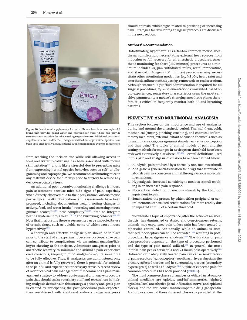
254 Navarro et al.
Figure 10: Nutritional supplements for mice. Shown here is an example of 1
brand that provides gelled water and nutrition for mice. These gels provide
easy to access nutrition for mice needing supportive care. Additional nutritional
supplements, such as EnerCal, though advertised for larger animal species, have
been used anecdotally as a nutritional supplement in mice by some researchers.
from reaching the incision site while still allowing access tofood and water. E-collar use has been associated with mouseskin irritation173 and is likely stressful due to preventing micefrom expressing normal species behavior, such as self- or allo-grooming and coprophagia. We recommend acclimating mice toany restraint device for 1–2 days prior to surgery to reduce anydevice-associated stress.
An additional post-operative monitoring challenge is mousepain assessment, because mice hide signs of pain, especiallywhen directly observed due to their prey nature. Various mousepost-surgical health observations and assessments have beenproposed, including documenting weight; noting changes inactivity, food, and water intake;1 recording and assessing mousegrimace scores;174,175 nest complexity176,177 time to integratenesting material into a nest;178,179 and burrowing behavior.180,181
Note that interpreting these assessments can be impacted by useof certain drugs, such as opioids, some of which cause mousehyperactivity.179
A thorough and effective analgesic plan should be in placeprior to the start of an experiment because post-operative paincan contribute to complications via an animal gnawing/lick-ing/or chewing at the incision. Administer analgesics prior toanesthetic recovery to minimize the animal’s pain experienceonce conscious, keeping in mind analgesics require some timeto be fully effective. Thus, if analgesics are administered onlyafter an animal is fully recovered, there is potential for animalsto be painful and experience unnecessary stress. A recent reviewof rodent clinical pain management182 recommends a pain man-agement strategy to address post-surgical or invasive procedurepain that should assist veterinary staff and researchers in mak-ing analgesia decisions. In this strategy, a primary analgesia planis created by anticipating the post-procedural pain expected,then readdressed with additional and/or stronger analgesics
should animals exhibit signs related to persisting or increasingpain. Strategies for developing analgesic protocols are discussedin the next section.
Authors’ Recommendation
Unfortunately, hypothermia is a far-too common mouse anes-thesia complication, necessitating external heat sources frominduction to full recovery for all anesthetic procedures. Anes-thetic monitoring for short (<30 minutes) procedures at a min-imum includes RR, paw withdrawal reflex, rectal temperature,and skin color. Longer (>30 minutes) procedures may neces-sitate other monitoring modalities (eg, %SpO2, heart rate) andanesthesia adjunct techniques (eg, remove/clean oral secretion).Although warmed SQ/IP fluid administration is required for allsurgical procedures, O2 supplementation is warranted. Based onour experiences, respiratory characteristics seem the most sen-sitive parameter to a mouse’s changing anesthetic plane; there-fore, it is critical to frequently monitor both RR and breathingpatterns.
PREVENTIVE AND MULTIMODAL ANALGESIAThis section focuses on the importance and use of analgesicsduring and around the anesthetic period. Thermal (heat, cold),mechanical (cutting, pinching, crushing), and chemical (inflam-matory mediators, external irritant or caustic chemicals such asformalin, capsaicin, carrageenan) stimuli can cause nociceptionand thus pain.1 The topics of animal models of pain and thetesting methods for changes in nociception threshold have beenreviewed extensively elsewhere.1,183,184 Several definitions usedin this pain and analgesia discussion have been defined below.
1. Allodynia: pain produced by a normally non-noxious stimuli.2. Analgesic: a general classification for drugs that attenuate or
abolish pain in a conscious animal through various molecularmechanisms.
3. Hyperalgesia: increased sensitivity to noxious stimuli result-ing in an increased pain response.
4. Nociception: detection of noxious stimuli by the CNS; notequivalent to pain.
5. Sensitization: the process by which either peripheral or cen-tral neurons (centralized sensitization) fire more readily dueto repeated stimuli of a certain threshold.
To reiterate a topic of importance, after the action of an anes-thetic(s) has diminished or abated and consciousness returns,animals may experience pain unless pain has been treated orotherwise controlled. Additionally, while an animal is anes-thetized, nociceptors can still be activated,185 resulting in post-procedural hyperalgesia or allodynia.186 The duration of painpost-procedure depends on the type of procedure performedand the type of pain model utilized.187 In general, the mostintense pain peaks between 4 and 24 hours post-operatively.188
Untreated or inadequately treated pain can cause sensitizationof pain receptors (ie, nociceptors), resulting in hyperalgesia in theprimary affected tissues and in surrounding tissues (secondaryhyperalgesia) as well as allodynia.188 A table of expected pain forcommon procedures has been provided (Table 5).
The most common classes of analgesics utilized in laboratoryanimal medicine are opioids, anti-inflammatories, alpha-2agonists, local anesthetics (local infiltration, nerve, and epiduralblocks), and the anti-convulsant/neuropathic drug gabapentin.A short overview of these different classes is provided at the
Dow
nloaded from https://academ
ic.oup.com/ilarjournal/article/62/1-2/238/6299201 by guest on 14 Septem
ber 2022

ILAR Journal, 2021, Vol. 62, No. 1–2 255
Table 5 Expected Pain for Common Procedures in Mice
Examples of Potentially Painful Procedures
Minimal to Mild Pain Mild to Moderate Pain Moderate to Severe Pain
Catheter implantation Embryo transfer Burn proceduresEar notching Hypophysectomy Heterotopic organ transplantationIntracerebral implantation
Multiple ID antigen injectionsOcular procedures
Minor laparotomy incisionsOrchidectomy thymectomy
Major laparotomy/organ incision orthopedicprocedures
Orbital sinus venotomySuperficial lymphadenectomy Superficialtumor implantation
Thyroidectomy Thoracotomy vertebral procedures
Tail clipping VasectomyVascular access
Port implantation
Common experimental procedures performed in mice and the expected pain induced from these procedures. These are guidelines, and animals should continue tobe monitoring post-procedure for signs of pain.
end of this section. The small body size of mice means access tovasculature is technically challenging. Thus, the routes of drugadministration are limited to more accessible routes, such assubcutaneous (SQ) or IP injection, or oral administration.189 IPinjection has been recommended over subcutaneous injectionsdue to variation in time for effects to occur, but the efficacyand safety of IP over SQ injections may differ depending onanesthetic protocol.78 Although oral administration is a commonmethod of drug delivery in other veterinary species, in orderto gavage mice technical expertise is required. Analgesicshave been formulated for incorporation into gel,190 food,191,192
and drinking water193 for laboratory rodents. Oral (ie, drugsadded to drinking water) and long-acting formulations ofanalgesics serve as a refinement in animal research by reducingthe stress associated with repeated injections of standardanalgesics, though oral formulations can result in inadequateor inconsistent dosing.191 The timing of analgesic adminis-tration, in addition to type of analgesic, is also important toconsider.
Analgesics ideally should be administered prior to aprocedure to prevent or mitigate anticipated nociception (ie,preventive or pre-emptive analgesia). This strategy is thoughtto help minimize post-operative pain. In humans, evidencefor usefulness of preventive analgesia to attenuate post-operative pain is conflicting.185,194–197 However, use of thisstrategy in animals has shown decreased post-surgical painin a variety of species, including post-invasive procedures indogs and cats,198–200 post-laparotomy in pigs,201 post-formalinadministration202 and post-surgically in rats,182,203–205 andpost painful procedures in mice.182,206–208 Opioids and localanesthetics are common drug choices for preventive analgesiain mice due to the systemic analgesic effects of opioids andthe ability to deliver analgesia locally with local anesthetics.Non-steroidal anti-inflammatory drugs (NSAIDs) have also beenrecommended in use for preventive analgesia for humans209
and animals,210,211 especially in cases where significant tis-sue trauma or inflammation is to be expected.211 Therehave been concerns in larger animals regarding prolongedbleeding times,212 gastrointestinal ulcerations,213 and possiblerenal toxicity214 secondary to hypotension during anesthesiaafter NSAID administration; however, these side effects arenot normally noted for short-term use or short anestheticprocedures for mice.215 In our experience, buprenorphine isone of the most common analgesics used preemptively and
peri-operatively in surgical and mild to moderately painfulprocedures in mice.206,207,216–221 Buprenorphine has a wide safetymargin207 and has minimal effects on the immune system.222
Buprenorphine administration can be accompanied with anumber of side effects in mice, including cardiovascular depres-sion, constipation, decreased food and water intake, possibleweight loss, and hyperactivity.207,219,36 Although discussed in theanesthetic portion of this review, alpha-2 antagonists (xylazine,dexmedetomidine) and dissociative drugs (ketamine) are alsocomponents of multimodal analgesia. The analgesic effects ofketamine are known, even at subanesthetic doses,8,223,224 thoughit is recommended to be used as an adjunctive analgesic and nota sole means of analgesia.
Using only preventive analgesia or only 1 analgesic agentpost-operatively may be insufficient to completely prevent paindepending on the type of procedure. A common strategy to com-bat breakthrough pain is to either utilize a higher dose of a singledrug or re-dose an animal. Even within the safety margin forsome analgesics, side effects can be more pronounced at higherdrug dosages. For example, opioids cause dose-dependent res-piratory depression8 and dysphoria225,226 on recovery in someveterinary species. Some opioids have a ceiling effect whereinhigher doses yield side effects without increased analgesia.207,227
As stated, high-NSAID doses have been associated with gas-trointestinal ulceration and perforation.212,228 Therefore, lowerNSAID doses combined with other analgesics should be used.This multimodal analgesia allows for analgesia to be maximizedthrough targeting different nociception sources. This permitslower analgesic doses and decreasing the risk of side effects overthose observed when using only 1 analgesic drug.210
Multimodal analgesia timing should include analgesicpain coverage during anesthetic emergence and the post-operative period. Several recently published reviews outlinethe impacts and benefits of analgesia in animal models post-operatively as a way to specify the impacts and address possibleconcerns regarding confounding results due to analgesicadministration.80,222,229,230 Untreated and/or inadequate painmanagement can confound experimental findings. Pain relatedto surgery is stressful to humans and animals, which in turnimpacts wound healing,80,231,232 can cause immune systemperturbation,233 reduces food/water intake,234 and can changeactivity,235 reduce sleep and alter circadian cycles,236 changenormal behaviors,178–181 and cause persistent pain due to CNSsensitization.237
Dow
nloaded from https://academ
ic.oup.com/ilarjournal/article/62/1-2/238/6299201 by guest on 14 Septem
ber 2022

256 Navarro et al.
Table 6 Commonly Used Mouse Analgesics
Drug Recommended Dosage and Reference
Opioids Buprenorphine-HCl (partial mu-agonist) 0.05–0.1 mg/kg SQ q8–12238
Buprenorphine (long-acting formulations;partial mu-agonist)
0.5–1.0 mg/kg SQ (Bup-SR) q48-72 h 3.25 mg/kgSQ (Ethiqa-XR) q48-72 h
Butorphanol (partial mu-agonist) 1–5 mg/kg SQ q4h328
Morphine (full mu-agonist) 2–5 mg/kg SQ q2-4h238
Tramadol (full mu-agonist) 5–40 mg/kg SQ, IP (dose frequency notdetermined)50,182
Non-steroidal anti-inflammatories Carprofen (COX-2 preferential) 2.5–5 mg/kg SQ q24h328
Carprofen (long-acting formulation) 15 mg/kg SQ q24h329
Meloxicam (COX-2 preferential) 1–5 mg/kg SQ q24h330
Meloxicam (long-acting formulation) 6 mg/kg SQ q24h329
Ketoprofen (non-selective COX inhibitor) 5 mg/kg SQ q12h50,83
Flunixin (non-selective COX inhibitor) 2.5 mg/kg SQ q12h50
Firocoxib (COX-2 elective) 10–20 mg/kg IP q24h331
Local anesthetics Lidocaine 2–4 mg/kg SQ over planned incision site; not toexceed 10 mg/kg50
Bupivacaine 1–2 mg/kg SQ over planned incision site; not toexceed 2 mg/kg.50
Liposomal Bupivacaine 6 mg/kg SQ243
Other drugs Gabapentin 10–30 mg/kg IP182,250
Maropitant 8 mg/kg IP252
This table is comprised of commonly used analgesic drugs and common dosages used in mice. IP = intraperitoneal; SQ = subcutaneous.
There is no one-size-fits-all analgesic plan that will ade-quately treat pain for all surgical/invasive procedures. Researchersand veterinarians must work together to create appropriatemouse analgesic plans. Analgesics should be administered overthe period where pain is expected to impact normal physiologicfunctions such as food and water intake, activity levels, excretoryfunctions, and self-injurious behavior (eg, chewing, licking, orgrooming at or around an incision site). Analgesic protocolsshould be designed for the expected procedural severity.The American College of Laboratory Animal Medicine haspublished guidelines to assess and manage rodent pain.238 Theseguidelines classify common laboratory rodent procedures intoexpected pain categories (minimal to mild pain, mild to moder-ate pain, and moderate to severe pain). We provide an overviewof the different drug classes used in mice below (summarizedin Table 6), with examples of preemptive and multimodalanalgesic protocols for several pain categories (Table 7).
Opioids
Opioid analgesics act on endogenous opioid receptors locatedthroughout the body in a variety of tissues, including the CNS,gastrointestinal tract, joint tissue, and urinary tract.8,239 Endoge-nous opioid receptors play a role in pain modulation240 andare thought to play a role in emotional regulation.241 Thereare 3 major classes of opioid receptors: mu, kappa, and delta.The most common opioid analgesics utilized in mice targetthe mu-receptor. The opioid analgesics most commonly usedin mice can be further classified as full or partial agonists.The efficacy to which both full and partial mu-agonists pro-duce analgesia is dose dependent, with the maximum effectsof partial agonists being less than full agonists at their peakefficacy.8 In general, opioids undergo gastrointestinal absorptionand hepatic metabolism and are then excreted via the biliaryand renal system.8 Opioid side effects vary by the class and typeof agonist; common side effects are decreased gastrointestinalmotility, decreased food and water intake, and sedation.8 Opioid
use has specific legal requirements for purchasing (procurementof a Drug Enforcement Agency, i.e. DEA, license), storage (lockedcabinets), and recordkeeping that may present a barrier to someresearchers.
Non-Steroidal Anti-Inflammatories
NSAIDs are a drug class useful for anti-pyretic, analgesic, andanti-inflammatory effects. The anti-inflammatory effects ofNSAIDs are due to inhibitory action along the arachidonic acidmetabolism pathway, specifically cyclo-oxygenase enzymes,COX-1 and COX-2. Inhibition of COX enzymes interruptsproduction of prostanoids and prostaglandins, both involvedin inflammatory processes.8 In general, NSAIDs undergohepatic metabolism and are excreted by the kidneys. In humanmedicine, NSAID administration prior to surgery has beendebated due to concerns over excessive bleeding secondaryto the anti-thrombotic effect of COX-1 inhibition,242 but, asdiscussed earlier, this is not a significant concern for short-termuse in mice or for short anesthetic procedures.
Local Anesthetics
Local anesthetics are sodium channel blockers. Sodium channelsare essential to neural transmission. The primary usefulnessof sodium channel blockers is in preventing nerve conductionfrom nociceptors to the CNS, thus blocking pain perception.Although an anesthetized patient will not experience pain, usinglocal anesthetics to infiltrate the tissues around an incision siteprior to the start of a procedure can prevent CNS sensitizationto nociceptive stimuli and reduce the risk of a post-surgeryhyperalgesia, also known as windup pain. Local anesthetic smallmammal overdoses, such as in mice, occur easily and care mustbe taken to use less than the maximum doses listed below.There is a lack of published standardized doses for mouse localanesthetic use, but non-toxic doses that are clinically effec-tive have been provided in tables at the end of this section. A
Dow
nloaded from https://academ
ic.oup.com/ilarjournal/article/62/1-2/238/6299201 by guest on 14 Septem
ber 2022

ILAR Journal, 2021, Vol. 62, No. 1–2 257
Table 7 Suggested Analgesic Plans for Various Pain Intensities
Suggested Analgesic Plans for Differing Pain Intensities
Minimal to Mild Pain Mild to Moderate Pain Moderate to Severe PainPre-operative: bupivacaine (1–2 mg/kg) local
infiltrationPre-operative: buprenorphine
(0.05–0.1 mg/kg SQ/IP)Pre-operative: lidocaine (2–4 mg/kg
local infiltration) + long-lastingbuprenorphine∗ once SQ
Post-operative: buprenorphine(0.05–0.1 mg/kg SQ/IP) every 6–12 hfor 1 d, then as needed
Post-operative: carprofen (2–5 mg/kgSQ) once per day for 2 d, then asneeded
Pre-operative: lidocaine (2–4 mg/kg localinfiltration)
Pre-operative: lidocaine (2–4 mg/kglocal infiltration) + buprenorphine(0.05–0.1 mg/kg SQ/IP)
Pre-operative: bupivacaine (1–2 mg/kglocal infiltration) + long-lastingbuprenorphine∗ once SQ
Post-operative: carprofen (5–10 mg/kg SQ)once, then as needed
Post-operative: carprofen (2–5 mg/kgSQ) once per day for 2 d, then asneeded
Pre-operative: buprenorphine (0.05–0.1 mg/kgSQ/IP); effective for 6–8 h
Pre-operative: bupivacaine (1–2 mg/kglocal infiltration) + long-lastingbuprenorphine∗ once SQ
Pre-operative: long-lastingbuprenorphine∗ once SQ
Post-operative: buprenorphine (0.05–0.1 mg/kgSQ/IP) if needed after initial dose has wornoff
Pre-operative: carprofen (5–10 mg/kg SQ)
Commonly used analgesic plans used in mice utilizing preemptive and multimodal analgesia that should be designed based on severity of pain. These can be usedas a template and other drugs within a similar class utilized. IP = intraperitoneal; SQ = subcutaneous. ∗Long-acting buprenorphine formulations are suggested to beadministered prior to surgical incision. The 2 currently available formulations of long-acting buprenorphine, Buprenorphine-SR & Ethiqa-XR, are recommended to beadministered at 0.5 mg/kg and 3.25 mg/kg SQ and are advertised to provide analgesia for up to 72 hours.
long-acting formulation of the local anesthetic bupivacaine iscurrently available and has been evaluated in rats.182,243 Thereare few studies evaluating its use in mice,244,245 but it may beefficacious for nerve blocks.245
Other Drugs
Other drugs that have gained popularity in other veterinaryspecies and have been utilized in mice are gabapentin and maro-pitant. Gabapentin is an anticonvulsant with analgesic proper-ties for neuropathic pain used in both human and veterinarymedicine.246 It provides analgesia in rats post-injury192,247,248 andin mouse models of induced nerve pain.249,250 Gabapentin’s clas-sification as a controlled substance within the United Statescurrently only applies to Kentucky, Michigan, and Tennessee;however this may pose as a barrier to some users.251 Maropitantis a neurokinin receptor antagonist and antiemetic, reportedpossibly to have anti-inflammatory effects in a study utilizinga mouse pancreatitis model.252
Authors’ Recommendation
Researchers should perform preventive and multimodal anal-gesia as part of the anesthesia protocol and tailor to painseverity. The addition of an opioid, that is, buprenorphine orother analgesics (lidocaine, etc), smooths out the anesthesiaplane without increasing anesthetic doses, sparing hemody-namic status. Long-lasting analgesics such as Bup-SR, Bup-XR, liposomal bupivacaine, etc have been used safely, reducingstress-related handling.
FACTORS AFFECTING ANESTHESIASuccessful anesthesia administration for research mice dependson several key factors, including background (eg, strain, sex, andage), procedure (procedure performed, surgeon’s performance,
length of procedure), and anesthetic plan (preventive andmultimodal analgesia). Consider these factors during theanesthesia planning and implementation to ensure optimalresearch results and preserve research reproducibility.230,253 Theconsideration of these factors also allows the modern mouseresearcher to move towards a more tailored anesthesia, similarto current human anesthesia trends.254
Strain
The research mouse continues to display significant advantagesover other animal models in genetic research due to its smallbody size and ease of genetic manipulation.255–260 Mouse modelsare also used in genetic research because their environments canbe tightly controlled, making studies on aging populations andwhole lifecycles possible.261–263 In the past 25 years, mouse straincharacterization,264 or “strain surveys,” have assisted research inthe areas of behavior,257,265 microbiota,266 pain,267 anesthesia,68
and analgesia.230,267 Variability in anesthesia between strainsshould be considered when planning for research studies.For example, anesthetic sensitivity can be linked to mousegenetic loci, which can be manipulated,268 enhancing theeffectiveness of select anesthetic agents.63 Similarly, geneexpression profiling has revealed mouse strain differencesassociated with anesthetic and analgesic sensitivity.269 Inaddition to strain, the mouse source may be an importantvariable affecting research paradigms utilizing anesthesia.Recent analgesic strain surveys230 identified literature gapswhere accurate strain and/or source nomenclature were absentor deficient, making comparisons between the strains andsources difficult, if not impossible. Future studies examiningmouse strain and source differences between specific anestheticagents are encouraged to better understand the role geneticsplay in anesthesia outcomes. Because genetically modifiedmouse use has expanded exponentially, it is essential to assessthe background strain and the gene(s) of interest when selecting
Dow
nloaded from https://academ
ic.oup.com/ilarjournal/article/62/1-2/238/6299201 by guest on 14 Septem
ber 2022
258 Navarro et al.
anesthesia and analgesia agents.68,230 This was demonstrated inan early study by Sonner et al, where inhalant anesthetic (isoflu-rane, halothane, desflurane, and ether) potency and convulsionthreshold were evaluated in several inbred, outbred, wild-type,and genetically modified mice (KO PKCγ ).68 Modest differencesin MAC of the anesthetic agents and convulsion thresholdwere demonstrated between strains, most significantly betweenthe PKCγ KO and their control background strain. Alzheimer’sdisease research highlights another area where differences ingenetically modified mouse strains can affect inhaled anestheticpotency.270 Although a complete listing of genetically modifiedmouse strain differences and their anesthetic potency is beyondthe scope of this article, it remains essential to consider mousestrain and genetics in the anesthetic selection process.265,271
Sex
Mouse sex differences have been observed in research involvingneuroimmunity,272,273 obesity,262 diabetes,262 aging,262 cardio-vascular health,274 cancer,274 neuroimaging,275 euthanasia,276
pain,230,277 and anesthesia.126 One hypothesis explaining mouseanesthetic sexual dimorphism is the difference in body fat per-centage (adult males > adult females), which can affect potencyand/or duration of some anesthetic and analgesic agents.263
However, inbred mouse sex differences to anesthesia are morelikely the result of genetic background, making variability bystrain and sex critical research factors.230,278,279 And becausethe majority of animal subjects in mouse research studies areoverwhelmingly male,280 careful interpretation of mouse studydata is warranted.281 Recent examples of literature describingsex and strain differences in mice undergoing anesthesiainclude the comparison of IP ket/xyl and ketamine/etomidate,76
the evaluation of IP sodium pentobarbital injection pain,282 theevaluation of alfaxalone/xylazine,98 the comparison of SQ andIP ket/xyl,78 and the impact of repeated ket/xyl or isoflurane onmouse well-being.61,283
Age
Of all the physiologic factors that could affect anesthesia, ageremains the least studied, especially for injectable agents. Incontrast, it is well known that MAC for volatile inhalant anes-thesia decreases with age in humans and animals, includingmice.284 For example, Loepke et al demonstrated that in neonatal(10-day-old) C57BL/6 J and 129/SvJ mouse strains, isofluraneMAC was higher than adult controls of the same strain.284 Thespecific mechanism of this age-related MAC decrease is yet tobe determined; however, it is likely due to a combination ofphysiologic factors that are altered by age.284 Advancing age canalso cause cognitive impairment in elderly human patients afteranesthesia285 and is hypothesized to be the result of blood brainbarrier disruption caused by anesthetic agents.286 However, inmice exposed to isoflurane anesthesia, the age-related risk ofanesthetic-induced cognitive impairment does not appear tooccur.285 Old mice have been shown to be more sensitive toketamine/xylazine anesthesia than young mice.95 Because themajority of anesthesia research has been conducted on inbredmouse strains, it is logical to suggest that mouse researchers alsoconsider age a significant factor in study planning.
Surgeon’s Skill
Preparation, training, and skill of the surgeon can have asignificant impact on the outcome of the procedure and the
anesthetic event.287 For example, in a mouse laparoscopicadhesion model, the duration of surgery was inversely relatedto amount of training the surgeon received.287 It is logical toassume those with minimal experience with the procedures orsurgeries performed may unintentionally inflict tissue traumaand/or pain. Similarly, the duration of the procedure and lengthof anesthesia period could be prolonged. Therefore, anestheticchoices should be selected wisely. A robust institutional surgicaltraining program may enhance animal well-being and minimizesurgeon variability and the negative effects of poor surgeonskill.288
Duration
Surgical or anesthesia duration will vary with the procedure orsurgery performed. Human surgical site infections positivelycorrelate with increasing procedure duration.289 In addition,hypothermia during rodent anesthesia is common137 and hasrecently been shown to interfere with post-operative painassessments.290 Thus, it is widely accepted that most proceduresinvolving rodent sedation or general anesthesia include nor-mothermia support and the shortest duration possible. However,in cases of prolonged anesthesia duration, the anesthesiaduration effects as a research variable must be addressed. Forexample, prolonged general anesthesia (isoflurane) can havea negative effect on the short-term and long-term anxietyresponse291 and intercranial CSF circulation,292 both of whichshould be controlled in neurological research.291 In cardiologyresearch, prolonged anesthesia (isoflurane or sevoflurane)affects blood vessel contractility for several days after theanesthetic event.293 In addition, many research studies utilizingimaging modalities require repeated anesthetic events on thesame animal. In these cases, repeated injectable61 or inhalant283
anesthetic events must be considered in the data analysis.
Authors’ Recommendations
Because of differences between mice due to sex, strain, andage, we recommend performing a pilot study with a smallnumber of mice to determine the effect of the anestheticprotocol prior to large-scale experiments. We are perhaps yearsaway from creating a personalized medicine approach for everymouse anesthetic event; however, identifying and addressingbasic physiologic and functional factors can affect anestheticoutcome and improve research reproducibility and animalwelfare.
SPECIFIC CIRCUMSTANCE ANESTHESIAImaging
Multiple modalities have been described to image mice,including ultrasound, electroencephalogram, (functional) mag-netic resonance imaging, positron emission tomography, andcomputerized tomography (Figure 11).275 In contrast to theprogress of these imaging modalities, optimization for safe,effective, and reproducible anesthetic paradigms that alsohave minimal effect on research are still necessary.50,294 Mostmouse imaging studies utilize inhalant anesthesia (isoflurane,sevoflurane).295–297 Additionally, high-throughput mouse imag-ing has identified the need for creative solutions to providedelivery of anesthetics and gases, positioning, temperature, andanesthetic monitoring.298–300
Dow
nloaded from https://academ
ic.oup.com/ilarjournal/article/62/1-2/238/6299201 by guest on 14 Septem
ber 2022

ILAR Journal, 2021, Vol. 62, No. 1–2 259
Figure 11: Mouse CT imaging. (Left) Individual mouse arrangement for CT imaging. The mouse can be secured with surgical tape. Care must be taken to ensure the chest
is not too tightly secured, limiting respiration. A heat supply has been arranged under the mouse to prevent hypothermia. (Middle) Individual mouse prepared to enter
a MRI scanner. The animal receives inhalant anesthesia via a nose cone at the end of the bed and is monitored via respiration (blue tube) and rectal temperature (black
cable). The body temperature is maintained with warm air. (Right) Anesthesia can be delivered by modifying commonplace equipment. Here, slip-tip 60-mL syringes
are connected to anesthetic tubing to deliver inhalant anesthesia to mice. Images courtesy of Dr Laura Pisani of Stanford Center for Innovation in in Vivo Imaging and
Dr Tim Doyle and Wu Tsai Neuroscience Imaging at Stanford University.
Figure 12: Monitoring during mouse CT imaging. (Left) An example set-up of pulse oximetry equipment for use during mouse anesthesia. (Middle) Arrangement of
pulse oximeter sensor attached to mouse. (Right) Arrangement of pulse oximetry and temperature probe to monitor vitals while the mouse is undergoing imaging in
a CT machine. Image courtesy of SA Instruments and Dr G. Ronald Morris.
Injectable anesthesia can be used for imaging procedureanesthesia301 and continues to be optimized for success.302
One optimization is continuous rate infusions, which permitsprecise dosing and eliminates inconsistent re-dosing.94 Perhapsthe most progressive imaging anesthesia approach is themultidrug approach to optimize data by overcoming the limitsof a single anesthesia technique.295,303 Imaging proceduredurations, especially MRI, can be long. For long procedures,appropriate monitoring techniques (ie, external heat sources[Bair hugger, hot air ventilation] and body temperature, warmedfluid bags, pulse oximeter [heart rate and %SpO2], breathingrate/patterns, etc) should be performed throughout (discussedabove; examples in Figure 12). In addition to monitoring, multi-modal analgesia (buprenorphine, lidocaine, etc) can smooth theanesthesia plane, which is recommended when possible evenfor nonpainful procedures. Tables 8 and 9 provide examples ofinhalant and injectable anesthetic imaging protocols. Additionalconsideration for mouse imaging anesthetic protocols294 hasbeen summarized in Table 10.
Stereotaxic Procedures
Stereotaxic procedures offer the neuroscience researcher acomprehensive tool to study and reproduce all aspects of thebrain, including surgical lesion mapping and implantationtechniques.304 Animal welfare improvements have offeredrefinements to this technique, further ensuring positive out-comes for neuroscience research.305 In some cases, innovativedevices can be 3D printed, improving head placement, especiallyin mice.306 This procedure can be painful during ear barapplication; therefore, mice should be at a surgical plane ofanesthesia (absent paw withdrawal reflex) before placing earbars. Coating the tips of the ear bars with lidocaine gel (Figure 13)or administration of buprenorphine at least 5–10 minutes prior
Table 8 Example Anesthetic Protocol for Mouse fMRI Imaging300
Species: mouseStock or strain: C57BL/6Age: 12 wkSex: maleWeight: 30 gBody Condition Score: 3Procedure needing anesthesia: surgical anesthesia for fMRIAcclimation time: 48 hFasting time: noneEquipment needed: anesthetic machine, circulating water
blanket, fiberoptic cable (for body temperature), foam pillow(for respiration), eye lubricant, warmed fluid
Pre-medication: noneInduction: isoflurane by induction boxMaintenance: etomidate CRI (0.75–1.5 mg/kg/min) after
etomidate initial bolus (4 mg/kg/min over first 3 min)Recovery: in home cage on circulating water blanket
to placing mice onto stereotaxic device will provide analgesiaduring this period. Note, pain will also be mitigated by utilizingdull/blunt-ended ear bars on all survival stereotactic procedures.Despite the fact that the animal is likely finished with thesurgical procedure and is expected to be entering the recoveryphase, the anesthesiologist should be prepared for the animal’sanesthetic plane to deepen after the removal of the ear bars dueto the decrease in stimulation to the anesthetized animal.
Neonatal Rodent Anesthesia Techniques
Neonatal mice are often used for anesthetic and survival surgicalprocedures; however, providing anesthesia is challenging.172
Dow
nloaded from https://academ
ic.oup.com/ilarjournal/article/62/1-2/238/6299201 by guest on 14 Septem
ber 2022

260 Navarro et al.
Table 9 Example Anesthetic Protocol for Mouse Cardiac CTImaging297
Species: mouseStock or strain: CD1Age: 8 wk
Sex: femaleWeight: 20 gBody Condition Score: 3Procedure needing anesthesia: surgical plane of anesthesia
for cardiac CTAcclimation time: 48 hFasting time: noneEquipment needed: anesthetic machine, circulating water
blanket, temperature probe, eye lubricant, warmed fluidPre-medication: noneInduction: isoflurane by induction boxMaintenance: isoflurane with O2 (25–50%) and N2O (50–75%)Recovery: in home cage on circulating water blanket
Table 10 Factors to be Considered When Developing AnestheticProtocols for Imaging Procedures
Considerations for Anesthetic Mouse Imaging ProceduresIACUC protocol requirementsType of imagingLength of procedure
Movement requirements (anesthesia plane needed)Sedation vs anesthesiaEquipment requirements
MRI compatibilityInhalant anesthesiaIntubation/ventilationCRI pumpPhysiologic monitoringCarrier gasesTemperature maintenancePositioning
Sedation/anesthetic agentsSedation/anesthetic planPreventive/multimodal analgesiaPremedicationNeuromuscular blockadeEmergency drugs
ReproducibilityTraining
The physiology and pharmacology of anesthetic agents usedin neonatal mice differs greatly from adults, affecting thepharmacodynamic adverse effects on the respiratory andcardiac systems and the propensity to develop hypothermia andhypoglycemia.172,307 Further physiological differences limitingneonatal anesthetic choices include increased blood–brainbarrier permeability, higher body-water content, less maturehepatic system, and lower albumin concentrations.8
Hypothermia. This is the most commonly reported anestheticmethod in neonatal mice up to 7 days of age.308 Newbornmice are poikilothermic for up to 7 days after birth and havea body temperature and metabolic rate that closely correlatewith the ambient temperature until the third week of life.309,310
Because neonates have small body mass, surface cooling quicklydecreases their body temperature.310 Furthermore, neonateshave hypoxia tolerance in various organ systems, including
Figure 13: Lidocaine gel for topical use. A small amount of topical lidocaine gel
can be used for stereotaxic bar placement to mitigate pain in mice. The gel should
be wiped off at the end of the procedure.
the heart and brain.311 Unlike adults, where tachycardia occursduring hypoxia, in the neonate, hypoxia induces bradycar-dia and shunting of blood from the peripheral tissues andbradypnea.311,312 To induce hypothermia, neonatal mice can beplaced on top of a latex sleeve in an ice bath and held in positionuntil anesthesia is achieved (Figure 14).312 Latex sleeves (oranother barrier material) serve to protect the pups’ skin duringchilling and decrease cold-induced pain.308 Anesthesia inductiontakes longer than when anesthetizing adults (paw withdrawalis present until 6 minutes post-induction).312 Bradycardia,hypoventilation or apnea, and hypoxemia are all seen in the first5–15 minutes of neonatal hypothermia anesthesia.312 Recoverytime is dependent on the hypothermia anesthesia period andtypically is as long or longer than the hypothermia anesthesiaperiod.308,312 During hypothermia anesthesia recovery, it isrecommended to provide gradual rewarming, because rapidwarming, such as with a heating lamp, can lead to tissuedamage.310 Providing supplemental 100% oxygen will facilitatehypoxia recovery. Hypothermia anesthesia is not recommendedfor anesthetic procedures longer than 30 minutes because thereis a reported increased risk of mortality (1 in 10 postnatal day 4rat pups died).308
Injectable Anesthesia. Neonatal mice have increased sensitivityand higher mortality risk when using common injectableanesthetic regimes. Typical mouse injectable anesthetics includea mixture of ketamine and sedatives or using medetomidine-based anesthesia.75,85 In adult mice, injectable anestheticregimes necessitate careful dosage calculation, monitoring,and management to minimize cardiorespiratory depression.313
These adverse effects are more pronounced in neonatal mice.Danneman and Mandrell previously indicated ketamine, pento-barbital, and fentanyl-droperidol combinations were unsafe andwere associated with a >50% mortality when used to induce asurgical anesthesia plane in 1- to 3-day-old neonatal rat pups.308
This increased mortality rate is also seen in older neonatalrodents. Tsukamoto et al evaluated ketamine-xylazine anes-thesia and medetomidine-midazolam-butorphanol anesthesiain 10-day-old rat pups.313 They discovered that when usingketamine-xylazine anesthesia at a 60-mg/kg (ketamine) and6-mg/kg (xylazine) dose, the 10-day-old pups exhibited suddendeath, and when using a lower dose of 40 mg/kg (ketamine)
Dow
nloaded from https://academ
ic.oup.com/ilarjournal/article/62/1-2/238/6299201 by guest on 14 Septem
ber 2022

ILAR Journal, 2021, Vol. 62, No. 1–2 261
Figure 14: Hypothermia anesthesia performed in neonatal mice. Neonate is
placed on top of a latex glove in crushed ice with water.
and 4 mg/kg (xylazine), rat pups did not reach a surgicalanesthesia plane. Based on these results, ketamine-xylazineanesthesia in neonatal mice is not recommended becauseof the narrow safety margin. The medetomidine-midazolam-butorphanol needed to reach a surgical plane in 10-day-old ratpups is higher than the adult rodent dose (0.3/4/5 mg/kg ofmedetomidine-midazolam-butorphanol, respectively, requiredfor neonates).313 When using injectable anesthetic regimes,supportive care measures including maintaining an externalheat source, providing 100% O2, and reversing with atipamezole(if applicable to the injectable protocol used) should be furtherevaluated and considered. Injectable anesthetics in neonatalmice are unpredictable, have high mortality risk, and should onlybe considered if gas anesthesia is not feasible or the neonatesare older than 6 days of age and hypothermia cannot be used.
Inhalant Anesthetics. These are generally considered safe andeffective for neonatal mice.308,312,313 When using isoflurane orsevoflurane anesthesia in neonatal mice, it is recommended tofirst rapidly induce pups in an induction chamber at 2 L/min of100% O2 containing either 5% isoflurane or 8% sevoflurane.312
The MAC of isoflurane (2.3%) in 10-day-old mice is higher than inadult mice.284 A study in 3-day-old neonatal rat pups successfullymaintained anesthesia using a nose cone with a glove cut to fitthe animal’s snout (500 mL/min of 100% O2 with isoflurane at 3–4% or sevoflurane at 5%) while placed on top of a warm water cir-culating pad (Figure 15).312 With these induction methods, pupslost their righting reflex within a minute; however, the perioduntil paw withdrawal was prolonged compared with adults (5–6 minutes).312 This study also indicated that when using inhalantanesthetic agents, RR, heart rate, and %SpO2 decrease but remainmore stable than with hypothermia anesthesia.312 Recovery timefor 3-day-old pups anesthetized for 15 minutes with isoflu-rane or sevoflurane was significantly shorter compared withhypothermia (time to recovery was 5–6 minutes shorter).312 Theexact percentage of inhalant agent needed is dependent onthe animal’s age because MAC decreases with age in humansand rodents.284 Increased inhalant anesthesia duration, whichlasts an hour in rodent neonates, can be associated with a
Figure 15: Inhalant anesthesia performed on a neonatal mouse. Snout of the
neonate is placed and sealed in a nose cone covered with a latex glove. A circular
hole is cut into the latex glove to fit around the snout of the neonate.
higher mortality rate.313 Because of an increased risk for mor-tality, hypothermia, bradycardia, and hypoventilation seen withincreased anesthetic times, it is recommended that inhalantanesthetics are used in rodent neonates for procedures lessthan 30 minutes in length and that a warm water circulatingpad and supplemental oxygen are provided during the recov-ery period. Hypoglycemia may occur with prolonged anestheticprocedures,284 and dextrose (eg, 0.25–1 mL/kg of 50% dextrose IP)may be administered.
Authors’ Recommendations
For long imaging and stereotaxic procedures, isoflurane,isoflurane with ket/xyl, or CRI including multimodal analgesia(buprenorphine, lidocaine) is recommended when possible.Appropriate monitoring, especially preventing hypothermiawith appropriate external heat sources and warmed fluidadministration, is highly recommended. For short anestheticprocedures (<30 minutes) on neonatal mice younger than 7 daysof age, isoflurane, sevoflurane (preferred), and hypothermiaanesthesia all provide a safe and reliable surgical planeof anesthesia.312 Authors have had good experiences withsevoflurane in neonatal mice with quick induction and recovery;it is critical to provide an external heat source and supplementalO2 throughout the anesthetic procedure and recovery period.
COMMON COMPLICATIONS ANDTROUBLESHOOTING COMPLICATIONS“An ounce of prevention is worth a pound a cure” is particularlyapt during anesthesia. When anesthesia and surgery involverodents, mortality can exceed 10 times the companion animalrate.117 To reduce anesthetic complications, anesthetists shouldbe confident of the animal’s health and well-being, selectappropriate anesthetic drugs/dosages, prepare anestheticequipment, and provide procedure-specific supportive care andphysiologic monitoring. Given that experience is a great teacher,it is imperative that the surgeon and anesthetist are not shared
Dow
nloaded from https://academ
ic.oup.com/ilarjournal/article/62/1-2/238/6299201 by guest on 14 Septem
ber 2022

262 Navarro et al.
roles during training, if ever. Practically, appropriate personnelsupport for mouse surgical procedures includes a surgeon, ananesthetist, and, depending on a given procedure’s complexity,at least 1 assistant. Anything that can shorten a procedure’sduration is more likely to yield a given procedure’s positiveoutcome. Conversely, an overly hastily performed proceduremay yield less than desirable outcomes. If during the courseof an anesthetic procedure there are unfavorable anestheticdevelopments, it is imperative that the surgeon undertakethe anesthetist’s direction. The first course of action wouldbe to pause any further procedural manipulations until theanesthetist can make a proper patient assessment. Duringmouse anesthesia, complications typically involve the centralnervous, respiratory, and cardiovascular systems; equipmentset-up; and human error. The following complications will bediscussed: (1) CNS: insufficient/excessive anesthetic plane; (2)respiratory system: irregular respiratory patterns/hypoventila-tion; (3) cardiovascular system: hypotension; and (4) other com-plications: hypothermia, excessive salivation, and prolongedrecovery. There has been very little research or evidence-basedmedicine addressing the treatment of anesthetic emergenciesin mice. Thus, much of the information presented in thissection comes from the authors’ clinical expertise and first-handexperiences.
CNS Complications
Insufficient or Excessive Anesthetic Plane. Reaching the correctanesthetic plane is particularly challenging when usinginjectable anesthetics because the doses needed to reach andmaintain a surgical plane of anesthesia must be titrated to theindividual animal. Additionally, employing injectable anesthesiafor prolonged anesthetic procedures presents additionalcomplications associated with re-dosing an animal that isphysiologically different from the animal receiving the initial (ormost recent) anesthetic administration, sometimes markedlyso. Other factors that must be taken into consideration for theanesthetic selection and dosing regimens include compromisedanimals (eg, immunocompromised, sick, pregnant, old, etc),which will require lower doses of anesthetics and/or may notbe safe to be anesthetized; mouse age; and hypothermia, whichwill be discussed below.
1. Causes of complications: incorrect animal weight; improperroute of administration for injectable anesthetics (such asaccidental intrahepatic, intracecal, incomplete injection, etc);anesthetic underdose/overdose.
2. Prevention/treatment: monitor anesthetic plane (discussedabove); if the animal is at an insufficient anesthetic plane:
a. Pause the procedure to assess the patient’s anestheticplane.
b. Administer analgesia (eg, buprenorphine, 0.05–0.1 mg/kgSQ/IP), which may help to stabilize the anesthetic planeand be sufficient to resume the procedure.
c. Incrementally increase gas anesthetics in 0.25–0.5%increases, or re-dose injectable anesthetics as discussedabove. Note that re-dosing injectable anesthetics mayprolong the anesthetic recovery; therefore, consideradministering reversal agents when the procedure ends(eg, atipamezole 0.1–1 mg/kg SQ/IP to reverse alpha-2agonists). Caution: any analgesia provided by alpha-2agonists will be reversed at this time.
d. Check the anesthetic machine connections and fittingsfor disconnections, misconnections, tubing kinks, or leaks.
Ensure any gas anesthetic scrubber or canister system isassembled and used according to manufacturer’s recom-mendations, especially that gas outflow is unimpeded.
If an excessively deep anesthetic plane is determined, thiscomplication must be treated as an emergency:
1. Incrementally decrease gas anesthesia (in 0.25–0.5% steps)or administer an injectable anesthetic reversal agent (eg,administer atipamezole for xylazine, medetomidine, ordexmedetomidine).
2. Administer naloxone if one must reverse buprenorphine.
Respiratory Complications
Irregular Respiratory Pattern (Including Hypoventilation). Mostanesthetic agents suppress the respiratory system in a dose-dependent manner. Under anesthesia, the RR and pattern shouldbe regular and characterized by good deep breaths. The rate willdepend on the anesthetic protocol being used, as discussedabove in the Monitoring section. Hypoventilation is a verycommon general anesthetic complication. Hypercapnia is asequela to hypoventilation and leads to respiratory acidosisand ultimately hypoxemia. Brick-red mucous membranes aresuggestive of hypercapnia. If one is conducting anesthesia underroom air (20% O2; ie, without oxygen supplementation), a mousecan quickly develop hypoxemia. ETCO2, or PaCO2 (the partialpressure of arterial CO2), are ideal indicators for assessing ananimal’s ventilatory status:
1. Causes of complications: wrong anesthetic plane (ie, too lightor too deep); an anesthetic overdose, resulting in a verydeep anesthesia plane; anesthetic side effects (eg, inducedpulmonary edema secondary to xylazine, or medetomidineadministration314,315); underlying health issues (eg, hypoven-tilation secondary to a neoplastic chest cavity mass, or obe-sity, or mice infected with pulmonary infectious agents likePneumocystis murina or Sendai virus); equipment malfunction(eg, kinked or obstructed tubing).
2. Prevention/treatment: monitor RR; keep mucous mem-branes pink by ensuring adequate oxygen delivery; keep%SpO2 > 95%; keep ETCO2 approximately 35–45 mmHg; ifRR/pattern is too low/slow and shallow and the mouse hasan absent paw withdrawal reflex or no jaw tone, the animalis too deep:
a. Lighten the anesthetic plane. If using injectable anes-thetics, promptly complete surgery while supporting themouse with 100% O2.316
b. As soon as possible, place the mouse in sternal recum-bency, which increases bilateral chest excursion andreduces the gastrointestinal tract’s pressure on thediaphragm.
c. Administer reversal agents, such as atipamezole for alpha-2 agonists.
If RR or pattern is too high/fast:
1. Pause the surgical manipulations as previously indicated.2. Incrementally increase inhalant anesthesia in 0.25% steps
to deepen the anesthetic plane (allow 3–5 minutes betweenincrements).
3. Administer injectable anesthetics or buprenorphine.4. Consider practicing balanced anesthesia/multimodal analge-
sia, especially with longer procedures (>45 minutes).
Dow
nloaded from https://academ
ic.oup.com/ilarjournal/article/62/1-2/238/6299201 by guest on 14 Septem
ber 2022

ILAR Journal, 2021, Vol. 62, No. 1–2 263
Cardiovascular Complications
Hypotension (Blood Pressure < 60 mmHg). Cardiac outputconsists of heart rate and stroke volume. Blood pressure isa product of cardiac output and systemic vascular resistance.Most anesthetics suppress the cardiovascular system in a dose-dependent manner. Without blood pressure monitoring, it isdifficult to identify the problem eliciting hypotension. Prolongedcapillary refill time may indicate hypotension; however, capillaryrefill time is difficult to assess in mice. A deeper anestheticplane or higher anesthetic doses are more likely to suppressthe cardiovascular system leading to hypotension. Hypotensionleads to decreased blood flow, resulting in decreased O2 andnutrient delivery to all tissues:
1) Causes of complications: anesthetic overdose; mice withimpaired cardiac pump function/cardiac contractility (ie,dilated cardiomyopathy models), decreased vascular tone,or reduced circulating volume (dehydration, hemorrhageetc); bradycardia; hypothermia; and compromised healthstatus mice.
2) Prevention/treatment: If hypotension is evident:
a. Lighten the anesthetic plane. Inhalant anesthetics cancause hypotension by vasodilation and reduced cardiaccontractility in a dose-dependent manner.
b. Administer balanced, warmed fluids at 5–20 mL/kg/h(IP, SC, IV). Appropriate fluid support is essential tooptimize surgical outcomes and minimize complications.Isoflurane-anesthetized mice should be supported withfluid administration regardless of surgical blood loss orinsensible fluid losses (ie, evaporated fluid loss). Fluidloss can also occur secondary to injectable anestheticcocktails containing alpha-2 agonists, because these cancause elevated blood glucose levels, resulting in osmoticpolyuria. The associated urinary (warm fluid) loss canprofoundly affect body weight, hydration status, bloodglucose, and electrolyte levels.
c. If the heart rate drops below normal ranges, which willdepend on the anesthetic protocol used, as discussedabove, atropine (0.04 mg/kg SC, IP, IV) may be indicated.50
Note that hypothermia may lead to bradycardia. Therefore,body temperature should be monitored and an externalheat source must be provided.317
d. Monitor blood pressure (if possible) and body temperature.
If the mouse has notable blood loss (ie, hypovolemia):
1. Blood loss >20% can cause hypovolemic shock. If blood lossoccurs, an estimated blood loss can be replaced with warmedbalanced fluid (3 times the blood volume lost), anesthesiaplane should be lightened, and promptly finish the proce-dure. Mouse blood volume 1.6–3.2 mL (6–8% body weight;approximately 80 mL blood/kg).318
Other Complications
Hypothermia. The mouse’s core body temperature is 36.5–38◦C (97.7–100.4◦F). Consequences of hypothermia are discussedabove but include prolonged recovery, shivering, discomfort,coagulopathy and platelet dysfunction, increased incidenceof wound infection, altered drug metabolism, cardiovascularsystem suppression, impaired tissue perfusion, and respiratorycompromise.50
1. Causes of complications: cold ambient temperature; coldsurgical prep solutions; wide shaving or cleansing area; longanesthesia; exposure to dry carrier gas, that is, O2.
2. Prevention/treatment: use active (external heating sources)and passive (fluid administration) cutaneous warmingsystems;317 reduce O2 flow to approximately 0.5 L/min witha non-rebreathing breathing circuit; wrap extremities (ie,paws and tail) and exposed surfaces with products likebubble wrap; administer warm, balanced fluids (IP, IV, SQ)at 5–20 mL/kg/h (IP, IV, SQ); monitor core body temperatureperiodically, especially for procedures longer than 15 min-utes. A non-burning, infrared temperature feedback systemis recommended. Avoid using heat lamps and electric heatingpads. Note: Do not use supplemental heat sources thatare not designed for anesthetized animals because theycan easily cause thermal injuries. If the procedure is long,researchers may consider using a humidifier connected tothe anesthetic machine.
Authors’ Recommendation
Circulating warm water heating pads can effectively controlmouse body temperature during anesthesia. Such an externalheat source should be provided from gas induction or immedi-ately after injectable anesthetic administration to full ambula-tory period.
Excessive Salivation. Excessive salivation can occur withketamine and prolonged anesthesia. Without tracheal intuba-tion, partial or total tracheal occlusion may occur, causing anes-thetic emergencies. Clinical signs indicating tracheal occlusionsecondary to saliva accumulation are labored breathing, poormucous membrane or skin color (due to low O2 saturation), oragonal breathing.
1. Causes of complications: ketamine use or prolonged anesthe-sia.
2. Prevention/treatment: check airway patency, if possible;clean/dry saliva with a Q-tip or cotton swab; administer ananticholinergic agent (ie, glycopyrrolate, 0.01 mg/kg SC, IP);provide 100% O2 via a mask; discontinue/complete surgeryas soon as possible; position animal in sternal recumbency;monitor %SpO2.
Prolonged Recovery. Prolonged recovery is commonly seen withinjectable anesthesia, long anesthetic procedures, and in com-promised animals. Depending on anesthetic doses, techniques,or duration, mice should recover within approximately 30–45 minutes of anesthetic discontinuation. As discussed earlier,provide aggressive supportive care in recovery:
1. Causes of complications: hypothermia, anesthetic overdose;long anesthesia (regardless of technique/protocol); lack ofappropriate peri-operative monitoring (resulting in hypoten-sion, hypoxemia, hypoventilation etc).
2. Prevention/treatment: if animals are compromised, pro-cedures should be postponed and the animal providedsupportive care until they are fully ambulatory. A veterinaryconsultation should be performed, and the veterinarianprovided the anesthetic and procedure-related records.Appropriate peri-operative monitoring must be provided.Injectable anesthetics should be reversed or gas anesthesiaincrementally decreased over time, when possible. If pro-longed recovery occurs, close monitoring and supportive care
Dow
nloaded from https://academ
ic.oup.com/ilarjournal/article/62/1-2/238/6299201 by guest on 14 Septem
ber 2022

264 Navarro et al.
(100% O2, warmed fluid administration, warm environmentetc) should be provided until full recovery.
Authors’ Recommendation
It is better to prevent anesthetic complications than have totreat them. Vigilant monitoring, effective interpretation of mon-itoring parameters, and appropriate supportive care are cru-cial to prevent anesthetic complications. Researchers shouldclosely monitor anesthetic plane, administer warmed balancedfluid with all procedures, provide 100% O2, prevent hypothermia,and administer reversal agents for injectable anesthesia at theprocedure’s end while ensuring appropriate postoperative anal-gesia has been administered before the reversal agent. Balancedanesthesia/multimodal analgesia should always be practiced.This technique allows lower doses of anesthetics/analgesics tobe used, leading to lower side effects. The anesthesia plane canbe adjusted easiest and more safely with gas anesthesia.
CONCLUSIONSMouse anesthesia can be challenging even for those withanesthesia experience. Although inhalant anesthesia receivesour top recommendation, inhalant anesthesia combined withinjectable anesthetics can be used safely, including for longerprocedures. If injectable anesthesia must be used, careful re-dosing may be required. To prevent anesthetic morbidity/mortal-ity, appropriate monitoring/interpretation and supportive caremust be provided. The authors strongly address the importanceof anesthetic monitoring throughout any anesthetic event,no matter how mundane (eg, RR, mucous membrane color,paw withdrawal reflex, and heating pads for short procedures;those techniques with short procedures together with advancetechniques/equipment [%SpO2, ECG, etc] for longer procedures).Some anesthetic complications lead to emergencies; therefore,as complications occur, they should be treated immediatelyand appropriately. Properly annotated anesthetic records areinvaluable to adjust anesthetic protocols, prevent mishaps,and assist consultation with scientific colleagues, includingveterinary colleagues. Finally, preventive and multimodalanalgesia should be “factory installed” into anesthetic plans(whether it is for sedation, general anesthesia, or restraint) whenpossible, especially those where the procedural outcome is likelyto yield a high pain severity.
Key Points
Introduction• Anesthesia is both an art and science. Although there
are multitudes of texts regarding how to perform safeand effective rodent anesthesia, experience is the besttraining.
• Mouse anesthesia is challenging due to the mouse’sunique biology and the wide range of experience inperforming mouse anesthesia for both veterinariansand animal researchers.
• Anesthesia and analgesia instruction is a vital elementto ensure there is a minimum level of understandingtowards ensuring the institutional regulatory mandatefor education and training is met and the introductionof institutional resources to assist those conductinganesthesia.
Anesthetic Depth
• General anesthesia causes CNS depression and canbe characterized by 4 notable stages: amnesia, loss ofconsciousness, loss of response to noxious stimuli, andblunted autonomic responses. These 4 stages are asso-ciated with an inhalant anesthetic’s specific minimumalveolar concentration (MAC).
• Loss of consciousness can be estimated by the loss ofthe righting reflex.
• Loss of movement in response to a noxious stimulusindicates the spinal reflexes have been anesthetized.
Dosing• When selecting an anesthetic protocol, consider the
anesthetic depth and duration required and theanesthetic’s impact on experimental variables beforeusing an anesthetic protocol.
• Inhalant anesthetics are strongly recommended inmice because of the steep dose-response curves andthe ability to finely regulate and rapidly change theanimal’s anesthetic depth.
• Ketamine/xylazine (ket/xyl) and ketamine/xylazine/a-cepromazine (ket/xyl/ace) injectable protocols can besafely used; however, careful and frequent monitor-ing of the animal’s anesthetic plane is necessary toensure adequate anesthesia for the procedure per-formed, while not being too deep, risking anesthetic-related death.
• New mouse anesthetic protocols are being developed,including the use of alfaxalone and a combinationof medetomidine, midazolam, and butorphanol, whichmay expand our arsenal of options to safely use differ-ent anesthetic protocols while minimizing the effectson experimental variables. Using these new protocolsand discussing the results within our field will beimportant in the continued development of these drugs.
Anesthetic Monitoring• Appropriate monitoring techniques (pre-, peri-, and
postoperative periods) are crucial to reduce mortality/-morbidity.
• The monitoring summary comprises an anestheticmonitoring record that includes supportive care mea-sures, vital signs, and all administered drugs. Theresulting anesthetic monitoring record helps anes-thetists assess trends in vital signs and identify poten-tial problems during an anesthetic procedure.
• Prolonged recovery may occur in hypothermic mice,mice with metabolic or respiratory complications, orin painful animals. Supportive care is essential inthe mouse recovery and post-operative periods andincludes providing supplemental heat, warmed par-enteral fluids, and easily available food and water.
• Institutional veterinarian consultation isrecommended for animals with an abnormal physical/health status.
Preventive and Multimodal Analgesia• Preventive and multimodal analgesia maximizes anal-
gesia while minimizing side effects. They should bedesigned as a part of the anesthesia plan. This tech-nique combines multiple anesthetics/analgesic classesinto a single anesthetic protocol.
• The most intense pain after a surgical procedure isexpected to occur in the first 24 hours after the proce-dure but will vary depending on the procedure.
Dow
nloaded from https://academ
ic.oup.com/ilarjournal/article/62/1-2/238/6299201 by guest on 14 Septem
ber 2022

ILAR Journal, 2021, Vol. 62, No. 1–2 265
• Mice are unlikely to show signs of pain post-operativelywhen directly observed, so indirect assessments arerequired (body weight, fecal production, grimace scores,nest building, activity level, etc).
Factors Affecting Anesthesia• Important factors affecting anesthetic choices are
strain, gender, age, surgeon’s skill, and anesthesiaduration/plane.
• Imaging and stereotaxic procedures: if the procedure islong (>45 minutes), consider a combination of isoflu-rane and CRI anesthetics. Perform appropriate monitor-ing techniques, including body temperature measure-ment.
• Hypothermia and inhalant anesthetics are the mostcommon anesthetic methods for neonatal mice.
Common Complications and Troubleshooting Complications• The best way to handle anesthetic complications/emer-
gencies is to prevent them.• Unfamiliarity with mouse anesthesia and monitoring
modalities may increase the occurrence of anestheticcomplications.
• Anesthetic monitoring (discussed above) and theanesthetist’s familiarity with anesthesia techniques arecrucial components towards preventing or reducinganesthetic complications and patient morbidity or mor-tality.
• Preventing anesthetic-associated morbidity/mortalityrequires vigilance and appropriate monitoring, under-standing and interpreting monitoring modalities, andrelevant and rapid intervention.
AcknowledgmentsWe thank Janis Atuk-Jones for her assistance with formattingthis manuscript, figures, and tables, and Benjamin Franco andEden Alamaw for their assistance with taking photos for thismanuscript.
Potential conflicts of interest. All authors: No reported con-flicts.
References1. Council NR. Recognition and Alleviation of Pain in Laboratory
Animals. Washington, DC: The National Academies Press;2009.
2. Roizen M, Horrigan R, Frazer B. Anesthetic doses blockingadrenergic (stress) and cardiovascular responses to inci-sion—MAC BAR. Anesthesiology 1981; 54(5):390–398.
3. Krnjevic K, Puil E. Cellular mechanisms of general anesthe-sia. In: Bittar E, Bittar N, eds. Principles of Medical Biology:Molecular and Cellular Pharmacology. Vol 8. Stamford, CT: JaiPress. 1997; 811–828.
4. Alkire M, Hudetz A, Consciousness TG. anesthesia. Science2008; 322(5903):876–880.
5. MacIver M. Loss of recall and the hippocampal circuiteffects produced by anesthetics. In: Hudetz A, Pearce R, eds.Suppressing the Mind: Anesthetic Modulation of Memory andConsciousness. New York, NY: Humana Press; 2010.
6. Antognini J, Barter L, Carstens E. Overview movement asan index of anesthetic depth in humans and experimentalanimals. Comp Med 2005; 55(5):413–418.
7. Franks N. General anesthesia: from molecular targets toneuronal pathways of sleep and arousal. Nat Rev Neurosci2008; 9(5):370–386.
8. Tranquilli, W. and Grimm, KA. Introduction: Use, Defini-tions, History, Concepts, Classification, and Considerationsfor Anesthesia and Analgesia. In: Grimm KA, Lamont LA,Tranquilli WJ, Greene SA, Robertson SA. eds. Veterinary Anes-thesia and Analgesia: The 5th edition of Lumb and Jones. Ames,IA: Wiley-Blackwell; 2015: Chapter 1.
9. Glannon W. Anaethesia, amnesia and harm. J Med Ethics2014; 40:651–657.
10. Voss L, Sleigh J. Monitoring consciousness: the current sta-tus of EEG-based depth of anaesthesia monitors. Best PractRes Clin Anaesth. 2007; 21(3):313–325.
11. Garcia-Larrea L, Bastuji H. Pain and consciousness. ProgNeuropharm Bio Psych 2018; 87:193–199.
12. Rosman E, Quartermain D, Turndorf H. Retrograde andanterograde amnesia in mice undergoing halothane anes-thesia (abstract). Anesth Analg 1990; 70(2):pS333.
13. Samuel N, Taub A, Paz R et al. Implicit aversive memoryunder anaesthesia in animal models: a narrative review. BrJ Anaesth 2018; 121(1):219–232.
14. Rosman E, Quartermain D, Pang R et al. Halothane anesthe-sia causes state-dependent retrieval failure in mice. PhysioBehav 1992; 52(3):449–453.
15. Alkire M, Nathan S. Does the amygdala mediate anesthetic-induced amnesia? Basolateral amygdala lesions blocksevoflurane-induced amnesia. Anesthesiology 2005;102(4):754–760.
16. Antognini J, Jinks S, Carstens E. The spinal cord, anesthe-sia and immobility: a re-examination. Int Cong Ser 2005;1283:125–131.
17. Sonner J, Antognini J, Dutton R et al. Inhaled Anesthet-ics and immobility: mechanisms, mysteries, and mini-mum alveolar anesthetic concentration. Anesth Analg 2003;97(3):718–740.
18. Yamamoto T, Schindler E. Where and how do anaestheticsact? Mechanisms of action in the central nervous system.Anaesthesiol Intensive Ther 2017; 49(4):288–293.
19. Roth D, Petersen-Felix S, Bak P et al. Analgesic effectin humans of subanaesthetic isoflurane concentrationsevaluated by evoked potentials. Br J Anaesth 1996; 76:38–42.
20. Petersen-Felix S, Arendt-Neilsen L, Bak P et al. Analgesiceffect in humans of subanaesthetic isoflurane concen-trations evaluated by experimentally induced pain. Br JAnaesth 1995; 75:55–60.
21. Houghton I, Cronin M, Redfern P et al. The analgesic effectof halothane. Br J Anaesth 1973; 45:1105–1110.
22. Zbinden A, Maggiorini M, Petersen-Felix S et al. Anestheticdepth defined using multiple noxious stimuli during isoflu-rane/oxygen anesthesia. I Motor reactions. Anesthesiology1994; 80(2):253–260.
23. Lichtner G, Auksztulewicz R, Velten H et al. Nociceptiveactivation in spinal cord and brain persists during deepgeneral anaesthesia. Br J Anaesth 2018; 121(1):291–302.
24. Carbone L, Pain AJ. Laboratory animals: publication prac-tices for better data reproducibility and better animal wel-fare. PLoS One 2016; 11(5):e0155001.
25. Khajuria DK, Razdan R, Mahapatra DR. Description of a newmethod of ovariectomy in female rats. Rev Bras Reumatol2012; 52(3):462–470.
26. Animal Welfare Act (PL-91-579). U. States Department ofAgriculture, & Animal and Plant Health Inspection Service.
Dow
nloaded from https://academ
ic.oup.com/ilarjournal/article/62/1-2/238/6299201 by guest on 14 Septem
ber 2022
266 Navarro et al.
USDA, ed1970. Last accessed: 25 Apr. 2021. https://www.aphis.usda.gov/aphis/ourfocus/animalwelfare/sa_publications/ct_publications_and_guidance_documents.
27. National Research Council NRCU. Guide for the Care and Useof Laboratory Animals. 8th ed. Washington, DC: The NationalAcademies Press; 2011.
28. OLAW. Public Health Service Policy on Humane Care and Useof Laboratory Animals. Bethesda, MD: NIH-Department ofHealth and Human Services; 2002.
29. National Research Council NRCU. Education and Training inthe Care and Use of Laboratory Animals: A Guide for Devel-oping Institutional Programs. Washington, DC: The NationalAcademies Press; 1991.
30. Siddiqui B, Kim P. Anesthesia stages. StatPearls [Internet].Treasure Island (FL): StatePearls Publishing. e1–19. (Lastaccessed: 25 Apr. 2021). https://www.ncbi.nlm.nih.gov/books/NBK557596/.
31. Campagna J, Miller K, Forman S. Mechanisms of actions ofinhaled anesthetics. NEJM 2003; 348:2110–2124.
32. Eger IIE, Saidman L, Brandstater B. Minimum alveolar anes-thetic concentration: a standard of anesthetic potency.Anesthesiology 1965; 26(6):756–763.
33. Aranake A, Mashour G, Avidan M. Minimum alveolar con-centration: ongoing relevance and clinical utility. Anesthesia2013; 68(5):512–522.
34. Antognini J, Schwartz K. Exaggerated anesthetic require-ments in the preferentially anesthetized brain. Anesthesiol-ogy 1993; 79(6):1244–1249.
35. Boscan P, Monnet E, Mama K et al. A dog model to studyovary, ovarian ligament and visceral pain. Vet Anaesth Analg2011; 38(3):260–266.
36. Buitrago S, Martin T, Tetens-Woodring J et al. Safety andefficacy of various combinations of injectable anestheticsin BALB/c mice. JALAAS. 2008; 47(1):11–17.
37. Mogil J, Smith S, O’Reilly M et al. Influence of nociceptionand stress-induced antinociception on genetic variationin isoflurane anesthetic potency among mouse strains.Anesthesiology 2005; 103(4):751–758.
38. Richardson CA, Flecknell PA. Anaesthesia and post-operative analgesia following experimental surgery in lab-oratory rodents: are we making progress? Altern Lab Anim2005; 33(2):119–127.
39. Stokes EL, Flecknell PA, Richardson CA. Reported anal-gesic and anaesthetic administration to rodents under-going experimental surgical procedures. Lab Anim 2009;43(2):149–154.
40. Rudeck J, Vogl S, Heinl C et al. Analgesic treatment withbuprenorphine should be adapted to the mouse strain.Pharmacol Biochem Behav 2020; 191:1–12, 172877.
41. Griebel G, Belzung C, Perrault G et al. Differences inanxiety-related behaviours and in sensitivity to diazepamin inbred and outbred strains of mice. Psychopharm 2000;148(2):164–170.
42. Lipiski M, Arras M, Jirkof P et al. Premedication withfentanyl-midazolam improves sevoflurane anesthesia forsurgical intervention in laboratory mice. Exp Biol Med (May-wood) 2017; 242(12):1287–1298.
43. Miller R. Miller’s Anesthesia. 9th ed. Philadelphia, PA:Churchill Livingstone/Elsevier; 2019.
44. de Jong R. Eger II E. MAC expanded: AD50 and AD95 valuesof common inhalation anesthetics in man. Anesthesiology1975; 42(4):384–389.
45. Forman S, Ishizawa Y. Inhaled anesthetic uptake, distri-bution, metabolism, and toxicity. In: Gropper M, Cohen N,
Eriksson L et al., eds. Miller’s Anesthesia. Philadelphia, PA:Elsevier; 2020: Chapter 20, 509–539.
46. Njoku DB, Chitilian HV, Kronish K. Hepatic, physiologyKK. pathophysiology, and anesthetic considerations. In:Gropper MA, Cohen NH, Eriksson LI et al., eds. Miller’sAnesthesia. 9th. Philadelphia, PA: Elsevier; 2020: Chapter 16,420–443.
47. Whittem T, Beths T, Bauquier SH. General pharmacologyof anesthetic and analgesic drugs. In: Grimm KA, LamontLA, Tranquilli WJ, Greene SA, Robertson SA. eds. VeterinaryAnesthesia and Analgesia: The 5th edition of Lumb and Jones.Ames, IA: John Wiley & Sons; 2015: Chapter 7. Pg 147–177.
48. Newcomer D, Chopra I. Evaluation of waste anestheticgas surveillance program and isoflurane exposures dur-ing animal and human surgery. J Occup Environ Hyg 2019;16(8):544–556.
49. Miller A, Theodore D, Widrich J. Inhalational anesthetic.StatPearls; 2020. (Last accessed date: 25 Apr. 2021). https://www.ncbi.nlm.nih.gov/books/NBK554540/.
50. Flecknell PA. Laboratory Animal Anaesthesia. 3rd ed.: Burling-ton, MA: Academic Press; 2016:135–137.
51. Groeben H, Meier S, Tankersley C et al. Influence of volatileanaesthetics on hypercapnoeic ventilatory responses inmice with blunted respiratory drive. Br J Anaesth 2004;92(5):697–703.
52. Low LA, Bauer LC, Klaunberg BA. Comparing the effects ofisoflurane and alpha chloralose upon Mouse Physiology.PLoS One 2016; 11(5):e0154936.
53. Massey CA, Richerson GB. Isoflurane, ketamine-xylazine, and urethane markedly alter breathing evenat subtherapeutic doses. J Neurophysiol 2017; 118(4):2389–2401.
54. Rödig G, Keyl C, Wiesner G et al. Effects of sevofluraneand isoflurane on systemic vascular resistance: use of car-diopulmonary bypass as a study model. Br J Anaesth 1996;76(1):9–12.
55. Steffey EP, Mama KR, Brosnan RJ. Inhalation anesthetics. In:Grimm KA, Lamont LA, Tranquilli WJ, Greene SA, RobertsonSA. eds. Veterinary Anesthesia and Analgesia: The 5th edition ofLumb and Jones. Ames, IA: Wiley-Blackwell; 2015: Chapter 16.Pg 297–331.
56. Wilding LA, Hampel JA, Khoury BM et al. Benefits of 21%oxygen compared with 100% oxygen for delivery of isoflu-rane to mice (Mus musculus) and rats (Rattus norvegicus).JALAAS 2017; 56(2):148–154.
57. Kavanagh B, Hedenstierna G. Respiratory physiology andpathophysiology. In: Gropper M, Cohen N, Eriksson L et al.,eds. Miller’s Anesthesia. Philadelphia, PA: Elsevier; 2020:Chapter. 13. 354–383.
58. Alves H, Valentim A, Olsson I et al. Intraperitoneal propo-fol and propofol fentanyl, sufentanil and remifentanilcombinations for mouse anaesthesia. Lab Anim 2007;41(3):329–336.
59. Burnside W, Flecknell P, Cameron A. Thomas a. a com-parison of medetomidine and its active enantiomerdexmedetomidine when administered with ketamine inmice. BMC Vet Res 2013; 9(48):1–9.
60. Groeben H, Meier S, Tankersley C et al. Heritable differ-ences in respiratory drive and breathing pattern in miceduring anaesthesia and emergence. Br J Anaesth 2003; 91(4):541–545.
61. Hohlbaum K, Bert B, Dietze S et al. Impact of repeatedanesthesia with ketamine and xylazine on the well-beingof C57BL/6JRj mice. PLoS One 2018; 13(9):e0203559.
Dow
nloaded from https://academ
ic.oup.com/ilarjournal/article/62/1-2/238/6299201 by guest on 14 Septem
ber 2022

ILAR Journal, 2021, Vol. 62, No. 1–2 267
62. Lee M, Suh H, Kim M et al. Comparison of the anestheticeffects of 2,2,2-tribromoethanol on ICR mice derived fromthree different sources. Lab Anim Res 2018; 34(4):270–278.
63. Tsukamoto Y, Yamada N, Miyoshi K et al. Anesthetic effectof a mixture of alfaxalone, medetomidine, and butorphanolfor inducing surgical anesthesia in ICR, BALB/c, and C57BL/6mouse strains. J Vet Med Sci 2019; 81(6):937–945.
64. Zuurbier CJ, Koeman A, Houten SM et al. Optimizing anes-thetic regimen for surgery in mice through minimizationof hemodynamic, metabolic, and inflammatory perturba-tions. Exp Biol Med (Maywood) 2014; 239(6):737–746.
65. Kehl F, Krolikowski J, Mraovic B et al. Kersten J. Is isoflurane-induced preconditioning dose related? Anesthesiology 2002;96(3):675–680.
66. LaTourette P II, David E, Pacharinsak C et al. Effects ofstandard and sustained-release buprenorphine on the min-imum alveolar concentration of Isolurane in C57BL/6 mice.JALAAS. 2020; 59(3):298–304.
67. Mazze RI, Rice SA, Baden JM. Halothane, isoflurane, andenflurane MAC in pregnant and nonpregnant femaleand male mice and rats. Anesthesiology 1985; 62(3):339–341.
68. Sonner J, Gong D, Li J et al. Mouse strain modestly influ-ences minimum alveolar anesthetic concentration andconvulsivity of inhaled compounds. Anesth Analg 1999;89(4):1030–1034.
69. Tsukamoto A, Iimuro M, Sato R et al. Effect of midazo-lam and butorphanol premedication on inhalant isofluraneanesthesia in mice. Exp Anim 2015; 64(2):139–145.
70. Zhou C, Liang P, Liu J et al. HCN1 channels contribute tothe effects of amnesia and hypnosis but not immobility ofvolatile anesthetics. Anesth Analg 2015; 121(3):661–666.
71. Roelofs S, Manjeri GR, Willems PH et al. Isoflurane anes-thetic hypersensitivity and progressive respiratory depres-sion in a mouse model with isolated mitochondrial com-plex I deficiency. J Anesth 2014; 28(6):807–814.
72. Cesarovic N, Nicholls F, Rettich A et al. Isoflurane andsevoflurane provide equally effective anaesthesia in labo-ratory mice. Lab Anim 2010; 44(4):329–336.
73. Brown E, Pavone K, Naranjo M. Multimodal generalanesthesia: theory and practice. Anesth Analg 2018;127(5):1246–1258.
74. Cesarovic N, Jirkof P, Rettich A et al. Combining sevoflu-rane anesthesia with fentanyl-midazolam or s-ketamine inlaboratory mice. JALAAS. 2012; 51(2):209–218.
75. Arras M, Autenried P, Rettich A et al. Optimizationof intraperitoneal injection anesthesia in mice: drugs,dosages, adverse effects, and anesthesia depth. Comp Med2001; 51(5):443–456.
76. Gergye C, Zhao Y, Moore R et al. A comparison ofketamine or etomidate combined with xylazine forintraperitoneal anesthesia in four mouse strains. JALAAS2020; 59(5):519–530.
77. Jaber S, Hankenson F, Heng K et al. Dose regimens, vari-ability, and complications associated with using repeat-bolus dosing to extend a surgical plane of anesthesia inlaboratory mice. JALAAS. 2014; 53(5):684–691.
78. Levin-Arama M, Abraham L, Waner T et al. Subcuta-neous compared with intraperitoneal ketamine-xylazinefor anesthesia of mice. JALAAS. 2016; 55(6):794–800.
79. Peltoniemi MA, Hagelberg NM, Olkkola KT et al.Ketamine: a review of clinical pharmacokinetics andpharmacodynamics in anesthesia and pain therapy. ClinPharmacokinet 2016; 55(9):1059–1077.
80. Huss M, Felt S, Pacharinsak C. Influence of pain and anal-gesia on orthopedic and wound-healing models in rats andmice. Comp Med. 2019; 69(6):535–545.
81. Maxwell CR, Ehrlichman RS, Liang Y et al. Ketamine pro-duces lasting disruptions in encoding of sensory stimuli. JPharmacol Exp Ther 2006; 316(1):315–324.
82. Richter W. Estimation of vasodilator drug effects in miceby measurements of paw skin temperature. Acta PharmacolToxicol (Copenh) 1964; 21:91–104.
83. Plumb D. Plumb’s Veterinary Drug Handbook. Blackwell Pub:Electronic-App; 2019. (Last accessed: 24 Apr. 2021). https://www.plumbsveterinarydrugs.com/#!/monograph/InADFktnHk.
84. Kawai S, Takagi Y, Kaneko S et al. Effect of three types ofmixed anesthetic agents alternate to ketamine in mice. ExpAnim 2011; 60(5):481–487.
85. Tsukamoto A, Serizawa K, Sato R et al. Vital signs monitor-ing during injectable and inhalant anesthesia in mice. ExpAnim 2015; 64(1):57–64.
86. Bourke D, Malit L, Smith T. Respiratory interactionsof ketamine and morphine. Anesthesiology 1987; 66(2):153–156.
87. Jaspar N, Mazzarelli M, Tessier C et al. Effect of ketamineon control of breathing in cats. J Appl Physiol Respir EnvironExerc Physiol 1983; 55(3):851–859.
88. Sarton E, Teppema LJ, Olievier C et al. The involvementof the mu-opioid receptor in ketamine-induced respira-tory depression and antinociception. Anesth Analg 2001;93(6):1495–1500 table of contents.
89. Teppema LJ, Baby S. Anesthetics and control of breathing.Respir Physiol Neurobiol 2011; 177(2):80–92.
90. Rankin DC. Sedatives and tranquilizers. In: Grimm KA,Robertson SA, Lamont LA, Tranquilli WJ, Greene SA, eds.Veterinary Anesthesia and Analgesia: The Fifth Edition of Lumband Jones. 5th ed. Ames, IA: John Wiley & Sons; 2015. Chapter10. p. 196–206.
91. Dholakia U, Clark-Price S, Keating S et al. Anestheticeffects and body weight changes associated with ketamine-xylazine-lidocaine administered to CD-1 mice. PLoS One2017; 12(9):e0184911.
92. Olson ME, Renchko P. Azaperone and azaperone-ketamineas a neuroleptic sedative and anesthetic in rats and mice.Lab Anim Sci 1988; 38(3):299–304.
93. Cruz J, Loste J, Burzaco O. Observations on the use ofmedetomidine/ketamine and its reversal with atipame-zole for chemical restraint in the mouse. Lab Anim 1998;32(1):18–22.
94. Erickson R, Terzi M, Jaber S et al. Intraperitonealcontinuous-rate infusion for the maintenance ofanesthesia in laboratory mice (Mus musculus). JALAAS.2016; 55(5):548–557.
95. Schuetze S, Manig A, Ribes S et al. Aged mice show anincreased mortality after anesthesia with a standard doseof ketamine/xylazine. Lab Anim Res. 2019; 35:8.
96. File S, Simmonds M. Myoclonic seizures in the mouseinduced by alphaxalone and related steroid anaesthetics.J Pharm Pharmacol 1988; 40(1):57–59.
97. Chiu K, Robson S, Devi J et al. The cardiopulmonary effectsand quality of anesthesia after induction with alfaxalone in2-hydroxypropyl-beta-cyclodextrin in dogs and cats: a sys-tematic review. J Vet Pharmacol Therap 2016; 39(6):525–538.
98. Erickson R, Blevins C, Souza Dyer C et al. Alfaxalone-xylazine anesthesia in laboratory mice (Mus musculus).JALAAS. 2019; 58(1):30–39.
Dow
nloaded from https://academ
ic.oup.com/ilarjournal/article/62/1-2/238/6299201 by guest on 14 Septem
ber 2022

268 Navarro et al.
99. Higuchi S, Yamada R, Hashimoto A et al. Evaluation of acombination of alfaxalone with medetomidine and butor-phanol for inducing surgical anesthesia in laboratory mice.Jpn J Vet Res 2016; 64(2):131–139.
100. Siriarchavatana P, Ayers JD, Kendall LV. Anesthetic activityof Alfaxalone compared with ketamine in mice. J Am AssocLab Anim Sci 2016; 55(4):426–430.
101. Kuusela E, Raekallio M, Vaisanen M et al. Comparison ofmedetomidine and dexmedetomidine as premedicants indogs undergoing propofol-isoflurane anesthesia. Am J VetRes 2001; 62(7):1073–1080.
102. Hablitz L, Vinitsky H, Sun Q et al. Increased glymphaticinflux is correlated with high EEG delta power and lowheart rate in mice under anesthesia. Sci Adv 2019; 5(2):eaav5447.
103. Wang Z, Schuler B, Vogel O et al. What is the optimal anes-thetic protocol for measurements of cerebral autoregula-tion in spontaneously breathing mice? Exp Brain Res 2010;207(3–4):249–258.
104. Field KJ, Lang CM. Hazards of urethane (ethyl carbamate): areview of the literature. Lab Anim 1988; 22(3):255–262.
105. Kirihara Y, Takechi M, Kurosaki K et al. Anesthetic effects ofa mixture of medetomidine, midazolam and butorphanol intwo strains of mice. Exp Anim 2013; 62(3):173–180.
106. Kirihara Y, Takechi M, Kurosaki K et al. Anesthetic effectsof a three-drugs mixture–comparison of administrativeroutes and antagonistic effects of atipamezole in mice. ExpAnim 2015; 64(1):39–47.
107. Meyer RE, Fish RE. A review of tribromoethanol anesthesiafor production of genetically engineered mice and rats. LabAnim (NY) 2005; 34(10):47–52.
108. Lieggi CC, Artwohl JE, Leszczynski JK et al. Efficacy andsafety of stored and newly prepared tribromoethanol in ICRmice. Contemp Top Lab Anim Sci 2005; 44(1):17–22.
109. Zeller W, Meier G, Burki K et al. Adverse effects of tribro-moethanol as used in the production of transgenic mice.Lab Anim 1998; 32(4):407–413.
110. Hill W, Tubbs J, Carter C et al. Repeated administrationof tribromoethanol in C57BL/6NHsd mice. JALAAS. 2013;52(2):176–179.
111. Maejima Y, Yokota S, O’Hashi R et al. The effect of avertinanesthesia and a mixture of three types of anestheticagents on food intake and body weight in high fat-inducedobese male and female mice. Exp Anim 2019; 68(1):57–69.
112. Oh SS, Hayes JM, Sims-Robinson C et al. The effects ofanesthesia on measures of nerve conduction velocity inmale C57Bl6/J mice. Neurosci Lett 2010; 483(2):127–131.
113. Cho Y, Lee Y, Lee J et al. Kinetics of proinflammatorycytokines after intraperitoneal injection of tribromoethanoland a tribromoethanol/xylazine combination in ICR mice.Lab Anim Res. 2011; 27(3):197–203.
114. Brosnan RJ, Steffey EP, Escobar A et al. Anesthetic inductionwith guaifenesin and propofol in adult horses. Am J Vet Res2011; 72(12):1569–1575.
115. Mees L, Fidler J, Kreuzer M et al. Faster emergence behav-ior from ketamine/xylazine anesthesia with atipamezoleversus yohimbine. PLoS One 2018; 13(10):e0199087.
116. Janssen CF, Maiello P, Wright MJ Jr et al. Comparison ofatipamezole with yohimbine for antagonism of xylazinein mice anesthetized with ketamine and xylazine. JALAAS.2017; 56(2):142–147.
117. Brodbelt D. Perioperative mortality in small animal anaes-thesia. Vet Journal 2008; 182(2):152–161.
118. Izer JM, Whitcomb TL, Wilson RP. Atipamezole reversesketamine-dexmedetomidine anesthesia without alteringthe antinociceptive effects of butorphanol and buprenor-phine in female C57BL/6J mice. JALAAS. 2014; 53(6):675–683.
119. Fleischmann T, Jirkof P, Henke J et al. Injection anaesthesiawith fentanyl-midazolam-medetomidine in adult femalemice: importance of antagonization and perioperative care.Lab Anim 2016; 50(4):264–274.
120. Papich M. Saunders Handbook of Veterinary Drugs. ElsevierSaunders: St. Louis, MO; 2007.
121. Johnston NA, Bosgraaf C, Cox L et al. Strategies for refine-ment of abdominal device implantation in mice: s train, car-boxymethylcellulose, thermal support, and atipamezole.JALAAS. 2007; 46(2):46–53.
122. Thal SC, Plesnila N. Non-invasive intraoperative monitor-ing of blood pressure and arterial pCO2 during surgicalanesthesia in mice. J Neurosci Methods 2007; 159(2):261–267.
123. Marian A, Bayman E, Gillett A et al. The influence of thetype and design of the anesthesia record on ASA physicalstatus scores in surgical patients: paper records vs. elec-tronic anesthesia records. BMC Med Inform Decis Mak 2016;2016(16):29.
124. Portier K, Ida K. The ASA physical status classification: whatis the evidence for recommending its use in veterinaryanesthesia?—A systematic review. Front Vet Sci 2018; 5:204.
125. Obernier JA, Baldwin RL. Establishing an appropriate periodof acclimatization following transportation of laboratoryanimals. ILAR J 2006; 47(4):364–369.
126. Gargiulo S, Greco A, Gramanzini M et al. Mice anesthesia,analgesia, and care, part I: anesthetic considerations inpreclinical research. ILAR J 2012; 53(1):E55–E69.
127. Rao S, Verkman AS. Analysis of organ physiology in trans-genic mice. Am J Physiol Cell Physiol 2000; 279(1):C1–C18.
128. Horn CC, Kimball BA, Wang H et al. Why can’t rodentsvomit? A comparative behavioral, anatomical, and physi-ological study. PLoS One 2013; 8(4):e60537.
129. Foltz C, Ullman-Cullere M. Guidelines for assessing thehealth and condition of mice. Lab Animal 1999; 28(4):28–32.
130. Hildebrandt I, Su H, Weber W. Anesthesia and other consid-erations for in vivo imaging of small animals. ILAR J 2008;49(1):17–26.
131. McNally JB, Kirkpatrick ND, Hariri LP et al. Task-based imag-ing of colon cancer in the Apc(min/+) mouse model. ApplOpt 2006; 45(13):3049–3062.
132. Gouveia K, Hurst JL. Improving the practicality of using non-aversive handling methods to reduce background stressand anxiety in laboratory mice. Sci Rep 2019; 9(1):20305.
133. Burkholder T, Foltz C, Karlsson E et al. Health evaluation ofexperimental laboratory mice. Curr Protoc Mouse Biol. 2012;2:145–165.
134. Bagis H, Odaman Mercan H, Dinnyes A. Exposure to warmerpostoperative temperatures reduces hypothermia causedby anaesthesia and significantly increases the implanta-tion rate of transferred embryos in the mouse. Lab Anim2004; 38(1):50–54.
135. Kober F, Iltis I, Cozzone PJ et al. Myocardial blood flowmapping in mice using high-resolution spin labeling mag-netic resonance imaging: influence of ketamine/xylazineand isoflurane anesthesia. Magn Reson Med 2005; 53(3):601–606.
136. Noel-Morgan J, Muir WW. Anesthesia-associated relativehypovolemia: mechanisms, monitoring, and treatmentconsiderations. Front Vet Sci 2018; 5:53.
Dow
nloaded from https://academ
ic.oup.com/ilarjournal/article/62/1-2/238/6299201 by guest on 14 Septem
ber 2022

ILAR Journal, 2021, Vol. 62, No. 1–2 269
137. Rufiange M, Leung VS, Simpson K et al. Prewarming fol-lowed by active warming is superior to passive warming inpreventing hypothermia for short procedures in adult rats(Rattus norvegicus) under isoflurane Anesthesia. JALAAS.2020; 59(4):377–383.
138. Guyton AC & Hall JE. Textbook of Medical Physiology. Philadel-phia, PA: Elsevier-Saunders; 2006.
139. Skorupski AM, Zhang J, Ferguson D et al. Quantification ofinduced hypothermia from aseptic scrub applications dur-ing rodent surgery preparation. JALAAS. 2017; 56(5):562–569.
140. Taylor DK. Study of two devices used to maintain normoth-ermia in rats and mice during general anesthesia. JALAAS.2007; 46(5):37–41.
141. Caro AC, Hankenson FC, Marx JO. Comparison of ther-moregulatory devices used during anesthesia of C57BL/6mice and correlations between body temperature andphysiologic parameters. JALAAS. 2013; 52(5):577–583.
142. Rembert MS, Smith JA, Hosgood G. A comparison of aforced-air warming system to traditional thermal supportfor rodent microenvironments. Lab Anim 2004; 38(1):55–63.
143. Waynforth H, Flecknell P. Experimental and Surgical Techniquein the Rat. 2nd ed: Cambridge, MA: Academic Press; 1992.
144. Nuntnarumit P, Swatesutipun B, Udomsubpayakul U et al.A randomized controlled trial of plastic drape for preven-tion of hypothermia during umbilical catheterization. Am JPerinatol 2013; 30(10):839–842.
145. Young VL, Watson ME. Prevention of perioperativehypothermia in plastic surgery. Aesthet Surg J 2006; 26(5):551–571.
146. Blevins C, Celeste N, Marx J. Effects of oxygen supplementa-tion on injectable and inhalant anesthesia in C57BL/6 mice.JALAAS 2021; 60(3). (In Press).
147. Emmer KM, Celeste NA, Bidot WA et al. Evaluation of thesterility of Press’n seal cling film for use in rodent surgery.JALAAS. 2019; 58(2):235–239.
148. Adams S, Pacharinsak C. Mouse anesthesia and analgesia.Curr Protoc Mouse Biol 2015; 5(1):51–63.
149. Danneman P, Suckow, M, Brayton, C. The Laboratory Mouse.2nd ed. Boca Raton, FL: CRC Press; 2013.
150. Ewald AJ, Werb Z, Egeblad M. Monitoring of vital signs forlong-term survival of mice under anesthesia. Cold SpringHarb Protoc 2011; 2011(2): pdb.prot5563.
151. Matsuda Y, Ohsaka K, Yamamoto H et al. Comparisonof newly developed inhalation anesthesia system andintraperitoneal anesthesia on the hemodynamic state inmice. Biol Pharm Bull 2007; 30(9):1716–1720.
152. Sessler Daniel I, McGuire J, Moayeri A et al. Isoflurane-induced vasodilation minimally increases cutaneous heatloss. Anesthesiology 1991; 74(2):226–232.
153. Støen R, Sessler Daniel I. The thermoregulatory thresh-old is inversely proportional to isoflurane concentration.Anesthesiology 1990; 72(5):822–827.
154. Rufiange M, Leung VSY, Simpson K et al. Pre-warmingbefore general anesthesia with isoflurane delays the onsetof hypothermia in rats. PLoS One 2020; 15(3):e0219722.
155. Rose N, Kwong GP. Pang DS. A clinical audit cycle of post-operative hypothermia in dogs. J Small Anim Pract 2016;57(9):447–452.
156. Redondo JI, Suesta P, Gil L et al. Retrospective study of theprevalence of postanaesthetic hypothermia in cats. Vet Rec.2012; 170(8):206.
157. Redondo JI, Suesta P, Serra I et al. Retrospective study of theprevalence of postanaesthetic hypothermia in dogs. Vet Rec2012; 171(15):374.
158. Pacharinsak C, Smith J. Handbook of Laboratory Animal Anes-thesia and Pain Management: Rodents. Boca Raton, FL: CRCPress, Taylor & Francis Group; 2017.
159. Matsukawa T, Sessler DI, Sessler AM et al. Heat flow anddistribution during induction of general anesthesia. Anes-thesiology 1995; 82(3):662–673.
160. Hershey J and Miller S. Correlation between surface temper-ature and core body temperature and the effect of exoge-nous heat during anesthesia in rats. Lab Anim Sci Prof .2016(March):40–43.
161. Newsom DM, Bolgos GL, Colby L et al. Comparison ofbody surface temperature measurement and conventionalmethods for measuring temperature in the mouse. ContempTop Lab Anim Sci 2004; 43(5):13–18.
162. Au - Kawakami Y, Au - Sielski R, Au - Kawakami T. Mousebody temperature measurement using infrared thermome-ter during passive systemic anaphylaxis and food allergyevaluation. JoVE. 2018(139):e58391.
163. Whary MT, Baumgarth N, Fox JG, Barthold SW. Biol-ogy and diseases of mice. In: Fox JG, Anderson LC, OttoGM, Pritchett-Corning KR, Whary MT, eds. Laboratory Ani-mal Medicine (Third Edition). Boston, MA: Academic Press;2015:43–149.
164. Ho D, Zhao X, Gao S et al. Heart rate and electrocardiographymonitoring in mice. Curr Protoc Mouse Biol 2011; 1:123–139.
165. Flegal MC, Kuhlman SM. Anesthesia monitoring equip-ment for laboratory animals. Lab Animal. 2004; 33(7):31–36.
166. Wagner AE, Brodbelt DC. Arterial blood pressure mon-itoring in anesthetized animals. JAVMA 1997; 210(9):1279–1285.
167. Tremoleda JL, Kerton A, Gsell W. Anaesthesia and physi-ological monitoring during in vivo imaging of laboratoryrodents: considerations on experimental outcomes andanimal welfare. EJNMMI Res 2012; 2(1):44–44.
168. Krege JH, Hodgin JB, Hagaman JR. Smithies O. A nonin-vasive computerized tail-cuff system for measuring bloodpressure in mice. Hypertension 1995; 25(5):1111–1115.
169. Brodbelt D, Pfeiffer D, Young L et al. Results of the confi-dential enquiry into perioperative small animal fatalitiesregarding risk factors for anesthetic-related death in dogs.JAVMA. 2008; 233(7):1096–1104.
170. Flecknell P. Anesthesia and Analgesia in Laboratory Animals.1st ed. San Diego, CA: Academic Press. 1997.
171. Young L, Brearly J, DL R et al. Medetomidine as a premedi-cant in dogs and its reversal by atipamezole. J Sm Anim Pract1990; 31:554–559.
172. National Research Council NRCU. Guidelines for the Careand Use of Mammals in Neuroscience and BehavioralResearch. Washington, DC: National Academies Press; 2003.
173. Jang Y, Park Y, Yun C et al. The vest-collar as a rodent collarto prevent licking and scratching during experiments. LabAnim 2016; 50(4):296–304.
174. Langford D, Bailey A, Chanda M et al. Coding of facialexpressions of pain in the laboratory mouse. Nat Methods2010; 7(6):447–449.
175. Miller A, Leach M. The mouse grimace scale: a clinicallyuseful tool? PLoS One 2015; 10(9):e0136000.
176. Jirkof P, Fleischmann T, Cesarovic N et al. Assessment ofpostsurgical distress and pain in laboratory mice by nestcomplexity scoring. Lab Anim 2013; 47(3):153–161.
177. Gaskill B, Karas A, Garner J et al. Nest building as an indica-tor of health and welfare in laboratory mice. J Vis Exp 2013;82:51012.
Dow
nloaded from https://academ
ic.oup.com/ilarjournal/article/62/1-2/238/6299201 by guest on 14 Septem
ber 2022

270 Navarro et al.
178. Rock M, Karas A, Gartrell Rodriguez K et al. The time-to-integrate-to-nest test as an Indicator of wellbeing inlaboratory mice. JAALAS 2014; 53(1):24–28.
179. Gallo M, Karas A, Pritchett-Corning K et al. Tell-tale TINT:does the time to incorporate into nest test evaluate post-surgical pain or welfare in mice? JALAAS. 2020; 59(1):37–45.
180. Jirkof P, Cesarovic N, Rettich A et al. Burrowing behavior asan indicator of post-laparotomy pain in mice. Front BehavNeurosci 2010; 4:165.
181. Deacon R. Burrowing in rodents: a sensitive methodfor detecting behavioral dysfunction. Nat Protoc 2006b;1:118–121.
182. Foley P, Kendall L, Turner P. Clinical management of pain inrodents. Comp Med. 2019; 69(6):468–489.
183. Hogan Q. Animal pain models. Reg Anesth Pain Med 2002;27(4):385–401.
184. Gregory N, Harris A, Robinson C et al. An overview of animalmodels of pain: disease models and outcome measures. JPain 2013; 14(11):1255–1269.
185. Woolf C, Chong M. Preemptive analgesia - treating post-operative pain by preventing the establishment of centralsensitization. Anesth Analg 1993; 77:362–279.
186. Gottschalk A, Smith D. New concepts in acute paintherapy: preemptive analgesia. Am Fam Physician 2001;63(10):1979–1984.
187. Brennan T. Postoperative models of nociception. ILAR J1999; 40(3):129–136.
188. National Research Council NRCU. Recognition and Allevia-tion of Pain in Laboratory Animals. Washington, DC: NationalAcademies Press; 2009.
189. Turner P, Brabb T, Pekow C et al. Administration of sub-stances to laboratory animals: routes of administration andfactors to consider. JALAAS. 2011; 50(5):600–613.
190. Seymour T, Adams S, Felt S et al. Postoperative analgesiadue to sustained-release buprenorphine, sustained-releasemeloxicam, and carprofen gel in a model of incisional painin rats (Rattus norvegicus). JALAAS. 2016; 55(3):300–305.
191. Leach M, Forrester A, Flecknell P. Influence of preferredfoodstuffs on the antinociceptive effects of orally admin-istered buprenorphine in laboratory rats. Lab Anim 2010;44:54–58.
192. Zude B, Jampachairsri K, Pacharinsak C. Use of flavoredtablets of gabapentin and carprofen to attenuate postop-erative hypersensitivity in an incisional pain model in rats(Rattus norvegicus). JALAAS. 2020; 59(2):163–169.
193. Goldkuhl R, Hau J, Abelson K. Effects of voluntarily-ingested buprenorphine on plasma corticosterone levels,body weight, water intake, and behaviour in permanentlycatherterised rats. In Vivo 2010; 24:131–135.
194. Bagul A, Taha R, Metcalfe M et al. Pre-incision infiltration oflocal anesthetic reduces postoperative pain with no effectson bruising and wound cosmesis after thyroid surgery.Thyroid 2005; 15(11):1245–1248.
195. Ejlersen E, Andersen H, Eliasen K et al. comparison betweenpreincisional and postincisional lidocaine infiltration andpostoperative Pain. Anesth Analg 1992; 74:495–498.
196. Grape S, Tramér M. Do we need preemptive analgesia forthe treatment of postoperative pain? Best Pract Res ClinAnaesth 2007; 21(1):51–63.
197. Filos K, Vagianos C. Pre-emptive analgesia: how importantis it in clinical reality? Eur Surg Res 1999; 31:122–132.
198. Watanabe R, Monteiro B, Evangelista M et al. The analgesiceffects of buprenorphine (Vetergesic or Simbadol)in combination with carprofen in dogs undergoing
ovariohysterectomy: a randomized, blinded, clinicaltrial. BMC Vet Res 2018; 14(1):304.
199. Brodbelt D, Taylor P, Stanway G. A comparison of pre-operative morphine and buprenorphine for postoperativeanalgesia for arthrotomy in dogs. J Vet Pharmacol Therap1997; 20:284–289.
200. Gassel A, Tobias K, Egger C et al. Comparison of oraland subcutaneous administration of buprenorphineand meloxicam for preemptive analgesia in catsundergoing ovariohysterectomy. JAVMA. 2005; 227(12):1937–1944.
201. Lykkegaard K, Lauritzen B, Tessem L et al. Local anesthet-ics attenuates spinal nociception and HPA-axis activationduring experimental laparotomy in pigs. Res Vet Sci 2005;79:245–251.
202. Coderre T, Vaccarino A, Melzack R. Central nervous sys-tem plasticity in the tonic pain response to subcutaneousformalin injection. Brain Res 1990; 535:155–158.
203. González-Darder J, Barberá J, Abellán M. Effects of prioranaesthesia on autotomy following sciatic transection inrats. Pain 1986; 24:87–91.
204. Page G, McDonald J, Ben-Eliyahu S. Pre-operative versuspostoperative administration of morphine: impact on theneuroendocrine, behavioural, and metastatic-enhancingeffects of surgery. Br J Anaesth 1998; 81:216–223.
205. Abram S, Yaksh T. Morphine, but not inhalational anes-thesia, block post-injury facilitation: the role of preemptivesuppression of afferent transmission. Anesthesiology 1993;78:713–721.
206. Jirkof P, Tourvieille A, Cinelli P et al. Buprenorphine forpain relief in mice: repeated injections vs sustained-releasedepot formulation. Lab Anim 2015; 49(3):177–187.
207. Roughan J, Flecknell P. Buprenorphine: a reappraisal ofits antinociceptive effects and therapeutic use in alle-viating post-operative pain in animals. Lab Anim 2002;36:322–343.
208. Reichert J, Daughters R, Rivard R et al. Peripheral and pre-emptive opioid antinociception in a mouse visceral painmodel. Pain 2001; 89(2–3):221–227.
209. O’Hanlon J, Lowry D. Improved postoperative analgesiawith preoperative piroxicam. Can J Anaesth 1996; 43(2):102–105.
210. Slingsby L. Multimodal analgesia for postoperative painrelief. In Practice 2008; 30:208–212.
211. Corletto F. Multimodal and balanced analgesia. Vet ResComm 2007; 31:59–63.
212. Luna S, Basílio A, Steagall P et al. Evaluation of adverseeffects of long-term oral administration of carprofen,etodolac, flunixin meglumine, ketoprofen, and meloxicamin dogs. Am J Vet Res 2007; 68(3):258–264.
213. Forsyth S, Guilford W, Haslett S et al. Endoscopy of thegastroduodenal mucosa after carprofen, meloxicam, andketoprofen administration in dogs. J Sm Anim Pract. 1998;39:421–424.
214. Forsyth S, Guilford W, Pfeiffer D. Effect of NSAID adminis-tration on creatinine clearance in healthy dogs undergoinganaesthesia and surgery. J Sm Anim Pract. 2000; 41:547–550.
215. Liles J, Flecknell P. The use of non-steroidal anti-inflammatory drugs for the relief of pain in laboratoryrodents and rabbits. Lab Anim 1992; 26:241–255.
216. Jirkof P, Durst M, Klopfleisch R et al. Administration oftramadol or buprenorphine via the drinking water for post-operative analgesia in a mouse-osteotomy model. Sci Rep2019; 9:1–16.
Dow
nloaded from https://academ
ic.oup.com/ilarjournal/article/62/1-2/238/6299201 by guest on 14 Septem
ber 2022

ILAR Journal, 2021, Vol. 62, No. 1–2 271
217. Healy J, Tonkin J, Kamarec S et al. Evaluation of an improvedsustained-release buprenorphine formulation for use inmice. AJVR 2014; 75(7):619–625.
218. Carbone E, Lindstrom K, Diep S et al. Duration of actionof sustained-release buprenorphine in 2 strains of mice.JALAAS. 2012; 51(6):815–819.
219. Lutfy K, Cowan A. Buprenorphine: a unique drug withcomplex pharmacology. Curr Neuropharmacol 2004; 2(4):395–402.
220. Gades N, Danneman P, Wixson S et al. The magnitude andduration of the analgesic effect of morphine, butorphanol,and buprenorphine in rats and mice. Contemp Top Lab AnimSci 2000; 39(2):8–13.
221. Christoph T, Kögel B, Schiene K et al. Broad analgesic profileof buprenorphine in rodent models of acute and chronicpain. Eur J Pharmacol 2005; 507:87–98.
222. DeMarco G, Nunamaker EA. Review of the effects of painand analgesia on immune system function and inflam-mation: relevance for preclinical studies. Comp Med. 2019;69(6):520–534.
223. Wang J, Goffer Y, Xu D et al. A single sub-anestheticdose of ketamine relieves depression-like behaviorsinduced by neuropathic pain in rats. Anesthesiology 2012;115(4):812–821.
224. Huang C, Long H, Shi Y et al. Ketamine enhances theefficacy to and delays the development of tolerance toelectroacupuncture-induced antinociception in rats. NeuroLet 2005; 375(2):138–142.
225. Becker W, Mama K, Rao S et al. Prevalence of dysphoria afterfentanyl in dogs undergoing stifle surgery. Vet Surg 2015;42(3):302–307.
226. Guedes A, Meadows J, Pypendop B et al. Evaluation oftramadol for treatment of osteoarthritis in geriatric cats.JAVMA. 2018; 252(5):565–571.
227. Malamed S. Pharmacology. In: Malamed S, ed. Sedation: AGuide to Patient Management. 6th ed. St. Louis, MO: Elsevier;2017.
228. Reed S. Nonsteroidal anti-inflammatory drug-induced duo-denal ulceration and perforation in a mature Rottweiler.Can Vet J 2002; 43(12):971–972.
229. Peterson N, Nunamaker E, Turner P. To treat or not to treat:the effects of pain on experimental parameters. Comp Med.2017; 67(6):469–482.
230. Smith JCA. review of strain and sex differences inresponse to pain and analgesia in mice. Comp Med. 2019;69(6):490–500.
231. Christian L, Graham J, Padgett D et al. Stress and woundhealing. Neuroimmunomodulation 2007; 13:337–346.
232. Carr D, Goudas L. Acute pain. Lancet 1999; 353:2051–2058.233. Padgett D, Glaser R. How stress influences the immune
response. Trends Immuno 2003; 24(8):444–448.234. Hayes J, Flecknell P. A comparison of pre-and post-
surgical administration of bupivacaine or buprenorphinefollowing laparotomy in the rat. Lab Anim 1998; 33:16–23.
235. Flecknell P, Liles J. The effects of surgical procedures,halothane anaesthesia and nalbuphine on the locomotoractivity and food and water consumption in rats. Lab Anim1991; 25:50–60.
236. Palada V, Gilron I, Canlon B et al. The circadian clock at theintercept of sleep and pain. Pain 2019; 161(5):894–900.
237. Curtin L, Grakowsky J, Suarez M et al. Evaluation ofbuprenorphine in a postoperative pain model in rats. CompMed 2009; 59(1):60–71.
238. Kohn D, Martin T, Foley P et al. Public statement: guidelinesfor the assessment and management of pain in rodents andrabbits. JAALAS. 2007; 46(2):97–108.
239. Pathan H, Williams J. Basic opioid pharmacology: an update.Br J Pain 2012; 6(1):11–16.
240. Benarroch E. Endogenous opioid systems: current conceptsand clinical correlations. Neurology 2012; 79(8):807–814.
241. Peciña M, Karp J, Mathew S et al. Endogenous opioid systemdysregulation in depression: implications for new thera-peutic approaches. Molec Psych 2018; 24:576–587.
242. Mathiesen O, Wetterslev J, Kontinen V et al. Adverseeffects of perioperative paracetamol, NSAIDs, glucocorti-coids, gabapentinoids and their combinations: a topicalreview. Acta Anaesth Scand 2014; 58(10):1182–1198.
243. Kang S, Jampachairsri K, Seymour T et al. Use of liposo-mal bupivacaine for postoperative analgesia in an inci-sional pain model in rats (Rattus norvegicus). JALAAS. 2017;56(1):63–68.
244. Grant G, Vermeulen K, Langerman L et al. Prolonged anal-gesia with liposomal bupivacaine in a mouse model. RegAnesth Pain Med 1994; 19(4):264–269.
245. Markova L, Umek N, Horvat S et al. Neurotoxicity of bupiva-caine and liposome bupivacaine after sciatic nerve block inhealthy and streptozotocin-induced diabetic mice. BMC VetRes 2020; 16:247.
246. Cheng J, Chiou L. Mechanisms of the antinociceptive actionof gabapentin. J Pharmacol Sci 2006; 100:471–486.
247. Hayashida K, DeGoes S, Curry R et al. Gabapentin acti-vates spinal noradrenergic activity in rats and humans andreduces hypersensitivity after surgery. Anesthesiology 2007;106(3):557–562.
248. McKeon G, Pacharinsak C, Long C et al. Analgesic effects oftramadol, tramadol-gabapentin, and buprenorphine in anincisional model of Pain in rats (Rattus norvegicus). JALAAS.2011; 50(2):192–197.
249. Takasaki I, Andoh T, Nojima H et al. Gabapentin Antinoci-ception in mice with acute herpetic pain induced by her-pes simplex virus infection. J Pharm Exp Therap. 2001;296(2):270–275.
250. Nishiyori M, Ueda H. Prolonged gabapentin analgesia in anexperimental mouse model of fibromyalgia. Mol Pain 2008;4:52.
251. Buscaglia M, Brandes H, Cleary J. Special report: the abusepotential of gabapentin and pregabalin. Practical painmanagement. https://www.practicalpainmanagement.com/treatments/pharmacological/non-opioids/special-report-abuse-potential-gabapentin-pregabalin. Date LastAccessed: 25 Apr 2021.
252. Tsukamoto A, Ohgoda M, Haruki N et al. The anti-inflammatory action of maropitant in a mouse model ofacute pancreatitis. J Vet Med Sci 2018; 80(3):492–498.
253. Smith J. Management of anesthesia. In: Pacharinsak C,Smith J, eds. Handbook of Laboratory Animal Anesthesia andPain Management Rodents. Boca Raton, FL: CRC Press; 2017. p.55–78.
254. McKinstry-Wu AR, Wasilczuk AZ, Harrison BA et al. Analy-sis of stochastic fluctuations in responsiveness is a criticalstep toward personalized anesthesia. Elife 2019; 8:e40143.
255. Kest B, Wilson SG, Mogil JS. Sex differences in supraspinalmorphine analgesia are dependent on genotype. J PharmExp Therap 1999; 289(3):1370–1375.
256. Allayee H, Andalibi A, Mehrabian M. Using inbred mousestrains to identify genes for complex diseases. Front Biosci2006; 11:1216–1226.
Dow
nloaded from https://academ
ic.oup.com/ilarjournal/article/62/1-2/238/6299201 by guest on 14 Septem
ber 2022

272 Navarro et al.
257. Bryant CD, Zhang NN, Sokoloff G et al. Behavioral differ-ences among C57BL/6 substrains: implications for trans-genic and knockout studies. J Neurogenet 2008; 22(4):315–331.
258. Mogil JS. Pain genetics: past, present and future. TrendsGenet 2012; 28(6):258–266.
259. Liu ET, Bolcun-Filas E, Grass DS et al. Of mice and CRISPR:the post-CRISPR future of the mouse as a model system forthe human condition. EMBO Rep 2017; 18(2):187–193.
260. Tuttle AH, Philip VM, Chesler EJ et al. Comparing phenotypicvariation between inbred and outbred mice. Nat Methods2018; 15(12):994–996.
261. Lariviere WR, Mogil JS. The genetics of pain and analgesiain laboratory animals. Methods Mol Biol 2010; 617:261–278.
262. Rodgers HM, Liban S, Wilson LM. Attenuated pain responseof obese mice (B6.Cg-lep(Ob)) is affected by aging and leptinbut not sex. Physiol Behav 2014; 123:80–85.
263. Greenspan JD, Craft RM, LeResche L et al. Studying sexand gender differences in pain and analgesia: a consensusreport. Pain 2007; 132(Suppl 1):S26–S45.
264. Mogil JS, Wilson SG, Bon K et al. Heritability of nociceptionI: responses of 11 inbred mouse strains on 12 measures ofnociception. Pain 1999; 80(1–2):67–82.
265. Crabbe JC, Wahlsten D, Dudek BC. Genetics of mouse behav-ior: interactions with laboratory environment. Science 1999;284(5420):1670–1672.
266. Franklin CL, Ericsson AC. Complex microbiota in labo-ratory rodents: management considerations. ILAR J 2020;60(2):289–297.
267. Mogil JS. The genetic mediation of individual differ-ences in sensitivity to pain and its inhibition. PNAS 1999;96(14):7744–7751.
268. Crowder CM. Mapping anesthesia genes: why and how?Anesthesiology 1998; 88(2):293–296.
269. Wijnvoord N, Albuquerque B, Haussler A et al. Inter-straindifferences of serotonergic inhibitory pain control in inbredmice. Mol Pain 2010; 6:70.
270. Xu H, Lu B, Zheng B et al. Smaller sized inhaled anesthet-ics have more potency on senescence-accelerated prone-8 mice compared with senescence-resistant-1 mice. JAlzheimers Dis 2014; 39(1):29–34.
271. Moloney RD, Dinan TG, Cryan JF. Strain-dependent varia-tions in visceral sensitivity: relationship to stress, anxietyand spinal glutamate transporter expression. Genes BrainBehav 2015; 14(4):319–329.
272. Rosen SF, Ham B, Haichin M et al. Increased pain sensitivityand decreased opioid analgesia in T-cell-deficient mice andimplications for sex differences. Pain 2019; 160(2):358–366.
273. Rosen S, Ham B, Mogil JS. Sex differences in neuroimmunityand pain. J Neurosci Res 2017; 95(1–2):500–508.
274. Miaskowski C GR, and Levine JD. Sex-related differencesin analgesic responses. In: Fillingim RB, ed. Progress in PainResearch and Managment. Vol 17. Seatle, WA: IASP Press;2000:209–230.
275. Da Silva JT, Seminowicz DA. Neuroimaging of pain in ani-mal models: a review of recent literature. Pain Rep 2019;4(4):e732.
276. Creamer-Hente MA, Lao FK, Dragos ZP et al. Sex- and strain-related differences in the stress response of mice to CO(2)euthanasia. JALAAS. 2018; 57(5):513–519.
277. Andrews NA, Latremoliere A, Basbaum AI et al. Ensuringtransparency and minimization of methodologic bias inpreclinical pain research: PPRECISE considerations. Pain2016; 157(4):901–909.
278. Boerner KE, Chambers CT, Gahagan J et al. Conceptualcomplexity of gender and its relevance to pain. Pain 2018;159(11):2137–2141.
279. Mogil JS. Interactions between sex and genotype in themediation and modulation of nociception in rodents. InMogil JS ed. Sex, Gender, and Pain. Seattle, WA: IASP Press;2000:25–40.
280. Rice AS, Cimino-Brown D, Eisenach JC et al. Animal modelsand the prediction of efficacy in clinical trials of analgesicdrugs: a critical appraisal and call for uniform reportingstandards. Pain 2008; 139(2):243–247.
281. Mogil JS, Bailey AL. Sex and gender differences in pain andanalgesia. Prog Brain Res 2010; 186:141–157.
282. Dutton JW 3rd, Artwohl JE, Huang X et al. Assessmentof pain associated with the injection of sodium pento-barbital in laboratory mice (Mus musculus). JALAAS. 2019;58(3):373–379.
283. Hohlbaum K, Bert B, Dietze S et al. Severity classifica-tion of repeated isoflurane anesthesia in C57BL/6JRj mice-assessing the degree of distress. PLoS One 2017; 12(6):e0179588.
284. Loepke A, McCann J, Kurth C et al. The physiologic effects ofisoflurane anesthesia in neonatal mice. Anesth Analg 2006;102(1):75–80.
285. Butterfield NN, Graf P, Ries CR et al. The effect of repeatedisoflurane anesthesia on spatial and psychomotor per-formance in young and aged mice. Anesth Analg 2004;98(5):1305–1311 table of contents.
286. Yang S, Gu C, Mandeville ET et al. Anesthesia and surgeryimpair blood-brain barrier and cognitive function in mice.Front Immunol 2017; 8:902.
287. Corona R, Verguts J, Binda MM et al. The impact of thelearning curve on adhesion formation in a laparoscopicmouse model. Fertil Steril 2011; 96(1):193–197.
288. Kelly RR, McCrackin MA, Russell DL et al. Teaching surgicalmodel development in research by using situated learningand instructional scaffolding. JALAAS. 2019; 58(3):321–328.
289. Cheng H, Chen BP, Soleas IM et al. Prolonged operative dura-tion increases risk of surgical site infections: a systematicreview. Surg Infect 2017; 18(6):722–735.
290. Klune CB, Robbins HN, Leung VS et al. Hypothermia duringgeneral anesthesia interferes with pain assessment in labo-ratory rats (Rattus norvegicus). JALAAS. 2020; 59(6):719–725.
291. Toyama K, Spin JM, Abe Y et al. Controlled isoflurane anes-thesia exposure is required for reliable behavioral testing inmurine surgical models. J Pharmacol Sci 2019; 140(1):106–108.
292. Gakuba C, Gaberel T, Goursaud S et al. General anesthesiainhibits the activity of the "glymphatic system". Theranostics2018; 8(3):710–722.
293. Terp R, Mieritz HB, Hansen PB. Long-term vascular effectsof anaesthesia in mice - what does it mean for our data andour patients? Acta Physiol (Oxf) 2016; 218(4):231–233.
294. Jensen J. Special techniques and species. In: PacharinsakC, Smith JC, eds. Handbook of Laboratory Animal Anesthesiaand Pain Managment Rodents. Boca Raton, FL: CRC Press;2017:89–100.
295. Vesce GMF, Chiavaccini L. Preclinical imaging anesthesia inrodents. Q J Nucl Med Mol Imaging 2017; 61(1):1–18.
296. Gargiulo S, Greco A, Gramanzini M et al. Mice anesthesia,analgesia, and care, part II: anesthetic considerations inpreclinical imaging studies. ILAR J 2012; 53(1):E70–E81.
297. Constantinides C, Mean R, Janssen BJ. Effects of isofluraneanesthesia on the cardiovascular function of the C57BL/6mouse. ILAR J 2011; 52(3):e21–e31.
Dow
nloaded from https://academ
ic.oup.com/ilarjournal/article/62/1-2/238/6299201 by guest on 14 Septem
ber 2022

ILAR Journal, 2021, Vol. 62, No. 1–2 273
298. Greenwood HE, Nyitrai Z, Mocsai G et al. High-throughputPET/CT imaging using a multiple-mouse imaging system. JNucl Med 2020; 61(2):292–297.
299. Suckow C, Kuntner C, Chow P et al. Multimodality rodentimaging chambers for use under barrier conditions with gasanesthesia. Mol Imaging Biol 2009; 11(2):100–106.
300. Petrinovic MM, Hankov G, Schroeter A et al. A novel anes-thesia regime enables neurofunctional studies and imaginggenetics across mouse strains. Sci Rep 2016; 6:24523.
301. Shim HJ, Jung WB, Schlegel F et al. Mouse fMRI underketamine and xylazine anesthesia: robust contralateralsomatosensory cortex activation in response to forepawstimulation. NeuroImage 2018; 177:30–44.
302. Nasrallah FA, Tay HC, Chuang KH. Detection of functionalconnectivity in the resting mouse brain. NeuroImage 2014;86:417–424.
303. Bascunana P, Thackeray JT, Bankstahl M et al. Anesthe-sia and preconditioning induced changes in mouse brain[(18)F] FDG uptake and kinetics. Mol Imaging Biol 2019;21(6):1089–1096.
304. Cetin A, Komai S, Eliava M et al. Stereotaxic gene deliveryin the rodent brain. Nat Protoc 2006; 1(6):3166–3173.
305. Fomari RV. Rodent stereotaxic surgery and animal welfareoutcome improvements for behavioral neuroscience [videoarticle]. JoVE 2012; 59:1–4. doi: 10.3791/3528. Published Jan-uary 30, 2012 Accessed November 12, 2020.
306. Yoshida S, Morimoto Y, Tonooka T et al. An inhalationanesthetic device for stereotaxic operation on mouse pups.J Neurosci Methods 2015; 243:63–67.
307. Clowry GJ, Flecknell PA. The successful use offentanyl/fluanisone (’Hypnorm’) as an anaesthetic forintracranial surgery in neonatal rats. Lab Anim 2000; 34(3):260–264.
308. Danneman PJ, Mandrell TD. Evaluation of five agents/meth-ods for anesthesia of neonatal rats. Lab Anim Sci 1997;47(4):386–395.
309. Lagerspetz KYH. Postnatal development of thermoreg-ulation in laboratory mice. Helgoländer Meeresun 1966;14(1):559–571.
310. Phifer CB, Terry LM. Use of hypothermia for generalanesthesia in preweanling rodents. Physiol Behav 1986;38(6):887–890.
311. Singer D. Neonatal tolerance to hypoxia: a comparative-physiological approach. Comp Biochem Physiol A Mol IntegrPhysiol 1999; 123(3):221–234.
312. Huss MK, Chum HH, Chang AG et al. The physiologic effectsof isoflurane, sevoflurane, and hypothermia used for anes-thesia in neonatal rats (Rattus norvegicus). JALAAS. 2016;55(1):83–88.
313. Tsukamoto A, Konishi Y, Kawakami T et al. Pharmacologi-cal properties of various anesthetic protocols in 10-day-oldneonatal rats. Exp Anim 2017; 66(4):397–404.
314. Amouzadeh HR, Qualls CW Jr, Wyckoff JH 3rdet al. Biochemical and morphological alterations inxylazine-induced pulmonary edema. Toxicol Pathol 1993;21(6):562–571.
315. Amouzadeh HR, Sangiah S, Qualls CW Jr et al. Xylazine-induced pulmonary edema in rats. Toxicol Appl Pharmacol1991; 108(3):417–427.
316. Mechelinck M, Kupp C, Krüger JC et al. Oxygen inhala-tion improves postoperative survival in ketamine-xylazineanaesthetised rats: an observational study. PLoS One 2019;14(12):e0226430.
317. Hankenson FC. Critical Care Management for Laboratory Miceand Rats. Boca Raton, FL: CRC Press; 2014.
318. McClure DE. Clinical pathology and sample collection in thelaboratory rodent. Vet Clin North Am Exot Anim Pract 1999;2(3):565–590 vi.
319. Yagi K. Veterinary fluid therapy update: calculating the rateand choosing the correct solution. DVM360. https://www.dvm360.com/view/veterinary-fluid-therapy-update-calculating-rate-and-choosing-correct-solution. Published2019. Accessed Nov 25, 2020.
320. Ullman-Culleré MH, Foltz CJ. Body condition scoring: a rapidand accurate method for assessing health status in mice.Lab Anim Sci 1999; 49(3):319–323.
321. Dwyer R, Bennett H, Eger IIE et al. Effects of isoflurane andnitrous oxide in subanesthetic concentrations on mem-ory and responsiveness in volunteers. Anesthesiology 1992;77(5):888–892.
322. Dwyer R, Bennett H, Eger IIE et al. Isoflurane anesthesiaprevents unconscious learning. Anesth Analg 1992; 75(1):107–112.
323. Chaves AA, Dech SJ, Nakayama T et al. Age and anes-thetic effects on murine electrocardiography. Life Sci 2003;72(21):2401–2412.
324. Pachon RE, Scharf BA, Vatner DE et al. Best anestheticsfor assessing left ventricular systolic function by echocar-diography in mice. Am J Physiol Heart Circ Physiol 2015;308(12):H1525–H1529.
325. Chu DK, Jordan MC, Kim JK et al. Comparing isoflurane withtribromoethanol anesthesia for echocardiographic pheno-typing of transgenic mice. J Am Assoc Lab Anim Sci 2006;45(4):8–13.
326. Schaefer A, Meyer GP, Brand B et al. Effects of anesthesiaon diastolic function in mice assessed by echocardiography.Echocardiography 2005; 22(8):665–670.
327. Papaioannou VE, Fox JG. Efficacy of tribromoethanol anes-thesia in mice. Lab Anim Sci 1993; 43(2):189–192.
328. Mayer J, Mans C. Rodents. In: Carpenter J, Marion C, eds.Exotic Animal Formulary. 5th ed. St. Louis, MO: Elsevier; 2018;459–493.
329. Kendall L, Hansen R, Dorsey K et al. Pharmacokineticsof sustained-release analgesics in mice. JALAAS. 2014;53(5):478–484.
330. Morrisey J, Carpenter J. Ferrets, Rabbits, and Rodents: Clin-ical Medicine and Surgery. 3rd ed. St. Louis, MO: Saunder-s/Elservier; 2012.
331. Reddyjarugu B, Pavek T, Southard T et al. Analgesic effi-cacy of firocoxib, a selective inhibitor of cyclooxygenase 2,in a mouse model of incisional pain. JALAAS. 2015; 54(4):405–410.
Dow
nloaded from https://academ
ic.oup.com/ilarjournal/article/62/1-2/238/6299201 by guest on 14 Septem
ber 2022
Copyright © 2022 FDOKUMEN