The International Student Journal of Nurse Anesthesia - AANA
67
Volume 19 Issue 3 Fall 2020 The International Student Journal of Nurse Anesthesia TOPICS IN THIS ISSUE Parturient w/scoliosis & pneumonectomy Dexmedetomidine-induced HTN Gnathodiaphyseal dysplasia Subcutaneous emphysema Chloroprocaine for SAB Situs inversus totalis Opioid dependence Sickle cell disease Mediastinal mass Preventing PDPH Alpha-gal allergy COVID-19 Articles UV light for PPE decontamination Ventilator splitting Mask availability Face shields
-
Upload
khangminh22 -
Category
Documents
-
view
2 -
download
0
Transcript of The International Student Journal of Nurse Anesthesia - AANA

Volume 19 Issue 3 Fall 2020
The International Student Journal of Nurse Anesthesia
TOPICS IN THIS ISSUEParturient w/scoliosis & pneumonectomyDexmedetomidine-induced HTNGnathodiaphyseal dysplasiaSubcutaneous emphysemaChloroprocaine for SABSitus inversus totalisOpioid dependenceSickle cell diseaseMediastinal massPreventing PDPHAlpha-gal allergy
COVID-19 ArticlesUV light for PPE decontaminationVentilator splittingMask availabilityFace shields

INTERNATIONAL STUDENT JOURNAL OF NURSE ANESTHESIA Vol. 19 No. 3 FALL 2020
Editor Associate Editor Vicki Callan, PhD, CRNA, CHSE Julie A. Pearson, PhD, CRNA Webster University CarolinaEast Health System
Editorial Board
Laura Ardizzone, DNP, CRNA, DCC Memorial Sloan Kettering Cancer Center; NY, NY LTC Sarah Bellenger, MSN, CRNA, ANC, USA LifeLinc Anesthesia Dawn Elizabeth Bent, DNP, MSN, CRNA University of Pennsylvania Laura S. Bonanno, PhD, DNP, CRNA Louisiana State University Health Sciences Center CDR Raymond Bonds, DNP, CRNA, CHSE, NC, USN Uniformed Services University Carrie C. Bowman Dalley, PhD, CRNA Georgetown University Greg Bozimowski, DNP, CRNA University of Detroit Mercy Terri M. Cahoon, DNP, CRNA Samford University Marianne S. Cosgrove, PhD, DNAP, CRNA Yale New Haven Hospital School of Nurse Anesthesia LTC Denise Cotton, DNAP, CRNA, ANC, USA Ryder Trauma Center; Miami, FL Janet A. Dewan, PhD, CRNA Northeastern University Marjorie A. Everson, PhD, CRNA Johns Hopkins University Anne Marie Hranchook, DNP, CRNA Oakland University-Beaumont Terri Kane, DNAP, CRNA Texas Wesleyan University Caroline Killmon, MSNA, CRNA Wake Forest School of Medicine Ilene Klassy, MSN, CRNA University of Wisconsin Medical School Brian Koonce, DNAP, CRNA Texas Wesleyan University John Litchfield, PhD, CRNA Anesthesia Miscellany Connie L. Lorette, PhD, CRNA Northeastern University Dan Lovinaria, DNP, MBA, APRN, CRNA,CHSE, FNAP Hennepin Healthcare Level 1 Trauma Center Ann Miller, DNP, CRNA Florida International University CDR Chad Moore, DNP, CRNA, CHSE, NC, Uniformed Services University Virginia “Chris” Muckler, DNP, CRNA, CHSE-A, FAAN Duke University Ryan L. Nations, PhD, CRNA Uniformed Services University Johanna Newman, DNAP, CRNA Florida Gulf Coast University Teresa Norris, EdD, CRNA University of Southern California Crystal Odle, DNAP, CRNA Lincoln Memorial University Shannon Pecka, PhD, CRNA Bryan College of Health Sciences Sarah Perez, MS, MSN, CRNA Washington University School of Medicine; St. Louis, J. Dru Riddle, PhD, DNP, CRNA, FAAN Texas Christian University Jackie Rowles, DNP, MBA, MA, CRNA, ANP-BC, NSPM-C, FNAP, FAAN The International Federation of Nurse Anesthetists LTC Peter Strube, DNAP, MSNA, MBA, CRNA, APRN, APNP, ARNP, AN, USA University of Wisconsin, Oshkosh Bryan Tune, DNP, CRNA California State University, Fresno Maria Van Pelt, PhD, CRNA Northeastern University Tina Williams, MSN, CRNA Carl R. Darnall Army Medical Center; Fort Hood, TX Kelly Wiltse Nicely, PhD, CRNA Emory University Lori Ann Winner, PhD, CRNA University of Pennsylvania Stephanie Woodruff, DNP, MSN, CRNA Cedar Crest College Kathleen R. Wren, PhD, CRNA University of Wisconsin, Oshkosh
2
Contributing Editors for this Issue
Steven L. Belmont, DNP, CRNA, APRN Fairfield University Alissa Blau, DNAP, CRNA University of Kansas Medical Center Leonardo Campero, DNAP, CRNA Keiser University LCDR Justin Hefley, DNP, CRNA Uniformed Services University Erica McCall, MSN, MPH, CRNA University of Southern California Carlene Mclaughlin, PhD, MSN, CRNA Crozer Chester Medical Center/Villanova University Scot Pettey, DNAP, CRNA, ARNP Gonzaga University LCDR Lauren Suszan, DNP, MSN, CRNA, NC, USN Uniformed Services University Elizabeth Schroeder, MSN, CRNA Washington University School of Medicine
Reviewers for this Issue
David Ayala, DNP, CRNA President & CEO of Ayala Anesthesia Associates Inc. Elizabeth Bamgbose, PhD, CRNA University of Southern California Garry Brydges, PhD, DNP, MBA, CRNA, ACNP-BC, FAAN MD Anderson Cancer Center Mark Caldwell, DNP, CRNA, CNE, Col., USAFR (Ret.) Case Western Reserve University Jose D. Castillo III, PhD, MS, CRNA, APRN Texas Wesleyan University Molly Condit, DNP, CRNA University of Wisconsin, Oshkosh Jeffrey R. Darna, DNP, CRNA, ACNP-BC University of Southern California Raymond Devlin, DNP, CRNA Louisiana State University Health Sciences Center Renee George, MS, CRNA Albany Medical College Marco Gidaro, MSN, CRNA Albert Einstein Medical Center; Philadelphia, PA Geoff Girolamo, DNP, CRNA US Naval Hospital Okinawa Susan Hall, DNP, CRNA Northeastern University Lisa Herbinger, DNP, CRNA Samford University Petra Hurt, MS, CRNA University of Detroit Mercy Jill Layman, DNAP, CRNA Missouri State University Maria Ledbetter, DNAP, CRNA, COI Samford University CPT Jordan Mark, DNP, CRNA Winn Army Community Hospital, Fort Stewart, GA David O'Connor, PhD, DNAP, CRNA Memorial Sloan Kettering MAJ Jacqueline Rushton, DNP, CRNA Womack Army Medical Center Matthew Stewart, MS, CRNA NorthCrest Medical Center; Springfield, TN Andrea Teitel, MS, CRNA University of Detroit Mercy
Front Cover: Brandon Ufert, MSN, RN, and Elvira Sayfutdinova, MSN, RN, are recent graduates of the Goldfarb School of Nursing at Barnes-Jewish College Nurse Anesthesia Program, and now preparing to take the National Certifying Exam. Mr. Ufert is shown practicing central venous catheter insertion on a simulated task trainer. Ms. Sayfutdinova, who has a case report published in this issue, performs an anesthesia machine safety check on her last day of clinical. The opinions contained in this journal are those of the authors and do not necessarily represent the opinions of the program or the University.

3
Disclaimer for all articles authored by military personnel: The views expressed in this journal are those of the authors and do not necessarily reflect the official policy or position of their respective Military Department, Department of Defense, nor the U.S. Government. The work was prepared as part of the official duties of the military service member. Title 17 U.S.C. 105 provides that ‘Copyright protection under this title is not available for any work of the United States Government’. Title 17 U.S.C. 101 defines a United States Government work as a work prepared by a military service member or employee of the United States Government as part of that person’s official duties. Publication Information: The International Student Journal of Nurse Anesthesia (ISSN 2688-5263) is published three times a year in the spring, summer, and fall. Current and past issues, and the Guide for Authors of this free, open access, electronic journal can be found at: www.aana.com - Member Resources Students International Student Journal https://www.aana.com/studentjournal https://ifna.site/international-publications/international-student-journal-for-nurse-anesthesia/ For additional information, please contact: Vicki Callan, PhD, CRNA, CHSE Webster University Department of Nurse Anesthesia Browning Hall, ISB 107 8274 Big Bend Blvd. St. Louis, MO 63119 314-246-5928; [email protected]

4
Table of Contents
COVID-19 Innovation Reports Face shields during the COVID-19 Crisis ...................................................................................6 Spencer Fugate, Keiser University The use of Ultraviolet Light to Decontaminate Personal Protective Equipment .....................9 Cody J. Reider, Keiser University Ventilator Splitting ......................................................................................................................12 Christopher Choate, Keiser University COVID-19: Effect on Availability of Masks for Healthcare Practitioners ............................17 Ryan Mona, Keiser University Case Reports Gnathodiaphyseal Dysplasia: Anesthesia for Rare Diseases ...................................................20 Kieran M. Shamash, University of Southern California Management of Dexmedetomidine-Induced Hypertension .....................................................23 Kelly Nagle, Uniformed Services University Chronic Pain and Opioid Dependence .......................................................................................27 Larry Beasley, Uniformed Services University Intrathecal Catheterization for Preventing Post dural Puncture Headaches ........................30 Raephael L. Garcia, Uniformed Services University Anesthesia for Delivery of Parturient with Scoliosis and Pneumonectomy ...........................33 Genna LeDrew, Providence Sacred Heart Medical Center/Gonzaga University Intrathecal 1% Chloroprocaine for Cervical Cerclage ............................................................37 Christian Panaligan, National University Clinical Implications of Patients with Situs Inversus Totalis ..................................................40 Renate Balitzky, Crozer Chester Medical Center/Villanova University Intraoperative Subcutaneous Emphysema ................................................................................43 Christopher G. Payne, Uniformed Services University Perioperative Management for Sickle Cell Disease: Efficacy of Recommendations .............46 Cleofas Espinoza, Fairfield University & Bridgeport Hospital
5
Anesthetic Management of a Patient with a Mediastinal Mass ...............................................50 Krista Brooks, University of Kansas Medical Center Galactose-Alpha-1,3-Galactose Allergy .....................................................................................53 Elvira Sayfutdinova, Goldfarb School of Nursing at Barnes Jewish College Editorial ........................................................................................................................................57 Vicki Callan, PhD, CRNA, CHSE Guide for Authors ........................................................................................................................58
6
Face shields during the COVID-19 Crisis
Spencer Fugate, BSN Keiser University
Keywords: Face shields, personal protective equipment, PPE, COVID-19, prevention Coronaviruses are zoonotic viruses including six sub-species that can cause disease in humans.1 Patients affected with coronaviruses can present with mild respiratory issues, such as upper airway infection, to complete respiratory failure including acute respiratory distress syndrome (ARDS). As of today, SARS Cov-2, the cause of COVID-19, has produced a worldwide pandemic with infection and mortality rates increasing every day. In a matter of months, after the initial outbreak in Wuhan, China, COVID-19 has since spread to all portions of the globe. Health care workers and facilities are overwhelmed with a large increase in patient volumes, the lack of medical and personal protective equipment (PPE). With the focus on PPE, large corporations, small businesses, and even individual patrons have attempted to relieve the burden on traditional PPE manufacturing companies by constructing face shields and other necessary equipment for hospitals and health care providers. Discussion Role of the Face Shield The World Health Organization has deemed face shields as part of the critical PPE that is lacking during this COVID-19 pandemic.2 Face shields play an important role when it comes to protecting healthcare workers from catching contagious pathogens. Protection is provided by decreasing the spread of pathogens via contact or droplet transmission by blocking projectile secretions that would contaminate the membranes of the eyes, nose, mouth, and face.3 Face shields also protect from inhalation of secretions and protect the skin from contaminated mucous that maybe coming from an infected patient. It is recommended by the U.S. Health Care Infection Control Practices Advisory that face shields be worn during procedures and during patient care when there is a possibility of projectile bodily fluids, such as blood or secretions.3 With the exponential increase of confirmed cases of COVID-19 worldwide, the demand for PPE, including face shields, has increased substantially. Components of the Face shield There are a variety of face shield designs, but they all have the same core components. The components include a visor, frame, and suspension system. In the United States, these models are designed and tested by criteria established from the American National Standards Institute to ensure quality and effectiveness.4 The visor consists of a front piece which can be made from a variety of materials such as polycarbonate or polyvinyl chloride.4 Visors come in a variety of styles including a full face, eyes only, or face and neck protection. The purpose for the visor is to protect the user from any potential exposure to patient body fluids. The frame of the face shield can also come in a variety of styles, most made of a lightweight plastic used as a supporting structure for the mask. Some frames offer the ability to disconnect the visor portion, allowing for exchange and reuse of the frame. The final piece to complete the face shield is the suspension system. The major purpose of the suspension system is to provide space between the visor and

7
the user’s face. There are a variety of mechanisms that are incorporated in suspension systems, such as a ratchet mechanism, pin-lock system or velcro to allow flexibility with the function and positioning of the face shield. Evidence of Clinical Study Research conducted on the effectiveness of face shields is limited. A study published by William Lindsley and associates measured the effectiveness of face shields and their ability to block bodily fluids. The study utilized a mannequin system, including a transmission unit and a breathing unit, to portray as a human model with breathing and coughing mechanics. The study experimented with variable distances to test the effectiveness of the face shield. Lindsley’s experiment involved distances between 46 to 183 cm showing a 96% decrease in contamination with larger aerosolized molecules and 68% decrease with small aerosolized molecules.4 A decrease in the overall area of contamination would reduce the probability of infection. Shoham & et al. conducted a study comparing the effectiveness of using face shields only verses in combination with other PPE equipment to observe for contamination rates. Results showed that the use of N95 mask and full-face shields was the only combination with negative contamination to the eyes, nares and mouth from small aerosolized molecules.5 When comparing full face mask to eye visors, contamination rates were decreased exponentially when involving aerosolizing medication in patients with known respiratory infection.5 The face shield provides not only protection for the face, eyes, nose and mouth but also to the mask worn underneath them. After the influenzas outbreak in 2009, the CDC recommended that health care workers consider the use of face shields to reduce surface contamination of respirators.3 Fluid exposure would decrease the integrity of mask, leading to higher risk for exposure and increase the frequency of new masks being used. COVID-19 has been described to have all modes of transmission including contact, droplet, airborne and aerosol.6 As interpreted from these studies, face shields fit the criteria for decreasing the risk of contact and droplet transmission of the virus. Community Action The community is helping to fight the shortage of PPE in a variety of ways. For example, Texas Tech University School of Medicine has coordinated with Texas Tech University (TTU) and the Center for Emerging Energy Sciences (CEES) to undergo the development and production of face shields, ventilator components, and prototype N-95s for hospitals across the county.7 Prototype N-95s have not yet been cleared for use and are currently undergoing evaluation.7 However, hundreds of face shields have already been delivered to both major and rural hospitals in the West Texas area. Texas Tech states that each printer possesses its own output capability; one printer alone can print up to 20 face shields per day.8 Although this stated output does not seem like large number, these shields are being made for reuse. In addition to 3D printing PPE, TTU is also testing a variety of ways to sterilize equipment with the use of UV light, heat, and hydrogen peroxide.8 These methods of sterilization are being investigated to assess the ability to reuse PPE; the results are still pending. The expectation is that sterilizing and reusing the PPE will help to relieve demand for this already scarce product. Stories similar to TTU’s show that face shields are being produced all over by large corporations, small hometown business, colleges, and even individuals with the capability to use a 3D printer. 3D printers are being used to create the frame and suspension components of the face shield, which is combined with visors being sourced from plastic distribution companies. Although other

8
forms of 3D printed PPE are being investigated for effectiveness, face shields are relatively simple in comparison and are the easiest to make right now. 7 Conclusion Face shields play a key role in protecting healthcare workers from spread of contagious diseases and are vital more than ever in this COVID-19 pandemic crisis. They provide additional protection for front line healthcare workers in combination with other PPE to decrease the spread of contamination. Face shields can also help decrease the stress associated with the lack of PPE available by protecting other forms of PPE and with their ability to be cleaned and reused. The community is performing its part by incorporating new ways to help the healthcare field by creating crucial PPE to protect healthcare professions. References 1. Siddaiah A, Ramesh N, Joseph B. Tackling coronavirus disease 2019 (COVID 19) in
workplaces. Indian J Occup Environ Med. 2020;24(1):16. doi:10.4103/ijoem.ijoem_49_20 2. Essential resource planning. World Health Organization. Published 2020. Accessed
December 7, 2020. www.who.int. https://www.who.int/emergencies/diseases/novel-coronavirus-2019/technical-guidance/covid-19-critical-items
3. Lindsley WG, Noti JD, Blachere FM, Szalajda JV, Beezhold DH. Efficacy of face shields Against Cough Aerosol Droplets from a Cough Simulator. J Occup Environ Hyg 2014;11(8):509-518. doi:10.1080/15459624.2013.877591
4. Roberge RJ. Face shields for infection control: A review. J Occup Environ Hyg . 2016;13(4):235–242. doi:10.1080/15459624.2015.1095302
5. Shoham, S, Acuna-Villaorduna C, Cotton M, Hardwick M. Comparison of protection against ocular contamination with disposable eyewear products. Accessed on December 7, 2020. http://www.medonyx.com/media/MedStarFullClinicalPoster.pdf
6. Coronavirus Disease 2019 (COVID-19) - Transmission. Centers for Disease Control and Prevention. Published March 17, 2020. Accessed December 7, 2020. https://www.cdc.gov/coronavirus/2019-ncov/prevent-getting-sick/how-covid-spreads.html
7. Young G. Texas Tech University Health Sciences Center, Texas Tech University join forces to develop 3D-printed face masks, shields to fight COVID-19. Published March 31, 2020. Accessed December 7, 2020.https://today.ttu.edu/posts/2020/03/Stories/3d-printed-face-masks-shields
Mentor: Leonardo Campero, DNAP, CRNA

9
The use of Ultraviolet Light to Decontaminate Personal Protective Equipment
Cody J. Reider BSN Keiser University
Keywords: Ultra Violet Light, COVID-19, N95, Coronaviruses, Decontamination Severe Acute Respiratory Syndrome Coronavirus 2 (SARS-CoV-2) is the virus responsible for the global pandemic of COVID-19. It has been a century since the last pandemic of this magnitude swept the globe. COVID-19’s spread has led to many new challenges within the healthcare population. The biggest concern for many professionals is the lack of personal protective equipment (PPE) available to protect healthcare providers. The food and drug administration have acknowledged the shortage of both surgical and N95 masks. According to the center for disease control and prevention, N95 masks are appropriate to prevent the transmission of airborne viruses.1 There has been increased effort in finding new methods to preserve the current supplies of PPE until manufacturers can keep up with the exponential growth in demand. Currently, several methods are attempting to sterilize PPE; ultraviolet light has been used for many years to sterilize water. The center for disease control recognizes that bacteria and viruses are capable of being destroyed by ultraviolet light at wavelengths of 240-280nm.2 It is well known that ultraviolet light is effective at destroying the DNA of mammalian cells, bacteria, and denaturing the RNA of certain viruses. This article aims to interpret current literature to determine the efficacy of ultraviolet light (UV) on coronaviruses.
Literature Review
Ultraviolet light used as a disinfectant consists of wavelengths of 200 to 280nm; ultraviolet C light is between these wavelengths. This light is significantly different from other forms of UV light, such as ultraviolet A light which is 320-400nm and ultraviolet B light at 280-320nm.3The benefit of using UV light as opposed to liquid cleansers or autoclaves is the preservation of the integrity of the mask. Due to the rapid emergence of COVID-19, there have not been specific UV tests on the efficacy of deactivating SARS-CoV-2. However, there has been research about its effectiveness in deactivating other coronaviruses. The International Ultraviolet Association has shown that the use of UVC light with a wavelength of 254nm is sufficient to produce high levels of inactivity in SARS-CoV. Thus, the International Ultraviolet Association believe that similar effects would be expected with the novel corona virus.3 Even more critical, UV light is effective at deactivating more than one type of virus. SARS-CoV and MERS-CoV are highly susceptible to UVC light at wavelengths between 200-280nm.3 This method is promising as a way to cleanse used PPE without risking the integrity of the mask. There have been increased interests in the use of ultraviolet light as a method of disinfection over the past two decades. The interest came mainly from the Department of Defense, and the biological warfare attacks with anthrax spores. The use of ultraviolet light could be multifaceted, providing not only cleansing properties but also aiding in the development of vaccines. 4 Acknowledgment by the journal of Virulence, of UVA light is capable of inactivating viruses, which would make a candidate for vaccine research thus helping with the development of vaccines.4 The detrimental effects of UVA light are that it is capable of reacting with oxygen
10
causing free radicals.4 UVB is the form of ultraviolet light that is capable of causing sunburns.4 UV light cleanses airborne carried pathogens such as bacteria, fungi, viruses, and specifically coronaviruses.4 The benefit of this knowledge is that currently, scientists know that SARS-CoV-2 shares 70 to 80% of its genome with SARS-CoV.5 Furthermore, introduction of SARS-CoV virus to the world in the early 2000s has given scientists the ability to study and perform tests on these viruses. The genetic similarities of SARS-CoV and SARS-CoV-2, has given researchers a foundation for future studies. In fact, after the photochemical treatment of SARS-CoV, the specimen was inactivated to a point where there were no viable viruses detected.5 Overall, this study concluded that the use of ultraviolet light is capable of deactivating viruses. Middle Eastern Respiratory Syndrome (MERS) is a coronavirus know as MERS-CoV, which is similar in the structural makeup of both SARS-CoV and SARS-CoV-2. MERS saw its first introduction into the general population in 2012. Saudi Arabia was where the first MERS patient was diagnosed. This study set out to determine the efficacy of UVC light on the MERS virus. The first step in the study was to place the samples of MERS four feet away from the source of UVC light.6 At several points, during a 30-minute period, the viruses were diluted. The diluted MERS virus was allowed to incubate for 40 minutes at 37C.6 The researchers discovered that as little as five minutes under ultraviolet light resulted in nearly undetectable levels of active MERS virus.6 Astonishingly this study concluded the effectiveness of 99.999% after only five minutes of exposure to UVC light on the MERS virus.6 Furthermore, this study showed efficacy in denaturing RNA viruses such as MERS-CoV and SARS-CoV.6 Within the Transfusion journal, one study looking at the effectiveness of using UVA light to deactivate MERS-CoV from fresh frozen plasma shows promising results.7 Nonetheless, this research still holds relevancy in decontaminating PPE, because if the virus can be rendered inactive within the plasma, testing on equipment such as N95 masks can be viable. The researchers were able to pool fresh frozen plasma and place diluted MERS-CoV viruses into it. Subsequently, they exposed the fresh frozen plasma to UVA light.7 This study showed that after subjecting the MERS-CoV virus to UVA light, no active viruses were detected.7 In a time where medical professionals are forced to improvise in order to protect themselves, this may serve as an option. The relevance of this article shows that in dire situations, using other forms of ultraviolet light to decontaminate surfaces or personal protective equipment is a viable option. For instance, if there is a shortage of devices that emit UVC light, then items such as tanning beds that emit UVA light could be used. While not ideal, due to the fact that UVA light causes degradation of polymers which could jeopardize the structural integrity of the mask. 8 The use of non-conventual options for decontamination is a last-ditch effort this still gives an option when all else fails.
Discussion
While reusing items designated for single use is not ideal, this is the current situation facing hospitals throughout the world. There needs to be a method to utilize the resources currently on hand and ease the stress on manufacturers who are trying to meet the new demands for crucial equipment. Fortunately, there is preexisting evidence showing the effectiveness of ultraviolet light to inactivate coronaviruses. If it were not for this research on ultraviolet light on the SARS and MERS viruses, researchers would not have a basis to start on.
11
There are currently two other methods for cleansing N95 masks, being heat and hydrogen peroxide vaporization. The use of moist heat at temperatures of 60 to 70 degrees Celsius and relative humidity at 80% to 85% is not recommended for decontamination due to the limited research on inactivating the SARS-CoV-2 virus.9 Vaporized hydrogen peroxide has been the most widely used method for decontamination by health systems through the United States.9 Whereas ultraviolet light has been utilized by far fewer facilities.9 There are several benefits to the use of ultraviolet light as compared to vaporized hydrogen peroxide. The first being that hydrogen peroxide, like personal protective equipment, must be replaced after use. Ventilating the vaporized hydrogen peroxide outside of the area of decontamination would require the supply to be restored. In contrast, after installation of an ultraviolet lighting system the quantities of personal protective equipment cleansed with the same light bulbs are exponentially more significant; the average ultraviolet light bulb’s life span is 9000 hours.
With the rapid spread of the SARS-CoV-2 viruses causing COVID 19 throughout the United States and the world, many hospitals are having to adopt new measures to attempt to protect their staff. While there have been many different proposed methods to preserve and reuse this equipment, not all of it has been backed by research. The viricidal effects of vaporized hydrogen peroxide is effective at decontaminating SARS-CoV. Though it too, can be used to such a great extent that the world could face a shortage in yet another critical supply. Ultraviolet light has a much higher lifespan. These devices are able to be used multiple times before the bulbs burn out and require replacement. Overall the battle against SARS-CoV-2 is far from over and will require a multifaceted approach. The goal is to make sure that the healthcare providers on the front lines are appropriately protected, ensuring they will be able to continue to provide the care that is essential to overcoming this pandemic. References 1. Strategies for optimizing the supply of N95 respirators: COVID-19. Centers for Disease
Control and Prevention (CDC). Published February 11, 2020. Accessed April 8, 2020.https://www.cdc.gov/coronavirus/2019-ncov/hcp/respirators-strategy/index.html
2. Miscellaneous inactivating agents. Centers for Disease Control and Prevention (CDC). Published 2020. Accessed April 9, 2020. https://www.cdc.gov/infectioncontrol/guidelines/disinfection/disinfection-methods/miscellaneous.html.
3. IUVA UV disinfection for COVID-19. International Ultraviolet Association. Accessed April 8, 2020. http://www.iuva.org/COVID-19
4. Vatansever F, Ferraresi C, de Sousa MVP, et al. Can biowarfare agents be defeated with light? Virulence. 2013;4(8):796-825. doi:10.4161/viru.26475
5. Ceccarelli M, Berretta M, Venanzi Rullo E, Nunnari G, Cacopardo B. Differences and similarities between severe acute respiratory syndrome (SARS)-coronavirus (CoV) and SARS-CoV-2. Would a Rose by Another Name Smell as sweet? Eur Rev Med Pharmacol Sci. 2020;24:2781-2783. Accessed April 9, 2020. https://www.europeanreview.org/wp/wp-content/uploads/2781-2783.pdf
6. Bedell K, Buchaklian A, Perlman S. Efficacy of an automated multiple emitter whole-room ultraviolet-C disinfection system against coronaviruses MHV and MERS-CoV. Infect Control Hosp Epidemiol. 2016;37(5):598-599. doi:10.1017/ice.2015.348

12
7. Hindawi SI, Hashem AM, Damanhouri GA, et al. Inactivation of Middle East respiratory syndrome-coronavirus in human plasma using amotosalen and ultraviolet A light. J Transfus. 2017;58(1):52-59. doi:10.1111/trf.14422
8. Li W, Wang Z, Xiao M, et al. Mechanism of UVA degradation of synthetic eumelanin. Biomacromolecules. 2019;20(12):4593-4601. doi:10.1021/acs.biomac.9b01433
9. N95 re-use strategies. SAGES. Published April 3, 2020. Accessed April 9, 2020. https://www.sages.org/n-95-re-use-instructions/
Mentor: Leonardo Campero, DNAP, CRNA
Ventilator Splitting
Christopher Choate, BSN
Keiser University
Key words: Vent Splitting, Vent Splitter, Flow Limiter, Multi-patient Ventilation, COVID-19, SARS-CoV-2, Crisis Planning
Severe acute respiratory syndrome coronavirus 2 (SARS-CoV-2) is a mutation of the CoV virus that emerged in November 2019. Generally, CoV infections result in mild upper respiratory congestion. SARS-COV-2 is distinguished from other CoV strains by an increased rate of acute respiratory distress syndrome (ARDS). The increased rate of ARDS due to SARS-CoV-2 can be associated with an exaggerated immune response called a cytokine storm1. The cytokine storm is caused by leukocytes releasing a pro-inflammatory glycoprotein called interleukin 6. The overproduction of interleukin 6 causes systemic inflammation, fever, multiple organ dysfunction, and tissue damage.1 Lai C. et al (2020) reported that out of 278 patients identified with SARS-CoV-2, 56 developed ARDS.2 Of the 278 patients, 23 required invasive mechanical ventilation.2 SARS-CoV-2 has caused a surge of patients requiring mechanical ventilation and the increase in demand and limited supply of ventilators has led health care institutions to explore alternative approaches to increase the utilization of ventilators.3 Vent splitting is the process of mechanically ventilating two or more people with one ventilator. A group of physicians have manufactured a 3D printed device called the Vent Splitter and Flow Limiter.4 The Vent Splitter and Flow Limiter are used to aid with ventilator splitting. Along with these devices, hospitals have released recommended ventilator settings for ventilator splitting. Ventilator splitting is an effective method for maximizing ventilator utilization during times of high demand. Although, ventilator splitting is not evidence-based practice, the FDA has approved it for the COVID-19 pandemic. With limited research, it is unknown what effect it will have on mortality rates.

13
Background A study conducted in 2006 by Neyman and Irvin simulated ventilating multiple patients through one ventilator.5 The investigators used a T-piece that connected directly to the inspiratory and expiratory valves of the ventilator. Tubing was then attached to each end of the T-piece, which exited into four artificial lungs. The tidal volume (Vt) was set to 2,000ml to provide adequate volume for each artificial lung. The study’s application is limited by using artificial lungs but was able to demonstrate that in theory, multiple people could be ventilated through a single ventilator.5 In 2007, Paladino et al. (2007) experimented with ventilating four sheep with one ventilator.6 The researchers sedated the sheep with intravenous (IV) thiopental and xylazine. The researchers then paralyzed the sheep with vecuronium boluses throughout the experiment. The inspiratory and expiratory limbs of the ventilator were both split using a T-piece. Ventilation occurred using synchronized intermittent mandatory ventilation (SIMV), respiratory rate of 16/min, Vt of 6 mL/kg, 5 cm H20 of positive end-expiratory pressure (PEEP), and 100% O2. The experiment concluded that it was possible to ventilate four sheep with one ventilator.6
In 2009, another study attempted to ventilate two physiologically healthy human beings with one ventilator.7 The ventilation of two volunteers was via facemasks. Y-connectors split the inspiratory and expiratory limbs. Pressure control ventilation was selected with an inspiratory pressure of 30 cm H20, PEEP of 2 cm H2O, and a mandatory rate of 18/min. Ventilation was monitored by ETCO2. The investigators concluded that two patients could be ventilated through one ventilator if both subjects had similar lung compliance.7 The first disaster scenario that used multi-patient ventilation occurred in 2017. A mass shooting of over 400 people occurred on October 1st, 2017, during a concert in Las Vegas. Nevada ER physician, Dr. Kevin Menes, decided to ventilate multiple people on one ventilator using a T-piece adaptor.8 The article did not detail the outcomes of these patients, and there have been no published results.
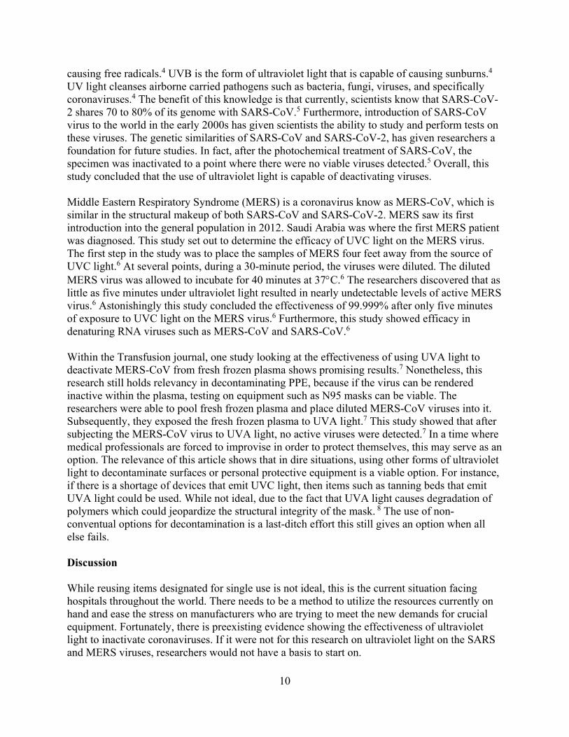
The Vent Splitter and Flow Limiter A group of physicians in San Antonio, Texas, have manufactured a Vent Splitter and Flow Limiter to provide multi-patient ventilation in response to the COVID-19 pandemic.4 The group has shared their design online, making it possible to for hospitals to 3D print. The Vent Splitter has a single port end and a dual port end (Figure 1). The single port is attached to the inspiratory and expiratory ports of the ventilator. The dual ports are attached to the patients. The Vent Splitter single port has an inside diameter of between 22.1 mm and 22.75 mm.4 The Vent Splitter dual ports have an outside diameter between 21.5 mm and 22.0 mm and are designed to be compatible with most hospital ventilators. The Vent Splitter can also be scaled up or down to accommodate ventilators that may not fit the 22mm internal diameter.4 The manufacturers recommend using the Vent Splitter, but if a 3D printer is not available, a T-Piece can be used. A factor that may limit the efficacy of ventilator splitting is lung compliance. Current research recommends patients of similar lung compliance be paired for vent splitting. A significant
14
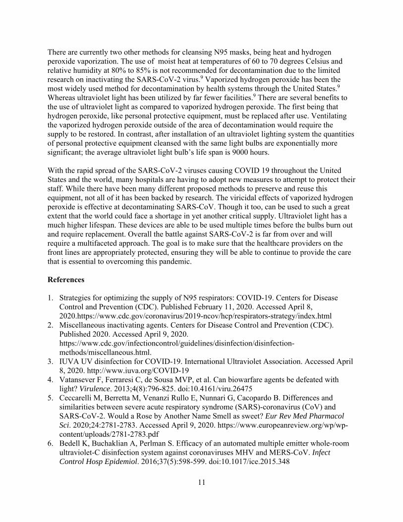
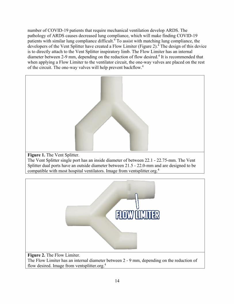
number of COVID-19 patients that require mechanical ventilation develop ARDS. The pathology of ARDS causes decreased lung compliance, which will make finding COVID-19 patients with similar lung compliance difficult.9 To assist with matching lung compliance, the developers of the Vent Splitter have created a Flow Limiter (Figure 2).4 The design of this device is to directly attach to the Vent Splitter inspiratory limb. The Flow Limiter has an internal diameter between 2-9 mm, depending on the reduction of flow desired.4 It is recommended that when applying a Flow Limiter to the ventilator circuit, the one-way valves are placed on the rest of the circuit. The one-way valves will help prevent backflow.4
Figure 1. The Vent Splitter. The Vent Splitter single port has an inside diameter of between 22.1 - 22.75-mm. The Vent Splitter dual ports have an outside diameter between 21.5 - 22.0-mm and are designed to be compatible with most hospital ventilators. Image from ventsplitter.org.4
Figure 2. The Flow Limiter. The Flow Limiter has an internal diameter between 2 - 9 mm, depending on the reduction of flow desired. Image from ventsplitter.org.4

15
Ventilator Settings Patients with ARDS need specific ventilator settings to meet their physiologic needs. One study recommends these settings for patients with ARDS: Volume control mode, Vt of 6mL/Kg predicted body weight (PBW), Plateau pressure < 30 cm H2O, I:E = 1:1, RR 20-30/min, and high PEEP.9 Providing these settings to multiple COVID-19-induced ARDS will be difficult, if not impossible, due to varying lung compliance. In response to this concern, a hospital in New York has provided recommendations for multi-patient ventilation. New York-Presbyterian Hospital has used vent splitting during the COVID-19 outbreak. The hospital has released a statement on their recommended ventilator settings.10 First, to pair two patients on a ventilator, a pre-assessment must be done. Each patient must have the following ventilator settings or physiology before pairing: Vt of 6-8mL/kg PBW, Driving pressure of 5-16cmH2O, 12-30/min, 5-18 PEEP, 21-60% O2, pH of 7.30 or higher, SpO2 of 92-100%, no recent major ventilator changes, no contraindication to neuromuscular blockade (NMB), same infectious organism, no baseline asthma or chronic obstructive pulmonary disease (COPD), and no major changes in hemodynamic stability. Acceptable differences were: 0-6 cm H20 of driving pressure, 0-8/min, and 0-5 cm H20.10 Once two patients are appropriately paired, the recommended settings are: pressure control mode, Vt of 6-8 mL/kg PBW, peak inspiratory pressure of 30cmH2O or less, driving pressure of 5-18 cm H2O, RR of 12-30/min, inspiratory time of 0.6-1.0 secs, PEEP of 5-16 cm H2O, FiO2 of 21-100% to achieve an oxygen saturation of 92-100%, pH between 7.25-7.45, and mandatory NMB. The statement also detailed treating respiratory acidosis or alkalosis in these patients. The article states to treat respiratory acidosis through ventilator changes and to treat respiratory alkalosis through increasing dead space on the ventilator circuit.10 Of note, the hospital recommends pressure control ventilation (PCV) over volume control ventilation (VCV). As mentioned previously, some studies have supported using VCV in patients with ARDS.10 VCV is effective when supporting one patient on a ventilator but can become dangerous when additional patients are added. If one patient has an obstruction or sudden decrease in lung compliance, the Vt is delivered to the other patient.11,12 This increases the risk of barotrauma and is why PCV is recommended in vent splitting. Conflicting Recommendations of Use A statement made by the Food and Drug Administration (FDA) on March 24th, 2020, approved the use of vent splitting during the COVID-19 pandemic.12 When more than one circuit is added the FDA recommends pressure control ventilation. Every patient is to receive equal pressure, volume, FiO2, and PEEP.12 Additionally the FDA recommends matching patients by size, physiologic condition, lung compliance, and ventilator needs as well as sedating and paralyzing vent matched patients to avoid one patient's respiratory efforts affecting another. 12 Soon after the FDA approved vent splitting for emergency circumstances, a joint statement was release by The Society of Critical Care Medicine (SCCM), American Association for Respiratory Care (AARC), American Society of Anesthesiologists (ASA), Anesthesia Patient Safety Foundation (ASPF), American Association of Critical-Care Nurses (AACN), and
16
American College of Chest Physicians (CHEST) on March 26th, 2020.13 The statement recommended practitioners not attempt vent splitting. The physiologic needs of patients with COVID-19 induced ARDS cannot be safely met while more than one patient is on a ventilator.13 According to the joint statement, patients suffering from ARDS have a 40-60% mortality rate.13 Splitting ventilators between multiple patients suffering from COVID-19 induced ARDS could further increase their mortality rate. The joint statement declares that ventilator splitting is not safe due to lung volumes favoring the more compliant lung, difficulty managing PEEP, and as patients begin to improve or deteriorate lung volume distribution will shift.13 Conclusion The COVID-19 pandemic has caused a shortage of resources. The limited supply of ventilators has resulted in unique practices and inventions. The Vent Splitter, developed by a group of physicians from San Antonio, Texas, is designed to ventilate multiple patients on a single machine. The limiting application of ventilator splitting is varying patient physiology that can lead to inadequate ventilation of one or both the patients attached to the ventilator. The team from San Antonio addressed this concern by developing the Flow Limiter. The Flow Limiter is used to match patients of varying lung compliance which decreases the risk of inadequate ventilation and barotrauma. Despite the development of the Vent Splitter and Flow Limiter there is currently no research to support its use. Furthermore, while the FDA has approved ventilator splitting for the COVID-19 pandemic, a joint statement by the AARC, ASA, ASPF, AACN, and CHEST refute their recommendation and suggest no provider attempt to ventilate multiple patients with a single ventilator.13 Until a retrospective analysis is performed, the decision to ventilator split will present as an ethical dilemma to health care providers. Hospitals and providers must decide to either provide adequate and safe ventilation to one patient at the expense of another or provide possibly inadequate ventilation and increased risk to both patients. References 1. Cascella M, Rajnik M, Cuomo A, Dulebohn SC, Di Napoli R. Features, evaluation and
treatment coronavirus. StatPearls. Accessed April 13, 2020. https://www.ncbi.nlm.nih.gov/books/NBK554776/
2. Lai CC, Shih TP, Ko WC, Tang HJ, Hsueh PR. Severe acute respiratory syndrome coronavirus 2 (SARS-CoV-2) and coronavirus disease-2019 (COVID-19): The epidemic and the challenges. Int J Antimicrob Agents. 2020;55(3):105924. doi:10.1016/j.ijantimicag.2020.105924
3. Matheny K, Shamus KJ. Detroit emergency doctor’s ventilator idea is getting global attention. Detroit Free Press. Published April 3, 2020. Accessed April 13, 2020. https://www.freep.com/story/news/nation/coronavirus/2020/04/03/coronavirus-doctor-idea-sharing-splitting-ventilators/5106757002/
4. Lai B, Erian J, Pew P, Eckmann M. Emergency Ventilator circuit splitter. ventsplitter.org. Accessed April 12, 2020. http://ventsplitter.org/
5. Neyman G, Irvin CB. A Single Ventilator for Multiple Simulated Patients to Meet Disaster Surge. Acad Emerg Med. 2006;13(11):1246-1249. doi:10.1197/j.aem.2006.05.009
17
6. Paladino L, Silverberg M, Charchaflieh JG, et al. Increasing ventilator surge capacity in disasters: Ventilation of four adult-human-sized sheep on a single ventilator with a modified circuit. Resuscitation. 2008;77(1):121-126. doi:10.1016/j.resuscitation.2007.10.016
7. Smith R, Brown JM. Simultaneous ventilation of two healthy subjects with a single ventilator. Resuscitation. 2009;80(9):1087. doi:10.1016/j.resuscitation.2009.05.018
8. Menes K, Tintinalli J, Plaster L. How one Las Vegas ED saved hundreds of lives after the worst mass shooting in U.S. history. epmonthly.com. Published November 3, 2017. Accessed April 12, 2020. https://epmonthly.com/article/not-heroes-wear-capes-one-las-vegas-ed-saved-hundreds-lives-worst-mass-shooting-u-s-history/
9. Cordingley JJ. The pulmonary physician in critical care middle dot 8: Ventilatory management of ALI/ARDS. Thorax. 2002;57(8):729-734. doi:10.1136/thorax.57.8.729
10. Beitler J, Kallet R, Kacmarek R, et al. Ventilator Sharing Protocol: Dual-Patient Ventilation with a Single Mechanical Ventilator for Use during Critical Ventilator Shortages. March 2020. Accessed April 12, 2020. https://www.gnyha.org/wp-content/uploads/2020/03/Ventilator-Sharing-Protocol-Dual-Patient-Ventilation-with-a-Single-Mechanical-Ventilator-for-Use-during-Critical-Ventilator-Shortages.pdf
11. Farkas J. PulmCrit - Splitting ventilators to provide titrated support to a large group of patients. EMCrit Project. Published March 15, 2020. Accessed April 14, 2020. https://emcrit.org/pulmcrit/split-ventilators/
12. U.S. Food & Drug Administration. Emergency use authorization. Published March 24, 2020. Accessed April 12, 2020. https://www.fda.gov/media/136423/download
13. The Society of Critical Care Medicine, American Association for Respiratory Care, American Society of Anesthesiologists, Anesthesia Patient Safety Foundation, American Association of Critical-Care Nurses, American College of Chest Physicians. Joint statement on multiple patients per ventilator. Published March 26, 2020. Accessed April 12, 2020. https://www.sccm.org/getattachment/Disaster/Joint-Statement-on-Multiple-Patients-Per-Ventilato/Joint-Statement-Patients-Single-Ventilator.pdf?lang=en-US
Mentor: Leonardo Campero, DNAP, CRNA
COVID-19: Effect on Availability of Masks for Healthcare Practitioners
Ryan Mona, BSN Keiser University
Keywords: Coronavirus, COVID-19, healthcare workers, masks, shortages Coronavirus Disease 2019 (COVID-19) has developed into a once in a century pandemic that has shut down national borders, caused economic instability globally, and left a health care industry in dismay. At the time of this writing, the number of confirmed positive cases of COVID-19 globally has reached 67,000,000 and continues to climb.1 The United States, the epicenter for COVID-19, continues to be ravaged with positive cases as the effects are seen across the country.
18
During these extremely difficult times, healthcare workers are at the forefront of battling COVID-19. Any provider that has direct physical contact with patients positive for COVID-19 is at an increased risk contracting the virus.2 Anesthesia providers face significant exposure risks each day, from known COVID-19 positive patients, as well as patients who do not know they have the virus. The Centers for Disease Control and Prevention (CDC) has stated that the time period between exposure to COVID-19 and the appearance of symptoms, called the incubation period, is estimated to be between two and 14 days.3 The CDC also stated that the median time from exposure to onset of symptoms is four-five days.4 Testing for the virus was a challenging issue in the early stages of the pandemic which contributed to rapid spread of the virus. Protective equipment used by healthcare providers provide protection against becoming infected with COVID-19, but unfortunately, protective equipment was in short supply in some hospitals. In March 2020, World Health Organization (WHO) Director-General Tedros Adhanom Ghebreyesus stated, “The chronic global shortage of personal protective equipment is now one of the most urgent threats to our collective ability to save lives.”5
The route of transmission of a virus plays an important part in determining how healthcare workers can protect themselves in terms of personal protective equipment (PPE). Research has shown that COVID-19 is predominantly transmitted through respiratory droplets meaning transmission can take place within close proximity (<2 meters) of an infected person through a cough or sneeze.6 As time has progressed from the onset of the virus, the CDC has established and published recommendations to healthcare facilities regarding aerosol-generating procedures such as extubation, mechanical ventilation, suctioning, bag-mask ventilation (BMV), and chest compressions which the risk of airborne transmission.7 A respiratory droplet is a droplet particle >5-10 µm in diameter, while anything less than 5 µm in diameter refers to a droplet nuclei.3 The measurement of a droplet particle is a determining factor as to whether a particular mask provides protection against the spread of the virus via the airway (mouth and nose). While debate exists whether an N95 respirator mask is superior to a surgical mask, each serves its purpose. The surgical mask provides barrier protection against respiratory droplets. In contrast, an N95 respirator mask uses a filter to remove at least 95% of airborne particles from the user’s breathing air along with large droplets.8 The surgical mask is not regulated for particulate filtration efficiency while the N95 respirator mask is. Further, the surgical mask may be loosely fitted around the face, the N95 must be properly fit tested so that minimal leakage occurs.8 The crucial point is that masks are essential for healthcare workers providing care for a COVID-19 positive patient. Without masks for airway protection, the healthcare worker is at significant risk of becoming infected.9
Prior to the COVID-19 pandemic, masks used in a hospital setting during direct patient care were discarded after each use. However, when hospitalizations were escalating for COVID-19 patients initially, there was a shortage of masks resulting in the reuse of masks in some circumstances.10 In an interview with a neonatal intensive care unit (NICU) nurse working at Children’s Hospital of Colorado in Aurora, CO, a first-hand account provided enlightening details as to how healthcare employees protect themselves with masks. During the time of this writing, the Children’s Hospital system in Aurora reported two positive COVID-19 cases. Healthcare workers entering the building for their shift had their temperature taken first via temporal route, then completed an online questionnaire with their employee ID number and list of symptoms. If their temperature was within normal limits (< 38 degrees Celsius) and they were asymptomatic,

19
they were allowed to proceed with obtaining a mask for the shift. Healthcare workers were provided surgical masks if performing normal patient care for individuals not confirmed positive or under investigation. The surgical mask had to remain on at all times during the shift unless the healthcare worker was using the restroom or in a breakroom. At the end of the shift, the mask was discarded. If a healthcare worker treated a confirmed COVID-19 positive case or a patient that was under investigation for having the virus, they were given an N95 mask to wear. The N95 had to be worn for four shifts in a row by that employee before the mask was discarded. At the end of each shift, the N95 had to be placed in a paper bag assigned to the employee and was taken for UV sterilization before being returned to the same employee for the next shift. Each employee, at the initial start of this protocol, was given one shield. At the end of each shift, the shield was stored in a paper bag in an isolated room where the healthcare worker could retrieve it for their next shift. The surgical masks and N95 respirator masks were heavily guarded due to the limited supply and were handed out by designated “PPE Protectors.” (B. Mona, oral communication, April 2020).
As supply chains continued to decrease and masks were overused or not available, concerns grew regarding how to protect the healthcare workers. WHO Director-General Dr. Tedros Adhanom Ghebreyesus stated on March 3, 2020, “Without secure supply chains, the risk to healthcare workers around the world is real. Industry and governments must act quickly to boost supply, ease export restrictions, and put measures in place to stop speculation and hoarding.” He added, “We can’t stop COVID-19 without protecting health workers first.”11
Around the world, healthcare workers demonstrated courage, compassion, and teamwork in helping to combat COVID-19. Without these remarkable men and women, the number of positive cases and deaths would be much higher. The shortages of equipment, in particular masks, left the individuals on the front line dangerously ill-equipped. However, healthcare workers across the world were and continue to be united in their commitment to care for patients with COVID-19. On behalf of the world… WE THANK YOU! References 1. Johns Hopkins University & Medicine. Coronavirus COVID-19 global cases by the Center
for Systems Science and Engineering (CSSE). Coronavirus Resource Center. https://coronavirus.jhu.edu/map.html. Published 2020. Updated April 29, 2020. Accessed April 30, 2020.
2. Habibzadeh P, Stoneman E. The novel coronavirus: a bird’s eye view. Int J Occup Environ Med. 2020;11(2):65-71. doi: 10.15171/ijoem.2020.1921
3. World Health Organization. Modes of transmission of virus causing COVID-19: implications for IPC precaution recommendations. https://www.who.int/news-room/commentaries/detail/modes-of-transmission-of-virus-causing-covid-19-implications-for-ipc-precaution-recommendations. Published March 29, 2020. Accessed April 9, 2020.
4. Centers for Disease Control and Prevention. Interim clinical guidance for management of patients with confirmed coronavirus disease (COVID-19). https://www.cdc.gov/coronavirus/2019-ncov/hcp/clinical-guidance-managment-patients.html. Updated November 3, 2020. Accessed December 7, 2020.

20
5. Burki, T. Global shortage of personal protective equipment. Lancet Infect Dis. 2020; 20(7):785-786. doi: 10.1016/S1473-3099(20)30501-6
6. Ong S, Tan Y, Chia P, et al. Air, surface environmental, and personal protective equipment contamination by severe acute respiratory syndrome coronavirus 2 (SARS-CoV-2) from a symptomatic patient. JAMA. 2020. doi: 10.1001/jama.2020.3227
7. Centers for Disease Control and Prevention. COVID-19 infection prevention and control in healthcare settings: questions and answers. https://www.cdc.gov/coronavirus/2019-ncov/hcp/infection-control-faq.html. Updated April 1, 2020. Accessed April 9, 2020.
8. D’Alessandro M, Cichowicz J. Proper N95 respirator use for respiratory protection preparedness. Centers for Disease Control and Prevention. https://blogs.cdc.gov/niosh-science-blog/2020/03/16/n95-preparedness/. Published March 16, 2020. Updated March 23, 2020. Accessed April 9, 2020.
9. Picard J, Cornec G, Baron R, et al. Wearing of face masks by healthcare workers during COVID-19 lockdown: what did the public observe through the French media. J Hosp Infect. 2020; 106(3):617-620. doi: 10/1016/j.jhin.2020.08.009
10. Czubryt M, Stecy T, Popke E, et al. N95 mask reuse in a major urban hospital: COVID-19 response process and procedure. Journal of Hospital Infection. 2020; 106(2):277-282. doi: 10.1016/j.jhin.2020.07.035
11. World Health Organization. Shortage of personal protective equipment endangering health workers worldwide. https://www.who.int/news/item/03-03-2020-shortage-of-personal-protective-equipment-endangering-health-workers-worldwide. Published March 3, 2020. Accessed April 9, 2020.
Mentor: Leonardo Campero, DNAP, CRNA
Gnathodiaphyseal Dysplasia: Anesthesia for Rare Diseases
Kieran M. Shamash, MSN University of Southern California
Keywords: gnathodiaphyseal dysplasia, rare diseases, anesthesia, airway Rare diseases can present a challenge to the anesthesia professional. Information about rare diseases and their anesthetic implications can be difficult to obtain.1 Gnathodiaphyseal dysplasia (GDD) is a hereditary autosomal dominant bone disorder with unknown prevalence, but is thought to be extremely rare. Few published reports of this disease exist in the literature.2 GDD is characterized by skeletal abnormalities such as cemento-osseous fibromas of the maxilla and mandible, as well as weak long bones with vulnerability to fracture after even minimal trauma.2-7 Anesthesia professionals may encounter a patient with GDD if they present for surgery. This case report will focus on a patient who presented for orthopedic surgery. Case Report A 16-year-old male (weight 55.6 kg, height 170 cm) with no known allergies presented with history of GDD and related long bone fractures for unilateral left tibia and fibula osteotomy and intramedullary nail placement. The patient had previously undergone a successful

21
intramedullary nail placement for the left femoral shaft following a fracture two years prior without any anesthetic complications. One year after that surgery, he suffered a left tibial shaft fracture that was treated conservatively with casting. Follow-up evaluations of the patient’s left lower extremity revealed delayed healing of the fracture, significant deformity of the left tibia, anterior bowing related to patient’s disease process, and multiple fractures of the left lower extremity. The decision was made to perform an osteotomy and intramedullary nail placement to stabilize the fractures. On the day of surgery, the patient and family were interviewed in the preoperative area. Of note, the patient’s mother was found to suffer from the same disease and had a jaw replacement, in addition to a below-the-knee amputation of the left lower extremity related to complications from a previous surgery. After a thorough chart review, the patient was examined, and consent was obtained for general anesthesia and placement of postoperative sciatic nerve block catheter. Airway exam revealed a Mallampati II class airway, thyromental distance less than 6 cm, adequate oral opening, a midline trachea, and full cervical range of motion. Per the patient and his chart review, the patient had no spinal involvement. The patient demonstrated the ability to prognath and had no history of jaw dislocation or clicking. Consistent with his diagnosis of GDD, the patient was found to have poor dentition and two loose teeth. Direct laryngoscopy was planned. A video laryngoscope and fiberoptic scope were present in the OR in case of difficult direct laryngoscopy. Midazolam 2 mg was administered prior to transfer to the OR. Once positioned supine in the OR, the patient’s bony prominences were carefully padded, and standard noninvasive monitors were applied. The patient was preoxygenated with O2 10 L/min via face mask and induction of general anesthesia was achieved with fentanyl 50 mcg, ketamine 20 mg, and propofol 200 mg. After establishing easy mask ventilation with an 80 mm oral airway, rocuronium 30 mg was administered for muscle relaxation. Direct laryngoscopy with a Macintosh #3 laryngoscope was performed. A Cormack-Lehane grade I view of the larynx was obtained with cricoid pressure and the trachea was intubated with a 7.0 cuffed endotracheal tube. General anesthesia was maintained with end-tidal sevoflurane 2.3%-3.2% throughout the 2.5-hour surgery. Analgesia was achieved with a combination of fentanyl, ketamine, and hydromorphone. Fluid management included infusions of lactated ringers and albumin 5% to maintain mean arterial pressure 55-75 mm Hg. The patient received a total of ondansetron 4 mg and dexamethasone 8 mg for postoperative nausea and vomiting prophylaxis. At the end of the case, neuromuscular blockade was fully antagonized with neostigmine 3 mg and glycopyrrolate 0.6 mg. The patient demonstrated an intrinsic respiratory rate of 20-25 and tidal volumes of 4-6 mL/kg. General anesthesia was maintained on sevoflurane 3%, combined with small intermittent boluses of propofol (10-20 mg/dose) to facilitate placement of a popliteal sciatic nerve block catheter for the purpose of postoperative analgesia. The patient received a bolus of 10 mL of ropivacaine 0.1% via the catheter followed by a continuous infusion. At the conclusion of the procedure, the patient’s oropharynx was suctioned and a deep extubation was performed after easy placement of an 80 mm oral airway and 24 french nasopharyngeal airway. The patient was spontaneously breathing at all times and required no additional airway support. The patient was then transferred to the post-anesthesia care unit in stable condition.

22
Discussion There is limited data on the implications of GDD, and almost no data on the anesthetic considerations for this extremely rare condition. There are an estimated 80 case reports in the literature of affected individuals, all of whom have suffered fractures.4 Diagnosis can be made based on clinical presentation and, if available, genetic data.4-6 GDD was found to be associated with a variety of genetic mutations of the anoctamin 5 (ANO5) gene located on chromosome 11.6 Careful assessment of clinical features and imaging can also assist with the diagnosis. Current evidence describes a syndrome including cemento-osseous fibromas of the maxillary bones with associated increased risk of osteomyelitis, as well as cortical thickening and diaphyseal bending of the long tubular bones.2-7 This leaves the individual vulnerable to fractures resulting from minor trauma.4 Of the available published case reports, only two2,4 reported involvement of the spine, including one patient who had cervical and thoracic vertebral fractures. When caring for GDD patients, careful assessment of any spinal abnormalities, particularly of the cervical spine, should be included in the preoperative evaluation. Involvement of the maxillary bones is well reported in the available literature on GDD. Three case reports describe surgical interventions of the maxillary bones, including mandibular prosthesis and free bone flap placements.3,5,6 When the anesthesia professional is presented with the opportunity to care for an individual with GDD, careful consideration of the attributes of this disease is paramount. In at least one study, airway management during a mandibular debulking was described as somewhat difficult due to challenging endotracheal intubation, stridor, and excessive secretions.3 No other details related to the anesthetic course were provided. When caring for the patient with GDD, the anesthesia provider must consider the impact of the disease process on the patient’s airway in particular. GDD is associated with increased incidence of osteomyelitis of the maxilla, poor dentition, and cemento-ossesous fibromas of the maxillary bones.7 This could result in loose or missing teeth, and could negatively impact mouth opening, mask seal, and neck extension during intubation. In this case study, direct laryngoscopy was selected as the patient had no major indicators of a difficult airway on exam. Additionally, the decision was made to do a deep extubation with this patient to ensure a smooth emergence, after consideration of the patient’s ease of mask ventilation and intubation during the induction of anesthesia. However, if the patient with GDD presents with an airway anomaly or other concerns for a difficult airway, an awake extubation may be a more appropriate. It is important to differentiate GDD from similar skeletal diseases, as the anesthetic implications may vary. Once thought to be polyostotic fibrous dysplasia (PFD), which also presents with multiple fractures, GDD can be differentiated due to its characteristic bone cortical thickening and bowing, attributes not seen in PFD.2 PFD also presents as McCune-Albright syndrome (MAS) when combined with endocrinopathies and the presence of café au lait spots.1,7 GDD is not associated with any endocrinopathies or cardiac anomalies. Additionally, patients with GDD present with normal cognitive development.3 In the case presented, the patient was an adolescent pediatric patient with normal for age cognitive development, which allowed for him to take an active role in his care.

23
Patients with GDD commonly present for multiple lower extremity surgeries, including placement of intramedullary nails.4 Anesthetic considerations for these patients are similar to other patients undergoing orthopedic procedures of the lower extremities and include consideration of regional anesthesia and analgesia. In the presented case, a regional block was placed for the purposes of postoperative analgesia. However, a subarachnoid block could be considered for similar lower extremity surgeries and could preserve a patent airway if a difficult airway is suspected. In the presented case, the decision was made to proceed with general anesthesia as the length of the surgery was uncertain. Since these patients may have a history of multiple surgeries, the anesthesia professional must also be prepared for fluid shifts if excessive bleeding related to instrumentation and revisions is encountered.8 Lastly, the patient with a rare disease presents a unique challenge for the anesthesia professional as it can be difficult to find information on recommended anesthesia management. Anesthesia professionals rely on case studies like this one, and articles in journals such as the International Student Journal of Nurse Anesthesia or the American Association of Nurse Anesthetists Journal, to share best practices for patients with rare diseases. One resource currently in development, is a website entitled Orphananesthesia.eu.1 This website is a continually updated, peer-reviewed resource for the anesthetic considerations for rare diseases. References 1. Munster T. OrphanAnesthesia - anesthesia recommendations for patients suffering from rare
diseases. Orphanet Journal of Rare Diseases. 2014;9:O16. 2. Ahluwalia J, Ly JQ, Norman E, Costello RF, Beall DP. Gnathodiaphyseal dysplasia. Clin
Imaging. 2007;31(1):67-69. 3. Herman TE, Siegel MJ, Sargar K. Gnathodiaphyseal dysplasia. J Perinatol. 2014;34(5):412-
414. 4. Kuroda T, Okano I, Sawada T, et al. Recurrent femoral shaft fractures in a child with
gnathodiaphyseal dysplasia: a case report. BMC Musculoskelet Disord. 2019;20(1):92. 5. Marechal G, Schouman T, Mauprivez C, et al. Gnathodiaphyseal dysplasia with a novel
R597I mutation of ANO5: Mandibular reconstruction strategies. J Stomatol Oral Maxillofac Surg. 2019 Nov;120(5):428-431.
6. Merlini A, Garibaldi J, Giorgis L, Balbi P. Gnathodiaphyseal dysplasia: Surgical treatment and prosthetic rehabilitation of 2 members of the same family. J Oral Maxillofac Surg. 2016;74(12):2441-2446.
7. Riminucci M, Collins MT, Corsi A, et al. Gnathodiaphyseal dysplasia: A syndrome of fibro-osseous lesions of jawbones, bone fragility, and long bone bowing. J Bone Miner Res. 2001;16(9):1710-1718.
8. Hemmings, H.C. & Egan, T.D. Pharmacology and Physiology for Anesthesia. Philadelphia, PA: Elsevier, Inc.; 2013.
Mentor: Erica McCall, MPH, CRNA

24
Management of Dexmedetomidine-Induced Hypertension
Kelly Nagle, DNP Uniformed Services University
Keywords: dexmedetomidine, alpha-2 agonist, hypertension, bradycardia Dexmedetomidine is a highly selective, potent alpha-2 adrenergic agonist that causes sedation, hypnosis, and analgesia. Dexmedetomidine attenuates the hemodynamic response to tracheal intubation and surgical stimuli and has been shown to decrease plasma catecholamine concentrations during surgery.1 Due to its sympatholytic and vagomimetic effects, hypotension and bradycardia are common adverse effects. Hypertension is a less common adverse effect; however, it is usually transient, occurs after large bolus doses, and typically does not require treatment. This case report presents a patient who experienced persistent hypertension with a high-dose dexmedetomidine infusion during general anesthesia. Case Report A 56-year-old, 57 kg, 154 cm female presented for left-sided functional endoscopic sinus surgery and bilateral myringotomy and tympanostomy tube placement for recurrent sinus pressure and ear pain. The patients’ past medical history included seasonal allergies, gastroesophageal reflux disease, and hypothyroidism. The patient had no known drug allergies, and previous surgical history included an adenotonsillectomy with no anesthetic complications. The patient denied smoking and alcohol use. Medications included levothyroxine and loratadine. Thyroxine (T4) levels were within normal levels, and the patient denied signs and symptoms of hypo and hyperthyroidism. Preoperative vital signs were within normal limits with blood pressure 120/80 mm Hg and heart rate 76/min. Preoperatively, the patient received midazolam 2 mg intravenously and scopolamine 1.5 mg transdermally. In the operating room, standard monitors were applied, the patient was preoxygenated with O2 100% via face mask, and general anesthesia was induced with propofol 130 mg, lidocaine 70 mg, and ketamine 30 mg; rocuronium 50 mg was used for neuromuscular blockade. The trachea was intubated with a 7.0 endotracheal tube via direct laryngoscopy with a Macintosh 3 blade, and the patient was mechanically ventilated. The patient remained normotensive during intubation and anesthesia was maintained with a total intravenous anesthetic technique using propofol 120 mcg/kg/min, lidocaine 30 mcg/kg/min, dexmedetomidine 0.7 mcg/kg/hr, and ketamine 10 mg/hr. Twenty minutes after surgical start time, the patient became hypertensive with a blood pressure of 150s/90s mm Hg and bradycardia with a heart rate of 50/min. The propofol infusion was increased incrementally up to 200 mcg/kg/min, and the dexmedetomidine infusion was decreased to 0.5 mcg/kg/min. Two additional propofol boluses of 50 mg were given to increase the anesthetic depth, and glycopyrrolate 0.1 mg was given to increase heart rate. The patient remained hypertensive and bradycardic with minimal changes in blood pressure or heart rate. The surgeon continued to have difficulty visualizing the surgical field due to bleeding. Nitroglycerin boluses 10-25 mcg were titrated to blood pressure response, and an infusion of

25
nitroglycerin 0.05-0.2 mcg/kg/min was started. During this time, the dexmedetomidine and ketamine infusions were discontinued as they were mixed together in one syringe. The nitroglycerin infusion was titrated to a systolic blood pressure <130 mmhg, and the propofol infusion was decreased to 150 mcg/kg/min. Heart rate remained 50-55/min. At the conclusion of the case, the nitroglycerin infusion was discontinued, and the neuromuscular blockade was antagonized with sugammadex 120 mg. Upon spontaneous ventilation, the patient was extubated and emerged from anesthesia. Vital signs remained within normal limits in the post-anesthesia care unit. Discussion Dexmedetomidine is a highly selective alpha-2 adrenoceptor agonist that is used for its analgesic, sedative, opioid sparing, and minimal respiratory depression properties. Alpha-2 adrenergic receptors are present throughout the central and peripheral nervous system, and therefore, have widespread physiologic effects. In addition to analgesia and sedation, side effects include decreased secretions, bowel motility, renin release, intraocular pressure, and insulin release and an increase in glomerular filtration.1 Analgesic effects are presumed to be mediated through the binding of alpha-2 receptors in the dorsal horn and locus coeruleus. Pain transmission is reduced through the hyperpolarization of interneurons and a subsequent decrease in pain neurotransmitters such as substance P and glutamate.1 When used in the intraoperative period, dexmedetomidine has been shown to reduce opioid and inhaled anesthetic requirements and decrease hemodynamic responses to surgical stimuli. In one study, a single bolus dose of 0.5 mcg/kg decreased intraoperative and postoperative analgesic requirements and improved patient satisfaction.2 While dexmedetomidine has mostly favorable properties, common adverse effects include hypotension and bradycardia.3 Less commonly, transient hypertension can occur after loading doses and usually subsides with a continued infusion. This hypertension is believed to be secondary to the stimulation of peripheral alpha-2B adrenoceptors located in vascular smooth muscle causing vasoconstriction that is accompanied by a baroreceptor reflex-induced bardycardia.4 The hypertension is transient due to the concurrent activation of peripheral alpha-2B receptors on vascular endothelial cells resulting in vasodilation; central alpha-2A receptors are also activated. Central alpha-2A receptors are located presynaptically in the central nervous system and prevent the release of norepinephrine, leading to hypotension and bradycardia.2 There is a dose-dependent reduction in plasma catecholamines by 60-80%, causing sympatholytic effects.5 On average, mean arterial pressure is reduced 13-27% when compared to baseline, yet higher maintenance doses are associated with increases in mean arterial pressures with hypertensive effects occurring at concentrations between 1.9 and 3.2 ng/mL.5 While dexmedetomidine-induced hypertension is transient and generally does not require treatment, in this case treatment was necessary to reduce intraoperative bleeding which was obscuring the surgical field. The differential diagnosis for intraoperative hypertension includes depth of anesthesia, pain, pre-existing hypertension, and hypermetabolic states. Since the patient had an adequate anesthetic depth after escalating doses of propofol and multimodal pain management approach with lidocaine, ketamine, dexmedetomidine, it was determined the hypertension and bradycardia was likely secondary to the dexmedetomidine infusion. The

26
dexmedetomidine infusion was discontinued and nitroglycerin boluses were used to manage the hypertension. Dexmedetomidine is relatively short acting; it undergoes linear pharmacokinetics with a distribution half-life of 6 minutes and an elimination half-life of 2 hours.1 The anesthesia professionals chose to treat the hypertension with nitroglycerin due to its quick onset and short duration. The literature is unclear as to the best medication for treating dexmedetomidine-induced hypertension.2 Labetalol, a combined alpha and beta antagonist, may have been a good choice except that the patient was already bradycardic and labetalol reduces heart rate through beta receptor blockade.3 Hydralazine was not used due to its slow onset. Phentolamine, a pure alpha antagonist, would be a reasonable choice in a hypertensive crisis; however, it was not readily available at the facility.2 Nicardipine, a calcium channel antagonist, would be another reasonable choice. The goal in treating dexmedetomidine-induced hypertension is to avoid drugs that potentiate unopposed alpha adrenoceptor stimulation, such as pure beta antagonists. This case was unusual in that the patient became hypertensive an hour after starting the dexmedetomidine infusion and no loading dose was administered. Additionally, the combination of dexmedetomidine and ketamine infusion was chosen because the combination has been shown to provide rapid onset sedation and analgesia with better hemodynamic stability compared to dexmedetomidine alone.5 However, it should be noted that since the ketamine was discontinued with the dexmedetomidine, the ketamine could also have contributed to the intraoperative hypertension in conjunction with the dexmedetomidine. Nevertheless, clinicians should be aware of the biphasic hemodynamic response that may occur with high dose dexmedetomidine boluses and infusions. In the pediatric population, dexmedetomidine-induced bradycardia treated with an anticholinergic has been shown to cause immediate, significant hypertension requiring urgent treatment.5 There was a concern that that the scopolamine patch and dexmedetomidine infusion led to persistent hypertension due to the onset of hypertension correlating similarly to the onset of the scopolamine patch. While dexmedetomidine has a mostly favorable side effect profile, clinicians should be aware of and have a plan to manage dexmedetomidine-induced hypertension. References 1. Kaur M, Singh PM. Current role of dexmedetomidine in clinical anesthesia and intensive
care. Anesth Essays Res. 2011;5(2):128–133. 2. Muthiah T, Moni A, Mathews L, Balaji S. Intravenous labetalol in treating hypertensive
crisis following dexmedetomidine infusion for procedural sedation. J Clin Anesth. 2016; 29:30-32.
3. Chavali S, Singh S, Kaushal A, Khandelwal A, Roy H. Persistent hypertensive response following dexmedetomidine infusion in a patient with cervical spinal cord injury. J Neurosci Rural Pract; 2018; 9:426-427.
4. Weerink MS, Struys, Michel MF, Hannivoort LN, Barends CM, Absalom AR, Colin P. Clinical pharmacokinetics and pharmacodynamics of dexmedetomidine. Clin Pharmacokin. 2017; 56:893-913.
5. Mahmoud M, Mason KP. Dexmedetomidine: review, update, and future considerations of paediatric perioperative and periprocedural applications and limitations. Br J Anaesth. 2015; 115:171-182.

27
6. Tang C, Xia Z. Dexmedetomidine in perioperative acute pain management: A non-opioid adjuvant analgesic. J Pain Res. 2017; 10:1899–1904.
Mentor: LCDR Lauren Suszan, DNP, MSN, CRNA, NC, USN
Chronic Pain and Opioid Dependence
Larry Beasley, DNP Uniformed Services University
Keywords: chronic pain, opioid dependence, hyperalgesia, oral morphine equivalents Chronic pain is nociception exceeding the temporal boundary of tissue injury and subsequent healing process, frequently approximated at 3 months.1 It is commonly delineated as either malignant or nonmalignant. Malignant chronic pain is primarily associated with cancer. Nonmalignant pain involves neuropathic, musculoskeletal, and inflammatory processes. Prevalence is estimated at one-fifth to one-half the general population.1 Treatment often requires an interdisciplinary and multimodal approach. Preoperative assessment should include baseline medication requirements, functional status, psychological history, as well as past medical and surgical history.2 The goal of perioperative care is meeting analgesic requirements without negatively affecting hemodynamics, respiratory drive, or mental functioning.1 Case Report A 45-year-old male presented for an open reduction and internal fixation of a left proximal humeral neck fracture. The patient was 185 cm and 83 kg with a medication allergy to penicillin. Past medical history was significant for chronic pain secondary to primary disease progression and treatment of nodular sclerosing Hodgkin’s lymphoma (NSHL). The patient developed ocular, cutaneous, and pulmonary graft versus host disease following bone marrow transplant. Other medical history included hypertension, coronary artery disease with a bare metal stent placed in the left anterior descending coronary artery following a myocardial infarction 2 years prior, emphysema, depression, post-traumatic stress disorder, and history of bilateral submassive pulmonary emboli. Daily home medications for the patient included oxycodone SR 60 mg, hydromorphone 16 mg, and oxycodone IR 30 mg. The approximate daily oral morphine equivalents (OME) totaled 215 mg.3 Other pertinent home medications included metoprolol, acetylsalicylic acid, fondaparinux, prednisone, and omeprazole. In the operating room, standard monitoring was applied, midazolam 2 mg was administered intravenously (IV), and preoxygenation was initiated. Subcutaneous lidocaine was administered and the right radial artery was cannulated for intraoperative blood pressure monitoring. General anesthesia was induced with IV administration of sufentanil 50 mcg, lidocaine 80 mg, and etomidate 20 mg. Neuromuscular blockade was achieved with IV rocuronium 100 mg. Visualization of the vocal cords was accomplished with a video laryngoscope, blade size S4. The trachea was intubated with a 7.5 mm endotracheal tube and secured at 22 cm at the lip. General anesthesia was maintained with sevoflurane, in 0.5 L/min O2 and 1.5 L/min air, and pressure

28
control ventilation. Surgery proceeded uneventfully and after 2 hours, neuromuscular blockade was reversed with sugammadex 200 mg IV. At the end of the case, sufentanil was titrated in response to respiratory rate, tidal volumes, and hemodynamic response to closure of the surgical site. The patient was successfully extubated in the operating room and transferred to the post-anesthesia care unit (PACU). Intraoperative IV medications given for analgesia totaled sufentanil 120 mcg (OME 240 mg) and ketamine 40 mg.3 On arrival to PACU, the patient was complaining of intense pain, describing it as sharp and stabbing at 10/10 on the Numeric Rating Scale. Medications given IV in PACU included hydromorphone 2 mg, ketamine 60 mg, fentanyl 100 mcg, dexmedetomidine 40 mcg, and sufentanil 50 mcg (total OME 155 mg).3 Subsequently, the patient described his pain as 8/10, but still sharp and stabbing. Consequently, the patient was transferred to the intensive care unit (ICU) and started on ketamine and dexmedetomidine infusions. The patient remained inpatient for 6 days, 2 of which were in the ICU. The acute pain service (APS) was consulted and followed the patient for 4 days postoperatively. The OME as calculated by the APS peaked at 446 mg in a 24-hour period. The patient’s daily OME remained elevated from baseline when discharged home. Prior to surgery, the patient’s pulmonologist and cardiologist requested that the patient continue the acetylsalicylic acid and fondaparinux throughout the perioperative period. Due to the risk of bleeding, the APS declined to perform an interscalene block. Discussion Notably missing from the literature related to managing patients with chronic pain and opioid dependence is a singular current standard of care.2 Consensus recommendations from subspecialty focus groups and expert opinions posit benefit in opioid sparing/opioid free intraoperative management, adjuvants of lidocaine, magnesium, ketamine, and dexmedetomidine. Recommendations also include use of regional and neuraxial anesthesia to preoperative opioid tapering and psychological optimization. However, common to all schools of thought is that a monomodal approach is a futile endeavor. A complement of multimodal analgesia and use of regional anesthesia when appropriate is highly encouraged. However, even without regional anesthesia, the combination of a non-steroidal anti-inflammatory and opioids with ketamine as an adjuvant has been documented as an appropriate analgesic plan.1 Of importance in preoperative assessment is establishing the patient’s daily analgesic requirements. The anesthesia practitioner should be equipped with the knowledge that doubling to quadrupling this amount may be required to meet perioperative analgesic needs.4 In cases where inpatient management of acute postoperative pain is anticipated, early involvement of an APS has been shown to be beneficial.4 Inherent risks of opioid use include most notably respiratory depression, sedation, and tolerance in long-term use.1 Opioid-induced changes occur at multiple levels, from modulation of the opioid receptor itself to entire organ systems.1 Some hypothesized mechanisms of tolerance include opioid receptor-G-protein uncoupling and decreased receptor recycling.1 Aside from pharmacokinetic changes, there are examples of learned tolerance, described as learning to mentally function while under the effects of opioids.1 While there are some case reports of

29
paradoxically induced hyperalgesia from opioid administration, more commonly, undertreating has precipitated withdrawal-induced hyperalgesia.1 In the case described above, the patient was taking fondaparinux for pulmonary emboli prophylaxis and acetylsalicylic acid for history of myocardial infarction. Fondaparinux is an irreversible factor Xa inhibitor with a half-life of approximately 21 hours.5 Acetylsalicylic acid is an irreversible nonselective cyclooxygenase inhibitor whose antiplatelet effects can linger for up to 7-10 days if at therapeutic levels.5 The American Society for Regional Anesthesia and Pain guidelines recommend a pre-procedure medication hold time of at least five half-lives before neuraxial and deep plexus regional anesthetics.5 However, the same recommendations are not made for superficial plexus blocks. Rather, the guidelines leave it to the discretion of the practitioner to weigh the risk versus the benefit.5
In retrospect, there are several aspects of this case that could have possibly led to a better outcome. The patient did not take his morning dose of oxycodone SR before arriving at the hospital and both he and his family fully expected for him to be discharged home the same day. Several specialists were involved in managing his care. Each specialist had cleared him for surgery; however, there was a failure in communication to best optimize him for the surgical procedure and recovery. A conversation with the patient about continuity of home medications for pain and setting realistic expectations for pain after surgery could have improved the experience. Employing the APS before the surgery date would have allowed for other methods to reduce postoperative pain to be explored, e.g. regional anesthesia. Lastly, intraoperative pain management objectively balanced the interplay between surgical stimulation, hemodynamic stability, and maintaining respiratory drive. Yet, subjectively the patient’s pain requirements were not met. Alternatively, Mitra and Sinatra have suggested starting an opioid infusion at the beginning of the case at a rate approximating the basal daily requirements.6 For this case, hydromorphone at 0.5 mg/hr would be an appropriate starting point.3 Theoretical benefit would be uninterrupted receptor coverage throughout the case in contrast to attempting to make up the deficit at the end of the case.6 Ketamine is another recommendation for use in opioid-tolerant patients with administration of an initial bolus of 0.35 mg/kg and infusions of up to 1 mg/kg/hr or 16 mcg/kg/min.7 References 1. Stein C, Kopf A. Management of the patient with chronic pain. In Leslie K, Eriksson LI, Wiener-Kronish JP, et al, eds. Miller’s Anesthesia. Philadelphia, PA: Elsevier; 2019:1604- 1621. 2. Edwards DA, Hedrick TL, Jayaram J, et al. American Society for Enhanced Recovery and Perioperative Quality Initiative joint consensus statement on perioperative management of of patients on preoperative opioid therapy. Anesth Analg. 2019;129:553-566. doi: 10.1213/ANE.0000000000004018 3. Nielsen S, Degenhardt L, Hoban B, Gisev N. A synthesis of oral morphine equivalents (OME) for opioid utilisation studies. Pharmacoepidemio Drug Saf. 2016;25:733-737. doi: 10.1002/pds.3945 4. Farrell C, McConaghy P. Perioperative management of patients taking treatment for chronic pain. BMJ. 2012;345:39-42. doi: 10.1136/bmj.e4148

30
5. Horlocker TT, Vandermeuelen E, Kopp SL, Gogarten W, Leffert LR, Benzon HT. Regional anesthesia in the patient receiving antithrombotic or thrombolytic therapy: American Society of Regional Anesthesia and Pain Medicine evidence-based guidelines. Reg Anesth Pain Med. 2018;43:263-309. doi: 10.1097/AAP.0000000000000763 6. Mitra S, Sinatra RS. Perioperative management of acute pain in the opioid-dependent patient. Anesthesiology. 2004;101:212-227. doi: 10.1097/00000542-200407000-00032 7. Schwenk ES, Viscusi ER, Buvanendran A, et al. Consensus guidelines on the use of intravenous ketamine infusions for acute pain management from the American Society of Regional Anesthesia and Pain Medicine, the American Academy of Pain Medicine, and the American Society of Anesthesiologists. Reg Anesth Pain Med. 2018;43:456-466. doi: 10.1097/AAP.0000000000000806 Mentor: LCDR Lauren Suszan, DNP, MSN, CRNA, NC, USN
Intrathecal Catheterization for Preventing Post dural Puncture Headaches
Raephael L. Garcia, DNP Uniformed Services University
Keywords: Postdural puncture headache, intrathecal catheters, accidental dural puncture, unintentional dural puncture Central neuraxial blockade is an effective means of providing adequate analgesia and anesthesia in the obstetric population.1 Specifically, epidural placement can be used for analgesia to manage pain during labor and vaginal delivery, and to provide anesthesia for cesarean delivery.1 A potential complication with placement of an epidural is inadvertent puncture of the dura mater, which can result in development of a postdural puncture headache (PDPH). The rate of dural puncture is 1.5%, with more than 50% of those patients developing a PDPH.2 This case report will discuss intrathecal catheterization as a technique to decrease the occurrence of PDPH. Case Report A 31-year-old, gravida 1, para 0 patient at 39 weeks and 5 days estimated gestation was admitted for induction of labor for spontaneous rupture of membranes. The patient reported no known drug allergies and taking only prenatal vitamins and omeprazole daily. Pertinent medical history included exercise-induced asthma, gastroesophageal reflux disease, body mass index (BMI) of 38 kg/m2, and a history of oral herpes simplex virus with no active lesions. On admission, the patient’s vaginal exam results were 2 centimeters dilated, 50% effaced, and -2 station of the fetus. Fetal heart rate (FHR) monitoring presented category-one tracing with moderate variability, accelerations present, with no late or variable decelerations noted. The patient’s hemoglobin was 12.2 g/dL, hematocrit 37.3%, and platelet count 292 103/L. Past surgical history included wisdom teeth extraction under general anesthesia in 2005 with no anesthetic complications. The anesthesia team evaluated and consented the patient for epidural, spinal, general anesthesia, and bilateral transabdominis plane blocks. The patient desired no epidural if

31
possible. Less than 5 hours following admission, the patient requested the placement of an epidural to alleviate labor pains. The patient was placed in the sitting position and the superior aspects of the iliac crests were palpated and the L4 to L5 interspace was identified and marked. The patient’s skin was prepped with a chlorhexidine applicator and a clear sterile plastic fenestrated drape was placed over the patient’s back. The L4 to L5 interspace was palpated and infiltrated with 1% lidocaine 2.5 mL of. A midline approach was utilized and a 17-gauge Tuohy needle was advanced through the skin until the interspinous ligament was reached. The needle was advanced in 1mm increments alternating with plunger compression until loss of resistance with saline was achieved at 4.5 cm. During epidural placement, the patient moved in response to a uterine contraction. A steady flow of cerebrospinal fluid (CSF) was noted from the Tuohy needle. A 20-gauge multi-orifice epidural catheter was threaded through the needle and the needle was removed. 5 cm of the catheter was left in the intrathecal space and the catheter was secured to the skin at 9.5 cm. An alligator clip was placed at the end of the catheter and a plastic 3 mL syringe was attached. A 10 cm x 12 cm transparent dressing was placed with additional silk tape used to secure the catheter along the patient’s back. Following confirmation by aspiration of clear CSF, 1 mL bolus of 0.125% bupivacaine and fentanyl 2 mcg was administered through the catheter. The patient was placed in the supine position with left uterine displacement. The intrathecal catheter was connected to a patient-controlled epidural analgesia (PCEA) pump programmed with a 1 mL/hr infusion of the same concentration as the bolus dose (0.125% bupivacaine with fentanyl 2 mcg/mL). A T9/T10 level of sensory blockade was assessed and the patient reported adequate pain control at the time. Approximately 12 hours later, the patient gave birth to a live boy via spontaneous vaginal delivery. The PCEA pump was disconnected and the end of the catheter was secured with a cap. The catheter was left in place for at least 24 hours and removed by the anesthesia provider the next morning. The patient was educated on the symptoms of PDPH. She was discharged from the hospital 48 hours later with no reports of PDPH symptoms. Discussion A PDPH is caused by CSF leakage from the subarachnoid space.3,5 The CSF exits through a defect in the dura caused by a large gauge Touhy needle.3,5 As a result, reduced CSF pressure causes tension on the meninges and reflex vasodilation of the meningeal vessels, which are the mechanisms responsible for the severe headache.3,5 The onset of PDPH symptoms usually develop within 12-36 hours following accidental dural puncture (ADP).2,3 The headache is described as throbbing and localized primarily in the fronto-occipital area.2-3 PDPH is most severe in the upright position and either moderately or completely relieved by lying in the supine position.2,3 Other symptoms which are most commonly reported are neck stiffness, photophobia, partial loss of hearing, and nausea.2,3,5 Any combination of the previously mentioned symptoms can disrupt a postpartum mother’s childbearing experience and prevent her from adequately caring for their her newborn and themself.3-4 The management of PDPH is associated with increased health care costs and prolonged hospital stays.2-5

32
Risk factors for developing PDPH include female gender, younger age, pregnancy, and vaginal delivery.2,3 In the obstetric population, PDPH occurs at a rate of 75% or greater following ADP.2 Obesity also adds to the challenge of managing the physiologic changes of pregnancy. Peralta et al.7 investigated the relationship between BMI and its effect on the development of PDPH following an unintentional dural puncture. The authors conducted a 10-year retrospective review at a large university hospital, which included 518 patients. 51% of the patients included in the study developed a PDPH after unintentional dural puncture. Their results identified that patients with BMI >=31.5 kg/m2 were less likely to develop a PDPH.7 Conservative treatment to manage PDPH include oral hydration, caffeine, oral analgesics, and bed rest.2,3,5,6 In addition, various nonpharmacologic interventions have been proposed in the literature to prophylactically prevent the development of PDPH. The placement of an intrathecal catheter (ITC) after a witnessed ADP is an intervention that was utilized in this case report. There are two proposed mechanisms by which ITC placement decreases the development of PDPH. The first theory suggests that the placement of an ITC through the opening of the damaged dura will reduce the leakage of CSF from the subarachnoid space, preventing the tension on the meninges.3,5,6,8 The second theory advocates for leaving the ITC in place for at least 24 hours. By allowing the catheter to remain in place, an inflammatory response is elicited, promoting closure of the dural defect.3,5,6,8 Deng et al4 published a meta-analysis that assessed the effectiveness of ITC placement on preventing the development of PDPH. The authors identified 13 articles to be included in the study. The sample population included 1,044 obstetric patients. The results of the study concluded that insertion of an ITC not only significantly reduced the incidence of PDPH but also decreased the need for a therapeutic epidural blood patch (EBP).4 Similarly, a 16-year study conducted by Verstraete et al.6 showed that placing an ITC compared to re-siting an epidural resulted in decreased PDPH occurrence. The authors further explored whether there were any differences in outcomes when catheters were continuously infused with saline, and no added benefit was found.7 To note, for all patients who received ITCs, the catheters remained in place for at least 24 hours.7 More recently, a study by Hessen et al.8, identified 13 studies to be included in their meta-analysis. The sample population of the study included 1,653 patients. Unlike the previous studies, their results did not yield statistically significant evidence to support that ITC placement decreased the incidence of PDPH.8 However, ITC placement did reduce the need for EBP.8 Currently, there is no established clinical practice guideline that provides recommendations for how long ITCs should remain in situ, which medications (and dosing regimens) are optimal. Communicating the presence of an ITC to patients, nursing staff and other anesthesia providers is paramount to the prevention of inadvertent injection of intravenous or epidural doses of medications. This communication should include verbal reporting, written signage as well as clearly labeling the catheter injection port as “intrathecal”. In addition to the decreased risk of post-dural puncture headache, the benefits of successfully placing an ITC include immediate and reliable pain relief to the parturient and reduced risk of additional dural damage by repeated epidural placement attempts.5
33
References 1. Barash PG, Cullen BF, Stoelting RK, Stock CM, Ortega R, Sharar SR, Holt NF. Clinical
Anesthesia. 8th ed. Philadelphia: Wolters Kluwer; 2017. 2. Harrington BE, Reina MA. Postdural Puncture Headache. NYSORA.
https://www.nysora.com/fountations-of-regional-anesthesia/complications/postdural-puncture-headache/. Published February 5, 2019. Accessed November 1, 2019.
3. Rana K, Jenkins S, Rana M. Insertion of an intrathecal catheter following a recognized accidental dural puncture reduces the need for an epidural blood patch in parturients: an Australian retrospective study. Int J Obstet Anesth. 2018: 36: 11-16. doi:10.1016/j.ijoa.2018.08.005
4. Deng J, Wang L, Zhang Y, Chang X, Ma X. Insertion of an intrathecal catheter in parturients reduces the risk of post-dural puncture headache: A retrospective study and meta-analysis. Plos One. 2017:12(7). doi:10.1371/journal.pone.0180504
5. Suescun H, Austin P, Gabaldon D. Nonpharmacologic Neuraxial Interventions for Prophylaxis of Postdural Puncture Headache in the Obstetric Patient. AANA J. 2016;84(1):15-22. http://search.ebscohost.com.lrc1.usuhs.edu/login.aspx?direct=true&db=rzh&AN=113683087&site=ehost-live. Accessed November 15, 2019.
6. Verstraete S, Walters MA, Devroe S, Roofthooft, E, Van De Velde M. Lower incidence of post-dural puncture headache with spinal catheterization after accidental dural puncture in obstetric patients. Acta Anaesthesiol Scand. 2014;58:1233-1239. doi:10.1111/aas.12394
7. Peralta F, Higgins N, Lange E, Wong C, McCarthy R. The Relationship of Body Mass Index with the Incidence of Postdural Puncture Headache in Parturients. Anesth Analg. 2015:121(2):451-456. doi:10.1213/ANE.0000000000000802
8. Heesen M, Hilbur N, Rijs K, van der Marel C, Rosaint R, Schaffer L, Kilmek M. Intrathecal catherisation after observed accidental dural puncture in labouring women: up-date of a meta-analysis and a trial-sequential analysis. Int J Obstet Anesth. 2019. https://doi.org/10.1016/j.ijoa.2019.08.001
Mentor: Michael Rucker, DNP, CRNA
Anesthesia for Delivery of Parturient with Scoliosis and Pneumonectomy
Genna LeDrew, BSN Providence Sacred Heart Medical Center/Gonzaga University
Keywords: Scoliosis, pregnancy, neuraxial anesthesia, epidural, spinal, pneumonectomy, hypoxia, asthma Anesthetic management of the parturient with scoliosis and pneumonectomy presents unique challenges. Scoliosis affects approximately 2% of the population with a greater prevalence among women.1 Laboring women with scoliosis have a higher rate of cesarean delivery and are more likely to have neuraxial block failure.1,2 Severe scoliosis may lead to restrictive lung disease, pneumonia and bronchiectasis resulting in pneumonectomy in rare cases.2,3 Patients with

34
a pneumonectomy are at increased risk for respiratory failure, pulmonary edema, pneumonia, and empyema.3 This case report presents anesthesia options for a parturient with severe scoliosis corrected with Harrington rods and pneumonectomy. Case Report A 23-year-old primigravida parturient presented at 22 weeks gestation to discuss the planned delivery of her fetus. Her past medical history was remarkable for severe juvenile scoliosis repaired at age 14 with Harrington rods that extended from the thoracic spine to the 5th lumbar vertebra. Despite instrumentation, she had persistent scoliosis with poor spinal mobility. In addition, the patient underwent a left pneumonectomy at 17 years old for recurrent pneumonia infections of a hypoplastic lung. Following the pneumonectomy, she had resting sinus tachycardia with a baseline heart rate of approximately 130/min /min and was not tolerant of beta blocker therapy. Her medical history was also significant for persistent asthma, characterized by multiple exacerbations requiring hospitalization and mechanical ventilation. Her pulmonary function tests demonstrated moderate to severe obstruction. The patient used a mometasone and formoterol combination inhaler twice daily. She was chronically underweight with a BMI of 16.1 kg/m2, but had been gaining weight in pregnancy. She also suffered from gastroesophageal reflux disease and nausea during pregnancy. The patient presented with a resting oxygen saturation of 93% decreasing to 84% with exertion on room air. She required supplemental O2 2 L/min via nasal cannula to achieve an SpO2 of 90% with exertion. She had been prescribed O2 2 L/min via nasal cannula during pregnancy. Her echocardiogram was normal other than sinus tachycardia at baseline. Various anesthetic plans were considered with the patient, including labor and vaginal delivery without neuraxial anesthesia, lumbar epidural analgesia or anesthesia for vaginal deliver or cesarean section, spinal anesthetic blockade for cesarean section, and general anesthesia for cesarean section. In concordance with the obstetric and anesthetic team, the patient elected to proceed with a planned cesarean section at 37 weeks gestation under general anesthesia. At 37 weeks gestation the patient returned to the hospital to undergo a planned cesarean section. A radial arterial catheter was placed prior to induction of anesthesia. A rapid sequence induction was performed with propofol 200 mg and succinylcholine 60 mg. The patient was intubated with a size 7.0 mm endotracheal tube without difficulty using a McGrath MAC Video Laryngoscope (Medtronic). General anesthesia was maintained with sevoflurane 2% inspired concentration in a mixture of O2 1.5 L/min and air 0.5 L/min. Volume control ventilation was maintained with a respiratory rate of 14/min, tidal volume of 430 mL, peak end expiratory pressure of 5 mm Hg. Her ventilation settings were maintained a respiratory rate of 14/min, tidal volume of 420 mL, PEEP of 5 cm H2O, and 60% FiO2. A healthy neonate was delivered within 1 minute of skin incision. Hydromorphone 1.4 mg and fentanyl 175 mcg were administered for pain management during the case. She received a total of 1 L of plasmalyte. A single dose of furosemide 15 mg was given prior to extubation to reduce the risk for pulmonary edema. The endotracheal tube was removed with the patient awake at the conclusion of surgery. Adequate post-operative pain control was achieved with hydromorphone patient-controlled analgesia. The patient experienced
35
an uncomplicated recovery and was discharged from the hospital 2 days after her cesarean section. Discussion The parturient presenting with scoliosis and pneumonectomy complicated by chronic hypoxemic respiratory failure poses complex challenges to anesthetic management. Options for delivery included labor and vaginal delivery without neuraxial analgesia, vaginal delivery or cesarean section with a continuous lumbar epidural catheter, cesarean section with spinal anesthesia block, and cesarean section under general anesthesia. Labor and vaginal delivery without neuraxial anesthesia: Pain associated with labor and vaginal delivery of the neonate is associated with significant maternal cardiopulmonary changes.4 Specifically, pain produces a surge of circulating catecholamines which increase maternal heart rate, cardiac output and minute ventilation.4 These changes are typically well tolerated in the healthy parturient, but may result in maternal cardiopulmonary decompensation in the parturient with a compromised cardiopulmonary system.4 This patient had a resting heart rate of 130/min, and refused beta blockade due to prior intolerance. Her health was further complicated by prior pneumonectomy, asthma, chronic hypoxemia, oxygen dependence and limited exercise tolerance. The obstetric and anesthesia providers agreed the risks for complications of an unmedicated vaginal delivery were too high for this patient. Lumbar epidural analgesia or anesthesia for vaginal/cesarean delivery: Evidence supporting the utility of lumbar epidural catheter placement for the parturient with a pneumonectomy is limited to a few case reports.4 There is a more robust body of literature that demonstrates the utility of epidural anesthesia for the parturient with obstructive or restrictive lung disease.1,4 Advantages of epidural anesthesia in this population include a gradual titration of analgesia, reduced catecholamine release, maintenance of maternal respiratory drive and avoidance of general anesthesia.4 Patients with severe scoliosis who have undergone previous surgical correction are at higher risk of experiencing a failed epidural compared to patients with uncorrected scoliosis or normal anatomy.1 Epidural catheter placement is complicated for patients with corrected scoliosis due to difficulties with positioning secondary to limited back mobility, reduced ability to palpate spinous processes, inability to access the epidural space due to fusion of vertebrae, reduced perception of loss of resistance in epidural space, midline deviation of epidural space toward convex curvature of spine, inadequate spread or unilateral spread anesthetic due to scarring and fibrosis in epidural space, and increased risk of inadvertent dural puncture.1 A literature review examining neuraxial blockade in parturients with corrected scoliosis demonstrated a 66% success rate of epidural anesthesia.1 The anesthesia team ruled out placement of a lumbar epidural catheter due to risk of maternal cardiopulmonary decompensation in the setting of a failed epidural. Spinal anesthetic block and cesarean delivery: Accessing the dural space in patients with corrected scoliosis pose similar challenges as discussed above for epidural placement, but have a slightly higher (72%) success rate.1 SAB may produce more reliable surgical anesthesia as compared to epidural anesthesia.5 Patients with Harrington rods for treatment of scoliosis may be at higher risk for rostral spread of local anesthetic in SAB, due to decreased thoracic kyphosis

36
and lumbar lordosis.5 Respiratory effects of SAB include decreased respiratory contribution from accessory muscles, which in a patient with preexisting respiratory failure may precipitate the need for mechanical ventilation.4 SAB and cesarean delivery was ruled out due to the risk for SAB failure and respiratory compromise. General anesthesia and cesarean delivery: General anesthesia for cesarean section is associated with a risk for difficult intubation, lower fetal Apgar scores, and higher maternal morbidity and mortality than neuraxial anesthesia.6 In patients with reactive airway disease and compromised respiratory function, general anesthesia increases the risk for adverse respiratory events such as bronchospasm, and prolonged intubation.7 Advantages of general anesthesia include control of airway, ventilation, and circulation.7 Additionally, the safety profile of general anesthesia for cesarean sections has improved dramatically in the last 20 years due to improvements in drugs, monitoring, and difficult airway management.8 After considering the risks and benefits of all anesthetic options, the care team and patient opted for a general anesthetic with rapid sequence induction. This patient was successfully managed with general anesthesia for cesarean delivery. When possible, we recommend having a detailed pre-anesthetic evaluation and plan completed by a member of the anesthesia team weeks prior to a planned cesarean section, to aid in care of the parturient with scoliosis and pneumonectomy. References 1. Bauchat JR, Mccarthy RJ, Koski TR, Wong CA. Labor Analgesia Consumption and Time to
Neuraxial Catheter Placement in Women with a History of Surgical Correction for Scoliosis. Anesth Analg. 2015;121(4):981-987. doi:10.1213/ane.0000000000000690
2. Keskinen H, Lukkarinen H, Korhonen K, Jalanko T, Koivusalo A, Helenius I. The lifetime risk of pneumonia in patients with neuromuscular scoliosis at mean age of 21 years: the role of spinal deformity surgery. J Child Orthop. 2015;9(5):357-364. doi: 10.1007/s11832-015-0682-8
3. Rivera C, Arame A, Pricopi C, et al. Pneumonectomy for benign disease: Indications and postoperative outcomes, a nationwide study. Eur J Cardiothorac Surg. 2015;48(2015):435-440. doi:10.1093/ejcts/ezu439
4. Wong, CA. Epidural and spinal analgesia: Anesthesia for labor and vaginal delivery. In Chestnut DH, Wong CA, Tsen LC, et al, eds. Chestnut’s Obstetric Anesthesia: Principles and Practice. 6th ed. Elsevier; 2019:474-539.
5. Sharma M, Mcconachie I. Neuraxial blocks in parturients with scoliosis and after spinal surgery. J Obstet Anaesth Crit Care. 2016;6(2):70. doi:10.4103/2249-4472.191594
6. Mhyre JM, Sultan P. General Anesthesia for Cesarean Delivery: Occasionally Essential but Best Avoided. Anesthesiology. 2019;130(6):864-866. doi: 10.1097/ALN.0000000000002708.
7. Mills GH. Respiratory complications of anaesthesia. Anaesthesia. 2018;73(1).doi:10.1111/anae.14137
8. Sumikura H, Hidetomo N, Sato M, Nakamoto T, Asai T, Hagihira, S. Rethinking general anesthesia for cesarean section. J Anesth. 2016;30(2):268-273. doi:10.1007/s00540-015-2099-4
Mentor: Scot Pettey, DNAP, CRNA, ARNP

37
Intrathecal 1% Chloroprocaine for Cervical Cerclage
Christian Panaligan, MS National University
Keywords: chloroprocaine, bupivacaine, intrathecal, obstetrics, cerclage, spinal anesthesia Spinal anesthesia is a common anesthetic method for sub-umbilical surgery, particularly in parturients. However, dosing requirements for spinal anesthesia remain controversial. Differences in cerebrospinal fluid (CSF) volume and composition, cephalad displacement of local anesthetic and increased sensitivity of nerve fibers are theories that have been proposed for the need of smaller doses of a local anesthetic utilized for spinal anesthesia when compared to epidural anesthesia. Intrathecal bupivacaine and intrathecal lidocaine have been commonly used for cervical cerclage. However, the use of chloroprocaine for spinal anesthesia may be superior due to its fast onset, shorter duration and overall safety profile.1
Case Report A 32-year-old, 74 kg, 165 cm, gravida 7, para 2, aborta 2 female, presented at 18 weeks' gestation for cervical cerclage. The patient has no known past medical history with latest platelet count being 247,000 μL. While in the preoperative area, the patient was evaluated and risks and benefits regarding the use of spinal anesthesia were discussed. The patient provided informed consent for a single injection spinal administration. Prior to surgery, the patient received 1,000 mL of Lactated Ringer's in 5% dextrose intravenously, ondansetron 4 mg intravenously, meclizine 25 mg orally, sodium citrate 30 mL orally and an additional 1 liter bolus of Lactated Ringer’s in preparation for spinal anesthesia placement. Upon arrival to the operating room (OR), anesthesia applied standard monitors on the patient and baseline vital signs were assessed and documented as blood pressure 118/77 mmHg, pulse 80/min, respiratory rate 12/min and SpO2 99% on room air. Upon transfer to the operating room table, an assistant aided and instructed the patient to sit with the back arched. Once the L 3-4 vertebral level was assessed and palpated, the spinal kit tray was opened, sterile gloves were donned and the patient’s back was prepped in a sterile fashion. One percent lidocaine 3 mL was injected for local infiltration. The introducer needle was inserted midline at L3-L4 vertebral interspace with a 10 to 20 degree cephalad insertion angle. The introducer needle was advanced until it was secured through the supraspinous ligament. A 25 gauge three inch spinal needle was then inserted through the introducer needle until loss of resistance sensation was felt. As the needle penetrated beyond the ligamentum flavum, through the dura and into the subarachnoid space, the stylet was withdrawn and confirmation of needle entry into the subarachnoid space was confirmed with positive cerebrospinal fluid flow, negative heme presence and negative paresthesia. The syringe containing 5 mL of preservative free 1% chloroprocaine was then attached to the spinal needle and cerebrospinal fluid aspirated for secondary confirmation of proper injection into the subarachnoid space. Bromage grip was
38
utilized and maintained during the procedure. It took 15 seconds to administer the entire 5 mL volume of 1% chloroprocaine. The syringe was re-aspirated at the end of injection to confirm placement within the subarachnoid space. The needle and introducer were then removed as an in-block unit. The patient was laid supine and O2 2L/min was administered via nasal cannula. The level of sympathectomy was assessed with an alcohol pad, noting that loss of sensation to cold occurred above the umbilicus at the T8 dermatome level. In addition, afferent function was assessed using a blunt needle with loss of pin prick sensation occurring at the umbilicus T10 dermatome level. Onset of motor block occurred within four minutes of spinal anesthesia administration with the patient reporting numbness and inability to lift both lower extremities. The patient was comfortable and remained hemodynamically stable during the entire case. On arrival to the post anesthesia care unit (PACU), noninvasive monitors were reapplied. The patient remained free from pain and consistently maintained baseline vital signs. The patient was reassessed one hour later and denied pain, pruritus, nausea or other symptoms. Two hours after spinal anesthesia administration, the patient voided 500 mL of clear yellow urine without difficulty. Within 2.5 hours after surgery, the patient was ambulating independently. She did not receive any additional medications in the PACU and was discharged home sooner than PACU nurses anticipated. Discussion Spinal anesthesia is commonly used for surgeries below the umbilicus and remains highly beneficial for short surgical procedures due to the decreased need for systemic opioids and earlier recovery times.2 Cervical cerclage is an ideal procedure under spinal anesthesia due to its short duration, outpatient setting, and minimal impact on the fetus. However, the preferred local anesthetic regimen/selection remains controversial due to each individual agent’s risks and benefits. Lidocaine is a rapid onset, short duration amide local anesthetic that has been used for many years in spinal anesthesia. However due to a high incidence of transient neurologic symptoms (TNS), anesthesia professionals have largely abandoned the use of intrathecal lidocaine. TNS is a condition of pain and cramping in the buttocks and lower extremities that can be experienced for several days.1 In contrast, bupivacaine is a long acting amide local anesthetic agent now commonly used for spinal anesthesia due to its lower incidence of TNS post intrathecal injection. However, in a cervical cerclage, bupivacaine has the disadvantage of a prolonged anesthetic recovery that may last 3 or more hours.3 In our presented case report, chloroprocaine was chosen over other local anesthetic agents for two reasons. First, the patient required an agent with short duration of action since the patient was to undergo a short surgical procedure with planned home discharge on the same day. Second, the patient met inclusion criteria for the use of intrathecal chloroprocaine in that she is a healthy 32-year-old with a singleton pregnancy and an American Society of Anesthesiologists (ASA) Physical Status Score of two.1

39
Chloroprocaine hydrochloride is preservative free and remains the only Food and Drug Administration (FDA) approved ester local anesthetic indicated for spinal anesthesia. Though the agent has a fast onset and short duration, chloroprocaine may be underutilized since its approved use for spinal anesthesia only came to the market in 2017. Initially used for obstetrical epidurals, the safety and reliability of chloroprocaine for spinal anesthesia has been reported since 1952.6 Despite its use in thousands of patients, anesthesia professionals remain reluctant to utilize chloroprocaine in routine practice due to controversy of possible neurotoxicity.4 In debunking myths, preservative free chloroprocaine is currently being utilized at research facilities for spinal anesthesia in ambulatory surgical patients, which include pregnant patients and lower extremity orthopedic procedures.1
Evidence has demonstrated significantly earlier discharge times with chloroprocaine than with low dose bupivacaine. A clinical study performed by Camponovo et al demonstrated that 50 mg of plain 1% chloroprocaine via spinal route provides effective spinal anesthesia for short surgical sub umbilical procedures lasting less than 40 minutes, with faster recovery and resolution of spinal block when compared with 10 mg of plain 0.5% bupivacaine. In addition, the group that received 50 mg of plain 1% chloroprocaine exhibited faster onsets of motor block (5 minutes versus 6 minutes with 10 mg plain 0.5% bupivacaine), maximum sensory block level (8.5 minutes versus 14 minutes with 10 mg plain 0.5% bupivacaine), resolution of sensory (105 minutes versus 225 minutes with 10 mg plain 0.5% bupivacaine) and motor (100 minutes versus 210 minutes with 10 mg plain 0.5% bupivacaine) blocks, unassisted ambulation (142.5 minutes versus 290.5 minutes with 10 mg plain 0.5% bupivacaine) and home discharge (150 minutes vs 325 minutes with 10 mg plain 0.5% bupivacaine). Moreover, none of the 66 patients that received chloroprocaine developed TNS.5,6 Another randomized double blind study comparing bupivacaine and chloroprocaine for spinal anesthesia found that time to micturition was lower in the chloroprocaine group.4
Our case report correlates with the latest evidence based studies regarding the intrathecal use of 50 mg plain 1% chloroprocaine. Chloroprocaine hydrochloride is an excellent option for a single injection spinal administration as it led to the patient’s fast recovery, reduced time to micturition and potentially earlier discharge from the hospital when compared to spinal administration of bupivacaine. In addition, intrathecal chloroprocaine offered appropriate duration and depth of surgical anesthesia for cervical cerclage as the patient reported full satisfaction throughout the entire intraoperative procedure. The search for the ideal local anesthetic for cervical cerclage and other ambulatory surgical procedures may soon end as preservative free chloroprocaine seems to be a promising alternative for spinal administration. References 1. Smiley R. Chloroprocaine Spinal Anesthesia for Cervical Cerclage (CP Spinal).
ClinicalTrials.gov Identifier: NCT02862912. U.S. National Library of Medicine. 2016. Retrieved from https://clinicaltrials.gov/ct2/show/NCT02862912
2. Olawin AM, Das J. Spinal Anesthesia. StatPearls Publishing. 2019. PMID: 30725984 3. Bottros MM, Christo PJ. Current perspectives on intrathecal drug delivery. J Pain Res.
2014;7:615–626. Published 2014 Nov 6. doi:10.2147/JPR.S37591

40
4. Lacasse MA, Roy JD, Forget J. et al. Comparison of bupivacaine and 2-chloroprocaine for spinal anesthesia for outpatient surgery: a double-blind randomized trial. Can J Anesth. 2011;58(4):384-91. doi:10.1007/s12630-010-9450-x
5. Camponovo C et al.. Intrathecal 1% 2-chloroprocaine vs. 0.5% bupivacaine in ambulatory surgery: a prospective, observer-blinded, randomised, controlled trial. Acta Anaesthesiol Scand. 2014 May;58(5):560-6. doi: 10.1111/aas.12291
6. Ghisi D, Bonarelli S. Ambulatory surgery with chloroprocaine spinal anesthesia: a review. Dove Medical Press. 2015;2:111-20. doi:10.2147/AA.S64884
Mentor: Bryan Tune, PhD, DNP, CRNA
Clinical Implications of Patients with Situs Inversus Totalis
Renate Balitzky, BSN Crozer Chester Medical Center/Villanova University
Keywords: Situs inversus totalis, dextrocardia, left atrial appendage clip, atrial fibrillation, anesthetic management of situs inversus totalis In patients with atrial fibrillation, up to 91% of thrombi form within the left atrial appendage (LAA).1 Patients with contraindications to oral anticoagulants therefore require closure of the LAA. Epicardial application of a LAA clip is simple, short, and safe with minimal adverse effects with regard to the device itself.2 In patients with situs inversus totalis, the thoracic and abdominal organs are in a mirror arrangement of normal anatomy; this rare condition can have numerous clinical implications for anesthesia professionals.3 A seemingly straightforward LAA clipping can become more challenging in these patients. Case Report: A 71-year-old, 79.5 kg, 167 cm male with no known allergies presented for a left atrial appendage (LAA) clip for atrial fibrillation (AF). His past medical history included ulcerative colitis, diverticulitis, peripheral vascular disease, hypertension, atrial fibrillation, emphysema, hepatitis C, benign prostate hyperplasia, hyperlipidemia, dextrocardia, situs inversus totalis, coronary artery disease, abdominal aortic aneurysm, diabetes, a previous myocardial infarct, and a gastrointestinal bleed. Patient medications included metformin, tamsulosin, atorvastatin, furosemide, levocetirizine, metoprolol, pantoprazole, sotalol, mesalamine, nitroglycerin as needed, and fluticasone furoate-vilanterol, tiotropium, and albuterol inhalers. A chest, abdomen, and pelvis cat scan revealed situs inversus totalis, normal heart size, and top-normal left atrium measuring 47 mm. Preoperative chest X-rays also showed situs inversus totalis. A pre-operative EKG performed was suspect for arm lead reversal, interpretation assumed no reversal and showed sinus rhythm with marked sinus arrythmia, right axis deviation, septal infarct, and an age-undetermined T-wave abnormality. Preoperatively an 18-gauge intravenous line (IV) and a 20-gauge left radial arterial line were placed. In the operating room the patient positioned himself supine, his head supported on a
41
pillow and non-invasive monitors were applied. The transduced arterial line revealed blood pressures in the range of 120 to 150 mmHg systolic and 70 to 80 mmHg diastolic. Oxygen (O2) was administered at rate of 10L/min via face mask. General anesthesia was induced with midazolam 2mg, fentanyl 150 mcg, and etomidate 16 mg. Ventilation was confirmed prior to the administration of vecuronium 10 mg. The trachea was intubated with an 8.0 mm oral endotracheal tube (ETT). The patient was subsequently placed on isoflurane 1.0% inspired concentration mixed with O2 2L/min, pressure control ventilation with inspiratory pressure of 24, a respiratory rate (RR) of 12/min, and tidal volumes (TV) in the range of 550 to 650 mL. Once the airway was established, a left internal jugular (IJ) central venous catheter (CVC) was inserted under sterile conditions, using the Seldinger technique and ultrasound guidance. After placing central line, the central venous port was transduced to measure central venous pressures which were maintained in the range of 10 to 14 mmHg. Using a fiberoptic bronchoscope, a bronchial blocker was inserted into the ETT and advanced into the right mainstem, subsequently blocking the right lung. After intubation and central line insertion a small bump was placed by the surgeon under the patient’s right hemi-thorax. Prior to incision, a transesophageal echocardiogram (TEE) was performed intraoperatively by the anesthesiologist to confirm there was no clot present in the LAA. The surgeon performed a right-sided mini thoracotomy rather than left-sided thoracotomy due to the anatomical positioning of the heart. The perfusion team was on standby in case the need for cardiac bypass arose. The LAA clip was placed by the surgeon without complications. Once the surgical procedure concluded, neuromuscular blockade was assessed. The patient had a post tetanic twitch count of two; his neuromuscular blockade was then antagonized using sugammadex 4 mg/kg. After reversing his paralysis, the patient was maintaining spontaneous ventilation; his RR was regular and tidal volumes were between 500 and 600 mL. Upon following commands, he was then extubated without complications. O2 4L/min was initiated via nasal cannula; he then was transferred to the heart and vascular intensive care unit with continuous vital sign monitoring. Discussion: The left atrial appendage (LAA) is a structure attached to the left atrium. With AF, a patient is extremely prone to stasis and thrombus formation.4 For patients with AF, the LAA is the primary source of clots. Anticoagulation is the standard treatment for patients with AF. However, some patients have contraindications to anticoagulation. These patients require procedures to close or isolate the LAA, in attempts to reduce the risk of an embolic stroke. LAA exclusion is an alternative to anticoagulation for stroke reduction in patients with AF.2 Endocardial devices such as the Watchman device and the Lariat device are in contact with the blood stream, for that reason it is recommended to resume anticoagulation for two months after devices have been implanted.2 The LAA clip is recommended for patients with contraindications to anticoagulation or abnormal LAA morphology. Epicardial heart-beating closure of the LAA is increasingly performed in a stand-alone fashion, mostly as a minimally invasive procedure with a small-left sided thoracotomy.1
42
Situs inversus totalis occurs when the midgut rotates 270° clockwise instead of rotating 270° counterclockwise during the embryonic period.5 This rare condition occurs at an incidence of 1 in 5,000 to 1 in 20,000.3 The exact cause is unclear, but it is thought to be due to an autosomal recessive gene of incomplete penetration, specifically the defect is on the long arm of chromosome 14.3 This condition tends to be asymptomatic and is characterized by dextrocardia with complete reversal of heart chambers, the aorta turns right, the left lung has three lobes, the right lung has two lobes, and the stomach, spleen, and pancreas are found on the right side of the body, while the liver and gallbladder are located on the left.3 This reversed anatomy therefore has numerous clinical implications for the anesthesia professional. Heart rhythm and EKG disturbances can be seen if EKG leads are improperly placed.6 It is necessary to place the EKG leads opposite of their normal placement to obtain correct EKG readings. Leads must be placed in the opposite direction due to the changed surface polarity, if not it may give a false picture of ischemia.7 Defibrillator pads need to be placed on the correct side, which is the actual right side of chest, as incorrect placement may not deliver appropriate shock. Right anterolateral positioning of pads is more effective. If chest compressions become necessary for the anesthesia professional to perform, chest compressions should be performed slightly to right of the chest to effectively compress the heart. 1 The mirrored positioning of thoracic viscera can impact the anesthesia professional when placing central venous catheters. It is preferred in these patients to cannulate the left internal jugular vein when placing a central venous catheter due to the direct access to the right atrium and to avoid injury to the thoracic duct.8 Lung separation with one-lung ventilation can be challenging due to the transposition of thoracic viscera. One-lung ventilation can be performed using either a bronchial blocker or a double-lumen ETT, both of which require placement confirmation using a fiberoptic bronchoscope. If using a double-lumen ETT, the ETT needs to be placed in reverse orientation because of the inverted bronchial and lung anatomy.8 It is important for the anesthesia professional to be aware of rare conditions like situs inversus totalis. The patient with situs inversus totalis who presents for cardiac and or non-cardiac surgery can cause numerous challenges for the anesthesia professional, as previously described. Knowing these potential complications, as well as, what should be implemented for patients with situs inversus totalis is of upmost importance. Without knowing a patient’s anatomy can lead to unnecessary serious events. The patients with situs inversus totalis require thorough preparation and care. References: 1. Hanke T. Surgical management of the left atrial appendage: A must or a myth? Eur J
Cardiothorac Surg. 2018;53(1):33-38. doi:10.1093/ejcts/ezy088. 2. Bedeir K, Warriner S, Kofsky E, Gullett C, Ramlawi B. Left atrial appendage epicardial clip
(atriClip): Essentials and post-procedure management. J Atr Fibrillation. 2019;11(6):2087. doi:10.4022/jafib.2087.
3. Hazarika P, Das P. Anesthetic management of a patient with situs inversus posted for laparoscopic cholecystectomy. Int J Res Med Sci. 2017;5(9), 4187-4190. doi:10.18203/2320-6012.ijrms20174010.

43
4. Tsai YC, Phan K, Munkholm-Larsen S, Tian DH, La Meir M, Yan TD. Surgical left atrial appendage occlusion during cardiac surgery for patients with atrial fibrillation: A meta-analysis. Eur J Cardiothorac Surg. 2015;47(5):847-854. doi:10.1093/ejcts/ezu291.
5. Kahraman A. Anesthesia management in a patient with situs inversus totalis: Case report. East J Med. 2018;23(2):132-134.
6. Rapoport, Y, Fox, C.J, Khade, P. et al. Perioperative implications and management of dextrocardia. J Anesth. 2015;29:769. doi:10.1007/s00540-015-2019-7.
7. Bajwa SJ, Kulshrestha A, Kaur J, Gupta S, Singh A, Parmar SS. The challenging aspects and successful anaesthetic management in a case of situs inversus totalis. Indian J Anaesth. 2012;56(3):295–297. doi:10.4103/0019-5049.9871.
8. Herald KJ, Tolpin DA. Images in anesthesiology: Mirror image: A patient with situs inversus totalis. Anesthesiology. 2017;126(1):163. doi:10.1097/ALN.0000000000001250.
Mentor: Carlene Mclaughlin, PhD, CRNA
Intraoperative Subcutaneous Emphysema
Christopher G. Payne, BSN Uniformed Services University
Keywords: subcutaneous emphysema, laparoscopic, pneumoperitoneum, transversus abdominus plane block, malignant hyperthermia The first hiatal hernia repair using a laparoscopic technique was performed in 1946 by the famous surgeon Rudolf Nissen.1 Laparoscopic surgery is frequently the preferred alternative to laparotomy due to its benefits. Laparoscopic technique provides less risk for postoperative pain, shorter hospitalization, decreased risk of hemorrhage, and the cosmetic benefit of less cutaneous scarring. Nonetheless, laparoscopy requires the creation of a pneumoperitoneum using CO2 insufflation resulting in potential complications. Insufflation resulting in subcutaneous emphysema has an occurrence rate of 0.43% to 2.3% and the potentially severe complication of pneumothorax formation.2 Case Report
A 36-year-old male presented for a laparoscopic hiatal hernia repair, diagnosed via endoscopy. The patient’s medical and surgical history included obesity, gastroesophageal reflux disease, and a ventral hernia repair. He denied any family or personal history of anesthetic complications. The patient was taking omeprazole 20 mg orally daily for gastroesophageal reflux disease and ferrous sulfate 30 mg orally daily for prevention of iron-deficiency anemia. The patient’s allergies included acetaminophen with codeine and stated a cutaneous rash as the allergic reaction. No preoperative testing or lab work was included in his preoperative workup. After anesthesia and surgical consent were obtained, the patient received midazolam 2 mg intravenously. After arriving in the main operating room, noninvasive blood pressure, pulse oximeter, and three lead electrocardiographic monitors were applied. Oxygen was administered
44
at 10 L/min via facemask. Once the patient was preoxygenated, general anesthesia was induced with fentanyl 100 mcg, lidocaine 50 mg, propofol 250 mg, and rocuronium 50 mg IV. The trachea was intubated with a size 7.5 mm endotracheal tubevia direct laryngoscopy using a Macintosh 3.5 size blade, and secured at 24 cm at the teeth. A 16 Fr orogastric tube was placed into the stomach via the mouth and attached to low intermittent suction. Controlled ventilation, using pressure-regulated volume control with a rate of 12/min and a volume of 600 mL, resumed after induction, and sevoflurane was administered for maintenance of anesthesia. Surgical instrumentations were placed via multiple port sites and insufflation was achieved. Thirty minutes after insufflation the patient’s EtCO2 increased from 40 mm Hg to 50 mm Hg then peaked at 70 mmHg until insufflation was discontinued. The patient’s blood pressure increased from a preoperative baseline of 120/80 mm Hg to 150/100 mmHg and heart rate increased from a preoperative baseline of 70/min sinus rhythm to 120/min sinus tachycardia. His blood pressure, heart rate, and EtCO2 remained increased above the pre-insufflation baseline despite adequate minute ventilation, and depth of anesthesia. The vital sign and EtCO2 changes, along with prompt verification of correct endotracheal tube position promoted physical exam and auscultation of the lung fields bilaterally. Physical exam was significant for palpable crepitus over the supraclavicular region, neck, and shoulders. The surgeon was informed of the patient’s hypercapnia and palpable crepitus. After thorough discussion, all known causes of subcutaneous emphysema were ruled out.2 Several minutes after crepitus and intractable hypercapnia were identified, a collaborative decision was made to continue with surgery. Differential diagnoses included malignant hyperthermia, inadequate ventilation, faulty equipment, and capnothorax. Malignant hyperthermia was ruled out due to lack of common presenting signs: succinylcholine exposure, masseter spasm, or other muscular abnormalities.3 The patient’s tidal volumes were attained at 6-8mL/kg of his ideal body weight. The anesthesia machine and EtCO2 sampling line both passed preoperative inspection, and the patient showed no signs of capnothorax (severe hypotension, hypoxia, high peak airway pressures, decreased breath sounds). .4 Surgery was completed and neuromuscular blockade was antagonized with sugammadex 200 mg IV. Sevoflurane was then discontinued. The patient’s airway was suctioned, a leak test was performed, and the endotracheal tube was removed after the return of spontaneous ventilation and emergence from anesthesia. In the post anesthesia care unit (PACU) the patient complained of lower abdominal pain (10/10). Physical exam noted a soft tender abdomen with palpable crepitus to the abdomen and supraclavicular regions. Capnothorax and pneumothorax were ruled out as the patient had clear bilateral breath sounds, stable SpO2, and denied any dyspnea. The patient’s pain remained intractable after the administration of fentanyl 250 mcg, hydromorphone 2 mg, ketamine 30 mg, and dexmedetomidine 20 mcg IV. A bilateral transversus abdominis plane (TAP) block, using a lateral approach, was performed with ultrasound guidance. The patient received 0.5% bupivacaine 30 mL and 1.3% liposomal bupivacaine 20 mL. The patient verbalized a tolerable pain level (4/10) approximately 20 minutes after the TAP block was administered. The rest of the patient’s course of care in recovery was uneventful and the patient was discharged to inpatient care.

45
Discussion
Abdominal insufflation with CO2 and the creation of a pneumoperitoneum is necessary for laparoscopy. It allows the surgeon visualization of the surgical field. In certain cases, this technique provides a surgical benefit but exposes the patient to specific risks and physiological changes. Decreased venous return, increased systemic vascular resistance, reduced pulmonary compliance, and increased heart rate can all occur from pneumoperitoneum creation.4 Subcutaneous emphysema, hypercarbia, arrhythmias, and CO2 embolism are also risk factors.2,4 With as many as half of all laparoscopic surgery complications due to the insertion and entry of trocars into the abdomen, misplaced trocar was the top differential diagnosis for the cause of the patient’s subcutaneous emphysema. Significant subcutaneous emphysema is a rare occurrence. During this case, the patient’s hypercapnia was thought to be associated with the absorption of insufflated CO2 or inadequate ventilation. This was ruled out when EtCO2, heart rate, and blood pressure continued to increase even with increased minute ventilation. Other factors such as right main stem intubation, bronchospasm, endotracheal tube malfunction, and exhausted CO2 absorbent were also ruled out. The diagnosis of subcutaneous emphysema was further confirmed with the palpation of crepitus. Fortunately, the short duration of surgery and early detection of the patient’s subcutaneous emphysema prevented deleterious physiological changes in the operative and postoperative periods. When subcutaneous emphysema continues, airway compromise can occur from the increased pressure on the trachea, lungs, and mediastinal vasculature.4 Intractable pain following surgery was the patient’s main adverse outcome related to the subcutaneous emphysema. The decision to minimize intraoperative opioids was made due to the patient’s increasing hypercapnia. Eliminating hypercapnia due to respiratory depression was important in an already compromised patient. No opioids were given one hour prior to emergence. The patient’s pain remained intractable despite opioid and non-opioid analgesia intervention. This may have been due to the peritoneal irritant and desiccating effects from the CO2 insufflation gas in the peritoneum.5 The TAP block provides analgesia/anesthesia via disruption of pain innervation to the abdominal skin, muscles and, parietal peritoneal cavity when local anesthetic is deposited adjacent to the anterior rami of thoracic spinal, subcostal, and iliohypogastric nerves.6 The TAP block provided analgesia and anesthesia to the lower anterior abdominal wall where the patient’s pain was located. Although effective in treating pain, continued opioid administration would have increased the patient’s risk for hypoventilation and airway obstruction. A regional anesthetic provided adequate pain relief without the dangerous side effect of respiratory depression. Intraoperative analgesic requirements must be taken into consideration when subcutaneous emphysema is suspected during laparoscopic surgery. If the patient had unmet intraoperative analgesic needs, this continued postoperatively resulting in a high level of intractable pain. While the patient’s postoperative concerns were related to pain management, the need for early recognition of subcutaneous emphysema and differential diagnoses was emphasized.

46
References 1. Nissen R. Gastroplexy as the lone procedure in the surgical repair of hiatus hernia. Am J
Surg. 1956;92(3)389-392. doi: 10.1016/S0002-9610(56)80111-6. 2. Ott D. Subcutaneous emphysema beyond the pneumoperitoneum. JSLS. 2014;18(1):1-7. doi:
10.4293/108680813X13693422520882. 3. Visoiu M, Young M, Wieland K, Brandom B. Anesthetic drugs and onset of malignant
hyperthermia. Anesth Analg. 2014;118(2):388-396. doi: 10.1213/ANE.0000000000000062 4. Rodriguez G, Joseph S. Anesthesia for laparoscopic and robotic surgery. In Barash P, Cullen
B, Stoelting R, Stock M, Ortega R, Sharar S, Holt N. 8th ed. Clinical Anesthesia. Philadelphia, PA: Wolters Kluwer; 2017:1261-1273. https://bookshelf.vitalsource.com/#/books/9781496383129/cfi/6/622!/4/2/2/6/16/2@0:0. Accessed December 22, 2019.
5. Balayssac D, Pereira B, Bazin J, LeRoy B, Pezet D, Gagniere J. Warmed and humidified carbon dioxide for abdominal laparoscopic surgery: Meta-analysis of the current literature. Surg Endosc. 2017;31:1-12. doi:10.1007/s00464-016-4866-1.
6. Tsai H, Yoshida T, Chuang T, et al. Transversus abdominis plane block: An updated review of anatomy and techniques. Biomed Res Int. 2017;2017:1-2. doi: 10.1155/2017//8284363.
Mentor: Justin B. Hefley, DNP, CRNA
Perioperative Management for Sickle Cell Disease: Efficacy of Recommendations
Cleofas Espinoza, BSN Fairfield University & Bridgeport Hospital Nurse Anesthesia Program
Keywords: anesthesia, sickle cell disease, perioperative management, hemoglobin S, sickle cell crises The management of patients with Sickle Cell Disease (SCD) presents unique challenges for the anesthesia professional. For these patients, there exists a heightened potential for catastrophic sequelae during the perioperative period. Currently, there is neither a universally accepted standard nor a consistent protocol for the management of patients with SCD. However, there are widely-published recommendations. This case report will discuss the efficacy of commonly endorsed management recommendations: optimal oxygenation, hydration, normothermia, proper positioning, and administration of preoperative anxiolytics.1 Case Report A 22-year-old, 178 cm, 59 kg male presented for an elective adenoidectomy under general anesthesia (GA). His medical history was significant for SCD with homozygous inheritance (SCD-SS), scoliosis, recurrent hospital admissions for pneumonia, and frequent blood transfusions. His past surgical history included a tonsillectomy, a bilateral myringotomy with tube placement, and a posterior spinal fusion from T3-L3. His labratory values revealed a Hgb

47
8.4 g/dL, Hct 22.4%, and platelets 284 x1000/µL. Renal function and electrolytes were within normal range. His medications included hydroxyurea, montelukast, and topiramate. The patient’s preoperative airway examination revealed bilateral carotid arteries pulsating submucosally along the posterior pharyngeal wall, just posterior to his tonsillar pillars; as well as a Mallampati classification of two, upper lip bite test of grade one, and a thyromental distance of > 6 cm. Anesthesia professionals conducted a thorough review of systems, history, medications, and physical assessment. The patient was premedicated with midazolam 2 mg intravenously (IV) and transferred to the operating room (OR). In preparation for the case, the ambient OR temperature was increased. The patient received a bolus of 500 mL lactated ringers (LR) en route to the OR, whereupon he was transferred to the operating table. To avoid pressue-induced venous stasis, additional warm blankets were applied, and assiduous attention was given to positioning the patient. All standard, noninvasive monitors were applied, and initial vital signs were noted. The patient was preoxygenated via face mask for three minutes with 100% oxygen (O2). Special attention was given to confirm an expired oxygen concentration of > 95% to ensure adequate denitrogenation. An IV induction was conducted with fentanyl 50 mcg IV, lidocaine 30 mg IV, and propofol 250 mg IV. The anesthesia professional administered rocuronium 50 mg IV after confirming adequate manual ventilation. Sevoflurane 3% inspired concentration was initiated, and manual ventilation resumed for three minutes. An atraumatic direct laryngoscopy, utilizing a Macintosh 3 blade, was conducted with special attention to avoid trauma to the patient’s superficial pharyngeal carotid arteries. A successful tracheal intubation with a 7.5 oral RAE endotracheal tube (ETT) was confirmed. The anesthesia professional placed a secondary 18g peripheral IV, and an axillary temperature probe. Dexamethasone 10 mg IV and cefazolin 2 g IV were administered. A full body forced-air warming blanket was applied. General anesthesia was maintained with sevoflurane 1.5-2.5% inspired concentration in a mixture of O2 1 L/min and air 4 L/min. A fraction of inspired oxygen (FiO2) was maintained at roughly 30% to minimize airway fire risk. The patient received goal-directed fluid management with LR at a rate of 8 mL/kg/hr for a total of 350 mL. Ondansetron 4 mg IV was administered, and emergence proceeded unremarkably and without adverse occurrence. An uneventful transfer to the post-anesthesia recovery unit was completed, and the patient’s vital signs were noted to be within 10% of his baseline values. Discussion For all surgical patients, there is a potential for hypoxia, hypovolemia, hypothermia, venous stasis, and inadequate analgesia, all of which are particularly threatening for the patient with SCD as each is a known initiator of HbS sickling.1 Consequently, deliberate planning for the anesthetic management for those with SCD is most crucial. A thorough preoperative airway examination must precede the administration of any anesthetic. Though there are several individual screening tests available, no single test possesses ample sensitivity to identify a difficult airway.2 Thus, a combination of screening tools must be used in conjunction to maximize predictive value.2 For this case, it was imperative to conduct a thorough preoperative airway assessment to identify the potential for difficulty with acquiring a secure

48
airway. The quick establishment of a secure airway minimizes the magnitude of apnea-induced arterial desaturation during induction and tracheal intubation. When an unanticipated difficult airway is encountered, prolonged apneic times produce a marked decrease in the partial pressure of arterial oxygen (PaO2).2 The low PaO2 level will cause the beta chains within the hemoglobin molecules of patients with SCD to polymerize, forming a crystal-like structure within the red blood cell (RBC).2 These molecular events distort the RBC structure, leading to the quintessential sickled shape. Once in this configuration, the cells become incapable of effectively transporting O2 to tissues. The ratio deficit between the metabolic requirement for O2, and the actual decreased delivered O2, leads to a vicious cycle of further deoxygenation and sickling.2-5 Adequate denitrogenation through preoxygenation is an effective method to prevent the initiation of this cycle during the period of apnea with laryngoscopy. This strategy washes nitrogen out of the patient’s functional residual capacity, replacing it with a higher partial pressure of O2, thus producing a longer duration of time from apnea to deoxygenation during airway manipulation. When utilized in patients with SCD, the maintained level of PaO2 can prevent the formation of sickled cells.4 Cellular dehydration, secondary to intravascular volume depletion, produces perioperative complications for those with SCD.1,4,5 This is due to hemoconcentration leading to RBC sequestration, which produces an increased potential for sickling.5,6 Excessive fluid administration is also inadvisable.2 This is primarily due to the association between excessive fluid loading and pulmonary edema, which is a well-known precipitant of acute chest syndrome and ventilation-perfusion mismatching, and therefore, must also be avoided.5 To avoid the potential detriments from underhydration or overhydration, the anesthesia professional used goal-directed fluid management with LR at an infusion rate of 8 mL/kg/hr, combined with a pre-induction bolus of LR 500 mL. Hypothermia has been implicated in precipitating RBC sickling for patients with SCD, and therefore, the maintenance of normothermia is widely-recommended.1,3-6 Sickling results from an exaggerated reflexive vasoconstriction and shunting of blood from bone marrow in response to hypothermia.1 Patients report skin chilling from exposure to cold ambient temperatures as the most common event preceding an acute vasoocclusive pain crisis.3,5 Patients lose body heat in the OR through convective, radiant, evaporative, and conductive losses. Anesthetic medications also attenuate thermoregulatory vasoconstriction.3 To best maintain body heat, it is prudent to take a multifaceted approach to combat losses. In this case, the patient received warm blankets, and the OR was heated to prevent losses via conduction. A forced air warming blanket was applied to avoid losses via convection. To minimize evaporative heat loss via the respiratory mucousa, a Heat and Moisture Exchanger was utilized in the circle system to retain the humidification of gases. The vicious cycle of sickling can be activated and propagated from the initiation of the inflammatory cascade.3,4 Inflammatory mediators are known to be released with surgical stress, inadvertent pressure-induced tissue injuries, and even subclinical episodes of ischemic reperfusion injuries after surgery.3 In this particular case, careful attention to patient positioning
49
was designed to minimize apparatus-induced focal pressure and decrease obstruction to distal blood flow, thus preventing hemostasis. Stress and fear have been identified as triggers for episodes of vasoocclusive crisis.1,5,6 Therefore, it is recommended that anesthesia professionals take appropriate measures to reduce the stress associated with surgery.1,5,6 Preoperative midazolam administration is one method to help decrease preoperative stress. Unfortunately, midazolam also causes dose-dependent decreases in ventilatory drive and competence. Anxiolytics should be administered with caution.1 In this case, the patient’s anxiety indicated the cautious use of preoperative midazolam 2 mg IV. After midazolam administration, the patient exhibited less anxiety, and was monitored closely for signs of respiratory impairment while transported to the OR. Anesthetic management of the patient with SCD presents many unique challenges during the perioperative period. As a result, the anesthesia professional must incorporate a comprehensive approach to care. The anesthetic management must include a thorough preoperative assessment, assiduous attention intraoperatively, and regular follow-up assessments postoperatively. Through adherence to the many widely-published management recommendations, patients with SCD can safely and efficaciously navigate through the perioperative continuum. References 1. Stanley AC, Christian JM. Sickle cell disease and perioperative considerations: Review and
retrospective report. J Oral Maxillofac Surg. 2013;71(6):1027-33. doi:10.1016/j.joms.2012.12.004
2. Roth D, Pace NL, Lee A, et al. Bedside tests for predicting difficult airways: An abridged Cochrane diagnostic test accuracy systematic review. Anaesthesia. 2019;74(7):915-28. doi:10.1111/anae.14608
3. Firth PG, Head CA. Sickle cell disease and anesthesia. Anesthesiology. 2004;101(3):766-785. doi:10.1097/00000542-200409000-00027
4. O'Meara M, Davies G. Anaesthesia for patients with sickle cell disease (and other haemoglobinopathies). Anaesth Intensive Care. 2013;14(2):54-56. https://doi.org/10.1016/j.mpaic.2012.11.014
5. Adjepong KO, Otegbeye F, Adjepong YA. Perioperative management of sickle cell disease. Mediterr J Hematol Infect Dis. 2018;10(1). doi:10.14423/SMJ.0000000000000524
6. Ballas SK, Kesen MR, Goldberg MF, et al. Beyond the definitions of the phenotypic complications of sickle cell disease: An update on management. Sci World J, 2012. doi:10.1100/2012/949535
Mentor: Steven Belmont, DNP, CRNA, APRN
50
Anesthetic Management of a Patient with a Mediastinal Mass
Krista Brooks, DNAP University of Kansas Medical Center
Keywords: mediastinal mass, spontaneous ventilation, sedation, tracheal obstruction, dexmedetomidine Mediastinal masses have been associated with fatal airway obstruction and cardiovascular collapse during the induction of anesthesia. Since the 1970s, the knowledge base and management techniques have improved, however, perioperative complications are estimated to occur during 9% to 20% of anesthetic procedures in patients with mediastinal masses.1 This article describes a case in which a patient with an anterior mediastinal mass required anesthesia for a thoracic magnetic resonance imaging (MRI). In addition, the scientific evidence is examined to identify the best anesthetic techniques in order to provide the safest care to this patient population.
Case Report A 50-year-old male presented to the emergency department with a progressive onset of chest pain, dyspnea on exertion, orthopnea, decreased breath sounds, nausea, vomiting, hypertension, and was admitted for further diagnostic testing. The patient’s past medical history was significant for malignant melanoma of the right chest, treated with surgical excision in 2001; and pericardial effusion with recent pericardiocentesis without resolution of symptoms; obstructive sleep apnea (OSA) in compliance with wearing CPAP device at night; morbid obesity with a BMI of 46 kg/m2; GERD well controlled with medication. A computed tomography (CT) of the chest showed a 10.7 x 7.6 cm anterior mediastinal mass with effect on pulmonary vasculature and mild narrowing of the left mainstem bronchus by extrinsic compression. A biopsy of the mass concluded metastatic melanoma. An MRI was ordered to assess mass involvement. The patient reported claustrophobia and requested anesthesia for the MRI, scheduled for 1.5 hours. The patient’s baseline vital signs were: blood pressure (BP) 128/73 mm Hg, heart rate (HR): 97/min, respiratory rate (RR): 17/min, SpO2: 98%, temperature: 36.8º C. The patient was alert and oriented with apparent anxiety regarding the MRI. The lung sounds were clear but diminished bilaterally; heart sounds were normal with regular rate and rhythm. The rest of the physical exam was unremarkable. After assessing the patient’s ability to lie flat on the pre-procedure cart, it was determined that the patient would be able to tolerate lying supine with head elevated 30 degrees without orthopnea. Monitored anesthesia care was selected to maintain spontaneous ventilations due to mass effect on pulmonary vasculature and mainstem bronchus. The potential complication of tracheal collapse was considered if the need for a general anesthetic should arise. An endotracheal tube, fiberoptic bronchoscope and Glidescope were immediately available in the case of an emergent intubation. A rigid bronchoscope was also available in the event that the mass occluded the trachea distal to the endotracheal tube. If the patient had become symptomatic, the anesthesia and MRI teams were prepared to turn the patient lateral or prone to alleviate the tracheal obstruction caused by the weight of the mass.
51
A dexmedetomidine infusion was initiated at 0.1 mcg/kg/min with an induction bolus dose of 20 mcg over 20 minutes in the MRI holding area prior to the maintenance infusion. In addition, midazolam 2 mg IV was administered. Vital signs remained stable throughout the MRI, SpO2 remained above 94% on O2 2 L/min via nasal cannula with ETCO2 waveform present, BP within 20% of baseline, HR 80-110/min, and RR 14-16/min. The patient was successfully able to lie supine without discomfort or respiratory compromise for the duration of the MRI. At the completion of the MRI, the dexmedetomidine infusion was discontinued, and the patient was alert and responsive to commands. The cardiopulmonary status and all vital signs remained stable. He and was transported to post anesthesia recovery unit (PACU) for observation. At the transfer of care the patient was able to participate at his baseline prior to the anesthetic. Discussion The case presented demonstrates successful intravenous sedation of a patient with a mediastinal mass involving tracheal compression during a diagnostic MRI. Anesthetic management of these patients requires a well-developed anesthesia plan. General anesthesia with muscle relaxation in this patient population poses an increased risk for tracheal and cardiovascular collapse. At the induction of general anesthesia, the patient becomes apneic and loses the protective airway mechanisms. Tracheal compression by the mediastinal mass may cause inability to ventilate due to compression distal to the endotracheal tube. Thoracic surgery was consulted prior to this anesthetic as there is a potential need for emergency femoral-femoral cardiopulmonary bypass if tracheal occlusion occurs with loss of ventilation, especially if a general anesthetic is planned. Femoral-femoral bypass may be required to provide oxygenation in the event that compromised ventilation by tracheal compression is irreversible leading to cardiopulmonary arrest. In addition, symptoms may be positional, worsening in the supine position. Keeping the head of the bed elevated reduces cephalad displacement of the diaphragm and the associated reduction in functional residual capacity.4 Moderate sedation with maintenance of spontaneous respirations is an alternative technique that may prevent cardiopulmonary compromise. Moderate sedation should be prioritized as an anesthetic technique in diagnostic procedures for this patient population.
The mediastinum is the area of the chest that separates the left and right lung lobes. A mediastinal mass may consist of a benign or malignant tumor, cyst, or aneurysm. The mass may arise from the lung, pleura, or any of the components of the anterior mediastinum.3 It may obstruct major airways, main pulmonary arteries, atria, and the superior vena cava. Patients with mediastinal masses present with symptoms including stridor, cyanosis, orthopnea, inability to lie flat, chest pain or fullness, and cough (especially when supine).3 Symptoms will vary based on the severity of mass involvement. It is important to understand to what extent the mass is affecting the patient and identify what structures are involved before proceeding with anesthesia. In this case, the patient presented with symptoms including chest pain, dyspnea, and orthopnea.
Moreover, in instances where general anesthesia is not required, anesthetic plans for patients with mediastinal masses undergoing diagnostic procedures should include moderate intravenous sedation, allowing the patient to maintain spontaneous ventilations and reduce the risk of cardiovascular and airway compromise. Most published case management reports involve the
52
pediatric population. Few studies and case reports were found to have success in sedating patients with mediastinal masses during diagnostic procedures.5,6,7 The evidence is limited on sedation techniques in the adults with mediastinal masses. In a retrospective review, the use of sedation and local anesthesia was performed for 384 video-assisted thoracoscopic surgeries (VATS) effectively.5 However, only 1 of those cases was for an adult patient involving a mediastinal mass biopsy. Furthermore, in a separate case report, dexmedetomidine and ketamine were used for procedural sedation for a biopsy on a child with a large mediastinal mass and respiratory compromise. During the 80-minute procedure a dexmedetomidine infusion was initiated at 0.5 mg/kg per minute. Additional doses of ketamine (0.3-0.5 mg/kg) were administered every 30 to 45 minutes as needed.6 The use of dexmedetomidine and ketamine successfully sedated the patient for the biopsy without respiratory compromise. Similarly, the adult male in this case report was successfully sedated while maintaining spontaneous respirations with the use of dexmedetomidine and midazolam. The administration of 2 mg midazolam alleviated the patient’s anxiety and claustrophobia. Moreover, reports on the use of dexmedetomidine for sedation in patients with a mediastinal mass are limited.6,7 Dexmedetomidine is an alpha-2 adrenergic agonist that has been used in a variety of clinical settings including for sedation during mechanical ventilation, procedural sedation, postoperative delirium prevention, postoperative analgesia, and the treatment of withdrawal from opioids and benzodiazepines.7 One of the most important advantages of this medication is that it provides sedation with a lower risk of respiratory depression than many other sedatives. In addition, dexmedetomidine has a rapid onset and a relatively rapid elimination half-life and neuroprotective properties.3 Furthermore, studies have concluded that a multimodal approach using combinations of dexmedetomidine, ketamine, midazolam, fentanyl, propofol, and local anesthetic can each be used to provide moderate to deep intravenous sedation for patients undergoing diagnostic procedures.6
Although general anesthesia is not recommended for patients with mediastinal masses, it is often required for mass resection through mediastinoscopy or VATs procedure. The anesthetic considerations for mediastinal masses mainly focus on the implications while administering general anesthesia and the effects the mass can have during induction and maintenance.4 During induction, airway obstruction is the most common and feared complication.3At any point in the perioperative period, the physiologic effects from a mediastinal mass can quickly result in acute respiratory and hemodynamic decompensation. Respiratory decompensation is caused by mechanical compression of the trachea or main bronchi by the mass. At the commencement of general anesthesia, the larger airways become more compressible due to the reduction of smooth muscle tone, and loss of tone of the chest muscles caused by the administration of muscle relaxants. This leads to a loss of structural support of the airway.
In summary, patients with mediastinal masses require careful evaluation and anesthetic preparation. Understanding the anatomic and physiologic effects and challenges imposed by mediastinal masses ultimately lead to the decision that moderate sedation would be the best anesthetic for this patient to avoid potential respiratory compromise. The use of

53
dexmedetomidine and effective head positioning allowed this patient to achieve an adequate level of sedation to tolerate being in the MRI while maintaining spontaneous respirations. While this case report did exemplify satisfactory sedation for a patient with a mediastinal mass, the diagnostic MRI was a noninvasive procedure with little stimulation or pain to the patient. Overall, this case report allowed for research and education on mediastinal masses, the anesthetic considerations, and a literature search identified the need for future studies on procedural sedation for diagnostic studies for the effected patients to better guide patient care in this specific population. References 1. Pearson JK, Tan GM. Pediatric Anterior Mediastinal Mass: A Review Article. Semin
Cardiothorac Vasc Anesth. 2015;19(3):248-254. doi:10.1177/1089253215578931 2. Rath L, Gullahorn G, Connolly N, Pratt T, Boswell G, Cornelissen C. Anterior Mediastinal
Mass Biopsy and Resection: Anesthetic Techniques and Perioperative Concerns. Seminars in Cardiothoracic and Vascular Anesthesia. 2012;16(4):235-242. doi:10.1177/1089253212454336
3. Campos J.H, Slinger P. Chapter 53: Anesthesia for Thoracic Surgery. In: Crohen N, Eriksson, L, Fleisher L, Gropper M, Leslie K, Wiener-Kronish J, eds. Miller’s Anesthesia. 9th ed. Saunders/Elsevier; 2020; 53: 1648-1716.
4. Erdös G, Tzanova I. Perioperative anaesthetic management of mediastinal mass in adults. Eur J Anaesthesiol. 2009;26(8):627-632. doi:10.1097/EJA.0b013e328324b7f8
5. Katlic MR, Facktor MA. Video-assisted thoracic surgery utilizing local anesthesia and sedation: 384 consecutive cases. Ann Thorac Surg. 2010;90(1):240-245. doi:10.1016/j.athoracsur.2010.02.113
6. Corridore M, Phillips A, Rabe AJ, Tobias JD. Dexmedetomidine-ketamine sedation in a child with a mediastinal mass. World J Pediatr Congenit Heart Surg. 2012;3(1):142-146. doi:10.1177/2150135111416523
7. Abdelmalak B, Marcanthony N, Abdelmalak J, MacHuzak MS, Gildea TR, Doyle DJ. Dexmedetomidine for anesthetic management of anterior mediastinal mass. Journal of Anesthesia. 2010 Aug;24(4):607-610. doi: 10.1007/s00540-010-0946-x
Mentor: Alissa Blau, DNAP, CRNA
Galactose-Alpha-1,3-Galactose Allergy
Elvira Sayfutdinova, MSN
Goldfarb School of Nursing at Barnes-Jewish College Keywords: alpha-gal allergy, alpha-gal syndrome, red meat allergy, gelatin allergy, lone star tick Alpha-gal allergy or syndrome is associated with production of IgE antibodies against the galactose-alpha-1,3-galactose allergen or alpha-gal.1 Oligosaccharide is present on the surface of most mammalian cells.1 The first alpha-gal antibodies were identified in patients who developed immediate-onset anaphylactic reactions to cetuximab.1 The same antibodies were found in

54
patients who developed delayed-onset anaphylaxis after consuming red meat.1 Delayed allergic reactions have also occurred in patients who received perioperative medications that contained inactive ingredients such as gelatin.2 Anesthesia practitioners should increase their knowledge about alpha-gal allergy so that they can select and safely administer medications without triggering an allergic response. Case Report A 63-year-old, 120 kg, 183 cm Caucasian male presented for a T7-T8 perineural cyst ligation with hemilaminectomy and decompression for progressive radicular pain. His medical history included chronic back pain unresponsive to steroid injections, hypertension, obstructive sleep apnea, diabetes mellitus, depression, anxiety, chronic opioid use, and basal cell carcinoma. The patient was not allergic to any medications. However, the patient reported episodes of hives on his hands and waist, shortness of breath, and occasional lip and tongue swelling over the past 25 years. The reactions happened three to four times a year and did not seem to be associated with taking specific medications or eating certain foods. When the episodes occurred, the patient took diphenhydramine, cetirizine, famotidine, and dexamethasone with resolution of symptoms. The patient also reported a history of numerous tick and chigger bites. The patient was evaluated by an allergist three years prior to his surgery. He was diagnosed with an alpha-gal syndrome (AGS) or red meat allergy. The laboratory result had demonstrated high levels of alpha-gal IgE antibodies (18.4 kU/L; a normal level is <0.35 kU/L)3. Since the diagnosis of his allergy, the patient has been able to control his symptoms by avoiding red meat products. In the preoperative area, the patient received oral acetaminophen 1000 mg. Midazolam 1 mg IV was administered prior to transfer to the operating room (OR). Upon arrival to the room, standard monitors and a bispectral index (BIS) monitor were placed on the patient. An additional midazolam 1 mg IV was administered upon entering the operating room. After three minutes of spontaneous breathing at FiO2 of 1 and end tidal O2> 90%, general anesthesia was induced with methadone 10 mg, fentanyl 100 mcg, lidocaine 100 mg, propofol 250 mg, and succinylcholine 100 mg. The patient’s trachea was intubated with a 7.5 endotracheal tube without difficulty. Tracheal placement of the endotracheal tube was confirmed by bilateral lung sounds and normal capnography. Total intravenous anesthesia with propofol and dexmedetomidine was initiated immediately after induction of general anesthesia due to the need for intraoperative monitoring of somatosensory evoked and motor evoked potentials. The patient was positioned in a prone position, and the surgical procedure began. Propofol and dexmedetomidine infusions were titrated based on the patient’s BIS and hemodynamic values. During the maintenance phase of anesthesia, the patient received a phenylephrine infusion 2.3 mg, cefazolin 2000 mg, dexamethasone 4 mg, ephedrine 25 mg, ondansetron 4 mg, and Lactated Ringer’s solution 1300 ml. The course of the patient’s surgery and anesthesia were uneventful. At the end of the surgery, the patient was positioned supine with the head of the bed elevated to facilitate diaphragmatic expansion, improve functional residual capacity, and decrease airway edema. The trachea was extubated, and the patient was transported to the post-anesthesia care unit on oxygen 8 L/min via face mask. The patient did not have any pruritis, angioedema, nausea, or vomiting postoperatively. The patient was discharged home on postoperative day two.
55
Discussion Alpha-gal is present on the cell surface of most mammalian animals.1 It is found in mammalian meat and cow’s milk.2 Humans do not produce alpha-gal due to lack of a functional gene that encodes for alpha-1,3-galactosyltransferase enzyme needed for its production.4 AGS is diagnosed when a human develops IgE antibodies directed toward the alpha-gal epitope.4 The exact mechanism of IgE antibody production to alpha-gal has not been elucidated.4 The literature demonstrates that individuals who develop IgE antibodies in the south-eastern United States most likely have been bitten by lone star ticks (Amblyomma Americanum).1,2,4,6 Anaphylactic reactions to cetuximab and red meat have predominantly been documented in North Carolina, Tennessee, Arkansas, Virginia, southern Missouri, and eastern Oklahoma.6
Novel IgE antibodies are associated with two types of allergic reactions: (1) immediate onset anaphylaxis during first infusions of cetuximab or (2) urticaria, pruritis, angioedema, and/or anaphylaxis three to five hours after consuming red meat.1,2, 4,5 In addition to reactions to red meat and cetuximab, there is growing evidence that some perioperative medications and medical devices contain alpha-gal.2,3,6 Gelatin, stearate, glycerin and lactic acid are common inactive ingredients that may contain alpha-gal epitopes.1-3 Examples of intravenous medications in which those inactive ingredients are found include insulin, heparin, hydromorphone, haloperidol, recombinant human coagulation factor VII, and albumin. 1,2,5,6 Some medications that the patient may receive preoperatively are acetaminophen, aspirin, naproxen, lisinopril, hydrocodone/acetaminophen, oxycodone, clonidine, methadone solution, celecoxib, pregabalin, gabapentin, lidocaine patch, gelatin containing vaccines.1,2,5,6 Furthermore, surgical products such as porcine-derived heart valves, gelatin-based hemostatic agents, surgical powder, thrombin, cat-gut suture and biologic mesh are capable of triggering anaphylaxis as well. 1-3 Since multiple commonly used surgical products can trigger an allergic reaction in patients with AGS, it is imperative for the anesthesia, surgical and perioperative nursing team to communicate preoperatively with each other about the patient’s risks.2
It is essential for anesthesia practitioners to have a better understanding of this recently described syndrome, along with its challenges. Proper identification and recognition of patients with AGS is the first step in patient care. Suspicions for AGS should be heightened by an irrelevant food or non-specific recurring allergic reaction.1,2 Moreover, patients with AGS as well as healthcare providers may not be aware of the need to avoid exposure to many medications or surgical products.2 High prevalence of alpha-gal IgE antibodies in the general population of southern United States residents requires anesthesia practitioners in these areas to be especially vigilant. Identification of alpha-gal as an inactive ingredient in many medications and surgical products is also required to avoid anaphylactic reactions. 1-3 Collaboration with hospital pharmacists in order to compile lists of triggering agents and medications will improve patient care and safety.1 It is important to note that inactive ingredients in pharmaceuticals may be derived from animal or plant sources. Manufacturers do not test medications for alpha-gal content.7 Furthermore, the US Food and Drug Administration does not require manufacturers to report changes in inactive

56
ingredients or alpha-gal content. Any medication that contains any type of meat product, such as gelatin or magnesium stearate, may potentially contain an alpha-gal epitope.7 In this case, the patient’s history of alpha-gal allergy was documented in the patient’s chart. It was also clarified during preoperative screening, and assessment and planning. The anesthesia team was aware and knowledgeable about the patient’s allergy and its anesthetic implications. On the day of surgery, the anesthesia team and the OR pharmacist discussed the safety of anticipated medications. The pharmacist provided the anesthesia team with reference documents outlining what medications and ingredients should be avoided in patients with AGS. A plan was implemented to not use any prefilled syringes supplied by the manufacturers due to unknown inactive ingredients within the syringes. The patient’s allergy was discussed with the surgical and nursing teams before patient’s arrival to the OR. The patient did not have a perioperative allergic reaction partly due to hypervigilance of anesthesia practitioners and careful selection of administered medications. Patient was assessed on the postoperative day two and did not demonstrate any signs and symptoms of a delayed hypersensitivity reaction. Despite judicious preoperative planning and communication, the patient inadvertently received intraoperative Surgiflo hemostatic matrix which contains porcine gelatin.3 There is evidence that patients with meat allergies can develop intraoperative anaphylactic reactions to Surgiflo.3 The patient also received preoperative acetaminophen. However, the acetaminophen that was supplied by the hospital’s pharmacy was safe to administer to patients with AGS. The published literature on this topic suggests preemptive administration of diphenhydramine and hydrocortisone to counteract possible hypersensitivity reaction.2,6 Retrospectively, the anesthesia practitioners should have considered preemptive medications. Fortunately, the patient did not experience a reaction to any perioperative medications or products and tolerated the surgery without receiving preemptive medications. References 1. Dunkman WJ, Rycek W, Manning MW. What does a red meat allergy have to do with
anesthesia? Perioperative management of alpha-gal syndrome. Anesth Analg. 2019;129(5):1242–1248. doi:10.1213/ANE.0000000000003460
2. D’Ercole FJ, Dhandha VH, Levi ML, Todd AB, Kumar PA. Perioperative challenges in patients with alpha-gal allergy. J Clin Anesth Pain Manag. 2019;3(1):70-78. doi:10.36959/377/330
3. Lied GA, Lund KB, Storaas T. Intraoperative anaphylaxis to gelatin-based hemostatic agents: a case report. J Asthma Allergy. 2019;12:163–167. doi:10.2147/JAA.S202784
4. Steinke JW, Platts-Mills TA, Commins SP. The alpha-gal story: lessons learned from connecting the dots. J Allergy Clin Immunol. 2015;135(3):589–597. doi:10.1016/j.jaci.2014.12.1947
5. Platts-Mills TA, Li RL, Keshavarz B, Smith AR, Wilson JM. Diagnosis and management of patients with the alpha-gal syndrome. J Allergy Clin Immunol Pract. 2020;8(1):15-23. doi:10.1016/j.jaip.2019.09.017
6. Sell-Dottin KA, Sola M, Caranaosos TG. Impact of newly emerging alpha-gal allergies on cardiac surgery: a case series. Clin Surg. 2017;2(1477):1-3. http://www.clinicsinsurgery.com/pdfs_folder/cis-v2-id1477.pdf. Accessed April 4, 2020.

57
7. Muglia C, Kar I, Gong M, Hermes-DeSantis ER, Monteleone C. Anaphylaxis to medicationscontaining meat byproducts in an alpha-gal sensitized individual. J Allergy Clin ImmunolPract. 2015;3:796–797. doi:10.1016/j.jaip.2015.04.004
Mentor: Elizabeth Schroeder, MSN, CRNA
Editorial
It is with sadness and much gratitude that I announce the departure of Michael Rieker, DNP, CRNA, FAAN from the editorial board. Having served in this role since 2007 and with his recent promotion to Chair of Academic Nursing for Wake Forest University School of Medicine, it was time for him to pass the reigns to his very capable colleague, Caroline Killmon, MSNA, CRNA. I would also like to welcome three more members to our editorial board:
Dan Lovinaria, DNP, MBA, APRN, CRNA,CHSE, FNAP APRN Hennepin Healthcare Level 1 Trauma Center Virginia “Chris” Muckler, DNP, CRNA, CHSE-A, FAAN Duke University Jackie Rowles, DNP, MBA, MA, CRNA, ANP-BC, NSPM-C, FNAP, FAAN
The International Federation of Nurse Anesthetists
Please join me in welcoming Ms. Killmon, Dr. Lovinaria, Dr. Muckler, and Dr. Rowles to our student journal family, and wishing Dr. Rieker the best in his new leadership role.
I am very pleased to announce the revision of our Guide for Authors, which reflects changes in the new edition of the AMA Manual of Style. This was spearheaded by Lisa Herbinger, DNP, CRNA from Samford University, and I am grateful for her help. Major changes are highlighted below in this issue, and section numbers are referenced for easy location within both hard copy and electronic versions.
While we are still struggling with the pandemic, I am optimistic that we will soon see improvement. I wish you all good health and happiness. Here’s to a much better 2021!
Sincerely,
Vicki Callan, PhD, CRNA, CHSE Editor
“The International Student Journal of Nurse Anesthesia is produced exclusively for publishing the work of nurse anesthesia students. It is
intended to be basic and introductory in its content. Its goal is to introduce the student to the world of writing for publication; to improve the practice of
nurse anesthesia and the safety of the patients entrusted to our care.”

58
INTERNATIONAL STUDENT JOURNAL OF NURSE ANESTHESIA
GUIDE FOR AUTHORS
MISSION STATEMENT
The International Student Journal of Nurse Anesthesia (ISJNA) is produced exclusively for publishing the work of nurse anesthesia students. It is intended to be basic and introductory in its content. Its goal is to introduce the student to the world of writing for publication; to improve the practice of nurse anesthesia and the safety of the patients entrusted to our care.
ITEM PREPARATION & SUBMISSION
Case reports, research abstracts, evidence-based practice (EBP) analysis reports, evidence-based practice project abstracts, and letters to the editor may be submitted. These items must be authored by a student under the guidance of an anesthesia practitioner mentor (CRNA or physician). Case reports must be single-authored, while EBP analysis reports and abstracts may have multiple authors. Submissions may list only one mentor. Mentors should take an active role in reviewing the item to ensure appropriate content, writing style, and format prior to submission. The mentor must submit the item for the student and serve as the contact person during the review process. Items submitted to this journal should not be under consideration with another journal. Authors and mentors should critically evaluate the topic and quality of the writing – multiple reviews of the item by the mentor, faculty, and peers (fellow graduate students) prior to submission is recommended. If the topic and written presentation are beyond the introductory publication level we strongly suggest that the article be submitted to a more prestigious publication such as the AANA Journal.
The journal is committed to publishing the work of nurse anesthesia students. The review process is always initiated with the following rare exceptions. We are conservative in accepting reports where the patient has expired, realizing that you can do everything right and still have a negative outcome. Submissions that report a case demonstrating failure to meet the standard of care (by any practitioner involved in the case) will not be accepted. Unfortunately, while the experiences in these cases can offer valuable insight, these submissions will not be accepted for review due to potential legal risks to the author, journal, and anyone else involved in evaluating the report.
It is the intent of this journal to publish items while the author is still a student. In order to consistently meet this goal, all submissions must be received by the editor at least 3 months prior (4-6 months recommended) to the author’s date of graduation. Manuscripts must be submitted by the mentor of the student author via e-mail to [email protected] as an attachment. The subject line of the e-mail should use the following format: ISJNA Submission_submission type_author last name_mentor last name. The item should be saved in the following format – two-three word descriptor of the article_author’s last name_school abbreviation_mentor’s last name_date (e.g.PedsPain_Smyth_GU_Pearson_5.19.09)
REVIEW PROCESS
Items submitted for publication are initially reviewed by the chief editor. If the chief editor does not acknowledge receipt of the item within two weeks, please inquire to ensure receipt. Upon receipt, the chief editor will review the submission for compliance with the Guide to Authors. If proper format has not been followed, the item will be returned to the mentor for correction. This is very important as all reviewers serve on a volunteer basis. Their time should be spent ensuring appropriate content, not making format corrections. It is the mentor and author’s responsibility to ensure formatting guidelines have been followed prior to submission.
All accepted submissions undergo a formal process of blind review by at least two reviewers. After review, items may be accepted without revision, accepted with revision, or rejected with comments. Once the item has been accepted for review the chief editor will assign a submission number and send a blinded copy to an editor, who will then coordinate a blinded review by two reviewers who are not affiliated with the originating program. Submissions are reviewed using the Track Changes function of Word. The editor will return the item to the chief editor, who will return it to the mentor for appropriate action. The mentor should guide the author through the revision process. The revised copy must be returned clean (no comments or Track Changes) with the original submission number in the filename and subject line of the email. Every effort is made to complete the process in an efficient, timely matter. Again, the goal is for all articles submitted by students to be published while the author is still a student. If an item is not ready for publication within 6 months after the student author has graduated it will no longer be eligible for publication. Mentors will be listed as contributing editors for the issue in which the item is published.

59
PHOTOS
Photos of students for the front cover of the Journal are welcome. Please contact the chief editor at [email protected] to submit photos for consideration. Only digital photos of high quality will be accepted. If the photo is accepted, consent forms must be completed and returned by all identifiable individuals in the photo, and the individual who took the photo.
ACADEMIC INTEGRITY
Issues of academic integrity are the responsibility of the author and mentor. Accurate and appropriate acknowledgement of sources is expected. The two most common breaches of academic integrity that have been identified in submissions to this journal are (AMA 11th ed., 5.4.2):
1. Direct plagiarism: verbatim lifting of passages without enclosing the borrowed material in quotation marks and crediting the original author.
2. Paraphrase: restating a phrase or passage, providing the same meaning but in a different form without attribution to the original author.
Please note that changing one or two words in a reference source passage (e.g. ‘of’ for ‘in’, or ‘classified’ for ‘categorized’) and then citing it as a paraphrase or summary is also not appropriate, and still falls within the definition of direct plagiarism. If plagiarism in any form is identified, review of the item will be suspended and it will be returned to the mentor. Repeated instances of plagiarism will result in rejection of the item.
Plagiarism detection software (Scribbr, TurnItIn, PlagScan, SafeAssign, etc . . .) can be used to analyze the document prior to submission to ensure proper citation and referencing, but is not required.
“Plagiarism is the presentation of someone else’s ideas, writings, or statements as one’s own. Plagiarism is a serious breach of academic integrity, and anyone who is found to have committed plagiarism will be subject to disciplinary action.
Paraphrase is the act of putting someone else’s ideas into one’s own words. The use of paraphrase can be an acceptable practice under some circumstances if it is used sparingly and if the original text is properly acknowledged. Unacknowledged paraphrase, like plagiarism, is a serious breach of academic integrity. Any improper use of sources may constitute plagiarism. Every quotation from another source, whether written, spoken, or electronic, must be bound by quotation marks and be properly cited. Mere citation alone is not sufficient when a scholar has used another person’s words. Similarly, every paraphrase or summary (a more concise restatement of another's ideas) must be properly cited.”
https://sites.google.com/a/georgetown.edu/gsas-graduate-bulletin/vi-academic-integrity-policies-procedures
GENERAL GUIDELINES
Items for publication must adhere to the American Medical Association Manual of Style (AMA 11th ed., the same guide utilized by the AANA Journal and such prominent textbooks as Nurse Anesthesia by Nagelhout and Elisha). Section numbers from the online version are provided for easy reference in the AMA Manual of Style throughout this document. The review process will not be initiated on items submitted with incorrect formatting and will be returned to the mentor for revision.
Reference: Christiansen S, Iverson C, Flanagin A, et al. AMA Manual of Style: A Guide for Authors and Editors. 11th ed. Oxford University Press; 2020.
Please note the following:
1. Use complete sentences. 2. Acronyms/Initialisms (2.1.5, 10.6, 13.9) - spell out with first use, do not capitalize the words from which the
acronym/initialism is derived unless it is a proper noun or official name. If you are using the phrase only once, do not list the acronym/initialism at all. Avoid beginning sentences with acronym/initialisms.
3. Abbreviations (13.0) 4. Use Index Medicus journal title abbreviations (3.11.2, http://www.ncbi.nlm.nih.gov/nlmcatalog/journals ) 5. Always provide units of measure (17.0). In most cases The International System of Units (SI) is used.
Abbreviations for units of measure do not need to be spelled out with first use. Report height in cm, weight in

60
kg, temperature in oC, pressure in mm Hg or cm H2O. Report heart and respiratory rate as X/min (e.g. the patient’s heart rate increased to 145/min). The manual includes a complete list of SI units (17.1 – 17.5).
6. In general, first use of pulmonary/respiratory abbreviations should be expanded, with the following exceptions:O2, CO2, PCO2, PaCO2, PO2, PaO2, EtCO2, N2O. Please use SpO2 for oxygen saturation as measured by pulseoximetry.
7. Use the nonproprietary (generic) name of drugs (2.1.3, 10.3.5) - avoid proprietary (brand) names. Type genericnames in lowercase. When discussing dosages state the name of the drug, then the dosage (midazolam 2 mg).
8. Use of descriptive terms for equipment and devices is preferred. If the use of a proprietary name is necessary(for clarity, or if more than one type is being discussed), give the name followed by the manufacturer inparenthesis (e.g. a GlideScope (Verathon Inc.) was used) (14.5.1). Please note, TM and ® symbols are not usedper the AMA manual.
9. Infusion rates and gas flow rates:a. Use mcg/kg/min or mg/kg/min for infusion rates. In some cases it may be appropriate to report dose or
quantity/hr (i.e. insulin, hyperalimentation). If a mixture of drugs is being infused give the concentration ofeach drug and report the infusion rate in mL/min.
b. Report gas flow of O2, N2O and Air in L/min (not %) and volatile agents in % as inspired or expiredconcentration (e.g. General anesthesia was maintained with sevoflurane 3% inspired concentration in amixture of O2 1 L/min and air 1 L/min.)
10. Only Microsoft Word file formats will be accepted with the following criteria:a. Font - 12 point, Times New Romanb. Single-spacing (except where indicated), paragraphs separated with a double space (do not indent)c. One-inch marginsd. End the sentence with the period before placing the superscript number for the reference.e. Do not use columns, bolds (except where indicated), or unconventional lettering styles or fonts.f. Do not use endnote/footnote formats.
11. If referencing software is used (Endnote, Zotero, etc.), any embedded formatting must be removed prior tosubmission.
12. Remove all hyperlinks within the text.13. Avoid jargon and slang terms. Use professional, scholarly, scientific language.
a. ‘The patient was reversed’ - Did you physically turn the patient around and point him in the oppositedirection? “Neuromuscular blockade was antagonized.”
b. The patient was put on oxygen. "Oxygen 2 L/min was administered via face mask."c. The patient was intubated and put on a ventilator. “The trachea was intubated and mechanical ventilation
was initiated.d. An IV drip was started. “An intravenous infusion was initiated.”e. Avoid the term “MAC” when referring to a sedation technique - the term sedation (light, moderate, heavy,
unconscious) may be used. Since all anesthesia administration is monitored, pharmacologic, rather thanreimbursement, terminology should be used.
14. Direct quotes are discouraged for reports of this length – please express in your own words.15. Use the words “anesthesia professionals” or “anesthesia practitioners” when discussing all persons who
administer anesthesia (avoid the reimbursement term “anesthesia providers”).16. Do not include ASA Physical Status unless it is germane to the report.17. Do not use the phrase “ASA standard monitors were applied”. Instead, “standard noninvasive monitors” is
acceptable – additional monitoring can be detailed as needed.18. References
a. The AMA Manual of Style must be adhered to for reference formatting.b. All sources should be published within the past 8 years. Seminal works essential to the topic being
presented will be considered.c. Primary sources are preferred.d. A maximum of one textbook (must be most recent edition available) may be used as reference for
case report submissions only.e. All items cited must be from peer-reviewed sources – use of sources found on the internet must be carefully
considered in this regard. URLs must be current and take the reader directly to the referenced source.

61
Heading – for all submission types (Case Report, Abstract, EBPA Report) use the following format.
1. Title is bolded, centered, 70 characters (including spaces) or less2. Author name (academic credentials only) and NAP are centered, normal font3. Graduation date and email address are centered, italicized, and will be removed prior to publication)4. Keywords is left-justified, bolded – list keywords that can be used to identify the report in an internet search
Title
Author Name Name of Nurse Anesthesia Program
Anticipated date of graduation E-mail address
Keywords: keyword one, keyword two, etc.
Case Reports - The student author must have had a significant role in the conduct of the case. The total word count should be between 1200 – 1400 words (references not counted). Case reports with greater than 1400 words will be returned to the mentor for revision prior to initiation of the review process. The following template demonstrates the required format for case report submission.
Heading (see above)
A brief introductory paragraph of less than 100 words to focus the reader’s attention and interest them to continue reading. This may include historical background, demographics or epidemiology (with appropriate references) of the problem about to be discussed. It is written in the present tense. Although it is introductory, the heading word ‘Introduction’ is not used. Be certain to cite references in this section, especially statistics and demographics pertaining to your topic.
Case Report (400-600 words)
This portion discusses the case performed and is written in the past tense. Do not justify actions or behaviors in this section; simply report the events as they unfolded. Present the case in an orderly sequence. Some aspects need considerable elaboration and others only a cursory mention. Under most circumstances if findings/actions are normal or not contributory to the case then they should not be described. Events significant to the focus of the report should be discussed in greater detail. The purpose of the case report is to set the stage (and ‘hook’ the reader) for the heart of your paper which is the discussion and teaching/learning derived from the case.
Give dosage and schedule only if that information is pertinent to the consequences of the case. Significant laboratory values, x-rays or other diagnostic testing pertinent to the case. Give the units of
measure after the values (eg. Mmol/L or mg/dL). Physical examination/pre-anesthesia evaluation - significant findings only. Anesthetic management (patient preparation, induction, maintenance, emergence, post-operative recovery).
Discussion (600-800 words)
Describe the anesthesia implications of the focus of the case report citing current literature. Describe the rationale for your actions and risk/benefits of any options you may have had. This section is not merely a pathophysiology review that can be found in textbooks. Relate the anesthesia literature with the conduct of your case noting how and why your case was the same or different from what is known in the literature. Photographs are discouraged unless they are essential to the article. Photos with identifiable persons must have a signed consent by the person photographed forwarded to the editor via first class mail. Diagrams must have permission from original author. This is the most important part of the article. In terms of space and word count this should be longer than the case presentation. End the discussion with a summary lesson you learned from the case, perhaps what you would do differently if you had it to do over again.
References
A minimum of 5 references is recommended, with a maximum of 8 allowed. One textbook may be used as a reference – it must be the most recent edition. All references should be no older than 8 years, except for seminal works essential to the topic. This is also an exercise in searching for and evaluating current literature.
Mentor: mentor name, credentials

62
E-mail address: (will be removed prior to publication)
EBP Analysis Reports - Evidence-based practice analysis reports are limited to 3000 words. Please do not include an abstract. The report should provide a critical evaluation of a practice pattern in the form of a clinical question about a specific intervention, population, and outcome. The manuscript should:
1. Articulate the practice issue and generate a concise question for evidence-based analysis. A focusedforeground question following either the PICO or SPICE format should be used.
2. Describe the methods of inquiry used in compiling the data.3. Critically analyze the quality of research reviewed and applicability to different practice settings.4. Draw logical conclusions regarding appropriate translation of research into practice.
The same general format guidelines apply with the exception of the section headings as below. Textbooks and non-peer reviewed internet sources may not be used, and sources of reference should be less than 8 years old unless they are seminal works specifically related to your topic of inquiry. A maximum of 16 references is allowed.
Heading
Introduction (bold)
Briefly introduce the reader to the practice issue or controversy, describe the scope or significance or problem, and identify the purpose of your analysis. Describe the theoretical, conceptual, or scientific framework that supports your inquiry.
Methods (bold)
Include the format used for formulating the specific question you seek to answer, search terms and methods used, and levels of evidence.
Literature Analysis (bold)
Analyze and critique the literature relevant to your question, determining scientific credibility and limitations of studies reviewed. Your synthesis table is included in this section. Please follow AMA formatting guidelines for your table (4.1.2, 10.2.3). Your review and discussion of the literature should logically lead to support a practice recommendation. Subheadings may be used if desired.
Conclusions (bold)
Summarize the salient points that support the practice recommendation and make research-supported recommendations that should improve the practice issue, while also acknowledging any limitations or weaknesses
[space]
References (bold, 16 maximum)
Mentor: (bold, followed by mentor name and credentials in normal text) E-mail address: (normal text, will be removed prior to publication)
Evidence Based Practice Project Abstracts - Evidence-based practice project abstracts are limited to 600 words. References do not impact the word count - a maximum of 5 are allowed. Note that the abstract is different from a project proposal. The following format should be used:
Heading
Introduction (bold)
A brief introductory paragraph including purpose (what change is intended) and rationale (why change is needed/evidence to support the change) here.
Design and Methods (bold)
Include population, intervention, and measures
Outcome (bold)

63
Present results from statistical analysis – do not justify or discuss here.
Conclusion (bold)
Discuss results (implications). Optionally include limitations, suggestions for future projects/research.
References (bold, 5 maximum)
Mentor: (bold, followed by mentor name and credentials in normal text) E-mail address: (normal text, will be removed prior to publication)
Research Abstracts - Research abstracts are limited to 600 words. References do not impact the word count - a maximum of 5 are allowed. Note that the abstract is different from a research proposal. The following format should be used:
Heading
Introduction (bold)
A brief introductory paragraph including purpose and hypotheses.
Methods (bold)
Include sample and research design
Results (bold)
Present results from statistical analysis – do not justify or discuss here.
Discussion (bold)
Discuss results (implications, limitations, suggestions for future research)
References (bold, 5 maximum)
Mentor: (bold, followed by mentor name and credentials in normal text) E-mail address: (normal text, will be removed prior to publication)
Letters to the Editor - Students may write letters to the editor topics of interest to other students. Topics may include comments on previously published articles in this journal. Personally offensive, degrading or insulting letters will not be accepted. Suggested alternative approaches to anesthesia management and constructive criticisms are welcome. The length of the letters should not exceed 100 words and must identify the student author and anesthesia program.
AMA MANUAL OF STYLE
The following is brief introduction to the AMA Manual of Style reference format along with some links to basic, helpful guides on the internet. The website for the text is http://www.amamanualofstyle.com/oso/public/index.html. It is likely your institution’s library has a copy on reserve.
Journal names should be in italics and abbreviated according to the listing in the PubMed Journals Database. PubMed can also be used to perform a search: http://www.ncbi.nlm.nih.gov/pubmed
The International Student Journal of Nurse Anesthesia (ISJNA) is not listed in the PubMed Database. For the purpose of citing the ISJNA in this Journal use “Int Student J Nurse Anesth” as the abbreviation.
Journals (3.11) - A comma is placed after the first initials until the last author, which has a period. If there are six or less authors cite all six. If there are more than six authors cite only the first three followed by “et al.” Only the first word of the title of the article is capitalized. The first letters of the major words of the journal title are capitalized. There is no space between the year, volume number, issue number, and page numbers. If there is no volume or issue number, use the month. If there is an issue number but no volume number use only the issue number (in parentheses). Page numbers are inclusive - do not omit digits (note - some online journals do not use page numbers). Some journals may be available both as hard copies and online. When referencing a journal that has been accessed online, the DOI (digital object identifier) or PMID (PubMed identification number, 3.15.2) should be included (see examples below).

64
Journal, 6 or fewer authors:
Han B, Liu Y, Zhang X, Wang J. Three-dimensional printing as an aid to airway evaluation after tracheotomy in a patient with laryngeal carcinoma. BMC Anesthesiol. 2016;16(6). doi:10.1186/s12871-015-0170-1
Journal, more than 6 authors:
Chen C, Nguyen MD, Bar-Meir E, et al. Effects of vasopressor administration on the outcomes of microsurgical breast reconstruction. Ann Plast Surg. 2010;65(1):28-31. PMID: 20548236
Elayi CS, Biasse L, Bai R, et al. Administration of isoproterenol and adenosine to guide supplemental ablation after pulmonary vein antrum isolation. J Cardiovasc Electrophysiol. 2013;24(11):1199-1206. doi: 10.1111/jce.12252
Electronic references (3.15) - Only established, peer-reviewed sources may be referenced. Please do not reference brochures, fact sheets, or informational websites where a peer-review process cannot be confirmed. The accessed date may be the only date available. The URL must be functional and take the reader directly to the source of the information cited.
Author (or if no author, the name of the organization responsible for the site). Title. Name of Website. Year;vol(issue no.):inclusive pages. Published [date]. Updated [date]. Accessed [date]. URL (with no period following).
Examples:
Kamangar N, McDonnell MS. Pulmonary embolism. eMedicine. Updated August 25, 2009. Accessed September 9, 2009. http://www.emedicine.com/med/topic1958.htm
Howlader N, Noone AM, Krapcho M, Garshell J, Miller D, et al. SEER Cancer statistics review, 1975-2012. National Cancer Institute. Published April 2015. Updated November 18, 2015. Accessed February 29, 2016. http://seer.cancer.gov/csr/1975_2012
Textbooks (3.12) - There are two types of books – 1) those that are fully authored by one or more individuals, and 2) those that are edited by one or more individuals, with chapters authored by different individuals. Edited textbooksgive primary credit to the chapter authors, who are listed first, and the inclusive page numbers of the entire chapterare provided at the end. Textbooks that are authored do not have different chapter authors and the chapter titles arenot listed, but the inclusive page numbers where the information was found are provided, unless the entire book iscited.
Authored text:
Shubert D, Leyba J, Niemann S. Chemistry and Physics for Nurse Anesthesia. 3rd ed. Springer; 2017:405-430.
Chapter from an edited text (3.12.4):
Pellegrini JE. Regional anesthesia. In Nagelhout JJ, Elisha S, eds. Nurse Anesthesia. 6th ed. Elsevier; 2017:1015-1041.

65
SUBMISSION CHECK LIST
Adheres to AMA Manual of Style and all other format instructions
___ Total word count not exceeded (1400 for case report, 600 for abstracts, 3000 for EBPA report) ___ The item is one continuous Word document without artificially created page breaks ___ All matters that are not common knowledge to the author are referenced appropriately ___ Generic names for drugs and products are used throughout and spelled correctly in lower-case ___ Units are designated for all dosages, physical findings, and laboratory results ___ Endnotes, footnotes not used ___ Jargon/slang is absent
Heading
___ Concise title less than 70 characters long (including spaces) ___ Author name, credentials, nurse anesthesia program, graduation date and email are included ___ Three to five Keywords are provided
Case Report
___ Introduction is less than 100 words. ___ Case Report section states only those facts vital to the account (no opinions or rationale) ___ Case report section is 400-600 words and not longer than the discussion ___ Discussion section is 600-800 words ___ Discussion of the case management is based on a review of current literature ___ Discussion concludes with lessons learned and how the case might be better managed in the future
Abstracts
___ The 600 word count maximum is not exceeded ___ Appropriate format used depending on type of abstract (research vs. EBP project)
EBPA Report
___ The 3000 word count maximum is not exceeded ___ A critical evaluation of a practice pattern in the form of a precise clinical question about a specific intervention,
population, and outcome is presented ___ A focused foreground question following either the PICO or SPICE format is used ___ Includes Introduction, Methodology, Literature Analysis (with synthesis table), and Conclusion sections
References
___ Adheres to AMA Style format ___ Reference numbers are sequenced beginning with 1 and superscripted ___ References are from anesthesia and other current (within past 8 years) primary source literature ___ Journal titles are abbreviated as they appear in the PubMed Journals Database ___ Number of references adheres to specific item guidelines (1 textbook allowed for case reports only) ___ Internet sources are currently accessible, reputable, and peer reviewed
Transmission
___ The article is sent as a Word document attachment to [email protected] ___ The file name is correctly formatted (e.g. PedsPain_Smyth_GU_Pearson_5.19.09) ___ Item is submitted by the mentor ___ Subject heading format - ISJNA Submission_submission type_author last name_mentor last name

66