Redox Modulation of HMGB1-Related Signaling
11
FORUM REVIEW ARTICLE Redox Modulation of HMGB1-Related Signaling Christina Janko, 1 Milos Filipovic ´, 2 Luis E. Munoz, 1 Christine Schorn, 1 Georg Schett, 1 Ivana Ivanovic ´ -Burmazovic ´, 2 and Martin Herrmann 1 Abstract Significance: In the cells’ nuclei, high-mobility group box protein 1 (HMGB1) is a nonhistone chromatin-binding protein involved in the regulation of transcription. Extracellularly, HMGB1 acts as a danger molecule with properties of a proinflammatory cytokine. It can be actively secreted from myeloid cells or passively leak from any type of injured, necrotic cell. Increased serum levels of active HMGB1 are often found in pathogenic inflammatory conditions and correlate with worse prognoses in cancer, sepsis, and autoimmunity. By damaging cells, superoxide and peroxynitrite promote leakage of HMGB1. Recent Advances: The activity of HMGB1 strongly depends on its redox state: Inflammatory-active HMGB1 requires an intramolecular disulfide bond (Cys23 and Cys45) and a reduced Cys106. Oxidation of the latter blocks its stimulatory activity and promotes immune tolerance. Critical Issues: Reactive oxygen and nitrogen species create an oxidative environment and can be detoxified by superoxide dismutase (SOD), catalase, and peroxidases. Modifications of the oxidative environment influence HMGB1 activity. Future Directions: In this review, we hypothesize that manipulations of an oxidative environment by SOD mimics or by hydrogen sulfide are prone to decrease tissue damage. Both the concomitant decreased HMGB1 release and its redox chemical modifications ameliorate inflammation and tissue damage. Antioxid. Redox Signal. 00, 000–000. Introduction T he discovery of the high-mobility group (HMG) proteins as nuclear factors (NFs) was more than 30 years ago when the proteins were named for their high mobility in electro- phoretic polyacrylamide gels (28). High-mobility group box protein 1 (HMGB1) is an evolutionary-conserved ubiqui- tously expressed member of the HMG family. Structurally, HMGB1 is a single polypeptide chain of 215-amino-acid length that contains two positively charged DNA-binding motifs, HMG-box A (1–79) and B (89–163), and an acidic C- terminus (186–215) with high amounts of aspartic or glutamic acid residues (80) (Fig. 1). HMGB1 is a protein with various functions, depending on its location outside or inside the cell. In the nucleus, it acts as a nonhistone architectural chromatin-binding factor (78). It binds to DNA in the minor grove without a sequence specificity and induces DNA bending, thereby participating in transcriptional regulation (5). HMGB1 is expressed by almost all cells, except those without the nucleus, for example, erythrocytes. HMGB1 either can be secreted by activated macrophages, dendritic cells, and natural killer cells or can passively leak from necrotic or injured cells (45). Distress, especially oxidative stress, in- duces the release of HMGB1 (76, 81). For migration out of the nucleus, HMGB1 is acetylated near its nuclear-localization se- quences, thus blocking the interaction with the nuclear im- porter and inhibiting re-entry to the nucleus (45). Acetylated cytosolic HMGB1 moves to cytoplasmic secretory vesicles that allow the regulated secretion of the protein (27). Extracellular HMGB1 can act as a proinflammatory mediator, whose secre- tion is delayed compared to the classical early proin- flammatory cytokines (95). HMGB1 signals through Toll-like receptor (TLR) 4 and the receptor for advanced glycation endproducts (RAGE) activating NF-jB signaling (54). Extracellular HMGB1 forms complexes with various mole- cules and, consequently, interacts with a plethora of cell surface receptors (Fig. 2) (65). Extracellular HMGB1 provokes the pro- duction of proinflammatory cytokines, cell proliferation, and cell migration and can thus be considered a proinflammatory cytokine (4, 15). 1 Department of Internal Medicine 3, Rheumatology and Immunology, Friedrich-Alexander-University of Erlangen-Nuremberg, Erlangen, Germany. 2 Department of Chemistry and Pharmacy, Friedrich-Alexander-University of Erlangen-Nuremberg, Erlangen, Germany. ANTIOXIDANTS & REDOX SIGNALING Volume 00, Number 00, 2013 ª Mary Ann Liebert, Inc. DOI: 10.1089/ars.2013.5179 1
-
Upload
independent -
Category
Documents
-
view
3 -
download
0
Transcript of Redox Modulation of HMGB1-Related Signaling

FORUM REVIEW ARTICLE
Redox Modulation of HMGB1-Related Signaling
Christina Janko,1 Milos Filipovic,2 Luis E. Munoz,1 Christine Schorn,1 Georg Schett,1
Ivana Ivanovic-Burmazovic,2 and Martin Herrmann1
Abstract
Significance: In the cells’ nuclei, high-mobility group box protein 1 (HMGB1) is a nonhistone chromatin-bindingprotein involved in the regulation of transcription. Extracellularly, HMGB1 acts as a danger molecule withproperties of a proinflammatory cytokine. It can be actively secreted from myeloid cells or passively leak fromany type of injured, necrotic cell. Increased serum levels of active HMGB1 are often found in pathogenicinflammatory conditions and correlate with worse prognoses in cancer, sepsis, and autoimmunity. By damagingcells, superoxide and peroxynitrite promote leakage of HMGB1. Recent Advances: The activity of HMGB1strongly depends on its redox state: Inflammatory-active HMGB1 requires an intramolecular disulfide bond(Cys23 and Cys45) and a reduced Cys106. Oxidation of the latter blocks its stimulatory activity and promotesimmune tolerance. Critical Issues: Reactive oxygen and nitrogen species create an oxidative environment andcan be detoxified by superoxide dismutase (SOD), catalase, and peroxidases. Modifications of the oxidativeenvironment influence HMGB1 activity. Future Directions: In this review, we hypothesize that manipulations ofan oxidative environment by SOD mimics or by hydrogen sulfide are prone to decrease tissue damage. Both theconcomitant decreased HMGB1 release and its redox chemical modifications ameliorate inflammation and tissuedamage. Antioxid. Redox Signal. 00, 000–000.
Introduction
The discovery of the high-mobility group (HMG) proteinsas nuclear factors (NFs) was more than 30 years ago when
the proteins were named for their high mobility in electro-phoretic polyacrylamide gels (28). High-mobility group boxprotein 1 (HMGB1) is an evolutionary-conserved ubiqui-tously expressed member of the HMG family. Structurally,HMGB1 is a single polypeptide chain of 215-amino-acidlength that contains two positively charged DNA-bindingmotifs, HMG-box A (1–79) and B (89–163), and an acidic C-terminus (186–215) with high amounts of aspartic or glutamicacid residues (80) (Fig. 1).
HMGB1 is a protein with various functions, depending onits location outside or inside the cell. In the nucleus, it acts as anonhistone architectural chromatin-binding factor (78). It bindsto DNA in the minor grove without a sequence specificity andinduces DNA bending, thereby participating in transcriptionalregulation (5). HMGB1 is expressed by almost all cells, exceptthose without the nucleus, for example, erythrocytes. HMGB1
either can be secreted by activated macrophages, dendriticcells, and natural killer cells or can passively leak from necroticor injured cells (45). Distress, especially oxidative stress, in-duces the release of HMGB1 (76, 81). For migration out of thenucleus, HMGB1 is acetylated near its nuclear-localization se-quences, thus blocking the interaction with the nuclear im-porter and inhibiting re-entry to the nucleus (45). Acetylatedcytosolic HMGB1 moves to cytoplasmic secretory vesicles thatallow the regulated secretion of the protein (27). ExtracellularHMGB1 can act as a proinflammatory mediator, whose secre-tion is delayed compared to the classical early proin-flammatory cytokines (95). HMGB1 signals through Toll-likereceptor (TLR) 4 and the receptor for advanced glycationendproducts (RAGE) activating NF-jB signaling (54).
Extracellular HMGB1 forms complexes with various mole-cules and, consequently, interacts with a plethora of cell surfacereceptors (Fig. 2) (65). Extracellular HMGB1 provokes the pro-duction of proinflammatory cytokines, cell proliferation, andcell migration and can thus be considered a proinflammatorycytokine (4, 15).
1Department of Internal Medicine 3, Rheumatology and Immunology, Friedrich-Alexander-University of Erlangen-Nuremberg, Erlangen,Germany.
2Department of Chemistry and Pharmacy, Friedrich-Alexander-University of Erlangen-Nuremberg, Erlangen, Germany.
ANTIOXIDANTS & REDOX SIGNALINGVolume 00, Number 00, 2013ª Mary Ann Liebert, Inc.DOI: 10.1089/ars.2013.5179
1

Release of HMGB1 from Apoptotic and Necrotic Cells
When a cell undergoes necrosis, its membrane loses itsintegrity. Intracellular molecules rapidly leaking from ne-crotic cells act as endogenous adjuvants and foster inflam-mation (55). HMGB1 has the features of an intracellulardeath-associated molecular pattern (DAMP): it is a ubiqui-tous molecule, but in healthy conditions not being detectableextracellularly. Generally, HMGB1 is not tightly bound tochromatin, and therefore it passively diffuses from necroticcells (Fig. 3). During apoptotic cell death, under-acetylationof histones and chromatin condensation cause irreversibleattachment of HMGB1 to the chromatin. In contrast, theacetylation status of HMGB1 itself is not changed duringapoptosis. Consequently, in apoptotic cells, HMGB1 issequestrated inside the nucleus, thus contributing to the anti-inflammatory response exerted by these cells under physi-ological conditions (60). Here apoptotic cells are swiftlycleared before their membrane integrity breaks down. Inconditions of clearance deficiency, they will persist and un-dergo secondary necrosis, where most of the HMGB1 isfrozen to nucleosomes inside the dying cell (60). However, afraction of nucleosomes with HMGB1 piggyback is releasedfrom secondary necrotic cells frequently seen in the tissues ofpatients suffering from systemic lupus erythematosus (SLE)(83) (Fig. 3). Despite usually being poorly immunogenic (29),nucleosomes and their constituents (DNA and histones)become the targets of the hallmark autoantibodies charac-terizing SLE (35, 50, 51); the HMGB1–nucleosome complexesreportedly promote inflammation and autoimmunity viaTLR 2 (83).
Complex Relations of Reactive OxygenSpecies/Reactive Nitrogen Speciesand HMGB1
During physiological processes, living cells constantly gen-erate low levels of reactive oxygen species (ROS) as the conse-quence of aerobic metabolism, with mitochondria being themajor site of ROS production. Beside ROS, cells produce reactivenitrogen species (RNS). Intracellular accumulation of ROS andRNS can be triggered by both exogenous and endogenous fac-tors as irradiation, inflammation, environmental toxins, cigarettesmoke, or air pollution (64). ROS/RNS can cause damage to allbiomolecules (proteins, lipids, and DNA) and ultimately lead tocell death, being implicated in the etiology of several pathologies(64). Nonenzymatic antioxidants and antioxidant enzymes suchas catalase, glutathione peroxidase, thioredoxin reductase, andsuperoxide dismutase (SOD) neutralize ROS and RNS beforedamage of cell organelles occurs (53) (Fig. 4). During apoptosis,increased levels of ROS/RNS have been reported (10).
The complex relations of ROS/RNS and HMGB1 can bebest demonstrated by the fact that ROS/RNS have beensuggested to be both the cause and consequence of HMGB1release. Namely, Tsung et al. showed that hypoxic culturedhepatocytes release HMGB1 through an active process facili-tated by TLR4-dependent ROS production (81). In this case,ROS induce HMGB1 release through calcium/calmodulin-dependent kinase-mediated signaling. They demonstratedthat antioxidants reduce HMGB1 release and tissue damagein the liver ischemia–reperfusion.
Oxidative/nitrosative stress from the massive generationof ROS/RNS is the major pathophysiological mechanism for
FIG. 2. HMGB1-associated molecules andinteractions with cell surface receptors. Be-side HMGB1 signalling per se employing re-ceptor for advanced glycation endproducts(RAGE) and Toll-like receptor 4 (TLR4),HMGB1 forms complexes with immuno-stimulatory molecules as the TLR4 ligandlipopolysaccharide (LPS), the TLR2 ligandsPam3CSK4 and nucleosomes, the IL-1R ligandIL-1b, the CXCR4 ligand CXCL12, as well asthe TLR ligands RNA and DNA. HMGB1 hasalso been described to bind to CD24 andthrombospondin, eliciting negative regulatorysignals.
FIG. 1. Structure of HMGB1.High-mobility group box pro-tein 1 (HMGB1) consists of 215amino acids organized into twoHMG boxes, which are theDNA-binding domains, and anacidic C-terminus.
2 JANKO ET AL.
cell death in myocardial ischemia and reperfusion (53). Inthe absence of extracellular antioxidants, peroxynitrite is amajor oxidant, and exposure to it has been shown to causemyocardial damage in the isolated rat hearts, anaesthetizedrats, and other pathological models of myocardial dys-function in several species in vivo or ex vivo (17). Myocardialnecrosis results in an increased HMGB1 release in themyocardium with severe proinflammatory effects (46). Inline with this finding, HMGB1 accumulation has been as-sociated with inflammatory responses in the heart (87),where HMGB1 signals through RAGE and TLR4, expressedby cardiac myocytes and non-myocytes (46). Loukili et al.reported that peroxynitrite promotes significant release of
HMGB1 in H9c2 cardiomyoblasts and in primary murinecardiac cells in vitro (46). In this study, they also used twodifferent peroxynitrite metalloporphyrin decompositioncatalysts to significantly lower down the levels of HMGB1and to reduce the myocardial infarct size after ischemia–reperfusion in rats. Mild ROS production is important forstimulating the translocation of HMGB1 from the nucleus tothe cytoplasm and subsequent activation of autophagy incultured mouse embryonic fibroblasts (73). Thus, HMGB1 isnecessary for the activation of autophagy in response tooxidative stress and serves as a redox sensor. In thesestudies, H2O2 or knockdown of SOD-1 increased the releaseof HMGB1.
FIG. 3. Passive release of HMGB1 from dying and dead cells. In viable cells, HMGB1 acts as nonhistone, architecturalchromatin-binding factor. During apoptosis under-acetylation of histones leads to chromatin condensation, and HMGB1 isirreversibly attached to the chromatin. If apoptotic cells are not cleared in time by phagocytes, they undergo secondarynecrosis, and HMGB1–nucleosome complexes can leak through the disrupted plasma membrane. Primary necrotic cellsrelease free HMGB1.
FIG. 4. Development ofreactive oxygen species(ROS) and reactive nitrogenspecies (RNS). Leakage ofelectrons from the mitochon-drial electron transport chain(ETC), NAD(P)H oxidase, andxanthine oxidase, and un-coupling of nitric oxide (NO)synthases lead to the devel-opment of O2�- . Peroxyni-trite can be produced fromNO and O2� - . ROS can bedetoxified by catalase, perox-idase, and superoxide dis-mutase. Free ROS/RNS causeoxidative damage of DNA,proteins, and lipids, resultingin the loss of the mitochon-drial membrane potential(Dwm), in apoptosis or ne-crosis.
REDOX MODULATION OF HMGB1 3

On the other side, numerous studies reported a dramaticincrease of ROS as a consequence of HMGB1 release, withparticular emphasis on peroxynitrite formation. In the studyby Sappington et al., the authors demonstrated that HMGB1causes alteration of the gut barrier by increasing the induciblenitric oxide synthase (iNOS) expression and subsequentlyperoxynitrite formation in Caco-2 human enterocytic mono-layers, an effect that was blockable by the use of NO, super-oxide, or ONOO - scavengers (59). This increase of ROSformation can be related to the effect that HMGB1 has on geneand protein expression of NAD(P)H oxidase. For example, inhemorrhagic shock/resuscitation, HMGB1 plays an importantrole in activating the TLR4 signaling pathway with over-expression of NAD(P)H oxidase in neutrophils ex vivo, leadingto sustained overproduction of superoxide and ROS (16).
From all these studies, it is obvious that superoxide andperoxynitrite play important roles, and that manipulation oftheir generation could (i) decrease tissue damage and HMGB1release or/and (ii) modulate further inflammation and tissuedamage due to inflammatory responses induced by releasedHMGB1.
HMGB1 Involved in Diseases
Increased concentrations of extracellular HMGB1 have beenreported in various pathological conditions such as sepsis(murine model) (95), infections (in vitro) (37), arthritis (clinicalresearch) (77), and cancer (clinical research) (92). After lipopo-lysaccharide (LPS) stimulation, HMGB1 was released by cul-tured macrophages, and also serum levels of HMGB1 weresignificantly increased in vivo, leading to the death of mice aftersystemic LPS injection. Consequently, exposure of mice toHMGB1 was also lethal (88). Under conditions of chronic in-flammation like rheumatoid arthritis, increased cytoplasmicand extracellular HMGB1 expression has been observed in thehuman synovial fluids, whereas in healthy persons, HMGB1expression was limited to the nuclei (77).
Elevated levels of serum and plasma HMGB1 circulate inpatients with SLE and in murine lupus models; in the former,HMGB1 levels correlated with the SLE disease activity index(1, 36, 83). In SLE, patients with myositis, vasculitis, renalinvolvement, or skin lesions displayed increased amounts ofcirculating HMGB1 (1). Overexpression of HMGB1 and itsreceptor RAGE has been associated with cancer progression,invasion, and metastasis of many tumor types, includingcancers of the breast (clinical research) and the colon (clinicalresearch), and in melanoma (cell lines) (14).
These findings highlight the central role of HMGB1 inseveral pathological conditions. HMGB1 may, therefore,qualify as a target for therapeutic interventions. Treatment ofarthritis-prone mice with a truncated A box, a competitiveHMGB1 antagonist, or monoclonal and polyclonal anti-HMGB1 antibodies showed beneficial therapeutic effects (61).
SOD Mimics as Potential Therapeutics to ReduceOxidative Stress, Cell Death, and Concomitant HMGB1Release: The Lack of Selectivity Is an Advantage
Pyruvate, an intermediate in the glucose metabolism, haspotent nonenzymatic antioxidant properties and serves asfree radical scavenger (24). It detoxifies peroxides, peroxyni-trite, and hydroxyl radicals (12, 96). Pyruvate and ethyl py-ruvate both inhibit the HMGB1 secretion (82) and show
protective effects in animal models of reperfusion injury (32),sepsis, hemorrhagic shock (6), and ischemia (57). Ulloa et al.suggested that the inhibition of signaling through NF-jB and/or the p38 MAPK pathway may underlie the regulation ofHMGB1 release from LPS-stimulated macrophages (82). Re-cently, in their study, Jang et al. investigated the molecular anti-inflammatory mechanism of ethyl pyruvate. They found thatethyl pyruvate induced heme oxygenase-1, mediated throughthe p38 MAPK and Nrf2-dependent pathway by decreasingcellular glutathione levels in RAW 264.7 cells (34). Ethyl pyru-vate significantly inhibited the LPS-stimulated iNOS expressionand HMGB1 release in RAW 264.7 cells. In septic mice, ethylpyruvate decreased serum HMGB1 and increased survival (34).Beside ethyl pyruvate, several other agents with antioxidativeproperties as quercetin, green tea, N-acetylcysteine, and cur-cumin showed anti-inflammatory effects by inhibiting the re-lease and the cytokine activities of HMGB1 (74).
The role of SOD mimics could have beneficial effects onboth, HMGB1 release and its immunogenic effects. Indeed,one study dealing with the effect of manganese SOD andcopper/zinc SOD overexpression (41) in vitro showed sup-pression of multicellular tumour spheroid growth throughinhibition of ROS induced necrosis and HMGB1 release.Conversely, SOD mimics and peroxynitrite decompositioncatalysts have been used mostly as a tool to demonstrate theessential role of ROS/RNS in HMGB1 release, but never reallystudied as potential therapeutics from that perspective. Su-peroxide is referred to as a member of ROS, acting as a re-ducing agent in its anionic state, and as an oxidant in itsprotonated form (33). Although being a radical, superoxide isnot a highly reactive initiator of free radical reactions (33). Oneof the main reasons for its toxicity is, in fact, its reaction withNO. NO has evolved from a signaling molecule that regulatesblood pressure to a molecule implicated in almost all meta-bolic pathways, as extensively reviewed elsewhere (47). Theseroles NO maintains by reacting with metal centers (9), bymodulating thiol groups of proteins (forming S-nitrosothiolsor/and disulfides) (25, 30), or/and by reacting with super-oxide to form peroxynitrite (18, 68, 79). While the first tworepresent the physiological mechanisms of action, the lattermechanism is characteristic for pathological processes.
SODs usually keep the level of superoxide below 100 pMunder physiological conditions (33). NO is rapidly removedby its diffusion through tissues into erythrocytes where it isconverted to nitrate after reaction with oxyhemoglobin, lim-iting the biological half-time (53). When the enzyme activity ofSOD is impaired or the enzyme level is insufficient, perox-ynitrite formation increases (11). When superoxide and NOmeet, they will spontaneously form the much more powerfuloxidant peroxynitrite by a diffusion-limited reaction (53). Inconditions of inflammation, the synthesis of superoxide andNO and the formation to peroxynitrite can be strongly en-hanced (53). Although peroxynitrite has a short half-life(*10–20 ms), it can cause oxidative damage by interactionwith proteins (nitration, oxidation, and aggregation), lipids(nitration and oxidation), and DNA (oxidation) (53, 68, 79).The formation of peroxynitrite and its extensive reactionswith biomolecules is implicated in inflammatory responsesand inflammation-related diseases. The design of the thera-peutics against the latter caused by peroxynitrite is trichoto-mous: (i) removal of superoxide (48), (ii) inhibition of iNOS(42, 85), and, recently, (iii) scavenging of peroxynitrite (68, 79).
4 JANKO ET AL.
Three particular classes of SOD mimics have been de-scribed in the literature as promising therapeutics that haveentered different phases of clinical trials: manganese salenderivatives, manganese(III) porphyrinato-complexes, andmanganese(II) penta-azamacrocyclic complexes, as recentlyreviewed (3, 33).
The main targets of successfully designed SOD mimics aresaid to be inflammation-based diseases governed by over-production of superoxide. SOD mimics would thus removesuperoxide, producing H2O2, or remove both (like in thecase of salen complexes) (3, 33). This way, the role of NO ispreserved, and subsequent formation of peroxynitrite is pre-vented (which can additionally be removed by porphyrin-type of SOD mimics) (3, 19, 68). Because of this main dogma,only few studies tested a direct reaction of the SOD mimicswith NO (20, 21, 56, 63). Since our previous findings showedthat manganese–SOD could react with NO through a dis-mutation mechanism (23), we studied in detail the reaction ofpenta-azamacrocyclic SOD mimics with NO in vitro (20) andon cellular models (21). We observed that these complexescatalyze NO dismutation, that is, generation of reactive ni-trosonium (NO + ) and nitroxyl (NO - /HNO) species; theformer being able to cause S-nitrosylation of protein thiolsand the latter their oxidation (66).
SOD mimics did not change the cell morphology or sur-vival (21). Surprisingly, however, the treatment with SODmimics completely abrogated the release of NO and perox-ynitrite from the cells, while the protein and mRNA levels ofiNOS remained unchanged. As of yet, this is the only chemicalcompound with a promising pharmacological effect that hasbeen reported to remove both superoxide and NO, trans-forming them into more benign species without altering theenzymatic pathways for their production. This also questionsthe need for peroxynitrite scavengers when, in fact, this classof complexes efficiently prevents generation of peroxynitriteand moderately reacts with it. In the light of the above-mentioned tightly linked ROS-RNS-induced HMGB1 releaseand/or HMGB1-induced ROS/RNS generation, it is temptingto speculate that the use of this class of mimics would sig-nificantly alter the level of all, superoxide, NO, and perox-ynitrite, switching the cells to survival or apoptosis ratherthan to necrotic cell death.
Hydrogen Sulfide: A Scavenger of Peroxynitrite?
Recent studies showed that hydrogen sulfide (H2S) playsan important role as a signaling molecule regulating bloodpressure (52, 91) and ameliorating ischemia–reperfusion in-jury (7, 39, 43, 67) and inflammation (58, 67, 90). Inflammation
is particularly interesting, as the results are quite contradic-tory. One is obvious; in most inflammatory disease models(rheumatoid arthritis and septic shock), H2S levels are in-creased (67, 90). It is not clear whether this is a defensemechanism against oxidative stress induced by immunestimulation or whether H2S promotes further immune stim-ulation. Whiteman et al. (89) originally suggested that H2Scould scavenge peroxynitrite, but a recent kinetic in vitrostudy by Carballal et al. (8) strongly argued that based on thekinetic parameters, H2S cannot compete with much moreabundant intracellular thiols, such as glutathione.
We recently questioned this finding both mechanisticallyand physiologically showing that even at low micromolarconcentrations, H2S could prevent peroxynitrite-inducedcell death (22). Further, peroxynitrite-induced DNA damageoften results in activation of poly[ADP-ribose] polymerase 1(PARP-1), which initiates repair of chromatin damage (53).After PARP-1 activation, high amounts of ADP-ribose aregenerated, which leads to inactivation of ATP-binding cas-sette (ABC) transporter (13). The addition of H2S to the cellculture medium partially prevented ABC transporter inacti-vation. Finally, nitration of the proteins was almost abolishedby the presence of H2S. The reaction of H2S with peroxynitritewas additionally interesting, as it leads to the generation ofHSNO2, a molecule that can spontaneously decompose andrelease NO (22).
This discrepancy between in vitro kinetic data, which favorsthe reaction of peroxynitrite with glutathione, and obviousstrong protective effects of H2S in vivo, could be explained bymuch higher, unconstrained diffusibility of H2S compared tolarger molecules. In addition, gaseous H2S is shown to dis-appear very fast from plasma presumably due to electrostaticinteractions of the H2S anion with the positively charged areasof proteins. Overall, these data suggest a strong antioxidantcapacity of H2S in preventing tissue damage, cell death, andpossibly HMGB1 release. Importantly, H2S could also affectthe redox status of cysteine residues, which, as a hypothesis,we address further in the following section.
Oxidation of HMGB1 Regulates Cytokine Function
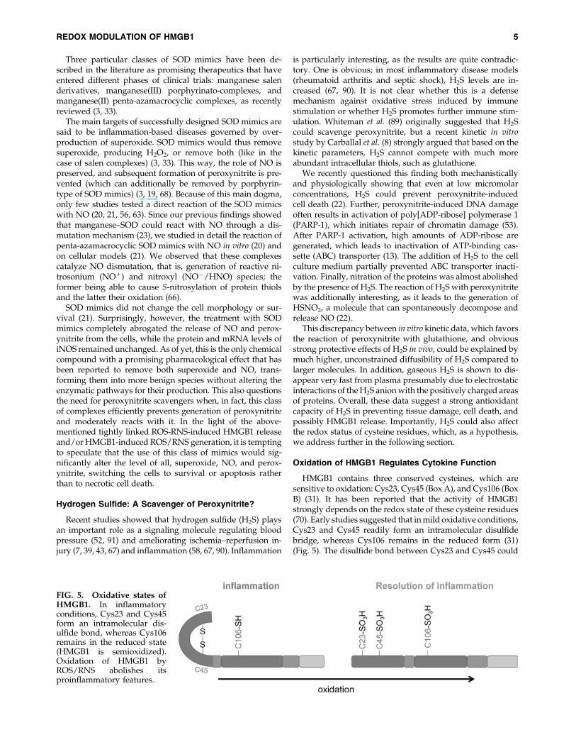
HMGB1 contains three conserved cysteines, which aresensitive to oxidation: Cys23, Cys45 (Box A), and Cys106 (BoxB) (31). It has been reported that the activity of HMGB1strongly depends on the redox state of these cysteine residues(70). Early studies suggested that in mild oxidative conditions,Cys23 and Cys45 readily form an intramolecular disulfidebridge, whereas Cys106 remains in the reduced form (31)(Fig. 5). The disulfide bond between Cys23 and Cys45 could
FIG. 5. Oxidative states ofHMGB1. In inflammatoryconditions, Cys23 and Cys45form an intramolecular dis-ulfide bond, whereas Cys106remains in the reduced state(HMGB1 is semioxidized).Oxidation of HMGB1 byROS/RNS abolishes itsproinflammatory features.
REDOX MODULATION OF HMGB1 5

be broken by glutaredoxin-catalyzed GSH-dependent reduc-tion. This study suggested that mutation of Cys106 impairsthe nuclear distribution and is critical for nucleoplasmicshuttling of HMGB1. The role of Cys106 was further con-firmed by Tang et al. (72). The authors showed that mutationof Cys106, but not vicinal Cys23 and Cys45, promotes cyto-solic localization and sustained autophagy while the in-tramolecular bridge Cys23–Cys45 is required for binding toBeclin1 and sustained autophagy. In another study, the samegroup demonstrated that the Cys106Ala mutant has de-creased autophagy compared with wild-type HMGB1 withCys106 in a reduced state (71). In contrast, oxidized HMGB1leads to activation of caspase-3 and caspase-9 and the in-duction of the mitochondrial pathway of apoptosis. The re-sults also suggested that Cys106 is required for HMGB1binding to TLR4 and activation of cytokine release in mac-rophages.
Oxidation of HMGB1 cysteine residues has been detectedduring cell death (84). The activation of caspases during ap-optosis leads to the production of ROS in the mitochondria,which subsequently oxidizes the potentially dangerousHMGB1. Kazama et al. has shown that oxidation of HMGB1 issufficient to block its stimulatory activity and to promoteimmune tolerance (38). Conversely, it has been speculatedthat oxidized and reduced forms of HMGB1 might differen-tially bind to their receptors (84), since it has been shownthat oxidation-specific epitopes e.g., oxidized low-densitylipoproteins are recognized by pattern recognition receptors(e.g., TLR4) of immune cells (49). Additionally, oxidationof HMGB1 might generate neo-epitopes, which foster thedevelopment of autoantibodies as detected in autoimmunediseases (40).
The most recent detailed mass spectrometric analysis cou-pled with functional studies of various oxidative modifica-tions of HMGB1 cleared up some of the hanging questionsabout HMGB1 activity. Using tandem-mass spectrometricanalysis, Yang et al. showed that both Cys106 in the reducedform and the Cys23–Cys45 disulfide bond are required forHMGB1 to induce nuclear NF-jB translocation and tumournecrosis factor production of macrophages. Both irreversibleoxidation to sulfonates and complete reduction to thiols ofthese cysteines inhibited cytokine production. Using acet-aminophen to induce hepatic necrosis, they observed thatduring inflammation, the predominant form of serumHMGB1 is the active one, (Cys106 thiol and Cys23–Cys45disulfide), whereas the inactive, oxidized form of HMGB1accumulates during resolution of inflammation and hepaticregeneration (94). The oxidation of HMGB1, therefore, servesas a feedback mechanism to control the proinflammatory ac-tivity of HMGB1 in vivo (94) (Fig. 6).
Recently, Venereau et al. confirmed the findings by Yanget al. and additionally described the influence of redox mod-ifications on the chemotactic activity (86). They showed thatonly the fully reduced form of HGMB1, where all cysteines arein the thiol state, can recruit motile cells, whereas terminaloxidation to sulfonates abrogates the cytokine-inducing andchemoattractant activity of HMGB1. Since the disulfide andthiol states of cysteines in HMGB1 are mutually exclusive,these different HMGB1 states orchestrate either leukocyterecruitment or activation of cytokines, respectively (86).
The recruitment of inflammatory cells to damaged tissuesand the induction of cytokines by different states of HMGB1
involve different receptors (62) (Fig. 7). Disulfide HMGB1promotes cytokine release via its interaction with the TLR4,whereas thiol HMGB1 forms a heterocomplex with the che-mokine CXCL12 that acts exclusively via CXCR4 and not viaother HMGB1 receptors (62). Reduced HMGB1 binds toRAGE, but not to TLR4, and promotes Beclin1-dependent
FIG. 6. Double-edged role of ROS/RNS in HMGB1 re-lease and resolution of inflammation. On one hand, ROS/RNS induce cell death leading to the leakage of proin-flammatory HMGB1, which further enforces cell death in theenvironment. On the other hand, the oxidation of HMGB1 byROS/RNS into its inactive form acts as a feedback mecha-nism to control its proinflammatory activity.
FIG. 7. Functions and receptors of HMGB1 are dependenton its redox state. HMGB1 with C23 and C45 in a disulfidebond signals via TLR4, eliciting the release of proin-flammatory cytokines. Complete reduction of cysteines tothiols abrogates the cytokine-stimulating activity; it inducesautophagy via RAGE. Thiol HMGB1-complexed CXCL12serves as a chemoattractant and induces cell migration. Theactions of disulfide and thiol HMGB1 are mutually exclusive.Terminal oxidation of cysteines to sulfonates completelyabolishes HMGB1 activity and contributes to the resolutionof inflammation.
6 JANKO ET AL.
autophagy (75). On the other hand, oxidized HMGB1 inducesapoptosis via a caspase-9/3 intrinsic pathway (71).
Detection of HMGB1
HMGB1 levels have been shown to be an important bio-logical marker in a plethora of pathogenic inflammatoryconditions. Increased serum levels of HMGB1 often correlatewith a worse prognosis, for example, in cancer, sepsis, andautoimmunity (77, 88, 92). Therefore, a reliable assay toquantify HMGB1 in serum and plasma is mandatory. Usually,ELISA, immunoblots, and EMSA are used to detect HMGB1.In this section, we want to highlight the possible problemsemerging in the analyses of HMGB1.
Occurrence of HMGB1 in various redox states
Detection of HMGB1 is problematic since it occurs in var-ious redox states, and detection antibodies may be specific foronly one form. Urbonaviciute et al. reported that HMGB1 isnot detectable in lysates from late-apoptotic Jurkat cells byimmunoblot analysis, employing an antibody specificallydetecting the reduced form of the molecule (84). Consequently,reduction of HMGB1 with b-mercaptoethanol or glutathioneenabled detection. In contrast, HMGB1 in the lysates of viablecells was detected under nonreducing conditions. Since de-tection of oxidized HMGB1 released from apoptotic cells mayfail because of the usage of a not suitable detection antibody, itis essential to check which specificities of HMGB1 are iden-tified by the respective assay agents. In this context it is es-sential to mention that HMGB1 is a highly evolutionarilyconserved protein with 99% sequence homology among allmammals (80). Only two residues out of its 215 amino acidsare substituted in the rodent and human versions. Therefore,immunization of animals with HMGB1 as an immunogenmight be difficult and especially create antibodies directedagainst the non-native forms of HMGB1.
Complex formation of molecules with HMGB1interferes with HMGB1 detection
It has been described that results of HMGB1 ELISA inplasma/serum do not correlate with those of immunoblotting(26). The formation of complexes of HMGB1 with IL-1b, LPSor anti-HMGB1 antibodies reduces the sensitivity of the con-ventional ELISA and causes false-negative results. Therefore,data from routine quantification of HMGB1 should be usedwith caution. Interestingly, anti-HMGB1 autoantibodies were
found in the sera not only of patients with rheumatic diseasesand SLE but also in healthy subjects. Formation of complexes ofthese autoantibodies with HMGB1 may play an important rolein limiting cytokine responses induced by HMGB1. Therefore,a new method to determine free and complex-bound HMGB1has been proposed. The authors showed that the dissociation ofpreformed HMGB1/protein complexes by perchloric acid be-fore ELISA allows the quantification of total HMGB1.
Modifications of HMGB1 by H2S and SOD MimicsCould Regulate Its Activity: A Hypothesison Potential Therapeutic Potential
The past decade has shown a lot of inconsistent effects ofHMGB1 in the same pathological models. It seems that theeffects of HMGB1 are tissue specific and often introduce ad-ditional contradictions. Despite the fact that HMGB1 acts as anearly mediator of inflammation and organ damage in ische-mia–reperfusion injury of the heart [with HMGB1 levels beingalready elevated 30 min after hypoxia in vitro and in ischemicinjury of the heart in vivo (2)], some studies demonstratedbeneficial effects of HMGB1 on myocardial regeneration afterinfarction. This effect occurs via enhanced cardiac C-Kit + cellproliferation and differentiation (44). Further, modulation ofthe local inflammation in the chronically failing postinfarctionmyocardium has been shown to proceed particularly via low-ered accumulation of dendritic cells. This resulted in attenuatedfibrosis and cardiomyocyte hypertrophy, thereby improvingglobal cardiac function (69). In addition, monoclonal anti-HMGB1 antibodies increased cell death and ROS generation inretinal ischemia–reperfusion (93).
However, when taking into consideration all the experimen-tal results as of yet, it is important to notice that some of themdealt with the effects of endogenously released HMGB1 underspecific stress situations (like ischemia–reperfusion), while oth-ers used externally added HMGB1 to modulate the existingpathological states. This may represent a major source for,sometimes, opposite effects of HMGB1 and may offer an idea forpotential future therapeutic approaches. As mentioned above,HMGB1 is a redox-sensitive protein, and the redox status of itscysteine residues is affected by the pro-oxidative environmentinduced by ROS/RNS. This subsequently modulates its patho-physiological signals (84). Several chemical modifications couldbe envisioned that affect the activity of HMGB1 (Fig. 8).
Under the conditions of mild oxidative stress, Cys23 and 45would probably be in form of a disulfide bridge, that is, activeform, so it is the modification of Cys106 that determines the
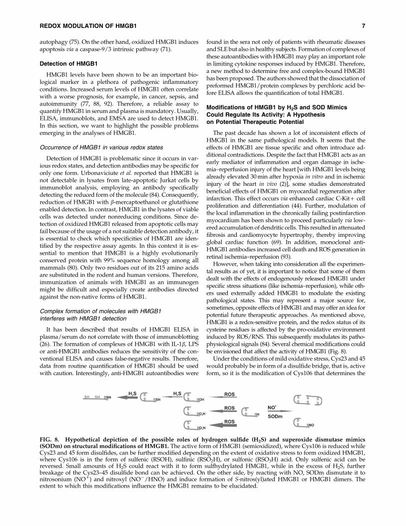
FIG. 8. Hypothetical depiction of the possible roles of hydrogen sulfide (H2S) and superoxide dismutase mimics(SODm) on structural modifications of HMGB1. The active form of HMGB1 (semioxidized), where Cys106 is reduced whileCys23 and 45 form disulfides, can be further modified depending on the extent of oxidative stress to form oxidized HMGB1,where Cys106 is in the form of sulfenic (RSOH), sulfinic (RSO2H), or sulfonic (RSO3H) acid. Only sulfenic acid can bereversed. Small amounts of H2S could react with it to form sulfhydrylated HMGB1, while in the excess of H2S, furtherbreakage of the Cys23–45 disulfide bond can be achieved. On the other side, by reacting with NO, SODm dismutate it tonitrosonium (NO + ) and nitroxyl (NO - /HNO) and induce formation of S-nitros(yl)ated HMGB1 or HMGB1 dimers. Theextent to which this modifications influence the HMGB1 remains to be elucidated.
REDOX MODULATION OF HMGB1 7

direction of HMGB1 action. Under such conditions NO or itsoxidized form (N2O3) could cause S-nitrosylation of Cys106.The magnitude of this modification has never been tested. It istempting to speculate that the S-nitrosylation of Cys106 couldbe one way to affect the activity of HMGB1 or at least tomodulate its mobility from the nucleus to the cytoplasm in anearly stage of oxidative/nitrosative stress. At the site of in-flammation, however, where more powerful oxidants arepresent (H2O2 and peroxynitrite among them), Cys106 wouldget oxidized to sulfenic (RSOH), sulfinic (RSO2H), or sulfonicacid (RSO3H), with only the first modification being readilyreversible. Additionally, formation of dimers where twoHMGB1 molecules are bridged via Cys106 could be envi-sioned as well. H2S, besides scavenging peroxynitrite, canreact as a reducing agent with both S-nitrosated HMGB1 andoxidized (RSOH) HMGB1. This would lead to the formationof S-sulfhydrylated proteins, whose activity remains to beestablished. Further, H2S can reduce disulfides betweenCys23 and 45, forming the fully reduced and thus in-flammatory inactive HMGB1. This could account for some ofthe beneficial effects of H2S observed on the course of in-flammation and can explain the increased production of H2Sobserved under such conditions, where the latter wouldrepresent a mechanism of natural defense.
The role of SOD mimics would strongly depend on theclass of the complexes, but it seems that all of them could beperfectly capable to reduce NO and to form HNO, as wediscussed recently (33). HNO reacts with thiols to induceformation of sulfenylamine, which can further oxidize toform sulfinic or/and sulfonic acids. Alternatively, they canreact with other free thiols to form disulfides. The use ofSOD mimics would not only remove superoxide and/orNO, preventing thus peroxynitrite, but could also modulateHMGB1, leading to its either aggregation or oxidation. Thiswould then represent an additional protective mechanismthrough which SOD mimics prevent necrotic cell deathduring inflammation and/or its further propagation due toinhibition of HMGB1.
As tempting as they look like, these hypotheses remain tobe evaluated and deeper insight provided into actual modi-fications of HMGB1 that could occur at various physiologicaland pathophysiological situations. We hope that raising thisas possibilities stimulates further research on these topics, asunderstanding of the magnitude by which these modifica-tions could affect HMGB1 activity will lead to the design ofsmall-molecule therapeutics for application at various path-ological states.
Acknowledgments
The project was supported by the Emerging Fields Initiative(EFI) of the FAU Erlangen-Nuremberg, by the Masterswitchproject of the EU, by SFB 643, the integrated research-traininggroup GK SFB 643 from the DFG, by the InterdisciplinaryCenter for Clinical Research (IZKF) at the University HospitalErlangen (Project A41), and the K&R Wucherpfennig-Stiftung.
References
1. Abdulahad DA, Westra J, Limburg PC, Kallenberg CG, and BijlM. HMGB1 in systemic lupus Erythematosus: its role in cuta-neous lesions development. Autoimmun Rev 9: 661–665, 2010.
2. Andrassy M, Volz HC, Igwe JC, Funke B, Eichberger SN,Kaya Z, Buss S, Autschbach F, Pleger ST, Lukic IK, Bea F,Hardt SE, Humpert PM, Bianchi ME, Mairbaurl H, NawrothPP, Remppis A, Katus HA, and Bierhaus A. High-mobilitygroup box-1 in ischemia-reperfusion injury of the heart.Circulation 117: 3216–3226, 2008.
3. Batinic-Haberle I, Reboucas JS, and Spasojevic I. Superoxidedismutase mimics: chemistry, pharmacology, and thera-peutic potential. Antioxid Redox Signal 13: 877–918, 2010.
4. Bianchi ME. HMGB1 loves company. J Leukoc Biol 86: 573–576, 2009.
5. Bianchi ME and Agresti A. HMG proteins: dynamic playersin gene regulation and differentiation. Curr Opin Genet Dev15: 496–506, 2005.
6. Cai B, Deitch EA, and Ulloa L. Novel insights for systemicinflammation in sepsis and hemorrhage. Mediators Inflamm2010: 642462, 2010.
7. Calvert JW, Elston M, Nicholson CK, Gundewar S, Jha S,Elrod JW, Ramachandran A, and Lefer DJ. Genetic andpharmacologic hydrogen sulfide therapy attenuates ische-mia-induced heart failure in mice. Circulation 122: 11–19,2010.
8. Carballal S, Trujillo M, Cuevasanta E, Bartesaghi S, MollerMN, Folkes LK, Garcia-Bereguiain MA, Gutierrez-Merino C,Wardman P, Denicola A, Radi R, and Alvarez B. Reactivityof hydrogen sulfide with peroxynitrite and other oxidants ofbiological interest. Free Radic Biol Med 50: 196–205, 2011.
9. Cary SP, Winger JA, Derbyshire ER, and Marletta MA. Nitricoxide signaling: no longer simply on or off. Trends BiochemSci 31: 231–239, 2006.
10. Curtin JF, Donovan M, and Cotter TG. Regulation andmeasurement of oxidative stress in apoptosis. J ImmunolMethods 265: 49–72, 2002.
11. Cuzzocrea S, Riley DP, Caputi AP, and Salvemini D. Anti-oxidant therapy: a new pharmacological approach in shock,inflammation, and ischemia/reperfusion injury. PharmacolRev 53: 135–159, 2001.
12. Dobsak P, Courderot-Masuyer C, Zeller M, Vergely C,Laubriet A, Assem M, Eicher JC, Teyssier JR, Wolf JE, andRochette L. Antioxidative properties of pyruvate and pro-tection of the ischemic rat heart during cardioplegia. J Car-diovasc Pharmacol 34: 651–659, 1999.
13. Dumitriu IE, Voll RE, Kolowos W, Gaipl US, Heyder P,Kalden JR, and Herrmann M. UV irradiation inhibits ABCtransporters via generation of ADP-ribose by concerted ac-tion of poly(ADP-ribose) polymerase-1 and glycohydrolase.Cell Death Differ 11: 314–320, 2004.
14. Ellerman JE, Brown CK, de Vera M, Zeh HJ, Billiar T, Ru-bartelli A, and Lotze MT. Masquerader: high mobility groupbox-1 and cancer. Clin Cancer Res 13: 2836–2848, 2007.
15. Erlandsson Harris H and Andersson U. Mini-review: thenuclear protein HMGB1 as a proinflammatory mediator. EurJ Immunol 34: 1503–1512, 2004.
16. Fan J, Li Y, Levy RM, Fan JJ, Hackam DJ, Vodovotz Y, YangH, Tracey KJ, Billiar TR, and Wilson MA. Hemorrhagicshock induces NAD(P)H oxidase activation in neutrophils:role of HMGB1-TLR4 signaling. J Immunol 178: 6573–6580,2007.
17. Ferdinandy P and Schulz R. Nitric oxide, superoxide, andperoxynitrite in myocardial ischaemia-reperfusion injuryand preconditioning. Br J Pharmacol 138: 532–543, 2003.
18. Ferrer-Sueta G and Radi R. Chemical biology of peroxyni-trite: kinetics, diffusion, and radicals. ACS Chem Biol 4: 161–177, 2009.
8 JANKO ET AL.

19. Ferrer-Sueta G, Vitturi D, Batinic-Haberle I, Fridovich I,Goldstein S, Czapski G, and Radi R. Reactions of manganeseporphyrins with peroxynitrite and carbonate radical anion.J Biol Chem 278: 27432–27438, 2003.
20. Filipovic MR, Duerr K, Mojovic M, Simeunovic V, Zimmer-mann R, Niketic V, and Ivanovic-Burmazovic I. NO dismutaseactivity of seven-coordinate manganese(II) pentaazamacro-cyclic complexes. Angew Chem Int Ed Engl 47: 8735–8739, 2008.
21. Filipovic MR, Koh AC, Arbault S, Niketic V, Debus A,Schleicher U, Bogdan C, Guille M, Lemaitre F, Amatore C,and Ivanovic-Burmazovic I. Striking inflammation fromboth sides: manganese(II) pentaazamacrocyclic SOD mimicsact also as nitric oxide dismutases: a single-cell study. AngewChem Int Ed Engl 49: 4228–4232, 2010.
22. Filipovic MR, Miljkovic J, Allgauer A, Chaurio R, Shubina T,Herrmann M, and Ivanovic-Burmazovic I. Biochemical in-sight into physiological effects of H(2)S: reaction with per-oxynitrite and formation of a new nitric oxide donor,sulfinyl nitrite. Biochem J 441: 609–621, 2012.
23. Filipovic MR, Stanic D, Raicevic S, Spasic M, and Niketic V.Consequences of MnSOD interactions with nitric oxide: ni-tric oxide dismutation and the generation of peroxynitriteand hydrogen peroxide. Free Radic Res 41: 62–72, 2007.
24. Fink MP. Ethyl pyruvate: a novel anti-inflammatory agent.Crit Care Med 31: S51–S56, 2003.
25. Foster MW, Hess DT, and Stamler JS. Protein S-nitrosylationin health and disease: a current perspective. Trends Mol Med15: 391–404, 2009.
26. Gaillard C, Borde C, Gozlan J, Marechal V, and Strauss F. Ahigh-sensitivity method for detection and measurement ofHMGB1 protein concentration by high-affinity binding toDNA hemicatenanes. PLoS One 3: e2855, 2008.
27. Gardella S, Andrei C, Ferrera D, Lotti LV, Torrisi MR,Bianchi ME, and Rubartelli A. The nuclear protein HMGB1is secreted by monocytes via a non-classical, vesicle-medi-ated secretory pathway. EMBO Rep 3: 995–1001, 2002.
28. Goodwin GH, Sanders C, and Johns EW. A new group ofchromatin-associated proteins with a high content of acidicand basic amino acids. Eur J Biochem 38: 14–19, 1973.
29. Herrmann M, Zoller OM, Hagenhofer M, Voll R, and KaldenJR. What triggers anti-dsDNA antibodies?. Mol Biol Rep. 23:265–267, 1996.
30. Hogg N. The biochemistry and physiology of S-nitrosothiols.Annu Rev Pharmacol Toxicol 42: 585–600, 2002.
31. Hoppe G, Talcott KE, Bhattacharya SK, Crabb JW, and SearsJE. Molecular basis for the redox control of nuclear transportof the structural chromatin protein Hmgb1. Exp Cell Res 312:3526–3538, 2006.
32. Hu X, Cui B, Zhou X, Xu C, Lu Z, and Jiang H. Ethyl py-ruvate reduces myocardial ischemia and reperfusion injuryby inhibiting high mobility group box 1 protein in rats. MolBiol Rep 39: 227–231, 2012.
33. Ivanovic I, Filipovic, and Milos R. Reactivity of manganesesuperoxide dismutase mimics toward superoxid and nitricoxide: selectivity versus cross-reactivity. In: Advances inInorganic Chemistry, Inorganic/Bioinorganic Reaction Mechan-isms, edited by Eldik RV. Waltham, Massachusetts: Aca-demic Press, 2012, pp. 53–97.
34. Jang HJ, Kim YM, Tsoyi K, Park EJ, Lee YS, Kim HJ, Lee JH,Joe Y, Chung HT, and Chang KC. Ethyl pyruvate inducesheme oxygenase-1 through p38 mitogen-activated proteinkinase activation by depletion of glutathione in RAW 264.7cells and improves survival in septic animals. Antioxid RedoxSignal 17: 878–889, 2012.
35. Janko C, Schorn C, Grossmayer GE, Frey B, Herrmann M,Gaipl US, and Munoz LE. Inflammatory clearance ofapoptotic remnants in systemic lupus erythematosus (SLE).Autoimmun Rev 8: 9–12, 2008.
36. Jiang W and Pisetsky DS. Expression of high mobility groupprotein 1 in the sera of patients and mice with systemiclupus erythematosus. Ann Rheum Dis 67: 727–728, 2008.
37. Jungas T, Verbeke P, Darville T, and Ojcius DM. Cell death,BAX activation, and HMGB1 release during infection withChlamydia. Microbes Infect 6: 1145–1155, 2004.
38. Kazama H, Ricci JE, Herndon JM, Hoppe G, Green DR, andFerguson TA. Induction of immunological tolerance by ap-optotic cells requires caspase-dependent oxidation of high-mobility group box-1 protein. Immunity 29: 21–32, 2008.
39. King AL and Lefer DJ. Cytoprotective actions of hydrogensulfide in ischaemia-reperfusion injury. Exp Physiol 96: 840–846, 2011.
40. Kurien BT, Hensley K, Bachmann M, and Scofield RH.Oxidatively modified autoantigens in autoimmune diseases.Free Radic Biol Med 41: 549–556, 2006.
41. Lee SY, Jeon HM, Kim CH, Jeong EK, Ju MK, Park SY, JungSY, Kim YJ, Lim SC, Han SI, and Kang HS. CuZnSOD andMnSOD inhibit metabolic stress-induced necrosis and mul-ticellular tumour spheroid growth. Int J Oncol 37: 195–202,2010.
42. Leiper J and Nandi M. The therapeutic potential of targetingendogenous inhibitors of nitric oxide synthesis. Nat RevDrug Discov 10: 277–291, 2011.
43. Li L, Hsu A, and Moore PK. Actions and interactions ofnitric oxide, carbon monoxide and hydrogen sulphide in thecardiovascular system and in inflammation—a tale of threegases! Pharmacol Ther 123: 386–400, 2009.
44. Limana F, Germani A, Zacheo A, Kajstura J, Di Carlo A,Borsellino G, Leoni O, Palumbo R, Battistini L, Rastaldo R,Muller S, Pompilio G, Anversa P, Bianchi ME, and Capo-grossi MC. Exogenous high-mobility group box 1 proteininduces myocardial regeneration after infarction via en-hanced cardiac C-kit + cell proliferation and differentiation.Circ Res 97: e73–e83, 2005.
45. Lotze MT and Tracey KJ. High-mobility group box 1 protein(HMGB1): nuclear weapon in the immune arsenal. Nat RevImmunol 5: 331–342, 2005.
46. Loukili N, Rosenblatt-Velin N, Li J, Clerc S, Pacher P, Feihl F,Waeber B, and Liaudet L. Peroxynitrite induces HMGB1release by cardiac cells in vitro and HMGB1 upregulation inthe infarcted myocardium in vivo. Cardiovasc Res 89: 586–594,2011.
47. Martinez MC and Andriantsitohaina R. Reactive nitrogenspecies: molecular mechanisms and potential significance inhealth and disease. Antioxid Redox Signal 11: 669–702, 2009.
48. Masini E, Cuzzocrea S, Mazzon E, Marzocca C, MannaioniPF, and Salvemini D. Protective effects of M40403, a selectivesuperoxide dismutase mimetic, in myocardial ischaemia andreperfusion injury in vivo. Br J Pharmacol 136: 905–917, 2002.
49. Miller YI, Viriyakosol S, Worrall DS, Boullier A, Butler S,and Witztum JL. Toll-like receptor 4-dependent and -inde-pendent cytokine secretion induced by minimally oxidizedlow-density lipoprotein in macrophages. Arterioscler ThrombVasc Biol 25: 1213–1219, 2005.
50. Munoz LE, Janko C, Grossmayer GE, Frey B, Voll RE, KernP, Kalden JR, Schett G, Fietkau R, Herrmann M, and GaiplUS. Remnants of secondarily necrotic cells fuel inflammationin systemic lupus erythematosus. Arthritis Rheum 60: 1733–1742, 2009.
REDOX MODULATION OF HMGB1 9

51. Munoz LE, Lauber K, Schiller M, Manfredi AA, and Herr-mann M. The role of defective clearance of apoptotic cells insystemic autoimmunity. Nat Rev Rheumatol 6: 280–289, 2010.
52. Mustafa AK, Sikka G, Gazi SK, Steppan J, Jung SM, BhuniaAK, Barodka VM, Gazi FK, Barrow RK, Wang R, Amzel LM,Berkowitz DE, and Snyder SH. Hydrogen sulfide as endo-thelium-derived hyperpolarizing factor sulfhydrates potas-sium channels. Circ Res 109: 1259–1268, 2011.
53. Pacher P, Beckman JS, and Liaudet L. Nitric oxide andperoxynitrite in health and disease. Physiol Rev 87: 315–424,2007.
54. Park JS, Svetkauskaite D, He Q, Kim JY, Strassheim D, Ish-izaka A, and Abraham E. Involvement of toll-like receptors 2and 4 in cellular activation by high mobility group box 1protein. J Biol Chem 279: 7370–7377, 2004.
55. Peter C, Wesselborg S, Herrmann M, and Lauber K. Dan-gerous attraction: phagocyte recruitment and danger sig-nals of apoptotic and necrotic cells. Apoptosis 15: 1007–1028,2010.
56. Pfeiffer S, Schrammel A, Koesling D, Schmidt K, and MayerB. Molecular actions of a Mn(III)Porphyrin superoxide dis-mutase mimetic and peroxynitrite scavenger: reaction withnitric oxide and direct inhibition of NO synthase and solubleguanylyl cyclase. Mol Pharmacol 53: 795–800, 1998.
57. Rabadi MM, Ghaly T, Goligorksy MS, and Ratliff BB.HMGB1 in renal ischemic injury. Am J Physiol Renal Physiol303: F873–F885, 2012.
58. Rivers JR, Badiei A, and Bhatia M. Hydrogen sulfide as atherapeutic target for inflammation. Expert Opin Ther Targets16: 439–449, 2012.
59. Sappington PL, Yang R, Yang H, Tracey KJ, Delude RL, andFink MP. HMGB1 B box increases the permeability of Caco-2enterocytic monolayers and impairs intestinal barrier func-tion in mice. Gastroenterology 123: 790–802, 2002.
60. Scaffidi P, Misteli T, and Bianchi ME. Release of chromatinprotein HMGB1 by necrotic cells triggers inflammation.Nature 418: 191–195, 2002.
61. Schierbeck H, Lundback P, Palmblad K, Klevenvall L, Er-landsson-Harris H, Andersson U, and Ottosson L. Mono-clonal anti-HMGB1 (high mobility group box chromosomalprotein 1) antibody protection in two experimental arthritismodels. Mol Med 17: 1039–1044, 2011.
62. Schiraldi M, Raucci A, Munoz LM, Livoti E, Celona B, Ve-nereau E, Apuzzo T, De Marchis F, Pedotti M, Bachi A,Thelen M, Varani L, Mellado M, Proudfoot A, Bianchi ME,and Uguccioni M. HMGB1 promotes recruitment of in-flammatory cells to damaged tissues by forming a complexwith CXCL12 and signaling via CXCR4. J Exp Med 209: 551–563, 2012.
63. Sharpe MA, Ollosson R, Stewart VC, and Clark JB. Oxida-tion of nitric oxide by oxomanganese-salen complexes: anew mechanism for cellular protection by superoxide dis-mutase/catalase mimetics. Biochem J 366: 97–107, 2002.
64. Silva JP and Coutinho OP. Free radicals in the regulation ofdamage and cell death - basic mechanisms and prevention.Drug Discov Ther 4: 144–167, 2010.
65. Sims GP, Rowe DC, Rietdijk ST, Herbst R, and Coyle AJ.HMGB1 and RAGE in inflammation and cancer. Annu RevImmunol 28: 367–388, 2010.
66. Stamler JS, Singel DJ, and Loscalzo J. Biochemistry of nitricoxide and its redox-activated forms. Science 258: 1898–1902,1992.
67. Szabo C. Hydrogen sulphide and its therapeutic potential.Nat Rev Drug Discov 6: 917–935, 2007.
68. Szabo C, Ischiropoulos H, and Radi R. Peroxynitrite: bio-chemistry, pathophysiology and development of therapeu-tics. Nat Rev Drug Discov 6: 662–680, 2007.
69. Takahashi K, Fukushima S, Yamahara K, Yashiro K, ShintaniY, Coppen SR, Salem HK, Brouilette SW, Yacoub MH, andSuzuki K. Modulated inflammation by injection of high-mobility group box 1 recovers post-infarction chronicallyfailing heart. Circulation 118: S106–S114, 2008.
70. Tang D, Billiar TA, and Lotze MT. A Janus tale of two activeHMGB1 redox states. Mol Med 18: 1360–1362, 2012.
71. Tang D, Kang R, Cheh CW, Livesey KM, Liang X, SchapiroNE, Benschop R, Sparvero LJ, Amoscato AA, Tracey KJ, ZehHJ, and Lotze MT. HMGB1 release and redox regulatesautophagy and apoptosis in cancer cells. Oncogene 29: 5299–5310, 2010.
72. Tang D, Kang R, Livesey KM, Cheh CW, Farkas A,Loughran P, Hoppe G, Bianchi ME, Tracey KJ, Zeh HJ, 3rd,and Lotze MT. Endogenous HMGB1 regulates autophagy. JCell Biol 190: 881–892, 2010.
73. Tang D, Kang R, Livesey KM, Zeh HJ, 3rd, and Lotze MT.High mobility group box 1 (HMGB1) activates an autopha-gic response to oxidative stress. Antioxid Redox Signal 15:2185–2195, 2011.
74. Tang D, Kang R, Zeh HJ, 3rd, and Lotze MT. High-mobilitygroup box 1, oxidative stress, and disease. Antioxid RedoxSignal 14: 1315–1335, 2011.
75. Tang D, Loze MT, Zeh HJ, and Kang R. The redox proteinHMGB1 regulates cell death and survival in cancer treat-ment. Autophagy 6: 1181–1183, 2010.
76. Tang D, Shi Y, Kang R, Li T, Xiao W, Wang H, and Xiao X.Hydrogen peroxide stimulates macrophages and monocytesto actively release HMGB1. J Leukoc Biol 81: 741–747, 2007.
77. Taniguchi N, Kawahara K, Yone K, Hashiguchi T, Yama-kuchi M, Goto M, Inoue K, Yamada S, Ijiri K, Matsunaga S,Nakajima T, Komiya S, and Maruyama I. High mobilitygroup box chromosomal protein 1 plays a role in the path-ogenesis of rheumatoid arthritis as a novel cytokine. ArthritisRheum 48: 971–981, 2003.
78. Thomas JO and Travers AA. HMG1 and 2, and related ‘ar-chitectural’ DNA-binding proteins. Trends Biochem Sci 26:167–174, 2001.
79. Trujillo M, Ferrer-Sueta G, and Radi R. Peroxynitrite de-toxification and its biologic implications. Antioxid RedoxSignal 10: 1607–1620, 2008.
80. Tsuda K, Kikuchi M, Mori K, Waga S, and Yoshida M.Primary structure of non-histone protein HMG1 revealedby the nucleotide sequence. Biochemistry 27: 6159–6163,1988.
81. Tsung A, Klune JR, Zhang X, Jeyabalan G, Cao Z, Peng X,Stolz DB, Geller DA, Rosengart MR, and Billiar TR.HMGB1 release induced by liver ischemia involves Toll-like receptor 4 dependent reactive oxygen species produc-tion and calcium-mediated signaling. J Exp Med 204: 2913–2923, 2007.
82. Ulloa L, Ochani M, Yang H, Tanovic M, Halperin D, Yang R,Czura CJ, Fink MP, and Tracey KJ. Ethyl pyruvate preventslethality in mice with established lethal sepsis and systemicinflammation. Proc Natl Acad Sci U S A 99: 12351–12356,2002.
83. Urbonaviciute V, Furnrohr BG, Meister S, Munoz L, HeyderP, De Marchis F, Bianchi ME, Kirschning C, Wagner H,Manfredi AA, Kalden JR, Schett G, Rovere-Querini P,Herrmann M, and Voll RE. Induction of inflammatory andimmune responses by HMGB1-nucleosome complexes: im-
10 JANKO ET AL.

plications for the pathogenesis of SLE. J Exp Med 205: 3007–3018, 2008.
84. Urbonaviciute V, Meister S, Furnrohr BG, Frey B, Guckel E,Schett G, Herrmann M, and Voll RE. Oxidation of the alar-min high-mobility group box 1 protein (HMGB1) duringapoptosis. Autoimmunity 42: 305–307, 2009.
85. Vallance P and Leiper J. Blocking NO synthesis: how, whereand why? Nat Rev Drug Discov 1: 939–950, 2002.
86. Venereau E, Casalgrandi M, Schiraldi M, Antoine DJ, Catta-neo A, De Marchis F, Liu J, Antonelli A, Preti A, Raeli L,Shams SS, Yang H, Varani L, Andersson U, Tracey KJ, BachiA, Uguccioni M, and Bianchi ME. Mutually exclusive redoxforms of HMGB1 promote cell recruitment or proin-flammatory cytokine release. J Exp Med 209: 1519–1528, 2012.
87. Volz HC, Kaya Z, Katus HA, and Andrassy M. The role ofHMGB1/RAGE in inflammatory cardiomyopathy. SeminThromb Hemost 36: 185–194, 2010.
88. Wang H, Bloom O, Zhang M, Vishnubhakat JM, OmbrellinoM, Che J, Frazier A, Yang H, Ivanova S, Borovikova L,Manogue KR, Faist E, Abraham E, Andersson J, AnderssonU, Molina PE, Abumrad NN, Sama A, and Tracey KJ. HMG-1 as a late mediator of endotoxin lethality in mice. Science285: 248–251, 1999.
89. Whiteman M, Armstrong JS, Chu SH, Jia-Ling S, Wong BS,Cheung NS, Halliwell B, and Moore PK. The novel neuro-modulator hydrogen sulfide: an endogenous peroxynitrite‘scavenger’? J Neurochem 90: 765–768, 2004.
90. Whiteman M, Le Trionnaire S, Chopra M, Fox B, andWhatmore J. Emerging role of hydrogen sulfide in healthand disease: critical appraisal of biomarkers and pharma-cological tools. Clin Sci (Lond) 121: 459–488, 2011.
91. Yang G, Wu L, Jiang B, Yang W, Qi J, Cao K, Meng Q,Mustafa AK, Mu W, Zhang S, Snyder SH, and Wang R. H2Sas a physiologic vasorelaxant: hypertension in mice withdeletion of cystathionine gamma-lyase. Science 322: 587–590,2008.
92. Yang GL, Zhang LH, Bo JJ, Huo XJ, Chen HG, Cao M, LiuDM, and Huang YR. Increased expression of HMGB1 isassociated with poor prognosis in human bladder cancer. JSurg Oncol 106: 57–61, 2012.
93. Yang H, Hirooka K, Liu Y, Fujita T, Fukuda K, NakamutraT, Itano T, Zhang J, Nishibori M, and Shiraga F. Deleteriousrole of anti-high mobility group box 1 monoclonal antibodyin retinal ischemia-reperfusion injury. Curr Eye Res 36: 1037–1046, 2011.
94. Yang H, Lundback P, Ottosson L, Erlandsson-Harris H,Venereau E, Bianchi ME, Al-Abed Y, Andersson U, TraceyKJ, and Antoine DJ. Redox modification of cysteine residues
regulates the cytokine activity of high mobility group box-1(HMGB1). Mol Med 18: 250–259, 2012.
95. Yang H, Ochani M, Li J, Qiang X, Tanovic M, Harris HE,Susarla SM, Ulloa L, Wang H, DiRaimo R, Czura CJ, Roth J,Warren HS, Fink MP, Fenton MJ, Andersson U, and TraceyKJ. Reversing established sepsis with antagonists of endog-enous high-mobility group box 1. Proc Natl Acad Sci U S A101: 296–301, 2004.
96. Zingarelli B. Ethyl pyruvate: a simple solution? Crit CareMed 32: 1603–1604, 2004.
Address correspondence to:Prof. Martin Herrmann
Department of Internal Medicine 3Rheumatology and Immunology
Friedrich-Alexander-University of Erlangen-NurembergUlmenweg 18
91054 ErlangenGermany
E-mail: [email protected]
Date of first submission to ARS Central, January 18, 2013;date of acceptance, February 1, 2013.
Abbreviations Used
ABC¼ATP-binding cassetteCys¼ cysteine
DAMP¼death-associated molecular patternH2S¼hydrogen sulfide
HMGB1¼high-mobility group box protein 1iNOS¼ inducible nitric oxide synthase
LPS¼ lipopolysaccharideNADPH oxidase¼nicotinamide adenine dinucleotide
phosphate oxidaseNF¼nuclear factorNO¼nitric oxide
PARP-1¼poly[ADP-ribose] polymerase 1RAGE¼ receptor for advanced glycation
end productsRNS¼ reactive nitrogen speciesROS¼ reactive oxygen speciesSLE¼ systemic lupus erythematosus
SOD¼ superoxide dismutaseTLR¼Toll-like receptor
REDOX MODULATION OF HMGB1 11