India's Export of Cereal and Cereal Preparations - a Constant Market Share Analysis
Upload
independentCategory
view
2download
0
2175
Received March 19, 2012; final acceptance September 11, 2012. Online prepub date: October 8, 2012.Address correspondence to Rita Nano or Lorenzo Piemonti, Diabetes Research Institute, San Raffaele Scientific Institute, Via Olgettina 60, 20132 Milan, Italy. Tel: +39 02 26432706; Fax: +39 02 26432871; E-mail: [email protected] or [email protected]
Cell Transplantation, Vol. 22, pp. 2175–2186, 2013 0963-6897/13 $90.00 + .00Printed in the USA. All rights reserved. DOI: http://dx.doi.org/10.3727/096368912X657783Copyright 2013 Cognizant Comm. Corp. E-ISSN 1555-3892 www.cognizantcommunication.com
Human Pancreatic Islet Preparations Release HMGB1: (Ir)Relevance for Graft Engraftment
Rita Nano,* Leda Racanicchi,* Raffaella Melzi,* Alessia Mercalli,* Paola Maffi,† Valeria Sordi,* Zhidong Ling,‡ Marina Scavini,* Olle Korsgren,§
Barbara Celona,¶ Antonio Secchi,†# and Lorenzo Piemonti*
*San Raffaele Diabetes Research Institute (HSR-DRI), San Raffaele Scientific Institute, Milan, Italy†Transplant Unit, Department of Medicine, San Raffaele Scientific Institute, Milan, Italy
‡Diabetes Research Center, Brussels Free University-VUB, Brussels, Belgium§Department of Immunology, Genetics and Pathology, Division of Immunology, Uppsala University, Uppsala, Sweden
¶Division of Genetics and Cell Biology, San Raffaele Scientific Institute, Milan, Italy #University Vita-Salute San Raffaele, Milan, Italy
High levels of donor-derived high-mobility group box 1 (HMGB1) protein have been associated with poor islet graft outcome in mouse models. The aim of our work was to determine whether HMGB1 released by human islets had independent proinflammatory effects that influence engraftment in humans. Human islet preparations contained and released HMGB1 in different amounts, as determined by Western blot and ELISA (median 17 pg/ ml/IEQ/24 h; min–max 0–211, n = 74). HMGB1 release directly correlated with brain death, donor hyper-amilasemia, and factors related to the pancreas digestion procedure (collagenase and digestion time). HMGB1 release was significantly positively associated with the release of other cytokines/chemokines, particularly with the highly released “proinflammatory” CXCL8/IL-8, CXCL1/GRO-a, and the IFN-g-inducible chemokines CXCL10/IP-10 and CXCL9/MIG. HMGB1 release was not modulated by Toll-like receptor 2, 3, 4, 5, and 9 agonists or by exposure to IL-1b. When evaluated after islet transplantation, pretransplant HMGB1 release was weakly associated with the activation of the coagulation cascade (evaluated as serum cross-linked fibrin prod-ucts), but not with the immediate posttransplant inflammatory response. Concordantly, HMGB1 did not affect short-term human islet function. Our data show that human islet HMGB1 release is a sign of “damaged” islets, although without any independent direct role in graft failure.
Key words: Islet transplantation; Engraftment; High-mobility group box 1 (HMGB1); Human
induced by hypoxia increases HMGB1 synthesis and release, suggesting that HMGB1 may be a useful marker of damaged islets (10). The importance of HMBG1 for islet engraftment in the mouse is supported by the obser-vation that treatment with an anti-HMGB1 antibody pre-vents the early loss of the islet graft (16). Furthermore, murine islets constitutively express TLR2 and TLR4, and stimulation with HMGB1 induces nuclear factor k-light-chain-enhancer of activated B-cells (NF-kB) activation (14). Accordingly, pretreatment of islets with rHMGB1 pre-vents primary engraftment of syngeneic graft through a TLR2/4-dependent inflammatory activation (14). Finally, HMGB-1 induces pancreatic b-cell apoptosis in vitro via ROS generation mediated by nicotinamide adenine dinucleotide phosphate (NADPH) oxidase, by activating RAGE (14,15).
INTRODUCTION
High-mobility group box 1 (HMGB1) is a damage-associated molecular pattern protein, or alarmin (3), that acts as a potent innate “danger signal” for the initiation of host defense or tissue repair (6). Receptor for advanced glycation end products (RAGE) and Toll-like receptors (TLR) 2 and 4 are potential receptors of HMGB1 (12, 22,30). HMGB1 has been postulated to play a role in the pathogenesis of acute and chronic inflammatory disor-ders, such as sepsis (34), autoimmune diseases (13,35), and ischemia/reperfusion injury (1).
In a mouse model, the abundant HMGB1 content of islets is released in vitro and in vivo soon after syngeneic islet transplantation into the liver (16), and HMGB1 lev-els are directly associated with graft acceptance of trans-planted pancreatic syngeneic islets (14,16). Islet damage

2176 NANO ET AL.
Based on these observations in a murine model, we hypothesize that HMGB1 plays a significant role in poor islet engraftment. If this hypothesis is confirmed, HMGB1 may represent a potential target for therapies aiming to improve the outcome of islets transplantation. Currently, data concerning the role of HMGB1 in human islets (HI) engraftment and survival after transplantation are lack-ing. Therefore, the objective of our study was to deter-mine whether HMGB1 is released by HI preparations, whether and how the release is modulated, and whether HMGB1 has independent proinflammatory effects affect-ing engraftment and/or survival after transplantation.
MATERIALS AND METHODS
HMGB1 Assay
HMGB1 was measured using a commercial kit (IBL International GMBH, Shino-Test Corporation, Tokyo, Japan) following the manufacturer’s instructions.
Pancreas Procurement, Islet Preparations, and Islet Transplantation
We obtained human islets from heart-beating, brain-dead pancreas donors (BD-HI, n = 63) through the North Italian Transplant Organization or from living pancreas donors (L-HI, n = 11) (see Tables 1 and 2 for donor char-acteristics). All living donors were patients undergoing autologous pancreatic islet transplantation. Five of them were critically ill patients suffering from septic/hemor-rhagic shock because of anastomosis leakage after pan-creatoduodenectomy. The remaining six living donors were patients in whom, during pancreatoduodenectomy or median pancreatectomy, pancreatic anastomosis was impossible because of technical problems and/or high risk of leakage. HI were isolated and transplanted as previously described (17,18) from January 16, 2001 to June 14, 2010 at the San Raffaele Scientific Institute in Milan. HI were cultured in Final Wash (Mediatech Cellgro, Manassas, VA, USA) plus 1% pen/strep, 1% glutamine (Lonza, Basel, Switzerland) at 30°C in 5% CO
2 and 95%
humidified air. To measure the pretransplant 24-h release of HMGB1, we used 500 equivalent HI (IEQ) from each preparation (BD-HI: n = 63; L-HI: n = 11). To study the correlation between pretransplant HMGB1 release and donor characteristics or isolation procedure, we included in the analysis all 74 islet preparations. To study the cor-relation between pretransplant HMGB1 release and graft outcome, we included in the analysis only islet prepara-tions from brain-dead pancreas donors because of the high variability in the number of islets obtained from living pan-creas donors. Furthermore, islet transplant in individuals with type 1 diabetes were to meet the following criteria: (i) first infusion with islets obtained from a single donor; (ii) availability of the 24-h pretransplant HMGB1 release by islets; (iii) Edmonton protocol (32) or a calcineurin
inhibitor-free protocol (NCT01346085) as immunosup-pressive treatment. Calcineurin inhibitor-free protocol consisted of (i) pretransplant rapamycin (Rapamune, Wyeth-Ayerst, Pearl River, NY, USA; 0.1 mg/kg/day, with target serum levels of 8–10 ng/ml) for at least 30 days prior to the first islet infusion; (ii) induction therapy with anti-thymocyte globulin (ATG; Thymoglobulin, Genzyme, Cambridge, MA, USA; 1.5 mg/kg/day for 4 days starting at day –1), and a steroid bolus [methyl-prednisolone (Solumedrol, Pfizer Italia Srl, Rome, Italy), 500-mg bolus, day –1] plus low-dose steroids [pred-nisone (Delatcortene, Bruno Farmaceutici S.p.A, Rome, Italy), 10 mg/day] and Anakirna (Kineret, Biovitrum AB, Stockholm, Sweden; 100 mg/day) for 2 weeks (starting at day –1); (iii) maintenance immunosuppressive therapy with rapamycin (0.1 mg/kg/day, with target serum levels of 12–15 ng/ml for the first 1–3 months after each islet infusion and 10–12 ng/ml thereafter) plus mycopheno-late mofetil (MMF; Roche, Nutley, NJ, USA; 2 g/day). Twenty-eight transplants out of 63 satisfied these criteria. The characteristics of those transplants (i.e., islet prepara-tions, donors, and recipients) are reported in Table 1. One month after islet infusion arginine test [30 g of arginine hydrochloride (Bioarginina, Farmaceutici Damor S.p.A. Naples, Italy) administered in 30 min] was performed in 21 recipients under fasting conditions and after overnight withdrawal of insulin administration. Blood samples were collected for the measurement of insulin at baseline and at 5, 10, 20, 30, 40, 50, and 60 min. Serum insulin levels were assayed with a microparticle enzyme immu-noassay (IMx, Abbott Laboratories, North Chicago, IL, USA) in which the lowest insulin sensitivity was 1 mU/ml. The index of the first-phase (acute insulin response to arginine, AIR
arg) insulin response was calculated as the
incremental area under the insulin curve in the intervals 0–10 min. All patients provided written informed consent for the investigations. The Ethics Committee of the San Raffaele Scientific Institute approved the study protocol.
Islet Quality Tests
Purity and number of islets were determined by micro-scope characterization and sizing on a grid after staining with diphenylthiocarbazone (dithizone, Sigma, St. Louis, MO, USA). Islet viability was assessed by double staining with propidium iodide and acridine orange (both Sigma). For insulin and tissue factor contents normalized for DNA, acetic acid (Merck, Darmstadt, Germany) extracts of islets were collected, and total DNA (PicoGreen, Invitrogen AB, Milan, Italy), tissue factor (Imubind, American Diagnostica, Stamford, CT, USA), and insu-lin (Mercodia, Uppsala, Sweden) were measured on a Gyrolab workstation (Gyros AB, Uppsala, Sweden). For insulin, proinsulin, and C-peptide contents normalized for islet, insulin was measured by radioimmunoassay (26),

HMGB1 AND HUMAN ISLET TRANSPLANTATION 2177
C-peptide by time-resolved fluorescence immunoassay (PerkinElmer, Turku, Finland), and proinsulin by ELISA as previously reported (33). Cellular composition was assessed by electron microscopy and immunocytochem-istry as previously described (26). Insulin secretion was evaluated in a dynamic perfusion system. Islets were challenged with two glucose concentrations [initially in 1.67 mM and then in 16.7 mM glucose (Sigma)]. Fractions were collected at 6-min intervals over 120 min, and the insulin content was measured by ELISA (Mercodia,
Uppsala, Sweden). The stimulation index was calculated by dividing the amount of insulin released per minute in 16.7 mM glucose by the amount of insulin released per minute in 1.67 mM glucose.
Western Blotting for HMGB1 in HI
Whole-cell extracts were prepared by direct lysis of 500 IEQ or 0.5 mg of spleen from the same donor (n = 4–5 ran-domly selected from those available) in sodium dodecyl-sulfate polyacrylamide gel electrophoresis (SDS-PAGE)
Table 1. Characterization of Islet Preparations, Donors, and Recipients
L-HMGB1 (n = 14) H-HMGB1 (n = 14) p
Donor Sex (M/F) 7/7 7/7 NS Age 49 ± 10 46 ± 14 NS BMI 27 ± 3.3 28.6 ± 5.8 NS Intensive care days 2 (1–18) 2 (1–13) NS Blood glucose 129 ± 35 157 ± 49 NS Amylase level 82 ± 60 118 ± 84 NSIslet Pancreas weight (g) 99 (75–200) 100 (74–160) NS Cold ischemia (h) 8 (5–15) 5 (5–14) NS Collagenase (type P/liberase/NP1)
3/5/6 5/6/3 NS
IEQ (´103) 324 (180–894) 366 (218–521) NS
Isolation index 0.85 (0.66–1.44) 0.97 (0.51–1.34) NS Islet purity (%) 52 (30–80) 40 (20–84) NS Volume (ml) 3.25 (1.5–5) 3.25 (2–5.5) NS Pre-Tx culture (h) 24 (0–52) 24 (2–138) NS
Edmonton Calcineurin Inhibitor Free
L-HMGB1 (n = 9) H-HMGB1 (n = 9) p L-HMGB1 (n = 5) H-HMGB1 (n = 5) p
Recipient Sex (M/F) 4/5 4/5 NS 3/2 3/2 NS Age 41 ± 13 39 ± 10 NS 32 ± 8 38 ± 7 NS Diabetes (years) 27 (5–40) 22 (14–37) NS 21 (15–38) 17 (7–40) NS Insulin requirement (U/kg)
0.57 (0.36–1)
0.57 (0.31–1.0)
NS 0.58 (0.41–0.75)
0.51 (0.40–0.69)
NS
Pre-Tx HbA1c (%) 8.5(7.5–10.2)
8.2(6.6–9.5)
NS 8.6(6.8–9.6)
9.2(7.4–10.2)
NS
Creatinine (mg/dl) 0.94 ± 0.35 0.75 ± 0.12 NS 0.92 ± 0.39 0.79 ± 0.1 NS
IEQ (´103) 327(180–894)
372(218–521)
NS 310(292–652)
348(305–457)
NS
IEQ/kg 5634(3214/9625)
5729(3749/9308)
NS 4642(4173/9597)
5862(4955/7641)
NS
Isolation index 0.85(0.66–1.44)
0.90(0.51–1.34)
NS 0.85(0.67–1.23)
0.97(0.58–1.33)
NS
Islet purity (%) 55 (30–80) 50 (20–80) NS 50 (40–60) 35 (30–84) NS Volume (ml) 4 (1.5–5) 3 (2.5–5) NS 2 (2–2.5) 4 (2–4) NS Pre-Tx culture (h) 23 (0–52) 24 (2–65) NS 36 (22–48) 44 (24–138) NS HMGB1 release 8 (4–23.13) 67 (29–110) 0.001 11.1 (2–20) 43 (25–63) 0.009
HMGB1, high mobility group box 1; L, low release; H, high release; BMI, body mass index; Tx, transplantation; HbA1c, glycosylated hemoglobin.

2178 NANO ET AL.
sample buffer (Life Technologies, Grand Island, NY, USA). DNA from 106 cells was extracted with the DNeasy Tissue Kit (Qiagen, Milan, Italy) and quantified by PicoGreen (Invitrogen). Following electrophoresis, blots were probed with a primary rabbit polyclonal anti-HMGB1 (Abcam, Cambridge, UK) or with a mouse anti-b-actin anti-body (Sigma, Milan, Italy) and visualized using an ECL detection kit (GE Healthcare, Milwaukee, WI, USA). Quantification of Western blot signals was performed with ImageQuant software (GE Healthcare).
In Vitro HMGB1, Chemokine, and Cytokine Release
Time-Course Release. Two hundred and fifty IEQ/ml were seeded in a low-adhesion 24-multiwell plate (Costar, Corning, Corning, NY, USA) immediately after purifica-tion and cultured in Final Wash (Mediatech Cellgro) plus 1% human serum albumin (HSA; Grifols Italia Spa, Pisa, Italy) at 37°C in 5% CO
2 and 95% humidified air. Medium
samplings were performed at days 1, 2, 3, 4, 5, and 7.
Stimulated Release. Five hundred IEQ/ml were seeded in a low-adhesion 24-multiwell plate in final wash and cultured for 24 h in the absence or presence of the following stimuli: IL-1b (IL-1R1 ligand, 100 ng/ml; Inalco, PreproTech, Milan, Italy); peptidoglycan (PGN TLR2 ligand, 100 ng/ml; Sigma); lipopolysaccharide (LPS TLR4 ligand, 100 ng/ml; Sigma); polyinosinic- polycytidylic acid [poly(I:C) TLR3 ligand, 50 µg/ml; Invitrogen]; flagellin (FLA TLR5 ligand, 30 ng/ml; Invitrogen); CpG oligodeoxy nucleotides 2006 sequence 5¢-TCGTCGTTTTGTCGTTTTGTCGTT-3¢ (CpG2006 TLR9 ligand, 1 µg/ml; Invitrogen). Media were collected by centrifugation and stored at –80°C until use.
Chemokine and Cytokine Assay
Human cytokines/chemokines were measured using multiplex bead-based assays based on xMAP technology (Bio-Plex, Bio-Rad Laboratories, Hercules, CA, USA) fol- lowing the manufacturer’s instructions. The limits of detect- ion and the coefficients of variability (intra-assay %CV; interassay %CV) of the cytokine/chemokine assays were interleukin (IL)-1a: 0.5 pg/ml (l4; 4); IL-1b: 0.6 pg/ml (6; 8) interleukin-1 receptor antagonist (IL-1ra): 5.5 pg/ml (9; 8); IL-2: 1.6 pg/ml (7; 9); IL-2 receptor a subunit (IL-2Ra): 2.1 (6; 4); IL-3: 4.8 pg/ml (7; 4); IL-4: 0.7 pg/ml (9; 8); IL-5: 0.6 pg/ml (8; 10); IL-6: 2.6 pg/ml (7; 11); IL-7: 1.1 pg/ml (6; 8); IL-9: 2.5 pg/ml (8; 9); IL-10: 0.3 pg/ml (5; 6); IL-12p40: 23.3 pg/ml (5; 8); IL-12p70: 3.5 pg/ml (6; 6); IL-13: 0.7 pg/ml (8; 7); IL-15: 2.4 pg/ml (5; 6); IL-16: 0.4 pg/ml (6; 4); IL-17: 3.3 pg/ml (8; 6); IL-18: 0.2 pg/ml (4; 5); interferon-g (IFN-g) 6.4 pg/ml (15; 9); tumor necrosis factor-a (TNF-a): 6 pg/ml (8; 6); TNF-b: 0.3 pg/ml (4; 4); granulocyte macrophage colony stimulat-ing factor (GM-CSF): 2.2 pg/ml (12; 6); G-CSF: 1.7 pg/ml
(10; 5); M-CSF: 0.9 pg/ml (4; 5); chemokine (C-C motif) ligand 2/monocyte chemoattractant protein-1 (CCL2/MCP-1): 1.1 pg/ml (9; 7); CCL3/macrophage inflamma-tory protein (MIP)-1a: 1.6 pg/ml (7; 8); CCL4/MIP-1b: 2.4 pg/ml (8; 8); CCL5/regulated on activation, normal T cell expressed and secreted (Rantes): 1.8 pg/ml (9; 6); CCL7/MCP-3: 1 pg/ml (7; 8); CCL11/eotaxin: 2.5 pg/ml (8; 11); CCL27/cutaneous T-cell-attracting chemokine (CTAK): 3.4 pg/ml (5; 6); chemokine (C-X-C motif) ligand 1/ (CXCL1/GRO1 oncogene (melanoma growth stimulating activity, a; GRO-a): 6.3 pg/ml (5; 8); CXCL9/monokine induced by interferon-g (MIG): 1.2 pg/ml (6; 6); CXCL10/10-kDa IFN-g-induced protein (IP-10): 6.1 pg/ml (11; 9); CXCL12/stromal-derived factor 1a (SDF1a): 8.7 pg/ml (6; 6); vascular endothelial factor (VEGF): 3.1 pg/ml (9; 7); CXCL8/IL-8: 1.0 pg/ml (9; 4); basic fibroblast growth factor (bFGF): 1.9 pg/ml (8; 8); platelet- derived growth factor (PDGF)-BB: 2.9 pg/ml (9; 8); hepa-tocyte growth factor (HGF): 4.9 pg/ml (5; 6); IFN-a2: 4.3 pg/ml (7; 3); leukemia inhibitory factor (LIF): 5.5 pg/ml (4; 3); macrophage migration inhibitory factor (MIF): 1.5 pg/ml (5; 8); b-nerve growth factor (b-NGF): 0.2 pg/ml (4; 7); stem cell factor (SCF): 1 pg/ml (5; 4); stem cell growth factor (SCGF)-b: 45.4 pg/ml (6; 8); TNF-related apoptosis-inducing ligand (TRAIL): 2.1 pg/ml (4; 8).
Human Blood
Serum aspartate and alanine aminotransferase activi-ties (AST and ALT) and C-reactive protein (CRP) were measured with the ADVIA 2400 Chemistry System (Bayer Healthcare, Tarrytown, NY, USA). Cross-linked fibrin deg-radation products (XDPs) were measured with an immu-noturbidimetric method (STA-Liatest, Diagnostica Stago, Asnières sur Seine, France). Serum C-peptide levels (intra-assay CV 3.0%; interassay CV 3.0%) were assayed by radioimmunoassay (RIA) using commercial kits (Medical System, Genoa, Italy). Serum cytokines/chemokines were detected using multiplex bead-based assays based on xMAP technology (Bio-Rad Laboratories).
Statistical Analysis
Data are expressed as mean ± standard error or median (min–max). Groups were compared using Student’s t test (continuous variables with normal distribution), Mann–Whitney U test (continuous variables with nonnormal distribution), or Fisher’s exact test (categorical variables). Bonferroni correction was performed where appropriate for multiple comparisons. ANOVA for repeated measures was used to analyze the relationships between islet HMGB1 release and early posttransplant events. Correlations were assessed with the Spearman rank correlation coefficient. A univariate general linear model was used to study the rela-tionships between HMGB1 islet release, the area under the curve for XDPs during the 3 days posttransplant, and the

HMGB1 AND HUMAN ISLET TRANSPLANTATION 2179
islet estimated function l and 3 months after islet infusion. A value of p < 0.05 was considered significant. Data were analyzed using the SPSS statistical package for Windows (SPSS Inc., Chicago, IL, USA).
RESULTS
Human Islets Express High Levels of HMGB1 and Release It in Culture After Isolation
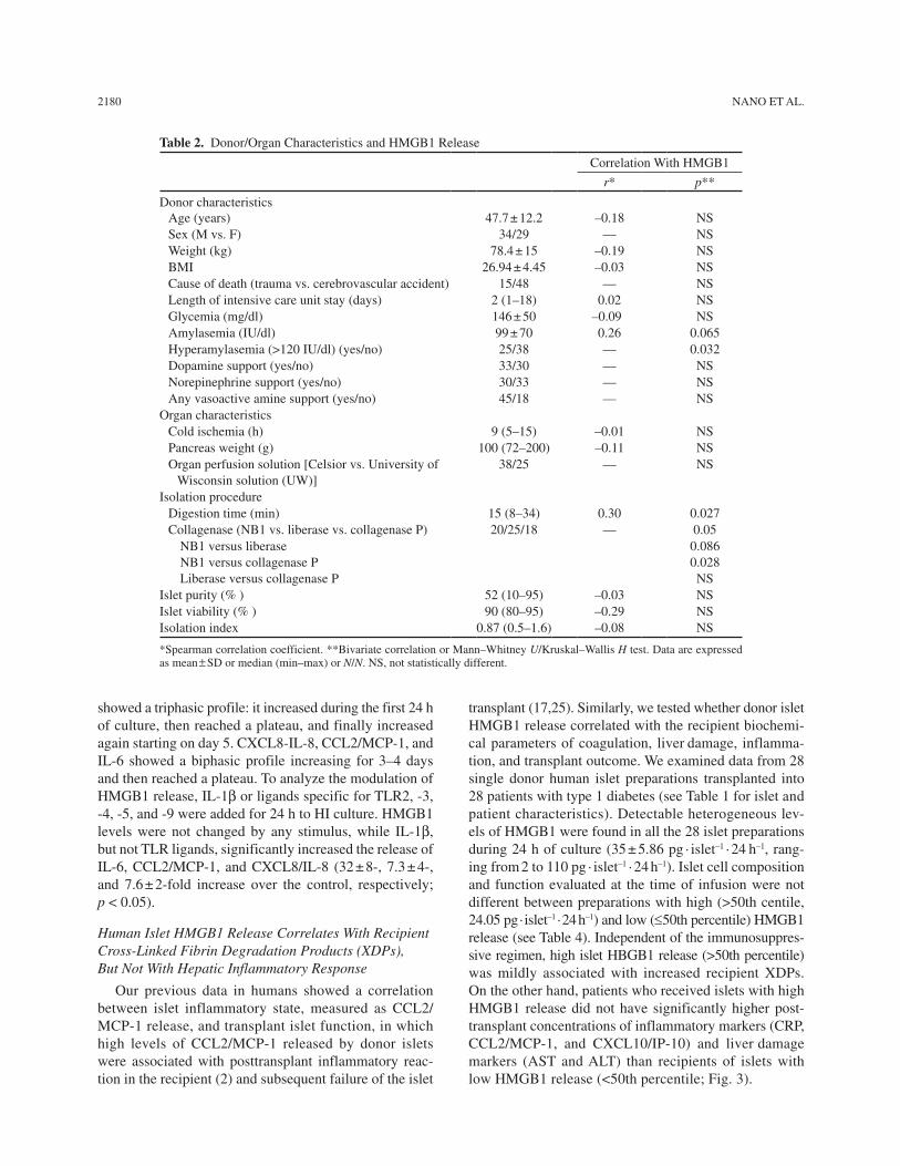
We measured the HMGB1 content in HI in compari-son to that in human splenocytes from the same donor. As previously reported in mouse (16), purified HI con-tained high levels of HMGB1, which were eight times greater than that in spleen cells (Fig. 1). We investigated the capacity of HI to release HMGB1 in vitro after iso-lation. Supernatants from 74 HI available preparations were studied (BD-HI, n = 63; L-HI, n = 11). High levels of HMGB1 with a wide range of concentrations were detected in the culture medium after 24 h (median 17 pg/ml/IEQ/24 h; min–max 0–211). A significantly different amount of released HMGB1 was measured in BD-HI and not critically ill L-HI (18.9 vs. 2 pg/ml/IEQ/24 h respectively; p = 0.007) and in not critically ill L-HI and critically ill L-HI (2 vs. 41 pg/ml/IEQ/24 h, respectively; p = 0.017). We assessed the relationship between HMGB1 release and donor characteristics and/or isolation proce-dure parameters in BD-HI (Table 2). HMGB1 directly correlated with the presence of donor hyperamylasemia
(41 vs. 18.5 pg/ml/IEQ/24 h, p = 0.032) and with digestion time during isolation (Spearman correlation coefficient 0.3; p = 0.027). Moreover, collagenase type significantly affected the HMGB1 release, with collagenase P being more detrimental compared to NB1 type (p = 0.028).
HMGB1 Release Is Associated With a Proinflammatory Status of the Islets But It Is Not Modulated by the Exposure to IL-1b or Toll-Like Receptor Ligands
In order to determine whether HMGB1 release is asso-ciated with a more generalized inflammatory state of the islet, we tested the release of chemokines and cytokines in 20 randomly selected samples out of the available 74 primary cultures of HI (Table 3). Copious amounts of CXCL8/IL-8, MIF, CXCL1/GRO-a, and CCL2/MCP-1, as well as appreciable levels of VEGF, IL-6, and CXCL10/IP-10, were secreted in the supernatant during the 24-h culture. Variable lower levels were observed for other analyzed cytokines. HMGB1 was significantly positively associated with the majority of the measured cytokines, in particular with the highly released “proinflammatory” CXCL8/IL-8, CXCL1/GRO-a, and the IFN-g inducible chemokines CXCL10/IP-10 and CXCL9/MIG. Of note, HMGB1 did not correlate with CCL2/MCP-1 (Table 3).
The 7-day release of HMGB1, CXCL8/IL-8, CCL2/MCP-1, and IL-6 was evaluated in five randomly chosen HI primary cultures (Fig. 2). The concentration of HMGB1
Figure 1. High-mobility group box 1 (HMGB1) expression in human islets. (A) Western blotting for HMGB1 was performed on equal amounts of total cell lysates from human islets (HI) and matched spleen from three independent preparations. Quantifications were performed on images with different exposures within the linear part of the dynamic range. Data are expressed as HMGB1 band intensities, normalized to total b-actin band intensities; error bars, SD of technical replicates in a representative experiment out of three performed. (B) For evaluating the pretransplant 24-h release of HMGB1 and its correlation with donor characteristics and isola-tion procedure, 500 equivalent HI (IEQ) from each available preparations was studied. A heterogeneous and significantly different distribution was evident among brain-dead donors (n = 63), not critically ill living donors (n = 6), and critically ill living donors (n = 5). Statistical analysis: Mann–Whitney U test post hoc with Bonferroni correction after Kruskal–Wallis test.
2180 NANO ET AL.
showed a triphasic profile: it increased during the first 24 h of culture, then reached a plateau, and finally increased again starting on day 5. CXCL8-IL-8, CCL2/MCP-1, and IL-6 showed a biphasic profile increasing for 3–4 days and then reached a plateau. To analyze the modulation of HMGB1 release, IL-1b or ligands specific for TLR2, -3, -4, -5, and -9 were added for 24 h to HI culture. HMGB1 levels were not changed by any stimulus, while IL-1b, but not TLR ligands, significantly increased the release of IL-6, CCL2/MCP-1, and CXCL8/IL-8 (32 ± 8-, 7.3 ± 4-, and 7.6 ± 2-fold increase over the control, respectively; p < 0.05).
Human Islet HMGB1 Release Correlates With Recipient Cross-Linked Fibrin Degradation Products (XDPs), But Not With Hepatic Inflammatory Response
Our previous data in humans showed a correlation between islet inflammatory state, measured as CCL2/MCP-1 release, and transplant islet function, in which high levels of CCL2/MCP-1 released by donor islets were associated with posttransplant inflammatory reac-tion in the recipient (2) and subsequent failure of the islet
transplant (17,25). Similarly, we tested whether donor islet HMGB1 release correlated with the recipient biochemi-cal parameters of coagulation, liver damage, inflamma-tion, and transplant outcome. We examined data from 28 single donor human islet preparations transplanted into 28 patients with type 1 diabetes (see Table 1 for islet and patient characteristics). Detectable heterogeneous lev-els of HMGB1 were found in all the 28 islet preparations during 24 h of culture (35 ± 5.86 pg · islet–1 · 24 h–1, rang-ing from 2 to 110 pg · islet–1 · 24 h–1). Islet cell composition and function evaluated at the time of infusion were not different between preparations with high (>50th centile, 24.05 pg · islet–1 · 24 h–1) and low (≤50th percentile) HMGB1 release (see Table 4). Independent of the immunosuppres-sive regimen, high islet HBGB1 release (>50th percentile) was mildly associated with increased recipient XDPs. On the other hand, patients who received islets with high HMGB1 release did not have significantly higher post-transplant concentrations of inflammatory markers (CRP, CCL2/MCP-1, and CXCL10/IP-10) and liver damage markers (AST and ALT) than recipients of islets with low HMGB1 release (<50th percentile; Fig. 3).
Table 2. Donor/Organ Characteristics and HMGB1 Release
Correlation With HMGB1
r* p**
Donor characteristicsAge (years) 47.7 ± 12.2 –0.18 NSSex (M vs. F) 34/29 — NSWeight (kg) 78.4 ± 15 –0.19 NSBMI 26.94 ± 4.45 –0.03 NSCause of death (trauma vs. cerebrovascular accident) 15/48 — NSLength of intensive care unit stay (days) 2 (1–18) 0.02 NSGlycemia (mg/dl) 146 ± 50 –0.09 NSAmylasemia (IU/dl) 99 ± 70 0.26 0.065Hyperamylasemia (>120 IU/dl) (yes/no) 25/38 — 0.032Dopamine support (yes/no) 33/30 — NSNorepinephrine support (yes/no) 30/33 — NSAny vasoactive amine support (yes/no) 45/18 — NS
Organ characteristicsCold ischemia (h) 9 (5–15) –0.01 NSPancreas weight (g) 100 (72–200) –0.11 NSOrgan perfusion solution [Celsior vs. University of
Wisconsin solution (UW)]38/25 — NS
Isolation procedureDigestion time (min) 15 (8–34) 0.30 0.027Collagenase (NB1 vs. liberase vs. collagenase P) 20/25/18 — 0.05
NB1 versus liberaseNB1 versus collagenase PLiberase versus collagenase P
0.0860.028NS
Islet purity (% ) 52 (10–95) –0.03 NSIslet viability (% ) 90 (80–95) –0.29 NSIsolation index 0.87 (0.5–1.6) –0.08 NS
*Spearman correlation coefficient. **Bivariate correlation or Mann–Whitney U/Kruskal–Wallis H test. Data are expressed as mean ± SD or median (min–max) or N/N. NS, not statistically different.

HMGB1 AND HUMAN ISLET TRANSPLANTATION 2181
Islet Function Posttransplant in Humans Is Not Associated With Pretransplant Islet HMGB1 Release
The above findings suggested that both the increased XDPs response to islet transplantation and islet HMGB1 release could contribute to the outcome of islet transplant (Fig. 4). Islet function in patients who received a single donor islet infusion was evaluated at 1 month (n = 24) and 3 months (n = 14) after transplantation as islet estimated function (IEF), an index estimating the average insulin secretion of the single transplanted islet (4,5). IEF at 1 and 3 months after transplantation was not associated either with posttransplant coagulation activation, expressed as the area under the curve for the recipient’s XDP during
the 3 days after islet infusion [XDPsAUC(0–3)
; p = 0.16 and p = 0.78, 1 and 3 months after transplantation, respec-tively] or with islet HMGB1 release (p = 0.59 and p = 0.90, 1 and 3 months after transplantation, respectively) when both were included as covariates in a general linear model (Fig. 4A). Moreover, we did not find a significant correla-tion between HMGB1 release and acute insulin response to arginine (AIR
arg) 1 month after the first islet infusion
(Fig. 4B). Taken together, these observations suggest that islet HMGB1 release affects the immediate coagulatory response in the liver, but not islet function. The HMGB1 release by the islets during the first infusion may also be relevant for the long-term function, since a more immu-nogenic “priming” to and/or “recall” to autoantigens may affect the survival of subsequent islet infusions. Of the 28 patients analyzed, 8 received a single infusion, 15 a second infusion, and 5 a third infusion. The cumulative incidence of insulin independence and the long-term graft function during follow-up were not influenced by islet HMGB1 release during the first infusion (Fig. 4C).
DISCUSSION
Despite recent publications on HMGB1 release by murine pancreatic islets (10,14–16), no data outlining the role of donor-derived HMGB1 on intrahepatic islet engraftment have been generated in humans. The objec-tive of our work was to determine whether HMGB1 is released by HI preparations, whether and how its release is modulated, and whether HMGB1 has proinflammatory effects that influence engraftment and/or survival after transplantation.
We observed that HI preparations express and release high amounts of HMGB1. In general, HMGB1 release from cells may occur through two distinct processes: (i) necrosis, in which cell membranes are permeabilized and intracellular constituents like HMGB1 may diffuse out of the cell (30); (ii) some forms of active or facilitated secretion induced by signaling through the NF-kB and TLR activation (8,27). Even if an in-depth study of the HMGB1 release pathways was beyond the scope of this work, our data suggest that early HMGB1 release from HI preparation is mainly passive. In fact, the 24-h HMGB1 release by HI preparations was correlated to the medi-cal condition of the donor (critically ill, brain death), to the donor pancreatic damage (i.e., hyperamilasemia), and to isolation-related damage factors (enzymes type and digestion time). It should be underlined that more than one factor may be responsible for the different levels of islet HMGB1 in living pancreas donors. We suggest in the first place the relevance of the critical ill condition at the time of pancreas removal because of septic and/or hem-orrhagic shock (i.e., dramatic events inducing splanchnic vasoconstriction and pancreas ischemia). However, we cannot exclude a priori other factors, such as the different
Table 3. Basal Release of Chemokines and Cytokines in 20 Freshly Isolated Human Islets
Human Islet (pg/500 IEQ/24 h)
Correlation With HMGB1*
r p
CXCL8/IL-8 13,651 (79–39,678) 0.49 0.027HMGB1 7,028 (0–77,820) — —MIF 1,515 (1.4–6,104) 0.44 0.052CXCL1/GRO-a 1,231 (1.8–12,647) 0.51 0.021CCL2/MCP-1 1,182 (17–33,666) 0.34 NSVEGF 401 (35–3,751) 0.603 0.005IL-6 286 (7.4–20,528) 0.45 0.046CXCL10/IP-10 127 (2.8–6,751) 0.57 0.008CXCL9/MIG 76.4 (<1.2–7,824) 0.57 0.009CCL11/eotaxin 73.2 (2.6–1,440) 0.29 NSG-CSF 45.4 (1.9–31,666) 0.45 0.046CXCL12/SDF1a 44.5 (1.8–187) 0.57 0.009M-CSF 34.2 (1.5–352) 0.42 0.065IFN-g 29.8 (<6.4–289) 0.606 0.006LIF 24.4 (<5.5–229) 0.31 NSIL-12p70 23.7 (3.7–82.4) 0.567 0.009CCL5/Rantes 22.2 (2–5,997) 0.498 0.026CCL4/MIP-1b 20.7 (1.8–383) 0.484 0.036CCL7/MCP-3 10.8 (1.2–1,087) 0.569 0.009CCL27/CTAK 9.7 (1.7–87.6) 0.11 NSTNF-a 7.6 (6.6–94) 0.13 NSBasic FGF 7.5 (1.8–62.2) –0.22 NSIL-13 4.9 (2.8–58.3) 0.527 0.017CCL3/MIP-1a 4.5 (<1.6 –258) 0.418 0.066IL-9 4 (<2.5–16.6) 0.53 0.015IL-7 2.5 (2.2–31.63) 0.72 <0.001IL-5 2.4 (2.1–2.4) 0.21 NSIL-1b 2.25 (2.2–43.6) –0.172 NSIL-10 1.73 (1.6–16.7) 0.55 0.012IL-18 1.1 (0.8–30.5) 0.16 NSIL-1a ND — —IL-2 ND — —IL-3 ND — —IL-4 ND — —IL-12p40 ND — —IL-15 ND — —IL-17 ND — —GM-CSF ND — —
*Bivariate correlation, r = Spearman correlation coefficient; ND, not detectable; NS, not statistically different. See text for abbreviations.

2182 NANO ET AL.
Figure 2. In vitro HMGB1 release. (A) Time-course release. Two hundred and fifty IEQ/ml were seeded in a low-adhesion 24-multiwell plate immediately after purification and cultured with final wash for 7 days. Medium samplings were performed at days 1, 2, 3, 4, 5, and 7. Data are expressed as mean ± SE of five independent experiments. (B) Stimulated release. Five hundred IEQ/ml were seeded in a low-adhesion 24-multiwell plate in final wash and cultured for 24 h in the absence (Ctrl) or in the presence of the following stimuli: interleukin (IL)-1b (IL-1R1 ligand, 100 ng/ml); peptidoglycan (PGN) (TLR2 ligand, 100 ng/ml); lipopolysaccharide (LPS) (TLR4 ligand, 100 ng/ml); poly(I:C) (TLR3 ligand, 50 µg/ml); flagellin (FLA) (TLR5 ligand, 30 ng/ml); and CpG oligodeoxynucleotides (CpG2006) (TLR9 ligand, 1 µg/ml). Media were collected by centrifugation and stored at –80°C until use. Data are expressed as fold increase versus untreated and represent the mean ± SE of four independent experiments. *p < 0.007 versus Ctrl (Mann–Whitney test with Bonferroni adjustment as a post hoc test after Kruskal–Wallis).
Table 4. Islet Preparation Characteristics and HMGB1
L-HMGB1 H-HMGB1 All
Correlation With HMGB1
p* r p**
Cellular composition % Exocrine 23 (12–39) 27 (15–37) NS 25 (12–39) –0.08 NS % Endocrine 34 (8–54) 28 (24–34) NS 28 (8–54) –0.08 NS % Nongranulated 37 (22–52) 40 (34–50) NS 38 (22–52) 0.11 NS % Dead 6 (1–16) 5 (3–10) NS 5.5 (1–16) 0.18 NS % Insulin 27 (10–40) 20 (11–27) NS 22 (10–40) –0.18 NS % Glucagon 7 (1–18) 11 (3–21) NS 7 (1–21) –0.21 NSIslet function Insulin secretion (SI) 5.5 (0.8–14.2) 5.7 (2.5–19.2) NS 5.6 (0.8–19.2) 0.03 NS Insulin content:
– ng/ng DNA 1.5 (0.8–5.1) 1.1 (0.4–2.1) NS 1.5 (0.4–5.1) –0.26 NS – ng/islet 12.34 (0.59–3.38) 10.61 (0.72–2.15) NS 1.061 (0.59–3.38 0.46 NS
C-peptide content (ng/islet) 0.71 (0.38–1.81) 0.700 (0.471.03) NS 0.7 (0.38–1.81) 0.32 NS Proinsulin content (pg/islet) 38 (18–60) 36 (16–55) NS 37 (16–60) –0.05 NSOther Tissue factor content
(pmol/ng DNA)38 (5.2–148) 25 (14–57) NS 37 (5–148) 0.17 NS
Data were available from 14 patients randomly selected from the available 28 study subjects; data are expressed as median (min–max); *Mann–Whitney U/Kruskal–Wallis H test; **Bivariate correlation, r = Spearman correlation coefficient; NS, not statistically different.

HMGB1 AND HUMAN ISLET TRANSPLANTATION 2183
time of pancreatic warm ischemia in case of anastomo-sis leakage (generally between 30 and 60 min) or when anastomosis was impracticable because of technical dif-ficulties. Exposure to IL-1b did not modify the HMGB1 release, while it increased the release of other NF-kB-regulated factors such as CXCL8/IL-8, CCL2/MCP-1, or IL-6. Moreover, TLR ligands failed to modify any of the tested factors.
The 24-h HMGB1 release by the HI preparations was significantly positively associated with the release of proinflammatory factors, suggesting that in the purified islets, HMGB1 is part of a more general inflammatory response (24). The obvious explanation for the release of all these factors is the stress imposed on the islets from the time that the donor is admitted to the intensive care unit (i.e., cortisol-induced peripheral insulin resistance and the direct effects of stress-related hormones), as
well as prolonged periods of hypoxia during transporta-tion, isolation, purification, and culture of the islets until engraftment of the islets in the recipient (10). However, a causative relationship could still justify the association between HMGB1 and the different factors released by the islets. For example, IL-12, IFN-g, CXCL12/SDF1a, and VEGF, all highly correlated with HMGB1 in our study, were previously described to be modulated directly by HMGB1 via RAGE in leukocytes and fibroblasts (7, 19,23,28). Of interest, HMGB1-induced recruitment of inflammatory cells was recently described to be depend-ent on the formation of the HMGB1-CXCL12 heterocom-plex (31), suggesting a synergistic role between these two factors in triggering inflammation after islet transplan-tation. On the other hand, other factors, such as CCL2/MCP-1 and CXCL8/IL-8, previously indicated as being modulated by HMGB1 and relevant for islet engraftment
Figure 3. Relationships between pretransplant islet HMGB1 release and early posttransplant events. Posttransplantation levels of C-reactive protein (CRP), cross-linked fibrin degradation products (XDPs), aspartate and alanine aminotransferases (AST and ALT), inducible inflammatory chemokines (CCL2, CXCL10, CCL20), insulin requirement (IR), and fasting C-peptide were evaluated in patients with type 1 diabetes who received islet allografts. The 28 patients (see Table 1 for islet and patient characteristics) were divided into two groups according to the amount of HMGB1 released by islet preparations during a 24-h culture (50th percentile = 24.05 pg · islet–1 · 24 h–1). The low-HMGB1 group (L-HMGB1) received islet preparations releasing an amount of HMGB1 below the 50th per-centile (mean HMGB1, 11.33 ± 1.83 pg · islet–1 · 24 h–1); the high-HMGB1 group (H-HMGB1) received an islet preparations releasing an amount of HMGB1 above the 50th percentile (mean HMGB1, 58.8 ± 7.26 pg · islet–1 · 24 h–1). Values are mean ± SE. Statistical analysis was performed using repeated measures ANOVA, including HMGB1 release (H-HMGB1 vs. L-HMGB1) (§) or immuno-suppression [Edmonton protocol vs. calcineurin inhibitor (CNI)-free protocol] (*).

2184 NANO ET AL.
(2,17,25), showed weaker or no correlation with HMGB1 in our study. Of note, CCL2/MCP-1 release by the islets is significantly positively associated with the release of CXCL8/IL-8 (17), possibly because they are all regulated by the same key transcription factor NF-kB (33), which in turn could be strongly activated by potential receptors for HMGB1, such as TLR2, TLR4, and RAGE (14,16). However, even if we cannot totally exclude this causative relationship, in our data, CCL2/MCP-1 release was not associated with HMGB1, which indirectly speaks against this possibility. Furthermore, CXCL8/IL-8 and CCL2/MCP-1 released in vitro after 24 h of culture showed dif-ferent kinetics in comparison with HMGB1, and finally,
TLR2 and TLR4 ligands did not increase CXCL8/IL-8 and CCL2/MCP-1 release by islets. Experiments are ongoing to conclusively clarify whether the association of HMGB1 with the inflammatory response in isolated islets is causative. Pretransplant HMGB1 release in vitro was associated with increased recipient XDPs after intrapor-tal islet infusion. This association was independent from the islet tissue factor expression (11,20), since its content was not different between preparations with high and low HMGB1 release. Both the procoagulant (9,21) and profi-brinolytic (29) activities of HMGB1 could justify this finding. Of note, the two different immunosuppressive regimens tested partially influenced early posttransplant
Figure 4. Relationships between pretransplant islet HMGB1 release, area under the curve for XDPs during the 3 days posttransplant, and posttransplant islet function. (A) No significant relationship was observed between HMGB1, area under the curve for XDPs during the 3 days post-Tx [XDPs
AUC(0–3)], and islet estimated function (IEF) 1 and 3 months after transplantation. Presented is a 3D bar plot.
Gray plan: regression fit plan. A univariate general linear model was used to test statistical significance after including as fixed factor the different immunosuppressive treatments (Edmonton vs. CNI-free). (B) No significant relationship was observed between islet HMGB1 release and acute insulin response to arginine (AIR
arg) 1 month after the first islet infusion. (C) No significant relationship was observed
between islet HMGB1 release, cumulative incidence of insulin independence, and long-term graft function (≥0.3 ng/ml). Represented are Kaplan–Meier curves. Log-rank test was used to test survival differences after including as strata the different immunosuppressive treatments (Edmonton vs. CNI-free). n = 24 patients at 1 month, and 14 patients at 3 months.
HMGB1 AND HUMAN ISLET TRANSPLANTATION 2185
events (the Edmonton immunosuppressive treatment was associated with a lower release of inflammatory media-tors), but did not modify the significant association of islet HMGB1 with the increased levels of XDPs in the recipi-ents. Unexpectedly, despite its correlation with a marker of activated coagulation, islet HMGB1 release was not related to the immediate local inflammatory responses in the liver and, concordantly, was unable to affect the short-term islet function and survival. This suggests that islet-derived HMGB1 is less relevant in determining early posttransplant events than other islet-derived fac-tors such as CCL2/MCP-1 or tissue factors, both known to be relevant for outcome of islet transplantation (11, 17,20). The clinical implication of these results is that the measurement of pretransplantation HMGB1 levels, as marker of “damaged” islets, may not be a useful tool to preselect islets to increase engraftment success rates. More generally, strategies to selectively prevent pretrans-plant HMGB1 release by the islets may not be sufficient to improve the outcome of islet transplantation.
In conclusion, we reported for the first time in humans that islet preparations contain high levels of HMGB1, which is released in different amounts after isolation. HMGB1 release is likely to be passive and influenced by tissue damage more than by NF-kB or TLR stimulation. High pretransplantation islet HMGB1 release predicts a more activated coagulation, although it has no independent proinflammatory effects influencing islet engraftment and acceptance after transplantation. Therefore, although we cannot exclude that blocking HMGB1 release after islet transplantation in the recipient might increase the success
of islet engraftment, our data suggest that pretransplant strategies to selectively decrease HMGB1 release by islet preparations may not yield a clear benefit.
ACKNOWLEDGMENTS: This work was supported by EU (HEALTH-F5-2009-241883-BetaCellTherapy), Juvenile Diabe tes Research Foundation (JDRF grants 6-2006-1098, 31-2008-416, and 4-2001-434). The authors declare no conflict of interest.
REFERENCES Andrassy, M.; Volz, H. C.; Igwe, J. C.; Funke, B.; Eichberger, 1. S. N.; Kaya, Z.; Buss, S.; Autschbach, F.; Pleger, S. T.; Lukic, I. K.; Bea, F.; Hardt, S. E.; Humpert, P. M.; Bianchi, M. E.; Mairbaurl, H.; Nawroth, P. P.; Remppis, A.; Katus, H. A.; Bierhaus, A. High-mobility group box-1 in ischemia-reperfusion injury of the heart. Circulation 117:3216–3226; 2008.Bertuzzi, F.; Marzorati, S.; Maffi, P.; Piemonti, L.; Melzi, R.; 2. de Taddeo, F.; Valtolina, V.; D’Angelo, A.; di Carlo, V.; Bonifacio, E.; Secchi, A. Tissue factor and CCL2/mono-cyte chemoattractant protein-1 released by human islets affect islet engraftment in type 1 diabetic recipients. J. Clin. Endocrinol. Metab. 89:5724–5728; 2004.Bianchi, M. E.; Manfredi, A. A. High-mobility group box 1 3. (HMGB1) protein at the crossroads between innate and adaptive immunity. Immunol. Rev. 220:35–46; 2007.
Caumo, A.; Maffi, P.; Nano, R.; Bertuzzi, F.; Luzi, L.; 4. Secchi, A.; Bonifacio, E.; Piemonti, L. Transplant estimated function: A simple index to evaluate b-cell secretion after islet transplantation. Diabetes Care 31:301–305; 2008.Caumo, A.; Maffi, P.; Nano, R.; Luzi, L.; Hilbrands, R.; 5. Gillard, P.; Jacobs-Tulleneers-Thevissen, D.; Secchi, A.; Keymeulen, B.; Pipeleers, D.; Piemonti, L. Comparative evaluation of simple indices of graft function after islet transplantation. Transplantation 92:815–821; 2011.Chavakis, E.; Hain, A.; Vinci, M.; Carmona, G.; Bianchi, 6. M. E.; Vajkoczy, P.; Zeiher, A. M.; Chavakis, T.; Dimmeler, S. High-mobility group box 1 activates integrin-dependent homing of endothelial progenitor cells. Circ. Res. 100:204–212; 2007.Dumitriu, I. E.; Baruah, P.; Valentinis, B.; Voll, R. E.; 7. Herrmann, M.; Nawroth, P. P.; Arnold, B.; Bianchi, M. E.; Manfredi, A. A.; Rovere-Querini, P. Release of high mobil-ity group box 1 by dendritic cells controls T cell activation via the receptor for advanced glycation end products. J. Immunol. 174:7506–7515; 2005.Gauley, J.; Pisetsky, D. S. The translocation of HMGB1 8. during cell activation and cell death. Autoimmunity 42: 299–301; 2009.Ito, T.; Kawahara, K.; Nakamura, T.; Yamada, S.; 9. Nakamura, T.; Abeyama, K.; Hashiguchi, T.; Maruyama, I. High-mobility group box 1 protein promotes development of microvascular thrombosis in rats. J. Thromb. Haemost. 5:109–116; 2007.Itoh, T.; Iwahashi, S.; Shimoda, M.; Chujo, D.; Takita, M.; 10. So Relle, J. A.; Naziruddin, B.; Levy, M. F.; Matsumoto, S. High-mobility group box 1 expressions in hypoxia-induced damaged mouse islets. Transplant. Proc. 43:3156–3160; 2011.Johansson, H.; Lukinius, A.; Moberg, L.; Lundgren, T.; 11. Berne, C.; Foss, A.; Felldin, M.; Kallen, R.; Salmela, K.; Tibell, A.; Tufveson, G.; Ekdahl, K. N.; Elgue, G.; Korsgren, O.; Nilsson, B. Tissue factor produced by the endocrine cells of the islets of langerhans is associated with a negative outcome of clinical islet transplantation. Diabetes 54:1755–1762; 2005.Kokkola, R.; Andersson, A.; Mullins, G.; Ostberg, T.; 12. Treutiger, C. J.; Arnold, B.; Nawroth, P.; Andersson, U.; Harris, R. A.; Harris, H. E. RAGE is the major receptor for the proinflammatory activity of HMGB1 in rodent macro-phages. Scand. J. Immunol. 61:1–9; 2005.Kokkola, R.; Sundberg, E.; Ulfgren, A. K.; Palmblad, K.; 13. Li, J.; Wang, H.; Ulloa, L.; Yang, H.; Yan, X. J.; Furie, R.; Chiorazzi, N.; Tracey, K. J.; Andersson, U.; Harris, H. E. High mobility group box chromosomal protein 1: A novel proinflammatory mediator in synovitis. Arthritis Rheum. 46:2598–2603; 2002.Kruger, B.; Yin, N.; Zhang, N.; Yadav, A.; Coward, W.; Lal, G.; 14. Zang, W.; S Heeger, P.; Bromberg, J. S.; Murphy, B.; Schroppel, B. Islet-expressed TLR2 and TLR4 sense injury and mediate early graft failure after transplantation. Eur. J. Immunol. 40:2914–2924; 2010.Lee, B. W.; Chae, H. Y.; Kwon, S. J.; Park, S. Y.; Ihm, J.; 15. Ihm, S. H. RAGE ligands induce apoptotic cell death of pancreatic b-cells via oxidative stress. Int. J. Mol. Med. 26:813–818; 2010.Matsuoka, N.; Itoh, T.; Watarai, H.; Sekine-Kondo, E.; 16. Nagata, N.; Okamoto, K.; Mera, T.; Yamamoto, H.; Yamada, S.; Maruyama, I.; Taniguchi, M.; Yasunami, Y. High-mobility group box 1 is involved in the initial events
2186 NANO ET AL.
of early loss of transplanted islets in mice. J. Clin. Invest. 120:735–743; 2010.Melzi, R.; Mercalli, A.; Sordi, V.; Cantarelli, E.; Nano, R.; 17. Maffi, P.; Sitia, G.; Guidotti, L. G.; Secchi, A.; Bonifacio, E.; Piemonti, L. Role of CCL2/MCP-1 in islet transplantation. Cell Transplant. 19:1031–1046; 2010.Melzi, R.; Piemonti, L.; Nano, R.; Clissi, B.; Calori, G.; 18. Antonioli, B.; Marzorati, S.; Perseghin, G.; Di Carlo, V.; Bertuzzi, F. Donor and isolation variables associated with human islet monocyte chemoattractant protein-1 release. Transplantation 78:1564–1567; 2004.Messmer, D.; Yang, H.; Telusma, G.; Knoll, F.; Li, J.; 19. Messmer, B.; Tracey, K. J.; Chiorazzi, N. High mobility group box protein 1: An endogenous signal for dendritic cell maturation and Th1 polarization. J. Immunol. 173:307–313; 2004.Moberg, L.; Johansson, H.; Lukinius, A.; Berne, C.; Foss, A.; 20. Kallen, R.; Ostraat, O.; Salmela, K.; Tibell, A.; Tufveson, G.; Elgue, G.; Nilsson Ekdahl, K.; Korsgren, O.; Nilsson, B. Production of tissue factor by pancreatic islet cells as a trigger of detrimental thrombotic reactions in clinical islet transplantation. Lancet 360:2039–2045; 2002.Nomura, S.; Fujita, S.; Ozasa, R.; Nakanishi, T.; Miyaji, M.; 21. Mori, S.; Ito, T.; Ishii, K. The correlation between platelet activation markers and HMGB1 in patients with dissemi-nated intravascular coagulation and hematologic malig-nancy. Platelets 22:396–397; 2011.Park, J. S.; Svetkauskaite, D.; He, Q.; Kim, J. Y.; Strassheim, 22. D.; Ishizaka, A.; Abraham, E. Involvement of toll-like receptors 2 and 4 in cellular activation by high mobility group box 1 protein. J. Biol. Chem. 279:7370–7377; 2004.Penzo, M.; Molteni, R.; Suda, T.; Samaniego, S.; Raucci, A.; 23. Habiel, D. M.; Miller, F.; Jiang, H. P.; Li, J.; Pardi, R.; Palumbo, R.; Olivotto, E.; Kew, R. R.; Bianchi, M. E.; Marcu, K. B. Inhibitor of NF-kB kinases a and b are both essential for high mobility group box 1-mediated chemo-taxis. J. Immunol. 184:4497–4509; 2010.Piemonti, L.; Guidotti, L. G.; Battaglia, M. Modulation of 24. early inflammatory reactions to promote engraftment and function of transplanted pancreatic islets in autoimmune diabetes. Adv. Exp. Med. Biol. 654:725–747; 2010.Piemonti, L.; Leone, B. E.; Nano, R.; Saccani, A.; Monti, P.; 25. Maffi, P.; Bianchi, G.; Sica, A.; Peri, G.; Melzi, R.; Aldrighetti, L.; Secchi, A.; Di Carlo, V.; Allavena, P.; Bertuzzi, F. Human pancreatic islets produce and secrete MCP-1/CCL2: Relevance in human islet transplantation. Diabetes 51:55–65; 2002.Pipeleers, D. G.; in’t Veld, P. A.; Van de Winkel, M.; 26. Maes, E.; Schuit, F. C.; Gepts, W. A new in vitro model for the study of pancreatic A- and B-cells. Endocrinology 117:806–816; 1985.
Pisetsky, D. S.; Jiang, W. Role of toll-like receptors in 27. HMGB1 release from macrophages. Ann. N. Y. Acad. Sci. 1109:58–65; 2007.Rossini, A.; Zacheo, A.; Mocini, D.; Totta, P.; Facchiano, 28. A.; Castoldi, R.; Sordini, P.; Pompilio, G.; Abeni, D.; Capogrossi, M. C.; Germani, A. HMGB1-stimulated human primary cardiac fibroblasts exert a paracrine action on human and murine cardiac stem cells. J. Mol. Cell. Cardiol. 44:683–693; 2008.Roussel, B. D.; Mysiorek, C.; Rouhiainen, A.; Jullienne, A.; 29. Parcq, J.; Hommet, Y.; Culot, M.; Berezowski, V.; Cecchelli, R.; Rauvala, H.; Vivien, D.; Ali, C. HMGB-1 promotes fibrinolysis and reduces neurotoxicity mediated by tis-sue plasminogen activator. J. Cell Sci. 124:2070–2076; 2011.Scaffidi, P.; Misteli, T.; Bianchi, M. E. Release of chroma-30. tin protein HMGB1 by necrotic cells triggers inflammation. Nature 418:191–195; 2002.Schiraldi, M.; Raucci, A.; Munoz, L. M.; Livoti, E.; 31. Celona, B.; Venereau, E.; Apuzzo, T.; De Marchis, F.; Pedotti, M.; Bachi, A.; Thelen, M.; Varani, L.; Mellado, M.; Proudfoot, A.; Bianchi, M. E.; Uguccioni, M. HMGB1 pro-motes recruitment of inflammatory cells to damaged tis-sues by forming a complex with CXCL12 and signaling via CXCR4. J. Exp. Med. 209:551–563; 2012.Shapiro, A. M.; Ricordi, C.; Hering, B. J.; Auchincloss, H.; 32. Lindblad, R.; Robertson, R. P.; Secchi, A.; Brendel, M. D.; Berney, T.; Brennan, D. C.; Cagliero, E.; Alejandro, R.; Ryan, E. A.; DiMercurio, B.; Morel, P.; Polonsky, K. S.; Reems, J. A.; Bretzel, R. G.; Bertuzzi, F.; Froud, T.; Kandaswamy, R.; Sutherland, D. E.; Eisenbarth, G.; Segal, M.; Preiksaitis, J.; Korbutt, G. S.; Barton, F. B.; Viviano, L.; Seyfert-Margolis, V.; Bluestone, J.; Lakey, J. R. International trial of the Edmonton protocol for islet trans-plantation. N. Engl. J. Med. 355:1318–1330; 2006.Truyen, I.; De Pauw, P.; Jorgensen, P. N.; Van Schravendijk, 33. C.; Ubani, O.; Decochez, K.; Vandemeulebroucke, E.; Weets, I.; Mao, R.; Pipeleers, D. G.; Gorus, F. K.; Belgian Diabetes Registry. Proinsulin levels and the proinsulin: C-peptide ratio complement autoantibody measurement for predicting type 1 diabetes. Diabetologia 48:2322–2329; 2005.Wang, H.; Bloom, O.; Zhang, M.; Vishnubhakat, J. M.; 34. Ombrellino, M.; Che, J.; Frazier, A.; Yang, H.; Ivanova, S.; Borovikova, L.; Manogue, K. R.; Faist, E.; Abraham, E.; Andersson, J.; Andersson, U.; Molina, P. E.; Abumrad, N. N.; Sama, A.; Tracey, K. J. HMG-1 as a late mediator of endotoxin lethality in mice. Science 285:248–251; 1999.Zhang, S.; Zhong, J.; Yang, P.; Gong, F.; Wang, C. Y. 35. HMGB1, an innate alarmin, in the pathogenesis of type 1 diabetes. Int. J. Clin. Exp. Pathol. 3:24–38; 2009.
Copyright © 2022 FDOKUMEN