Tracking the Mechanism of Water Oxidation in Photosystem II ...
Electron paramagnetic resonance study of the electron transfer reactions in photosystem II membrane...
11
Electron paramagnetic resonance study of the electron transfer reactions in photosystem II membrane preparations from Arabidopsis thaliana Guiying Chen a , Yagut Allahverdiyeva b , Eva-Mari Aro b , Stenbjörn Styring a , Fikret Mamedov a, ⁎ a Department of Photochemistry and Molecular Science, Ångström Laboratory, Box 523, Uppsala University, SE-751 20 Uppsala, Sweden b Department of Biochemistry and Food Chemistry, Plant Physiology and Molecular Biology, University of Turku, FI-20014 Turku, Finland abstract article info Article history: Received 1 August 2010 Received in revised form 6 October 2010 Accepted 8 October 2010 Available online 15 October 2010 Keywords: Photosystem II Electron paramagnetic resonance Arabidopsis thaliana Arabidopsis thaliana is widely used as a model organism in plant biology as its genome has been sequenced and transformation is known to be efficient. A large number of mutant lines and genomic resources are available for Arabidopsis. All this makes Arabidopsis a useful tool for studies of photosynthetic reactions in higher plants. In this study, photosystem II (PSII) enriched membranes were successfully isolated from thylakoids of Arabidopsis plants and for the first time the electron transfer cofactors in PSII were systematically studied using electron paramagnetic resonance (EPR) spectroscopy. EPR signals from both of the donor and acceptor sides of PSII, as well as from auxiliary electron donors were recorded. From the acceptor side of PSII, EPR signals from Q A ˉ Fe 2+ and Pheˉ Q A ˉ Fe 2+ as well as from the free Pheˉ radical were observed. The multiline EPR signals from the S 0 - and S 2 -states of CaMn 4 O x -cluster in the water oxidation complex were characterized. Moreover, split EPR signals, the interaction signals from Y Z • and CaMn 4 O x -cluster in the S 0 -, S 1 -, S 2 -, and the S 3 -state were induced by illumination of the PSII membranes at 5 K and characterized. In addition, EPR signals from auxiliary donors Y D •, Chl + and cytochrome b 559 were observed. In total, we were able to detect about 20 different EPR signals covering all electron transfer components in PSII. Use of this spectroscopic platform opens a possibility to study PSII reactions in the library of mutants available in Arabidopsis. © 2010 Elsevier B.V. All rights reserved. 1. Introduction Higher plants are widely spread over terrestrial ecosystems and exhibit a high degree of diversity, which enables them to grow under very different environmental conditions. One of the reasons for such plasticity is that plants possess the most efficient and dynamic photosynthetic apparatus. Their photosynthetic machinery is situated in chloroplasts and the light harvesting and energy transduction systems are embedded into highly organized mem- brane structures. Arabidopsis thaliana (hereafter Arabidopsis) is a small flowering plant with a modest genome size which has been sequenced in the year 2000 [1]. Since then Arabidopsis has been widely used as a model organism in plant biology for the following reasons: (i) it has a rapid life cycle (about 6 weeks from germination to mature seed); (ii) extensive genetic and physical maps of all 5 chromosomes of Arabidopsis are available; (iii) efficient transformation methods have been developed and (iv) a large number of mutant lines and genomic resources are available from Stock Centers [2–4]. All this makes Arabidopsis an important object also for studies of the primary photosynthetic reactions in higher plants. In recent years, the use Arabidopsis mutants for detailed analysis of the photosynthetic apparatus in the thylakoid membrane has increased. This approach has been pivotal for studies focusing on regulation of light harvesting [5–7], linear and cyclic electron flow [8– 12], the structure and function of photosystem I (PSI) 1 , Cyt b 6 f and NDH-complexes [8,10–14], oxidative, temperature stress and photo- inhibition [15–18], chloroplast development and biogenesis of the photosynthetic complexes [19,20], the redox signaling in chloroplasts [19], etc. This also holds for studies of structure and function of PSII. PSII is a large multiprotein–pigment complex which in its active form in higher plants is mostly found in the stacked granal membranes of chloroplasts [21–23]. It initiates the photosynthetic electron flow by using light energy to extract electrons from water and to reduce the pool of the plastoquinone molecules [24,25]. About ten redox active cofactors, mostly bound to the D1/D2 protein heterodimer participate in this reaction. The CaMn 4 O x -cluster and the redox active tyrosine Biochimica et Biophysica Acta 1807 (2011) 205–215 Abbreviations: Car, carotenoid; Chl, chlorophyll; Cyt b 559 , cytochrome b 559 ; DCIP, 2,6-dichlorophenolindophenol; DPC, 2,2′-diphenylcarbonic dihydrazide; EPR, electron paramagnetic resonance; HP and LP, high and low potential forms of oxidized Cyt b 559 ; MA, modulation amplitude; NIR, near infrared; RT, room temperature; P680, primary electron donor chlorophylls in PSII; PpBQ, phenyl-p-benzoquinone; PSII, photosystem II; Phe, pheophytin, acceptor of PSII; Q A and Q B , primary and secondary quinine acceptors of PSII; S states, intermediates in the cyclic turnover of the WOC; WOC, water oxidation complex; WT, wild type; Y D and Y D •, tyrosine 161 of the D2 polypeptide of PSII and its radical; Y Z and Y Z •, tyrosine 161 of the D1 polypeptide of PSII and its radical ⁎ Corresponding author. Tel.: + 46 18 471 6581; fax: + 46 471 6844. E-mail address: fi[email protected] (F. Mamedov). 0005-2728/$ – see front matter © 2010 Elsevier B.V. All rights reserved. doi:10.1016/j.bbabio.2010.10.010 Contents lists available at ScienceDirect Biochimica et Biophysica Acta journal homepage: www.elsevier.com/locate/bbabio
-
Upload
independent -
Category
Documents
-
view
3 -
download
0
Transcript of Electron paramagnetic resonance study of the electron transfer reactions in photosystem II membrane...

Biochimica et Biophysica Acta 1807 (2011) 205–215
Contents lists available at ScienceDirect
Biochimica et Biophysica Acta
j ourna l homepage: www.e lsev ie r.com/ locate /bbab io
Electron paramagnetic resonance study of the electron transfer reactions inphotosystem II membrane preparations from Arabidopsis thaliana
Guiying Chen a, Yagut Allahverdiyeva b, Eva-Mari Aro b, Stenbjörn Styring a, Fikret Mamedov a,⁎a Department of Photochemistry and Molecular Science, Ångström Laboratory, Box 523, Uppsala University, SE-751 20 Uppsala, Swedenb Department of Biochemistry and Food Chemistry, Plant Physiology and Molecular Biology, University of Turku, FI-20014 Turku, Finland
Abbreviations: Car, carotenoid; Chl, chlorophyll; Cy2,6-dichlorophenolindophenol; DPC, 2,2′-diphenylcarboparamagnetic resonance; HP and LP, high and low potenMA, modulation amplitude; NIR, near infrared; RT, roomelectron donor chlorophylls in PSII; PpBQ, phenyl-p-benII; Phe, pheophytin, acceptor of PSII; QA and QB, priacceptors of PSII; S states, intermediates in the cyclic turoxidation complex; WT, wild type; YD and YD•, tyrosinePSII and its radical; YZ and YZ•, tyrosine 161 of the D1 po⁎ Corresponding author. Tel.: +46 18 471 6581; fax:
E-mail address: [email protected] (F. M
0005-2728/$ – see front matter © 2010 Elsevier B.V. Aldoi:10.1016/j.bbabio.2010.10.010
a b s t r a c t
a r t i c l e i n f oArticle history:Received 1 August 2010Received in revised form 6 October 2010Accepted 8 October 2010Available online 15 October 2010
Keywords:Photosystem IIElectron paramagnetic resonanceArabidopsis thaliana
Arabidopsis thaliana is widely used as a model organism in plant biology as its genome has been sequencedand transformation is known to be efficient. A large number of mutant lines and genomic resources areavailable for Arabidopsis. All this makes Arabidopsis a useful tool for studies of photosynthetic reactions inhigher plants. In this study, photosystem II (PSII) enriched membranes were successfully isolated fromthylakoids of Arabidopsis plants and for the first time the electron transfer cofactors in PSII were systematicallystudied using electron paramagnetic resonance (EPR) spectroscopy. EPR signals from both of the donor andacceptor sides of PSII, as well as from auxiliary electron donors were recorded. From the acceptor side of PSII,EPR signals from QAˉ Fe
2+ and Pheˉ QAˉ Fe2+ as well as from the free Pheˉ radical were observed. The multiline
EPR signals from the S0- and S2-states of CaMn4Ox-cluster in the water oxidation complex were characterized.Moreover, split EPR signals, the interaction signals from YZ• and CaMn4Ox-cluster in the S0-, S1-, S2-, and theS3-state were induced by illumination of the PSII membranes at 5 K and characterized. In addition, EPR signalsfrom auxiliary donors YD•, Chl+ and cytochrome b559 were observed. In total, we were able to detect about 20different EPR signals covering all electron transfer components in PSII. Use of this spectroscopic platformopens a possibility to study PSII reactions in the library of mutants available in Arabidopsis.
t b559, cytochrome b559; DCIP,nic dihydrazide; EPR, electrontial forms of oxidized Cyt b559;temperature; P680, primary
zoquinone; PSII, photosystemmary and secondary quininenover of theWOC; WOC, water161 of the D2 polypeptide of
lypeptide of PSII and its radical+46 471 6844.amedov).
l rights reserved.
© 2010 Elsevier B.V. All rights reserved.
1. Introduction
Higher plants are widely spread over terrestrial ecosystems andexhibit a high degree of diversity, which enables them to growunder very different environmental conditions. One of the reasonsfor such plasticity is that plants possess the most efficient anddynamic photosynthetic apparatus. Their photosynthetic machineryis situated in chloroplasts and the light harvesting and energytransduction systems are embedded into highly organized mem-brane structures.
Arabidopsis thaliana (hereafter Arabidopsis) is a small floweringplant with a modest genome size which has been sequenced in theyear 2000 [1]. Since then Arabidopsis has been widely used as a model
organism in plant biology for the following reasons: (i) it has a rapidlife cycle (about 6 weeks from germination to mature seed); (ii)extensive genetic and physical maps of all 5 chromosomes ofArabidopsis are available; (iii) efficient transformation methods havebeen developed and (iv) a large number of mutant lines and genomicresources are available from Stock Centers [2–4]. All this makesArabidopsis an important object also for studies of the primaryphotosynthetic reactions in higher plants.
In recent years, the use Arabidopsis mutants for detailed analysisof the photosynthetic apparatus in the thylakoid membrane hasincreased. This approach has been pivotal for studies focusing onregulation of light harvesting [5–7], linear and cyclic electron flow [8–12], the structure and function of photosystem I (PSI)1, Cyt b6f andNDH-complexes [8,10–14], oxidative, temperature stress and photo-inhibition [15–18], chloroplast development and biogenesis of thephotosynthetic complexes [19,20], the redox signaling in chloroplasts[19], etc.
This also holds for studies of structure and function of PSII. PSII is alarge multiprotein–pigment complex which in its active form inhigher plants is mostly found in the stacked granal membranes ofchloroplasts [21–23]. It initiates the photosynthetic electron flow byusing light energy to extract electrons from water and to reduce thepool of the plastoquinone molecules [24,25]. About ten redox activecofactors, mostly bound to the D1/D2 protein heterodimer participatein this reaction. The CaMn4Ox-cluster and the redox active tyrosine

206 G. Chen et al. / Biochimica et Biophysica Acta 1807 (2011) 205–215
residue, YZ, constitute the catalytic site — WOC, where the wateroxidation takes place [26–30]. P680 is the primary electron donor inPSII and is composed of a tetramer of Chls. After excitation fromantenna Chls, P680 transfers an electron to the acceptor side of PSII.P680+ is strongly oxidizing and extracts electrons from the WOC whichcirculates through the five intermediate states, denoted S0→S4[26,27,30,31]. After excitation, electrons from P680 first reduce thePhe and subsequently QA and QB, the primary and secondary quinoneacceptors in PSII. After accepting two electrons and two protons, QB
leaves PSII in the plastoquinol form.The many redox components in PSII can be studied by a
diversity of spectroscopic methods. One of the few techniques thatgive access to nearly all of the redox components, including theWOC in all S states, is EPR spectroscopy. With EPR spectroscopyboth the structure and the function of the redox center can bestudied, often in molecular details. In PSII research EPR has beenapplied also to studies of the effects of many site-directed mutantson for example the CaMn4Ox-cluster. Most EPR work applied tomutants has been performed in cyanobacteria and algae but therealso valuable EPR studies in naturally occurring mutants in plants.This does not hold for Arabidopsis and there are very few EPRstudies in this plant despite its huge genetic importance.
The wild type and mutants in PSII subunits of Arabidopsis havebeen widely used in studies of the protein composition of PSII [32–38], electron transfer reactions and the mechanism of wateroxidation [33,35,38]. However, these experiments were mostlyperformed in intact leaves or isolated thylakoid membranes. Inthis study, we have used highly active PSII enriched membranesisolated from thylakoids of Arabidopsis plants [38] to perform asystematic characterization with EPR spectroscopy of the electrontransfer cofactors in PSII and intermediates in the water oxidationprocess. Our study describes nearly 20 different EPR signalsrepresenting all redox components in PSII. Thus, presented resultsprovide the first strong spectroscopic platform for PSII studies inArabidopsis and extend the molecular studies of PSII to anotherhigher plant species.
2. Materials and methods
2.1. Plant material and isolation of PSII-enriched membranes
Arabidopsis plants (ecotype Columbia) were grown on soilunder standard growth chamber conditions (23 °C, 120 μmolphotons m−2 s−1, a light/dark cycle 8/16 h) for 7 weeks. PSII-enriched membranes (BBY-type) were isolated from mature plantleaves according to the procedure of Berthold et al. [39] with somemodifications. Leaves were ground in ice-cold buffer containing20 mM Tricine/NaOH (pH 8.4), 0.45 M sorbitol, 10 mM Na-EDTA,5 mM NaCl, 5 mM MgCl2 and freshly added 0.2% BSA and 0.2% Na-ascorbate. The homogenate was filtered through Miracloth andcentrifuged at 4200×g for 10 min at 4 °C. The pellet was washedwith 20 mM Tricine/NaOH (pH 7.6), 0.33 M sorbitol, 5 mM MgCl2and re-suspended in a buffer containing 20 mM Tricine/NaOH (pH7.6) and 5 mM MgCl2. After centrifugation at 4200×g, the pelletwas re-suspended in 20 mM MES/NaOH (pH 6.3), 5 mM MgCl2 and15 mM NaCl. After the Chl concentration was adjusted to 2.67 mg/ml, 1/3 volume of 20% Triton X-100 was added slowly to the samplesuspension and stirred for 30 min on ice in darkness. The samplewas then centrifuged at 9300×g for 3 min and the supernatantagain at 42000×g for 30 min. The pellet was re-suspended in thesame buffer without Triton and again centrifuged at 42000×g for30 min. Finally, the pellet was suspended in buffer containing20 mM Mes/NaOH (pH 6.3), 0.4 M sorbitol, 15 mM NaCl, 10 mMCaCl2 and 5 mM MgCl2. The Chl concentration was measuredaccording to Porra et al. [40].
2.2. Analysis of the protein composition and general characterization
Polypeptides were separated with SDS-PAGE (15% polyacrilamide,6 M urea) [41]. After electrophoresis polypeptides were stained withCoomassie Blue or electroblotted to a polyvinylidenefluoride mem-brane and immunodetected with specific antibodies. Oxygen evolu-tion and variable fluorescence were measured as in [34]. The numberof active PSII centers was determined by measuring DCIP reduction inthe absence and presence exogenous electron donor DPC as describedin [21]. The PSI/PSII ratio was determined by EPR measurements atroom temperature as in [42].
2.2.1. EPR samples preparationPSII enrichedmembraneswere diluted to 2 mgChl/ml andfilled into
calibrated EPR tubes. For quantification of YD•, the sample was exposedto room light for 3 min to fully oxidize YD and thereafter dark incubatedfor 15 min at room temperature before freezing or application of thepre-flash protocol [43]. All spectra obtained at this point are consideredas EPR spectra of dark-adapted samples.
2.2.2. Synchronization of the WOC in the S1-statePSII in the samples with fully oxidized YD were synchronized to
contain an absolute majority of the dark stable S1-state by theapplication of two saturating pre-flashes from a Nd:YAG laser fromSpectra Physics, Newport, USA (532 nm, 450 mJ, 6 ns, 1.25 Hz) followedby dark adaptation for 20 min at room temperature [43–46].
2.2.3. Flash-induced turnover of the WOCTo study EPR signals from PSII in the different S states, the
synchronized samples were advanced to the other S states by giving acorresponding number of saturating laser flashes [44]. Before theflashes, PpBQ was added as an external electron acceptor to a finalconcentration of 0.5 mM (from a stock solution in DMSO or methanol,final solvent concentration 3% v/v) in darkness at room temperature.30 s after the addition of PpBQ, the samples were transferred to anethanol bath at 1 °C and allowed to equilibrate for 1 min. After theequilibration, the samples were transferred to the flash cell and wereimmediately given one, two or three turnover flashes (532 nm,450 mJ, 6 ns, 1.25 Hz). After flashes, the EPR samples were immedi-ately frozen in an ethanol-dry ice bath (198 K) within 1–2 s andflushed with argon gas before being transferred to liquid nitrogen forEPR measurements.
2.2.4. Induction of EPR signals from the acceptor side of PSIITo chemically induce the QAˉ Fe2+ interaction signal, EPR samples
were incubated for 15 min after the addition of 50 mM Na-formatefollowed by addition with 50 mM Na-dithionite and a secondincubation for 10 min in the darkness at room temperature [47–50].To induce the split Pheˉ signal (the Pheˉ QAˉ Fe2+ interaction signal)the formate and dithionite treated samples were illuminated at 198 Kfor 10 min [48,51]. The Pheˉ radical signal was photo-accumulated inthe formate and dithionite treated samples by illumination at roomtemperature for 10 min [47,52].
To photo-induce the spin-polarized triplet 3P680 EPR signal, PSIIsamples were incubated at anaerobic conditions with 50 mM Na-dithionite and 30 μM benzyl viologen at room temperature for 1 h[53]. The signal was generated by direct illumination into the EPRcavity.
2.2.5. Illumination conditionsThe S2-state multiline EPR signal was in some cases also induced
by illumination for 6 min at 198 K in an ethanol-dry ice bath.Complete oxidation of Cyt b559 was achieved by illumination for6 min at 77 K. White light from a halogen lamp (800 W) filtered witha 5 cm thick CuSO4 solution was used in both cases.

Fig. 1. Protein profiles of the thylakoid membrane and the PSII enriched membranesisolated from Arabidopsis. A— Coomassie Blue-stained gel demonstrating markers (M),the thylakoid membrane (TM) and the PSII enriched membrane (PSII). B — analysis ofthe PSII and PSI content of the thylakoid membrane (TM), PSII enriched membranes(PSII) and supernatant obtained after Triton-treatment (S). Samples were subjected toSDS-PAGE electrophoresis in the presence of 6 M urea, followed by immunoblottingwith antibodies against D1 protein of PSII complex and PsaB protein of PSI complex.Gels were loaded on the Chl basis.
Table 1General characteristics of the PSII membrane preparation from Arabidopsis.
O2 evolution FV/FM PSI/PSII ratio Chl a/b ratio
Thylakoids 333±18 0.73 0.91 3.2PSII membranes 399±17 0.78 0.09 2.1
Theoxygen evolution is given in μmol ofO2 permgof Chl per h andwasmeasured in 50 mMHEPES/KOH(pH7.5), 0.1 Msorbitol, 10 mMMgCl2 and2.5 mMCaCl2 (thylakoids)or20 mMMES/NaOH (pH6.3), 0.4 M sorbitol, 5 mM MgCl2, 15 mM NaCl, and 2.5 mM CaCl2 (PSIImembranes). The PSI/PSII ratio was determined as described in [42].
207G. Chen et al. / Biochimica et Biophysica Acta 1807 (2011) 205–215
2.2.6. Induction of split EPR signals from the WOCThe split EPR signals from the S1, S2, S3 and the S0-states were
induced by direct illumination of samples in the EPR cavity asdescribed in [54]. The Split S1, Split S2 (in the presence of methanol)and Split S0 signals were induced with illumination by visible light for30 s at 5 K. The light was filtered through a 5 cm thick CuSO4 solutionand directed into the EPR cavity using a transparent Plexiglas lightguide. The intensitymeasured at the position of the resonatorwindowwas 450 W/m2. The Split S3 EPR signal was induced by either visible orby NIR illumination at 830 nm for 20 min at 5 K (LQC830-135E laserdiode, Newport, USA), with a beam-spreader lens placed in front ofthe EPR cavity window. The NIR light intensity at the EPR resonatorwindow was 280 W/m2. The Split S2 EPR signal in the absence ofmethanol was induced according to [55] in the following way. First,the temperature in the resonator with the sample was raised to about200 K and illumination with visible light was applied. Subsequently,the temperature was decreased to 5 K during illumination and thespectrum of the Split S2 signal was recorded.
2.3. EPR measurements
Continuous-wave X-band EPRmeasurements were performed witha Bruker ELEXYS E500 spectrometer using a SuperX EPR049microwavebridge and a SHQ4122 resonator. The systemwasfittedwith an ESR 900liquid helium cryostat and ITC 503 temperature controller (OxfordInstruments Ltd., UK). EPR settings are given in the correspondingfigurelegends. Signal processing and analysiswere carriedoutwith theBrukerXepr software. EPR signals were corrected for variations in the samplevolume and Chl concentration using the non-saturated EPR signal fromYD• as described in [43,56], when it was necessary for comparison.
3. Results and discussion
3.1. Preparation and characterization of the PSII membranesfrom Arabidopsis
Thylakoid membrane preparations from Arabidopsis have beendescribed and used for characterization of the PSII properties [38]. Inthe preparation of the PSII enriched membranes the well-knownprotocol from Bethold, Babcock and Yocum, developed for spinach,was used as a basis [39]. However, some modifications, like a higherpH and a high amount of Na-Asc and BSA in the grinding buffer toinhibit myrosinase and to neutralize proteases, were necessary forobtaining active PSII membrane preparations from Arabidopsis.
Protein composition of the obtained PSII membranes is shown inFig. 1. SDS-PAGE in the presence of 6 M urea revealed the presence ofthe major PSII protein subunits visible after Coomassie staining: CP47,CP43, LHCII and three extrinsic subunits PsbO, PsbP and PsbQ. It alsoshows that proteins associated with other complexes such as ATP-synthase are absent in our preparation (Fig. 1A). Furthermore, theabsence of PSI proteins was confirmed by immunoblot analysis —
PsaB, the core subunit of PSI, was not detected in our preparation ofPSII membranes (Fig. 1B).
Some characterization of the PSII membranes from Arabidopsis isshown in Table 1. The quality of the PSII membrane preparation isaffirmed by the low Chl a/b ratio which is similar to ratio in forexample preparations from spinach or pea. Increased oxygenevolution rates and variable fluorescence (FV/FM ratio, [57]) wereobserved in the PSII membranes if compared to thylakoid prepara-tions. The increases are not very large. This could reflect a partiallydamaged WOC in the PSII membrane preparation. This was tested bymeasurements of DCIP reduction in the presence and absence of theelectron donor DPC which showed that b5% of PSII centers in ourpreparation were inactive in oxygen evolution (not shown). Insteadwe attribute unexpectedly low oxygen evolution to a modified QB site,which is common to PSII in purified preparations. Importantly, the
contamination from the remaining PSI centers on the basis of EPRmeasurements (much more sensitive if compared to the proteinanalysis, [42]) was found to be b10%, which is important for thespectroscopic characterization of PSII.
3.1.1. EPR signals originating from the acceptor side of PSIIThe EPR signals associated with the acceptor side of PSII from
Arabidopsis are shown in Fig. 2 (see also Table 2 for a compilation of allEPR signals). It is known that the reduced primary acceptor, QAˉ, is asemiquinone anion radical which magnetically interacts with anearby Fe2+ ion. PSII membranes from other organisms (cyanobac-teria, green algae and spinach) show two types of EPR signals atg=1.90 and 1.64, or at g=1.82 and 1.67, depending on the signalinduction conditions [47,58,59]. These signals are usually not easilyobserved in the EPR spectra. Addition of Na-formate to the PSIImembranes isolated from spinach (and some other species) has beenfound to greatly increase the amplitude of the g=1.82 form withconcomitant decrease in g=1.90 form [48]. The EPR signal from QAˉ
Fe2+ complex obtained in the presence of formate and dithionite fromPSII enrichedmembranes of Arabidopsis is depicted in Fig. 2A. It showsa peak at g=1.84 and a broad minimum at g=1.73 with full width of230 G (Table 2) and is similar to the spectra reported from spinach[23,47] and cyanobacterial preparations [58,60].
The non-heme iron is located between the electron acceptors, QA
and QB, in PSII [61]. The high spin Fe2+ can be oxidized to Fe3+ bysome external oxidants such as PpBQˉ•, giving rise to broad EPRfeatures in the g=5–8 region [62–64]. The corresponding signals

Fig. 2. EPR characterization of the acceptor side of PSII in Arabidopsis. (A) EPR spectrum from the primary semiquinone–iron (QAˉ Fe2+) complex after incubation of PSII enrichedmembranes in the presence of 50 mM Na-formate and 50 mM Na-dithionite. EPR conditions: microwave power 25 mW, microwave frequency 9.275 GHz, modulation amplitude20 G, temperature, 4.4 K. (B) Light minus dark difference EPR spectra obtained after a saturating laser flash in the presence of 0.5 mM PpBQ (spectrum a) or continuous illuminationat 200 K for 8 min (spectrum b). Spectrum a shows two signals from the oxidized non-heme iron in PSII. EPR settings: microwave power 10 mW, microwave frequency 9.275 GHz,modulation amplitude 20 G, temperature 7 K. (C) EPR spectrum from Phe radical after chemical reduction as in A followed by illumination at 295 K for 10 min. EPR conditions:microwave power 1.3 μW, microwave frequency 9.275 GHz, modulation amplitude 3.5 G, temperature 15 K. (D) Photoinduction of the Pheˉ QAˉ Fe2+ interaction EPR signal afterchemical reduction as in (A). The spectra shown are taken before illumination (dotted line), after illumination at 200 K for 10 min (dashed line) and as light minus dark difference(solid line). EPR conditions: microwave power 25 mW, microwave frequency 9.275 GHz, modulation amplitude 10 G, temperature, 4.4 K.
208 G. Chen et al. / Biochimica et Biophysica Acta 1807 (2011) 205–215
from Arabidopsis are presented in Fig. 2B and show two peaks atg=5.6 and g=7.8 in the presence of PpBQ (Fig. 2B, spectrum a,Table 2). The two peaks were suggested to originate from the groundstate and the first excited state of high spin non-heme Fe3+
respectively [65]. Apparently, these peaks are not detectable in theabsence of artificial electron acceptor (spectrum b). Our observationhere implies that the oxidation of the non-heme iron by the semi-reduced external acceptor PpBQˉ [62,66] also occurs in PSII enrichedmembranes of Arabidopsis.
If the primary quinone acceptor, QA, can be overreduced to theabnormal double reduced diamagnetic QAH2 state, continued illumi-nation can allow the observation of the reduced intermediate electronacceptor in PSII, Pheˉ. In PSII samples reduced with dithionite, furtherillumination at room temperature forces the double reduction of QAˉ
and enables accumulation of the Pheˉ radical [51,52,67,68]. The EPRspectrum from Pheˉ obtained after reduction of the samples with Na-dithionate and illumination for 10 min (photoaccumulation) at roomtemperature is depicted in Fig. 2C. The EPR signal obtained from thePSII enriched membranes of Arabidopsis is centered at g=2.0036 andexhibits a symmetrical single line with a ΔH=13.7 G from peak totrough (Table 2). It was noted that YD is completely reduced by the
addition of 50 mMNa-dithionite as no YD•was detected in the spectrabefore illumination (not shown). Illumination of samples for 10 mininduced 80 % of Pheˉ (on the basis of the YD• signal present beforedithionite addition) which is comparable to the maximal inductionlevel in similar preparations from other species [49,68].
When illumination in the presence of formate and dithionite wasdone at 200 K instead of room temperature, the primary quinoneacceptor stayed single reduced and accumulation of the Phe radicalleads to a complex interaction signal involving QAˉ, Fe2+ and Pheˉ[51,59]. This so called split Pheˉ EPR signal, originating from the Pheˉradical and being broadened by the presence of the nearby QAˉ Fe2+ inPSII from Arabidopsis, is shown in Fig. 1D. Before illumination, no splitPheˉ signal was detected around the g=2 region (dotted line). 200 Killumination induced a signal (Fig. 2D, dashed line) which is alsoshown as a light minus dark difference spectrum (Fig. 2D, solid line).The EPR spectrum obtained from PSII enriched membranes ofArabidopsis is different from, but resembles, the split Pheˉ signalreported from spinach [49,51,59]. It is known that the splittingdepends on the type of the QAˉ Fe2+ signal that is present [59]. Thepresence of formate in our samples, and therefore the dominance ofthe g=1.84 form of the QAˉ Fe2+ signal (Fig. 2A), lead to the 40 Gwide

Table 2Summary of EPR signals recorded in PSII membranes from Arabidopsis. Only the EPR signals induction conditions used in this study are listed. All signals are recorded with X-bandperpendicular mode EPRa.
Signal identity Spectral features Measuring conditions Induction conditions Refs.b
Acceptor side of PSII
Pheˉ primary acceptor g=2.0036, 13.7 G wide 15 K, 1.3 μW, Light at RT, chemical reduction Fig. 2C,3.5 G MA (50 mM dithionite) [52]
QAˉ Fe2+ Pheˉ interaction signal g~2, ~40 G wide 4–5 K, 25 mW, Light at 200 K, chemical reduction(50 mM dithionite and 50 mM formate)
Fig. 2D,10 G MA [51]
QAˉ Fe2+ interaction signal g=1.84 peak,g=1.73 trough
4–5 K, 25 mW, RT, chemical reduction(50 mM dithionite and 50 mM formate)
Fig. 2A,
230 G wide10 G MA [58,59]
Fe3+ signal g=5.6 and 7.8 peaks 7 K, 10 mW, Light at RT, 0.5 mM PpBQ Fig. 2B,20 G MA [62]
Donor side of PSII — primary and auxiliary donors
3P680, triplet state of the primary donor g~2 4–5 K, 63 μW, Light at 4–5 K, chemical reduction(50 mM dithionite and 30 μMbenzyl viologen)
Fig. 3,N600 G wide 20 G MA [69,71]
YD• tyrosine radical, auxiliary donor g=2.0046, 19 G wide 15 K, 1.3 μW, Light at RT, very stable Fig. 4A,3.5 G MA [79,80]
ChlZ+, Car+ auxiliary donors g=2.0026, 9–10 G wide 15 K, 1.3 μW, Light at ≤200 K Fig. 4A,3.5 G MA [81]
Cyt b559 auxiliary donor/acceptor gz=2.9–3.1, gy=2.2–2.1,gz=1.43 (hardly observable)
15 K, 5 mW, Light at ≤77 K Fig. 4B,15 G MA [75]
Donor side of PSII — water oxidizing complex
S2 state multiline signal g~2, ≥18 lines, 80–90 G spacing,N1800 G wide
7 K, 10 mW, S2 state, light at RT or 200 K,0.5 mM PpBQ (optional)
Fig. 5,20 G MA [87,90]
S2 state g=4.1 signal g=4.1, 240 G wide 7 K, 10 mW, S2 state, light at 200 K, –MeOH Fig. 5,20 G MA [89]
S0 state multiline signal g~2, ≥24 lines, 80–90 G spacing,N2200 G wide
7 K, 20 mW, S0 state, light at RT, 0.5 mM PpBQ and MeOH Fig. 5,20 G MA [45,97]
S1 state YZ• interaction signal, Split S1 g=2.035 peak,g~2.0 derivative, asymmetrical
5 K, 20 mW S1 state, light at 5 K, 0.5 mM PpBQ, –MeOH Fig. 6A,15 G MA [101]
S2 state YZ• interaction signal, Split S2 g=2.055 peak, g~2.0 derivative,asymmetrical
5 K, 50 mW S2 state, light at 200 K and cooling underillumination to 5 K, 0.5 mM PpBQ, –MeOH
Fig. 6B,15 G MA [55]
S2 state YZ• interaction signal, Split S2 g~2.0 derivative, symmetrical,140 G wide
5 K, 50 mW S2 state, light at 5 K, 0.5 mM PpBQ, +MeOH Fig. 6B,15 G MA [55]
S3 state YZ• interaction signal, Split S3 g=2.06 peak, g=1.95 and1.93 trough, asymmetrical
5 K, 20 mW S3 state, visible or NIR light at 5 K,0.5 mM PpBQ, –MeOH
Fig. 6C,15 G MA [107,113]
S0 state YZ• interaction signal, split S0 g~2.0 derivative, symmetrical,165 G wide
5 K, 20 mW S0 state, light at 5 K, 0.5 mM PpBQ, ±MeOH Fig. 6D,15 G MA [101]
a So far we have not been able to record successfully EPR signals from Arabidopsis in the parallel mode EPR or with higher frequency EPR. This partly reflects lack of suitablebiological material, partly the small size of the signals.
a Figure number refers to figure in this work. Reference number refers to EPR spectra obtained in other organisms, plant or cyanobacteria.
209G. Chen et al. / Biochimica et Biophysica Acta 1807 (2011) 205–215
splitting of the Pheˉ QAˉ Fe2+ split signal in Arabidopsis (Fig. 1D,Table 2).
3.1.2. Induction of the spin-polarized triplet 3P680 stateThe triplet state of the primary electron donor in PSII is formed
when samples, in which the quinone-iron acceptor complex is fullyreduced or absent, are illuminated at cryogenic temperatures [69,70].Under these conditions charge separation occurswith the formation ofthe radical pair, P680+ Pheˉ [71]. The short-lived radical pair decays via aspin-polarized triplet state of P680 which is relatively long lived andexhibits a characteristic doublet EPR signal [53,71–73]. Fig. 3 shows thelight minus dark difference spectrum of the spin-polarized 3P680 EPR
signal induced by continuous illumination at 4 K. The dithionite andbenzyl viologen reduced PSII enriched membranes from Arabidopsisdisplayed no signal from QAˉ Fe2+ indicating that QA was doublereduced by dithionite at room temperature forming a protonatedQAH2
statewhich is EPR silent (not shown). This reactionwas accelerated bythe presence of benzyl viologen [71]. The light spectrum was takenduring illumination at 4 K and shows the 3P680 signal formation. Thesignal decays within our time resolution after termination of light. Thedecay has been reported to occur with a half-time of severalmicroseconds at 4 K in other plant materials [71]. The light minusdark difference spectrum exhibited symmetrical splitting feature ofthe polarized triplet 3P680 EPR signal centered around g=2 and
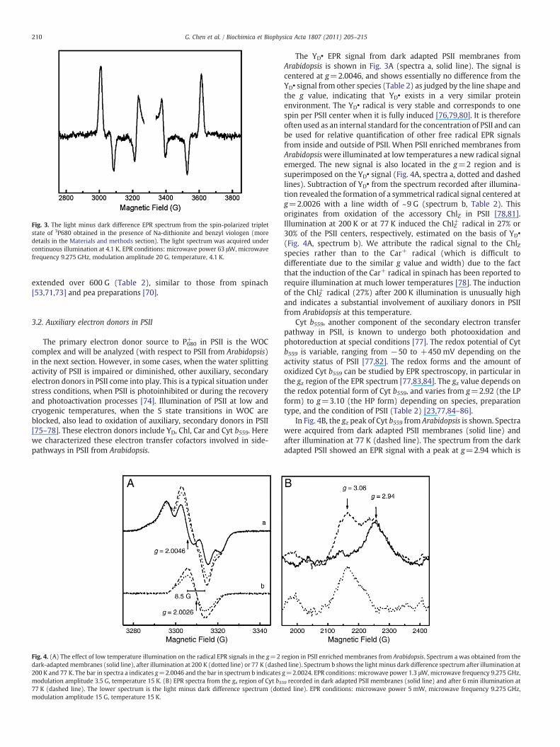
Fig. 3. The light minus dark difference EPR spectrum from the spin-polarized tripletstate of 3P680 obtained in the presence of Na-dithionite and benzyl viologen (moredetails in the Materials and methods section). The light spectrum was acquired undercontinuous illumination at 4.1 K. EPR conditions: microwave power 63 μW, microwavefrequency 9.275 GHz, modulation amplitude 20 G, temperature, 4.1 K.
210 G. Chen et al. / Biochimica et Biophysica Acta 1807 (2011) 205–215
extended over 600 G (Table 2), similar to those from spinach[53,71,73] and pea preparations [70].
3.2. Auxiliary electron donors in PSII
The primary electron donor source to P680+ in PSII is the WOCcomplex and will be analyzed (with respect to PSII from Arabidopsis)in the next section. However, in some cases, when the water splittingactivity of PSII is impaired or diminished, other auxiliary, secondaryelectron donors in PSII come into play. This is a typical situation understress conditions, when PSII is photoinhibited or during the recoveryand photoactivation processes [74]. Illumination of PSII at low andcryogenic temperatures, when the S state transitions in WOC areblocked, also lead to oxidation of auxiliary, secondary donors in PSII[75–78]. These electron donors include YD, Chl, Car and Cyt b559. Herewe characterized these electron transfer cofactors involved in side-pathways in PSII from Arabidopsis.
Fig. 4. (A) The effect of low temperature illumination on the radical EPR signals in the g=2 rdark-adaptedmembranes (solid line), after illumination at 200 K (dotted line) or 77 K (dashe200 K and 77 K. The bar in spectra a indicates g=2.0046 and the bar in spectrum b indicatesmodulation amplitude 3.5 G, temperature 15 K. (B) EPR spectra from the gz region of Cyt b5577 K (dashed line). The lower spectrum is the light minus dark difference spectrum (dotmodulation amplitude 15 G, temperature 15 K.
The YD• EPR signal from dark adapted PSII membranes fromArabidopsis is shown in Fig. 3A (spectra a, solid line). The signal iscentered at g=2.0046, and shows essentially no difference from theYD• signal from other species (Table 2) as judged by the line shape andthe g value, indicating that YD• exists in a very similar proteinenvironment. The YD• radical is very stable and corresponds to onespin per PSII center when it is fully induced [76,79,80]. It is thereforeoften used as an internal standard for the concentration of PSII and canbe used for relative quantification of other free radical EPR signalsfrom inside and outside of PSII. When PSII enriched membranes fromArabidopsiswere illuminated at low temperatures a new radical signalemerged. The new signal is also located in the g=2 region and issuperimposed on the YD• signal (Fig. 4A, spectra a, dotted and dashedlines). Subtraction of YD• from the spectrum recorded after illumina-tion revealed the formation of a symmetrical radical signal centered atg=2.0026 with a line width of ~9 G (spectrum b, Table 2). Thisoriginates from oxidation of the accessory ChlZ in PSII [78,81].Illumination at 200 K or at 77 K induced the ChlZ+ radical in 27% or30% of the PSII centers, respectively, estimated on the basis of YD•
(Fig. 4A, spectrum b). We attribute the radical signal to the ChlZspecies rather than to the Car+ radical (which is difficult todifferentiate due to the similar g value and width) due to the factthat the induction of the Car+ radical in spinach has been reported torequire illumination at much lower temperatures [78]. The inductionof the ChlZ+ radical (27%) after 200 K illumination is unusually highand indicates a substantial involvement of auxiliary donors in PSIIfrom Arabidopsis at this temperature.
Cyt b559, another component of the secondary electron transferpathway in PSII, is known to undergo both photooxidation andphotoreduction at special conditions [77]. The redox potential of Cytb559 is variable, ranging from −50 to +450 mV depending on theactivity status of PSII [77,82]. The redox forms and the amount ofoxidized Cyt b559 can be studied by EPR spectroscopy, in particular inthe gz region of the EPR spectrum [77,83,84]. The gz value depends onthe redox potential form of Cyt b559, and varies from g=2.92 (the LPform) to g=3.10 (the HP form) depending on species, preparationtype, and the condition of PSII (Table 2) [23,77,84–86].
In Fig. 4B, the gz peak of Cyt b559 from Arabidopsis is shown. Spectrawere acquired from dark adapted PSII membranes (solid line) andafter illumination at 77 K (dashed line). The spectrum from the darkadapted PSII showed an EPR signal with a peak at g=2.94 which is
egion in PSII enriched membranes from Arabidopsis. Spectrum a was obtained from thed line). Spectrum b shows the lightminus dark difference spectrum after illumination atg=2.0024. EPR conditions: microwave power 1.3 μW,microwave frequency 9.275 GHz,9 recorded in dark adapted PSII membranes (solid line) and after 6 min illumination atted line). EPR conditions: microwave power 5 mW, microwave frequency 9.275 GHz,
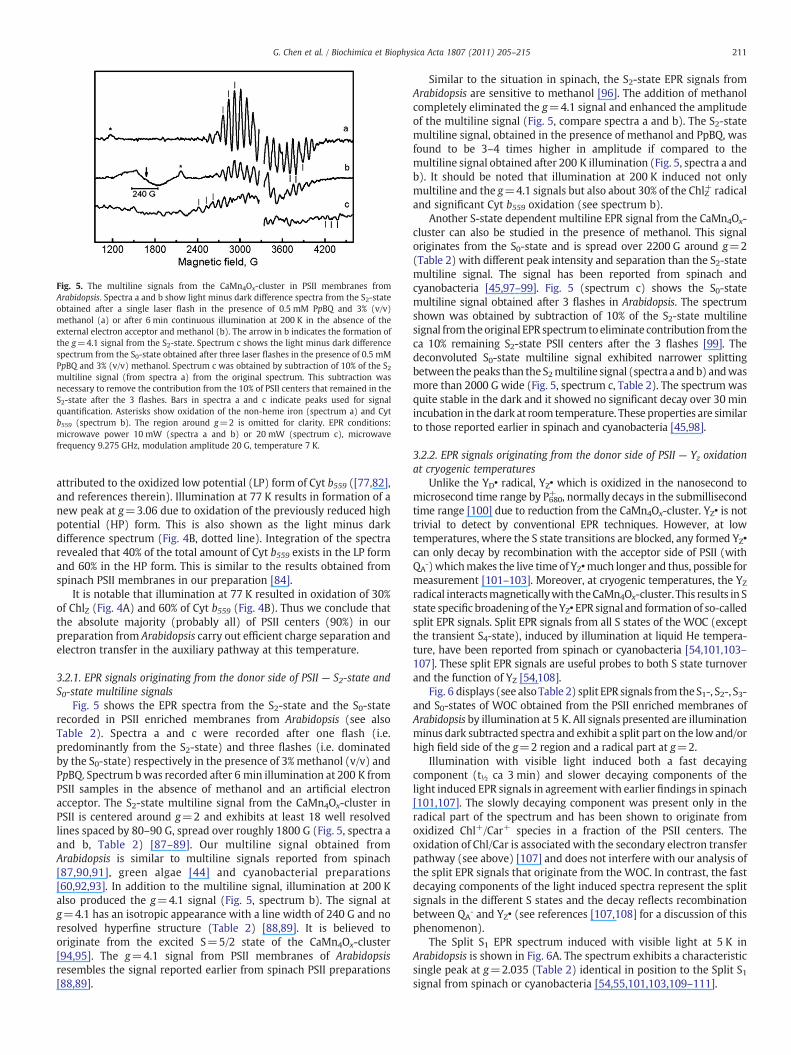
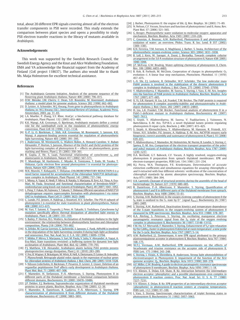
Fig. 5. The multiline signals from the CaMn4Ox-cluster in PSII membranes fromArabidopsis. Spectra a and b show light minus dark difference spectra from the S2-stateobtained after a single laser flash in the presence of 0.5 mM PpBQ and 3% (v/v)methanol (a) or after 6 min continuous illumination at 200 K in the absence of theexternal electron acceptor and methanol (b). The arrow in b indicates the formation ofthe g=4.1 signal from the S2-state. Spectrum c shows the light minus dark differencespectrum from the S0-state obtained after three laser flashes in the presence of 0.5 mMPpBQ and 3% (v/v) methanol. Spectrum c was obtained by subtraction of 10% of the S2multiline signal (from spectra a) from the original spectrum. This subtraction wasnecessary to remove the contribution from the 10% of PSII centers that remained in theS2-state after the 3 flashes. Bars in spectra a and c indicate peaks used for signalquantification. Asterisks show oxidation of the non-heme iron (spectrum a) and Cytb559 (spectrum b). The region around g=2 is omitted for clarity. EPR conditions:microwave power 10 mW (spectra a and b) or 20 mW (spectrum c), microwavefrequency 9.275 GHz, modulation amplitude 20 G, temperature 7 K.
211G. Chen et al. / Biochimica et Biophysica Acta 1807 (2011) 205–215
attributed to the oxidized low potential (LP) form of Cyt b559 ([77,82],and references therein). Illumination at 77 K results in formation of anew peak at g=3.06 due to oxidation of the previously reduced highpotential (HP) form. This is also shown as the light minus darkdifference spectrum (Fig. 4B, dotted line). Integration of the spectrarevealed that 40% of the total amount of Cyt b559 exists in the LP formand 60% in the HP form. This is similar to the results obtained fromspinach PSII membranes in our preparation [84].
It is notable that illumination at 77 K resulted in oxidation of 30%of ChlZ (Fig. 4A) and 60% of Cyt b559 (Fig. 4B). Thus we conclude thatthe absolute majority (probably all) of PSII centers (90%) in ourpreparation from Arabidopsis carry out efficient charge separation andelectron transfer in the auxiliary pathway at this temperature.
3.2.1. EPR signals originating from the donor side of PSII — S2-state andS0-state multiline signals
Fig. 5 shows the EPR spectra from the S2-state and the S0-staterecorded in PSII enriched membranes from Arabidopsis (see alsoTable 2). Spectra a and c were recorded after one flash (i.e.predominantly from the S2-state) and three flashes (i.e. dominatedby the S0-state) respectively in the presence of 3% methanol (v/v) andPpBQ. Spectrum bwas recorded after 6 min illumination at 200 K fromPSII samples in the absence of methanol and an artificial electronacceptor. The S2-state multiline signal from the CaMn4Ox-cluster inPSII is centered around g=2 and exhibits at least 18 well resolvedlines spaced by 80–90 G, spread over roughly 1800 G (Fig. 5, spectra aand b, Table 2) [87–89]. Our multiline signal obtained fromArabidopsis is similar to multiline signals reported from spinach[87,90,91], green algae [44] and cyanobacterial preparations[60,92,93]. In addition to the multiline signal, illumination at 200 Kalso produced the g=4.1 signal (Fig. 5, spectrum b). The signal atg=4.1 has an isotropic appearance with a line width of 240 G and noresolved hyperfine structure (Table 2) [88,89]. It is believed tooriginate from the excited S=5/2 state of the CaMn4Ox-cluster[94,95]. The g=4.1 signal from PSII membranes of Arabidopsisresembles the signal reported earlier from spinach PSII preparations[88,89].
Similar to the situation in spinach, the S2-state EPR signals fromArabidopsis are sensitive to methanol [96]. The addition of methanolcompletely eliminated the g=4.1 signal and enhanced the amplitudeof the multiline signal (Fig. 5, compare spectra a and b). The S2-statemultiline signal, obtained in the presence of methanol and PpBQ, wasfound to be 3–4 times higher in amplitude if compared to themultiline signal obtained after 200 K illumination (Fig. 5, spectra a andb). It should be noted that illumination at 200 K induced not onlymultiline and the g=4.1 signals but also about 30% of the ChlZ+ radicaland significant Cyt b559 oxidation (see spectrum b).
Another S-state dependent multiline EPR signal from the CaMn4Ox-cluster can also be studied in the presence of methanol. This signaloriginates from the S0-state and is spread over 2200 G around g=2(Table 2) with different peak intensity and separation than the S2-statemultiline signal. The signal has been reported from spinach andcyanobacteria [45,97–99]. Fig. 5 (spectrum c) shows the S0-statemultiline signal obtained after 3 flashes in Arabidopsis. The spectrumshown was obtained by subtraction of 10% of the S2-state multilinesignal from theoriginal EPR spectrum toeliminate contribution fromtheca 10% remaining S2-state PSII centers after the 3 flashes [99]. Thedeconvoluted S0-state multiline signal exhibited narrower splittingbetween the peaks than theS2multiline signal (spectra a andb) andwasmore than 2000 G wide (Fig. 5, spectrum c, Table 2). The spectrum wasquite stable in the dark and it showed no significant decay over 30 minincubation in the dark at room temperature. These properties are similarto those reported earlier in spinach and cyanobacteria [45,98].
3.2.2. EPR signals originating from the donor side of PSII — Yz oxidationat cryogenic temperatures
Unlike the YD• radical, YZ• which is oxidized in the nanosecond tomicrosecond time range by P680+ , normally decays in the submillisecondtime range [100] due to reduction from the CaMn4Ox-cluster. YZ• is nottrivial to detect by conventional EPR techniques. However, at lowtemperatures, where the S state transitions are blocked, any formed YZ•
can only decay by recombination with the acceptor side of PSII (withQAˉ)whichmakes the live time of YZ•much longer and thus, possible formeasurement [101–103]. Moreover, at cryogenic temperatures, the YZ
radical interactsmagneticallywith theCaMn4Ox-cluster. This results in Sstate specific broadening of theYZ• EPR signal and formation of so-calledsplit EPR signals. Split EPR signals from all S states of the WOC (exceptthe transient S4-state), induced by illumination at liquid He tempera-ture, have been reported from spinach or cyanobacteria [54,101,103–107]. These split EPR signals are useful probes to both S state turnoverand the function of YZ [54,108].
Fig. 6 displays (see also Table 2) split EPR signals from the S1-, S2-, S3-and S0-states of WOC obtained from the PSII enriched membranes ofArabidopsis by illumination at 5 K. All signals presented are illuminationminus dark subtracted spectra and exhibit a split part on the low and/orhigh field side of the g=2 region and a radical part at g=2.
Illumination with visible light induced both a fast decayingcomponent (t½ ca 3 min) and slower decaying components of thelight induced EPR signals in agreement with earlier findings in spinach[101,107]. The slowly decaying component was present only in theradical part of the spectrum and has been shown to originate fromoxidized Chl+/Car+ species in a fraction of the PSII centers. Theoxidation of Chl/Car is associated with the secondary electron transferpathway (see above) [107] and does not interfere with our analysis ofthe split EPR signals that originate from the WOC. In contrast, the fastdecaying components of the light induced spectra represent the splitsignals in the different S states and the decay reflects recombinationbetween QAˉ and YZ• (see references [107,108] for a discussion of thisphenomenon).
The Split S1 EPR spectrum induced with visible light at 5 K inArabidopsis is shown in Fig. 6A. The spectrum exhibits a characteristicsingle peak at g=2.035 (Table 2) identical in position to the Split S1signal from spinach or cyanobacteria [54,55,101,103,109–111].

Fig. 6. Induction of the metallo-radical (YZ•) split EPR signals in different S states of the WOC in Arabidopsis by continuous white light illumination for 30 s at 5 K. The spectra shownare the difference spectra between spectra recorded during illumination and spectra recorded before illumination from the PSII membranes poised in the S1-state (Split S1 signal, A),in the S2-state (Split S2 signal, B), in the S3-state (Split S3 signal, C) and in the S0-state (Split S0 signal, D). The spectra in grey color (B) were induced in the presence of 3% (v/v)methanol and (C) by illumination by NIR light at 830 nm for 20 min. The region around g=2 is omitted for clarity. The insets show the entire spectra including the g=2 region. EPRconditions: microwave power 20 mW or 50 mW (for Split S2 only), microwave frequency 9.275 GHz, modulation amplitude 15 G, temperature 5 K.
212 G. Chen et al. / Biochimica et Biophysica Acta 1807 (2011) 205–215
The Split S2 EPR signal was reported to be induced either in thepresence of methanol at 10 K (note that the presence of methanolinhibits the Split S1 and split S3 signals and modifies the Split S0 signal[55,112] or by illumination at higher temperatures (77–190 K) [55].Wewere able to trap both of the Split S2 signals inArabidopsis as shownin Fig. 6B. Illumination at 200 K and continuous illumination duringlowering the temperature to 5 K allowed us to trap a complicatedinteraction signal, different from that reported in spinach [55]. Thissignal consists of a symmetrical 130 G wide split feature and anadditional peak at g=2.055 (Fig. 6B, black line). The symmetrical partis wider than that reported in spinach PSII membranes (116 G) wherethe additional low field peakwas not reported [55]. Illumination of thePSII samples in the presence of methanol (3% v/v) at 5 K induced a140 G wide symmetrical split signal (Table 2) also with a possible lowfield peak (Fig. 6B, grey line). The symmetrical part now was found tobe narrower than spinach signal (160 G in presence ofmethanol, [55]).
The Split S3 signal is formed in PSII samples exposed to 2 flashes(dominated by the S3-state) by illumination both with visible light[104,107] and near infrared light [104,113] at cryogenic temperatures.In contrast to the signal induced by visible light, the near infraredinduced signal is very stable at 5 K and shows no observable decay forat least 30 min [104]. Fig. 6C displays the Split S3 signals induced byvisible light (black line) and 830 nm light (grey line) at 5 K inArabidopsis. Both signals are very similar to the corresponding signals
reported from spinach [107,109,113] and cyanobacteria [114]. Thesignal has a double trough at g=1.95 and 1.93 position and a peak(for the near infrared induced signal) at g=2.06 (Table 2). It is alsoclear that the central part of the signal induced by the near infraredlight is smaller which reflects that it lacks any contributions from theChl+/Car+ radicals (Fig. 6C, inset).
The light induced Split S0 signal obtained from PSIImembranes after3 laser flashes (sample dominated by S0-state) is shown in Fig. 6D.Illumination at 5 K gave rise to a broad symmetrical signal separated by165 G (Table 2). The shape and size of this signal resemble the ca 160 Gwide signal previously assigned to the S0YZ• state in spinach [101] andcyanobacteria [114].
Thus, the split EPR signals from theWOC inArabidopsiswere inducedwith high yield and precision. Taken together with themultiline signalsthese EPR probes provide powerful tools to study the WOC in differentmutants available in Arabidopsis.
4. Conclusions
A complete register of perpendicular mode X-band EPR signalsfrom the donor and acceptor sides of PSII, as well as from the auxiliaryelectron donors, is reported from the PSII enriched membranesisolated from Arabidopsis plants. The EPR signals were obtained inhigh yieldwith precision, allowing also quantitativemeasurements. In
213G. Chen et al. / Biochimica et Biophysica Acta 1807 (2011) 205–215
total, about 20 different EPR signals covering almost all of the electrontransfer components in PSII were recorded. This study extends thecomparison between plant species and opens a possibility to studyPSII electron transfer reactions in the library of mutants available inArabidopsis.
Acknowledgements
This work was supported by the Swedish Research Council, theSwedish Energy Agency and the Knut and AliceWallenberg Foundation.EMA and YA acknowledge the financial support from the Academy ofFinland (CoE project 118637). The authors also would like to thankMs. Maija Holmstrom for excellent technical assistance.
References
[1] The Arabidopsis Genome Initiative, Analysis of the genome sequence of theflowering plant Arabidopsis thaliana, Nature 408 (2000) 796–815.
[2] D.W. Meinke, J.M. Cherry, C. Dean, S.D. Rounsley, M. Koornneef, Arabidopsisthaliana: a model plant for genome analysis, Science 282 (1998) 662–682.
[3] D. Leister, A. Schneider, W.J. Kwang, From gene to photosynthesis in Arabidopsisthaliana, in: W.J. Kwang (Ed.), International Review of Cytology, Academic Press,Amsterdam, 2003, pp. 31–83.
[4] L.A. Mueller, P. Zhang, S.Y. Rhee, AraCyc: a biochemical pathway database forArabidopsis, Plant Physiol. 132 (2003) 453–460.
[5] K.K. Niyogi, A.R. Grossman, O. Bjorkman, Arabidopsis mutants define a centralrole for the xanthophyll cycle in the regulation of photosynthetic energyconversion, Plant Cell 10 (1998) 1121–1134.
[6] X.-P. Li, O. Bjorkman, C. Shih, A.R. Grossman, M. Rosenquist, S. Jansson, K.K.Niyogi, A pigment-binding protein essential for regulation of photosyntheticlight harvesting, Nature 403 (2000) 391–395.
[7] J. Andersson, M. Wentworth, G. Walters Robin, A. Howard Caroline, V. RubanAlexander, P. Horton, S. Jansson, Absence of the Lhcb1 and Lhcb2 proteins of thelight-harvesting complex of photosystem II — effects on photosynthesis, granastacking and fitness, Plant J. 35 (2003) 350–361.
[8] R. Gupta, Z. He, S. Luan, Functional relationship of cytochrome c6 andplastocyanin in Arabidopsis, Nature 417 (2002) 567–571.
[9] Y. Munekage, M. Hashimoto, C. Miyake, K. Tomizawa, T. Endo, M. Tasaka, T.Shikanai, Cyclic electron flow around photosystem I is essential for photosyn-thesis, Nature 429 (2004) 579–582.
[10] M.K. Munshi, Y. Kobayashi, T. Shikanai, CHLORORESPIRATORY REDUCTION 6 is anovel factor required for accumulation of the chloroplast NAD(P)H dehydroge-nase complex in Arabidopsis, Plant Physiol. 141 (2006) 737–744.
[11] L. Minna, A. Yagut, K. Heidi, P. Mirva, B. Natalia, S. Marjaana, R. Eevi, A.S. Tiina, A. Eva-Mari, M. Paula, Structural and functional characterization of ferredoxin-NADP+-oxidoreductase using knock-outmutants ofArabidopsis, Plant J. 49 (2007)1041–1052.
[12] L. Peng, Y. Fukao,M. Fujiwara, T. Takami, T. Shikanai, Efficient operation of NAD(P)Hdehydrogenase requires supercomplex formation with photosystem I via minorLHCI in Arabidopsis, Plant Cell 21 (2009) 3623–3640.
[13] C. Lunde, P.E. Jensen, A. Haldrup, J. Knoetzel, H.V. Scheller, The PSI-H subunit ofphotosystem I is essential for state transitions in plant photosynthesis, Nature408 (2000) 613–615.
[14] M. Yuri, T. Satomi, E. Tsuyoshi, J. Peter, H. Takashi, S. Toshiharu, Cytochrome b6fmutation specifically affects thermal dissipation of absorbed light energy inArabidopsis, Plant J. 28 (2001) 351–359.
[15] S. Bailey, P. Horton, R.G. Walters, Acclimation of Arabidopsis thaliana to the lightenvironment: the relationship between photosynthetic function and chloroplastcomposition, Planta 218 (2004) 793–802.
[16] A. Zelisko, M. Garcia-Lorenzo, G. Jackowski, S. Jansson, C. Funk, AtFtsH6 is involvedin the degradation of the light-harvesting complex II during high-light acclimationand senescence, Proc. Nat. Acad. Sci. U. S. A. 102 (2005) 13699–13704.
[17] T. Mikko, P. Mirva, S. Marjaana, S. Sari, M. Paula, V. Julia, V. Alexander, A. Yagut, A.Eva-Mari, State transitions revisited—a buffering system for dynamic low lightacclimation of Arabidopsis, Plant Mol. Biol. 62 (2006) 779–793.
[18] P.J. Matthew, V.R. Alexander, Arabidopsis plants lacking PsbS protein possessphotoprotective energy dissipation, Plant J. 61 (2010) 283–289.
[19] V. Fey,R.Wagner,K. Bräutigam,M.Wirtz,R.Hell, A.Dietzmann,D. Leister,R.Oelmüller,T. Pfannschmidt, Retrograde plastid redox signals in the expression of nuclear genesfor chloroplast proteins of Arabidopsis thaliana, J. Biol. Chem. 280 (2005) 5318–5328.
[20] S. Nilsson Cederholm, H. Bäckman, P. Pesaresi, D. Leister, E. Glaser, Deletion of anorganellar peptidasome PreP affects early development in Arabidopsis thaliana,Plant Mol. Biol. 71 (2009) 497–508.
[21] F. Mamedov, H. Stefansson, P.-Å. Albertsson, S. Styring, Photosystem II indifferent parts of the thylakoid membrane: a functional comparison betweendifferent domains, Biochemistry 39 (2000) 10478–10486.
[22] J.P. Dekker, E.J. Boekema, Supramolecular organization of thylakoid membraneproteins in green plants, Biochim. Biophys. Acta 1706 (2005) 12–39.
[23] F. Mamedov, R. Danielsson, R. Gadjieva, P.-Å. Albertsson, S. Styring, EPRcharacterization of photosystem II from different domains of the thylakoidmembrane, Biochemistry 47 (2008) 3883–3891.
[24] J. Barber, Photosystem II: the engine of life, Q. Rev. Biophys. 36 (2003) 71–89.[25] N. Nelson, C.F. Yocum, Structure and function of photosystems I and II, Annu. Rev.
Plant Biol. 57 (2006) 521–565.[26] G. Renger, Photosynthetic water oxidation to molecular oxygen: apparatus and
mechanism, Biochim. Biophys. Acta 1503 (2001) 210–228.[27] C. Goussias, A. Boussac, A.W. Rutherford, Photosystem II and photosynthetic
oxidation of water: an overview, Philos. Trans. R. Soc. Lond. B 357 (2002)1369–1381.
[28] K.N. Ferreira, T.M. Iverson, K. Maghlaoui, J. Barber, S. Iwata, Architecture of thephotosynthetic oxygen-evolving center, Science 303 (2004) 1831–1838.
[29] B. Loll, J. Kern, W. Saenger, A. Zouni, J. Biesiadka, Towards complete cofactorarrangement in the 3.0 Å resolution structure of photosystem II, Nature 438 (2005)1040–1044.
[30] J.P. McEvoy, G.W. Brudvig, Water-splitting chemistry of photosystem II, Chem.Rev. 106 (2006) 4455–4483.
[31] B. Kok, B. Forbush, M. McGloin, Cooperation of charges in photosynthetic O2
evolution—I. A linear four step mechanism, Photochem. Photobiol. 11 (1970)457–475.
[32] L.-X. Shi, Z.J. Lorkovic, R. Oelmuller, W.P. Schröder, The low molecular massPsbW protein is involved in the stabilization of the dimeric photosystem IIcomplex in Arabidopsis thaliana, J. Biol. Chem. 275 (2000) 37945–37950.
[33] Y. Allahverdiyeva, F. Mamedov, M. Suorsa, S. Styring, I. Vass, E.-M. Aro, Insightsinto the function of PsbR protein in Arabidopsis thaliana, Biochim. Biophys. Acta1767 (2007) 677–685.
[34] X. Yi, S.R. Hargett, H. Liu, L.K. Frankel, T.M. Bricker, The PsbP protein is requiredfor photosystem II complex assembly/stability and photoautotrophy in Arabi-dopsis thaliana, J. Biol. Chem. 282 (2007) 24833–24841.
[35] H. Liu, L.K. Frankel, T.M. Bricker, Functional analysis of photosystem II in aPsbO-1-deficient mutant in Arabidopsis thaliana, Biochemistry 46 (2007)7607–7613.
[36] S. Sirpiö, Y. Allahverdiyeva, M. Suorsa, V. Paakkarinen, J. Vainonen, N.Battchikova, E.-M. Aro, TLP18.3, a novel thylakoid lumen protein regulatingphotosystem II repair cycle, Biochem. J. 406 (2007) 415–425.
[37] S. Sirpiö, A. Khrouchtchova, Y. Allahverdiyeva, M. Hansson, R. Fristedt, A.V.Vener, H.V. Scheller, P.E. Jensen, A. Haldrup, E.-M. Aro, AtCYP38 ensures earlybiogenesis, correct assembly and sustenance of photosystem II, Plant J. 55 (2008)639–651.
[38] Y. Allahverdiyeva, F. Mamedov, M. Holmström, M. Nurmi, B. Lundin, S. Styring, C.Spetea, E.-M. Aro, Comparison of the electron transport properties of the psbo1and psbo2 mutants of Arabidopsis thaliana, Biochim. Biophys. Acta 1787 (2009)1230–1237.
[39] D.A. Berthold, G.T. Babcock, C.F. Yocum, A highly resolved, oxygen-evolvingphotosystem II preparation from spinach thylakoid membranes: EPR andelectron-transport properties, FEBS Lett. 134 (1981) 231–234.
[40] R.J. Porra, W.A. Thompson, P.E. Kriedemann, Determination of accurateextinction coefficients and simultaneous equations for assaying chlorophylls aand b extracted with four different solvents: verification of the concentration ofchlorophyll standards by atomic absorption spectroscopy, Biochim. Biophys.Acta 975 (1989) 384–394.
[41] U.K. Laemmli, Cleavage of structural proteins during the assembly of the head ofbacteriophage T4, Nature 227 (1970) 680–685.
[42] R. Danielsson, P.-Å. Albertsson, F. Mamedov, S. Styring, Quantification ofphotosystem I and II in different parts of the thylakoid membrane from spinach,Biochim. Biophys. Acta 1608 (2004) 53–61.
[43] S. Styring, A.W. Rutherford, In the oxygen-evolving complex of photosystem II theS0 state is oxidized to the S1 state by D+ (signal IIslow), Biochemistry 26 (1987)2401–2405.
[44] S. Styring, A.W. Rutherford, Deactivation kinetics and temperature dependenceof the S-state transitions in the oxygen-evolving system of photosystem IImeasured by EPR spectroscopy, Biochim. Biophys. Acta 933 (1988) 378–387.
[45] K.A. Åhrling, S. Peterson, S. Styring, An oscillating manganese electronparamagnetic resonance signal from the S0 state of the oxygen evolvingcomplex in photosystem II, Biochemistry 36 (1997) 13148–13152.
[46] F.M. Ho, S.F. Morvaridi, F. Mamedov, S. Styring, Enhancement of YD• spin relaxationby the CaMn4 cluster in photosystem II detected at room temperature: a newprobefor the S-cycle, Biochim. Biophys. Acta 1767 (2007) 5–14.
[47] A.W. Rutherford, J.L. Zimmermann, A new EPR signal attributed to the primaryplastosemiquinone acceptor in photosystem II, Biochim. Biophys. Acta 767 (1984)168–175.
[48] W.F.J. Vermaas, A.W. Rutherford, EPR measurements on the effects ofbicarbonate and triazine resistance on the acceptor side of photosystem II,FEBS Lett. 175 (1984) 243–248.
[49] S. Styring, I. Virgin, A. Ehrenberg, B. Andersson, Strong light photoinhibition ofelectrontransport in Photosystem II. Impairment of the function of the firstquinone acceptor, QA, Biochim. Biophys. Acta 1015 (1990) 269–278.
[50] A.F. Miller, G.W. Brudvig, A guide to electron paramagnetic resonance spectroscopyof photosystem II membranes, Biochim. Biophys. Acta 1056 (1991) 1–18.
[51] V.V. Klimov, E. Dolan, E.R. Shaw, B. Ke, Interaction between the intermediaryelectron acceptor (pheophytin) and a possible plastoquinone–iron complex inphotosystem II reaction centers, Proc. Nat. Acad. Sci. U. S. A. 77 (1980)7227–7231.
[52] V.V. Klimov, E. Dolan, B. Ke, EPR properties of an intermediary electron acceptor(pheophytin) in photosystem-II reaction centers at cryogenic temperatures,FEBS Lett. 112 (1980) 97–100.
[53] I. Vass, S. Styring, Spectroscopic characterization of triplet forming states inphotosystem II, Biochemistry 31 (1992) 5957–5963.

214 G. Chen et al. / Biochimica et Biophysica Acta 1807 (2011) 205–215
[54] G. Han, F.M. Ho, K.G.V. Havelius, S.F. Morvaridi, F. Mamedov, S. Styring, Directquantification of the four individual S states in photosystem II using EPRspectroscopy, Biochim. Biophys. Acta 1777 (2008) 496–503.
[55] N. Ioannidis, G. Zahariou, V. Petrouleas, Trapping of the S2 to S3 stateintermediate of the oxygen-evolving complex of photosystem II, Biochemistry45 (2006) 6252–6259.
[56] G. Bernat, F. Morvaridi, Y. Feyziyev, S. Styring, pH dependence of the fourindividual transitions in the catalytic S-cycle during photosynthetic oxygenevolution, Biochemistry 41 (2002) 5830–5843.
[57] H. Dau, Molecular mechanisms and quantitative models of variable photosystemII fluorescence, Photochem. Photobiol. 60 (1994) 1–23.
[58] J.H.A. Nugent, B.A. Diner, M.C.W. Evans, Direct detection of the electron acceptor ofphotosystem II: evidence that Q is an iron–quinone complex, FEBS Lett. 124 (1981)241–244.
[59] A.W. Rutherford, P. Mathis, A relationship between the midpoint potential of theprimary acceptor and low temperature photochemistry in photosystem II, FEBSLett. 154 (1983) 328–334.
[60] F. Mamedov, M.M. Nowaczyk, A. Thapper, M. Rögner, S. Styring, Functionalcharacterization of monomeric photosystem II core preparations from Thermo-synechococcus elongatuswith orwithout the Psb27 protein, Biochemistry 46 (2007)5542–5551.
[61] V. Petrouleas, A.R. Crofts, The iron–quinone accepter complex, in: T.J.Wydrzynski, K. Satoh (Eds.), Photosystem II — The Light-Driven Water:Plastoquinone Oxidoreductase, Springer, The Netherlands, 2005, pp. 177–206.
[62] J.L. Zimmermann, A.W. Rutherford, Photoreductant-induced oxidation of Fe2+ intheelectron-acceptor complexofphotosystemII, Biochim.Biophys. Acta 851 (1986)416–423.
[63] V. Petrouleas, B.A. Diner, Light-induced oxidation of the acceptor-side Fe(II) ofphotosystem II by exogenous quinones acting through the QB binding site. I.Quinones, kinetics and pH-dependence, Biochim. Biophys. Acta 893 (1987)126–137.
[64] B.A. Diner, V. Petrouleas, Q400, the non-heme iron of the photosystem II iron–quinone complex. A spectroscopic probe of quinone and inhibitor binding to thereaction center, Biochim. Biophys. Acta 895 (1987) 107–125.
[65] R. Aasa, L.-E. Andreasson, S. Styring, T. Vänngård, The nature of the Fe(III) EPR signalfrom the acceptor-side iron in photosystem II, FEBS Lett. 243 (1989) 156–160.
[66] V. Petrouleas, B.A. Diner, Identification of Q400, a high-potential electron acceptorof photosystem II, with the iron of the quinone–iron acceptor complex, Biochim.Biophys. Acta 849 (1986) 264–275.
[67] H.A. Frank, Ö. Hansson, P. Mathis, EPR and optical changes of the photosystemII reaction center produced by low temperature illumination, Photosynth. Res.20 (1989) 279–289.
[68] J.H.A. Nugent, A. Telfer, C. Demetriou, J. Barber, Electron transfer in the isolatedphotosystem II reaction centre complex, FEBS Lett. 255 (1989) 53–58.
[69] A.W. Rutherford, D.R. Paterson, J.E. Mullet, A light-induced spin-polarizedtriplet detected by EPR in photosystem II reaction centers, Biochim. Biophys.Acta 635 (1981) 205–214.
[70] A.W. Rutherford, J.E. Mullet, Reaction center triplet states in photosystem I andphotosystem II, Biochim. Biophys. Acta 635 (1981) 225–235.
[71] F.J.E.v. Mieghem, W. Nitschke, P. Mathis, A.W. Rutherford, The influence of thequinone–iron electron acceptor complex on the reaction centre photochemistryof photosystem II, Biochim. Biophys. Acta 977 (1989) 207–214.
[72] A.W. Rutherford, How close is the analogy between the reaction center ofphotosystem-II and that of purple bacteria, Biochem. Soc. Trans. 14 (1986) 15–17.
[73] I. Vass, S. Styring, T. Hundal, A. Koivuniemi, E. Aro, B. Andersson, Reversibleand irreversible intermediates during photoinhibition of photosystem II:stable reduced QA species promote chlorophyll triplet formation, Proc. Nat.Acad. Sci. U. S. A. 89 (1992) 1408–1412.
[74] A. Magnuson, M. Rova, F. Mamedov, P.-O. Fredriksson, S. Styring, The role ofcytochrome b559 and tyrosineD in protection against photoinhibition during invivo photoactivation of photosystem II, Biochim. Biophys. Acta 1411 (1999)180–191.
[75] J.C. De Paula, J.B. Innes, G.W. Brudvig, Electron transfer in photosystem II atcryogenic temperatures, Biochemistry 24 (1985) 8114–8120.
[76] B.A. Barry, G.T. Babcock, Tyrosine radicals are involved in the photosyntheticoxygen-evolving system, Proc. Nat. Acad. Sci. U. S. A. 84 (1987) 7099–7103.
[77] D.H. Stewart, G.W. Brudvig, Cytochrome b559 of photosystem II, Biochim.Biophys. Acta 1367 (1998) 63–87.
[78] J. Hanley, Y. Deligiannakis, A. Pascal, P. Faller, A.W. Rutherford, Carotenoidoxidation in photosystem II, Biochemistry 38 (1999) 8189–8195.
[79] B. Commoner, J.J. Heise, J. Townsend, Light-induced paramagnetism inchloroplasts, Proc. Natl. Acad. Sci. U. S. A. 42 (1956) 710–718.
[80] G.T. Babcock, K. Sauer, Electron paramagnetic resonance signal II in spinachchloroplasts. I. Kinetic analysis for untreated chloroplasts, Biochim. Biophys. Acta(BBA) — Bioenerg. 325 (1973) 483–503.
[81] R. Malkin, A.J. Bearden, Detection of a free radical in the primary reaction ofchloroplast photosystem II, Proc. Nat. Acad. Sci. U. S. A. 70 (1973) 294–297.
[82] O. Kaminskaya, V.A. Shuvalov, G. Renger, Evidence for a novel quinone-bindingsite in the photosystem II (PS II) complex that regulates the redox potential ofcytochrome b559, Biochemistry 46 (2007) 1091–1105.
[83] F.A. Walker, H. Boi Hanh, W.R. Scheidt, S.R. Osvath, Models of the cytochromes b.Effect of axial ligand plane orientation on the EPR and Moessbauer spectra oflow-spin ferrihemes, J. Am. Chem. Soc. 108 (1986) 5288–5297.
[84] L.K. Thompson, A.F. Miller, C.A. Buser, J.C. De Paula, G.W. Brudvig, Characterizationof themultiple forms of cytochrome b559 in photosystem II, Biochemistry 28 (1989)8048–8056.
[85] G.T. Babcock, W.R. Widger, W.A. Cramer, W.A. Oertling, J.G. Metz, Axialligands of chloroplast cytochrome b-559: identification and requirement fora heme-crosslinked polypeptide structure, Biochemistry 24 (1985)3638–3645.
[86] C. Berthomieu, A. Boussac, W. Maentele, J. Breton, E. Nabedryk, Molecularchanges following oxidoreduction of cytochrome b559 characterized by Fouriertransform infrared difference spectroscopy and electron paramagnetic reso-nance: photooxidation in photosystem II and electrochemistry of isolatedcytochrome b559 and iron protoporphyrin IX-bisimidazole model compounds,Biochemistry 31 (1992) 11460–11471.
[87] G.C. Dismukes, Y. Siderer, EPR spectroscopic observations of a manganese centerassociated with water oxidation in spinach chloroplasts, FEBS Lett. 121 (1980)78–80.
[88] J.L. Casey, K. Sauer, EPR detection of a cryogenically photogenerated interme-diate in photosynthetic oxygen evolution, Biochim. Biophys. Acta 767 (1984)21–28.
[89] J.L. Zimmermann, A.W. Rutherford, EPR studies of the oxygen-evolving enzymeof photosystem II, Biochim. Biophys. Acta 767 (1984) 160–167.
[90] G.C. Dismukes, Y. Siderer, Intermediates of a polynuclear manganese centerinvolved inphotosynthetic oxidationofwater, Proc. Nat. Acad. Sci. U. S. A. 78 (1981)274–278.
[91] J.L. Zimmermann, A.W. Rutherford, Electron paramagnetic resonance propertiesof the S2 state of the oxygen-evolving complex of photosystem II, Biochemistry25 (1986) 4609–4615.
[92] A.E. McDermott, V.K. Yachandra, R.D. Guiles, J.L. Cole, S.L. Dexheimer, R.D. Britt, K.Sauer, M.P. Klein, Characterization of the manganese oxygen-evolving complexand the iron–quinone acceptor complex in photosystem II from a thermophiliccyanobacterium by electron paramagnetic resonance and X-ray absorptionspectroscopy, Biochemistry 27 (1988) 4021–4031.
[93] D.L. Kirilovsky, A.G.P. Boussac, F.J.E. Van Mieghem, J.M.R.C. Ducruet, P.R. Setif, J.Yu, W.F.J. Vermaas, A.W. Rutherford, Oxygen-evolving photosystem II prepara-tion from wild-type and photosystem II mutants of Synechocystis Sp. PCC 6803,Biochemistry 31 (1992) 2099–2107.
[94] A. Haddy, W.R. Dunham, R.H. Sands, R. Aasa, Multifrequency EPR investigationsinto the origin of the S2-state signal at g=4 of the O2-evolving complex,Biochim. Biophys. Acta 1099 (1992) 25–34.
[95] A. Haddy, K.V. Lakshmi, G.W. Brudvig, H.A. Frank, Q-band EPR of the S2 state ofphotosystem II confirms an S=5/2 origin of the X-band g=4.1 signal, Biophys. J.87 (2004) 2885–2896.
[96] R.J. Pace, P. Smith, R. Bramley, D. Stehlik, EPR saturation and temperaturedependence studies on signals from the oxygen-evolving centre of photosystemII, Biochim. Biophys. Acta 1058 (1991) 161–170.
[97] J. Messinger, J.H. Nugent, M.C. Evans, Detection of an EPR multiline signal for theS0* state in photosystem II, Biochemistry 36 (1997) 11055–11060.
[98] J. Messinger, J.H. Robblee, W.O. Yu, K. Sauer, V.K. Yachandra, M.P. Klein, The S0state of the oxygen-evolving complex in photosystem II is paramagnetic:detection of an EPR multiline signal, J. Am. Chem. Soc. 119 (1997)11349–11350.
[99] K.A. Åhrling, S. Peterson, S. Styring, The S0 state EPR signal from the Mn cluster inphotosystem II arises froman isolated S=1/2 groundstate, Biochemistry 37(1998)8115–8120.
[100] G. Renger, Coupling of electron and proton transfer in oxidative water cleavagein photosynthesis, Biochim. Biophys. Acta 1655 (2004) 195–204.
[101] C. Zhang, S. Styring, Formation of split electron paramagnetic resonance signalsin photosystem II suggests that tyrosineZ can be photooxidized at 5 K in the S0and S1 states of the oxygen-evolving complex, Biochemistry 42 (2003)8066–8076.
[102] D. Koulougliotis, C. Teutloff, Y. Sanakis, W. Lubitz, V. Petrouleas, The S1YZ•
metalloradical intermediate in photosystem II: an X- and W-band EPR study,Phys. Chem. Chem. Phys. 6 (2004) 4859–4863.
[103] C. Zhang, A. Boussac, A.W. Rutherford, Low-temperature electron transfer inphotosystem II: a tyrosyl radical and semiquinone charge pair, Biochemistry 43(2004) 13787–13795.
[104] N. Ioannidis, J.H.A. Nugent, V. Petrouleas, Intermediates of the S3 state of theoxygen-evolving complex of photosystem II, Biochemistry 41 (2002)9589–9600.
[105] J.H.A. Nugent, I.P. Muhiuddin, M.C.W. Evans, Electron transfer from the wateroxidizing complex at cryogenic temperatures: the S1 to S2 step, Biochemistry 41(2002) 4117–4126.
[106] V. Petrouleas, D. Koulougliotis, N. Ioannidis, Trapping of metalloradicalintermediates of the S-states at liquid helium temperatures. Overview of thephenomenology and mechanistic implications, Biochemistry 44 (2005)6723–6728.
[107] K.G.V. Havelius, J.-H. Su, Y. Feyziyev, F. Mamedov, S. Styring, Spectral resolutionof the split EPR signals induced by illumination at 5 K from the S1, S3, and S0states in photosystem II, Biochemistry 45 (2006) 9279–9290.
[108] K.G.V. Havelius, J. Sjoholm, F.M. Ho, F. Mamedov, S. Styring, Metalloradical EPRsignals from the YZ·S-state intermediates in photosystem II Appl, Magn. Reson.37 (2010) 151–176.
[109] J.-H. Su, K.G.V. Havelius, F. Mamedov, F.M. Ho, S. Styring, Split EPR signalsfrom photosystem II are modified by methanol, reflecting S state-dependentbinding and alterations in the magnetic coupling in the CaMn4 cluster,Biochemistry 45 (2006) 7617–7627.
[110] K.G.V. Havelius, S. Styring, pH dependent competition between YZ and YD inphotosystem II probed by illumination at 5 K, Biochemistry 46 (2007)7865–7874.

215G. Chen et al. / Biochimica et Biophysica Acta 1807 (2011) 205–215
[111] J.-H. Su, K.G.V. Havelius, F.M. Ho, G. Han, F. Mamedov, S. Styring, Formationspectra of the EPR split signals from the S0, S1, and S3 states in photosystem IIinduced by monochromatic light at 5 K, Biochemistry 46 (2007) 10703–10712.
[112] N. Ioannidis, G. Zahariou, V. Petrouleas, The EPR spectrum of tyrosine Z•• and itsdecay kinetics in O2-evolving photosystem II preparations, Biochemistry 47 (2008)6292–6300.
[113] N. Ioannidis, V. Petrouleas, Electron paramagnetic resonance signals from the S3state of the oxygen-evolving complex. A broadened radical signal induced by low-temperature near-infrared light illumination, Biochemistry 39 (2000) 5246–5254.
[114] A. Boussac, M. Sugiura, T.L. Lai, A.W. Rutherford, Low-temperature photochem-istry in photosystem II from Thermosynechococcus elongatus induced by visibleand near-infrared light, Philos. Trans. R. Soc. B Biol. Sci. 363 (2008) 1203–1210.